Predicting ischaemic stroke subtype from presenting systolic blood pressure: the BASIC Project

© 2015 Anakwue et al. This work is published by Dove Medical Press Limited, and licensed under Creative Commons Attribution – Non Commercial (unported, v3.0) License. The full terms of the License are available at http://creativecommons.org/licenses/by-nc/3.0/. Non-commercial uses of the work are permitted without any further
permission from Dove Medical Press Limited, provided the work is properly attributed. Permissions beyond the scope of the License are administered by Dove Medical Press Limited. Information on how to request permission may be found at: http://www.dovepress.com/permissions.php
Therapeutics and Clinical Risk Management 2015:11 189–200
Therapeutics and Clinical Risk Management Dovepress
submit your manuscript | www.dovepress.com
Dovepress 189
O R i g i n a l R e s e a R C h
open access to scientific and medical research
Open access Full Text article
http://dx.doi.org/10.2147/TCRM.S68752
echocardiographic assessment of left ventricular function in thyrotoxicosis and implications for the therapeutics of thyrotoxic cardiac disease
Correspondence: RC anakwueDepartment of Pharmacology and Therapeutics, College of Medicine, University of nigeria, enugu Campus, ituku Ozalla, PMB 01129, enugu state, nigeriaTel +234 803 334 3044email [email protected]
Raphael C anakwue1,2
Basden J Onwubere2
Vincent ikeh2
Benedict anisiuba2
samuel ike2
angel-Mary C anakwue3
1Department of Pharmacology and Therapeutics, 2Department of Medicine, 3Department of Radiography and Radiological sciences, imaging Unit, College of Medicine, University of nigeria, nsukka, enugu state, nigeria
Introduction: Thyrotoxicosis is an endocrine disorder with prominent cardiovascular
manifestations. Thyroid hormone acts through genomic and non-genomic mechanisms to regulate
cardiac function. Echocardiography is a useful, non-invasive, easily accessible, and affordable
tool for studying the structural and physiological function of the heart.
Aim: We studied thyrotoxicosis patients in a Nigerian Teaching Hospital and employed
trans-thoracic echocardiography to find out if there were abnormalities in the hearts of these
patients.
Methods: Fifty adult thyrotoxicosis patients diagnosed with clinical and thyroid function tests
in the medical out-patient unit of the hospital were recruited and we performed transthoracic
echocardiography with a Sonos 2000 HP machine.
Results: We documented the presence of abnormalities in the following proportion of thyrotoxi-
cosis patients: left ventricular enhanced systolic function in 30%, enhanced diastolic function in
34%, diastolic dysfunction in 34%, heart failure with preserved ejection fraction in10%, heart
failure with reduced ejection fraction in 6%, and left ventricular hypertrophy in 34%.
Conclusion: Echocardiography was useful in the stratification of cardiac function abnormalities
and is indispensable as a guide in the choice of therapeutic options in patients with thyrocar-
diac disease. The finding of left ventricular enhanced systolic and diastolic functions signify
early echocardiographic detectable cardiac abnormalities in thyrotoxicosis, and the clinical
management includes the use of anti-thyroid drugs and β-adrenoceptor blockade. Diastolic
dysfunction in thyrotoxicosis patients asymptomatic for cardiac disease should be treated with
anti-thyroid drugs, and β-adrenoceptor blockade. The judicious application of clinical thera-
peutics will guide the use of anti-thyroid drugs, diuretics, digoxin, angiotensin inhibitors, and
β-adrenoceptor blockade in the successful management of thyrotoxicosis patients with heart
failure and reduced, preserved, or increased ejection fraction: parameters which are derived
from echocardiography.
Keywords: thyrotoxicosis, left ventricle, echocardiography, therapeutics, thyrocardiac
disease
IntroductionThyrotoxicosis is the syndrome resulting from an excess of circulating free thyroxine
and free triiodothyronine.1 When thyrotoxicosis is associated with thyroid gland over-
activity, hyperthyroidism is said to occur. However, thyrotoxicosis can occur without
hyperthyroidism when stored hormone is released from a damaged thyroid gland
(eg, sub-acute thyroiditis, post-partum thyroiditis, amiodarone-induced thyroiditis) or
when excess thyroid hormone is taken.1 In 1935 Robert Graves, an Irishman, and in
1940 Karl Adolph van Basedow, a German, separately described Basedow–Graves’
Journal name: Therapeutics and Clinical Risk ManagementArticle Designation: Original ResearchYear: 2015Volume: 11Running head verso: Anakwue et alRunning head recto: Echocardiographic assessment of left ventricular function in thyrotoxicosisDOI: 68752

Therapeutics and Clinical Risk Management 2015:11submit your manuscript | www.dovepress.com
Dovepress
Dovepress
190
anakwue et al
or Graves’ disease which is now responsible for 70%–80%
of all cases of hyperthyroidism.2
Thyrotoxicosis affects the normal functioning of many tis-
sues including their growth, differentiation, metabolism, and
oxygen consumption. It affects the cardiovascular system pro-
foundly. The cardiovascular manifestations of thyrotoxicosis
are due to direct cellular effects of thyroid hormones on the
heart and indirect cellular effects resulting from interactions
with the sympathetic nervous system, alterations in periph-
eral vascular smooth muscle (VSM), rennin–angiotensin–
aldosterone system, and erythropoietin production.3
Thyroid hormone modulates cardiac function through
regulation of the expression of some structural and regula-
tory genes. Within the myocardial cells are the fast α-myosin
heavy chain and the slow β-myosin heavy chain which mediate
contraction. Thyroid hormone upregulates α-gene which has
higher ATPase activity and contractile properties and decreases
the expression of β-gene with lower contractile properties.
Thyroid hormone also upregulates the rate of Ca2+ release and
reuptake by the sarcoplasmic reticulum and downregulates the
inhibitor, phospholamban. The increase in cytosolic calcium
increases systolic contraction and the more rapid calcium
reuptake enhances diastolic relaxation in the heart.4–6
The sodium potassium ATPase, the voltage-gated potas-
sium channels, and the sodium calcium exchanger are ion
channels that are activated in thyrotoxicosis and they coor-
dinate the electrochemical responses of the myocardium
during cardiac contraction and relaxation.6–8 The β-adrenergic
receptors are thought to be stimulated in thyrotoxicosis,
leading to an increase in the intracellular second messenger,
cAMP, which in turn accelerates diastolic depolarization and
increases heart rate. The natriuretic peptides are secreted by
cardiac myocytes9 and are said to be upregulated by thyroid
hormones. The pacemaker-related genes, hyperpolarization-
activated cyclic nucleotide-gated channels 2 and 4, are also
transcriptionally regulated by thyroid hormone.10,11
It has been suggested that hyperthyroidism resembles a
hyperadrenergic state; however, there is no evidence that thy-
roid hormone excess enhances the sensitivity of the heart to
adrenergic stimulation.11–13 Indeed, the role of the sympathetic
nervous system in the pathophysiology of hyperthyroidism
is unclear.12 The clinical spectrum of symptoms in thyrotoxi-
cosis suggests a hyperadrenergic state strengthened by the
fact that the administration of β-adrenoceptor antagonists
dramatically ameliorates the clinical state.14–16 In spite of this
known fact, the concentrations of catecholamines in both
plasma17 and urine18 are normal or low in hyperthyroidism.
Thyrotoxicosis increases endothelial nitric oxide produc-
tion via the triiodothyronine (T3)-mediated effects of thyroid
receptor on the protein kinase pathway.19–21 Nitric oxide syn-
thesized in endothelial cells then acts in a paracrine manner
on adjacent VSM cells to facilitate vascular relaxation. Relax-
ation of VSM leads to decreased peripheral resistance and
pressure, increased blood return to the heart, and increased
blood volume which increases cardiac output (CO). Increased
vascularity and angiogenesis reported in thyrotoxicosis may
also lead to increased blood volume and CO.22
The other factors that increase CO include activation
of renin–angiotensin–aldosterone system, increased red
cell mass, as well as increased blood volume: all these
contribute to systemic hypertension seen in thyrotoxicosis
patients.23 Hypertension contributes in causing ventricular
hypertrophy and myocardial remodeling in patients with
thyrotoxicosis.23 Diastolic and mean arterial blood pres-
sures are reduced because of peripheral vasodilatation that
occurs in thyrotoxicosis.23 Pulmonary arterial hypertension
also occurs in hyperthyroidism and it has been attributed to
pulmonary vascular endothelial dysfunction, damage due
to autoimmune process, increased metabolism of intrinsic
pulmonary vasodilators, and high CO state.24 In some cases,
pulmonary hypertension may result in right heart failure.24
Another effect of thyrotoxicosis on the vasculature is its
anti-atherosclerotic effects through blood vessel dilatation and
production of vasodilatory molecules. In contrast, in people
with hypothyroidism, atherosclerosis has been attributed to
hypercholesterolemia, hypertension, and impaired endothelial
function, leading to increased cardiovascular risk. Treatment
of hypothyroidism with thyroid hormone replacement restores
euthyroidism and reverses the associated risk ratio.25
Echocardiography is a very useful, non-invasive, easily
accessible, and perhaps an affordable tool for studying the
structure and physiological function of the heart. Assess-
ment of ventricular function, particularly the left ventricle,
is one of the commonest and most important applications of
echocardiography. Echocardiographic examination of left
ventricular (LV) function is useful in assessment of the effect
of thyrotoxicosis on the heart.
The presence of LV dysfunction is a reliable prognostic
indicator in all forms of cardiac disease. Indeed, echocar-
diographic findings may alter the course of management
and provide opportunities for appropriate therapeutic
intervention.
ObjectiveWe studied patients who had thyrotoxicosis and assessed
its effect on LV function. Echocardiographic study of these
patients was done with a view to stratifying them according
to the pattern of LV dysfunction.

Therapeutics and Clinical Risk Management 2015:11 submit your manuscript | www.dovepress.com
Dovepress
Dovepress
191
echocardiographic assessment of left ventricular function in thyrotoxicosis
MethodsWe recruited 50 subjects with thyrotoxicosis who were
aged 15 years and above and of both sexes, over a period
of 1 year. The patients were recruited consecutively as they
attended the medical out-patient unit of the University of
Nigeria Teaching Hospital, Enugu after clinical and thyroid
function assessments. Written consent was signed by the
subjects and controls. The University of Nigeria Teach-
ing Hospital Ethical committee approved the study and
it was carried out in accordance with the Declaration of
Helsinki ethical principles for medical research involving
human subjects.26
Echocardiography was done with a Sonos 2000 HP
machine (Hewlett Packard, the Netherlands, Amsterdam)
to assess LV systolic and diastolic functions. Transthoracic
echocardiographic examinations were performed in all par-
ticipants with 3.5 MHz transducer according to the recom-
mendations of the American Society of Echocardiography.27
The American Society of Echocardiography recommends
that measurements should be taken from trailing edge to
leading edge. All the measurements were done with the
picture frozen on the screen and with in-built calipers of
the echocardiographic equipment. The mean measurements
were taken from three consecutive cycles. The measure-
ments were initially taken by two examiners. Thereafter,
all the measurements were taken by the same echocar-
diographer. Electrocardiography was also done in all the
participants.
Thyroid function tests were done with kits from Syn-
tron Bioresearch Inc., Carlsbad, CA, USA. These kits had
a correlation coefficient of 0.09869 when compared with a
standard kit made by Abbott Laboratories (North Chicago,
IL, USA). The Syntron kits have the following intra-assay
coefficient of variation: serum-free T3 – (6.8%), thyroid-
stimulating hormone (TSH) – (4.3%), total free T3 – (4.4%),
total tetraiodothyronine – (7.2%).28
Exclusion criteria included patients with:
a) pre-existing hypertension (blood pressure $140/
90 mmHg, or were on antihypertensive drugs before the
onset of thyrotoxicosis);
b) diabetes mellitus (fasting blood glucose .6.1 mmol/L);
c) coronary artery disease (detected by electrocardio graphy);
d) anemia (hemogram ,12 gm/L);
e) chronic alcohol consumption;
f) history of smoking, intake of illicit drugs, intake of
herbal drugs, cardiotoxic drug consumption, pregnant
state.
The controls were 50 age- and sex-matched subjects who
do not have thyrotoxicosis and/or any co-morbidity.
DefinitionsA serum-free T3 .4.2 pg/L and a concomitant suppressed
TSH of .0.5 μU/mL was used to diagnose thyrotoxicosis.
The following parameters were defined as follows:27–32
a) LV systolic dysfunction was present if there was any one
of the following: i) LV ejection fraction ,50%, ii) frac-
tional shortening (FS) ,30%, iii) cardiac index ,2.8 L/
min/m2, iv) CO ,4 L/min, v) mean velocity of circumfer-
ential fiber (VCF) shortening ,1.02 circumferences/sec,
and vi) peak aortic systolic velocity ,72 cm/sec;
b) LV diastolic dysfunction was present if there was any one
of the following: LV E/A (early diastolic velocity/velocity
with atrial contraction) ,1.1, isovolumic relaxation time
.90 msec, E wave deceleration time .210 msec, peak
filling rate ,5);
c) enhanced (meaning increased myocardial contractility)
systolic function33 was present if there was any one of
the following: ejection fraction .75%, FS .42%, CO
.7 L/min, peak aortic systolic velocity .120 cm/sec;
d) enhanced (meaning increased) diastolic function34 was pres-
ent if there was documentation of any one of the following:
E wave velocity .72 cm/sec, A wave velocity .59 cm/sec,
isovolumic relaxation time ,76 msec, E wave deceleration
time ,179 msec in the thyrotoxicosis patients and age- and
sex-matched control subjects;
e) left ventricular mass index (LVMI)27 normal value:
43–96 g/m2 (for women) and 49–116 g/m2 (for men);
f) relative wall thickness27 of .0.43 indicates con-
centric hypertrophy; and ,0.43 indicates eccentric
hypertrophy.
statisticsContinuous variables were expressed as mean ± 1 standard
error of mean. Statistical comparisons were performed using
SPSS software (version 15.0; SPSS Inc., Chicago, IL, USA).
P,0.05 was considered significant.
ResultsThyrotoxicosis patients were leaner, and when their blood
pressure was compared with controls there was no signifi-
cant difference (Table 1). Palpitation was the commonest
symptom, enlarged thyroid gland was present in more than
50% of the patients, and Graves’ disease was documented
in 40% of the subjects (Table 2).
Echocardiographic measurements showed that the mean
intra-observer agreement was 97% (k=0.86) and the mean
inter-observer agreement was 92% (k=0.78). This indicated
that intra-observer and inter-observer variability were
negligible. Out of all 50 patients studied, eight (16%) had

Therapeutics and Clinical Risk Management 2015:11submit your manuscript | www.dovepress.com
Dovepress
Dovepress
192
anakwue et al
Table 1 anthropometric data and blood pressure of patients and controls
Parameters Thyrotoxicosis patients: mean (SD)
Control: mean (SD) Student’s t-test value
P-value
age 44 (3.1) 43.5 (5.2) 0.85 .0.05Weight (kg) 56.0 (5.5) 65 (7.6) -6.4 ,0.05*height (meters) 159.5 (6.8) 160.8 (15.9) 1.35 .0.05Body surface area (m2) 1.2 (2.4) 1.6 (0.17) 1.85 ,0.5*Body mass index (kg/m2) 22.04 (6.4) 25.09 (5.4) -1.87 ,0.05*Pulse (beats/min) 102 (11.9) 78 (5.2) 8.57 ,0.01*systolic blood pressure (mmhg) 124.2 (12.5) 122.9 (2.8) 0.12 .0.05Diastolic blood pressure (mmhg) 75 (11.4) 79.1 (6.5) 1.5 .0.05
Note: *Represents significant value.Abbreviation: sD, standard deviation.
Table 2 Frequencies of symptoms in thyrotoxicosis patients
Symptoms Patients (percentage)
Palpitation 30 (60)enlarged thyroid gland 26 (52)Weight loss 24 (48)heat intolerance 23 (46)Tremulousness 21 (42)Proptosis (graves’ patients) 20 (40)increased sweating 18 (38)Polyphagia 16 (32)
heart failure. Five of these patients had systolic heart failure
with reduced ejection fraction: a typical echocardiogram
is depicted in Figure 1. The other three patients had heart
failure with preserved ejection fraction. No patient had the
so-called high-output heart failure, ie, heart failure with
increased ejection fraction.
The left ventricle was dilated in 28% of the patients. The LV
end diastolic diameter was significantly higher in the thyrotoxico-
sis patients than in the controls (5.35 cm 0.76 standard deviation
[SD] versus [vs] 5.0 cm 0.73 SD – P,0.05) (Table 3). When all
the thyrotoxicosis subjects were compared with control, it showed
that the index patients with thyrotoxicosis had significantly
increased systolic function (P,0.01), supporting the hyperdy-
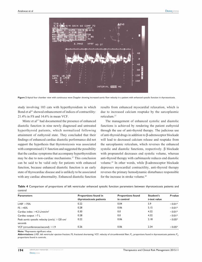
namic clinical state of the disease (Table 3). Enhanced systolic
function depicted in Figure 2 was documented in 14 patients,
and their systolic parameters showed significant difference when
compared with that of the control – P,0.01 (Table 4).
Enhanced diastolic function depicted in Figure 3 was
documented in 17 thyrotoxicosis patients and when param-
eters of diastolic function were compared with control, there
was significant difference – P,0.01 (Table 5) . It is obvi-
ous that some patients had combined enhanced systolic and
enhanced diastolic functions.
Diastolic dysfunction (Figure 4) was seen among
17 patients. Table 6 shows that there was significant
difference between thyrotoxicosis patients and controls when
parameters of diastolic function were assessed – (P,0.01).
The inter-ventricular septal thickness and the posterior
ventricular wall thickness were significantly higher in the thy-
rotoxicosis group than in the control: (0.95 cm vs 0.77 cm –
P,0.05.) and (0.89 cm vs 0.79 cm – P,0.05), respectively.
Four percent of the patients had inter-ventricular septal
thickness .12 mm. The LVMI of subjects (127.03 g/m2)
was also significantly higher than in the control (84.14 g/m2)
at P,0.05, and the relative wall thickness was signifi-
cantly higher in thyrotoxicosis patients than in the control
(0.53 vs 0.33 – P,0.05). In effect, the thyrotoxicosis patients
had eccentric hypertrophy. Figure 5 shows that 17 (34%)
thyrotoxic patients had LV hypertrophy using LVMI. Thirty-
three (66%) thyrotoxic patients had normal LVMI. Only one
(2%) subject in the control had increased LVMI.
Figure 6 summarizes the echocardiographic findings
in the 50 thyrotoxicosis patients studied. Table 7 shows
the multivariate regression analysis between all the
systolic and diastolic parameters and TSH and T3 of the
patients with thyrotoxicosis and control. Both T3 and TSH
models showed a good fit (R2=0.42, P,0.1, and P,0.05,
respectively). T3 retained its independent positive asso-
ciation with the systolic and diastolic parameters. TSH
also retained its independent negative association with the
systolic and diastolic parameters.
DiscussionOur study documented enhanced systolic and diastolic
functions when the subjects were compared with controls
(Tables 4 and 5), and these findings are in keeping with other
reports which documented that patients who have thyrotoxi-
cosis also have increased LV systolic and diastolic contractile
functions, resulting from upregulation of contractile and
calcium-regulatory proteins.35–37

Therapeutics and Clinical Risk Management 2015:11 submit your manuscript | www.dovepress.com
Dovepress
Dovepress
193
echocardiographic assessment of left ventricular function in thyrotoxicosis
Friedman et al38 in the USA had reported enhanced
systolic function by documenting the echocardiographic
tracing of the septum and left posterior wall. Kral et al39 in
Czecholoslavakia studied 12 patients with hyperthyroidism
and documented significant increase in mean VCF as well as
cardiac index, stroke volume, and LV end diastolic volume.
In Poland, Marcisz et al while studying hyperthyroidism,
noted that it was associated with enhanced systolic function.40
They demonstrated increased LV ejection function, FS, mean
VCF, cardiac index and output-pressure index. So our study,
which showed an increase of 18% and 19% in the LV ejec-
tion fraction and FS, respectively, in thyrotoxicosis patients
when compared with control is not a new finding. Enhanced
systolic function has also been documented in an animal
Figure 1 Two-dimensional echocardiogram showing dilated heart chambers – systolic dysfunction.
Table 3 Comparison of parameters of left ventricular systolic function between thyrotoxicosis patients and control
Parameters Mean values for thyrotoxicosis patients (SD) [reference range]
Mean values for control (SD)
Student’s t-test value
P-value
lVeF (%) 69.7 (10.2)[41-79]
59 (3.3) 2.47 ,0.01*
Fs (%) 43.9 (9.8)[12-48]
36.83 (8.4) 2.8 ,0.01*
Cardiac output (l) 7.15 (1.47)[3.5–8.0]
4.7 (0.67) 7.64 ,0.01*
Cardiac index l/min/m2 4.1 (0.84)[2.1-4.8]
3.2 (0.29) 4.13 ,0.01*
Peak aortic systolic velocity (cm/seconds) 144 (26.7)[81-165]
113 (11.5) 7.5 ,0.01*
VCF (circumferences/second) 1.74 (0.34)[1.8-2.1]
1.21 (0.12) 11.0 ,0.01*
end diastolic diameter 5.35 cm (0.76) 5.0 cm (0.73) ,0.01*interventricular septal thickness 0.95 cm (0.12) 0.77 cm (0.7) 0.05*Posterior wall thickness 0.89 cm (0.4) 0.79 cm (0.9) 0.05*left ventricular mass index 127 (32.3) 84 (13.8) 0.05*Relative wall thickness 0.53 (0.15) 0.33 (1.4) 0.05*
Note: *Represents significant value.Abbreviations: LVEF, left ventricular ejection fraction; SD, standard deviation; FS, fractional shortening; VCF, velocity of circumferential fiber.
Therapeutics and Clinical Risk Management 2015:11submit your manuscript | www.dovepress.com
Dovepress
Dovepress
194
anakwue et al
study involving 103 cats with hyperthyroidism in which
Bond et al41 showed enhancement of indices of contractility:
21.4% in FS and 14.6% in mean VCF.
Mintz et al37 had documented the presence of enhanced
diastolic function in nine newly diagnosed and untreated
hyperthyroid patients, which normalized following
attainment of euthyroid state. They concluded that their
findings of enhanced cardiac diastolic performance did not
support the hypothesis that thyrotoxicosis was associated
with compromised LV function and suggested the possibility
that the cardiac symptoms that accompany hyperthyroidism
may be due to non-cardiac mechanisms.37 This conclusion
can be said to be valid only for patients with enhanced
function, because enhanced diastolic function is an early
state of thyrocardiac disease and is unlikely to be associated
with any cardiac abnormality. Enhanced diastolic function
results from enhanced myocardial relaxation, which is
due to increased calcium reuptake by the sarcoplasmic
reticulum.37
The management of enhanced systolic and diastolic
functions is achieved by rendering the patient euthyroid
through the use of anti-thyroid therapy. The judicious use
of anti-thyroid drugs in addition to β-adrenoceptor blockade
will lead to decreased calcium release and reuptake from
the sarcoplasmic reticulum, which reverses the enhanced
systolic and diastolic functions, respectively. β blockade
with propranolol decreases end systolic volume, whereas
anti-thyroid therapy with carbimazole reduces end diastolic
volume.42 In other words, while β-adrenoceptor blockade
depresses myocardial contractility, anti-thyroid therapy
reverses the primary hemodynamic disturbance responsible
for the increase in stroke volume.42
Figure 2 Apical four chamber view with continuous wave Doppler showing increased aortic flow velocity in a patient with enhanced systolic function in thyrotoxicosis.
Table 4 Comparison of proportions of left ventricular enhanced systolic function parameters between thyrotoxicosis patients and control
Parameters Proportions found in thyrotoxicosis patients
Proportions found in control
Student’s t-test value
P-value
lVeF .75% 0.22 0.04 5.9 ,0.01*Fs .45% 0.28 0.06 5.15 ,0.01*Cardiac index .4.2 l/min/m2 0.30 0.0 4.32 ,0.01*Cardiac output .7 l 0.28 0.0 4.32 ,0.01*Peak aortic systolic velocity (cm/s) .120 cm/seconds
0.22 0.06 2.18 ,0.05*
VCF (circumferences/second) .1.9 0.26 0.06 2.34 ,0.05*
Note: *Represents significant value.Abbreviations: LVEF, left ventricular ejection fraction; FS, fractional shortening; VCF, velocity of circumferential fiber; PT, proportions found in thyrotoxicosis patients; PC, proportions found in controls.

Therapeutics and Clinical Risk Management 2015:11 submit your manuscript | www.dovepress.com
Dovepress
Dovepress
195
echocardiographic assessment of left ventricular function in thyrotoxicosis
β-adrenoceptor blockade has been used to modify the
severity of hyperadrenergic symptoms of thyrotoxicosis and
to treat tachycardia. Many β-adrenoceptors have been used,
but propranolol and the longer-acting atenolol are more popu-
lar.43 Propranolol has two roles in the treatment of hyperthy-
roidism, determined by the different isomers of propranolol.
L-propranolol causes β-blockade, thus treating the symptoms
associated with hyperthyroidism such as tremor, palpitations,
anxiety, and heat intolerance.43 D-propranolol inhibits
thyroxine deiodinase, thereby blocking the conversion of
tetraiodothyronine to T3, providing some though minimal
therapeutic effect.44 Atenolol is a selective beta blocker and
so less likely than propranolol to cause bronchoconstriction
in patients who have bronchial disease.44
Impaired diastolic dysfunction was found in at least 28%
of the thyrotoxicosis patients in our study (Figure 4). These
patients with diastolic dysfunction had no symptoms arising
from the cardiovascular system. But it has been documented
that the finding of diastolic dysfunction in an asymptomatic
patient is a risk factor for the future development of heart
failure, and the early identification of such patients provides
a window of opportunity to prevent progression of what
appears to be a preclinical heart disease.45–47
In a study in India, which involved 43 patients with newly
diagnosed hyperthyroidism and 45 healthy participants, Jing
et al48 reported that the patients had impaired LV diastolic
function. Diastolic dysfunction can also occur in sub-clinical
hyperthyroidism. Smit et al in the Netherlands showed
in a randomized placebo-controlled study that reversible
diastolic dysfunction can occur after long-term exogenous
subclinical hyperthyroidism.49 They were concerned that
even isolated diastolic dysfunction may be associated with
increased mortality.49 Yue et al in the People’s Republic of
China also documented that LV diastolic dysfunction can
occur in patients who present with heart failure.50 In that
study, out of the 6% of patients who had heart failure, half of
them had heart failure with preserved ejection fraction. They
concluded that diastolic dysfunction may play an important
Figure 3 apical four chamber view with pulse wave Doppler showing increased mitral e and a wave velocities in keeping with enhanced diastolic function.
Table 5 Comparison of proportions of left ventricular enhanced diastolic function parameters of thyrotoxicosis patients and control
Parameters Proportions of thyrotoxicosis patients
Proportions of control
Student’s t-test value
P-value
Peak early filling velocity (E) .72 cm/s 0.34 0.02 12.77 ,0.01*Peak velocity at atrial contraction (a) .59 cm/s 0.20 0.00 4.32 ,0.01*iVRT ,76 m/sec 0.30 0.04 8.41 ,0.01*eDT ,179 m/sec 0.30 0.00 4.32 ,0.01*
Note: *Represents significant value.Abbreviations: iVRT, isovolumic relaxation time; eDT, e wave deceleration time; e, early diastolic velocity; a, velocity with atrial contraction.

Therapeutics and Clinical Risk Management 2015:11submit your manuscript | www.dovepress.com
Dovepress
Dovepress
196
anakwue et al
role in the pathogenesis of heart failure in thyrotoxicosis.50
We also documented diastolic dysfunction as being a cause
of heart failure with preserved ejection fraction in this study
(Figure 6).
In effect, thyrotoxic heart failure may present with pre-
served, reduced, or increased ejection fraction (high-output
heart failure).32,51 In our earlier study, we reported that 10%
of the patients with thyrotoxicosis had systolic dysfunction
with preserved ejection fraction.52 Similar findings have
been found in other studies.53,54 This is an indication that
echocardiography can be used to classify cardiac dysfunction
in patients with thyrotoxicosis, including the determination
of the ejection fraction, and these data are indispensable in
planning a therapeutic regimen.
Patients who have diastolic dysfunction can only be
detected using echocardiography, and this invariably
influences their management. Therapeutic management of
patients with diastolic dysfunction may differ from those
with systolic dysfunction. Patients with isolated diastolic
dysfunction will respond to anti-thyroid drugs (to render
them euthyroid), β-blockers, and diuretics in moderate
doses.50 Inotropes which are useful in patients with systolic
dysfunction will not be required in patients with isolated
diastolic dysfunction.50
In thyrotoxicosis patients who have heart failure with
preserved or increased ejection fraction, inotropes are of
doubtful therapeutic effect.51 Anti-hyperthyroid drugs, judi-
cious use of diuretics, and some β-adrenoceptor blockers
are more useful in managing this group of patients.55 The
use of vasodilators like angiotensin-converting inhibitors,
angiotensin receptor blockers, and β-blockers with vasodi-
latory properties (eg, carvedilol, nebivolol) may aggravate
the clinical condition in heart failure with increased ejection
fraction because of associated decreased peripheral vascular
resistance.51
Heart failure with reduced ejection fraction may be found
in patients with thyrotoxicosis in spite of the fact that it is
a hypermetabolic condition. Some predisposing conditions
include pre-existing hypertension, ischemic heart disease,
and mitral valve disease, which may be found in Graves’ and
Hashimoto’s diseases.56,57 The pathophysiology of thyrotoxic
cardiac disease with reduced ejection fraction may include
direct damage due to autoimmune myocarditis, congestive
circulation secondary to excess sodium, and fluid retention
Figure 4 Pulse wave Doppler images demonstrating diastolic dysfunction.Notes: (A) Pulse wave Doppler showing reversed transmitral diastolic velocities in thyrotoxicosis. (B) Pulse wave Doppler of transmitral flow showing prolonged isovolumic relaxation time.
Table 6 Comparison of proportions of left ventricular diastolic dysfunction parameters of thyrotoxicosis patients and control
Parameters Proportions of thyrotoxicosis patients
Proportions of control
Student’s t-test value
P-value
e/a ratio ,1.1 0.34 0.02 14.65 ,0.01*iVRT .90 msec 0.28 0.00 4.10 ,0.01*eDT .210 msec 0.28 0.06 5.98 ,0.01*PFR ,5 0.34 0.00 4.90 ,0.01*
Note: *Represents significant value.Abbreviations: E, early diastolic velocity; A, velocity with atrial contraction; E/A ratio, ratio of early mitral flow velocity to velocity during atrial contraction; IVRT, isovol umic relaxation time; EDT, E wave deceleration time; PFR, peak filling rate.

Therapeutics and Clinical Risk Management 2015:11 submit your manuscript | www.dovepress.com
Dovepress
Dovepress
197
echocardiographic assessment of left ventricular function in thyrotoxicosis
66%
34%
20
10
20
30
40
50
60
70
Parameter
Perc
ent
Thyrotoxicosis patients with increased LVMI (hypertrophy)
Thyrotoxicosis patients with normal LVMI
Control subjects with increased LVMI (hypertrophy)
Figure 5 Bar chart showing the percentage of thyrotoxicosis patients with left ventricular hypertrophy using left ventricular mass index (lVMi).
30%
34% 34%
10%
6%
34%
0%
5%
10%
15%
20%
25%
30%
35%
40%
LVESF LVEDF LVDDF HFREF LVHHFPEF
Perc
enta
ge o
f thy
roto
xico
sis
patie
nts
Echocardiographic abnormality
Figure 6 The echocardiographic abnormalities seen in the study.Abbreviations: lVesF, left ventricular enhanced systolic function; lVeDF, left ventricular enhanced diastolic function; lVDDF, left ventricular diastolic dysfunction; hFPeF, heart failure with preserved ejection fraction; hFReF, heart failure with reduced ejection fraction; lVh, left ventricular hypertrophy.
related to hyperthyroidism, upregulation of the renin–
angiotensin–aldosterone system, sustained tachycardia, and/
or atrial fibrillation.41 Some of the arrhythmias, particularly
atrial fibrillation, predisposes to increased mortality in thy-
rotoxicosis patients and worsens prognosis when associated
with heart failure.58,59 It should be emphasized that atrial
fibrillation is associated with increased mortality in thyro-
toxicosis and so it should be treated appropriately whenever it
occurs.60 β-adrenoceptor blockade, digoxin, calcium channel
blockers, and anticoagulants are all useful in the management
of atrial fibrillation associated with thyrotoxicosis.
Patients with systolic dysfunction with reduced ejection
fraction and tachycardia will benefit from treatments aimed at
slowing the heart rate or controlling the ventricular response
in atrial fibrillation. This appears to improve LV function
even before initiation of anti-thyroid therapy.57 This class
of patients with dilated, poorly contracting hearts will also
benefit from inotropes, in conjunction with classic forms of
treatment for congestive heart failure. However, larger than
usual doses of inotropes such as digoxin may be required.
A relative resistance to digoxin may be present, due both to
increased renal clearance,61 increased biliary excretion,62 and
the increased number of Na/K ATPase units in the cardiac
muscle.63
There may be no clinical trials in support of the use
of β-adrenoceptor blockers in thyrotoxic heart failure, but
they are effective in alleviating the attendant symptoms of
hyperthyroidism and also in reducing heart rate. However,
invasive monitoring in hyperthyroid patients with cardiac
failure64 has demonstrated depressed myocardial function
in response to β-adrenoceptor blockade, demonstrated by
decreased stroke volume and increased pulmonary artery
diastolic pressure.
If thyrotoxic heart failure is truly congestive with asso-
ciated fluid retention, β-adrenoceptor blockers, which are
really negatively inotropic, should be stepped down until
the patient is hemodynamically stable. This is in spite of the
fact that β-adrenoceptor blockers are indicated in the cur-
rent guidelines for management of heart failure. A practical
guideline for the use of β-adrenoceptor blockers in heart
failure in thyrotoxicosis may be to optimize the reduction of
fluid retention before the introduction of β blockers that have
been found to be useful in previous clinical trials on heart
failure therapeutics, namely carvedilol (COPERNICUS),65
metoprolol (MERIT-HF),66 and bisoprolol (CIBIS II).67
The ultra-short acting β-adrenoceptor blockers esmolol,
propranolol, and atenolol have been used in cases of heart
failure associated with thyrotoxicosis as a therapeutic trial,
but this practice is not widely accepted because there may
be worsening of heart function.68–71
Eccentric hypertrophy was documented in this work,
but concentric hypertrophy has also been seen in some
thyrotoxicosis patients.72 LV hypertrophy has also been
documented in patients receiving thyroxine in the absence
of significant changes in heart rate and blood pressure,
suggesting that there is a direct trophic effect of thyroid
hormone on the myocardium.72,73 There have been reports
from Asia and Germany, documenting the presence of LV
hypertrophy in thyrotoxicosis patients.74,75 LV hypertrophy
has been associated with cardiovascular events and should
therefore be treated. Thyrotoxicosis patients who have LV
hypertrophy should receive anti-thyroid drugs as well as
beta-blockers and calcium blockers, which are known to
reverse ventricular remodeling.76

Therapeutics and Clinical Risk Management 2015:11submit your manuscript | www.dovepress.com
Dovepress
Dovepress
198
anakwue et al
Table 7 Correlation of thyroid-stimulating hormone and free T3 with echocardiographic parameters
Parameters Pearson’s correlation coefficient for thyroid-stimulating hormone (TSH)
P-value (TSH) Pearson’s correlation coefficient for free T3
P-value (FT3)
lVeF -0.402 ,0.01* 0.365 ,0.01*Fs -0.396 ,0.01* 0.347 ,0.01*CO -0.283 ,0.05* 0.319 ,0.05*Ci -0.259 ,0.05* 0.301 ,0.05*aOVMaX -0.406 ,0.01* 0.390 ,0.05*MVCF -0.517 ,0.01* 0.370 ,0.01*lVe -0.368 ,0.01* 0.301 ,0.01*lVa -0.309 ,0.01* 0.307 ,0.01*lVe/a -0.013 .0.05 0.133 .0.05lViVRT -0.033 .0.05 0.129 .0.05lVDT -0.064 .0.05 0.206 .0.05PFR -0.068 .0.05 0.267 .0.05RVe -0.402 ,0.01 0.417 ,0.01RVa -0.396 ,0.01 0.365 ,0.01RVe/a -0.145 .0.05 -0.145 .0.05RVDT -0.158 .0.05 -0.106 .0.05
Note: *P-value ,0.05 is significant.Abbreviations: CI, cardiac index; AOVMAX, peak aortic maximal velocity; MVCF, mean velocity of circumferential fibre shortening; LVE, left ventricular E wave velocity; LVA, left ventricular A wave velocity; LVE/A, left ventricular E wave velocity/A wave velocity ratio; LVDT, left ventricular E wave deceleration time; PFR, peak filling rate; RVE, right ventricular e wave velocity; RVa, right ventricular a wave velocity; RVe/a, right ventricular e wave velocity/right ventricular a wave velocity ratio; RVDT, right ventricular e wave deceleration time; T3, triodothyronine; FT3, free triodothyronine; lViVRT, left ventricular isovolumic relaxation time; lVeF, left ventricular ejection fraction.
Table 8 summary of drug treatment of thyrotoxic cardiac disease and management of associated hemodynamic changes and complications
Clinical condition
Excessive thyroid hormone
Tachycardia Atrial fibrillation
Hypertension Left ventricular hypertrophy
HFpEF HFrEF HFiEF
Drug treatment
anti-thyroid drugs eg carbimazole and propylthiouracil
• Beta blockers- propranolol, atenolol and esmolol
• CCB-diltiazem
• Beta blockers- propranolol, atenolol and esmolol
• CCB • Digoxin• amiodarone• anticoagulants
• Beta blockers- propranolol and atenolol
• CCB
• Beta blockers
• CCB
• Beta blockers- propranolol and atenolol
• CCB• Diuretics
• Diuretics • Digoxin • Beta
blockers- metoprolol, carvedilol and busoprolol
• Beta blockers • CCB
Comments Required in all cases of thyrotoxicosis. Pharmacokinetics and pharmaco-dynamics of drugs are altered in thyrotoxiocosis.
Propranolol 20–40 mg qds, od/iv; Diltiazem 60–120 mg qds, od
esmolol is short acting. amiodarone for refractory atrial fibrillation.
Propranolol also inhibits T4 to T3 conversion.
These drugs reverse ventricular re-modelling.
atenolol is less likely than propranolol to cause broncho-constriction.
large doses of digoxin may be required.Optimize reduction of fluid retention before BB.
BB like carvedilol and nebivolol, as well as aCei/aRB are contraindicated.
Abbreviations: hFreF, heart failure with reduced ejection fraction; hFieF, heart failure with increased ejection fraction; BB, beta blockers; aCei, angiotensin receptor inhibitors; aRB, angiotensin receptor blockers; hFpeF, heart failure with preserved ejection fraction.
In summary, thyrotoxicosis is a treatable cause of heart
failure and so diagnosis is important and rewarding.77 Heart
failure in people with thyrotoxicosis who have increased
or preserved ejection fraction, almost always resolves after
euthyroid state is established, but when there is associated
reduced ejection fraction, complete resolution of the heart
failure may be less predictable.33,78 Table 8 summarizes the
definitive treatment of thyrotoxic cardiac disease as well as
the management of the associated hemodynamic changes
and complications.
ConclusionEchocardiography is useful in the stratification of cardiac
function abnormalities and is indispensable in the management

Therapeutics and Clinical Risk Management 2015:11 submit your manuscript | www.dovepress.com
Dovepress
Dovepress
199
echocardiographic assessment of left ventricular function in thyrotoxicosis
of patients with thyrotoxicosis. The finding of LV-enhanced
systolic and diastolic functions signify early echocardiographic
detectable cardiac abnormalities in thyrotoxicosis, and the clin-
ical management is with anti-thyroid drugs and β-adrenoceptor
blockade. Diastolic dysfunction in thyrotoxicosis patients
asymptomatic for cardiac disease should be treated with anti-
thyroid drugs and β-adrenoceptor blockade.
Thyrocardiac patients with heart failure will require
echocardiography to determine their ejection fraction, as
this may influence a specific therapeutic regimen. A sound
knowledge of clinical therapeutics and its application is
indispensable in the use of anti-thyroid drugs, β-adrenoceptor
blockade, diuretics, and digoxin, and in the management of
thyrotoxicosis patients with heart failure who have increased,
preserved, or reduced ejection fraction.
DisclosureThe authors have no conflicts of interest to disclose.
References 1. Gordon HW, Leonard SL, Ellen WS. The heart in endocrine and
nutritional disorders. In: Braunwald E, editor. Heart Disease. 5th ed. Philadelphia; W B Saunders; 1997:1890–1894.
2. Weetman AP. Grave’s disease 1835–2002. Horm Res. 2003;59 Suppl 1: 114–118.
3. Kahaly GJ, Dillmann WH. Thyroid hormone action in the heart. Endo-crine Rev. 2005;26(5):704–728.
4. Kiss E, Jakab G, Kranias EG, Edes I. Thyroid hormone-induced alterations in phospholamban protein expression: regulatory effects on sarcoplasmic reticulum Ca2+ transport and myocardial relaxation. Circ Res. 1994;75(2):245–251.
5. Klein I, Ojamaa K. Thyroid hormone and the cardiovascular system. N Engl J Med. 2001;344(7):501–509.
6. Dillmann WH. Cellular action of thyroid hormone on the heart. Thyroid. 2002;12(6):447–452.
7. Danzi S, Klein I. Thyroid hormone and the cardiovascular system. Minerva Endocrinol. 2004;29(3):139–150.
8. Ladenson PW, Sherman SI, Baughman KL, Ray PE, Feldman AM. Reversible alterations in myocardial gene expression in a young man with dilated cardiomyopathy and hypothyroidism. Proc Natl Acad Sci U S A. 1992;89(12):5251–5255.
9. Lewicki JA, Protter AA. Physiological studies of the natriuretic peptide family. In: Laragh JH, Brenner BM, editors. Hypertension: Pathophysiology, Diagnosis and Management. New York: Raven Press; 1995:1029–1053.
10. Shi W, Wymore R, Yu H, et al. Distribution and prevalence of hyperpo-larization-activated cation channel (HCN) mRNA expression in cardiac tissues. Circ Res. 1999;85(1):e1–e6.
11. Pachucki J, Burmeister LA, Larsen PR. Thyroid hormone regulates hyperpolarization-activated cyclic nucleotide-gated channel (HCN2) mRNA in the rat heart. Circ Res. 1999;85(6):498–503.
12. Hoit BD, Khoury SF, Shao Y, Gabel M, Ligget SB, Walsh RA. Effects of thyroid hormone on cardiac b-adrenergic responsiveness in conscious baboons. Circulation. 1997;96(2):592–598.
13. Levey GS. Catecholamine sensitivity, thyroid hormone and the heart. Am J Med. 1971;50:413–420.
14. Klein I. Endocrine disorders and cardiovascular disease. In: Zipes DP, Libby P, Bonow R, Braunwald E, editors. Braunwald’s Heart Disease: A Textbook of Cardiovascular Medicine. 7th ed. Philadelphia: W.B. Saunders; 2005:2051–2065.
15. Ventrella SM, Klein I. Beta-adrenergic receptor blocking drugs in the man-agement of hyperthyroidism. The Endocrinologist. 1994;4(5):391–399.
16. Levey GS, Klein I. Catecholamine-thyroid hormone interactions and the cardiovascular manifestations of hyperthyroidism. Am J Med. 1990; 88(6):642–646.
17. Coulombe P, Dussault JH, Walker P. Plasma catecholamine concentra-tion in hyperthyroidism and hypothyroidism. Metabolism. 1976;25(9): 973–978.
18. Bayliss RI, Edwards OM. Urinary excretion of free catecholamines in Graves’ disease. J Endocrinol. 1971;49(1):167–173.
19. Davis PJ, Davis FB. Nongenomic actions of thyroid hormone on the heart. Thyroid. 2002;12(6):459–466.
20. Hiroi Y, Kim HH, Ying H, et al. Rapid nongenomic actions of thyroid hormone. Proc Natl Acad Sci U S A. 2006;103(38):14104–14109.
21. Park KW, Dai HB, Ojamaa K, Lowenstein E, Klein I, Sellke FW. Direct vasomotor effect of thyroid hormones on rat skeletal muscle resistance arteries. Anesth Analg. 1997;85(4):734–738.
22. Napoli R, Biondi B, Guardasole V, et al. Impact of hyperthyroidism and its correction on vascular reactivity in humans. Circulation. 2001; 104(25):3076–3080.
23. Woeber KA. Thyrotoxicosis and the heart. N Engl J Med. 1992; 327:94–98.
24. Armigliato M, Paolini R, Aggio S, et al. Hyperthyroidism as a cause of pulmonary arterial hypertension: a prospective study. Angiology. 2006; 57(5):600–606.
25. Ichiki T. Thyroid hormone and atherosclerosis. Vascul Pharmacol. 2010;52(3–4):151–156.
26. World Health Organization [homepage on the Internet]. World Medical Association Declaration of Helsinki. Ethical Principles for Medical Research Involving Human Subjects. Bulletin of the World Health Organization; 2001. Available from: www.who.int/bulletin/archives/79(4)373.pdf. Accessed October 15, 2014.
27. Ilercil A, O’Grady MJ, Roman MJ, et al. Reference values for echocardio-graphic measurements in urban and rural populations of differing ethnicity: the Strong Heart Study. J Am Soc Echocardiogr. 2001;14(6):601–611.
28. Syntron Bioresearch Inc. Laboratory Instruction Manuscript. Carlsbad, CA, USA. 1998; Catalog numbers, 1006–2210.
29. Feigenbaum H, editor. Echocardiography. 5th ed. Philadelphia: Lip-pincott Williams and Wilkins; 1994.
30. Schiller NB, Shah PM, Crawford M, et al. Recommendations for quan-tification of the left ventricle by two-dimensional echocardiography. J Am Soc Echocardiogr. 1989;2(5):362–364.
31. Fazio S, Palmieri EA, Lombardi G, Biondi B. Effects of Thyroid Hormone on the Cardiovascular System. Recent Prog Horm Res. 2004; 59:31–50.
32. Mehta PA, Dubrey SW. High output heart failure. QJM. 2009;102(4): 235–241.
33. Sangster JK, Panciera DL, Abbott JA. Cardiovascular Effects of Thyroid Disease. Compend Contin Educ Vet. 2013;35(7):E5.
34. Shapiro SM, Bersotin MM, Laks MM. In search of the Holy Grail: the study of diastolic ventricular functions by use of Doppler echocardiog-raphy. J Am Coll Cardiol. 1991;17(7):1517–1519.
35. Buccino RA, Spann JF Jr, Pool PE, Sonnenblick EH, Braunwald E. Influence of the thyroid state on the intrinsic contractile proper-ties and the energy stores of the myocardium. J Clin Invest. 1967; 46(10):1669–1682.
36. Feldman T, Borow KM, Sarne DH, Neumann A, Lang RM. Myo-cardial mechanics in hyperthyroidism: importance of left ventricular loading conditions, heart rate and contractile state. J Am Coll Cardiol. 1986;7(5):967–974.
37. Mintz G, Pizzarello R, Klein I. Enhanced left ventricular diastolic function in hyperthyroidism: noninvasive assessment and response to treatment. J Clin Endocrinol Metab. 1991;73(1):146–150.
38. Friedman MJ, Okada RD, Ewy GA, Hellman DJ. Left ventricular systolic and diastolic function in hyperthyroidism. Am Heart J. 1982;104(6): 1303–1308.
39. Kral J, Hradec J, Limanova J. Heart in thyroid diseases. Cor Vasa. 1992; 34(2):108–114.

Therapeutics and Clinical Risk Management
Publish your work in this journal
Submit your manuscript here: http://www.dovepress.com/therapeutics-and-clinical-risk-management-journal
Therapeutics and Clinical Risk Management is an international, peer-reviewed journal of clinical therapeutics and risk management, focusing on concise rapid reporting of clinical studies in all therapeutic areas, outcomes, safety, and programs for the effective, safe, and sustained use of medicines. This journal is indexed on PubMed Central, CAS,
EMBase, Scopus and the Elsevier Bibliographic databases. The manuscript management system is completely online and includes a very quick and fair peer-review system, which is all easy to use. Visit http://www.dovepress.com/testimonials.php to read real quotes from published authors.
Therapeutics and Clinical Risk Management 2015:11submit your manuscript | www.dovepress.com
Dovepress
Dovepress
Dovepress
200
anakwue et al
40. Marcisz C, Kucharz EJ, Jonderko G, Wojewódka J. The Systolic function of the Left Ventricle of heart in patients with hyperthyroidism during therapy. Pol Arch Med Wewn. 2001;105(2):131–138.
41. Bond BR, Fox PR, Peterson ME, Skavaril RV. Echocardiographic findings in 103 cats with hyperthyroidism. J Am Vet Med Assoc. 1988; 192(11):1546–1549.
42. Merillon JP, Passa PH, Chastre J, Wolf A, Gourgon R. Left ventricular function and hyperthyroidism. Br Heart J. 1981;46(2):137–143.
43. Eber O, Buchinger W, Lindner W, et al. The effect of D-versus L-propranolol in the treatment of hyperthyroidism. Clin Endocrinol (Oxf). 1900;32(3):363–372.
44. Geffner DL, Hershman JM. β-adrenoceptor blockade in treatment of hyperthyroidism. Am J Med. 1992;93(1):61–68.
45. Aurigemma GP, Gottdiener JS, Shemanski L, Gardin J, Kitzman D. Predictive valve of systolic and diastolic function for incident congestive heart failure in the elderly: the cardiovascular health study. Am J Coll Cardiol. 2001;37(4):1042–1048.
46. Redfield MM, Jacobsen SJ, Burnett JC Jr, Mahoney DW, Bailey KR, Rodeheffer RJ. Burden of systolic and diastolic ventricular dysfunction in the community: appreciating the scope of the heart failure epidemic. JAMA. 2003;289(2):194–202.
47. Gaasch WH. Diagnosis and treatment of heart failure based on left ventricular systolic or diastolic dysfunction. JAMA. 1994;271(16): 1276–1280.
48. Jing XC, Liu Y, Huang H. [Left ventricular diastolic function of patients with newly diagnosed hyperthyroidism]. Sichuan Da Xue Xue Bao Yi Xue Ban. 2012;43(3):462–466. Chinese.
49. Smit JW, Eustatia-Rutten CF, Corssmit EP, et al. Reversible Diastolic Dysfunction after Long-term exogenous subclinical hyperthyroidism: a randomized placebo-controlled study. J Clin Endocrinolol Metab. 2005;90(11):6041–6047.
50. Yue WS1, Chong BH, Zhang XH , et al. Hyperthyroidism-induced left ventricular diastolic dysfunction: implication in hyperthyroidism-related heart failure. Clin Endocrinol (Oxf). 2011;74(5):636–643.
51. Anand IS, Florea VG. High output cardiac failure. Curr Treat Options Cardiovasc Med. 2001;3(2):151–159.
52. Anakwue RC, Onwubere BJ, Anisiuba BC, Ikeh VO, Mbah A, Ike SO. Congestive heart failure in subjects with thyrotoxicosis in a black community. Vasc Health Risk Manag. 2010;6:473–477.
53. Kolawole BA, Balogun MO. Thyrotoxicosis and heart-a review of the literature. Niger J Med. 2001;10(2):50–54.
54. Danbauchi SS, Anumah FE, Alhassan MA, et al. Thyrocardiac Disease in Zaria: Clinical and Echocardiographic Characteristics. Echocardiog-raphy Journal. 2004;2:1–5.
55. Choudhury RP, MacDermot J. Heart failure in thyrotoxicosis, an approach to management. Br J Clin Pharmacol. 1998;46(5):421–424.
56. Ikram H. The nature and prognosis of thyrotoxic heart disease. Q J Med. 1985;54(213):19–28.
57. Levey GS, Klein I. Catecholamine-thyroid hormone interactions and the cardiovascular manifestations of hyperthyroidism. Am J Med. 1990;88(6):642–646.
58. Siu CW, Yeung CY, Lau CP, Kung AW, Tse HF. Incidence, clinical characteristics and outcome of congestive heart failure as the initial presentation in patients with primary hyperthyroidism. Heart. 2007; 93(4):483–487.
59. Niakara A, Bama A, Nebie LV. Signs and outcome of 61 cases of thyrotoxic heart disease. Trop Cardiol. 2004;30:118:24–27.
60. Osman F, Daykin J, Sheppard MC, Gammage MD, Frankln JA. Atrial fibrillation predicts mortality in thyrotoxicosis. British Endocrinology Society meeting 2002. Bioscientifica. 2002;275–278.
61. Huffman DH, Klaassen CD, Hartman CR. Digoxin in hyperthyroidism. Clin Pharmacol Ther. 1977;22(5 Pt 1):533–538.
62. Shenfield GM, Thompson J, Horn DB. Plasma and urinary digoxin in thyroid dysfunction. Eur J Clin Pharmacol. 1977;12(6):437–443.
63. Chaudhury S, Ismail-Beigi F, Gick GG, Levenson R, Edelman IS. Effect of thyroid hormone on the abundance of Na,K adenosine triphosphate-subunit messenger ribonucleic acid. Mol Endocrinol. 1987;1(1): 83–89.
64. Ikram H. Haemodynamic effects of beta adrenergic blockade in hyperthyroid patients with and without heart failure. Br Med J. 1977; 1(6075):1505–1507.
65. Packer M, Coats AJ, Fowler MB, et al. Effect of Carvedilol on Sur-vival in Severe Chronic Heart Failure. N Engl J Med. 2001;344(22): 1651–1658.
66. No authors listed. Effects of Metoprolol CR/XL in chronic heart failure: Metoprolol CR/XL randomised Intervention Trial in Congestive Heart Failure (MERIT-HF). Lancet. 1999;353(9169):2001–2007.
67. No authors listed. The Cardiac Insufficiency Bisoprolol Study II (CIBIS-II): a randomised trial. Lancet. 1999;353(9146):9–13.
68. Isley WL, Dahl S, Gibbs H. Use of esmolol in managing a thyrotoxic patient needing emergency surgery. Am J Med. 1990;89(1):122–123.
69. Rubenfeld S, Silverman VE, Welch KM, Mallette LE, Kohler PO. Variable plasma propranolol levels in thyrotoxicosis. N Engl J Med. 1979;300(7):353–354.
70. Routledge PA, Shand DG. Clinical pharmacokinetics of propranolol. Clin Pharmacokinet. 1979;4(2):73–90.
71. Geffner DL, Sladek J, Hershman JM. Pharmacokinetics and clinical effects of atenolol therapy of hyperthyroidism. Drugs Exp Clin Res. 1990;16(4):167–173.
72. Biondi B, Palmieri EA, Lombardi G, Fazio S. Effects of thyroid hormone on cardiac function: the relative importance of heart rate, loading conditions, and myocardial contractility in the regulation of cardiac performance in human hyperthyroidism. J Clin Endocrinol Metab. 2002;87(3):968–974.
73. Donatelli M, Assennato P, Abbadi V, et al. Cardiac changes in subclini-cal and overt hyperthyroid women: retrospective study. Int J Cardiol. 2003;90(2–3):159–164.
74. Ching GW, Franklyn JA, Stallard TJ, et al. Cardiac hypertrophy as a result of long term thyroxine therapy and thyrotoxicosis. Heart. 1996; 75(4):363–368.
75. Dorr M, Wolff B, Robinson DM, et al. The association of thyroid function with cardiac mass and left ventricular hypertrophy. J Clin Endocrinol Metab. 2005;90(2):673–677.
76. Klein I, Levey GS. The cardiovascular system in thyrotoxicosis. In: Braverman LE, Utiger RD, editors. Werner and Ingbar’s The Thyroid: a Fundamental and clinical Text, 8th ed. Philadelphia: Lippincott Williams and Wilkins; 2000:596–604.
77. Umpierrez GE, Challapalli S, Patterson C. Congestive heart failure due to reversible cardiomyopathy in patients with hyperthyroidism. Am J Med Sci. 1995;310(3):99–102.
78. Riaz K, Forker AD, Isley WL, Hamburg MS, McCullough PA. Hyper-thyroidism: a curable cause of congestive heart failure–three case reports and a review of literature. Congest Heart Fail. 2003;9(1):40–46.
Copyright © 2022 FDOKUMEN