Growth Differentiation Factor 5 Regulates Cardiac Repair After Myocardial Infarction

EVIDENCE OF UNBALANCED REGULATORY MECHANISM OF HEART RATE
AND SYSTOLIC PRESSURE AFTER ACUTE MYOCARDIAL INFARCTION
Giandomenico Nollo a, Luca Faes a, Alberto Porta b, Barbara Pellegrini a, Flavia Ravelli a,
Maurizio Del Greco c, Marcello Disertori c, Renzo Antolini a
a Dipartimento di Fisica, Università di Trento, INFM, and ITC-irst, via Sommarive, 38050
Povo, Trento, Italy
b DiSP LITA di Vialba, Università di Milano, 20157 Milano, Italy
c Unità Operativa di Cardiologia, Ospedale S. Chiara, 38100 Trento, Italy
Running title: Enhanced feedforward mechanism in post infarction
Corresponding author:
Giandomenico Nollo
Physics Department, University of Trento
Via Sommarive, 14
38050 Povo – Trento
Tel +39 0461 882041; Fax +39 0461 881696
Copyright 2002 by the American Physiological Society.
AJP-Heart Articles in PresS. Published on May 16, 2002 as DOI 10.1152/ajpheart.00882.2001

1
Abstract
The interactions between systolic arterial pressure (SAP) and RR interval fluctuations after
acute myocardial infarction (AMI) were investigated by measures of synchronization
separating the feedback from the feedforward control and capturing both linear and nonlinear
contributions. The causal synchronization, evaluating the ability of RR to predict SAP (χs/t) or
vice versa (χt/s), and the global synchronization (χ), were estimated at rest and after head-up
tilt in 35 post-AMI patients, 20 Young and 12 Old. Significance and nonlinearity of the
coupling were assessed by surrogate data analysis. Tilting increased the number of Young
subjects in which RR-SAP link was significant (from 17 to 19) and linear (from 11 to 18). In
AMI, both significance and linearity of the coupling were low at rest (26 significant, 24
nonlinear) and further reduced after tilt (17 significant, 16 nonlinear). Old showed a partial
recovery of linearity after tilt (rest: 1 linear out of 7 significant; tilt: 5 linear out of 8
significant). In Young, the causal synchronization indexes were balanced and increased from
rest (χt/s=0.072±0.037, χs/t=0.054±0.028) to tilt (χt/s=0.125±0.071, χs/t=0.108±0.053). On the
contrary, in Old and AMI the feedforward was prevalent to the feedback coupling at rest (Old:
χt/s=0.041±0.023, χs/t=0.069±0.042; AMI: χt/s=0.050±0.030, χs/t=0.089±0.053). Tilting
blunted the unbalance in Old (χt/s=0.065±0.052, χs/t=0.069±0.044) but not in AMI
(χt/s=0.040±0.019, χs/t=0.060±0.040). Thus, after AMI nonlinear mechanisms are elicited in
RR-SAP interactions. Further, the neural regulation of the cardiovascular system resulted
unbalanced as a consequence of impaired feedback and enhanced feedforward control
mechanisms.
Keywords: causal analysis; nonlinear coupling; synchronization; baroreflex regulation

2
Introduction
The neural regulation of circulation is accomplished by the central control and the
peripheral reflex mechanisms, which continuously interact in modulating the dynamics of
heart rate and arterial blood pressure. A major role in the maintenance of a dynamic form of
“homeostasis” is played by the arterial baroreflex (24). However, in humans feedforward
mechanisms of cardiovascular regulation operating through mechanically coupled changes in
systolic arterial pressure (SAP) and RR interval have been suggested (1, 27). Furthermore, it
has been demonstrated that age and acute myocardial infarction (AMI) can strongly damage
the cardiovascular performance by affecting the capability of the system to accomplish the
beat-to-beat regulation of the heart rate (3, 5, 6, 13). Thus, for describing the complex
regulatory mechanisms appropriate causal models able to disentangle the causal verses of the
RR-SAP regulation have been proposed (2, 18).
On the other side, the awareness of the complex interactions between hemodynamic,
electrophysiological and humoral variables makes inappropriate the assumption of pure linear
dynamics in the genesis of cardiovascular fluctuations and of their interactions. The most
common tools used for analyzing the variability of the cardiovascular signals fail in detecting
couplings between rhythmicities occurring at different frequencies, suggesting the
introduction of statistics able to characterize nonlinearity in time series (9). While several
authors have provided evidence that nonlinear dynamics are present in the heart rate
variability (8, 12, 26), recognizing also a role for predicting cardiac death after AMI (7),
nonlinear approaches to the study of RR-SAP coupling have not been followed yet. Recently,
cross-conditional entropy measures were proposed for evaluating the coupling between short
time variability series in biological systems (23).
The aim of this study was to investigate the changes occurring after AMI in the

3
synchronization between the spontaneous variabilities of the cardiac cycle length and the
arterial pressure, by disentangling the causal verses of their mutual relationship and without
imposing any linear assumption. This approach made it possible to evaluate separately the
feedback and feedforward regulation of RR and SAP and to verify the existence of nonlinear
mechanisms originating the cardiovascular interactions.
Methods
Study populations
The study included 35 post-AMI patients (58.5±10.2 years), examined 10±3 days after
acute myocardial infarction, and two control groups of healthy subjects: 20 Young (25.0±2.6
years) and 12 Old (63.1±8.3 years).
Post-AMI patients were part of larger database collected for GISSI-3 arrhythmias
substudy from February 1992 to July 1993 (16). According to the general study protocol,
when present (eight patients) beta-blocker therapy was discontinued 2 half lives before the
recording session to avoid any interference with the autonomic and cardiovascular systems.
Eligible patients presented sinus rhythm and were not taking antiarrhythmic drugs.
All control subjects were normotensive and free from any known disease based on
anamnesis and physical examination at the time of the study.
Experimental protocol and measurements
Cardiovascular signals were recorded in the electrophysiology laboratory in the
morning, in comparably comfortable and quiet ambience conditions with subjects in sinus
rhythm and breathing spontaneously. After a period of 15 min allowed for subjects
stabilization, ECG and arterial pressure signals were recorded for 10 minutes in supine rest
position, followed by 10 min of passive 60 degrees head-up tilt. Arterial blood pressure was

4
recorded at finger level by a photoplethysmographic Finapres device (Ohmeda 2300,
Englewood, CO). All signals were digitized with 1 KHz sampling rate.
RR intervals and SAP values were automatically measured on digitized
electrocardiogram and arterial blood pressure signal. The series were then cleaned up from
artifacts, windowed to 300 points, and detrended by a high-pass filter, in order to fulfill
stationarity criteria (17). The normalized tachogram (t) and systogram (s) series were
eventually obtained by subtracting the mean values and dividing by the standard deviation.
Causal nonlinear analysis of RR-SAP coupling
Starting from the systogram series of N samples, s={s(i), i=1,…,N}, N-L+1 patterns
sL(i) of length L were extracted as (s(i),s(i-1),…,s(i-L+1)) and their Shannon Entropy (SE) was
estimated. By measuring the dispersion of the patterns in the L-dimensional space, SE
evaluates the amount of information carried by s given its partition in L-length patterns. In
order to consider the information lead from the systogram to the tachogram, single samples of
the series t={t(i), i=1,…,N} were jointed to the patterns of s obtaining the mixed patterns
(t(i),sL(i)) (23). An example with L=3 is reported in Figure 1. The cross-conditional entropy
(CCE) was then calculated as a function of L as the difference between the SE of the mixed
patterns and the SE of the patterns of s. As shown in Figure 2a, CCE quantifies the amount of
information carried by the tachogram that cannot be derived from the systogram, i.e. the
unpredictability of t(i) starting from sL(i). To prevent the poor estimation of SE due to the
limited number of samples available for the cardiovascular signals, in this study the corrected
CCE introduced by Porta et. al. (23) was utilized. By normalizing (by the division by the SE
of t) and complementing to unity the corrected CCE values, a measure of the predictability of
the tachogram when L-length patterns of the systogram are observed, the synchronization
function, was obtained (see Figure 2b). The maximum over L of the synchronization function,
quantifying the maximum amount of information of t explained by s, was taken as causal

5
synchronization index, named χt/s. In an analogous way, the predictability of the systolic
pressure starting from the cardiac period, χs/t, was evaluated by inverting the role of the series
s and t. The higher of χt/s and χs/t was eventually assumed as the global synchronization χ,
indicating the maximum amount of information exchanged by the two series. Since the i-th
cardiac period cannot influence the i-th SAP value, the series s was one beat delayed before
evaluating χt/s (17). We make reference to (23) for the methodological details and simulations.
Surrogate data analysis
The method of surrogate data (19, 28) was applied to test (i) the significance and (ii)
the nonlinearity of the coupling between RR and SAP variability series. For this purpose, two
types of surrogate data were generated according to the null hypothesis of (i) uncorrelated and
(ii) linearly correlated series. In the first case, the original RR and SAP series were Fourier-
transformed and their Fourier phases were substituted with independent random numbers.
After performing the inverse Fourier Transform, two surrogate series (type I surrogates)
having the same frequency distribution and power spectra as the original pair of signals, but
completely uncoupled, were derived. The second type of surrogate data (type II surrogates)
preserved not only the individual RR and SAP spectra, but also the magnitude of their cross-
spectrum. This was obtained by adding the same random number to the Fourier phases of the
two series. In this way, the linear coupling was maintained, while nonlinear interactions were
destroyed (19).
Fifteen independent pairs of type I and type II surrogate series were derived from each
pair of original RR and SAP series. The synchronization index χ was then computed on the
original and on the set of surrogate series, and finding a statistical difference leaded to reject
the null hypothesis and thus to detect the presence of the searched property.

6
Statistical Analysis
ANOVA test was used to assess the significance of the comparison between all
indexes across the three groups (unpaired data). The differences between χs/t and χt/s within
groups were checked by the Student t-test (paired data), and were considered statistically
significant at P<0.05.
The significance and the nonlinearity of RR-SAP coupling were verified by the
hypothesis testing included in the method of surrogate data. The null hypothesis was rejected
when the synchronization of the original series was higher than the critical value of the χ
distribution evaluated on the surrogate pairs. The critical χ value was obtained, under the
hypothesis of normal distribution, assuming a P-value of 0.05.
The Chi square test for 2×2 contingency table was performed to assess the statistical
difference of the significance and of the nonlinearity of RR-SAP coupling between pairs of
groups.
Results
Table 1 reports the baseline characteristics of the variability series measured in rest
and tilt conditions on patients belonging to the three populations. During head-up tilt, all
groups showed a significant decrease of the cycle length along with a decrease of the total RR
variability in Old and AMI. In AMI patients the tilt test determined significant variations of
mean and standard deviation of the systolic pressure. An increase in variability of SAP series
was also observed in Young.

7
Global coupling
At rest, the global synchronization index χ resulted equal to 0.079±0.037 in Young,
0.070±0.042 in Old, and 0.092±0.053 in AMI. The three values were not statistically different
(ANOVA, P>0.05). Figure 3 shows how the tilt testing pointed out a differentiated response
of χ for the three populations. Indeed, after tilt the synchronization was markedly increased in
Young (χ=0.139±0.068) and was not significantly changed in Old (χ=0.080±0.048), while
AMI show a significant decrease (χ=0.062±0.039). The index increased in 16 out of 20 young
subjects (80%), and decreased in 28 out of 35 AMI patients (80%). As a result, the amount of
coupling measured by the synchronization index after the tilt manoeuvre was higher for
Young than for Old (P<0.05) and AMI (P<0.005). The analysis of significance of χ,
performed by type I surrogate data, supported these results. Indeed, moving from rest to tilt
the number of subjects showing uncoupling (filled circles in Figure 3 and first row of Table 2)
resulted slightly decreased in Young, substantially unchanged in Old, and markedly increased
in AMI. As expected, the average value of the synchronization index evaluated on type I
surrogate series was independent of groups and experimental conditions.
The presence of nonlinear features underlying the interactions between RR and SAP
was investigated by means of type II surrogate data analysis on subjects for which the
synchronization χ resulted significantly larger than zero. Table 2 and Figure 4 evidence the
number of subjects showing nonlinear coupling for the three populations in both rest and tilt
conditions. At rest, this number was lower for Young (6 out of 17 cases) than for Old (6 out of
7 cases, P=0.07) and AMI (24 out of 26 cases, P<0.001). After tilt, the coupling resulted
highly linear in young subjects (1 nonlinear out of 19 significant) and remained highly
nonlinear in post-AMI patients (16 nonlinear out of 17 significant, P<0.0001 vs. Young),
while elderly showed a reduced number of nonlinearly interacting series (3 nonlinear out of 8
8
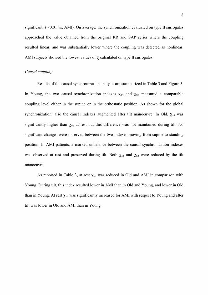
significant, P<0.01 vs. AMI). On average, the synchronization evaluated on type II surrogates
approached the value obtained from the original RR and SAP series where the coupling
resulted linear, and was substantially lower where the coupling was detected as nonlinear.
AMI subjects showed the lowest values of χ calculated on type II surrogates.
Causal coupling
Results of the causal synchronization analysis are summarized in Table 3 and Figure 5.
In Young, the two causal synchronization indexes χs/t and χt/s measured a comparable
coupling level either in the supine or in the orthostatic position. As shown for the global
synchronization, also the causal indexes augmented after tilt manoeuvre. In Old, χs/t was
significantly higher than χt/s at rest but this difference was not maintained during tilt. No
significant changes were observed between the two indexes moving from supine to standing
position. In AMI patients, a marked unbalance between the causal synchronization indexes
was observed at rest and preserved during tilt. Both χt/s and χs/t were reduced by the tilt
manoeuvre.
As reported in Table 3, at rest χt/s was reduced in Old and AMI in comparison with
Young. During tilt, this index resulted lower in AMI than in Old and Young, and lower in Old
than in Young. At rest χs/t was significantly increased for AMI with respect to Young and after
tilt was lower in Old and AMI than in Young.
9
Discussion
This work points out that the study of the complex cardiovascular regulation in
impaired conditions can be improved by considering the causal dependencies between cardiac
cycle length and systolic pressure, and without a-priori assuming the linearity of their
coupling. In particular, the use of cross-conditional entropy allows to incorporate both linear
and nonlinear contributions into a quantitative measure of RR-SAP coupling. Moreover, the
introduction of causality leads to the simultaneous and separate evaluation of feedback and
feedforward mechanisms, quantifying their relative contribution to the overall cardiovascular
regulation.
Nonlinearity in RR-SAP dynamic interaction
The presence of significant coupling was verified by generating a set of pairs of
surrogate series (type I surrogates) in which any link was totally destroyed by randomizing the
phase spectra of original RR and SAP series, then looking for statistical differences in the
synchronization index. Nonlinear mechanisms in RR-SAP link were then assessed with the
same methodology by generating a new set of pairs of surrogate series (type II surrogates) in
which the linear coupling was maintained and nonlinear coupling canceled by conserving
phase differences between RR and SAP randomized phase spectra. Analysis of type I
surrogates assessed at about 0.03 the synchronization level of completely uncoupled series,
while the χ value for type II surrogates resulted dependent on the nature of RR-SAP
interactions. Indeed, after type II surrogates generation the synchronization level was
unchanged for linearly coupled series and was blunted when nonlinearities significantly
affected the coupling. Moreover, when nonlinearity is detected the synchronization gap
between original and surrogate series may provide information about the extent of linear vs.
nonlinear components of the coupling. Thus, the fact that in AMI patients the synchronization

10
of type II surrogates resulted very low, approaching the uncoupling level, indicates the
predominance of nonlinear features with respect to linear ones in determining the coupling
between cardiac period and arterial pressure fluctuations.
Although in resting condition the average coupling strength was not significantly
different among the three groups, the presence of nonlinear dynamics in RR-SAP interactions
was found in much more post-AMI patients than Young subjects. Differences across groups
became more evident after sympathetic stimulation as synchronization increased in Young,
was substantially unchanged in Old, and decreased in AMI group. In physiological condition,
tilting is supposed to reduce the complexity of RR and SAP, entraining all the operating
mechanisms in the LF band (15, 22) and increasing the number of correlated sequences
between the two series (11). In our study, this tilt-induced simplification of the coupling was
documented in Young by the increase of the synchronization index and of the number of
patients showing pure linear RR-SAP interactions. This feature was partially verified also in
elderly that after tilt showed a prevalence of linear interactions between RR and SAP
fluctuations. Differently, in AMI the synchronization index was reduced after tilt, the number
of subjects showing uncoupled RR and SAP dynamics was doubled, and where the coupling
was significant its nature resulted non linear in all but 1.
Thus, the dumping of the coupling strength documented by the synchronization index
confirms the complex response of post-AMI patients to tilt-induced sympathetic activation
and vagal deactivation, also previously demonstrated by spectral analysis (13). Moreover,
results of surrogate data analysis demonstrate that in this class of patients the use of nonlinear
measures of coupling adds new knowledge on the study of RR-SAP dynamic interactions.
Indeed, the demonstrated marked prevalence of non linear links at rest and its persistence
during orthostatic stimulation suggest that after myocardial infarction mechanisms of genesis

11
of cardiovascular oscillations are tightly complicated by the rise non linear features otherwise
not evidenced in basal physiological condition (22).
Unbalanced RR-SAP regulation
As demonstrated by Young, in physiological conditions the coupling strength
evaluated on the two regulatory pathways is substantially balanced and preserved also after the
sympathetic activation. In elderly, our results showed an unbalanced RR-SAP regulation with
increased feedforward and a decreased feedback mechanism, thus confirming a recent study
pursued by linear cross-spectral analysis (21). This unbalancing was more marked in patients
two weeks after the infarction, mostly due to an increase of coupling on the feedforward
regulatory pathway. Previous studies based on the analysis of the concurrent changes in RR
and SAP demonstrated a dependence of the balancing between feedforward and feedback
mechanisms on the efficiency of the neural control (11, 20). In our study, the increased extent
of coupling on the feedforward arm could be explained by considering the passive behavior of
the vascular bed due to the arterial stiffening induced by age and disease, that favors the
mechanical matching between the left ventricle and the vasculature.
By the passive assumption of the orthostatic position the unbalance was reduced in Old
through a partial recovery of the coupling on the baroreflex path, while it was kept in AMI. It
has been suggested that the alterations in the sympathovagal balance at sinus node present at
rest after AMI prevent its further modifications after the tilt manoeuvre (13). In the same way,
tilt seems able neither to improve the synchronization in both arms of the RR-SAP regulatory
mechanism, as happen in Young, nor to recover the balancing between the two causal
regulations, as happens in Old.
The results of previous (11, 18, 27) and present investigations assess the importance of
separately investigate the two arms of the regulatory loop, indicating a possible differentiate
capability of one cardiovascular variable to affect the other. The increased strength of

12
coupling on the nonbaroreflex path found in the present study for AMI patients supports the
concept that feedforward mechanisms play a dominant role in the control of circulation in
impaired pathological conditions. As the nonbaroreflex coupling does not merely reflect a
mechanical matching but can be mediated by the autonomic nervous system (11), the measure
of the balancing between feedback and feedforward mechanisms could provide a new
perspective for evaluating the neural regulation of the cardiovascular function.
Impairment of heart rate regulation
In basal condition, the synchronization causal index indicated in elderly and post-AMI
patients a reduction in the ability of the systolic pressure changes to drive heart rate
variability. This finding agrees with others demonstrating a decrease of baroreflex sensitivity
with age and coronary disease (6, 10). However, in the regulation of the heart rate two
opposite feedback mechanisms, the vagally mediated arterial baroreflex (negative feedback)
and the excitatory sympathetic efferent discharge (positive feedback), are involved (14). The
causal synchronization index χt/s, accounting for both these mechanisms, cannot be strictly
related to the baroreflex gain. Hence, in post-AMI patients the reduced vagal activity could be
counterbalanced by the enhanced sympathetic activity, thus explaining the comparable values
found in patients and healthy age-matched control group.
Again, the continuous interaction between excitatory and inhibitory feedback
mechanisms should be considered to explain the different effects of the tilt manoeuvre on the
causal measure of coupling across the three groups. While it has been demonstrated that the
baroreflex gain is reduced when subjects change from lying to standing position (4, 25), in
Young we found a consistent increase of coupling. Thus, it seems that in physiological
condition the tilt-induced increase in sympathetic activity overwhelms the vagal deactivation
and determines the observed growth of χt/s. On the other side, the smoothed response of

13
elderly to the assumption of the orthostatic position, documented by a lower shortening of RR
interval length, was reflected also by the poor increase of the synchronization measure.
Finally, the decrease of this index observed in AMI can be attributed to the inability of these
patients to respond to tilt-induced changes in cardiac output by further sympathetic activation
(13). Therefore, the reduction of causal synchronization after tilt demonstrated in AMI an
impairment of mechanisms regulating heart rate with a depressed negative feedback and not
responding overloaded positive feedback. This uncoupling could also contribute to the lack of
the cardiac output documented by the lowering of SAP during standing position.
Potential Limitations
According to the study protocol, cardiovascular signals of AMI group were recorded at pre-
discharge time on patients in pharmacological washout. Due to this constraint, only patients
able to support without appreciable risk the suspension of beta-blockers treatment and no
taking antiarrhythmic drugs were enrolled for the study. This criterion of selection, based on
the demonstration of both preserved ventricular function and absence of myocardial
ischaemia, characterized a subgroup of very-low-risk post-AMI patients. Thus, results of our
study cannot be generalized to the whole post-AMI population. Furthermore, the lower heart
rate shown by AMI patients could affect part of findings of the study. In fact high vagal tone
has been previously associated to nonlinearity in the dynamic of the cardiovascular variables
(8), thus one cannot exclude that the non linear nature of coupling mechanisms elicited in
AMI could be attributable to the vagal tone predominance rather than to the underlying
pathology. However, the mean heart rate shown by AMI is comparable to that of a large part
of post-MI patients characterized by positive prognosis as it has been recently shown that after
myocardial infarction more than 50% of patients had heart rate ranging from 50 to 69 beats
per minute and higher heart rate was associated to increased risk of death (29). Furthermore,
the increase of nonlinearity and unbalancing in the regulatory mechanism demonstrated by

14
ours study for age and pathology may suggest a further raise of complexity in patients with
complicated MI and poor prognosis.
Conclusion
In summary, our study emphasizes the importance of considering nonlinearity and
causality for investigating on the interactions between spontaneous fluctuations of the
cardiovascular parameters in nonphysiological conditions. Indeed, while in healthy young
subjects the coupling between heart rate and arterial pressure occurs mainly through linear
interactions, in elderly and post-AMI patients the presence of nonlinear mechanisms was
found to play an important role. Further, differently from physiological conditions two weeks
after AMI the baroreflex feedback regulation seems to be deeply damaged as its capability to
respond to sympathetic stimulation was denied, while the strength of feedforward regulation
was markedly enforced. This finding supports the concept that a balance between the two
arms of the regulatory mechanism is essential to achieve the most adequate regulation of
cardiovascular system.

15
References
1. Akselrod S, Gordon D, Madwed JB, Snidman NC, Shannon DC, and Cohen RJ.
Hemodynamic regulation: investigation by spectral analysis. Am J Physiol 249: H867-
H875, 1985.
2. Baselli G, Porta A, Rimoldi O, Pagani M, and Cerutti S. Spectral decomposition in
multichannel recordings based on multivariate parametric identification. IEEE Trans
Biomed Eng 44: 1092-1101, 1997.
3. Bigger JTJ, Fleiss JL, Steinman RC, Rolnitzky LM, Kleiger RE, and Rottman JN.
Frequency domain measures of of heart period variability and mortality after myocardial
infarction. Circulation 85: 164-171, 1992.
4. Cooke WH, Hoag JB, Crossman AA, Kuusela TA, Tahvanainen KUO, and
Eckberg DL. Human response to upright tilt: a window on central autonomic
integration. J Physiol 517: 617-628, 1999.
5. Craft N and Schwartz JB. Effects of age on intrinsic heart rate, heart rate variability,
and AV conduction in healthy humans. Am J Physiol 268: 1441-1452, 1995.
6. Gribbin B, Pickering TG, Sleight P, and Peto R. Effect of age and high blood
pressure on baroreflex sensitivity in man. Circ Res 29: 424-431, 1971.
7. Huikuri HV, Makikallio TH, Peng CK, Goldberger AL, Hintze U, and Moller M.
Fractal correlation properties of R-R interval dynamics and mortality in patients with
depressed left ventricular function after an acute myocardial infarction. Circulation 101:
47-53, 2000.

16
8. Kanters JK, Hojgaard MV, Agner E, and Holstein-Rathlou NH. Short- and long-
term variations in non-linear dynamics of heart rate variability. Cardiovasc Res 31: 400-
409, 1996.
9. Kaplan DT. The analysis of variability. J Cardiovasc Electrophysiol 5: 16-19, 1994.
10. La Rovere MT, Bigger JTJ, Marcus FI, Mortara A, and Schwartz PJ. Baroreflex
sensitivity and heart-rate variability in prediction of total cardiac mortality after
myocardial infarction. Lancet 351: 478-484, 1998.
11. Legramante JM, Raimondi G, Massaro M, and Iellamo F. Positive and negative
feedback mechanisms in the neural regulation of cardiovascular function in healthy and
spinal cord-injured humans. Circulation 103: 1250-1255, 2001.
12. Lombardi F, Sandrone G, Mortara A, Torzillo D, La Rovere MT, Signorini MG,
Cerutti S, and Malliani A. Linear and nonlinear dynamics of heart rate variability after
acute myocardial infarction with normal and reduced left ventricular ejection fraction.
Am J Cardiol 77: 1283-1288, 1996.
13. Lombardi F, Sandrone G, Pernpruner S, Sala R, Garimoldi M, Cerutti S, Baselli
G, Pagani M, and Malliani A. Heart rate variability as an index of sympathovagal
interaction after acute myocardial infarction. Am J Cardiol 60: 1239-1245, 1987.
14. Malliani A. Homeostasis and instability: the hypothesis of tonic interaction in the
cardiovascular regulation of negative and positive feedback mechanisms. In Lown, B,
Malliani A, and Prosdocimi M, eds. Neural mechanisms and cardiovascular disease.
Padova-Berlin, Liviana Press Springer Verlag. 1986, p. 1-9.

17
15. Montano N, Gnecchi Ruscone T, Porta A, Lombardi F, Pagani M, and Malliani A.
Power spectrum analysis of heart rate variability to assess the change in sympathovagal
balance during graded orthostatic tilt. Circulation 90: 1826-1831, 1994.
16. Nollo G, Del Greco M, Disertori M, Santoro E, Maggioni AP, and Sanna GP.
Absence of slowest oscillations in short term heart rate variability of post-myocardial
infarction patients. GISSI-3 arrhythmias substudy. GISSI-3 Arrhythmias Substudy
Investigators. Auton Neurosci 90: 127-131, 2001.
17. Nollo G, Faes L, Porta A, Pellegrini B, and Antolini R. Synchronization index for
quantifying nonlinear causal coupling between RR interval and systolic arterial pressure
after myocardial infarction. Comp in Cardiol 27: 143-146, 2000.
18. Nollo G, Porta A, Faes L, Del Greco M, Disertori M, and Ravelli F. Causal linear
parametric model for baroreflex gain assessment in patients with recent myocardial
infarction. Am J Physiol Heart Circ Physiol 280: H1830-H1839, 2001.
19. Palus M. Detecting phase synchronization in noisy systems. Phys Lett A 235: 341-351,
1997.
20. Parati G, Di Rienzo M, Bertinieri G, Pomidossi G, Casadei R, Groppelli A, Pedotti
A, Zanchetti A, and Mancia G. Evaluation of the baroreceptor-heart rate reflex by 24-
hour intra-arterial blood pressure monitoring in humans. Hypertension 12: 214-222,
1988.
21. Pitzalis MV, Massari F, Mastropasqua F, Fioretti A, Guida P, Colombo R,
Balducci C, and Rizzon P. Age effect on phase relations between respiratory
oscillations of the RR interval and systolic pressure. PACE 23: 847-853, 2000.

18
22. Porta A, Baselli G, Guzzetti S, Pagani M, Malliani A, and Cerutti S. Prediction of
short cardiovascular variability signals based on conditional distribution. IEEE Trans
Biomed Eng 47: 1555-1564, 2000.
23. Porta A, Baselli G, Lombardi F, Montano N, Malliani A, and Cerutti S. Conditional
entropy approach for the evaluation of the coupling strength. Biol Cybern 81: 119-129,
1999.
24. Scher AM. Carotid and aortic regulation of arterial blood pressure. Circulation 56: 521-
528, 1977.
25. Steptoe A and Vogele C. Cardiac baroreflex function during postural change assessed
using non-invasive spontaneous sequence analysis in young men. Cardiovasc Res 24:
627-632, 1990.
26. Task force of the European Society of Cardiology and the North American Society
of Pacing and Electrophysiology. Heart rate variability. Standards of measurement,
physiological interpretation, and clinical use. Eur Heart J 17: 354-381, 1996.
27. Taylor JA and Eckberg DL. Fundamental relations between short-term RR interval
and arterial pressure oscillations in humans. Circulation 93: 1527-1532, 1996.
28. Theiler J, Eubank S, Longtin A, Galdrikian B, and Farmer JD. Testing for
nonlinearity in time series: the method of surrogate data. Physica D 58: 77-94, 1992.
29. Zuanetti G, Mantini L, Hernandez-Bernal F, Barlera S, di Gregorio D, Latini R,
and Maggioni AP. Relevance of heart rate as a prognostic factor in patients with acute
myocardial infarction: insights from the GISSI-2 study. Eur Heart J 19 Suppl F: F19-
F26, 1998.

19
Figure Legends
Figure 1
Selection of the samples of the systogram (s) for the prediction of the tachogram (t). The
example shows the construction of the patterns of length three of s, s3(i)=(s(i),s(i-1),s(i-2)),
and of the mixed patterns, (t(i),s3(i)).
Figure 2
Corrected cross-conditional entropy (CCE) (a) and synchronization function (b) evaluating the
predictability of the tachogram from the systogram as functions of the length L of the patterns
obtained from the systogram. The maximum of CCE is obtained for L=0 and represent the
Shannon Entropy of the tachogram. At a given value of L, the amount of information of the
tachogram explained by the systogram is represented by the difference between the CCE
maximum (dashed line) and the CCE value (circle symbol) in (a), and is reported after
normalization in (b). The maximum of the synchronization function (filled circle in (b))
represents the causal synchronization χt/s.
Figure 3
Distributions and mean values ± SD of the synchronization global indexes evaluated in the
three populations during rest and tilt conditions. Filled circles represent the subjects in which
no significant coupling was detected by the surrogate data analysis. Moving from rest to tilt,
the synchronization increased in Young and decreased in AMI, while no significant changes
were revealed in Old. Paired t-test: * P=0.001 vs. Rest.
Figure 4
Results of surrogate data analysis for testing the significance and the presence of nonlinearity
in the coupling between heart rate and systolic pressure. Bar graphs show the partition of the
three populations in subjects exhibiting nonlinear coupling (black area of bars) and pure linear

20
coupling (white area of bars) obtained by type II surrogate data analysis during rest and after
tilt manoeuvre. The height of the bars represent the total number of subjects for which RR and
SAP series resulted significantly coupled after type I surrogate data analysis. Chi-square test:
* P<0.01, ** P<0.001 vs. AMI.
Figure 5
Mean values ± SD of the causal synchronization indexes (χs\t and χt\s) in the three populations
during rest and tilt conditions. At rest, whereas in Young the coupling on the two causal
verses was balanced, in Old and AMI χs\t was prevalent on χt\s. After tilt, both χs\t and χt\s
increased in Young, preserving their balancing. While a slight increase of χt\s balanced the
regulation in Old, a decrease of both causal synchronization indexes and the maintenance of
the unbalance were observed in AMI. Paired t-test: * P<0.05, ** P<0.005 vs. Rest; # P<0.01,
## P<0.001 χs\t vs. χt\s.

21
Table 1. Population characteristics and time domain measures of the cardiovascular
parameters at rest and during tilt.
Young Old AMI
N. of subjects 20 12 35
Age, years 25.0 ± 2.6 63.1 ± 8.3 58.5 ± 10.2
Rest
Mean RR, ms 884 ± 124 797 ± 85 1019 ± 154
SD RR, ms 46 ± 25 27 ± 12 29 ± 23
Mean SAP, mmHg 122.1 ± 20.7 129.8 ± 12.3 120.7 ± 18.5
SD SAP, mmHg 3.8 ± 1.3 4.1 ± 1.2 2.7 ± 1.1
Tilt
Mean RR, ms 703 ± 66 ‡ 719 ± 121 † 861 ± 163 ‡
SD RR, ms 42 ± 16 18 ± 15 † 21 ± 15 †
Mean SAP, mmHg 117.7 ± 14.9 131.8 ± 12.8 112.3 ± 26.5 †
SD SAP, mmHg 6.0 ± 1.6 ‡ 4.4 ± 1.6 3.3 ± 1.3 †
† P<0.05, ‡ P<0.005 vs. Rest
Values are mean ± SD.

22
Table 2. Summary of surrogate data analysis
Young Old AMI
Rest Tilt Rest Tilt Rest Tilt
Non significant coupling
N. of subjects 3 1 5 4 9 18
χ 0.036 0.047 0.037 0.042 0.038 0.038
χ, type I surrogates 0.032 0.035 0.031 0.033 0.031 0.033
Significant linear coupling
N. of subjects 11 18 1 5 2 1
χ 0.078 0.141 0.058 0.089 0.138 0.066
χ, type II surrogates 0.068 0.134 0.059 0.087 0.132 0.064
Significant nonlinear coupling
N. of subjects 6 1 6 3 24 16
χ 0.101 0.188 0.099 0.111 0.108 0.089
χ, type II surrogates 0.053 0.078 0.050 0.066 0.043 0.042
χ: global synchronization index. Values of χ are mean over the specified number of subjects.

23
Table 3. Nonlinear causal synchronization between systolic pressure and cardiac cycle length
variability series.
Young Old AMI
Rest
χs/t 0.054 ± 0.028 0.069 ± 0.042 0.089 ± 0.053 ‡
χt/s 0.072 ± 0.037 0.041 ± 0.023 † 0.050 ± 0.030 †
Significance >0.05 0.006 0.00003
Tilt
χs/t 0.108 ± 0.053 0.069 ± 0.044 † 0.060 ± 0.040 ‡
χt/s 0.125 ± 0.071 0.065 ± 0.052 †* 0.040 ± 0.019 ‡
Significance >0.05 >0.05 0.0002
† P<0.05, ‡ P<0.005 vs. Young
* P<0.05 vs. AMI
Values are mean ± SD. χs/t: causal synchronization from tachogram to systogram; χt/s: causal
synchronization from systogram to tachogram.

t (i )
s (i )s (i -1)s (i -2)
132
138
860
940
s 3 (i )
(t (i ),s 3 (i ))ii -1i -2i -3i -4 i +1 i +2
ii -1i -2i -3i -4 i +1 i +2
s [m
mH
g]t
[mse
c]
Figure 1

0 1 2 3 4 5L
1.0
1.1
1.2
1.3
1.4
1.5
Cor
rect
edC
CE
(a)
0 1 2 3 4 5L
0.00
0.02
0.04
0.06
0.08
0.10Sy
nchr
oniz
atio
nfu
nctio
n
0 1 2 3 4 5L
0.00
0.02
0.04
0.06
0.08
0.10Sy
nchr
oniz
atio
nfu
nctio
n
(b)
χt/s
Figure 2

YOUNG
Rest Tilt
*
0.00
0.05
0.10
0.15
0.20
0.25
0.30
Sync
hron
izat
ion
0.00
0.05
0.10
0.15
0.20
0.25
0.30
Sync
hron
izat
ion
0.00
0.05
0.10
0.15
0.20
0.25
0.30
Sync
hron
izat
ion
0.00
0.05
0.10
0.15
0.20
0.25
0.30
Sync
hron
izat
ion
0.00
0.05
0.10
0.15
0.20
0.25
0.30
Sync
hron
izat
ion
0.00
0.05
0.10
0.15
0.20
0.25
0.30
Sync
hron
izat
ion
0.00
0.05
0.10
0.15
0.20
0.25
0.30
Sync
hron
izat
ion
0.00
0.05
0.10
0.15
0.20
0.25
0.30
Sync
hron
izat
ion
0.00
0.05
0.10
0.15
0.20
0.25
0.30
Sync
hron
izat
ion
0.00
0.05
0.10
0.15
0.20
0.25
0.30
Sync
hron
izat
ion
0.00
0.05
0.10
0.15
0.20
0.25
0.30
Sync
hron
izat
ion
0.00
0.05
0.10
0.15
0.20
0.25
0.30
Sync
hron
izat
ion
0.00
0.05
0.10
0.15
0.20
0.25
0.30
Sync
hron
izat
ion
0.00
0.05
0.10
0.15
0.20
0.25
0.30
Sync
hron
izat
ion
0.00
0.05
0.10
0.15
0.20
0.25
0.30
Sync
hron
izat
ion
0.00
0.05
0.10
0.15
0.20
0.25
0.30
Sync
hron
izat
ion
0.00
0.05
0.10
0.15
0.20
0.25
0.30
Sync
hron
izat
ion
0.00
0.05
0.10
0.15
0.20
0.25
0.30
Sync
hron
izat
ion
0.00
0.05
0.10
0.15
0.20
0.25
0.30
Sync
hron
izat
ion
0.00
0.05
0.10
0.15
0.20
0.25
0.30
Sync
hron
izat
ion
0.00
0.05
0.10
0.15
0.20
0.25
0.30
Sync
hron
izat
ion
0.00
0.05
0.10
0.15
0.20
0.25
0.30
Sync
hron
izat
ion
0.00
0.05
0.10
0.15
0.20
0.25
0.30
Sync
hron
izat
ion
0.00
0.05
0.10
0.15
0.20
0.25
0.30
Sync
hron
izat
ion
OLD
Rest Tilt0.00
0.05
0.10
0.15
0.20
0.25
Sync
hron
izat
ion
0.00
0.05
0.10
0.15
0.20
0.25
Sync
hron
izat
ion
0.00
0.05
0.10
0.15
0.20
0.25
Sync
hron
izat
ion
0.00
0.05
0.10
0.15
0.20
0.25
Sync
hron
izat
ion
0.00
0.05
0.10
0.15
0.20
0.25
Sync
hron
izat
ion
0.00
0.05
0.10
0.15
0.20
0.25
Sync
hron
izat
ion
0.00
0.05
0.10
0.15
0.20
0.25
Sync
hron
izat
ion
0.00
0.05
0.10
0.15
0.20
0.25
Sync
hron
izat
ion
0.00
0.05
0.10
0.15
0.20
0.25
Sync
hron
izat
ion
0.00
0.05
0.10
0.15
0.20
0.25
Sync
hron
izat
ion
0.00
0.05
0.10
0.15
0.20
0.25
Sync
hron
izat
ion
0.00
0.05
0.10
0.15
0.20
0.25
Sync
hron
izat
ion
0.00
0.05
0.10
0.15
0.20
0.25
Sync
hron
izat
ion
0.00
0.05
0.10
0.15
0.20
0.25
Sync
hron
izat
ion
0.00
0.05
0.10
0.15
0.20
0.25
Sync
hron
izat
ion
0.00
0.05
0.10
0.15
0.20
0.25
Sync
hron
izat
ion
0.00
0.05
0.10
0.15
0.20
0.25
Sync
hron
izat
ion
0.00
0.05
0.10
0.15
0.20
0.25
Sync
hron
izat
ion
AMI
*
0.00
0.05
0.10
0.15
0.20
0.25
Sync
hron
izat
ion
0.00
0.05
0.10
0.15
0.20
0.25
Sync
hron
izat
ion
0.00
0.05
0.10
0.15
0.20
0.25
Sync
hron
izat
ion
0.00
0.05
0.10
0.15
0.20
0.25
Sync
hron
izat
ion
0.00
0.05
0.10
0.15
0.20
0.25
Sync
hron
izat
ion
0.00
0.05
0.10
0.15
0.20
0.25
Sync
hron
izat
ion
0.00
0.05
0.10
0.15
0.20
0.25
Sync
hron
izat
ion
0.00
0.05
0.10
0.15
0.20
0.25
Sync
hron
izat
ion
0.00
0.05
0.10
0.15
0.20
0.25
Sync
hron
izat
ion
0.00
0.05
0.10
0.15
0.20
0.25
Sync
hron
izat
ion
0.00
0.05
0.10
0.15
0.20
0.25
Sync
hron
izat
ion
0.00
0.05
0.10
0.15
0.20
0.25
Sync
hron
izat
ion
0.00
0.05
0.10
0.15
0.20
0.25
Sync
hron
izat
ion
0.00
0.05
0.10
0.15
0.20
0.25
Sync
hron
izat
ion
0.00
0.05
0.10
0.15
0.20
0.25
Sync
hron
izat
ion
0.00
0.05
0.10
0.15
0.20
0.25
Sync
hron
izat
ion
0.00
0.05
0.10
0.15
0.20
0.25
Sync
hron
izat
ion
0.00
0.05
0.10
0.15
0.20
0.25
Sync
hron
izat
ion
0.00
0.05
0.10
0.15
0.20
0.25
Sync
hron
izat
ion
0.00
0.05
0.10
0.15
0.20
0.25
Sync
hron
izat
ion
0.00
0.05
0.10
0.15
0.20
0.25
Sync
hron
izat
ion
0.00
0.05
0.10
0.15
0.20
0.25
Sync
hron
izat
ion
0.00
0.05
0.10
0.15
0.20
0.25
Sync
hron
izat
ion
0.00
0.05
0.10
0.15
0.20
0.25
Sync
hron
izat
ion
0.00
0.05
0.10
0.15
0.20
0.25
Sync
hron
izat
ion
0.00
0.05
0.10
0.15
0.20
0.25
Sync
hron
izat
ion
0.00
0.05
0.10
0.15
0.20
0.25
Sync
hron
izat
ion
0.00
0.05
0.10
0.15
0.20
0.25
Sync
hron
izat
ion
0.00
0.05
0.10
0.15
0.20
0.25
Sync
hron
izat
ion
0.00
0.05
0.10
0.15
0.20
0.25
Sync
hron
izat
ion
0.00
0.05
0.10
0.15
0.20
0.25
Sync
hron
izat
ion
0.00
0.05
0.10
0.15
0.20
0.25
Sync
hron
izat
ion
0.00
0.05
0.10
0.15
0.20
0.25
Sync
hron
izat
ion
0.00
0.05
0.10
0.15
0.20
0.25
Sync
hron
izat
ion
0.00
0.05
0.10
0.15
0.20
0.25
Sync
hron
izat
ion
0.00
0.05
0.10
0.15
0.20
0.25
Sync
hron
izat
ion
0.00
0.05
0.10
0.15
0.20
0.25
Sync
hron
izat
ion
0.00
0.05
0.10
0.15
0.20
0.25
Sync
hron
izat
ion
0.00
0.05
0.10
0.15
0.20
0.25
Sync
hron
izat
ion
0.00
0.05
0.10
0.15
0.20
0.25
Sync
hron
izat
ion
0.00
0.05
0.10
0.15
0.20
0.25
Sync
hron
izat
ion
0.00
0.05
0.10
0.15
0.20
0.25
Sync
hron
izat
ion
0.00
0.05
0.10
0.15
0.20
0.25
Sync
hron
izat
ion
0.00
0.05
0.10
0.15
0.20
0.25
Sync
hron
izat
ion
0.00
0.05
0.10
0.15
0.20
0.25
Sync
hron
izat
ion
0.00
0.05
0.10
0.15
0.20
0.25
Sync
hron
izat
ion
0.00
0.05
0.10
0.15
0.20
0.25
Sync
hron
izat
ion
0.00
0.05
0.10
0.15
0.20
0.25
Sync
hron
izat
ion
0.00
0.05
0.10
0.15
0.20
0.25
Sync
hron
izat
ion
0.00
0.05
0.10
0.15
0.20
0.25
Sync
hron
izat
ion
0.00
0.05
0.10
0.15
0.20
0.25
Sync
hron
izat
ion
0.00
0.05
0.10
0.15
0.20
0.25
Sync
hron
izat
ion
0.00
0.05
0.10
0.15
0.20
0.25
Sync
hron
izat
ion
0.00
0.05
0.10
0.15
0.20
0.25
Sync
hron
izat
ion
Figure 3

REST
Young Old AMI
**
0
10
20
30
N.o
fsub
ject
s
0
10
20
30N
.ofs
ubje
cts
TILT
Young Old AMI
**
*
Figure 4

χt\s
χs\tYOUNG
Rest Tilt
**
*
0.00
0.05
0.10
0.15
0.20
0.25
Sync
hron
izat
ion
0.00
0.05
0.10
0.15
0.20
0.25
Sync
hron
izat
ion χt\s
χs\tOLD
Rest Tilt
#
0.00
0.05
0.10
0.15
0.20
0.25
Sync
hron
izat
ion
0.00
0.05
0.10
0.15
0.20
0.25
Sync
hron
zatio
n χt\s
χs\tAMI
Rest Tilt
##
**
*
##
0.00
0.05
0.10
0.15
0.20
0.25
Sync
hron
zatio
n
Figure 5
Copyright © 2022 FDOKUMEN