Block copolymer micelles conjugated with anti-EGFR antibody for targeted delivery of anticancer drug
Upload
independentCategory
view
4download
0
Treatment of Alzheimer’s Disease with Anti-Homocysteic acid Antibody
(Homocysteic acid induces Alzheimer’s pathology; memory impairment and
neurodegeneration. Amyloid accelerates HA toxicity)
Tohru Hasegawa*, Nobuyuki Mikoda # Masashi Kitazawa** and Frank M. LaFerla**
*Saga Women’s Junior College, Honjyou, Saga 840-8550, Japan #Kyudou Ltd., Saga 841-0075, Japan
**1109 Gillespie Neuroscience Research Facility,Department of Neurobiology & Behavior. University of California, Irvine Irvine, CA 92697-4545
Abstract
Homocysteic acid (HA) may play an important role in Alzhiemer disease (AD) as we
previously reported that HA induced accumulation of intraneuronal Aβ42. In this
study, we first analyzed HA levels in a mouse model of AD. 4-month old
pre-pathologic 3xTg-AD mice exhibited higher levels of HA in the hippocampus as
compared to age-matched nontransgenic, suggesting that HA accumulation may
precede both Aβ and tau pathologies. To further determine the pathogenic role of HA
in AD, we treated young 3xTg-AD mice with vitamin B6-deficient food for 3 weeks to
induce the production of HA in the brain. Concominantly, mice received either saline
or anti-HA antibody intraventricularly using a guide cannula every 3 days. Mice
received anti-HA antibody significantly rescued cognitive impairment induced by
vitamin B6 deficiency. Pathologically, 3-week treatment with vitamin B-6 deficient
food resulted in strong neurodegeneration in the hippocampal CA1 zone and decreased
hippocampal volume. In contrast, anti-HA antibody treatment attenuated these
pathological changes. Taken together, we conclude that increased brain HA
triggers memory impairment whose condition was deteriorated by amyloid and
subsequent neurodegeneration and reduction of neurogenesis. Our results indicate a
pathogenic role of HA in AD.

Introduction
Amyloid plaques and neurofibrillary tangles are the two pathophysiological hallmarks
of Alzheimer’s disease (AD). Intracellular amyloid beta 42 is increasingly recognized as
an early pathological trigger that can lead to amyloid plaques and even induce
neurofibrillary tangles. We previously found that homocysteic acid (HA) induces
intracellular accumulation of amyloid beta 42 and the production of α-synuclein in the
presence of methionine. It is known that α-synuclein facilitates the formation of tau
aggregates. Consequently, HA affects the two pathophysiological hallmarks of AD and
may be involved in its etiology.
Previous studies have reported that psychological stress accelerates Alzheimer’s
disease (AD); however, recently it has been established that stress directly induces
pathological changes in this disease (1, 2). The underlying mechanism, however, is
unclear. Hasegawa et al. found that homocysteic acid (HA) induced amyloid beta 42
accumulation in neuronal cells, resulting in cell dysfunction and finally cell death (3).
HA is also known as an NMDA receptor agonist (4), and is released from astrocytes by
the activation of beta-adrenergic receptors under mental stress (5).
HA can therefore induce AD. The 3xTg-AD mouse model developed by Oddo et al (6)
showed memory impairment at an early age because of amyloid beta accumulation in
neuronal cells (7). We observed increased level of HA in the hippocampus of 3xTg-AD
homozygous mice compared with that of 4-month-old nontrangenic mice, when the
former showed accumulation of amyloid beta in neuron. We then confirmed the
etiological effect of HA on the disease process of AD in 3xTg-AD hemizygous mice.
Hemizygous mice were used instead of homozygous mice because they have reduced
transgenic gene expression for amyloid precursor protein (APP), presenilin, and Tau
compared with homozygous mice. Disease pathogenesis does not easily occur in
hemizygous mice through the action of certain etiological agents such as APP,
presenilin and Tau compared with homozygous mice. We expected that if HA were a
true etiological agent of AD, it would accelerate memory impairment in 3xTg-AD
hemizygous mice and that administering anti-HA antibodies would inhibit this
acceleration.

Methods
3xTg-AD mice* The mouse germline used in this study was kindly gifted by Professor F. M. Laferla
(University of Calfornia, Irvine). Housing environment (12h/12h light/dark cycle)
was germ-free clean room. 3xTg-AD hemizygous male mice (5 and 7 month-old) were 7
mice used. Also Non-Tg mice were 4 mice used. The 3xTg-AD mice develop both plaque
and tangle pathology in AD-relevant brain regions. The 3xTg-AD mice develop
extracellular Aß deposits prior to tangle formation, consistent with the amyloid
cascade hypothesis. Despite equivalent overexpression of the human ßAPP and human
tau transgenes, Aß deposition develops prior to the tangle pathology, consistent with
the amyloid cascade hypothesis. In addition, these mice exhibit deficits in synaptic
plasticity, including long-term potentiation (LTP) that occurs prior to extracellular Aß
deposition and tau pathology, but is associated with intracellular Aß immunoreactivity.
These studies support the view that synaptic dysfunction is a proximal defect in the
pathobiology of AD, preceding extracellular plaque formation and neurofibrillary
pathology. As these 3xTg-AD mice phenocopy critical aspects of AD neuropathology,
this model will be useful in pre-clinical intervention trials, particularly because the
efficacy of anti-AD compounds in mitigating the neurodegenerative effects mediated by
both signature lesions can be evaluated.
Vitamin B6-deficient food Vitamin B6-deficient food was purchased from Kyudo Ltd. Nutrient composition has
been described further in the study.
Anti-HA antibody Anti-HA antibodies were purchased from MoBiTec Co. (Germany). Polyclonal antisera
were raised in rabbits after immunization with a glutaraldehyde-containing HA
conjugate, following which antibody specificity was determined by performing ELISA
with competition experiments involving HA-G-BSA (compound cross-reactivity ratio
1:1), cysteine-G-BSA (1:85), and homocysteine-G-BSA (1:231)
Morris water maze test

The apparatus used for all Morris water maze tasks comprised a circular aluminum
tank (1.5 m in diameter) painted white and filled with water maintained at 26–29°C.
The maze was located in a room containing several simple, visual extramaze cues. To
reduce stress, mice were placed on a platform in both the hidden and cued versions of
the task for 10 s prior to the first training trial.
Spatial reference Morris water maze training
Mice were trained to swim to a 14-cm circular clear Plexiglas platform placed 1.5 cm
beneath the water surface that was invisible to the mice while swimming. The platform
location was randomly selected for each mouse, but was kept constant for that mouse
throughout the training period. In each trial, the mouse was placed in the tank at one
of the four designated start points in a pseudorandom order. Mice were allowed to
search for and escape to the submerged platform. If a mouse failed to find the platform
within 60 s, it was manually guided to it and allowed to remain there for 10 s. Then,
each mouse was placed in a holding cage under a warming lamp for 25 s until the start
of the next trial. To ensure that memory differences were not due to the lack of task
learning, the mice were trained for four trials a day for as many days as required to
meet the criterion, which was defined as <20-s mean escape latency before the first
probe trial was run. To prevent overtraining, probe trials were run for each group as
soon as they met the group criterion and stopped after all the groups met the criterion.
Retention of spatial training was assessed 1.5 and 24 h after the last training trial.
Both probe trials consisted of a 60-s free swim in the pool without the platform. Mice
were monitored by a camera mounted on the ceiling directly above the pool, and all
trials were stored on videotape (burnt onto a DVD.)or subsequent analysis. The
parameters measured during the probe trial comprised (1) initial latency time to reach
the platform,
Immunohistochemistry
Mice were sacrificed by CO2 asphyxiation, and the brains were rapidly removed and
fixed for 48 h in 4% paraformaldehyde. Sections (50-µm thick) were processed for
free-floating immunohistochemistry as previously described (6). Anti-amyloid beta
(6E10), anti-APP (22C11), and amyloid beta (40/42)-specific antibodies were applied
overnight at 4°C. Sections were developed with diaminobenzidine (Vector Laboratories)

substrate using the avidin–biotin–horseradish peroxidase system (Vector Laboratories).
Quantification of amyloid beta was performed as described previously (8). Mice were
excluded from the antibody group analysis (behavior and histology) if the cannulae
were found to be placed incorrectly. To obtain the percentage difference between the
antibody- and PBS-treated tissues (controls), we applied the following formula: number
of pixels in the antibody-treated hippocampus − number of pixels in the PBS-treated
hippocampus/number of pixels in PBS-treated hippocampus. The number of pixels
calculated in each case is the sum of five readings per mouse averaged across the entire
group.
HA level measurement HA was extracted from mouse brain (hippocampus and cortex) with an acid of
trichloroacetic acid. Brain samples were prepared by a modification of the method of
Reed and Bellerche (8). Brains (1.50−2.00 g) were isolated from 4-month-old 3xTg-AD
homozygous male mice. The mice were killed by rapid decapitation and their brains
were quickly excised and placed on an ice-cold Petri dish. For the gradient
high-performance liquid chromatography (HPLC) method, tissue samples were
weighed and homogenized using a sonicator for 10 s in ice in 4 ml of ice-cold 10% (w/v)
trichloroacetic acid per 100 mg tissue (wet weight). HA (4 µg) was added as the
internal standard. For isocratic HPLC, tissue samples were divided into six aliquots.
The samples were homogenized as described above. The homogenates for isocratic or
gradient HPLC were left on ice for 1 h and centrifuged at 20,000 ×g for 25 min. The
supernatant was washed five times with an equal volume of diethyl ether and the
aqueous phase was maintained. Residual ether was evaporated under nitrogen at room
temperature for 5 min. Immediately thereafter, 20 µL was injected into the HPLC
system.
Ventricular cannula The mice were anesthetized, 2-mm-wide incisions were made in the left hemisphere,
and a guide cannula was inserted into the left ventricular space using a Teflon tube
(1 mm in diameter). This operation did not impair learning and memory performance,
and the abilities of the operated mice were similar to those of mice that did not undergo
surgery.
Plasma catecholamine levels Plasma was collected from the eyelid arteries. under anesthesia, and catecholamine
levels were measured by HPLC.
Membrane APP measurement Rat embryonic neuronal cells were cultured using the method described in (3) and
membrane APP was detected using the c-terminal antibody of Sigma A8717.
Homocysteic acid was added to the culture medium and incubation was performed for
48 h.
Results
HA levels in mouse model brain We measured HA levels in the brains of 4-month-old 3xTg-AD-homozygous mice; at
this age, mice display intracellular accumulation of amyloid beta in the brain regions
affected by AD. This accumulation also appears to be associated with the early
memory deficit exhibited by these mice (7). As shown in Table 1, HA levels in the
3xTg-AD mice were clearly higher than those in control nontransgenic mice. This
finding, in combination with those from our in vitro studies, indicates that HA may
modulate the pathology of AD. To confirm our hypothesis, we fed 3xTg-AD-h and
nontransgenic mice with vitamin B6-deficient food because B6 deficiency has been
reported to increase HA levels in these mice (9).
Anxiety reaction At 3 weeks of vitamin B6-deficient feeding, both 3xTg-AD-h and nontransgenic mice
showed a strong anxiety reaction (see DVD). That is, they preferred dark areas, showed
lower locomotor activity, and moved along the edge of the cage in the open field. In
contrast, the experimental group treated with anti-HA antibodies did not show any
anxiety reaction (i.e., they preferred bright areas, showed high locomotor activity, and
moved to the center, not along the edge of the cage in the open field). This anxiety
behavior was confirmed by determining the plasma catecholamine levels. Transgenic
control and nontransgenic mice showed higher levels of catecholamine than
experimental transgenic mice (Fig. 1). Hemizygous mice aged 7 months showed strong
freezing behavior and occasional seizures, but experimental mice of the same age did
not show any of the behavior problems that were evident in the control group (see
DVD).
Memory experiment We next evaluated the hippocampus-dependent memory by Morris water maze.
5-month old 3xTg-AD mice fed with vitamin B6 deficient diet showed a significant
memory impairment compared to 2-month old normal mice (Fig. 2). Notably,
co-treatment with anti-HA antibody significantly rescued vitamin B6 deficient-induced
memory impairment (Fig. 2). However, the nontransgenic and transgenic control
groups showed memory impairment.

This result indicates that HA can induce memory impairment. Memory impairment
induced by HA was greater in the presence of amyloid beta, because transgenic control
mice, in whom there was amyloid beta production and accumulation (see below),
showed the worst memory impairment of the three groups (Fig. 2a). Amyloid alone did
not induce memory impairment, despite the presence of higher level of both amyloid
beta 40 and 42 in the transgenic 5-month-old mice (hemizygous +B6 deficient +
anti-HA antibody) compared with nontransgeneic mice (10). Moreover, transgenic
experimental mice showed better memory performance than nontransgenic mice.
We also confirmed that HA induced memory impairment. As shown in Fig. 2b, C57BL
mice showed memory impairment according with B6 deficient food feeding time. And
final complete memory impairment mice which were fed B6 deficient food for 3 months
could recover their impairment by the addition of anti-HA antibody, indicating that
these memory impairment was induced by HA. The thing that should be paid
attention is the time which needed to become memory impairment. In the case of
C57BL, the time needed to show memory impairment was 3 months, but 3xTg-AD case,
the time was 3 weeks. In other words, HA toxicity was strengthend by the presence of
amyloid.
Pathological change Figure 3 demonstrates the immunohistochemical staining for amyloid beta in the
amygdala, cortical, and hippocampal cells of transgenic control and transgenic
experimental mice. Numerous stained neurons were seen in transgenic control mice,
indicating amyloid beta accumulation in these neurons. In contrast, transgenic
experimental mice did not show any stained neurons in the hippocampus and showed
fewer stained neurons in the amygdala and cortex compared with transgenic control
mice, indicating inhibition of amyloid beta accumulation. We did not observe any
amyloid beta accumulation in the neurons of nontransgenic and transgenic control
mice fed normal food (data not shown).
Since we observed a preventive effect of anti-HA antibody on the acceleration of
pathogenesis of AD in 3xTg-AD mice induced by vitamin B6-deficient food, we looked
for a curative effect of anti-HA antibody on the established pathological changes in
3xTg-AD mice with AD.
3xTg-AD hemizyous 7-month-old male mice were fed B6-deficient food for 3 weeks

(control), and anti-HA antibody (5 μl) was injected into their ventricular space every 3
days using guide cannulae. After 3 weeks, we measured memory performance in the
Morris water maze tasks following which we evaluated the hematoxylin–eosin and
immunohistochemical staining of the brain specimens.
The results are shown in Figure 4. Control mice showed poor memory performance
but experimental mice injected with anti-HA antibody showed strong recovery of
performance after 2 days of training, indicating good memory performance.
Consistent with the results of the memory performance task, the hippocampi of
control mice showed a marked deficiency of neuronal cells in the CA1 region (as
indicated by hematoxylin–eosin and especially immunohistochemical staining). A
marked loss of cells was observed in control mice (indicated by the ellipses in Fig. 5)
compared with experimental mice. In addition, many macrophages appeared in the
CA1 area of control mice compared with the experimental group (indicated by the
arrow in Fig. 5b). Fig. 6 showed that control mice accumulated amyloid beta into
neuronal cells in cortex (circled line), but experimental mice accumulated less amyloid
beta than that of control.
Figures 5 illustrate that B6-deficient food induced strong neurodegeneration in the
hippocampal CA1 region and anti-HA antibody treatment induced marked recovery of
both neurogenesis and memory performance. Hippcampal volume was decreased in
control mice, but it recovered in the experimental group (Table 2).
Neuronal death induced by HA HA induced the neurodegenerative cell loss in CA1 zone (Fig. 5), we would like to
confirm this HA effect in vitro system. Rat embryonic brain neurons were cultured
according to the method (3), HA showed the neurodegenerative effect compared with
that of homocysteine. Cleary shown in Fig. 7, HA showed strong neurodegenerative
effect compared with that of homocysteine.

Discussion
Non Tg: anxiety, memory impairment (A)
B6-deficient food Tg control: anxiety, memory impairment (B)
Tg control fed normal food: normal, good memory (C)
Tg experiment: normal, good memory (D)
Tg: Transgenic
Figure
A and B illustrate that vitamin B6-deficient food induced an anxiety reaction and D
illustrates that HA did the same. A and B also illustrate that vitamin B6-deficient food
accelerated cognitive (memory) impairment in AD mice. It can be deduced that
B6-deficient food itself induced memory impairment in nontransgenic mice.
Additionally, it can be inferred that transgenic control mice themselves functioned
normally, as on being fed normal food they displayed normal memory performance.
B6-deficient food induces an increase in homocysteine, homocysteine sulfinic acid, and
homocysteic acid levels (9). Recently, the New England Journal of Medicine reported
that lowering the level of homocysteine alone does not suppress cognitive decline in
elderly people, and that treating homocysteine levels should not be focused upon.
Homocysteine sulfinic acid is formed by the peroxidation of homocysteine (12) following
which the latter does not play a role in the pathogenesis of AD, indicating that
homocysteine sulfinic acid does not contribute to the disease pathogenesis of AD.
Finally, because HA is formed from homocysteine and methionine (13), it can
possibly accelerate the pathogenesis of AD. The results of our transgenic experiments
confirmed that anti-HA antibody suppressed memory impairment induced by
B6-deficient food. This hypothesis is supported by our data suggesting that higher
levels of HA are present in 3xTg-AD-h compared with nontransgenic mice (Table 1).
Figure 2 shows that memory impairment induced by HA was stronger in the
presence of amyloid beta. However, amyloid beta itself does not induce memory
impairment, because transgenic experimental mice, who had higher levels of amyloid

beta 40 and 42 (10), displayed better memory performance than nontransgenic mice.
Recently, it has been reported that amyloid oligomers induce LTP suppression through
the action of NMDA (14); this finding supports our observations.
Our observations indicate that anti-HA antibody can bring about normal recovery in AD. It can therefore be concluded that HA is a true etiological agent for AD.
Our hypothesis that HA accelerated the pathogenesis of AD was confirmed by the
immunohistochemical observations. Amyloid beta accumulated in hippocampal
neurons in the transgenic control group but not in the transgenic experimental group.
The curative effect of anti-HA antibody was so strong that a substantive recovery in
the memory performance tasks was observed. This recovery was attributed to
neurogenesis in the hippocampus brought about by anti-HA antibodies. This is the first
study reporting such a phenomenon.
We observed that neuronal cell death was induced by HA (Fig. 7) which also destroyed the membrane APP (Fig. 8). APP has been reported to affect neurogenesis (16), therefore, anti-HA antibodies can alleviate the destructive effect of HA on APP
and can consequently induce neurogenesis.
HA induced memory impairment in our study, and this impairment was greater in
the presence of amyloid beta (Fig. 2a,b), i.e., even if amyloid beta were to be decreased,
HA would still be present in the affected brain tissue. Hence, amyloid vaccination could
not restore the cognitive function despite reduced amyloid levels (17). Our findings on
anti-HA antibody treatment in transgenic mice show that the toxicity of HA were
decreased and neurodegeneration was inhibited following which hippocampus volume
increased, suggesting that the neurogenesis might be occurred. (Figs. 5 and 6 and
Table 2). This led to recovery of memory performance to a normal state. Recently, it has
been reported that NMDA antagonists such as MK-801 induce neurogenesis in the
dentate gyrus of the hippocampus (16) and that HA is an agonist of the NMDA receptor
(4). Thus, our observation that anti-HA antibodies induced neurogenesis is feasible and
is consistent with the previous study (18).
Imagawa et al reported a case of treatment-influenced recovery of familial AD in Japan
The author prescribed vitamin B6, coenzyme Q10, and iron citric acid supplements

to two sisters and noted marked recovery in both (19).
In contrast, mice that were fed B6-deficient food showed increased levels of HA,
which then induced strong neurodegeneration in the presence of amyloid beta, because
HA induced amyloid beta accumulation in neuronal cells (Fig. 6) (3).
In the above-mentioned study (19), the authors suggested that recovery from familial
AD occurred because treatment with vitamin B6 decreased HA levels in the patients.
Unfortunately, this treatment did not prevent cognitive decline after 2 years of
treatment, which may have been related to the fact that HA can be produced in two
ways. One is via the homocysteine pathway, which can be inhibited by vitamin B6; the
other is via the methionine pathway, which cannot be inhibited by vitamin B6. In any
event, anti-HA antibody can decrease the toxicity of HA and can consequently induce
marked recovery of cognitive ability in the mouse model.
In conclusion, (1) HA can accelerate pathological changes in AD; (2) HA toxicity was
decreased by anti-HA antibody, which induced strong neurogenesis in the
hippocampus, consequently resulting in marked recovery of memory performance; and
(3) HA induced early pathological changes in normal mice, i.e., memory impairment.
(Fig. 2b). According to Koch’s postulates (15), these three points indicate that HA is a
true etiological agent of AD.
Our hypothesis is also supported by the observation that anti-HA antibodies induced
marked recovery in 7-month-old hemizygous mice (see immunohistochemical
observations in Figures 5 and 6 and the behavioral observations included in the DVD).
This is the first study to demonstrate marked recovery from AD induced by treatment
with anti-HA antibodies. Our findings prove the strong curative effect of anti-HA
antibody treatment and support the idea that HA is a true etiological agent and an
accelerator in the pathogenesis of AD.
In addition to our discussion mentioned above, one more thing should be paid
attention. From our experiment, amyloid hypothesis is not in central dogma of
Alzheimer’s disease. From Fig. 2a, 2b, it can be clarified that amyloid accerelated HA
toxicity and amyloid itself has no effect on memory impairment. In other words,
amyloid toxicity in Alzheimer’s disease is not central dogma.
Alzheimer’s pathology is composed two phenomena. One is memory impairment
and the other is neurodegeneration. HA showed these two phenomena (Fig.2a,2b and
Fig. 7 ) And recent unsatisfied reports, amyloid treated therapy faild in clinical trail.

That is, amyloid level decreased , but cognitive ability became worse.
From these reports and our observations, it can conclude that amyloid hypothesis is not
central dogma of Alzheimer’s disease and HA can be one of true etiological compounds.

Acknowledgment
Dr Masayasu Ohyagi (Kyushu University, Japan) was aknowleged to his technical
support for the histochemical and immunochemical staining observations and his
scientific advice.

References
(1) Wilson RS, Schneider JA, Boyle PA, Arnold SE, Tang Y, Bennett DA. Chronic
distress and incidence of mild cognitive impairment. Neurology 2007;68(24):2085-2092
(2) Peavy GM, Lange KL, Salmon DP, Patterson TL, Goldman S, Gamst AC, Mills PJ,
Khandrika S, Galasko D. The effects of prolonged stress and ApoE genotype on
memory and cortisol in older adults. Biol Psych. 2007 (in press)
(3) Hasegawa T, Ukai W, Jo DG, Xu X, Mattson MP, Nakagawa M, Araki W, Saito T,
Yamada T. Homocysteic Acid Induces Intraneuronal Accumulation of Neurotoxic Aβ42:
Implication for the Pathogenesis of Alzheimer’s disease. J Neurosci Res.
2005;80:869-876
(4) Do KQ, Herrling PL, Streit P, Cuénod M. Release of neuroactive substances:
homocysteic acid as an endogenous agonist of the NMDA receptor. J Neural Transm.
1988;72(3):185-190
(5) Do KQ, Benz B, Sorg O, Pellerin L, Magistretti PJ. Beta-Adrenergic stimulation
promotes homocysteic acid release from astrocyte cultures: evidence for a role of
astrocytes in the modulation of synaptic transmission. J Neurochem.
1997;68(6):2386-2394
(6) Oddo S, Caccamo A, Shepherd JD, Murphy MP, Golde TE, Kayed R, Metherate R,
Mattson MP, Akbari Y, LaFerla FM. Triple-Transgenic Model of Alzheimer’s Disease
with Plaques and Tangles: Intracellular Aβ and Synaptic Dysfunction. Neuron
2003;39:409-421
(7) Billings LM, Oddo S, Green KN, McGaugh JL, LaFerla FM. Intraneuronal Aβ
Causes the Onset of Early Alzheimer’s Disease-Related Cognitive Deficits in
Transgenic Mice. Neuron 2005;45:675-688
(8) Benz B, Grima G, Do KQ. Glutamate-induced homocysteic acid release from

astrocytes: possible implication in glia-neuron signaling. Neuroscience 2004;377-386.
(9) Ohmori S. Biosynthesis of homocysteine sulfinic acid in the vitamin B6-deficient rat.
Hoppe Seylers Z Physiol Chem. 1975;356(9):1369-1373
(10) Oddo S, Caccamo A, Shepherd JD, Murphy MP, Golde TE, Kayed R, Metherate R,
Mattson MP, Akbari Y, LaFerla FM. Triple-Transgenic Model of Alzheimer’s Disease
with Plaques and Tangles Intracellular Aβ and Synaptic Dysfunction. Neuron
2003;39(3):409-421
(11) Luo D, Anderson BD. Kinetics and mechanism for the reaction of cysteine with
hydrogen peroxide in amorphous polyvinylpyrrolidone lyophiles. Pharm Res.
2006;23(10):2239-2253
(12) Lieberman M, Kunishi AT. Ethylene production from methionine. Biochem J.
1965;97(2):449-459
(13) Porter VR, Buxton WG, Fairbanks LA, Strickland T, O’Connor SM,
Rosenberg-Thompson S, Cummings JL. Frequency and characteristics of anxiety
among patients with Alzheimer’s disease and related dementias. J Neuropsychiatry
Clin Neurosci. 2003;15(2):180-186
(14) Shankar GM, Bloodgood BL, Townsend M, Walsh DM, Selkoe DJ, Sabatini BL.
Natural Oligomers of the Alzheimer Amyloid-ß Protein Induce Reversible Synapse
Loss by Modulating an NMDA-Type Glutamate Receptor-Dependent Signaling
Pathway. J Neurosci. 2007;27(11):2866-2875
(15) http://en.wikipedia.org/wiki/Koch%27s_postulates
(16) Neve RL, McPhie DL. Dysfunction of amyloid precursor protein signaling in neurons leads to DNA synthesis and apoptosis. Biochimica et Biophysica Acta (BBA)-Molecular Basis of Disease 2007;1772(4):430-437
(17) Takeshi T., Brain and Nerve 2007, 375-382
18) Gould E, McEwen BS, Tanapat P, Galea LAM, Fuchs E. Neurogenesis in the Dentate
Gyrus of the Adult Tree Shrew Is Regulated by Psychosocial Stress and NMDA Receptor
Activation. J Neurosci. 1997;17(7):2492-2498
(19) Imagawa M, Naruse S, Tsuji S, Fujioka A, Yamaguchi H. Coenzyme Q10, Iron, and
vitamin B6 in genetically-confirmed Alzheimer’s disease. The Lancet 1992;340:671

Figure legends
Fig. 1 Plasma levels of catecholamines in nontransgenic (NonTg), transgenic (Tg)
control, and experimental mice. a, adrenaline; b, noradrenaline; c, dopamine.
The figure shows average data for three mice
Fig. 2a Long-term memory test in the Morris water maze. Nontransgenic mice had an
average score of three mice each day. Hemizygous transgenic control (hemizygous + B6
deficeint) mice had an average score of three transgenic control mice each day.
Transgenic experimental mice (hemizygous +B6 deficient + anti-HA antibody) had an
average score of three transgenic experimental mice each day.
Fig. 2b C57BL mice memory impairment induced by HA
C57BL mice were 5 month-old male mice. Mice were fed B6 deficient food for 1.5
months and 3 months. Morris water maze test was conducted at 1.5 months and 3
months. Each 5 mice were used memory test. After 3 months, memory impairment
mice were added anti-HA antibody according to same method of 3xTg-AD. After one
month, memory test was conducted.
Fig. 3 Immunohistochemical observations of amygdalar, cortical, and hippocampal neurons. Anti-HA antibody was diluted 100-fold. Immunohistochemical observations were made thrice, and each observation gave the same result. Five hemizygous transgenic 5-month-old mice and 10 homozygous transgenic 3-month-old mice were fed B6-deficient food for 3 weeks. Transgenic experimental mice were injected with anti-HA antibody every 3 days. For details, see Methods. CTL, transgenic control mice; Ex, transgenic experimental mice.
Fig. 4 Curative effect of anti-HA antibody as shown by long-term memory performance.
Seven-month-old male 3xTg-AD-h mice fed B6-deficient food for 3 weeks served as the
control. Experimental mice were treated with anti-HA antibody every 3 days; the
antibody (100-fold dilution) was injected into the brain as described in Methods. The
figure shows average data for five male mice. To demonstrate the strong curative effect
of the antibody, we show for comparison the results for 2-month-old hemizygous male
mice, whose memory performance was normal.
Fig. 5 (a) Hematoxylin–eosin staining of cortical and hippocampal neurons in control

and experimental groups. Mice were the same as those shown in Fig. 4. (b)
Magnification of (a) showing macrophages in the CA1 area of the hippocampus of
control and experimental mice.
Fig. 6 Immunohistochemical staining of cortical and hippocampal neurons in control
and experimental mice. Staining was performed as described in the Methods section.
Fig. 7 Comparison of neuronal cell death between mice treated with homocysteine and
those treated with homocysteic acid. Cells were cultured using the method described in
(3). Neuronal death was measured by the MTT method (3).
Fig. 8 Western blot analysis of APP with c-terminal antibody of Sigma A8717. Rat
embryonic neuronal cells were cultured using the method described in (3) and
membrane APP was detected using the c-terminal antibody of Sigma A8717.
Homocysteic acid was added to the culture medium and incubation was performed for
48 h.

0
5000
10000
15000
20000
25000
pg/ ml
NonTg Tg control Tg Ex
Adrenalin
Fig. 1a Plasma Adrenalin
02000400060008000
100001200014000
pg/ ml
NonTg Tg control Tg Ex
Noradrenalin
Fig. 1b Plasma Noradrenalin

0
200
400
600
800
pg/ ml
NonTg Tg control Tg Ex
Dopamine
Fig. 1c Plasma Dopamin

C57Bl memory impairment induced by HA
Normal (5 month-old)
Trial day
Late
ncy
esca
pe (s
ec)
0
10
20
30
40
50
60
70
1 2 3 4 5
B6 deficient food for 3 months
B6 deficent food for 1.5 months
Normal (5 month-old)
Anti-HA antibody

CT: Male 7 month-old+ B6 defcient EX: Male 7 month-old+ B6 defcient+ Anti-HA antibody
CA1
CA1
Hematoxylin Staining
Bar=100μm

Hippocampus major axis (cm)
Average
Control 9 8.8 7.8 9 8.2 8.6 + 0.5
P<0.01
Experiment 9.6 10.1 9.6 10.3 9.9 + 0.4
Control: 7 month-old hemizygous + B6 deficientExperiment: 7 month-old hemizygous male +B6 deficient + anti-HA antibody
5 different sections were observed and measured its major axis.Average (n=2) is shown.

7 month-old +B6 deficient Male +Anti-HA antibody
Bar=100μm
Fig.5b(1)

7 month-old +B6 deficient Male Fig.5b(2) Macrophage in CA1 area Arrow indicates marcorphage
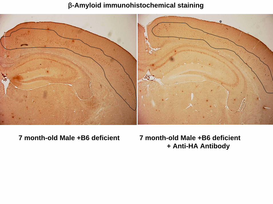
β-Amyloid immunohistochemical staining
7 month-old Male +B6 deficient 7 month-old Male +B6 deficient + Anti-HA Antibody

Comparison of Neural Impairment between Homocysteine and Homocysteic acid
0
20
40
60
80
100
-6 -5 -4 -3 -2
Drug Concentration (log(M))
Neu
rona
l inj
ury
(%L-Homocysteic acidDL-Homocysteine
IC50:44 μM
IC50:932 μM
Fig. 7

APP decreasing by Homocysteic acid
0 10 100 1000
μ M ✕ 48 HHomocysteic acid
209 kDa
130 kDa
Fig. 8
Copyright © 2022 FDOKUMEN