Dexamethasone induces apoptosis of multiple myeloma cells in a JNK/SAP kinase independent mechanism
Upload
independentCategory
view
1download
0
Therapeutic anti-tumor efficacy of anti-CD137 agonisticmonoclonal antibody in mouse models of myeloma
Oihana Murillo1, Ainhoa Arina1, Sandra Hervas-Stubbs1, Anjana Gupta3, BrandonMcCluskey3, Juan Dubrot1, Asís Palazón1, Arantza Azpilikueta1, Maria C. Ochoa1, CarlosAlfaro1, Sarai Solano1, José L. Pérez-Gracia2, Babatunde O. Oyajobi3, and IgnacioMelero1,2
1Gene Therapy Unit. Centro de Investigación Médica aplicada (CIMA), Universidad de Navarra, Pamplona,Spain.
2Clinica Universitaria, Universidad de Navarra, Pamplona, Spain.
3Department of Cellular and Structural Biology, University of Texas Health Science Center at San Antonio,San Antonio, TX.
AbstractPurpose: Eradication of post-treatment residual myeloma cells is needed to prevent relapses andimmunostimulatory monoclonal antibodies (mAbs) such as anti-CD137, CTLA-4, CD40, etc, thatenhance the immune response against malignancies represent a means of achieving this purpose.This study explores anti-CD137 mAbs for mutiple myeloma (MM) treatment in preclinical modelsof the disease because they safely augment tumor immunity and are in clinical trials for other cancers.
Experimental design: The anti-tumor effect of anti-CD137 mAb on mouse plasmacytomasderived from HOPC and NS0 cell lines was studied and compared with that of anti-CTLA-4, anti-CD40 and anti-ICAM-2 mAbs. The anti-tumor effect of anti-CD137 mAb was also examined in amouse syngeneic disseminated myeloma (5TGM1) model, which more closely resembles humanMM. Depletions of specific cell populations and gene-targeted mice were used to unravel therequirements for tumor rejection.
Results: Agonistic mAb against CD137 and blocking anti-CTLA-4 mAb showed activity againstintra-peritoneal HOPC tumors, resulting in extended survival of mice that also became immune tore-challenge. Anti-CD137 mAbs induced complete eradications of established subcutaneous NS0-derived tumors that were dependent on IFN-γ, NK cells and CD8+ T lymphocytes. NK cellsaccumulated in tumor draining lymph nodes (TDLNs) and showed increased IFN-γ production. Anti-tumor efficacy of anti-CD137 mAb was preserved in CD28-deficient mice, despite the fact that CD28signaling increases the expression of CD137 on CD8+ T cells. Importantly, anti-CD137 mAbtreatment significantly decreased systemic tumor burden in the disseminated 5TGM1 model.
Request for reprints: Ignacio Melero MD PhD, CIMA. Av. Pio XII, 55 (31008) Pamplona. Spain. [email protected] last two authors will equally share credit for senior authorship.STATEMENT OF CLINICAL RELEVANCE.Multiple myeloma treatment is fast improving. New therapeutic agents such as bortezomib and lenolinomede have been introduced withimpressive results, albeit a curative treatment constitutes an unmet need. This article explores at the preclinical level the use ofimmunostimulatory monoclonal antibodies for myeloma treatment. These agents act augmenting the antitumor immune response actingon molecules of the immune system. Such monoclonal antibodies either release the brakes of inhibitory activities or agonistically enhancetumor-rejecting functions. This article focuses on agonist anti-CD137 monoclonal antibodies that are known to enhance immunity againstseveral tumor types in mice and are currently undergoing phase I and II clinical trials in patients with other malignancies. The in vivoeffects of these antibodies in various myeloma models indicate the suitability of these agents for clinical trials in multiple myeloma.
NIH Public AccessAuthor ManuscriptClin Cancer Res. Author manuscript; available in PMC 2009 November 1.
Published in final edited form as:Clin Cancer Res. 2008 November 1; 14(21): 6895–6906. doi:10.1158/1078-0432.CCR-08-0285.
NIH
-PA Author Manuscript
NIH
-PA Author Manuscript
NIH
-PA Author Manuscript
Conclusions: Anti-CD137 mAb's immune-mediated anti-tumor activity in mouse models holdspromise for myeloma treatment in humans.
KeywordsCD137 (4-1BB); myeloma; NK cells; immunotherapy; Interferon-γ
INTRODUCTIONMultiple myeloma (MM) is a fatal neoplasm characterized by the uncontrolled proliferationof monoclonal plasma cells (1). Currently, the two most efficacious treatment options forpatients with MM are tandem high-dose chemotherapy followed by autologous stem cellinfusion, or allogeneic haematopoietic stem cell transplantation after myeloablative therapy orreduced-intensity conditioning (1,2). New drugs have recently been incorporated in ourarmamentarium including the proteasome inhibitor bortezomib (Velcade) (3) and thalidomidederivatives that act as immunomodulators (4). Nevertheless, cure is very rarely achieved, dueto persistence of residual disease. Therefore, new therapeutic approaches to control or eveneradicate residual tumor cells are definitely needed, opening an opportunity for immunotherapy(5).
Over the last few years, cancer immunotherapy has emerged as a novel experimental treatmentmodality in multiple myeloma (6). This approach harnesses the potential of the host immunesystem to recognize and eradicate neoplastic tissue. Therefore, the success of cancerimmunotherapy depends on the efficient induction and maintenance of endogenous anti-tumorimmune responses mediated by innate and adaptive immune cells, that in the case of myelomaare counterbalanced by immunosuppressive factors produced by the tumor (6).
Immunostimulatory monoclonal antibodies (mAbs) represent a new and exciting strategy incancer immunotherapy to potentiate the immune responses of the host against the malignancy(7). Such agonistic or antagonistic mAbs bind to key receptors in cells of the immune systemacting to enhance antigen presentation (e.g. anti-CD40), provide co-stimulation (e.g anti-CD137), or to counteract immunoregulation (e.g. anti-CTLA-4). The aim is to boost weak,ineffectual, endogenous anti-tumor immunity to therapeutic levels. This potential has beendemonstrated in animal models with a number of these mAbs showing impressive therapeuticactivity in preclinical settings (7,8). Anti-CTLA-4 mAbs are in advanced clinical trials formelanoma and other indications (8,9). However, a possible obstacle to the clinical developmentof some of the immunostimulatory mAbs is their associated toxicity, most commonly reversibleautoimmunity and/or systemic inflammatory reactions (7). In this regard, anti-CD137 is oneof the most interesting immunostimulatory mAbs tested for cancer therapy (10-12), since thevery same anti-CD137 mAbs that potently enhance tumor rejection are capable of reducingthe incidence and severity of experimental autoimmune diseases (12-16).
CD137 (also called 4-1BB) is a T-cell co-stimulatory receptor induced upon TCR activation(11,17). In addition to its expression on activated CD4+ and CD8+ T cells, CD137 is alsoexpressed on CD4+CD25+ regulatory T cells (Tregs), NK and NK-T cells, monocytes,neutrophils and dendritic cells (DCs). Its natural ligand, CD137L has been described on APCsincluding B cells, monocyte/macrophages and DCs (17). Upon interaction with its ligand,CD137 leads to increased TCR-induced T cell proliferation, cytokine production, functionalmaturation, and prolonged CD8+ T cell survival (11,17). Moreover, ligation of CD137increases the proliferation and IFN-γ secretion of NK cells in response to IL-2 (18). Consistentwith the co-stimulatory function of CD137, agonistic mAbs against this receptor have beenshown to provoke powerful tumor-specific T cell responses capable of eradicating tumor cellsin a variety of murine syngeneic tumor models leaving the animal immune to re-challenge
Murillo et al. Page 2
Clin Cancer Res. Author manuscript; available in PMC 2009 November 1.
NIH
-PA Author Manuscript
NIH
-PA Author Manuscript
NIH
-PA Author Manuscript
(10,19). Depletion and functional experiments indicate that CD8+ T and NK cells are the mostconsistent protagonists of the immune rejection process (10,11,19-21).
However, little is known about the potential therapeutic effect of this and otherimmunostimulatory mAbs in MM. In this study, we examined and compared the anti-myelomaeffect of various immunostimulatory mAbs including anti-CD137 in two distinct mouseplasmacytoma models and investigated the requirements for the anti-tumor response generatedby anti-CD137 mAb in these models. Finally, we have corroborated the anti-myeloma effectof anti-CD137 mAb in a disseminated myeloma model transplantable to immunocompetentmice that more closely resembles many features of the corresponding human disease.
MATERIALS AND METHODSMice and cell lines
Female BALB/c wild-type mice (5-6 weeks old) were purchased from Harlan Laboratories(Barcelona, Spain). Female C57BL/KaLwRijHsd mice (6-8 weeks old) were from Harlan (TheNetherlands). IFN-γ−/− (C.129S7 (B6)-Ifngtm1Ts/J) and CD28−/− mice (C.129S2 (B6)-Cd28tm1Mak/J) and respective wild type (WT) littermates, both on BALB/c background wereobtained from the Jackson Laboratory (Bar Harbor, ME) and were bred in our animal facilityunder specific pathogen-free conditions. Rag-1−/− BALB/c mice were also purchased formJackson. All animal procedures were conducted under institutional guidelines (study approvalnumber 003/02) that comply with national laws and policies.
The HOPC, NS0, P815 and YAC-1 cell lines were obtained from American Type CultureCollection (ATCC, Manassas, VA). CT26 cells were received from Dr. MP. Colombo (Milano.Italy). Cell lines were maintained in complete RPMI medium (RPMI 1640 with Glutamax[Gibco, Invitrogen, CA] containing 10% heat-inactivated FBS [SIGMA-ALDRICH, UK], 100IU/ml penicillin and 100 μg/ml streptomycin [Biowhittaker, Walkersville, MD] and 5×10⁻⁵
mol/L 2-mercaptoethanol [Gibco]). Mouse 5TGM1 myeloma cells expressing enhanced GFP(5TGM1-GFP) (22), were generated from the parental 5TGM1 cell line (23), which was inturn established from the transplantable mouse 5T33 myeloma (24).
Antibodies and reagentsThe hybridoma cell lines anti-CD40 (FGK-45), anti-CD4 (GK 1.5), anti-CD8β (H3S-17-2)and anti-IFN-γ (HB170) were obtained from ATCC; the anti-4-1BB (2A) and anti-CTLA-4(9H10) hybridomas were kind gifts respectively from Dr. L. Chen (Johns Hopkins. Baltimore,MD) (25) and from Dr. J. Allison (Memorial Sloan Kettering, New York, NY) (26). Anti-ICAM 2 (4G8) mAb was produced in our laboratory as described before (27,28). Themonoclonal antibodies produced by these hybridomas were purified from respective culturesupernatant by affinity chromatography in sepharose protein G columns according tomanufacturer's instructions (GE Healthcare Bio-sciences AB, Uppsala, Sweden). IgG from ratserum was used as control antibody and was obtained from SIGMA (SIGMA-ALDRICH, UK).Anti-Asialo GM1 antiserum was used for in vivo NK cell depletion and was purchase fromWako (Wako, Neuss, Germany). PolyI:C was purchased from Pharmacia (Uppsala, Sweden).
In vivo tumor growth and depletion of lymphocyte subsetsFor the intra-peritoneal (i.p.) myeloma models, BALB/c mice received an i.p. injection of eitherHOPC or NS0 cells (5×10⁵ per mouse) on day 0, and on days 4 and 7 were treated intravenously(i.v.) with the corresponding mAb at 100μg per injection. These mice were examined weeklyfor palpable abdominal tumors or ascites.
Murillo et al. Page 3
Clin Cancer Res. Author manuscript; available in PMC 2009 November 1.
NIH
-PA Author Manuscript
NIH
-PA Author Manuscript
NIH
-PA Author Manuscript
For the subcutaneous (s.c.) myeloma model, BALB/c mice received an s.c. injection of5×105 NS0 cells on day 0, and on days 9, 11, 13 and 15 were treated i.p. with either anti-CD137mAb or the control rat IgG at 100μg per injection. Tumor diameters were measured using aelectronic caliper every 2-4 days, and tumor size was determined by multiplying perpendiculardiameters.
For in vivo leukocyte subset depletion, mice bearing NS0 s.c. tumors were injected with eitherdepleting CD4 or CD8β-specific mAbs (200μg per dose), or anti-Asialo GM1 antiserum (50μl per dose) prior to anti-CD137 treatment. Both depleting mAbs and anti-Asialo GM1antiserum were administered daily for 5 consecutive days beginning 3 days before treatmentonset and then every 5th day for the remainder of the experiment.
For in vivo IFN-γ blockade, mice bearing s.c. NS0 tumors were given 200 μg of neutralizinganti-IFN- γ one day after treatment onset and every 4 day thereafter for the next 2 weeks.
Experiments with the 5TGM1 MM model5TGM1-GFP cells (106 per mouse) were intravenously inoculated, via tail vein, into 6-8 weeksold female naïve C57BL/KaLwRijHsd mice. Immediately following tumor cell inoculation,mice were randomly assigned to one of four different groups (n ≥ 8/group) and treated thereafterfor 28 days by i.p. injection according to the following protocol: Group I: Vehicle (PBS);Group II: Bortezomib (1mg/kg body weight, three times a week); Group III: Rat IgG (controlIgG) (100μg on days 4, 8, 14, 18 post-tumor cell inoculation). Group IV: anti-CD137 mAb(100μg on days 4, 8, 14, 18 post-tumor cell inoculation). Body weights were obtained at base-line and at weekly intervals thereafter. At the end of the experiment, mice were sacrificed andwhole skeletons and visceral organs (spleens, livers, kidneys, ovaries, brains, lungs, hearts)were harvested and immediately imaged for fluorescent tumor foci as previously described(22). Briefly, selective excitation of EGFP was achieved using an Illumatools fiberopticfluorescence lighting system (Epi model LT-9500; Lightools Research Inc., Encinitas, CA)with a 470/40 nm band-pass filter and a dichroic mirror. Emitted fluorescence was collectedthrough a long-pass. Filter at 515nm with a MagnaFire® SP cooled color charge-coupleddevice (CCD) camera (Optronics, Goleta, CA) with an 11-48mm zoom lens, with sameexposure times. High-resolution images (1,300 × 1030 pixels) were captured directly on aMacintosh laptop computer and are presented here unadjusted for contrast or brightness. Tumorburden was also assessed by assaying 5TGM1-specific monoclonal paraprotein (IgG2bκ) insera prepared from whole blood obtained by retro-orbital sinus bleed of tumor-bearing micejust prior to sacrifice, under light methoxyflurane-induced anesthesia. Mouse IgG2bκ levelswere assayed using a specific in-house sandwich ELISA as previously described (23), with arat IgG1κ that binds mouse IgG2 γ heavy chain (CLONE LO-MG2b-2; Research DiagnosticsInc., Concord, MA) as the capture antibody and a horseradish peroxidase-conjugated ratIgG1κ that binds mouse IgG κ light chain (CLONE LO-MK-2; Biodesign) as detectionantibody. In this ELISA, there is no species cross-reactivity or cross-reactivity to other mouseimmunoglobulins.
Isolation of mononuclear cells from lymph nodes and tumorsAt the indicated time points, tumor draining lymph nodes (TDLNs) or minced tumornodules were harvested from tumor-bearing mice that had been treated with either anti-CD137mAb or the control Ab. Both axillary and inguinal TDLNs from individual mice were pooled.Then, the LNs were incubated in Collagenase and DNase (Roche Basel, Switzerland) for 15minutes at 37°C, and pressed between 2 semi-frosted microscopic slides. Finally the dissociatedcells were passed through a 70 μm nylon mesh filter (BD Falcon, BD Bioscience, San Jose,CA) and washed before further use.
Murillo et al. Page 4
Clin Cancer Res. Author manuscript; available in PMC 2009 November 1.
NIH
-PA Author Manuscript
NIH
-PA Author Manuscript
NIH
-PA Author Manuscript
FACS Analysis and cytotoxicity experimentsCells resuspended in PBS with 5% FBS and pre-treated with Fc-Block (anti-CD16/32,eBioscience, San Diego, CA) to reduce the non-specific staining. Afterwards, cells wereincubated with the staining Abs. Monoclonal antibodies to the following mouse antigensconjugated to fluorescein isothiocyanate (FITC), phycoerythrin (PE), allophycocyanin (APC)or biotin were used: CD40, CD137, ICAM 2, CD80 , CD86, IAd , H2Kd, CD49b, CD3, CD4,CD8, IFN-γ, and CD69. These specific mAbs and their corresponding isotype controls wereobtained from BD Pharmingen. Biotinylated antibodies were visualized with streptavidin-FITC (Sav-FITC) (BD Pharmingen). A FACS Calibur (BD) was used for cell acquisition anddata analysis was carried out using Cell Quest Pro (BD) or FlowJo (Tree Star, Inc.) software.Five hour 51Cr release assays were performed to measure NK and CTL activity in spleen andLN cell suspensions as previously described (20,27). For CTL activity five day restimulationcocultures with irradiated (120 Gy) NSO cells were performed as described (27,29).
Intracellular cytokine stainingLymphocytes from TDLNs were resuspended in complete RPMI medium, set up in 96-wellplates at a concentration of 106 cells/well, and incubated with PMA (5ng/ml) and ionomycin(500ng/ml) for 5 hours. After 1 hour of incubation, Brefeldin-A was added at 10μg/ml andmonensin at 5μg/ml. Finally, the cells were surface-stained using NK markers, processed withthe Fix & Perm kit (BD Biosciences), and stained for the intracellular cytokine IFN-γ. In caseof T cells restimulated with irradiated NS0 cells a similar protocol was used but surface stainingwith FITC-tagged anti-CD107a mAb (BD) was performed prior to intracellular staining.
In vitro antibody stimulationNaïve CD8+ lymphocytes were prepared from spleen cells of naïve BALB/c mice by generatinga single-cell suspension which was enriched in CD8+ T cells by negative selection usingimmuno-magnetic beads according to the manufacturer's protocol (Miltenyi Biotec). Forstimulation with anti-CD3 96-well plates were coated with 0.1 ml/well of anti-CD3 mAb(145.2C11) at 1 μg/ml for 3 hours at 37°C. When anti-CD28 (BD Biosciences) was used it wasadded at 5 μg/ml. After 4-day incubation, CD137 expression was analyzed on CD3+CD8+
population by FACS.
Statistical analysisKaplan-Meier plots were used to analyze survival. Prism software (Graph Pad Software, Inc.)was used to analyze tumor growth and the percentages of NK cells and to determine statisticalsignificance of difference between groups by applying an unpaired Student's t-test. Comparisonof survival curves was made by the log-rank test. P values of <0.05 were considered significant.For tumor burdens, comparison of means was performed by ANOVA and posthoc analysis byFisher's PLSD test using Statview Software (SAS Institute, Cary, NC).
RESULTSAnti-CD137 mAb increases overall survival in aggressive plasmacytoma models, inducinglong-lasting tumor-specific immunity
Anti-CTLA-4, anti-CD40, and anti-CD137 are some of the most efficaciousimmunostimulatory mAbs (7). In numerous animal models, they have been shown to promotepowerful tumor-specific T cell responses capable of clearing established tumors. In addition,we have previously observed that ICAM-2 specific mAb exhibited anti-tumor activity in theCT26 mouse colon cancer model that is mediated by inhibition of activation induced cell death(AICD) in T lymphocytes (27,28).
Murillo et al. Page 5
Clin Cancer Res. Author manuscript; available in PMC 2009 November 1.
NIH
-PA Author Manuscript
NIH
-PA Author Manuscript
NIH
-PA Author Manuscript
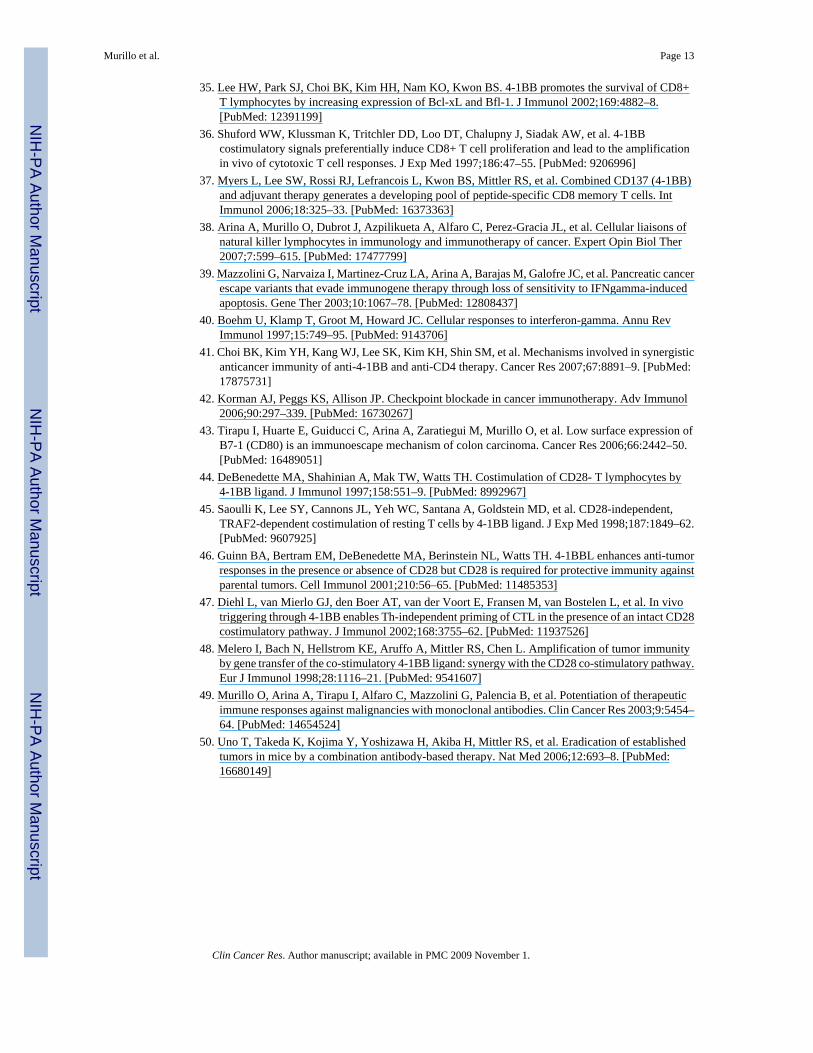
In this study, we examined and compared the therapeutic efficacy of these fourimmunostimulatory mAbs in the highly tumorigenic HOPC myeloma model. To this end,HOPC cells (5×10⁵ cells per mouse) were inoculated in the peritoneal cavity of BALB/c miceon day 0, and on days 4 and 7 the corresponding mAb was i.v. injected. In this experimentalsystem, we found that both anti-CD137 and anti-CTLA-4 mAbs showed a clearly defined anti-myeloma effect, with 40-50% of animals surviving long-term (>120 days), while anti-ICAM-2and anti-CD40 mAbs at these dose regimes showed little and no therapeutic activity,respectively (Fig. 1A). It is noteworthy that while CD137, CTLA-4 and CD40 are absent fromthe plasma membrane of tumor cells, ICAM-2 is readily expressed (Fig1B).
To determine whether treatment with anti-CD137 or anti-CTLA-4 mAbs concomitantly elicitsanti-tumor immunity that is long-lasting, mice that had been cured of the HOPC tumor bytreatment with these two immunostimulatory mAbs were re-challenged s.c. with HOPC cells.We found that while cured mice did not develop palpable HOPC tumors, all naive mice showedprogressive tumor growth (Fig. 1C). In parallel, we observed that the long-lasting immunememory developed in mice cured by anti-CD137 or anti-CTLA-4 mAbs was specific forHOPC, since these mice did not reject the syngeneic CT26 tumor cell line, inoculated on theiropposite flank (Fig. 1C). These findings indicate that both anti-CD137 and anti-CTLA-4 mAbsmay be useful for myeloma therapy.
Although, there was previous published information about anti-CTLA-4 mAbs inplasmacytoma models (30), the potential of anti-CD137 mAbs which have a safer preclinicalprofile (31,32) remains unexplored. Therefore, we next confirmed the anti-tumor effect of anti-CD137 mAb in another experimental plasmacytoma model. With this aim, NS0 myeloma cells(5×10⁵) were inoculated i.p. in BALB/c mice on day 0, and on days 4 and 7 post-tumor cellinoculation, anti-CD137 mAb or the control rat IgG were i.v. injected. In this experimentalsystem, we found that anti-CD137 mAb showed a potent therapeutic effect, with 70-80% ofanimals surviving long-term (>120 days) (Fig. 2A).
Interestingly, neither HOPC cells nor NS0 cells expressed the CD137 molecule on their plasmamembranes, indicating that the therapeutic effect of the agonistic mAb is not mediated viadirect targeting of the malignant plasma cells (Fig 1B and 2B).
We also evaluated the therapeutic effect of anti-CD137 mAb in established subcutaneous NS0tumors that were clearly palpable before commencement of treatment. To this end, micereceived a s.c. injection of 5×10⁵ NS0 cells on day 0, and on days 9, 11, 13 and 15 post-tumorcell inoculation were treated i.p. with either anti-CD137 mAb or control rat IgG. Consistentwith our previous results, anti-CD137 mAb treatment resulted in profound inhibition of tumorgrowth and more than 60% of mice bearing NS0 tumors were completely cured (Fig. 2C). Thisrobust therapy model was thereafter chosen to study the mechanistic requirements behind thetherapeutic effects of anti-CD137 mAb.
NSO tumor rejections are accompanied by CTL induction—The spleen and tumordraining lymph nodes from the mice that had been cured from NS0 subcutaneous tumors byanti-CD137 mAb treatment contained cells that upon 5 day restimulation in culture withirradiated NSO cells showed tumor specific cytolytic activity against NS0 cells in 51Cr-releaseassays (Fig 2D right graphs). Moreover, these cocultures contained CD8+ T cells thatupregulated intracellular IFN-γ and showed degranulation (measured as surface CD107a)specifically upon rexposure to NSO cells, but not CT26 cells (Fig 2D dot plots on the left).This indicates that CD8 T cells can recognize tumor antigens on NS0 cells.
Murillo et al. Page 6
Clin Cancer Res. Author manuscript; available in PMC 2009 November 1.
NIH
-PA Author Manuscript
NIH
-PA Author Manuscript
NIH
-PA Author Manuscript
Both NK and CD8+ T cells are required for tumor rejectionTo identify the cell types responsible for the anti-tumor activity of anti-CD137 mAb, we carriedout in vivo leukocyte subset depletion prior to anti-CD137 treatment. As shown in Fig. 3A,depletion of either NK cells or CD8+ T cells significantly impaired the therapeutic effect ofthe treatment. In this regard, we found that NSO cells are almost as susceptible to NK-mediatedlysis as the sensitive YAC-1 cells (inset to figure 3A in the NK depletion graph) despite of thefact that NSO cells intensely express surface MHC class I (Figure 2B). In vitro NK cytotoxicitywas observed with NK cells obtained from poly I:C-preinjected Rag−/− syngeneic mice. TheseNK cells unsuccessfully killed NK-resistant P815 cells in the same assays (inset to figure 3ANK depletion graph). In contrast, CD4+ T cell depletion had no significant effect on tumorrejection. These results indicate that both NK and CD8+ T cells, but not CD4+ T cells, arerequired for tumor rejection. It is noteworthy that depletion of CD8+ subset was performedwith an anti-CD8β depleting antibody to ensure that only peripheral CD8+ T cells but notCD8α+ DCs were affected.
Normal function of IFN-γ is an absolute requirement for tumor rejectionIFN-γ production is critical for the cell-mediated anti-tumor immune response. Here, weexamined whether IFN-γ was required for the anti-myeloma effect of anti-CD137 treatmentas described for other tumor models (33). To this end, both WT and IFN-γ-deficient mice wereinoculated with NS0 cells and treated with anti-CD137 mAb or the control rat IgG as describedin Materials and Methods. Whereas tumors in the WT BALB/c mice regressed after treatment,all of IFN-γ-deficient mice developed progressively growing tumors (Fig. 3B). Similarly,tumor regression was significantly impaired in mice that received a neutralizing anti-IFN-γmAb (Fig. 3B). The fact that the anti-IFN-γ mAb did not completely abolish the therapeuticeffect of anti-CD137 mAb may simply be a result of incomplete blockade. Therefore, tumoreradication after treatment with agonistic anti-CD137 mAb is dependent on IFN-γ.
Anti-CD137 mAb induced NK cell augment in TDLNs and IFN-γ; production by NK cellsThe abolishment of efficacy upon NK cell depletion was remarkable (Fig 3A). At the timewhen tumor rejections were first observed (8-10 days after treatment onset), both inguinal andaxillary TDLNs had increased relative and absolute numbers of NK cells (CD3−DX5+cells)when the mice had been treated with anti-CD137 mAb (Fig. 4A). We wondered if such NKcells under anti-CD137 treatment would also show a higher degree of activation and we foundthat DLN NK cells showed similar levels of CD69 expression regardless of the antibody usedfor treatment. However, anti-CD137 treatment up-regulated the capability of NK cells in DLNto produce IFN-γ as assessed by intracellular staining (Fig. 4B). These effects on NK biologyat the DLN are likely involved in the absolute requirement of NK cells for the antitumor effects.
The tumor infiltrate of NSO tumors that are responding to anti-CD137 treatment are enrichedin CD8 T cells
Cell suspensions from NSO tumors taken from mice which have been treated with anti-CD137and showed signs of growth delay or shrinkage were obtained to study their lymphocytecontent. It was found that there was an increase of CD8 T cells while NK and CD4 cells werenot increased in the tumor rejecting infiltrate (Figure 4C). These data indicate that the mainrole at the final execution of tumor rejection corresponds to CD8 T cells that abundantlypopulate the tumor lesion. This does not preclude that NK cells could be playing a role at earlierstages or that they cooperate with CTLs in an orchestrated fashion (29). However percentageand absolute numbers (not shown) of intratumour leukocytes provide evidence indicating thatCTLs are the main players when rejections become clinically meaningful.
Murillo et al. Page 7
Clin Cancer Res. Author manuscript; available in PMC 2009 November 1.
NIH
-PA Author Manuscript
NIH
-PA Author Manuscript
NIH
-PA Author Manuscript
The anti-tumor effect of anti-CD137 is independent of CD28To determine whether or not co-stimulation through CD28 is a pre-requisite for CD137signaling, we first investigated whether triggering the CD28 pathway contributes to the up-regulation of CD137 on naïve T cells. To this end purified CD8+ T cells were stimulated invitro with plate bound anti-CD3 mAb in the presence or absence of soluble anti-CD28 mAb.We observed that TCR triggering by anti-CD3 mAbs in combination with CD28 co-stimulationwas much more efficient at inducing surface expression of CD137 on CD8+ T cells than TCR-CD3 triggering alone (Fig. 5A).
Based on these results we hypothesized that the in vivo blockade of the CD28 pathway wouldabrogate the anti-myeloma effect of anti-CD137 mAb. To test this hypothesis both WT andCD28-deficient mice were inoculated with NS0 cells and treated with anti-CD137 mAb or thecontrol rat IgG as described before. Surprisingly, the anti-myeloma effect of anti-CD137 mAbwas preserved in CD28-deficient mice. Thus, although CD28 signaling increases theexpression of CD137 on CD8+ T cells in vitro, the therapeutic effect of anti-CD137 isindependent of CD28 (Fig 5B).
Anti-CD137 decreased tumor burden in 5TGM1 myeloma-bearing miceTo extend our observations about the anti-plasmacytoma effect of anti-CD137 to a MM modelconsidered more relevant to the human disease, we used the 5TGM1 cell line transfected withthe EGFP gene to accurately monitor tumor progression (22). This cell line does not expressdetectable levels of CD137 on its plasma membrane upon FACS analysis (data not shown).Intravenous injection of these tumor cells in naïve syngeneic C57BL/KaLwRijHsd mice givesrise to a disseminated tumor model in which fluorescent tumor cells can be visualized astumoral foci in multiple organs in a distribution that very closely resembles severe cases ofMM in humans. This is true particularly with regard to skeletal involvement (Fig. 6A and B).An Anti-CD137 mAb treatment course started four days after tumor cell inoculation clearlyreduced multi-organ tumor burden in a significant proportion of mice (Fig. 6 A and B), althoughit did not attain the efficacy achieved by a course of bortezomib in a similar experimentalschedule (Fig. 6A). Anti-CD137 mAb-mediated reduction of tumor burden was evident in theskeleton of 7 out of 10 mice and although tumor disappearance was complete in only 3 out of7 mice, the tumor fluorescent foci in the remaining four mice was nonetheless significantlyreduced and comparable to those seen in bortezomib-treated mice. The efficacy of anti-CD137mAb treatment was even more dramatic in visceral organs with spleen and liver involvementin only one out of 10 mice inoculated with tumor cells compared to ≥ 7 out of 10 in tumor-inoculated mice treated either with vehicle or a control IgG (Fig. 6A, B). Consistent with theseobservations, weight loss assessed on day 30 post-tumor cell inoculation when the mice weresacrificed (data not shown), was also less pronounced in anti-CD137 mAb-treated micecompared to the other two groups. Moreover, the serum monoclonal paraprotein concentration,a marker of overall myeloma tumor load, was significantly decreased by the course of anti-CD137 mAb treatment compared to the vehicle or rat control Ab treatment (Fig. 6C), althoughin this respect anti-CD137 mAb was again surpassed by the efficacy of bortezomib. Overallthese data indicate that anti-CD137 mAb is clearly efficacious in this MM model even asmonotherapy and further suggest the clinical potential of the approach.
DISCUSSIONThis study demonstrates that pharmacological agonistic manipulation of CD137 with a specificmAb shows benefit in various murine myeloma models and provides important clues on theinvolvement of CD8+ T lymphocytes and NK cells in the therapeutic immune response. Theeffects of anti-CD137 mAb are not directed against the malignant plasma cells but indirectly
Murillo et al. Page 8
Clin Cancer Res. Author manuscript; available in PMC 2009 November 1.
NIH
-PA Author Manuscript
NIH
-PA Author Manuscript
NIH
-PA Author Manuscript
mediated by immune regulation, as suggested by the absence of CD137 from the cell membraneof the different plasmacytomas used.
It has been postulated that the antibody engages CD137 expressed on activated T cells andmaybe on some activated NK cells in the tumor bearing mice (7). CD137 induction on CD8+
T lymphocytes must be the result of tumor antigen presentation either by tumor cells orprofessional APCs. The advantageous effects of CD137 ligation on CTL differentiation andmemory have been extensively reported (34-37). However, the direct or indirect pathway thatleads in vivo to the activation of NK cells, as we have observed in DLN, is still unclear (38).We are investigating whether ligation of the CD137 molecules expressed on NK cells plays arole in tumor rejection. In this regard, we have recently reported that tumor rejection is oftenthe result of the concerted action of multiple leukocyte partners including NK cells, which playa pivotal role and can be found to be producing IFN-γ in vivo (29). The finding of the sensitivityof NSO cells to autologous NK cells despite high surface MHC class I indicates the expressionof ligands for NK activatory receptors (38).
NK-cell production of IFN-γ at sentinel LN have been reported to be important in the initiationof T cell immunity, and tumoricidal NK cells have been detected in tumor rejecting infiltrates(38). In our case, NK cells at tumor DLNs show activation features and numeric increases thatcould be dependent on either recruitment or local proliferation of NK cells. In spite of theabsolute need of NK cells to achieve efficacy, it is clear that at the time when the tumors startto be clinically controlled by treatment, the rejecting infiltrate is dominated by CTLs. Moreinvestigation is needed to precisely define the train of events that leads to tumor rejectionthrough the cooperative crosstalk between NK and CTLs. Our results further highlight animportant role for NK cells operating at the draining LN (38).
IFN-γ is a pleiotropic cytokine and it has been reported to induce changes in tumor endothelialcells thereby facilitating homing of effector T cells into solid tumors and to inhibitangiogenesis. In addition, IFN-γ may damage tumor cells by means of anti-proliferative andpro-apoptotic signaling pathways (39), while up-regulating antigen presentation, as well asCTL and macrophage activity (40). In our hands, recombinant IFN-γ had no effect on theproliferation and survival of NS0 plasma cells in culture (data not shown), strongly suggestingthat it plays an indirect role in the efficacy of anti-CD137 mAb treatments. The criticalrequirement of IFN-γ for tumor rejection suggests its potential as a biomarker to correlate withefficacy in eventual clinical trials for myeloma.
The injected therapeutic antibody can bind CD137 on other leukocyte and endothelial celllineages (17). The role of these interactions at inducing tumor immunity remains unexplored.We can conclude from selective depletions that intervention of CD4+ T cells is dispensable atleast in the NS0 model. In this sense, it has been recently reported that in mouse melanomaCD4 depletion is even beneficial for antitumor effects (41).
We confirm that anti-CTLA-4 mAb can also have a role in the treatment of myeloma (30) ina fashion that looks comparable to the effects anti-CD137 mAb as observed in an i.p.plasmacytoma model in which suboptimal efficacy is attained by both antibodies. Themechanism of action of anti-CTLA-4 has been described by the group of J. Allison (42) andin essence involves blockade of anti-CTLA-4 negative signals, in such a way that the co-stimulatory activity of CD28 is liberated. Therefore the mechanism of action of anti-CTLA4mAb is different from that of anti-CD137 mAb, although both converge in the generation of atumor-rejecting lymphocyte infiltrate. It is noteworthy that tumor cells used in this workexpress CD80 that may bind CTLA-4 and CD28 on activated T cells. Thus, in these cases,CTLA-4 mAb could block this inhibitory interaction upon in vivo lymphocyte-tumor cellencounter(43).
Murillo et al. Page 9
Clin Cancer Res. Author manuscript; available in PMC 2009 November 1.
NIH
-PA Author Manuscript
NIH
-PA Author Manuscript
NIH
-PA Author Manuscript
Over the last few years, there has been intense interest in whether or not the CD80/86-CD28co-stimulatory pathway is required for CD137 mAb activity. In 1997, TH. Watts et al reportedthat ligation of CD137 can co-stimulate human and mouse CD28−/− T cells (44), suggestingthat CD137 triggering can take place independently of the CD28 pathway (45). This group alsodemonstrated that enforced co-stimulation through CD137 by CD137L could promote anti-tumor responses in the presence or in the absence of CD28 (46). However, R Offringa andcolleagues (47) recently reported that blockade of the costimulatory pathway abrogated thecapacity of agonistic anti-CD137 mAb to trigger CTL immunity in response to the Ad5E1Apeptide vaccine. This was probably because under weaker TCR signals, CD137 expression onnaive T cells is not attained, and therefore the susceptibility of these cells to anti-CD137 mAbsrequired both stimulation of the TCR and CD28-dependent co-stimulation. However, in ourhands, CD28 function is dispensable for CD137 activity, albeit it is easy to find CD8+ T cellactivation conditions in which CD28 co-stimulation results in much higher CD137 expressionby CD8+ T cells in vitro, as previously suggested by experiments with the natural ligand(48).
This study describes the therapeutic effects of anti-CD137 mAb as monotherapy in myelomabut it is clear that CD137 targeted therapy would be most efficacious in combinationimmunotherapy. Combinations of tumor vaccines, adoptive T cell therapy, chemo and/orradiotherapy are known to display additive and synergistic effects in cancer, and combinationtherapies of anti-CD137 mAb and other immunostimulatory mAbs have shown remarkableactivity in other tumor models (7,32,49,50). However for practical reasons, work on myelomamodels should first define anti-CD137 combination potential with standard treatments for MMsuch as bone-marrow transplantation and chemotherapy.
The significant reduction of tumor burden in the experiments performed with the 5TGM1model is remarkable due to the nature of the systemic dissemination of this experimentaldisease. The comparison of anti-CD137 mAb with bortezomib seems to favor the latter agentbut should be considered with caution. On the one hand, bortezomib has direct cytotoxic effectson myeloma cells and likely acts immediately on the tumor cells in vivo, whereas the full effectof the anti-CD137 mAb would need a latency period until the immune response is sufficientlyup-regulated. On the other hand, the full potential of anti-CD137 mAb has yet to be exploited,in particular with regard to combination strategies either with a tumor vaccine (25) or anintervention to increase tumor immunogenicity. There is also the possibility of combining sub-optimal doses of bortezomib with anti-CD137 mAb in order to reduce some of the well knownside effects of bortezomib such as neuropathies. The use of the 5TGM1 myeloma model forsuch combination therapy studies in the future has obvious advantages since it utilizesimmunocompetent mice and accumulation of the monoclonal paraprotein in serum accuratelyreflects tumor burden. In addition, transfection of 5TGM1 cells with EGFP permits imageassessment (22) of tumor load in real time without increasing immunogenicity, since CTLs donot recognize EGFP as foreign in the H-2b class I molecules (29).
There are a number of rationale for proposing clinical trials with agonist anti-CD137 mAb inmyeloma: (i) the results of the present study on three different myeloma mouse models,including the disseminated 5TGM1 model that shares many characteristic features with thehuman disease; (ii) a safe clinical leading profile (ASCO 2008, abstract 3007) and (31)); (iii)the abundance of NK cells and memory T cells in the bone marrow which is the most commonprimary site for malignant plasma cells in myeloma (1); (iv) an agent of this kind(BMS663513) is already undergoing phase I and II trials for melanoma, renal cell carcinoma,lung and ovarian cancer. The effects of the anti-CD137 mAb on the disseminated 5TGM1model reported herein provide a base-line to further optimize combination strategies andimprove preclinical efficacy to guide future clinical trial design.
Murillo et al. Page 10
Clin Cancer Res. Author manuscript; available in PMC 2009 November 1.
NIH
-PA Author Manuscript
NIH
-PA Author Manuscript
NIH
-PA Author Manuscript
AcknowledgementsWe are grateful to Dr. L. Chen both for providing an agonist anti-CD137 producing hybridoma as well as for helpfulcriticism and discussion. Dr. James Allison kindly provided anti-CTLA-4 mAb producing hybridoma cells. Drs. JesúsPrieto, Ascensión Lopez-Diaz de Cerio, Mercedes Rodriguez-Calvillo, Maurizio Bendandi, and Juan José Lasarte areacknowledged for support and scientific discussion. Elena Ciordia, Javier Guillén, Juan Percaz and Eneko Elizaldeare acknowledged for excellent animal care.
Grant suport: Research grants were from: Ministerio de Educación y Ciencia (MEC-SAF2005-03131), Departamentode Educación del Gobierno de Navarra, Departamento de Salud del Gobierno de Navarra (Beca Ortiz de Landázuri).Redes temáticas de investigación cooperativa RETIC (RD06/0020/0065), European commission VII fameworkprogram (ENCITE) and “UTE for project FIMA”. OM and AA were recipients of scholarships from Ministerio deEducación (MEC) and Fondo de investigación sanitaria (BEFI) respectively. S H-S is sponsored by the AECC(Asociación Española contra el Cancer). BOO is supported by a NIH/NCI Career Development Award (KO1CA104180). The use of the flow cytometry facilities at UTHSC at San Antonio is supported by a Cancer CenterSupport Grant from the NCI (P30 CA054174) to the Cancer Therapy & Research center (CTRC@UTHSCSA).
REFERENCES1. Hideshima T, Mitsiades C, Tonon G, Richardson PG, Anderson KC. Understanding multiple myeloma
pathogenesis in the bone marrow to identify new therapeutic targets. Nat Rev Cancer 2007;7:585–98.[PubMed: 17646864]
2. Barlogie B, Shaughnessy J, Tricot G, Jacobson J, Zangari M, Anaissie E, et al. Treatment of multiplemyeloma. Blood 2004;103:20–32. [PubMed: 12969978]Epub 2003 Sep 11
3. Armand JP, Burnett AK, Drach J, Harousseau JL, Lowenberg B, San Miguel J. The emerging role oftargeted therapy for hematologic malignancies: update on bortezomib and tipifarnib. Oncologist2007;12:281–90. [PubMed: 17405892]
4. Richardson PG, Mitsiades C, Hideshima T, Anderson KC. Lenalidomide in multiple myeloma. ExpertRev Anticancer Ther 2006;6:1165–73. [PubMed: 16925483]
5. Mihelic R, Kaufman JL, Lonial S. Maintenance therapy in multiple myeloma. Leukemia2007;21:1150–7. [PubMed: 17344913]
6. Chiriva-Internati M, Cobos E, Kast WM. Advances in immunotherapy of multiple myeloma: from thediscovery of tumor-associated antigens to clinical trials. Int Rev Immunol 2007;26:197–222. [PubMed:17558744]
7. Melero I, Hervas-Stubbs S, Glennie M, Pardoll DM, Chen L. Immunostimulatory monoclonalantibodies for cancer therapy. Nat Rev Cancer 2007;7:95–106. [PubMed: 17251916]
8. Peggs KS, Segal NH, Allison JP. Targeting immunosupportive cancer therapies: accentuate thepositive, eliminate the negative. Cancer Cell 2007;12:192–9. [PubMed: 17785201]
9. Ribas A, Hanson DC, Noe DA, Millham R, Guyot DJ, Bernstein SH, et al. Tremelimumab(CP-675,206), a cytotoxic T lymphocyte associated antigen 4 blocking monoclonal antibody in clinicaldevelopment for patients with cancer. Oncologist 2007;12:873–83. [PubMed: 17673618]
10. Melero I, Shuford WW, Newby SA, Aruffo A, Ledbetter JA, Hellstrom KE, et al. Monoclonalantibodies against the 4-1BB T-cell activation molecule eradicate established tumors. Nat Med1997;3:682–5. [PubMed: 9176498]
11. Nam KO, Kang WJ, Kwon BS, Kim SJ, Lee HW. The therapeutic potential of 4-1BB (CD137) incancer. Curr Cancer Drug Targets 2005;5:357–63. [PubMed: 16101383]
12. Mittler RS, Foell J, McCausland M, Strahotin S, Niu L, Bapat A, et al. Anti-CD137 antibodies in thetreatment of autoimmune disease and cancer. Immunol Res 2004;29:197–208. [PubMed: 15181282]
13. Seo SK, Choi JH, Kim YH, Kang WJ, Park HY, Suh JH, et al. 4-1BB-mediated immunotherapy ofrheumatoid arthritis. Nat Med 2004;10:1088–94. [PubMed: 15448685]
14. Sun Y, Chen HM, Subudhi SK, Chen J, Koka R, Chen L, et al. Costimulatory molecule-targetedantibody therapy of a spontaneous autoimmune disease. Nat Med 2002;8:1405–13. [PubMed:12426559]
15. Sun Y, Chen JH, Fu Y. Immunotherapy with agonistic anti-CD137: two sides of a coin. Cell MolImmunol 2004;1:31–6. [PubMed: 16212918]
Murillo et al. Page 11
Clin Cancer Res. Author manuscript; available in PMC 2009 November 1.
NIH
-PA Author Manuscript
NIH
-PA Author Manuscript
NIH
-PA Author Manuscript

16. Myers LM, Vella AT. Interfacing T-cell effector and regulatory function through CD137 (4-1BB)co-stimulation. Trends Immunol 2005;26:440–6. [PubMed: 15979409]
17. Watts TH. TNF/TNFR family members in costimulation of T cell responses. Annu Rev Immunol2005;23:23–68. [PubMed: 15771565]
18. Wilcox RA, Tamada K, Strome SE, Chen L. Signaling through NK cell-associated CD137 promotesboth helper function for CD8+ cytolytic T cells and responsiveness to IL-2 but not cytolytic activity.J Immunol 2002;169:4230–6. [PubMed: 12370353]
19. Miller RE, Jones J, Le T, Whitmore J, Boiani N, Gliniak B, et al. 4-1BB-specific monoclonal antibodypromotes the generation of tumor-specific immune responses by direct activation of CD8 T cells ina CD40-dependent manner. J Immunol 2002;169:1792–800. [PubMed: 12165501]
20. Melero I, Johnston JV, Shufford WW, Mittler RS, Chen L. NK1.1 cells express 4-1BB (CDw137)costimulatory molecule and are required for tumor immunity elicited by anti-4-1BB monoclonalantibodies. Cell Immunol 1998;190:167–72. [PubMed: 9878117]
21. Ye Z, Hellstrom I, Hayden-Ledbetter M, Dahlin A, Ledbetter JA, Hellstrom KE. Gene therapy forcancer using single-chain Fv fragments specific for 4-1BB. Nat Med 2002;8:343–8. [PubMed:11927939]
22. Oyajobi BO, Munoz S, Kakonen R, Williams PJ, Gupta A, Wideman CL, et al. Detection of myelomain skeleton of mice by whole-body optical fluorescence imaging. Mol Cancer Ther 2007;6:1701–8.[PubMed: 17541032]
23. Dallas SL, Garrett IR, Oyajobi BO, Dallas MR, Boyce BF, Bauss F, et al. Ibandronate reducesosteolytic lesions but not tumor burden in a murine model of myeloma bone disease. Blood1999;93:1697–706. [PubMed: 10029599]
24. Radl J, Croese JW, Zurcher C, Van den Enden-Vieveen MH, de Leeuw AM. Animal model of humandisease. Multiple myeloma. Am J Pathol 1988;132:593–7. [PubMed: 3414786]
25. Wilcox RA, Flies DB, Zhu G, Johnson AJ, Tamada K, Chapoval AI, et al. Provision of antigen andCD137 signaling breaks immunological ignorance, promoting regression of poorly immunogenictumors. J Clin Invest 2002;109:651–9. [PubMed: 11877473]
26. Leach DR, Krummel MF, Allison JP. Enhancement of antitumor immunity by CTLA-4 blockade.Science 1996;271:1734–6. [PubMed: 8596936]
27. Melero I, Gabari I, Corbi AL, Relloso M, Mazzolini G, Schmitz V, et al. An anti-ICAM-2 (CD102)monoclonal antibody induces immune-mediated regressions of transplanted ICAM-2-negative coloncarcinomas. Cancer Res 2002;62:3167–74. [PubMed: 12036930]
28. Melero I, Gabari I, Tirapu I, Arina A, Mazzolini G, Baixeras E, et al. Anti-ICAM-2 monoclonalantibody synergizes with intratumor gene transfer of interleukin-12 inhibiting activation-induced T-cell death. Clin Cancer Res 2003;9:3546–54. [PubMed: 14506140]
29. Arina A, Murillo O, Hervas-Stubbs S, Azpilikueta A, Dubrot J, Tirapu I, et al. The combined actionsof NK and T lymphocytes are necessary to reject an EGFP+ mesenchymal tumor through mechanismsdependent on NKG2D and IFN gamma. Int J Cancer 2007;121:1282–95. [PubMed: 17520674]
30. Mokyr MB, Kalinichenko T, Gorelik L, Bluestone JA. Realization of the therapeutic potential ofCTLA-4 blockade in low-dose chemotherapy-treated tumor-bearing mice. Cancer Res1998;58:5301–4. [PubMed: 9850053]
31. Niu L, Strahotin S, Hewes B, Zhang B, Zhang Y, Archer D, et al. Cytokine-mediated disruption oflymphocyte trafficking, hemopoiesis, and induction of lymphopenia, anemia, and thrombocytopeniain anti-CD137-treated mice. J Immunol 2007;178:4194–213. [PubMed: 17371976]
32. Kocak E, Lute K, Chang X, May KF Jr. Exten KR, Zhang H, et al. Combination therapy with anti-CTL antigen-4 and anti-4-1BB antibodies enhances cancer immunity and reduces autoimmunity.Cancer Res 2006;66:7276–84. [PubMed: 16849577]
33. Wilcox RA, Flies DB, Wang H, Tamada K, Johnson AJ, Pease LR, et al. Impaired infiltration oftumor-specific cytolytic T cells in the absence of interferon-gamma despite their normal maturationin lymphoid organs during CD137 monoclonal antibody therapy. Cancer Res 2002;62:4413–8.[PubMed: 12154048]
34. Lee HW, Nam KO, Park SJ, Kwon BS. 4-1BB enhances CD8+ T cell expansion by regulating cellcycle progression through changes in expression of cyclins D and E and cyclin-dependent kinaseinhibitor p27kip1. Eur J Immunol 2003;33:2133–41. [PubMed: 12884287]
Murillo et al. Page 12
Clin Cancer Res. Author manuscript; available in PMC 2009 November 1.
NIH
-PA Author Manuscript
NIH
-PA Author Manuscript
NIH
-PA Author Manuscript
35. Lee HW, Park SJ, Choi BK, Kim HH, Nam KO, Kwon BS. 4-1BB promotes the survival of CD8+T lymphocytes by increasing expression of Bcl-xL and Bfl-1. J Immunol 2002;169:4882–8.[PubMed: 12391199]
36. Shuford WW, Klussman K, Tritchler DD, Loo DT, Chalupny J, Siadak AW, et al. 4-1BBcostimulatory signals preferentially induce CD8+ T cell proliferation and lead to the amplificationin vivo of cytotoxic T cell responses. J Exp Med 1997;186:47–55. [PubMed: 9206996]
37. Myers L, Lee SW, Rossi RJ, Lefrancois L, Kwon BS, Mittler RS, et al. Combined CD137 (4-1BB)and adjuvant therapy generates a developing pool of peptide-specific CD8 memory T cells. IntImmunol 2006;18:325–33. [PubMed: 16373363]
38. Arina A, Murillo O, Dubrot J, Azpilikueta A, Alfaro C, Perez-Gracia JL, et al. Cellular liaisons ofnatural killer lymphocytes in immunology and immunotherapy of cancer. Expert Opin Biol Ther2007;7:599–615. [PubMed: 17477799]
39. Mazzolini G, Narvaiza I, Martinez-Cruz LA, Arina A, Barajas M, Galofre JC, et al. Pancreatic cancerescape variants that evade immunogene therapy through loss of sensitivity to IFNgamma-inducedapoptosis. Gene Ther 2003;10:1067–78. [PubMed: 12808437]
40. Boehm U, Klamp T, Groot M, Howard JC. Cellular responses to interferon-gamma. Annu RevImmunol 1997;15:749–95. [PubMed: 9143706]
41. Choi BK, Kim YH, Kang WJ, Lee SK, Kim KH, Shin SM, et al. Mechanisms involved in synergisticanticancer immunity of anti-4-1BB and anti-CD4 therapy. Cancer Res 2007;67:8891–9. [PubMed:17875731]
42. Korman AJ, Peggs KS, Allison JP. Checkpoint blockade in cancer immunotherapy. Adv Immunol2006;90:297–339. [PubMed: 16730267]
43. Tirapu I, Huarte E, Guiducci C, Arina A, Zaratiegui M, Murillo O, et al. Low surface expression ofB7-1 (CD80) is an immunoescape mechanism of colon carcinoma. Cancer Res 2006;66:2442–50.[PubMed: 16489051]
44. DeBenedette MA, Shahinian A, Mak TW, Watts TH. Costimulation of CD28- T lymphocytes by4-1BB ligand. J Immunol 1997;158:551–9. [PubMed: 8992967]
45. Saoulli K, Lee SY, Cannons JL, Yeh WC, Santana A, Goldstein MD, et al. CD28-independent,TRAF2-dependent costimulation of resting T cells by 4-1BB ligand. J Exp Med 1998;187:1849–62.[PubMed: 9607925]
46. Guinn BA, Bertram EM, DeBenedette MA, Berinstein NL, Watts TH. 4-1BBL enhances anti-tumorresponses in the presence or absence of CD28 but CD28 is required for protective immunity againstparental tumors. Cell Immunol 2001;210:56–65. [PubMed: 11485353]
47. Diehl L, van Mierlo GJ, den Boer AT, van der Voort E, Fransen M, van Bostelen L, et al. In vivotriggering through 4-1BB enables Th-independent priming of CTL in the presence of an intact CD28costimulatory pathway. J Immunol 2002;168:3755–62. [PubMed: 11937526]
48. Melero I, Bach N, Hellstrom KE, Aruffo A, Mittler RS, Chen L. Amplification of tumor immunityby gene transfer of the co-stimulatory 4-1BB ligand: synergy with the CD28 co-stimulatory pathway.Eur J Immunol 1998;28:1116–21. [PubMed: 9541607]
49. Murillo O, Arina A, Tirapu I, Alfaro C, Mazzolini G, Palencia B, et al. Potentiation of therapeuticimmune responses against malignancies with monoclonal antibodies. Clin Cancer Res 2003;9:5454–64. [PubMed: 14654524]
50. Uno T, Takeda K, Kojima Y, Yoshizawa H, Akiba H, Mittler RS, et al. Eradication of establishedtumors in mice by a combination antibody-based therapy. Nat Med 2006;12:693–8. [PubMed:16680149]
Murillo et al. Page 13
Clin Cancer Res. Author manuscript; available in PMC 2009 November 1.
NIH
-PA Author Manuscript
NIH
-PA Author Manuscript
NIH
-PA Author Manuscript
Fig.1. Mice treated with anti-CD137 mAb showed increased survival to HOPC plasmacytoma andacquire long-lasting and tumor-specific immunity. The therapeutic effect of anti-CD137 treatmentwas independent from direct targeting of HOPC myeloma cells(A) To compare the relative efficacy of the different immunostimulatory mAbs on the treatmentof HOPC tumors, BALB/c mice (16 per group) were i.p. injected with 5×10⁵ HOPC viablecells on day 0, and on days 4 and 7 were i.v. treated with control rat IgG or the indicated mAbsat the dose of 100μg. These mice were examined weekly for palpable abdominal tumors orascites. Mice survival was plotted using the Kaplan-Meier method and analyzed forsignificance using the log-rank test. Pooled data from two identical experiments are shown.(B) HOPC tumor cells were evaluated for MHC I, MHC II, CD80, CD86, ICAM-2, CD40,
Murillo et al. Page 14
Clin Cancer Res. Author manuscript; available in PMC 2009 November 1.
NIH
-PA Author Manuscript
NIH
-PA Author Manuscript
NIH
-PA Author Manuscript
CD137 and CTLA-4 expression by FACS. The grey area represents the fluorochrome-taggedisotype control antibody and the white area represents the relevant antibody. (C) To examinewhether a long-lasting and tumor-specific immunity could be generated, mice that had beencured of HOPC tumors with either anti-CTLA-4 or anti-CD137 mAbs were re-challenge 4month later with HOPC and CT26 cells. Tumor cells (5×10⁵ per mouse) were s.c. inoculatedin the opposite flanks of long-term surviving mice from experiment (A) as represented in thescheme. Mice were monitored for tumor growth and compared to naïve age-matched mice.Tumor sizes were assessed by measuring (in millimeters) perpendicular diameters of tumorsand the results are expressed as tumor area. These experiments were repeated at least twiceyielding similar results. Representative data are shown.
Murillo et al. Page 15
Clin Cancer Res. Author manuscript; available in PMC 2009 November 1.
NIH
-PA Author Manuscript
NIH
-PA Author Manuscript
NIH
-PA Author Manuscript
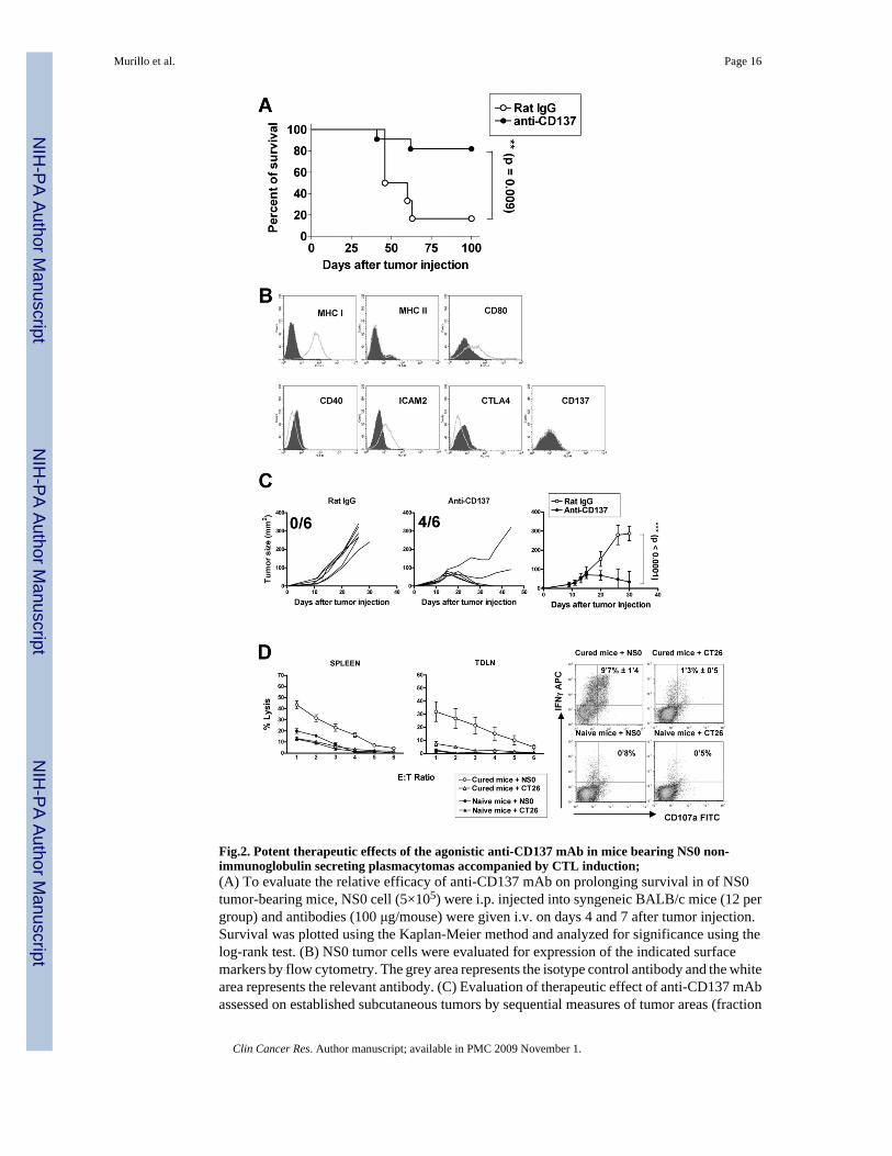
Fig.2. Potent therapeutic effects of the agonistic anti-CD137 mAb in mice bearing NS0 non-immunoglobulin secreting plasmacytomas accompanied by CTL induction;(A) To evaluate the relative efficacy of anti-CD137 mAb on prolonging survival in of NS0tumor-bearing mice, NS0 cell (5×10⁵) were i.p. injected into syngeneic BALB/c mice (12 pergroup) and antibodies (100 μg/mouse) were given i.v. on days 4 and 7 after tumor injection.Survival was plotted using the Kaplan-Meier method and analyzed for significance using thelog-rank test. (B) NS0 tumor cells were evaluated for expression of the indicated surfacemarkers by flow cytometry. The grey area represents the isotype control antibody and the whitearea represents the relevant antibody. (C) Evaluation of therapeutic effect of anti-CD137 mAbassessed on established subcutaneous tumors by sequential measures of tumor areas (fraction
Murillo et al. Page 16
Clin Cancer Res. Author manuscript; available in PMC 2009 November 1.
NIH
-PA Author Manuscript
NIH
-PA Author Manuscript
NIH
-PA Author Manuscript

of surviving tumor free mice is provided in each graph). BALB/c mice (6 per group) receiveda s.c. injection of 5×10⁵ NS0 cells on day 0, and on days 9, 11, 13 and 15 mice were treatedi.p with mAb 2A or a control IgG at 100μg per injection. Statistical analyses were performedby the T test. This experiment was repeated at least three times yielding similar results.Representative data are shown and compiled data for statistical analysis are presented in aseparate graph. (D) Cell suspensions from spleens and tumor draining lymph nodes of micecured from NS0 sc tumors by anti-CD137 mAb or naïve mice were restimulated in cocultulturewith iradiated NS0 cells (1: 25) for 5 days and tested in 51Cr-release assays (mean±SEM fromsix independent mice) against CT26 and NSO cells (left) and for upregulation of intracellularIFNγ and surface CD107a in gated CD3+CD8+ splenocytes by FACS (right). Percentage ofdouble positive cells expressed as the mean±SEM from 5-day cocultures prepared from 6different mice are shown inside the corresponding dot plots.
Murillo et al. Page 17
Clin Cancer Res. Author manuscript; available in PMC 2009 November 1.
NIH
-PA Author Manuscript
NIH
-PA Author Manuscript
NIH
-PA Author Manuscript
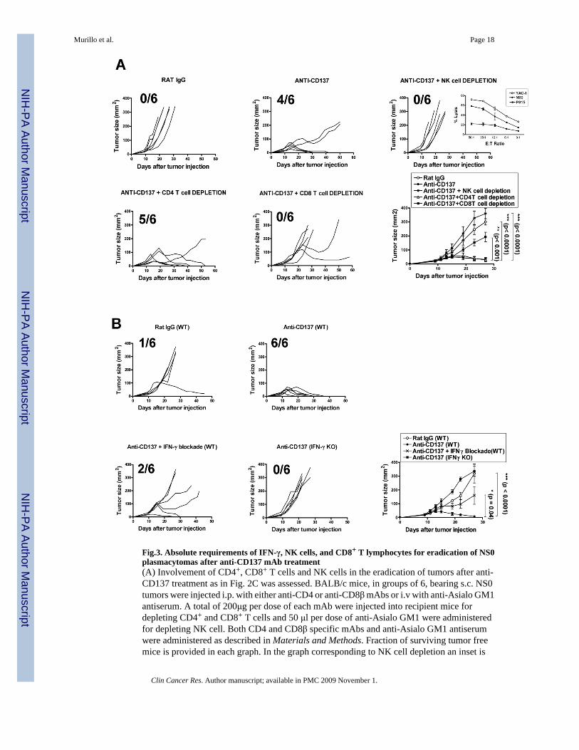
Fig.3. Absolute requirements of IFN-γ, NK cells, and CD8+ T lymphocytes for eradication of NS0plasmacytomas after anti-CD137 mAb treatment(A) Involvement of CD4+, CD8+ T cells and NK cells in the eradication of tumors after anti-CD137 treatment as in Fig. 2C was assessed. BALB/c mice, in groups of 6, bearing s.c. NS0tumors were injected i.p. with either anti-CD4 or anti-CD8β mAbs or i.v with anti-Asialo GM1antiserum. A total of 200μg per dose of each mAb were injected into recipient mice fordepleting CD4+ and CD8+ T cells and 50 μl per dose of anti-Asialo GM1 were administeredfor depleting NK cell. Both CD4 and CD8β specific mAbs and anti-Asialo GM1 antiserumwere administered as described in Materials and Methods. Fraction of surviving tumor freemice is provided in each graph. In the graph corresponding to NK cell depletion an inset is
Murillo et al. Page 18
Clin Cancer Res. Author manuscript; available in PMC 2009 November 1.
NIH
-PA Author Manuscript
NIH
-PA Author Manuscript
NIH
-PA Author Manuscript
provided that shows specific lysis (mean±SEM) in 51Cr release assays demonstrating thesensitivity of NS0 cells to killing by activated DX5+ NK cells isolated from the spleens ofRag1−/− mice that had been pretreated 18h earlier with 50 μg of poly I:C ip. % of lysis werecompared to those achieved against YAC-1 cells and P815 targets. (B) To determine whetherIFN-γ was required for eradication of NS0-derived tumors after anti-CD137 mAb treatment,WT or IFN-γ−/− (IFN-γ K.O.) BALB/c mice (6 per group) were inoculated s.c. with 5×10⁵ NS0viable cells and then treated with either anti-CD137 mAb or control antibody. Alternatively,tumor-bearing mice (n = 6) were treated with anti-CD137 mAb, and were subsequently given200 μg of neutralizing anti-IFN-γ as described in Materials and Methods. Statistical analyseswere performed using the t test. These experiments were performed at least twice, yieldingsimilar results. Representative data are shown and compiled data for statistical analysis arepresented in separate graphs for A and B.
Murillo et al. Page 19
Clin Cancer Res. Author manuscript; available in PMC 2009 November 1.
NIH
-PA Author Manuscript
NIH
-PA Author Manuscript
NIH
-PA Author Manuscript
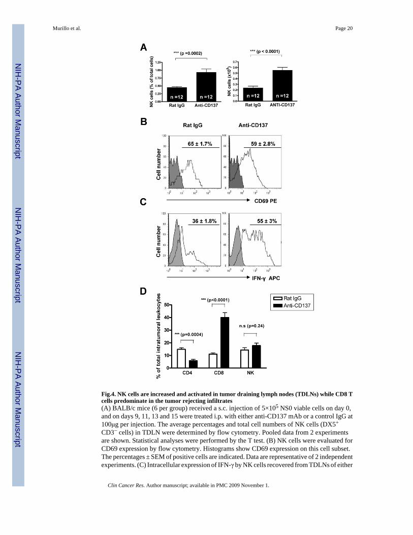
Fig.4. NK cells are increased and activated in tumor draining lymph nodes (TDLNs) while CD8 Tcells predominate in the tumor rejecting infiltrates(A) BALB/c mice (6 per group) received a s.c. injection of 5×10⁵ NS0 viable cells on day 0,and on days 9, 11, 13 and 15 were treated i.p. with either anti-CD137 mAb or a control IgG at100μg per injection. The average percentages and total cell numbers of NK cells (DX5+
CD3− cells) in TDLN were determined by flow cytometry. Pooled data from 2 experimentsare shown. Statistical analyses were performed by the T test. (B) NK cells were evaluated forCD69 expression by flow cytometry. Histograms show CD69 expression on this cell subset.The percentages ± SEM of positive cells are indicated. Data are representative of 2 independentexperiments. (C) Intracellular expression of IFN-γ by NK cells recovered from TDLNs of either
Murillo et al. Page 20
Clin Cancer Res. Author manuscript; available in PMC 2009 November 1.
NIH
-PA Author Manuscript
NIH
-PA Author Manuscript
NIH
-PA Author Manuscript
control IgG or anti-CD137-treated-tumor-bearing mice was examined. The mononuclear cellsfrom TDLNs were stimulated with PMA/Ionomycin in vitro for 5 hours and subsequentlystained for intracellular IFN-γ. The percentages ± SEM of NK cells producing IFN-γ areindicated. In panels (B) and (C) shaded histograms represent isotype control antibody and whitehistograms represent the relevant antibody. (D) percentage± SEM of CD4 (CD3+CD4+), CD8(CD3+CD8+) and NK cells (CD3−DX5+) in lymphoid cell suspensions obtained fromdisaggregated NS0 tumor nodules. The lesions were excised on day 19 from mice that had beentreated with anti-CD137 mAb or control antibody on days 9,11,13,15 after tumor cell injection.Absolute numbers also showed clear increases in intratumoral CD8 T cell counts and decreasesin CD4 T cell counts (data not shown).
Murillo et al. Page 21
Clin Cancer Res. Author manuscript; available in PMC 2009 November 1.
NIH
-PA Author Manuscript
NIH
-PA Author Manuscript
NIH
-PA Author Manuscript
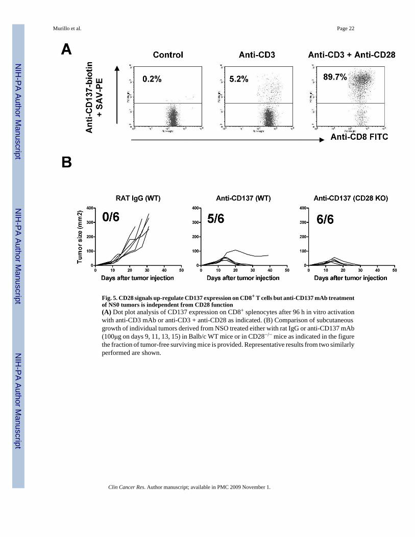
Fig. 5. CD28 signals up-regulate CD137 expression on CD8+ T cells but anti-CD137 mAb treatmentof NS0 tumors is independent from CD28 function(A) Dot plot analysis of CD137 expression on CD8+ splenocytes after 96 h in vitro activationwith anti-CD3 mAb or anti-CD3 + anti-CD28 as indicated. (B) Comparison of subcutaneousgrowth of individual tumors derived from NSO treated either with rat IgG or anti-CD137 mAb(100μg on days 9, 11, 13, 15) in Balb/c WT mice or in CD28−/− mice as indicated in the figurethe fraction of tumor-free surviving mice is provided. Representative results from two similarlyperformed are shown.
Murillo et al. Page 22
Clin Cancer Res. Author manuscript; available in PMC 2009 November 1.
NIH
-PA Author Manuscript
NIH
-PA Author Manuscript
NIH
-PA Author Manuscript
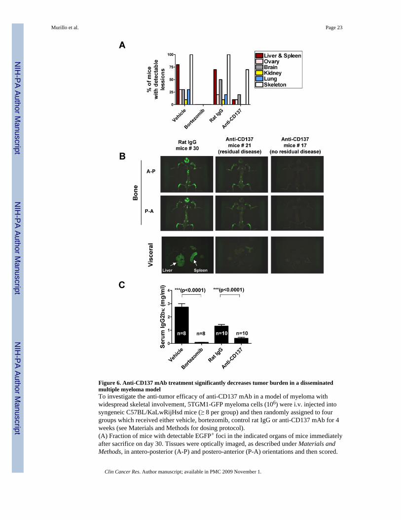
Figure 6. Anti-CD137 mAb treatment significantly decreases tumor burden in a disseminatedmultiple myeloma modelTo investigate the anti-tumor efficacy of anti-CD137 mAb in a model of myeloma withwidespread skeletal involvement, 5TGM1-GFP myeloma cells (106) were i.v. injected intosyngeneic C57BL/KaLwRijHsd mice (≥ 8 per group) and then randomly assigned to fourgroups which received either vehicle, bortezomib, control rat IgG or anti-CD137 mAb for 4weeks (see Materials and Methods for dosing protocol).(A) Fraction of mice with detectable EGFP+ foci in the indicated organs of mice immediatelyafter sacrifice on day 30. Tissues were optically imaged, as described under Materials andMethods, in antero-posterior (A-P) and postero-anterior (P-A) orientations and then scored.
Murillo et al. Page 23
Clin Cancer Res. Author manuscript; available in PMC 2009 November 1.
NIH
-PA Author Manuscript
NIH
-PA Author Manuscript
NIH
-PA Author Manuscript
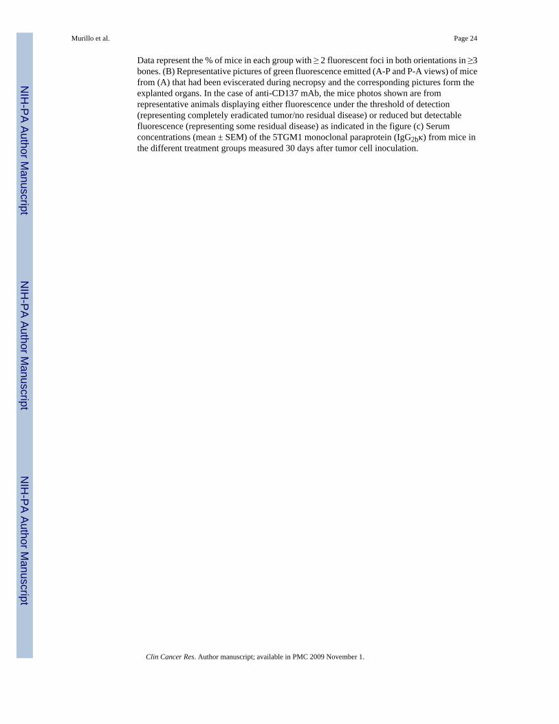
Data represent the % of mice in each group with ≥ 2 fluorescent foci in both orientations in ≥3bones. (B) Representative pictures of green fluorescence emitted (A-P and P-A views) of micefrom (A) that had been eviscerated during necropsy and the corresponding pictures form theexplanted organs. In the case of anti-CD137 mAb, the mice photos shown are fromrepresentative animals displaying either fluorescence under the threshold of detection(representing completely eradicated tumor/no residual disease) or reduced but detectablefluorescence (representing some residual disease) as indicated in the figure (c) Serumconcentrations (mean ± SEM) of the 5TGM1 monoclonal paraprotein (IgG2bκ) from mice inthe different treatment groups measured 30 days after tumor cell inoculation.
Murillo et al. Page 24
Clin Cancer Res. Author manuscript; available in PMC 2009 November 1.
NIH
-PA Author Manuscript
NIH
-PA Author Manuscript
NIH
-PA Author Manuscript
Copyright © 2022 FDOKUMEN


![Agonistic depictions of communication: Vaikeneminen [silence] vs. puhuminen [speaking] in classroom settings for adult education in Finland.](https://static.fdokumen.com/doc/165x107/631ef79a7509c0131f095b3e/agonistic-depictions-of-communication-vaikeneminen-silence-vs-puhuminen-speaking.jpg)