Anti-tumour necrosis factor monoclonal antibody treatment for ocular Behçet's disease
15
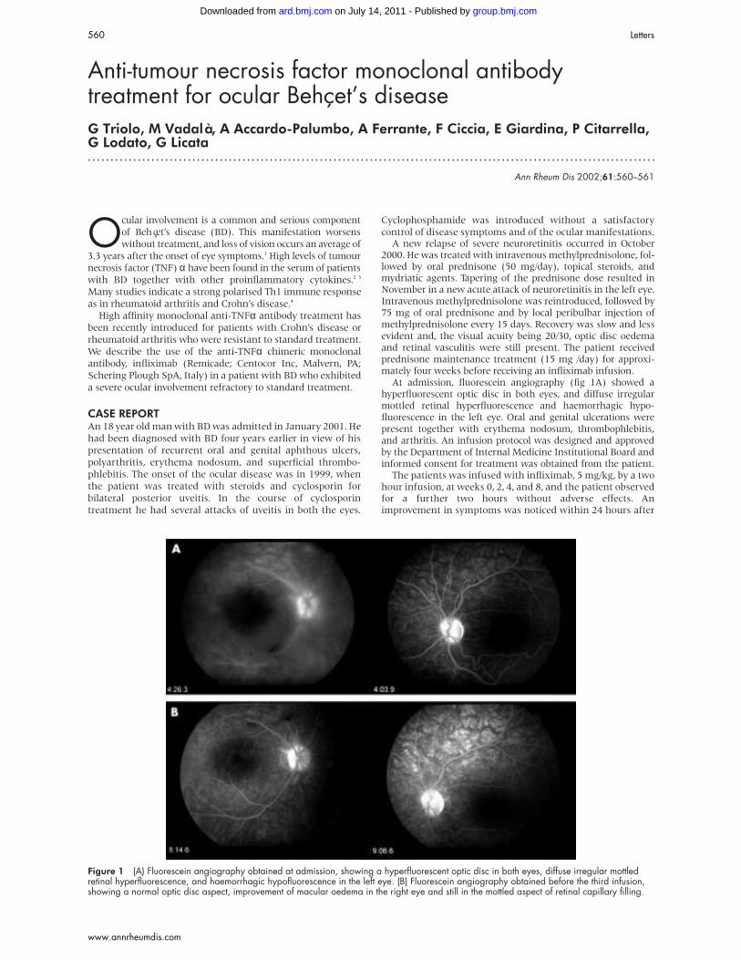
LETTERS Safety and efficacy of TNFα blockade in relapsing vasculitis A D Booth, H J Jefferson, W Ayliffe, P A Andrews, D R Jayne ............................................................................................................................. Ann Rheum Dis 2002;61:559 B lockade of tumour necrosis factor alpha (TNFα) using in- fliximab, a chimeric monoclonal antibody against TNFα, is an effective treatment in rheumatoid arthritis and Crohn’s disease. 12 Sifikakis reported success using infliximab in sight threatening Behçet’s disease. 3 A preliminary study has also reported clinical improvements in the primary systemic vascu- litis, Wegener’s granulomatosis, with the soluble TNFα receptor etanercept. 4 The benefit of lenercept, a soluble p55 TNFα recep- tor fusion protein, on digital vasculitis in rheumatoid arthritis has also been reported. 5 We report the compassionate treatment of six patients with refractory vasculitis using infliximab. Diagnoses were Wegener’s granulomatosis in three and microscopic polyangiitis in three. Three patients were positive for proteinase-3 antineutrophil cytoplasmic antibodies (PR3-ANCA) and one for myeloperoxidase (MPO)-ANCA. Four were female, with a mean age of 58 years (range 23–77) and mean disease duration of 3.5 years. All had had at least three clinical relapses and had received prolonged treatment with corticosteroids and at least four immunosuppressive drugs. At the time of infliximab treatment the eyes were affected in four patients and the lung in three; in addition, five had profound con- stitutional symptoms. The mean prednisolone dose was 17 mg. Three intravenous doses of infliximab 200 mg were given at monthly intervals for three months. One patient complained of fatigue, myalgia, and blurred vision 24 hours after the first infusion, which did not recur on rechallenge. Infliximab was otherwise well tolerated. Five patients had remission of their disease, four within two weeks of treatment. This allowed steroid withdrawal in three and reduction by more than 50% in two. Disease activity assessed by the Birmingham Vasculitis Activity Scores (BVAS) improved from a mean of 6.3 to 0.8 at three months (fig 1). 6 One patient receiving continued inflixi- mab for six months relapsed when the treatment interval was extended to two months. Mean falls in erythrocyte sedimen- tation rate and C reactive protein were 17 mm/1st h and 13 mg/l, respectively. The ANCA status was unchanged. Anti-TNFα treatment heralds a new wave of specifically targeted biological interventions of potential value in the treatment of vasculitis. It offers the hope of improved thera- peutic efficacy over current agents and the possibility of reducing exposure to steroids and immunosuppressive drugs. Further studies are warranted to confirm these observations and explore the role of infliximab as a component of initial protocols. ..................... Authors’ affiliations A D Booth, D R Jayne, Department of Medicine, Addenbrooke’s Hospital, Cambridge CB2 2QQ, UK H J Jefferson, P A Andrews, SW Thames Renal and Transplantation Unit, St Helier Hospital, Carshalton SM1 AA, UK W Ayliffe, Mayday University Teaching Hospital, Croydon CR7 7YE, UK Correspondence to: Dr A D Booth, Department of Renal Medicine, Box No 118, Addenbrooke’s NHS Trust, Hills Road, Cambridge CB2 2QQ, UK; [email protected] Accepted 25 February 2002 REFERENCES 1 Maini R, St Clair EW, Breedveld F, Furst D, Kalden J, Weisman M, et al. Infliximab (chimeric anti-tumour necrosis factor alpha monoclonal antibody) versus placebo in rheumatoid arthritis patients receiving concomitant methotrexate: a randomised phase III trial. Lancet 1999;354:1932–9. 2 Present DH, Braakman T, DeWoody KL, Schaible TF, Rutgeerts PJ. A short-term study of chimeric monoclonal antibody cA2 to tumour necrosis factor alpha for Crohn’s Disease cA2 Study Group. N Engl J Med 1997;337:1029–35. 3 Sifikakis PP, Theodossiadis PG, Katsiari CG, Kaklamanis P, Markomichelakis NN. Effect of infliximab on sight-theatening panuveitis in Behçet’s disease. Lancet 2001;358:295–6. 4 Stone JH, Uhlfelder ML, Hellmann DB, Crook S, Bedocs NM, Hoffman GS. Etanercept combined with conventional treatment in Wegener’s granulomatosis: a six-month open-label trial to evaluate safety. Arthritis Rheum 2001;44:1149–54. 5 Den Broeder AA, van den Hoogan FHJ, van de Putte LBA. Isolated digital vasculitis in a patient with rheumatoid arthritis: good response to tumour necrosis factor α blocking treatment. Ann Rheum Dis 2001;60:538–9. 6 Luqmani RA, Bacon PA, Moots RJ, Janssen BA, Pall A, Emery P, et al. Birmingham vasculitis activity score (BVAS) in systemic necrotizing vasculitis. Q J Med 1994;87:671–8 . Figure 1 BVAS scores for the six patients treated with infliximab. 14 12 10 8 6 4 2 0 3 Months after treatment 6 Weeks after treatment Patients P1 P2 P3 P4 P5 P6 Before treatment BVAS Score 559 www.annrheumdis.com group.bmj.com on July 14, 2011 - Published by ard.bmj.com Downloaded from
-
Upload
independent -
Category
Documents
-
view
1 -
download
0
Transcript of Anti-tumour necrosis factor monoclonal antibody treatment for ocular Behçet's disease
LETTERS
Safety and efficacy of TNFα blockade in relapsingvasculitisA D Booth, H J Jefferson, W Ayliffe, P A Andrews, D R Jayne. . . . . . . . . . . . . . . . . . . . . . . . . . . . . . . . . . . . . . . . . . . . . . . . . . . . . . . . . . . . . . . . . . . . . . . . . . . . . . . . . . . . . . . . . . . . . . . . . . . . . . . . . . . . . . . . . . . . . . . . . . . . .
Ann Rheum Dis 2002;61:559
Blockade of tumour necrosis factor alpha (TNFα) using in-
fliximab, a chimeric monoclonal antibody against TNFα, is
an effective treatment in rheumatoid arthritis and Crohn’s
disease.1 2 Sifikakis reported success using infliximab in sight
threatening Behçet’s disease.3 A preliminary study has also
reported clinical improvements in the primary systemic vascu-
litis, Wegener’s granulomatosis, with the soluble TNFα receptor
etanercept.4 The benefit of lenercept, a soluble p55 TNFα recep-
tor fusion protein, on digital vasculitis in rheumatoid arthritis
has also been reported.5
We report the compassionate treatment of six patients with
refractory vasculitis using infliximab. Diagnoses were Wegener’s
granulomatosis in three and microscopic polyangiitis in three.
Three patients were positive for proteinase-3 antineutrophil
cytoplasmic antibodies (PR3-ANCA) and one for myeloperoxidase
(MPO)-ANCA. Four were female, with a mean age of 58 years
(range 23–77) and mean disease duration of 3.5 years. All had had
at least three clinical relapses and had received prolonged treatment
with corticosteroids and at least four immunosuppressive drugs. At
the time of infliximab treatment the eyes were affected in four
patients and the lung in three; in addition, five had profound con-
stitutional symptoms. The mean prednisolone dose was 17 mg.Three intravenous doses of infliximab 200 mg were given at
monthly intervals for three months. One patient complained
of fatigue, myalgia, and blurred vision 24 hours after the first
infusion, which did not recur on rechallenge. Infliximab was
otherwise well tolerated. Five patients had remission of their
disease, four within two weeks of treatment. This allowed
steroid withdrawal in three and reduction by more than 50%
in two. Disease activity assessed by the Birmingham Vasculitis
Activity Scores (BVAS) improved from a mean of 6.3 to 0.8 at
three months (fig 1).6 One patient receiving continued inflixi-
mab for six months relapsed when the treatment interval was
extended to two months. Mean falls in erythrocyte sedimen-
tation rate and C reactive protein were 17 mm/1st h and 13
mg/l, respectively. The ANCA status was unchanged.
Anti-TNFα treatment heralds a new wave of specifically
targeted biological interventions of potential value in the
treatment of vasculitis. It offers the hope of improved thera-
peutic efficacy over current agents and the possibility of reducing
exposure to steroids and immunosuppressive drugs. Further
studies are warranted to confirm these observations and explore
the role of infliximab as a component of initial protocols.
. . . . . . . . . . . . . . . . . . . . .Authors’ affiliationsA D Booth, D R Jayne, Department of Medicine, Addenbrooke’sHospital, Cambridge CB2 2QQ, UKH J Jefferson, P A Andrews, SW Thames Renal and TransplantationUnit, St Helier Hospital, Carshalton SM1 AA, UKW Ayliffe, Mayday University Teaching Hospital, Croydon CR7 7YE, UK
Correspondence to: Dr A D Booth, Department of Renal Medicine, BoxNo 118, Addenbrooke’s NHS Trust, Hills Road, Cambridge CB2 2QQ,UK; [email protected]
Accepted 25 February 2002
REFERENCES1 Maini R, St Clair EW, Breedveld F, Furst D, Kalden J, Weisman M, et al.
Infliximab (chimeric anti-tumour necrosis factor alpha monoclonalantibody) versus placebo in rheumatoid arthritis patients receivingconcomitant methotrexate: a randomised phase III trial. Lancet1999;354:1932–9.
2 Present DH, Braakman T, DeWoody KL, Schaible TF, Rutgeerts PJ. Ashort-term study of chimeric monoclonal antibody cA2 to tumour necrosisfactor alpha for Crohn’s Disease cA2 Study Group. N Engl J Med1997;337:1029–35.
3 Sifikakis PP, Theodossiadis PG, Katsiari CG, Kaklamanis P,Markomichelakis NN. Effect of infliximab on sight-theatening panuveitisin Behçet’s disease. Lancet 2001;358:295–6.
4 Stone JH, Uhlfelder ML, Hellmann DB, Crook S, Bedocs NM, HoffmanGS. Etanercept combined with conventional treatment in Wegener’sgranulomatosis: a six-month open-label trial to evaluate safety. ArthritisRheum 2001;44:1149–54.
5 Den Broeder AA, van den Hoogan FHJ, van de Putte LBA. Isolateddigital vasculitis in a patient with rheumatoid arthritis: good response totumour necrosis factor α blocking treatment. Ann Rheum Dis2001;60:538–9.
6 Luqmani RA, Bacon PA, Moots RJ, Janssen BA, Pall A, Emery P, et al.Birmingham vasculitis activity score (BVAS) in systemic necrotizingvasculitis. Q J Med 1994;87:671–8 .
Figure 1 BVAS scores for the six patients treated with infliximab.
14
12
10
8
6
4
2
03 Months after
treatment6 Weeks after
treatment
PatientsP1P2P3P4P5P6
Before treatment
BVA
S Sc
ore
559
www.annrheumdis.com
group.bmj.com on July 14, 2011 - Published by ard.bmj.comDownloaded from
Anti-tumour necrosis factor monoclonal antibodytreatment for ocular Behçet’s diseaseG Triolo, M Vadalà, A Accardo-Palumbo, A Ferrante, F Ciccia, E Giardina, P Citarrella,G Lodato, G Licata. . . . . . . . . . . . . . . . . . . . . . . . . . . . . . . . . . . . . . . . . . . . . . . . . . . . . . . . . . . . . . . . . . . . . . . . . . . . . . . . . . . . . . . . . . . . . . . . . . . . . . . . . . . . . . . . . . . . . . . . . . . . .
Ann Rheum Dis 2002;61:560–561
Ocular involvement is a common and serious component
of Behçet’s disease (BD). This manifestation worsens
without treatment, and loss of vision occurs an average of
3.3 years after the onset of eye symptoms.1 High levels of tumour
necrosis factor (TNF) α have been found in the serum of patients
with BD together with other proinflammatory cytokines.2 3
Many studies indicate a strong polarised Th1 immune response
as in rheumatoid arthritis and Crohn’s disease.4
High affinity monoclonal anti-TNFα antibody treatment has
been recently introduced for patients with Crohn’s disease or
rheumatoid arthritis who were resistant to standard treatment.
We describe the use of the anti-TNFα chimeric monoclonal
antibody, infliximab (Remicade; Centocor Inc, Malvern, PA;
Schering Plough SpA, Italy) in a patient with BD who exhibited
a severe ocular involvement refractory to standard treatment.
CASE REPORTAn 18 year old man with BD was admitted in January 2001. He
had been diagnosed with BD four years earlier in view of his
presentation of recurrent oral and genital aphthous ulcers,
polyarthritis, erythema nodosum, and superficial thrombo-
phlebitis. The onset of the ocular disease was in 1999, when
the patient was treated with steroids and cyclosporin for
bilateral posterior uveitis. In the course of cyclosporin
treatment he had several attacks of uveitis in both the eyes.
Cyclophosphamide was introduced without a satisfactory
control of disease symptoms and of the ocular manifestations.A new relapse of severe neuroretinitis occurred in October
2000. He was treated with intravenous methylprednisolone, fol-lowed by oral prednisone (50 mg/day), topical steroids, andmydriatic agents. Tapering of the prednisone dose resulted inNovember in a new acute attack of neuroretinitis in the left eye.Intravenous methylprednisolone was reintroduced, followed by75 mg of oral prednisone and by local peribulbar injection ofmethylprednisolone every 15 days. Recovery was slow and lessevident and, the visual acuity being 20/30, optic disc oedemaand retinal vasculitis were still present. The patient receivedprednisone maintenance treatment (15 mg /day) for approxi-mately four weeks before receiving an infliximab infusion.
At admission, fluorescein angiography (fig 1A) showed ahyperfluorescent optic disc in both eyes, and diffuse irregularmottled retinal hyperfluorescence and haemorrhagic hypo-fluorescence in the left eye. Oral and genital ulcerations werepresent together with erythema nodosum, thrombophlebitis,and arthritis. An infusion protocol was designed and approvedby the Department of Internal Medicine Institutional Board andinformed consent for treatment was obtained from the patient.
The patients was infused with infliximab, 5 mg/kg, by a twohour infusion, at weeks 0, 2, 4, and 8, and the patient observedfor a further two hours without adverse effects. Animprovement in symptoms was noticed within 24 hours after
Figure 1 (A) Fluorescein angiography obtained at admission, showing a hyperfluorescent optic disc in both eyes, diffuse irregular mottledretinal hyperfluorescence, and haemorrhagic hypofluorescence in the left eye. (B) Fluorescein angiography obtained before the third infusion,showing a normal optic disc aspect, improvement of macular oedema in the right eye and still in the mottled aspect of retinal capillary filling.
560 Letters
www.annrheumdis.com
group.bmj.com on July 14, 2011 - Published by ard.bmj.comDownloaded from
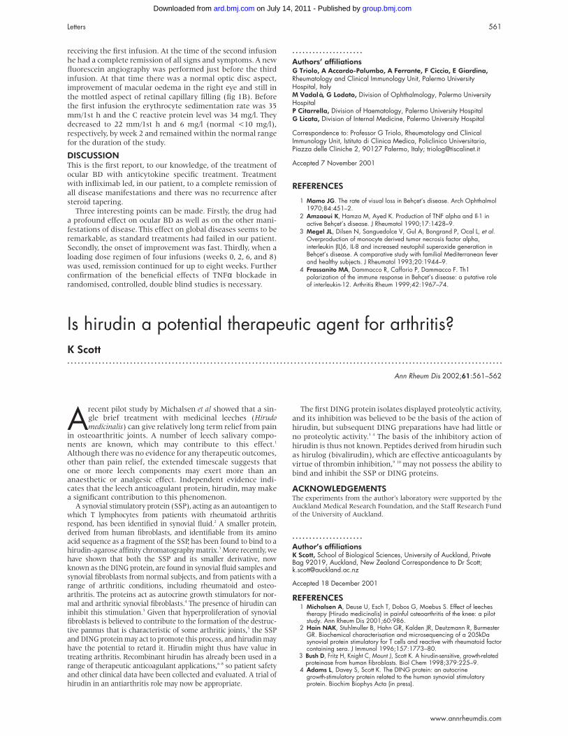
receiving the first infusion. At the time of the second infusion
he had a complete remission of all signs and symptoms. A new
fluorescein angiography was performed just before the third
infusion. At that time there was a normal optic disc aspect,
improvement of macular oedema in the right eye and still in
the mottled aspect of retinal capillary filling (fig 1B). Before
the first infusion the erythrocyte sedimentation rate was 35
mm/1st h and the C reactive protein level was 34 mg/l. They
decreased to 22 mm/1st h and 6 mg/l (normal <10 mg/l),
respectively, by week 2 and remained within the normal range
for the duration of the study.
DISCUSSIONThis is the first report, to our knowledge, of the treatment of
ocular BD with anticytokine specific treatment. Treatment
with infliximab led, in our patient, to a complete remission of
all disease manifestations and there was no recurrence after
steroid tapering.
Three interesting points can be made. Firstly, the drug had
a profound effect on ocular BD as well as on the other mani-
festations of disease. This effect on global diseases seems to be
remarkable, as standard treatments had failed in our patient.
Secondly, the onset of improvement was fast. Thirdly, when a
loading dose regimen of four infusions (weeks 0, 2, 6, and 8)
was used, remission continued for up to eight weeks. Further
confirmation of the beneficial effects of TNFα blockade in
randomised, controlled, double blind studies is necessary.
. . . . . . . . . . . . . . . . . . . . .
Authors’ affiliationsG Triolo, A Accardo-Palumbo, A Ferrante, F Ciccia, E Giardina,Rheumatology and Clinical Immunology Unit, Palermo UniversityHospital, ItalyM Vadalà, G Lodato, Division of Ophthalmology, Palermo UniversityHospitalP Citarrella, Division of Haematology, Palermo University HospitalG Licata, Division of Internal Medicine, Palermo University Hospital
Correspondence to: Professor G Triolo, Rheumatology and ClinicalImmunology Unit, Istituto di Clinica Medica, Policlinico Universitario,Piazza delle Cliniche 2, 90127 Palermo, Italy; [email protected]
Accepted 7 November 2001
REFERENCES
1 Mamo JG. The rate of visual loss in Behçet’s disease. Arch Ophthalmol1970;84:451–2.
2 Amzaoui K, Hamza M, Ayed K. Production of TNF alpha and Il-1 inactive Behçet’s disease. J Rheumatol 1990;17:1428–9.
3 Megel JL, Dilsen N, Sanguedolce V, Gul A, Bongrand P, Ocal L, et al.Overproduction of monocyte derived tumor necrosis factor alpha,interleukin (IL)6, IL-8 and increased neutophil superoxide generation inBehçet’s disease. A comparative study with familial Mediterranean feverand healthy subjects. J Rheumatol 1993;20:1944–9.
4 Frassanito MA, Dammacco R, Cafforio P, Dammacco F. Th1polarization of the immune response in Behçet’s disease: a putative roleof interleukin-12. Arthritis Rheum 1999;42:1967–74.
Is hirudin a potential therapeutic agent for arthritis?K Scott. . . . . . . . . . . . . . . . . . . . . . . . . . . . . . . . . . . . . . . . . . . . . . . . . . . . . . . . . . . . . . . . . . . . . . . . . . . . . . . . . . . . . . . . . . . . . . . . . . . . . . . . . . . . . . . . . . . . . . . . . . . . .
Ann Rheum Dis 2002;61:561–562
Arecent pilot study by Michalsen et al showed that a sin-gle brief treatment with medicinal leeches (Hirudomedicinalis) can give relatively long term relief from pain
in osteoarthritic joints. A number of leech salivary compo-nents are known, which may contribute to this effect.1
Although there was no evidence for any therapeutic outcomes,other than pain relief, the extended timescale suggests thatone or more leech components may exert more than ananaesthetic or analgesic effect. Independent evidence indi-cates that the leech anticoagulant protein, hirudin, may makea significant contribution to this phenomenon.
A synovial stimulatory protein (SSP), acting as an autoantigen towhich T lymphocytes from patients with rheumatoid arthritisrespond, has been identified in synovial fluid.2 A smaller protein,derived from human fibroblasts, and identifiable from its aminoacid sequence as a fragment of the SSP, has been found to bind to ahirudin-agarose affinity chromatography matrix.3 More recently, wehave shown that both the SSP and its smaller derivative, nowknown as the DING protein, are found in synovial fluid samples andsynovial fibroblasts from normal subjects, and from patients with arange of arthritic conditions, including rheumatoid and osteo-arthritis. The proteins act as autocrine growth stimulators for nor-mal and arthritic synovial fibroblasts.4 The presence of hirudin caninhibit this stimulation.3 Given that hyperproliferation of synovialfibroblasts is believed to contribute to the formation of the destruc-tive pannus that is characteristic of some arthritic joints,5 the SSPand DING protein may act to promote this process, and hirudin mayhave the potential to retard it. Hirudin might thus have value intreating arthritis. Recombinant hirudin has already been used in arange of therapeutic anticoagulant applications,6–8 so patient safetyand other clinical data have been collected and evaluated. A trial ofhirudin in an antiarthritis role may now be appropriate.
The first DING protein isolates displayed proteolytic activity,
and its inhibition was believed to be the basis of the action of
hirudin, but subsequent DING preparations have had little or
no proteolytic activity.3 4 The basis of the inhibitory action of
hirudin is thus not known. Peptides derived from hirudin such
as hirulog (bivalirudin), which are effective anticoagulants by
virtue of thrombin inhibition,9 10 may not possess the ability to
bind and inhibit the SSP or DING proteins.
ACKNOWLEDGEMENTSThe experiments from the author’s laboratory were supported by theAuckland Medical Research Foundation, and the Staff Research Fundof the University of Auckland.
. . . . . . . . . . . . . . . . . . . . .Author’s affiliationsK Scott, School of Biological Sciences, University of Auckland, PrivateBag 92019, Auckland, New Zealand Correspondence to Dr Scott;[email protected]
Accepted 18 December 2001
REFERENCES1 Michalsen A, Deuse U, Esch T, Dobos G, Moebus S. Effect of leeches
therapy (Hirudo medicinalis) in painful osteoarthritis of the knee: a pilotstudy. Ann Rheum Dis 2001;60:986.
2 Hain NAK, Stuhlmuller B, Hahn GR, Kalden JR, Deutzmann R, BurmesterGR. Biochemical characterisation and microsequencing of a 205kDasynovial protein stimulatory for T cells and reactive with rheumatoid factorcontaining sera. J Immunol 1996;157:1773–80.
3 Bush D, Fritz H, Knight C, Mount J, Scott K. A hirudin-sensitive, growth-relatedproteinase from human fibroblasts. Biol Chem 1998;379:225–9.
4 Adams L, Davey S, Scott K. The DING protein: an autocrinegrowth-stimulatory protein related to the human synovial stimulatoryprotein. Biochim Biophys Acta (in press).
Letters 561
www.annrheumdis.com
group.bmj.com on July 14, 2011 - Published by ard.bmj.comDownloaded from
5 Mizel SB, Dayer JM, Krane SM, Mergenhagen SE. Stimulation ofrheumatoid synovial cell collagenase and prostaglandin production bypartially purified lymphocyte-activating factor. Proc Natl Acad Sci USA1981;78:2474–7.
6 Topol E, GUSTO IIb investigators. A comparison of recombinant hirudinwith heparin for the treatment of acute coronary syndromes. N Engl JMed 1996;335:775–82.
7 Reilly MP, Weiss R, Askenase A, Tuite C, Soulen M, Mohler ER. Hirudintherapy during thrombolysis for venous thrombosis in heparin-inducedthrombocytopenia. Vasc Med 2000;5:239–42.
8 Saner F, Hertl M, Broelsch CE. Anticoagulation with hirudin forcontinuous veno-venous hemodialysis in liver transplantation. ActaAnaesthesiol Scand 2001;45:914–18.
9 Maraganore JM, Bourdon P, Jablonski J, Ramachandran KL, Fenton JW.Design and characterisation of hirulogs, a novel class of bivalent peptideinhibitors of thrombin. Biochemistry 1990;29:7095–101.
10 Kong DF, Topol EJ, Bittl JA, White HD, Theroux P, Hasselblad V, et al.Clinical outcomes of bivalirudin for ischemic heart disease. Circulation1999;100:2049–53.
Steroid induced psychosis in systemic lupuserythematosus: a possible role of serum albumin levelF López-Medrano, R Cervera, O Trejo, J Font, M Ingelmo. . . . . . . . . . . . . . . . . . . . . . . . . . . . . . . . . . . . . . . . . . . . . . . . . . . . . . . . . . . . . . . . . . . . . . . . . . . . . . . . . . . . . . . . . . . . . . . . . . . . . . . . . . . . . . . . . . . . . . . . . . . . .
Ann Rheum Dis 2002;61:562–563
Steroids may have diverse and sometimes severe adverse
effects in the short and long term.1 We present three
patients with systemic lupus erythematosus (SLE)2 and
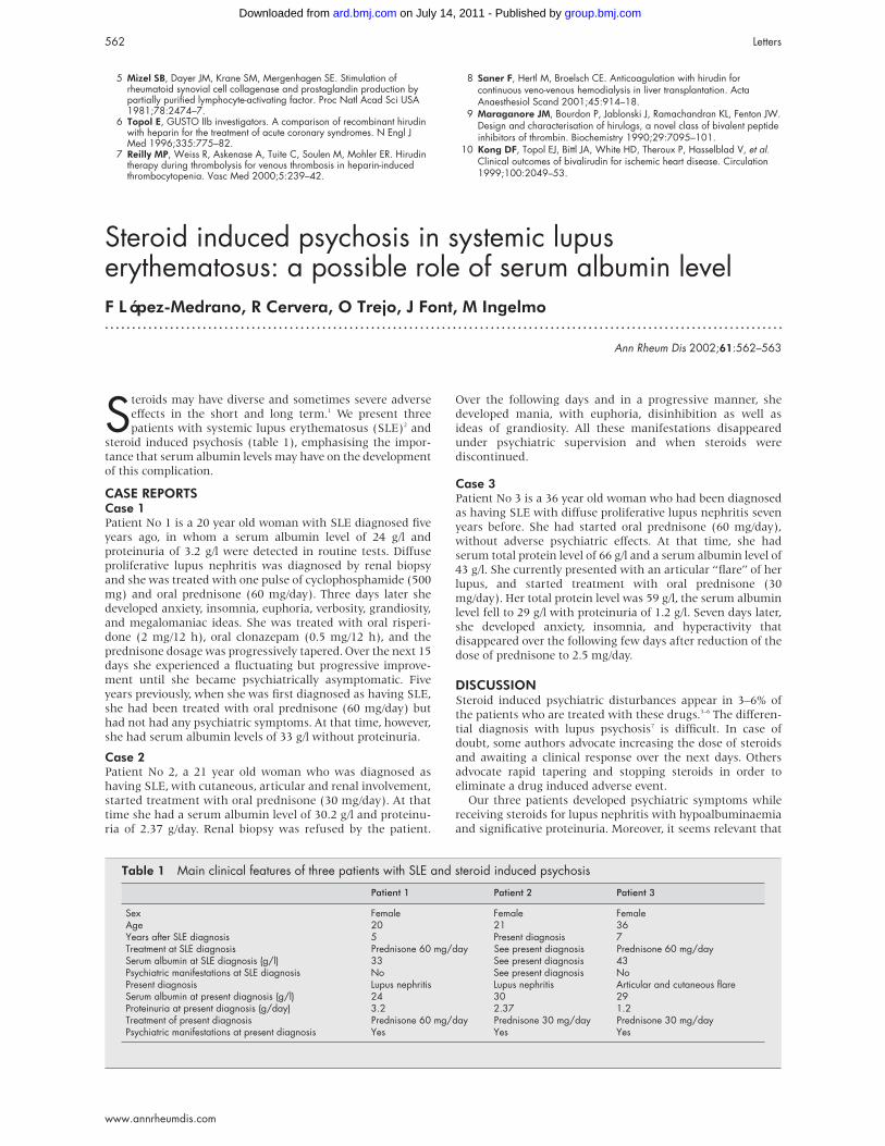
steroid induced psychosis (table 1), emphasising the impor-
tance that serum albumin levels may have on the development
of this complication.
CASE REPORTSCase 1Patient No 1 is a 20 year old woman with SLE diagnosed five
years ago, in whom a serum albumin level of 24 g/l and
proteinuria of 3.2 g/l were detected in routine tests. Diffuse
proliferative lupus nephritis was diagnosed by renal biopsy
and she was treated with one pulse of cyclophosphamide (500
mg) and oral prednisone (60 mg/day). Three days later she
developed anxiety, insomnia, euphoria, verbosity, grandiosity,
and megalomaniac ideas. She was treated with oral risperi-
done (2 mg/12 h), oral clonazepam (0.5 mg/12 h), and the
prednisone dosage was progressively tapered. Over the next 15
days she experienced a fluctuating but progressive improve-
ment until she became psychiatrically asymptomatic. Five
years previously, when she was first diagnosed as having SLE,
she had been treated with oral prednisone (60 mg/day) but
had not had any psychiatric symptoms. At that time, however,
she had serum albumin levels of 33 g/l without proteinuria.
Case 2Patient No 2, a 21 year old woman who was diagnosed as
having SLE, with cutaneous, articular and renal involvement,
started treatment with oral prednisone (30 mg/day). At that
time she had a serum albumin level of 30.2 g/l and proteinu-
ria of 2.37 g/day. Renal biopsy was refused by the patient.
Over the following days and in a progressive manner, she
developed mania, with euphoria, disinhibition as well as
ideas of grandiosity. All these manifestations disappeared
under psychiatric supervision and when steroids were
discontinued.
Case 3Patient No 3 is a 36 year old woman who had been diagnosed
as having SLE with diffuse proliferative lupus nephritis seven
years before. She had started oral prednisone (60 mg/day),
without adverse psychiatric effects. At that time, she had
serum total protein level of 66 g/l and a serum albumin level of
43 g/l. She currently presented with an articular “flare” of her
lupus, and started treatment with oral prednisone (30
mg/day). Her total protein level was 59 g/l, the serum albumin
level fell to 29 g/l with proteinuria of 1.2 g/l. Seven days later,
she developed anxiety, insomnia, and hyperactivity that
disappeared over the following few days after reduction of the
dose of prednisone to 2.5 mg/day.
DISCUSSIONSteroid induced psychiatric disturbances appear in 3–6% of
the patients who are treated with these drugs.3–6 The differen-
tial diagnosis with lupus psychosis7 is difficult. In case of
doubt, some authors advocate increasing the dose of steroids
and awaiting a clinical response over the next days. Others
advocate rapid tapering and stopping steroids in order to
eliminate a drug induced adverse event.
Our three patients developed psychiatric symptoms while
receiving steroids for lupus nephritis with hypoalbuminaemia
and significative proteinuria. Moreover, it seems relevant that
Table 1 Main clinical features of three patients with SLE and steroid induced psychosis
Patient 1 Patient 2 Patient 3
Sex Female Female FemaleAge 20 21 36Years after SLE diagnosis 5 Present diagnosis 7Treatment at SLE diagnosis Prednisone 60 mg/day See present diagnosis Prednisone 60 mg/daySerum albumin at SLE diagnosis (g/l) 33 See present diagnosis 43Psychiatric manifestations at SLE diagnosis No See present diagnosis NoPresent diagnosis Lupus nephritis Lupus nephritis Articular and cutaneous flareSerum albumin at present diagnosis (g/l) 24 30 29Proteinuria at present diagnosis (g/day) 3.2 2.37 1.2Treatment of present diagnosis Prednisone 60 mg/day Prednisone 30 mg/day Prednisone 30 mg/dayPsychiatric manifestations at present diagnosis Yes Yes Yes
562 Letters
www.annrheumdis.com
group.bmj.com on July 14, 2011 - Published by ard.bmj.comDownloaded from
two of them (patients 1 and 3) had not developed psychiatric
manifestations when they had previously been receiving simi-
lar doses of steroids, but had higher serum albumin levels at
that time. Lewis et al found a 37% incidence of adverse effects
of steroids in those patients with serum albumin <25 g/l but
of only 15% in those with higher serum albumin.8 The expla-
nation for these findings may be that corticosteroid binding
globulin does not bind to synthetic steroids, whose transport
depends on serum albumin which, by contrast, presents low
affinity but a great capacity to transport the steroids because
of its high plasma concentration. Steroids are biologically
inactive when bound to albumin. Therefore, the free (and
active) fraction of steroids is higher in patients with low
plasma albumin levels, and this will expose the patient to
more adverse effects.
Interestingly, there is a lower incidence of psychosis in other
groups of patients treated with steroids (for example, those
with chronic obstructive pulmonary disease).9 Therefore,
those patients whose disease causes low levels of serum
proteins (as in those with SLE) will be more predisposed to
have adverse effects of steroids.
. . . . . . . . . . . . . . . . . . . . .Authors’ affiliationsF López-Medrano, R Cervera, O Trejo, J Font, M Ingelmo,Department of Autoimmune Diseases, Institut Clínic d’Infeccions iImmunologia, Hospital Clínic, Institut d’Investigacions BiomèdiquesAugust Pi i Sunyer (IDIBAPS), University of Barcelona, Barcelona,Catalonia, Spain
F López-Medrano is currently at Servicio de Medicina Interna, HospitalUniversitario 12 de Octubre, Madrid, Spain.
Correspondence to: Dr R Cervera, Servei de Malalties Autoimmunes,Hospital Clínic, Villarroel 170, 08036-Barcelona, Catalonia, Spain;[email protected]
Accepted 10 December 2001
REFERENCES1 Chatham WW. Treatment of lupus with corticosteroids. Lupus
2001;10:140–7.2 Tan EM, Cohen AS, Fries JF, Masi AT, McShane DJ, Rothfield NF, et al.
The 1982 revised criteria for the classification of systemic lupuserythematosus. Arthritis Rheum 1982;25:1271–7.
3 Patten SB, Neutel I. Corticosteroid-induced adverse psychiatric effects.Incidence, diagnosis and management. Drug Saf 2000;22:111–22.
4 Kohen M, Asherson RA, Gharavi AE, Lahita RG. Lupus psychosis:differentiation from the steroid-induced state. Clin Exp Rheumatol1993;11:323–6.
5 Wysenbeek AJ, Leibovici L, Zoldan J. Acute central nervous systemcomplications after pulse steroid therapy in patients with SLE. J Rheumatol1990;17:1695–6.
6 Boston Collaborative Drug Surveillance Program. Acute adversereactions to prednisone in relation to dosage. Clin Pharmacol Ther1972;13:694–8.
7 ACR ad hoc committee on neuropsychiatry lupus nomenclature.The American College of Rheumatology nomenclature and casedefinitions for neuropsychiatry lupus syndromes. Arthritis Rheum1999;42:599–608.
8 Lewis GP, Jusko WJ, Burke CW, Graves L. Prednisone side-effects andserum-protein levels. Lancet 1971;ii:778–80.
9 Niewoehner DE, Erbland ML, Deupree RH, Collins D, Gross NJ, LightRW, et al. Effect of systemic glucocorticoids on exacerbations of chronicobstructive pulmonary disease. N Engl J Med 1999;340:1941–7.
Autoantibodies against C1q: view on associationbetween systemic lupus erythematosus diseasemanifestation and C1q autoantibodiesD Monova, S Monov, K Rosenova, T Argirova. . . . . . . . . . . . . . . . . . . . . . . . . . . . . . . . . . . . . . . . . . . . . . . . . . . . . . . . . . . . . . . . . . . . . . . . . . . . . . . . . . . . . . . . . . . . . . . . . . . . . . . . . . . . . . . . . . . . . . . . . . . . .
Ann Rheum Dis 2002;61:563–564
Ativation of the complement system is the first step in theprevention of damage by immune complexes. Systemiclupus erythematosus (SLE) is the prototype of immune
complex diseases. The classical pathway of the complementsystem is considered to be the most important pathway inimmune complex clearance. This pathway may be activated byIgM- and IgG-containing immune complexes after binding toC1q.1
In 1984 autoantibodies to C1q (C1qAb) were reported to bepresent in serum of patients with SLE.2 The recognition thatC1q may serve as a non-organ-specific autoantigen hasattracted a growing number of investigators.3
We studied 42 patients (38 female, four male, aged 19–64)with SLE. Twenty eight (67%) had proven renal biopsy lupusnephritis (two patients had WHO class II lesions, four hadWHO class III, 18 had WHO class IV, four had WHO class V),14 (33%) patients had evidence of lupus pneumonitis, and 11(26%) central nervous system disease (table 1).
All patients were tested by both basic and subclass enzymelinked immunosorbent assays (ELISAs) for C1qAb using amodification of the method of Wisnieski and Jones.4 WholeC1q was purified from human plasma by the method of Ten-ner et al.5 Raised C1qAb titres were found in 18 (43%) patients.Of the patients with C1qAb, 12 had renal manifestations ofSLE (10 (83%) of them had focal or diffuse proliferative
glomerulonephritis), six central nervous system disease, andfive lupus pneumonitis. Patients with raised C1qAb titres wereyounger, seven of them were positive for antibodies to dsDNA.The magnitude of proteinuria was positively associated withthe presence of C1qAb.
Selective complete C1q deficiency was established in seven ofour patients (Nos 6, 11, 16, 18, 28, 32, 33); in two of them (Nos18, 28), clinical data showed the presence of SLE in the family.
Available serum samples testing positive for IgG C1qAbwere analysed for C1qAb IgG subclass distribution. Six (33%)of the 18 patients had IgG2 C1qAb only, 3/18 (17%) patientshad IgG1 C1qAb only, and 9/18 (50%) had both IgG1 and IgG2C1qAb. Therefore, IgG2 C1qAb was present in 15/18 (83%)patients. The subset of sera from patients with IgG1 or IgG2C1qAb was assayed for total serum IgG1 and IgG2 levels byradial immunodiffusion. The mean total serum IgG1 was 7.9(4.5) mg/ml, the mean total serum IgG2 was 2.6 (1.4) mg/ml.The mean ratio of IgG1/IgG2 (3.4 (2.1)) was similar to thatreported in the literature for disease free subjects.6 Thepercentage of IgG2 C1qAb relative to total IgG2 wassignificantly greater than the percentage of IgG1 C1qAb rela-tive to total IgG1 (0.03 (0.06)% v 0.01 (0.02)% respectively,p<0.005, t test). Thus in our patient group the IgG2component of the autoantibody response to C1q wasdisproportionately enriched relative to the overall IgG
Letters 563
www.annrheumdis.com
group.bmj.com on July 14, 2011 - Published by ard.bmj.comDownloaded from

subclasses distribution, as no alteration in IgG subclass
distribution was noted. The C1qAb in our group were
predominantly of IgG2 and IgG1 subclasses. This distribution
is consistent with that found by Wisnieski and Jones in a
study characterising C1qAb in patients with SLE and
hypocomplementaemic urticarial vasculitis,4 but contrasts
with the IgG3 and IgG2 predominance reported by Coremans
et al in patients with SLE.7
The mechanisms mediating autoantibody pathogenicity
remain unclear. It has been proposed that C1qAb may act sys-
temically by up regulating activation of classical complement
pathway.8 Alternatively, C1qAb may act locally within the
renal glomerulus to enhance tissue injury initiated by
immune complex deposition. The association of C1qAb with
proliferative lupus nephritis is now well established,2 but the
significance of C1qAb for lupus pneumonitis and cerebro-
vasculitis should be a target for future investigations.
. . . . . . . . . . . . . . . . . . . . .Authors’ affiliationsD Monova, S Monov, Department of Internal Medicine, MedicalUniversity, Sofia, BulgariaK Rosenova, T Argirova, Department of Biochemistry, Sofia University,Bulgaria
Correspondence to: Dr D Monova, Department of Internal Medicine,Medical University, University Hospital “St J Rilski”, 15 D Nestorov St,1431 Sofia, Bulgaria; [email protected]
Accepted 7 December 2001
REFERENCES1 Cooper NZ. The classical complement pathway: activation and
regulation of the first complement component. Adv Immunol1985;37:151–216.
2 Uwatoko S, Aotsuka S, Okawa M, Egusa Y, Yokohari R, Aisawa C, K..Characterization of C1q-binding IgG complexes in systemic lupuserythematosus. Clin Immunol Immunopathol 1984;30:104–16.
3 Siegert CEH, Kazatchkine MD, Sjoholm A, Wursner R, Loos M, DahaMR. Autoantibodies against C1q: view on clinical relevance andpathogenic roles. Clin Exp Immunol 1999;116:4–8.
4 Wisnieski JJ, Jones SM. Comparison of autoantibodies to thecollagen-like region of C1q in hypocomplementemic urticarial vasculitissyndrome and systemic lupus erythematosus. J Immunol1992;148:1396–403.
5 Tenner AJ, Lesavre PH, Cooper NR. Purification and radiolabeling ofhuman C1q. J Immunol 1981;127:648–53.
6 Schur P. IgG subclass - a review. Ann Allergy 1987;58:89–99.7 Coremans IEM, Spronk PE, Bootsma H, Daha MR, van der Voort EAM,
Kater L, et al. Changes in antibodies to C1q predict renal relapses insystemic lupus erythematosus. Am J Kidney Dis 1995;26:505–601.
8 Moreland LW, Gay RE, Gay S. Collagen autoantibodies in patients withvasculitis and systemic lupus erythematosus. Clin Immunol Immunopathol1991;60:412–18.
Table 1 Basic clinicolaboratory parameters in study patients
No/sex/age(years) C1qAb
Anti-dsDNA ANA
Lupus nephritis/WHO class/proteinuria (g/24 h (SD)) Pneumonitis
Central nervoussystem involvement
1/F/28 + + WHO class IV (6.8 (2.4)) +2/F/19 + + + + WHO class IV (8.7 (3.1)) + +3/F/36 +4/F/25 + + + WHO class V (5.4 (0.5)) +5/F/346/M/33 + WHO class II (2.1 (0.9)) + +7/F/418/F/27 + + + + WHO class IV (7.2 (3.4))9/F/34 + +10/F/50 + WHO class IV (3.5 (1.4))11/F/44 + + WHO class IV (4.1 (1.5)) +12/F/26 + + + WHO class IV (4.4 (2.3))13/F/30 + + WHO class V (7.1 (1.2)) + +14/F/64 + + WHO class IV (2.5 (0.9)) +15/F/24 + + + + WHO class IV (3.4 (1.7))16/F/38 + +17/F/40 + WHO class IV (1.2 (0.7))18/F/47 + + + WHO class IV (2.4 (0.8)) + +19/F/29 + + WHO class IV (4.2 (1.3)) +20/F/22 + + +21/F/34 + + WHO class IIIB (3.8 (1.7))22/F/31 +23/F/28 + + + WHO class IV (5.3 (2.0)) +24/F/4225/M/36 + + + + WHO class IIIB (4.8 (1.4))26/F/32 + +27/F/25 +28/F/31 + + + WHO class IV (3.1 (1.7)) +29/F/36 + +30/F/27 +31/F/23 +32/M/24 + + + WHO class V (4.0 (1.1)) +33/F/34 + WHO class IIIA (2.3 (0.8)) +34/F/41 + + WHO class IV (4.2 (2.3)) +35/F/28 + + + WHO class IV (3.0 (0.6))36/F/2337/M/31 + + + WHO class IV (2.4 (0.3))38/F/36 + WHO class V (1.7 (0.3))39/F/25 + WHO class IV (1.7 (0.5))40/F/34 + WHO class IIIB (2.1 (0.4))41/F/27 + + + WHO class IV (5.2 (2.6))42/F/20 + WHO class II (0.9 (0.3))
ANA, antinuclear antibodies; C1qAb, autoantibodies to C1q; Anti-dsDNA, anti-double stranded DNAantibodies; F, female; M, male.
564 Letters
www.annrheumdis.com
group.bmj.com on July 14, 2011 - Published by ard.bmj.comDownloaded from
Silent thyroiditis associated with etanercept inrheumatoid arthritisE Andrès, F-X Limbach, B Goichot, J Sibilia. . . . . . . . . . . . . . . . . . . . . . . . . . . . . . . . . . . . . . . . . . . . . . . . . . . . . . . . . . . . . . . . . . . . . . . . . . . . . . . . . . . . . . . . . . . . . . . . . . . . . . . . . . . . . . . . . . . . . . . . . . . . .
Ann Rheum Dis 2002;61:565
In recent years, monoclonal antibodies (infliximab) and a
recombinant human tumour necrosis factor receptor
(p75)-Fc fusion protein (TNFR:Fc) (etanercept) have been
successfully used to treat rheumatoid arthritis (RA).1 These
TNF blocking agents are now widely employed in patients
with treatment resistant RA. TNF inhibitors are generally well
tolerated with <0.5% of patients developing a drug induced
lupus syndrome, although 4–16% may develop antibodies to
double stranded DNA (dsDNA). Apart from two patients pre-
senting autoimmune skin diseases associated with etanercept
(discoid lupus and necrotising vasculitis),2 there are no other
reports of well documented autoimmune disease. In this paper
we describe the first case of silent autoimmune thyroiditis
during TNFR:Fc treatment for severe RA.
CASE REPORTA 43 year old woman followed up since 1991 for erosive RA
had been successively treated with various disease modifying
antirheumatic drugs (Allochrysine, D-penicillamine, and
methotrexate) combined with low dose corticosteroids. In
March 1998 she reported an important flare up despite
corticosteroids (10 mg/day) and methotrexate (15 mg/week).
Methotrexate was replaced by etanercept (25 mg twice a
week), which led to a dramatic improvement after one month
of treatment. The evaluation before TNFR-Fc treatment
disclosed no evidence of thyroid disorders: the patient had no
clinical features, serum thyroid stimulating hormone (TSH)
and free thyroid hormones were normal, and thyroid antibod-
ies were negative. No other autoantibody (anti-dsDNA,
anticardiolipin, anti-extractable nuclear antigen (ENA)) was
present except a high titre of IgM rheumatoid factor. In Janu-
ary 2000 the patient developed a non-tender moderate goitre.
Thyroid evaluation disclosed modest hypothyroidism: serum
TSH 6.3 mU/ml (normal <4.5) and serum free thyroxine (T4)
11 pmol/l (normal 11–23) (Elecsys assay). Titres of native
antimicrosomal and antithyroglobulin antibodies were raised
at 820 IU/ml (normal <60) and 230 IU/ml (normal <60),
respectively (radioimmunoassay, Brahms). Anti-TSH receptor
antibodies were negative (<5 IU/l, normal <11) (Radio
Receptor Assay, Brahms) and no other autoantibodies (anti-
dsDNA, anticardiolipin, anti-ENA) were found except an IgM
rheumatoid factor. A technetium-99m pertechnetate thyroid
scintigraphic scan showed reduced uptake.
In the absence of other known cause, the diagnosis was
silent thyroiditis induced by TNFR:Fc. Etanercept treatment
was completed four months after the onset of hypothyroidism,
and there was no aggravation of the thyroid disorder without
hormonal substitution after a year and a half of follow up.
DISCUSSIONThis report describes a case of silent autoimmune thyroiditis
which developed during TNFR:Fc treatment in a patient with
RA without evidence of previous thyroid disorders. Thyroiditis
has not to our knowledge been described as a side effect of
TNFR:Fc treatment and a causal relationship cannot formally
be established in our case report. However, cytokines like
interferon γ or interleukin 2 often induce thyroiditis in
patients with pre-existing autoimmune thyroid disease. T cell
(Th1) depletion with monoclonal antibodies (Campath-1) can
also lead to the development of antibody mediated thyroid
autoimmunity.3 The mechanism of this effect of TNF blocking
agents is not well understood, but modulation of the homing
of Th1 and Th2 cells may explain the induction of
autoimmune thyroiditis.4 5 In our opinion, TNFR:Fc treatment
should be considered as a potential cause of drug induced
autoimmune thyroiditis. Nevertheless, further studies are
needed to estimate the incidence and the mechanism of this
side effect.
. . . . . . . . . . . . . . . . . . . . .Authors’ affiliationsE Andrès, B Goichot, Department of Internal Medicine, UniversityHospital Strasbourg, FranceF-X Limbach, J Sibilia, Department of Rheumatology, University HospitalStrasbourg
Correspondence to: Professor J Sibilia, Service de Rhumatologie, AvenueMolière, 67 098 Strasbourg Cedex France; [email protected]
Accepted 15 October 2001
REFERENCES
1 Weinblatt ME, Kremer JM, Bankhust AD, Bulpitt KJ, Fleischmann RM,Fox RI, et al. A trial of etanercept, a recombinant tumor necrosis factorreceptor: Fc fusion protein, in patients with rheumatoid arthritis receivingmethotrexate. N Engl J Med 1999;340:253–9.
2 Brion Ph, Mittal-Henckle A, Kalunien KC. Autoimmune skin rashesassociated with etanercept for rheumatoid arthritis. Ann Intern Med1999;131:634.
3 Coles AJ, Wing M, Smith S, Coraddu F, Greer S, Taylor C, et al. Pulsedmonoclonal antibody treatment and autoimmune thyroid disease inmultiple sclerosis. Lancet 1999;354:1691–5.
4 Maurice MM, Van der Graaf WL, Leow A, Breedveld FC, van Lier RA,Verweij CL. Treatment with monoclonal anti-tumor necrosis factor αantibody results in an accumulation of Th1 CD4+ T cells in the peripheralblood of patients with rheumatoid arthritis. Arthritis Rheum1999;42:2166–73.
5 Guo J, Rapoport B, McLachlan SM. Balance of Th1/Th2 cytokines inthyroid autoantibody synthesis in vitro. Autoimmunity 1999;30:1–9.
Letters 565
www.annrheumdis.com
group.bmj.com on July 14, 2011 - Published by ard.bmj.comDownloaded from
Synovial T cell proliferation to the Yersinia enterocolitica19 kDa antigen differentiates yersinia triggered reactivearthritis (ReA) from ReA triggered by other enterobacteriaH Appel, M Rudwaleit, P Wu, M Grolms, J Sieper, A Mertz. . . . . . . . . . . . . . . . . . . . . . . . . . . . . . . . . . . . . . . . . . . . . . . . . . . . . . . . . . . . . . . . . . . . . . . . . . . . . . . . . . . . . . . . . . . . . . . . . . . . . . . . . . . . . . . . . . . . . . . . . . . . .
Ann Rheum Dis 2002;61:566–567
Reactive arthritis (ReA) mostly presents as an asymmetri-cal oligoarthritis, usually affecting the leg joints.1 It is a Tcell dependent inflammatory disease following infections
with various enteropathic bacteria—for example, Yersinia ente-rocolitica 0:3, Salmonella enteritidis, Shigella fexneri, or microbespathogenic for the urogenital tract such as Chlamydiatrachomatis.2 Identification of the structure of antigens of trig-gering microbes which drive immune responses in thesynovial fluid would be of great interest.
We recently described two major antigens, the Ye 19 kDa andthe Ye HSP60 inducing synovial T cell proliferation in patientswith Ye triggered ReA.3–7 They seemed to overcome the specifi-city problem of proliferation assays with T cells cultured fromthe site of inflammation to ReA triggering bacteria, because thelocal immune response to whole bacteria is accompanied by a
restricted specificity probably due to cross reactivity with com-
mon epitopes.8 Since Ye is the only known ReA triggering bac-
teria expressing the Ye 19 kDa protein from the β subunit of
urease,6 we investigated the possibility that synovial T cell pro-
liferation to this protein might help to differentiate Ye triggered
arthritis from arthritides caused by other bacteria.
In view of this specificity problem it was not unexpected to
find a large number of patients in our clinic who showed clini-
cal features of ReA and had synovial T cell proliferation to two
or more enteropathic bacteria.
METHODSWe tested the synovial T cells of 66 patients with arthritis of
one or more joints. Ye triggered ReA was diagnosed based on a
typical history of previous symptomatic gastrointestinal
infection or significant agglutinin titre. Chlamydia trachomatistriggered ReA was diagnosed in patients with arthritis and
positive urogenital swabs or chlamydia-specific antibodies and
a recent history of symptomatic urethritis or cervicitis. Syno-
vial fluid mononuclear cells were cultured in the presence of
heat inactivated bacterial antigens such as Yersinia enterocolitica,
Salmonella enteritidis, Shigella flexneri, Campylobacter jejuni, and
Chlamydia trachomatis. For stimulation with Ye 19 kDa we used
1 µg/ml recombinant protein, which was expressed as
described previously.4 Stimulation indices were calculated in
comparison with background activity by T cell medium. A
stimulation index (SI) of >5 was classified as positive.
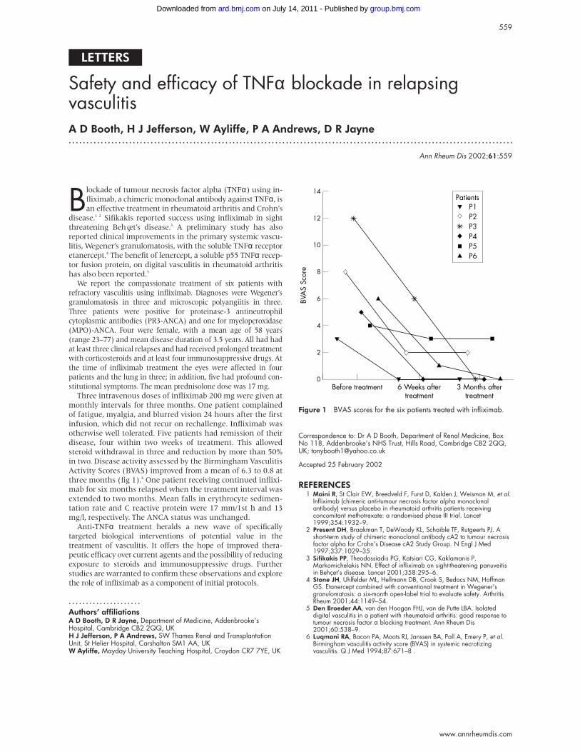
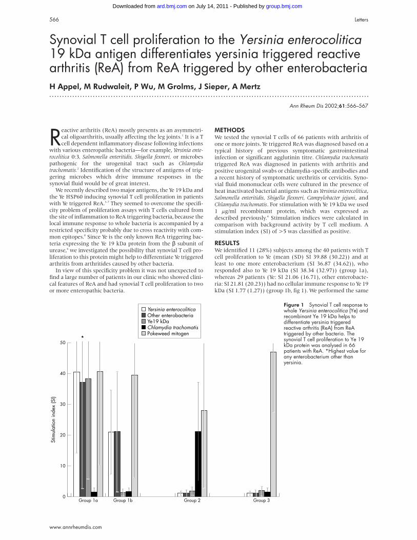
RESULTSWe identified 11 (28%) subjects among the 40 patients with T
cell proliferation to Ye (mean (SD) SI 39.88 (30.22)) and at
least to one more enterobacterium (SI 36.87 (34.62)), who
responded also to Ye 19 kDa (SI 38.34 (32.97)) (group 1a),
whereas 29 patients (Ye: SI 21.06 (16.71), other enterobacte-
ria: SI 21.81 (20.23)) had no cellular immune response to Ye 19
kDa (SI 1.77 (1.27)) (group 1b, fig 1). We performed the same
Figure 1 Synovial T cell response towhole Yersinia enterocolitica (Ye) andrecombinant Ye 19 kDa helps todifferentiate yersinia triggeredreactive arthritis (ReA) from ReAtriggered by other bacteria. Thesynovial T cell proliferation to Ye 19kDa protein was analysed in 66patients with ReA. *Highest value forany enterobacterium other thanyersinia.
50
40
30
Yersinia enterocoliticaOther enterobacteriaYe19 kDaChlamydia trachomatisPokeweed mitogen
20
10
0
Stim
ulat
ion
inde
x (S
I)
Group 1a Group 1b Group 2 Group 3
566 Letters
www.annrheumdis.com
group.bmj.com on July 14, 2011 - Published by ard.bmj.comDownloaded from
experiment with synovial T cells from 11 patients with
chlamydia triggered ReA (SI 20.8 (15.51)) (group 2). In this
case none of the patients had a T cell proliferation to Ye 19 kDa
(SI 1.72 (1.5)) (fig 1). In 15 patients with the clinical diagno-
sis of ReA or undifferentiated oligoarthritis synovial T cells
proliferated to mitogen, but not to any of the ReA triggering
bacteria or Ye 19 kDa (SI 1.78 (1.25)) (group 3, fig 1).
Because the Ye 19 kDa as the β subunit of urease6 is a yers-
inia antigen not shared by other ReA triggering enterobacteria
we believe that we have identified the cause of disease of a
substantial number of patients with ReA in clinical practice
which would not have been known by other means. We
assume that Ye 19 kDa used in synovial T cell proliferation
assays is a useful antigen to specify Ye as the disease trigger-
ing bacteria and might be of diagnostic value in ReA.
. . . . . . . . . . . . . . . . . . . . .Authors’ affiliationsH Appel, M Rudwaleit, P Wu, M Grolms, J Sieper, Benjamin FranklinMedical Centre, Division of Gastroenterology and Rheumatology, FreeUniversity Berlin, GermanyP Wu, J Sieper, Deutsches Rheumaforschungszentrum Berlin, GermanyA Mertz, Medical Centre Augsburg, Division of Nephrology, Augsburg,Germany
Correspondence to: Professor J Sieper; [email protected]
Accepted 17 December 2001
REFERENCES
1 Toivanen P, Toivanen A, Olkkonen L, Aantaa S. Hospital outbreak ofYersinia enterocolitica infection. Lancet 1973;i:801–3.
2 Sieper J, Kingsley G. Recent advances in the pathogenesis of reactivearthritis. Immunol Today 1996;17:160–4.
3 Appel H, Mertz A, Distler A, Sieper J, Braun J. The 19kD protein ofYersinia enterocolitica is recognized on the cellular and humoral level bypatients with yersinia induced reactive arthritis. J Rheumatol1999;26:1964–71.
4 Mertz AKH, Ugrinovic S, Lauster R, Wu P, Grolms M, Böttcher U, et al.Characterization of the synovial T cell response to various recombinantyersinia antigens in Yersinia enterocolitica triggered reactive arthritis.Arthritis Rheum 1998;41:315–26.
5 Mertz AKH, Wu P, Sturniolo T, Stoll D, Rudwaleit M, Lauster R, et al.Multispecific CD4+ T cell response to a single 12-mer epitope of theimmunodominant heat-shock protein 60 of Yersinia enterocolitica inyersinia-triggered reactive arthritis: overlap with B27-restricted CD8epitope, functional properties and epitope presentation by multiple DRalleles. J Immunol 2000;164:1529–37.
6 Skurnik M, Batsford SR, Mertz A, Schiltz E, Toivanen P. The putativearthritogenic cationic 19kD antigen of Yersinia enterocolitica is a ureaseβ-subunit. Infect Immun 1993;61:2498–503.
7 Mertz AKH, Batsford SR, Curschellas E, Kist MJ, Gondolf KB. Cationicyersinia-induced chronic allergic arthritis in rats; a model for reactivearthritis in humans. J Clin Invest 1991;88:632–42.
8 Kaluza W, Meyer zum Buschenfelde KH, Galle PR, Marker-Hermann E.Synovial fluid lymphocyte proliferation in response to crude microbialantigens is not useful as a diagnostic test to specifically indicatea bacterial cause of arthritis. Clin Exp Rheumatol 2000;18:39–46.
Leflunomide and hypertensionB Rozman, S Praprotnik, D Logar, M Tomsic, M Hojnik, M Kos-Golja, R Accetto,P Dolenc. . . . . . . . . . . . . . . . . . . . . . . . . . . . . . . . . . . . . . . . . . . . . . . . . . . . . . . . . . . . . . . . . . . . . . . . . . . . . . . . . . . . . . . . . . . . . . . . . . . . . . . . . . . . . . . . . . . . . . . . . . . . .
Ann Rheum Dis 2002;61:567–569
Leflunomide is a new isoxazole drug with diseasemodifying properties for the treatment of rheumatoidarthritis (RA). Hypertension has been mentioned as a
common side effect of the treatment. It was found in up to10.6% of patients receiving 25 mg leflunomide in a phase IIstudy.1 New onset hypertension occurred in 3.7% of patientsin a phase III European study,2 and in 2.1% of patients, with amean increase in systolic and diastolic blood pressure of 2.2and 1.9 mm Hg, respectively, in an American phase IIIstudy.3 There was no evidence that hypertension was relatedto an impairment of renal function or proteinuria. Thechanges in blood pressure during leflunomide treatment havenot been studied in detail.
PATIENTS AND METHODSThirty consecutive patients fulfilling the American Rheuma-
tism Association criteria for RA were recruited into a
prospective study and treated with standard doses of lefluno-
mide. Other enrolment criteria included stable treatment
with non-steroidal anti-inflammatory drugs up to the maxi-
mum recommended dose and/or corticosteroid treatment up
to 10 mg/day for at least three months before starting treat-
ment with leflunomide. The patients were followed up at two
week intervals. A trained nurse according to the Slovenian
and WHO/ISH hypertension guidelines measured blood
pressure.4 Automatic oscillometric monitors (Spacelabs
90209) were used for ambulatory blood pressure monitoring
(ABPM).5 Seventeen patients finished the study according to
the protocol with 6.5 (1) months between the two ABPM
procedures.
RESULTSA statistically significant increase in conventional blood pres-
sure measurements of both systolic and diastolic blood
pressure was seen (table 1). The rise in systolic blood pressure
was seen relatively early—in 2–4 weeks (from 127.03 (20.2)
mm Hg to 134.1 (24.3) mm Hg, p=0.034). On the contrary, the
rise in diastolic blood pressure was not significant after 2–4
and 6–8 weeks, respectively. In 7/17 patients, the initially nor-
mal blood pressure values exceeded the systolic and/or diasto-
lic blood pressure values of 140/90 mm Hg in the follow up
measurements. Moreover, in four patients the systolic blood
pressure was, at least once in the follow up period, more than
40 mm Hg and diastolic blood pressure more than 20 mm Hg
above the initial values. According to the ambulatory blood
pressure monitoring (ABPM) measurements the overall trend
after the start of leflunomide treatment was an increase in
both systolic and diastolic blood pressure and heart rate,
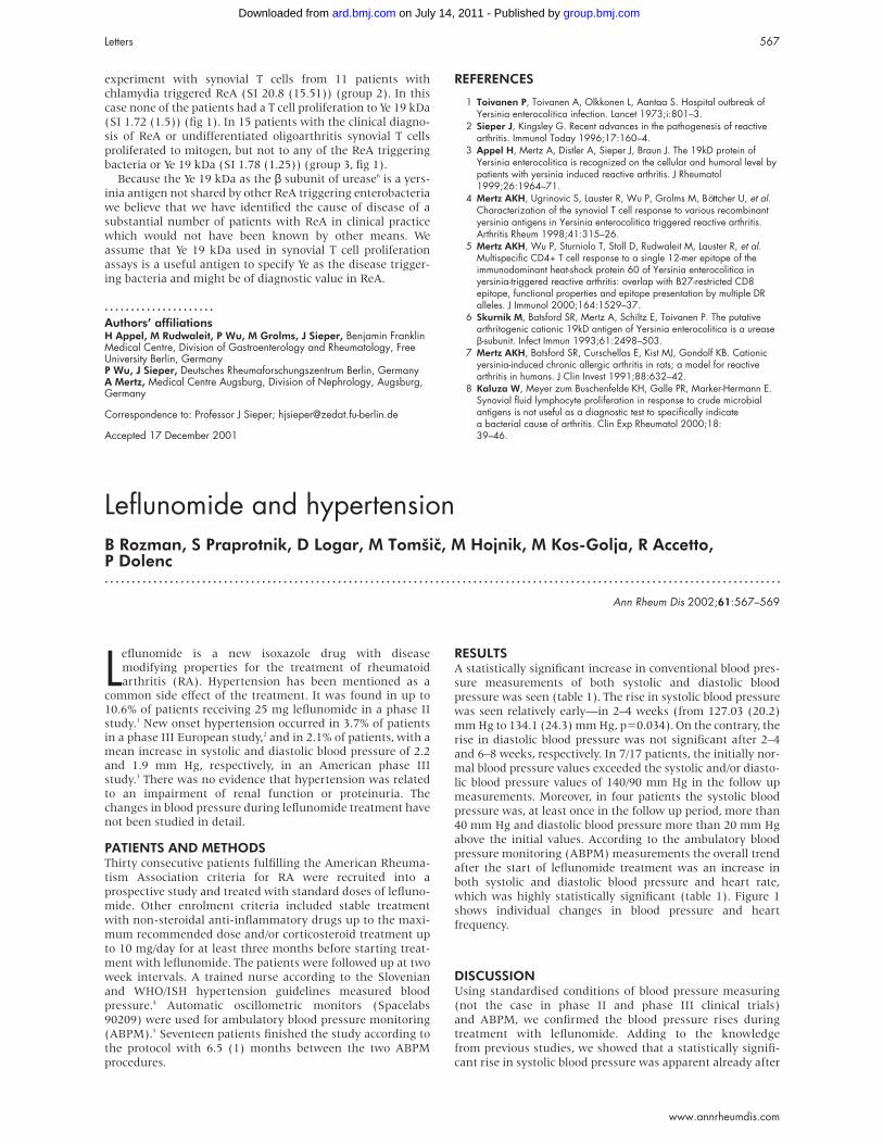
which was highly statistically significant (table 1). Figure 1
shows individual changes in blood pressure and heart
frequency.
DISCUSSIONUsing standardised conditions of blood pressure measuring
(not the case in phase II and phase III clinical trials)
and ABPM, we confirmed the blood pressure rises during
treatment with leflunomide. Adding to the knowledge
from previous studies, we showed that a statistically signifi-
cant rise in systolic blood pressure was apparent already after
Letters 567
www.annrheumdis.com
group.bmj.com on July 14, 2011 - Published by ard.bmj.comDownloaded from
2–4 weeks of the treatment, thus pointing to the need for
early blood pressure monitoring. By contrast, the rise in
diastolic blood pressure appeared later. Hypertensive values
in individual patients suggest that regular measuring of
blood pressure is required during treatment with lefluno-
mide.
Employing ABPM, we confirmed the significant rise in
blood pressure during the leflunomide treatment, thus
making the role of the “white coat” phenomenon unlikely. It
should be mentioned that it has been confirmed that
non-invasive ABPM has no effect on blood pressure because of
discomfort during cuff inflation. We are also not aware of any
special device developed to measure blood pressure in patients
with painful limbs. However, as a clinically relevant (>5
joints) improvement in tender and swollen joint count was
seen in 14 (83%) of the 17 patients analysed, the degree of
pain imposed by blood pressure measurements and its effect
on blood pressure were expected to decrease rather than rise
during the study.
The results do not allow us to speculate on the
mechanism of the blood pressure increase associated with the
leflunomide treatment. As the heart rate also rises during
leflunomide treatment, it has been assumed that hyper-
tension may be caused by an increased sympathetic drive.
Figure 1 Changes in (A) systolic blood pressure, (B) diastolic blood pressure, and (C) heart rate in 17 patients with RA treated withleflunomide. Twenty four hour averages of individual patients during ABPM performed during and after 6.5 (1) months of treatment withleflunomide are shown. The overall trends in individual variables are shown by the grey line.
180
160
140
100
120
Initia
l
Leflun
omide
A
Systo
lic b
lood
pre
ssur
e (m
m H
g)
100
80
60
Initia
l
Leflun
omide
B
Dia
stolic
blo
od p
ress
ure
(mm
Hg)
100
80
60
Initia
l
Leflun
omide
C
Hea
rt ra
te (m
in–1
)
Table 1 Conventional systolic and diastolic blood pressure measurements, 24 houraverages of blood pressure, and heart frequency before (initial ABPM) and aftertreatment with leflunomide (final ABPM) in 17 patients with rheumatoid arthritis.Twenty four hour, day time (6 00 am to 10 00 pm), and night time (10 00 pm to 600 am) mean values and standard deviations are shown. Statistical significance ofdifferences was tested with Student’s t test. p Values of <0.05 were consideredsignificant
Initialmeasurement
Finalmeasurement t Test
Significance(p)
Conventional measurements (mean)Systolic blood pressure (mm Hg) 127.3 (20.2) 140.7 (20.1) 3.55 0.003Diastolic blood pressure (mm Hg) 76.7 (9.3) 84.0 (8.6) 3.08 0.007
ABPM (mean)Systolic blood pressure (mm Hg) 127.8 (19.7) 132.1 (21.4) 3.01 0.003Diastolic blood pressure (mm Hg) 74.9 (12.4) 79.7 (13.0) 5.43 0.000Mean arterial pressure (mm Hg) 93.6 (14.8) 98.7 (15.8) 4.76 0.000Pulse pressure (mm Hg) 52.9 (14.0) 52.4 (15.5) 0.46 NSHeart frequency (min−1) 77.6 (13.7) 80.2 (13.5) 2.71 0.007
Day time (6 00 am to 10 00 pm)Systolic blood pressure (mm Hg) 130.2 (18.2) 135.2 (19.6) 5.18 0.000Diastolic blood pressure (mm Hg) 77.8 (11.4) 83.1 (11.7) 9.04 0.000Mean arterial pressure (mm Hg) 96.1 (13.4) 83.1 (11.7) 7.86 0.000Pulse pressure (mm Hg) 52.5 (14.2) 52.1 (15.4) 0.44 NSHeart frequency (min−1) 81.8 (13.7) 83.7 (13.3) 2.83 0.005
Night time (10 00 pm to 6 00 am)Systolic blood pressure (mm Hg) 121.9 (20.6) 124.1 (20.6) 1.20 NSDiastolic blood pressure (mm Hg) 69.1 (12.2) 72.3 (11.8) 3.07 0.002Mean arterial pressure (mm Hg) 87.8 (15.5) 91.5 (14.6) 2.77 0.006Pulse pressure (mm Hg) 52.8 (13.3) 51.7 (15.0) 0.86 NSHeart frequency (min−1) 68.7 (9.2) 72.9 (10.9) 4.78 0.000
568 Letters
www.annrheumdis.com
group.bmj.com on July 14, 2011 - Published by ard.bmj.comDownloaded from
This hypothesis remains to be tested. The changes in the
raised blood pressure after six months of leflunomide
treatment will be clarified after the final report of all
extended studies.
. . . . . . . . . . . . . . . . . . . . .Authors’ affiliationsB Rozman, S Praprotnik, D Logar, M Tomsic,M Hojnik, M Kos-Golja, Department of Rheumatology, Medical CentreLjubljana, Vodnikova 62, 1000 Ljubljana, SloveniaR Accetto, P Dolenc Department of Hypertension, Medical CentreLjubljana
Correspondence to: Dr B Rozman; [email protected]
Accepted 10 December 2001
REFERENCES
1 Rozman B, for the Leflunomide Investigator’s Group. Clinical experiencewith leflunomide in rheumatoid arthritis. J Rheumatol 1998;25(suppl53):27–32.
2 Smolen JS, Kalden JR, Rozman B, Kvien T, Scott DL, Larsen A, et al.Efficacy and safety of leflunomide compared to placebo andsulfasalazine in active rheumatoid arthritis. A double-blind, randomized,multicenter study. Lancet 1999;353:259–66.
3 Strand V, Cohen S, Schiff M, Weaver A, Fleischmann R, Cannon G, etal. Treatment of active rheumatoid arthritis with leflunomide comparedwith placebo and methotrexate. Arch Intern Med 1999;159:2542–50.
4 WHO/ISH. The 1999 WHO/ISH hypertension guidelines. J Hypertens1999;17:151–83.
5 O’Brien E, Coats A, Owens P, Petrie J, Padfield PL, Littler WA, et al. Useand interpretation of ambulatory blood pressure monitoring:recommendations of the British Hypertension Society. BMJ2000;320:1128–34.
Adhesion molecule expression in the synovial membraneof psoriatic arthritisV Riccieri, A Spadaro, E Taccari, A Zoppini, E Koo, J Ortutay, M Sesztak, I Markus. . . . . . . . . . . . . . . . . . . . . . . . . . . . . . . . . . . . . . . . . . . . . . . . . . . . . . . . . . . . . . . . . . . . . . . . . . . . . . . . . . . . . . . . . . . . . . . . . . . . . . . . . . . . . . . . . . . . . . . . . . . . .
Ann Rheum Dis 2002;61:569–570
Endothelium may play a part in the pathogenesis of long-
standing psoriatic arthritis (PsA),1 whereas a higher vas-
cularisation and a less intense adhesion molecule expres-
sion have been found in PsA synovial membrane compared
with rheumatoid arthritis.2 Some proinflammatory molecules,
such as tumour necrosis factors (TNFs), can induce synovial
endothelial cells and fibroblast-like synoviocytes to express
adhesion molecules.3 4
PATIENTS AND METHODSIn two groups of patients with PsA—eight patients with syno-
vitis of <1 year and six patients with synovitis >1 year—we
studied the expression and pattern of the synovial distribution
of endothelial leucocyte adhesion molecule-1 (ELAM-1 or
E-selectin) (CD62E), intercellular adhesion molecule-1
(ICAM-1) (CD54), vascular cell adhesion molecule-1
(VCAM-1) (CD106) (Immunotech, Marseille, France), and of
TNFα and TNFβ cytokines (Chemicon International, Te-
mecula, CA, USA) using a standard three stage immuno-
peroxidase labelling technique (LAB VISION, Fremont, CA,
USA).5 The lining layer, the infiltrating elements, and the
endothelial cells were evaluated for the number of positive
cells per high power field (×40).6
RESULTSTable 1 summarises the main clinical and laboratory data of
the two groups; no significant clinical or laboratory differences
were seen.
E-selectin was present more often at endothelial, cellular
infiltrate, and lining layer levels in 7/8 (88%) patients with a
disease duration <1 year, where only 3/6 patients (50%) with
disease duration >1 year were positive. ICAM-1 was
Table 1 Main clinical and demographic features of 14 patients with PsA with a disease duration of less (group 1) ormore (group 2) than one year
Patientnumber Sex
Age(years)
Duration ofarthritis (years)
Duration ofpsoriasis (years) PASI
Ritchieindex Subgroup
CRP(mg/l)
ESR(mm/1st h) Treatment
Group 11 F 60 <1 1 3.2 17 Polyarthritis 5 14 NSAIDs2 F 28 <1 1 4.5 11 Polyarthritis 4 28 NSAIDs3 F 48 <1 <1 0.3 3 Oligoarthritis 35 52 Steroids4 M 37 <1 4 0.9 18 Polyarthritis 6 8 NSAIDs5 M 31 <1 2 0.3 9 Polyarthritis 12 24 None6 F 35 <1 1 9.0 20 Polyarthritis 24 66 NSAIDs7 F 25 <1 19 2.1 5 Oligoarthritis 6 16 HCQ/NSAIDs8 F 35 <1 18 0.9 15 Polyarthritis 6 23 HCQ/NSAIDs
Group 21 M 35 2 10 3.1 9 Polyarthritis 21 38 NSAIDs2 M 36 5 13 6.6 10 Polyarthritis 80 86 MTX/steroids3 M 53 3 37 8.9 21 Polyarthritis 6 3 NSAIDs4 M 39 3 2 1.2 9 Polyarthritis 25 11 SSZ5 M 43 5 30 4.2 6 Oligoarthritis 6 8 NSAIDs6 M 50 10 25 9.0 15 Polyarthritis 6 10 NSAIDs
PASI, Psoriasis Areas Severity Index; CRP, C reactive protein; ESR, erythrocyte sedimentation rate (Westergren); NSAIDs, non-steroidal anti-inflammatorydrugs; HCQ, hydroxychloroquine; MTX, methotrexate; SSZ, sulfasalazine.
Letters 569
www.annrheumdis.com
group.bmj.com on July 14, 2011 - Published by ard.bmj.comDownloaded from
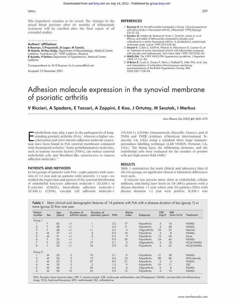
overexpressed in the lining layer of the early synovitic
specimens compared with the longstanding samples (100% v33%; p<0.001) (fig 1). On the contrary, VCAM-1 positivity was
more commonly found in patients with longstanding PsA (5/5
(100%) v 4/7 (57%)). Cells containing TNFα and TNFβ were
consistently found in the synovial lining layer, in the
infiltrates, and in the blood vessels, with no appreciable differ-
ence between the two groups.
DISCUSSIONOur results show that longstanding psoriatic synovitis may
reduce the E-selectin expression, as already found in different
forms of synovitis,7 and confirms the presence of ICAM-1 and
VCAM-1 positivity in PsA, as already described.2 As ICAM-1
was present in vessel walls in all tissue samples, this supports
the view that this adhesion molecule is not only constitutively
expressed on endothelial cells but is also increased during
activation and is the most important adhesion molecule for
cell binding to endothelium in inflamed tissue.7 VCAM-1
expression, generally absent on normal synovium, is found on
activated endothelium and its up regulation has been recently
implicated in various pathological conditions.8
The different expression of these adhesion molecules seems
to be connected to the disease duration. A more frequent posi-
tivity for E-selectin, and partly for ICAM-1, in earlier synovitis
compared with longstanding disease, where VCAM-1 expres-
sion was constantly found, shows for the first time how these
molecules may separately participate in the synovitic process
in the different phases of PsA, with a changing involvement as
the disease evolves.
The presence of TNFα and TNFβ, together with
E-selectin, ICAM-1, and VCAM-1 positivity in the same sam-
ples, confirms the ability of TNFs to induce the expression of
such adhesion molecules.3 Their localisation on the endothe-
lial cells also suggests that these cells can produce TNFs, indi-
cating the involvement of TNFs in the regulation of cell adhe-
sion before migration into diseased joints. Our findings gain
more importance in view of a recent immunohistochemical
study, which showed a convincing effect of anti-TNF
treatment on synovium in spondyloarthropathy, suggesting
immunomodulatory mechanisms involving adhesion mol-
ecule expression.9
In conclusion, the variations in the presence of some adhe-
sion molecules and TNFs shown in our study, partly related to
disease duration, indicate their relative importance in mediat-
ing the succeeding mechanisms of psoriatic synovitis. This
should be taken into account in the assessment of disease
progression and in developing possible new therapeutic
approaches.
. . . . . . . . . . . . . . . . . . . . .Authors’ affiliationsV Riccieri, A Spadaro, E Taccari, A Zoppini, Rheumatology Unit,Department of Medical Therapy, University of Rome “La Sapienza”,Rome, ItalyE Koo, J Ortutay, Polyclinic of the Hospitaller Brothers of St John ofGod, Budapest, HungaryM Sesztak, I Markus, National Institute of Rheumatology andPhysiotherapy, Budapest, Hungary
Correspondence to: Dr V Riccieri, Rheumatology Unit, Department ofMedical Therapy, University of Rome “La Sapienza”, P le Aldo Moro 5,00185, Rome, Italy; [email protected].
Accepted 10 December 2001
REFERENCES1 Taccari E, Fattorossi A, Moretti S, Riccieri V, Fasani M, Zoppini A.
Phenotypic profile of major synovial cell populations in longstandingpsoriatic arthritis. J Rheumatol 1987;14:525–30.
2 Veale D, Yanni G, Rogers S, Barnes L, Bresnihan B, Fitzgerald O.Reduced synovial membrane macrophage numbers, ELAM-1 expression,and lining layer hyperplasia in psoriatic arthritis as compared withrheumatoid arthritis. Arthritis Rheum 1993;36:893–900.
3 Abbot SE, Kaul A, Stevens CR, Blake DR. Isolation and culture ofsynovial microvascular endothelial cells: characterization and assessmentof adhesion molecule expression. Arthritis Rheum 1992;35:401–6.
4 Marlor CW, Webb DL, Bombara MP, Greve JM, Blue ML. Expression ofvascular cell adhesion molecule-1 in fibroblast-like synoviocytes afterstimulation with tumor necrosis factor. Am J Pathol 1992;140:1055–60.
5 Hsu SM, Raine L, Fanger H. Use of avidin-biotin-immunoperoxidasecomplex (ABC) in immunoperoxidase techniques: a comparison betweenABC and unlabelled antibody) (PAP) procedures. J Histochem Cytochem1981;29:577–80.
6 Grom AA, Murray KJ, Luyrink L, Emery H, Passo MH, Glass DN, et al.Patterns of expression of tumor necrosis factor α, tumor necrosis factor β,and their receptors in synovia of patients with juvenile rheumatoidarthritis and juvenile spondylarthropathy. Arthritis Rheum1996;39:1703–10.
7 Mellbye OJ, Shen Y, Hogasen K, Mollnes TE, Forre O. Adhesionmolecule expression and complement activation in vessel walls insynovial tissue from patients with chronic inflammatory joint disease. ClinRheumatol 1996;15:441–7.
8 Mojcik CF, Shevach EM. Adhesion molecules. A rheumatologicperspective. Arthritis Rheum 1997;40:991–1004.
9 Baeten D, Kruithof E, Van den Bosch F, Demetter P, Van Damme N,Cuvelier C, et al. Immunomodulatory effects of anti-tumor necrosis factorα therapy on synovium in spondyloarthropathy. Histologic findings ineight patients from an open-label pilot study. Arthritis Rheum2001;44:186–95.
Figure 1 Representative specimen from the synovial membraneof a patient with psoriatic arthritis of <1 year; staining withICAM-1 using monoclonal antibody CD54. Antigen positive cells arepresent throughout the entire specimen (original magnification×250).
570 Letters
www.annrheumdis.com
group.bmj.com on July 14, 2011 - Published by ard.bmj.comDownloaded from
Headache as the initial presentation of Wegener’sgranulomatosisI G S Lim, P J Spira, H P McNeil. . . . . . . . . . . . . . . . . . . . . . . . . . . . . . . . . . . . . . . . . . . . . . . . . . . . . . . . . . . . . . . . . . . . . . . . . . . . . . . . . . . . . . . . . . . . . . . . . . . . . . . . . . . . . . . . . . . . . . . . . . . . .
Ann Rheum Dis 2002;61:571–572
In Wegener’s granulomatosis (WG), neurological involve-
ment is rare at onset. We present an unusual case where
headache was the initial, dominant presentation of WG.
CASE REPORTA 34 year old white man presented with a three month history of
headache. The headaches were migratory, throbbing, and were
accentuated with head movement. Physical examination was
normal. Computed tomography (CT) of the sinuses was normal.
The patient was diagnosed with non-specific vascular headaches,
and was prescribed pizotifen, which alleviated his headaches.
One month later, the patient developed a red right eye. Bilat-
eral papilloedema was noted. He was now unable to work
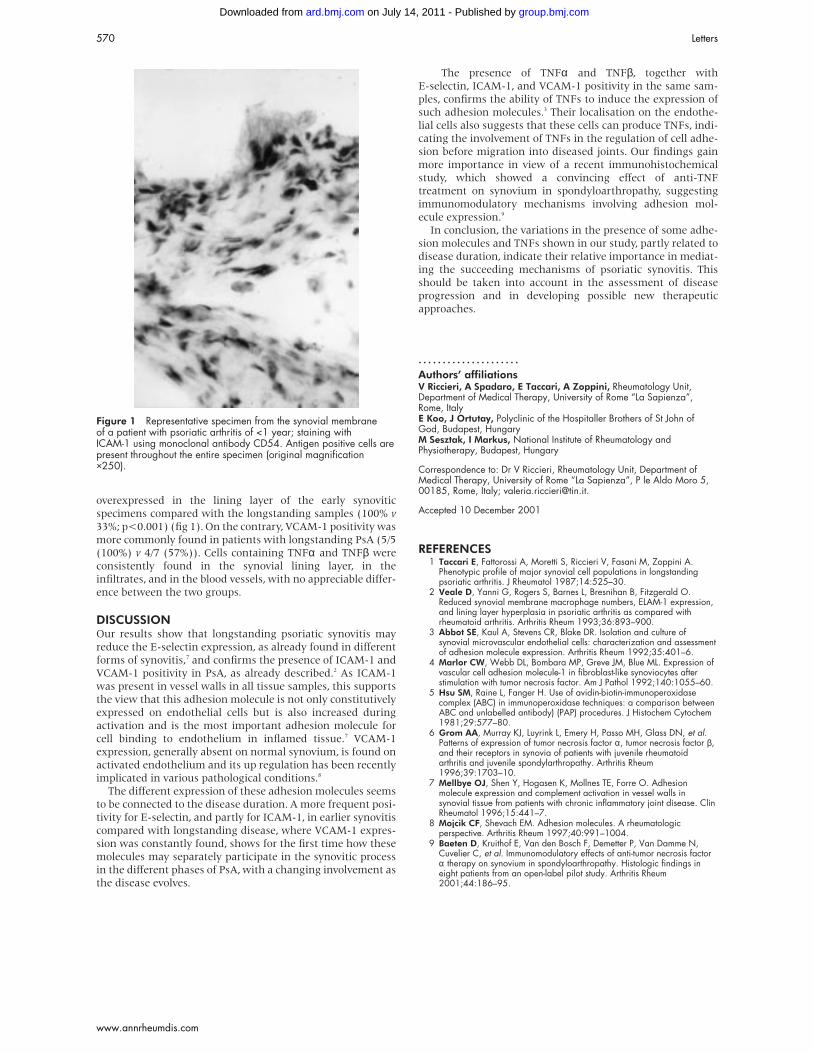
because of the headache. Magnetic resonance imaging (MRI) of
the brain disclosed a normal ventricular system, but pro-
nounced gadolinium enhancement of the meninges around the
entire left hemisphere, most of the parieto-occipital region on
the right, as well as the tentorium bilaterally (fig 1). Lumbar
puncture disclosed a high cerebrospinal fluid (CSF) opening
pressure of 27 cm (13–18 cm). A CSF examination was entirely
normal and cultures were negative. The headache was partially
relieved by CSF drainage, and acetazolamide was started.
One week later, the patient developed a red left eye and left
knee arthritis. Over the course of the next week, his condition
progressed rapidly with purpuric lesions appearing on his
hands and feet, followed by pericarditis and pulmonary haem-
orrhage. Biopsy of the purpura disclosed leucocytoclastic
vasculitis. Antineutrophil cytoplasmic antibody (cANCA)
taken at the time that he complained of the red left eye was
positive at a titre of 1/80, with specificity for proteinase-3. A
week later, repeat testing showed that cANCA had risen to
1/320. There was also a mild normochromic, normocytic anae-
mia, and raised inflammatory markers. Urine analysis dis-
closed microscopic haematuria and mild proteinuria. No casts
were identified. A CSF examination was again normal, but the
opening pressure had risen to 36 cm. WG was diagnosed.
Treatment was started with a 1 g pulse of intravenous
methylprednisone, followed by oral daily doses of 1 mg/kg
prednisone and 2 mg/kg cyclophosphamide. A few days after
the start of treatment, the headaches had resolved and the
CSF opening pressure was normal. Six months later, the
patient is symptom-free, the papilloedema has resolved, MRI
is normal, and the patient has returned to full-time work.
DISCUSSIONIt is rare for WG to present with neurological symptoms.
Neurological presentations described include ataxia, ocular
nerve palsies, seizures, and deteriorating mental status.1–4
Shiotani et al described a 37 year old man with chronic sinusi-
tis, who presented with fever and headache for 10 days before
CT disclosed subdural and paranasal masses with marked
thickening of the nasal mucosa.5 Our patient presented far
more insidiously, with significant headache that persisted and
worsened with time. The headache had clear vascular features
but, beyond this, was non-specific. It was only four months
later that musculoskeletal, cutaneous, ophthalmic, and cardio-
respiratory features developed. Although neurological involve-
ment may eventually develop in 33.6% of patients with WG,6
meningeal involvement, as gauged by meningeal enhancement
on MRI or by biopsy is particularly uncommon, being recorded
in only a handful of case reports.2–5 7–10 There also seems to be no
relation between CSF abnormalities, clinical symptoms, or
extent of meningeal involvement on MRI.7 A CSF examination
may show no abnormality3 4 7 or a pleocytosis.3 5 8 9 High open-
ing pressures are unusual but have been described.9 10
In conclusion, we have presented a case of WG with exten-
sive meningeal involvement. The exceptional feature in this
case is the fact that headache was the sole symptom of the
disease over several months, before a dramatic activation of
the disorder with more typical features of WG.
. . . . . . . . . . . . . . . . . . . . .Authors’ affiliationsI G S Lim, H P McNeil, Department of Rheumatology, Prince of WalesHospital, and University of New South Wales, Sydney, Australia 2052P J Spira, Institute of Neurological Sciences, Prince of Wales Hospital
Correspondence to: Associate Professor H P McNeil, Department ofRheumatology, Prince of Wales Hospital, Randwick, New South Wales,Australia 2031; [email protected]
Accepted 13 December 2001
REFERENCES1 Atcheson SG, Van Horn G. Subacute meningitis heralding a diffuse
granulomatous angiitis: (Wegener’s granulomatosis?). Neurology1977;27:262–4.
2 Weinberger LM, Cohen ML, Remler BF, Naheedy MH, Leigh RJ.Intracranial Wegener’s granulomatosis. Neurology 1993;43:1831–4.
3 Newman NJ, Slamovits TL, Friedland S, Wilson WB. Neuro-ophthalmicmanifestations of meningocerebral inflammation from the limited form ofWegener’s granulomatosis. Am J Ophthalmol 1995;120:613–21.
4 Burrell HC, McConachie NS. Pachymeningitis in Wegener’sgranulomatosis. Australas Radiol 1998;42:364–6.Figure 1 MRI showing diffuse meningeal enhancement.
Letters 571
www.annrheumdis.com
group.bmj.com on July 14, 2011 - Published by ard.bmj.comDownloaded from
5 Shiotani A, Mukobayashi C, Oohata H, Yamanishi T, Hara T, Itoh H, etal. Wegener’s granulomatosis with dural involvement as the initial clinicalmanifestation. Intern Med 1997;36:514–18.
6 Nishino H, Rubino FA, DeRemee RA, Swanson JW, Parisi JE. Neurologicalinvolvement in Wegener’s granulomatosis: an analysis of 324 consecutivepatients at the Mayo Clinic. Ann Neurol 1993;33:4–9.
7 Specks U, Moder KG, McDonald TJ. Meningeal involvement in Wegenergranulomatosis. Mayo Clin Proc 2000;75:856–9.
8 Spranger M, Schwab S, Meinck HM, Tischendorf M, Sis J, Breitbart A,et al. Meningeal involvement in Wegener’s granulomatosis confirmedand monitored by positive circulating antineutrophil cytoplasm incerebrospinal fluid. Neurology 1997;48:263–5.
9 Katrib A, Portek I, Corbett AJ, Hersch MI, Zagami AS. Meningealinvolvement in Wegener’s granulomatosis. J Rheumatol 1998;25:1009–11.
10 Scarrow AM, Segal R, Medsger TA Jr, Wasko MC. Communicatinghydrocephalus secondary to diffuse meningeal spread of Wegener’sgranulomatosis: case report and literature review. Neurosurgery1998;43:1470–3.
Extremely high dose pravastatin may suppressamyloidogenesis in a mouse modelS Shtrasburg, M Pras, M Lidar, A Livneh. . . . . . . . . . . . . . . . . . . . . . . . . . . . . . . . . . . . . . . . . . . . . . . . . . . . . . . . . . . . . . . . . . . . . . . . . . . . . . . . . . . . . . . . . . . . . . . . . . . . . . . . . . . . . . . . . . . . . . . . . . . . .
Ann Rheum Dis 2002;61:572
Pravastatin is a cholesterol lowering agent,1 recently re-ported to have anti-inflammatory properties.2 It wassuggested that the prevention and regression of atheroscle-
rosis by pravastatin is partially related to its anti-inflammatoryeffect,2 probably mediated by inhibition of proinflammatorycytokines.3 Pravastatin’s anti-inflammatory effect is associatedwith, and probably reflected by, reduced levels of the acutephase reactants, C reactive protein and serum amyloid A(SAA).2 The N-terminal fragment of the SAA is amyloid A (AA),which is deposited in a fibrillar form in the tissues of up to 30%of patients with a variety of chronic infectious and chronicinflammatory disorders, leading to reactive amyloidosis.4
Because prevention and treatment of AA amyloidosis arecurrently unsatisfactory and reactive amyloidosis is a poten-tially lethal complication, it is important to find out whetherpravastatin affects amyloidogenesis.
The effect of pravastatin on amyloidogenesis was studied inseveral groups of male Swiss mice 7–17 weeks old, which were
subjected to amyloid induction, using intravenous amyloid
enhancing factor (1 µg in 0.5 ml phosphate buffered saline on
day 0) and subcutaneous AgNO3 (0.5 ml 2% daily, on days 0, 1,
and 2), according to our published protocol.5 Two groups of
study mice (groups I and II) received intraperitoneal
pravastatin 0.4 mg/day in 0.5 ml saline, and two other groups
(III and IV) received intraperitoneal pravastatin 10 mg/day.
The human oral dose analogous to these regimens is 0.5 mg/kg
and 12.5 mg/kg respectively (assuming a 20-fold increase in
drug catabolism in mice as compared with man). The experi-
ments lasted for 72 hours (groups I and III) or 96 hours
(groups II and IV). The 72 hour interval, during which the
amount of amyloid deposits is still low, allows the detection of
a mild inhibition. All experiments were controlled by mice of
the same strain, sex, and age, which received the same
amyloid induction regimen, but 0.5 ml saline intraperitoneally
instead of pravastatin. The amount of amyloid deposition in
the spleen was studied by the crush and smear technique and
a five grade score, estimated by polarised microscopy.6 All
experiments were repeated two to three times.
Amyloidosis in mice receiving pravastatin was somewhat
less abundant and developed in fewer animals than in controls
(table 1). This trend was noted only in animals receiving 10
mg/day and only in the short term experiments, but the
statistical significance obtained was inconsistent (table 1). No
amyloid inhibition by pravastatin was seen in any of the other
experiments, either when a lower pravastatin dose (0.4
mg/day) was used or when mice were subjected to a longer (96
hours) amyloidogenic stimulus.
These findings suggest that pravastatin in a very high dose
may have a mild amyloid protecting effect and thus increases
the spectrum of drugs with a possible tangible amyloid
preventive effect. Further studies are warranted to determine
underlying mechanisms and to see whether other statins also
have anti-amyloidogenic qualities.
. . . . . . . . . . . . . . . . . . . . .Authors’ affiliationsS Shtrasburg, M Pras, M Lidar, A Livneh, Heller Institute of MedicalResearch, Sheba Medical Centre, Tel-Hashomer, Israel
Correspondence to: Dr S Shtrasburg, Heller Institute of Medical Research,Sheba Medical Centre, Tel Hashomer 52621, Israel;[email protected]
Accepted 13 December 2001
REFERENCES1 Keidar S, Aviram M, Maor I, Oiknine J, Brook JG. Pravastatin inhibits
cellular cholesterol synthesis and increases low density lipoproteinreceptor activity in macrophages: in vitro and in vivo studies. Br J ClinPharmacol 1994;38:513–19.
2 Ridker PM, Rifai N, Pfeffer MA, Sacks FM, Moye LA, Goldman S, et al.Inflammation, pravastatin and risk of coronary events after myocardialinfarction in patients with average cholesterol levels. Circulation1998;98:839–44.
3 Rosenson RS, Tangney CC, Casey LC. Inhibition of proinflammatorycytokine production by pravastatin. Lancet 1999;353:983–4.
4 Yakar S, Livneh A, Kaplan B, Pras M. The molecular basis of reactiveamyloidosis. Semin Arthritis Rheum 1995;24:255–61.
5 Shtrasburg S, Livneh A, Gal R, Pras M. Extremely active murine amyloidenhancing factor. Clin Exp Rheumatol 1996;14:37–42.
6 Shtrasburg S, Gal R, Pras M. Crush and smear technique for rapiddetection and semiquantitation of amyloid deposition. Biotech Histochem1991;66:203–7.
Table 1 High dose pravastatin may suppressamyloidogenesis
Group III*experimentnumber
Type ofexperiment
Amyloidpositive miceper group
Median(range) ofamyloid grade
pValues†
1 Study 5/6 2.5 (0–3) 0.24Control 6/6 1.5 (1–3)
2 Study 4/6 0.75 (0–1.5) 0.03Control 6/6 2.0 (0.5–3)
3 Study 2/5 0.5 (0–3) 0.07Control 5/6 2.0 (0–2.5)
Combined Study 11/17 1.0 (0–3) 0.05Control 17/18 2.0 (0–3)
*Amyloidosis was induced by amyloid enhancing factor and AgNO3.Study animals received intraperitoneally pravastatin 10 mg/day ondays 0, 1, and 2. Control mice received 0.5 ml saline instead. Themice were killed after 72 hours (24 hours after the last pravastatininjection) and the amount of splenic amyloid was estimated by theCrush and Smear technique; †using Fisher’s exact probability test.
572 Letters
www.annrheumdis.com
group.bmj.com on July 14, 2011 - Published by ard.bmj.comDownloaded from
doi: 10.1136/ard.61.6.560 2002 61: 560-561Ann Rheum Dis
G Triolo, M Vadalà, A Accardo-Palumbo, et al.
diseaseantibody treatment for ocular Behçet's Anti-tumour necrosis factor monoclonal
http://ard.bmj.com/content/61/6/560.full.htmlUpdated information and services can be found at:
These include:
References
http://ard.bmj.com/content/61/6/560.full.html#related-urlsArticle cited in:
http://ard.bmj.com/content/61/6/560.full.html#ref-list-1This article cites 4 articles, 1 of which can be accessed free at:
serviceEmail alerting
box at the top right corner of the online article.Receive free email alerts when new articles cite this article. Sign up in the
Notes
http://group.bmj.com/group/rights-licensing/permissionsTo request permissions go to:
http://journals.bmj.com/cgi/reprintformTo order reprints go to:
http://group.bmj.com/subscribe/To subscribe to BMJ go to:
group.bmj.com on July 14, 2011 - Published by ard.bmj.comDownloaded from