The oestrogen metabolite 2-methoxy- oestradiol alone or in combination with tumour necrosis...
18
The oestrogen metabolite 2-methoxy- oestradiol alone or in combination with tumour necrosis factor-related apoptosis-inducing ligand mediates apoptosis in cancerous but not healthy cells of the human endometrium Sumie Kato 1 , Anil Sadarangani 2 , Soledad Lange 2 , Manuel Villalo ´n 2 , Jorge Bran ˜es 1 , Jan J Brosens 3 , Gareth I Owen 2 and Mauricio Cuello 1 Departments of 1 Obstetrics and Gynaecology, Faculty of Medicine and 2 Physiology, Faculty of Biological Sciences, Pontificia Universidad Cato ´ lica de Chile, Alameda 340, Santiago, Casilla 114-D, Chile 3 Institute of Reproductive and Developmental Biology, Imperial College London, Hammersmith Hospital, London W12 0NN, UK (Requests for offprints should be addressed to M Cuello; Email: [email protected]) S Kato and A Sadarangani contributed equally to this work Abstract Cancers of the reproductive tract account for 12% of all malignancies in women. As previous studies have shown that oestrogen metabolites can cause apoptosis, we characterised the effect of oestrogen and oestrogen metabolites on non-cancerous and cancerous human endometrial cells. Herein, we demonstrate that 2-methoxyoestradiol (2ME), but not 17b-oestradiol, induces apoptosis in cancer cell lines and primary cultured tumours of endometrial origin. In contrast, 2ME had no effect on cell viability of corresponding normal tissue. This ability of 2ME to induce apoptosis does not require oestrogen receptor activation, but is associated with increased entry into the G2/M phases of the cell cycle and the activation of both the intrinsic and the extrinsic apoptotic pathways. The selective behaviour of 2ME on cancerous as opposed to normal tissue may be due to a reduction in 17b-hydroxysteroid dehydrogenase type II levels in cancer cells and to a differential down-regulation of superoxide dismutase. Furthermore, we demonstrate that pre-treatment with 2ME enhances the sensitivity of reproductive tract cancer cells to the apoptotic drug tumour necrosis factor-related apoptosis-inducing ligand (TRAIL), without the loss in cell viability to normal cells incurred by currently chemotherapeutic drugs. In conclusion, 2ME, alone or in combination with TRAIL, may be an effective treatment for cancers of uterine origin with minimal toxicity to corresponding healthy female reproductive tissue. Endocrine-Related Cancer (2007) 14 351–368 Introduction Cancer of the reproductive tract encompasses malig- nancies of the uterine cervix, uterine corpus, ovary, vulva, fallopian tube, vagina and other related genital tissue. According to recent estimates by the American Cancer Society, cancer of the reproductive tract will account for 12% of all female cancers (79 480 of 662 870 estimated cases) and w15% of female cancer- related mortalities (28 910 of 275 000 estimated cases). Endometrial cancer is the most common gynaecologic malignancy and the fourth most common cancer in women (Amant et al. 2005, Carter & Pather 2006). Approximately 75% of women present with diseases clinically confined to the uterus (stage 1), for which the overall 5-year survival is 75% (2). The prognosis for patients with more advanced or recurrent endometrial disease is poor, with a median survival of !1 year (Amant et al. 2005). Chemotherapy is the mainstay of the treatment for patients with advanced or recurrent disease. Due to the well-documented secondary effects Endocrine-Related Cancer (2007) 14 351–368 Endocrine-Related Cancer (2007) 14 351–368 1351–0088/07/014–351 q 2007 Society for Endocrinology Printed in Great Britain DOI:10.1677/ERC-07-0008 Online version via http://www.endocrinology-journals.org
-
Upload
independent -
Category
Documents
-
view
2 -
download
0
Transcript of The oestrogen metabolite 2-methoxy- oestradiol alone or in combination with tumour necrosis...

Endocrine-Related Cancer (2007) 14 351–368
The oestrogen metabolite 2-methoxy-oestradiol alone or in combinationwith tumour necrosis factor-relatedapoptosis-inducing ligand mediatesapoptosis in cancerous but not healthycells of the human endometrium
Sumie Kato1, Anil Sadarangani 2, Soledad Lange2, Manuel Villalon2,Jorge Branes1, Jan J Brosens3, Gareth I Owen 2 and Mauricio Cuello1
Departments of 1Obstetrics and Gynaecology, Faculty of Medicine and2Physiology, Faculty of Biological Sciences, Pontificia Universidad Catolica de Chile, Alameda 340, Santiago, Casilla 114-D, Chile3Institute of Reproductive and Developmental Biology, Imperial College London, Hammersmith Hospital, London W12 0NN, UK
(Requests for offprints should be addressed to M Cuello; Email: [email protected])
S Kato and A Sadarangani contributed equally to this work
Abstract
Cancers of the reproductive tract account for 12% of all malignancies in women. As previous studieshaveshown thatoestrogenmetabolites cancauseapoptosis,wecharacterised theeffectof oestrogenand oestrogen metabolites on non-cancerous and cancerous human endometrial cells. Herein, wedemonstrate that 2-methoxyoestradiol (2ME), but not 17b-oestradiol, inducesapoptosis in cancer celllines andprimary cultured tumours of endometrial origin. In contrast, 2MEhadnoeffect on cell viabilityof corresponding normal tissue. This ability of 2ME to induce apoptosis does not require oestrogenreceptor activation, but is associated with increased entry into the G2/M phases of the cell cycle andthe activation of both the intrinsic and the extrinsic apoptotic pathways. The selective behaviour of2ME on cancerous as opposed to normal tissue may be due to a reduction in 17b-hydroxysteroiddehydrogenase type II levels in cancer cells and to a differential down-regulation of superoxidedismutase. Furthermore, we demonstrate that pre-treatment with 2ME enhances the sensitivity ofreproductive tract cancer cells to theapoptotic drug tumour necrosis factor-relatedapoptosis-inducingligand (TRAIL), without the loss in cell viability to normal cells incurred by currently chemotherapeuticdrugs. In conclusion, 2ME, alone or in combination with TRAIL, may be an effective treatment forcancers of uterine origin with minimal toxicity to corresponding healthy female reproductive tissue.
Endocrine-Related Cancer (2007) 14 351–368
Introduction
Cancer of the reproductive tract encompasses malig-
nancies of the uterine cervix, uterine corpus, ovary,
vulva, fallopian tube, vagina and other related genital
tissue. According to recent estimates by the American
Cancer Society, cancer of the reproductive tract will
account for 12% of all female cancers (79 480 of
662 870 estimated cases) andw15% of female cancer-
related mortalities (28 910 of 275 000 estimated cases).
Endometrial cancer is the most common gynaecologic
Endocrine-Related Cancer (2007) 14 351–368
1351–0088/07/014–351 q 2007 Society for Endocrinology Printed in Great
malignancy and the fourth most common cancer in
women (Amant et al. 2005, Carter & Pather 2006).
Approximately 75% of women present with diseases
clinically confined to the uterus (stage 1), for which the
overall 5-year survival is 75% (2). The prognosis for
patients with more advanced or recurrent endometrial
disease is poor, with a median survival of !1 year
(Amant et al. 2005). Chemotherapy is the mainstay of
the treatment for patients with advanced or recurrent
disease. Due to the well-documented secondary effects
Britain
DOI:10.1677/ERC-07-0008
Online version via http://www.endocrinology-journals.org

S Kato, A Sadarangani et al.: 2ME-mediated apoptosis in endometrial cancer
of chemotherapy, there is a need to find new effective
treatments for advanced endometrial cancers and other
malignancies of the reproductive tract which confer
minimal toxicity to normal healthy tissues.
One such candidate compound is the oestrogen
metabolite 2-methoxyoestradiol (2ME), which is now
in clinical trials for the treatment of various cancers
(Mooberry 2003). 2ME is producedby sequential hepatic
hydroxylation and methylation from the parent
compounds and is present in human blood and the
urine (Watanabe et al. 1991, Suchar et al. 1995, Zhu &
Conney 1998). Oestradiol or oestrone can be hydroxyl-
ated at various positions by nicotinamide adenine
dinucleotide phosphate (NADPH)-dependent cyto-
chrome p450 enzymes CYP1A2 and CYP3A present in
the liver and extrahepatic target cells (Franks et al. 1982,
Watanabe et al. 1991). The chemically reactive catechol
oestrogens (2- or 4-hydroxyoestradiol (2OHor 4OH) and
2- or 4-hydroxyoestrone (2OH or 4OH)) can be
metabolically O-methylated to monomethylethers by
catechol-O-methyltransferase (COMT), an enzyme
present in large amounts in many organs and cells such
as liver, kidney, placenta, uterus and mammary gland
(Kopin 1994,Williams et al. 2002, Yue et al. 2003). The
metabolite 2ME can bind to oestrogen receptors (ERs),
albeit at a 500- and 3200-fold lower affinity than that of
oestradiol for ERa and ERb respectively. Along with thecapability of utilising ER signalling pathways, several
publications have highlighted that the anti-proliferative
activities of 2ME can occur independently of ER
signalling and are not altered by either oestrogen
antagonists or agonists (Merriam et al. 1980, van
Aswegen et al. 1989). 2ME has been shown to be a
potent inhibitor of angiogenesis and suppresses tumour
growth. It appears to target only active proliferating cells
and not cytotoxic to quiescent cells (LaVallee et al.
2002). The exact action of 2ME is currently unknown;
however, various mechanisms have been implicated.
There is accordance in the literature that 2ME inhibits
tumour proliferation, in vitro and in vivo, by inducing cell
cycle arrest and apoptosis and by inhibiting angiogenesis.
A dose-dependentG2/Marrestwas found in awide range
of cancer cells and this effect is thought to be due to its
toxicity tomicrotubules (Sidor et al. 2005) and inhibition
of regulators of cell cycle progression (Qadan et al.
2001). Apoptosis is the programmed cell death that is
essential for the development and tissue homeostasis of
healthy cells.An apoptotic effect of 2MEhas been shown
both in vitro and in vivo (Basu et al. 2006, Gui & Zheng
2006, Ray et al. 2006) and this mechanism involves both
the intrinsic (up-regulation of p53, accumulation of
reactive oxygen species (ROS), Bcl-2 phosphorylarion
and inactivation) and extrinsic (up-regulation of DR5)
352
apoptotic pathways (Bu et al. 2002, LaVallee et al. 2003,
Zhou et al. 2004, Joubert et al. 2005, Thews et al. 2005).
Reports indicate that certain breast cancer cell lines
are insensitive to 2ME. Mechanistically, this insensi-
tivity is largely attributable to the presence of high
levels of a steroid-selective metabolising enzyme, the
type II 17b-hydroxysteroid dehydrogenase (17b-HSD),which rapidly converts 2ME to the inactive
2-methoxyoestrone (Liu et al. 2005).
There is evidence that 2ME can overcome resistance
to chemotherapy in several drug-resistant cancer cells,
and its also been shown that 2ME has no anti-tumour
effects on human endometrial carcinoma in in vivo
models (Chauhan et al. 2002, Li et al. 2007). However,
in these and other models, 2ME has been added orally
or subcutaneously and has thus been subject to
problems of metabolism in the liver. Due to the side
effects of the current combined chemotherapy treat-
ments, the combination of locally administered 2ME
with other apoptotic drugs with minimal toxicity to
healthy tissues would be an attractive alternative
therapeutic strategy for the treatment of cancers of
the female reproductive tract. One such drug is tumour
necrosis factor-related apoptosis-inducing ligand
(TRAIL), which we have previously shown to induce
apoptosis in cancer cells but not in healthy cells of
cervical, endometrial, fallopian tube or ovarian origin
(Sadarangani et al. 2007). TRAIL acts by binding to
the death receptors TRAIL-R1 (also called DR4) and
TRAIL-R2 (also called DR5), thereby inducing
caspase-mediated apoptosis.
In this study, we characterised the anti-proliferative
and pro-apoptotic effects of 2ME in endometrial and
cervical cancer cell lines, primary cultured cancer cells
and corresponding non-malignant cells from the
female reproductive tract. In addition, we demonstrate
that 2ME enhances the apoptotic response to TRAIL.
Materials and methods
Primary tissue of human female reproductive tract was
obtained with patient consent from the Gynaecology
Department of the Hospital of the Pontificia
Universidad Catolica de Chile, in Santiago, Chile.
Ethical approval for the study was obtained from both
the ethical committees of the Faculty of Medicine and
the Faculty of Biological Sciences at the Pontificia
Universidad Catolica de Chile and the South Eastern
Health Service of Santiago, Chile.
Cell culture
Primary culture of normal and cancerous human
endometrial epithelium, endometrial stroma, cervical,
www.endocrinology-journals.org

Endocrine-Related Cancer (2007) 14 351–368
ovarian and fallopian tube were derived from tissue
from pre-menopausal or postmenopausal women
undergoing either hysterectomy alone or hysterectomy
with bilateral salpingo-oophorectomy and from benign
or malignant tumours as published previously
(Sadarangani et al. 2007). The endometrial cancer
cell lines Ishikawa (Nishida et al. 1985), HEC1A
(Kuramoto 1972), the cervical cancer cell line HeLa
(Eagle 1955), the breast cancer cell lines ZR-75 (Engel
et al. 1978) and MDA-MB-231 (Cailleau et al. 1978)
cells were maintained in Dulbecco’s modified Eagle
medium (DMEM)/F12 media supplemented with 10%
foetal bovine serum (Invitrogen). For protein and RNA
experiments, cells were plated at 50% confluence in
10 cm2 Petri dishes (Falcon, Becton-Dickinson, NJ,
USA). The medium was changed to charcoal-treated
medium containing 5% serum for 24 h before the
addition of 2ME or TRAIL.
Reagents
17b-oestradiol (E2) and 2ME were purchased from
Sigma–Aldrich. All other oestrogen metabolites were
purchased from Steraloids Inc. (Rhode Island, USA).
glutathione-S-transferase (GST)-TRAIL fusion
protein was produced as described previously (Cuello
et al. 2001) and stored in aliquots at K20 8C. An equal
volume of vehicle (DMEM/F12medium or ethanol) was
added as a control in each experiment. The tetrapeptide
caspase inhibitor ZVAD-fmk(Z-Val-Ala-Asp (OCH3)-
fmk), caspase inhibitors 2, 8 and 9 (Enzyme Systems
Products, Livermore, CA, USA) were resuspended in
DMSO (Sigma–Aldrich Co.) and added to the cells at
a final concentration of 50 mM, 30 min before TRAIL.
N-acetylcysteine (NAC) and ICI 182 780 (ICI) were
purchased from Sigma–Aldrich.
The chemotherapeutics drugs (doxorubicin (DOX),
paclitaxel (PTX) and cisplatin (CIS)) were kindly
given by the Cancer Center of the Pontificia
Universidad Catolica de Chile, and used at a final
concentration of 5 mM.
Measurement of cell viability and apoptosis
TheMTS assay was used to assess oestrogen, oestrogen
metabolite and TRAIL-mediated cytotoxicity.
Primary cultured cells or cancer cell lines were plated
at 1–5!104 cells/well in 96-well microtiter plates and
allowed to adhere to the plates 24 h in DMEM/F12 with
10%FBS.Cellswere incubated in 5%charcoal-stripped
FBS containing DMEM/F12 medium for 24 h before
addition of oestrogen metabolites, at concentrations
stated in figure legends, for 48 h. GST-TRAIL fusion
protein was added under the same conditions and
www.endocrinology-journals.org
incubated for 18 h. In experiments where cells were
co-treated with 2ME and GST-TRAIL, the procedure
was identical for that of the above-mentioned oestrogen
metabolites, with GST-TRAIL being added to the
medium during the last 18-h treatment period. Cell
viability was assessed by the MTS dye reduction assay
(Cell Titer 96 AQueous One Solution Cell Proliferation
Assay, Promega) at 490 nm (Keane et al. 1996). All
MTS data points were performed five times in each
assay and each experiment was carried out a minimum
of three times. Results of representative experiments are
given as the meanGS.D. and of multiple experiments as
the meanGS.E.M.
DNA fragmentation assay
Cells (2!106) were washed in cold PBS and harvested
in cold lysis buffer containing 10 mMTris–HCl, pH 8.0,
150 mM NaCl, 2 mM MgCl2, 1 mM dithiothreitol
(DTT) and 0.5% NP-40 on ice for 40 min. Lysates
were centrifuged at 13 000 g and the pellets resus-
pended in cold buffer containing 10 mM Tris–HCl, pH
8.0, 350 mM NaCl, 1 mM MgCl2 and 1 mM DTT
before incubation on ice for 20 min. Lysates were then
extracted once with phenol/chloroform and the DNA
precipitated with 10 mM MgCl2 and 2.5 volumes of
100% ethanol overnight atK20 8C. DNAwas collected
by centrifugation at 14 000 g for 20 min, resuspended in
TEbuffer (10 mMTris–HCl, pH8.0, 1 mMEDTA) plus
0.1 mg/ml RNase A and incubated at 37 8C for 1 h.
Proteinase K (1 mg/ml) was added and the mixture was
further incubated at 37 8C for 1 h. Fragmented DNA
was separated by electrophoresis in a 1.5% agarose gel
and visualised with ethidium bromide (0.5 mg/ml).
Measurement of caspase-3, -8 and -9 activities
Cultured cells were harvested and washed once in cold
PBS. After a brief centrifugation (3000 g, 5 min) cells
were incubated in lysis buffer containing 20 mMHepes
(pH 7.4), 100 mM NaCl, 0.5% (v/v) NP-40 and 10 mM
DTT on ice for 30 min. Following centrifugation at
13 000 g for 10 min at 4 8C, supernatants were
collected, transferred to a 96 well plate and correspond-
ing substrate to caspase-3, -8 and -9 (Calbiochem-
Novabiochem Corp., San Diego, CA, USA) added. The
samples were incubated for 24 h at room temperature.
Optical density at 405 nm was measured using an
ELISA plate reader (EL310 Boots-Celtech). Caspase
activity was expressed as percentage with respect to
control, which was set at 100%. Statistical analysis
was performed by Mann–Whitney analysis with
significance set at P!0.05. Bars represent GS.D. of
the mean.
353

S Kato, A Sadarangani et al.: 2ME-mediated apoptosis in endometrial cancer
Western blotting
Cells were harvested in cold PBS and the pellet
resuspended in lysis buffer (10 mM Tris–HCl pH 7.4,
150 mM NaCl, 0.5% Triton X-100) for 20 min in ice.
After this incubation, the lysate was sonicated and
centrifuged at 14 000 g for 20 min at 4 8C to separate
membrane (pellet) and cytosolic (supernatant) frac-
tions as previously described (Basak et al. 2005).
Protein concentration was determined by Bradford
assay. 100 mg of crude membrane extract was loaded in
each lane, separated by 10% polyacrylamide gel
electrophoresis in the presence of sodium dodecyl-
sulphate, transferred to nitrocellulose membranes and
incubated overnight with primary antibodies (1:1000).
Bcl-2 (DAKO, Carpinteria, CA, USA), cytochrome C
(BD Biosciences, San Jose, CA, USA), phospho-Bcl-2
PARP, Bad, AIF, Bcl-xL, caspase-9 and b-actin (SantaCruz, CA, USA), Survivin (R&D System,Minneapolis,
MN, USA), Flip, caspase-3, -8 and Bid (Calbiochem,
San Diego, CA, USA), SMAC/Diablo (BD Transduc-
tion Laboratories, CA, USA). Secondary antibody, goat
anti-mouse/rabbit IgG secondary antibody coupled to
hydrogen peroxidase (1:5000, The reaction was
developed with chemiluminescence using ECL western
blot analysis system (NEN, Western lightning, Perkin–
Elmer, Boston, MA).
RT-PCR
Total RNA was isolated using the Chomczynski method
(Chomczynski & Sacchi 1987). cDNA was generated
using reverse transcriptase (Superscript II, Invitrogen).
TRAIL-R1 or DR4 sense: 50-CCGCGGCCACACC-
CAGAAAGT-3 0, TRAIL-R1 or DR4 antisense:
50-GTACATGGGAGGCAAGCAAACAAA-30 (gener-
ates a fragment of 415 bp); TRAIL-R2 or DR5 sense:
50-GGGAGCCGCTCATGAGGAAGTTGG-30, TRAIL-
R2 or DR5 antisense: 5 0-GGCAAGTCTCTCTCC-
CAGCGTCTC-3 0 (generates a fragment of 181 bp);
SOD type I sense: 5 0-CTAGCGAGTTATGGCGAC-
3 0, SOD type I antisense: 5 0-GAATGTTTATTGGGC-
GATC-3 0 (generates a fragment of 483 bp); 17b-HSDtype I sense: 5 0-TATGCGAGAGTCTGGCGGTT-3 0,
17b-HSD type I antisense: 5 0-TGCACTGGGCCGCA-
CT-3 0 (generates a fragment of 80 bp); 17b-HSD type
II sense: 5 0-TTACCTGTGGATCAGAAGGCAGT-3 0,
17b-HSD type II antisense: 5 0-TTGCACAAAGCA-
TGGCCA-3 0 (generates a fragment of 80 bp; BiosChile,
Santiago, Chile). Semi-quantitative PCRs were per-
formed from cDNA generated from cell lines and
primary cultured cells maintained under maintenance
conditions, using Taq polymerase (Invitrogen). As an
internal control, primers amplifying a region of
354
glyceraldehyde-3-phosphate dehydrogenase (GAPDH)
were used to test the integrity of the starting cDNA as
previously published (Sadarangani et al. 2007).
Immunocytochemistry
Cancer cell lines were resuspended in DMEM/F12 with
10% FBS and plated in 6 cm2 Petri dishes (tissue culture
grade, Falcon, BD Labware, NJ, USA) containing glass
cover slides. Once plated, the cell culture medium was
changed to DMEM/F12 with 5% charcoal-treated FBS
for 24 h. Cells were washed twice in PBS and fixed in
1%paraformaldehyde. Cells were washed again thrice in
PBS and pseudoperoxidase activity blocked by 10 min
incubation with 10% hydrogen peroxidase (Labvision,
Cheshire, UK). Cells were washed as before and blocked
in serum-free protein block solution (DAKO) for
30 min. TRAIL-R1/DR4 (BD Pharmingen, San Jose,
CA, USA), TRAIL-R2/DR5 (Imgenex, San Diego, CA,
USA), antibodies were added at a dilution of 1:250 for
18 h. The slides were washed thrice and incubated with
secondary anti-rabbit IgG peroxidase-labelled antibody
(Kirkegaard Perry Labs Inc., Gaithersburg, MD, USA)
for 1 h at room temperature. Covers were washed thrice
and DAB Plus substrate (Labvision) added until a colour
change was detected (w10–20 min). The reaction was
stopped by washing in distilled water and cells were
examined by light microscopy.
Flow cytometry analysis
Cell cycle distribution and the detection of a sub-G1
apoptotic peak were analysed by flow cytometry using
propidium iodide DNA staining. Cells were harvested,
centrifuged, washed and resuspended in a cold solution
of 1 ml 1!PBS and 4 ml 70% ethanol. The cells were
incubated overnight at 4 8C, washed in 1!PBS and
resuspended in a solution of 250 ml of propidium iodide
(50 mg/ml) in 1!PBSand 1 ml RNase (20 mg/ml;Gibson
et al. 2002). Cells were incubated (protected from light)
for 15 min at room temperature before analysis on a
FACScan cytometer using the Cell Quest software
(Becton Dickinson, Mountain View, CA, USA).
Statistical analysis
Statistical analysis is performed using the Mann–
Whitney test, with P!0.05 regarded as statistically
significant (GraphPad Instat 3 GraphPad Software Inc.,
San Diego, CA, USA). Data are represented as a
percentage with respect to vehicle control. Standard
deviation of the means is given from a minimum of
three individual experiments performed, each consisting
of five replicates.
www.endocrinology-journals.org

Endocrine-Related Cancer (2007) 14 351–368
Results
Apoptosis in response to oestrogen and
oestrogen metabolites
To determine if oestrogen and oestrogen metabolites
altered cellular viability in cells of human female
oestrogen responsive tissue origin, we incubated
breast cancer cell lines (ZR-75 and MDA-231),
endometrial cancer cell lines (Ishikawa and HEC1A)
and a cervical cancer cell line (HeLa) for 48 h with
2 mM concentrations of E2, 2ME, 4ME, 2OH, 4OH or
ethanol vehicle. As shown in Fig. 1A, analysis of cell
viability by the MTS assay demonstrated that only
HeLa cells were sensitive to treatment with 2 mM E2
Figure 1 2-Methoxyoestradiol, but not other oestrogen metabolites,human reproductive tract origin. Cell viability wasmeasured by theMTcontrol (ethanol, C) in the presence or absence of 17b-oestradiol (E2
methoxyoestradiol (2ME) and 4-methoxyoestradiol (4ME). Two breacancer cell lines (Ishikawa and HEC1A) and a cervical cancer cell linvehicle. Standard deviation of the mean is shown from a minimum ofMann–Whitney test statistical significance (*) was set at P!0.05 withcells demonstrating the presence of cleaved PARP only in presenceTRAIL (200 ng/ml) for 18 h were used as a control for apoptosis (CC
www.endocrinology-journals.org
after 48 h. Treatment with 2OH, 4OH or 4ME had no
effect on cellular viability at this concentration in any
of the cell lines tested. However, 2ME elicited a
significant reduction in cell viability in the endometrial
and cervical but not in the breast cancer cell lines. To
confirm that this reduction in cell viability by the MTS
assay, which measures mitochondrial redox potential,
was due to apoptosis, cell lysates were analysed for
cleaved poly (ADP-ribose) polymerase (PARP)
expression by western blotting. As shown in Fig. 1B,
PARP cleavage occurred only in the presence of 2ME,
demonstrating that reduced cell viability was a
consequence of increased apoptosis. Consequently,
we chose the metabolite 2ME and selected the
reduces cell viability and induces apoptosis in cancer cell lines ofS assay and the data represented as a percentagewith respect to), 2-hydroxyoestradiol (2OH), 4-hydroxyoestradiol (4OH), 2-st cancer cell lines (ZR-75 and MDA-MB-231), two endometriale (HeLa) were tested. All concentrations were 2 mM in ethanolthree experiments performed, each consisting of five replicates.respect to control (vehicle). (B) Western blot analysis in Ishikawaof 2-methoxyoestradiol (2ME) for 48 h. Ishikawa cells treated with).
355

S Kato, A Sadarangani et al.: 2ME-mediated apoptosis in endometrial cancer
endometrial cancer cell lines, principally Ishikawa, for
all further studies.
Characterisation of 2ME-mediated apoptosis in
the Ishikawa endometrial cancer cell line
To characterise apoptosis in response to 2ME, we first
determined the optimal concentration for experimental
use. As shown in Fig. 2A, a significant decrease in
cellular viability was observed when cells were treated
with 2 mM 2ME and viability was further reduced upon
treatment with 5 mM 2ME. Interestingly, 5 mM 2OH
was capable of significantly reducing cell viability
(albeit !5%) in the Ishikawa and HEC1A cell lines
(results not shown). With respect to 2ME, analysis of
PARP cleavage demonstrated that apoptosis was
triggered at a much lower concentration (1 mM) than
that detected by theMTS assay (Fig. 2B). In accordance
Figure 2 2-Methoxyoestradiol reduces Ishikawacells viabilityandinduces apoptosis in a concentration-dependent manner. (A)Concentration–response curve in the presence of 2-methox-yoestradiol (2ME, mM) for 48 h in Ishikawa cells. Cell viability wasmeasured by the MTS assay and the data represented as apercentage with respect to control. Standard deviation of themeans is shown fromaminimumof threeexperiments performed,each consisting of five replicates. Mann–Whitney test *P!0.05with respect to control (vehicle), **P!0.05 with respect topreviousconcentration. (B)Westernblotanalysis in Ishikawacellsdemonstrating the presence of cleavedPARPafter the addition ofvarying concentrations of 2ME for 48 h. (C) DNA fragmentationanalysis of apoptosis induced by increasing concentrations of2ME for 48 h in the Ishikawa cell line in comparison withvehicle-treated controls (C).
356
with this, Fig. 2C demonstrates an increase in DNA
laddering in Ishikawa cells treated with increasing
concentrations of 2ME. Next, we examined the kinetics
of response in Ishikawa cells treated with 5 mM 2ME.
Figure 3A shows that although loss of cell viability was
not apparent upon 24 h treatment with 2ME, the
apoptotic machinery was already activated as demon-
strated by PARP cleavage at this time point (Fig. 3B).
Interestingly, PARP cleavage was no longer detectable
after 72 h treatment, suggesting degradation of the
protein as apart of the apoptotic process. We also
examined the effect of 2ME on cell cycle progression in
Ishikawa cells. A representative flow cytometry result is
shown (Fig. 3C) after 48 h 2ME exposure, were the sub
G0/G1 phase (apoptotic cells) is increased. To address
this issue, in Fig. 3D, we performed time-course
experiments where we demonstrate that 15% apoptosis
was present after 24 h 2ME treatment, which may
explain the appearance of PARP cleavage at this time in
Fig. 3B. These results also confirm the results obtained
by MTS at 48 and 72 h after 2ME exposure.
Elucidation of the mechanism of 2ME-mediated
cell death in the Ishikawa cell line
In an effort to understand the mechanism of action,
flow cytometry demonstrated that 2ME treatment
increased the percentage of cells in the G2/M phase
of the cell cycle before induction of the apoptotic
process. Figure 3D shows that there is an accumulation
in the G2/M phase of the cell cycle at 9 h before the
first appearance of apoptosis at 18 h. This increase in
G2/M was sustained until 24 h, after which the
majority of the cells had entered the apoptotic process
by 48 h. To evaluate if the activation of the ER is
required for 2ME-mediated apoptosis, Ishikawa cells
were pre-incubated with the pure ER antagonist ICI
182 780 (ICI). However, ICI did not alter the ability of
2ME to induce apoptosis as seen by MTS assays
(Fig. 4A), suggesting that the mechanism of action is
via an ER-independent pathway. This observation
was confirmed by flow cytometry (Fig. 4B), were no
differences in the percentage of cells in the sub G0/G1
phase was observed. As seen in Fig. 1A, the cell line
HeLa, which does not express ER, also undergoes
apoptosis after 2ME exposure, adding further evidence
that 2ME is utilising an ER-independent mechanism to
bring about apoptosis. To determine if 2ME-mediated
apoptosis was utilising the intrinsic or extrinsic
apoptotic pathways, we examined the activation of
caspases. After 24 h 2ME exposure, we observed
activation of caspase-3, the executor caspase, and the
initiator caspases, caspase-8 and caspase-9, which
www.endocrinology-journals.org

Figure 3 2-Methoxyoestradiol reduces Ishikawa cells viability and induces apoptosis in a time-dependent manner. (A) Time-course inIshikawa cells in response to 2-methoxyoestradiol (5 mM). Cell viability was measured by the MTS assay and the data represented as apercentage with respect to control (vehicle). Standard deviation of themeans is shown from aminimumof three experiments performed,each consisting of five replicates. Mann–Whitney test set statistical significance at *P!0.05 with respect to control and previous timepoints, **P!0.05 with respect to 48 h. (B) Western blot analysis of cleaved PARP after varying incubation times (hours) of2-methoxyoestradiol (5 mM) in Ishikawa cells. (C)Representative flow cytometry analysis of Ishikawa cells, demonstrating each phase ofthe cell cycle (GO/G1ZM2, SZM3, G2/MZM4) and an apoptotic sub-GO/G1 peak (M1) after 2ME (5 mM) for 48 h. (D) Graphicalrepresentation of the changes in the percentage of cells in cell cycle phases.
Endocrine-Related Cancer (2007) 14 351–368
www.endocrinology-journals.org 357

Figure 5 2-Methoxyoestradiol induces apoptosis via caspases8 and 9 in Ishikawa cells. (A) Caspase activity assays (caspase-3, -8 and -9) in Ishikawa cells after the addition of 2ME (5 mM)for 24 h. Cells in the absence of 2ME are set at 100%.Mann–Whitney test *P!0.05 with respect to control. (B) Cellviability was measured by the MTS assay and the datarepresented as a percentage with respect to control (vehicle,C). Standard deviation of the mean is shown from a minimum ofthree experiments performed, each consisting of five replicates.The caspase inhibitors 2, 8, 9 and Z-VAD (all 50 mM) wereadded for 30 min previous to and for 24 h after 2-methoxyoes-tradiol (2ME, 5 mM) addition for 48 h. Using the Mann–Whitneytest, significance was set at P!0.05, with aZ significant withrespect to vehicle, bZsignificant with respect to 2-methox-yoestradiol (5 mM) in Ishikawa cells.
Figure 4 2-Methoxyoestradiol mediates apoptosis via anoestrogen receptor-independent pathway (A) Cell viability wasmeasuredby theMTSassay. The competitive oestrogen receptorantagonist ICI 182 780 (ICI, 6 mM)was added for 30 min previousto 2-methoxyoestradiol (2ME, 5 mM) addition for 48 h. Datarepresented as a percentage with respect to control (vehicle, C).Standard deviation of themean is shown fromaminimumof threeexperiments performed, each consisting of five replicates.Mann–Whitney test set statistical significance at *P!0.05 withrespect to control. (B) Graphical representation of the changes inthe percentage of cells in cell cycle phases and apoptosis asmeasured by flow cytometry analysis.
S Kato, A Sadarangani et al.: 2ME-mediated apoptosis in endometrial cancer
respectively support the participation of the extrinsic
and intrinsic pathway (Fig. 5A). To confirm caspase
activation, Ishikawa cells were treated with general and
specific caspase inhibitors. To further characterise the
extrinsic and intrinsic pathways, Fig. 5B demonstrates
that incubation with a general caspase inhibitor
(Z-VAD) completely abolished the ability of 2ME to
alter cell viability. Specific inhibition of caspase-8
and -9 caused partial reversion of cell mortality, while
inhibition of caspase-2 had not effect on 2ME-
mediated apoptosis. To further characterise the
sequence of activation of the intrinsic and extrinsic
pathways, we determined the temporal expression of
key members of each pathway by western blot. To
characterise one of the primary events in the apoptotic
pathway, we examined Bid, a protein downstream of
death-inducing signalling complex (DISC). Figure 6
demonstrates the loss of cytosolic Bid and the
appearance of cleaved cytosolic Bid (t-Bid) at 6 h
after 2ME exposure and its translocation to the
358
mitochondrial membrane. We further demonstrate
that Flip, a known DISC inhibitory protein, was also
down-regulated. Analysis of the intrinsic pathway
demonstrated an early loss of the cytosolic form of
Bad and a translocation to the mitochondria at 3 h. This
coincided with cytochrome C and SMAC/Diablo
release to the cytosol. No alteration was observed in
AIF release in response to 2ME. Analysis of anti-
apoptotic proteins acting at the mitochondrial level,
demonstrated a reduction in Bcl-2 and Bcl-xL levels at
later time points (9–18 h). In the case of Bcl-2, the loss
from the mitochondria coincided with an increase in
the cytosolic fraction at these time points. Further
analysis of Bcl-2 demonstrated a correlation between
phosphorylation of this protein and loss of expression
in the mitochondrial membrane (result not shown).
www.endocrinology-journals.org

Figure 6 2-Methoxyoestradiol activation of the extrinsic and intrinsic apoptotic pathways. Western blot analysis of extrinsic andintrinsic apoptotic pathway proteins at varying time points after 2ME (5 mM) addition (hours). Western blot analysis of cytosolic andmembrane factions was performed to examine the translocation of apoptotic proteins between the cytoplasm (cytosol) and themitochondrial membrane (mitochondria).
Endocrine-Related Cancer (2007) 14 351–368
Finally, we observed that Survivin, a downstream anti-
apoptotic protein, was also down-regulated during the
same time frame. To explore the mechanism by which
2ME could activate the intrinsic apoptotic pathway, we
www.endocrinology-journals.org
explored the participation of ROS generation. RT-PCR
was performed to examine alterations in the levels of
superoxide dismutase (SOD) type 1 mRNA. Figure 7A
shows a reduction in SOD type 1 mRNA with
359

Figure 7 Down-regulation of superoxide dismutase type 1 by2ME. (A).RT-PCRanalysis of SOD type ImRNA in Ishikawa cellsin response to varying concentrations of 2ME for 9 h. GAPDH isshownasa loading control. (B).Cell viability wasmeasured by theMTS assay and the data represented as a percentage withrespect to control (vehicle, C). Standard deviation of the mean isshown from a minimum of three experiments performed,each consisting of five replicates. The anti-oxidant NAC wasco-incubated at stated concentrations with 2ME (5 mM) for 48 h.Using the Mann–Whitney test, significance was set at P!0.05,with aZsignificant with respect to vehicle, bZsignificant withrespect to 2ME (5 mM) in Ishikawa cells.
S Kato, A Sadarangani et al.: 2ME-mediated apoptosis in endometrial cancer
increasing concentrations of 2ME. As shown in
Fig. 7B, the co-incubation with the anti-oxidant NAC
reversed the reduction in cell viability mediated by
2ME, substantiating the role of ROS generation in
2ME-mediated apoptosis. This result was also
confirmed with pre-incubation with the anti-oxidant
vitamin C (data not shown).
2ME action in cells originating from the human
female reproductive tract
Having characterised the action of 2ME in the
endometrial cancer line Ishikawa, we determined if
this action was conserved in another endometrial
cancer cell line, HEC1A, and in primary cultures of
malignant and non-malignant cells of the human
female reproductive tract obtained from hysterectomy
and bilateral salpingo-oophorectomy specimens. As
shown in Fig. 8A, 2ME did not trigger apoptosis in
cultured ovarian, cervical, fallopian tube or endo-
metrial and stromal epithelial cells of non-malignant
(normal) origin. In contrast, 2ME-mediated apoptosis
in HEC1A cells, the cervical cancer cell line, HeLa,
and in primary cell cultures of a primary cultured
uterine sarcoma and one endometrial tumour. Some
cultures were harvested for western blot analysis,
which confirmed the absence of cleaved PARP in all
360
2ME-treated primary non-malignant cultures and its
presence in cancer cell lines and primary cultured
uterine sarcoma (Fig. 8B). To establish the possible
differences in the sensibility to 2ME, between normal
and cancer tissues, we evaluated levels of the SOD type
I mRNA. Figure 9 demonstrates the exclusive down-
regulation of SOD type 1 mRNA levels in cancerous
but normal cells of endometrial epithelium and cervical
origin. This result reflects the regulation observed
previously in the Ishikawa cell line (Fig. 7a). Given a
previously reported anti-apoptotic role of the enzyme
17b-HSD, we evaluated the mRNA levels of type 1 and
type II of this enzyme in our model of normal
cancerous cells. As seen in Fig. 10, no apparent
differences were observed in relation to the type I
transcript (Fig. 10A), however, we found that tissues
that undergo apoptosis in response to 2ME presented
higher levels of 17b-HSD type II transcript.
Combination of 2ME with known anti-tumoural
agents
The main disadvantage of currently used chemother-
apy is that it affects both cancer and normal tissues. To
evaluate this, we performed MTS assays to detect
reduction in cell viability in normal cells obtained from
hysterectomies of healthy patients. As showed in
Fig. 11A, we observed a reduction in cell viability
with chemotherapy currently used to treat endometrial
cancer, such as DOX, PTX and CIS. In contrast to this,
both the potential therapeutic agent TRAIL
(Sadarangani et al. 2007) and 2ME or their com-
bination did not reduce cell viability. Repeating this
study in the endometrial cancer cell line Ishikawa, all
chemotherapeutics, TRAIL and 2ME brought about a
loss in cell viability (dark bars). When cells were
previously exposed to 2ME for 24 h, a significant
decrease in cell viability was observed with TRAIL
and PTX in comparison with each agent alone.
Combination of 2ME with the apoptotic agent
TRAIL
Previous results from our laboratory demonstrated that
the apoptotic potential ofTRAIL in normal andmalignant
reproductive tissues mimics that of 2ME (Sadarangani
et al. 2007). To confirm that no change in cell viability
occurred in normal cells (Fig. 11A) and that the additive
effect on cell viability (observed in Fig. 11B) was due to
apoptosis, we performed flow cytometry analysis. As
shown in Fig. 12, no apoptosis was detected in primary
cultured endometrial epithelial cells. Furthermore, the
www.endocrinology-journals.org

Figure 8 2-Methoxyoestradiol induces apoptosis in cancerous cells but not in normal cultured human cells of endometrial, ovarian,cervical or fallopian origin. MTS cell viability assays after 2-methoxyoestradiol (2ME, 5 mM) addition for 48 h in cultured normalendometrial epithelium, normal endometrial stroma, endometrial cancer cell lines Ishikawa and HEC1A, two cancerous endometrialepithelium (TEE), sarcoma (cancer originating in the endometrial stromal compartment), normal cultured ovarian cells, normalcultured cervical cells, a cervical cancer cell line (HeLa) and cultured normal epithelial cells of the fallopian tube. Data arerepresented as a percentage with respect to control. Standard derivation of the means is shown, each consisting of five replicates.Cell viability is set at 100% in the presence of vehicle (first bar, C). Statistical significance (*) with respect to vehicle-treated control foreach cell type was set at P!0.05 using the Mann–Whitney test. (B) Western blot analysis demonstrating the presence of cleavedPARP (apoptosis) in cancerous but not normal (healthy) cells in parallel cultures of the cells used in panel A.
Endocrine-Related Cancer (2007) 14 351–368
combination of TRAIL and 2ME in Ishikawa cancer cells
significantly increased cell death via apoptosis. To
confirm that combined 2ME and TRAIL treatment-
mediated apoptosis by caspase activation, the in vitro
activity of caspase-3 was analysed. Figure 13 shows that
caspase-3 is activated and that this activation is
significantly increased with respect to both agents alone.
Further confirmation of an additive effect is demonstrated
by the increased cleavage of procaspase-3 and the
appearance of the 17 kDa active caspase-3 form in the
presence of both apoptotic agents. This effect was also
observed with procaspase-9, but not with procaspase-8.
Figure 9 Differing regulation of superoxide dismutase type 1 by2ME in cancerous and normal cells of human endometrial andcervical origin. (A). RT-PCR analysis of SOD type I mRNA inprimary cultured normal human endometrial and cervicalepithelium, HEC1A endometrial cancer cells and HeLa cervicalcancer cells in response to 9-h treatment with 2ME (5 mM).GAPDH is shown as a loading control.
Mechanism by which 2ME enhances the apopto-
tic effects of TRAIL
We next evaluated a potential mechanism by which
2ME could enhance TRAIL-mediated apoptosis.
Based on previous reports in the literature, we
evaluated whether the TRAIL receptors TRAIL-
R1/DR4 and TRAIL-R2/DR5 were up-regulated. As
shown in Fig. 14A and B, 2ME up-regulates both DR4
and DR5 mRNA levels in the endometrial cancer cell
lines Ishikawa and HEC1A. This result was further
confirmed at the protein level by immunocytochem-
www.endocrinology-journals.org
istry in both endometrial cancer cell lines (Fig. 14C and
data not shown). As we demonstrated that 2ME was
increasing TRAIL receptors, we speculated that 2ME
pre-treatment could lower the concentrations of
TRAIL required to bring about the additive effect on
apoptosis. To this end, Ishikawa cells were treated with
2ME (5 mM) for 48 h with variable doses of TRAIL,
ranging from 50 to 500 ng/ml for 18 h. Under these
conditions, pre-treatment with 2ME enhanced the
sensitivity of Ishikawa cells to lower concentrations
of TRAIL and increasing the concentration of TRAIL
361

Figure 12 Flow cytometry analysis of 2ME- and TRAIL-mediatedapoptosis in Ishikawa cells but not normal endometrial epithelium.Percentage of Ishikawa cells or primary cultured humanendometrial epithelium (normal endometrium) in the sub G0/G1fraction (apoptotic) are represented after treatment with ethanolvehicle (C), TRAIL 200 ng/ml for 18 h, 2ME for 48 h or 2ME for
Figure 10 Differing expression of 17b-HSD type II in cancerousand normal cells of the human female reproductive tract.RT-PCR analysis of 17b-HSD type I and type II mRNA inprimary cultures of stated origin. The presence (#) or absence(!) of apoptosis mediated by 2ME (5 mM for 48 h) inco-cultured cells is presented.
S Kato, A Sadarangani et al.: 2ME-mediated apoptosis in endometrial cancer
beyond 50 ng/ml did not further enhance loss of cell
viability (Fig. 15).
48 h with TRAIL addition occurring 18 h before the end of theexperiment (TRAILC2ME). Using the Mann–Whitney test,statistical significance in change in cell viability with respect tovehicle-treated control (*) and individual administration (#) foreach drug was set at P!0.05.
Discussion
Endometrial cancer is the most common invasive
neoplasm of the female genital tract and one of the
Figure 11 2ME and TRAIL mediate apoptosis in endometrialcancer cells without the reduction in normal cell viability observedwith conventional chemotherapies.Cell viabilitywasmeasuredbythe MTS assay in normal cultured human endometrial epithelialcells (A), and the Ishikawa endometrial cancer cell line (B). In bothpanels, cells were treated with ethanol vehicle (C), TRAIL200 ng/ml for 18 h, 2ME for 48 h or 2ME for 48 h withTRAIL addition occurring 18 h before the end of the experiment(TRAILC2ME). The chemotherapeutics, Doxorubicin (DOX,5 mM), Paclitaxel (PTX, 5 mM) and Cisplatin (CIS, 5 mM), wereadministered for 48 h. Using the Mann–Whitney test, statisticalsignificance in change in cell viability with respect to vehicle-treated control (*) and individual administration (#) for each drugwas set at P!0.05.
362
most frequently diagnosed cancers (Carter & Pather
2006). There exists a strong association between the
endometrial cancer risk and oestrogen exposure,
primarily due to the mitotic role of oestrogen during
the follicular stage of the menstrual cycle (Amant et al.
2005, Carter & Pather 2006), however, growing
evidence is demonstrating that oestrogen metabolites
alone at higher than physiological concentrations may
display oestrogenic, anti-oestrogenic and/or unique
behaviour. The objective of this study was to determine
the effect of oestrogen metabolites on the oestrogen-
responsive normal and cancerous human endometrium
and on other reproductive target tissues. Herein, we
demonstrate that endometrial and cervical cancer cells,
but not primary cultured cells, corresponding to
respective normal tissue undergo apoptosis in response
to the oestrogen metabolite, 2ME. Similarly, a mild
(5%) reduction of cell viability was observed with 2OH
in certain cell lines, but only at the highest
concentration tested and this could be explained by
differences in the expression of the enzymes that
control the biogenesis of the oestrogen metabolites,
such as COMT (Merriam et al. 1980). Our observation
that E2 at high concentrations can cause apoptosis in
HeLa cells, a cervical carcinoma cell line, has also
been reported previously and thus not pursued further
in this report (Acconcia et al. 2005). Physiological
(picomolar) or low pharmacological concentrations of
2ME did not trigger cell death in Ishikawa cells,
suggesting that the endogenous 2ME production does
not act as a pro-apoptotic stimulus. However, at higher
www.endocrinology-journals.org

Figure 13 Additiveactivation of caspasesby combined2MEandTRAIL treatment. (A) Caspase-3 activity in the Ishikawaendometrial cancer cell line. Cells were treated with ethanolvehicle (C), TRAIL 200 ng/ml for 18 h, 2-methoxyoestradiol(2ME) for 48 hor 2ME for 48 hwithTRAILadditionoccurring18 hbefore the end of the experiment (TRAILC2ME). Data arerepresented as a percentage with respect to control. Standarddeviation of the mean is shown from a minimum of threeexperiments performed, each consisting of five replicates.Mann–Whitney test *P!0.05 with respect to control (vehicle),**P!0.05 with respect to individual treatments. (B)Western blotanalysis of procaspase-3, -8 and -9 cleavage in the presence ofabove-stated treatments. b-actin is used as a loading control.
Endocrine-Related Cancer (2007) 14 351–368
concentrations (1–5 mM), 2ME induces cell death in
endometrial and cervical cancer cells, providing
further support for its therapeutic potential in repro-
ductive tract malignancies.
The mechanism by which 2MEmediates apoptosis is
accompanied by an increase in the number of cells in the
S orG2/Mphase of the cell cycle, as has been previously
reported in other cancer cell types (Qadan et al. 2001)
and the endometrial cancer cell line, HEC1A (Li et al.
2007). Furthermore, Zhou et al. (2004) reported in
nasopharyngeal carcinoma cells that 2ME-induced cell
www.endocrinology-journals.org
cycle arrest at G2/M phase and that apoptosis was
associated to Bcl-2 down-regulation. Herein, in the
most detailed study of 2ME-mediated apoptosis to date
in the reproductive tract, we demonstrate an apoptotic
effect that is AIF independent and caspase activation
dependent, involving the utilisation of both the intrinsic
and extrinsic pathways. Our results suggest that 2ME is
activating both the intrinsic pathway, as demonstrated
by the early activation of the pro-apoptotic components
of the intrinsic pathway (BAD, Cyt-c, SMAC/Diablo),
followed by later down-regulation of the anti-apoptotic
proteins Bcl-2 and Bcl-xL. To examine amechanism by
which 2ME could activate the intrinsic pathway, we
examined the hypothesis that 2ME was generating
reactive agent species and thus damage the mito-
chondrial membrane. We demonstrated that 2ME
down-regulates anti-oxidant enzyme SOD and that
2ME-mediated apoptosis is completely reversed by the
anti-oxidants (NAC and Vitamin-C). Activation of the
extrinsic pathway is demonstrated by cleavage of
procaspase-8, Bid cleavage and down-regulation of
the extrinsic inhibitor FLIP.
Although 2ME binds to ERa and ERb with 500- and
3200-fold lower affinities respectively, than that of E2,
2ME has been demonstrated previously to mediate cell
fate through this union (LaVallee et al. 2002). Despite
the presence of both ERa and ERb in Ishikawa cells,
competitive inhibition with an ER binding antagonist
did not alter 2ME-mediated apoptosis. Furthermore, a
receptor-independent mechanism of action was
confirmed by the ability of 2ME to mediate apoptosis
in an ER-negative cancer cell line (HeLa). Although
previously reported in certain breast cancer cell lines,
the mechanism of action of 2ME in endometrial cancer
is currently under investigation; however, the use of an
ER-independent pathway for 2ME action raises the
possibility of using 2ME as an effective treatment for
advanced (more aggressive) endometrial and cervical
cancers, which tend to be hormone receptor negative
(Maeda et al. 2002).
A requirement for any cancer treatment is that the
potential agent has greater toxicity on cancerous cells
than on corresponding normal tissue. Our results clearly
show that 2ME does not alter cell viability of primary
cultured endometrial cells or corresponding reproduc-
tive tract tissue, such as the endometrial stroma, ovary,
cervix and fallopian tube epithelium. This further
supports the use of 2ME as a treatment for endo-
metrial-derived tumours. A recent report by Li et al.
(2007) demonstrated that oral administration of 2ME
did not bring about death of a xenograph of the
endometrial cell line HEC1A in SCID mice. However,
this may be due to the problems of 2ME metabolism in
363
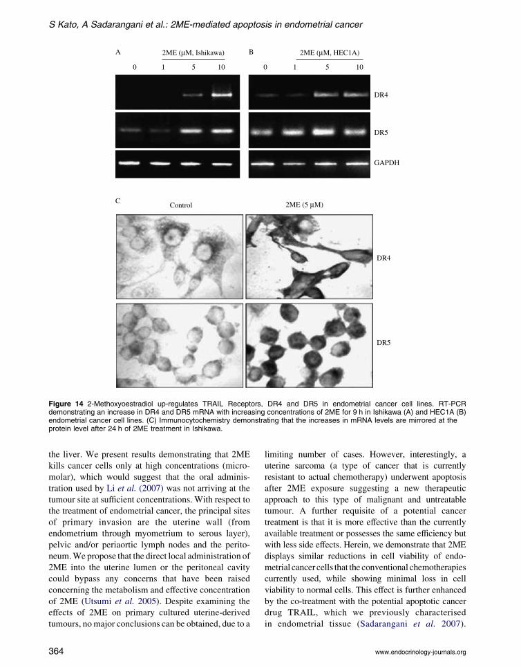
Figure 14 2-Methoxyoestradiol up-regulates TRAIL Receptors, DR4 and DR5 in endometrial cancer cell lines. RT-PCRdemonstrating an increase in DR4 and DR5 mRNA with increasing concentrations of 2ME for 9 h in Ishikawa (A) and HEC1A (B)endometrial cancer cell lines. (C) Immunocytochemistry demonstrating that the increases in mRNA levels are mirrored at theprotein level after 24 h of 2ME treatment in Ishikawa.
S Kato, A Sadarangani et al.: 2ME-mediated apoptosis in endometrial cancer
the liver. We present results demonstrating that 2ME
kills cancer cells only at high concentrations (micro-
molar), which would suggest that the oral adminis-
tration used by Li et al. (2007) was not arriving at the
tumour site at sufficient concentrations. With respect to
the treatment of endometrial cancer, the principal sites
of primary invasion are the uterine wall (from
endometrium through myometrium to serous layer),
pelvic and/or periaortic lymph nodes and the perito-
neum.We propose that the direct local administration of
2ME into the uterine lumen or the peritoneal cavity
could bypass any concerns that have been raised
concerning the metabolism and effective concentration
of 2ME (Utsumi et al. 2005). Despite examining the
effects of 2ME on primary cultured uterine-derived
tumours, nomajor conclusions can be obtained, due to a
364
limiting number of cases. However, interestingly, a
uterine sarcoma (a type of cancer that is currently
resistant to actual chemotherapy) underwent apoptosis
after 2ME exposure suggesting a new therapeutic
approach to this type of malignant and untreatable
tumour. A further requisite of a potential cancer
treatment is that it is more effective than the currently
available treatment or possesses the same efficiency but
with less side effects. Herein, we demonstrate that 2ME
displays similar reductions in cell viability of endo-
metrial cancer cells that the conventional chemotherapies
currently used, while showing minimal loss in cell
viability to normal cells. This effect is further enhanced
by the co-treatment with the potential apoptotic cancer
drug TRAIL, which we previously characterised
in endometrial tissue (Sadarangani et al. 2007).
www.endocrinology-journals.org
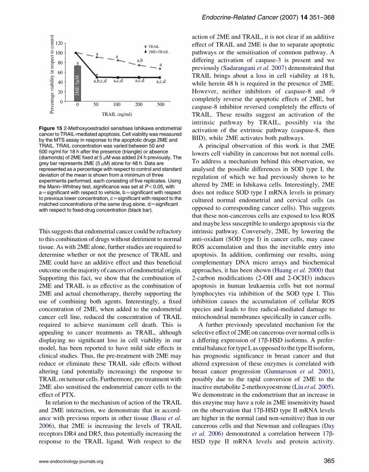
Figure 15 2-Methoxyoestradiol sensitises Ishikawa endometrialcancer to TRAIL-mediated apoptosis. Cell viability wasmeasuredby the MTS assay in response to the apoptotic drugs 2ME andTRAIL. TRAIL concentration was varied between 50 and500 ng/ml for 18 h after the presence (triangle) or absence(diamonds) of 2ME fixed at 5 mMwas added 24 h previously. Thegrey bar represents 2ME (5 mM) alone for 48 h. Data arerepresented as a percentagewith respect to control and standarddeviation of the mean is shown from a minimum of threeexperiments performed, each consisting of five replicates. Usingthe Mann–Whitney test, significance was set at P!0.05, withaZsignificant with respect to vehicle, bZsignificant with respectto previous lower concentration, cZsignificant with respect to thematched concentrations of the same drug alone, dZsignificantwith respect to fixed-drug concentration (black bar).
Endocrine-Related Cancer (2007) 14 351–368
This suggests that endometrial cancer could be refractory
to this combination of drugs without detriment to normal
tissue. As with 2ME alone, further studies are required to
determine whether or not the presence of TRAIL and
2ME could have an additive effect and thus beneficial
outcomeon themajorityof cancers of endometrial origin.
Supporting this fact, we show that the combination of
2ME and TRAIL is as effective as the combination of
2ME and actual chemotherapy, thereby supporting the
use of combining both agents. Interestingly, a fixed
concentration of 2ME, when added to the endometrial
cancer cell line, reduced the concentration of TRAIL
required to achieve maximum cell death. This is
appealing to cancer treatments as TRAIL, although
displaying no significant loss in cell viability in our
model, has been reported to have mild side effects in
clinical studies. Thus, the pre-treatment with 2ME may
reduce or eliminate these TRAIL side effects without
altering (and potentially increasing) the response to
TRAIL on tumour cells. Furthermore, pre-treatmentwith
2ME also sensitised the endometrial cancer cells to the
effect of PTX.
In relation to the mechanism of action of the TRAIL
and 2ME interaction, we demonstrate that in accord-
ance with previous reports in other tissue (Basu et al.
2006), that 2ME is increasing the levels of TRAIL
receptors DR4 and DR5, thus potentially increasing the
response to the TRAIL ligand. With respect to the
www.endocrinology-journals.org
action of 2ME and TRAIL, it is not clear if an additive
effect of TRAIL and 2ME is due to separate apoptotic
pathways or the sensitisation of common pathway. A
differing activation of caspase-3 is present and we
previously (Sadarangani et al. 2007) demonstrated that
TRAIL brings about a loss in cell viability at 18 h,
while herein 48 h is required in the presence of 2ME.
However, neither inhibitors of caspase-8 and -9
completely reverse the apoptotic effects of 2ME, but
caspase-8 inhibitor reversed completely the effects of
TRAIL. These results suggest an activation of the
intrinsic pathway by TRAIL, possibly via the
activation of the extrinsic pathway (caspase-8, then
BID), while 2ME activates both pathways.
A principal observation of this work is that 2ME
lowers cell viability in cancerous but not normal cells.
To address a mechanism behind this observation, we
analysed the possible differences in SOD type I, the
regulation of which we had previously shown to be
altered by 2ME in Ishikawa cells. Interestingly, 2ME
does not reduce SOD type I mRNA levels in primary
cultured normal endometrial and cervical cells (as
opposed to corresponding cancer cells). This suggests
that these non-cancerous cells are exposed to less ROS
and maybe less susceptible to undergo apoptosis via the
intrinsic pathway. Conversely, 2ME, by lowering the
anti-oxidant (SOD type I) in cancer cells, may cause
ROS accumulation and thus the inevitable entry into
apoptosis. In addition, confirming our results, using
complementary DNA micro arrays and biochemical
approaches, it has been shown (Huang et al. 2000) that
2-carbon modifications (2-OH and 2-OCH3) induces
apoptosis in human leukaemia cells but not normal
lymphocytes via inhibition of the SOD type I. This
inhibition causes the accumulation of cellular ROS
species and leads to free radical-mediated damage to
mitochondrial membranes specifically in cancer cells.
A further previously speculated mechanism for the
selective effect of 2ME on cancerous over normal cells is
a differing expression of 17b-HSD isoforms. A prefer-
ential balance for type I, as opposed to the type II isoform,
has prognostic significance in breast cancer and that
altered expression of these enzymes is correlated with
breast cancer progression (Gunnarsson et al. 2001),
possibly due to the rapid conversion of 2ME to the
inactive metabolite 2-methoxyoestrone (Liu et al. 2005).
We demonstrate in the endometrium that an increase in
this enzyme may have a role in 2ME insensitivity based
on the observation that 17b-HSD type II mRNA levels
are higher in the normal (and non-sensitive) than in our
cancerous cells and that Newman and colleagues (Day
et al. 2006) demonstrated a correlation between 17b-HSD type II mRNA levels and protein activity.
365

S Kato, A Sadarangani et al.: 2ME-mediated apoptosis in endometrial cancer
Currently, experiments are on the way to test the
hypothesis that Ishikawa cells stably transfected with
17b-HSD type II are more resistant to 2ME treatment.
2ME is already being considered as a cancer
therapeutic for breast cancer (Lakhani et al. 2003).
However, despite ongoing clinical trials reporting an
absence of grade 4 toxicity and only minor grade 3
toxicity in relation to disease progression, less than
encouraging beneficial effects on breast cancer
progression are being presented (Lakhani et al. 2003).
As our results, albeit in vitro, also demonstrate minimal
effects on breast cancer cells, but significantly reduced
viability of endometrial cancer cells, we postulate that
2ME alone or in combination with TRAILwould have a
beneficial outcome in clinical trials of patients
presenting with uterine-derived cancers. Furthermore,
the metabolism of 2ME in the liver is also hindering the
application of this drug; however, due to the nature of
endometrial cancer and its principal invasive sites, drug
administration could occur directly into the uterine
lumen and/or the peritoneum thus circumventing the
problem of drug metabolism.
Acknowledgements
At the Pontificia Universidad Catolica de Chile, we
acknowledge Drs Croxatto and Orihuela for their
advice and help with 2ME; Dr David Mayerson,
member of Gynaecologic Oncology Unit at the
Department of Obstetrics and Gynaecology for their
clinical advice and sample collection; all the path-
ologists working at the Department of Pathology for
their critical review of the samples collected; Dr
Lipkowitz at the NIH, USA for the use of GST-TRAIL
and Dr Lam at Imperial College London for his
assistance with flow cytometry. The authors declare
that there is no conflict of interest that would prejudice
the impartiality of this scientific work.
Funding
This work was supported by a grant from theWellcome
Trust GR071469 (GIO) and the Chilean national
science grants FONDECYT 1060495(GIO) and
1050744 (MC).
References
Acconcia F, Totta P, Ogawa S, Cardillo I, Inoue S, Leone S,
Trentalance A, Muramatsu M & Marino M 2005 Survival
versus apoptotic 17beta-estradiol effect: role of ER alpha
and ER beta activated non-genomic signaling. Journal of
Cellular Physiology 203 193–201.
366
Amant F, Moerman P, Neven P, Timmerman D, van
Limbergen E & Vergote I 2005 Endometrial cancer.
Lancet 366 491–505.
van Aswegen CH, Purdy RH & Wittliff JL 1989 Binding of
2-hydroxyestradiol and 4-hydroxyestradiol to estrogen
receptors from human breast cancers. Journal of Steroid
Biochemistry 32 485–492.
Basak C, Pathak SK, Bhattacharyya A, Pathak S, Basu J &
Kundu M 2005 The secreted peptidyl prolyl cis,trans-
isomerase HP0175 of Helicobacter pylori induces
apoptosis of gastric epithelial cells in a TLR4- and
apoptosis signal-regulating kinase 1-dependent manner.
Journal of Immunology 174 5672–5680.
Basu A, Castle VP, Bouziane M, Bhalla K & Haldar S 2006
Crosstalk between extrinsic and intrinsic cell death
pathways in pancreatic cancer: synergistic action of
estrogen metabolite and ligands of death receptor family.
Cancer Research 66 4309–4318.
Bu S, Blaukat A, Fu X, Heldin NE & Landstrom M 2002
Mechanisms for 2-methoxyestradiol-induced apoptosis of
prostate cancer cells. FEBS Letters 531 141–151.
Cailleau R, Olive M & Cruciger QV 1978 Long-term human
breast carcinoma cell lines of metastatic origin: pre-
liminary characterization. In Vitro 14 911–915.
Carter J & Pather S 2006 An overview of uterine cancer and
its management. Expert Review of Anticancer Therapy 6
33–42.
Chauhan D, Catley L, Hideshima T, Li G, Leblanc R,
Gupta D, Sattler M, Richardson P, Schlossman RL,
Podar K et al. 2002 2-Methoxyestradiol overcomes
drug resistance in multiple myeloma cells. Blood 100
2187–2194.
Chomczynski P & Sacchi N 1987 Single-step method of
RNA isolation by acid guanidinium thiocyanate–
phenol–chloroform extraction. Analytical Biochemistry
162 156–159.
Cuello M, Ettenberg SA, Nau MM & Lipkowitz S 2001
Synergistic induction of apoptosis by the combination of
trail and chemotherapy in chemoresistant ovarian cancer
cells. Gynecologic Oncology 81 380–390.
Day JM, Tutill HJ, Newman SP, Purohit A, Lawrence HR,
Vicker N, Potter BV & Reed MJ 2006 17Beta-hydroxy-
steroid dehydrogenase type 1 and type 2: association
between mRNA expression and activity in cell lines.
Molecular and Cellular Endocrinology 248 246–249.
Eagle H 1955 Propagation in a fluid medium of a human
epidermoid carcinoma, strain KB. Proceedings of the
Society for Experimental Biology and Medicine 89
362–364.
Engel LW, Young NA, Tralka TS, Lippman ME, O’Brien SJ
& Joyce MJ 1978 Establishment and characterization of
three new continuous cell lines derived from human breast
carcinomas. Cancer Research 38 3352–3364.
Franks S, MacLusky NJ & Naftolin F 1982 Comparative
pharmacology of oestrogens and catechol oestrogens:
actions on the immature rat uterus in vivo and in vitro.
Journal of Endocrinology 94 91–98.
www.endocrinology-journals.org

Endocrine-Related Cancer (2007) 14 351–368
Gibson EM, Henson ES, Haney N, Villanueva J & Gibson SB
2002 Epidermal growth factor protects epithelial-derived
cells from tumor necrosis factor-related apoptosis-inducing
ligand-induced apoptosis by inhibiting cytochrome c
release. Cancer Research 62 488–496.
Gui Y & Zheng XL 2006 2-Methoxyestradiol induces cell
cycle arrest and mitotic cell apoptosis in human vascular
smooth muscle cells. Hypertension 47 271–280.
Gunnarsson C, Olsson BM & Stal O 2001 Abnormal
expression of 17beta-hydroxysteroid dehydrogenases in
breast cancer predicts late recurrence. Cancer Research
61 8448–8451.
Huang P, Feng L, Oldham EA, Keating MJ & Plunkett W
2000 Superoxide dismutase as a target for the selective
killing of cancer cells. Nature 407 390–395.
Joubert A, Maritz C & Joubert F 2005 Bax/Bcl-2 expression
levels of 2-methoxyestradiol-exposed esophageal cancer
cells. Biomedical Research 26 131–134.
Keane MM, Ettenberg SA, Lowrey GA, Russell EK &
LipkowitzS1996Fas expression and function innormal and
malignant breast cell lines.CancerResearch 56 4791–4798.
Kopin IJ 1994 Monoamine oxidase and catecholamine
metabolism. Journal of Neural Transmission. Supple-
mentum 41 57–67.
Kuramoto H 1972 Studies of the growth and cytogenetic
properties of human endometrial adenocarcinoma in
culture and its development into an established line. Acta
Obstetrica et Gynaecologica Japonica 19 47–58.
Lakhani NJ, Sarkar MA, Venitz J & Figg WD 2003 2-
Methoxyestradiol, a promising anticancer agent. Phar-
macotherapy 23 165–172.
LaVallee TM, Zhan XH, Herbstritt CJ, Kough EC, Green SJ
& Pribluda VS 2002 2-Methoxyestradiol inhibits
proliferation and induces apoptosis independently of
estrogen receptors alpha and beta. Cancer Research 62
3691–3697.
LaVallee TM, Zhan XH, JohnsonMS, Herbstritt CJ, Swartz G,
Williams MS, Hembrough WA, Green SJ & Pribluda VS
2003 2-Methoxyestradiol up-regulates death receptor 5 and
induces apoptosis through activation of the extrinsic
pathway. Cancer Research 63 468–475.
Li L, Yu F, Wu X, Cheng J, Ulmsten U & Fu X 2007 Effects
of 2-methoxyestradiol on endometrial carcinoma xeno-
grafts. Journal of Cancer Research and Clinical
Oncology 133 315–320.
Liu ZJ, Lee WJ & Zhu BT 2005 Selective insensitivity of ZR-
75-1 human breast cancer cells to 2-methoxyestradiol:
evidence for type II 17beta-hydroxysteroid dehydrogenase
as the underlying cause. Cancer Research 65 5802–5811.
Maeda K, Tsuda H, Hashiguchi Y, Yamamoto K, Inoue T,
Ishiko O & Ogita S 2002 Relationship between p53
pathway and estrogen receptor status in endometrioid-
type endometrial cancer. Human Pathology 33 386–391.
Merriam GR, MacLusky NJ, Picard MK & Naftolin F 1980
Comparative properties of the catechol estrogens, I:
methylation by catechol-O-methyltransferase and binding
to cytosol estrogen receptors. Steroids 36 1–11.
www.endocrinology-journals.org
Mooberry SL 2003 New insights into 2-methoxyestradiol, a
promising antiangiogenic and antitumor agent. Current
Opinion in Oncology 15 425–430.
Nishida M, Kasahara K, Kaneko M, Iwasaki H & Hayashi K
1985 Establishment of a new human endometrial
adenocarcinoma cell line, Ishikawa cells, containing
estrogen and progesterone receptors. Nippon Sanka
Fujinka Gakkai Zasshi 37 1103–1111.
Qadan LR, Perez-Stable CM, Anderson C, D’Ippolito G,
Herron A, Howard GA & Roos BA 2001 2-Methox-
yestradiol induces G2/M arrest and apoptosis in prostate
cancer. Biochemical and Biophysical Research Com-
munications 285 1259–1266.
Ray G, Dhar G, van Veldhuizen PJ, Banerjee S,
Saxena NK, Sengupta K & Banerjee SK 2006
Modulation of cell-cycle regulatory signaling network
by 2-methoxyestradiol in prostate cancer cells is
mediated through multiple signal transduction
pathways. Biochemistry 45 3703–3713.
Sadarangani A, Kato S, Espinoza N, Lange S, Llados C,
Espinosa M, Villalon M, Lipkowitz S, Cuello M & Owen
GI 2007 TRAIL mediates apoptosis in cancerous but not
normal primary cultured cells of the human reproductive
tract. Apoptosis 12 73–85.
Sidor C, D’Amato R &Miller KD 2005 The potential and
suitability of 2-methoxyestradiol in cancer therapy. Clinical
Cancer Research 11 6094–6095 (author reply 6095–6096).
Suchar LA, Chang RL, Rosen RT, Lech J & Conney AH
1995 High-performance liquid chromatography separ-
ation of hydroxylated estradiol metabolites: formation of
estradiol metabolites by liver microsomes from male and
female rats. Journal of Pharmacology and Experimental
Therapeutics 272 197–206.
Thews O, Lambert C, Kelleher DK, Biesalski HK, Vaupel P &
Frank J 2005 Possible protective effects of alpha-tocopherol
on enhanced induction of reactive oxygen species by
2-methoxyestradiol in tumors. Advances in Experimental
Medicine and Biology 566 349–355.
Utsumi T, Leese MP, Chander SK, Gaukroger K, Purohit A,
Newman SP, Potter BV & Reed MJ 2005 The effects
of 2-methoxyoestrogen sulphamates on the in vitro and
in vivo proliferation of breast cancer cells. Journal of Steroid
Biochemistry and Molecular Biology 94 219–227.
Watanabe K, Takanashi K, Imaoka S, Funae Y, Kawano S,
Inoue K, Kamataki T, Takagi H & Yoshizawa I 1991
Comparison of cytochrome P-450 species which catalyze
the hydroxylations of the aromatic ring of estradiol and
estradiol 17-sulfate. Journal of Steroid Biochemistry and
Molecular Biology 38 737–743.
Williams AE, Maskarinec G, Franke AA & Stanczyk FZ
2002 The temporal reliability of serum estrogens,
progesterone, gonadotropins, SHBG and urinary estrogen
and progesterone metabolites in premenopausal women.
BMC Women’s Health 2 13.
Yue W, Santen RJ, Wang JP, Li Y, Verderame MF,
Bocchinfuso WP, Korach KS, Devanesan P,
Todorovic R, Rogan EG et al. 2003 Genotoxic
367

S Kato, A Sadarangani et al.: 2ME-mediated apoptosis in endometrial cancer
metabolites of estradiol in breast: potential mechanism
of estradiol induced carcinogenesis. Journal of
Steroid Biochemistry and Molecular Biology 86
477–486.
Zhou NN, Zhu XF, Zhou JM, Li MZ, Zhang XS, Huang
P & Jiang WQ 2004 2-Methoxyestradiol induces
368
cell cycle arrest and apoptosis of nasopharyngeal
carcinoma cells. Acta Pharmacologica Sinica 25
1515–1520.
Zhu BT & Conney AH 1998 Functional role of estrogen
metabolism in target cells: review and perspectives.
Carcinogenesis 19 1–27.
www.endocrinology-journals.org