Apoptosis in amphibian organs during metamorphosis
24
Apoptosis in amphibian organs during metamorphosis Atsuko Ishizuya-Oka 1,* , Takashi Hasebe 1 , and Yun-Bo Shi 2,* 1 Department of Biology, Nippon Medical School, Kawasaki, Kanagawa 211-0063, Japan 2 Laboratory of Gene Regulation and Development, National Institute for Child Health and Human Development, NIH, Bethesda, Maryland, MD 20892-5431, USA Abstract During amphibian metamorphosis, the larval tissues/organs rapidly degenerate to adapt from the aquatic to the terrestrial life. At the cellular level, a large quantity of apoptosis occurs in a spatiotemporally-regulated fashion in different organs to ensure timely removal of larval organs/ tissues and the development of adult ones for the survival of the individuals. Thus, amphibian metamorphosis provides us a good opportunity to understand the mechanisms regulating apoptosis. To investigate this process at the molecular level, a number of thyroid hormone (TH) response genes have been isolated from several organs of Xenopus laevis tadpoles and their expression and functional analyses are now in progress using modern molecular and genetic technologies. In this review, we will first summarize when and where apoptosis occurs in typical larva-specific and larval-to-adult remodeling amphibian organs to highlight that the timing of apoptosis is different in different tissues/organs, even though all are induced by the same circulating TH. Next, to discuss how TH spatiotemporally regulates the apoptosis, we will focus on apoptosis of the X. laevis small intestine, one of the best characterized remodeling organs. Functional studies of TH response genes using transgenic frogs and culture techniques have shown that apoptosis of larval epithelial cells can be induced by TH either cell-autonomously or indirectly through interactions with extracellular matrix (ECM) components of the underlying basal lamina. Here, we propose that multiple intra- and extracellular apoptotic pathways are coordinately controlled by TH to ensure massive but well-organized apoptosis, which is essential for the proper progression of amphibian metamorphosis. Keywords Amphibian metamorphosis; Apoptosis; Thyroid hormone; Organ remodeling; Cell-ECM interactions Introduction The animal body undergoes rapid and dramatic changes during amphibian (anuran) metamorphosis to adapt from the aquatic to the terrestrial life. The larva-specific organs such as the tail and the gill degenerate, while the adult-specific organs such as forelimbs develop. Numerous other organs, which exist both before and after metamorphosis, remodel from the larval to the adult form. Cell death occurs widely in not only the larva-specific organs but also the larva-to-adult remodeling organs, and thus is a key phenomenon in amphibian metamorphosis. The problem how larval cell death proceeds in balance with the * Correspondence: A. Ishizuya-Oka, Department of Biology, Nippon Medical School, Kawasaki, Kanagawa 211-0063, Japan, a- [email protected]; tel: 81-44-733-3580; fax: 81-722-1231/ Or Y.-B. Shi, Laboratory of Gene Regulation and Development, National Institute for Child Health and Human Development, NIH, Bethesda, Maryland, MD 20892-5431, USA, [email protected]; tel: 301-402-1004; fax: 301-402-1323. NIH Public Access Author Manuscript Apoptosis. Author manuscript; available in PMC 2012 August 06. Published in final edited form as: Apoptosis. 2010 March ; 15(3): 350–364. doi:10.1007/s10495-009-0422-y. NIH-PA Author Manuscript NIH-PA Author Manuscript NIH-PA Author Manuscript
Transcript of Apoptosis in amphibian organs during metamorphosis

Apoptosis in amphibian organs during metamorphosis
Atsuko Ishizuya-Oka1,*, Takashi Hasebe1, and Yun-Bo Shi2,*
1 Department of Biology, Nippon Medical School, Kawasaki, Kanagawa 211-0063, Japan2 Laboratory of Gene Regulation and Development, National Institute for Child Health and HumanDevelopment, NIH, Bethesda, Maryland, MD 20892-5431, USA
AbstractDuring amphibian metamorphosis, the larval tissues/organs rapidly degenerate to adapt from theaquatic to the terrestrial life. At the cellular level, a large quantity of apoptosis occurs in aspatiotemporally-regulated fashion in different organs to ensure timely removal of larval organs/tissues and the development of adult ones for the survival of the individuals. Thus, amphibianmetamorphosis provides us a good opportunity to understand the mechanisms regulatingapoptosis. To investigate this process at the molecular level, a number of thyroid hormone (TH)response genes have been isolated from several organs of Xenopus laevis tadpoles and theirexpression and functional analyses are now in progress using modern molecular and genetictechnologies. In this review, we will first summarize when and where apoptosis occurs in typicallarva-specific and larval-to-adult remodeling amphibian organs to highlight that the timing ofapoptosis is different in different tissues/organs, even though all are induced by the samecirculating TH. Next, to discuss how TH spatiotemporally regulates the apoptosis, we will focuson apoptosis of the X. laevis small intestine, one of the best characterized remodeling organs.Functional studies of TH response genes using transgenic frogs and culture techniques have shownthat apoptosis of larval epithelial cells can be induced by TH either cell-autonomously orindirectly through interactions with extracellular matrix (ECM) components of the underlyingbasal lamina. Here, we propose that multiple intra- and extracellular apoptotic pathways arecoordinately controlled by TH to ensure massive but well-organized apoptosis, which is essentialfor the proper progression of amphibian metamorphosis.
KeywordsAmphibian metamorphosis; Apoptosis; Thyroid hormone; Organ remodeling; Cell-ECMinteractions
IntroductionThe animal body undergoes rapid and dramatic changes during amphibian (anuran)metamorphosis to adapt from the aquatic to the terrestrial life. The larva-specific organssuch as the tail and the gill degenerate, while the adult-specific organs such as forelimbsdevelop. Numerous other organs, which exist both before and after metamorphosis, remodelfrom the larval to the adult form. Cell death occurs widely in not only the larva-specificorgans but also the larva-to-adult remodeling organs, and thus is a key phenomenon inamphibian metamorphosis. The problem how larval cell death proceeds in balance with the
*Correspondence: A. Ishizuya-Oka, Department of Biology, Nippon Medical School, Kawasaki, Kanagawa 211-0063, Japan, [email protected]; tel: 81-44-733-3580; fax: 81-722-1231/ Or Y.-B. Shi, Laboratory of Gene Regulation and Development, NationalInstitute for Child Health and Human Development, NIH, Bethesda, Maryland, MD 20892-5431, USA, [email protected]; tel:301-402-1004; fax: 301-402-1323.
NIH Public AccessAuthor ManuscriptApoptosis. Author manuscript; available in PMC 2012 August 06.
Published in final edited form as:Apoptosis. 2010 March ; 15(3): 350–364. doi:10.1007/s10495-009-0422-y.
NIH
-PA Author Manuscript
NIH
-PA Author Manuscript
NIH
-PA Author Manuscript

development of adult tissues/organs has attracted the attention of many biologists in thisfield, but the challenge to address this problem has just been started at the molecular level[1-3].
Apoptosis is an evolutionary conserved phenomenon essential for both development andtissue/organ homeostasis in all multi-cellular organisms. Cell death during amphibianmetamorphosis is mainly, if not exclusively, through apoptosis. This has been shown bynumerous previous studies with electron microscopy (EM) and TUNEL, which observed“apoptosis”, characterized ultrastructurally by condensation of chromatins on the nuclearmembrane and subsequent fragmentation of nuclei and cytoplasm into apoptotic bodies [4],in amphibian organs including the tail [5], brain [6], and the intestine [7]. As expected,apoptosis during amphibian metamorphosis occurs in a well-organized fashion, dependingon the cell-type and/or the kind of tissues/organs. This is essential for animals to overcomemetamorphosis and survive. In this sense, the amphibian metamorphosis provides us a niceunique opportunity to understand regulatory mechanisms of apoptosis as a whole body/organ. It should be noted that the apoptosis of amphibian metamorphosis is triggered by asingle hormone, thyroid hormone (TH) [8, 9]. In addition, previous culture studies showedthat TH can organ-autonomously induce the degeneration of the larva-specific tail [10] andgill [11], and epithelial apoptosis of the remodeling small intestine [7]. These suggest thatthe molecular mechanisms of apoptosis during amphibian metamorphosis are controlled by acascade of TH-dependent gene expressions in this animal model, even though the apoptoticprocess may involve complex tissue interactions as suggested in the tail [12-14]. Since the1990s, toward determining the molecular mechanisms of amphibian metamorphosis, anumber of TH response genes have been isolated from several organs of the African clawedfrog, Xenopus laevis, including the tail and intestine by subtractive hybridization [15, 16]and more recently by cDNA microarray [17, 18]. Their expression and functional analyses,which are still ongoing, have already shed light on the molecular mechanisms regulatingapoptosis during amphibian metamorphosis. Here we will first survey apoptosis occurring invarious amphibian organs during metamorphosis and then review the current knowledge onthe apoptotic factors encoded by TH response genes in these organs. Next, we will focus onthe X. laevis intestine, one of the best-studied remodeling organs and summarize how THregulates apoptosis of the larval epithelium both directly and indirectly through cell-ECMand/or cell-cell interactions to enable massive and cell type-dependent apoptosis duringintestinal remodeling.
Apoptosis in larva-specific and larval-to-adult remodeling organsIn this section, we will begin with the description of when and where apoptosis occurs in thelarva-specific and larval-to-adult remodeling organs during amphibian metamorphosis.Although apoptosis mainly occurs during metamorphic climax when TH level in the plasmais high (Fig. 1), the stage when apoptosis most frequently occurs is differently programmedin different organs and even in different tissues or cell-types of a single organ (Table 1). Inaddition, the timing of apoptosis in each organ sometimes varies with among different frogspecies, probably due to their different habitats and life histories. While we will mainlyfocus on the metamorphic changes in X. laevis, we will also describe available informationin other anurans such as Rana and Bufo for comparison. The developmental stages of frogshave been assigned independently according to the genus: Nieuwkoop and Faber (NF) stagefor Xenopus species [19], Taylor and Kollros (TK) stage for Rana species [20] and Rossi'sstage for Bufo bufo [21].
Larva-specific organsTail—One of the most dramatic events during amphibian metamorphosis is the regressionof the tail which occupies a large proportion of the tadpole. Thus, the mechanisms of tail
Ishizuya-Oka et al. Page 2
Apoptosis. Author manuscript; available in PMC 2012 August 06.
NIH
-PA Author Manuscript
NIH
-PA Author Manuscript
NIH
-PA Author Manuscript

resorption have been extensively studied for many years [5, 16, 22-31]. The tadpole tailconsists of the epidermis, muscles, connective tissue (dermis), nervous tissue (spinal cord),and notochord. By the end of metamorphosis, the tail is totally removed through apoptosisof essentially all of the tissues (Fig. 1), although only limited cell types have been shown toundergo apoptosis. In the tail of X. laevis tadpoles, muscles begin to undergo apoptosis atNF stage 57 (prometamorphosis) long before the start of the tail shortening at stage 62 [28],and the number of apoptotic cells in the muscles peaks at stages 63/64, when the taildrastically shortens [29]. On the other hand, in the caudal spinal cord, the number ofapoptotic cells reaches its peak at stage 58 (the onset of metamorphic climax) [24], when theforelimbs protrude. Similarly, in Japanese brown frog, Rana japonica, the epidermisundergoes cell death at TK stages XIX-XX (around NF stage 61 for X. laevis [8], Fig. 1),when forelimbs extend away from the body [32]. Although TH organ-autonomously inducesthe regression of the tail [10], previous culture studies indicated that interactions betweenthe epidermis and dermis are necessary for the tail regression of the bullfrog, R. catesbeiana[12, 13, 33].
Gill—The gill is also a larva-specific organ that degenerates nearly completely [34] orperhaps leaves a small remnant [35] during metamorphosis. As metamorphosis proceeds, thegill decreases in weight and becomes less vascularized. In the R. catesbeiana gill, most ofthe epithelial cells are closely associated with blood vessels during premetamorphosis, butare no longer in close proximity to the blood vessels at TK stage XXII (metamorphicclimax, NF stage 63 for X. laevis) [34]. In the X. laevis gill, the filter plates, which occupymore than 3/4 of the buccal floor arena's rostrocaudal extension during prometamorphosis,become very small and are confined to the space bilateral to the glottis at NF stages 61/62(metamorphic climax). By using microvascular casting, it has been shown that the branchingof filter row veins reaches its maximum at stage 58 and then gradually decreases in numberand frequency until stage 62, when many apoptotic cells are observed in the gills. Thisindicates that vascular regression is organized by progressive fusion of opposite vessels [36].
Larval-to-adult remodeling organsBrain—As metamorphosis involves a habitat shift from an aquatic to a terrestrialenvironment, where adult frogs exhibit behaviors and other life history characteristicsdramatically different from those in tadpoles, gross morphology of the amphibian brainchanges from larval to adult types during metamorphosis, e.g., the widening and shorteningof the diencephalons and the narrowing and shortening of the fossa rhomboidalis of themedulla [37]. Although apoptosis has not been reported with EM or TUNEL analysis in thisorgan, caspase-9, one of the key apoptotic factors, is active in the remodeling of the X.laevis brain during NF stages 55-63, peaking at stage 60. Its mRNA is expressed in theventricular zone of the brain. Cells expressing caspase-9 are localized in the ventral part ofthe anterior brain and the subpallium area at stage 61 [38]. As for other larval neurons,Mauthner neurons in the medulla and the sensory and motoneurons supplying the tail areeliminated during remodeling of the central nervous system [39]. Massive cell death in themesencephalic fifth nucleus of the trigeminal nerve also occurs at NF stage 61 in X. laevis[40] and at TK stage XXI (around NF stage 62/63 for X. laevis) in the leopard frog, R.pipiens [41].
Intestine—As an herbivorous tadpole metamorphoses into a carnivorous frog, the intestineis drastically remodeled from the larval to the adult form [1, 42-44]. In the X. laevis smallintestine, apoptotic cells are very few until NF stage 59 and then rapidly increase in numberat stage 60 [45] (Fig. 1), when stem cells of the adult epithelium appear. The larval epithelialcells are totally removed through apoptosis by stage 63, while the adult epithelial cellsreplacing the larval ones finally differentiate by the end of metamorphosis. Although
Ishizuya-Oka et al. Page 3
Apoptosis. Author manuscript; available in PMC 2012 August 06.
NIH
-PA Author Manuscript
NIH
-PA Author Manuscript
NIH
-PA Author Manuscript

massive apoptosis occurs only in the larval epithelium throughout metamorphosis, a smallnumber of fibroblasts also undergo apoptosis at stages 61/62 [7, 45].
Pancreas—Pancreas, which mainly consists of the endocrine tissue producing hormonesand exocrine tissue producing digestive enzymes, is also one of the digestive organs that areremodeled during metamorphosis [46]. The X. laevis pancreas loses about 40% of its cellsby NF stage 62 (metamorphic climax), when many cells, though their cell-type is notspecified, undergo apoptosis. It is suggested that some, if not all, larval acinar cellsdedifferentiate during metamorphic climax and then redifferentiate into the adult type aftermetamorphosis [47]. In both the R. pipiens [48] and R. catesbeiana pancreas [49], β-cellsproducing insulin decrease in number at TK stages XX or XXI (metamorphic climax,around NF stage 62 for X. laevis). Similarly, the common European toad, Bufo bufo, showsthe reduction of pancreatic volume during Rossi's stages IX (late prometamorphic period,around NF stage 56) - XV (the end of metamorphosis, NF stage 66). This degeneration ofpancreas is caused in part by apoptosis of cells around the blood vessels and pancreaticducts. The number of apoptotic cells reaches a peak at Rossi's stage XIII (around NF stage58-62), when forelimbs emerge [50].
Spleen—During metamorphosis, the immune system is also remodeled. Although thelarval immune system is capable of defending against potential pathogens, it is notequivalent to the adult immune system acquired during metamorphosis. Remodeling of theimmune system may serve to eliminate unnecessary larval lymphocytes, which could bedestructive if they recognize newly emerging adult-specific antigens on the adult typetissues [51]. The spleen is known to be a major organ that is responsible for the immunesystem. In the X. laevis spleen, which becomes lymphoid at NF stage 50 [52], apoptoticcells are very few during premetamorphosis and then become detectable mostly in the redpulp, but not in the white pulp, throughout metamorphosis. The number of apoptoticlymphocytes in the red pulp reaches its peak at stage 59 [53].
Thymus—The thymus is another organ responsible for the immune system and a periodicremoval of thymocytes as well as apoptosis of lymphocytes during metamorphosis has beenreported. In the thymus of X. laevis, apoptotic levels are high during premetamorphosis (NFstages 49-54), low from prometamorphosis to early climax (NF stages 56-61) and high againat middle climax (NF stages 62, 63). While apoptotic cells are distributed nearly uniformlybetween cortex and medulla of the thymus from premetamorphosis to early climax, thedying lymphocytes are localized in the medulla at middle climax [53].
Red blood cells (RBCs)—The circulating red blood cells (RBCs) also change fromlarval RBCs containing larval-type hemoglobin (Hb) to adult RBCs containing adult-typeHb (so-called Hb switching) during metamorphosis [54]. The major difference in these twotypes of Hb is their affinity to oxygen. Thus, the Hb switching is physiologically importantto adapt from aquatic to terrestrial habitat. Several lines of evidence have shown that Hbswitching in anuran amphibians is due to an overall replacement of the RBC populationrather than an alteration of globin-gene expression in the single RBC population [55-57]. InR. catesbeiana, adult RBCs first appear in the systemic circulation at TK stage XX (the onsetof metamorphic climax, around NF stage 62 for X. laevis) and become predominant aslarval RBCs totally disappear by stage XXIV just before the end of metamorphosis (aroundNF stage 65 for X. laevis) [54]. On the other hand, in X. laevis, adult RBCs becomedetectable at NF stage 53 (premetamorphosis) and increase in number as metamorphosisproceeds. Surprisingly, approximately 10% of total RBCs are still larval-type at 3 weeksafter the completion of metamorphosis, and it takes another 5 months to complete Hbswitching [57] This delayed removal of larval cells may be related to the fact that
Ishizuya-Oka et al. Page 4
Apoptosis. Author manuscript; available in PMC 2012 August 06.
NIH
-PA Author Manuscript
NIH
-PA Author Manuscript
NIH
-PA Author Manuscript
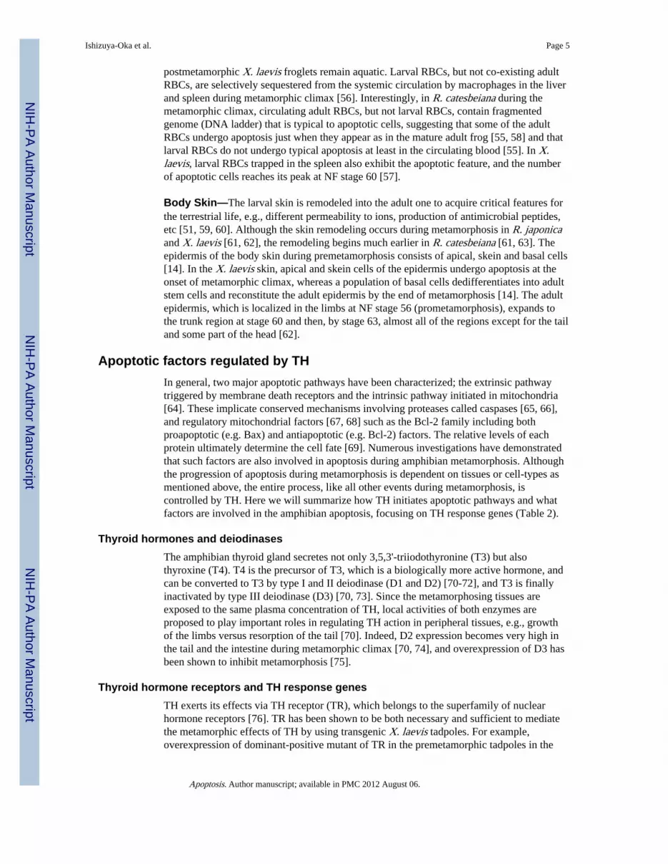
postmetamorphic X. laevis froglets remain aquatic. Larval RBCs, but not co-existing adultRBCs, are selectively sequestered from the systemic circulation by macrophages in the liverand spleen during metamorphic climax [56]. Interestingly, in R. catesbeiana during themetamorphic climax, circulating adult RBCs, but not larval RBCs, contain fragmentedgenome (DNA ladder) that is typical to apoptotic cells, suggesting that some of the adultRBCs undergo apoptosis just when they appear as in the mature adult frog [55, 58] and thatlarval RBCs do not undergo typical apoptosis at least in the circulating blood [55]. In X.laevis, larval RBCs trapped in the spleen also exhibit the apoptotic feature, and the numberof apoptotic cells reaches its peak at NF stage 60 [57].
Body Skin—The larval skin is remodeled into the adult one to acquire critical features forthe terrestrial life, e.g., different permeability to ions, production of antimicrobial peptides,etc [51, 59, 60]. Although the skin remodeling occurs during metamorphosis in R. japonicaand X. laevis [61, 62], the remodeling begins much earlier in R. catesbeiana [61, 63]. Theepidermis of the body skin during premetamorphosis consists of apical, skein and basal cells[14]. In the X. laevis skin, apical and skein cells of the epidermis undergo apoptosis at theonset of metamorphic climax, whereas a population of basal cells dedifferentiates into adultstem cells and reconstitute the adult epidermis by the end of metamorphosis [14]. The adultepidermis, which is localized in the limbs at NF stage 56 (prometamorphosis), expands tothe trunk region at stage 60 and then, by stage 63, almost all of the regions except for the tailand some part of the head [62].
Apoptotic factors regulated by THIn general, two major apoptotic pathways have been characterized; the extrinsic pathwaytriggered by membrane death receptors and the intrinsic pathway initiated in mitochondria[64]. These implicate conserved mechanisms involving proteases called caspases [65, 66],and regulatory mitochondrial factors [67, 68] such as the Bcl-2 family including bothproapoptotic (e.g. Bax) and antiapoptotic (e.g. Bcl-2) factors. The relative levels of eachprotein ultimately determine the cell fate [69]. Numerous investigations have demonstratedthat such factors are also involved in apoptosis during amphibian metamorphosis. Althoughthe progression of apoptosis during metamorphosis is dependent on tissues or cell-types asmentioned above, the entire process, like all other events during metamorphosis, iscontrolled by TH. Here we will summarize how TH initiates apoptotic pathways and whatfactors are involved in the amphibian apoptosis, focusing on TH response genes (Table 2).
Thyroid hormones and deiodinasesThe amphibian thyroid gland secretes not only 3,5,3'-triiodothyronine (T3) but alsothyroxine (T4). T4 is the precursor of T3, which is a biologically more active hormone, andcan be converted to T3 by type I and II deiodinase (D1 and D2) [70-72], and T3 is finallyinactivated by type III deiodinase (D3) [70, 73]. Since the metamorphosing tissues areexposed to the same plasma concentration of TH, local activities of both enzymes areproposed to play important roles in regulating TH action in peripheral tissues, e.g., growthof the limbs versus resorption of the tail [70]. Indeed, D2 expression becomes very high inthe tail and the intestine during metamorphic climax [70, 74], and overexpression of D3 hasbeen shown to inhibit metamorphosis [75].
Thyroid hormone receptors and TH response genesTH exerts its effects via TH receptor (TR), which belongs to the superfamily of nuclearhormone receptors [76]. TR has been shown to be both necessary and sufficient to mediatethe metamorphic effects of TH by using transgenic X. laevis tadpoles. For example,overexpression of dominant-positive mutant of TR in the premetamorphic tadpoles in the
Ishizuya-Oka et al. Page 5
Apoptosis. Author manuscript; available in PMC 2012 August 06.
NIH
-PA Author Manuscript
NIH
-PA Author Manuscript
NIH
-PA Author Manuscript
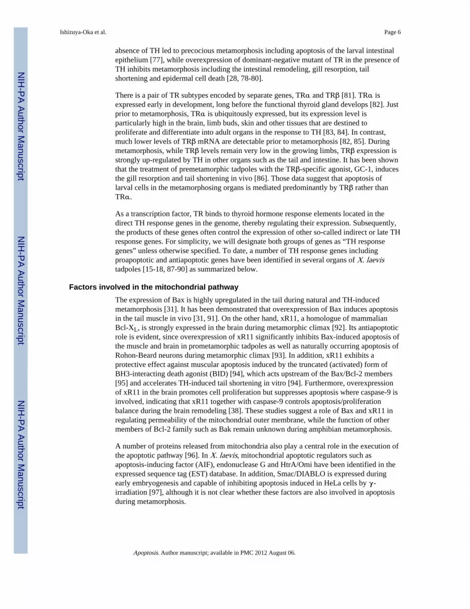
absence of TH led to precocious metamorphosis including apoptosis of the larval intestinalepithelium [77], while overexpression of dominant-negative mutant of TR in the presence ofTH inhibits metamorphosis including the intestinal remodeling, gill resorption, tailshortening and epidermal cell death [28, 78-80].
There is a pair of TR subtypes encoded by separate genes, TRα and TRβ [81]. TRα isexpressed early in development, long before the functional thyroid gland develops [82]. Justprior to metamorphosis, TRα is ubiquitously expressed, but its expression level isparticularly high in the brain, limb buds, skin and other tissues that are destined toproliferate and differentiate into adult organs in the response to TH [83, 84]. In contrast,much lower levels of TRβ mRNA are detectable prior to metamorphosis [82, 85]. Duringmetamorphosis, while TRβ levels remain very low in the growing limbs, TRβ expression isstrongly up-regulated by TH in other organs such as the tail and intestine. It has been shownthat the treatment of premetamorphic tadpoles with the TRβ-specific agonist, GC-1, inducesthe gill resorption and tail shortening in vivo [86]. Those data suggest that apoptosis oflarval cells in the metamorphosing organs is mediated predominantly by TRβ rather thanTRα.
As a transcription factor, TR binds to thyroid hormone response elements located in thedirect TH response genes in the genome, thereby regulating their expression. Subsequently,the products of these genes often control the expression of other so-called indirect or late THresponse genes. For simplicity, we will designate both groups of genes as “TH responsegenes” unless otherwise specified. To date, a number of TH response genes includingproapoptotic and antiapoptotic genes have been identified in several organs of X. laevistadpoles [15-18, 87-90] as summarized below.
Factors involved in the mitochondrial pathwayThe expression of Bax is highly upregulated in the tail during natural and TH-inducedmetamorphosis [31]. It has been demonstrated that overexpression of Bax induces apoptosisin the tail muscle in vivo [31, 91]. On the other hand, xR11, a homologue of mammalianBcl-XL, is strongly expressed in the brain during metamorphic climax [92]. Its antiapoptoticrole is evident, since overexpression of xR11 significantly inhibits Bax-induced apoptosis ofthe muscle and brain in prometamorphic tadpoles as well as naturally occurring apoptosis ofRohon-Beard neurons during metamorphic climax [93]. In addition, xR11 exhibits aprotective effect against muscular apoptosis induced by the truncated (activated) form ofBH3-interacting death agonist (BID) [94], which acts upstream of the Bax/Bcl-2 members[95] and accelerates TH-induced tail shortening in vitro [94]. Furthermore, overexpressionof xR11 in the brain promotes cell proliferation but suppresses apoptosis where caspase-9 isinvolved, indicating that xR11 together with caspase-9 controls apoptosis/proliferationbalance during the brain remodeling [38]. These studies suggest a role of Bax and xR11 inregulating permeability of the mitochondrial outer membrane, while the function of othermembers of Bcl-2 family such as Bak remain unknown during amphibian metamorphosis.
A number of proteins released from mitochondria also play a central role in the execution ofthe apoptotic pathway [96]. In X. laevis, mitochondrial apoptotic regulators such asapoptosis-inducing factor (AIF), endonuclease G and HtrA/Omi have been identified in theexpressed sequence tag (EST) database. In addition, Smac/DIABLO is expressed duringearly embryogenesis and capable of inhibiting apoptosis induced in HeLa cells by γ-irradiation [97], although it is not clear whether these factors are also involved in apoptosisduring metamorphosis.
Ishizuya-Oka et al. Page 6
Apoptosis. Author manuscript; available in PMC 2012 August 06.
NIH
-PA Author Manuscript
NIH
-PA Author Manuscript
NIH
-PA Author Manuscript

Caspases and other related factorsThe expression of caspase-9, one of the key apoptotic factors, is up-regulated in the brain,intestine and tail during metamorphic climax [30, 98]. Activation of caspase-9 protein isdependent on Bax, and the level of active caspase-9 is the highest in the tail muscle andbrain during metamorphic climax [30, 38]. Overexpression of dominant-negative mutant ofcaspase-9 (dnCasp9) has been shown to inhibit Bax-induced and naturally occurringapoptosis in the metamorphosing tail and brain, indicating the involvement of caspase-9 inapoptosis [30, 38]. These findings further suggest that dnCasp9 is associated with apoptoticprotease-activating factor (Apaf)-1 and cytochrome c to form the inactive apoptosome,although direct evidence has not been obtained [30]. In the tail, the expression of caspase-1and -3 is also up-regulated during metamorphosis [30, 98, 99], suggesting their involvementin apoptosis. In fact, a specific inhibitor of caspase-3 suppresses TH-induced apoptosis ofXLT-15, a myoblastic cell line derived from the tadpole tail [99] in vitro, whereas caspase-1is capable of inducing apoptosis in XLT-15-11, a subline of XLT-15 [98]. In addition,caspase-2 and -8 are involved in apoptosis in the tail, since the specific inhibitor of eitherone blocks TH-induced tail shortening in vitro. These caspases are thought to function byactivating BID [100]. The levels of capase-2 and -8 are the highest in the tail duringmetamorphic climax, whereas the levels of other enzymes such as calpains are eitherundetectable or unchanged, suggesting that caspase-2 and -8 are responsible for activation ofBID [94]. Other factors suggested to play a role in apoptosis of the tail are caspase-7 andinhibitor of growth (ING) family [30, 98, 101].
Among the other organs, the expression of caspase-3 has also been reported in theremodeling of skin [79]. In the X. laevis skin, active caspase-3 is rarely detectable beforemetamorphic climax, but is widespread at NF stage 60, when the larval epidermis is beingreplaced by the adult one [79]. In the intestine, the expression of caspase-1, -2, -3, -6, -7, -8,-9 and -10 has been shown to be up-regulated during metamorphic climax [98]. Althoughmost of caspases identified so far are able to induce apoptosis of XLT-15-11 in vitro [98],the roles of some of them in vivo still remain to be investigated. Among the other apoptoticfactors, the inhibitor of apoptosis (IAP) family of proteins containing the highly conservedbaculoviral IAP repeat (BIR) are known to inhibit apoptosis, in some cases, through directbinding of caspases. XLX and XIAP, the X. laevis BIR family proteins, have been shown toinhibit the apoptotic process in the cell-free system of X. laevis egg extract [102, 103].However, their involvement in metamorphosis as well as TH dependency remains to bedetermined.
Death receptorsThe cell surface receptors that belong to the tumor necrosis factor receptor (TNFR)superfamily are the major death receptors involved in the extrinsic pathway of apoptosis. Todate, several members of the TNFR superfamily and one of their ligands, xTNFα, have beenreported in X. laevis tadpoles [104-107]. It has been shown that xTNFα binds to its cognatereceptor, xTNFR1 and inhibits TH-induced apoptosis of XLgoo, a vascular endothelial cellline derived from tadpole tail. Exogenous xTNFα also partially inhibits TH-induced taildegeneration in vitro, at least partly through repressing the levels of TRβ and caspase-3[106]. It still remains unclear whether xTNFα and xTNFR1 are TH response genes, sincethe level of xTNFR1 mRNA does not change in XLgoo treated with T3, and xTNFα isabsent in this cell line. However, when total RNAs from the entire tadpoles at differentmetamorphic stages are assessed, both xTNFα and xTNFR1 are expressed in a bimodalway, i.e., very weak expression at NF stage 55 occurring between stronger expression atstage 51 and at stage 57. Thereafter, the expression of both genes becomes weak again atstage 61, suggesting that these genes may be negatively regulated by TH in a tissue-specificmanner [106].
Ishizuya-Oka et al. Page 7
Apoptosis. Author manuscript; available in PMC 2012 August 06.
NIH
-PA Author Manuscript
NIH
-PA Author Manuscript
NIH
-PA Author Manuscript

KinasesSeveral lines of evidence have shown the importance of phosphorylation in the tailregression [108]. Inhibition of cyclin-dependent-kinase (Cdk) 2, whose expression is down-regulated during metamorphic climax, accelerates TH-induced tail regression [109]. Incontrast, inhibition of protein kinase C prevents TH-induced tail regression [110]. Inaddition, the activity of Cdk8 together with its regulatory unit, cyclin C, whose expression isTH dependent, is required for the tail shortening, suggesting their proapoptotic role [111].
Lessons from intestinal remodeling: Assurance of apoptosis through cell-ECM and cell-cell interactionsLarval epithelial apoptosis associated with adult epithelial development and ECMremodeling
The X. laevis small intestine is one of the typical remodeling organs where apoptosis hasbeen well characterized at the cellular level. Using this organ, we delve deeper into theproblem how TH successfully induces massive apoptosis in a cell type-dependent fashion.To adapt to a new food habit, the X. laevis intestine dramatically shortens and remodelsfrom the larval to adult form during a short period of metamorphosis [43, 112]. Apoptoticprocess of the larval epithelium during the intestinal remodeling has been preciselyinvestigated by EM and TUNEL [7, 45]. Before metamorphic climax the tadpole intestineconsists of a single layer of larval epithelium and thin layers of the immature connectivetissue and muscles. Apoptotic cells are very few, if any, in any tissues until NF stage 59.Around stage 60 (the onset of metamorphic climax), when a small number of adult stemcells appear and actively proliferate, apoptotic cells suddenly increase in number in theremaining larval epithelium [45] (Fig. 2). These cells share common characteristics ofapoptosis; formation of apoptotic bodies following the condensation of chromatin againstthe nuclear envelope. The apoptotic bodies are then phagocytosed by intraepithelialmacrophages which increase in number in the degenerating larval epithelium. Themacrophages become enlarged by engulfed phagosomes including the apoptotic bodies andare finally extruded into the intestinal lumen [7]. In this way, almost all of the larvalepithelial cells except for the adult stem cells are lost during stages 60-62 (the early periodof metamorphic climax). Characteristically, apoptosis of the larval epithelium is closelyrelated to the modifications of the underlying basal lamina [113] (Fig. 2). The basal laminais thin before metamorphic climax and then, concomitantly with the start of apoptosis of thelarval epithelium, suddenly becomes thick by vigorous folding and amorphous. Throughsuch modified basal lamina, the macrophages and other leukocytes migrate from theconnective tissue into the degenerating larval epithelium. The basal lamina underneath thelarval epithelium remains amorphous until the larval epithelial cells finally disappearthrough apoptosis, whereas the basal lamina underneath the adult stem cells and theirdescendent cells becomes thin again as they differentiate into the adult epithelium analogousto the mammalian adult one. These chronological observations suggest that apoptosis of thelarval epithelium, which progresses in balance with development of the adult epithelium, isstrictly regulated spatiotemporally, possibly involving extracellular matrix (ECM)remodeling of the basal lamina.
TH induces epithelial apoptosis under the influences of ECM in vitroTo clarify the mechanisms of the intestinal remodeling at the molecular level, we firstestablished an organ culture system and found that the apoptotic process during the intestinalremodeling described above could be reproduced by the inductive action of TH in vitro [7].In organ cultures of X. laevis intestines at stage 57 (before metamorphic climax), apoptosisof the larval epithelial cells occurs most frequently after 3 days of TH-treatment. Apoptotic
Ishizuya-Oka et al. Page 8
Apoptosis. Author manuscript; available in PMC 2012 August 06.
NIH
-PA Author Manuscript
NIH
-PA Author Manuscript
NIH
-PA Author Manuscript

bodies derived from the larval epithelial cells are phagocytosed by intraepithelialmacrophages, which are extruded into the outside. Similar to apoptosis during naturalmetamorphosis, TH-induced apoptosis of the larval epithelium in vitro coincides well withthe modification of the basal lamina underneath the epithelium, suggesting the involvementof the remodeling of the basal lamina ECM in the epithelial apoptosis. Thus, it seems likelythat apoptosis of the larval epithelium is induced by TH organ-autonomously but under thecontrol of cell-ECM interactions, which may function as a spatiotemporal regulator of theepithelial apoptosis.
Next, to examine whether TH can cell-autonomously induce apoptosis, epithelial cells wereisolated from the small intestine of X. laevis premetamorphic tadpoles, scattered, andcultured on plastic dishes in vitro. TH induces some of these epithelial cells to undergoapoptosis [114], whereas TH stimulates proliferation of other cells. In addition, this TH-induced apoptosis has been shown to be inhibited by cell death inhibitors such asCyclosporine A (Cys A), which is generally known to inhibit the mitochondrial permeabilitytransition pore (MPTP) opening by binding with cyclophilin D [115], showing similaritybetween the amphibian TH-induced apoptosis and the mammalian apoptosis [116].Although cyclophilin D has not yet been identified in amphibians, it has been shown thatCsA suppresses TH-induced mitochondrial swelling, which results in the activation ofcaspases, in the Rana liver [117]. Thus, it is highly possible that MPTP also plays animportant role in the amphibian TH-induced apoptosis. Moreover, to clarify whether ECMcomponents affect apoptosis of the X. laevis intestinal epithelium in cell cultures, isolatedepithelial cells were cultured on the dishes coated with various components of ECMincluding laminin, fibronectin, collagens I and III. In the dishes coated with any ECMcomponent, the cells become resistant to TH-induced apoptosis [114]. These results indicatethat TH cell- autonomously induces apoptosis of the intestinal epithelium, but this apoptoticprocess can be inhibited by ECM components.
Function of ST3 and other matrix metalloproteinases (MMPs)To understand molecular aspects of the intestinal remodeling, a number of TH responsegenes have been isolated from the X. laevis intestine by using PCR-based subtractivescreening [15, 87] and cDNA microarray [17]. Proteins encoded by these genes includeintracellular proteins such as TRβ and other transcription factors that directly regulatedownstream genes, membrane-bound proteins, and secretory proteins such as signalingmolecules and MMPs. Previous in situ hybridization (ISH) analysis showed that theexpression of TRβ mRNA is highly up-regulated in all of the larval epithelial cells justbefore the start of their apoptosis [118], in agreement with cell-autonomous induction ofapoptosis by TH in vitro. As for other TH response genes, we paid special attentions toMMPs, which are a superfamily of Zn-dependent proteases capable of degrading variouscomponents of ECM, since the epithelial apoptosis shows a close correlation with themodification of the basal lamina both in vivo and in vitro as mentioned above. Among theTH response genes identified previously were a number of MMP genes includingstromelaysin-3 (ST3; MMP-11) [119], collagenase-3 (MMP-13), -4 (MMP-18) [120],membrane type-1 MMP-1 (MMP-14), and gelatinase A (MMP-2) [121]. ISH analyses haveshown different expression profiles of different MMPs, suggesting their different roles inintestinal remodeling [122]. Of particular interest is ST3. This is because the expressionprofile of ST3 mRNA most closely correlates with the epithelial apoptosis, and ST3expression is directly up-regulated by TH in fibroblasts through the TR-TREs binding [123].
In mammals, ST3 is known to be expressed in fibroblasts surrounding invading cancer cells[124, 125] and epithelial tissues undergoing apoptosis [126]. To date, ST3 is the only MMPwhose key roles in apoptosis of the intestinal epithelium have been experimentallydemonstrated both in vivo and in vitro. Since ST3 is secreted as an active protease [127], we
Ishizuya-Oka et al. Page 9
Apoptosis. Author manuscript; available in PMC 2012 August 06.
NIH
-PA Author Manuscript
NIH
-PA Author Manuscript
NIH
-PA Author Manuscript

first performed organ-culture experiments of the X. laevis intestine using a function-blocking antibody against the catalytic domain of ST3. The addition of ST3 antibody to theculture medium inhibits TH-induced apoptosis of the larval epithelium in a dose-dependentmanner and also inhibits the basal lamina modification [128]. Moreover, using transgenictadpoles expressing ST3 under a heat shock-inducible promoter, we have shown thatoverexpression of ST3 in the premetamorphic tadpoles induces the epithelial apoptosisfollowing the basal lamina modification even in the absence of exogenous TH [129, 130].This indicates that ST3 alone can induce apoptosis of some of larval epithelial cells in vivo,possibly through altering interactions between epithelial cells and the basal lamina ECM.However, the number of apoptotic cells in the transgenic intestine overexpressing ST3 issmaller than that in the wild-type intestine at the onset of metamorphic climax, when bothlevels of endogenous TH and ST3 are high [130]. This suggests that massive apoptosisoccurring during normal metamorphosis are caused by the coordinated function of ST3 withother TH response genes. Although apoptotic pathway induced by ST3 has not been fullydetermined, a cell-surface laminin receptor (LR) has been identified as a substrate of ST3 inthe remodeling intestine [131-133] and the tail [26]. Importantly, its cleavage by ST3 duringmetamorphosis correlates well with apoptosis of larval intestinal epithelial cells and tailepidermal cells [26, 131-133]. Thus, one possible pathway is that ST3, whose expression isup-regulated by TH in fibroblasts, cleaves LR of larval epithelial cells and alters theinteraction between them and the basal lamina laminin, which in turn signals the epithelialcells to undergo apoptosis (Fig. 3). Interestingly, tail muscle cells do not appear to expressLR and transgenic expression of ST3 causes significant muscle cell apoptosis withoutdetectable LR cleavage in the tail [26]. Consistent with a potential role of LR cleavage inepidermal cell death, little epidermal apoptosis is induced in the tail by the transgenicoverexpression of ST3 in premetamorphic tadpoles under the experimental conditions [26].Thus, ST3 appears to be capable of inducing cell death via two pathways, one involving LRcleavage for epithelial or epidermal cells which express LR for attachment to basementmembrane, and another independent of LR cleavage. Clearly, identification of additionalST3 substrates is needed to determine how ST3 causes muscle cell death. As for the otherMMPs, MMP-9 and MMP-9TH have also been identified as TH response genes [134].Recently, an ISH study showed that the expression profile of MMP9-TH, one of theduplicated MMP-9 genes, is similar to that of ST3 [135]. Since mammalian MMP-9 isknown to degrade type IV collagen, a major component of the basal lamina [136],MMP-9TH may be also involved in apoptosis by altering cell-ECM interactions. It isintriguing to study how ST3 cooperates with other MMPs or intracellular signals andenables the massive and cell type-dependent apoptosis during metamorphosis.
Other local cell-cell interactionsIt is generally known that a transition of major histocompatibility complex antigens (MHC)occurs during amphibian metamorphosis [137, 138]. Accordingly, the immune system of X.laevis tadpoles is also remodeled as mentioned above. In the X. laevis intestine, previousmorphological studies showed that, other than macrophages, lymphocytes are frequentlylocated in both the larval and adult epithelia throughout metamorphosis [139, 140].Although it still remains unknown when larval lymphocytess are replaced by adult ones inthis organ, larval epithelial cells must be recognized as nonself by adult lymphocytes asproposed in the skin [63, 141]. This immunological reaction is biologically significant as anassurance program to remove larval epithelial cells if they remain by accident in the adultintestine after intestinal remodeling.
Amphibian metamorphosis as a model for apoptosisAmphibian metamorphosis is a systematic phenomenon to adapt from the aquatic to theterrestrial life, which possibly mimics the evolutionary process from fish to tetrapods in the
Ishizuya-Oka et al. Page 10
Apoptosis. Author manuscript; available in PMC 2012 August 06.
NIH
-PA Author Manuscript
NIH
-PA Author Manuscript
NIH
-PA Author Manuscript

Devonian period of the Paleozoic era. Since the entire processes of metamorphosis includingapoptosis can be experimentally induced by TH, amphibian metamorphosis serves as aunique and excellent animal model to clarify mechanisms regulating apoptosis in a wholebody and/or in each organ at the molecular level [142]. Although only fragmentaryinformation is available on its mechanisms at present, comparison of recent genome-widemicroarray data across multi-organs including the tail, brain, and intestine [17, 18] shouldhelp to establish a molecular framework for further clarification of the mechanismsregulating apoptosis during amphibian metamorphosis.
In the X. laevis small intestine, a number of TH response genes have been alreadycharacterized and their functional analyses are in progress by using transgenic and culturetechniques both in vivo [77, 78] and in vitro [143]. One of the important notions obtainedfrom such studies so far is that TH simultaneously initiates multiple apoptotic signalingpathways in the same cell. TH can induce apoptosis of the larval epithelium not only byintracellular (cell-autonomous) pathways where TH up-regulates the expression ofapoptosis-related genes such as caspases directly or indirectly, but also several extracellularpathways through cell-ECM interactions or immunological reactions (Fig. 3). Thesepathways may cross-talk with each other to ensure apoptosis of specific cell-types occurringat appropriate time and locations. One of the major remaining questions is why adult stemcells, which are recently demonstrated by using transgenic tadpoles expressing GFP to bededifferentiated from a small number of larval epithelial cells [144] as proposed by previousobservations [112, 145, 146], escape from apoptotic pathways and begin to activelyproliferate. Previous studies did not report on differences between larval epithelial cells andadult progenitor cells in underlying ECM components of the basal lamina or MMPs. Toanswer the question, it is worth proceeding further the expression and functional analyses ofTH response genes that have been identified in the intestine but not yet been investigated.
Another major remaining question is to what extent apoptotic signaling pathways duringamphibian metamorphosis are conserved in the mammalian organs. From an evolutionarystandpoint, amphibian metamorphosis is often compared to mammalian postembryonicdevelopment, a period around birth in the case of humans. Amphibian organs remodeledafter metamorphosis are mostly analogous to the mammalian adult ones. Furthermore, it isknown that TH levels in the mammalian plasma increase transiently, and its high levels arecritical for late embryonic and postnatal development such as organ maturation [2, 147], aperiod that bears many similarities to anuran metamorphosis [2]. Subsequently, TH levelsbecome lower, but appropriate levels of TH continue to function in the adult mammals andamphibians as a metabolism-modulating hormone to stimulate the basic metabolic activityof cells. Interestingly, it has been shown that TH regulates both cell proliferation andapoptosis in the epithelium of adult mammalian intestine after irradiation, similar to thatduring amphibian intestinal remodeling. In such intestine during regeneration after DNAdamage, TH not only promotes proliferation of surviving epithelial progenitor cells but alsoapoptosis of damaged epithelial cells for their fast elimination through TRα [148]. Thesefindings implicate that mechanisms underlying the amphibian intestinal remodeling may beat least partly conserved in the adult mammalian intestine during organ regeneration.Currently, we do not have sufficient molecular data on the similarity and difference betweenamphibian metamorphosis and mammalian postnatal development and/or regeneration. Onthe other hand, an increasing number of TH response genes are being identified in the adultmammals [149-151]. Functional studies of the TH response genes involved in apoptosis inmammals and frogs by using modern molecular and genetic methodologies such as thetransgenic frog technology [152] should help to achieve full-understanding of thecomplicated mechanisms controlling apoptosis and provide insights into the likelyevolutionally conserved regulatory mechanisms.
Ishizuya-Oka et al. Page 11
Apoptosis. Author manuscript; available in PMC 2012 August 06.
NIH
-PA Author Manuscript
NIH
-PA Author Manuscript
NIH
-PA Author Manuscript

AcknowledgmentsThis work was supported in part by the JSPS Grants-in-Aid for Scientific Research (C) (Grant number 20570060 toA. I.-O.) and in part by the Intramural Research Program of NICHD, NIH.
References1. Shi, Y-B. Amphibian Metamorphosis: From morphology to molecular biology. John Wiley & Sons,
Inc.; New York: 1999.
2. Tata JR. Gene expression during metamorphosis: an ideal model for post-embryonic development.Bioessays. 1993; 15:239–248. [PubMed: 8517853]
3. Yoshizato K. Biochemistry and cell biology of amphibian metamorphosis with a special emphasison the mechanism of removal of larval organs. Int Rev Cytol. 1989; 119:97–149. [PubMed:2695486]
4. Wyllie AH, Kerr JF, Currie AR. Cell death: the significance of apoptosis. Int Rev Cytol. 1980;68:251–306. [PubMed: 7014501]
5. Kerr JF, Harmon B, Searle J. An electron-microscope study of cell deletion in the anuran tadpoletail during spontaneous metamorphosis with special reference to apoptosis of striated muscle fibers.J Cell Sci. 1974; 14:571–585. [PubMed: 4830834]
6. Decker RS. Influence of thyroid hormones on neuronal death and differentiation in larval Ranapipiens. Dev Biol. 1976; 49:101–118. [PubMed: 1082824]
7. Ishizuya-Oka A, Shimozawa A. Programmed cell death and heterolysis of larval epithelial cells bymacrophage-like cells in the anuran small intestine in vivo and in vitro. J Morphol. 1992; 213:185–195. [PubMed: 1518071]
8. Dodd, MHI.; Dodd, JM. The biology of metamorphosis.. In: Lofts, B., editor. Physiology ofAmphibia. Academic Press; New York: 1976. p. 467-599.
9. Kikuyama S, Kawamura K, Tanaka S, Yamamoto K. Aspects of amphibian metamorphosis:hormonal control. Int Rev Cytol. 1993; 145:105–148. [PubMed: 8500980]
10. Tata JR. Requirement for RNA and protein synthesis for induced regression of the tadpole tail inorgan culture. Dev Biol. 1966; 13:77–94. [PubMed: 5923195]
11. Derby A, Jeffrey JJ, Eisen AZ. The induction of collagenase and acid phosphatase by thyroxine inresorbing tadpole gills in vitro. J Exp Zool. 1979; 207:391–398.
12. Kinoshita T, Sasaki F, Watanabe K. Regional specificity of anuran larval skin duringmetamorphosis: dermal specificity in development and histolysis of recombined skin grafts. CellTissue Res. 1986; 245:297–304. [PubMed: 3527422]
13. Niki K, Namiki H, Kikuyama S, Yoshizato K. Epidermal tissue requirement for tadpole tailregression induced by thyroid hormone. Dev Biol. 1982; 94:116–120. [PubMed: 6983986]
14. Yoshizato K. Molecular mechanism and evolutional significance of epithelial-mesenchymalinteractions in the body- and tail-dependent metamorphic transformation of anuran larval skin. IntRev Cytol. 2007; 260:213–260. [PubMed: 17482907]
15. Shi Y-B, Brown DD. The earliest changes in gene expression in tadpole intestine induced bythyroid hormone. J Biol Chem. 1993; 268:20312–20317. [PubMed: 7690754]
16. Wang Z, Brown DD. Thyroid hormone-induced gene expression program for amphibian tailresorption. J Biol Chem. 1993; 268:16270–16278. [PubMed: 8344914]
17. Buchholz DR, Heimeier RA, Das B, Washington T, Shi Y-B. Pairing morphology with geneexpression in thyroid hormone-induced intestinal remodeling and identification of a core set ofTH-induced genes across tadpole tissues. Dev Biol. 2007; 303:576–590. [PubMed: 17214978]
18. Das B, Cai L, Carter MG, Piao YL, Sharov AA, Ko MS, Brown DD. Gene expression changes atmetamorphosis induced by thyroid hormone in Xenopus laevis tadpoles. Dev Biol. 2006; 291:342–355. [PubMed: 16458881]
19. Nieuwkoop, PD.; Faber, J. Normal table of Xenopus laevis (Daudin).. Garland Publishing Inc;New York: 1994.
20. Taylor AC, Kollros JJ. Stages in the normal development of Rana pipiens larvae. Anat Rec. 1946;94:7–23. [PubMed: 21013391]
Ishizuya-Oka et al. Page 12
Apoptosis. Author manuscript; available in PMC 2012 August 06.
NIH
-PA Author Manuscript
NIH
-PA Author Manuscript
NIH
-PA Author Manuscript

21. Rossi A. Tavole cronologiche dello sviluppo embrionale e larvale del Bufo bufo (L.). MonitoreZool Ital. 1958; 66:133–149.
22. Berry DL, Schwartzman RA, Brown DD. The expression pattern of thyroid hormone responsegenes in the tadpole tail identifies multiple resorption programs. Dev Biol. 1998; 203:12–23.[PubMed: 9806769]
23. Davis BP, Jeffrey JJ, Eisen AZ, Derby A. The induction of collagenase by thyroxine in resorbingtadpole tailfin in vitro. Dev Biol. 1975; 44:217–222. [PubMed: 166001]
24. Estabel J, Mercer A, Konig N, Exbrayat JM. Programmed cell death in Xenopus laevis spinal cord,tail and other tissues, prior to, and during, metamorphosis. Life Sci. 2003; 73:3297–3306.[PubMed: 14561534]
25. Little GH, Flores A. Programmed cell death in the anuran tadpole tail requires expression of a cellsurface glycoprotein. Comp Biochem Physiol B Biochem Mol Biol. 1996; 113:289–293.[PubMed: 8653585]
26. Mathew S, Fu L, Fiorentino M, Matsuda H, Das B, Shi Y-B. Differential regulation of cell type-specific apoptosis by stromelysin-3: a potential mechanism via the cleavage of the lamininreceptor during tail resorption in Xenopus laevis. J Biol Chem. 2009; 284:18545–18556.[PubMed: 19429683]
27. Nakajima K, Fujimoto K, Yaoita Y. Programmed cell death during amphibian metamorphosis.Semin Cell Dev Biol. 2005; 16:271–280. [PubMed: 15797837]
28. Nakajima K, Yaoita Y. Dual mechanisms governing muscle cell death in tadpole tail duringamphibian metamorphosis. Dev Dyn. 2003; 227:246–255. [PubMed: 12761852]
29. Nishikawa A, Hayashi H. Spatial, temporal and hormonal regulation of programmed muscle celldeath during metamorphosis of the frog Xenopus laevis. Differentiation. 1995; 59:207–214.[PubMed: 8575642]
30. Rowe I, Le Blay K, Du Pasquier D, Palmier K, Levi G, Demeneix B, Coen L. Apoptosis of tailmuscle during amphibian metamorphosis involves a caspase 9-dependent mechanism. Dev Dyn.2005; 233:76–87. [PubMed: 15765509]
31. Sachs LM, Abdallah B, Hassan A, Levi G, De Luze A, Reed JC, Demeneix BA. Apoptosis inXenopus tadpole tail muscles involves Bax-dependent pathways. FASEB J. 1997; 11:801–808.[PubMed: 9271365]
32. Kinoshita T, Sasaki F, Watanabe K. Autolysis and heterolysis of the epidermal cells in anurantadpole tail regression. J Morphol. 1985; 185:269–275.
33. Niki K, Yoshizato K. An epidermal factor which induces thyroid hormone-dependent regression ofmesenchymal tissues of the tadpole tail. Dev Biol. 1986; 118:306–308. [PubMed: 3490413]
34. Atkinson BG. Biochemical and histological changes in the respiratory system of Rana catesbeianalarvae during normal and induced metamorphosis. Dev Biol. 1975; 45:151–165. [PubMed:1081056]
35. Cooper EL. Lympho-myeloid organs of Amphibia. I. Appearance during larval and adult stages ofRana catesbeiana. J Morphol. 1967; 122:381–397. [PubMed: 6050993]
36. Minnich B, Bartel H, Lametschwandtner A. How a highly complex three-dimensional network ofblood vessels regresses: the gill blood vascular system of tadpoles of Xenopus duringmetamorphosis. A SEM study on microvascular corrosion casts. Microvasc Res. 2002; 64:425–437. [PubMed: 12453437]
37. Gilbert, LI.; Frieden, E. Metamorphosis, a problem in developmental biology. Plenum Press; NewYork: 1981.
38. Coen L, Le Blay K, Rowe I, Demeneix BA. Caspase-9 regulates apoptosis/proliferation balanceduring metamorphic brain remodeling in Xenopus. Proc Natl Acad Sci USA. 2007; 104:8502–8507. [PubMed: 17488818]
39. Denver RJ. The molecular basis of thyroid hormone-dependent central nervous system remodelingduring amphibian metamorphosis. Comp Biochem Physiol C Pharmacol Toxicol Endocrinol.1998; 119:219–228. [PubMed: 9826995]
40. Kollros JJ, Thiesse ML. Growth and death of cells of the mesencephalic fifth nucleus in Xenopuslaevis larvae. J Comp Neurol. 1985; 233:481–489. [PubMed: 3980781]
Ishizuya-Oka et al. Page 13
Apoptosis. Author manuscript; available in PMC 2012 August 06.
NIH
-PA Author Manuscript
NIH
-PA Author Manuscript
NIH
-PA Author Manuscript

41. Kollros JJ. Growth and death of cells of the mesencephalic fifth nucleus in Rana pipiens larvae. JComp Neurol. 1984; 224:386–394. [PubMed: 6609174]
42. Ishizuya-Oka A, Shi Y-B. Molecular mechanisms for thyroid hormone-induced remodeling in theamphibian digestive tract: a model for studying organ regeneration. Dev Growth Differ. 2005;47:601–607. [PubMed: 16316405]
43. Shi Y-B, Ishizuya-Oka A. Biphasic intestinal development in amphibians: embryogenesis andremodeling during metamorphosis. Curr Top Dev Biol. 1996; 32:205–235. [PubMed: 8929670]
44. Shi Y-B, Li Q, Damjanovski S, Amano T, Ishizuya-Oka A. Regulation of apoptosis duringdevelopment: Input from the extracellular matrix (Review). Int J Mol Med. 1998; 2:273–282.[PubMed: 9855698]
45. Ishizuya-Oka A, Ueda S. Apoptosis and cell proliferation in the Xenopus small intestine duringmetamorphosis. Cell Tissue Res. 1996; 286:467–476. [PubMed: 8929349]
46. Pearl EJ, Bilogan CK, Mukhi S, Brown DD, Horb ME. Xenopus pancreas development. Dev Dyn.2009; 238:1271–1286. [PubMed: 19334283]
47. Mukhi S, Mao J, Brown DD. Remodeling the exocrine pancreas at metamorphosis in Xenopuslaevis. Proc Natl Acad Sci USA. 2008; 105:8962–8967. [PubMed: 18574144]
48. Kaung HC. Changes of pancreatic beta cell population during larval development of Rana pipiens.Gen Comp Endocrinol. 1983; 49:50–56. [PubMed: 6337927]
49. Farrar ES, Hulsebus JJ. Morphometry of pancreatic beta cell populations during larval growth andmetamorphosis of Rana catesbeiana. Gen Comp Endocrinol. 1988; 69:65–70. [PubMed: 3282978]
50. Accordi F, Chimenti C. Programmed cell death in the pancreas of Bufo bufo duringmetamorphosis. J Anat. 2001; 199:419–427. [PubMed: 11693302]
51. Rollins-Smith LA. Metamorphosis and the amphibian immune system. Immunol Rev. 1998;166:221–230. [PubMed: 9914915]
52. Manning MJ, Horton JD. Histogenesis of lymphoid organs in larvae of the South African clawedtoad, Xenopus laevis (Daudin). J Embryol Exp Morphol. 1969; 22:265–277. [PubMed: 5361557]
53. Grant P, Clothier RH, Johnson RO, Ruben LN. In situ lymphocyte apoptosis in larval Xenopuslaevis, the South African clawed toad. Dev Comp Immunol. 1998; 22:449–455. [PubMed:9699490]
54. Dorn AR, Broyles RH. Erythrocyte differentiation during the metamorphic hemoglobin switch ofRana catesbeiana. Proc Natl Acad Sci USA. 1982; 79:5592–5596. [PubMed: 6182567]
55. Hasebe T, Kawamura K, Kikuyama S. Genomic DNA fragmentation in red blood cells of thebullfrog during metamorphosis. Dev Growth Differ. 1996; 38:605–615.
56. Hasebe T, Oshima H, Kawamura K, Kikuyama S. Rapid and selective removal of larvalerythrocytes from systemic circulation during metamorphosis of the bullfrog, Rana catesbeiana.Dev Growth Differ. 1999; 41:639–643. [PubMed: 10545036]
57. Tamori Y, Wakahara M. Conversion of red blood cells (RBCs) from the larval to the adult typeduring metamorphosis in Xenopus: specific removal of mature larval-type RBCs by apoptosis. IntJ Dev Biol. 2000; 44:373–380. [PubMed: 10949046]
58. Mello MLS, Maria SS, Schildknecht PHPA, Grazziotin NA. DNA fragmentation in programmedcell death in nucleate erythrocytes: A cytochemical analysis. Acta Histochem Cytochem. 2000;33:355–359.
59. Balls, M.; Bownes, M. Metamorphosis. Oxford University Press; New York: 1985.
60. Kawasaki H, Iwamuro S. Potential roles of histones in host defense as antimicrobial agents. InfectDisord Drug Targets. 2008; 8:195–205. [PubMed: 18782037]
61. Kawai A, Ikeya J, Kinoshita T, Yoshizato K. A three-step mechanism of action of thyroid hormoneand mesenchyme in metamorphic changes in anuran larval skin. Dev Biol. 1994; 166:477–488.[PubMed: 7813771]
62. Suzuki K, Machiyama F, Nishino S, Watanabe Y, Kashiwagi K, Kashiwagi A, Yoshizato K.Molecular features of thyroid hormone-regulated skin remodeling in Xenopus laevis duringmetamorphosis. Dev Growth Differ. 2009; 51:411–427. [PubMed: 19382937]
63. Izutsu Y, Yoshizato K. Metamorphosis-dependent recognition of larval skin as non-self by inbredadult frogs (Xenopus laevis). J Exp Zool. 1993; 266:163–167. [PubMed: 8501439]
Ishizuya-Oka et al. Page 14
Apoptosis. Author manuscript; available in PMC 2012 August 06.
NIH
-PA Author Manuscript
NIH
-PA Author Manuscript
NIH
-PA Author Manuscript

64. Danial NN, Korsmeyer SJ. Cell death: critical control points. Cell. 2004; 116:205–219. [PubMed:14744432]
65. Degterev A, Boyce M, Yuan J. A decade of caspases. Oncogene. 2003; 22:8543–8567. [PubMed:14634618]
66. Thornberry NA, Lazebnik Y. Caspases: enemies within. Science. 1998; 281:1312–1316. [PubMed:9721091]
67. Green DR, Kroemer G. The pathophysiology of mitochondrial cell death. Science. 2004; 305:626–629. [PubMed: 15286356]
68. Spierings D, McStay G, Saleh M, Bender C, Chipuk J, Maurer U, Green DR. Connected to death:the (unexpurgated) mitochondrial pathway of apoptosis. Science. 2005; 310:66–67. [PubMed:16210526]
69. Cory S, Adams JM. The Bcl2 family: regulators of the cellular life-or-death switch. Nat RevCancer. 2002; 2:647–656. [PubMed: 12209154]
70. Becker KB, Stephens KC, Davey JC, Schneider MJ, Galton VA. The type 2 and type 3iodothyronine deiodinases play important roles in coordinating development in Rana catesbeianatadpoles. Endocrinology. 1997; 138:2989–2997. [PubMed: 9202244]
71. Gereben B, Zavacki AM, Ribich S, Kim BW, Huang SA, Simonides WS, Zeold A, Bianco AC.Cellular and molecular basis of deiodinase-regulated thyroid hormone signaling. Endocr Rev.2008; 29:898–938. [PubMed: 18815314]
72. Morvan Dubois G, Sebillot A, Kuiper GG, Verhoelst CH, Darras VM, Visser TJ, Demeneix BA.Deiodinase activity is present in Xenopus laevis during early embryogenesis. Endocrinology.2006; 147:4941–4949. [PubMed: 16825318]
73. St Germain DL, Schwartzman RA, Croteau W, Kanamori A, Wang Z, Brown DD, Galton VA. Athyroid hormone-regulated gene in Xenopus laevis encodes a type III iodothyronine 5-deiodinase.Proc Natl Acad Sci USA. 1994; 91:7767–7771. [PubMed: 8052658]
74. Cai L, Brown DD. Expression of type II iodothyronine deiodinase marks the time that a tissueresponds to thyroid hormone-induced metamorphosis in Xenopus laevis. Dev Biol. 2004; 266:87–95. [PubMed: 14729480]
75. Huang H, Marsh-Armstrong N, Brown DD. Metamorphosis is inhibited in transgenic Xenopuslaevis tadpoles that overexpress type III deiodinase. Proc Natl Acad Sci USA. 1999; 96:962–967.[PubMed: 9927676]
76. Mangelsdorf DJ, Thummel C, Beato M, Herrlich P, Schutz G, Umesono K, Blumberg B, KastnerP, Mark M, Chambon P, Evans RM. The nuclear receptor superfamily: the second decade. Cell.1995; 83:835–839. [PubMed: 8521507]
77. Buchholz DR, Tomita A, Fu L, Paul BD, Shi Y-B. Transgenic analysis reveals that thyroidhormone receptor is sufficient to mediate the thyroid hormone signal in frog metamorphosis. MolCell Biol. 2004; 24:9026–9037. [PubMed: 15456876]
78. Buchholz DR, Hsia SC, Fu L, Shi Y-B. A dominant-negative thyroid hormone receptor blocksamphibian metamorphosis by retaining corepressors at target genes. Mol Cell Biol. 2003;23:6750–6758. [PubMed: 12972595]
79. Schreiber AM, Brown DD. Tadpole skin dies autonomously in response to thyroid hormone atmetamorphosis. Proc Natl Acad Sci USA. 2003; 100:1769–1774. [PubMed: 12560472]
80. Schreiber AM, Das B, Huang H, Marsh-Armstrong N, Brown DD. Diverse developmentalprograms of Xenopus laevis metamorphosis are inhibited by a dominant negative thyroid hormonereceptor. Proc Natl Acad Sci USA. 2001; 98:10739–10744. [PubMed: 11517345]
81. Yaoita Y, Shi Y-B, Brown DD. Xenopus laevis α and β thyroid hormone receptors. Proc NatlAcad Sci USA. 1990; 87:7090–7094. [PubMed: 2402492]
82. Yaoita Y, Brown DD. A correlation of thyroid hormone receptor gene expression with amphibianmetamorphosis. Genes Dev. 1990; 4:1917–1924. [PubMed: 2276625]
83. Berry DL, Rose CS, Remo BF, Brown DD. The expression pattern of thyroid hormone responsegenes in remodeling tadpole tissues defines distinct growth and resorption gene expressionprograms. Dev Biol. 1998; 203:24–35. [PubMed: 9806770]
Ishizuya-Oka et al. Page 15
Apoptosis. Author manuscript; available in PMC 2012 August 06.
NIH
-PA Author Manuscript
NIH
-PA Author Manuscript
NIH
-PA Author Manuscript

84. Kawahara A, Baker BS, Tata JR. Developmental and regional expression of thyroid hormonereceptor genes during Xenopus metamorphosis. Development. 1991; 112:933–943. [PubMed:1935702]
85. Kanamori A, Brown DD. The regulation of thyroid hormone receptor β genes by thyroid hormonein Xenopus laevis. J Biol Chem. 1992; 267:739–745. [PubMed: 1730665]
86. Furlow JD, Yang HY, Hsu M, Lim W, Ermio DJ, Chiellini G, Scanlan TS. Induction of larvaltissue resorption in Xenopus laevis tadpoles by the thyroid hormone receptor agonist GC-1. J BiolChem. 2004; 279:26555–26562. [PubMed: 15056670]
87. Amano T, Yoshizato K. Isolation of genes involved in intestinal remodeling during anuranmetamorphosis. Wound Repair Regen. 1998; 6:302–313. [PubMed: 9824549]
88. Buckbinder L, Brown DD. Thyroid hormone-induced gene expression changes in the developingfrog limb. J Biol Chem. 1992; 267:25786–25791. [PubMed: 1464592]
89. Denver RJ, Pavgi S, Shi Y-B. Thyroid hormone-dependent gene expression program for Xenopusneural development. J Biol Chem. 1997; 272:8179–8188. [PubMed: 9079635]
90. Helbing CC, Werry K, Crump D, Domanski D, Veldhoen N, Bailey CM. Expression profiles ofnovel thyroid hormone-responsive genes and proteins in the tail of Xenopus laevis tadpolesundergoing precocious metamorphosis. Mol Endocrinol. 2003; 17:1395–1409. [PubMed:12690095]
91. Sachs LM, Le Mevel S, Demeneix BA. Implication of bax in Xenopus laevis tail regression atmetamorphosis. Dev Dyn. 2004; 231:671–682. [PubMed: 15497139]
92. Cruz-Reyes J, Tata JR. Cloning, characterization and expression of two Xenopus bcl-2-like cell-survival genes. Gene. 1995; 158:171–179. [PubMed: 7607538]
93. Coen L, du Pasquier D, Le Mevel S, Brown S, Tata J, Mazabraud A, Demeneix BA. Xenopus Bcl-X(L) selectively protects Rohon-Beard neurons from metamorphic degeneration. Proc Natl AcadSci USA. 2001; 98:7869–7874. [PubMed: 11427732]
94. Du Pasquier D, Rincheval V, Sinzelle L, Chesneau A, Ballagny C, Sachs LM, Demeneix B,Mazabraud A. Developmental cell death during Xenopus metamorphosis involves BID cleavageand caspase 2 and 8 activation. Dev Dyn. 2006; 235:2083–2094. [PubMed: 16786588]
95. Wang K, Yin XM, Chao DT, Milliman CL, Korsmeyer SJ. BID: a novel BH3 domain-only deathagonist. Genes Dev. 1996; 10:2859–2869. [PubMed: 8918887]
96. van Loo G, Saelens X, van Gurp M, MacFarlane M, Martin SJ, Vandenabeele P. The role ofmitochondrial factors in apoptosis: a Russian roulette with more than one bullet. Cell Death Differ.2002; 9:1031–1042. [PubMed: 12232790]
97. Montesanti A, Deignan K, Hensey C. Cloning and characterization of Xenopus laevis Smac/DIABLO. Gene. 2007; 392:187–195. [PubMed: 17336467]
98. Nakajima K, Takahashi A, Yaoita Y. Structure, expression, and function of the Xenopus laeviscaspase family. J Biol Chem. 2000; 275:10484–10491. [PubMed: 10744739]
99. Yaoita Y, Nakajima K. Induction of apoptosis and CPP32 expression by thyroid hormone in amyoblastic cell line derived from tadpole tail. J Biol Chem. 1997; 272:5122–5127. [PubMed:9030578]
100. Esposti MD. The roles of Bid. Apoptosis. 2002; 7:433–440. [PubMed: 12207176]
101. Wagner MJ, Gogela-Spehar M, Skirrow RC, Johnston RN, Riabowol K, Helbing CC. Expressionof novel ING variants is regulated by thyroid hormone in the Xenopus laevis tadpole. J BiolChem. 2001; 276:47013–47020. [PubMed: 11600495]
102. Greenwood J, Gautier J. XLX is an IAP family member regulated by phosphorylation duringmeiosis. Cell Death Differ. 2007; 14:559–567. [PubMed: 17008917]
103. Tsuchiya Y, Murai S, Yamashita S. Apoptosis-inhibiting activities of BIR family proteins inXenopus egg extracts. FEBS J. 2005; 272:2237–2250. [PubMed: 15853809]
104. Hutson LD, Bothwell M. Expression and function of Xenopus laevis p75(NTR) suggest evolutionof developmental regulatory mechanisms. J Neurobiol. 2001; 49:79–98. [PubMed: 11598917]
105. Mangurian C, Johnson RO, McMahan R, Clothier RH, Ruben LN. Expression of a Fas-likeproapoptotic molecule on the lymphocytes of Xenopus laevis. Immunol Lett. 1998; 64:31–38.[PubMed: 9865599]
Ishizuya-Oka et al. Page 16
Apoptosis. Author manuscript; available in PMC 2012 August 06.
NIH
-PA Author Manuscript
NIH
-PA Author Manuscript
NIH
-PA Author Manuscript

106. Mawaribuchi S, Tamura K, Okano S, Takayama S, Yaoita Y, Shiba T, Takamatsu N, Ito M.Tumor necrosis factor-α attenuates thyroid hormone-induced apoptosis in vascular endothelialcell line XLgoo established from Xenopus tadpole tails. Endocrinology. 2008; 149:3379–3389.[PubMed: 18403482]
107. Tamura K, Noyama T, Ishizawa YH, Takamatsu N, Shiba T, Ito M. Xenopus death receptor-M1and -M2, new members of the tumor necrosis factor receptor superfamily, trigger apoptoticsignaling by differential mechanisms. J Biol Chem. 2004; 279:7629–7635. [PubMed: 14668340]
108. Domanski D, Helbing CC. Analysis of the Rana catesbeiana tadpole tail fin proteome andphosphoproteome during T3-induced apoptosis: identification of a novel type I keratin. BMCDev Biol. 2007; 7:94. [PubMed: 17683616]
109. Skirrow RC, Helbing CC. Decreased cyclin-dependent kinase activity promotes thyroid hormone-dependent tail regression in Rana catesbeiana. Cell Tissue Res. 2007; 328:281–289. [PubMed:17225171]
110. Ji L, Domanski D, Skirrow RC, Helbing CC. Genistein prevents thyroid hormone-dependent tailregression of Rana catesbeiana tadpoles by targetting protein kinase C and thyroid hormonereceptor α. Dev Dyn. 2007; 236:777–790. [PubMed: 17279574]
111. Skirrow RC, Veldhoen N, Domanski D, Helbing CC. Roscovitine inhibits thyroid hormone-induced tail regression of the frog tadpole and reveals a role for cyclin C/Cdk8 in theestablishment of the metamorphic gene expression program. Dev Dyn. 2008; 237:3787–3797.[PubMed: 19035345]
112. Marshall JA, Dixon KE. Cell proliferation in the intestinal epithelium of Xenopus laevis tadpoles.J Exp Zool. 1978; 203:31–40.
113. Ishizuya-Oka A, Shimozawa A. Ultrastructural changes in the intestinal connective tissue ofXenopus laevis during metamorphosis. J Morphol. 1987; 193:13–22. [PubMed: 3612815]
114. Su Y, Shi Y, Stolow MA, Shi Y-B. Thyroid hormone induces apoptosis in primary cell cultures oftadpole intestine: cell type specificity and effects of extracellular matrix. J Cell Biol. 1997;139:1533–1543. [PubMed: 9396758]
115. Halestrap AP. Mitochondria and reperfusion injury of the heart--a holey death but not beyondsalvation. J Bioenerg Biomembr. 2009; 41:113–121. [PubMed: 19357938]
116. Su Y, Shi Y, Shi Y-B. Cyclosporin A but not FK506 inhibits thyroid hormone-induced apoptosisin tadpole intestinal epithelium. FASEB J. 1997; 11:559–565. [PubMed: 9212079]
117. Hanada H, Katsu K, Kanno T, Sato EF, Kashiwagi A, Sasaki J, Inoue M, Utsumi K. CyclosporinA inhibits thyroid hormone-induced shortening of the tadpole tail through membranepermeability transition. Comp Biochem Physiol B Biochem Mol Biol. 2003; 135:473–483.[PubMed: 12831767]
118. Shi Y-B, Ishizuya-Oka A. Autoactivation of Xenopus thyroid hormone receptor b genescorrelates with larval epithelial apoptosis and adult cell proliferation. J Biomed Sci. 1997; 4:9–18. [PubMed: 11725128]
119. Patterton D, Hayes WP, Shi Y-B. Transcriptional activation of the matrix metalloproteinase genestromelysin-3 coincides with thyroid hormone-induced cell death during frog metamorphosis.Dev Biol. 1995; 167:252–262. [PubMed: 7851646]
120. Stolow MA, Bauzon DD, Li J, Sedgwick T, Liang VC, Sang QA, Shi Y-B. Identification andcharacterization of a novel collagenase in Xenopus laevis: possible roles during frogdevelopment. Mol Biol Cell. 1996; 7:1471–1483. [PubMed: 8898355]
121. Hasebe T, Hartman R, Matsuda H, Shi Y-B. Spatial and temporal expression profiles suggest theinvolvement of gelatinase A and membrane type 1 matrix metalloproteinase in amphibianmetamorphosis. Cell Tissue Res. 2006; 324:105–116. [PubMed: 16418836]
122. Shi Y-B, Ishizuya-Oka A. Thyroid hormone regulation of apoptotic tissue remodeling:implications from molecular analysis of amphibian metamorphosis. Prog Nucleic Acid Res MolBiol. 2001; 65:53–100. [PubMed: 11008485]
123. Fu L, Tomita A, Wang H, Buchholz DR, Shi Y-B. Transcriptional regulation of the Xenopuslaevis Stromelysin-3 gene by thyroid hormone is mediated by a DNA element in the first intron. JBiol Chem. 2006; 281:16870–16878. [PubMed: 16606608]
Ishizuya-Oka et al. Page 17
Apoptosis. Author manuscript; available in PMC 2012 August 06.
NIH
-PA Author Manuscript
NIH
-PA Author Manuscript
NIH
-PA Author Manuscript

124. Basset P, Bellocq JP, Wolf C, Stoll I, Hutin P, Limacher JM, Podhajcer OL, Chenard MP, RioMC, Chambon P. A novel metalloproteinase gene specifically expressed in stromal cells of breastcarcinomas. Nature. 1990; 348:699–704. [PubMed: 1701851]
125. Egeblad M, Werb Z. New functions for the matrix metalloproteinases in cancer progression. NatRev Cancer. 2002; 2:161–174. [PubMed: 11990853]
126. Lefebvre O, Wolf C, Limacher JM, Hutin P, Wendling C, LeMeur M, Basset P, Rio MC. Thebreast cancer-associated stromelysin-3 gene is expressed during mouse mammary glandapoptosis. J Cell Biol. 1992; 119:997–1002. [PubMed: 1429845]
127. Pei D, Weiss SJ. Furin-dependent intracellular activation of the human stromelysin-3 zymogen.Nature. 1995; 375:244–247. [PubMed: 7746327]
128. Ishizuya-Oka A, Li Q, Amano T, Damjanovski S, Ueda S, Shi Y-B. Requirement for matrixmetalloproteinase stromelysin-3 in cell migration and apoptosis during tissue remodeling inXenopus laevis. J Cell Biol. 2000; 150:1177–1188. [PubMed: 10974004]
129. Fu L, Hasebe T, Ishizuya-Oka A, Shi Y-B. Roles of matrix metalloproteinases and ECMremodeling during thyroid hormone-dependent intestinal metamorphosis in Xenopus laevis.Organogenesis. 2007; 3:14–19. [PubMed: 19279695]
130. Fu L, Ishizuya-Oka A, Buchholz DR, Amano T, Matsuda H, Shi Y-B. A causative role ofstromelysin-3 in extracellular matrix remodeling and epithelial apoptosis during intestinalmetamorphosis in Xenopus laevis. J Biol Chem. 2005; 280:27856–27865. [PubMed: 15929979]
131. Amano T, Fu L, Marshak A, Kwak O, Shi Y-B. Spatio-temporal regulation and cleavage bymatrix metalloproteinase stromelysin-3 implicate a role for laminin receptor in intestinalremodeling during Xenopus laevis metamorphosis. Dev Dyn. 2005; 234:190–200. [PubMed:16059908]
132. Amano T, Kwak O, Fu L, Marshak A, Shi Y-B. The matrix metalloproteinase stromelysin-3cleaves laminin receptor at two distinct sites between the transmembrane domain and lamininbinding sequence within the extracellular domain. Cell Res. 2005; 15:150–159. [PubMed:15780176]
133. Fiorentino M, Fu L, Shi Y-B. Mutational analysis of the cleavage of the cancer-associatedlaminin receptor by stromelysin-3 reveals the contribution of flanking sequences to siterecognition and cleavage efficiency. Int J Mol Med. 2009; 23:389–397. [PubMed: 19212658]
134. Fujimoto K, Nakajima K, Yaoita Y. One of the duplicated matrix metalloproteinase-9 genes isexpressed in regressing tail during anuran metamorphosis. Dev Growth Differ. 2006; 48:223–241. [PubMed: 16681648]
135. Hasebe T, Kajita M, Fujimoto K, Yaoita Y, Ishizuya-Oka A. Expression profiles of the duplicatedmatrix metalloproteinase-9 genes suggest their different roles in apoptosis of larval intestinalepithelial cells during Xenopus laevis metamorphosis. Dev Dyn. 2007; 236:2338–2345.[PubMed: 17654707]
136. Morodomi T, Ogata Y, Sasaguri Y, Morimatsu M, Nagase H. Purification and characterization ofmatrix metalloproteinase 9 from U937 monocytic leukaemia and HT1080 fibrosarcoma cells.Biochem J. 1992; 285:603–611. [PubMed: 1379048]
137. Du Pasquier L, Flajnik MF. Expression of MHC class II antigens during Xenopus development.Dev Immunol. 1990; 1:85–95. [PubMed: 2136209]
138. Flajnik MF, Hsu E, Kaufman JF, Du Pasquier D. Changes in the immune system duringmetamorphosis of Xenopus. Immunol Today. 1987; 8:58–64.
139. Marshall JA, Dixon KE. Cell specialization in the epithelium of the small intestine of feedingXenopus laevis tadpoles. J Anat. 1978; 126:133–144. [PubMed: 649494]
140. McAvoy JW, Dixon KE. Cell specialization in the small intestinal epithelium of adult Xenopuslaevis: structural aspects. J Anat. 1978; 125:155–169. [PubMed: 632211]
141. Mukaigasa K, Hanasaki A, Maeno M, Fujii H, Hayashida SI, Itoh M, Kobayashi M, Tochinai S,Hatta M, Iwabuchi K, Taira M, Onoe K, Izutsu Y. The keratin-related Ouroboros proteinsfunction as immune antigens mediating tail regression in Xenopus metamorphosis. Proc NatlAcad Sci USA. 2009 in press.
142. Tata JR. Hormonal regulation of programmed cell death during amphibian metamorphosis.Biochem Cell Biol. 1994; 72:581–588. [PubMed: 7654331]
Ishizuya-Oka et al. Page 18
Apoptosis. Author manuscript; available in PMC 2012 August 06.
NIH
-PA Author Manuscript
NIH
-PA Author Manuscript
NIH
-PA Author Manuscript
143. Ikuzawa M, Shimizu K, Yasumasu S, Iuchi I, Shi Y-B, Ishizuya-Oka A. Thyroid hormone-induced expression of a bZip-containing transcription factor activates epithelial cell proliferationduring Xenopus larval-to-adult intestinal remodeling. Dev Genes Evol. 2006; 216:109–118.[PubMed: 16292540]
144. Ishizuya-Oka A, Hasebe T, Buchholz DR, Kajita M, Fu L, Shi Y-B. Origin of the adult intestinalstem cells induced by thyroid hormone in Xenopus laevis. FASEB J. 2009; 23:2568–2575.[PubMed: 19299481]
145. Amano T, Noro N, Kawabata H, Kobayashi Y, Yoshizato K. Metamorphosis-associated andregion-specific expression of calbindin gene in the posterior intestinal epithelium of Xenopuslaevis larva. Dev Growth Differ. 1998; 40:177–188. [PubMed: 9572360]
146. Schreiber AM, Cai L, Brown DD. Remodeling of the intestine during metamorphosis of Xenopuslaevis. Proc Natl Acad Sci USA. 2005; 102:3720–3725. [PubMed: 15738398]
147. Hodin RA, Meng S, Chamberlain SM. Thyroid hormone responsiveness is developmentallyregulated in the rat small intestine: a possible role for the α-2 receptor variant. Endocrinology.1994; 135:564–568. [PubMed: 8033803]
148. Kress E, Rezza A, Nadjar J, Samarut J, Plateroti M. The thyroid hormone receptor-α (TRα) geneencoding TRα1 controls deoxyribonucleic acid damage-induced tissue repair. Mol Endocrinol.2008; 22:47–55. [PubMed: 17872380]
149. Ishizuya-Oka A, Shi Y-B. Thyroid hormone regulation of stem cell development during intestinalremodeling. Mol Cell Endocrinol. 2008; 288:71–78. [PubMed: 18400374]
150. Plateroti M, Kress E, Mori JI, Samarut J. Thyroid hormone receptor α1 directly controlstranscription of the β-catenin gene in intestinal epithelial cells. Mol Cell Biol. 2006; 26:3204–3214. [PubMed: 16581794]
151. Qi JS, Yuan Y, Desai-Yajnik V, Samuels HH. Regulation of the mdm2 oncogene by thyroidhormone receptor. Mol Cell Biol. 1999; 19:864–872. [PubMed: 9858609]
152. Rankin SA, Hasebe T, Zorn AM, Buchholz DR. Improved cre reporter transgenic Xenopus. DevDyn. 2009; 238:2401–2408. [PubMed: 19653309]
153. Leloup J, Buscaglia M. Triiodothyronine, hormone of amphibian metamorphosis. C R HebdSeances Acad Sci. 1977; 284:2261–2263.
Ishizuya-Oka et al. Page 19
Apoptosis. Author manuscript; available in PMC 2012 August 06.
NIH
-PA Author Manuscript
NIH
-PA Author Manuscript
NIH
-PA Author Manuscript

Fig. 1.Stage-dependent apoptosis in X. laevis organs during metamorphosis. The developmentalstages are based on Nieuwkoop and Faber [19]. Thyroid hormone (T3) levels in the plasmaare high during metamorphic climax [153], when the larva-specific tail becomes entirelysmaller and undetectable by stage 66. In the larval-to-adult remodeling intestine, massiveapoptosis occurs only in the larval epithelium (LE, dotted area) during stages 60-62. AE,adult epithelium; CT, connective tissue; M, muscles; pre- and promet, pre- andprometamorphosis, respectively.
Ishizuya-Oka et al. Page 20
Apoptosis. Author manuscript; available in PMC 2012 August 06.
NIH
-PA Author Manuscript
NIH
-PA Author Manuscript
NIH
-PA Author Manuscript

Fig. 2.A diagram showing the larval-to-adult intestinal remodeling of the X. laevis intestine.During the onset of metamorphic climax, the larval epithelium (LE) begins to undergoapoptosis, whereas stem cells of the adult epithelium (AE) appear and actively proliferate(mitotic cells, mc). At the same time, the basal lamina becomes thick and amorphous.Through the modified basal lamina, macrophages migrate from the connective tissue (CT)into LE, but not AE. They phagocytose apoptotic bodies (ab) derived from LE and are thenextruded into the intestinal lumen.
Ishizuya-Oka et al. Page 21
Apoptosis. Author manuscript; available in PMC 2012 August 06.
NIH
-PA Author Manuscript
NIH
-PA Author Manuscript
NIH
-PA Author Manuscript

Fig. 3.Model of apoptotic pathways of the larval epithelium during X. laevis intestinal remodeling.T3, some of which is converted from T4 by deiodinases D1 or D2, up-regulates apoptoticgenes such as caspases directly by TR/RXR (9-cis retinoic acid receptor) (the blue/greencolumns shown in the nuclei, respectively), the heterodimer that mediate TH signal, orindirectly through other TH response genes in the larval epithelial cell, resulting inactivation of intracellular apoptotic pathway (1). On the other hand, T3 also directly up-regulates ST3 expression in fibroblasts. ST3 protein is then secreted towards the epitheliumand alters the cell-laminin interaction by cleaving laminin receptor (LR) of the epithelial cellsurface. This extracellular pathway also leads to apoptosis of the larval epithelial cell (2). Inaddition, other MMPs such as MMP-9TH may be involved in apoptosis by altering cell-ECM interactions. These intra- and extracellular pathways are integrated to cause apoptosisat the appropriate time during metamorphosis. If the larval epithelial cell remains after theremodeling, the immunological reaction by the newly formed functional adult lymphocytesmay cause deleterious effect on the metamorphosing animal. See text for details.
Ishizuya-Oka et al. Page 22
Apoptosis. Author manuscript; available in PMC 2012 August 06.
NIH
-PA Author Manuscript
NIH
-PA Author Manuscript
NIH
-PA Author Manuscript

NIH
-PA Author Manuscript
NIH
-PA Author Manuscript
NIH
-PA Author Manuscript
Ishizuya-Oka et al. Page 23
Table 1
Location and timing of apoptosis during amphibian metamorphosis
Organs Tissues, cell types or areas that exhibitapoptotic features
Developmental stages of massive cell death Ref
(Larva-specific organs)
Tail Muscle NF62-64 (X. laevis) [28]
Spinal cord NF58 (X. laevis) [24]
Epidermis TK XIX-XX (R. japonica) [32]
Notochord NF63 (X. laevis) [27]
Gill Not specified (probably vascular and parenchymalcells)
NF62 (X. laevis), TK XXII (R. catesbeiana) [34, 36]
(Remodeling organs)
Brain Ventral part of the anterior brain (the subpalliumarea)
NF61 (X. laevis) [38]
Mesencephalic fifth nucleus of the trigeminalnerve
NF61 (X. laevis), TK XXI (R. pipiens) [40, 41]
Intestine Epithelium NF60, 61 (X. laevis) [45]
Fibroblast NF61-62 (X. laevis) [7, 45]
Pancreas Beta-cells and other cells (including parenchymaland acinar cells)
NF62 (X. laevis), TK XX-XXI (R. catesbeiana, R.pipiens), Rossi IX (B. bufo)
[47-50]
Spleen Lymphocytes in the red pulp NF59 (X. laevis) [53]
Thymus Lymphocytes in the medulla NF62, 63 (X. laevis) [53]
Blood Red blood cells NF60 (X. laevis) [57]
Skin Apical and skein cells NF60 (X. laevis) [79]
Apoptosis. Author manuscript; available in PMC 2012 August 06.
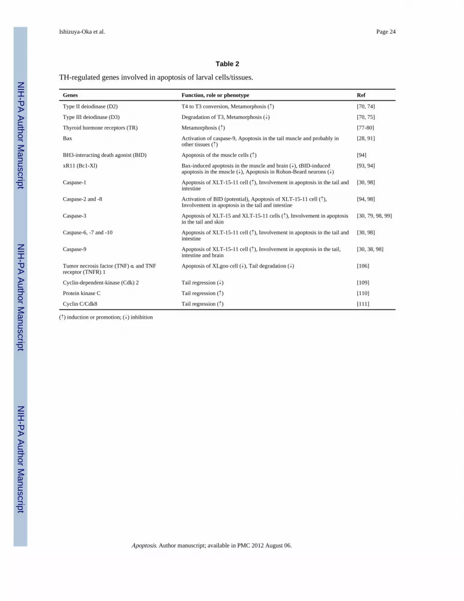
NIH
-PA Author Manuscript
NIH
-PA Author Manuscript
NIH
-PA Author Manuscript
Ishizuya-Oka et al. Page 24
Table 2
TH-regulated genes involved in apoptosis of larval cells/tissues.
Genes Function, role or phenotype Ref
Type II deiodinase (D2) T4 to T3 conversion, Metamorphosis (↑) [70, 74]
Type III deiodinase (D3) Degradation of T3, Metamorphosis (↓) [70, 75]
Thyroid hormone receptors (TR) Metamorphosis (↑) [77-80]
Bax Activation of caspase-9, Apoptosis in the tail muscle and probably inother tissues (↑)
[28, 91]
BH3-interacting death agonist (BID) Apoptosis of the muscle cells (↑) [94]
xR11 (Bc1-Xl) Bax-induced apoptosis in the muscle and brain (↓), tBID-inducedapoptosis in the muscle (↓), Apoptosis in Rohon-Beard neurons (↓)
[93, 94]
Caspase-1 Apoptosis of XLT-15-11 cell (↑), Involvement in apoptosis in the tail andintestine
[30, 98]
Caspase-2 and -8 Activation of BID (potential), Apoptosis of XLT-15-11 cell (↑),Involvement in apoptosis in the tail and intestine
[94, 98]
Caspase-3 Apoptosis of XLT-15 and XLT-15-11 cells (↑), Involvement in apoptosisin the tail and skin
[30, 79, 98, 99]
Caspase-6, -7 and -10 Apoptosis of XLT-15-11 cell (↑), Involvement in apoptosis in the tail andintestine
[30, 98]
Caspase-9 Apoptosis of XLT-15-11 cell (↑), Involvement in apoptosis in the tail,intestine and brain
[30, 38, 98]
Tumor necrosis factor (TNF) α and TNFreceptor (TNFR) 1
Apoptosis of XLgoo cell (↓), Tail degradation (↓) [106]
Cyclin-dependent-kinase (Cdk) 2 Tail regression (↓) [109]
Protein kinase C Tail regression (↑) [110]
Cyclin C/Cdk8 Tail regression (↑) [111]
(↑) induction or promotion; (↓) inhibition
Apoptosis. Author manuscript; available in PMC 2012 August 06.