Plasticity and reprogramming of differentiated cells in amphibian regeneration
9
566 | AUGUST 2002 | VOLUME 3 www.nature.com/reviews/molcellbio REVIEWS The only adult vertebrates that can regenerate their limbs are the URODELE amphibians, of the order CAU- DATA. Regeneration of the salamander limb, together with several other examples of urodele regeneration, was first reported by Spallanzani in 1768. The ability to regenerate large sections of the body plan is widespread in METAZOAN PHYLOGENY, and the discovery of this ability was an important aspect of the emergence of experi- mental biology in the eighteenth century 1 . It provoked intense public discussion on the nature of generation and the basis of individual identity, which are issues that continue to be explored in present-day debates about reproductive cloning. As a problem in molecular cell biology, urodele regeneration provides crucial informa- tion about the reversal and plasticity of the differenti- ated state 2,3 . In addition, limb regeneration is a key sys- tem in which to study how positional identity in cells is established 4–7 . Although much work on development and evolution serves to underline the similarities and continuity between different phylogenetic contexts 8,9 , it is an all-embracing concern of research on regeneration to understand the relevant distinctions between species that regenerate and those that do not 3,10 . In the case of urodeles and mammals, this takes on a biomedical imperative in view of the current interest in regenera- tive medicine. From our mammalian perspective, the ability of an adult newt to regenerate its limbs (FIG. 1a, b) might seem exceptional or even exotic, but it is unlikely that regenera- tion arose independently in different phylogenetic con- texts 3,10,11 . In only six phyla are there no examples of adult regeneration, yet in all contexts in which it is found, there seem to be examples of closely related species that have marked differences in regenerative ability (BOX 1). The hypothesis from phylogeny is that regeneration is a pri- mordial attribute of metazoans that has been lost subse- quently for reasons that are not yet understood. In analysing the cellular and molecular mechanisms that underlie the regenerative responses in urodeles, it has been informative to compare them with the mammalian case. This helps to pinpoint the crucial differences between urodeles and mammals, and will shed light ulti- mately on the evolutionary basis of regeneration. Regenerative responses in urodeles An adult newt can regenerate its jaws 12 , lens 13 , retina 14 and large sections of the heart 15 , as well as its limbs and tail, in response to molecular events that signal tissue damage or removal. Although the processes of tissue restoration unfold in a different way in the heart, limbs and tail of an adult newt, in all cases, the outcome seems to depend on the plasticity of differentiated cells that remain after tissue removal. Plasticity in the heart. After removal of the apical region of the newt ventricle, the heart seals by contraction PLASTICITY AND REPROGRAMMING OF DIFFERENTIATED CELLS IN AMPHIBIAN REGENERATION Jeremy P. Brockes* and Anoop Kumar Adult urodele amphibians, such as the newt, can regenerate their limbs and various other structures. This is the result of the plasticity and reprogramming of residual differentiated cells, rather than the existence of a ‘reserve-cell’ mechanism. The recent demonstrations of plasticity in mouse myotubes should facilitate comparative studies of the pathways that underlie the regenerative response, as well as proposing new approaches to promote mammalian regeneration. Department of Biochemistry & Molecular Biology, University College London, Gower Street, London WC1E 6BT, UK. Correspondence to J.P.B. e-mail: [email protected] doi:10.1038/nrm881 URODELE An order of the class Amphibia, which comprises newts and salamanders, which have elongated bodies, short limbs and a tail. METAZOAN Refers to the kingdom Animalia (animals), which comprises ~35 phyla of multicellular organisms. PHYLOGENY Evolutionary history that is sometimes represented by the hypothesized ancestor–descendant relationship of a group of organisms. © 2002 Nature Publishing Group
-
Upload
independent -
Category
Documents
-
view
1 -
download
0
Transcript of Plasticity and reprogramming of differentiated cells in amphibian regeneration

566 | AUGUST 2002 | VOLUME 3 www.nature.com/reviews/molcellbio
R E V I E W S
The only adult vertebrates that can regenerate theirlimbs are the URODELE amphibians, of the order CAU-DATA. Regeneration of the salamander limb, togetherwith several other examples of urodele regeneration,was first reported by Spallanzani in 1768. The ability toregenerate large sections of the body plan is widespreadin METAZOAN PHYLOGENY, and the discovery of this abilitywas an important aspect of the emergence of experi-mental biology in the eighteenth century1. It provokedintense public discussion on the nature of generationand the basis of individual identity, which are issues thatcontinue to be explored in present-day debates aboutreproductive cloning. As a problem in molecular cellbiology, urodele regeneration provides crucial informa-tion about the reversal and plasticity of the differenti-ated state2,3. In addition, limb regeneration is a key sys-tem in which to study how positional identity in cells isestablished4–7. Although much work on developmentand evolution serves to underline the similarities andcontinuity between different phylogenetic contexts8,9, itis an all-embracing concern of research on regenerationto understand the relevant distinctions between speciesthat regenerate and those that do not3,10. In the case ofurodeles and mammals, this takes on a biomedicalimperative in view of the current interest in regenera-tive medicine.
From our mammalian perspective, the ability of anadult newt to regenerate its limbs (FIG. 1a, b) might seem
exceptional or even exotic, but it is unlikely that regenera-tion arose independently in different phylogenetic con-texts3,10,11. In only six phyla are there no examples of adultregeneration, yet in all contexts in which it is found, thereseem to be examples of closely related species that havemarked differences in regenerative ability (BOX 1). Thehypothesis from phylogeny is that regeneration is a pri-mordial attribute of metazoans that has been lost subse-quently for reasons that are not yet understood. Inanalysing the cellular and molecular mechanisms thatunderlie the regenerative responses in urodeles, it hasbeen informative to compare them with the mammaliancase. This helps to pinpoint the crucial differencesbetween urodeles and mammals, and will shed light ulti-mately on the evolutionary basis of regeneration.
Regenerative responses in urodelesAn adult newt can regenerate its jaws12, lens13, retina14
and large sections of the heart15, as well as its limbs andtail, in response to molecular events that signal tissuedamage or removal. Although the processes of tissuerestoration unfold in a different way in the heart, limbsand tail of an adult newt, in all cases, the outcome seemsto depend on the plasticity of differentiated cells thatremain after tissue removal.
Plasticity in the heart. After removal of the apical regionof the newt ventricle, the heart seals by contraction
PLASTICITY AND REPROGRAMMINGOF DIFFERENTIATED CELLS INAMPHIBIAN REGENERATIONJeremy P. Brockes* and Anoop Kumar
Adult urodele amphibians, such as the newt, can regenerate their limbs and various otherstructures. This is the result of the plasticity and reprogramming of residual differentiatedcells, rather than the existence of a ‘reserve-cell’ mechanism. The recent demonstrations ofplasticity in mouse myotubes should facilitate comparative studies of the pathways thatunderlie the regenerative response, as well as proposing new approaches to promotemammalian regeneration.
Department of Biochemistry& Molecular Biology,University College London,Gower Street, London WC1E6BT, UK.Correspondence to J.P.B.e-mail: [email protected]:10.1038/nrm881
URODELE
An order of the class Amphibia,which comprises newts andsalamanders, which haveelongated bodies, short limbsand a tail.
METAZOAN
Refers to the kingdom Animalia(animals), which comprises ~35 phyla of multicellularorganisms.
PHYLOGENY
Evolutionary history that is sometimes represented by the hypothesizedancestor–descendantrelationship of a group oforganisms.
© 2002 Nature Publishing Group

NATURE REVIEWS | MOLECULAR CELL BIOLOGY VOLUME 3 | AUGUST 2002 | 567
R E V I E W S
CARDIOMYOCYTE
A muscle cell of the heart.
S (SYNTHESIS) PHASE
The phase of the eukaryotic cellcycle in which DNA issynthesized.
MYOFIBRIL
The structural unit of striatedmuscle fibres. Several myofibrilsmake up each fibre.
MYOTUBE
The multinucleate structure thatis formed by the fusion ofproliferating myoblasts and ischaracterized by the presence ofcertain muscle-specific markerproteins.
MYOFIBRE
A skeletal-muscle fibre thatconsists of one longmultinucleate cell.
PLURIPOTENT CELL
A stem cell that can give rise tomore than one differentiated celltype.
HEPATOCYTES
The parenchymal cells of theliver that are responsible for thesynthesis, degradation andstorage of a wide range ofsubstances.
cells to a local progenitor cell, rather than a PLURIPOTENT
CELL. So, if iris epithelial cells are transplanted to the limbblastema, they give rise to a lens32,33, and limb blastemasalways give rise to a limb after transplantation, even afterrelocation to the anterior chamber of the eye34. This is incontrast to recent findings of the extensive plasticity ofstem cells after transplantation in mammals35 (BOX 2). Atpresent, there is no evidence that adult stem cells cancontribute to urodele limb regeneration, although it isnot possible to exclude this possibility completely. Oneadvantage of the urodele mechanism might be that itallows the progenitor cells to derive local cues from theirdifferentiated parental cells.
Plasticity of differentiated cells in mammals?Plasticity of cellular differentiation provides a convenientcellular assay to compare a differentiated urodele cellwith its mammalian counterpart. It is important to rec-ognize that there are examples of regeneration in mam-mals that do involve plasticity. For example, liver regener-ation seems comparable to cardiac regeneration in newtsin that the HEPATOCYTES divide without loss of differenti-ated function36. The regeneration of myelinated periph-eral nerves requires that SCHWANN CELLS divide and lose
around the clot (FIG. 2a). The adult CARDIOMYOCYTES re-enter the cell cycle and divide in a zone that surroundsthe clot15,16. If the animal is injected with tritiated thymi-dine — to identify those cells that are in S PHASE — ~10%of the cardiomyocytes in this region are labelled in aone-day period. In comparable experiments with theadult mammalian heart, very few cells label afterinjury17. Thymidine labelling carried out in conjunc-tion with ultrastructural studies has identified cardiacMYOFIBRILS as a marker of cell identity.
Plasticity in the iris. After removal of the newt lens (len-tectomy), the reactive population comprises pigmentedepithelial cells, which are invariably located at the dorsalpupillary margin of the iris (FIG. 2b). These cells re-enterthe cell cycle, lose their pigment granules and convertinto lens cells, a process that is referred to as transdiffer-entiation18–20 (BOX 2). Elegant clonal cultures of newt pig-ment-epithelial cells have established conclusively thatthese cells transdifferentiate into lens20,21.
Plasticity in the limb. After amputation of the limb at anypoint along its proximodistal axis (shoulder to fingertip),the wound surface is covered rapidly by epithelial cells,which form the wound epidermis at the end of thestump. In contrast to lens regeneration, which dependson the plasticity of epithelial cells of the iris, in limbregeneration, it is the mesenchymal cells in a zone under-lying the wound epidermis that re-enter the cell cycle.The cartilage, connective tissue and muscle cells losetheir differentiated characteristics, and become blastemalcells — the progenitor cells of the regenerate. These cellsdivide to form the blastema, a mesenchymal growthzone that undergoes proliferation, differentiation andmorphogenesis to regenerate the limb22,23 (FIG. 1b).
Extensive evidence indicates that multinucleate newtMYOTUBES and MYOFIBRES re-enter the cell cycle andundergo conversion to mononucleate cells. Such evi-dence comes from the results of experiments on musclecells in culture24–26, cells implanted from culture into alimb blastema27–29, and observations of live myofibres ina regenerating tail30, which are all discussed later. Inaddition, earlier observations of implantation oflabelled cartilage also provide strong evidence for rever-sal of this cell type31.
In each of these three cases of regeneration, the initialmobilization of differentiated cells occurs within a zone of~100 µm from the site of tissue removal. It is obviouslycrucial that such changes do not propagate into the mainbody of the tissues that are concerned, and the signallingevents that link tissue removal with plasticity clearlyaccomplish this spatial restriction.Although these threecases involve re-entry into the cell cycle, the cardiomyocyteis the only example to retain differentiated function,a fea-ture that is also observed in culture.Although in one sensecell division is necessary to generate the extra cells that areneeded for the regenerate, it is less clear how important itis for the events of plasticity and reversal, and this issueneeds to be addressed experimentally in each context.
The urodele strategy for regeneration of most struc-tures is therefore the respecification of differentiated
Figure 1 | Urodele limb regeneration. a | The NorthAmerican red spotted newt, Notophthalmus viridescens. b | Stages of limb regeneration in an adult newt66. Reproducedwith permission from REF. 66 © 1973 Springer-Verlag.
Amputation Wound healing De-differentiation
Early bud Medium bud Late bud
Palette Early differentiation Regenerated limb
a
b
© 2002 Nature Publishing Group

568 | AUGUST 2002 | VOLUME 3 www.nature.com/reviews/molcellbio
R E V I E W S
from any labelled mononucleate cells27. Other methodsof labelling involve microinjecting the myotubes with alineage tracer, such as rhodamine dextran28,29, orlabelling them with a lipophilic cell tracker dye27.
Implantation of labelled myotubes under the woundepidermis of an early limb blastema showed that manywere converted to mononucleate cells, which dividedand contributed to the regenerate — a process that isreferred to here as cellularization27–29 (FIG. 3a, b). If themyotubes were double labelled in their nuclei and cyto-plasm before implantation, the mononucleate progenyderived both markers, which shows that both compart-ments of the multinucleate muscle cells were conservedafter cellularization27,28.
A second index for the reversal of differentiation is re-entry to S phase by the nuclei in a myotube.After implan-tation of myotubes into a newt blastema, and injection ofthe animals with 5′ BROMODEOXYURIDINE (BrdU) to labelnuclei in S phase, residual myotubes had several positivelystained nuclei. In some cases, all of the nuclei in amyotube were labelled27. It is not clear if such myotubesundergo subsequent cellularization after re-entry, but it isclear that the environment of the blastema leads to rever-sal of both aspects of myogenic differentiation.
Implantation is an extremely useful approach becauseit enables prior transfection, labelling and purification ofthe myotubes in culture (FIG. 3b). Importantly, bothaspects of reversal that are discussed above have also beenobserved in endogenous multinucleate muscle fibres afteramputation. First, the nuclei within muscle fibres at thesite of limb amputation were labelled after BrdU injec-tion into adult newts27. The identity of the labellednuclei was verified after serial sectioning, which showedthat they were present in muscle fibres. Second, the
expression of MYELIN before they redifferentiate in con-junction with the regenerating axons37,38. It thereforeseems to be more closely related than liver regenerationto the general case of the urodele mechanism.
It seems unlikely that the regulation of the differenti-ated state is fundamentally different in urodeles andmammals. To study this further, we have comparedurodele multinucleate skeletal muscle cells with theirmouse counterparts. To what extent are the two intrinsi-cally different in terms of regulation of the differentiatedstate or responsiveness to injury-related signals, or arethere distinctive signals in the urodele context thatmouse cells never encounter?
Reversal of muscle differentiationMultinucleate skeletal myotubes are formed by thefusion of mononucleate precursor cells. The myotubeenters a state of post-mitotic arrest in which it isentirely refractory to the growth factors that stimulatedivision of its precursors39. The change in cytology —from mononucleate to multinucleate — together withthe post-mitotic arrest provides two indices for thereversal of the myogenic phenotype (de-differentia-tion). Newt A1 cells, which were originally derivedfrom limb MESENCHYME40, fuse in culture to form multin-ucleate myotubes, which express markers of late muscledifferentiation, such as myosin heavy chain, and are alsorefractory to protein growth factors25,40.
Assaying for de-differentiation in the blastema. A1 cellscan be stably infected with retroviruses that expressgenes for alkaline phosphatase or GREEN FLUORESCENT
PROTEIN (GFP), so that after differentiation, labelledmyotubes can be readily distinguished and purified
ROTIFERA
A small phylum of microscopicmulticellular organisms. Theyhave a wheel-like ciliated organ(from which they derive theirname) that they use forswimming and feeding.
TURBELLARIANS
A class of platyhelminthes thatcomprises mostly aquatic andfree-living organisms. They havea ciliated epidermis forlocomotion and a simple gut.
ANNELID
A segmented worm.
PLANARIAN
Describes free-living membersof the invertebrate phylumplatyhelminthes.
SCHWANN CELL
A cell that produces myelin andensheathes axons in theperipheral nervous system.
MYELIN
Proteins that are produced by Schwann cells oroligodendrocytes that causeadjacent plasma membranes tostack tightly together.
MESENCHYME
Immature connective tissue thatconsists of cells that areembedded in extracellularmatrix.
GREEN FLUORESCENT PROTEIN
An autofluorescent protein thatwas originally isolated from thejellyfish Aequorea victoria. It canbe genetically conjugated withproteins to make themfluorescent. The most widelyused mutant, EGFP, has anemission maximum at 510 nm.
5′ BROMODEOXYURIDINE
(BrdU). A base analogue ofthymidine, which is often usedexperimentally to label dividingcells.
Box 1 | Phylogeny and regeneration
There are several phyla, such as ROTIFERS and nematodes, thatshow cell constancy in the adult, and are not capable ofregeneration61. Furthermore, the phyla that includeexamples of regeneration often have marked interspeciesvariation. Lineus ruber and Lineus viridis are two species ofnemertine worm, which are so similar that it is said torequire a specialist to distinguish them. However, L. ruberhas excellent bidirectional regeneration after transecting theprimary body axis, whereas L. viridis has no discernibleregenerative abilility62.
It has often been noted that in some invertebrates, theprocess of asexual reproduction is closely related toregeneration10,61. Some animals undergo a process of self-induced fission, after which they regenerate from thefragments. This might be achieved by tearing forces that aregenerated from opposite movements of parts in some starfishor sea anemones, or even by twisting into several pieces61. InTURBELLARIANS and ANNELIDS, fission involves distinct divisionplanes, and in some species, the new organs form beforeseparation — a process that is known as paratomy.
So, what is the most promising system for future studies ofregeneration? Although it is important to continue to studyseveral models, two compelling candidates are fin regeneration in zebrafish63, which has allowed the isolation of severalconditional mutants, and PLANARIAN regeneration, in which powerful new approaches have been developed.
Lineus ruberLineus viridis
© 2002 Nature Publishing Group

NATURE REVIEWS | MOLECULAR CELL BIOLOGY VOLUME 3 | AUGUST 2002 | 569
R E V I E W S
These experiments have established that the responseof myotubes that are implanted into the limb blastemais similar to that of resident myofibres, and underlinethe importance of a more detailed analysis of both re-entry to the cell cycle and cellularization. Furthermore, itis noteworthy that the muscle fibres make an importantcellular contribution to the blastema30. The authors esti-mate that a 4-day old blastema contains ~900 cells, ofwhich approximately 150 come from the cellularizationof muscle fibres.
Cell-cycle re-entry in newt myotubesNewt myotubes clearly differ from their vertebratecounterparts in that they enter and traverse S phaseafter serum stimulation in culture24,25. The nuclei in themyotube double their DNA content and are stablyarrested in G2. The response to serum is not observedfor other vertebrate myotubes, with the exception ofmouse cells that lack both copies of the retinoblastomagene Rb 41. pRb has a familiar and essential role in reg-ulating the transition from G1 to S phase. Several linesof evidence42,43 indicate that it is important for mainte-nance of the differentiated state in vertebratemyotubes, not only for stable arrest from the cellcycle41, but also for transcription from certain musclepromoters that depend on activation of members ofthe myocyte enhancer factor 2 (MEF2) family of tran-scription factors44. Newt myotubes express pRb, butserum stimulates a pathway that leads to its inactiva-tion by phosphorylation, and hence triggers progres-sion from G1 into S phase24. If the myotubes areinjected with a plasmid that encodes mammalianp16INK4 — a specific inhibitor of the cyclin-D–CDK4protein kinase that inactivates Rb — they are effec-tively blocked from entering S phase after serum treat-ment24. These data provide the first clear evidence thatdifferentiated urodele cells are intrinsically differentfrom their mammalian counterparts. The newtmyotube can activate a pathway that leads to phospho-rylation of pRb and re-entry to the cell cycle, and we
regenerating tail of the larval axolotl was visualized underNOMARSKI OPTICS to observe the behaviour of single fibresafter microinjection of a lineage tracer30. Such fibres frag-mented synchronously into mononucleate cells andunderwent rapid proliferation. The activation of thisprocess apparently required both ‘clipping’ at the end ofthe fibre as well as tissue injury in the vicinity.
Figure 2 | Plasticity of differentiated cells during regeneration of newt heart or lens.a | After removal of the apex of the ventricle, the heart is sealed by a clot (red), and the adultcardiomyocytes return to the cell cycle in a zone (green) around the clot, proliferate andregenerate the ventricle. If cardiomyocytes are dissociated into a culture dish, they can divideand form groups of beating cells. It is striking that some cardiomyocytes complete the cell cycleand then resume beating. b | After removal of the lens, the pigmented epithelial cells of the dorsalmargin of the iris give rise to the new lens. If the iris is dissociated, pigmented cells will re-enterthe cell cycle, de-pigment and transdifferentiate into lentoid bodies (a small cluster of lens cells)that express lens markers.
a
Ventricle
Cell-cycle zoneHeart
Dissociation
Dissociation
b Eye
Clot
Iris Cell-cyclere-entry
Transdifferentiation
Lentoid body
Lens
Box 2 | Transdifferentiation
Transdifferentiation was originally defined by Okada18 as the conversion of one differentiated cell type into another. Theoriginal, and still the most studied, example is the conversion of pigmented epithelial cells from the iris into lens cells,which occurs during lens regeneration in newts and in cultures of pigment cells from various species (FIG. 2b). This canstill occur to some extent if cell division is blocked, although it normally occurs in conjunction with cell-cycle re-entry.
In urodele limb regeneration, transdifferentiation could occur in a switch between the chondrogenic–connective-tissue lineage and the myogenic lineage. If cartilage is selectively labelled and introduced into a limb blastema, theoccurrence of labelled nuclei in muscle is negligible. However, if cultured myotubes are labelled by microinjection ofrhodamine-dextran28 or with an integrated retrovirus27, small clones of labelled mononucleate cells are detectedoccasionally in the cartilage of the regenerate.
There are several cases of transdifferentiation in mammals64, such as the conversion of pancreatic EXOCRINE CELLS tohepatocytes in vivo under conditions of copper deficiency, as well as in vitro. Other examples include the transitionbetween smooth and skeletal muscle in the developing oesophagus, and the conversion of myoblasts to adipocytes.
There has been much recent interest in transdifferentiation of stem cells; for example, the ability of haematopoieticstem cells or mesenchymal stem cells to give rise to neural and other epithelial derivatives after transplantation. Theinterpretation of these results is controversial at present65, in view of the possibility of heterogeneity in the startingstem-cell populations and the contribution of cell fusion between different stem cells. However, transdifferentiationremains an important area for understanding cell plasticity.
NOMARSKI OPTICS
Also known as differentialinterference contrastmicroscopy, this techniqueforms images of high contrastand resolution in unstained cellsusing birefringent prisms andpolarized light.
EXOCRINE CELL
A cell that makes up part of anexocrine gland, which dischargesits secretion through a duct.
G2
The phase of the cell cyclethrough which cells progressafter S phase but before M phase.
© 2002 Nature Publishing Group

570 | AUGUST 2002 | VOLUME 3 www.nature.com/reviews/molcellbio
R E V I E W S
pivotal role of pRb in both cases. The distinction is thatthe urodele myotubes are responsive to an activity inserum that has no effect on the mouse cells. Serum isthe soluble fraction of clotted blood, and results fromthe activation of prothrombin to generate the serineprotease thrombin. Thrombin activates the clottingcascade and various other events that mediate theresponse to injury. It is possible to pre-incubate sub-threshold concentrations of serum with thrombin,and then inactivate all of the residual protease activity.Such digests can generate considerable activity on thenewt myotubes25. When crude prothrombin is acti-vated in vitro, the resulting thrombin preparations con-tain a distinct activity that acts directly on newtmyotubes in serum-free medium25. We hypothesize thatthis ligand is generated downstream of prothrombinactivation both in regeneration and in culture, and it isthis factor that acts on the myotubes and other differen-tiated cells to promote re-entry (FIG. 4).
The activation of vascular prothrombin after injuryoccurs in relation to a protease complex known asTissue Factor, which is assembled on the cell surface.Thrombin formation is subject to strict spatial and tem-poral regulation, as it is essential that clot formation isrestricted to the wound area and does not spread. It istherefore tempting to speculate that urodele regenera-tion — for example, in the heart, limb or eye — islinked to the acute events of injury or tissue removal bythe local activity of thrombin. Thrombin activity isincreased locally in the early mesenchymal blastema ofthe limb25, but more striking are recent findings thatprothrombin is selectively activated on the dorsal mar-gin of the iris after lentectomy, and that inhibition ofthrombin in this context blocks cell-cycle re-entry onthe dorsal margin (Y. Imokawa and J. P. B., unpublishedobservations). We propose that activation of prothrom-bin and generation of an activity referred to as Fa (FIG. 4)
are important for the initiation of urodele regenerationin several contexts. It will be important to determinewhether Fa is active on various differentiated cell types.
Cell-cycle re-entry by mouse myonuclei. Althoughmyotubes of the mouse C2 cell line are not normallyresponsive to serum and the thrombin-based manipula-tions24, their nuclei can be induced to re-enter the cellcycle by this pathway under some experimental condi-tions. This involves fusing mononucleate newt A1 andmouse C2 cells to obtain viable hybrid interspeciesmyotubes (HETEROKARYONS)26. The mouse nuclei can beidentified by a species-specific antibody to nuclearlamin B. These cells grow at 33 °C under conditions thatsupport serum-induced cell-cycle re-entry of A1myotubes, as well as the post-mitotic arrest. If the het-erokaryons are stimulated with serum or thrombin,mouse nuclei frequently enter S phase — sometimes inconjunction with newt nuclei and sometimes alone26.So, mouse myotubes have apparently lost responsive-ness to the thrombin-derived factor24–26, but their nucleiremain responsive to the intracellular consequences ofthe pathway, such as the phosphorylation of pRb, and inthis respect, are quite comparable to newt nuclei.
propose that the regulation of this pathway is pivotalto the initiation of regeneration.
Regulation of cell-cycle re-entry by thrombin. The activityof vertebrate serum on newt myotubes is not due to thepresence of typical protein growth factors, such as platelet-derived growth factor (PDGF) or epidermal growth factor(EGF); these factors act on mononucleate A1 cells but noton the myotubes24,25 (FIG. 4). The post-mitotic arrest asdefined in mouse myotubes is therefore comparable tothat in newt myotubes, and this is consistent with the
HETEROKARYON
A cell that contains two nuclei ina common cytoplasm.
Figure 3 | Plasticity of urodele myogenesis. A | Mononucleate cells fuse to form multinucleatemyotubes, but this process can be reversed. After implantation into a limb blastema, the nucleiwithin myotubes re-enter S phase and the myotubes are fragmented into viable mononucleatecells, which divide and contribute to the regenerate. B | Examples of implantation assays withnewt myotubes that are labelled by different methods. Top panels show myotubes in culturelabelled with an integrated provirus that expresses a | the marker enzyme human placentalalkaline phosphatase, b | the lineage tracer rhodamine-dextran, c | the lipophilic cell tracker dyePKH-26 , or d | green fluorescent protein. Bottom panels show mononucleate cells observed7–10 days after implantation of the corresponding myotubes. Nuclei in (b), (c) and (d) are stainedwith Hoechst dye, whereas those in (a) are stained with haematoxylin. Reproduced withpermission from REF. 3 © 2001 Cambridge Univ. Press.
A
B
Mononucleate cells Multinucleate myotube
(withdrawal from the cellcycle and fusion)
Differentiation
Re-entry into cell cycle,fragmentation of myotube
Limb blastema
Implantation
a b c d
Mononucleate cells observed 7–10 d after implantation of the corresponding myotubes
© 2002 Nature Publishing Group

NATURE REVIEWS | MOLECULAR CELL BIOLOGY VOLUME 3 | AUGUST 2002 | 571
R E V I E W S
observations). This accessible system offers thepotential for a more detailed exploration of themechanisms of cellularization.
Cellularization of mammalian myotubes. An importantimpetus for the study of cellularization has come fromthe recent discovery of two methods that induce thisprocess in mouse myotubes. In one approach, mononu-cleate C2 cells were stably transfected with the homeoboxgene Msx-1 linked to a conditional promoter46. Severalstudies have previously indicated that Msx genes pro-mote cell proliferation, and that their expression isinversely correlated with differentiation47–49. After fusionof the stably transfected C2 cells, expression of Msx-1 inmyotubes was induced and this led to a decrease in theexpression of myogenic regulatory genes. About 5% ofthe myotubes were induced to cleave into viable frag-ments — as described above for the larval myofibres —and another 5% fragmented into mononucleate cells,which proliferated. In some cases, the clonal progeny of asingle myotube was isolated, propagated and shown to becapable of either CHONDROGENIC, ADIPOGENIC or myogenicdifferentiation, depending on the culture conditions46. Ithas been proposed50 that Msx-1 is a master regulator of
CellularizationAn important mechanistic issue is the relationshipbetween the two aspects of plasticity — re-entry to thecell cycle and cellularization27. One possibility is thatnuclei proceed into mitosis and the myotube is frag-mented by CYTOKINESIS. Alternatively, myotube nucleicould bud off the main axis of the myotube in the pres-ence or absence of cell-cycle re-entry. A1 newt myotubescan be blocked from cell-cycle re-entry either by X-irra-diation or by microinjection of a plasmid that encodesmammalian p16INK4. Such arrested myotubes, whenimplanted into a limb blastema together with a differen-tially labelled control population, were effectively con-verted to mononucleate cells29 (FIG. 5). So, although re-entry and cellularization occur in parallel afterimplantation, they are not linked mechanistically. Oneother example of cellularization that is not dependenton mitosis, and which has some parallels with theprocess in newt myotubes, is that of the avianOSTEOCLAST-like multinucleated GIANT CELLS45. These cellsare formed by fusion of mononuclear monocytes, andin culture they bud off mononuclear osteoclasts fromthe apical surface.
These findings on the independence of re-entryand cellularization have been reinforced by observa-tions on striated muscle fibres, which can be dissoci-ated from the limbs of larval salamanders and main-tained in culture as adherent cells. These cells areapparently activated by the dissociation process toundergo several aspects of morphological plasticity,which can be analysed by time-lapse studies and bymicroinjection of a lineage tracer into the fibres.Some of them break into smaller fibres, or dischargeviable multinucleate buds that subsequently adhereto the substrate. The most striking observation isthat nuclei can aggregate in some fibres to form alobulated, ‘cauliflower’ structure, which gives rise toa colony of dividing mononucleate cells. Theseevents all occur in the absence of S-phase entry inthe muscle-fibre nuclei (A. Kumar et al., unpublished
CYTOKINESIS
The process of cytoplasmicdivision.
OSTEOCLAST
A mesenchymal cell that candifferentiate into a bone-degrading cell.
GIANT CELLS
Large multinucleated cells thatare thought to result from thefusion of macrophages.
CHONDROGENIC
Able to form cartilage.
ADIPOGENIC
Able to form fat or adipose tissue.
Figure 4 | Thrombin activity counteracts post-mitoticarrest in newt myotubes. Thrombin acts indirectly bycleaving a substrate (F) that is present in vertebrate serum orplasma25 to induce cell-cycle re-entry. Interestingly, theresulting activity, referred to as Fa, does not affectmononucleate cells, whereas growth factors such as platelet-derived growth factor (PDGF) have the reverse specificity.Mouse myotubes are completely refractory to the thrombin-generated activity.
Newt myotubes
Mouse myotubes
PDGFMononucleateprecursors
Thrombin
Cell-cyclere-entry
F Fa
Figure 5 | Arrested myotubes undergo cellularizationafter implantation into the newt blastema. The figureshows a cultured newt myotube (purple) injected with aplasmid that expresses the cyclin-dependent-kinase inhibitorp16INK4 — to arrest cell-cycle progression — along with acontrol myotube that is labelled with the lineage tracerrhodamine-dextran (red). After implantation, both give rise tomononucleate cells in the blastema (see REF 29).
Blastema
Mononucleate cells
Implantation
Control myotube Arrested myotube
p16INK4
plasmid
© 2002 Nature Publishing Group
572 | AUGUST 2002 | VOLUME 3 www.nature.com/reviews/molcellbio
R E V I E W S
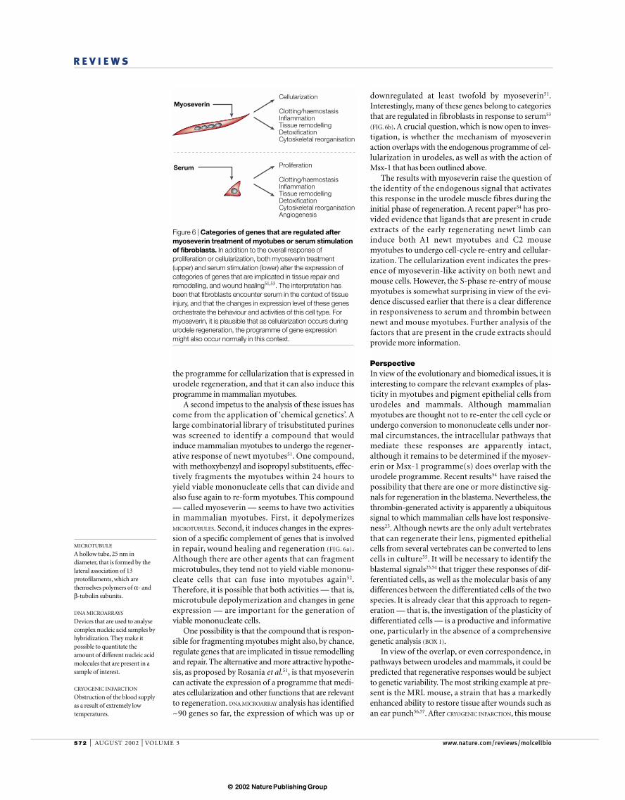
downregulated at least twofold by myoseverin51.Interestingly, many of these genes belong to categoriesthat are regulated in fibroblasts in response to serum53
(FIG. 6b). A crucial question, which is now open to inves-tigation, is whether the mechanism of myoseverinaction overlaps with the endogenous programme of cel-lularization in urodeles, as well as with the action ofMsx-1 that has been outlined above.
The results with myoseverin raise the question ofthe identity of the endogenous signal that activatesthis response in the urodele muscle fibres during theinitial phase of regeneration. A recent paper54 has pro-vided evidence that ligands that are present in crudeextracts of the early regenerating newt limb caninduce both A1 newt myotubes and C2 mousemyotubes to undergo cell-cycle re-entry and cellular-ization. The cellularization event indicates the pres-ence of myoseverin-like activity on both newt andmouse cells. However, the S-phase re-entry of mousemyotubes is somewhat surprising in view of the evi-dence discussed earlier that there is a clear differencein responsiveness to serum and thrombin betweennewt and mouse myotubes. Further analysis of thefactors that are present in the crude extracts shouldprovide more information.
PerspectiveIn view of the evolutionary and biomedical issues, it isinteresting to compare the relevant examples of plas-ticity in myotubes and pigment epithelial cells fromurodeles and mammals. Although mammalianmyotubes are thought not to re-enter the cell cycle orundergo conversion to mononucleate cells under nor-mal circumstances, the intracellular pathways thatmediate these responses are apparently intact,although it remains to be determined if the myosev-erin or Msx-1 programme(s) does overlap with theurodele programme. Recent results54 have raised thepossibility that there are one or more distinctive sig-nals for regeneration in the blastema. Nevertheless, thethrombin-generated activity is apparently a ubiquitoussignal to which mammalian cells have lost responsive-ness25. Although newts are the only adult vertebratesthat can regenerate their lens, pigmented epithelialcells from several vertebrates can be converted to lenscells in culture55. It will be necessary to identify theblastemal signals25,54 that trigger these responses of dif-ferentiated cells, as well as the molecular basis of anydifferences between the differentiated cells of the twospecies. It is already clear that this approach to regen-eration — that is, the investigation of the plasticity ofdifferentiated cells — is a productive and informativeone, particularly in the absence of a comprehensivegenetic analysis (BOX 1).
In view of the overlap, or even correspondence, inpathways between urodeles and mammals, it could bepredicted that regenerative responses would be subjectto genetic variability. The most striking example at pre-sent is the MRL mouse, a strain that has a markedlyenhanced ability to restore tissue after wounds such asan ear punch56,57. After CRYOGENIC INFARCTION, this mouse
the programme for cellularization that is expressed inurodele regeneration, and that it can also induce thisprogramme in mammalian myotubes.
A second impetus to the analysis of these issues hascome from the application of ‘chemical genetics’. Alarge combinatorial library of trisubstituted purineswas screened to identify a compound that wouldinduce mammalian myotubes to undergo the regener-ative response of newt myotubes51. One compound,with methoxybenzyl and isopropyl substituents, effec-tively fragments the myotubes within 24 hours toyield viable mononucleate cells that can divide andalso fuse again to re-form myotubes. This compound— called myoseverin — seems to have two activitiesin mammalian myotubes. First, it depolymerizesMICROTUBULES. Second, it induces changes in the expres-sion of a specific complement of genes that is involvedin repair, wound healing and regeneration (FIG. 6a).Although there are other agents that can fragmentmicrotubules, they tend not to yield viable mononu-cleate cells that can fuse into myotubes again52.Therefore, it is possible that both activities — that is,microtubule depolymerization and changes in geneexpression — are important for the generation ofviable mononucleate cells.
One possibility is that the compound that is respon-sible for fragmenting myotubes might also, by chance,regulate genes that are implicated in tissue remodellingand repair. The alternative and more attractive hypothe-sis, as proposed by Rosania et al.51, is that myoseverincan activate the expression of a programme that medi-ates cellularization and other functions that are relevantto regeneration. DNA MICROARRAY analysis has identified~90 genes so far, the expression of which was up or
MICROTUBULE
A hollow tube, 25 nm indiameter, that is formed by thelateral association of 13protofilaments, which arethemselves polymers of α- andβ-tubulin subunits.
DNA MICROARRAYS
Devices that are used to analysecomplex nucleic acid samples byhybridization. They make itpossible to quantitate theamount of different nucleic acidmolecules that are present in asample of interest.
CRYOGENIC INFARCTION
Obstruction of the blood supplyas a result of extremely lowtemperatures.
Figure 6 | Categories of genes that are regulated aftermyoseverin treatment of myotubes or serum stimulationof fibroblasts. In addition to the overall response ofproliferation or cellularization, both myoseverin treatment(upper) and serum stimulation (lower) alter the expression ofcategories of genes that are implicated in tissue repair andremodelling, and wound healing51,53. The interpretation hasbeen that fibroblasts encounter serum in the context of tissueinjury, and that the changes in expression level of these genesorchestrate the behaviour and activities of this cell type. Formyoseverin, it is plausible that as cellularization occurs duringurodele regeneration, the programme of gene expressionmight also occur normally in this context.
Serum
Myoseverin
Proliferation
Clotting/haemostasisInflammationTissue remodellingDetoxificationCytoskeletal reorganisationAngiogenesis
Cellularization
Clotting/haemostasisInflammationTissue remodellingDetoxificationCytoskeletal reorganisation
© 2002 Nature Publishing Group

NATURE REVIEWS | MOLECULAR CELL BIOLOGY VOLUME 3 | AUGUST 2002 | 573
R E V I E W S
applications are sufficiently problematic to warrantconsideration of alternative and complementaryapproaches. The urodele strategy — the limitedrespecification of residual differentiated cells — is sosuccessful that it would be surprising if it were noteventually tried as a therapeutic approach in somecontexts of mammalian regeneration. The example ofmyoseverin51,52 shows that the responses that are dis-cussed in this review are a potential target for thera-peutics that are directed at regulating the stability ofthe differentiated state.
can restore significant cardiac function, and thisresponse is associated with cell-cycle progression of car-diomyocytes in the vicinity of the lesion58. The analysisof the various loci that are associated with these aspectsof the MRL phenotype should be informative59.
At present, the main approach to mammalianregenerative medicine is the isolation of appropriatestem cells, followed by manipulations that are aimedat directing their differentiation towards the mor-phogenesis of complex structures60. Although thisattracts considerable interest at present, many of the
1. Dinsmore, C. E. A History of Regeneration Research(Cambridge Univ. Press, Cambridge, 1991).
2. Brockes, J. P. Amphibian limb regeneration: rebuilding acomplex structure. Science 276, 81–87 (1997).An overview of urodele regeneration that focuseson plasticity and positional identity.
3. Brockes, J. P., Kumar, A. & Velloso, C. P. Regeneration asan evolutionary variable. J. Anat. 199, 3–11 (2001).
4. Pecorino, L. T., Entwistle, A. & Brockes, J. P. Activation ofa single retinoic acid receptor isoform mediatesproximodistal respecification. Curr. Biol. 6, 563–569(1996).
5. Torok, M. A., Gardiner, D. M., Shubin, N. H. & Bryant, S. V.Expression of HoxD genes in developing andregenerating axolotl limbs. Dev. Biol. 200, 225–233(1998).
6. Maden, M. Vitamin A and pattern formation in theregenerating limb. Nature 295, 672–675 (1982).
7. Nardi, J. B. & Stocum, D. L. Surface properties ofregenerating limb cells: evidence for gradation along theproximodistal axis. Differentiation 25, 27–31 (1983).
8. Raff, R. A. The Shape of Life (Univ. Chicago, Chicago,1996).
9. Carroll, S. B. Chance and necessity: the evolution ofmorphological complexity and diversity. Nature 409,1102–1109 (2001).
10. Alvarado, A. S. Regeneration in the metazoans: whydoes it happen? Bioessays 22, 578–590 (2000).This reviews the evolutionary questions aboutregeneration and its origins, such as why someanimals regenerate but others apparently do not.
11. Goss, R. J. Principles of Regeneration (Academic, NewYork, 1969).
12. Ghosh, S., Thorogood, P. & Ferretti, P. Regenerativecapability of upper and lower jaws in the newt. Int. J. Dev.Biol. 38, 479–490 (1994).
13. Reyer, R. W. Regeneration of the lens in the amphibianeye. Quart. Rev. Biol. 29, 1–46 (1954).
14. Mitashov, V. I. Mechanisms of retina regeneration inurodeles. Int. J. Dev. Biol. 40, 833–844 (1996).
15. Oberpriller, J. O. & Oberpriller, J. C. Response of theadult newt ventricle to injury. J. Exp. Zool. 187, 249–253(1974).
16. Oberpriller, J. O., Oberpriller, J. C., Matz, D. G. &Soonpaa, M. H. Stimulation of proliferative events in theadult amphibian cardiac myocyte. Ann. NY Acad. Sci.752, 30–46 (1995).
17. Soonpaa, M. H. & Field, L. J. Survey of studies examiningmammalian cardiomyocyte DNA synthesis. Circ. Res. 83,15–26 (1998).
18. Okada, T. S. Transdifferentiation (Clarendon, Oxford,1991).
19. Eguchi, G. Cellular and molecular background of wolffianlens regeneration. Cell Differ. Dev. 25, S147–S158 (1988).
20. Eguchi, G., Abe, S. I. & Watanabe, K. Differentiation oflens-like structures from newt iris epithelial cells in vitro.Proc. Natl Acad. Sci. USA 71, 5052–5056 (1974).
21. Eguchi, G. & Okada, T. S. Differentiation of lens tissuefrom the progeny of chick retinal pigment cells cultured in vitro: a demonstration of a switch of cell types in clonalcell culture. Proc. Natl Acad. Sci. USA 70, 1495–1499(1973).
22. Tsonis, P. A. Limb Regeneration (Cambridge Univ. Press,Cambridge, 1996).
23. Wallace, H. Vertebrate Limb Regeneration (Wiley andSons, New York, 1981).
24. Tanaka, E. M., Gann, A. A., Gates, P. B. & Brockes, J. P.Newt myotubes re-enter the cell cycle by
phosphorylation of the retinoblastoma protein. J. CellBiol. 136, 155–165 (1997).
25. Tanaka, E. M., Drechsel, D. N. & Brockes, J. P. Thrombinregulates S-phase re-entry by cultured newt myotubes.Curr. Biol. 9, 792–799 (1999).References 24 and 25 provide the first clearevidence that a urodele differentiated cell — theskeletal myotube — is intrinsically different from itsmammalian counterpart.
26. Velloso, C. P., Simon, A. & Brockes, J. P. Mammalianpostmitotic nuclei re-enter the cell cycle after serumstimulation in newt/mouse hybrid myotubes. Curr. Biol.11, 855–858 (2001).
27. Kumar, A., Velloso, C. P., Imokawa, Y. & Brockes, J. P.Plasticity of retrovirus-labelled myotubes in the newt limbregeneration blastema. Dev. Biol. 218, 125–136 (2000).
28. Lo, D. C., Allen, F. & Brockes, J. P. Reversal of muscledifferentiation during urodele limb regeneration. Proc.Natl Acad. Sci. USA 90, 7230–7234 (1993).This study found that implanted labelled myotubesunderwent reversal of muscle differentiation in thelimb blastema.
29. Velloso, C. P., Kumar, A., Tanaka, E. M. & Brockes, J. P.Generation of mononucleate cells from post-mitoticmyotubes proceeds in the absence of cell cycleprogression. Differentiation 66, 239–246 (2000).
30. Echeverri, K., Clarke, J. D. & Tanaka, E. M. In vivoimaging indicates muscle fiber dedifferentiation is a majorcontributor to the regenerating tail blastema. Dev. Biol.236, 151–164 (2001).
31. Steen, T. P. Stability of chondrocyte differentiation andcontribution of muscle to cartilage during limbregeneration in the axolotl (Siredon mexicanum). J. Exp.Zool. 167, 49–78 (1968).
32. Reyer, R. W., Woolfitt, R. A. & Withersty, L. T. Stimulationof lens regeneration from the newt dorsal iris whenimplanted into the blastema of the regenerating limb.Dev. Biol. 32, 258–281 (1973).
33. Ito, M., Hayashi, T., Kuroiwa, A. & Okamoto, M. Lensformation by pigmented epithelial cell reaggregate fromdorsal iris implanted into limb blastema in the adult newt.Dev. Growth Differ. 41, 429–440 (1999).
34. Kim, W. S. & Stocum, D. L. Retinoic acid modifiespositional memory in the anteroposterior axis ofregenerating axolotl limbs. Dev. Biol. 114, 170–179(1986).
35. Clarke, D. L. et al. Generalized potential of adult neuralstem cells. Science 288, 1660–1663 (2000).
36. Michalopoulos, G. K. & DeFrances, M. C. Liverregeneration. Science 276, 60–66 (1997).
37. Aguayo, A. J., Epps, J., Charron, L. & Bray, G. M.Multipotentiality of Schwann cells in cross-anastomosedand grafted myelinated and unmyelinated nerves:quantitative microscopy and radioautography. Brain Res.104, 1–20 (1976).
38. Weinberg, H. J. & Spencer, P. S. Studies on the control ofmyelinogenesis. II. Evidence for neuronal regulation ofmyelin production. Brain Res. 113, 363–378 (1976).
39. Olwin, B. B. & Hauschka, S. D. Cell surface fibroblastgrowth factor and epidermal growth factor receptors arepermanently lost during skeletal muscle terminaldifferentiation in culture. J. Cell Biol. 107, 761–769(1988).
40. Ferretti, P. & Brockes, J. P. Culture of newt cells fromdifferent tissues and their expression of a regeneration-associated antigen. J. Exp. Zool. 247, 77–91 (1988).
41. Schneider, J. W., Gu, W., Zhu, L., Mahdavi, V. & Nadal-Ginard, B. Reversal of terminal differentiation mediated
by p107 in Rb–/– muscle cells. Science 264, 1467–1471(1994).
42. Novitch, B. G., Mulligan, G. J., Jacks, T. & Lassar, A. B.Skeletal muscle cells lacking the retinoblastoma proteindisplay defects in muscle gene expression andaccumulate in S and G2 phases of the cell cycle. J. CellBiol. 135, 441–456 (1996).
43. Zacksenhaus, E. et al. pRb controls proliferation,differentiation, and death of skeletal muscle cells andother lineages during embryogenesis. Genes Dev. 10,3051–3064 (1996).
44. Novitch, B. G., Spicer, D. B., Kim, P. S., Cheung, W. L. &Lassar, A. B. pRb is required for MEF2-dependent geneexpression as well as cell-cycle arrest during skeletalmuscle differentiation. Curr. Biol. 9, 449–459 (1999).
45. Solari, F. et al. Multinucleated cells can continuouslygenerate mononucleated cells in the absence of mitosis:a study of cells of the avian osteoclast lineage. J. Cell Sci.108, 3233–3241 (1995).
46. Odelberg, S. J., Kollhoff, A. & Keating, M. T.Dedifferentiation of mammalian myotubes induced bymsx1. Cell 103, 1099–1109 (2000).This study showed that the expression of Msx-1 inmouse myotubes leads to the generation ofmononucleate pluripotent progeny.
47. Hu, G., Lee, H., Price, S. M., Shen, M. M. & Abate-Shen, C.Msx homeobox genes inhibit differentiation throughupregulation of cyclin D1. Development 128, 2373–2384(2001).
48. Song, K., Wang, Y. & Sassoon, D. Expression of Hox-7.1in myoblasts inhibits terminal differentiation and inducescell transformation. Nature 360, 477–481 (1992).
49. Woloshin, P. et al. MSX1 inhibits myoD expression infibroblast x 10T1/2 cell hybrids. Cell 82, 611–620 (1995).
50. Koshiba, K., Kuroiwa, A., Yamamoto, H., Tamura, K. &Ide, H. Expression of Msx genes in regenerating anddeveloping limbs of axolotl. J. Exp. Zool. 282, 703–714(1998).
51. Rosania, G. R. et al. Myoseverin, a microtubule-bindingmolecule with novel cellular effects. Nature Biotechnol.18, 304–308 (2000).This study isolated a trisubstituted purine from acombinatorial library that induces cellularization ofmouse myotubes.
52. Perez, O. D., Chang, Y. T., Rosania, G., Sutherlin, D. &Schultz, P. G. Inhibition and reversal of myogenicdifferentiation by purine-based microtubule assemblyinhibitors. Chem. Biol. 9, 475–483 (2002).
53. Iyer, V. R. et al. The transcriptional program in theresponse of human fibroblasts to serum. Science 283,83–87 (1999).
54. McGann, C. J., Odelberg, S. J. & Keating, M. T.Mammalian myotube dedifferentiation induced by newtregeneration extract. Proc. Natl Acad. Sci. USA 98,13699–13704 (2001).
55. Agata, K. et al. Genetic characterization of themultipotent dedifferentiated state of pigmented epithelialcells in vitro. Development 118, 1025–1030 (1993).
56. Heber-Katz, E. The regenerating mouse ear. Semin. CellDev. Biol. 10, 415–419 (1999).
57. Clark, L. D., Clark, R. K. & Heber-Katz, E. A new murinemodel for mammalian wound repair and regeneration.Clin. Immunol. Immunopathol. 88, 35–45 (1998).
58. Leferovich, J. M. et al. Heart regeneration in adult MRLmice. Proc. Natl Acad. Sci. USA 98, 9830–9835 (2001).
59. McBrearty, B. A., Clark, L. D., Zhang, X. M.,Blankenhorn, E. P. & Heber-Katz, E. Genetic analysis of amammalian wound-healing trait. Proc. Natl Acad. Sci.
© 2002 Nature Publishing Group

574 | AUGUST 2002 | VOLUME 3 www.nature.com/reviews/molcellbio
R E V I E W S
USA 95, 11792–11797 (1998).60. Bianco, P. & Robey, P. G. Stem cells in tissue
engineering. Nature 414, 118–121 (2001).61. Hughes, R. N. A Functional Biology of Clonal Animals
(Chapman and Hall, London, 1989).62. Newth, D. R. New (and Better?) Parts for Old (eds
Johnson, M. L., Abercrombie, M. & Fogg, G. E.)(Penguin, Middlesex, 1958).
63. Johnson, S. L. & Weston, J. A. Temperature-sensitivemutations that cause stage-specific defects in Zebrafishfin regeneration. Genetics 141, 1583–1595 (1995).
64. Tosh, D. & Slack, J. M. How cells change their phenotype.Nature Rev. Mol. Cell Biol. 3, 187–194 (2002).
65. Tsai, R. Y., Kittappa, R. & McKay, R. D. Plasticity, niches,and the use of stem cells. Dev. Cell 2, 707–712 (2002).
66. Iten, L. & Bryant, S. V. Forelimb regeneration fromdifferent levels of amputation in the newt N. viridescens.Length, rate and stages. Wilhelm Roux Arch. Dev. Biol.173, 263–282 (1973).
Online links
DATABASESThe following terms in this article are linked online to:LocusLink: http://www.ncbi.nlm.nih.gov/LocusLink/
CDK4 | EGF | Msx-1 | pRb | prothrombinSwiss-Prot: http://www.expasy.ch/GFP | p16INK4
FURTHER READINGEncyclopedia of Life Sciences: http://www.els.net/Regeneration: growth factors in limb development |Regeneration of the urodele limb | Regeneration of thevertebrate lens and other eye structures | Regeneration of thevertebrate tail | Regeneration of vertebrate appendages |Regeneration of vertebrate tissues: model systems |Regeneration: principles | SpallanzaniAccess to this interactive links box is free online.
© 2002 Nature Publishing Group