Selective activation of tumour necrosis factor receptor ...
302
i Selective activation of tumour necrosis factor receptor-mediated intracellular signalling pathways VIOLET RUDO SAMANTHA MUKARO (B. Lab. Med. with Hons) Thesis submitted for the degree of Doctor of Philosophy Department of Immunopathology Children, Youth and Women’s Health Service Faculty of Health Sciences Discipline of Paediatrics The University of Adelaide February 2009
-
Upload
khangminh22 -
Category
Documents
-
view
2 -
download
0
Transcript of Selective activation of tumour necrosis factor receptor ...

i
Selective activation of tumour necrosis
factor receptor-mediated intracellular
signalling pathways
VIOLET RUDO SAMANTHA MUKARO
(B. Lab. Med. with Hons)
Thesis submitted for the degree of Doctor of Philosophy
Department of Immunopathology
Children, Youth and Women’s Health Service
Faculty of Health Sciences
Discipline of Paediatrics
The University of Adelaide February 2009

ii
TABLE OF CONTENTS
Summary ......................................................................................................................................................ix
Declaration ...................................................................................................................................................xi
Acknowledgements .....................................................................................................................................xii
Publications, presentations and awards...................................................................................................xiv
Abbreviations ............................................................................................................................................xvii
Index of Figures ........................................................................................................................................xxii
Index of Tables.........................................................................................................................................xxvi
1.0 Chapter One: Introduction............................................................................................................1
1.1 General introduction ......................................................................................................................2
1.2 Neutrophils and inflammation ......................................................................................................7
1.2.1 Functional cell surface receptors and phagocytosis .....................................................................9
1.2.2 Neutrophil microbicidal mechanisms ........................................................................................11
1.2.2.1 Non-oxidative mechanisms ..............................................................................................13
1.2.3 Oxygen dependent mechanisms.................................................................................................13
1.2.4 Neutrophils as a source of cytokines and chemokines...............................................................17

iii
1.2.5 Role in adaptive immunity.........................................................................................................19
1.3 Monocytes/macrophages..............................................................................................................22
1.3.1 Origin and activation of macrophages .......................................................................................23
1.3.2 Functional surface receptors on mononuclear phagocytes.........................................................26
1.3.3 Antimicrobial function...............................................................................................................26
1.4 Inflammatory mediators ..............................................................................................................28
1.4.1 Arachidonic acid-derived mediators..........................................................................................28
1.4.2 Complement ..............................................................................................................................29
1.4.3 Nitric oxide (NO).......................................................................................................................30
1.4.4 Cytokines ...................................................................................................................................31
1.5 Tumour necrosis factor (TNF) ....................................................................................................36
1.5.1 Regulation of TNF expression...................................................................................................37
1.5.2 Biology of TNF .........................................................................................................................38
1.5.3 Pathophysiologic actions of TNF ..............................................................................................41
1.6 TNF receptors ...............................................................................................................................45
1.6.1 TNFRI and TNFRII mediated signalling...................................................................................46
1.6.2 TNFR adaptor proteins ..............................................................................................................48
1.7 Signalling pathways activated by TNF .......................................................................................50
1.7.1 Activation of NF-κB..................................................................................................................50
1.7.2 Activation of sphingomyelinase-ceramide by TNFRI ...............................................................52
1.7.3 Activation of sphingosine kinase by TNF .................................................................................54
1.7.4 Activation of MAPK .................................................................................................................55
1.7.4.1 Extracellular-signal-regulated kinase (ERK) ...................................................................55
1.7.4.2 c-Jun NH2-terminal kinases (JNK)...................................................................................57
1.7.4.3 p38 kinase ........................................................................................................................58
1.7.4.4 Summary ..........................................................................................................................60

iv
1.8 Targeting TNF-TNFR in pathogenesis .......................................................................................61
1.8.1 Anti-TNF antibodies and soluble TNF receptors.......................................................................63
1.8.2 Anti-TNF therapy: shortcomings and failures ...........................................................................65
1.9 Targeting intracellular signalling molecules in inflammatory diseases ...................................71
1.10 Cytokine mimetics as therapeutics..............................................................................................73
1.11 Rationale, aims and hypothesis ...................................................................................................78
1.11.1 Hypotheses............................................................................................................................78
1.11.2 Aims......................................................................................................................................79
2.0 Chapter two: Materials and methods .........................................................................................80
2.1 Reagents ........................................................................................................................................81
2.1.1 Biochemicals and antibodies .....................................................................................................81
2.1.2 Plasmids.....................................................................................................................................82
2.2 Animals..........................................................................................................................................82
2.3 Cytokines and peptides ................................................................................................................82
2.3.1 Cytokines and receptors.............................................................................................................82
2.3.2 Peptides......................................................................................................................................83
2.4 Purification of human leukocytes................................................................................................84
2.4.1 Purification of peripheral mononuclear cells and neutrophils from whole blood......................84
2.4.2 Production of TNF-rich medium (TNF-RM) culture fluids.......................................................85
2.5 Cell lines and their maintenance .................................................................................................86
2.5.1 WEHI-164 .................................................................................................................................86
2.5.2 HEK 293T .................................................................................................................................86
2.5.3 Mono Mac 6 cells ......................................................................................................................87
2.5.4 Pre B-cell line 70Z/3..................................................................................................................87

v
2.5.4.1 Transfection .....................................................................................................................87
2.5.5 Endothelial cell culture ..............................................................................................................88
2.6 Construction of TNFRI mutants .................................................................................................88
2.7 Solid-phase ligand binding assay.................................................................................................89
2.8 Activation of intracellular signalling molecules.........................................................................90
2.8.1 Preparation of cell lysates ..........................................................................................................90
2.8.2 Lowry’s protein assay................................................................................................................90
2.8.3 ERK and p38 activity.................................................................................................................91
2.8.4 JNK assay ..................................................................................................................................92
2.8.5 Measurement of NF-κB activation ............................................................................................93
2.9 Cytotoxicity assay .........................................................................................................................94
2.10 Chemiluminescence assay ............................................................................................................95
2.11 Measurement of CR3 Expression................................................................................................95
2.12 Analysis of cytokine and chemokine gene expression by quantitative real-time PCR ...........96
2.12.1 Isolation of total RNA...........................................................................................................96
2.12.2 Synthesis of cDNA (Reverse Transcription).........................................................................97
2.12.3 Quantitative real-time PCR...................................................................................................97
2.13 In vivo models of inflammation ...................................................................................................98
2.13.1 Lipopolysaccharide-induced peritoneal inflammation..........................................................98
2.13.2 Delayed-type hypersensitivity...............................................................................................99
2.13.3 Carrageenan-induced paw inflammation ..............................................................................99
2.14 Statistics and analysis.................................................................................................................100
3.0 Chapter three: TNF70-80 acts through TNF-receptors .............................................................101

vi
3.1 Introduction ................................................................................................................................102
3.2 TNF70-80 stimulates p38 activation through TNFRI.................................................................102
3.3 TNF70-80- binds to TNFRI...........................................................................................................103
3.4 Summary .....................................................................................................................................108
4.0 Chapter four: The selective activation of TNFRI-induced intracellular signalling pathways
by TNF70-80 and TNF132-150 ........................................................................................................................109
4.1 Introduction ................................................................................................................................110
4.2 Activation of NFκκκκB pathway by TNF70-80 .................................................................................111
4.3 Ability of TNF70-80 to recruit adaptor proteins.........................................................................113
4.3.1 TNF70-80 stimulates p38 activity via TRAF2............................................................................113
4.3.2 Lack of activation of germinal centre kinase (GCK), an upstream kinase in the JNK pathway
115
4.3.3 Lack of activation of ASK-1 by TNF70-80 ................................................................................115
4.4 Effect of TNF132-150 on MAPK activation in WEHI-164 cells..................................................119
4.5 Summary .....................................................................................................................................126
5.0 Chapter five: Identification of the TNF70-80 binding region and generation of peptides to
these regions ..............................................................................................................................................128
5.1 Introduction ................................................................................................................................129
5.2 Construction of TNFRI mutants ...............................................................................................130
5.3 The effect of HM4 on TNF70-80-induced superoxide production in neutrophils ....................135
5.4 The effect of –HM4 on TNF70-80-induced superoxide production in neutrophils ..................138

vii
5.5 The effect of TNFRI209-211 on TNF-induced superoxide production in neutrophils..............141
5.6 The effect of TNFRI206-211 on TNF-induced superoxide production in neutrophils..............145
5.7 The effect of TNFRI209-211 and TNFRI206-211 on the ability of cytokine containing MNL
conditioned medium to stimulate neutrophil superoxide production ..................................................148
5.8 Effect of TNFRI209-211 and TNFRI206-211 on fMLP-induced superoxide production in
neutrophils.................................................................................................................................................152
5.9 Effect of TNFRI206-211 and TNFRI209-211 on TNF-induced p38 activation ..............................155
5.10 Effect of TNFRI206-211 on CR3 (CD11b/CD18) expression ......................................................160
5.11 Effect of TNFRI206-211 on TNF-induced cytokine production in neutrophils.........................164
5.11.1 Effect of TNFRI206-211 on TNF-induced IL-Iβ production ..................................................164
5.11.2 Effect of TNFRI206-211 on TNF-induced IL-8 production....................................................165
5.12 Effect of the D-amino form of TNFRI206-211 and other variants of TNFRI on TNF-induced
superoxide production..............................................................................................................................172
5.13 Summary .....................................................................................................................................177
6.0 Chapter Six: Effects of TNFRI-derived peptides on inflammation .......................................180
6.1 Introduction ................................................................................................................................181
6.2 The effect of TNFRI206-211 on a murine model of acute LPS-induced peritonitis ..................182
6.3 The effect of TNFRI206-211 on carrageenan-induced paw swelling.........................................188
6.4 The effect of TNFRI206-211 on antigen-induced delayed type hypersensitivity .......................191
6.5 Summary .....................................................................................................................................193

viii
7.0 Chapter Seven: Discussion.........................................................................................................194
7.1 Interaction of TNF70-80 with the TNFRI ...................................................................................195
7.2 Selective activation of MAPK pathways...................................................................................197
7.3 TNF70-80 binding region- identification of a TNF antagonist ..................................................202
7.3.1 Structural modification to TNFRI derived peptides ................................................................205
7.4 Anti-inflammatory properties of TNFRI derived peptides.....................................................209
7.5 The therapeutic potential of TNFRI derived peptides ............................................................214
7.6 Peptides as viable therapeutics..................................................................................................218
7.7 Concluding remarks...................................................................................................................221
References..................................................................................................................................................223

ix
Summary
Tumour necrosis factor (TNF) is a pleiotropic cytokine that has been shown to play a
major role in defence against infections and malignancy, and regulation of the innate and
adaptive immune responses. Despite its beneficial role, the cytokine has been implicated
in the pathophysiology of a range of diseases including sepsis, cerebral malaria and
autoimmune diseases such as rheumatoid arthritis and multiple sclerosis. While blocking
the activity of excessive TNF has become a therapeutic approach to managing patients
with these diseases, there are concerns since this also decreases resistance against
infection and cancer. Attempts to target intracellular signalling pathways used by TNF,
such as the p38 mitogen activated protein kinase (MAPK) have also met with limitations
and studies have been discontinued due to toxicity. Since most proteins exert their
biological activity through the interaction between very small regions of their folded
surfaces to their cognate receptors, smaller peptides which mimic the shape of the
proteins at these points of contact with the receptors can be used to mimic and/or block
the actions of these proteins. We have previously demonstrated that the TNF mimetic
peptides TNF70-80 and TNF132-150 exhibited distinct biological activities, which in
combination represented the spectrum of biological activities displayed by TNF.
Research in this thesis sought to use these properties to develop new targets for
development of anti-inflammatory agents. The mimetic TNF70-80 was shown to bind and
act as a ligand for the TNF receptor I (TNFRI) and selectively activated the p38 MAPK
pathway, and not the c-Jun NH2-terminal kinase (JNK) and extracellular-signal-regulated
kinase 1 and 2 (ERK1/ERK2) pathways. In contrast TNF132-150 selectively activated the
JNK and ERK1/ERK2 pathways. This is consistent with the biological properties of

x
these peptides. The basis for the activation of a restricted signalling pathway by TNF70-80
was related to a reduced capability to recruit adapter proteins. The peptide mimetic
ligated TNFR was able to functionally couple TNF receptor associated factor 2 (TRAF2)
to the p38 and NF-κB pathway but was unable to effect the coupling of germinal centre
kinase (GCK) and apoptosis signal-regulating kinase (ASK1) to TRAF2, probably
explaining the lack of activation of JNK and ERK1/ERK2 pathways. Using the ability of
TNF70-80 to activate p38, we identified the region to which TNF70-80 binds to the TNFRI.
Synthetic peptides representing the 206-211 amino acid residues of the TNFRI were
made and examined for anti-TNF effects in vitro and in vivo. These TNFR mimetic
peptides were found to selectively block TNF induced p38 activation and associated
functions of neutrophil superoxide production, CD11b upregulation and cytokine
production. Similar results were found with the monocytic cell line, Mono Mac 6. These
TNFRI-derived peptides were found to inhibit leukocyte infiltration into inflammatory
sites in acute and chronic inflammation models. Our findings open new opportunities for
the development of therapeutics which selectively target the TNFR-p38 signalling
pathway in chronic inflammatory diseases.

xi
Declaration
This work contains no material which has been accepted for the award of any other
degree or diploma in any university or other tertiary institution and, to the best of my
knowledge and belief, contains no material previously published or written by another
person, except where due reference has been made in the text.
I give consent to this copy of my thesis, when deposited in the University Library, being
made available for loan and photocopying, subject to the provisions of the Copyright Act
1968.
………………………………………. ………………………..
Violet R.S. Mukaro Date

xii
Acknowledgements
First foremost, I would like to thank my supervisor Professor Antonio Ferrante and my
co-supervisor Associate Professor Charles Hii for their, unending guidance, support and
dedication throughout my candidacy. I am deeply indebted to them, their encouragement,
advice and friendship which have helped me to achieve my best and continue to improve
to become a successful researcher.
I would like to thank Dr Sophia Gao and Dr George Mayne for their assistance with the
TNFR constructs and signalling studies on the TNFR adapter proteins respectively.
I would like to thank the department of Immunopathology, who over the years have
become my home away from home. I would like to thank the diagnostic staff: Trish,
Kathie, Jess, Lily, Renee and Tuyen for the assistance and friendship. I would also like to
extend warm thanks to the following: Christos, Yong, Bernadette (BM), Mei, Alex and
Michelle (Ponch) for your advice, patience with all my questions and most importantly
for your friendship over the years. I would also like to especially thank Alex for helping
me with the molecular and flow cytometry in my early days.
I would like to thank my friends, both in Adelaide and abroad who have been there
through all my many moods of my PhD, special thanks to Claire, Tino, Rumbi, Lungisa,
Kelly, Kudzai and Kate. I would also like to thank Virginia and Gloria who have been
the perfect ‘vana sisi’, thank you for the support.

xiii
Finally, I would like to extend warm thanks to my family: my parents, Kumbi,
Mandifadza, and Michelle, for their endless support and encouragement over the years.
This would not be possible in fact impossible without you guys.

xiv
Publications, presentations and awards
Publications
1. Mukaro, V. R., M. Costabile, K. J. Murphy, C. S. Hii, P. R. Howe, and A.
Ferrante. 2008. Leukocyte numbers and function in subjects eating n-3 enriched
foods: selective depression of natural killer cell levels. Arthritis Res Ther 10 (3):
R57.
2. Marantos, C., V. Mukaro, J. Ferrante, C. Hii, and A. Ferrante. 2008. Inhibition of
the Lipopolysaccharide-Induced Stimulation of the Members of the MAPK
family in Human Monocytes/Macrophages by 4-Hydroxynonenal, a Product of
Oxidized Omega-6 Fatty Acids. Am J Pathol. 173(4):1057-66.
3. Mukaro, V., X. Gao, C. Haddad, G. Mayne, H. Sundqvist, R. Flower, Z. H
Huang, C. S. T. Hii, and A. Ferrante. 2008. Selective signaling via p38 MAP
kinase by the TNF peptide mimetic TNF70-80 involving the TNF receptor. In
preparation.
4. Yeh, M, V. Mukaro, C. Marantos, C. Hii and A. Ferrante. 2008. Regulation of
neutrophil chemotaxis and phagocytosis by c-jun NH2 terminal kinases.
Submitted.
xv
5. Thathaisong, U., V. Mukaro, C. Marantos, C. S. T. Hii, A. Ferrante and N. N.
Gorgani. 2008. Arachidonic Acid Inhibits Complement Receptor
Immunoglobulins (CRIg) mRNA Expressed During Human Monocytes
Maturation to Macrophages and a Role of Protein Kinase C. In preparation.
Abstracts presented at conferences
1. Mukaro, V., Z. Huang, X. Gao, C. Haddad, G. Mayne, H. Sundqvist, R. Flower,
C. S. T. Hii, and A. Ferrante. Selective activation of tumour necrosis factor
receptor-mediated intracellular signaling pathways. Australian Society for
Medical Research Week, SA Scientific Meeting, Adelaide, Australia June 4 2008.
2. Mukaro, V., Z. Huang, X. Gao, C. Haddad, G. Mayne, H. Sundqvist, R. Flower,
C. S. T. Hii, and A. Ferrante. Selective activation of TNFR-induced intracellular
signalling. Cambridge Healthtech Institute’s 15th International Molecular
Medicine Tri-Conference. San Francisco, CA, USA March 25-28, 2008.
3. Mukaro, V., M. Costabile, K. J. Murphy, C. S.T. Hii, P. R.C. Howe and A.
Ferrante. Effect of long term consumption of omega-3 enriched foods on
circulatory leukocyte numbers and function: Selective depression of natural killer
xvi
cell levels. 37th Annual Scientific Meeting of Australasian Society for
Immunology Sydney, Australia, December 3-6, 2007.
Awards
Australasian Society for Immunology (ASI) student travel bursary- 2007
Faculty of Health Sciences Postgraduate Travelling Fellowship- 2008

xvii
Abbreviations
ACTH adenocortiotropic hormone
ADAM a disintegrin and matrix metalloprotease domains
APC antigen presenting cells
AREs adenosine-uridine rich elements
ASK1 apoptosis signal-regulating kinase
A-SMase acid sphingomyelinase
ATF activation transcription factors
BAL bronchoalveolar lavage
CAMS cell adhesion molecules
CAPK ceramide-activated protein kinase
cIAP cellular inhibitor of apoptosis protein
CINC cytokine-induced neutrophil chemoattractant
CM cerebral malaria
COPD chronic obstructive pulmonary disease
COX cyclo-oxygenases
CR complement receptor
CRD cysteine rich domains
DC dendritic cells
DC-SIGN DC specific-ICAM-3-grabbing non-integrin
DD death domain
DED death effector domain
DISC death-inducing signal complex
DMEM Dulbecco's Modified Eagle's Medium
xviii
DTH delayed-type hypersensitivity
EPO erythropoietin
ERK extracellular-signal-regulated kinases
FAD flavin adenine nucleotide
FADD Fas-associated death domain
FAN factor associated with N-SMase activation
FCS foetal calf serum
FLICE FADD-like ICE
fMLP formyl-methionine-leucine-phenylalanine
FSH follicle stimulating hormone
GAPDH glyceraldehyde-3-phosphate dehydrogenase
GCK germinal centre kinase
GH growth hormone
GM-CSF granulocyte-macrophage colony stimulating factor
GROα growth-related gene product
HBSS Hanks’ Balanced salt solution
HF heart failure
HSP heat shock proteins
HUVECs human umbilical vein endothelial cells
ICAM intracellular-adhesion-molecule
IFNγ interferon gamma
IgG immunoglobulins
IκB inhibitor kappa B
IKK IκB kinase

xix
IL interleukin
IP interferon-γ-inducible protein
ITAMS immunoreceptor tyrosine-based activation motifs
JNK c-Jun NH2-terminal kinase
KC keratinocyte chemoattractant
LIF leukaemia inhibitory factor
LOX lipoxygenases
LPS lipopolysaccharide
LT lymphotoxin
LTBI latent tuberculosis infection
MAPK mitogen activated protein kinase
MCP monocyte chemotactic protein
M-CSF macrophage CSF
MEF2C myocyte enhancing factor 2C
MEKK1 MAPK kinase kinase
MHC major histocompatability complex
MIP macrophage inflammatory protein
MMP matrix metalloproteinase
MNL mononuclear cells
MPO myeloperoxidase
MPS mononuclear phagocyte system
mTNF membrane bound TNF
mTOR mammalian target of rapamycin
NADPH nicotinamide adenine nucleotide phosphate

xx
NEMO NF-κB essential modulator
NF-κB nuclear factor kappa B
NIK NF-κB inducing kinase
NK natural killer cells
NOS nitric oxide synthase
NSD neutral sphingomyelinase domain
N-SMase neutral sphingomyelinase
PAMPs pathogen-associated molecular patterns
PDK1 phosphoinositide dependent kinase
PECAM-1 platelet endothelial adhesion molecule-1
PI 3’-phosphoinositide
PI3K phosphatidylinositol-3-kinase
PLAD pre-ligand assembly domain
PRR pattern recognition receptors
RA rheumatoid arthritis
RIP receptor interacting protein
ROS reactive oxygen species
RPMI Roswell Park Memorial Institute
S1P sphingosine-1-phosphate
SLE systemic lupus erythematosus
SMase sphingomyelinase
SODD silencer of death domain
SphK sphingosine kinases
SRBC sheep red blood cell

xxi
STAT signal transduction and activators of transcription
T regs regulatory T cells
TACE TNFα converting enzyme
TANK TRAF-associated NF-κB activator
TGFα transforming growth factor alpha
TLR toll-like receptors
TMB 3’,3’,5’,5’-tetramethylbenzidine
TNF tumour necrosis factor
TNF-RM TNF rich medium
TNFRI TNF receptor I
TNFRII TNF receptor II
TPO thrombopoietin
TRADD TNF receptor-associated death domain
TRAF2 TNF receptor associated factor 2
TSH thyroid stimulating hormone
VEGF vascular endothelial growth factor

xxii
Index of Figures
Figure 1.1 Relationship between innate and adaptive immunity. .......................................................6
Figure 1.2 The components and activation of the NADPH oxidase. .................................................18
Figure 1.3 Origin and activation of macrophages ..............................................................................25
Figure 1.4 “Good versus Evil”: TNF, a critical component of effective immune surveillance and
immunity also promotes a wide range of inflammatory diseases..................................................43
Figure 1.5 Signalling pathways activated by TNFRI and TNFRII. ..................................................49
Figure 1.6 The location of TNF70-80 and TNF132-150 within the TNF structure (monomer). .............77
Figure 3.1 Inability of TNF70-80 to stimulate p38 activation in cells lacking TNFR.......................105
Figure 3.2 Estimation of TNF70-80 binding affinity using a simple microtitre-plate based
competition binding. .......................................................................................................................106
Figure 3.3. Lack of effect of control peptide on the binding of biotin-labelled TNF70-80 to
immobilised rHusTNFRI. ...............................................................................................................107
Figure 4.1 Activation of NFκκκκB by TNF and TNF70-80 in human neutrophils. ................................112
Figure 4.2 TNF70-80 stimulates p38 activity via TRAF2....................................................................114
Figure 4.3 Lack of activation of GCKR by TNF70-80. .......................................................................117
Figure 4.4 Lack of activation of ASK-1 by TNF70-80.........................................................................118
Figure 4.5 The effect of peptides TNF70-80 and TNF132-150 on superoxide (chemiluminescence)
production in neutrophils. ..............................................................................................................120
Figure 4.6 The cytotoxic effect of TNF on WEHI- 164 fibrosarcoma cells. ...................................121
Figure 4.7 The cytotoxic effect of TNF132-150 on WEHI- 164 fibrosarcoma cells............................122
Figure 4.8 Activation of JNK by TNF132-150 in WEHI-164 cells.......................................................123
Figure 4.9 ERK1/ERK2 activation by TNF132-150 in WEHI-164 cells .............................................124
Figure 4.10 Lack of effect of TNF132-150 on p38 kinase activity in WEHI-164 cells.....................125
Figure 4.11 Differential coupling of TNFRI to downstream signalling pathways by TNF and
TNF70-80. 127
Figure 5.1 Diagrammatic generation of TNFRI mutants. ...............................................................132

xxiii
Figure 5.2 p38 activation in cells transfected with WT or mutant TNFRI. ...................................133
Figure 5.3 Diagrammatic representation of generation of TNFRI-derived peptides. ...................134
Figure 5.4 Kinetics of TNF70-80-induced superoxide (chemiluminescence) production from
neutrophils in the presence of varying concentrations of TNFRI fragment HM4. ...................136
Figure 5.5 The effect of his-tagged TNFRI fragment- HM4 on TNF70-80-induced superoxide
(chemiluminescence) production in neutrophils. ..........................................................................137
Figure 5.6 Kinetics of TNF70-80-induced superoxide (chemiluminescence) production from
neutrophils in the presence of varying concentrations of TNFRI fragment -HM4. ..................139
Figure 5.7 The effects of TNFRI fragment -HM4 on TNF70-80-induced superoxide
(chemiluminescence) production in neutrophils. ..........................................................................140
Figure 5.8 The effects of TNFRI209-211 on TNF-induced superoxide (chemiluminescence)
production in neutrophils. ..............................................................................................................143
Figure 5.9 Comparison of the effects of TNFRI209-211 and a control peptide on TNF-induced
superoxide (chemiluminescence) production in neutrophils. ......................................................144
Figure 5.10 The effects of TNFRI206-211 on TNF-induced superoxide (chemiluminescence)
production in neutrophils ...............................................................................................................146
Figure 5.11 The effects of TNFRI206-211 and control peptide on TNF-induced superoxide
(chemiluminescence) production in neutrophils. ..........................................................................147
Figure 5.12 Neutrophil stimulating activities of TNF-rich medium and control medium on
neutrophil superoxide (chemiluminescence) production. ............................................................149
Figure 5.13 Effect of TNFRI206-211 on TNF-RM-induced superoxide (chemiluminescence)
production in neutrophils. ..............................................................................................................150
Figure 5.14 Effect of receptor peptide TNFRI209-211 on TNF-RM-induced superoxide
(chemiluminescence) production in neutrophils. ..........................................................................151
Figure 5.15 Effect of TNFRI209-211 on fMLP-induced superoxide (chemiluminescence)
generation in neutrophils................................................................................................................153
Figure 5.16 Effect of TNFRI206-211 on the fMLP-induced superoxide (chemiluminescence)
generation in neutrophils................................................................................................................154
Figure 5.17 Kinetics of p38 activation in neutrophils following stimulation with TNF. ............157

xxiv
Figure 5.18 Inhibition of TNF-induced p38 activation in neutrophils by TNFRI peptides. ......158
Figure 5.19 The effect of TNFRI206-211 on TNF-induced p38 activation in Mono Mac 6 cells....159
Figure 5.20 The effect of TNFRI206-211 on (A) Basal levels of CD11b expression and (B) TNF
stimulated up-regulation of neutrophil CD11b expression..........................................................161
Figure 5.21 The effect of the scrambled control peptide on (A) Basal levels of CD11b expression
and (B) TNF stimulated up-regulation of neutrophil CD11b expression. ..................................162
Figure 5.22 The effects of TNFRI206-211 on TNF induced up-regulation of neutrophil CD11b
expression in neutrophils. ...............................................................................................................163
Figure 5.23 The effect of TNF on IL-1ββββ expression in neutrophils .............................................166
Figure 5.24 Kinetics of IL-1ββββ expression in TNF-stimulated neutrophils. ..................................167
Figure 5.25 The effect of TNFRI206-211 on TNF-induced IL-1ββββ mRNA in neutrophils. ..............168
Figure 5.26 The effect of TNF on IL-8 expression in neutrophils. ...............................................169
Figure 5.27 Kinetics of IL-8 expression in TNF-stimulated neutrophils. ....................................170
Figure 5.28 The effect of TNFRI206-211 on the production of TNF-induced IL-8 mRNA in
neutrophils. ......................................................................................................................................171
Figure 5.29 Effect of D-amino form of TNFRI206-211 on TNF-induced superoxide
(chemiluminescence) production....................................................................................................174
Figure 5.30 Effect of TNFRI198-211 on TNF-induced superoxide (chemiluminescence) production.
175
Figure 5.31 Effect of a tandem repeat form of TNFRI209-211 on TNF-induced superoxide
(chemiluminescence) production....................................................................................................176
Figure 6.1 Effect of TNFRI206-211 on total leukocyte infiltration in response to LPS.....................184
Figure 6.2 Photomicrographs of peritoneal exudate preparation for the effects TNFRI206-211 on
LPS-induced acute peritonitis. .......................................................................................................185
Figure 6.3 Effect of TNFRI206-211 on the accumulation of neutrophils in the peritoneal cavity
induced by LPS................................................................................................................................186
Figure 6.4 Effect of TNFRI206-211 on the accumulation of macrophages induced by LPS.............187
Figure 6.5 The effect of TNFRI206-211 on carrageenan-induced paw inflammation. ......................189

xxv
Figure 6.6 The effect of local application of TNFRI206-211 on carrageenan-induced paw
inflammation....................................................................................................................................190
Figure 6.7 The effect of local application of TNFRI206-211 on DTH response to SRBC..................192
Figure 7.1 Diagrammatic representation of coupling of TNF70-80 to TNFRI and associated adaptor
proteins and the inferred coupling of TNF132-150...........................................................................201

xxvi
Index of Tables
Table 1.1 Neutrophil functional receptors.........................................................................................12
Table 1.2 Neutrophil granules and constituents................................................................................15
Table 1.3 Cytokines produced by neutrophils...................................................................................21
Table 1.4 Classification of cytokines based on major functional activities.....................................34
Table 1.5 Summary of the biological effects of TNF.........................................................................42
Table 1.6 Inhibitors of TNF in current clinical use ..........................................................................66
Table 1.7 Anti-TNF based therapies in various diseases..................................................................67
Table 1.8 Summary of human clinical studies with p38 inhibitors .................................................72
Table 1.9 Comparison of the biological effects of TNF, TNF70-80 and TNF132-150............................76
Table 2.1 Primer sequences ................................................................................................................98
Table 5.1 Summary of biological activities of TNFRI-derived peptides .......................................177
Table 7.1 Inhibition of TNF-induced superoxide production by TNFRI-derived peptides in
neutrophils .......................................................................................................................................208
1
1.0 CHAPTER ONE: INTRODUCTION

2
1.1 GENERAL INTRODUCTION
The immune system operates as a network of organs, tissues, cells and molecules
strategically positioned or deployed throughout the body, to protect against infection and
cancer. Its intricate properties enable discrimination between self and non-self antigenic
structures. The cellular components of the immune system communicate in this network
through direct cell-to-cell interaction and by synthesising a variety of molecules,
including immunoglobulins, complement proteins, cytokines, growth factors and lipid
mediators which act on cellular receptors to orchestrate the immune response and
inflammation required to eliminate foreign matter from body tissues.
These immunological networks operate as innate immunity and adaptive immunity. In
innate immunity, the local tissue cells are perturbed by exogenous mediators of bacterial
origin as well as endogenous mediators resulting from tissue damage and cell activation
(Figure 1.1). The innate immune reaction may be precipitated through the activation of
complement, pattern recognition systems and toll-like receptors (Gasque, 2004, Salaun et
al., 2007). Interaction with this recognition system leads to cell activation and the release
of chemokines for attracting and activating neutrophils and monocytes, leading to the
release of a more complex and intense cytokine networks, as well as oxygen derived
radicals and tissue damaging enzymes (Figure 1.1) (Brown and Gordon, 2005, Ferrante,
2005).

3
The innate immune response provides an opportunity to the immune system to become
sensitised as part of the adaptive immune response. As neutrophils engulf and degrade
microbial pathogens and altered tissues the cells become a source of antigens for antigen
presenting cells (APC) to process and present to lymphocytes. Neutrophils themselves
also generate chemokines and leukocyte activators, promoting infiltration and activation
of monocytes and dendritic cells (DC) (Chertov et al., 1997, Bennouna et al., 2003,
Nathan, 2006) (Figure 1.1). As antigens are processed and expressed on major
histocompatability complex (MHC) class II by APC they engage the T cell receptors on
CD4+ helper T lymphocytes. Other surface molecules on APC provide co-stimulatory
signals to T cells. Consequently, the T cells are activated resulting in their expansion
which, amongst many properties have the ability to help in B cell responses and antibody
production. The adaptive immune response has a high level of specificity, with key
regulating arms and a more complex and intricate cellular and molecular network (Brown
and Gordon, 2005).
The types of leukocytes that predominate in the reactions, to some extent, classifies the
inflammatory reactions and may be based on the content of T cells, B cells,
macrophages, natural killer cells, neutrophils, eosinophils or basophils/mast cells of
varying proportions. Endogenous mediators such as cytokines and chemokines, which
form a network of intercellular signalling molecules, regulate the migration of these cells
into inflammatory sites and their function. These molecules thereby control the
characteristics of such inflammatory reactions. An acute inflammatory reaction is
generally of a rapid onset and short lived (minutes, several hours or a few days). Its main

4
features are the exudation of fluid and the infiltration of leukocytes, predominantly
neutrophils.
In contrast, when an acute inflammation is not resolved, a persistent and prolonged
inflammation ensues, chronic inflammation. It is characterised by the presence of
lymphocytes and macrophages, the proliferation of blood vessels, fibrosis and tissue
necrosis. The reaction persists for weeks, and years (Chaplin, 2003). The body can also
be subjected to an autoimmune inflammatory response in which the defence mechanisms
break down and the immune system cannot distinguish self from non-self, resulting in
major inflammatory conditions such as rheumatoid arthritis (RA) and systemic lupus
erythematosus (SLE), and manifested by the destruction of key organs/tissues as in type I
diabetes (Kurien and Scofield, 2008).
The intercellular mediator interactions stimulate a variety of intracellular signalling
pathways. The pattern of signals activated may also be considered as elements of the type
of inflammation operating. Their activation and importance is also likely to be a
reflection of the cell type infiltrating the infection site. For example, of the mitogen-
activated protein kinase (MAPK) family, JNK, ERK1/ERK2 and p38, only the latter is
activated in neutrophils under the influence of inflammatory mediators (Waterman et al.,
1996, Guo et al., 1998, Zu et al., 1998).
Innate and chronic inflammation are characterised by quantitative and qualitative
differing cytokine networks. However, at the “heart” of the inflammatory reaction there
are some key mediators and pathways which dictate the events, and in some cases in both

5
the innate and adaptive immune responses. Tumour necrosis factor (TNF) is one of these
molecules of such interest. The effects of TNF are mediated by specific cell surface
receptors, TNF receptor I (TNFRI) and TNFRII, with the former being responsible for
the majority of its biological actions (Tartaglia et al., 1993). TNF has been shown to play
a major role in the body’s defence against infections due to a wide range of microbial
pathogens of viral, bacterial or parasitic origin and malignancy (Haranaka et al., 1986,
Mestan et al., 1986, Nakane et al., 1988). At present, the ability to exploit the biological
properties of TNF for therapeutic purposes is impeded by its highly toxic nature and
multiple biological actions. Despite its beneficial role, the cytokine has been implicated
in the pathogenesis of sepsis, cerebral malaria and autoimmune diseases such as RA and
multiple sclerosis (Tracey et al., 1988, Grau et al., 1989, Kwiatkowski et al., 1993,
Hohlfeld, 1996). Thus blocking the activity of excessive TNF has become a therapeutic
goal in managing patients with some of these diseases (Feldmann and Maini, 2001,
Taylor, 2003). However, studies have shown potential risk for worsening heart failure
and increased risk of skin infections, lymphoma and reactivation of latent tuberculosis
and other infections caused by intracellular microbes in patients undergoing anti-TNF
therapy (Anker and Coats, 2002, Coletta et al., 2002, Mann et al., 2004, Dixon et al.,
2006).

6
Bacteria
PAMPs
PRR/TLR (local tissues)
Cytokines
Cellular activation and infiltration
Neutrophils NK cells Monocytes/macrophages
APCs/DC
Lymphocytes
B cellsCD8+ T cells Th (CD4+)
Th 1
Th 2
Chronic inflammation
T cell and macrophage
infiltration
Cytotoxicity
Cytokines
Antibodies
HSPs
CR3
Ag presentation
INN
AT
E
AD
AP
TIV
EA
ntim
icrob
ial
fun
ction
Necrotic cells
TNF drives DC /Mø
differentiation
Th 17T regs
An
tim
icro
bia
l
fun
ctio
n
Figure 1.1 Relationship between innate and adaptive immunity. Abbreviations:
APC, antigen presenting cells; CR3, complement receptor 3; DC, dendritic cells; HSP,
heat shock proteins; NK, natural killer cells; PAMPs, pathogen-associated molecular
patterns; PRR, pattern recognition receptors; T regs, regulatory T cells; TLR, toll-like
receptors.

7
1.2 NEUTROPHILS AND INFLAMMATION
The neutrophil plays a pivotal role as the body’s “first line of defence”, phagocytosing
bacteria, fungi, protozoa, viruses, virally infected cells and tumour cells (Smith, 1994,
Witko-Sarsat et al., 2000, Ferrante, 2005). Critical in the objective of containing
infection early is the rapid response of the neutrophil to the infectious environment.
Using a regulated set of functions of adhesion, extravasation from blood vessels,
chemotaxis, phagocytosis, oxygen radical generation and degranulation the cell can
effectively deal with such microbial pathogens. A defect in these key functions can
ultimately lead to a failure in resolving infections and risk of death (Ferrante, 2005).
The neutrophil responds to infection by expressing a co-ordinated array of biophysical
and biochemical responses. Products of microbial origin such as the lipopolysaccharide
(LPS) of the bacterial cell wall and bacterial peptides act either directly or indirectly to
stimulate production of pro-inflammatory cytokines including TNF, interleukin-1 (IL-1),
IL-6 and IL-8 by local tissues, endothelial cells and leukocytes (Witko-Sarsat et al.,
2000). These mediators promote the local adherence of neutrophils to the endothelium,
diapedesis and migration of cells via electrostatic linkage of L-selectin molecules with
endothelial carbohydrate ligands (Ley, 2002).
As neutrophils migrate into the sites of infections, they become primed as a result of their
interaction with mediators such as TNF making these cells respond to bacteria much
more efficiently (Ferrante et al., 1993). Neutrophils exposed to adhesion cell molecules

8
on the endothelium and chemotactic factors, generated locally, cause the rolling
neutrophils to become stationary. These induce the expression of neutrophil β2-integrins
which have high-affinity interactions with intracellular adhesion molecule-1 (ICAM-1)
expressed on endothelial cells (Gahmberg, 1997, Witko-Sarsat et al., 2000). The
diffusion of chemoattractants (IL-8) from infection sites induces the expression of
platelet endothelial adhesion molecule-1 (PECAM-1) or CD31 on the surfaces of both
the neutrophil and endothelial cell junctions (Carlos and Harlan, 1990, Carlos and
Harlan, 1994). The neutrophils are signalled to increase their CD11/CD18 binding
capacity, to manoeuvre through the endothelial layer by diapedesis (Carlos and Harlan,
1994), and then migrate towards the site of infection via a gradient of chemoattractants.
These chemoattractants include the complement component, C5a, TNF, IL-8 and the
bacterial tripeptide, formyl-methionine-leucine-phenylalanine (fMLP) (Burg and
Pillinger, 2001), which interact with cell surface receptors to signal contractile
microfilaments that orient and direct the neutrophils through tissues. Eventually, they
adhere to extracellular matrix components such as laminin and fibronectin and
accumulate at the infection site (Vaday and Lider, 2000).
Lack of neutrophils is not compatible with life. In severe neutropenia, life-threatening
infections are experienced. There have been several lines of evidence, which show that
neutrophils are important in resistance to microbial pathogens. Depletion of neutrophils
in animals leads to increased susceptibility to bacterial, fungal and parasitic infections.
Using a granulocyte-specific antibody RB6-8C5, studies have shown that neutrophils are
essential in the clearance of both extracellular microbial pathogens such as Escherichia
coli (Haraoka et al., 1999) and intracellular bacteria such as Listeria monocytogenes

9
(Czuprynski et al., 1994). There is also evidence that in a number of inflammatory
diseases neutrophils play a key role in the pathogenesis of these conditions. Thus,
depletion of neutrophils leads to protection against diseases such as collagen induced
arthritis and atherogenesis in mice (Eliason et al., 2005, Tanaka et al., 2006, Zernecke et
al., 2008).
1.2.1 Functional cell surface receptors and phagocytosis
Recognition of foreign matter and altered tissues by neutrophils occurs through well
defined receptors (Table 1.1) (Ferrante, 2005). These include immunoglobulin G (IgG)
Fcγ receptors: FcγRI (CD64) FcγRIIA (CD32) and FcγRIIIB (CD16) which function to
recognise the Fc domain of IgG (Ferrante, 2005). The importance of FcγRI is unclear as
it is only expressed following pre-exposure to interferon-γ (IFNγ), which suggests a role
in activation and priming as is evident in macrophages (Ferrante, 1992). Neutrophils also
have an FcαR receptor which recognises the Fc domain on IgA and considered to be
important for phagocytosis and activation of the neutrophil respiratory burst (Ferrante,
2005). The complement receptors: CR1 (CD35), CR3 (CD11b/CD18) and CR4
(Cd11c/CD18) contribute to the phagocytic process by binding to complement
components that are released during inflammation and act as opsonins (Witko-Sarsat et
al., 2000, Ferrante, 2005). Neutrophils also express numerous pattern recognition
receptors (PRR) that facilitate identification of invading microbial pathogens by
binding/recognising highly conserved germline encoded patterns known as pathogen-

10
associated molecular patterns (PAMPs) (Medzhitov and Janeway, 1997, Moller et al.,
2005). The type 1 transmembrane toll-like receptors (TLR) are PRRs that play an
important role in innate immune recognition of microbial pathogens (Hayashi et al.,
2003). Neutrophils have been shown to express TLR1, 2, 4, 5, 6, 7, 8, 9, and 10 i.e. all
the TLRs except TLR3 (Hayashi et al., 2003). Neutrophils also express various cytokine
receptors such as GM-CSF and TNF receptors that serve to modulate neutrophil
responses. Lastly, neutrophils express two main chemokine receptors on their surfaces:
CXCR1 and CXCR2. CXCR2 is thought to be predominantly responsible for neutrophil
recruitment in response to its many ligands such as IL-8, while CXCR1 is thought to be
involved in activation (Sabroe et al., 2005, Tarlowe et al., 2005)
Phagocytosis of complement and IgG opsonised particles occurs via two distinct
processes. The ingestion of IgG coated targets is promoted by FcγRII receptors and leads
to the phosphorylation of their cytoplasmic immunoreceptor tyrosine-based activation
motifs (ITAMS) via the activation of Src-tyrosine kinases (Greenberg et al., 1996). The
phosphorylation of ITAMS triggers several downstream signalling pathways, including
to the activation of the small G-protein, Rho. This results in membrane protrusions
extending over the surface of the opsonised particle to form a “phagocytic cup” which
engulfs the particle (Greenberg et al., 1996, Massol et al., 1998). Phagocytosis of
complement opsonised targets involves a different process, whereby targets “sink” into
the neutrophil, producing very little protrusions (Greenberg and Grinstein, 2002).
However, in both processes the neutrophil plasma membrane invaginates into a
phagosome, trapping the organism and facilitating the subsequent release of anti-
microbial substances into phagolysosome, preventing host cell damage.

11
The formation of a phagolysosome during phagocytosis creates a microenvironment in
which neutrophils can release their highly toxic granules against pathogens. This
prevents release of the contents into the proximal extracellular environment. However, it
is also evident that this process is not properly regulated during some inflammatory
conditions such as the inflamed joints of patients with RA. Tissue damage occurs as a
result of frustrated phagocytosis of neutrophils binding to surfaces coated with immune
complexes and activated complement components (Witko-Sarsat et al., 2000).
1.2.2 Neutrophil microbicidal mechanisms
When neutrophils re-localise to sites of infection, the cells are stimulated to release a
wide range of anti-microbial substances; which can be divided into oxidative or non-
oxidative mechanisms (Ferrante, 2005). These responses are coordinated with the event
of phagocytosis of microbial pathogens enabling the release of toxic substances within
the phagosome.

12
Table 1.1 Neutrophil functional receptors
Adapted from (Witko-Sarsat et al., 2000, Hayashi et al., 2003, Ferrante, 2005).
Abbreviations: CR, complement receptors; IL-1R, interleukin 1 receptor; PRR, pattern
recognition receptor; TLR, toll-like receptor; TNFRI, tumour necrosis factor receptor I.
Receptors Function/responses
FcR (FcγRI, FcγRIIA FcγRIIIB) Bind to exposed Fc domains of Ig on
opsonised microbial pathogens
CR (CR1, CR3, CR4) Bind to complement components on
opsonised microbial pathogens
Chemokine receptors ( CXCR1,
CXCR2)
Allow chemokines to bind and recruitment of
neutrophils to inflammatory sites
Cytokine receptors (TNFRI, IL-1R) Allow cytokines such as TNF, IL-1β to bind
resulting in degranulation and release of
antimicrobial and inflammatory mediators
PRR Facilitate identification of invading microbial
pathogens and ultimately promote resolution
of disease
TLR ( TLR1, 2, 4, 5, 6, 7, 8, 9, 10) Detect the presence of a pathogen by
recognising microbe-associated molecular
patterns

13
1.2.2.1 Non-oxidative mechanisms
In non-oxidative microbicidal mechanisms, lysosomes (azurophilic or primary granules)
fuse with the plasma membrane of the phagosome and degranulate, releasing an array of
cytotoxic peptides and proteolytic enzymes into the phagosome (Smith, 1994). The
process begins with the release of the peroxidase-negative lactoferrin, lipocalin,
lysozyme, matrix metalloproteinase (MMP) 8, 9 and 25 as they degranulate (Table 1.2).
MMP are known to play an important role in facilitating neutrophil recruitment and
tissue breakdown (Faurschou and Borregaard, 2003, Nathan, 2006). This release is
accompanied by the degranulation of the peroxidase-positive (azurophilic) primary
granules. These azurophilic granules contain defensins and proteases such as elastase and
cathespin G (Smith, 1994, Witko-Sarsat et al., 2000, Nathan, 2006) which contribute to
the microbial killing. Defensins, which account for 30-50 % of the granule protein are
small potent antimicrobial peptides that are cytotoxic to a broad range of bacteria, fungi
and viruses (Smith, 1994). They act in synergy with bactericidal permeability-increasing
protein (BPI), which renders bacterial cell membranes more permeable, particularly
gram-negative bacteria (Burg and Pillinger, 2001).
1.2.3 Oxygen dependent mechanisms
In association with degranulation, the oxidative microbicidal mechanism is also initiated
by the consumption of molecular oxygen (O2) and the production of toxic oxygen-

14
derived radicals, which oxidise the lipid, protein and nucleic acid components of
phagocytosed organisms (Heyworth et al., 1998). The consumption of oxygen and
generation of oxygen reactive species (ROS) is referred to as the respiratory burst. It is a
key event during neutrophil activation and is mediated and catalysed by the action of a
plasma membrane enzyme, nicotinamide adenine dinucleotide phosphate (NADPH)
oxidase (Figure 1.2). This enzyme is composed of a number of cytoplasmic proteins and
a plasma membrane localised cytochrome b558 protein, which are assembled on the
plasma membrane. Cytochrome b558 itself consists of two subunits: p22phox (p, protein;
phox, phagocyte oxidase) and gp91phox (gp-glycoprotein) in association with rap1A. The
gp91phox contains prosthetic haeme groups and flavin adenine nucleotide (FAD) binding
sites as well as NADPH binding sites (Cross and Segal, 2004). Activation of the oxidase
occurs when phagocyte receptors are engaged by opsonised particles (Goldblatt and
Thrasher, 2000). It is only on activation that p22phox and gp91phox fuse with the plasma
membrane- resulting in the formation of the cytochrome b558 complex.

15
Table 1.2 Neutrophil granules and constituents
Adapted from (Witko-Sarsat et al., 2000, Ferrante, 2005). Abbreviations: BPI,
bactericidal/permeability increasing protein; CR, complement receptor; fMLP, formyl-
methionine-leucine-phenylalanine; NADPH, nicotinamide adenine dinucleotide
phosphate.
Azurophilic/primary
granules
Specific/secondary
granules
Tertiary
granules
Secretory vesicles
Myeloperoxidase Lysozyme Gelatinase CR1
β-Glucoronidase Lactoferrin Albumin
Elastase Vitamin B12-binding
protein
Cathespin G Receptors- CR3
Defensins Components of the
NADPH oxidase
BPI Collagenase
Lysozyme Gelatinase
16
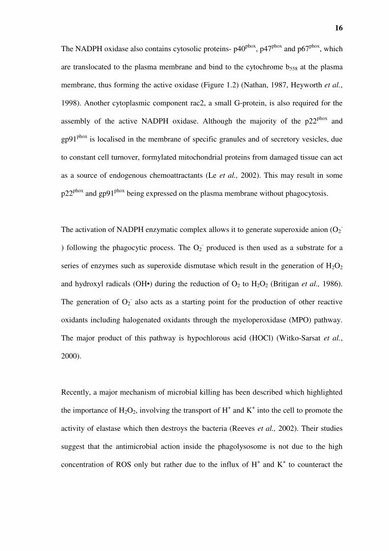
The NADPH oxidase also contains cytosolic proteins- p40phox, p47phox and p67phox, which
are translocated to the plasma membrane and bind to the cytochrome b558 at the plasma
membrane, thus forming the active oxidase (Figure 1.2) (Nathan, 1987, Heyworth et al.,
1998). Another cytoplasmic component rac2, a small G-protein, is also required for the
assembly of the active NADPH oxidase. Although the majority of the p22phox and
gp91phox is localised in the membrane of specific granules and of secretory vesicles, due
to constant cell turnover, formylated mitochondrial proteins from damaged tissue can act
as a source of endogenous chemoattractants (Le et al., 2002). This may result in some
p22phox and gp91phox being expressed on the plasma membrane without phagocytosis.
The activation of NADPH enzymatic complex allows it to generate superoxide anion (O2-
) following the phagocytic process. The O2- produced is then used as a substrate for a
series of enzymes such as superoxide dismutase which result in the generation of H2O2
and hydroxyl radicals (OH•) during the reduction of O2 to H2O2 (Britigan et al., 1986).
The generation of O2- also acts as a starting point for the production of other reactive
oxidants including halogenated oxidants through the myeloperoxidase (MPO) pathway.
The major product of this pathway is hypochlorous acid (HOCl) (Witko-Sarsat et al.,
2000).
Recently, a major mechanism of microbial killing has been described which highlighted
the importance of H2O2, involving the transport of H+ and K+ into the cell to promote the
activity of elastase which then destroys the bacteria (Reeves et al., 2002). Their studies
suggest that the antimicrobial action inside the phagolysosome is not due to the high
concentration of ROS only but rather due to the influx of H+ and K+ to counteract the

17
anions (ROS), this increase in ionic strength causes the release of cationic proteins,
elastase and cathespin G which consequently destroy the bacteria. Using mice lacking
elastase and cathespin G, they showed that these animals were ineffective at clearing
Staphylococcus. aureus and Candida albicans (Reeves et al., 2002). A commentary by
Bokoch, 2002, suggests that MPO might play a role in the buffering of proteases to
protect them from oxidation from H2O2 as opposed to a direct role in the killing of
microbial pathogens (Bokoch, 2002).
1.2.4 Neutrophils as a source of cytokines and chemokines
Recent interest has also focussed on the finding that neutrophils are capable of producing
a variety of cytokines (IL-1β, IL-1Ra, IL-12 and TNF), (Table 1.3) and chemokines such
as IL-8, growth-related gene product, macrophage inflammatory protein (MIP)-1α,
MIP1β and interferon-γ-inducible protein (IP-10) (Table 1.3) (Cassatella, 1999, Ethuin,
2005). These chemokines primarily act as chemotactic factors for neutrophils,
monocytes, immature dendritic cells (DC), and T-lymphocyte subsets (Ludwig et al.,
2006). The cytokines such as IL-1β and TNF are primarily inflammatory and cell
activating. In this manner neutrophils have the ability to contribute to cytokine levels
during physiologic and pathophysiologic processes. This may play an important role in
innate immunity and acute inflammatory conditions (Feldmann et al., 1996, Kmiec,
1998, Taylor, 2003).
18
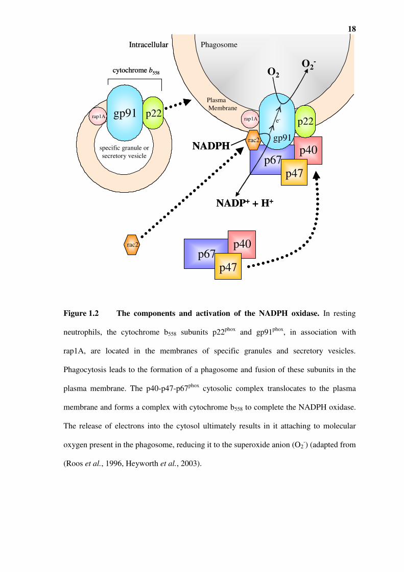
Figure 1.2 The components and activation of the NADPH oxidase. In resting
neutrophils, the cytochrome b558 subunits p22phox and gp91phox, in association with
rap1A, are located in the membranes of specific granules and secretory vesicles.
Phagocytosis leads to the formation of a phagosome and fusion of these subunits in the
plasma membrane. The p40-p47-p67phox cytosolic complex translocates to the plasma
membrane and forms a complex with cytochrome b558 to complete the NADPH oxidase.
The release of electrons into the cytosol ultimately results in it attaching to molecular
oxygen present in the phagosome, reducing it to the superoxide anion (O2-) (adapted from
(Roos et al., 1996, Heyworth et al., 2003).
gp91
p40p67
p47
rac2
O2
O2-
NADPH
NADP+ + H+
gp91 p22
Plasma
Membrane
PhagosomeIntracellular
rap1A rap1A
specific granule or
secretory vesicle
cytochrome b558
p22
rac2 p40p67
p47
e-
gp91
p40p67
p47
rac2
O2
O2-
NADPH
NADP+ + H+
gp91 p22
Plasma
Membrane
PhagosomeIntracellular
rap1A rap1A
specific granule or
secretory vesicle
cytochrome b558
p22
rac2 p40p67
p47
e-

19
Cytokine release by neutrophils occurs via secretion of pre-synthesised stores of
cytokines e.g. vascular endothelial growth factor (VEGF) and shedding of membrane-
bound cytokine such as the release of TNF under the action of TNFα converting enzyme
(TACE) (Ethuin, 2005). In addition, de novo protein synthesis such as the production of
IL-8 mRNA.
In the first instance the rapid release of cytokines via degranulation may be a significant
contribution in the first line of defence in cellular activation for microbial pathogen
elimination (Kasama et al., 2005), as well as influencing the processing and presentation
of antigen to lymphocytes and thus contributing to the nature of adaptive immune
response (Megiovanni et al., 2006). In chronic inflammation, neutrophils become
prominent in the exacerbation phases of diseases such as RA and atherosclerosis
(Gelderman et al., 1998, Lefkowitz and Lefkowitz, 2001). The neutrophils may hence be
a source of the pathogenesis-mediated processes in these chronic illnesses (Nambi, 2005,
Bathoorn et al., 2008)
1.2.5 Role in adaptive immunity
Although neutrophils are primarily recognised for their role in innate immunity, in recent
years there has been increasing evidence for their role in adaptive immunity. Currently,
there are three mechanisms by which neutrophils form a bridge between innate and
adaptive immunity. Firstly, neutrophils have been shown to produce chemokines that

20
attract DC and T cells to sites of inflammation and mediators that promote the cells’
adaptive immune responses (Ludwig et al., 2006). Neutrophils can also migrate to local
lymph nodes where they undergo apoptosis allowing DC to present neutrophil-derived
antigens to T cells. Secondly neutrophils have been shown to acquire antigen-presenting
function (Ferrante et al., 2007); and have even been reported to express T cell receptors
based on the variable immunoreceptor (Puellmann et al., 2006). The interaction of
neutrophils and DC during infection results in neutrophils inducing maturation of DC.
This process is mediated by TNF and the cellular contact is regulated by receptors such
as the neutrophil CD11b/CD18 and the C-type lectin receptor such as DC specific
intracellular-adhesion-molecule 3-grabbing non-integrin (DC-SIGN) (Ludwig et al.,
2006). Megiovanni et al. (2006) demonstrated through co-culture of neutrophils and
immature DC, that neutrophils were able to directly transfer Candida albicans antigens
to DC allowing them to stimulate sensitised T cells to produce IL-2 and IFNγ
(Megiovanni et al., 2006).

21
Table 1.3 Cytokines produced by neutrophils
Adapted from (Cassatella, 1999). Abbreviations: CINC, cytokine-induced neutrophil
chemoattractant; G-CSF, granulocyte colony-stimulating-factor; GROα, growth-related
gene product; IP, interferon-γ-inducible protein; KC, keratinocyte chemoattractant
(murine equivalent of GROα MCP, monocyte chemotactic protein; M-CSF, macrophage
CSF; TGFα, transforming growth factor; VEGF, vascular endothelial growth factor.
In vitro In vivo
a1001984
Text Box
a1172507
Text Box
NOTE: This table is included on page 21 of the print copy of the thesis held in the University of Adelaide Library.

22
1.3 MONOCYTES/MACROPHAGES
Macrophages belong to the mononuclear phagocyte system (MPS) which consists of
committed myeloid progenitor cells from the bone marrow that differentiate to form
blood monocytes, circulate in the blood and consequently enter tissues to become
resident tissue macrophages (Hume et al., 2002) (Figure 1.3). Proliferation and
differentiation into monocytes is dependent on the presence of lineage determining
cytokines such as colony-stimulating 1 (CSF-1 also known as macrophage colony-
stimulating factor), and granulocyte-macrophage colony-stimulating factor (GM-CSF),
as well as on interactions with stroma in haematopoietic organs.
During inflammation the first wave of neutrophil migration into tissues is followed by a
second wave of monocytes, typically occurring several hours after the initial
inflammatory insult, but persists in chronic inflammation when neutrophils are no longer
present (Schmid-Schonbein, 2006). Monocytes are more closely related to neutrophils in
regards to their anti-microbial systems. As such, monocytes respond to stimuli with a
brisk respiratory burst as in neutrophils, though its magnitude is much less than that of
neutrophils, which is further diminished in resident macrophages (Kumaratilake and
Ferrante, 1988). Recently it has been shown that the transcriptomes of macrophages and
neutrophils are clearly very similar. In vitro experiments have shown that in response to
various stimuli, granulocytes can be induced to adopt macrophage and DC-like
phenotypes (Araki et al., 2004, Lindemann et al., 2004). This inter-conversion while

23
likely, opposes the accepted dogma of neutrophils rapidly infiltrating, then
dying/removed and replaced by monocytes (Hume, 2006).
1.3.1 Origin and activation of macrophages
Macrophages have three major functions, which can be broadly classified as
phagocytosis, antigen presentation, and immunomodulation. These functions have been
further classified and attributed to the different subsets of macrophages, which have
developed as a result of different local stimuli (Figure 1.3).
a) Classically activated macrophages (M1) develop as a result of stimulation with
either IFN-γ or TNF or upon recognition of PPAR such as LPS, bacterial dsRNA
and other microbial products. This activation leads to increased production of NO
and ROS, thus facilitating their microbicidal and tumoricidal activities (Gordon,
2003). As this subset of macrophages contains ingested microbial products they
have an important function as APC inducing Th1 adaptive immune responses by
releasing cytokines such as, IL-1, IL-6, TNF, type I IFNs (IFNα/β), IL-10, IL-12
and IL-18 (Fujiwara and Kobayashi, 2005, Van Ginderachter et al., 2008).
b) Alternatively activated macrophages (M2a) develop from stimulation with IL-4
and/or IL-13 and tend to have an increased expression of mannose and scavenger
receptors and decreased inflammatory cytokine production (Gordon, 2003). These
macrophages have low NO production and are poor APC, but the presence of
receptors allows them to phagocytose debris, aid in wound healing and promote

24
Th2 responses by a high production of IL-10 and low production of IL-2
(Gordon, 2003, Fujiwara and Kobayashi, 2005, Van Ginderachter et al., 2008).
c) Type II activated macrophages (M2b) differentiate from immature macrophages
subsequent to the ligation to Fcγ receptors, TLR, CD40 or CD44 (van der Bij et
al., 2005, Van Ginderachter et al., 2008). M2b macrophages are less cytotoxic
and tend to promote the production of Th2 cytokine and antibody production
(Anderson and Mosser, 2002).
d) Deactivated macrophages (M2c) develop following stimulation with IL-10, TGF-
β, and glucocorticosteroids or after phagocytosis of apoptotic cells. MHC class I
and II expression is downregulated, while cytokine production is skewed towards
and anti-inflammatory profile thus, downregulating inflammation (Gordon,
2003).

25
Figure 1.3 Origin and activation of macrophages. Myeloid progenitor cells
undergo differentiation into monocytes/macrophages after stimulation with GM-CSF,
CSF-1 and IL-3. The presence of diverse environmental stimuli leads to differentiation
into different macrophage subsets with dissimilar functional characteristics. Adapted
from (van der Bij et al., 2005). Abbreviations: GM-CSF, granulocyte-macrophage
colony-stimulating factor; CSF, colony-stimulating factor; IL-13, interleukin 13.
a1172507
Text Box
NOTE: This figure is included on page 25 of the print copy of the thesis held in the University of Adelaide Library.

26
1.3.2 Functional surface receptors on mononuclear phagocytes
Mononuclear phagocytes like neutrophils express similar receptors that aid in the
recognition of microbial pathogens. Macrophages express Fc receptors that are known to
be important for mediating endocytosis, antibody-dependent cellular cytotoxicity and
triggering the release of inflammatory mediators (Mellman et al., 1988). Like
neutrophils, macrophages similarly express PRRs that recognise conserved microbial
structures such as PAMP, LPS of gram-negative bacteria and β-glucan of fungi. The
TLRs in macrophages unlike the PRRs that are involved in the binding and
internalisation of microbes are able to generate appropriate responses by activating
transcription factors and leading to the generation of inflammatory mediators (Brown and
Gordon, 2005, Moller et al., 2005). Phagocytosis of microbial pathogens is the primary
function of macrophages. This function is aided by the complement receptors CR1, CR2,
CR3, CR4 and a recently described Ig superfamily member, complement receptor Ig
(CRIg) (Helmy et al., 2006, van Lookeren Campagne et al., 2007). Macrophages also
possess various other receptors that play important roles in their response to infection but
are not involved in recognition of microbial pathogens. These include chemokines and
cytokine receptors that modulate a wide range of functions such as migration, adhesion
and antigen presentation (Brown and Gordon, 2005).
1.3.3 Antimicrobial function

27
Macrophages contain a spectrum of systems which play roles in the inactivation and
elimination of microbial pathogens. Many of these have similarities to those of
neutrophils (described above). Recognition through appropriate cell surface receptors
enables macrophages to contain microbial pathogens within phagocytic vacuoles. The
release of toxic metabolites, peptides and enzymes in the phagolysosome leads to
microbial killing. These processes of microbial recognition and killing are most effective
when the pathogen is coated with antibody and complement.
However, for some bacteria and parasites these recognition systems are not adequate.
Several pathogens can invade and multiply within the macrophages. This includes
Listeria (Cossart and Mengaud, 1989), Salmonella (Ohl and Miller, 2001), Mycobacteria
(Pai et al., 2003) Toxoplasma (Yarovinsky, 2008), Trypansoma cruzi (Maganto-Garcia et
al., 2008) and Leishmania (Alexander and Russell, 1985). To prevent the survival and
multiplication of these pathogens, macrophages need to undergo activation. This is best
achieved through the induction of cell-mediated immunity, involving the stimulation of
Th1 cells and the production of IFNγ, which is a recognised classical activator of
macrophages (Gordon, 2003, Fujiwara and Kobayashi, 2005).
Infiltration of macrophages into tissue sites can also be regulated by altered self-
molecules, autoantibodies, deposited antigen-antibody complexes and complement. The
very same processes which macrophages use to kill microbial pathogens are
unfortunately responsible for the tissue damage and long term loss of tissue and organ
function (Fujiwara and Kobayashi, 2005). The accumulation and activation of
macrophages in tissues is the hallmark of chronic inflammatory diseases such as RA,

28
atherosclerosis and granuloma formation in diseases such as tuberculosis (Brown and
Gordon, 2005).
1.4 INFLAMMATORY MEDIATORS
The propagation and resolution of inflammation is orchestrated by a network of a host of
soluble mediators such as, chemokines, complement, clotting factors, leukotrienes,
protease inhibitors and cytokines. These mediators act in unison to either stimulate or
downregulate cellular responses. In recent years, these have been intensively investigated
to identify some opportunities for targeting to prevent disease.
1.4.1 Arachidonic acid-derived mediators
The arachidonic acid cascade is activated by a number of inflammatory stimuli that result
in activation of phospholipase A2, cyclo-oxygenases (COX), and lipoxygenases (LOX).
Arachidonic acid metabolism results in the production of eicosanoids such as
thromboxanes, leukotrienes and prostaglandins (Kozik and Tweddell, 2006). Eicosanoids
are rudimentary hormones or mediators in their own right, regulating processes and
conditions such as pain, platelet aggregation, blood clotting, smooth muscle contraction,
leukocyte chemotaxis, inflammatory cytokine production (IL-1, IL-6 and TNF) and

29
immune function, in paracrine and autocrine manner (Tilley et al., 2001, Harris et al.,
2002).
1.4.2 Complement
The major effector of innate immunity is the complement system. The complement
system is made up of soluble proteins as well as complement receptors and regulatory
proteins. The soluble proteins constitute 5 % of the plasma; their major role is to
recognise and promote the clearance by phagocytosis of invading organisms or
damaged/altered host cells (Morgan and Harris, 2003). In innate immunity, complement
is activated via the alternative pathway by molecules such as techoic acid, LPS and
polysaccharide released during infection (Gasque, 2004, Nauta et al., 2004). This type of
immunity is also dependent on the lectin (mannose-binding) pathway for complement
activation by microbes with terminal mannose groups. Once the adaptive immune
response is set in motion, the antibody produced initiates the classical pathway to
activate complement. As a result of complement activation by all three systems, several
biological fragments are generated in a cascade manner (Sim and Tsiftsoglou, 2004).
Apart from components with anaphylatoxin and microbial membrane attack complex,
activities relevant to chemotaxis, C5a and opsonisation and phagocytosis, C3b, iC3b are
generated. Neutrophils and monocytes use receptors to these components to infiltrate
infection site, recognise and engulf bacteria (Gasque, 2004, Nauta et al., 2004, Sim and
Tsiftsoglou, 2004).

30
1.4.3 Nitric oxide (NO)
NO• is derived from the conversion of the amino acid L-arginine to L-citrulline by nitric
oxide synthase (NOS). NOS isoforms generally fall into two categories: (i) constitutive
NO synthases (NOS1 and NOS3) that are dependent on Ca2+/calmodulin and (ii)
inducible NOS (NOS2 or iNOS), the expression of which is increased by cytokines such
as TNF, and other inflammatory stimuli. The latter, is responsible for the role of NO in
inflammatory processes and is expressed by leukocytes, including macrophages and
neutrophils, and its activity is independent of [Ca2+]. NO• may affect diverse cellular
responses and can have both pro- and anti-inflammatory effects (Kubes et al., 1994).
Similar to ROS, unregulated NO• synthesis may have detrimental effects, as has been
suggested in sepsis and autoimmune diseases, where increased NO levels are related
increased pro-inflammatory cytokines (TNF, IL-6, IL-8) (Zamora et al., 2000, Bateman
et al., 2003). Again similar to ROS, NO• participates in diverse cellular signalling,
including regulation of plasma membrane receptors, endocytic pathways, GTP-binding
proteins, ion channels, transcription factors, and tyrosine kinases (Fialkow et al., 2007).
It is noteworthy that there are physiologically important interactions between NO• and
ROS such as O2 NO• can interact with O2−, thus acting as a biologic scavenger or
inactivator of ROS (Fialkow et al., 2007).

31
1.4.4 Cytokines
Cytokines are a large group of soluble proteins and peptides that act as intercellular
signalling molecules. They are inducible molecules produced in response to infection, to
become involved in regulation of inflammation, immunity, differentiation, proliferation
and many other functions. Although cytokines share common properties with hormones
they can be differentiated by the fact that hormones are made by specialised cells and
glands whilst cytokines are produced by a number of different cell types. Another
important distinction is the fact that cytokines often act locally while hormones act
systemically (Ethuin, 2005, Ferrante, 2005, Romagnani, 2005, Via, 2007). Many
cytokines are pleiotropic and in addition many have redundant activity. Cytokines can be
tentatively divided according to their functions (Table 1.4). Colony stimulating factors
(CSF) are cytokines that promote growth and development of haematopoietic cells and
are secreted by a variety of cell types which include fibroblasts, B cells and bone marrow
cells. Erythropoietin and thrombopoietin are also included with growth factors (Table
1.4).
Growth and differentiation factors for lymphoid cells include a number of ILs, molecules
capable of acting between leukocytes (Cameron and Kelvin 2003). These cytokines
regulate functions such as maturation, differentiation and growth factors of T cells, B
cells and NK cells (Table 1.4).
Chemokines are a group of chemotactic cytokines that have structural homology and
overlapping functions with cytokines. They are the only group that act on the

32
superfamily of 7-transmembrane G-protein-coupled receptors (Cameron and Kelvin
2003). While their main role is to guide leukocytes to inflammatory sites, they also
perform other functions such as T cell differentiation, wound healing, embryonic
development (Cameron and Kelvin, 2003, Via, 2007)
IL-1β, an inflammatory cytokine is mainly produced by monocytes and macrophages,
and to lesser amounts by cells such as neutrophils and endothelial cells. IL-1β is also
known as a pyrogenic cytokine, as very small doses of this cytokine induce fever in mice
and humans. IL-1 induces production of inflammatory cytokines and chemokines such as
IL-6 and neutrophilia (Romagnani, 2005). The other inflammatory cytokines including
TNF, IFNγ, IL-6, IL-12 and IL-16 are summarised and presented in (Table 1.4). TNF
will be the focus of this thesis and is described in greater detail below.
Anti-inflammatory and regulatory cytokines include IL-1 receptor I antagonist (IL-1Ra)
which binds competitively to IL-1Ra thus inhibiting IL-1α and IL-1β’s actions. It is
mainly produced by monocytes and macrophages and is a natural control mechanism for
IL-1’s pro-inflammatory activities. IL-4, when induced by microbial pathogens such as
Plasmodium falciparum, was shown to inhibit the anti-parasitic function of macrophages,
thereby allowing survival of pathogen (Kumaratilake and Ferrante, 1992). IL-10, a
powerful cytokine produced by regulatory T cells is a powerful suppressor of production
of IFNγ, IL-2, IL-3, TNF and GM-CSF by macrophages, neutrophils and Th1 cells
(Cools et al., 2007, Via, 2007). IL-10 is also known to down regulate the expression of
activating and co-stimulatory molecules on macrophages and DC; and upregulate the

33
expression of neutralising IL-1Ra and TNF receptor 2 (Romagnani, 2005). IL-13 is
another important regulatory cytokine which is known to have anti-inflammatory effects
by inhibiting production of IL-1, IL-6, IL-8 and chemokines MIP-1 and MCP. It is
mainly produced by activated T cells. IL-13 is also known to induce B cells to produce
IgE (Via, 2007).
TNF plays a key role in this cytokine network as all of the cytokine producing leukocytes
express TNF receptors. Thus neutrophils, macrophages, T cells, NK cells, B cells, mast
cells, eosinophils will all come under the influence of this cytokine. The rapid production
of TNF at inflammatory sites means that it is likely to be critical in both the initiation of
the innate and adaptive immune response (Getz, 2005, Ludwig et al., 2006)).

34
Table 1.4 Classification of cytokines based on major functional activities
Cytokines Predominant producer Comments
Growth factors for haemopoietic precursors, myeloid cells and thrombocytes
SCF, CSF B cells, epithelial cells, fibroblasts, bone marrow
cells
Promote growth and development
IL-3 Activated T cells, NK cells, endothelial cells, mast
cells
Supports growth and development of
hematopoietic cells
G-CSF, M-CSF;
GM-CSF
B cells, epithelial cells, fibroblasts, macrophages,
bone marrow stromal cells
Development of colonies from
granulocyte and monocyte-macrophage
precursors
EPO, TPO Kidney, liver Regulate production of erythrocytes and
thrombocytes
IL-5 Activated T cells Promotes growth and differentiation for
eosinophils and granulocytes
IL-11 Bone marrow stromal cells and mesenchymal cells Growth factor for thromobocytes
Growth and differentiation factors for lymphoid cells
IL-2 Activated T cells, NK cells, endothelial cells, mast
cells
Stimulates T and B cell growth
IL-4 Activated T cells, NK cells, endothelial cells, mast
cells
Proliferation and differentiation of
activated B cells
IL-7 Bone marrow stromal cells, intestinal epithelial cells Critical in maturation of immune system,
proliferation of T cells
IL-9 Activated Th2 cells Promotes the growth of mast cells, B
cells and other T cells
IL-15 Monocytes, fibroblasts, endothelial cells Acts as a T cell growth factor
IL-21 Activated CD4+ T cells Regulates NK and CD8+ T cell function
IL-27 Macrophages and dendritic cells Synergises with IL-2 to induce
IFNγ production by NK cells
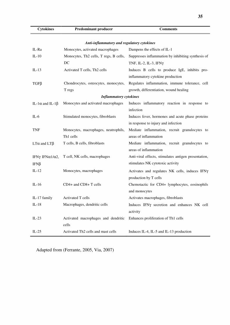
35
Cytokines Predominant producer Comments
Anti-inflammatory and regulatory cytokines
IL-Ra Monocytes, activated macrophages Dampens the effects of IL-1
IL-10 Monocytes, Th2 cells, T regs, B cells,
DC
Suppresses inflammation by inhibiting synthesis of
TNF, IL-2, IL-3, IFNγ
IL-13 Activated T cells, Th2 cells Induces B cells to produce IgE, inhibits pro-
inflammatory cytokine production
TGFβ Chondrocytes, osteocytes, monocytes,
T regs
Regulates inflammation, immune tolerance, cell
growth, differentiation, wound healing
Inflammatory cytokines
IL-1α and IL-1β Monocytes and activated macrophages Induces inflammatory reaction in response to
infection
IL-6 Stimulated monocytes, fibroblasts Induces fever, hormones and acute phase proteins
in response to injury and infection
TNF Monocytes, macrophages, neutrophils,
Th1 cells
Mediate inflammation, recruit granulocytes to
areas of inflammation
LTα and LTβ T cells, B cells, fibroblasts Mediate inflammation, recruit granulocytes to
areas of inflammation
IFNγ IFNα1/α2,
IFNβ
T cell, NK cells, macrophages Anti-viral effects, stimulates antigen presentation,
stimulates NK cytotoxic activity
IL-12 Monocytes, macrophages Activates and regulates NK cells, induces IFNγ
production by T cells
IL-16 CD4+ and CD8+ T cells Chemotactic for CD4+ lymphocytes, eosinophils
and monocytes
IL-17 family Activated T cells Activates macrophages, fibroblasts
IL-18 Macrophages, dendritic cells Induces IFNγ secretion and enhances NK cell
activity
IL-23 Activated macrophages and dendritic
cells
Enhances proliferation of Th1 cells
IL-25 Activated Th2 cells and mast cells Induces IL-4, IL-5 and IL-13 production
Adapted from (Ferrante, 2005, Via, 2007)

36
1.5 TUMOUR NECROSIS FACTOR (TNF)
TNF, a pro-inflammatory cytokine with pleiotropic effects, was initially described in
mice in the 1970s as a macrophage-derived product that caused haemorrhagic necrosis of
solid tumours (Carswell et al., 1975, Haranaka et al., 1986). Its cytotoxic effect towards
tumour cells gave rise to its name. Two functionally related species of tumour necrosis
factor exist, TNF and LT (TNFβ). The two forms have approximately 30 % sequence
homology. LT can exist either as a secreted LTα homotrimer that binds to the two
TNFR, or as a membrane-associated LTβ heterodimer composed of LTα and LTβ
chains. LTαβ binds to a unique LTβ receptor (Schneider et al., 2004, Schottelius et al.,
2004). LT is produced by stimulated T-lymphocytes while TNF is mainly produced by
stimulated macrophages. Newly synthesised TNF is initially displayed on the plasma
membrane, and is then cleaved in the extracellular domain by a transmembrane
metalloprotease enzyme- TNF converting enzyme (TACE) into a 157 amino acid, 17.3-
kDa soluble protein which oligomerises to form the active homotrimer (Moss et al.,
1997). Membrane bound TNF (mTNF) is biologically active and is presumed to mediate
the cytotoxic and inflammatory effects of TNF through cell-to-cell contact (Schottelius et
al., 2004). Recently mTNF has been shown to be essential in host defence mechanisms
against mycobaterical infection (Allie et al., 2008). The human TNF gene is located on
chromosome 6 and is translated as a 233 amino acid, 26-kDa precursor protein (Kriegler
et al., 1988). TACE is an adamlysin, which belongs to a class of membrane-associated
enzymes that contain both a disintegrin and matrix metalloprotease domains (ADAM)
(Kriegler et al., 1988). TNF production by macrophages is stimulated by a variety of

37
agents that includes bacterial LPS, bacterial toxins, viruses and fungi (Ferrante et al.,
1990, Beutler, 1992). TNF is secreted by a variety of cell types: immune cells (T-
lymphocytes, NK-cells, B-lymphocytes, macrophages, monocytes, mast cells,
neutrophils, basophils, eosinophils), non-immune cells (astrocytes, fibroblasts, glial cells,
granuloma cells, keratinocytes, neurons, osteoblasts, smooth muscle cells and many
kinds of tumour cells (Bazzoni and Beutler, 1996). However, macrophages and
monocytes serve as primary sources of TNF, especially during the inflammatory
responses (Beutler et al., 1985, Clark, 2007).
1.5.1 Regulation of TNF expression
Given that TNF plays a key role in both physiologic and pathophysiologic processes it is
not surprising that the expression and activity of TNF are tightly regulated at many
different levels. TNF gene expression is stimulated by a wide variety of agents. In
macrophages, expression is induced by stimuli that include viruses, bacterial and
parasitic products, tumour cells, complement, cytokines [such as IL-1β, IL-2, IFNγ and
GM-CSF], ischemia, trauma and irradiation (Bazzoni and Beutler, 1996). In other cell
types, other stimuli such as LPS, the cross-linking of immunoglobulins, ultraviolet light
and phorbol esters are effective in inducing TNF gene expression (Bazzoni and Beutler,
1996). Under resting conditions the amount of TNF in cells other than macrophages is
barely detectable. However, following stimulation, levels of TNF mRNA increase
rapidly, resulting in large quantities of soluble TNF being released. TNF expression is

38
also controlled at the post transcriptional level by adenosine-uridine rich elements
(AREs) (Chen and Shyu, 1995). AREs have been shown to destabilise heterologous
transcripts into which they are inserted and thus determine their translational efficiency.
Such sequences are usually common on the mRNA of pro-inflammatory cytokines (Han
and Beutler, 1990, Beutler and Brown, 1993, Chen and Shyu, 1995).
Recently, Ferrante and Ferrante (2005), have shown that the product of the 15-
lipoxygenase of the metabolism of arachidonic acid, 15-hydroperoxyeicosatetraenoic
acid inhibits the LPS-induced TNF production by macrophages occurs by decreasing the
stability of TNF mRNA (Ferrante and Ferrante, 2005). Downregulation of TNF
production has also been described to be due to cytokines such as IL-4 (Hart et al.,
1991), IL-10 (Rajasingh et al., 2006) and IL-13 (Manna and Aggarwal, 1998). Inhibition
of cytokine production including TNF occurs through the consumption of n-3
polyunsaturated fatty acids (Trebble et al., 2003).
1.5.2 Biology of TNF
The wide spectrum of biological effects of TNF has been appreciated for some time
(Table 1.5) (Schottelius et al., 2004, Rahman and McFadden, 2006). Its anti-cancer
activity is mediated through a direct effect on the malignant cells, inducing apoptosis or
indirectly by upregulating the immune system (Younes and Kadin, 2003). However, its
wider activity has been appreciated in defence against microbial pathogens on the one

39
hand and microbial pathogenesis in the other (Roilides et al., 1998, Ehlers, 2003). Even
more concerning has been the finding that the cytokine is a key player in the
pathogenesis of several chronic inflammatory diseases (Feldmann et al., 1996, Sekut and
Connolly, 1998) and indeed TNF has become a target for therapeutics (LaDuca and
Gaspari, 2001, Taylor, 2003).
The biological characteristics of TNF clearly support its role in physiologic and
pathophysiologic processes. Apart from its action on all leukocyte types, it stimulates
local tissue cells at the foci of infection or autoimmune reaction and the vascular
endothelium. The cytokine through its receptor stimulates transcription of key players of
the inflammatory reaction. The cytokine promotes extravasation of leukocytes by
upregulation/inducing the expression of cell adhesion molecules (CAMS) on endothelial
cells. These include ICAM-1, E-selectin and VCAM-1 (Jersmann et al., 2001). TNF will
also induce pro-coagulant activity and cytokine production in endothelial cells
(Bevilacqua et al., 1986). The stimulation of chemokines production in endothelial cells
and local tissue cells by TNF is a further mechanism by which it contributes to cellular
accumulation at inflammatory sites (Clauss et al., 2001, Varfolomeev and Ashkenazi,
2004).
Leukocyte stimulation by TNF can lead to major functional changes in these cells. The
cells show upregulation of receptors, production of cytokines, lipid mediator generation
and release of a host of products which promote microbial killing, inflammation and
tissue damage. Neutrophils are primed by TNF to exhibit enhanced antimicrobial activity
against bacteria, fungi and parasites (Ferrante, 1992) as well as tissue damage (Kowanko
40
and Ferrante, 1996, Witko-Sarsat et al., 2000). Pre-treatment of neutrophils with TNF
leads to migration inhibition, enhancement of FcγR and CR3 expression and associated
interaction with ligands for enhanced release of oxygen derived reactive species and
degranulation (Ferrante et al., 1988, Ferrante, 1992). Apart from stimulating cytokine
release from macrophages, TNF promotes the adaptive immune response by increasing
expression of MHC class II antigens and presentation of antigen by APC to T cells
(Varfolomeev and Ashkenazi, 2004).
At low concentrations, it acts in a paracrine and autocrine manner, upregulating vascular
adhesion molecules, activating neutrophils, and stimulating monocytes to secrete IL-1,
IL-6 and more TNF (Figari et al., 1987, Ferrante et al., 1988, Jupin et al., 1989). The
importance of TNF in host defence against pathogens was demonstrated through the use
of TNF neutralising antibodies (Skerrett et al., 1997). Similarly, a lack of TNF was
shown to impair host defence mechanisms severely (Nakane et al., 1988, Hauser et al.,
1990). Mice lacking the TNFRI gene (p55) while resistant to lethal doses of LPS or
enterotoxins, readily succumb to infections by Listeria monocytogenes, an intracellular
bacterial pathogen, despite having fully functional anti-microbial systems that generate
reactive oxygen radicals and reactive nitrogen intermediates (Pfeffer et al., 1993).
Interestingly, the re-expression of TNFRI on bone marrow derived cells was able to
control infection in these mice, showing that the expression of TNFRI is essential for the
killing of intracellular bacterial infections such as L. monocytogenes (Pfeffer et al., 1993,
Endres et al., 1997). Similarly, mice that have had their TNFRI gene disrupted show
increased susceptibility to infection (Rothe et al., 1993).

41
TNF has been demonstrated to regulate and/or interfere with adipocyte metabolism via a
number of mechanisms. These include transcriptional regulation, glucose and fatty acid
metabolism and hormone receptor signalling (Sethi and Hotamisligil, 1999). It has been
shown that TNF deficient mice exhibit lower circulating levels of free fatty acids and
triglycerides than the wild-types (Uysal et al., 1997). This reduction is thought to be
attributed to inhibition of lipoprotein lipase activity and expression of free fatty acid
transporters in adipose tissue by TNF (Cornelius et al., 1988, Memon et al., 1998). TNF
is therefore likely to contribute to the hyperlipidemia that is observed during infections
and obesity (Sethi and Hotamisligil, 1999).
1.5.3 Pathophysiologic actions of TNF
The role of TNF in the pathogenesis of a range of diseases is highly appreciated and as
such has been the focus of intense research. While some of the actions of TNF are
physiologically beneficial to the human body, especially in assisting in the defence
against microbial pathogens, prolonged or excessive production often becomes harmful
and impedes normal physiological processes. Localised action of TNF is clearly
important but production in excess leads to extensive diffusion into the circulation and
acts like an endocrine hormone, so displaying its pyrogenic properties, stimulating other
cytokines, activating the coagulation system, and suppressing bone marrow stem cell
maturation.
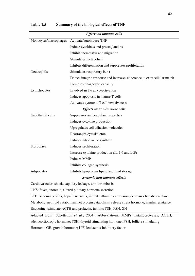
42
Table 1.5 Summary of the biological effects of TNF
Effects on immune cells
Monocytes/macrophages Activate/autoinduce TNF
Induce cytokines and prostaglandins
Inhibit chemotaxis and migration
Stimulates metabolism
Inhibits differentiation and suppresses proliferation
Neutrophils Stimulates respiratory burst
Primes integrin response and increases adherence to extracellular matrix
Increases phagocytic capacity
Lymphocytes Involved in T-cell co-activation
Induces apoptosis in mature T cells
Activates cytotoxic T cell invasiveness
Effects on non-immune cells
Endothelial cells Suppresses anticoagulant properties
Induces cytokine production
Upregulates cell adhesion molecules
Rearranges cytoskeleton
Induces nitric oxide synthase
Fibroblasts Induces proliferation
Increase cytokine production (IL-1,6 and LIF)
Induces MMPs
Inhibits collagen synthesis
Adipocytes Inhibits lipoprotein lipase and lipid storage
Systemic non-immune effects
Cardiovascular: shock, capillary leakage, anti-thrombosis
CNS: fever, anorexia, altered pituitary hormone secretion
GIT: ischemia, colitis, hepatic necrosis, inhibits albumin expression, decreases hepatic catalase
Metabolic: net lipid catabolism, net protein catabolism, release stress hormone, insulin resistance
Endocrine: stimulate ACTH and prolactin, inhibits TSH, FSH, GH
Adapted from (Schottelius et al., 2004). Abbreviations: MMPs metalloproteases, ACTH,
adenocortiotropic hormone; TSH, thyroid stimulating hormone; FSH, follicle stimulating
Hormone; GH, growth hormone; LIF, leukaemia inhibitory factor.

43
Figure 1.4 “Good versus Evil”: TNF, a critical component of effective immune
surveillance and immunity also promotes a wide range of inflammatory diseases.
Adapted from (Aggarwal et al., 2006).
TNF
•Immune surveillance
•Tumour regression
•Haematopoesis
•Protection from bacterial
infection
•Innate immunity
•Fever
•Psoriasis
•Crohn’s disease
•Allergic asthma
•Septic shock
•Inflammatory bowel disease
•Cystic fibrosis
•Tumorigenesis
•Lymphoproliferative disease
•Scleroderma
•Osteopororsis
•Alzheimer’s disease
•Diabetes (type II)
•Heart failure
•Atherosclerosis
•Hepatitis
•Multiple sclerosis
•AIDS
•Pruritic inflammatory mucocutaneaous disease
•SLE
•Rheumatoid arthritis
•Chronic juvenile arthritis
•Transplant rejection
•Atopic dermatitis
•Sarcodosis

44
Other events seen with further increases in TNF include myocardial depression,
hypotension (probably through induction of NO synthesis), and induction of
disseminated intravascular coagulation. During invasive bacterial infections, bacterial
LPS stimulates macrophages to produce excessive levels of IL-1 and TNF. Bacterial
septic shock is then triggered resulting in diffuse capillary leakage, hypotension,
myocardial suppression, and haemorrhagic necrosis of tissues, multiple organ failure and
death (Beutler, 1992).
Excessive production of TNF as seen in chronic disease states or major injury such as
burns, greatly alters homeostasis and results in cachexia, characterised by anorexia,
accelerated catabolism, extreme weight loss, anaemia and depletion of body tissues
(Beutler, 1992). Although TNF itself can produce cachexia in experimental animals,
other cytokines such as IL-1β (induced by TNF) are thought to contribute to the
cachectic state accompanying some diseases such as cancer (Beutler and Cerami, 1986).
The pathological actions of TNF are also manifested by upregulation in expression of
adhesion molecules on the endothelium. These adhesion molecules include E-selectin,
VCAM-1 and ICAM-1 (Pohlman and Harlan, 1989). Upregulation of these molecules
promotes the adhesion of cells to the endothelium (Carlos and Harlan, 1990) and can
result in disorders such as atherosclerosis (Ross, 1993).
While TNF is known to cause the death of tumour cells, the cytokine can also cause
apoptosis of normal tissues (Ferrante, 1992). In physiological terms, these cytotoxic

45
effects of TNF can result in extensive tissue damage, such as that seen in chronic
inflammatory disorders.
1.6 TNF RECEPTORS
TNF exerts its biological activity through its receptors, the 55-kDa TNFRI/CD120a and
the 75-kDa TNFRII/120b, both of which belong to the TNF receptor super-family
(Tartaglia and Goeddel, 1992). These receptors are classified as type I transmembrane
glycoprotein receptors and are characterised by the presence of multiple cysteine-rich
repeats of about 40 amino acids in their extracellular amino-terminal domain (Herbein
and O'Brien, 2000). TNFRI and TNFRII are present virtually on all cell types except for
red blood cells. TNFRI is more ubiquitously expressed, while TNFRII is found in
abundance on endothelial cells and haematopoietic cell lineage (Hohmann et al., 1989)
(Brockhaus et al., 1990).
TNF receptors can be shed to yield the soluble form which inhibit the effects of TNF by
reducing the number of binding sites on cells. Serum levels of TNF receptors are
increased in a large number of inflammatory diseases such as AIDS, rheumatoid arthritis,
heart failure and cancer (Gabay et al., 1997, Lee et al., 1997, Nozaki et al., 1997, Goetz
et al., 2002, Hunt and Grau, 2003, Herbein and Khan, 2008). Abnormalities in TNF
receptor shedding has been linked to autoimmune diseases such as multiple sclerosis
(Jurewicz et al., 1999).

46
1.6.1 TNFRI and TNFRII mediated signalling
TNFR is responsible for the majority of this cytokine’s biological actions. While both
TNFRI and TNFRII have cysteine-rich repeats in their extracellular domains, only
TNFRI has a death domain (DD) in its cytoplasmic tail. TNFRI mediates both cell
survival and death signals due to its DD, as opposed to TNFRII which primarily mediates
cell survival signals (Gupta, 2002).
It is widely accepted that TNFRI mediates the majority of TNF’s inflammatory
properties. The kinetics of association between TNF and its ligands and receptors show
that TNF associates more rapidly with TNFRI than with TNFRII. Many studies have
been carried out in order to define the exact role of TNFRII in mediating the effects of
TNF. Some investigators have proposed that TNFRII potentiates TNF-induced apoptosis.
(Tartaglia et al., 1993) suggested that TNFRII acts as a high-affinity trap of TNF that
delivers TNF to TNFRI i.e. serves as a “ligand passer” by delivering/passing TNF to
TNFRI for signalling when TNF concentrations are low (Figure 1.5). When TNF has
access to both receptors on the same cell the presence of TNFRII greatly enhances the
rate of association of TNF with TNFRI, thus in vivo TNFRII contributes to cytotoxicity
of TNF via TNFRI (Van Ostade et al., 1993). Weiss et al. proposed that the cytoplasmic
domain of TNFRII may be responsible for the potentiation of apoptosis (Weiss et al.,
1998).

47
Using TNFRII knockout mice Peschon et al., (1998), showed that in some cases TNFRII
acts as a decoy receptor since these knockout (KO) mice manifested an exacerbated
inflammatory response, with dramatically increased serum TNF levels, suggesting that
TNFRII may play a down-regulatory role in pulmonary responses. Similarly, mice
lacking TNFRI are more susceptible whereas TNFRII KO mice are resistant to infection
by L. monocytogenes. This resistance is conferred by activated macrophages, a function
which is dependent upon TNF and IFNγ (Pfeffer et al., 1993, Rothe et al., 1993). TNFRII
seems to have no intrinsic inflammatory properties of its own but potentiates the actions
of TNFRI (Erickson et al., 1994, Peschon et al., 1998), as discussed above.
The differences in signalling properties between TNFRI and TNFRII can also be related
structurally i.e. the intracellular domains of TNFRI shares greater homology with the
domains of the Fas rather than TNFRII, coupled with the lack of the death domain on
TNFRII. Recently it has been shown that due to the lack of a cytoplasmic death domain
(DD) on TNFRII, binding of TNF to TNFRII results in the recruitment of TNFR
associated factor 1 (TRAF1) and TRAF2 to the cytoplasmic portion of TNFRII
independently of TNFR associated death domain (TRADD). This consequently leads to
the recruitment of cellular inhibitor of apoptosis proteins (cIAP)-1 and cIAP-2 (Deveraux
et al., 1999). These proteins are known to directly bind to caspases 3 and 7 hence
preventing the activation of caspase 9 (Deveraux et al., 1999). Pimentel-Muinos and
Seed (1999) demonstrated that in the Jurkat T-cell line, in the presence of receptor
interacting protein (RIP), TNFRII triggers apoptosis, whereas in the absence of RIP,
TNFRII activates nuclear factor kappa B (NF-κB) (Pimentel-Muinos and Seed, 1999).

48
However, the requirement of RIP seems to be distinct among different cell types. In
fibroblasts, RIP is essential for TNFRI mediated NF-κB activation but not for apoptosis.
In contrast, in T cells, RIP is required for TNFRI dependent NF-κB activation and
apoptosis and for TNFRII mediated apoptosis but not TNFRII mediated NF-κB
activation (Kelliher et al., 1998, Pimentel-Muinos and Seed, 1999). These studies
support the role of TNFRII as a more potent regulator of apoptosis, rather than serving an
accessory role as originally proposed.
1.6.2 TNFR adaptor proteins
The cytoplasmic domain of TNFRI and TNFRII do not have any intrinsic signalling
capability. Hence, they recruit cytoplasmic molecules known as “adaptor proteins” to the
cytoplasmic domain of the receptors to bind to downstream signalling molecules. The
binding of TNF to TNFRI leads to the release of the inhibitory protein, silencer of death
domain (SODD) from the receptor’s intracellular domain, and this leads to the
recruitment of the important adaptor protein, TRADD. This facilitates the recruitment of
additional adaptor proteins which include RIP, TRAF-2 and Fas-associated death domain
(FADD) (Chen and Goeddel, 2002) (Figure 1.5). As above, TNFRII recruits TRAF2
independently of TRADD. These adaptor proteins in turn recruit additional key pathway-
specific enzymes to the TNFRI such as procaspase-8 [also called FADD-like IL-1β
converting enzyme (FLICE)] by interaction via homologous death effector domain
(DED) to form a death-inducing signal complex (DISC) (Gupta, 2002).

49
TRADD
TRAF2
FADD
GCK
RIP
IKK complex
NIK
Iκκκκ Bαααα
NF-κκκκ B
pIκκκκB
MKK4/7
JNK
ASK1MEKK1
MKK3/6
p38
FLICE
Caspase 3
Caspase 9
Cytochrome C
Apaf 1
Mitochondria
MEK1/2
ERK1/2
c-Raf
Ras
SOS
Grb2
Bid
tBid
Apoptosis
TNFRITNFRII
DD
Soluble TNFMembrane -bound
TNF
Ligand passingShedding
Degradation
Caspases
NFκκκκB
FANCeramide
CAPK
N-SMase
A-SMase
Secretory proteins
Endosome
Sphingosine
SphK1/2
SIP
ceramide
Figure 1.5 Signalling pathways activated by TNFRI and TNFRII. Adapted from
(Kronke, 1999, Gupta, 2002, MacEwan, 2002, Aggarwal et al., 2006). The highlighted pathways
emanating from TNFRII are not fully understood yet, and the above represents possible
mechanisms of biological responses. Not every signalling pathway depicted is activated in all cell
types by TNF. Abbreviations: A-SMase, acid sphingomyelinase; ASK1, apoptosis signal-
regulating kinase; CAPK, ceramide activated protein kinase; DD, death domain; ERK1/2,
extracellular-signal-regulated kinases; FAN, factor associated with N-SMase activation; FADD,
Fas-associated DD; FLICE, FADD-like IL-1β converting enzyme; MEK1/2, MAPK/ERK kinase,
GCK, germinal centre kinase; IKK, IκB kinase; MEKK1, MAPK kinase kinase; NFκB, nuclear
factor kappa B; NIK, NF-κB inducing kinase; IκB, inhibitor kappa B; N-SMase, neutral
sphingomyelinase; RIP, receptor interacting protein; TRADD, TNFR associated DD; TRAF2,
TNFR associated factor 2.

50
During DISC formation, procaspase-8 is activated by self-cleavage and initiates a
protease cascade that leads to apoptosis (Chen and Goeddel, 2002, Gupta, 2002). The
complex of adaptor molecules is also responsible for coupling TNFRs to signalling
pathways such as the MAPKs, the inhibitor of kappa B (IκB) kinase-nuclear factor kappa
B (NF-κB) pathway and the sphingomyelinase-ceramide pathway, which are responsible
for mediating the actions of TNF (Gupta, 2002).
1.7 SIGNALLING PATHWAYS ACTIVATED BY TNF
1.7.1 Activation of NF-κκκκB
The activation of transcription factor NF-κB plays an important role in the regulation of
the immune response and inflammation (Baeuerle and Henkel, 1994). Recent studies
have increased our knowledge and understanding of this component of the TNF
signalling network (Locksley et al., 2001, Ghosh and Karin, 2002). TNF can activate the
NF-κB pathway via the classical or non-canonical mechanism. The classical pathway
requires the activation of IKK/NEMO (see below), whereas the non-canonical pathway
can be activated independently (Karin, 2006, Niederberger and Geisslinger, 2008).
Although the enzymatic activity of RIP is not required for TNF-induced activation of
NF-κB, knockout studies (Rip-/- mice) have shown that RIP is essential for the activation
of this transcription factor in fibroblasts (Kelliher et al., 1998). In the resting cell, NF-κB
resides in the cytoplasm through an interaction with the inhibitory protein, inhibitor of

51
kappa B (IκB). Identification of the multiprotein, IκB kinase (IKK) complex which
mediates the phosphorylation of IκB in a TNF-dependent manner, was a significant step
in the understanding of NF-κB signalling (Ghosh and Karin, 2002). The IKK complex
contains two catalytic subunits, IKKα and IKKβ, and a regulatory subunit, NF-κB
essential modulator (NEMO or IKKγ) (Karin and Lin, 2002). The IKK complex also
contains a kinase-specific chaperone consisting of Cdc37 and Hs90 which play a role in
the shuttling of the complex from the cytoplasm to the membrane (Chen and Goeddel,
2002). The phosphorylation of two sites at the activation loop of IKKβ mediates NF-κB
activation by TNF, while the IKKα subunit mediates activation by LT (Delhase et al.,
1999). The NF-κB inducing kinase (NIK) has been reported to stimulate the activation of
IKKβ. Upon phosphorylation, IκΒ is proteolytically degraded by 26S proteasome,
resulting in the release of NF-κB, which translocates to the nucleus to regulate gene
transcription (Garrington and Johnson, 1999).
It has also been recently reported that Akt, a serine-threonine kinase is similarly capable
of phosphorylating IκB (Ozes et al., 1999). Akt is a kinase which is downstream of
phosphatidylinositol-3-kinase (PI3K) which is also activated by TNF (Vanhaesebroeck
and Alessi, 2000). The PI3-kinase/Akt pathway is thought to be important in protecting
cells from nonapoptotic necrotic cell death (Shaik et al., 2007, Zhou et al., 2008). PI3K
phosphorylates the 3’-OH position of the inositol ring in inositol phospholipids,
generating 3’-phosphoinositides (PI). Inside cells, this produces two lipid products: PI3P,
and PI (3,4,5) P3 which subsequently breaks down to PI (3,4) P2 (Vanhaesebroeck and
Alessi, 2000). PI (3,4) P2 and PI(3,4,5)P3 are crucial for activation of Akt

52
(Vanhaesebroeck and Alessi, 2000). Accumulation of the 3’ phosphorylated PIs results in
the activation of phosphoinositide dependent kinase (PDK1), which phosphorylates and
activates Akt on tyrosine 308. Full activation of Akt requires phosphorylation on serine
473 by another kinase, most likely mammalian target of rapamycin (mTOR) (Wang et
al., 2008) thereby allowing Akt to activate IκB in turn (Ozes et al., 1999).
1.7.2 Activation of sphingomyelinase-ceramide by TNFRI
Ceramide is a neutral sphingosine-based lipid signalling molecule that regulates cellular
differentiation, proliferation and apoptosis (Pushkareva et al., 1995, Spiegel et al., 1996).
Due to its neutral lipid nature ceramide does not distribute in the cytosol but rather
remains within the membrane bilayer (Kronke, 1999). Ceramide is produced by the
hydrolysis of the phosphodiester bond of sphingomyelin (by sphingomyelinases
(SMases), which results in the formation of ceramide and phosphorycholine. There are
two distinct forms SMases, a membrane-associated and soluble neutral (N-) SMase and
an acidic form (Wiegmann et al., 1994, Liu and Anderson, 1995).
Studies have shown that through the binding to TNFRI, TNF rapidly activates both forms
of SMases resulting in the generation of ceramide. By using mutant TNFRI, (Wiegmann
et al., 1994) showed that the activation of A-SMase is signalled through the carboxyl
terminus containing the DD, whereas, N-SMase is activated through a cytoplasmic
portion of the TNFRI separate from the DD. This portion contains a motif of 11 amino

53
acids known as neutral sphingomyelinase domain (NSD) (Adam et al., 1996). TNF
causes the rapid, within seconds, activation of N-SMase in most cells. This activation has
been demonstrated to occur through the WD-repeat protein, Factor Associated with N-
SMase activation (FAN), which specifically binds to the NSD region of TNFRI which
results in the coupling of NSD to N-SMase (Adam-Klages et al., 1996). The release of
ceramide results in the activation of ceramide-activated protein kinase (CAPK) (Mathias
et al., 1991). CAPK then phosphorylates Raf-1 kinase, which stimulates a dual
specificity MAPK/extracellular-signal-regulated kinases (ERK) kinase (MEK)-1 and
MEK-2, leading to the downstream activation of ERK1/ERK2 (Yao et al., 1995, Zhang
et al., 1997).
The activation of A-SMase was identified to occur through the association of TNFRI DD
with adaptor proteins TRADD and FADD (Schwandner et al., 1998). This group showed
that neither TRAF2 nor RIP were required for A-SMase activation while overexpression
of TRADD and FADD in HEK293 cells enhanced TNF-induced A-SMase activation. It
is believed that these adaptor proteins link the TNF-TNFRI complex to the A-SMase in
the endosomal-lysosomal compartment. A-SMase is thought to couple the TNFRI to the
secretory pathway and to apoptosis via protein-protein interaction known as the caspase
cascade and thus plays an active role in cytokine-induced apoptosis (Deiss et al., 1996,
Kronke, 1999, Carpinteiro et al., 2008). Unlike most cell types TNF does not stimulate
the activity of the SMases in neutrophils (Robinson et al., 1996).
54
1.7.3 Activation of sphingosine kinase by TNF
As discussed above, TNF stimulates the breakdown of sphingomyelin to produce
ceramide, which is subsequently metabolised to the single sphingoid chain, sphingosine
(Melendez, 2008). Sphingosine can be phosphorylated by sphingosine kinases (SphKs)
to generate sphingosine-1-phosphate (S1P). Two mammalian SphKs have been
characterised thus far, SphK1 and SphK2 which phosphorylated erytho-sphingosine,
dihydrosphingosine and phytosphingosine. There is increasing evidence that this
bioactive phospholipid, S1P, acts as either an extracellular or intracellular second
messenger (Melendez, 2008). While its role as an extracellular mediator has been
reported during its release by activated platelets (English et al., 2000), its role as
intracellular mediator is more defined. S1P, like other intracellular messengers increases
cytosolic Ca2+ in immune cell activation mediating a wide range of biological effects
such as proliferation, differentiation, chemotaxis, cytokine/chemokines generation and
apoptosis (Niwa et al., 2000, MacKinnon et al., 2002, Melendez, 2008). Specifically,
increases in S1P levels have been shown to be increased by agents that activate MAPK
pathways and increased expression of adhesion molecules. Recently using a SphK
inhibitor, it was shown that fMLP and TNF-stimulated superoxide production in
neutrophils can be regulated by SphK (Niwa et al., 2000). SphK1 activity has also been
implicated in macrophage properties of wound healing (English et al., 2001),
mobilisation of Ca2+ for the monocytic NADPH oxidase activity (Melendez et al., 1998),
and adhesion molecule expression by endothelial cells (Kee et al., 2005). However,
recently two independent studies using either SphK1 or both SphK1 and SphK2 KO
mice, reported normal inflammatory cell recruitment during thioglycollate, KC, MIP-2 or

55
LPS-induced peritonitis, collagen-induced arthritis and normal responses to fMLP by
neutrophils (SphK KO mice), while a SphK2 KO showed an accelerated bacterial lung
inflammation (Michaud et al., 2006, Zemann et al., 2007). These two studies highlight
the need for further research into the exact role of S1P in inflammation as the in vitro
observations do not mirror the KO studies, suggesting SphK’s role inflammation may not
be as essential as previously thought, perhaps due to redundancy in the system.
1.7.4 Activation of MAPK
In mammalian cells at least three MAPK have been identified, the classical MAPKs,
ERK1 and ERK2, and the more recently described c-Jun NH2-terminal kinase JNK
1/JNK2 and p38 MAPK pathways. MAPK have been implicated in regulating a range of
biological functions, including cell proliferation, differentiation and death, expression of
adhesion molecules, and cytokine production (Seger and Krebs, 1995, Kyriakis and
Avruch, 2001). Studies have shown that TNF activates all three MAPKs (Karin, 1998,
Davis, 1999).
1.7.4.1 Extracellular-signal-regulated kinase (ERK)
The ERK pathway comprises the greatest assembly of proto-oncogenes in any known
signalling pathway (Kolch, 2002). This includes ligands such as sis and TGFα, tyrosine

56
kinase receptors (neu/erbB2, fms, kit), G-proteins (Ha-ras, Ki-ras, N-ras), kinases (raf,
mos and tpl2/cot) and nuclear transcription factors (fos, ets, myc). Tyrosine kinases such
as ErbB2 is often overexpressed in human tumours and B-raf mutated in 60% of
melanomas (Davis, 1999). Hence ERKs are primarily involved in mediating responses to
mitogenic signals (Boldt and Kolch, 2004). In addition, they are involved in
proliferation, differentiation, apoptosis and metabolism (Dhillon and Kolch, 2002).
Several isoforms of ERK have been described e.g. ERK1, ERK2, ERK3, ERK4, ERK5
and ERK7 (Pearson et al., 2001). ERK1 and ERK2 are proteins of 43 and 41 kDa
respectively that are nearly 85 % identical overall, with much greater identity in the core
regions involved in binding substrates (Boulton et al., 1990, Boulton et al., 1991). Both
are ubiquitously expressed, although their relative abundance in tissues is variable. In
immune cells for example ERK2 is the predominant species, while in cells of
neuroendocrine origin these are equally expressed (Pearson et al., 2001). They are
activated by serum, growth factors, certain stresses, cytokines, and ligands for G-protein-
coupled receptors, adhesion signals and transforming agents (Pearson et al., 2001, Boldt
and Kolch, 2004). Receptor mediated stimulation of ERK1/ERK2 activity requires the
activation of ras, protein kinase C (PKC) or PI3K. These act on raf-1, a serine/threonine
kinase which phosphorylates and activates the dual specificity of MAPK/ERK kinase
(MEK) 1 and MEK2 (Pearson et al., 2001). The MEKs phosphorylate the tyrosine and
threonine residues on the TEY activation motif of ERK1/ERK2, resulting in the
activation of ERK1/ERK2. Activated ERK1/ERK2 phosphorylate their substrates e.g.
Elk1, Ets2 on serine or threonine residues in the PXS/TP consensus phosphorylation
motif where P = proline, X = any amino acid and S/T = serine/threonine.

57
1.7.4.2 c-Jun NH2-terminal kinases (JNK)
JNKs are a family of serine/threonine kinases which phosphorylate c-Jun in its N-
terminal transactivation domain at serine residues 63 and 73 (Liu and Lin, 2005). JNK
activation is controlled by MEKK1 which phosphorylates MKK4 and MKK7. The
activated MKK4/MKK7 subsequently phosphorylate JNK isoforms on the TPY motif
resulting in their activation (Yan et al., 1994, Weston and Davis, 2002). JNK consists of
at least 10 isoforms created from the alternative splicing of three distinct genes: JNK1,
JNK2 and JNK3. JNK1 and 2 are ubiquitously expressed, whilst JNK3 is predominantly
expressed in the brain (Gupta et al., 1996, Dreskin et al., 2001, Cui et al., 2005). Two
alternative splice sites present in JNK1 and JNK2 give rise to the p46 isoforms, JNK1α1,
JNK1β1, JNK2α1 and JNK2β1 and the p54 isoforms, JNK1α2, JNK1β2, JNK2α2, and
JNK2β2 (Dreskin et al., 2001). JNK substrates include transcription factors c-Jun and
ATF2 (Kyriakis and Avruch, 2001). Depending on the cell type and agonist involved,
JNK may regulate gene expression, the stability of target proteins, cell transformation
(Antonyak et al., 1998), T-cell function (Dong et al., 2000), apoptosis (Hirata et al.,
1998), cell proliferation (Wisdom et al., 1999) and tissue regeneration (Brenner, 1998).
Scaffold proteins regulate the specificity in the targets and cellular function of JNK
signalling. There are four groups of JNK scaffolds known to date namely: JNK-
interacting protein (JIP), JNK/SAPK associated proteins (JSAP), filamin, p130CAS and
β-arrestin (Manning and Davis, 2003). These scaffolds then assemble different external

58
stimuli e.g. filamin mediates JNK activation by the TNFR while β-arrestin links JNK to
G-protein couples receptors (Boldt and Kolch, 2004).
JNK is mainly known to promote apoptosis through its phosphorylation of proapoptic
proteins such as BAD or BH3 domain proteins Bim and Baf (Donovan et al., 2002, Lei
and Davis, 2003). However, observations such as the increase in liver cell death
apoptosis seen in MKK4 knockout mice, clearly suggests a protective role of JNK
(Nishina et al., 1999). JNK activation is also believed to promote insulin resistance in
diabetes by blocking the phosphorylation of serine 307 on insulin-receptor substrate 1
(Aguirre et al., 2000).
1.7.4.3 p38 kinase
The p38 MAPK family are serine-threonine kinases, that are activated by environmental
stresses such as UV irradiation, heat or osmotic stress but which can also be triggered by
products of microorganisms such as endotoxins and pro-inflammatory cytokines such as
IL-1 or TNF (Lee et al., 1994). Hence it has a role in innate immunity (Guo et al., 2003).
p38 is not only involved in cytokine induction but its activation is induced by
inflammatory cytokines. The role of p38 in innate immunity was reinforced by the
discovery that SB203580 inhibited the release of TNF or IL-1 from endotoxin-stimulated
monocytes (Lee et al., 1994). Subsequently, p38 was shown to have four isoforms: p38α,
p38β, p38γ and p38δ (Lee et al., 1994, Stein et al., 1997, Wang et al., 1997, Young et

59
al., 1997). The p38α and p38β are expressed in most tissues; p38γ is mainly expressed in
skeletal muscle and in normal and diseased cardiomyocytes, p38δfound in several adult
tissues and during development (Kumar et al., 2003). Activation of p38 occurs through
the dual phosphorylation of the threonine and tyrosine residues in the TGY motif by the
upstream MAPK kinases, MKK3 and MKK6, which subsequently phosphorylate a small
heat shock protein 27 (Freshney et al., 1994, Hii et al., 1998).
From an inflammatory aspect, p38α is most commonly expressed in the different
inflammatory cell lineages i.e. monocytes/macrophages, neutrophils and CD4+ T cells.
The p38 inhibitor SB203580, which has been shown to inhibit the production of
inflammatory cytokines such as TNF and IL-1β, inhibits the activation of the p38α and
p38β isoforms (Lee et al., 1994, Lee et al., 2000, Kumar et al., 2003). p38 regulates gene
expression by four different general mechanisms:
a) via the phosphorylation of transcription factors e.g. activation transcription
factors 2 and 6 (ATF2/6) and myocyte enhancing factor 2C (MEF2C) (Lee et al.,
2000)
b) by regulating mRNA stability via the action of downstream kinases such as the
MAPK-activated protein kinase 2 (MAPKAP K2). Messages for IL-2,
cyclooxygenase-2 (COX-2) and other inflammatory mediators is short lived due
to the presence of AU-rich elements in the 3’ untranslated region of the message
that associate with AU-binding proteins that destabilise the message (Caput et al.,

60
1986, Shaw and Kamen, 1986). Phosphorylation of these proteins by p38 results
in increased message stability (Winzen et al., 1999, Ming et al., 2001).
c) regulate mRNA translation into protein via MAPKAP K2 which also control
translation and production of mediators e.g. TNF by phosphorylating AU-binding
proteins (Kumar et al., 2003).
d) promote the phosphorylation of histone H3 in chromatin at NF-κB binding sites
of certain genes. The activity of this histone is required for successful
transcriptional induction of IL-8 and monocyte chemotactic protein 1 (MCP-1) by
NF-κB (Saccani et al., 2002).
The roles of p38 MAPKs in physiological responses are multifaceted, and these include
processes such as cellular differentiation, myogenesis and cellular infiltration into
inflammatory sites.
1.7.4.4 Summary
Although TNF-induced activation of MAPKs has been studied intensively in terms of
biological effects, the molecular mechanisms that regulate the activation of these MAPKs
are still incompletely understood. Studies have shown that TRAF2 is essential for TNF-
induced activation of JNK, ERK1/ERK2 and p38 (Liu et al., 1996, Reinhard et al., 1997,
Yeh et al., 1997, Liu and Han, 2001). Binding of TNF to TNFRI activates the MAPK
kinase kinase kinase kinase (MKKKK) known as the germinal centre kinase (GCK)

61
through TRAF2 and TRAF-associated NF-κB activator (TANK). TANK is thought to
increase the affinity of TRAF2 for GCK, thus increasing MAPK activation by TNF. This
ultimately leads to the activation of MAPK kinase 1 (MEKK) 1 (Chin et al., 1999,
Chadee et al., 2002). Although both GCK and MEKK1 interact with TRAF2, and GCK
is required for MEKK1 activation by TNF, GCK kinase activity does not appear to be
required for MEKK1 activation (Chadee et al., 2002). It has been shown that GCK
activates MEKK1 by causing MEKK1 oligomerisation and autophosphorylation (Chadee
et al., 2002). Once activated, MEKK1 activates the downstream MAPK kinase kinase
(MKK) 4 and MKK7, leading to JNK activation with downstream effects on growth,
differentiation, survival and apoptosis (Garrington and Johnson, 1999) (Figure 1.5).
Recently, using (Rip-/-) fibroblasts, it was shown that RIP is required for TNF-induced
activation of all three MAPKs, while the enzymatic activity of the kinase is only required
for the activation of ERK, but not JNK and p38 (Devin et al., 2003). Using SB203580 an
inhibitor of the enzymatic activity of p38, it was shown that TNF stimulated IL-8
production and superoxide generation from human neutrophils were completely
abolished (Zu et al., 1998). fMLP induced chemotaxis and superoxide production were
also markedly suppressed by the p38 inhibitor.
1.8 TARGETING TNF-TNFR IN PATHOGENESIS
Owing to its ability to kill tumour cells, TNF held promise as a potent anti-tumour agent.
Extensive studies have demonstrated direct cytostatic and cytotoxic effects of TNF

62
against subcutaneous human xenografts and lymph node metastases in nude mice, as well
as immunomodulatory effects to promote tumour cell killing by various cell types,
including neutrophils, macrophages and T-cells (Ohnuma et al., 1993). On the other
hand, TNF has also been shown to protect haematopoietic progenitors against irradiation
and cytotoxic agents, a property which could be applied to aplasia induced by
chemotherapy or bone marrow transplantation (Warren et al., 1990).
It is interesting that TNF can initiate two opposing actions, proliferation versus
apoptosis. This is likely to be due to the signalling pathways which are activated via the
TNF receptors. The IKK-NFκB pathway is believed to promote cell survival whereas the
Fas-like IL1-converting enzyme (FLICE -caspase 8), sphingomyelinase-ceramide and
JNK pathways are likely to mediate apoptosis. Thus, it has been shown that in the
absence of NF-κB activity, susceptibility to TNF-induced apoptosis increases (Beg et al.,
1995, Li et al., 1999) whereas enforced activation of NF-κB protects against apoptosis
(Chen and Goeddel, 2002). The type of response is very much dependent on the cell type
since it has been argued that not only do different cell types express different levels of
the various signalling molecules but, the signalling pathways to which receptors are
coupled to are also dependent on the cell-types (Nutchey et al., 2005). For example, TNF
has been widely demonstrated to activate the sphingomyelinase-ceramide pathway in
cell-types such as HL-60 and U937 cells but this effect is not seen in neutrophils
(Robinson et al., 1996), as discussed above.
The ability of TNF to enhance the killing of micro-organisms by neutrophils suggests
that it could be useful as a natural, non-specific immune enhancer especially in

63
immunocompromised patients (Ferrante et al., 1995). During the late 1980’s, there were
many phase 1 trials of recombinant TNF in patients with advanced cancer, in particular
in the regional treatment of sarcomas and melanomas (in conjunction with anti-neoplastic
agents) (Spriggs et al., 1988). Despite the large number of trials being conducted, these
failed to demonstrate significant improvements in cancer treatment, with TNF resistance
and TNF-induced systemic toxicity being two major limitations for their use. The most
common systemic side effects included fever, rigors, headache, fatigue, myalgia, nausea
and vomiting (Schottelius et al., 2004).
Due to its protein-nature TNF has to be administered subcutaneously, intramuscularly or
intravenously at doses ranging from 0.04 to 13 mg/kg, with skin ulceration and necrosis
at the injection site not uncommon at the higher doses (Zamkoff et al., 1989). Thus, the
many pathophysiological properties associated with a pleiotropic molecule such as TNF
make it highly unsuitable for use in this manner unless the beneficial properties of the
molecule can be harnessed, eliminating the risk of pathological damage.
1.8.1 Anti-TNF antibodies and soluble TNF receptors
The use of TNF inhibitors in inflammatory diseases, such as rheumatoid arthritis, where
the cytokine is found in copious amounts in the synovial fluid of patients, has been
largely successful. At present, the only drugs that are in clinical practice or in clinical
trials to block TNF are biologicals, protein-based drugs, either antibody to TNF or based

64
on TNF receptors (e.g. linked to Fc dimmers) (Table 1.6). Table 1.7 shows a list of
diseases which have been treated with this anti-TNF approach. Although these inhibitors
have the major advantage of specificity they also have significant disadvantages such as
the need for repeated injection and their relative high cost compared to small organic
chemical drugs (Breedveld, 2000, Tugwell, 2000).
The rationale behind the use of anti-TNF therapy in RA is evident in the following:
a) In the joints of the inflamed synovium and the destructive pannus-cartilage
junction TNF/TNFR expression is upregulated (Chu et al., 1991, Deleuran et al.,
1992, Gabay et al., 1997)
b) When anti-TNF antibodies were used to neutralise inflammatory cytokines in
rheumatoid synovial cultures, the results were striking. IL-1 mRNA levels were
reduced and within 3 days, IL-1 bioactivity had virtually disappeared. Thus it has
been concluded that TNF was the major stimulus inducing IL-1 synthesis in RA
synovial cultures (Brennan et al., 1989, Feldmann et al., 1996).
c) Anti-TNF antibodies ameliorate inflammation and joint destruction in murine
collagen-induced arthritis (Piguet et al., 1992, Wooley et al., 1993).
Infliximab (Table 1.6) is a chimeric human IgG1 constant region/mouse V region anti-
TNF monoclonal antibody which neutralises human TNF but not LT. The ability of
infliximab to neutralise TNF both in vitro and in vivo has been well established (Elliott et
al., 1994a, Elliott et al., 1994b, Sekut and Connolly, 1998). Infliximab works by binding
to soluble TNF monomers, trimers and membrane-bound TNF to form a stable complex
which prevents TNF from binding to its receptor thus blocking its action (Geletka and St

65
Clair, 2005). The in vivo TNF-neutralising action of infliximab may be enhanced due to
its ability to activate complement, probably via its gamma-1 heavy chain, and thus to kill
cells expressing membrane-bound TNF (Gardnerova et al., 2000). Infliximab has also
been reported to have therapeutic effects on patients with psoriasis, Crohn’s disease,
juvenile chronic arthritis as well as RA (Table 1.7) (Antoni and Kalden, 1999, Chaudhari
et al., 2001, Ogilvie et al., 2001). Other anti-TNF monoclonal antibodies include
CDP571, adalimumab and PEG Fab (Table 1.6).
An alternative approach has been to use TNFR based agents. Examples of these are:
etanercept (Table 1.6), a soluble TNFRII (p75) construct fused to the Fc portion of
human IgG1, lenercept, a soluble TNFRI (p55) construct fused to the Fc portion of
human IgG1, that binds to and neutralises circulating TNF (Kapadia et al., 1995, Kubota
et al., 2000) and onercept, the recombinant unmodified fully human soluble TNFRI,
which has shown good efficacy in the treatment of psoriasis and psoriatic arthritis.
1.8.2 Anti-TNF therapy: shortcomings and failures
The use of anti-TNF therapy (infliximab, adalimumab and etanercept) in RA has
dramatically improved the outcome of the disease. However, as TNF is also important in
host defence mechanisms, especially against intracellular microbial pathogens, the
blockade of this cytokine has the potential to lead to an increased rate of infection.
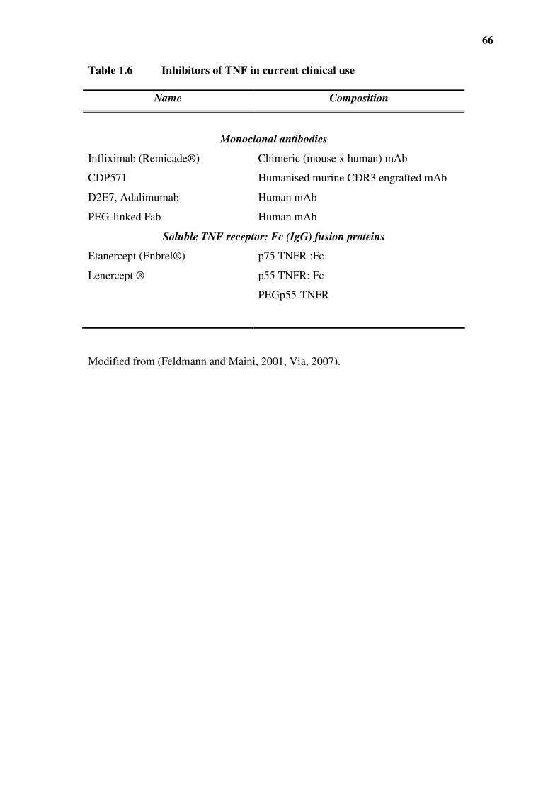
66
Table 1.6 Inhibitors of TNF in current clinical use
Name Composition
Monoclonal antibodies
Infliximab (Remicade®) Chimeric (mouse x human) mAb
CDP571 Humanised murine CDR3 engrafted mAb
D2E7, Adalimumab Human mAb
PEG-linked Fab Human mAb
Soluble TNF receptor: Fc (IgG) fusion proteins
Etanercept (Enbrel®) p75 TNFR :Fc
Lenercept ® p55 TNFR: Fc
PEGp55-TNFR
Modified from (Feldmann and Maini, 2001, Via, 2007).

67
Table 1.7 Anti-TNF based therapies in various diseases
Disease Agent
Rheumatoid Arthritis Etanercept; pegsunercept infliximab; adalimumab;
CDP870
Psoriatic arthritis Etanercept; infliximab; oncercept
Psoriasis Etanercept; infliximab; oncercept; adalimumab
Sarcoidosis Infliximab
Adult's Still's disease Infliximab
Severe acute ulcerative colitis Infliximab
Spondyloarthropathies Etanercept; infliximab
Anklosing spondylitis Etanercept; infliximab
Behcet's syndrome Infliximab
Crohn's disease Infliximab; onercept; adalimumab; CDP870
Orofacial Crohn's disease Infliximab
Uveitis Etanercept; infliximab
HIV-1 associated psoriatic arthritis Etanercept
Graft-vs-host disease Etanercept
Advanced heart failure Etanercept
CVID Etanercept
Wegener's granulomatosis Etanercept
Some studies are reports of non-controlled observations in small number of selected
patients (Schottelius et al., 2004). Abbreviations: CVID, common variable
immunodeficiency

68
Recently it has been reported that in patients with active RA, anti-TNF therapy namely
infliximab, adalimumab and etanercept, was associated with an increased rate of
incidence and seriousness of soft skin infections (Dixon et al., 2006). However, it must
be noted that a simple cause and effect relationship between anti-TNF therapy and
infections has not been proven, since ageing patients with severe RA who have never
been exposed to TNF antagonists have an increased risk of serious infections and cancer
(especially lymphoma).
Recently, the elevated levels of TNF in patients with heart failure (HF) triggered interest
in investigating TNF’s role in the pathogenesis of the condition (Kapadia et al., 1997,
Torre-Amione et al., 1999). Experimental and clinical evidence has shown that the high
levels of TNF lead to progression of left ventricular dysfunction (Sarzi-Puttini et al.,
2005). This has led to anti-TNF clinical trials of etanercept and infliximab in patients
with HF. A case report (Kwon et al., 2003), showed that etanercept and infliximab
induced a ‘new’ onset of HF in patients suffering from arthritis and Crohn’s disease.
Further evidence from the large multi-centre trials of etanercept, RENAISSANCE-
Randomised Etanercept North American Strategy to Study Antagonism of Cytokines,
RECOVER- Research into Etanercept Cytokine antagonism in Ventricular dysfunction
and RENEWAL- Randomised Etanercept Worldwide evaluation, showed that in
moderate to severe HF, etanercept did not have any clinical benefits and even suggested
that it in fact worsens the disease (Anker and Coats, 2002, Coletta et al., 2002, Mann et
al., 2004).

69
Similarly with infliximab, a clinical trial ATTACH (Anti-TNF Therapy Against Chronic
Heart failure) concluded that TNF antagonism did not improve HF and high doses
adversely affected the clinical condition of patients with moderate to severe HF (Chung
et al., 2003). Failure of anti-TNF therapies on HF and development of HF in RA and
recent studies have further highlighted the beneficial role of TNF. Recently, studies have
shown that anti-TNF therapy will be more beneficial against RA and HF when there is no
concomitant therapy with glucocorticoids or COX-2 inhibitors (Listing et al., 2008).
However, it should be noted that this study could not conclusively eliminate the risk of
worsening of HF or de novo HF as a result of anti-TNF therapy. As the risk of
developing HF could be reduced by 30-35 % but that the risk could be increased by 84 %
(Gabriel, 2008). At low physiologic levels TNF has been shown to have a cytoprotective
effect in the heart during ischemic injury (Nakano et al., 1998, Kurrelmeyer et al., 2000).
It is thought that TNF is vital in tissue remodelling and repair (Mann, 2003), as well as
enhancing the production of vascular nitric oxide, thus maintaining peripheral blood flow
in patients with HF (Katz et al., 1994, Sugamori et al., 2002).
Another major drawback has been the reactivation of latent tuberculosis infection (LTBI)
in patients undergoing anti-TNF therapy. The importance of TNF in immunity against M.
tuberculosis is well documented. Using TNFRI KO and TNF-deficient mice, studies
have shown that the formation of granulomas is delayed and not well organised in these
animals, resulting in poorly contained foci of infection, due to the lack of recruitment of
inflammatory cells by TNF (Roach et al., 2002, Ehlers, 2005). TNF also mediates the
bactericidal activity of macrophages against intracellular pathogens, such as M.
tuberculosis by synergising with IFNγ to reduce bacterial replication and to induce

70
synthesis of bactericidal NO (Briscoe et al., 2000, Ehlers, 2005). Thus, it is not
surprising that the clinical use of anti-TNF therapy is associated with an increased risk of
reactivation of LTBI, as animal models have shown regression of formed granulomas
and consequently reactivation under such conditions (Keane, 2005, Theis and Rhodes,
2008). Therefore, it is evident that anti-TNF therapies may decrease TNF below
physiological levels required for repair of the myocardium in the case of HF or levels
required to clear intracellular pathogens in the case of infection.
71
1.9 TARGETING INTRACELLULAR SIGNALLING MOLECULES IN INFLAMMATORY
DISEASES
The well-accepted finding that inflammatory diseases are a consequence of intercellular
signalling by cytokines and the appropriate activation of intracellular signalling pathways
leading to non-genomic and genomic changes which promote tissue damage, has made
the targeting of intracellular signalling molecules an attractive approach in the
development of therapeutics for these diseases. This includes inflammatory disorders in
which TNF plays a major role. The MAPK family of intracellular signalling molecules
has been one of these target focus, namely JNK, ERK and p38. Targeting p38 has been a
particularly active area (Table.1.8) (Saklatvala, 2004, Cuenda and Rousseau, 2007,
Schindler et al., 2007).

72
Table 1.8 Summary of human clinical studies with p38 inhibitors
Abbreviations: RA, rheumatoid arthritis; COPD, chronic obstructive pulmonary disease
Compound Company Clinical indication Study phase Status Reference
GSK-
681323 GlaxoSmithKline
RA, Crohn's psoriasis,
COPD I Awaited (Molfino, 2005)
GSK-
856553 GlaxoSmithKline
RA, Crohn's psoriasis,
COPD I Awaited (Molfino, 2005)
RWJ-67657 Johnson and
Johnson
Crohn's, RA, psoriasis,
rhinitis I Discontinued
(Fijen et al.,
2001)
VX-745 Vertex RA, Crohn's psoriasis,
COPD II
Discontinued-
crosses blood
brain barrier
(Stelmach et al.,
2003)
BIRB-796 Boehringer Asthma, allergy, RA II Awaited (Kuma et al.,
2005)
Scio-469 Scios RA, Crohn's psoriasis,
COPD II Awaited
(Nikas and
Drosos, 2004)

73
1.10 CYTOKINE MIMETICS AS THERAPEUTICS
While the use of TNF against certain tumours seemed logical, two major limitations have
prevented its use, that is, TNF resistance and TNF-induced systemic side effects
(Schottelius et al., 2004). Similarly, IL-2, a cytokine which stimulates T lymphocyte
proliferation, was used in cancer therapy but its therapeutic potential is limited by various
side effects such as fever and influenza-like symptoms, a significant vascular leak
syndrome, which involves damage to vascular endothelial cells, oedema and organ
failure (Rose et al., 2003, Schwinger et al., 2005).
Since most proteins exert their biological activity through the interaction between very
small regions of their folded surfaces to their cognate receptors, smaller peptides which
mimic the shape of the proteins at the point of contact with the receptors can be used to
mimic and/or block the actions of these proteins (Fairlie et al., 1998). Recently, peptide
technology has resulted in the discovery of specific inhibitors for cytokine receptors such
as a disulphide-cyclised 18-mer peptide AF1721, which inhibits the binding of IL-5 to
IL-5Rα and blocks IL-5 dependent eosinophil activation (England et al., 2000). Similarly
a 30-mer peptide mimetic of IL-2 that binds to the dimeric IL-2Rβ2 receptor has also
recently been discovered. The 30-residue peptide mimetic, like its parent cytokine,
activates Shc and p56lck but unlike IL-2, the peptide could not activate the protein
kinases, janus kinase-1 (Jak) and Jak-2. Consequently, the signal transduction and
activators of transcription (STAT) pathway is not activated by the IL-2 mimetic peptide
(Rose et al., 2003). Using phage-display screening with a neutralising anti- IFN-β

74
monoclonal antibody a 15-mer mimetic peptide of IFNβ was recently isolated (Sato and
Sone, 2003). This peptide has been shown to compete with IFN-β for binding to type I
IFN receptor in a dose-dependent manner (Sato and Sone, 2003).
With the above precedents, it is envisaged that TNF mimetics can be generated which
could produce some of the major actions of TNF. In line with this thinking, (Rathjen et
al., 1991) demonstrated, with the use of neutralising anti-human TNF antibodies, that the
sequence from amino acids 65 to 85 of the TNF molecule was involved in binding the
TNF receptor (Rathjen et al., 1991). This subsequently led to the discovery of a TNF
mimetic peptide, TNF70-80, with substitution of leucine-76 for isoleucine to increase
stability in vitro in the presence of serum (Figure 1.6). This peptide was shown to
stimulate and prime neutrophils for increased respiratory burst, which was mirrored in
vivo as the peptide enhanced neutrophil-mediated killing of intraerythrocytic asexual
blood stages of P. falciparum, to a degree almost equal to that observed with TNF, and
increased the resistance of mice to P. chabaudi (Kumaratilake et al., 1995) (Table 1.9).
Britton et al (1998) also showed that TNF70-80 synergised with interferon γ to stimulate
nitric oxide-dependent inhibition of mycobacterial growth. However, several studies
have shown that not all the effects of TNF are mimicked by TNF70-80. Kumaratilake et al
(1995) demonstrated that TNF70-80 failed to increase the expression of E-selectin-1,
ICAM-1 and VCAM-1 on endothelial cells and the peptide did not kill tumour cells such
as WEHI-164 or trigger death in a sepsis model (Kumaratilake et al., 1995) (Table 1.9).
Another mimetic peptide TNF132-150 corresponding to a different region of TNF was
found to have properties that differed from those of TNF70-80. TNF132-150 lacked the

75
neutrophil stimulating ability in vitro, but interestingly exhibited cytotoxic effects
towards tumour cells (Figure 1.6). This effect was mirrored in vivo, where the mimetic
peptide was shown to be effective in halting tumour growth (Rathjen et al., 1993) (Table
1.9).

76
Table 1.9 Comparison of the biological effects of TNF, TNF70-80 and TNF132-150
Abbreviations: CAM, cell adhesion molecules; EC, endothelial cells, PMN, neutrophils;
MØ, macrophages; ND, not determined.
TNF TNF70-80 TNF132-150
In vitro
Induction of CAM expression on EC + - ND
Primes PMN/MØ anti-microbial killing of bacteria,
fungi and parasites
+ + -
Tumour cell cytotoxicity + - +
In vivo
Promotes clearance of bacteria, fungi and parasites + + ND
Anti-tumour activity + - +
Sepsis + - ND
Cachexia + - ND
Neutrophil and MØ priming + + ND

77
Figure 1.6 The location of TNF70-80 and TNF132-150 within the TNF structure
(monomer). TNF70-80 sequence corresponds to the region in red while TNF132-150 is
depicted in the blue region.

78
1.11 RATIONALE, AIMS AND HYPOTHESIS
Preliminary data (Haddad, 1998) indicated that while TNF induced the activation of
ERK1/ERK2, p38 and JNK, TNF70-80 only activated the p38 pathway and in contrast,
TNF132-150, due to its inability to stimulate neutrophils may not activate the p38 pathway.
Thus, it can be rationalised that if TNF70-80 functions through the TNFR then it interacts
with unique sites on the receptor compared with TNF or TNF132-150. Identification of the
site could lead to the development of small TNFR mimetic peptides to block TNF-
induced p38 activation. Since the TNFR and p38 are key targets in the same
inflammatory disorders, it is likely that a more selective inhibition of p38 could be
achieved with the TNFR mimetics, sparing the toxicity encountered with inhibiting all
response going through p38 or the TNFR.
1.11.1 Hypotheses
The characteristics of the selective biological properties of the TNF mimetics such as
TNF70-80 and TNF132-150 suggest that when they engage the TNFR, the peptides induce a
distinct set of intracellular signals. We propose that the difference is related to the ability
to recruit adaptor proteins to the cytoplasmic portion of the TNFR. TNF70-80 binds
specifically to certain amino acid residues on TNFR and that peptides made from such
residues on TNFR can be used to inhibit the TNFR-p38 signalling pathway and
associated cellular activation and inflammation.

79
1.11.2 Aims
The general aim of this project is to identify the basis for the difference in the properties
between these TNF mimetic peptides, and identify a new approach to developing new
therapeutics for chronic inflammatory diseases.
Specifically to:
1. determine whether or not these mimetics bind specifically to TNF receptor.
2. identify whether these peptides stimulate distinct intracellular signalling
pathways.
3. attempt to identify the region of the TNFRI which binds these mimetics
4. investigate whether peptides derived from the TNF70-80 binding region on TNFRI
selectively block TNF responses.
5. examine the anti-inflammatory properties of the TNFR mimetic peptides.

80
2.0 CHAPTER TWO: MATERIALS AND
METHODS

81
2.1 REAGENTS
2.1.1 Biochemicals and antibodies
Ficoll 400, percoll and lymphoprep were obtained from Pharmacia Biotech (Uppsala,
Sweden). RPMI-1640, HBSS, foetal bovine serum (FCS) and DMEM were purchased
from JRH Biosciences (Lenexa, KA). N-formyl-methionyl-L-leucyle-L-phenylalanine
(fMLP), lucigenin (9, 9’-bis (N-methyl-acridinium nitrate), lipolysaccharide (LPS),
3’,3’,5’,5’-tetramethylbenzidine (TMB) substrate, benzamidine, DL-dithiothreitol (DTT),
leupeptin, pepstatin A, p-nitro phenylphosphate, phenylmethylsulfonyl fluoride (PMSF)
and carrageenan type IV were purchased from Sigma Chemical Company Ltd (St Louis,
Mo). Angiografin was obtained from Bayer Schering Pharma (Berlin, Germany),
aprotinin (Bovine Lung, 10000 U/5 ml) and agarose were purchased from Calbiochem
(San Diego, CA). Anti-p38 antibody, anti-NF-κB (p65) and anti-ERK1/ERK2 antibodies
were purchased from Santa Cruz Biotech (Santa Cruz, CA). Horseradish peroxidase
streptavidin conjugate was obtained from Endogen (Rockford, ILL). PE-labelled anti-
CD11b antibody and PE-labelled IgG1 isotype were purchased from Becton Dickinson
(San Jose, CA). LipofectAMINE 2000, TRIzol reagent and cytokine primers were
purchased from Invitrogen (Mount Waverley, VIC, Australia). Nitrocellulose membranes
were purchased from Schleicher and Schuell (Keene, NH). Isoton II and bovine albumin
(BSA) were purchased from Beckman Coulter (Fullerton, CA). iScript cDNA synthesis
kit and iQ SYBR Green supermix were obtained from Bio-Rad (Gladesville, NSW,
Australia). Sheep blood was obtained from the Institute of Medical and Veterinary
Science (IMVS) (Adelaide, SA, Australia).

82
2.1.2 Plasmids
The pRK5-TRAF2-FLAG and pRK5-TRAF287-501-FLAG plasmids were provided by Dr
P Xia, Hanson Institute, Adelaide, Australia). The hTNFRI plasmid was kindly provided
by Dr. M. Kronke (Institute of Medical Microbiology and Hygiene, Medical Center
University or Cologne, Koln, Germany).
2.2 ANIMALS
Female BALB/c mice, 6 - 8 weeks were purchased from the Adelaide University Animal
House. The animals were housed in the Children, Youth and Women's Health Services
(CYWHS) Animal House. The study was approved by the CYWHS Animal Ethics
Committee and the University of Adelaide Animal Ethics Committee.
2.3 CYTOKINES AND PEPTIDES
2.3.1 Cytokines and receptors.

83
Human recombinant TNF was a gift from Dr. G.R. Adolf (Ernst-Boehringer Ingelheim
Institut, Vienna, Austria) and produced by Genetech Inc. (California, USA). The specific
activity was 6 x 10⁷ U/mg as assessed for cytotoxicity by the supplier on actinomycin-D-
treated murine fibrosarcoma cell line, L929. The concentration of TNF stock solution
was confirmed by ELISA (Ferrante et al., 1990) to be 1 pg/ml. TNF was also purchased
from ProSpec-Tany TechnoGene (Rehovot, Israel). TNF stock solution was stored at -
20°C until required. Fresh dilutions of TNF were prepared in HBSS just prior to use.
Human recombinant soluble TNFRI (rHusTNFRI, prepared as a chimaeric protein with
the 6X histidine tagged Fc portion part of human IgG1) was purchased from Prospec-
Tany TechnoGene (Rehovot, Israel).
2.3.2 Peptides
TNF70-80 (Kumaratilake et al., 1995) and its control (GGDPGIVTH) were synthesised by
Auspep Pty Ltd, (Victoria, Australia). While it is difficult to directly equate the TNF
activity to TNF70-80 because of the lack of tumour cell cytotoxicity of this peptide,
previous comparisons on their ability to stimulate nitric oxide production in macrophage
show that 5.0 µg/ml of TNF70-80 equates to 1000 U/ml of TNF (Britton et al., 1998). The
sequence from the natural TNF-α was modified by substitution of isoleucine for leucine
at position 79. The modification extended the serum half-life of the peptide to > 90 min
and increased water solubility. Fresh dilutions of the peptide were prepared daily in
HBSS or HBSS with 1 % BSA. The His-tagged M4 (HM4) (HHHHHHLKPGTT) and

84
M4 (LKPGTT) were purchased from Auspep Pty Ltd (Victoria, Australia). TNF132-150
(LSAEINRPDYLDFAESGQV), TNFRI209-211 peptide (GTT); scrambled control (TGT)
and TNFRI206-211 (EDSGTT); scrambled control (GEDTST), D-amino form of TNFRI206-
211 ({d-GLU} {d-ASP} {d-SER} {d-GLY} {d-THR} {d-THR}), TNFRI198-211
(QIENVKGTEDSGTT) and tandem repeats of TNFRI209-211 (GTTGTTGTTGTT) were
purchased from GenScript (New Jersey, USA). Peptides were purified by high-
performance liquid chromatography (HPLC) and analysed by mass spectrometry to be >
85 % pure. Peptides were stored at -20ºC and solutions were made in HBSS or RPMI
1640 prior to use.
2.4 PURIFICATION OF HUMAN LEUKOCYTES
2.4.1 Purification of peripheral mononuclear cells and neutrophils from whole
blood
Peripheral blood mononuclear leukocytes (MNL) were purified essentially by the
methods outlined of Ferrante and Thong (1982). Briefly, whole blood was layered onto
Hypaque-Ficoll gradient with a specific gravity of 1.114 and cells separated by
centrifugation at 600 g for 35 min. This resolves leukocytes into two bands, the top
containing the MNL and the lower band neutrophils. The MNL were harvested, re-
suspended in RPMI 1640 and washed in the medium by repeated centrifugation.
Neutrophils were similarly harvested and prepared. The MNL and neutrophils were of >
98 % viability as judged by the ability to exclude Trypan blue. In some cases, the

85
neutrophil preparations were further enriched in purity by centrifugation over
lymphoprep which removed any contaminating MNL. In addition in some, cases when
erythrocyte contamination was evident these were removed by centrifugation at 80 g for
5 min.
2.4.2 Production of TNF-rich medium (TNF-RM) culture fluids
Cell culture fluids rich in TNF and neutrophil priming properties were prepared
essentially as described previously (Bates et al., 1991). Briefly, MNL (4 x 10⁷) in 20 ml
were resuspended in 20 ml RPMI 1640 medium with 200 mM L-glutamine solution, 100
U/ml penicillin and 100 µg/ml streptomycin and mixed with heat-killed Staphylococcus
aureus (2 x 10⁸) in 20 ml RPMI 1640 with 5 % human blood group AB serum. The cell
cultures were set up in 250 ml flasks and incubated at 37ºC for 24 h in 5 % CO2 and high
humidity. After incubation the cells were removed by centrifugation and the supernatants
collected by using a 0.22 µm filter and stored in 1ml aliquots at -70 ºC. Medium
conditioned by S. aureus-treated MNL was termed ‘TNF-rich medium’ (TNF-RM).
Medium conditioned by non-stimulated MNL alone was termed control medium (CM).

86
2.5 CELL LINES AND THEIR MAINTENANCE
2.5.1 WEHI-164
A mouse fibroblastic fibrosarcoma cell line WEHI-164 was obtained from Dr Geeta
Chauhdri (John Curtin School of Medical Research, Australian National University,
Canberra). The cells were maintained in RPMI 1640 medium (supplemented with 10 %
heat-inactivated foetal calf-serum, 2 mM L-glutamine, 100 U/ml penicillin and 100
µg/ml streptomycin) and passaged every 2-3 days. Cells were detached with 0.25 %
trypsin in a 0.02 % EDTA solution for 1 min.
2.5.2 HEK 293T
HEK 293T cells were obtained from the American Type Culture Collection (Virginia,
USA) and maintained in DMEM, supplemented with 10 % foetal calf serum, 2 mM L-
glutamine, 100 U/ml penicillin and 100 µg/ml streptomycin. Cells were stably
transfected with pM1, pM3, pM4, pWT of the p55 TNFRI or an empty plasmid using
LipofectAMINE 2000. HEK293T cells were stably transfected with WT-TRAF2,
∆TRAF2 or empty plasmid (Xia et al., 2002).

87
2.5.3 Mono Mac 6 cells
The monocytic cell line Mono Mac 6 was obtained from Dr H.W. L. Ziegler-Heitbrock
(Institute of Clinical Chemistry and Pathobiochemistry, Klinikum rechts der Isar,
Technische Universitat, Munchen, Germany). The cell line was maintained in RPMI
1640 supplemented with 10 % heat-inactivated foetal calf-serum, 1 mM pyruvate, 2 mM
L-glutamine, 100 U/ml penicillin and 100 µg/ml streptomycin (Ferrante et al., 1997).
2.5.4 Pre B-cell line 70Z/3
70Z/3 cells, a mouse Pre B cell line that lacks binding sites for human TNF were
obtained from Prof. Wallace Langdon (School of Pharmacy and Laboratory Medicine,
University of Western Australia) (Kruppa et al., 1992). Cells were maintained in RPMI
1640 with 4.5 g/l glucose, 2 mM L-glutamine, 50 µM 2-mercaptoethanol and 10 % FCS
in an atmosphere of 95 % air and 5 % CO2.
2.5.4.1 Transfection
Cells were plated into 10 cm plate at 4 x10⁷ cells in 15 ml culture medium. Before
transient transfection, plasmid DNA (hTNFR1WT) (20 µg in 1ml of RPMI 1640) were

88
mixed with LipofectAMINE 2000 (Invitrogen) (60 µl in 1ml of RPMI 1640) and the
mixture were left to stand at room temperature for 20 min. This was directly added to
cells and incubated for 24 h.
2.5.5 Endothelial cell culture
Endothelial cells were prepared essentially as described previously (Jersmann et al.,
2001). Fresh human umbilical cords were obtained from consenting mothers according to
the institution’s guidelines on human ethics. Human umbilical vein endothelial cells
(HUVECs) were obtained from fresh umbilical cords by collagenase digestion and
maintained in RPMI 1640, supplemented with 20 % AB serum, L-glutamine, penicillin
and streptomycin (Jaffe et al., 1973). Cells were grown on gelatin (0.2 %) coated 75 cm2
flasks until confluent. They were trypsinised from the first passage of primary cultures.
For experiments the cells were transferred to 0.2 % gelatin coated 60 mm culture dishes
and grown to confluency (2-3 days after seeding) prior to use.
2.6 CONSTRUCTION OF TNFRI MUTANTS
The signal sequence from the p55 TNF-receptor cDNA was isolated by PCR and cloned,
via a NheI site incorporated into the 5’ primer and a KpnI site in the 3’ primer, into the
expression vector pcDNA3.1. A double stranded oligonucleotide encoding His 6-tag,
89
with KpnI and BamHI sticky ends, in frame with the signal peptide, was cloned
downstream of the signal sequence. The transmembrane and cytoplasmic domains of the
TNF receptor were also isolated by PCR and cloned, via a 3’ BstXI site, and a 3’ XhoI
site incorporated into the PCR primers, 3’ of the His 6 sequence. This construct was
designated pcDNA3sigHIScTNF. The mutants designated M1 and M4 which lacked the
1st, and all four cysteine rich domains respectively were isolated using the restriction
enzyme sites incorporated into the primers, cloned between the BamHI and 5’ BstXI sites
of the pcDNA3sigHIScTNF, such that the reading was contiguous with the signal His 6
and transmembrane/cytoplasmic coding sequences.
2.7 SOLID-PHASE LIGAND BINDING ASSAY
Microtitre plates were coated with 100 µl/well of 3 µg/ml rHusTNFRI (prepared as a
chimaeric protein with the 6X histidine tagged Fc portion of human IgG1) in 18 mQ-cm
H2O overnight at 4°C and blocked with 1 % BSA and 0.05 % Tween 20 in PBS for 1 hr
at 4°C. 10 µM of biotin-labelled TNF70-80 plus various concentrations of unlabelled
TNF70-80 or control peptide were added to each well and incubated for 3 h at 4°C. The
wells were washed four times with wash buffer (PBS containing 0.05 % Tween 20) and
the plates were then incubated with 100 µl/well of horseradish peroxidase streptavidin
conjugate (1: 6000 dilution in 1 % BSA and 0.05 % Tween 20 in PBS) for 45 min at 4°C.
Wells were then washed four times with wash buffer and bound enzyme detected by the
90
addition of 3’,3’,5’,5’-tetramethylbenzidine (TMB) substrate. Absorbance was measured
using dual filter at 450/570 nm on a Dynatech MR700 Plate Reader.
2.8 ACTIVATION OF INTRACELLULAR SIGNALLING MOLECULES
2.8.1 Preparation of cell lysates
Cells were treated with TNF or peptide in a serum-free medium for a specific time
period. After washing twice with cold HBSS, the cells were harvested by scraping (if
applicable) and lysed in 300 µl of buffer A (20 mM HEPES, pH 7.4, 0.5 % (v/v) Nonidet
P-40, 100 mM NaCl, 1mM EDTA, 2 mM Na3VO4, 2 mM DTT, 1 mM PMSF, and 10
µg/ml p-nitrophenylphosphate, leupeptin, aprotonin, pepstatin A, and benzamidine) for 2
h at 4 ºC with constant mixing (Hii et al., 1998). After centrifugation (12,000 g x 5 min),
the supernatants were collected and stored at -70 ºC for kinase assays.
2.8.2 Lowry’s protein assay
Lysates from nuclear and cytoplasmic fractions were assayed for protein content
according to the Lowry method (Harrington, 1990). Protein standards were obtained by
diluting BSA (1 mg/ml) with H2O, resulting in six standards (0 µg, 3.125 µg, 6.25 µg,
12.5 µg. 25 µg, 50 µg). Each unknown sample (5 µl) was diluted in 45 µl of H2O, then

91
150 µl CuSO4/Lowry’s solution (2 % Na2CO3, 1 % SDS, 0.4 % NaOH, 0.16 %, Na/K
tartrate (1:100) was added to all tubes and left to stand at room temperature for 15min.
Folin and Ciocalteau’s Phenol Reagent: H2O (1:1), (15 µl), was then added and the tubes
were left to stand for 20 min. An aliquot (180 µl) of each sample was transferred into a
96-well microtitre plate and the absorbance at 540 nm was measured in a Dynatech
MR7000 plate reader (Guernsey, Channel Islands). The absorbance of the proteins
standards was plotted on a linear regression nomogram (Instat, GraphPad Software
Incorporated, San Diego, CA, USA) and the protein content of the test samples was
calculated.
2.8.3 ERK and p38 activity
The activity of ERK1/ERK2 and p38 was determined as previously described (Hii et al.,
1998). Cell lysates containing equal amounts of protein (0.5-1 mg) were precleared with
15 µl of protein A sepharose beads (1:1 slurry) (4ºC) before being incubated with anti-
p38 antibody (3 µg/sample). After mixing for 2 h (4ºC), the immune complexes were
precipitated by the addition of protein A sepharose. The immunoprecipitates were
collected by centrifugation (16,000 g x 15 s) and washed once with buffer A (4 °C), once
with buffer B (10 mM Tris/HCl, pH 7.6, 100 mM NaCl, 1 mM EDTA and 100 µΜ
Νa3VΟ4) and once with assay buffer [(20 mM HEPES, pH 7.2, 20 mM β-
glycerophosphate, 10 mM Sigma 104, 10 mM MgCl2, 1 mM DTT, 50 µM orthovanadate,
20 µM cold ATP). The assay was initiated by adding 10 µCi of [γ-32P] ATP to the tubes,

92
which were then incubated at 30°C for 20 min (Hii et al., 1998). ERK and p38 activities
were determined as described using myelin basic protein as a substrate (Hii et al., 1998).
The reaction was terminated by the addition of Laemmli buffer (20 mM Tris-HCl, pH
6.8, 40 % sucrose, 6 % SDS and 10 mM β-mercaptoethanol) and the samples heated at
100°C for 5 min. Phosphorylated myelin basic protein was fractionated on a 16 % SDS
polyacrylamide gel, and the radioactive bands were detected using an Instant Imager
(Packard Instruments, Canberra, Australia).
2.8.4 JNK assay
A solid phase assay was used for the determination of JNK activity as described
previously (Hii et al., 1998). Lysates containing equal amounts of protein (0.6 mg-1 mg)
were added to 50 µl of GST-c-jun (1-79) beads (1:1 slurry) and MgCl2 and ATP added to
a final concentration of 15 mM and 10 µM respectively, and the samples mixed for 2 h at
4°C. The samples were then washed with lysis buffer (2 x 300 µl), with wash buffer (10
mM PIPES, pH 7.0, 100 mM NaCl, 2 mM DTT, 1 mM PMSF, 10 µg/ml aprotinin, 10
µg/ml leupeptin, 10 µg/ml pepstatin A and 10 µg/ml benzamidine) (2 x 300 µl) and once
in assay buffer (300 µl). Kinase assays were initiated and terminated as described above
for ERK1/ERK2 and p38. Phosphorylated GST-c-jun (1-79) was separated on a 12 %
SDS-PAGE gel run at 175 volts for 90 min and incorporated radioactivity present in the
GST-c-jun (1-79) peptide was quantified using a Packard Instant Imager (Packard,
Canberra, Australia) and integrated using the imager software (Version 2.0.5).

93
2.8.5 Measurement of NF-κκκκB activation
The nuclear translocation of NF-κB was used as an indicator of the activation of the NF-
κB pathway (Ferrante et al., 2006). Neutrophils (3 x 10⁷) were treated with HBSS,
TNF70-80 (10 µM) or TNF (1000 U/ml) for 30 min or 1 h. The cells were then centrifuged
(1400 g for 5 min) and the pellet was resuspended in cold HBSS. Following futher
centrifugation the cells were lysed for 30 min on ice by adding 200 µl NF-κB lysis buffer
(10 mM HEPES pH 7.8, 1.5 mM MgCl2, 10 mM KCl, 300 mM sucrose, 0.5 % Nonidet
P-40, 1 mM dithioreitol, 1 mM phenylmethylsulfonyl fluoride, 10 µg/ml aprotinin, 10
µg/ml leupeptin, 10 µg/ml pepstatin A and 10 µg/ml benzamidine). The tubes were
centrifuged as before, and the pellets were washed with 200 µl NF-κB lysis buffer,
centrifuged and resuspended in 40 µl NF-κB lysis buffer. The nuclei were then disrupted
by sonication. After centrifugation (1400 g 2 min), the supernatants containing nuclear
fractions were mixed with Laemmli buffer (20 mM Tris-HCl pH 6.8, 40 % sucrose, 6 %
SDS and 10 mM β-mercaptoethanol ), boiled (100 °C for 5 min) and stored at – 70 °C.
For western blotting, equal amounts of denatured protein from each lysate were separated
by 12 % SDS polyacrylamide gel electrophoresis (PAGE) for approximately 60 min. The
proteins were transferred to nitrocellulose membranes (3 h at 40 V). After transfer, the
blots were stained with ponceau S (0.1 % in 5 % acetic acid) to confirm equal protein
loading and complete transfer of all proteins. The membrane was then blocked for 1 h at
94
room temperature in blocking buffer (25 mM Tris/HCl pH 7.4, 100 mM NaCl, 5 % skim
milk). Subsequently blots were incubated (1 h at 37° C) with rabbit primary antibody
(NF-κB) at 1:1000 (diluted in blocking buffer + 0.1 % Tween-20). The membrane was
then washed (3 x 5 min) and incubated with HRP conjugated secondary antibody (sheep
anti-rabbit) at 1:1000, for another hour at 37 °C. After 3 x 5 min washes, the
immunocomplexes were visualised with enhanced chemiluminescence. The blots were
scanned and quantifed by laser densitometry using Image Quant™ scanner and software
(Molecular Dynamics, USA).
2.9 CYTOTOXICITY ASSAY
The cell survival assay was performed using a modified method described by (Espevik
and Nissen-Meyer) (1986) and (O'Toole et al.) (2001). Briefly, WEHI-164 cells were
grown in 96-well culture dishes to a density of 5 x 10⁴ cells/well, in growth medium with
1 mg/ml of actinomycin D. After incubation with specific dilutions of TNF and TNF132-
150 for 20 h at 37 °C, 5 % CO2, cells were washed once with PBS and incubated with 3-
[4,5-dimethyl-thiazol-2-yl]-2,5-diphenyltetrazolium bromide (MTT) solution (5 mg/ml in
PBS; 20 µl/well) for a further 4 h. The reaction was terminated by the addition of 50 µl
of 20 % SDS in 20 mM HCl and absorbance read at 540 nm after overnight incubation to
solubilise the reduced MTT.

95
2.10 CHEMILUMINESCENCE ASSAY
Neutrophil superoxide production was measured by an indirect assay involving the
reduction of lucigenin, (9, 9’-bis (N-methyl-acridinium nitrate) and measuring the light
output (Gyllenhammar, 1987). Lucigenin-dependent chemiluminescence was measured
as described previously (Hardy et al., 1995). Neutrophils (5x10⁵) were incubated with
TNF or other stimuli that had previously been pre-incubated in the absence or presence
of receptor peptides for 20 min. After incubation 500 µl of lucigenin (250 µM, final) was
added and the final volume was adjusted to 1 ml. The cells were placed in luminometer
(Autolumat Plus Model LB 953, Berthold Technologies, Bundoora, Australia) and the
resulting chemiluminescence (in relative light units, RLU) measured, the data was
automatically stored in Microsoft Excel. The results are expressed as peak
chemiluminescence produced unless specified otherwise.
2.11 MEASUREMENT OF CR3 EXPRESSION
The upregulation in the expression of the complement receptor type 3 (CR3;
CD18/CD11b) in neutrophils was measured by flow cytometry using anti-CD11b
antibodies as described (Moghaddami et al., 2003). Neutrophils (1x10⁶) were incubated
for 30 min with TNF that had previously been pre-incubated in the absence or presence
of receptor peptides for 20 min. After treatment, 2 µl of IgG1 isotype control or PE-
labelled anti-CD11b antibody was added to neutrophils in 200 µl of Isoton II

96
supplemented with 0.1 % (w/v) BSA. After 30 min the cells were washed, fixed with
formaldehyde (0.1 %) and analysed on a FACScan (Becton Dickinson). Data analysis
was performed on WinMDI 2.9 software.
2.12 ANALYSIS OF CYTOKINE AND CHEMOKINE GENE EXPRESSION BY QUANTITATIVE
REAL-TIME PCR
2.12.1 Isolation of total RNA
Total RNA was extracted from 1 x 10⁶ neutrophils using the TRIzol reagent according to
the manufacturer's instructions. Cells (1x10⁶) were pelleted and lysed in 1 ml Trizol for 5
min. To extract the RNA, 200 µl of chloroform was added, vigorously shaken for 15 s
and incubated for 5 min. Samples were then centrifuged at 12,000 g for 10 min at 4°C.
The upper aqueous phase was transferred to a new tube, and the RNA was precipitated
by adding 500 µl of isopropanol, gently inverted, and then incubated at room temperature
for 10 min. After centrifugation (12,000 g for 10 min at 4°C), the supernatant was
discarded and the RNA pellet was washed with 1ml of 75 % ethanol. After brief
centrifugation (7500 g for 5 min at 4°C) the RNA pellet was allowed to air-dry briefly
and then 20 µl of RNAse/DNAse free water was added to each tube and pulse-vortexed
briefly (~5 sec). The RNA was dissolved by incubation at 55°C for 10 min and stored at
–20°C, until required for cDNA synthesis. Purity of the RNA samples was assessed by
determining the optical density at 260 nm and 280 nm expressed as a ratio.

97
2.12.2 Synthesis of cDNA (Reverse Transcription)
Single-strand (first-strand) cDNA was synthesised from total RNA using iScript cDNA
synthesis kit according to the manufacturer’s instructions. In this reaction 4 µl of 5
iScript reaction Mix and 1 µl of iScript Reverse Transcriptase was added to 100 fg- 1 µg
of RNA and water up to 15 µl. The conditions were as follows: 25°C for 5 min, 42°C for
30 min and 85 °C for 5 min.
2.12.3 Quantitative real-time PCR
Primers for cytokines were designed using Invitrogen Oligo Perfect Designer website
(Table 2.1). These primers were synthesized by Invitrogen. The synthesised cDNA was
amplified in 20 µl triplicate reactions with iQ SYBR Green supermix, 1µl of cDNA and
500 nM of each specific primer pair and house keeping gene glyceraldehyde-3-phosphate
dehydrogenase (GAPDH) using iQ5 system with v.3.1 software. The were as follows: an
initial denaturation at 95°C for 5 min, followed by 40 cycles of 95 °C for 30 s, 60 °C for
30 s and 72 °C for 30 s.
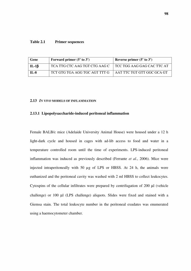
98
Table 2.1 Primer sequences
Gene Forward primer (5’ to 3’) Reverse primer (5’ to 3’)
IL-1ββββ TCA TTG CTC AAG TGT CTG AAG C TCC TGG AAG GAG CAC TTC AT
IL-8 TCT GTG TGA AGG TGC AGT TTT G AAT TTC TGT GTT GGC GCA GT
2.13 IN VIVO MODELS OF INFLAMMATION
2.13.1 Lipopolysaccharide-induced peritoneal inflammation
Female BALB/c mice (Adelaide University Animal House) were housed under a 12 h
light-dark cycle and housed in cages with ad-lib access to food and water in a
temperature controlled room until the time of experiments. LPS-induced peritoneal
inflammation was induced as previously described (Ferrante et al., 2006). Mice were
injected intraperitoneally with 50 µg of LPS or HBSS. At 24 h, the animals were
euthanized and the peritoneal cavity was washed with 2 ml HBSS to collect leukocytes.
Cytospins of the cellular infiltrates were prepared by centrifugation of 200 µl (vehicle
challenge) or 100 µl (LPS challenge) aliquots. Slides were fixed and stained with a
Giemsa stain. The total leukocyte number in the peritoneal exudates was enumerated
using a haemocytometer chamber.

99
2.13.2 Delayed-type hypersensitivity
The delayed-type hypersensitivity (DTH) response was induced in 12 week old female
BALB/c mice as described previously (Ferrante et al., 2006). Briefly mice were injected
with 100 µl of a 10 % haematocrit sheep red blood cell (SRBC) suspension. After 6 days,
mice were challenged subcutaneously via the right hind footpad with 25 µl of 40 %
suspension of SRBC. The DTH response was determined 24 h post challenge by
measuring the footpad thickness of the unchallenged vs. SRBC injected footpads with a
calliper and micrometre screw gauge. The inflammatory response was expressed as the
difference in thickness (mm) between the unchallenged and challenged footpads.
2.13.3 Carrageenan-induced paw inflammation
Carrageenan-induced paw inflammation was induced as described previously (Costabile
et al., 2001). Briefly, BALB/c mice were inoculated with 1 ml/kg of a 1% solution of
carrageenan type IV into the right hind footpad. The swelling/inflammation was assessed
24 h later by comparing the thickness between the unchallenged vs. carrageenan
challenged footpads as described above.
100
2.14 STATISTICS AND ANALYSIS
Data are presented as the mean ± standard error of the mean (SEM) values unless stated
otherwise. Statistical significance was assessed by analysis of variance (ANOVA).
Difference were considered statistically significant when p < 0.05. All statistical analyses
were performed on GraphPad Prism 5 software.

101
3.0 CHAPTER THREE: TNF70-80 ACTS
THROUGH TNF-RECEPTORS

102
3.1 INTRODUCTION
It has been previously shown that the TNF70-80 mimetic peptide demonstrated selective
biological properties in comparison to its parent cytokine TNF (Kumaratilake et al.,
1995). The results suggested that the TNF mimetic was likely to act via the TNFR. The
results showed that an anti-TNF monoclonal antibody (mAb54) and mAb against either
the p75 TNFRII or the p55 TNFRII inhibited the ability of TNF70-80 to stimulate
superoxide production in neutrophils. Other studies provided further evidence for this
suggestion (Briscoe et al., 2000). These anti-human TNF mAb54 and a soluble TNFRI
human IgG fusion protein blocked the synergistic bacteriostatic action of IFNγ and TNF,
and IFNγ and the TNF mimetic peptide on bone marrow derived macrophages infected
with mycobacteria (Briscoe et al., 2000). However, direct evidence for a ligand-receptor
interaction is lacking. Studies were therefore conducted to address this issue.
3.2 TNF70-80 STIMULATES P38 ACTIVATION THROUGH TNFRI
Since TNF and the TNF70-80 stimulate p38 and it was previously shown that this is
important in the neutrophil antimicrobial activity (Zu et al., 1998, Forsberg et al., 2001),
we used p38 activation as a marker for neutrophil activation to study the interaction
between TNF70-80 and the TNFR. This was addressed by making use of the pre B 70Z/3
cell line which lacks binding sites for and proved to be non-responsive to human TNF
(Kruppa et al., 1992). The B cell line and HL-60 cells (positive control), were treated

103
with TNF for 5 min and p38 activity assayed. The results show that indeed the pre B 70
Z/3 cell line was unable to respond to TNF, while HL-60 responded (data not shown).
The pre B cells were then transiently transfect with wild type human TNFRI
(hTNFRIWT) using the lipofectamine 2000 reagent. After 24 h the cells (non-transfected
and transfected) were treated with diluent, TNF70-80 (10 µM), TNF (1000 U/ml) or
control peptide (10 µM) at 37°C for 5 min. The cells were then lysed and the protein
contents were quantified and p38 activity was assayed. The data show that neither TNF
nor TNF70-80 activated p38 in the non-transfected 70Z/3 cells (Fig 3.1). In comparison
both molecules caused the activation of p38 in cells transfected with hTNFRI. The
control peptide failed to activate p38 in cells expressing the TNFRI (data not shown).
3.3 TNF70-80- BINDS TO TNFRI
Previously, attempts to label TNF70-80 with an 125I-tyrosine residue to study ligand-
receptor binding had resulted in its altered function and failure to associate with
neutrophils in binding assays (Kumaratilake et al., 1995). Thus a biotinylated form of
TNF70-80 was synthesised by Auspep (Victoria, Australia). To determine whether the
synthetic TNF70-80 peptide was indeed a ligand for the TNFRI, competitive binding of
TNF70-80 and biotin-labelled TNF70-80 to recombinant human soluble TNF receptor
inhibitor/Fc chimera (rHusTNFRI) was investigated using a solid-phase binding assay as
described in Materials and Methods. The data show that unlabelled TNF70-80 was able to
inhibit biotin-labelled TNF70-80 binding to immobilised rHusTNFRI in a concentration-
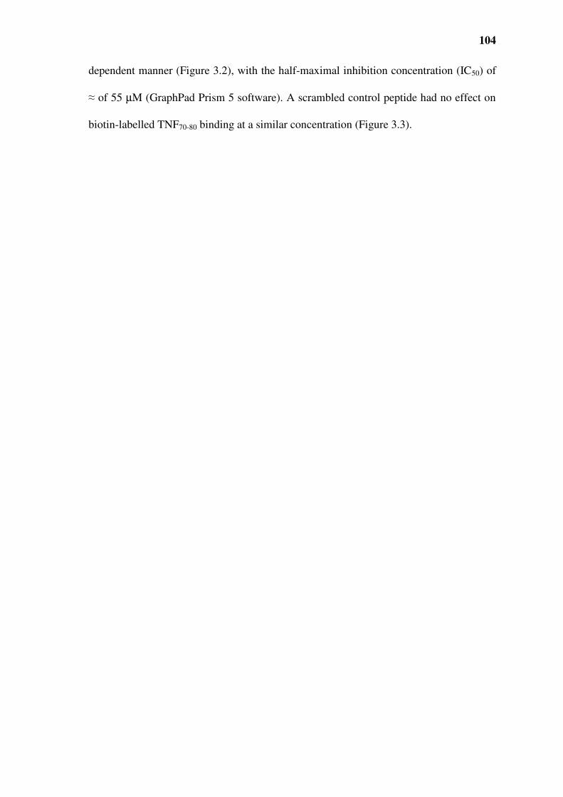
104
dependent manner (Figure 3.2), with the half-maximal inhibition concentration (IC50) of
≈ of 55 µM (GraphPad Prism 5 software). A scrambled control peptide had no effect on
biotin-labelled TNF70-80 binding at a similar concentration (Figure 3.3).

105
Figure 3.1 Inability of TNF70-80 to stimulate p38 activation in cells lacking
TNFR. Transfected (hTNFRIWT) (solid bars) or non-transfected (open bars) 70Z/3 cells
were treated with TNF70-80 (10 µM), TNF (1000 U/ml) or control peptide (10 µM) at
37°C for 5min. The cells were then lysed, protein contents quantified and p38 activity
was assayed. The results are presented as the mean ± SEM of 3 experiments.
Significance of difference between non-transfected cells and transfected cells: ** p <
0.01 and *** p <0.001 (Tukey-Kramer Multiple’s Comparison Test).
0
100
200
300
400
Control TNF70-80TNF
p3
8 a
ctiv
ity (
% o
f co
ntr
ol) **
***
0
100
200
300
400
Control TNF70-80TNF
p3
8 a
ctiv
ity (
% o
f co
ntr
ol) **
***

106
Figure 3.2 Estimation of TNF70-80 binding affinity using a simple microtitre-
plate based competition binding. Biotinylated TNF70-80 (10 µM) as the tracer was
incubated with the indicated concentrations of unlabelled TNF70-80. In a microtitre plate
with wells coated with rHusTNFRI (3 µg/ml) as described in Chapter 2. Bound
biotinylated TNF70-80 was detected by the addition of poly-horseradish peroxidase
streptavidin and 3’,3’,5’,5’-tetramethylbenzidine (TMB) substrate. The IC50 of ≈ 55 µM
was obtained (mean of duplicate wells from two experiments) using GraphPad Prism 4.
-9 -8 -7 -6 -5 -4 -3 -2
0.0
0.1
0.2
0.3
Log [TNF70-80] M
Rel
ati
ve
TN
FR
I b
ind
ing
0.4
-9 -8 -7 -6 -5 -4 -3 -2
0.0
0.1
0.2
0.3
Log [TNF70-80] M
Rel
ati
ve
TN
FR
I b
ind
ing
0.4

107
0
0.1
0.2
0.3
0.4
0.5
Control peptide TNF70-80
Rel
ati
ve
bin
din
g t
o T
NF
RI
***
Figure 3.3. Lack of effect of control peptide on the binding of biotin-labelled
TNF70-80 to immobilised rHusTNFRI. Biotinylated TNF70-80 (10 µM) plus 1000 µM of
TNF70-80 or control peptide were added to microtitre wells previously coated with
rHusTNFRI (3 µg/ml). Bound biotinylated TNF70-80 was detected as described in legend
of Figure 3.2. All data represent means ± SEM from duplicate wells from 3 independent
experiments. Significance of difference between control peptide and TNF70-80: *** p
<0.001 (Students t test).

108
3.4 SUMMARY
The data demonstrate that the biological properties of the mimetic peptide TNF70-80 were
due to its specific interaction with TNFRI. This was shown from a functional aspect in
the cell line lacking TNFRs and in solid phase ligand-receptor binding assays.

109
4.0 CHAPTER FOUR: THE SELECTIVE
ACTIVATION OF TNFRI-INDUCED
INTRACELLULAR SIGNALLING PATHWAYS
BY TNF70-80 AND TNF132-150
110
4.1 INTRODUCTION
Although our studies have demonstrated that TNF70-80 and TNF exert identical effects in
neutrophils both in vitro and in vivo, not all of the effects of TNF are mimicked by
TNF70-80. Previously studies have shown that TNF70-80 failed to increase the expression of
cell adhesion molecules, E selectin-1, ICAM-1 and VCAM-1, in human endothelial cells
and did not kill tumour cells such as WEHI-164 or precipitate death in a sepsis mode
(Kumaratilake et al., 1995). These distinct properties suggest a difference in activation of
intracellular signalling pathways. The p38 MAPK has been implicated in the activation
of neutrophils in response to invading micro-organisms resulting in the production of
reactive oxygen intermediates and chemotaxis (Detmers et al., 1998, Zu et al., 1998). In
contrast, the JNK MAPK pathway has been implicated in the modulation of apoptosis
(Donovan et al., 2002, Lei and Davis, 2003). TNF is known to activate all three MAPK
pathways, which mediate most of its pleiotropic effects (Modur et al., 1996, Read et al.,
1997, Zu et al., 1998). Therefore we investigated if the selective/restricted biological
properties of TNF70-80 were due to selective activation of MAPK pathways.
Work form our group has demonstrated in human endothelial cells (HUVEC) and
neutrophils that TNF (100-1000 U/ml) and TNF70-80 (1-50 µM) caused similar activation
of the p38 MAPK module (Haddad, 1998). But in contrast, to TNF, TNF70-80 was unable
to stimulate JNK activity and ERK1/ERK2 in HUVEC. Hence the ability of TNF70-80 to
stimulate neutrophil anti-microbial activity and its lack of cytotoxic effects are mirrored
in the selective activation of the p38 and not the JNK and ERK/ERK2 pathways.

111
4.2 ACTIVATION OF NFκκκκB PATHWAY BY TNF70-80
TNF receptors are coupled to the NFκB pathway via TRAF2 and it has been proposed
that this action protects cells against TNF-induced apoptosis (Baud and Karin, 2001). We
investigated whether TNF70-80 was able to couple TNF receptors to the NFκB pathway.
This pathway, similar to the p38 pathway is dependent on TRAF-2 and RIP (Kyriakis
and Avruch, 2001). We measured the nuclear translocation of NFκB as an indicator of
the activation of this pathway. Neutrophils were treated with either TNF or TNF70-80.
After incubation, cells were lysed and nuclear fractions were prepared and subjected to
western blotting to determine the amount of NF-κB translocated to the nucleus. The
results showed that TNF70-80 caused the translocation of NFκB (p65) to the nucleus in a
similar manner to TNF (Figure 4.1).

112
Figure 4.1 Activation of NFκκκκB by TNF and TNF70-80 in human neutrophils.
Cells were stimulated with either TNF (100 U/ml) or TNF70-80 (10 µM) at 37°C for the
times indicated. After cell lysis, nuclear fractions were prepared and NF-κB (p65)
detected by Western blotting. Results shown are from one experimental run,
representative of three experiments.

113
4.3 ABILITY OF TNF70-80 TO RECRUIT ADAPTOR PROTEINS
4.3.1 TNF70-80 stimulates p38 activity via TRAF2
The selective biological activity of TNF70-80 suggests an inability by the peptide-ligated
receptors to recruit key adaptor proteins to the ligand-receptor complex and thus activate
downstream signalling pathway. The actions of TNF are mediated via the TNF receptor
associated factor 2 (TRAF2) and activation of p38 by TNF has been demonstrated to
involve TRAF2, receptor interacting protein (RIP) and apoptosis signal regulating
kinase-1 (ASK-1) (Kyriakis and Avruch, 2001). On the other hand, JNK can be activated
by TNF via at least two parallel pathways, one involving TRAF2, germinal centre kinase
(a member of the STE20 family of kinases) and MAP kinase/ERK kinase kinase 1
(MEKK1), and the other involving TRAF2 and ASK-1 (Kyriakis and Avruch, 2001). To
attempt to understand how TNF70-80 selectively activated the p38 module, it is important
to determine whether TRAF2 was involved in the action of the peptide. After
establishing that p38 can be activated in HEK293T cells, we stably transfected these cells
with wild-type TRAF2, a dominant-negative TRAF2 (TRAF287-501, ∆ TRAF2) or an
empty vector. The cells were treated with diluent or TNF70-80 (10 µM) for 5 min at 37 °C,
lysed and p38 kinase activity was assayed after immunoprecipitation. The data (Figure
4.2) demonstrate that while over expression of TRAF2 enhanced the activation of p38 by
TNF70-80 compared to cells transfected with an empty plasmid, expression of ∆ TRAF2
inhibited this response. These data imply that TRAF2 was recruited to TNFR when the
receptors were engaged by TNF70-80.
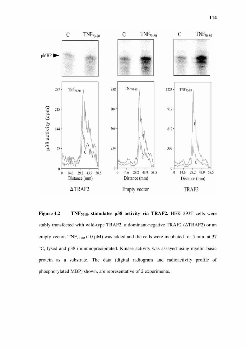
114
Figure 4.2 TNF70-80 stimulates p38 activity via TRAF2. HEK 293T cells were
stably transfected with wild-type TRAF2, a dominant-negative TRAF2 (∆TRAF2) or an
empty vector. TNF70-80 (10 µM) was added and the cells were incubated for 5 min. at 37
°C, lysed and p38 immunoprecipitated. Kinase activity was assayed using myelin basic
protein as a substrate. The data (digital radiogram and radioactivity profile of
phosphorylated MBP) shown, are representative of 2 experiments.

115
4.3.2 Lack of activation of germinal centre kinase (GCK), an upstream kinase in
the JNK pathway
The STE20 family of kinases constitutes a group of upstream regulators of the JNK
module, acting directly on MEKK1 (Kyriakis and Avruch, 2001). Although TNF has
been demonstrated to stimulate the activities of some of these kinases, which include
germinal centre kinase (GCK), GCK-related kinase (GCKR) and GCK-like kinase
(GLK) in some cell-type (Kyriakis and Avruch, 2001), such an effect has not been
reported in neutrophils. We therefore investigated whether TNF and TNF70-80 were able
to stimulate the activity of GLK and GCKR. Neutrophils were pre-adhered to plasma-
coated dishes, stimulated with TNF, lysed and the lysates were incubated with an anti-
GCKR antibody which detects both GCKR and GLK. The data in (Figure 4.3) show that
TNF 70-80 in comparison to TNF did not cause the activation of GCKR/GLK in adherent
neutrophils.
4.3.3 Lack of activation of ASK-1 by TNF70-80
The p38 and JNK modules can also be activated by TNF via the upstream apoptosis
signal-regulating kinase (ASK) , and this has been demonstrated in cell types such as
HEK293T cells (Tobiume et al., 2001). ASK-1 has also been reported to bind to and is
activated via TRAF2 (Kyriakis and Avruch, 2001). We therefore compared the ability of
TNF and TNF70-80 to activate ASK-1 in HEK293T cells, by determining the level of

116
autophosphorylation of the kinase, a hallmark of ASK-1 activation (Kyriakis and
Avruch, 2001). While TNF caused a transient increase in ASK-1 autophosphorylation,
peaking at 3 min, TNF70-80 did not affect the level of ASK-1 phosphorylation (Figure
4.4). In contrast, treatment of HEK293T cells with TNF70-80 for 5 min increased p38
activity by 243 ± 27 % over control (p < 0.05).

117
Figure 4.3 Lack of activation of GCKR by TNF70-80. TNF but not TNF70-80
activated GCKR in neutrophils. Neutrophils were adhered to autologous plasma coated
culture dishes and treated for 15 min with either HBSS, TNF (1000 U/ml) or TNF70-80
(10 µM). GCKR was immunoprecipitated from cell lysates and activity assayed using
myelin basic protein (MBP) as the substrate. The data for the phosphorylated MBP is
presented as mean kinase activity ± SEM of three experiments. Significance of difference
between control and TNF or TNF70-80 treated cells: * p < 0.05.

118
Figure 4.4 Lack of activation of ASK-1 by TNF70-80. TNF but not TNF70-80 caused
the autophosphorylation of ASK-1 in HEK293T cells. HEK293T cells were incubated
with TNF or TNF70-80 for 3 or 7 min, lysed and autophosphorylation of
immunoprecipitated ASK-1 was determined. Results shown are the means ± SEM of 3
experiments. Significance of difference between control and TNF or TNF70-80 treated
cells: * p < 0.05.

119
4.4 EFFECT OF TNF132-150 ON MAPK ACTIVATION IN WEHI-164 CELLS
Previously, (Rathjen et al., 1993) demonstrated that peptide TNF132-150 lacked the
neutrophil stimulating properties of TNF and mimetic TNF70-80, to confirm this 10 µM of
TNF132-150, TNF70-80 or diluent was added to neutrophils (5 x10⁵) and 250 µl of lucigenin
in HBSS (250 µM final), was added and the final volume adjusted to 500 µl and the
resultant chemiluminescence production measured in a luminometer. The kinetics of
chemiluminescence production over the incubation period is shown in (Figure 4.5). It is
evident that TNF70-80 caused a dramatic increase in chemiluminescence response which
reached a peak within approximately 5 min, while TNF132-150 failed to stimulate
chemiluminescence production at similar concentrations. In comparison, while TNF132-
150 was cytotoxic for target cells (Figure 4.6 and 4.7), TNF70-80 did not exhibit similar
properties (Rathjen et al., 1993).
To investigate the effect of TNF132-150 on MAPK activation, WEHI-164 cells were
treated under the same conditions as those in the cytotoxic experiments. The results show
that both TNF and TN132-150 were able to activate JNK activity to a similar degree (Figure
4.8). Under similar conditions ERK1/ERK2 activity was increased after treatment with
TNF or TNF132-150 (Figure 4.9). The activity of p38 was increased by TNF at a
concentration of 1000 U/ml. Interestingly, 100 µg/ml of TNF132-150 failed to stimulate the
activity of p38 in WEHI-164 cells (Figure 4.10).

120
0
5
10
15
20
25
0 5 10 15 20 25
Time (min)
Ch
emil
um
ines
cen
ce (
RL
U x
10
4)
Figure 4.5 The effect of peptides TNF70-80 and TNF132-150 on superoxide
(chemiluminescence) production in neutrophils. To 50 µl of neutrophils (5x10⁵ in
HBSS), 50 µl of peptide (10 µM final) or diluent and 250 µl of lucigenin (250 µM final)
were added and the resultant chemiluminescence produced measured. Data from a
representative experiment of three are presented. Diluent control (─), TNF132-150 (■),
TNF70-80 (▲).

121
0
20
40
60
80
100
1 10 100 1000 10000 100000
Log [TNF] U/ml
Cel
l v
iab
ilit
y (
%) ***
***
***
***
Figure 4.6 The cytotoxic effect of TNF on WEHI- 164 fibrosarcoma cells.
WEHI-164 cells (5x10⁴ cells/well) were cultured in 96-well plates and pre-treated with
actinomycin D (1 µg/ml) for 15 min before the addition of the different dilutions of TNF
and incubated for 20 h. After, 20 µl of 3-[4,5-dimethyl-thiazol-2-yl]-2,5-
diphenyltetrazolium bromide dye (MTT) (5mg/ml) was added and incubated for a further
4 h. The reaction was terminated by the addition of 50 µl of 20 % SDS in 20 mM HCl
and absorbance read at 540 nm after overnight incubation to solubilise the reduced MTT.
Percentage viability was determined by comparison with untreated control cells. Results
shown are means ± SEM for three experiments. Significance of difference between
control and TNF: *** p < 0.001 (Dunnett’s Multiple Comparisons Test).

122
0
20
40
60
80
100
0 50 100 150 200 250
TNF132-150 µµµµg/ml]
Cel
l v
iab
ilit
y (
%)
***
*** ***
Figure 4.7 The cytotoxic effect of TNF132-150 on WEHI- 164 fibrosarcoma cells.
WEHI-164 cells (5x10⁴ cells/well) were cultured in 96-well plates and pre-treated with
actinomycin D (1 µg/ml) for 15 min before the addition of the different dilutions of
TNF132-150 and incubated for 20 h. After, 20 µl of 3-[4,5-dimethyl-thiazol-2-yl]-2,5-
diphenyltetrazolium bromide dye (MTT) (5 mg/ml) was added and incubated for a
further 4 h. The reaction was terminated by the addition of 50 µl of 20 % SDS in 20 mM
HCl and absorbance read at 540 nm after overnight incubation to solubilise the reduced
MTT. Percentage viability was determined by comparison with untreated control cells.
Results shown are means ± SEM for three experiments. Significance of difference
between control and TNF132-150: *** p < 0.001 (Dunnett’s Multiple Comparisons Test).

123
Figure 4.8 Activation of JNK by TNF132-150 in WEHI-164 cells. WEHI-164 cells
(2x 10⁶) were cultured in 28 cm2 dishes to approximately 80 % confluence. Serum
starved cells were incubated with 1000 U/ml TNF or TNF132-150 (100 µg/ml) for 15 min
at 37°C, the cells were lysed and the degree of JNK activity was assayed using GST-jun
(1-79) as a substrate. Data are presented as mean ± SEM of 3 experiments. Significance
of difference between control and TNF or TNF132-150 treated cells: * p < 0.05 (Dunnett’s
Multiple Comparisons Test).
0
100
200
300
Control TNF132-150 TNF
JN
K a
ctiv
ity
(%
Con
tro
l) **
0
100
200
300
Control TNF132-150 TNF
JN
K a
ctiv
ity
(%
Con
tro
l) **
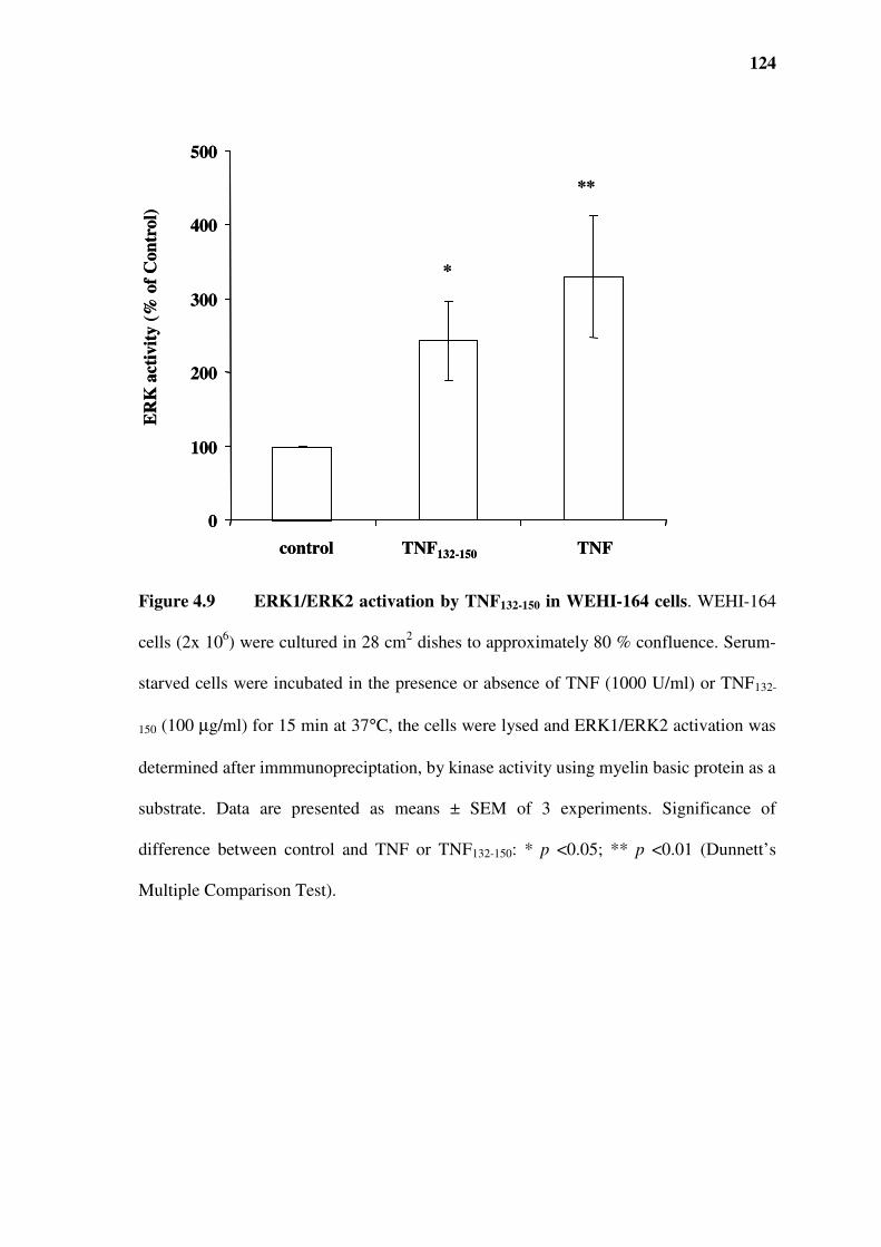
124
Figure 4.9 ERK1/ERK2 activation by TNF132-150 in WEHI-164 cells. WEHI-164
cells (2x 10⁶) were cultured in 28 cm2 dishes to approximately 80 % confluence. Serum-
starved cells were incubated in the presence or absence of TNF (1000 U/ml) or TNF132-
150 (100 µg/ml) for 15 min at 37°C, the cells were lysed and ERK1/ERK2 activation was
determined after immmunopreciptation, by kinase activity using myelin basic protein as a
substrate. Data are presented as means ± SEM of 3 experiments. Significance of
difference between control and TNF or TNF132-150: * p <0.05; ** p <0.01 (Dunnett’s
Multiple Comparison Test).
0
100
200
300
400
500
control TNF132-150 TNF
ER
K a
ctiv
ity
(%
of
Con
tro
l)
*
**
0
100
200
300
400
500
control TNF132-150 TNF
ER
K a
ctiv
ity
(%
of
Con
tro
l)
*
**

125
Figure 4.10 Lack of effect of TNF132-150 on p38 kinase activity in WEHI-164 cells.
WEHI-164 cells (2x 10⁶) were cultured in 28 cm2 dishes to approximately 80 %
confluence. Serum starved cells were incubated with 1000 U/ml TNF or TNF132-150 (100
µg/ml) for 15. The cells were lysed, p38 immunoprecipitated and kinase activity was
assayed using myelin basic protein as a substrate. Data presented as means ± SEM of 3
experiments). Significance of difference between control and TNF or TNF132-150: * p <
0.05 (Dunnett’s Multiple Comparisons Test).
0
100
200
300
400
500
Control TNF132-150 TNF
p3
8 a
ctiv
ity (
% o
f C
on
trol)
**
0
100
200
300
400
500
Control TNF132-150 TNF
p3
8 a
ctiv
ity (
% o
f C
on
trol)
**

126
4.5 SUMMARY
The data presented here show that indeed there is a selective activation of MAPK
occurring in cells treated with TNF70-80 compared to TNF. TNF70-80 was able to
selectively activate the p38 and NF-κB pathway in neutrophils and endothelial cells.
These data imply that the TNF70-80-ligated TNF receptors were able to functionally
couple TRAF2 to the p38 and NFκB pathway. The inability of TNF70-80 to cause
coupling of GCK and ASK 1 to TRAF2 is likely to explain the lack of activation of the
JNK and ERK1/ERK2 pathways. These results are summarised in Figure 4.11.
While TNF was able to activate all three MAP kinases in adherent WEHI-64 cells,
TNF132-150 only enhanced the activity of JNK and ERK1/2 and not p38 at concentrations
which have previously been shown to cause cytotoxicity in WEHI-164 cells following
pre-treatment with actinomycin D. This data is consistent with the inability of TNF132-150
to stimulate neutrophil chemiluminescence production, a response which is dependent on
p38.
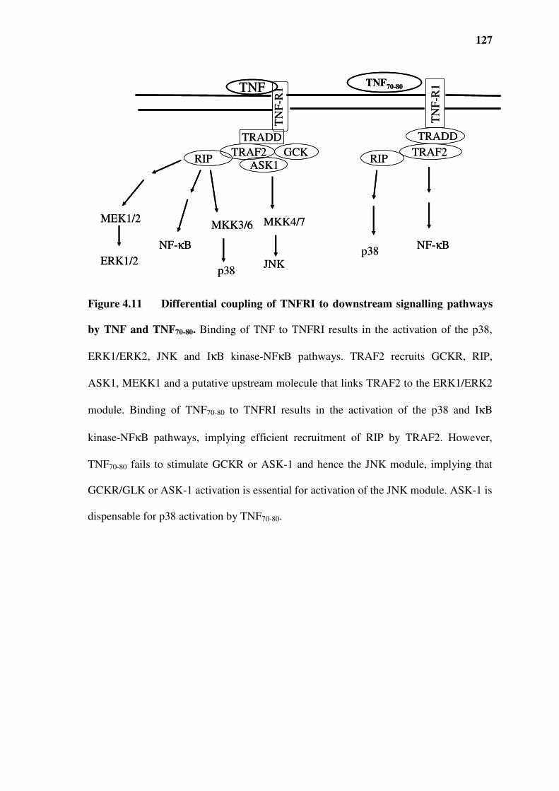
127
Figure 4.11 Differential coupling of TNFRI to downstream signalling pathways
by TNF and TNF70-80. Binding of TNF to TNFRI results in the activation of the p38,
ERK1/ERK2, JNK and IκB kinase-NFκB pathways. TRAF2 recruits GCKR, RIP,
ASK1, MEKK1 and a putative upstream molecule that links TRAF2 to the ERK1/ERK2
module. Binding of TNF70-80 to TNFRI results in the activation of the p38 and IκB
kinase-NFκB pathways, implying efficient recruitment of RIP by TRAF2. However,
TNF70-80 fails to stimulate GCKR or ASK-1 and hence the JNK module, implying that
GCKR/GLK or ASK-1 activation is essential for activation of the JNK module. ASK-1 is
dispensable for p38 activation by TNF70-80.
MKK3/6
p38
MEK1/2
ERK1/2
TN
F-R
1TNF
NF-κB
MKK4/7
JNK
TRADD
GCKASK1
TRAF2RIP
TNF70-80
TRADD
TRAF2RIP
p38NF-κB
TN
F-R
1
MKK3/6
p38
MEK1/2
ERK1/2
TN
F-R
1TNF
NF-κB
MKK4/7
JNK
TRADD
GCKASK1
TRAF2RIP
TNF70-80
TRADD
TRAF2RIP
p38NF-κB
TN
F-R
1
128
5.0 CHAPTER FIVE: IDENTIFICATION
OF THE TNF70-80 BINDING REGION AND
GENERATION OF PEPTIDES TO THESE
REGIONS
129
5.1 INTRODUCTION
The neutralisation of TNF has become a major strategy in the treatment of many
inflammatory diseases (Chapter 1, section 1.7) (LaDuca and Gaspari, 2001, Aggarwal et
al., 2006). Use of antagonists based on monoclonal anti-TNF antibodies and recombinant
soluble TNF receptors is well documented. Pharmacologically, administration of proteins
[cytokines] as drugs has many drawbacks such as conformational instability,
susceptibility to proteolytic degradation, antigenicity, high cost and poor membrane
penetration (Sedmak et al., 1981, Fairlie et al., 1998). Clinically, protein-based drugs
have to be administered parentally e.g. by subcutaneous, intramuscular or intravenous
injection (Sato and Sone, 2003). Thus small molecules that can be prepared synthetically
and administered at high concentrations might offer a more appropriate alternative for
anti-TNF therapy.
Studies on the characteristics of binding between TNF and TNFR have led to the
discovery of regions and consequently peptides to those regions that mimic the function
of TNFRs and could consequently lead to the development of new therapeutic agents.
For example, a 20 amino acid synthetic peptide corresponding to residues 175-194 of
TNFRI was capable of inhibiting TNF-induced cytolysis, as well as displacing the
binding of 125I-labelled TNF to mouse L929 cells (Hwang et al., 1991). Another peptide,
corresponding to residues 159-178 of TNFRI was also reported to similarly displace the
binding of 125I-TNF and inhibit TNF cytotoxicity to L-M cells (Lie et al., 1992). These
receptor-derived mimetics highlight the potential usefulness of small molecule inhibitors

130
of TNF functions, giving an alternative approach to treating inflammation associated
with autoimmune and malignant diseases.
5.2 CONSTRUCTION OF TNFRI MUTANTS
Initially, truncated mutants of TNFRI were constructed by sequentially deleting the
cysteine rich domains on the extracellular portion of TNFRI (Figure 5.1). This would
allow us to test whether TNF70-80 was able to activate cells via the TNFR mutants, and
allow the identification of the minimal portion of TNFRI that is required for TNF70-80
action. That is, the truncation of mutants could permit the identification of the region on
TNFRI that TNF70-80 binds.
70Z/3 pre B cells were then transiently transfected with wild type (WT) or a mutant of
TNFRI using lipofectamine 2000. After 24 h the cells were treated with diluent, TNF70-80
(10 µM) or TNF (1000 U/ml) at 37°C for 5 min. The cells were then lysed and the
protein contents were quantified and p38 activity was assayed after immunoprecipitation.
The data showed that TNF-induced activation of p38 was most in WT TNFRI transfected
cells (Figure 5.2), with reduced effect using the M1 mutant and essentially no activity
with further truncation (M4). TNF70-80-induced activation of p38 was also clearly evident
in the WT TNFRI transfected cells and while there was still substantial activity with M1
transfected cells, it was evident that M4 transfected cells still showed a significant p38

131
response (Figure 5.2). Since all three mutants contained the amino acid residues present
in the M4 mutant, the minimum region required by TNF70-80 to activate p38 was likely to
be within the M4 region. These results imply that TNF70-80 binds to the remnant amino
acid residues in the M4 mutant. This therefore raised the possibility that peptides of this
region may be also to block the function of TNF70-80. To test this peptides corresponding
to the extracellular portion of the M4 mutant were made and their ability to inhibit
TNF70-80 induced superoxide production in neutrophils was investigated. Figure 5.3
shows the various peptides that were made.

132
WT
Signal peptide6xHis
Extracellular domain
CRD1 CRD2 CRD3 CRD4
Transmembranedomain
Cytoplasmicdomain
(1-171) (172-194) (194-415)
M1
M4
CRD2 CRD3 CRD4
Gly Thr Thr
Figure 5.1 Diagrammatic generation of TNFRI mutants. The signal peptide is
cleaved from the mature receptor protein. Abbreviations: WT, wild type; CRD, cysteine
rich domains; His, histidine; M, mutant.

133
TNF70-80
TNF
p3
8 a
ctiv
ity
(%
in
crea
se)
WT TNFRI M1 TNFRI M4 TNFRI
0
100
200
300
400
*
*
**
Figure 5.2 p38 activation in cells transfected with WT or mutant TNFRI. 70Z/3
cells transfected with WT TNFRI or different mutant forms of TNFRI, M1, and M4 were
treated with HBSS, TNF70-80 (10 µM) or TNF (1000 U/ml) at 37°C for 5 min. The cells
were then lysed, protein contents quantified. After immunoprecipitation p38 activity was
assayed. The results are presented as the mean ± SEM of 3(WT), 1(M1), 3 (M4)
experiments. Significance of difference between control and TNF70-80 or TNF: * p <
0.05, ** p < 0.01 (Tukey's Multiple Comparison Test).

134
Figure 5.3 Diagrammatic representation of generation of TNFRI-derived
peptides. The signal peptide was cleaved from the mature receptor protein. The
boldfaced text represents residues in the natural TNFRI sequence. Leu-Lys-Pro were
introduced as a consequence of generating a restriction site for coupling to the His-tag.
Abbreviations: WT, wild type; CRD, cysteine rich domains; His, histidine; M, mutant.
WT
Signal peptide6xHis
Extracellular domain
CRD1 CRD2 CRD3 CRD4
Transmembranedomain
Cytoplasmic
domain
(1-171) (172-194) (194-415)
Gly Thr Thr
M4
M4 peptide with 6-His tag HM4 Leu Lys Pro Gly Thr ThrH OH
6 His-tag Restriction enzyme linker
Leu Lys Pro Gly Thr ThrH OH
Gly Thr ThrH OH Glu Asp Ser Gly Thr ThrH OH
TNFRI209-211 TNFRI206-211
M4 peptide with linker ─ HM4
WT
Signal peptide6xHis
Extracellular domain
CRD1 CRD2 CRD3 CRD4
Transmembranedomain
Cytoplasmic
domain
(1-171) (172-194) (194-415)
Gly Thr Thr
M4
M4 peptide with 6-His tag HM4 Leu Lys Pro Gly Thr ThrH OH
6 His-tag Restriction enzyme linker
Leu Lys Pro Gly Thr ThrH OH
6 His-tag Restriction enzyme linker
Leu Lys Pro Gly Thr ThrH OHLeu Lys Pro Gly Thr ThrH OH
Gly Thr ThrH OHGly Thr ThrH OH Glu Asp Ser Gly Thr ThrH OHGlu Asp Ser Gly Thr ThrH OH
TNFRI209-211 TNFRI206-211
M4 peptide with linker ─ HM4

135
5.3 THE EFFECT OF HM4 ON TNF70-80-INDUCED SUPEROXIDE PRODUCTION IN
NEUTROPHILS
The first peptide included a 6 histidine tag which was used for purification and extraction
purposes and a restriction enzyme linker consisting of 3 amino acids linked to the
remnant 3 amino acids from the TNFRI sequence (HHHHHHLKPGTT) (Figure 5.3).
Neutrophils were incubated with varying concentrations of HM4 in HBSS at 37 °C for
30 min and then tested for their ability to produce superoxide when challenged with
TNF70-80, determined by the lucigenin-based chemiluminescence response. The kinetics
of chemiluminescence production over the incubation period is shown in (Figure 5.4). It
is evident that TNF70-80 caused a dramatic increase in chemiluminescence response which
reached a peak after approximately 8 min. The results showed that incubation with HM4
caused a reduction in the TNF70-80-induced chemiluminescence production (Figure 5.5).
Maximum inhibition was observed with 3.5 µM HM4. The degree of inhibition
diminished as the concentration of HM4 increased. The reason for this inverse
relationship is not clear but could be due to the His tag overcoming the inhibitory action
of GTT. This is consistent with the results from subsequent experiments using peptides
devoid of the His tag (Figure 5.6-7).

136
Figure 5.4 Kinetics of TNF70-80-induced superoxide (chemiluminescence)
production from neutrophils in the presence of varying concentrations of TNFRI
fragment HM4. Neutrophils (1x10⁶ in 100 µl HBSS) were treated with 100 µl of HM4
at 37 °C for 30 min. Lucigenin and 100 µl of TNF70-80 (10 µM) were then added and the
resultant chemiluminescence production measured in a luminometer. Data from a
representative experiment of three are presented. Diluent (♦); 70 µM HM4 (▲); TNF70-
80 (■); TNF70-80 + 3.5 µM HM4 (+); TNF70-80 + 35 µM HM4 (���� ); TNF70-80 + 70 µM HM4
(●); TNF70-80 + 105 µM HM4 (οοοο).
Ch
emil
um
ines
cen
ce (R
LU
x 1
04)
Time (min)
0
1
2
3
4
5
6
7
8
9
10
0 5 10 15 20
Ch
emil
um
ines
cen
ce (R
LU
x 1
04)
Time (min)
0
1
2
3
4
5
6
7
8
9
10
0 5 10 15 20

137
Figure 5.5 The effect of his-tagged TNFRI fragment- HM4 on TNF70-80-induced
superoxide (chemiluminescence) production in neutrophils. Neutrophils (1x10⁶ in
100 µl HBSS) were treated with 100 µl of HM4 at 37 °C for 30 min. After incubation,
lucigenin and 100 µl of TNF70-80 (10 µM) were added and the resultant
chemiluminescence production measured in a luminometer. The results are expressed as
maximal rate of chemiluminescence production. Significance of difference between
TNF70-80 and TNF70-80 + HM4: ** p < 0.01, *** p < 0.001 (Dunnett's Multiple
Comparison Test).
TNF70-80 10 µµµµM
HM4 µµµµM
***
**
Ch
emil
um
ines
cen
ce
(RL
U x
10
4 )
0
1
2
3
4
5
6
7
8
9
Vehicle HM4
(70 µµµµM)
Vehicle 3.5 35 70 100
TNF70-80 10 µµµµM
HM4 µµµµM
***
**
Ch
emil
um
ines
cen
ce
(RL
U x
10
4 )
0
1
2
3
4
5
6
7
8
9
Vehicle HM4
(70 µµµµM)
Vehicle 3.5 35 70 100

138
5.4 THE EFFECT OF –HM4 ON TNF70-80-INDUCED SUPEROXIDE PRODUCTION IN
NEUTROPHILS
The above results prompted us to test the effects of –HM4 (LKPGTT) that lacked the His
tag (Figure 5.3). Neutrophils were incubated with 8-50 µM of –HM4 in HBSS or vehicle
at 37°C for 30 min. After incubation, 500 µl of lucigenin (250 µM, final), 100 µl of
TNF70-80 (10 µM) in HBSS were added, the final volume was adjusted to 1 ml and the
resultant chemiluminescence was measured in a luminometer. TNF70-80 caused a marked
increase in chemiluminescence response which reached a peak after approximately 6 min
(Figure 5.6). In contrast to the results in Figure 5.5, -HM4 caused a dose dependent
suppression of the ability of TNF70-80 to stimulate superoxide production. The results
showed that incubation of cells in the presence of 25 µM and 50 µM of M4 caused a
reduction in the TNF70-80 induced response by 17 % and 66 %, respectively (Figure 5.7).

139
Figure 5.6 Kinetics of TNF70-80-induced superoxide (chemiluminescence)
production from neutrophils in the presence of varying concentrations of TNFRI
fragment -HM4. Neutrophils (1x10⁶ in 100 µl HBSS) were treated with 100 µl of -HM4
at 37 °C for 30 min. After incubation, lucigenin and 100 µl of TNF70-80 (10 µΜ) were
added and the resultant chemiluminescence was measured in a luminometer. Data from a
representative experiment of three are presented. Diluent control (■); 50 µM -HM4 (■);
TNF70-80 (▲); TNF70-80 + 8 µM –HM4 (□); TNF70-80 + 25 µM –HM4 (●); TNF70-80 + 50
µM –HM4 (*).
Ch
emil
um
ines
cen
ce (
RL
U x
10
4)
0
1
2
3
4
5
6
7
0 5 10 15 20
Time (min)
Ch
emil
um
ines
cen
ce (
RL
U x
10
4)
0
1
2
3
4
5
6
7
0 5 10 15 20
Time (min)

140
Figure 5.7 The effects of TNFRI fragment -HM4 on TNF70-80-induced
superoxide (chemiluminescence) production in neutrophils. Neutrophils (1x10⁶ in
100 µl HBSS) were treated with 100 µl of -HM4 at 37 °C for 30 min. After incubation,
500 µl of lucigenin (250 µM, final) and 100 µl of TNF70-80 (10 µM) were added and the
resultant chemiluminescence measured in a luminometer. The results are expressed as
maximal rate of chemiluminescence production. Significance of difference between
TNF70-80 and TNF70-80 + -HM4: * p < 0.05, *** p < 0.001 (Dunnett’s Multiple
Comparisons Test).
TNF70-80 10 µµµµM
258 50Vehicle -HM4
(50 µµµµM)Vehicle
-HM4 µµµµM
0
1
2
3
4
5
6
Ch
emil
um
ines
cen
ce (
RL
U x
10
4)
***
*
TNF70-80 10 µµµµM
258 50Vehicle -HM4
(50 µµµµM)Vehicle
-HM4 µµµµM
0
1
2
3
4
5
6
Ch
emil
um
ines
cen
ce (
RL
U x
10
4)
***
*

141
5.5 THE EFFECT OF TNFRI209-211 ON TNF-INDUCED SUPEROXIDE PRODUCTION IN
NEUTROPHILS
Having obtained the above results with –HM4, it was of interest to investigate whether
the effect was due to the three linker amino acids (LKP) or to GTT (TFNRI209-211)
(Figure 5.3). The above results also raised the more important question of whether a
peptide based entirely on TNFRI was able to block the actions of TNF given that the
ultimate aim was to design therapeutic agents to block the actions of TNF. We therefore
synthesised and tested the effect of TNFRI209-211 on TNF-mediated effects on neutrophil
superoxide production.
TNFRI209-211 and a control peptide (TGT) were reconstituted just prior to conducting the
experiment by dissolving in HBSS with 1 % bovine serum albumin (BSA). In these
experiments, 50 µl of TNFRI209-211 (50-400 µM) or diluent were incubated with 50 µl of
TNF (1 ng/ml) at 37 °C for 20 min to optimise the interaction and then 50 µl of
neutrophils (5x 10⁵) in HBSS were added. Following a further incubation for 30 min at
37 °C, 250 µl of lucigenin in HBSS (250 µM final) was added and the final volume
adjusted to 500 µl. The resultant chemiluminescence measured in a luminometer.
The results showed that the treatment of TNF with TNFRI209-211 caused a dose-dependent
inhibition of TNF-induced increase in chemiluminescence (Figure 5.8). Maximum
inhibition occurred at TNFRI209-211 of 200 µM and above and the half maximal inhibition
(IC50) occurred at 122.5 µM. A scrambled control peptide of the TNFRI209-211 was tested

142
to determine the specificity of the natural sequence of the receptor on TNF-induced
superoxide production. This peptide contained the sequence TGT. This control peptide
was shown to have no inhibitory effect on chemiluminescence induced by TNF at the
same concentration as that which TNFRI209-211 inhibited the response (Figure 5.9).

143
% I
nh
ibit
ion
of
TN
F-i
nd
uce
d r
esp
on
se
0
20
40
60
80
100
0 100 200 300 400
TNFRI209-211 [µµµµM]
*
***
***
***
Figure 5.8 The effects of TNFRI209-211 on TNF-induced superoxide
(chemiluminescence) production in neutrophils. TNF (1 ng/ml) was treated with
varying concentrations (50, 100, 200 and 400 µM) of TNFRI209-211 at 37°C for 20 min.
Neutrophils were added and further incubated for 30 min at 37°C. Then lucigenin was
added and the chemiluminescence generated measured in a luminometer. Data represent
the % inhibition of the TNF response and are presented as mean ± SEM of three
experiments. The maximal rate of chemiluminescence production in non-stimulated
neutrophils was 654 ± 120 RLU and in TNF-stimulated cells was 3767 ± 673 RLU.
Significance of difference between TNF and TNF + TNFRI209-211: * p < 0.05, *** p <
0.001 (Dunnett’s Multiple Comparisons test).

144
% o
f T
NF
-in
du
ced
res
po
nse
TNF 1 ng/ml
0
20
40
60
80
100
120
140
160
180
Vehicle TNFRI209-211Control peptide
*
Figure 5.9 Comparison of the effects of TNFRI209-211 and a control peptide on
TNF-induced superoxide (chemiluminescence) production in neutrophils. TNF (1
ng/ml) was treated with diluent, 200 µM of either TNFRI209-211 or control peptide for 20
min at 37°C. Neutrophils were added and incubated for 30 min at 37°C. Following this,
lucigenin was added and the resultant chemiluminescence production measured. Data are
presented as mean ± SEM of three experiments and expressed as a % of the control
response. The maximal rate of chemiluminescence production in non-stimulated
neutrophils was 144 ± 46 RLU and in TNF-stimulated cells was 1270 ± 237 RLU.
Significance of difference between TNF and TNF+ TNFRI209-211: * p < 0.05 (Dunnett’s
Multiple Comparisons test).

145
5.6 THE EFFECT OF TNFRI206-211 ON TNF-INDUCED SUPEROXIDE PRODUCTION IN
NEUTROPHILS
The above results prompted us to synthesise and test a longer TNFRI peptide that
contained 3 more amino acids N-terminal to GTT. This peptide was TNFRI206-211 (Figure
5.3). TNF (1 ng/ml) was incubated with varying amounts of TNFRI206-211 for 20 min at
37°C prior to the addition of neutrophils. The cells were examined for production
chemiluminescence. Maximum inhibition occurred at TNFRI206-211 of 400 µM and the
half maximal inhibition (IC50) occurred at 171.4 µM (Figure 5.10). TNFRI209-211 caused
a greater inhibition compared to TNFRI206-211 at 50 µM (p < 0.05). A scrambled control
peptide for TNFRI206-211 (GEDTST) was shown to have no effect on TNF-induced
chemiluminescence in neutrophils at similar concentrations, suggesting that the action of
TNFRI206-211 on TNF is not a non-specific effect (Figure 5.11).

146
*
**
**
TNFRI206-211[µµµµM]
% I
nh
ibit
ion
of
TN
F-i
nd
uce
d r
esp
on
se
-20
0
20
40
60
80
100
0 100 200 300 400
Figure 5.10 The effects of TNFRI206-211 on TNF-induced superoxide
(chemiluminescence) production in neutrophils. TNF (1 ng/ml) was treated with
varying concentrations (50, 100, 200 and 400 µM) of TNFRI206-211 at 37°C for 20 min.
Neutrophils were added and further incubated for 30 min at 37°C. After incubation,
lucigenin was added and the resultant chemiluminescence production measured. Data
represent the % inhibition of the TNF response and are presented as mean ± SEM of
three experiments. The maximal rate of chemiluminescence production in non-stimulated
neutrophils was 361 ± 63 RLU and in TNF-stimulated cells was 3536 ± 706 RLU.
Significance of difference between TNF and TNF + TNFRI206-211: * p < 0.05, ** p < 0.01
(Dunnett’s Multiple Comparisons test).

147
0
20
40
60
80
100
120
% o
f T
NF
-in
du
ced
res
po
nse
Vehicle TNFRI206-211 Control peptide
TNF 1 ng/ml
**
Figure 5.11 The effects of TNFRI206-211 and control peptide on TNF-induced
superoxide (chemiluminescence) production in neutrophils. TNF (1 ng/ml) was
treated with 200 µM of either TNFRI206-211 or control peptide for 20 min. Neutrophils
were added and incubated for 30 min at 37°C. After incubation lucigenin was added and
the resultant chemiluminescence production measured in a luminometer. Data represent
the % inhibition of the TNF response and are presented as mean ± SEM of three
experiments. The maximal rate of chemiluminescence production in non-stimulated
neutrophils was 219 ± 23 RLU and in TNF-stimulated cells was 1516 ± 613 RLU.
Significance of difference between TNF and TNF + TNFRI206-211: ** p < 0.01 (Dunnett’s
Multiple Comparisons test).

148
5.7 THE EFFECT OF TNFRI209-211 AND TNFRI206-211 ON THE ABILITY OF CYTOKINE
CONTAINING MNL CONDITIONED MEDIUM TO STIMULATE NEUTROPHIL
SUPEROXIDE PRODUCTION
It has been shown that stimulation of MNL with Staphylococcus aureus results in a
conditioned medium containing TNF as the major cytokine responsible for stimulating
neutrophils (Ferrante et al., 1987, Ferrante et al., 1990). The amount of TNF was evident
in culture within 2 h and reached a maximum of 10-20 ng/ml after 1-2 days which
remained stable over a 7 day culture period. No lymphotxin was detected but IFNγ and
IL-1β were present (Ferrante et al., 1990, Bates et al., 1991). It was of interest to see if
the TNFR peptides could also inhibit TNF action when the cytokine was present within
an environment of activated leukocytes.
The TNF-rich medium (TNF-RM) caused a direct stimulation of neutrophil
chemiluminescence (Figure 5.12). When neutrophils were pre-treated with TNF- RM for
30 min the chemiluminescence response was 333.3 ± 18 RLU in control medium (CM)
and 19726 ± 2221 RLU in the TNF-RM. Pre-treatment of TNF-RM (1:10 dilution in
HBSS) with different concentrations of TNFRI206-211 before incubation with neutrophils
resulted in dose-dependent inhibition of the ability of TNF-rich medium to stimulate
neutrophil superoxide production, reaching a maximum effect at 200 µM (Figure 5.13).
Pre-treatment of TNF-RM with receptor peptide TNFRI209-211 resulted in a similar dose-
dependent reduction of the neutrophil stimulating activity (Figure 5.14).

149
Figure 5.12 Neutrophil stimulating activities of TNF-rich medium and control
medium on neutrophil superoxide (chemiluminescence) production. Neutrophils
from an individual donor were treated with CM (shaded bars) or TNF-RM (open bars)
for 30 min and chemiluminescence assayed. This data represents maximal rates peak of
chemiluminescence production and expressed as mean ± SEM of triplicates. Significance
of difference between each dilution of TNF-RM and its corresponding CM dilution: * p
< 0.05, *** p < 0.001 (Tukey's Multiple Comparison Test).
*
***
***
***
Vehicle 1:10 1:4 1:2 Neat
0
5
10
15
20
25
30
Ch
emil
um
ines
cen
ce
(RL
U x
10
4)
Levels of conditioned medium
*
***
***
***
Vehicle 1:10 1:4 1:2 Neat
0
5
10
15
20
25
30
Ch
emil
um
ines
cen
ce
(RL
U x
10
4)
*
***
***
***
Vehicle 1:10 1:4 1:2 Neat
0
5
10
15
20
25
30
Ch
emil
um
ines
cen
ce
(RL
U x
10
4)
Levels of conditioned medium

150
Figure 5.13 Effect of TNFRI206-211 on TNF-RM-induced superoxide
(chemiluminescence) production in neutrophils. TNF-RM (1:10 dilution in HBSS)
was treated with various concentrations of TNFRI206-211 and then tested for ability to
stimulate neutrophil superoxide (chemiluminescence) production. Data represent the %
inhibition of the TNF-RM response and expressed as mean ± SEM of three separate
experiments. The maximal rate of chemiluminescence production in non-stimulated
neutrophils was 444 ± 69 RLU and in TNF-rich medium-stimulated cells was 8593 ±
1854 RLU. Significance of difference between TNF-RM and TNF-RM + TNFRI206-211:
** p < 0.01 (Dunnett’s Multiple Comparisons test).
0
20
40
60
80
100
0 100 200 300 400
TNFRI206-211 [µµµµM]
% I
nh
ibit
ion
of
TN
F-R
M-i
nd
uce
d r
esp
on
se
**
**
**
**
0
20
40
60
80
100
0 100 200 300 400
TNFRI206-211 [µµµµM]
% I
nh
ibit
ion
of
TN
F-R
M-i
nd
uce
d r
esp
on
se
**
**
**
**

151
Figure 5.14 Effect of receptor peptide TNFRI209-211 on TNF-RM-induced
superoxide (chemiluminescence) production in neutrophils. TNF-RM (1:10 dilution
in HBSS) was treated with various concentrations of TNFRI209-211 and tested for ability
to stimulate neutrophil chemiluminescence. Data represent the % inhibition of the TNF-
RM-induced response and expressed as mean ± SEM for three separate experiments. The
maximal rate of chemiluminescence production in non-stimulated neutrophils was 261 ±
24 RLU and in TNF-RM-stimulated cells was 6328 ± 1495 RLU. Significance of
difference between TNF-RM and TNF-RM + TNFRI209-211: * p < 0.05, ** p < 0.01
(Dunnett’s Multiple Comparisons test).
TNFRI209-211 [µµµµM]
% I
nh
ibit
ion
of
TN
F-R
M-i
nd
uce
d r
esp
on
se
*
**
**
-20
0
20
40
60
80
100
0 100 200 300 400
TNFRI209-211 [µµµµM]
% I
nh
ibit
ion
of
TN
F-R
M-i
nd
uce
d r
esp
on
se
*
**
**
-20
0
20
40
60
80
100
0 100 200 300 400

152
5.8 EFFECT OF TNFRI209-211 AND TNFRI206-211 ON FMLP-INDUCED SUPEROXIDE
PRODUCTION IN NEUTROPHILS
The specificity of these receptor peptides was further assessed by examining their effect
on the fMLP-stimulated chemiluminescence production. fMLP activates neutrophils by
binding to its surface receptor- formyl peptide receptor (FPR). Pre-treatment of fMLP
with TNFRI209-211 did not affect the ability of neutrophils to respond to fMLP, even at
TNFRI209-211 concentrations up to 400 µM (Figure 5.15). Similarly TNFRI206-211 did not
affect the fMLP-induced response at these concentrations (Figure 5.16). Incubation with
the peptides alone neither increased nor decreased the baseline chemiluminescence in all
experiments (data not shown).

153
TNFRI209-211 [µµµµM]
0
30
60
90
120
150
0 100 200 300 400
% o
f fM
LP
-in
du
ced
res
pon
se
Figure 5.15 Effect of TNFRI209-211 on fMLP-induced superoxide
(chemiluminescence) generation in neutrophils. fMLP (5 x 10⁻⁶M final) was incubated
with different concentrations of TNFRI209-211 at 37°C for 5 min. Neutrophils and
lucigenin were added and the resultant chemiluminescence production measured. The
results are expressed as a % of fMLP-induced chemiluminescence production. The
maximal rate of chemiluminescence production in non-stimulated neutrophils was 1106±
272 RLU and in fMLP-stimulated cells was 5617± 495 RLU. Data are presented as mean
± SEM of three experiments.

154
0
20
40
60
80
100
120
140
160
0 100 200 300 400
TNFRI206-211 [µµµµM]
% o
f fM
LP
-in
du
ced
res
po
nse
Figure 5.16 Effect of TNFRI206-211 on the fMLP-induced superoxide
(chemiluminescence) generation in neutrophils. To test specificity of TNFRI206-211
fMLP (5 x 10⁻⁶M final) was incubated with different concentrations of TNFRI206-211 at
37°C for 5 min. Neutrophils and lucigenin were added and the resultant
chemiluminescence production measured in a luminometer. The results are expressed as
a % of fMLP-induced chemiluminescence production. The maximal rate of
chemiluminescence production in non-stimulated neutrophils was 1106± 272 RLU and in
fMLP-stimulated cells was 5617± 495 RLU. Data are presented as mean ± SEM of three
experiments.
155
5.9 EFFECT OF TNFRI206-211 AND TNFRI209-211 ON TNF-INDUCED P38 ACTIVATION
To gain a better understanding of the activities of the TNFR mimetics we examined their
ability to prevent TNF from stimulating p38 MAPK. Initial experiments were conducted
to determine the kinetics of p38 activation in neutrophils treated with TNF. Neutrophils
were treated with TNF (1 ng/ml) for 0, 5, 10 and 15 min, and then assayed for p38
activity (Figure 5.17). p38 activity was found to be rapidly increase following treatment
with TNF, reached a maximum after 5 min and decreased from 10 min onwards. The
basal level of p38 activity did not vary much over this time period. In subsequent
experiments the cells were incubated with TNF for 5 min.
To investigate the effect of TNFRI peptides, TNF was pre-incubated with TNFRI209-211
or TNFRI206-211 for 20 min before being added to neutrophils. The cells were then
incubated for 5 min at 37°C, lysed and p38 precipitated for kinase activity assay.
Incubation with TNFRI206-211 and TNFRI209-211 inhibited p38 activity by 53 % and 40 %
respectively (Figure 5.18).
To determine if this effects could be extended to mononuclear phagocytes, we
investigated the effects of TNFRI206-211 on TNF-induced p38 activation in the monocytic
cell line, Mono Mac 6 cells. In accordance with recent studies on monocytes or
monocytic cell lines the amount of TNF required to activate p38 was 5 ng/ml (Nguyen et
al., 2006, Tahan et al., 2008). The cells were treated with 5 ng/ml of TNF which had

156
been pre-treated with 200 µM of TNFRI206-211 for 20 min. TNFRI206-211 reduced TNF
induced p38 activity by 70 % (p < 0.05) (Figure 5.19)
.

157
Figure 5.17 Kinetics of p38 activation in neutrophils following stimulation with
TNF. Neutrophils were treated with 1 ng/ml of TNF for 0, 5, 10 and 15 min at 37°C. The
cells were lysed, p38 immunoprecipitated and kinase activity was assayed using myelin
basic protein as a substrate. Control (x), TNF (□). Data from a representative experiment
of 3.
0
500
1000
1500
2000
2500
0 5 10 15
Time (min)
p3
8 a
ctiv
ity
(C
PM
)
0
500
1000
1500
2000
2500
0 5 10 15
Time (min)
p3
8 a
ctiv
ity
(C
PM
)

158
0
50
100
150
200
250
300
350
p3
8 a
ctiv
ity (
% o
f co
ntr
ol)
Vehicle
Peptides
TNFRI209-211 TNFRI206-211 TNFRI206-211TNFRI209-211Vehicle
TNF challenge HBSS challenge
Peptides
**
*
Figure 5.18 Inhibition of TNF-induced p38 activation in neutrophils by TNFRI
peptides. Cells were incubated for 5 min in the presence of TNF (1 ng/ml) which had
been pre-treated with 200 µM of TNFRI209-211 or TNFRI206-211 for 20 min at 37°C. The
cells were lysed, p38 immunoprecipitated and kinase activity was assayed using myelin
basic protein as a substrate. Data are expressed as % of vehicle control in the absence of
TNF, presented as the mean ± SEM of 3 experiments. Significance of difference between
control and TNF or TNF and TNF + TNFRI209-211 or TNF + TNFRI206-211: * p < 0.05
(Dunnett’s Multiple Comparison test).

159
Figure 5.19 The effect of TNFRI206-211 on TNF-induced p38 activation in Mono
Mac 6 cells. Cells were incubated for 15 min in the presence of TNF (5 ng/ml) which
had been pre-treated with 200 µM of TNFRI206-211 for 20 min at 37°C. The cells were
lysed, p38 immunoprecipitated and kinase activity was assayed using myelin basic
protein as a substrate. Data are expressed as % of vehicle control in the absence of TNF,
presented as the mean ± SEM of 3 experiments. Significance of difference between
control and TNF or TNF and TNF + TNFRI206-211: * p < 0.05 (Dunnett’s Multiple
Comparison test).
Control TNFRI206-211 TNF TNF + TNFRI206-211
p3
8 a
ctiv
ity
(%
of
con
tro
l
0
50
100
150
200
250
*
*
Treatment
Control TNFRI206-211 TNF TNF + TNFRI206-211
p3
8 a
ctiv
ity
(%
of
con
tro
l
0
50
100
150
200
250
*
*
Treatment

160
5.10 EFFECT OF TNFRI206-211 ON CR3 (CD11B/CD18) EXPRESSION
Stimulation of neutrophils by TNF primes the cells and induces β2 integrin
expression/activation and adhesion-dependent functions such as spreading degranulation
and oxidative burst. The depressed oxidative burst observed in neutrophils treated with
TNF in the presence of TNFRI206-211 may have been due to the down regulation of
CD11b expression or the inability to up-regulate its expression. TNF-induced CR3
upregulation has been shown to be mediated by the p38 pathway as pre-treating
neutrophils with p38 inhibitor SB203580 blocked the observed TNF-induced CR3
upregulation (Forsberg et al., 2001) .Thus the effect of TNFRI206-211 or its scrambled
control (50-200 µM) on TNF (5 ng/ml)-induced CR3 upregulation was examined by
measuring the expression of CD11b, which together with the β2 integrin, CD18
constitute CR3.
The data show that while TNFRI206-211 did not affect basal levels of CD11b expression, it
inhibited TNF-induced expression by 56 % at 200 µM (Figure 5.20). A scrambled
control of this sequence neither affected the basal levels nor the TNF-induced
upregulation (Figure 5.21). The effect of TNFFRI206-211 was concentration-dependent
(Figure 5.22).
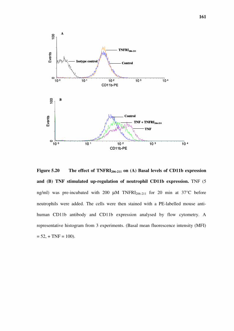
161
Figure 5.20 The effect of TNFRI206-211 on (A) Basal levels of CD11b expression
and (B) TNF stimulated up-regulation of neutrophil CD11b expression. TNF (5
ng/ml) was pre-incubated with 200 µM TNFRI206-211 for 20 min at 37°C before
neutrophils were added. The cells were then stained with a PE-labelled mouse anti-
human CD11b antibody and CD11b expression analysed by flow cytometry. A
representative histogram from 3 experiments. (Basal mean fluorescence intensity (MFI)
= 52, + TNF = 100).
10 0 10 1 10 2 10 3 10 4
CD11b-PE
0100
Events
10 0 10 1 10 2 10 3 10 4
CD11b-PE
01
00
Eve
nts
Isotype control
TNFRI206-211
Control
Control
TNF + TNFRI206-211
TNF
A
B
10 0 10 1 10 2 10 3 10 4
CD11b-PE
0100
Events
10 0 10 1 10 2 10 3 10 4
CD11b-PE
01
00
Eve
nts
Isotype control
TNFRI206-211
Control
Control
TNF + TNFRI206-211
TNF
A
B

162
Figure 5.21 The effect of the scrambled control peptide on (A) Basal levels of
CD11b expression and (B) TNF stimulated up-regulation of neutrophil CD11b
expression. TNF (5 ng/ml) was pre-incubated with 200 µM control peptide for 20 min at
37°C before neutrophils were added. The cells were then stained with a PE-labelled
mouse anti-human CD11b antibody and CD11b expression analysed by flow cytometry.
A representative histogram from 3 experiments. (Basal MFI = 70.15, + TNF = 136.21).
10 0 10 1 10 2 10 3 10 4
CD11b-PE
0100
Events
10 0 10 1 10 2 10 3 10 4
CD11b-PE
0100
Events
Isotype control
Control peptide
Control
Control
TNF + Control peptide
TNF
A
B
10 0 10 1 10 2 10 3 10 4
CD11b-PE
0100
Events
10 0 10 1 10 2 10 3 10 4
CD11b-PE
0100
Events
Isotype control
Control peptide
Control
Control
TNF + Control peptide
TNF
A
B

163
Figure 5.22 The effects of TNFRI206-211 on TNF induced up-regulation of
neutrophil CD11b expression in neutrophils. Different concentrations of TNFRI206-211
were incubated with TNF (5 ng/ml) at 37°C for 20 min. Neutrophils were added and
further incubated for 30 min at 37°C. The cells were then stained with a PE-labelled
mouse anti-human CD11b antibody and CD11b expression analysed by flow cytometry.
Data are expressed as % inhibition of the response seen with TNF alone, presented as
mean ± SEM of three experiments. Significance of difference between TNF and TNF +
TNFRI206-211: * p < 0.05, ** p < 0.01 (Dunnett’s Multiple Comparisons test).
0
20
40
60
80
100
0 50 100 150 200
TNFRI206-211 [µµµµM]
% i
nh
ibit
ion
of
CD
11b
ex
pre
ssio
n
*
**
0
20
40
60
80
100
0 50 100 150 200
TNFRI206-211 [µµµµM]
% i
nh
ibit
ion
of
CD
11b
ex
pre
ssio
n
*
**

164
5.11 EFFECT OF TNFRI206-211 ON TNF-INDUCED CYTOKINE PRODUCTION IN
NEUTROPHILS
A large range of stimuli has been reported to induce cytokine synthesis in neutrophils.
These include classic agonist such as lipopolysaccharide, cytokines themselves,
phagocytic particles, micro-organisms, fMLP, leukotriene B4 and complement
component C5a. The kinetics and magnitude of cytokine release vary depending on the
stimulus used (Cassatella, 1995). Recently NF-κB, p38 and ERK1/EK2 pathways have
been demonstrated to be involved in the generation of inflammatory cytokines by
neutrophils (Cloutier et al., 2007). It was our objective to determine if the TNFRI derived
peptide, TNFRI206-211, was able to inhibit TNF-induced cytokine production by
neutrophils. In vitro assessment of de novo synthesis of cytokines and chemokines by
neutrophils is a major contentious issue which might partly explain recent contradictory
results (Altstaedt et al., 1996, Xing and Remick, 2003, Ethuin, 2005). As neutrophils
themselves do not produce copious amounts of cytokines like other cell types such as
peripheral mononuclear cells, it is highly imperative to work with highly purified
neutrophil preparations (> 99.5 %) and avoid pre-stimulation of cells during experiments.
5.11.1 Effect of TNFRI206-211 on TNF-induced IL-Iββββ production
Highly purified preparations of neutrophils were treated with 0, 0.1, 1, 10 ng/ml of TNF
for 1 h. The RNA was extracted and levels of IL-1β mRNA expression were determined

165
by quantitative real-time PCR. The results show that the level of Il-1β expression was
dose-dependently increased by TNF (Figure 5.23). The kinetics of IL-1β expression was
also investigated at a TNF concentration of 1 ng/ml. The results show that mRNA
expression of IL-1β could be detected as early as 30 min and peaked within 1 h after
stimulation and began to decrease after 2 h (Figure 5.24). When TNF was pre-treated
with 200 µM of TNFRI206-211, there was a significant, 30 %, reduction in IL-1β mRNA
production (Figure 5.25).
5.11.2 Effect of TNFRI206-211 on TNF-induced IL-8 production
Neutrophils were treated with 0, 0.1, 1, 10 ng/ml of TNF for 1 h. The RNA was extracted
and levels of IL-8 mRNA expression was determined by quanitative real-time PCR. The
results show that the level of IL-8 expression was increased by a 4 fold at the highest
TNF concentration tested (figure 5.26). The kinetics of IL-8 production showed a similar
pattern to that of IL-1β production. Thus, IL-8 could be detected as early as 30 min and
peaked within 1 h after stimulation and began to decrease after 2 h (Figure 5.27). When
TNF was pre-treated with 200 µM of TNFRI206-211, there was a significant, 26 %,
reduction in IL-8 mRNA production (Figure 5.28)

166
Figure 5.23 The effect of TNF on IL-1ββββ expression in neutrophils. Cells (1 x 10⁶
in RPMI 1640 supplemented with medium with 2 mM L-glutamine, 100 U/ml penicillin
and 100 µg/ml streptomycin, 2 % AB serum) were stimulated for 1 h with 0, 0.1, 1 or 10
ng/ml of TNF. Levels of IL-1β mRNA were determined by real-time quantitative PCR.
The housekeeping gene, GAPDH, was used to normalize the samples. The data are
expressed as % fold change (2-∆∆Ct), in which ∆Ct = the Ct for cytokine gene minus the
Ct of the housekeeping gene, GAPDH: ∆∆Ct = the ∆Ct of stimulated cells at different
concentrations minus the ∆Ct of unstimulated cells. Data are expressed as % of vehicle
control in the absence of TNF, presented as mean ± SEM of three experiments.
Significance of difference between control and TNF: * p < 0.05 (Dunnett’s Multiple
Comparison test).
0
10
20
30
40
50
0 2 4 6 8 10 12
TNF [ng/ml]
IL-1
ββ ββm
RN
A
(fo
ld c
han
ge)
*
*
0
10
20
30
40
50
0 2 4 6 8 10 12
TNF [ng/ml]
IL-1
ββ ββm
RN
A
(fo
ld c
han
ge)
*
*

167
0
10
20
30
40
50
60
0 20 40 60 80 100 120 140
Time (min)
IL-1
β
β
β
β m
RN
Α (
Α (
Α (
Α (
fold
ch
an
ge)
**
**
Figure 5.24 Kinetics of IL-1ββββ expression in TNF-stimulated neutrophils. Cells (1
x 106 in RPMI 1640 supplemented with medium with 2 mM L-glutamine, 100 U/ml
penicillin and 100 µg/ml streptomycin, 2 % AB serum) were stimulated for 0, 15 min, 30
min, 1 or 2 h with 1ng/ml TNF. Levels of IL-1β mRNA were determined by real-time
quantitative PCR. The housekeeping gene, GAPDH, was used to normalize the samples.
The data are expressed as % fold change (2-∆∆Ct), in which ∆Ct = the Ct for cytokine
gene minus the Ct of the housekeeping gene, GAPDH: ∆∆Ct = the ∆Ct of stimulated
cells at different time points minus the ∆Ct of cells at 0 h. Data are expressed as % of
time zero control, presented as mean ± SEM of three experiments. Significance of
difference at time zero and the different time points: ** p < 0.01 (Dunnett’s Multiple
Comparison test).

168
TNF TNF + TNFRI206-211
IL-1
ββ ββm
RN
A (%
of
TN
F r
esp
on
se) **
0
20
40
60
80
100
Figure 5.25 The effect of TNFRI206-211 on TNF-induced IL-1ββββ mRNA in
neutrophils. TNF (1 ng/ml) was pre-incubated with 200µM TNFRI206-211 for 20 min at
37°C, added to neutrophils in RPMI 1640 supplemented with medium with 2 mM L-
glutamine, 100 U/ml penicillin and 100 µg/ml streptomycin, 2 % AB serum and
incubated for 1 h. Levels of IL-1β mRNA were determined by quantitative real-time
PCR. The housekeeping gene, GAPDH, was used to normalize the samples. The data
(mean ± SEM) are expressed as the % of the TNF response of three experiments.
Significance of difference between TNF and TNF + TNFRI206-211: ** p < 0.01 (Student t
test).

169
Figure 5.26 The effect of TNF on IL-8 expression in neutrophils. Cells (1 x 10⁶ in
RPMI 1640 supplemented with medium with 2 mM L-glutamine, 100 U/ml penicillin
and 100 µg/ml streptomycin, 2 % AB serum) were stimulated for 1 hour with 0, 0.1, 1 or
10ng/ml of TNF. Levels of IL-8 mRNA were determined by real-time quantitative PCR.
The housekeeping gene, GAPDH, was used to normalize the samples. The data are
expressed as % fold change (2-∆∆Ct), in which ∆Ct = the Ct for cytokine gene minus the
Ct of the housekeeping gene, GAPDH: ∆∆Ct = the ∆Ct of stimulated cells at different
concentrations minus the ∆Ct of unstimulated cells. Data are expressed as % of vehicle
control in the absence of TNF, presented as mean ± SEM of three experiments.
Significance of difference between control and TNF: * p < 0.05; ** p < 0.01 (Dunnett’s
Multiple Comparison test).
TNF [ng/ml]
0
1
2
3
4
5
6
0 2 4 6 8 10 12
IL-8
mR
NA
(fo
ld c
han
ge)
*
**
TNF [ng/ml]
0
1
2
3
4
5
6
0 2 4 6 8 10 12
IL-8
mR
NA
(fo
ld c
han
ge)
*
**

170
Figure 5.27 Kinetics of IL-8 expression in TNF-stimulated neutrophils. Cells (1 x
106 in RPMI 1640 supplemented with medium with 2 mM L-glutamine, 100 U/ml
penicillin and 100 µg/ml streptomycin, 2 % AB serum) were stimulated for 0, 15 min, 30
min, 1 or 2 h with 1ng/ml TNF. Levels of IL-8 mRNA were determined by real-time
quantitative PCR. The housekeeping gene, GAPDH, was used to normalize the samples.
The data are expressed as % fold change (2-∆∆Ct), in which ∆Ct = the Ct for cytokine
gene minus the Ct of the housekeeping gene, GAPDH: ∆∆Ct = the ∆Ct of stimulated
cells at different time points minus the ∆Ct of cells at 0 h. Data are expressed as % of
time zero control, presented as mean ± SEM of three experiments. Significance between
time zero and the different time points: ** p < 0.01 (Dunnett’s Multiple Comparison
test).
0
1
2
3
4
5
6
7
0 20 40 60 80 100 120 140
Time (min)
IL-8
mR
NA
(fo
ld c
han
ge)
**
0
1
2
3
4
5
6
7
0 20 40 60 80 100 120 140
Time (min)
IL-8
mR
NA
(fo
ld c
han
ge)
**

171
TNF TNF + TNFRI206-211
IL-8
mR
NA
(%
of
TN
F r
esp
on
se)
0
20
40
60
80
100
*
Figure 5.28 The effect of TNFRI206-211 on the production of TNF-induced IL-8
mRNA in neutrophils. TNF (1 ng/ml) was pre-incubated with 200µM TNFRI206-211 for
20 min at 37°C before neutrophils were added. Neutrophils (1 x 10⁶ in RPMI 1640
supplemented with medium with 2 mM L-glutamine, 100 U/ml penicillin and 100 µg/ml
streptomycin, 2 % AB serum) were stimulated for 1 h. Levels of IL-8 mRNA were
determined by quantitative real-time PCR. The housekeeping gene, GAPDH, was used to
normalize the samples. The data (mean ± SEM) are expressed as the % of the TNF
response of three experiments. Significance of difference between TNF and TNF +
TNFRI206-211: * p < 0.05 (Student t test).

172
5.12 EFFECT OF THE D-AMINO FORM OF TNFRI206-211 AND OTHER VARIANTS OF
TNFRI ON TNF-INDUCED SUPEROXIDE PRODUCTION
Studies using polypeptides or peptides to manipulate the interaction between proteins
have demonstrated that small peptides in their natural (L-form) are susceptible to
proteolytic degradation. When administered in vivo, the L-forms easily succumb to rapid
hepatic and renal clearance. Substitution of susceptible residues in the N or C-terminal
residues with the D-amino forms results in peptides of increased stability (Hong et al.,
1999, Hamamoto et al., 2002, Fischer, 2003, Lien and Lowman, 2003). Thus, since
TNFRI206-211 exhibited the most potent and consistent inhibitory properties, we
synthesised the D-form of this peptide (D-TNFRI206-211), with all residues (EDSGTT)
substituted.
A longer peptide (TNFRI198-211), containing the sequence QIENVKGTEDSGTT was also
synthesised. As the common sequence in these peptides was GTT, a peptide consisting of
tandem repeats of this sequence repeated was also synthesised (GTTGTTGTTGTT) and
tested for the ability to inhibit TNF-induced chemiluminescence generation.
TNF (1 ng/ml) was incubated with D-amino TNFRI206-211 (5-50 µM) for 20 min and then
tested for ability to induce superoxide (chemiluminescence) production in neutrophils.
The data show that at lower concentrations the D-TNFRI206-211 did not affect the ability
of TNF to induce chemiluminescence production. However, pre-treatment of TNF with
50 µM of D-TNFRI206-211 led to a 50 % reduction in chemiluminescence. At this

173
concentration its natural counterpart did not exhibit an inhibitory effect (Figure 5.29 vs.
Figure 5.10).
The effect of the longer TNFRI198-211 was tested on the ability of TNF to stimulate
superoxide production. While TNFRI198-211 did not affect this response when tested at 5
and 10 µM, at concentrations of 20 and 50 µM it caused a 50 % and 75 % reduction
respectively (Figure 5.30). Similarly, the tandem repeats of TNFRI209-211 had no effects
on the neutrophil response when tested at concentrations up to 20 µM. However, this
peptide caused a 45 % reduction in the ability of TNF to stimulate superoxide production
when tested at 50 µM (Figure 5.31).

174
D-amino form of TNFRI206-211 [µµµµM]
% I
nh
ibit
ion
of
TN
F-i
nd
uce
d r
esp
on
se
**
-20
0
20
40
60
80
100
0 10 20 30 40 50
Figure 5.29 Effect of D-amino form of TNFRI206-211 on TNF-induced superoxide
(chemiluminescence) production. TNF (1 ng/ml) was treated with various
concentrations (5, 10, 20 and 50 µM) of D-amino TNFRI206-211 at 37°C for 20 min before
being added to neutrophils. The cells were incubated for 30 min at 37°C. Lucigenin was
then added and the resultant chemiluminescence was measured in a luminometer. Data
represent the % inhibition of the TNF response and are presented as mean ± SEM of
three experiments. The maximal rate of chemiluminescence production in non-stimulated
neutrophils was 101 ± 16 RLU and in TNF-stimulated cells was 3051 ± 606 RLU.
Significance of difference between TNF and TNF + D-TNFRI206-211: ** p < 0.01
(Dunnett’s Multiple Comparisons test).
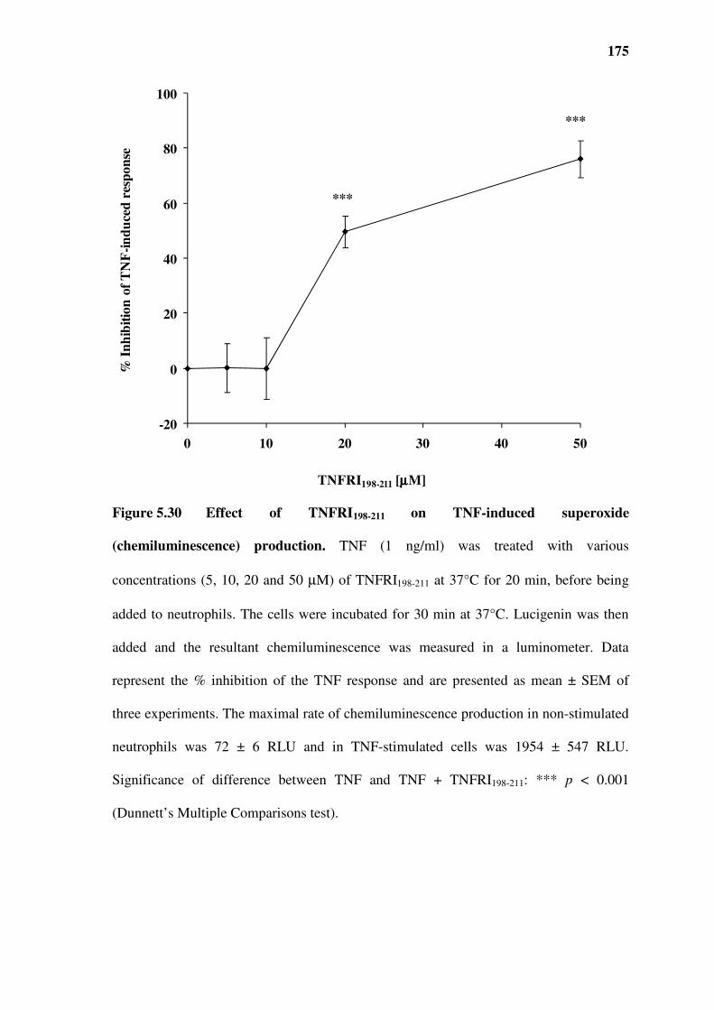
175
TNFRI198-211 [µµµµM]
% I
nh
ibit
ion
of
TN
F-i
nd
uce
d r
esp
on
se
***
***
-20
0
20
40
60
80
100
0 10 20 30 40 50
Figure 5.30 Effect of TNFRI198-211 on TNF-induced superoxide
(chemiluminescence) production. TNF (1 ng/ml) was treated with various
concentrations (5, 10, 20 and 50 µM) of TNFRI198-211 at 37°C for 20 min, before being
added to neutrophils. The cells were incubated for 30 min at 37°C. Lucigenin was then
added and the resultant chemiluminescence was measured in a luminometer. Data
represent the % inhibition of the TNF response and are presented as mean ± SEM of
three experiments. The maximal rate of chemiluminescence production in non-stimulated
neutrophils was 72 ± 6 RLU and in TNF-stimulated cells was 1954 ± 547 RLU.
Significance of difference between TNF and TNF + TNFRI198-211: *** p < 0.001
(Dunnett’s Multiple Comparisons test).

176
-40
-20
0
20
40
60
80
100
0 10 20 30 40 50
Tandem repeats of TNFRI209-211 [µµµµM]
Inh
ibit
ion
of
TN
F-i
nd
uce
d r
esp
on
se
*
Figure 5.31 Effect of a tandem repeat form of TNFRI209-211 on TNF-induced
superoxide (chemiluminescence) production. TNF (1 ng/ml) was treated with various
concentrations (5, 10, 20 and 50 µM) of tandem repeat form of TNFRI209-211 at 37°C for
20 min, before being added to neutrophils. The cells were incubated for 30 min at 37°C.
Lucigenin was then added and the resultant chemiluminescence was measured. Data
represent the % inhibition of the TNF response and are presented as mean ± SEM of
three experiments. The maximal rate of chemiluminescence production in non-stimulated
neutrophils was 112 ± 80 RLU and in TNF-stimulated cells was 7636 ± 2720 RLU.
Significance of difference between TNF and TNF + tandem repeat form of TNFRI209-211:
* p < 0.05 (Dunnett’s Multiple Comparisons test).

177
Table 5.1 Summary of biological activities of TNFRI-derived peptides
TNFRI209-211 TNFRI206-211 D-TNFRI206-211 TNFRI198-211 TNFRI(209-211) (TR)
TNF-induced superoxide 123 µM* 171 µM* 51 µM 50 µM 50 µM
TNF-RM-induced superoxide 216.4 µM* 146.3 µM* ND ND ND
TNF-induced CD11b ND 200 µM ND ND ND
TNF-induced p38 in PMN 200 µM 200 µM ND ND ND
TNF-induced p38 in Mono Mac ND 200 µM ND ND ND
TNF-induced IL-1β production ND 200 µM ND ND ND
TNF-induced IL-8 production ND 200 µM ND ND ND
*Quoted as IC50s.
Abbreviations: IL, interleukin; ND, not determined; PMN, polymorphonuclear cells;
TNF-RM, TNF rich medium, TR, tandem repeat.

178
5.13 SUMMARY
The data show that the mimetic peptide TNF70-80 is able to activate p38 kinase activity by
specifically ligating with a region of the extracellular portion of the TNFRI proximal to
the transmembrane domain. Using truncated TNFRI mutants, we showed that the
remnant 3 amino acids (GTT) are the minimum sequence required for the receptor to
activate p38 activity. Peptides (TNFRI209-211 and TNFRI206-211) to this region of the
receptor were able to block the neutrophil stimulating properties of TNF70-80 and TNF
from stimulated leukocytes when examined at the level of the superoxide production.
These effects were specific since scrambled control peptides did not affect TNF-induced
response tested at similar concentrations. The specificity of these receptor derived
peptides was further established by the lack of effect on fMLP-induced neutrophil
activation.
Furthermore, these studies (Table 5.1) have shown that TNFRI206-211 was able to block
TNF-induced p38 activation in neutrophils and Mono Mac 6 cells. It inhibited TNF-
induced CD11b upregulation in neutrophils. Additionally, TNFRI206-211 was also able to
block TNF-induced IL-1β and IL-8 mRNA production in neutrophils. Increased activity
was achieved by further modification of the TNFR peptide. The D-amino form of
TNFRI206-211 was shown to have inhibitory activity at lower concentrations, compared to
TNFRI198-211 and a tandem repeat of TNFR1209-211 exhibited inhibitory activity at lower
concentrations than TNFRI209-211 (Table 5.1). These data demonstrate that TNF70-80
activated p38 by binding to a specific site of the TNFRI. Thus, peptides to this region

179
were able to block p38 activation and p38-dependent functions such as superoxide
production, CR3 upregulation and cytokine in production in neutrophils. These data
identifies a novel way of targeting the TNF-TNFR-p38 pathway to inhibit specific
actions. Lastly modifications can be made to the TNFRI mimetics to enhance their
activity.

180
6.0 CHAPTER SIX: EFFECTS OF TNFRI-
DERIVED PEPTIDES ON INFLAMMATION

181
6.1 INTRODUCTION
It is well established that TNF plays a major role in several inflammatory diseases such
as RA. This central role of TNF in inflammation has made it an attractive therapeutic
target (Aggarwal et al., 2006). Studies using TNFRI or TNFRII knockout mice have
shown that the neutrophil accumulation in response to LPS is mediated via the TNFRI
(Calkins et al., 2001). The use of anti-TNF antibodies has also shown the importance of
this cytokine in the pathogenesis of inflammatory diseases (Andreakos et al., 2002).
Consistent with these findings, several anti-TNF strategies are now in clinical use to treat
debilitating inflammatory conditions (Table 1.7).
Recently research has been focussing on small peptide inhibitors of TNF. An exocyclic
peptide mimetic of a critical binding site on TNFRI was shown to inhibit bone
destruction in murine collagen-induced arthritis model, to a similar extent as anti-TNF
Ab (Saito et al., 2007). Another peptide inhibitor of TNF based on the pre-ligand
assembly domain (PLAD) of TNFRI has recently been shown to inhibit TNF signalling
by binding to the PLAD of TNFRI thereby preventing TNF aggregation and TNF-TNFR
signalling (Deng et al., 2005). This peptide was similarly shown to inhibit murine CIA
(Deng et al., 2005).
The ability of the TNFRI mimetics to prevent TNF-induced activation of p38 and related
cellular activation, suggest a singular approach to inhibiting inflammation and was
182
examined in three different types of (inflammatory models) LPS-induced peritonitis,
carrageenan-induced paw swelling and delayed type hypersensitivity..
6.2 THE EFFECT OF TNFRI206-211 ON A MURINE MODEL OF ACUTE LPS-INDUCED
PERITONITIS
To investigate the effects of the TNFRI-derived peptides in acute inflammation BALB/c
female mice were injected intraperitoneally with 50 µg of LPS or HBSS, 2 h later the
mice received an injection of different concentrations of TNFRI206-211 or HBSS. At 24 h,
the animals were euthanized and the total leukocytes in the peritoneal exudates
enumerated by haemocytometer chamber. Cytospins of the cellular infiltrates were
prepared by centrifugation of 200 µl (vehicle challenge) or 100 µl (LPS challenge)
aliquots. Slides were fixed and stained with a Giemsa stain. LPS caused a significant
increase in total leukocytes infiltration into the peritoneum, in comparison to the vehicle
or TNFRI206-211 only treated animals (Figure 6.1). Treatment with TNFRI206-211
significantly reduced the total number of infiltrating leukocytes compared to vehicle
treated animals. At the highest dose of 40 mg/kg, TNFRI206-211 caused a complete
reduction in leukocyte numbers (p < 0.001). This effect is displayed in the
photomicrographs of the Giemsa stained cytospins (Figure 6.2)
TNFRI206-211 caused a decrease in total leukocytes which can be explained by differences
observed in the neutrophils and macrophages. LPS caused a dramatic influx of

183
neutrophils into the peritoneal cavity, incresing from a baseline level of 1.2 x 10⁵ to 2.35
x 106 cells. This number was significantly reduced by treating with of 40 mg/kg of
TNFRI206-211 to 1.5 x 10⁶ cells (p < 0.01) (Figure 6.3). The peptide itself did not induce
neutrophil influx. Interestingly, treatment with TNFRI206-211 significantly reduced the
number of peritoneal macrophages even at the lower doses of the peptide tested (Figure
6.4). Although the total macrophages in the peritoneal cavity did not change, it is likely
that this level is a result of reduced resident peritoneal macrophages, from LPS-induced
apoptosis (Xaus et al., 2000, Soler et al., 2001), and monocyte influx. The latter must
likely being inhibited by TNFRI206-211 leading to a lower level of macrophages in the
peritoneal cavity. Indeed studies from our laboratory showed that within 4 h after
injection of LPS, the number of mouse resident peritoneal macrophages was reduced by
approximately 50 %.

184
Vehicle TNFRI206-211 Vehicle 1 4010
Control LPS
TNFRI206-211(mg/kg)
To
tal l
euk
ocy
tes/
x1
06
ml
in p
erit
on
eal
wa
sho
ut
0.0
1.0
2.0
3.0
4.0
5.0
*
**
***
Figure 6.1 Effect of TNFRI206-211 on total leukocyte infiltration in response to
LPS. Mice were challenged intraperitoneally with LPS or HBSS (control) and then
treated with different concentrations of TNFRI206-211 or HBSS. At 24 h, the animals were
euthanized and number of cells in peritoneal washouts enumerated. Data are presented as
mean ± SEM. Control (HBSS) n= 4, TNFRI206-211: n= 3; LPS: n=7; LPS + TNFRI206-211:
n= 7. Significance of difference between LPS challenge alone and LPS and TNFRI206-211
treated: * p < 0.05; ** p < 0.01; *** p < 0.001 (Dunnett’s Multiple Comparisons test).

185
MØ
N
L
M
MØ
M
MØ
M
MØ
N
A B
CD
M
Figure 6.2 Photomicrographs of peritoneal exudate preparation for the effects
TNFRI206-211 on LPS-induced acute peritonitis. Mice received an intraperitoneal
injection of either HBSS (A, B) or LPS (C, D), 2 h later the mice received HBSS (A, C)
or TNFRI206-211 40 mg/kg body weight intraperitoneally. After 24 h animals were
euthanized and cytospins of the cellular infiltrates were prepared by centrifugation of 200
µl (vehicle challenge) or 100 µl (LPS challenge) aliquots. Slides were fixed and stained
with Giemsa (magnification x 40). Mast cell -M; macrophage- MØ; neutrophil- N;
lymphocyte- L.

186
Vehicle TNFRI206-211Vehicle 1 4010
Control LPS
Neu
tro
ph
ils
/ x
10
6m
l in
per
ito
nea
l w
ash
ou
t
TNFRI206-211(mg/kg)
0.0
0.5
1.0
1.5
2.0
2.5
3.0
**
*
Figure 6.3 Effect of TNFRI206-211 on the accumulation of neutrophils in the
peritoneal cavity induced by LPS. Treatments were as per figure 6.1 legend. Slides
were fixed and stained with Giemsa stain and the number of neutrophils in peritoneal
exudate fluid was enumerated. Data are presented as mean ± SEM. Control (HBSS) n= 4,
TNFRI206-211: n= 3; LPS: n=7; LPS + TNFRI206-211: n= 7. Significance of difference
between LPS challenge alone and LPS and TNFRI206-211 treated: * p < 0.05; * p < 0.01
(Dunnett’s Multiple Comparisons test).

187
Vehicle TNFRI206-211 Vehicle 1 4010
Control LPS
Ma
cro
ph
ag
es /x
10
6m
l in
per
ito
nea
l w
ash
ou
t
TNFRI206-211(mg/kg)
0.0
0.5
1.0
1.5
2.06
2.5
* *
***
Figure 6.4 Effect of TNFRI206-211 on the accumulation of macrophages induced
by LPS. Treatments were as per figure 6.1 legend. Slides were fixed and stained with
Giemsa stain and the number of macrophages in the peritoneal exudate was enumerated.
Data are presented as mean ± SEM. Control (HBSS) n= 4, TNFRI206-211: n= 3; LPS: n=7;
LPS + TNFRI206-211: n= 7. Significance of difference between LPS challenge alone and
LPS and TNFRI206-211 treated: * p < 0.05; *** p < 0.001 (Dunnett’s Multiple
Comparisons test).

188
6.3 THE EFFECT OF TNFRI206-211 ON CARRAGEENAN-INDUCED PAW SWELLING
To investigate the effect of TNFRI206-211 on carrageenan-induced paw swelling, female
BALB/c mice received a subplantar injection of Type IV carrageenan (1 ml/kg of a 1 %
solution) in the left hind paw. After 2 h mice were injected intraperitoneally with 40
mg/kg of TNFRI206-211 or control peptide. Footpad thickness was assessed 24 h after
challenge. Injection of carrageenan produced marked paw swelling. Treating mice with
this dose of TNFRI206-211 had no effect on the swelling (Figure 6.5).
One likely reason for the lack of effect of the TNFR mimetic is the limited amount of
peptide which reached the inflammatory foci. Sekut et al., (1994) demonstrated that in
the carrageenan induced paw inflammation model in both mice and rats very little serum
TNF was detected, in contrast to a marked increase in local TNF levels after carrageenan
challenge in the paws. To overcome this potential problem, we took the alternative
approach of injecting TNFRI206-211 directly into the paw, simultaneously with the
carrageenan. Thus in the modified approach mice received a 20 µl injection which
contained a combination of carrageenan equivalent to (1 ml/kg of a 1 % solution) and 10
mg/kg of TNFRI206-211 or control peptide. The footpad thickness was assessed 24 h after
challenge. The results showed that carrageenan induced a marked increase in paw
thickness of 0.9 ± 0.07 mm which was significantly (p < 0.001) reduced by the TNFRI
mimetic but not by the control peptide (Figure 6.6).

189
Incr
ease
in
fo
otp
ad
th
ick
nes
s (m
m)
Vehicle Control Peptide TNFRI206-211
0
0.2
0.4
0.6
0.8
1
1.2
Figure 6.5 The effect of TNFRI206-211 on carrageenan-induced paw
inflammation. Mice were injected with carrageenan (1 ml/kg of a 1 % solution) into the
hind footpad and 2 h later, the animals were treated intraperitoneally with 40 mg/kg of
control peptide TNFRI206-211. The reaction was assessed by measuring the difference in
footpad thickness between challenged and unchallenged footpads 24 h after challenge.
Data are presented as mean ± SEM of 5 mice in each group.

190
Vehicle Control Peptide TNFRI206-211
p < 0.001
Incr
ease
in
fo
otp
ad
th
ick
nes
s (m
m)
0.00
0.20
0.40
0.60
0.80
1.00
p < 0.01
Figure 6.6 The effect of local application of TNFRI206-211 on carrageenan-
induced paw inflammation. Mice were injected with a 20 µl mixture of carrageenan (1
ml/kg of a 1 % solution) and control peptide or TNFRI206-211 (10 mg/kg) or vehicle into
the hind footpad. The reaction was assessed by measuring the difference in footpad
thickness between challenged and unchallenged footpads 24 h after challenge. Data are
presented as mean ± SEM of 4 (Vehicle) and 5 (control peptide and TNFRI206-211).
Significance of difference between carrageenan challenge + vehicle and carrageenan +
TNFRI206-211: *** p < 0.001 (Dunnett’s Multiple Comparisons test). Significance of
difference between control peptide and TNFRI206-211 treated: ** p < 0.01 (Tukey's
Multiple Comparison Test).

191
6.4 THE EFFECT OF TNFRI206-211 ON ANTIGEN-INDUCED DELAYED TYPE
HYPERSENSITIVITY
The delayed type hypersensitivity response to sheep red blood cell (SRBC) is an
important in vivo manifestation of the cell-mediated immune response commonly seen
against intracellular bacteria, several viral, parasitic pathogens and systemic fungal
pathogens as well in chronic inflammatory diseases (Arvin, 2008, Bogdan, 2008,
Mustafa et al., 2008). To investigate the effects of TNFRI206-211 on the DTH response,
female BALB/c mice were primed with an injection of 10 % haemotocrit, SRBC. After
six days, mice received a subplantar 20 µl injection which contained a combination of 40
% SRBC suspension and 12.5 mg/kg of TNFRI206-211 or control peptide. The DTH
response was assessed by measuring the difference between challenged and unchallenged
footpad thickness 24 h after challenge. The results showed that a dose of 12.5 mg/kg of
TNFRI206-211 significantly inhibited the DTH response in mice by 33 % (Figure 6.7)
while the control peptide had no effect.

192
0
0.4
0.8
1.2
1.6
Vehicle Control Peptide TNFRI206-211
Incr
ease
in
fo
otp
ad
th
ick
nes
s (m
m)
**
Figure 6.7 The effect of local application of TNFRI206-211 on DTH response to
SRBC. Mice were injected with 100 µl of a 10 % SRBC solution. Six days later the mice
were challenged with the antigen containing either control peptide or TNFRI206-211 (12.5
mg/kg) or vehicle in the hind footpad. The reaction was assessed by measuring the
difference in footpad thickness between challenged and unchallenged footpads 24 h after
challenge. Data are presented as mean ± SEM of the increase in footpad thickness.
Vehicle n = 9, control peptide n = 9, TNFRI206-211 n = 8. Significance of difference
between SRBC challenge + vehicle and SRBC + TNFRI206-211: ** p < 0.01 (Dunnett’s
Multiple Comparisons test). Significance of difference between control peptide and
TNFRI206-211 treated: * p < 0.05 (Tukey's Multiple Comparison Test).

193
6.5 SUMMARY
The data presented show that the in vitro anti-inflammatory properties of TNFRI206-211
were also evident in these models of inflammation. TNFRI206-211 inhibited the infiltration
of leukocytes, namely neutrophils and macrophages into the peritoneal cavity following
challenge with LPS. Studies with carrageenan induced acute inflammatory model
similarly showed that TNFRI206-211 was able to inhibit swelling. The effects on
macrophages were confirmed in a chronic inflammation model where the peptide was
also shown to inhibit inflammation in the DTH model when challenged with SRBC. A
lack of effect after intraperitoneal administration of TNFRI206-211 demonstrated that its
inhibitory properties were more effective when administered directly into local sites of
inflammation. Nevertheless, the data presented provide proof of principle/concept that
peptides that block cytokine receptors can potentially be developed into therapeutics. The
findings show that TNFRI206-211 can inhibit acute and chronic inflammatory responses.

194
7.0 CHAPTER SEVEN: DISCUSSION

195
7.1 INTERACTION OF TNF70-80 WITH THE TNFRI
Previously the biological actions of TNF70-80 had not been shown to be directly mediated
by the interaction of the peptide with the TNFR. Using labelled TNF70-80 in a competition
assay, we demonstrate for the first time that the peptide is a ligand for TNFRI. The
previously observed selective biological properties of TNF70-80 (Kumaratilake et al.,
1995) were due to its ability to activate only the p38 and NF-κB and not the JNK and
ERK1/ERK2 pathways. Similarly the selective properties of TNF132-150 were due to its
ability to activate JNK and ERK1/ERK2 and not p38 pathways. The inability of TNF70-80
to activate all three MAPK pathways, activated by TNF was found to be due to the
partial recruitment of adaptor proteins. TNF70-80 was able to couple TRAF2 and not GCK
or ASK-1 to the TNFR. The incomplete activation of the MAPK could be due to the
small size of the peptide and its inability to aggregate the TNFR into a functional trimer,
leading to an inappropriate recruitment of adaptor proteins. By means of truncated
mutant extracellular regions of TNFRI, we have demonstrated that TNF70-80 activates p38
by interacting with a specific site of the TNFR (GTT). Longer peptides to this region
were able to inhibit TNF-induced p38 activation and p38-mediated effects in vitro and in
vivo, and consequently could be potential therapeutics in targeting the TNF-TNFR-p38
axis.
The striking finding that TNF70-80 has selective TNF activity has raised the issue as to
whether the peptide ligates the TNFRI in a manner similar to TNF. While previous
findings showed that the activity of TNF70-80 could be neutralised by treatment with

196
soluble TNFRI and TNFRII or anti-TNF antibodies (Kumaratilake et al., 1995, Britton et
al., 1998, Briscoe et al., 2000), issues still remain as to whether the differential biological
activity is through the receptor. We have provided direct evidence of the importance of
the TNFRI for its biological activity. Thus TNF70-80 failed to stimulate activity (p38) in
the pre-B cell line 70 Z/3 which lack TNFRs. Transfection of the TNFRI into these cells
resulted in their responsiveness to TNF70-80.
While these results show that TNFRI is essential for the activity of TNF70-80, they do not
prove that the mimetic is a ligand for the receptor. Results from further investigations
demonstrated that labelled TNF70-80 could be specifically displaced from the TNFRI by
unlabelled TNF70-80 using scatchard analysis. Previously the cytotoxic mimetic peptide
TNF132-150 was shown, to displace radiolabelled 125I-TNF binding to TNFR on WEHI-
164 cells (Rathjen et al., 1993). These results therefore show that both are ligands for
TNFR but transduce different signals from each other and TNF.
The hallmark of a true cytokine mimetic is its ability to interact with its putative receptor.
One such is a recently described peptide of IL-2, corresponding to amino acids 1-30
which are essential for binding to the IL-2Rβ2 chain and are critical for signal
transduction (Rose et al., 2003). Using theoretical 3-D models of free peptide amino acid
1-30 (p1-30) and IL-2Rβ2, a model of p1-30•IL-2Rβ2 complex they proposed a
mechanism by which signal transduction occurs through a conformational change of the
COOH-terminal domains of the receptor, resulting in the transmembrane regions coming
together facilitating the transmission of signal to the cytoplasm (Rose et al., 2003). Thus

197
it is possible for a small peptide region such as TNF70-80 to interact specifically with a
receptor allowing for signal transduction.
The previous findings that the biological properties of TNF70-80 could be blocked by
soluble TNFRs and anti-TNF antibodies, together with our present findings show that not
only is TNFRI required for activation of cellular signals, but that there is a specific
interaction of the mimetic with TNFRI, strongly favouring a selective signalling of cells
through interacting with a specific region of TNFRI.
7.2 SELECTIVE ACTIVATION OF MAPK PATHWAYS
In neutrophils, both TNF and TNF70-80 was shown to stimulate p38 but not JNK or ERK
activity. In endothelial cells, however, there were striking differences in the ability to
activate MAPK between TNF and TNF70-80. Our results showed that while both TNF and
TNF70-80 were able to stimulate p38 activity in HUVECs, only TNF stimulated JNK and
ERK1/ERK2 activity. In contrast, while TNF was able to activate all three MAP kinases
in adherent WEHI-64 cells, TNF132-150 only stimulated the activity of JNK and ERK1/2
but not p38 at concentrations which were shown to cause cytotoxicity in WEHI-164 cells
(Rathjen et al., 1993). Our data demonstrated that TNF132-150 stimulated JNK and
ERK1/ERK2 activity to a similar extent to TNF. It is therefore likely that an inability of
TNF132-150 to activate neutrophils can be attributed to its inability to stimulate p38 kinase
activity.

198
Binding of the trimeric TNF to the p55 kDa TNF receptor complexes initiates the
recruitment of receptor-associated proteins such as TRAF-2, TRADD and FADD to the
TNF receptors (Chen and Goeddel, 2002). One possible explanation for the restricted
activation of the MAPK family by TNF70-80 is an inappropriate recruitment of these
receptor-associated proteins compared to TNF, and in this manner having a major impact
on the range of downstream signalling molecules stimulated. Studies have shown that
TRAF2 is essential for TNF-induced activation of MAPK (Liu et al., 1996, Reinhard et
al., 1997, Yeh et al., 1997, Liu and Han, 2001). Interestingly transfection of the
dominant negative form of TRAF2 blocked the activation of p38 by TNF70-80. We
conclude that TRAF2 was recruited to the TNF receptors and was required for the
activation of p38 by TNF70-80. TNF70-80 was found in comparison to TNF to be poor at
stimulating GCK activity in adherent neutrophils. This implies that TNF70-80 is unable to
cause the coupling of TRAF2 to an upstream regulator(s) of the JNK module (Figure
7.1).
The selective activation of the peptides could also be due to their inability to cause the
TNFR to form a functional trimer, as the binding of a single TNF to single receptor is not
sufficient to transduce a signal, rather the homotrimer of TNF is essential for intracellular
domains of the TNFR to dimerise and trimerise for signal transduction to occur
(Locksley et al., 2001, Schottelius et al., 2004). Thus the peptides existing as monomers
might not be able to cause trimerisation of the TNFR resulting in partial recruitment of
adaptor proteins and consequently partial activation.

199
The ability of TNF70-80 to prime neutrophils and activate microbicidal properties such as
the respiratory burst is comparable to that of TNF, whose properties have been well
established. (Kumaratilake et al., 1995). The role p38 in TNF-induced
chemiluminescence response is well established. While the p38 inhibitor SB203580
inhibited the TNF-induced respiratory burst activity, the ERK1/ERK2 inhibitor,
PD98059 had no effect (Zu et al., 1998). Thus the production of oxygen intermediates by
neutrophils is primarily mediated by p38 MAPK and not by ERK. Thus it is likely that
TNF70-80 activates neutrophils by stimulating p38 activity. The inhibitory effect of
SB203580 on TNF70-80-induced chemiluminescence response provides further evidence
of TNF70-80 mediating its effects via the p38 pathway. p38 inhibitors have also been
shown to reduce CD11b/CD18 upregulation in response to TNF; while treatment with
ERK1/ERK2 inhibitors had no significant effect (Tandon et al., 2000, Forsberg et al.,
2001, Bouaouina et al., 2004).
Studies using cell lines expressing dominant negative mutants of MKK4 or JNK were
protected from TNF-induced apoptosis, thus delineating the importance of JNK in
promoting apoptosis (Roulston et al., 1998). The fact that TNF70-80 was unable to
stimulate JNK activity is not surprising as previous work has shown that TNF70-80 lacked
cytotoxic effects via the p38 pathway both in vitro and in vivo against tumour cells
(Rathjen et al., 1993).
The findings have therefore revealed that is possible to exploit the TNFR-p38 signalling
pathway to promote anti-microbial function of the immune system. When injected into
mice the peptide TNF70-80, while enhancing the clearance of microbial pathogens, did not

200
show the usual TNF toxic properties and the upregulation of adhesion molecules
expression in endothelial cells (Rathjen et al.) Presumably this was due to an inability or
poor ability to stimulate JNK and ERK1/ERK2.

201
Figure 7.1 Diagrammatic representation of coupling of TNF70-80 to TNFRI and
associated adaptor proteins and the inferred coupling of TNF132-150. The __
represents the characterised coupling of adaptor proteins while the --- represent the
inferred coupled proteins. Abbreviations: ASK1, apoptosis signal-regulating kinase;
ERK1/2, extracellular-signal-regulated kinases; GCK, germinal centre kinase; IKK, IκB
kinase; MEK1/2, MAPK/ERK kinase, MEKK3/6, MAPK kinase kinase 3/6; NFκB,
nuclear factor kappa B; NIK, NF-κB inducing kinase; IκB, inhibitor kappa B; NSMase,
neutral sphingomyelinase; RIP, receptor interacting protein; TRADD, TNFR associated
DD; TRAF2, TNFR associated factor 2.
TRADDTRADD
TRAF2TRAF2
GCKGCK
MKK4/7MKK4/7
JNKJNK
ASK1ASK1MEKK1MEKK1
MEK1/2MEK1/2
ERK1/2ERK1/2
TNFRITNFRITNFRITNFRI
TRADDTRADD
TRAF2TRAF2
RIPRIP
IKK complexIKK complex
NIKNIK
IIκκκκκκκκBBαααααααα
NFNF--κκκκκκκκBB
pIpIκκκκκκκκBBMKK3/6MKK3/6
p38p38DegradationDegradation
TNFTNF7070--8080
TNFTNF132132--150150
NSMase?NSMase?
TRADDTRADD
TRAF2TRAF2
GCKGCK
MKK4/7MKK4/7
JNKJNK
ASK1ASK1MEKK1MEKK1
MEK1/2MEK1/2
ERK1/2ERK1/2
TNFRITNFRITNFRITNFRI
TRADDTRADD
TRAF2TRAF2
RIPRIP
IKK complexIKK complex
NIKNIK
IIκκκκκκκκBBαααααααα
NFNF--κκκκκκκκBB
pIpIκκκκκκκκBBMKK3/6MKK3/6
p38p38DegradationDegradation
TNFTNF7070--8080
TNFTNF132132--150150
NSMase?NSMase?

202
7.3 TNF70-80 BINDING REGION- IDENTIFICATION OF A TNF ANTAGONIST
Using truncated constructs of TNFRI we were able to localise the sites on the receptor
involved in the TNF70-80 induced p38 activation. Constructs designated M1, M3 and M4
were made and transfected into the pre B 70Z/3 cell line. TNF70-80 was able to stimulate
p38 activity in cells transfected with M1, M3 and M4 mutant receptors. Since the M4
region was common to all three mutants this region was the minimum region of the
TNFR required by TNF70-80 to activate p38.
Based on the interaction of TNF70-80 and TNFRI, peptides derived from the remnant
receptor sequence (GTT) were generated to determine whether it had the ability to block
TNF stimulation of the p38 pathway. TNFRI206-211 blocked TNF-induced p38 activation,
superoxide production, CD11b/CD18 upregulation and cytokine production in
neutrophils. Similar results were found with TNFRI209-211. The specificity of the action of
these TNFR mimetics was shown by the fact that these did not inhibit the fMLP-induced
superoxide production in neutrophils and that control peptides of these mimetics were
not inhibitory to the TNF-induced response in neutrophils. The inhibition of the TNFR-
p38 signalling axis was not restricted to neutrophils; TNFRI206-211 was also able to block
p38 activation in Mono Mac 6 cells, a macrophage cell line. The fact that TNFRI206-211
(EDSGTT) was also able to inhibit these functions is further evidence to this peptide
blocking the TNF-p38 pathway.

203
TNF is a key cytokine in many types of inflammatory diseases. Neutrophils have also
been reported to play an important role in pathogenesis of inflammation (Harris, 1990,
Sewell and Trentham, 1993, Keshavarzian et al., 1999, Witko-Sarsat et al., 2000,
Kasama et al., 2005, Nathan, 2006). In RA, neutrophils are found in large numbers
during the early stages and acute exacerbation of the disease. Neutrophils accumulate at
the pannus-cartilage junction (site of early cartilage erosion), where they are thought to
contribute directly to cartilage damage by production of reactive ROS and chloraminated
oxidants. This activation of neutrophils is mediated by TNF and IL-1β (Witko-Sarsat et
al., 2000, Dayer, 2003, Aggarwal et al., 2006). Here we demonstrate that the events
stimulated by TNF can be controlled by the TNFRI mimetic. This includes oxygen
radical generation, upregulation of CR3 receptors and IL-1β production. All were
significantly depressed by TNFRI206-211. It should be noted that under cytokine
stimulation the conformational changes leading to change in affinity of the integrins are
more important, as increased CD11b expression does not necessarily equate to
augmented CD11b-adhesive capacity (Orr et al., 2007). It was however evident that
while there was complete prevention of TNF-induced p38 activation the biological
functional responses were not inhibited to the same degree. This suggests that p38 is
utilised to maximise the TNF response. Perhaps this property may be beneficial in
enabling the maintenance of a level of microbial pathogen killing ability by phagocytic
cells.
The identification of TNF inhibitors is the goal of research into this cytokine. Early
structure function research identified two TNFRI derived synthetic peptides that inhibited
TNF-induced cytotoxicity and binding to TNFRI (Hwang et al., 1991, Lie et al., 1992).

204
These peptides are in fact overlapping in their amino acid sequences and are located
within the third and fourth cysteine rich domains of the extracellular portion of the
TNFRI. The first peptide, corresponding to amino acids 175-194, inhibited TNF-induced
cytolysis in L929 cells at a concentration of 200 µM by 50% (Hwang et al., 1991). It is
evident that the majority of the residues in this peptide (RENECVSCSNCKKSLESTKL)
are hydrophilic in nature. Hydrophilic regions of proteins are known to be exposed
compared to their hydrophobic counterparts and thus are thought to play an important
role in the binding and interaction with their ligands (Lie et al., 1992). The subsequent
receptor peptide corresponded to amino acid residues, 159-178. This peptide, although
found to inhibit TNF-induced cytolysis in L929 cells to a similar degree as TNFRI175-194,
required a concentration of 1000 µM in comparison to the former which inhibited at 200
µM. Interestingly, this peptide (QEKQNTVATAHAGFFLRENEG) contained more
hydrophobic residues such as alanines which were used as substitutes for cysteine and
phenylalanine (Lie et al., 1992). The authors suggest that the difference in the activities
of the two TNFR mimetics is due to the increased hydrophobicity of this region. The
more hydrophilic peptide (TNFRI175-194) could have improved binding between the
peptide and TNF, but the specific sequence of the binding site must be taken into
account. However, perhaps this region (TNFRI159-178) is not a critical in the interaction
with TNF and also the substitution of cysteines for alanines might have interfered with
the specific interaction leading to lower inhibitory activity.

205
7.3.1 Structural modification to TNFRI derived peptides
Our studies demonstrated that the longer peptide of the region, TNFRI198-211
(QIENVKGTEDSGTT) exhibited greater inhibitory activity, with an IC50 of 50 µM
compared to 171 µM of the shorter TNFRI206-211. This improved activity may be due to
its longer length providing a greater binding surface for TNF (Table 7.1).
Although the tandem repeat form of TNFRI209-211 did not show inhibitory properties at
lower concentrations, it did cause a 45 % reduction at 50 µM which is greater than that
observed at a similar concentration of TNFRI209-211, suggesting that the repeat form was
able to achieve a more stable interaction (Table 7.1). The use of tandem repeats of
peptides to enhance activity has recently been used to increase the anti-viral activity of a
mimetic peptide of lactoferrin. The peptide corresponds to residues 600-632, which are
thought to be important in the binding to hepatitis c virus envelope protein, thus
inhibiting the infection (Nozaki et al., 2003). Tandem repeats (two and three repeats) of
the 33-mer peptide, decreased the IC50 against viral infected cultured hepatocytes from
23 µM to 10 µM (Abe et al., 2007). Thus making tandem repeats of a small peptide is a
viable way to enhance activity.
Extending our studies to a D-amino acid substituted form of TNFRI206-211 showed that,
this form had enhanced inhibitory effect on TNF-induced respiratory burst in comparison
to the natural L-form, most likely due to increased stability of the D-form. D-amino acid
substitution has recently been applied to a mimetic peptide of granulysin- a cytolytic

206
protein localised to cytotoxic lymphocytes and natural killer cell granules (Hamamoto et
al., 2002). Different mimetic peptides of granulysin have been shown to have
antimicrobial properties against various microorganisms such as: Escherichia coli and
Staphylococcus aureus (Wang et al., 2000, Hamamoto et al., 2002). However, it was
found that the natural L-form of the peptide (residues 23-42) was susceptible to
proteolytic degradation by proteases present in serum. Partial or total substitution with D-
amino acids resulted in decreased susceptibility to degradation by trypsin and foetal calf
serum (FCS) compared to the natural L-form of the peptide, with the resistance to
proteolytic degradation being greater in the wholly substituted peptide compared to the
partially substituted one. When the FCS was pre-incubated with protease inhibitors such
as aprotinin, the antimicrobial activity of the L-peptides was recovered (Hamamoto et al.,
2002).
Total D-amino acid substitution compared to single D-amino acid replacement was
similarly proven to be advantageous with selectin based mimetic peptides that inhibit
neutrophil infiltration into inflammatory sites (Briggs et al., 1996). When all the residues
in the heptapeptide were replaced with their D-amino acid forms, the in vitro IC50 was
less than half that of the natural L-form. Interestingly, single substitutions at various
positions resulted in loss of activity of the peptide, with IC50 increasing by 10 fold
(Briggs et al., 1996). The L-form of TNFRI206-211 had an IC50 of 171 µM for TNF-
induced chemiluminescence response while its D-amino acid form had an IC50 of 51 µM
(Table 7.1).

207
Thus the finding that the D-amino form of TNFRI206-211 was a more potent inhibitor than
its natural L-amino acid form highlights the importance of making this L to D-form
substitution to increase not only the in vitro but the in vivo stability of peptides in order
to maximise their effects.

208
Table 7.1 Inhibition of TNF-induced superoxide production by TNFRI-
derived peptides in neutrophils
Peptide Sequence IC50 (µµµµM)
TNFRI209-211 GTT 123
Tandem repeat of TNFRI209-211 GTTGTTGTTGTT 50 (45 % inhibition)
TNFRI206-211 EDSGTT 171
D-amino-TNFRI206-211 EDSGTTa 51
TNFRI198-211 QIENVKGTEDSGTT 50
aItalic letters denote D-amino acids.
209
7.4 ANTI-INFLAMMATORY PROPERTIES OF TNFRI DERIVED PEPTIDES
The in vitro inhibitory properties of the TNFRI peptides on leukocyte functions were
associated with in vivo anti-inflammatory activities. The leukocyte infiltration induced by
LPS injection into the peritoneal cavity of mice was inhibited by TNFRI206-211. This was
reflected both in a significant decrease in neutrophil and macrophages accumulation after
24 h. The inhibitory effects were likely a result of the prevention of TNF-induced p38
activation, as LPS-induced inflammation is dependent on TNF and TNFRI (Skerrett et
al., 1999). Previously, in mice challenged with LPS TNF production reached a peak 2 h
after challenge (Calkins et al., 2001), coinciding with the peak production of TNF. TNF
is critical for normal neutrophil recruitment into inflammatory sites. TNFRI knockout
(KO) mice, challenged with LPS exhibited a reduction in neutrophil accumulation and in
particular a reduction in the chemokines MIP-2 and KC, compared to WT and TNFRII
KO mice and most likely explain the inhibitory effects of our TNFRI mimetic (Calkins et
al., 2001). These findings highlight the importance of TNF in the production of the
chemotactic chemokines which promote the large influx of leukocytes (Calkins et al.,
2001).
While the mechanisms of the inhibitory action of the receptor peptide were not
investigated, these were likely to result from the blocking of p38 and NF-κB activation
through TNFRI and downregulating cytokine and chemokines production from resident
peritoneal macrophages. Inhibiting these pathways is also likely to be responsible for the

210
inhibition of phagocyte oxygen radical production, and upregulation of CR3 expression
ultimately contributing to decreased leukocyte activation and infiltration.
The pro-inflammatory and destructive potential of TNF is mediated through activation of
p38. There is evidence to support a key role in inflammation, such as its important in
stimulating the production of pro-inflammatory mediators responsible for leukocyte
recruitment and activation including IL-1β, TNF, IL-8, and IL-6. In a recent study, levels
of phosphorylated MKK3/6 (an upstream regulator of p38) were found to be increased in
the synovial membranes of RA patients, in comparison to those suffering from
degenerative joint disease such osteoarthritis. MKK3/MKK6 was constitutively
expressed in both patient groups (Chabaud-Riou and Firestein, 2004). The use of p38
inhibitors has also been profiled in several inflammatory models in vivo. Accordingly, its
inhibitory activity has been displayed in a wide variety of TNF-mediated disease models
such as arthritis, bone resorption and endotoxin shock (Badger et al., 1996, Escott et al.,
2000, Haddad et al., 2001, Nishikori et al., 2002, Kumar et al., 2003, Miwatashi et al.,
2005).
The p38 inhibitors have been specifically used in LPS-induced inflammation models to
achieve the same goals as those involving anti-TNF or TNFRI KO mice application.
Underwood et al., (2000) showed that when the p38 inhibitor was administered 1 h
before and 4 h after LPS challenge (via inhalation), there was significant and dose
dependent reduction in neutrophil and mononuclear cells found in the bronchoalveolar
lavage (BAL) fluid (Underwood et al., 2000). Similarly, another study also demonstrated
the reduction in BAL neutrophil numbers following oral administration of p38 inhibitor
211
30 min prior to LPS challenge (Haddad et al., 2001). The decreased number of
infiltrating leukocytes observed in our study could also be due to TNFRI206-211 inhibiting
the p38-mediated TNF-induced expression of cell adhesion molecules by endothelial
cells. LPS or TNF alone have been shown to induce upregulation of cell adhesion
molecule on endothelial cells. Interestingly, together they act synergistically to increase
cell adhesion molecules (CAM), via NF-κB and p38 pathways and JNK (Jersmann et al.,
2001). Thus the reduction in leukocyte infiltration in the peritoneal cavity observed in
our studies could be due to decreased CAM expression resulting in a decreased number
of infiltrating leukocytes due to reduced extravasation. TNF is known to induce IL-6 and
chemokines, IL-8 and MCP-1 production in endothelial cell expression via the p38
pathway (Pober, 2002, Westra et al., 2005). Thus, the reduction in macrophages and
neutrophils observed in our studies could also be due to reduced chemokine expression
resulting in reduced number of infiltrating leukocytes.
The inhibitory effects of the TNFRI peptides were not restricted to the LPS model. Local
application of TNFRI206-211 significantly inhibited the carrageenan-induced paw
inflammation. The carrageen-induced acute paw inflammation has been linked to
neutrophil infiltration and the production of neutrophil-derived oxygen free radicals
(Salvemini et al., 1996, Posadas et al., 2004). The importance of TNF in this
inflammatory response has recently been demonstrated. Using TNFRI KO mice, Rocha
et al., 2006 showed that paw swelling was greatly reduced in TNFRI KO mice compared
to WT mice. Interestingly, intravenous administration of murine neutralising anti-TNF
antibody 5 min before carrageenan challenge reduced the initial paw oedema formation
for up to 4 h. Treatment with the antibody 5 min before and 3 h after challenge reduced

212
paw oedema throughout the 24 h period (Rocha et al., 2006). In our studies on
carrageenan-induced paw inflammation when the TNFRI206-211 was administered
intraperitoneally no effect was observed suggesting a need to control/inhibit the action of
TNF that was produced locally at the site of inflammation (Sekut et al., 1994).
Interestingly, co-administration of carrageenan and TNFRI206-211 into the paw led to a
significant reduction in the swelling formation. The lack of effect of TNFRI206-211 when
administered into the peritoneum, suggests that either the peptide, owing to its small size
was highly labile or was rapidly removed by the kidneys or due to its natural L-amino
acid confirmation, was susceptible to degradation by proteases present in serum before it
reached the site of inflammation. Our data is thus in agreement with other studies that
have used TNFRI knockout mice, anti-TNF therapy or anti-TNF synthesis agents
(thalidomide) showing that TNF plays a major role in the carrageenan inflammation
response (Rocha et al., 2006, Mazzon et al., 2008). Furthermore our findings suggest an
important role for the p38 pathway in this disease model, which is also in agreement with
studies which have utilised a specific p38 inhibitor in the same model (Nishikori et al.,
2002). However, with pharmacological inhibitors of p38, all responses requiring this
signalling pathway will be affected.
The DTH response is a type IV hypersensitivity reaction which is characterised by a
large cellular infiltrate into the affected sites on subsequent exposure to antigen,
particularly macrophages, sensitised antigen specific T cells and neutrophils, as well as
the production of a cascade of chemokines and cytokines namely IFNγ, TNF, LT and IL-
8 (Li et al., 1994, Buchanan and Murphy, 1997). Studies have shown that Th1 cytokines,
IFNγ, IL-2 and LT, are important in eliciting the DTH response with IFNγ as the main

213
contributor, as treatment with anti-IFNγ antibodies led to a 50 % reduction of the
response (Li et al., 1994). In our studies, local application of TNFRI206-211 caused a
significant inhibition of the DTH response to SRBC. However, the inhibition was only
30 % compared to 60 % of the carrageenan response. This discrepancy could be due to
the differences in cellular infiltrates and cytokine milieu in these two models. The
carrageenan response has been shown to have a predominantly neutrophil influx with
TNF playing an essential role in the development of the response (Sekut et al., 1994,
Rocha et al., 2006, Mazzon et al., 2008) . In contrast, although, TNF, is also produced in
the DTH response, the amounts produced are significantly less than those of IFNγ
(Buchanan and Murphy, 1997). In some DTH responses TNF has been shown to play a
central role in the inflammation since treatment with anti-TNF antibodies abolished the
response. For example, in the DTH model in response to the chemical irritant
trinitrochlorobenzene, anti-TNF antibodies caused complete abolition of the response
while anti-IFNγ antibodies only caused a 50 % reduction (Piguet et al., 1991). Thus there
is a differential use of TNF and IFNγ between different DTH models and attempts to
block a DTH responses (a chronic inflammatory response) need to take this into
consideration.
The kinetics of cytokine production in the DTH response compared to that of the
carrageenan model should also be taken into consideration to explain the differences in
the action of TNFRI206-211. For example, in the DTH response to Cryptococcus neoforms,
the local production of TNF was significantly increased between 18 – 24 h post challenge
and thereafter (Buchanan and Murphy, 1997). However, it should be noted that their
kinetic studies started 6 h after initial challenge and that the baseline levels of TNF were

214
200 pg/ml which is suggestive of an early surge in TNF production. The same group also
reported that during the cellular infiltration stage, neutrophils were the first to appear
followed by lymphocytes and monocytes (Buchanan and Murphy, 1997). The initial
increase in local TNF production is often associated with an increase in infiltrating
neutrophils in different inflammation models (Zhang et al., 1992). Thus, it can be
inferred that since their kinetic studies started 6 h after challenge, it might be possible
that there is an initial surge in TNF production which leads to the influx of neutrophils.
Since our peptide was injected along with the SRBC on challenge we would assume that
it was acting on TNF released during the first few hours. Subsequent to this antigen
challenge, antigen specific T cells arrive and begin to secrete large amounts of IFNγ,
which results in activation of macrophages, promoting the secretion of additional
inflammatory cytokines. However, if the levels of TNF in our DTH response to SRBC
reached a peak as late and are as sustained as in the study by Buchanan and Murphy
(1997), then it is possible that the decreased inhibitory effects were due to the
consistently high levels of TNF during the course of the response (Buchanan and
Murphy, 1997). Our studies thus show that targeting the TNF-TNFR-p38 pathway using
TNFRI206-211 is a viable and effective way to curb inflammation. We have shown that
blocking one of the pathways by which TNF mediates its effects can lead to significant
inhibitory effects.
7.5 THE THERAPEUTIC POTENTIAL OF TNFRI DERIVED PEPTIDES

215
The driving force behind anti-TNF therapy has come from our increasing knowledge of
the pathogenesis of RA. Originally thought to be a cell-mediated disease, the discovery
of inflammatory cytokines in the synovium and plasma of RA patients shifted the focus
to attempts to develop neutralising agents of TNF (Feldmann and Maini, 2001, Taylor,
2003, Tracey et al., 2008). Three protein-based drugs in the form of MAbs or soluble
receptors are currently approved by FDA for use namely: etanercept, infliximab and
adalimumab (Taylor, 2003, Via, 2007). Due to their protein nature all have to be
administered via injection. Although these biologics have revolutionalised the treatment
of RA, the most prevalent concerns with all three drugs has been an increased incidence
of re-exacerbation of latent tuberculosis, an increased incidence of lymphoma and a
potential for worsening congestive heart failure (Gabriel, 2008, Tracey et al., 2008).
This, coupled with the cost of manufacturing these large molecules resulted in a shift to
develop small molecule inhibitors. However the specificity of these new compounds is
paramount to ensure desired blockade of a specific target. A poor example of such an
agent is CC-10004, an oral anti-inflammatory drug for use in psoriasis, which is currently
in phase II clinical trials. Although results so far have been favourable, CC-10004
inhibits the production of TNF as well as IL-2, IFNγ, phosphodiesterase-4, leukotrienes
and nitric oxide synthase (Via, 2007). It can be envisaged that there will be many
considerable side effects due to the non-specific nature of the agent.
The evidence that many of the pathological effects associated with TNF are mediated by
the p38 pathway, as well as p38 itself promoting the stability of inflammatory cytokine
mRNA, has led research to focus on p38 inhibitors as a way to curb TNF’s deleterious
effects. In 2005, the combined global sales of etanercept, infliximab and adalimumab

216
were about US$7.5 billion. This figure includes sales for RA, Crohn’s disease,
ankylosing spondylitis and psoriatic arthritis (Via, 2007). Not surprisingly, many
pharmaceutical companies have intensified their search for drugs that target p38. Many
studies have demonstrated the efficacy of p38 inhibitors in animal models of diseases and
inflammation. The arthritis model is the most common of animal models (Wadsworth et
al., 1999, Badger et al., 2000, McLay et al., 2001), though the p38 pathway has also
proved to be important in lung inflammation, cerebral ischemia and burn wounds
(Barone et al., 2001, Kawashima et al., 2001, Ipaktchi et al., 2006). A few of these
inhibitors have progressed to phase I and II clinical trials, such as the GSK-681323 and
GSK-856553 for RA, Crohn’s disease and psoriasis (Molfino, 2005), RWJ-67657 for
Crohn’s, RA, psoriasis and rhinitis (Fijen et al., 2001) and VX-745 for RA, Crohn’s and
psoriasis (Stelmach et al., 2003). The results of the phase I clinical trials for the former
two are yet to be released, while trials had to be discontinued for the latter. p38 is
expressed ubiquitously in all cell types, and in addition to its well known inflammatory
functions, it is also involved in regulating other cellular responses such as apoptosis, cell
cycle regulation and proliferation (Ono and Han, 2000). As a result of the wide range of
cellular processes that p38 regulates, the possibility of adverse reactions from
pharmacological blockade has been the bottleneck in development of anti-p38
therapeutics. The Vertex drug mentioned above (VX-745), was discontinued because of
an undisclosed toxic effect in the central nervous system (Palladino et al., 2003). Most
other compounds have run into dose-limiting toxicities thus limiting their development,
with unwanted side effects such as liver enzyme elevations with p38 inhibitor BIRB 796
which was in phase I trial for asthma, allergy and RA (Behr et al., 2003, Dambach, 2005,
Kuma et al., 2005, Schindler et al., 2007).

217
Our TNFRI derived peptide offers a “safer” alternative as it only affects the TNF-TNFR
p38 pathway, thus eliminating the possibilities of unwanted adverse effects from the
systemic blockade of the p38 pathway. TNFRI206-211 has shown potential to have
increased specificity without compromising the full immune enhancing effects of TNF,
as seen with anti-TNF therapies such as antibodies and soluble receptors that block the
entire action of this cytokine (Aggarwal et al., 2006, Tracey et al., 2008).
It should be noted nonetheless, that despite not being able to provide direct evidence of
these TNFRI-derived peptides interfering with TNFRII; the following factors should be
taken into account. Firstly, the neutrophil stimulatory properties of TNF70-80 were shown
to be neutralised by mAbs to both TNFRI and TNFRII (Kumaratilake et al., 1995).
Accordingly, we can infer that TNF70-80 has a similar direct interaction with TNFRII as
shown with TNFRI in our binding studies. Secondly, the initial peptides synthesised,
were shown to inhibit TNF70-80 induced effects in neutrophils. The exact contribution of
TNFRII signalling to TNF’s biological effects remains a contentious and poorly
understood area. While, the predominant evidence supports the accessory role of
TNFRII, i.e. “ligand passing” of TNF to TNFRI, recent studies have elucidated an
important role for TNFRII (Tartaglia et al., 1993). It follows that, while soluble TNF
(sTNF) and membrane bound (mTNF) ligands can bind to both TNFRI and TNFRII,
there is a tendency for sTNF to couple to TNFRI and mTNF to TNFRII (Grell et al.,
1995, Tracey et al., 2008). While, mTNF has been shown to be essential in host defence
mechanisms against mycobaterical infection (Olleros et al., 2002, Allie et al., 2008), but
it has also been implicated in the pathogenesis of cerebral malaria (CM) (Lou et al.,

218
2001). In contrast to most other infectious diseases in which TNF is involved that
indicate a direct role for TNFRI, TNFRII KO mice were found to be protected from CM
but not TNFRI KO mice (Lucas et al., 1997). The same study also showed that mTNF,
through binding to TNFRII is responsible for ICAM-1 upregulation on brain endothelial
cells, which eventually leads to increased adhesiveness for parasitic RBC and leukocytes
(Lucas et al., 1997, Lou et al., 2001). Therefore as mTNF contains the TNF70-80 region, it
can be inferred that our TNFRI-derived peptides could interact with mTNF, under
conditions where mTNF is expressed and thus prevent its interaction with TNFRII.
7.6 PEPTIDES AS VIABLE THERAPEUTICS
The development of peptides as viable therapeutics has steadily increased over the years.
As cytokines and their cognate receptors exert their biological activity through
comparatively small regions of their entire structure, which come into contact with
specific sites on their receptors, it is possible to use these interactions to design smaller
molecules that retain the ability to interact with these sites on the receptors. This
approach for rational drug design has resulted in the interest in therapeutic peptides. Our
TNF mimetic TNF70-80 was initially discovered and developed using this approach.
Peptides have several advantages over antibodies as viable drug candidates, including
cheaper manufacturing costs, higher activity per mass, less chance of interacting with the
immune system and better organ/tumour penetration (Lien and Lowman, 2003, Ladner et
al., 2004). Our TNFRI mimetic peptide also appeared to have little or no toxicity and had
219
no observable effects on the well-being of the recipient mice under our treatment
conditions. When TNFRI206-211 was administered intraperitoneally alone, the mice did
not show any visible signs of toxicity such as ruffled fur, hunched posture or touch
sensitivity. This was also evident in the fact that peritoneal exudates from the mice that
received the peptide alone were similar in cellular contents to those which had received
vehicle only, i.e. the predominant cell types were the resident peritoneal macrophage and
mast cells. Currently the vast majority of peptides available on the market are agonists,
and if they are true agonists, only small quantities are required to activate the targeted
receptor. In the treatment of cancer and inflammation as in our studies, antagonists are
the most sought after therapeutic agents.
There has been extensive research into small peptide inhibitors of TNF with focus on
developing agents with high specificity and a low IC50. Different approaches have been
utilised to design peptides. A recently described peptide that corresponds to the pre-
ligand assembly domain (PLAD) of TNFRI was shown to inhibit TNF-signalling by
binding to the PLAD region of TNFRI, thereby preventing receptor aggregation and
consequently TNF-TNFR signalling (Deng et al., 2005). This peptide was able to inhibit
in vitro functions such as TNF-induced cytolysis and NF-κB activation and was also
shown to block arthritis in animal models (Deng et al., 2005). However, as this peptide
was administered with as a GST fusion protein, the large size leads to Ab generation in
the in vivo models (Deng et al., 2005). Another approach that has been used was the
interaction between TNFβ and TNFRI to identify critical binding sites on TNFR
(Takasaki et al., 1997). This led to the discovery of a cyclic mimetic peptide that
corresponds to a critical binding site of TNFRI, which exhibited the same efficacy as

220
anti-TNF Ab in inhibit bone destruction (Saito et al., 2007). Interestingly, in vitro 25 µM
of the peptide was needed to inhibit TNF binding to TNFR to the same extent as 10 nM
of anti-TNF Ab, whereas in vivo similar doses (4 mg/kg/day) of peptide and Ab gave
similar effects on the arthritis model (Takasaki et al., 1997, Saito et al., 2007). This
discrepancy suggests that the peptide might be inhibiting by different mechanisms and
not solely as a TNF antagonist or perhaps the TNF has a greater affinity for the peptide
than the Ab.
Recently a novel 12-mer peptide isolated from a phage-display library exhibited
antagonistic properties to vascular endothelial growth factor and its receptor (Hetian et
al., 2002). In vitro studies showed that this peptide to have an IC50 of ∼ 100 µM but in
vivo 60 µl of 500 µM was sufficient to inhibit metastasis of carcinomas. Differences
between in vitro and in vivo experiments could be due to the possibility that in the in
vitro studies quantities of the peptide could be lost due to binding to plastic surfaces,
whereas during the in vivo studies of breast carcinoma metastasis the peptide was
injected directly into the tumour site. This action reduces the risk of the peptide
undergoing rapid clearance by the renal or hepatic systems. In our studies, we found
administering the peptide directly into the site of inflammation to have greater effect than
at a distant site. Thus, when developing peptide based drugs, it is important to take into
account factors such as degradation, clearance and dilution effect when a peptide is
injected at a site different to the targeted local reaction.

221
7.7 CONCLUDING REMARKS
This study has demonstrated that the pleiotropic effects of TNF can be dissected by the
use of synthetic peptides corresponding to different regions of TNF. For the first time we
demonstrate that TNF70-80 interacts with TNFRI to modulate its effects. The TNF
mimetic peptides’ selective properties were reflected in their ability to differentially
activate the MAPK pathways which can be explained by the inability of TNF70-80 to
recruit all the necessary adaptor proteins to the TNF70-80-TNFRI complex.
By use of the interaction of TNF70-80 and TNFRI we identified the minimum specific
region required by TNF70-80 to activate the p38 pathway. Peptides to this region were
designed and were shown to inhibit TNF70-80 induced respiratory burst in neutrophils.
Modifications to this region led to TNFRI-based mimetics which were demonstrated to
inhibit TNF-induced p38 activation, and p38-dependent effects of respiratory burst,
CD11b/CD18 upregulation, and cytokine production in neutrophils. When a longer
peptide, D-amino acid substitution or tandem repeats of this region (EDSGTT) were
made their inhibitory properties were enhanced.
The anti-inflammatory effects of the TNFR mimetics were not limited to evidence from
in vitro studies. When the natural L-form of TNFRI206-211 was used in various models of
inflammation in which the TNF-TNFR-p38 pathway plays an important role, the TNFRI
mimetic inhibited acute leukocyte infiltration in response to LPS, inhibited carrageenan-
induced paw swelling and inhibited chronic inflammation of DTH response to SRBC. In
view of the limitation of therapeutics targeting either the TNF/TNFR or p38 signalling

222
pathway, our research has identified a potential improvement in targeting this receptor
and intracellular signalling system in inflammatory disorders. We have identified a way
of restricting the target to a TNFR-p38 signalling axis.

223
REFERENCES
224
Abe, K., A. Nozaki, K. Tamura, M. Ikeda, K. Naka, H. Dansako, H. O. Hoshino, K.
Tanaka, and N. Kato. 2007. Tandem repeats of lactoferrin-derived anti-hepatitis C
virus peptide enhance antiviral activity in cultured human hepatocytes. Microbiol
Immunol 51: 117-25.
Adam-Klages, S., D. Adam, K. Wiegmann, S. Struve, W. Kolanus, J. Schneider-
Mergener, and M. Kronke. 1996. FAN, a novel WD-repeat protein, couples the
p55 TNF-receptor to neutral sphingomyelinase. Cell 86: 937-47.
Adam, D., K. Wiegmann, S. Adam-Klages, A. Ruff, and M. Kronke. 1996. A novel
cytoplasmic domain of the p55 tumor necrosis factor receptor initiates the neutral
sphingomyelinase pathway. J Biol Chem 271: 14617-22.
Aggarwal, B. B., S. Shishodia, Y. Takada, D. Jackson-Bernitsas, K. S. Ahn, G. Sethi, and
H. Ichikawa. 2006. TNF blockade: an inflammatory issue. Ernst Schering Res
Found Workshop: 161-86.
Aguirre, V., T. Uchida, L. Yenush, R. Davis, and M. F. White. 2000. The c-Jun NH(2)-
terminal kinase promotes insulin resistance during association with insulin
receptor substrate-1 and phosphorylation of Ser(307). J Biol Chem 275: 9047-54.
Alexander, J., and D. G. Russell. 1985. Parasite antigens, their role in protection,
diagnosis and escape: the leishmaniases. Curr Top Microbiol Immunol 120: 43-
67.
Allie, N., L. Alexopoulou, V. J. Quesniaux, L. Fick, K. Kranidioti, G. Kollias, B. Ryffel,
and M. Jacobs. 2008. Protective role of membrane tumour necrosis factor in the
host's resistance to mycobacterial infection. Immunology.
Altstaedt, J., H. Kirchner, and L. Rink. 1996. Cytokine production of neutrophils is
limited to interleukin-8. Immunology 89: 563-8.
225
Anderson, C. F., and D. M. Mosser. 2002. A novel phenotype for an activated
macrophage: the type 2 activated macrophage. J Leukoc Biol 72: 101-6.
Andreakos, E. T., B. M. Foxwell, F. M. Brennan, R. N. Maini, and M. Feldmann. 2002.
Cytokines and anti-cytokine biologicals in autoimmunity: present and future.
Cytokine Growth Factor Rev 13: 299-313.
Anker, S. D., and A. J. Coats. 2002. How to RECOVER from RENAISSANCE? The
significance of the results of RECOVER, RENAISSANCE, RENEWAL and
ATTACH. Int J Cardiol 86: 123-30.
Antoni, C., and J. R. Kalden. 1999. Combination therapy of the chimeric monoclonal
anti-tumor necrosis factor alpha antibody (infliximab) with methotrexate in
patients with rheumatoid arthritis. Clin Exp Rheumatol 17: S73-7.
Antonyak, M. A., D. K. Moscatello, and A. J. Wong. 1998. Constitutive activation of c-
Jun N-terminal kinase by a mutant epidermal growth factor receptor. J Biol Chem
273: 2817-22.
Araki, H., N. Katayama, Y. Yamashita, H. Mano, A. Fujieda, E. Usui, H. Mitani, K.
Ohishi, K. Nishii, M. Masuya, N. Minami, T. Nobori, and H. Shiku. 2004.
Reprogramming of human postmitotic neutrophils into macrophages by growth
factors. Blood 103: 2973-80.
Arvin, A. M. 2008. Humoral and cellular immunity to varicella-zoster virus: an
overview. J Infect Dis 197 Suppl 2: S58-60.
Badger, A. M., J. N. Bradbeer, B. Votta, J. C. Lee, J. L. Adams, and D. E. Griswold.
1996. Pharmacological profile of SB 203580, a selective inhibitor of cytokine
suppressive binding protein/p38 kinase, in animal models of arthritis, bone

226
resorption, endotoxin shock and immune function. J Pharmacol Exp Ther 279:
1453-61.
Badger, A. M., D. E. Griswold, R. Kapadia, S. Blake, B. A. Swift, S. J. Hoffman, G. B.
Stroup, E. Webb, D. J. Rieman, M. Gowen, J. C. Boehm, J. L. Adams, and J. C.
Lee. 2000. Disease-modifying activity of SB 242235, a selective inhibitor of p38
mitogen-activated protein kinase, in rat adjuvant-induced arthritis. Arthritis
Rheum 43: 175-83.
Baeuerle, P. A., and T. Henkel. 1994. Function and activation of NF-kappa B in the
immune system. Annu Rev Immunol 12: 141-79.
Barone, F. C., E. A. Irving, A. M. Ray, J. C. Lee, S. Kassis, S. Kumar, A. M. Badger, R.
F. White, M. J. McVey, J. J. Legos, J. A. Erhardt, A. H. Nelson, E. H. Ohlstein,
A. J. Hunter, K. Ward, B. R. Smith, J. L. Adams, and A. A. Parsons. 2001. SB
239063, a second-generation p38 mitogen-activated protein kinase inhibitor,
reduces brain injury and neurological deficits in cerebral focal ischemia. J
Pharmacol Exp Ther 296: 312-21.
Bateman, R. M., M. D. Sharpe, and C. G. Ellis. 2003. Bench-to-bedside review:
microvascular dysfunction in sepsis--hemodynamics, oxygen transport, and nitric
oxide. Crit Care 7: 359-73.
Bates, E. J., A. Ferrante, and L. J. Beard. 1991. Characterization of the major neutrophil-
stimulating activity present in culture medium conditioned by Staphylococcus
aureus-stimulated mononuclear leucocytes. Immunology 72: 448-50.
Bathoorn, E., H. Kerstjens, D. Postma, W. Timens, and W. MacNee. 2008. Airways
inflammation and treatment during acute exacerbations of COPD. Int J Chron
Obstruct Pulmon Dis 3: 217-29.

227
Baud, V., and M. Karin. 2001. Signal transduction by tumor necrosis factor and its
relatives. Trends Cell Biol 11: 372-7.
Bazzoni, F., and B. Beutler. 1996. The tumor necrosis factor ligand and receptor families.
N Engl J Med 334: 1717-25.
Beg, A. A., W. C. Sha, R. T. Bronson, S. Ghosh, and D. Baltimore. 1995. Embryonic
lethality and liver degeneration in mice lacking the RelA component of NF-kappa
B. Nature 376: 167-70.
Behr, T. M., M. Berova, C. P. Doe, H. Ju, C. E. Angermann, J. Boehm, and R. N.
Willette. 2003. p38 mitogen-activated protein kinase inhibitors for the treatment
of chronic cardiovascular disease. Curr Opin Investig Drugs 4: 1059-64.
Bennouna, S., S. K. Bliss, T. J. Curiel, and E. Y. Denkers. 2003. Cross-talk in the innate
immune system: neutrophils instruct recruitment and activation of dendritic cells
during microbial infection. J Immunol 171: 6052-8.
Beutler, B. 1992. Application of transcriptional and posttranscriptional reporter
constructs to the analysis of tumor necrosis factor gene regulation. Am J Med Sci
303: 129-33.
Beutler, B., and T. Brown. 1993. Polymorphism of the mouse TNF-alpha locus:
sequence studies of the 3'-untranslated region and first intron. Gene 129: 279-83.
Beutler, B., and A. Cerami. 1986. Cachectin/tumor necrosis factor: an endogenous
mediator of shock and inflammation. Immunol Res 5: 281-93.
Beutler, B., D. Greenwald, J. D. Hulmes, M. Chang, Y. C. Pan, J. Mathison, R. Ulevitch,
and A. Cerami. 1985. Identity of tumour necrosis factor and the macrophage-
secreted factor cachectin. Nature 316: 552-4.
228
Bevilacqua, M. P., J. S. Pober, G. R. Majeau, W. Fiers, R. S. Cotran, and M. A.
Gimbrone, Jr. 1986. Recombinant tumor necrosis factor induces procoagulant
activity in cultured human vascular endothelium: characterization and comparison
with the actions of interleukin 1. Proc Natl Acad Sci U S A 83: 4533-7.
Bogdan, C. 2008. Mechanisms and consequences of persistence of intracellular
pathogens: leishmaniasis as an example. Cell Microbiol 10: 1221-34.
Bokoch, G. M. 2002. Microbial killing: hold the bleach and pass the salt! Nat Immunol 3:
340-2.
Boldt, S., and W. Kolch. 2004. Targeting MAPK signalling: Prometheus' fire or
Pandora's box? Curr Pharm Des 10: 1885-905.
Bouaouina, M., E. Blouin, L. Halbwachs-Mecarelli, P. Lesavre, and P. Rieu. 2004. TNF-
induced beta2 integrin activation involves Src kinases and a redox-regulated
activation of p38 MAPK. J Immunol 173: 1313-20.
Boulton, T. G., S. H. Nye, D. J. Robbins, N. Y. Ip, E. Radziejewska, S. D. Morgenbesser,
R. A. DePinho, N. Panayotatos, M. H. Cobb, and G. D. Yancopoulos. 1991.
ERKs: a family of protein-serine/threonine kinases that are activated and tyrosine
phosphorylated in response to insulin and NGF. Cell 65: 663-75.
Boulton, T. G., G. D. Yancopoulos, J. S. Gregory, C. Slaughter, C. Moomaw, J. Hsu, and
M. H. Cobb. 1990. An insulin-stimulated protein kinase similar to yeast kinases
involved in cell cycle control. Science 249: 64-7.
Breedveld, F. C. 2000. Therapeutic monoclonal antibodies. Lancet 355: 735-40.
Brennan, F. M., D. Chantry, A. Jackson, R. Maini, and M. Feldmann. 1989. Inhibitory
effect of TNF alpha antibodies on synovial cell interleukin-1 production in
rheumatoid arthritis. Lancet 2: 244-7.

229
Brenner, D. A. 1998. Signal transduction during liver regeneration. J Gastroenterol
Hepatol 13 Suppl: S93-5.
Briggs, J. B., R. A. Larsen, R. B. Harris, K. V. Sekar, and B. A. Macher. 1996.
Structure/activity studies of anti-inflammatory peptides based on a conserved
peptide region of the lectin domain of E-, L- and P-selectin. Glycobiology 6: 831-
6.
Briscoe, H., D. R. Roach, N. Meadows, D. Rathjen, and W. J. Britton. 2000. A novel
tumor necrosis factor (TNF) mimetic peptide prevents recrudescence of
Mycobacterium bovis bacillus Calmette-Guerin (BCG) infection in CD4+ T cell-
depleted mice. J Leukoc Biol 68: 538-44.
Britigan, B. E., G. M. Rosen, Y. Chai, and M. S. Cohen. 1986. Do human neutrophils
make hydroxyl radical? Determination of free radicals generated by human
neutrophils activated with a soluble or particulate stimulus using electron
paramagnetic resonance spectrometry. J Biol Chem 261: 4426-31.
Britton, W. J., N. Meadows, D. A. Rathjen, D. R. Roach, and H. Briscoe. 1998. A tumor
necrosis factor mimetic peptide activates a murine macrophage cell line to inhibit
mycobacterial growth in a nitric oxide-dependent fashion. Infect Immun 66: 2122-
7.
Brockhaus, M., H. J. Schoenfeld, E. J. Schlaeger, W. Hunziker, W. Lesslauer, and H.
Loetscher. 1990. Identification of two types of tumor necrosis factor receptors on
human cell lines by monoclonal antibodies. Proc Natl Acad Sci U S A 87: 3127-
31.
230
Brown, G. D., and S. Gordon. 2005. Phagocytes part 1: Macrophages. Pages 21-32 in S.
Kaufmann and M. Steward, eds. Topley and Wilson Microbiology and Microbial
Infections (Immunology Volume). Arnold Publisher, London.
Buchanan, K. L., and J. W. Murphy. 1997. Kinetics of cellular infiltration and cytokine
production during the efferent phase of a delayed-type hypersensitivity reaction.
Immunology 90: 189-97.
Burg, N. D., and M. H. Pillinger. 2001. The neutrophil: function and regulation in innate
and humoral immunity. Clin Immunol 99: 7-17.
Calkins, C. M., J. K. Heimbach, D. D. Bensard, Y. Song, C. D. Raeburn, X. Meng, and
R. C. McIntyre, Jr. 2001. TNF receptor I mediates chemokine production and
neutrophil accumulation in the lung following systemic lipopolysaccharide. J
Surg Res 101: 232-7.
Cameron, M. J., and D. J. Kelvin. 2003. Cytokines and chemokines--their receptors and
their genes: an overview. Adv Exp Med Biol 520: 8-32.
Caput, D., B. Beutler, K. Hartog, R. Thayer, S. Brown-Shimer, and A. Cerami. 1986.
Identification of a common nucleotide sequence in the 3'-untranslated region of
mRNA molecules specifying inflammatory mediators. Proc Natl Acad Sci U S A
83: 1670-4.
Carlos, T. M., and J. M. Harlan. 1990. Membrane proteins involved in phagocyte
adherence to endothelium. Immunol Rev 114: 5-28.
—. 1994. Leukocyte-endothelial adhesion molecules. Blood 84: 2068-101.
Carpinteiro, A., C. Dumitru, M. Schenck, and E. Gulbins. 2008. Ceramide-induced cell
death in malignant cells. Cancer Lett 264: 1-10.

231
Carswell, E. A., L. J. Old, R. L. Kassel, S. Green, N. Fiore, and B. Williamson. 1975. An
endotoxin-induced serum factor that causes necrosis of tumors. Proc Natl Acad
Sci U S A 72: 3666-70.
Cassatella, M. A. 1999. Production of cytokines by polymorphonuclear neutrophils.
Pages 151-229 in D. M. Gabrilovich, ed. The Neutrophils: New Outlook for Old
Cells. Imperial College Press, London.
Chabaud-Riou, M., and G. S. Firestein. 2004. Expression and activation of mitogen-
activated protein kinase kinases-3 and -6 in rheumatoid arthritis. Am J Pathol
164: 177-84.
Chadee, D. N., T. Yuasa, and J. M. Kyriakis. 2002. Direct activation of mitogen-
activated protein kinase kinase kinase MEKK1 by the Ste20p homologue GCK
and the adapter protein TRAF2. Mol Cell Biol 22: 737-49.
Chaplin, D. D. 2003. 1. Overview of the immune response. J Allergy Clin Immunol 111:
S442-59.
Chaudhari, U., P. Romano, L. D. Mulcahy, L. T. Dooley, D. G. Baker, and A. B.
Gottlieb. 2001. Efficacy and safety of infliximab monotherapy for plaque-type
psoriasis: a randomised trial. Lancet 357: 1842-7.
Chen, C. Y., and A. B. Shyu. 1995. AU-rich elements: characterization and importance
in mRNA degradation. Trends Biochem Sci 20: 465-70.
Chen, G., and D. V. Goeddel. 2002. TNF-R1 signaling: a beautiful pathway. Science
296: 1634-5.
Chertov, O., H. Ueda, L. L. Xu, K. Tani, W. J. Murphy, J. M. Wang, O. M. Howard, T. J.
Sayers, and J. J. Oppenheim. 1997. Identification of human neutrophil-derived

232
cathepsin G and azurocidin/CAP37 as chemoattractants for mononuclear cells
and neutrophils. J Exp Med 186: 739-47.
Chin, A. I., J. Shu, C. Shan Shi, Z. Yao, J. H. Kehrl, and G. Cheng. 1999. TANK
potentiates tumor necrosis factor receptor-associated factor-mediated c-Jun N-
terminal kinase/stress-activated protein kinase activation through the germinal
center kinase pathway. Mol Cell Biol 19: 6665-72.
Chu, C. Q., M. Field, M. Feldmann, and R. N. Maini. 1991. Localization of tumor
necrosis factor alpha in synovial tissues and at the cartilage-pannus junction in
patients with rheumatoid arthritis. Arthritis Rheum 34: 1125-32.
Chung, E. S., M. Packer, K. H. Lo, A. A. Fasanmade, and J. T. Willerson. 2003.
Randomized, double-blind, placebo-controlled, pilot trial of infliximab, a
chimeric monoclonal antibody to tumor necrosis factor-alpha, in patients with
moderate-to-severe heart failure: results of the anti-TNF Therapy Against
Congestive Heart Failure (ATTACH) trial. Circulation 107: 3133-40.
Clark, I. A. 2007. How TNF was recognized as a key mechanism of disease. Cytokine
Growth Factor Rev 18: 335-43.
Clauss, M., C. Sunderkotter, B. Sveinbjornsson, S. Hippenstiel, A. Willuweit, M.
Marino, E. Haas, R. Seljelid, P. Scheurich, N. Suttorp, M. Grell, and W. Risau.
2001. A permissive role for tumor necrosis factor in vascular endothelial growth
factor-induced vascular permeability. Blood 97: 1321-9.
Cloutier, A., T. Ear, E. Blais-Charron, C. M. Dubois, and P. P. McDonald. 2007.
Differential involvement of NF-kappaB and MAP kinase pathways in the
generation of inflammatory cytokines by human neutrophils. J Leukoc Biol 81:
567-77.

233
Coletta, A. P., A. L. Clark, P. Banarjee, and J. G. Cleland. 2002. Clinical trials update:
RENEWAL (RENAISSANCE and RECOVER) and ATTACH. Eur J Heart Fail
4: 559-61.
Cools, N., P. Ponsaerts, V. F. Van Tendeloo, and Z. N. Berneman. 2007. Regulatory T
cells and human disease. Clin Dev Immunol 2007: 89195.
Cornelius, P., S. Enerback, G. Bjursell, T. Olivecrona, and P. H. Pekala. 1988.
Regulation of lipoprotein lipase mRNA content in 3T3-L1 cells by tumour
necrosis factor. Biochem J 249: 765-9.
Cossart, P., and J. Mengaud. 1989. Listeria monocytogenes. A model system for the
molecular study of intracellular parasitism. Mol Biol Med 6: 463-74.
Costabile, M., C. S. Hii, B. S. Robinson, D. A. Rathjen, M. Pitt, C. Easton, R. C. Miller,
A. Poulos, A. W. Murray, and A. Ferrante. 2001. A novel long chain
polyunsaturated fatty acid, beta-Oxa 21:3n-3, inhibits T lymphocyte proliferation,
cytokine production, delayed-type hypersensitivity, and carrageenan-induced paw
reaction and selectively targets intracellular signals. J Immunol 167: 3980-7.
Cross, A. R., and A. W. Segal. 2004. The NADPH oxidase of professional phagocytes--
prototype of the NOX electron transport chain systems. Biochim Biophys Acta
1657: 1-22.
Cuenda, A., and S. Rousseau. 2007. p38 MAP-Kinases pathway regulation, function and
role in human diseases. Biochim Biophys Acta.
Cui, J., M. Holgado-Madruga, W. Su, H. Tsuiki, P. Wedegaertner, and A. J. Wong. 2005.
Identification of a specific domain responsible for JNK2alpha2
autophosphorylation. J Biol Chem 280: 9913-20.

234
Czuprynski, C. J., J. F. Brown, N. Maroushek, R. D. Wagner, and H. Steinberg. 1994.
Administration of anti-granulocyte mAb RB6-8C5 impairs the resistance of mice
to Listeria monocytogenes infection. J Immunol 152: 1836-46.
Dambach, D. M. 2005. Potential adverse effects associated with inhibition of
p38alpha/beta MAP kinases. Curr Top Med Chem 5: 929-39.
Davis, R. J. 1999. Signal transduction by the c-Jun N-terminal kinase. Biochem Soc Symp
64: 1-12.
Dayer, J. M. 2003. The pivotal role of interleukin-1 in the clinical manifestations of
rheumatoid arthritis. Rheumatology (Oxford) 42 Suppl 2: ii3-10.
Deiss, L. P., H. Galinka, H. Berissi, O. Cohen, and A. Kimchi. 1996. Cathepsin D
protease mediates programmed cell death induced by interferon-gamma,
Fas/APO-1 and TNF-alpha. Embo J 15: 3861-70.
Deleuran, B. W., C. Q. Chu, M. Field, F. M. Brennan, T. Mitchell, M. Feldmann, and R.
N. Maini. 1992. Localization of tumor necrosis factor receptors in the synovial
tissue and cartilage-pannus junction in patients with rheumatoid arthritis.
Implications for local actions of tumor necrosis factor alpha. Arthritis Rheum 35:
1170-8.
Delhase, M., M. Hayakawa, Y. Chen, and M. Karin. 1999. Positive and negative
regulation of IkappaB kinase activity through IKKbeta subunit phosphorylation.
Science 284: 309-13.
Deng, G. M., L. Zheng, F. K. Chan, and M. Lenardo. 2005. Amelioration of
inflammatory arthritis by targeting the pre-ligand assembly domain of tumor
necrosis factor receptors. Nat Med 11: 1066-72.

235
Detmers, P. A., D. Zhou, E. Polizzi, R. Thieringer, W. A. Hanlon, S. Vaidya, and V.
Bansal. 1998. Role of stress-activated mitogen-activated protein kinase (p38) in
beta 2-integrin-dependent neutrophil adhesion and the adhesion-dependent
oxidative burst. J Immunol 161: 1921-9.
Deveraux, Q. L., H. R. Stennicke, G. S. Salvesen, and J. C. Reed. 1999. Endogenous
inhibitors of caspases. J Clin Immunol 19: 388-98.
Devin, A., Y. Lin, and Z. G. Liu. 2003. The role of the death-domain kinase RIP in
tumour-necrosis-factor-induced activation of mitogen-activated protein kinases.
EMBO Rep 4: 623-7.
Dhillon, A. S., and W. Kolch. 2002. Untying the regulation of the Raf-1 kinase. Arch
Biochem Biophys 404: 3-9.
Dixon, W. G., K. Watson, M. Lunt, K. L. Hyrich, A. J. Silman, and D. P. Symmons.
2006. Rates of serious infection, including site-specific and bacterial intracellular
infection, in rheumatoid arthritis patients receiving anti-tumor necrosis factor
therapy: results from the British Society for Rheumatology Biologics Register.
Arthritis Rheum 54: 2368-76.
Dong, C., D. D. Yang, C. Tournier, A. J. Whitmarsh, J. Xu, R. J. Davis, and R. A.
Flavell. 2000. JNK is required for effector T-cell function but not for T-cell
activation. Nature 405: 91-4.
Donovan, N., E. B. Becker, Y. Konishi, and A. Bonni. 2002. JNK phosphorylation and
activation of BAD couples the stress-activated signaling pathway to the cell death
machinery. J Biol Chem 277: 40944-9.

236
Dreskin, S. C., G. W. Thomas, S. N. Dale, and L. E. Heasley. 2001. Isoforms of Jun
kinase are differentially expressed and activated in human monocyte/macrophage
(THP-1) cells. J Immunol 166: 5646-53.
Ehlers, S. 2003. Role of tumour necrosis factor (TNF) in host defence against
tuberculosis: implications for immunotherapies targeting TNF. Ann Rheum Dis
62 Suppl 2: ii37-42.
—. 2005. Tumor necrosis factor and its blockade in granulomatous infections:
differential modes of action of infliximab and etanercept? Clin Infect Dis 41
Suppl 3: S199-203.
Eliason, J. L., K. K. Hannawa, G. Ailawadi, I. Sinha, J. W. Ford, M. P. Deogracias, K. J.
Roelofs, D. T. Woodrum, T. L. Ennis, P. K. Henke, J. C. Stanley, R. W.
Thompson, and G. R. Upchurch, Jr. 2005. Neutrophil depletion inhibits
experimental abdominal aortic aneurysm formation. Circulation 112: 232-40.
Elliott, M. J., R. N. Maini, M. Feldmann, J. R. Kalden, C. Antoni, J. S. Smolen, B. Leeb,
F. C. Breedveld, J. D. Macfarlane, H. Bijl, and et al. 1994a. Randomised double-
blind comparison of chimeric monoclonal antibody to tumour necrosis factor
alpha (cA2) versus placebo in rheumatoid arthritis. Lancet 344: 1105-10.
Elliott, M. J., R. N. Maini, M. Feldmann, A. Long-Fox, P. Charles, H. Bijl, and J. N.
Woody. 1994b. Repeated therapy with monoclonal antibody to tumour necrosis
factor alpha (cA2) in patients with rheumatoid arthritis. Lancet 344: 1125-7.
Endres, R., A. Luz, H. Schulze, H. Neubauer, A. Futterer, S. M. Holland, H. Wagner, and
K. Pfeffer. 1997. Listeriosis in p47(phox-/-) and TRp55-/- mice: protection
despite absence of ROI and susceptibility despite presence of RNI. Immunity 7:
419-32.

237
England, B. P., P. Balasubramanian, I. Uings, S. Bethell, M. J. Chen, P. J. Schatz, Q.
Yin, Y. F. Chen, E. A. Whitehorn, A. Tsavaler, C. L. Martens, R. W. Barrett, and
M. McKinnon. 2000. A potent dimeric peptide antagonist of interleukin-5 that
binds two interleukin-5 receptor alpha chains. Proc Natl Acad Sci U S A 97:
6862-7.
English, D., J. G. Garcia, and D. N. Brindley. 2001. Platelet-released phospholipids link
haemostasis and angiogenesis. Cardiovasc Res 49: 588-99.
English, D., Z. Welch, A. T. Kovala, K. Harvey, O. V. Volpert, D. N. Brindley, and J. G.
Garcia. 2000. Sphingosine 1-phosphate released from platelets during clotting
accounts for the potent endothelial cell chemotactic activity of blood serum and
provides a novel link between hemostasis and angiogenesis. Faseb J 14: 2255-65.
Erickson, S. L., F. J. de Sauvage, K. Kikly, K. Carver-Moore, S. Pitts-Meek, N. Gillett,
K. C. Sheehan, R. D. Schreiber, D. V. Goeddel, and M. W. Moore. 1994.
Decreased sensitivity to tumour-necrosis factor but normal T-cell development in
TNF receptor-2-deficient mice. Nature 372: 560-3.
Escott, K. J., M. G. Belvisi, M. A. Birrell, S. E. Webber, M. L. Foster, and C. A. Sargent.
2000. Effect of the p38 kinase inhibitor, SB 203580, on allergic airway
inflammation in the rat. Br J Pharmacol 131: 173-6.
Espevik, T., and J. Nissen-Meyer. 1986. A highly sensitive cell line, WEHI 164 clone 13,
for measuring cytotoxic factor/tumor necrosis factor from human monocytes. J
Immunol Methods 95: 99-105.
Ethuin, F., Chollet-Martin, S. 2005. Cytokine production by neutrophils. Pages 229-251
in D. Gabrilovich, ed. The Neutrophils. New Outlook for Old Cells. Imperial
College Press, London.

238
Fairlie, D. P., M. L. West, and A. K. Wong. 1998. Towards protein surface mimetics.
Curr Med Chem 5: 29-62.
Faurschou, M., and N. Borregaard. 2003. Neutrophil granules and secretory vesicles in
inflammation. Microbes Infect 5: 1317-27.
Feldmann, M., F. M. Brennan, and R. N. Maini. 1996. Role of cytokines in rheumatoid
arthritis. Annu Rev Immunol 14: 397-440.
Feldmann, M., and R. N. Maini. 2001. Anti-TNF alpha therapy of rheumatoid arthritis:
what have we learned? Annu Rev Immunol 19: 163-96.
Ferrante, C. Hii, and D. Hume. 2007. Neutrophilic schizophrenia: breaching the barrier
between innate and adaptive immunity. Immunol Cell Biol 85: 265-6.
Ferrante, A. 1992. Activation of neutrophils by interleukins-1 and -2 and tumor necrosis
factors. Pages 417-36 in R. G. Coffey, ed. Granulocyte responses to cytokines,
basic and clinical research. Marcel Dekker Inc, Tampa, FL.
—. 2005. Phagocytes Part 2: Neutrophils. . Pages 35-54 in S. Kaufmann and M. Steward,
eds. Topley and Wilson Microbiology and Microbial Infections (Immunology
Volume). Arnold Publisher, London.
Ferrante, A., C. S. T. Hii, and D. A. Rathjen. 1995. Positive perspectives on TNFs:
promise versus popular purpose. Today’s Life Sci.: 40–47.
Ferrante, A., A. J. Martin, E. J. Bates, D. H. Goh, D. P. Harvey, D. Parsons, D. A.
Rathjen, G. Russ, and J. M. Dayer. 1993. Killing of Staphylococcus aureus by
tumor necrosis factor-alpha-activated neutrophils. The role of serum opsonins,
integrin receptors, respiratory burst, and degranulation. J Immunol 151: 4821-8.
Ferrante, A., M. Nandoskar, E. J. Bates, and D. H. Goh. 1987. Staphylococcus aureus-
stimulated human mononuclear leucocyte-conditioned medium augments the

239
basal and stimuli-induced neutrophil respiratory burst and degranulation.
Immunology 60: 431-8.
Ferrante, A., M. Nandoskar, A. Walz, D. H. Goh, and I. C. Kowanko. 1988. Effects of
tumour necrosis factor alpha and interleukin-1 alpha and beta on human
neutrophil migration, respiratory burst and degranulation. Int Arch Allergy Appl
Immunol 86: 82-91.
Ferrante, A., B. S. Robinson, H. Singh, H. P. Jersmann, J. V. Ferrante, Z. H. Huang, N.
A. Trout, M. J. Pitt, D. A. Rathjen, C. J. Easton, A. Poulos, R. H. Prager, F. S.
Lee, and C. S. Hii. 2006. A novel beta-oxa polyunsaturated fatty acid
downregulates the activation of the IkappaB kinase/nuclear factor kappaB
pathway, inhibits expression of endothelial cell adhesion molecules, and
depresses inflammation. Circ Res 99: 34-41.
Ferrante, A., R. E. Staugas, B. Rowan-Kelly, S. Bresatz, L. M. Kumaratilake, C. M.
Rzepczyk, and G. R. Adolf. 1990. Production of tumor necrosis factors alpha and
beta by human mononuclear leukocytes stimulated with mitogens, bacteria, and
malarial parasites. Infect Immun 58: 3996-4003.
Ferrante, A., and Y. H. Thong. 1982. Separation of mononuclear and polymorphonuclear
leucocytes from human blood by the one-step Hypaque-Ficoll method is
dependent on blood column height. J Immunol Methods 48: 81-5.
Ferrante, J. V., and A. Ferrante. 2005. Novel role of lipoxygenases in the inflammatory
response: promotion of TNF mRNA decay by 15-hydroperoxyeicosatetraenoic
acid in a monocytic cell line. J Immunol 174: 3169-72.
Ferrante, J. V., Z. H. Huang, M. Nandoskar, C. S. Hii, B. S. Robinson, D. A. Rathjen, A.
Poulos, C. P. Morris, and A. Ferrante. 1997. Altered responses of human

240
macrophages to lipopolysaccharide by hydroperoxy eicosatetraenoic acid,
hydroxy eicosatetraenoic acid, and arachidonic acid. Inhibition of tumor necrosis
factor production. J Clin Invest 99: 1445-52.
Fialkow, L., Y. Wang, and G. P. Downey. 2007. Reactive oxygen and nitrogen species as
signaling molecules regulating neutrophil function. Free Radic Biol Med 42: 153-
64.
Figari, I. S., N. A. Mori, and M. A. Palladino, Jr. 1987. Regulation of neutrophil
migration and superoxide production by recombinant tumor necrosis factors-
alpha and -beta: comparison to recombinant interferon-gamma and interleukin-1
alpha. Blood 70: 979-84.
Fijen, J. W., J. G. Zijlstra, P. De Boer, R. Spanjersberg, J. W. Tervaert, T. S. Van Der
Werf, J. J. Ligtenberg, and J. E. Tulleken. 2001. Suppression of the clinical and
cytokine response to endotoxin by RWJ-67657, a p38 mitogen-activated protein-
kinase inhibitor, in healthy human volunteers. Clin Exp Immunol 124: 16-20.
Fischer, P. M. 2003. The design, synthesis and application of stereochemical and
directional peptide isomers: a critical review. Curr Protein Pept Sci 4: 339-56.
Forsberg, M., R. Lofgren, L. Zheng, and O. Stendahl. 2001. Tumour necrosis factor-
alpha potentiates CR3-induced respiratory burst by activating p38 MAP kinase in
human neutrophils. Immunology 103: 465-72.
Freshney, N. W., L. Rawlinson, F. Guesdon, E. Jones, S. Cowley, J. Hsuan, and J.
Saklatvala. 1994. Interleukin-1 activates a novel protein kinase cascade that
results in the phosphorylation of Hsp27. Cell 78: 1039-49.
Fujiwara, N., and K. Kobayashi. 2005. Macrophages in inflammation. Curr Drug
Targets Inflamm Allergy 4: 281-6.

241
Gabay, C., N. Cakir, F. Moral, P. Roux-Lombard, O. Meyer, J. M. Dayer, T. Vischer, H.
Yazici, and P. A. Guerne. 1997. Circulating levels of tumor necrosis factor
soluble receptors in systemic lupus erythematosus are significantly higher than in
other rheumatic diseases and correlate with disease activity. J Rheumatol 24: 303-
8.
Gabriel, S. E. 2008. Tumor necrosis factor inhibition: a part of the solution or a part of
the problem of heart failure in rheumatoid arthritis? Arthritis Rheum 58: 637-40.
Gahmberg, C. G. 1997. Leukocyte adhesion: CD11/CD18 integrins and intercellular
adhesion molecules. Curr Opin Cell Biol 9: 643-50.
Gardnerova, M., R. Blanque, and C. R. Gardner. 2000. The use of TNF family ligands
and receptors and agents which modify their interaction as therapeutic agents.
Curr Drug Targets 1: 327-64.
Garrington, T. P., and G. L. Johnson. 1999. Organization and regulation of mitogen-
activated protein kinase signaling pathways. Curr Opin Cell Biol 11: 211-8.
Gasque, P. 2004. Complement: a unique innate immune sensor for danger signals. Mol
Immunol 41: 1089-98.
Gelderman, M. P., R. Stuart, D. Vigerust, S. Fuhrmann, D. L. Lefkowitz, R. C. Allen, S.
S. Lefkowitz, and S. Graham. 1998. Perpetuation of inflammation associated with
experimental arthritis: the role of macrophage activation by neutrophilic
myeloperoxidase. Mediators Inflamm 7: 381-9.
Geletka, R. C., and E. W. St Clair. 2005. Infliximab for the treatment of early rheumatoid
arthritis. Expert Opin Biol Ther 5: 405-17.
Getz, G. S. 2005. Thematic review series: the immune system and atherogenesis.
Bridging the innate and adaptive immune systems. J Lipid Res 46: 619-22.

242
Ghosh, S., and M. Karin. 2002. Missing pieces in the NF-kappaB puzzle. Cell 109 Suppl:
S81-96.
Goetz, M., X. Schiel, H. Heussel, T. Steinmetz, W. Hiddemann, and M. Weiss. 2002.
Elevation of soluble tumor necrosis factor receptor II in non-febrile patients with
acute myeloid leukemia. Eur J Med Res 7: 487-90.
Goldblatt, D., and A. J. Thrasher. 2000. Chronic granulomatous disease. Clin Exp
Immunol 122: 1-9.
Gordon, S. 2003. Alternative activation of macrophages. Nat Rev Immunol 3: 23-35.
Grau, G. E., P. F. Piguet, P. Vassalli, and P. H. Lambert. 1989. Tumor-necrosis factor
and other cytokines in cerebral malaria: experimental and clinical data. Immunol
Rev 112: 49-70.
Greenberg, S., P. Chang, D. C. Wang, R. Xavier, and B. Seed. 1996. Clustered syk
tyrosine kinase domains trigger phagocytosis. Proc Natl Acad Sci U S A 93:
1103-7.
Greenberg, S., and S. Grinstein. 2002. Phagocytosis and innate immunity. Curr Opin
Immunol 14: 136-45.
Grell, M., E. Douni, H. Wajant, M. Lohden, M. Clauss, B. Maxeiner, S. Georgopoulos,
W. Lesslauer, G. Kollias, K. Pfizenmaier, and P. Scheurich. 1995. The
transmembrane form of tumor necrosis factor is the prime activating ligand of the
80 kDa tumor necrosis factor receptor. Cell 83: 793-802.
Guo, X., R. E. Gerl, and J. W. Schrader. 2003. Defining the involvement of p38alpha
MAPK in the production of anti- and proinflammatory cytokines using an SB
203580-resistant form of the kinase. J Biol Chem 278: 22237-42.

243
Guo, Y. L., B. Kang, and J. R. Williamson. 1998. Inhibition of the expression of
mitogen-activated protein phosphatase-1 potentiates apoptosis induced by tumor
necrosis factor-alpha in rat mesangial cells. J Biol Chem 273: 10362-6.
Gupta, S. 2002. A decision between life and death during TNF-alpha-induced signaling.
J Clin Immunol 22: 185-94.
Gupta, S., T. Barrett, A. J. Whitmarsh, J. Cavanagh, H. K. Sluss, B. Derijard, and R. J.
Davis. 1996. Selective interaction of JNK protein kinase isoforms with
transcription factors. Embo J 15: 2760-70.
Gyllenhammar, H. 1987. Lucigenin chemiluminescence in the assessment of neutrophil
superoxide production. J Immunol Methods 97: 209-13.
Haddad, C. 1998. Characteristics of the intracellular signalling response induced by a
tumor necrosis factor mimetic peptide Department of Physiology. University of
Adelaide, Adelaide.
Haddad, E. B., M. Birrell, K. McCluskie, A. Ling, S. E. Webber, M. L. Foster, and M. G.
Belvisi. 2001. Role of p38 MAP kinase in LPS-induced airway inflammation in
the rat. Br J Pharmacol 132: 1715-24.
Hamamoto, K., Y. Kida, Y. Zhang, T. Shimizu, and K. Kuwano. 2002. Antimicrobial
activity and stability to proteolysis of small linear cationic peptides with D-amino
acid substitutions. Microbiol Immunol 46: 741-9.
Han, J., and B. Beutler. 1990. The essential role of the UA-rich sequence in endotoxin-
induced cachectin/TNF synthesis. Eur Cytokine Netw 1: 71-5.
Haranaka, K., E. A. Carswell, B. D. Williamson, J. S. Prendergast, N. Satomi, and L. J.
Old. 1986. Purification, characterization, and antitumor activity of

244
nonrecombinant mouse tumor necrosis factor. Proc Natl Acad Sci U S A 83:
3949-53.
Haraoka, M., L. Hang, B. Frendeus, G. Godaly, M. Burdick, R. Strieter, and C.
Svanborg. 1999. Neutrophil recruitment and resistance to urinary tract infection. J
Infect Dis 180: 1220-9.
Hardy, S. J., B. S. Robinson, A. Ferrante, C. S. Hii, D. W. Johnson, A. Poulos, and A. W.
Murray. 1995. Polyenoic very-long-chain fatty acids mobilize intracellular
calcium from a thapsigargin-insensitive pool in human neutrophils. The
relationship between Ca2+ mobilization and superoxide production induced by
long- and very-long-chain fatty acids. Biochem J 311 ( Pt 2): 689-97.
Harrington, M. G. 1990. Elution of protein from gels. Methods Enzymol 182: 488-95.
Harris, E. D., Jr. 1990. Rheumatoid arthritis. Pathophysiology and implications for
therapy. N Engl J Med 322: 1277-89.
Harris, S. G., J. Padilla, L. Koumas, D. Ray, and R. P. Phipps. 2002. Prostaglandins as
modulators of immunity. Trends Immunol 23: 144-50.
Hart, P. H., R. L. Cooper, and J. J. Finlay-Jones. 1991. IL-4 suppresses IL-1 beta, TNF-
alpha and PGE2 production by human peritoneal macrophages. Immunology 72:
344-9.
Hauser, T., K. Frei, R. M. Zinkernagel, and T. P. Leist. 1990. Role of tumor necrosis
factor in Listeria resistance of nude mice. Med Microbiol Immunol (Berl) 179:
95-104.
Hayashi, F., T. K. Means, and A. D. Luster. 2003. Toll-like receptors stimulate human
neutrophil function. Blood 102: 2660-9.

245
Helmy, K. Y., K. J. Katschke, Jr., N. N. Gorgani, N. M. Kljavin, J. M. Elliott, L. Diehl,
S. J. Scales, N. Ghilardi, and M. van Lookeren Campagne. 2006. CRIg: a
macrophage complement receptor required for phagocytosis of circulating
pathogens. Cell 124: 915-27.
Herbein, G., and K. A. Khan. 2008. Is HIV infection a TNF receptor signalling-driven
disease? Trends Immunol 29: 61-7.
Herbein, G., and W. A. O'Brien. 2000. Tumor necrosis factor (TNF)-alpha and TNF
receptors in viral pathogenesis. Proc Soc Exp Biol Med 223: 241-57.
Hetian, L., A. Ping, S. Shumei, L. Xiaoying, H. Luowen, W. Jian, M. Lin, L. Meisheng,
Y. Junshan, and S. Chengchao. 2002. A novel peptide isolated from a phage
display library inhibits tumor growth and metastasis by blocking the binding of
vascular endothelial growth factor to its kinase domain receptor. J Biol Chem
277: 43137-42.
Heyworth, P. G., A. R. Cross, and J. T. Curnutte. 2003. Chronic granulomatous disease.
Curr Opin Immunol 15: 578-84.
Heyworth, P. G., J. T. Curnutte, and J. A. Badwey. 1998. Structure and regulation of
NADPH oxidase of phagocytic leukocytes: insights from chronic granulomatous
disease in C. N. Serrhan and P. A. Ward, eds. Molecular and Cellular Basis of
Inflammation. Humana Press Inc., Totowa, NJ, USA.
Hii, C. S., Z. H. Huang, A. Bilney, M. Costabile, A. W. Murray, D. A. Rathjen, C. J. Der,
and A. Ferrante. 1998. Stimulation of p38 phosphorylation and activity by
arachidonic acid in HeLa cells, HL60 promyelocytic leukemic cells, and human
neutrophils. Evidence for cell type-specific activation of mitogen-activated
protein kinases. J Biol Chem 273: 19277-82.

246
Hirata, Y., K. Adachi, and K. Kiuchi. 1998. Activation of JNK pathway and induction of
apoptosis by manganese in PC12 cells. J Neurochem 71: 1607-15.
Hohlfeld, R. 1996. Inhibitors of tumor necrosis factor-alpha: promising agents for the
treatment of multiple sclerosis? Mult Scler 1: 376-8.
Hohmann, H. P., R. Remy, M. Brockhaus, and A. P. van Loon. 1989. Two different cell
types have different major receptors for human tumor necrosis factor (TNF
alpha). J Biol Chem 264: 14927-34.
Hong, S. Y., J. E. Oh, and K. H. Lee. 1999. Effect of D-amino acid substitution on the
stability, the secondary structure, and the activity of membrane-active peptide.
Biochem Pharmacol 58: 1775-80.
Hume, D. A. 2006. The mononuclear phagocyte system. Curr Opin Immunol 18: 49-53.
Hume, D. A., I. L. Ross, S. R. Himes, R. T. Sasmono, C. A. Wells, and T. Ravasi. 2002.
The mononuclear phagocyte system revisited. J Leukoc Biol 72: 621-7.
Hunt, N. H., and G. E. Grau. 2003. Cytokines: accelerators and brakes in the
pathogenesis of cerebral malaria. Trends Immunol 24: 491-9.
Hwang, C. D., M. Gatanaga, E. K. Innins, R. S. Yamamoto, G. A. Granger, and T.
Gatanaga. 1991. A 20 amino acid synthetic peptide of a region from the 55 kDa
human TNF receptor inhibits cytolytic and binding activities of recombinant
human tumour necrosis factor in vitro. Proc Biol Sci 245: 115-9.
Ipaktchi, K., A. Mattar, A. D. Niederbichler, L. M. Hoesel, M. R. Hemmila, G. L. Su, D.
G. Remick, S. C. Wang, and S. Arbabi. 2006. Topical p38MAPK inhibition
reduces dermal inflammation and epithelial apoptosis in burn wounds. Shock 26:
201-9.

247
Jaffe, E. A., R. L. Nachman, C. G. Becker, and C. R. Minick. 1973. Culture of human
endothelial cells derived from umbilical veins. Identification by morphologic and
immunologic criteria. J Clin Invest 52: 2745-56.
Jersmann, H. P., C. S. Hii, J. V. Ferrante, and A. Ferrante. 2001. Bacterial
lipopolysaccharide and tumor necrosis factor alpha synergistically increase
expression of human endothelial adhesion molecules through activation of NF-
kappaB and p38 mitogen-activated protein kinase signaling pathways. Infect
Immun 69: 1273-9.
Jupin, C., M. Parant, and L. Chedid. 1989. Involvement of reactive oxygen metabolites in
the candidacidal activity of human neutrophils stimulated by muramyl dipeptide
or tumor necrosis factor. Immunobiology 180: 68-79.
Jurewicz, A. M., A. K. Walczak, and K. W. Selmaj. 1999. Shedding of TNF receptors in
multiple sclerosis patients. Neurology 53: 1409-14.
Kapadia, S., G. Torre-Amione, T. Yokoyama, and D. L. Mann. 1995. Soluble TNF
binding proteins modulate the negative inotropic properties of TNF-alpha in vitro.
Am J Physiol 268: H517-25.
Kapadia, S. R., H. Oral, J. Lee, M. Nakano, G. E. Taffet, and D. L. Mann. 1997.
Hemodynamic regulation of tumor necrosis factor-alpha gene and protein
expression in adult feline myocardium. Circ Res 81: 187-95.
Karin, M. 1998. Mitogen-activated protein kinase cascades as regulators of stress
responses. Ann N Y Acad Sci 851: 139-46.
—. 2006. Nuclear factor-kappaB in cancer development and progression. Nature 441:
431-6.

248
Karin, M., and A. Lin. 2002. NF-kappaB at the crossroads of life and death. Nat Immunol
3: 221-7.
Kasama, T., Y. Miwa, T. Isozaki, T. Odai, M. Adachi, and S. L. Kunkel. 2005.
Neutrophil-derived cytokines: potential therapeutic targets in inflammation. Curr
Drug Targets Inflamm Allergy 4: 273-9.
Katz, S. D., R. Rao, J. W. Berman, M. Schwarz, L. Demopoulos, R. Bijou, and T. H.
LeJemtel. 1994. Pathophysiological correlates of increased serum tumor necrosis
factor in patients with congestive heart failure. Relation to nitric oxide-dependent
vasodilation in the forearm circulation. Circulation 90: 12-6.
Kawashima, Y., I. Takeyoshi, Y. Otani, Y. Koibuchi, D. Yoshinari, T. Koyama, M.
Kobayashi, K. Matsumoto, and Y. Morishita. 2001. FR167653 attenuates
ischemia and reperfusion injury of the rat lung with suppressing p38 mitogen-
activated protein kinase. J Heart Lung Transplant 20: 568-74.
Keane, J. 2005. TNF-blocking agents and tuberculosis: new drugs illuminate an old
topic. Rheumatology (Oxford) 44: 714-20.
Kee, T. H., P. Vit, and A. J. Melendez. 2005. Sphingosine kinase signalling in immune
cells. Clin Exp Pharmacol Physiol 32: 153-61.
Kelliher, M. A., S. Grimm, Y. Ishida, F. Kuo, B. Z. Stanger, and P. Leder. 1998. The
death domain kinase RIP mediates the TNF-induced NF-kappaB signal. Immunity
8: 297-303.
Keshavarzian, A., R. D. Fusunyan, M. Jacyno, D. Winship, R. P. MacDermott, and I. R.
Sanderson. 1999. Increased interleukin-8 (IL-8) in rectal dialysate from patients
with ulcerative colitis: evidence for a biological role for IL-8 in inflammation of
the colon. Am J Gastroenterol 94: 704-12.

249
Kmiec, Z. 1998. Cytokines in inflammatory bowel disease. Arch Immunol Ther Exp
(Warsz) 46: 143-55.
Kolch, W. 2002. Ras/Raf signalling and emerging pharmacotherapeutic targets. Expert
Opin Pharmacother 3: 709-18.
Kowanko, I. C., and A. Ferrante. 1996. Adhesion and TNF priming in neutrophil-
mediated cartilage damage. Clin Immunol Immunopathol 79: 36-42.
Kozik, D. J., and J. S. Tweddell. 2006. Characterizing the inflammatory response to
cardiopulmonary bypass in children. Ann Thorac Surg 81: S2347-54.
Kriegler, M., C. Perez, K. DeFay, I. Albert, and S. D. Lu. 1988. A novel form of
TNF/cachectin is a cell surface cytotoxic transmembrane protein: ramifications
for the complex physiology of TNF. Cell 53: 45-53.
Kronke, M. 1999. Involvement of sphingomyelinases in TNF signaling pathways. Chem
Phys Lipids 102: 157-66.
Kruppa, G., B. Thoma, T. Machleidt, K. Wiegmann, and M. Kronke. 1992. Inhibition of
tumor necrosis factor (TNF)-mediated NF-kappa B activation by selective
blockade of the human 55-kDa TNF receptor. J Immunol 148: 3152-7.
Kubes, P., I. Kurose, and D. N. Granger. 1994. NO donors prevent integrin-induced
leukocyte adhesion but not P-selectin-dependent rolling in postischemic venules.
Am J Physiol 267: H931-7.
Kubota, T., G. S. Bounoutas, M. Miyagishima, T. Kadokami, V. J. Sanders, C. Bruton, P.
D. Robbins, C. F. McTiernan, and A. M. Feldman. 2000. Soluble tumor necrosis
factor receptor abrogates myocardial inflammation but not hypertrophy in
cytokine-induced cardiomyopathy. Circulation 101: 2518-25.

250
Kuma, Y., G. Sabio, J. Bain, N. Shpiro, R. Marquez, and A. Cuenda. 2005. BIRB796
inhibits all p38 MAPK isoforms in vitro and in vivo. J Biol Chem 280: 19472-9.
Kumar, S., J. Boehm, and J. C. Lee. 2003. p38 MAP kinases: key signalling molecules as
therapeutic targets for inflammatory diseases. Nat Rev Drug Discov 2: 717-26.
Kumaratilake, L. M., and A. Ferrante. 1988. Purification of human
monocytes/macrophages by adherence to cytodex microcarriers. J Immunol
Methods 112: 183-90.
—. 1992. IL-4 inhibits macrophage-mediated killing of Plasmodium falciparum in vitro.
A possible parasite-immune evasion mechanism. J Immunol 149: 194-9.
Kumaratilake, L. M., D. A. Rathjen, P. Mack, F. Widmer, V. Prasertsiriroj, and A.
Ferrante. 1995. A synthetic tumor necrosis factor-alpha agonist peptide enhances
human polymorphonuclear leukocyte-mediated killing of Plasmodium falciparum
in vitro and suppresses Plasmodium chabaudi infection in mice. J Clin Invest 95:
2315-23.
Kurien, B. T., and R. H. Scofield. 2008. Autoimmunity and oxidatively modified
autoantigens. Autoimmun Rev 7: 567-73.
Kurrelmeyer, K. M., L. H. Michael, G. Baumgarten, G. E. Taffet, J. J. Peschon, N.
Sivasubramanian, M. L. Entman, and D. L. Mann. 2000. Endogenous tumor
necrosis factor protects the adult cardiac myocyte against ischemic-induced
apoptosis in a murine model of acute myocardial infarction. Proc Natl Acad Sci U
S A 97: 5456-61.
Kwiatkowski, D., M. E. Molyneux, S. Stephens, N. Curtis, N. Klein, P. Pointaire, M.
Smit, R. Allan, D. R. Brewster, G. E. Grau, and et al. 1993. Anti-TNF therapy
inhibits fever in cerebral malaria. Q J Med 86: 91-8.

251
Kwon, H. J., T. R. Cote, M. S. Cuffe, J. M. Kramer, and M. M. Braun. 2003. Case
reports of heart failure after therapy with a tumor necrosis factor antagonist. Ann
Intern Med 138: 807-11.
Kyriakis, J. M., and J. Avruch. 2001. Mammalian mitogen-activated protein kinase
signal transduction pathways activated by stress and inflammation. Physiol Rev
81: 807-69.
Ladner, R. C., A. K. Sato, J. Gorzelany, and M. de Souza. 2004. Phage display-derived
peptides as therapeutic alternatives to antibodies. Drug Discov Today 9: 525-9.
LaDuca, J. R., and A. A. Gaspari. 2001. Targeting tumor necrosis factor alpha. New
drugs used to modulate inflammatory diseases. Dermatol Clin 19: 617-35.
Le, Y., P. M. Murphy, and J. M. Wang. 2002. Formyl-peptide receptors revisited. Trends
Immunol 23: 541-8.
Lee, C. S., K. H. Chen, and P. C. Wang. 1997. Soluble tumor necrosis factor receptor in
serum of patients with arthritis. J Formos Med Assoc 96: 573-8.
Lee, J. C., S. Kumar, D. E. Griswold, D. C. Underwood, B. J. Votta, and J. L. Adams.
2000. Inhibition of p38 MAP kinase as a therapeutic strategy.
Immunopharmacology 47: 185-201.
Lee, J. C., J. T. Laydon, P. C. McDonnell, T. F. Gallagher, S. Kumar, D. Green, D.
McNulty, M. J. Blumenthal, J. R. Heys, S. W. Landvatter, and et al. 1994. A
protein kinase involved in the regulation of inflammatory cytokine biosynthesis.
Nature 372: 739-46.
Lefkowitz, D. L., and S. S. Lefkowitz. 2001. Macrophage-neutrophil interaction: a
paradigm for chronic inflammation revisited. Immunol Cell Biol 79: 502-6.

252
Lei, K., and R. J. Davis. 2003. JNK phosphorylation of Bim-related members of the Bcl2
family induces Bax-dependent apoptosis. Proc Natl Acad Sci U S A 100: 2432-7.
Ley, K. 2002. Integration of inflammatory signals by rolling neutrophils. Immunol Rev
186: 8-18.
Li, L., J. F. Elliott, and T. R. Mosmann. 1994. IL-10 inhibits cytokine production,
vascular leakage, and swelling during T helper 1 cell-induced delayed-type
hypersensitivity. J Immunol 153: 3967-78.
Li, Z. W., W. Chu, Y. Hu, M. Delhase, T. Deerinck, M. Ellisman, R. Johnson, and M.
Karin. 1999. The IKKbeta subunit of IkappaB kinase (IKK) is essential for
nuclear factor kappaB activation and prevention of apoptosis. J Exp Med 189:
1839-45.
Lie, B. L., D. Tunemoto, H. Hemmi, Y. Mizukami, H. Fukuda, H. Kikuchi, S. Kato, and
N. Numao. 1992. Identification of the binding site of 55kDa tumor necrosis factor
receptor by synthetic peptides. Biochem Biophys Res Commun 188: 503-9.
Lien, S., and H. B. Lowman. 2003. Therapeutic peptides. Trends Biotechnol 21: 556-62.
Lindemann, S. W., C. C. Yost, M. M. Denis, T. M. McIntyre, A. S. Weyrich, and G. A.
Zimmerman. 2004. Neutrophils alter the inflammatory milieu by signal-
dependent translation of constitutive messenger RNAs. Proc Natl Acad Sci U S A
101: 7076-81.
Listing, J., A. Strangfeld, J. Kekow, M. Schneider, A. Kapelle, S. Wassenberg, and A.
Zink. 2008. Does tumor necrosis factor alpha inhibition promote or prevent heart
failure in patients with rheumatoid arthritis? Arthritis Rheum 58: 667-77.
Liu, J., and A. Lin. 2005. Role of JNK activation in apoptosis: a double-edged sword.
Cell Res 15: 36-42.

253
Liu, P., and R. G. Anderson. 1995. Compartmentalized production of ceramide at the cell
surface. J Biol Chem 270: 27179-85.
Liu, Z. G., and J. Han. 2001. Cellular responses to tumor necrosis factor. Curr Issues Mol
Biol 3: 79-90.
Liu, Z. G., H. Hsu, D. V. Goeddel, and M. Karin. 1996. Dissection of TNF receptor 1
effector functions: JNK activation is not linked to apoptosis while NF-kappaB
activation prevents cell death. Cell 87: 565-76.
Locksley, R. M., N. Killeen, and M. J. Lenardo. 2001. The TNF and TNF receptor
superfamilies: integrating mammalian biology. Cell 104: 487-501.
Lou, J., R. Lucas, and G. E. Grau. 2001. Pathogenesis of cerebral malaria: recent
experimental data and possible applications for humans. Clin Microbiol Rev 14:
810-20, table of contents.
Lucas, R., J. N. Lou, P. Juillard, M. Moore, H. Bluethmann, and G. E. Grau. 1997.
Respective role of TNF receptors in the development of experimental cerebral
malaria. J Neuroimmunol 72: 143-8.
Ludwig, I. S., T. B. Geijtenbeek, and Y. van Kooyk. 2006. Two way communication
between neutrophils and dendritic cells. Curr Opin Pharmacol 6: 408-13.
MacEwan, D. J. 2002. TNF receptor subtype signalling: differences and cellular
consequences. Cell Signal 14: 477-92.
MacKinnon, A. C., A. Buckley, E. R. Chilvers, A. G. Rossi, C. Haslett, and T. Sethi.
2002. Sphingosine kinase: a point of convergence in the action of diverse
neutrophil priming agents. J Immunol 169: 6394-400.

254
Maganto-Garcia, E., C. Punzon, C. Terhorst, and M. Fresno. 2008. Rab5 activation by
Toll-like receptor 2 is required for Trypanosoma cruzi internalization and
replication in macrophages. Traffic 9: 1299-315.
Mann, D. L. 2003. Stress-activated cytokines and the heart: from adaptation to
maladaptation. Annu Rev Physiol 65: 81-101.
Mann, D. L., J. J. McMurray, M. Packer, K. Swedberg, J. S. Borer, W. S. Colucci, J.
Djian, H. Drexler, A. Feldman, L. Kober, H. Krum, P. Liu, M. Nieminen, L.
Tavazzi, D. J. van Veldhuisen, A. Waldenstrom, M. Warren, A. Westheim, F.
Zannad, and T. Fleming. 2004. Targeted anticytokine therapy in patients with
chronic heart failure: results of the Randomized Etanercept Worldwide
Evaluation (RENEWAL). Circulation 109: 1594-602.
Manna, S. K., and B. B. Aggarwal. 1998. IL-13 suppresses TNF-induced activation of
nuclear factor-kappa B, activation protein-1, and apoptosis. J Immunol 161: 2863-
72.
Manning, A. M., and R. J. Davis. 2003. Targeting JNK for therapeutic benefit: from junk
to gold? Nat Rev Drug Discov 2: 554-65.
Massol, P., P. Montcourrier, J. C. Guillemot, and P. Chavrier. 1998. Fc receptor-
mediated phagocytosis requires CDC42 and Rac1. Embo J 17: 6219-29.
Mathias, S., K. A. Dressler, and R. N. Kolesnick. 1991. Characterization of a ceramide-
activated protein kinase: stimulation by tumor necrosis factor alpha. Proc Natl
Acad Sci U S A 88: 10009-13.
Mazzon, E., E. Esposito, R. Di Paola, C. Muia, C. Crisafulli, T. Genovese, R. Caminiti,
R. Meli, P. Bramanti, and S. Cuzzocrea. 2008. Effect of tumour necrosis factor-

255
alpha receptor 1 genetic deletion on carrageenan-induced acute inflammation: a
comparison with etanercept. Clin Exp Immunol 153: 136-49.
McLay, L. M., F. Halley, J. E. Souness, J. McKenna, V. Benning, M. Birrell, B. Burton,
M. Belvisi, A. Collis, A. Constan, M. Foster, D. Hele, Z. Jayyosi, M. Kelley, C.
Maslen, G. Miller, M. C. Ouldelhkim, K. Page, S. Phipps, K. Pollock, B. Porter,
A. J. Ratcliffe, E. J. Redford, S. Webber, B. Slater, V. Thybaud, and N. Wilsher.
2001. The discovery of RPR 200765A, a p38 MAP kinase inhibitor displaying a
good oral anti-arthritic efficacy. Bioorg Med Chem 9: 537-54.
Medzhitov, R., and C. A. Janeway, Jr. 1997. Innate immunity: impact on the adaptive
immune response. Curr Opin Immunol 9: 4-9.
Megiovanni, A. M., F. Sanchez, M. Robledo-Sarmiento, C. Morel, J. C. Gluckman, and
S. Boudaly. 2006. Polymorphonuclear neutrophils deliver activation signals and
antigenic molecules to dendritic cells: a new link between leukocytes upstream of
T lymphocytes. J Leukoc Biol 79: 977-88.
Melendez, A., R. A. Floto, D. J. Gillooly, M. M. Harnett, and J. M. Allen. 1998.
FcgammaRI coupling to phospholipase D initiates sphingosine kinase-mediated
calcium mobilization and vesicular trafficking. J Biol Chem 273: 9393-402.
Melendez, A. J. 2008. Sphingosine kinase signalling in immune cells: potential as novel
therapeutic targets. Biochim Biophys Acta 1784: 66-75.
Mellman, I., T. Koch, G. Healey, W. Hunziker, V. Lewis, H. Plutner, H. Miettinen, D.
Vaux, K. Moore, and S. Stuart. 1988. Structure and function of Fc receptors on
macrophages and lymphocytes. J Cell Sci Suppl 9: 45-65.

256
Memon, R. A., K. R. Feingold, A. H. Moser, J. Fuller, and C. Grunfeld. 1998. Regulation
of fatty acid transport protein and fatty acid translocase mRNA levels by
endotoxin and cytokines. Am J Physiol 274: E210-7.
Mestan, J., W. Digel, S. Mittnacht, H. Hillen, D. Blohm, A. Moller, H. Jacobsen, and H.
Kirchner. 1986. Antiviral effects of recombinant tumour necrosis factor in vitro.
Nature 323: 816-9.
Michaud, J., M. Kohno, R. L. Proia, and T. Hla. 2006. Normal acute and chronic
inflammatory responses in sphingosine kinase 1 knockout mice. FEBS Lett 580:
4607-12.
Ming, X. F., G. Stoecklin, M. Lu, R. Looser, and C. Moroni. 2001. Parallel and
independent regulation of interleukin-3 mRNA turnover by phosphatidylinositol
3-kinase and p38 mitogen-activated protein kinase. Mol Cell Biol 21: 5778-89.
Miwatashi, S., Y. Arikawa, E. Kotani, M. Miyamoto, K. Naruo, H. Kimura, T. Tanaka,
S. Asahi, and S. Ohkawa. 2005. Novel inhibitor of p38 MAP kinase as an anti-
TNF-alpha drug: discovery of N-[4-[2-ethyl-4-(3-methylphenyl)-1,3-thiazol-5-
yl]-2-pyridyl]benzamide (TAK-715) as a potent and orally active anti-rheumatoid
arthritis agent. J Med Chem 48: 5966-79.
Modur, V., G. A. Zimmerman, S. M. Prescott, and T. M. McIntyre. 1996. Endothelial
cell inflammatory responses to tumor necrosis factor alpha. Ceramide-dependent
and -independent mitogen-activated protein kinase cascades. J Biol Chem 271:
13094-102.
Moghaddami, N., M. Costabile, P. K. Grover, H. P. Jersmann, Z. H. Huang, C. S. Hii,
and A. Ferrante. 2003. Unique effect of arachidonic acid on human neutrophil

257
TNF receptor expression: up-regulation involving protein kinase C, extracellular
signal-regulated kinase, and phospholipase A2. J Immunol 171: 2616-24.
Molfino, N. A. 2005. Drugs in clinical development for chronic obstructive pulmonary
disease. Respiration 72: 105-12.
Moller, A. S., R. Ovstebo, K. B. Haug, G. B. Joo, A. B. Westvik, and P. Kierulf. 2005.
Chemokine production and pattern recognition receptor (PRR) expression in
whole blood stimulated with pathogen-associated molecular patterns (PAMPs).
Cytokine 32: 304-15.
Morgan, B. P., and C. L. Harris. 2003. Complement therapeutics; history and current
progress. Mol Immunol 40: 159-70.
Moss, M. L., S. L. Jin, J. D. Becherer, D. M. Bickett, W. Burkhart, W. J. Chen, D.
Hassler, M. T. Leesnitzer, G. McGeehan, M. Milla, M. Moyer, W. Rocque, T.
Seaton, F. Schoenen, J. Warner, and D. Willard. 1997. Structural features and
biochemical properties of TNF-alpha converting enzyme (TACE). J
Neuroimmunol 72: 127-9.
Mustafa, A. S., A. M. El-Shamy, N. M. Madi, H. A. Amoudy, and R. Al-Attiyah. 2008.
Cell-mediated immune responses to complex and single mycobacterial antigens
in tuberculosis patients with diabetes. Med Princ Pract 17: 325-30.
Nakane, A., T. Minagawa, and K. Kato. 1988. Endogenous tumor necrosis factor
(cachectin) is essential to host resistance against Listeria monocytogenes
infection. Infect Immun 56: 2563-9.
Nakano, M., A. A. Knowlton, Z. Dibbs, and D. L. Mann. 1998. Tumor necrosis factor-
alpha confers resistance to hypoxic injury in the adult mammalian cardiac
myocyte. Circulation 97: 1392-400.

258
Nambi, V. 2005. The use of myeloperoxidase as a risk marker for atherosclerosis. Curr
Atheroscler Rep 7: 127-31.
Nathan, C. 2006. Neutrophils and immunity: challenges and opportunities. Nat Rev
Immunol 6: 173-82.
Nathan, C. F. 1987. Neutrophil activation on biological surfaces. Massive secretion of
hydrogen peroxide in response to products of macrophages and lymphocytes. J
Clin Invest 80: 1550-60.
Nauta, A. J., A. Roos, and M. R. Daha. 2004. A regulatory role for complement in innate
immunity and autoimmunity. Int Arch Allergy Immunol 134: 310-23.
Nguyen, J., J. Gogusev, P. Knapnougel, and B. Bauvois. 2006. Protein tyrosine kinase
and p38 MAP kinase pathways are involved in stimulation of matrix
metalloproteinase-9 by TNF-alpha in human monocytes. Immunol Lett 106: 34-
41.
Niederberger, E., and G. Geisslinger. 2008. The IKK-NF-{kappa}B pathway: a source
for novel molecular drug targets in pain therapy? Faseb J.
Nikas, S. N., and A. A. Drosos. 2004. SCIO-469 Scios Inc. Curr Opin Investig Drugs 5:
1205-12.
Nishikori, T., K. Irie, T. Suganuma, M. Ozaki, and T. Yoshioka. 2002. Anti-
inflammatory potency of FR167653, a p38 mitogen-activated protein kinase
inhibitor, in mouse models of acute inflammation. Eur J Pharmacol 451: 327-33.
Nishina, H., C. Vaz, P. Billia, M. Nghiem, T. Sasaki, J. L. De la Pompa, K. Furlonger, C.
Paige, C. Hui, K. D. Fischer, H. Kishimoto, T. Iwatsubo, T. Katada, J. R.
Woodgett, and J. M. Penninger. 1999. Defective liver formation and liver cell

259
apoptosis in mice lacking the stress signaling kinase SEK1/MKK4. Development
126: 505-16.
Niwa, M., O. Kozawa, H. Matsuno, Y. Kanamori, A. Hara, and T. Uematsu. 2000.
Tumor necrosis factor-alpha-mediated signal transduction in human neutrophils:
involvement of sphingomyelin metabolites in the priming effect of TNF-alpha on
the fMLP-stimulated superoxide production. Life Sci 66: 245-56.
Nozaki, A., M. Ikeda, A. Naganuma, T. Nakamura, M. Inudoh, K. Tanaka, and N. Kato.
2003. Identification of a lactoferrin-derived peptide possessing binding activity to
hepatitis C virus E2 envelope protein. J Biol Chem 278: 10162-73.
Nozaki, N., S. Yamaguchi, M. Shirakabe, H. Nakamura, and H. Tomoike. 1997. Soluble
tumor necrosis factor receptors are elevated in relation to severity of congestive
heart failure. Jpn Circ J 61: 657-64.
Nutchey, B. K., J. S. Kaplan, P. P. Dwivedi, J. L. Omdahl, A. Ferrante, B. K. May, and
C. S. Hii. 2005. Molecular action of 1,25-dihydroxyvitamin D 3 and phorbol ester
on the activation of the rat cytochrome P450C24 (CYP24)promoter: role of MAP
kinase activities and identification of an important transcription factor binding
site. Biochem J.
O'Toole, A., S. K. Moule, P. J. Lockyer, and A. P. Halestrap. 2001. Tumour necrosis
factor-alpha activation of protein kinase B in WEHI-164 cells is accompanied by
increased phosphorylation of Ser473, but not Thr308. Biochem J 359: 119-27.
Ogilvie, A. L., C. Antoni, C. Dechant, B. Manger, J. R. Kalden, G. Schuler, and M. Luftl.
2001. Treatment of psoriatic arthritis with antitumour necrosis factor-alpha
antibody clears skin lesions of psoriasis resistant to treatment with methotrexate.
Br J Dermatol 144: 587-9.

260
Ohl, M. E., and S. I. Miller. 2001. Salmonella: a model for bacterial pathogenesis. Annu
Rev Med 52: 259-74.
Ohnuma, T., L. Szrajer, J. F. Holland, M. Kurimoto, and J. Minowada. 1993. Effects of
natural interferon alpha, natural tumor necrosis factor alpha and their combination
on human mesothelioma xenografts in nude mice. Cancer Immunol Immunother
36: 31-6.
Olleros, M. L., R. Guler, N. Corazza, D. Vesin, H. P. Eugster, G. Marchal, P. Chavarot,
C. Mueller, and I. Garcia. 2002. Transmembrane TNF induces an efficient cell-
mediated immunity and resistance to Mycobacterium bovis bacillus Calmette-
Guerin infection in the absence of secreted TNF and lymphotoxin-alpha. J
Immunol 168: 3394-401.
Ono, K., and J. Han. 2000. The p38 signal transduction pathway: activation and function.
Cell Signal 12: 1-13.
Orr, Y., J. M. Taylor, S. Cartland, P. G. Bannon, C. Geczy, and L. Kritharides. 2007.
Conformational activation of CD11b without shedding of L-selectin on
circulating human neutrophils. J Leukoc Biol 82: 1115-25.
Ozes, O. N., L. D. Mayo, J. A. Gustin, S. R. Pfeffer, L. M. Pfeffer, and D. B. Donner.
1999. NF-kappaB activation by tumour necrosis factor requires the Akt serine-
threonine kinase. Nature 401: 82-5.
Pai, R. K., M. Convery, T. A. Hamilton, W. H. Boom, and C. V. Harding. 2003.
Inhibition of IFN-gamma-induced class II transactivator expression by a 19-kDa
lipoprotein from Mycobacterium tuberculosis: a potential mechanism for immune
evasion. J Immunol 171: 175-84.

261
Palladino, M. A., F. R. Bahjat, E. A. Theodorakis, and L. L. Moldawer. 2003. Anti-TNF-
alpha therapies: the next generation. Nat Rev Drug Discov 2: 736-46.
Pearson, G., F. Robinson, T. Beers Gibson, B. E. Xu, M. Karandikar, K. Berman, and M.
H. Cobb. 2001. Mitogen-activated protein (MAP) kinase pathways: regulation
and physiological functions. Endocr Rev 22: 153-83.
Peschon, J. J., D. S. Torrance, K. L. Stocking, M. B. Glaccum, C. Otten, C. R. Willis, K.
Charrier, P. J. Morrissey, C. B. Ware, and K. M. Mohler. 1998. TNF receptor-
deficient mice reveal divergent roles for p55 and p75 in several models of
inflammation. J Immunol 160: 943-52.
Pfeffer, K., T. Matsuyama, T. M. Kundig, A. Wakeham, K. Kishihara, A. Shahinian, K.
Wiegmann, P. S. Ohashi, M. Kronke, and T. W. Mak. 1993. Mice deficient for
the 55 kd tumor necrosis factor receptor are resistant to endotoxic shock, yet
succumb to L. monocytogenes infection. Cell 73: 457-67.
Piguet, P. F., G. E. Grau, C. Hauser, and P. Vassalli. 1991. Tumor necrosis factor is a
critical mediator in hapten induced irritant and contact hypersensitivity reactions.
J Exp Med 173: 673-9.
Piguet, P. F., G. E. Grau, C. Vesin, H. Loetscher, R. Gentz, and W. Lesslauer. 1992.
Evolution of collagen arthritis in mice is arrested by treatment with anti-tumour
necrosis factor (TNF) antibody or a recombinant soluble TNF receptor.
Immunology 77: 510-4.
Pimentel-Muinos, F. X., and B. Seed. 1999. Regulated commitment of TNF receptor
signaling: a molecular switch for death or activation. Immunity 11: 783-93.
Pober, J. S. 2002. Endothelial activation: intracellular signaling pathways. Arthritis Res 4
Suppl 3: S109-16.

262
Pohlman, T. H., and J. M. Harlan. 1989. Human endothelial cell response to
lipopolysaccharide, interleukin-1, and tumor necrosis factor is regulated by
protein synthesis. Cell Immunol 119: 41-52.
Posadas, I., M. Bucci, F. Roviezzo, A. Rossi, L. Parente, L. Sautebin, and G. Cirino.
2004. Carrageenan-induced mouse paw oedema is biphasic, age-weight
dependent and displays differential nitric oxide cyclooxygenase-2 expression. Br
J Pharmacol 142: 331-8.
Puellmann, K., W. E. Kaminski, M. Vogel, C. T. Nebe, J. Schroeder, H. Wolf, and A. W.
Beham. 2006. A variable immunoreceptor in a subpopulation of human
neutrophils. Proc Natl Acad Sci U S A 103: 14441-6.
Pushkareva, M., L. M. Obeid, and Y. A. Hannun. 1995. Ceramide: an endogenous
regulator of apoptosis and growth suppression. Immunol Today 16: 294-7.
Rahman, M. M., and G. McFadden. 2006. Modulation of tumor necrosis factor by
microbial pathogens. PLoS Pathog 2: e4.
Rajasingh, J., E. Bord, C. Luedemann, J. Asai, H. Hamada, T. Thorne, G. Qin, D.
Goukassian, Y. Zhu, D. W. Losordo, and R. Kishore. 2006. IL-10-induced TNF-
alpha mRNA destabilization is mediated via IL-10 suppression of p38 MAP
kinase activation and inhibition of HuR expression. Faseb J 20: 2112-4.
Rathjen, D., D. Parsons, and A. Ferrante. Resolution of murine Pseudomonas aeruginosa
lung infection by enhancement of bacterial clearance and neutrophil function by a
non-toxic synthetic TNF alpha peptide.
Rathjen, D. A., K. Cowan, L. J. Furphy, and R. Aston. 1991. Antigenic structure of
human tumour necrosis factor: recognition of distinct regions of TNF alpha by
different tumour cell receptors. Mol Immunol 28: 79-86.

263
Rathjen, D. A., A. Ferrante, and R. Aston. 1993. Differential effects of small tumour
necrosis factor-alpha peptides on tumour cell cytotoxicity, neutrophil activation
and endothelial cell procoagulant activity. Immunology 80: 293-9.
Read, M. A., M. Z. Whitley, S. Gupta, J. W. Pierce, J. Best, R. J. Davis, and T. Collins.
1997. Tumor necrosis factor alpha-induced E-selectin expression is activated by
the nuclear factor-kappaB and c-JUN N-terminal kinase/p38 mitogen-activated
protein kinase pathways. J Biol Chem 272: 2753-61.
Reeves, E. P., H. Lu, H. L. Jacobs, C. G. Messina, S. Bolsover, G. Gabella, E. O. Potma,
A. Warley, J. Roes, and A. W. Segal. 2002. Killing activity of neutrophils is
mediated through activation of proteases by K+ flux. Nature 416: 291-7.
Reinhard, C., B. Shamoon, V. Shyamala, and L. T. Williams. 1997. Tumor necrosis
factor alpha-induced activation of c-jun N-terminal kinase is mediated by
TRAF2. Embo J 16: 1080-92.
Roach, D. R., A. G. Bean, C. Demangel, M. P. France, H. Briscoe, and W. J. Britton.
2002. TNF regulates chemokine induction essential for cell recruitment,
granuloma formation, and clearance of mycobacterial infection. J Immunol 168:
4620-7.
Robinson, B. S., C. S. Hii, A. Poulos, and A. Ferrante. 1996. Effect of tumor necrosis
factor-alpha on the metabolism of arachidonic acid in human neutrophils. J Lipid
Res 37: 1234-45.
Rocha, A. C., E. S. Fernandes, N. L. Quintao, M. M. Campos, and J. B. Calixto. 2006.
Relevance of tumour necrosis factor-alpha for the inflammatory and nociceptive
responses evoked by carrageenan in the mouse paw. Br J Pharmacol 148: 688-
95.

264
Roilides, E., A. Dimitriadou-Georgiadou, T. Sein, I. Kadiltsoglou, and T. J. Walsh. 1998.
Tumor necrosis factor alpha enhances antifungal activities of polymorphonuclear
and mononuclear phagocytes against Aspergillus fumigatus. Infect Immun 66:
5999-6003.
Romagnani, S. 2005. Cytokines. Pages 273-299 in S. Kaufmann and M. Steward, eds.
Topley and Wilson Microbiology and Microbial Infections (Immunology Volume).
Edward Arnold Ltd, London.
Roos, D., M. de Boer, F. Kuribayashi, C. Meischl, R. S. Weening, A. W. Segal, A. Ahlin,
K. Nemet, J. P. Hossle, E. Bernatowska-Matuszkiewicz, and H. Middleton-Price.
1996. Mutations in the X-linked and autosomal recessive forms of chronic
granulomatous disease. Blood 87: 1663-81.
Rose, T., J. L. Moreau, R. Eckenberg, and J. Theze. 2003. Structural analysis and
modeling of a synthetic interleukin-2 mimetic and its interleukin-2Rbeta2
receptor. J Biol Chem 278: 22868-76.
Ross, R. 1993. The pathogenesis of atherosclerosis: a perspective for the 1990s. Nature
362: 801-9.
Rothe, J., W. Lesslauer, H. Lotscher, Y. Lang, P. Koebel, F. Kontgen, A. Althage, R.
Zinkernagel, M. Steinmetz, and H. Bluethmann. 1993. Mice lacking the tumour
necrosis factor receptor 1 are resistant to TNF-mediated toxicity but highly
susceptible to infection by Listeria monocytogenes. Nature 364: 798-802.
Roulston, A., C. Reinhard, P. Amiri, and L. T. Williams. 1998. Early activation of c-Jun
N-terminal kinase and p38 kinase regulate cell survival in response to tumor
necrosis factor alpha. J Biol Chem 273: 10232-9.

265
Sabroe, I., E. C. Jones, M. K. Whyte, and S. K. Dower. 2005. Regulation of human
neutrophil chemokine receptor expression and function by activation of Toll-like
receptors 2 and 4. Immunology 115: 90-8.
Saccani, S., S. Pantano, and G. Natoli. 2002. p38-Dependent marking of inflammatory
genes for increased NF-kappa B recruitment. Nat Immunol 3: 69-75.
Saito, H., T. Kojima, M. Takahashi, W. C. Horne, R. Baron, T. Amagasa, K. Ohya, and
K. Aoki. 2007. A tumor necrosis factor receptor loop peptide mimic inhibits bone
destruction to the same extent as anti-tumor necrosis factor monoclonal antibody
in murine collagen-induced arthritis. Arthritis Rheum 56: 1164-74.
Saklatvala, J. 2004. The p38 MAP kinase pathway as a therapeutic target in
inflammatory disease. Curr Opin Pharmacol 4: 372-7.
Salaun, B., P. Romero, and S. Lebecque. 2007. Toll-like receptors' two-edged sword:
when immunity meets apoptosis. Eur J Immunol 37: 3311-8.
Salvemini, D., Z. Q. Wang, P. S. Wyatt, D. M. Bourdon, M. H. Marino, P. T. Manning,
and M. G. Currie. 1996. Nitric oxide: a key mediator in the early and late phase of
carrageenan-induced rat paw inflammation. Br J Pharmacol 118: 829-38.
Sarzi-Puttini, P., F. Atzeni, A. Doria, L. Iaccarino, and M. Turiel. 2005. Tumor necrosis
factor-alpha, biologic agents and cardiovascular risk. Lupus 14: 780-4.
Sato, A., and S. Sone. 2003. A peptide mimetic of human interferon (IFN)-beta. Biochem
J 371: 603-8.
Schindler, J. F., J. B. Monahan, and W. G. Smith. 2007. p38 pathway kinases as anti-
inflammatory drug targets. J Dent Res 86: 800-11.
Schmid-Schonbein, G. W. 2006. Analysis of inflammation. Annu Rev Biomed Eng 8: 93-
131.

266
Schneider, K., K. G. Potter, and C. F. Ware. 2004. Lymphotoxin and LIGHT signaling
pathways and target genes. Immunol Rev 202: 49-66.
Schottelius, A. J., L. L. Moldawer, C. A. Dinarello, K. Asadullah, W. Sterry, and C. K.
Edwards, 3rd. 2004. Biology of tumor necrosis factor-alpha- implications for
psoriasis. Exp Dermatol 13: 193-222.
Schwandner, R., K. Wiegmann, K. Bernardo, D. Kreder, and M. Kronke. 1998. TNF
receptor death domain-associated proteins TRADD and FADD signal activation
of acid sphingomyelinase. J Biol Chem 273: 5916-22.
Schwinger, W., V. Klass, M. Benesch, H. Lackner, H. J. Dornbusch, P. Sovinz, A.
Moser, G. Schwantzer, and C. Urban. 2005. Feasibility of high-dose interleukin-2
in heavily pretreated pediatric cancer patients. Ann Oncol.
Sedmak, J. J., P. Jameson, and S. E. Grossberg. 1981. Procedures for stabilization of
interferons. Methods Enzymol 78: 591-5.
Seger, R., and E. G. Krebs. 1995. The MAPK signaling cascade. Faseb J 9: 726-35.
Sekut, L., and K. Connolly. 1998. AntiTNF-alpha agents in the treatment of
inflammation. Expert Opin Investig Drugs 7: 1825-39.
Sekut, L., J. A. Menius, Jr., M. F. Brackeen, and K. M. Connolly. 1994. Evaluation of the
significance of elevated levels of systemic and localized tumor necrosis factor in
different animal models of inflammation. J Lab Clin Med 124: 813-20.
Sethi, J. K., and G. S. Hotamisligil. 1999. The role of TNF alpha in adipocyte
metabolism. Semin Cell Dev Biol 10: 19-29.
Sewell, K. L., and D. E. Trentham. 1993. Pathogenesis of rheumatoid arthritis. Lancet
341: 283-6.

267
Shaik, Z. P., E. K. Fifer, and G. Nowak. 2007. Protein kinase B/Akt modulates
nephrotoxicant-induced necrosis in renal cells. Am J Physiol Renal Physiol 292:
F292-303.
Shaw, G., and R. Kamen. 1986. A conserved AU sequence from the 3' untranslated
region of GM-CSF mRNA mediates selective mRNA degradation. Cell 46: 659-
67.
Sim, R. B., and S. A. Tsiftsoglou. 2004. Proteases of the complement system. Biochem
Soc Trans 32: 21-7.
Skerrett, S. J., G. J. Bagby, R. A. Schmidt, and S. Nelson. 1997. Antibody-mediated
depletion of tumor necrosis factor-alpha impairs pulmonary host defenses to
Legionella pneumophila. J Infect Dis 176: 1019-28.
Skerrett, S. J., T. R. Martin, E. Y. Chi, J. J. Peschon, K. M. Mohler, and C. B. Wilson.
1999. Role of the type 1 TNF receptor in lung inflammation after inhalation of
endotoxin or Pseudomonas aeruginosa. Am J Physiol 276: L715-27.
Smith, J. A. 1994. Neutrophils, host defense, and inflammation: a double-edged sword. J
Leukoc Biol 56: 672-86.
Soler, C., R. Valdes, J. Garcia-Manteiga, J. Xaus, M. Comalada, F. J. Casado, M.
Modolell, B. Nicholson, C. MacLeod, A. Felipe, A. Celada, and M. Pastor-
Anglada. 2001. Lipopolysaccharide-induced apoptosis of macrophages
determines the up-regulation of concentrative nucleoside transporters Cnt1 and
Cnt2 through tumor necrosis factor-alpha-dependent and -independent
mechanisms. J Biol Chem 276: 30043-9.
Spiegel, S., D. Foster, and R. Kolesnick. 1996. Signal transduction through lipid second
messengers. Curr Opin Cell Biol 8: 159-67.

268
Spriggs, D. R., M. L. Sherman, H. Michie, K. A. Arthur, K. Imamura, D. Wilmore, E.
Frei, 3rd, and D. W. Kufe. 1988. Recombinant human tumor necrosis factor
administered as a 24-hour intravenous infusion. A phase I and pharmacologic
study. J Natl Cancer Inst 80: 1039-44.
Stein, B., M. X. Yang, D. B. Young, R. Janknecht, T. Hunter, B. W. Murray, and M. S.
Barbosa. 1997. p38-2, a novel mitogen-activated protein kinase with distinct
properties. J Biol Chem 272: 19509-17.
Stelmach, J. E., L. Liu, S. B. Patel, J. V. Pivnichny, G. Scapin, S. Singh, C. E. Hop, Z.
Wang, J. R. Strauss, P. M. Cameron, E. A. Nichols, S. J. O'Keefe, E. A. O'Neill,
D. M. Schmatz, C. D. Schwartz, C. M. Thompson, D. M. Zaller, and J. B.
Doherty. 2003. Design and synthesis of potent, orally bioavailable
dihydroquinazolinone inhibitors of p38 MAP kinase. Bioorg Med Chem Lett 13:
277-80.
Sugamori, T., Y. Ishibashi, T. Shimada, N. Takahashi, T. Sakane, S. Ohata, Y.
Kunizawa, S. Inoue, K. Nakamura, Y. Ohta, H. Shimizu, H. Katoh, N. Oyake, Y.
Murakami, and M. Hashimoto. 2002. Increased nitric oxide in proportion to the
severity of heart failure in patients with dilated cardiomyopathy: close correlation
of tumor necrosis factor-alpha with systemic and local production of nitric oxide.
Circ J 66: 627-32.
Tahan, F., E. Jazrawi, T. Moodley, G. E. Rovati, and I. M. Adcock. 2008. Montelukast
inhibits tumour necrosis factor-alpha-mediated interleukin-8 expression through
inhibition of nuclear factor-kappaB p65-associated histone acetyltransferase
activity. Clin Exp Allergy 38: 805-11.

269
Takasaki, W., Y. Kajino, K. Kajino, R. Murali, and M. I. Greene. 1997. Structure-based
design and characterization of exocyclic peptidomimetics that inhibit TNF alpha
binding to its receptor. Nat Biotechnol 15: 1266-70.
Tanaka, D., T. Kagari, H. Doi, and T. Shimozato. 2006. Essential role of neutrophils in
anti-type II collagen antibody and lipopolysaccharide-induced arthritis.
Immunology 119: 195-202.
Tandon, R., R. I. Sha'afi, and R. S. Thrall. 2000. Neutrophil beta2-integrin upregulation
is blocked by a p38 MAP kinase inhibitor. Biochem Biophys Res Commun 270:
858-62.
Tarlowe, M. H., A. Duffy, K. B. Kannan, K. Itagaki, R. F. Lavery, D. H. Livingston, P.
Bankey, and C. J. Hauser. 2005. Prospective study of neutrophil chemokine
responses in trauma patients at risk for pneumonia. Am J Respir Crit Care Med
171: 753-9.
Tartaglia, L. A., and D. V. Goeddel. 1992. Two TNF receptors. Immunol Today 13: 151-
3.
Tartaglia, L. A., D. Pennica, and D. V. Goeddel. 1993. Ligand passing: the 75-kDa tumor
necrosis factor (TNF) receptor recruits TNF for signaling by the 55-kDa TNF
receptor. J Biol Chem 268: 18542-8.
Taylor, P. C. 2003. Anti-TNFalpha therapy for rheumatoid arthritis: an update. Intern
Med 42: 15-20.
Theis, V. S., and J. M. Rhodes. 2008. Review article: minimizing tuberculosis during
anti-tumour necrosis factor-alpha treatment of inflammatory bowel disease.
Aliment Pharmacol Ther 27: 19-30.

270
Tilley, S. L., T. M. Coffman, and B. H. Koller. 2001. Mixed messages: modulation of
inflammation and immune responses by prostaglandins and thromboxanes. J Clin
Invest 108: 15-23.
Tobiume, K., A. Matsuzawa, T. Takahashi, H. Nishitoh, K. Morita, K. Takeda, O.
Minowa, K. Miyazono, T. Noda, and H. Ichijo. 2001. ASK1 is required for
sustained activations of JNK/p38 MAP kinases and apoptosis. EMBO Rep 2: 222-
8.
Torre-Amione, G., B. Bozkurt, A. Deswal, and D. L. Mann. 1999. An overview of tumor
necrosis factor alpha and the failing human heart. Curr Opin Cardiol 14: 206-10.
Tracey, D., L. Klareskog, E. H. Sasso, J. G. Salfeld, and P. P. Tak. 2008. Tumor necrosis
factor antagonist mechanisms of action: a comprehensive review. Pharmacol
Ther 117: 244-79.
Tracey, K. J., S. F. Lowry, and A. Cerami. 1988. The pathophysiologic role of
cachectin/TNF in septic shock and cachexia. Ann Inst Pasteur Immunol 139: 311-
7.
Trebble, T., N. K. Arden, M. A. Stroud, S. A. Wootton, G. C. Burdge, E. A. Miles, A. B.
Ballinger, R. L. Thompson, and P. C. Calder. 2003. Inhibition of tumour necrosis
factor-alpha and interleukin 6 production by mononuclear cells following dietary
fish-oil supplementation in healthy men and response to antioxidant co-
supplementation. Br J Nutr 90: 405-12.
Tugwell, P. 2000. Pharmacoeconomics of drug therapy for rheumatoid arthritis.
Rheumatology (Oxford) 39 Suppl 1: 43-7.
Underwood, D. C., R. R. Osborn, S. Bochnowicz, E. F. Webb, D. J. Rieman, J. C. Lee,
A. M. Romanic, J. L. Adams, D. W. Hay, and D. E. Griswold. 2000. SB 239063,

271
a p38 MAPK inhibitor, reduces neutrophilia, inflammatory cytokines, MMP-9,
and fibrosis in lung. Am J Physiol Lung Cell Mol Physiol 279: L895-902.
Uysal, K. T., S. M. Wiesbrock, M. W. Marino, and G. S. Hotamisligil. 1997. Protection
from obesity-induced insulin resistance in mice lacking TNF-alpha function.
Nature 389: 610-4.
Vaday, G. G., and O. Lider. 2000. Extracellular matrix moieties, cytokines, and enzymes:
dynamic effects on immune cell behavior and inflammation. J Leukoc Biol 67:
149-59.
van der Bij, G. J., S. J. Oosterling, S. Meijer, R. H. Beelen, and M. van Egmond. 2005.
The role of macrophages in tumor development. Cell Oncol 27: 203-13.
Van Ginderachter, J. A., K. Movahedi, J. Van den Bossche, and P. De Baetselier. 2008.
Macrophages, PPARs, and Cancer. PPAR Res 2008: 169414.
van Lookeren Campagne, M., C. Wiesmann, and E. J. Brown. 2007. Macrophage
complement receptors and pathogen clearance. Cell Microbiol 9: 2095-102.
Van Ostade, X., P. Vandenabeele, B. Everaerdt, H. Loetscher, R. Gentz, M. Brockhaus,
W. Lesslauer, J. Tavernier, P. Brouckaert, and W. Fiers. 1993. Human TNF
mutants with selective activity on the p55 receptor. Nature 361: 266-9.
Vanhaesebroeck, B., and D. R. Alessi. 2000. The PI3K-PDK1 connection: more than just
a road to PKB. Biochem J 346 Pt 3: 561-76.
Varfolomeev, E. E., and A. Ashkenazi. 2004. Tumor necrosis factor: an apoptosis
JuNKie? Cell 116: 491-7.
Via, M. C. 2007. Cytokine Therapies and Inhibitors: A vibrant pipeline and active
approved market. Cambridge Healthtech Institute, Needham.

272
Wadsworth, S. A., D. E. Cavender, S. A. Beers, P. Lalan, P. H. Schafer, E. A. Malloy,
W. Wu, B. Fahmy, G. C. Olini, J. E. Davis, J. L. Pellegrino-Gensey, M. P.
Wachter, and J. J. Siekierka. 1999. RWJ 67657, a potent, orally active inhibitor of
p38 mitogen-activated protein kinase. J Pharmacol Exp Ther 291: 680-7.
Wang, X., P. Yue, Y. A. Kim, H. Fu, F. R. Khuri, and S. Y. Sun. 2008. Enhancing
mammalian target of rapamycin (mTOR)-targeted cancer therapy by preventing
mTOR/raptor inhibition-initiated, mTOR/rictor-independent Akt activation.
Cancer Res 68: 7409-18.
Wang, X. S., K. Diener, C. L. Manthey, S. Wang, B. Rosenzweig, J. Bray, J. Delaney, C.
N. Cole, P. Y. Chan-Hui, N. Mantlo, H. S. Lichenstein, M. Zukowski, and Z.
Yao. 1997. Molecular cloning and characterization of a novel p38 mitogen-
activated protein kinase. J Biol Chem 272: 23668-74.
Wang, Z., E. Choice, A. Kaspar, D. Hanson, S. Okada, S. C. Lyu, A. M. Krensky, and C.
Clayberger. 2000. Bactericidal and tumoricidal activities of synthetic peptides
derived from granulysin. J Immunol 165: 1486-90.
Warren, D. J., L. Slordal, and M. A. Moore. 1990. Tumor-necrosis factor induces cell
cycle arrest in multipotential hematopoietic stem cells: a possible radioprotective
mechanism. Eur J Haematol 45: 158-63.
Waterman, W. H., T. F. Molski, C. K. Huang, J. L. Adams, and R. I. Sha'afi. 1996.
Tumour necrosis factor-alpha-induced phosphorylation and activation of
cytosolic phospholipase A2 are abrogated by an inhibitor of the p38 mitogen-
activated protein kinase cascade in human neutrophils. Biochem J 319 ( Pt 1): 17-
20.

273
Weiss, T., M. Grell, K. Siemienski, F. Muhlenbeck, H. Durkop, K. Pfizenmaier, P.
Scheurich, and H. Wajant. 1998. TNFR80-dependent enhancement of TNFR60-
induced cell death is mediated by TNFR-associated factor 2 and is specific for
TNFR60. J Immunol 161: 3136-42.
Weston, C. R., and R. J. Davis. 2002. The JNK signal transduction pathway. Curr Opin
Genet Dev 12: 14-21.
Westra, J., J. M. Kuldo, M. H. van Rijswijk, G. Molema, and P. C. Limburg. 2005.
Chemokine production and E-selectin expression in activated endothelial cells are
inhibited by p38 MAPK (mitogen activated protein kinase) inhibitor RWJ 67657.
Int Immunopharmacol 5: 1259-69.
Wiegmann, K., S. Schutze, T. Machleidt, D. Witte, and M. Kronke. 1994. Functional
dichotomy of neutral and acidic sphingomyelinases in tumor necrosis factor
signaling. Cell 78: 1005-15.
Winzen, R., M. Kracht, B. Ritter, A. Wilhelm, C. Y. Chen, A. B. Shyu, M. Muller, M.
Gaestel, K. Resch, and H. Holtmann. 1999. The p38 MAP kinase pathway signals
for cytokine-induced mRNA stabilization via MAP kinase-activated protein
kinase 2 and an AU-rich region-targeted mechanism. Embo J 18: 4969-80.
Wisdom, R., R. S. Johnson, and C. Moore. 1999. c-Jun regulates cell cycle progression
and apoptosis by distinct mechanisms. Embo J 18: 188-97.
Witko-Sarsat, V., P. Rieu, B. Descamps-Latscha, P. Lesavre, and L. Halbwachs-
Mecarelli. 2000. Neutrophils: molecules, functions and pathophysiological
aspects. Lab Invest 80: 617-53.

274
Wooley, P. H., J. Dutcher, M. B. Widmer, and S. Gillis. 1993. Influence of a
recombinant human soluble tumor necrosis factor receptor FC fusion protein on
type II collagen-induced arthritis in mice. J Immunol 151: 6602-7.
Xaus, J., M. Comalada, A. F. Valledor, J. Lloberas, F. Lopez-Soriano, J. M. Argiles, C.
Bogdan, and A. Celada. 2000. LPS induces apoptosis in macrophages mostly
through the autocrine production of TNF-alpha. Blood 95: 3823-31.
Xia, P., L. Wang, P. A. Moretti, N. Albanese, F. Chai, S. M. Pitson, R. J. D'Andrea, J. R.
Gamble, and M. A. Vadas. 2002. Sphingosine kinase interacts with TRAF2 and
dissects tumor necrosis factor-alpha signaling. J Biol Chem 277: 7996-8003.
Xing, L., and D. G. Remick. 2003. Relative cytokine and cytokine inhibitor production
by mononuclear cells and neutrophils. Shock 20: 10-6.
Yan, M., T. Dai, J. C. Deak, J. M. Kyriakis, L. I. Zon, J. R. Woodgett, and D. J.
Templeton. 1994. Activation of stress-activated protein kinase by MEKK1
phosphorylation of its activator SEK1. Nature 372: 798-800.
Yao, B., Y. Zhang, S. Delikat, S. Mathias, S. Basu, and R. Kolesnick. 1995.
Phosphorylation of Raf by ceramide-activated protein kinase. Nature 378: 307-
10.
Yarovinsky, F. 2008. Toll-like receptors and their role in host resistance to Toxoplasma
gondii. Immunol Lett 119: 17-21.
Yeh, W. C., A. Shahinian, D. Speiser, J. Kraunus, F. Billia, A. Wakeham, J. L. de la
Pompa, D. Ferrick, B. Hum, N. Iscove, P. Ohashi, M. Rothe, D. V. Goeddel, and
T. W. Mak. 1997. Early lethality, functional NF-kappaB activation, and increased
sensitivity to TNF-induced cell death in TRAF2-deficient mice. Immunity 7: 715-
25.

275
Younes, A., and M. E. Kadin. 2003. Emerging applications of the tumor necrosis factor
family of ligands and receptors in cancer therapy. J Clin Oncol 21: 3526-34.
Young, P. R., M. M. McLaughlin, S. Kumar, S. Kassis, M. L. Doyle, D. McNulty, T. F.
Gallagher, S. Fisher, P. C. McDonnell, S. A. Carr, M. J. Huddleston, G. Seibel, T.
G. Porter, G. P. Livi, J. L. Adams, and J. C. Lee. 1997. Pyridinyl imidazole
inhibitors of p38 mitogen-activated protein kinase bind in the ATP site. J Biol
Chem 272: 12116-21.
Zamkoff, K. W., N. B. Newman, A. R. Rudolph, J. Young, and B. J. Poiesz. 1989. A
phase I trial of subcutaneously administered recombination tumor necrosis factor
to patients with advanced malignancy. J Biol Response Mod 8: 539-52.
Zamora, R., Y. Vodovotz, and T. R. Billiar. 2000. Inducible nitric oxide synthase and
inflammatory diseases. Mol Med 6: 347-73.
Zemann, B., N. Urtz, R. Reuschel, D. Mechtcheriakova, F. Bornancin, R. Badegruber, T.
Baumruker, and A. Billich. 2007. Normal neutrophil functions in sphingosine
kinase type 1 and 2 knockout mice. Immunol Lett 109: 56-63.
Zernecke, A., I. Bot, Y. Djalali-Talab, E. Shagdarsuren, K. Bidzhekov, S. Meiler, R.
Krohn, A. Schober, M. Sperandio, O. Soehnlein, J. Bornemann, F. Tacke, E. A.
Biessen, and C. Weber. 2008. Protective role of CXC receptor 4/CXC ligand 12
unveils the importance of neutrophils in atherosclerosis. Circ Res 102: 209-17.
Zhang, Y., B. F. Ramos, and B. A. Jakschik. 1992. Neutrophil recruitment by tumor
necrosis factor from mast cells in immune complex peritonitis. Science 258:
1957-9.

276
Zhang, Y., B. Yao, S. Delikat, S. Bayoumy, X. H. Lin, S. Basu, M. McGinley, P. Y.
Chan-Hui, H. Lichenstein, and R. Kolesnick. 1997. Kinase suppressor of Ras is
ceramide-activated protein kinase. Cell 89: 63-72.
Zhou, Z., P. Gengaro, W. Wang, X. Q. Wang, C. Li, S. Faubel, C. Rivard, and R. W.
Schrier. 2008. Role of NF-{kappa}B and PI 3-kinase/Akt in TNF-{alpha}-
induced cytotoxicity in microvascular endothelial cells. Am J Physiol Renal
Physiol.
Zu, Y. L., J. Qi, A. Gilchrist, G. A. Fernandez, D. Vazquez-Abad, D. L. Kreutzer, C. K.
Huang, and R. I. Sha'afi. 1998. p38 mitogen-activated protein kinase activation is
required for human neutrophil function triggered by TNF-alpha or FMLP
stimulation. J Immunol 160: 1982-9.