Antibiotic consumption and resistance selection in Streptococcus pneumoniae
Upload
independentCategory
view
5download
0
INTERNATIONAL JOURNAL OF SYSTEMATIC BACTERIOLOGY, July 1996, p. 774-781
Copyright 0 1996, International Union of Microbiological Societies O020-7713/96/$04.00 + 0
Vol. 46, No. 3
Taxonomic Study of Lancefield Streptococcal Groups C, G, and L (Streptococcus dysgalactiae) and Proposal of S. dysgalactiae
subsp. equisimilis subsp. nov. P. VANDAMME,1.2* B. POT,3 E. FALSEN,4 K. KERSTERS,l AND L. A. DEVRIESE’
Laboratory of Microbiology’ and BCCMILMG Culture Collection, Faculty of Sciences, and Laboratory of
Microbiology, University Hospital Antwerp ULA) Antwerp, * Belgium, and De artment of Microbiology, Faculty of Veterinary Medicine, University of Ghent, Ghent, and Laboratory of Medical
Clinical Bacteriology) University of Goteboig, Goteboig) Sweden a Streptococcus dysgalactiue consists of at least five distinct subgroups on the basis of serogroups, biotypes, and
hosts. A chemotaxonomic and phenotypic examination of 80 S. dysgalactiue strains representing the known diversity within this species and 49 reference strains representing all members of the streptococcal pyogenic species group revealed two subpopulations of strains within S. dysgalactiue. The name S. dysgulactiue subsp. dysgulactiue is proposed for strains of animal origin. These strains belong to Lancefield serogroups C and L, are alpha-, beta-, or nonhemolytic, and do not exhibit streptokinase activity on human plasminogen or proteolytic activity on human fibrin. The name S. dysgulactiue subsp. equisimilis is proposed for human isolates. These strains belong to Lancefield serogroups C and G, are beta-hemolytic, and exhibit streptokinase activity on human plasminogen and proteolytic activity on human fibrin.
In 1936, Frost and Engelbrecht proposed the name Strepto- coccus equisimilis for a group of beta-hemolytic streptococci belonging to Lancefield serogroup C (10, 12). S. equisimilis ,strains were isolated from the nose, throat, vagina, and skin of humans and were thought to be uncommon in domestic ani- mals (2). About a decade later, the bovine organism Strepto- lcoccus dysgalactiae was reported to be identical to S. equisi- ,milis, except for the absence of beta-hemolysis (2). However, in 1980, both species names lost standing in nomenclature when they were not included on the Approved Lists of Bacterial Names (15). S. dysgalactiae was revived in 1983, but was re- stricted to the alpha-hemolytic, group C strains of bovine ori- gin (1 1). Subsequently, Farrow and Collins (9) demonstrated that S. dysgalactiae, S. equisimilis, and streptococci belonging to serogroups G (the large-colony-forming strains) and L all exhibited high levels of DNA-DNA binding and therefore be- longed to a single species, S. dysgalactiae. Except for Strepto- coccus agalactiae (serogroup B), the serological groups de- signed by Lancefield (12) do not correspond to individual species and for species other than S. agalactiae are useful primarily for differentiating infraspecific biovars or ecovars (5) . In the case of S. dysgalactiae, serotyping made species level identification extremely complex. Among the group C strepto- cocci, the following five taxa have been differentiated: (i) the human, beta-hemolytic strains; (ii) the porcine, beta-hemolytic strains previously named S. equisimilis, which belong to S. dysgalactiae; (iii) the bovine, alpha-hemolytic strains, which are the S. dysgalactiae strains defined by Garvie et al. (11); (iv) the equine, beta-hemolytic strains which belong to the two subspe- cies of Streptococcus equi (S. equi subsp. equi and S. equi subsp. zooepidemicus); and (v) the human, small-colony-forming, be- ta-hemolytic strains which belong to the “Streptococcus milleri” group (3 , 5) . Among the group G streptococci, the following three taxa have been differentiated: (i) the human, large-col-
* Corresponding author. Mailing address: Laborator ium voor Mi- crobiologie, Ledeganckstraat 35, B-9000 Ghent , Belgium. Phone: (32)9.264.51.14. Fax: (32)9.264.53.46. Electronic mail address: Peter [email protected].
ony-forming, beta-hemolytic strains which belong to S. dysga- lactiae; (ii) the human, small-colony-forming, beta-hemolytic strains which belong to the “S. milleri” group; and (iii) the bovine, canine, and feline, beta-hemolytic strains, which belong to Streptococcus canis (6). Finally, as explained above, the beta-hemolytic group L streptococci also belong to S. dysga- lactiae (9).
Although invalid, the name S. equisirnilis is still widely used (3, 7, 13), and the nomenclature of the entire group remains confused. In order to shed light on the taxonomy of these organisms, we examined 80 S. dysgalactiae strains representing the various subgroups within this species and 49 additional reference strains by using whole-cell protein electrophoresis, a technique that has been shown to be very useful for differen- tiating strains at or below the species level (4). The phenotypic characteristics of the clusters of strains obtained were exten- sively examined, and a new classification for these bacteria is proposed.
MATERIALS AND METHODS Bacterial strains. Type and other rcfcrence strains were obtained from inter-
national culture collections as listed in Table 1. The other strains examined were mostly our field isolates (isolates of L.A.D.). All of the strains used and their sources are listed in Table 1. Bacteriological purity was checked by plating and examining living and Gram-stained cells.
Whole-cell protein analysis. All strains were grown for 24 h on brain heart infusion agar (catalog no. 0037-17-8; Difco Laboratories, Detroit, Mich.) incu- bated at 36 to 37°C in a microaerobic atmosphere containing approximately 5% O,,, 10% CO,, and 85% N,. Preparation of cellular protein extracts, polyacryl- amide gel electrophoresis (PAGE), a dcnsitometric analysis, normalization and interpolation of the protein profiles, and a numerical analysis werc performed as described by Pot et al. (14) by using the GelCompar 3.1 software package (Applied Maths, Kortrijk, Belgium). The profiles were recorded and stored on a PC computer. The levels of similarity between pairs of traces were expressed by the Pearson product moment correlation coefficient convertcd for convenience to a percentage.
Physiological tests. Strains were grown on Columbia agar (Lab M, Paisley, United Kingdom) supplementcd with 5% cattle blood at 37°C in an atmosphere containing 5% CO, in air. Serological grouping with Lancefield group A, B, C, D, F, and G antigcns was carried out by using an Oxoid streptococcal grouping kit (Unipath, Basingstoke, United Kingdom). Acid (0.2 N HCl) Lancefield ex- tracts of strains that did not react in these tests were tested by using group L antiserum obtained from D. G. Groothuis (National Institute of Public Health and Environmental Protection, Bilthoven, The Netherlands).
Streptokinase activity was determined on fibrin-plasma plates made up from
774
VOL. 46, 1996 LANCEFIELD STREPTOCOCCAL GROUPS C, G, AND L 775
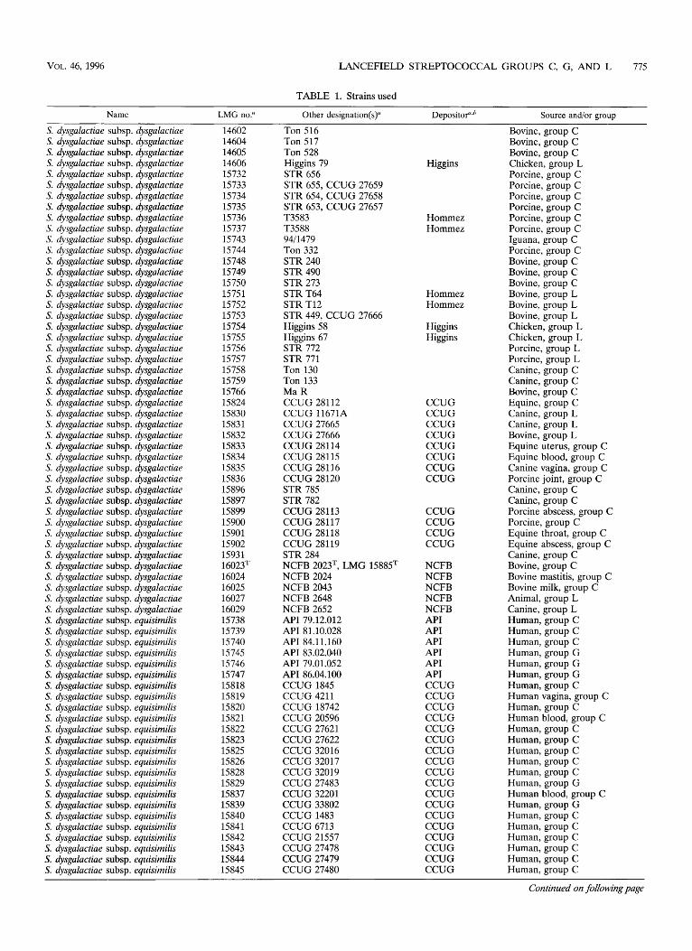
TABLE 1. Strainsused
Name LMG no? Other designation(s)” Depositof,” Source and/or group
S. dysga la ctiae subsp . dysga la ctiae S. dysgalactiae subsp. dysgalactiae S. dysgalactiae subsp. dysgalactiae S. dysga lactia e subsp . dysgala ctiae S. dysgalactiae subsp. dysgalactiae S. dysgalactiae subsp. dysgalactiae S. dysgalactiae subsp. dysgalactiae S. dysgalactiae subsp. dysgalactiae S. dysgalactiae subsp. dysgalactiae S. dysgalactiae subsp. dysgalactiae S. dysgalactiae subsp. dysgalactiae S. dysga la ctiae subsp . dysga la ctiae S. dysgalactiae subsp. dysgalactiae S. dysgalactiae subsp. dysgalactiae S. dysga la c tia e subsp . dysga la ctiae S. dysga la c tia e subsp . dysga la ctiae S. dysgalactiae subsp . dysgalactiae S. dysgalactiae subsp. dysgalactiae S. dysgalactiae subsp. dysgalactiae S. dysgalactiae subsp. dysgalactiae S. dysgalactiae subsp. dysgalactiae S. dysgalactiae subsp. dysgalactiae S. dysgalactiae subsp. dysgalactiae S. dysgalactiae subsp. dysgalactiae S. dysgalactiae subsp. dysgalactiae S. dysgalactiae subsp. dysgalactiae S. dysgalactiae subsp. dysgalactiae S. dysga la ctia e subsp . dysgala ctiae S. dysgalactiae subsp. dysgalactiae S. dysgalactiae subsp. dysgalactiae S. dysga la ctia e subs p . dysgala ctiae S. dysgalactiae subsp. dysgalactiae S. dysgalactiae subsp. dysgalactiae S. dysgalactiae subsp. dysgalactiae S. dysgalactiae subsp. dysgalactiae S. dysgalactiae subsp. dysgalactiae S. dysgalactiae subsp. dysgalactiae S. dysgalactiae subsp. dysgalactiae S. dysgalactiae subsp. dysgalactiae S. dysgalactiae subsp. dysgalactiae S. dysgalactiae subsp. dysgalactiae S. dysgalactiae subsp. dysgalactiae S. dysga la ctia e subs p . dysga la ctiae S. dysga la c tia e subs p . dysga la ctiae S. dysgalactiae subsp. dysgalactiae S. dysgalactiae subsp. equisimilis S. dysgalactiae subsp. equisimilis S. dysga la ctia e subs p . equisimilis S. dysgalactiae subsp. equisimilis S. dysgalactiae subsp. equisimilis S. dysgalactiae subsp. equisimilis S. dysgalactiae subsp. equisimilis S. dysgalactiae subsp. equisimilis S. dysgalactiae subsp. equisimilis S. dysgalactiae subsp. equisimilis S. dysgalactiae subsp. equisimilis S. dysgalactiae subsp. equisimilis S, dysgalactiae subsp. equisimilis S. dysgalactiae subsp. equisimilis S. dysgalactiae subsp. equisimilis S. dysgalactiae subsp. equisimilis S. dysgalactiae subsp. equisimilis S. dysgalactiae subsp. equisimilis S. dysgalactiae subsp. equisimilis S. dysgalactiae subsp. equisimilis S. dysgalactiae subsp. equisimilis S. dysgalactiae subsp. equisimilis S. dysgalactiae subsp. equisimilis S. dysgalactiae subsp. equisimilis
14602 14604 14605 14606 15732 15733 15734 15735 15736 15737 15743 15744 15748 15749 15750 15751 15752 15753 15754 15755 15756 15757 15758 15759 15766 15824 15830 15831 15832 15833 15834 15835 15836 15896 15897 15899 15900 15901 15902 15931 16023= 16024 16025 16027 16029 15738 15739 15740 15745 15746 15747 15818 15819 15820 15821 15822 15823 15825 15826 15828 15829 15837 15839 15840 15841 15842 15843 15844 15845
Ton 516 Ton 517 Ton 528 Higgins 79 STR 656 STR 655, CCUG 27659 STR 654, CCUG 27658 STR 653, CCUG 27657 T3583 T3588 9411479 Ton 332 STR 240 STR 490 STR 273 STR T64 STR T12 STR 449, CCUG 27666 Higgins 58 Higgins 67 STR 772 STR 771 Ton 130 Ton 133 Ma R CCUG 28112 CCUG 11671A CCUG 27665 CCUG 27666 CCUG 28114 CCUG 28115 CCUG 28116 CCUG 28120 STR 785 STR 782 CCUG 28113 CCUG 28117 CCUG 28118 CCUG 28119 STR 284 NCFB 2023T, LMG 15885= NCFB 2024 NCFB 2043 NCFB 2648 NCFB 2652 API 79.12.012 API 81.10.028 API 84.11.160 API 83.02.040 API 79.01.052 API 86.04.100 CCUG 1845 CCUG 4211 CCUG 18742 CCUG 20596 CCUG 27621 CCUG 27622 CCUG 32016 CCUG 32017 CCUG 32019 CCUG 27483 CCUG 32201 CCUG 33802 CCUG 1483 CCUG 6713 CCUG 21557 CCUG 27478 CCUG 27479 CCUG 27480
Higgins
Hommez Hommez
Hommez Hommez
Higgins Higgins
CCUG CCUG CCUG CCUG CCUG CCUG CCUG CCUG
CCUG CCUG CCUG CCUG
NCFB NCFB NCFB NCFB NCFB API API API API API API CCUG CCUG CCUG CCUG CCUG CCUG CCUG CCUG CCUG CCUG CCUG CCUG CCUG CCUG CCUG CCUG CCUG CCUG
Bovine, group C Bovine, group C Bovine, group C Chicken, group L Porcine, group C Porcine, group C Porcine, group C Porcine, group C Porcine, group C Porcine, group C Iguana, group C Porcine, group C Bovine, group C Bovine, group C Bovine, group C Bovine, group L Bovine, group L Bovine, group L Chicken, group L Chicken, group L Porcine, group L Porcine, group L Canine, group C Canine, group C Bovine, group C Equine, group C Canine, group L Canine, group L Bovine, group L Equine uterus, group C Equine blood, group C Canine vagina, group C Porcine joint, group C Canine, group C Canine, group C Porcine abscess, group C Porcine, group C Equine throat, group C Equine abscess, group C Canine, group C Bovine, group C Bovine mastitis, group C Bovine milk, group C Animal, group L Canine, group L Human, group C Human, group C Human, group C Human, group G Human, group G Human, group G Human, group C Human vagina, group C Human, group C Human blood, group C Human, group C Human, group C Human, group C Human, group C Human, group C Human, group G Human blood, group C Human, group G Human, group C Human, group C Human, group C Human, group C Human, group C Human, group C
Continued on following page

776 VANDAMME ET AL. INT. J. SYST. BACTERIOL.
TABLE l-Continued
Name LMG no.a Other designation(s)" Depositof.' Source andlor group
5'. dysgalactiae subsp. equisimilis 5'. dysgalactiae subsp. equisimilis S. dysgalactiae subsp. equisimilis 5'. dysgalactiae subsp. equisimilis 5:. dysgalactiae subsp. equisimilis 5:. dysgalactiae subsp. equisimilis S dysgalactiae subsp. equisimilis 5:. dysgalactiae subsp. equisimilis 5;. dysgalactiae subsp. equisimilis 5;. dysgalactiae subsp. equisimilis 5;. dysgalactiae subsp. equisimilis
S. agalactiae S. agalactiae S agalactiae S. agalactiae S. agazactiae S agalactiae
2;. anginosus S. anginosus S anginosus S. anginosus S. anginosus
S. canis S. canis S. canis :i. canis :i. canis S. canis S. canis
S. equi subsp. zooepidemicus S. equi subsp. zooepidemicus :i. equi subsp. zooepidemicus S. equi subsp. zooepidemicus S. equi subsp. equi S. equi subsp. equi S. equi subsp. zooepidemicus S. equi subsp. zooepidemicus 5: equi subsp. zooepidemicus
S. hyoin testina lis S. hyointestinalis S. hyointestinalis
S. iniae S. iniae
S. parauberis S. parauberis
S'. porcinus S. porcinus S. porcinus
S. pyogenes
S. pyogenes
,S. pyogenes S. pyogenes S. pyogenes S. pyogenes (5. pyogenes (5. pyogenes
,S. shiloi
~
15846 15847 15848 15849 15850 15851 15852 15853 15854 16026T 15903
14694T 14747 14847 15084 15085 15095
145 02* 14612 14613 14696 15994
14833 14834 15890T 15891 15892 15893 15894
15760 15761 15 762 15763 15764 15886T 15888 15889 16030T
14579T 14581 14582
1 4520T 14521
12173 12174T
14615 15980T 15981
14237
14238
14700T 15855 15856 15857 15858 15859
15978'
CCUG 502 CCUG 1859 CCUG 7975 CCUG 15679 CCUG 15680 CCUG 24070 CCUG 26147 CCUG 27477 CCUG 27482 NCFB 1356T CCUG 28238
CCUG 4208T STR 765 SHV 357 18RS21 M732 455889
CCUG 27298T STR 778 STR 200 CCUG 223 CCUG 28467
CCUG 27660, STR 290 STR 642 STR-TI~ 21 650- 1 I473 03342-1106 2083717820 2366812140
Ton6 P11264 P13997 Ton 174 STR 674 CCUG 232ST CCUG 23257 CCUG 23258 NCFB 1358T, LMG 15887T
S93T S85 EV 28b
CCUG 27303T CCUG 27623
NCFB 2018, LMG 14377 NCFB 2020T, LMG 14376T
STR 827 CCUG 27628T CCUG 27629
CIP 56.58
CIP 70.3
CCUG 4207T A4260 A6825 H1667 C8185 P409 1
CIP 103769T
CCUG CCUG CCUG CCUG CCUG CCUG CCUG CCUG CCUG NCFB CCUG
CCUG
Hommez Melin Melin Melin
CCUG
CCUG CCUG
CCUG CCUG CCUG NCFB
CCUG CCUG
NCFB NCFB
CCUG CCUG
CIP
CIP
CCUG Goossens Goossens Goossens Goossens Goossens
CIP
Human, group G Human, group G Human, group G Human, group G Human, group G Human throat, group G Human wound, group G Human, group G Human, group G Human, group G Human throat, group C
Bovine milk Bovine mastitis Porcine Human Human Human, respiratory infection
Canine Canine
Human
Canine Feline Bovine mastitis Mink Fox Fox Mink
Equine Equine Equine Equine Equine Equine
Bovine mastitis
Porcine Porcine Porcine
Dolphin Dolphin
Bovine mastitis Bovine mastitis
Human Porcine Porcine
Patient with rheumatic fever,
Patient with bronchopneumonia, throat
pleural fluid
Human throat Human throat Human throat Human throat Human throat
Fish, brain
Continued on following page

VOL. 46, 1996 LANCEFIELD STREPTOCOCCAL GROUPS C, G, AND L 777
TABLE 1-Continued
Name LMG no.u Other designation(s)” DepositoPb Source and/or group
S. uberis S. uberis S. uberis
14395 STR 192 14610 STR 203 14686 ATCC 13386
Duck, lung Bovine
ATCC ~~
a API, Appareils et ProcCdCs #Identification, La-Balme-les-Grottes, Montalieu-Vercieu, France; ATCC, American Type Culture Collection, Rockville, Md.; CCUG, Culture Collection of the University of Goteborg, Department of Clinical Bacteriology, University of Goteborg, Goteborg, Sweden; CIP, Collection bactkrienne de 1’Institut Pasteur, Paris, France; Goossens, H. Goosens, University Hospital Antwerp UIA, Antwerp, Belgium; Higgins, R. Higgins, Department of Pathology and Microbiology, Faculty of Veterinary Medicine, University of Montreal, Sainte-Hyaeinthe, Canada; Hommez, J. Hommez, Regional Veterinary Investigation Centre, Torhout, Belgium; LMG, Laboratorium voor Microbiologie Gent Culture Collection, Universiteit Ghent, Gent, Belgium; Melin, P. Melin, Centre Hospitalier Universitaire de Likge, Service de Microbiologie MCdicinale, Li?ge, Belgium; NCFB, National Collection of Food Bacteria, Agricultural and Food Research Council, Institute of Food Research, Reading Laboratory, Reading, Berkshire, United Kingdom. ’ Our isolate if not indicated otherwise.
Columbia blood agar base supplemented with 1 g of bovine fibrinogen fraction 1 (catalog no. F-8630; Sigma) per liter and 0.5% human plasma. Fibrinogen (100 mg) was first dissolved in 10 ml of phosphate-buffered saline and warmed for 10 min in a shaking water bath at 37°C. This preparation was then mixed with 100 ml of melted blood agar base and incubated for 10 min at 56°C to allow for precipitation of fibrin. Plates were poured immediately after plasma was added and were stored for a maximum of 3 days. A plate containing the same base and bovine fibrin without plasma was used as a control. In other tests, canine, porcine, equine, bovine, and avian (chicken) plasmas were used as plasminogen sources at the same concentration. A limited number of strains giving positive or negative results on Columbia agar supplemented with bovine fibrin and plasma were also tested on brain heart infusion agar (Oxoid, Basingstoke, United King- dom) and on nutrient agar (Oxoid) similarly supplemented with human plasma and bovine fibrin. Strains were spot inoculated onto the plates, and clearing of bovine fibrin on plates containing plasma and not on plates containing only fibrin was considered evidence of activation of plasminogen to plasmin (streptokinase) activity. Reactions occurring on plasma-free as well as on plasma-supplemented plates were interpreted as evidence of fibrinolysis caused by protease activity. This activity was confirmed by tests on casein plates (Columbia agar supple- mented with 0.5% skim milk) and gelatin plates (Columbia agar supplemented with 0.15% Bacto Gelatin [Difco]). Another test involved the use of Columbia agar made up from its separate components but with starch omitted (see below). Tests for protease activity on fibrin from other mammal species (humans, horses, and dogs) were also carried out. Fibrin plates prepared with fibrinogens from human plasma (catalog no. F-4129; fraction I, type 111; Sigma), from horse plasma (catalog no. F-9516; fraction I; Sigma), and from dog plasma (catalog no. F-7128; Sigma) as described above were used, but plasma was not added.
RESULTS
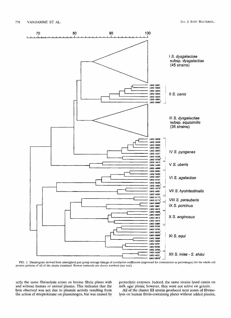
PAGE of whole-cell proteins. A total of 129 streptococci were examined. Duplicate protein extracts of several strains were prepared to check the reproducibility of the growth con- ditions and the preparation of the extracts. The level of cor- relation between duplicate protein patterns was at least 94% (data not shown). After visual comparison of the protein pat- terns with the dendrogram obtained after numerical compar- ison and clustering of the profiles, we identified 12 stable clusters above a similarity level of 84% (Fig. 1). Cluster I comprised 45 S. dysgalactiae strains, including type strain LMG 16023, which clustered above a similarity level of 84.8%. These strains were isolated from various animals and belong to sero- groups C and L (Fig. 1; Table 1). Cluster I11 comprised 35 S. dysgalactiae strains that clustered above a similarity level of 84.7%. All of the cluster I11 strains were isolated from human sources and belong to serogroups C and G (Fig. 1; Table 1). As shown in Fig. 1, the reference strains of all of the other strep- tococcal species examined constituted homogeneous clusters, as follows: S. canis (cluster 11; grouping above a similarity level of 87.2%), Streptococcus pyogenes (cluster IV; grouping above a similarity level of 88.9%), Streptococcus uberis (cluster V; grouping above a similarity level of 84.1%), S. agalactiae (clus- ter VI; grouping above a similarity level of 85.8%), Streptococ- cus hyointestinalis (cluster VII; grouping above a similarity level of 91.7%), Streptococcus parauberis (cluster VIII; group- ing above a similarity level of 96.3%), Streptococcus porcinus
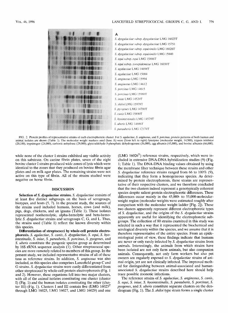
(cluster IX; grouping above a similarity level of 88.7%), Strep- tococcus anginosus (cluster X; grouping above a similarity level of 89.2%), and S. equi (cluster XI; grouping above a similarity level of 88.4%). Cluster XI1 comprised the two Streptococcus iniae reference strains and the type strain of Streptococcus shiloi, which grouped above a correlation level of 93.2%. Fig- ure 2 shows the whole-cell protein patterns of representative strains belonging to different clusters.
Physiological tests. Physiological tests were performed on all 80 S. dysgalactiae strains. The serotypes of these strains accord- ing to the Lancefield scheme are shown in Table 1. All of the strains except the bovine group C strains which were isolated from tonsils (strains LMG 14602, LMG 14604, and LMG 14605) and milk (strains LMG 15748, LMG 15749, LMG 16024, LMG 16025, and LMG 16023T [T = type strain]) and a single group C strain isolated from a pig (strain LMG 15744) exhibited beta-hemolysis on bovine blood agar. The bovine group C strains were alpha-hemolytic or nonhemolytic. Por- cine strain LMG 15744 was also alpha-hemolytic.
All 35 strains belonging to cluster I11 as determined by the whole-cell protein analysis (Fig. 1) produced streptokinase ac- tivity on human plasminogen. This was shown by the presence of neat and easily visible zones of clearing surrounding strep- tococcal growth spots on bovine fibrin-plasma plates. Most reaction zones were visible after 1 day of incubation, but in a few cases, when growth was weak, they appeared only after 2 days. No strain reacted with plasminogens from the five animal species tested. The strains also did not cause fibrinolysis on bovine fibrin plates without plasma, and they were all negative in protease tests on milk agar and gelatin agar. The amylase reaction, which is visible with the naked eye on Columbia agar base without fibrin, did not interfere with the streptokinase tests because the starch contained in the medium was hydro- lyzed and could not be demonstrated, even with iodine, fol- lowing the addition of the fibrin preparations to the Columbia agar base. Tests performed with laboratory-prepared Colum- bia agar without starch gave the same results.
One equine protein electrophoretic cluster I strain (LMG 15824) produced some clearing on bovine fibrin-horse plasma plates. This clearing was visible beneath the growth spot only when the spot was washed off after 2 days of incubation. None of the 44 other cluster I strains tested, including the 4 other equine Lancefield group C strains, gave evidence of streptoki- nase activity in the plate tests. All of these strains except the eight bovine group C strains listed above were negative in tests performed on bovine fibrin plates with or without different types of plasma, including human plasma, and on milk agar and gelatin agar. These strains lacked streptokinase activity. However, the eight alpha- or nonhemolytic bovine group C strains (including the S. dysgalactiae type strain) produced ex-
'778 VANDAMME ET AL. INT. J. SYST. BACTERIOL.
I S. dysgalacfiae subsp. dysgalacfiae (45 strains)
I
I , z i 1 11 s. canis -
LMG ISSQO'
Ill S. dysgalacfiae subsp. equisimilis (35 strains)
I
LMG 15084 LMG 15085
LMG 15095 LMG 14694'
I ::;zT -1 VII S. hyointestinalis I LMG 14582
LMG 12173 LMG 121 74' LMG 15980T LMG 15981 LMG 14615
LMG 14612 LMG 14613
LMG 15761 LMG 15762 LMG 15780-
J Vlll S. parauberis IX S. porcinus
X S. anginosus
1 XI S. equi
1 I LMG14521 XI1 s. iniae - S. shiloi 1 LMG 14520'
LMG 15S7aT
FIG. 1. Dendrogram derived from unweighted pair group average linkage of correlation coefficients (expressed for convenience as percentages) for the whole-cell protein patterns of all of the strains examined. Roman numerals are cluster numbers (see text).
actly the same fibrinolysis zones on bovine fibrin plates with and without human or animal plasma. This indicates that the lysis observed was not due to plasmin activity resulting from the action of streptokinase on plasminogen, but was caused by
proteolytic enzymes. Indeed, the same strains lysed casein on milk agar plates; however, they were not active on gelatin.
All of the cluster 111 strains produced neat zones of fibrino- lysis on human fibrin-containing plates without added plasma,
VOL. 46, 1996 LANCEFIELD STREPTOCOCCAL GROUPS
h
A S. dysgaluc~iuc aubsp. dysgalurliac LMG 16023T S. dysgalucucic. auhsp. dysgulucriue LMG 1575 1
S. dysgalar.ricie subsp. equisimilis LMG 1 6026T S . dysgiilacuac suhq. cquisimilu LMG 15840
S . cqui suhsp. cciw LMG 15886T
S. equi subsp. zonepidcmicuv LMG 16030T
. 7 - S. agalacliae LMC; 14604T
S. rtgalucficic LMG 15084 P S. angincisus LMC; 15904
S. un,gino.su.s LMG 1461 2 S porcinuv LMG 11615
S porc-iniLs LMC; 1 SWOT S . iniar LMG 1452OT
S . . h i l o i LMG 1507ST S. pyc7~cne.s LMG 14700T S. C(iI;ii.S LMG 1 SX9OT
S. hyoinlrvfinulu LhlG 1457OT S uhcris LMC; l46SOl.
S . purciribcri.\ L MC; 1 7, I 71T I1 n
C, G, AND L 779
uatterns of both human and FIG. 2. Protein profiles of representative strains of each electrophoretic cluster. For S. agalactzae, S. anginosus, and S. porcinus, protein I
animal isolates are shown (Table 1). The molecular weight markers used (lane A) were (from left to right) lysozyme (molecular weight, 14,500), trypsin inhibitor (20,100), trypsinogen (24,000), carbonic anhydrase (29,000), glyceraldehyde-3-phosphate dehydrogenase (36,000), egg albumin (45,000), and bovine albumin (66,000).
while none of the cluster I strains exhibited any visible activity on this substrate. On canine fibrin plates, seven of the eight bovine cluster I strains produced wide zones of lysis which were identical to the zones that they produced on bovine fibrin agar plates and on milk agar plates. The remaining strains were not active on this type of fibrin. All of the strains studied were negative on horse fibrin.
DISCUSSION
Selection of S. dysguluctiue strains. S. dysgalactiae consists of at least five distinct subgroups on the basis of serogroups, biotypes, and hosts (5, 7). In the present study, the sources of the strains used included humans, horses, cows (and milk), pigs, dogs, chickens, and an iguana (Table 1). These isolates represented nonhemolytic, alpha-hemolytic and beta-hemo- lytic S. dysgalactiae strains and serogroups C, G, and L. Thus, the strains used (Table 1) reflect the known diversity within this species.
Differentiation of streptococci by whole-cell protein electro- phoresis. S. agalactiae, S. canis, S. dysgalactiae, S. equi, S. hyo- intestinalis, S. iniae, S. parauberis, S. porcinus, S. pyogenes, and S. uberis constitute the pyogenic species group as determined by 16s rRNA sequence analysis (1). Other streptococcal spe- cies are more remotely related to members of this group. In the present study, we included representative strains of all of these taxa as reference strains. In addition, S. anginosus was also included, as this species also comprises Lancefield group C and G strains. S. dysgalactiae strains were easily differentiated from other streptococci by whole-cell protein electrophoresis (Fig. 1 and 2). However, these organisms fell into two major clusters, with all of the animal isolates constituting one cluster (cluster I) (Fig. 1) and the human isolates constituting the other (clus- ter 111) (Fig. 1). Clusters I and I11 contain five (LMG 16023T through LMG 16025, LMG 16027, and LMG 16029) and one
(LMG 16026T) reference strains, respectively, which were in- cluded in extensive DNA-DNA hybridization studies (9) (Fig. 1; Table 1). The DNA-DNA binding values obtained by using the membrane filter technique between these strains and other S. dysgalactiae reference strains ranged from 66 to 100% (9), indicating that they form a homogeneous species. As deter- mined by protein electrophoresis, these strains are represen- tative of their respective clusters, and we therefore concluded that the two clusters indeed represent a genotypically coherent species despite salient protein electrophoretic differences. These differences occur mainly in the 45,000- to 55,000-molecular- weight region (molecular weights were estimated roughly after comparison with the molecular weight ladder [Fig. 21). These two clusters apparently represent different electrophoretic types of S. dysgalactiae, and the origins of the S. dysgalactiae strains apparently are useful for identifying the electrophoretic sub- groups. The collection of 80 strains examined in this study was selected in such a way that it represented the biochemical and serological diversity within the species, and we assume that it is therefore representative of the entire species. From an epide- miological point of view, these findings indicate that humans are never or only rarely infected by S. dysgalactiae strains from animals. Interestingly, the animals from which strains have been isolated are not only farm animals, but also companion animals. Consequently, not only farm workers but also pet owners are regularly exposed to S. dysgalactiae strains of ani- mal origin, yet are not clinically infected. The improved meth- od for distinguishing between animal-associated and human- associated s. dysgalactiae strains described here should help trace possible zoonotic infections.
The reference strains of S. agalactiae, S. anginosus, S. canis, S. equi, S. iniae, S. hyointestinalis, S. parauberis, S. porcinus, S. pyogenes, and S. uberis constitute separate clusters on the den- drogram (Fig. 1). The S, shiloi type strain forms a single cluster

780 VANDAMME ET AL. INT. J. SYST. BACTERIOL.
TABLE 2. Characteristics that differentiate S. dysgalactiae subspecies
Taxon Lancefield Strepokinase Proteolysis Acid production a-L-Glutamate L-Prolyl-L-argi-
dase activitya aminopeptidase nine aminopepti- activity on from“:
minogen fibrin Sorbitol Glycogen activity”
Hemolysis carbohydrate human of human antigens
- b - S. dysgalactiae subsp. dysgalactiae Alpha, beta, or C, L D D D
S. dysgalactiae subsp. equisimilis Beta c, G + + none
+ - -
+ D
“ Data from reference 7. -, negative; +, positive; D, strain dependent.
with the S. iniae strains at a correlation level of 93.2%, con- firming the reported synonymy of these two species (8). As illustrated in Fig. 1 and 2, S. equi subsp. equi and S. equi subsp. zooepidemicus cannot be differentiated by whole-cell protein electrophoresis. In contrast to the S. dysgalactiae strains, hu- man and animal isolates of S. agalactiae, S. anginosus, and S. porcinus do not seem to exhibit significant differences in their whole-cell protein patterns (Table 1; Fig. 2).
Phenotypic differentiation of S. dysgalactiae strains. Despite the superficial resemblance of the assay methods, direct pro- teolytic activity on human fibrin and streptokinase activity on human plasminogen are two different characteristics which can be used as easy ways to distinguish between human and animal isolates of S. dysgalactiae in diagnostic laboratories. McCoy and coworkers (13) found that streptokinases of different spe- cies obtained from different hosts were antigenically related. In their study, all of the group C strains tested, including strains referred to as S. equisimilis, S. equi subsp. equi, and S. equi subsp. zooepidemicus, had surface receptors that bound human, equine, and porcine plasmins. Human S. equisimilis strains were active only on human plasminogen, and this finding was confirmed and extended in the present study with avian, bovine, and canine plasmas. However, equine S. equisimilis strains and strains from the two S. equi subspecies activated horse plasma, while porcine S. equisimilis strains were active on porcine plasminogen and, very weakly, also on human plasmin- ogen. In the study of McCoy et al. (13), plasminogen activator activity was identified by hydrolysis of a plasmin-specific chro- mogenic substrate. This was detected in growth patches by a solid-phase assay. In our study, the enzyme was detected by lysis of fibrin, the normal substrate of plasmin. The difference in test results may be due to differences in the sensitivities of the two test systems.
Taxonomic status of S. dysgulactiae strains of human origin and animal origin. Species level identification of S. dysgulactiae strains is complex and provides little information for clinicians or epidemiologists. Beta-hemolytic group L S. dysgalactiae strains and alpha-hemolytic group C S. dysgalactiae strains colonize or infect only animals, whereas beta-hemolytic group G S. dysgalactiae strains colonize or infect only humans. Beta- hemolytic group C S. dysgalactiae strains occur in humans and animals, but the animal isolates do not occur in humans and vice versa. Clearly, from a practical point of view, S. dysguluc- tiue consists of two distinct subpopulations of strains, which produce very different whole-cell protein electrophoretic pat- terns, as demonstrated in Fig. 1. The protein electrophoretic differences are comparable to the differences between distinct species in the group containing the pyogenic streptococci (Fig. 1 and 2). In contrast, human and animal isolates of several other members of this species group do not differ significantly (Fig. 1 and 2; Table 1). The two subpopulations of S. dysga- lactiae can also be readily identified by phenotypic tests, as described above. In our opinion, these observations warrant
distinct taxonomic status for each subpopulation of strains. As Farrow and Collins (9) demonstrated that both of the sub- populations belong to a single species, we propose that they should be classified as distinct subspecies of S. dysgalactiae. The name S. dysgalactiae subsp. dysgalactiae is used for the subpopulation of strains of animal origin because this sub- population includes the type strain. Historically, the name S. equisimilis was given to human isolates resembling S. equi strains which occurred in animals, and therefore it is logical to use the name S. dysgalactiae subsp. equisimilis for the S. dys- galuctiae strains of human origin. In addition, this avoids cre- ating more confusion by the introduction of a new name.
S. dysgalactiae subsp. dysgulactiae strains can be distinguished from S. dysgalactiae subsp. equisimilis strains by proteolysis of human fibrin, by the results of the human plasminogen-strep- tokinase test, by origin, and by whole-organism protein elec- trophoretic patterns. Lancefield serogrouping is also of value for differentiating the two subspecies; group L streptococci always belong to S. dysgalactiae subsp. dysgalactiae, while group G streptococci of human origin belong to S. dysgulactiae subsp. equisimilis (all animal group G strains described to date belong to S. cunis). These identifications can be confirmed by physio- logical tests (6,7). Also, for group C streptococci, origin can be used for presumptive identification; however, the strains should first be differentiated from S. equi and “S, milleri” (7, 9). Fi- nally, Efstratiou et al. (7) described additional phenotypic tests that can be used to differentiate the two subspecies (Table 2).
Description of S. dysgulactiae subsp. dysgulactiae subsp. nov. The description of S. dysgalactiae subsp. dysgalactiae is the same as the description given by Farrow and Collins (9) for S. dysgalactiae, with following exceptions. Most strains are beta-hemolytic, but alpha-hemolytic and nonhemolytic strains occur, especially in bovine sources. Strains may react with Lancefield group C or L antigen but not with Lancefield group G antigen. Streptokinase activity on human plasminogen and proteolytic activity on human fibrin do not occur. The habitat is the respiratory and genital tracts of various animals, but apparently not humans.
Description of S. dysgalactiae subsp. equisimilis subsp. nov. The description of S. dysguluctiae subsp. equisimilis is the same as the description given by Farrow and Collins (9) for S. dys- galactiae, with following exceptions. Strains are beta-hemolytic and may react with Lancefield group C or G antigen but not with Lancefield group L antigen. Streptokinase activity occurs on human plasminogen, and proteolytic activity occurs on hu- man fibrin. The habitat is the respiratory tracts and vaginas of humans. The type strain is LMG 16026 (= NCFB 1356), which belongs to Lancefield group G.
ACKNOWLEDGMENTS
We thank Urbain Torck and Dirk Dewettinck for excellent technical assistance and all depositors of strains listed in Table 1.
VOL. 46, 1996 LANCEFIELD STREPTOCOCCAL GROUPS C, G, AND L 781
P.V. is indebted to the National Fund for Scientific Research (Bel- gium) for a position as a postdoctoral research fellow. K.K. is indebted to the Fund for Medical Scientific Research, Belgium, for research and personnel grants. Part of this work was supported by the Federal Office for Scientific, Technical and Cultural Affairs of the Belgian State.
REFERENCES 1. Bentley, R. W., J. A. Leigh, and M. D. Collins. 1991. Intrageneric structure
of Streptococcus based on comparative analysis of small-subunit rRNA se- quences. Int. J. Syst. Bacteriol. 41:487-494.
2. Breed, R. S., E. G. D. Murray, and A. P. Hitchens. 1948. Manual of deter- minative bacteriology, 6th ed., p. 318-319. Williams and Wilkins, Baltimore.
3. Carmeli, Y., and K. L. Ruoff. 1995. Report of cases of and taxonomic considerations for large-colony-forming Lancefield group C streptococcal bacteremia. J. Clin. Microbiol. 33:2114-2117.
4. Costas, M. 1992. Classification, identification, and typing of bacteria by the analysis of their one-dimensional polyacrylamide gel electrophoretic protein patterns, p. 351-408. In A. Chambrach, M. J. Dunn, and B. J. Radola (ed.), Advances in electrophoresis, vol. 5. VCH Verlagsgesellschaft, Weinheim, Germany.
5. Devriese, L. A. 1991. Streptococcal ecovars associated with different animal species: epidemiological significance of serogroups and biotypes. J. Appl. Bacteriol. 71:478483.
6. Devriese, L. A., J. Hommez, R. Kilpper-Balz, and K.-H. Schleifer. 1986. Streptococcus canis sp. nov.: a species of group G streptococci from animals. Int. J. Syst. Bacteriol. 36422425.
7. Efstratiou, A., G. Colman, G. Hahn, J. F. Timoney, J. M. Boeufgras, and D. Monget. 1994. Biochemical differences among human and animal strepto- cocci of Lancefield group C or group G. J. Med. Microbiol. 41:145-148.
8. Eldar, A., P. F. Frelier, L. Assenta, P. W. Varner, S. Lawhon, and H. Bercovier. 1995. Streptococcus shiloi, the name for an agent causing septice- mic infection in fish, is a junior synonym of Streptococcus iniae. Int. J. Syst. Bacteriol. 458404342.
9. Farrow, J. A. E., and M. D. Collins. 1984. Taxonomic studies on streptococci of serological groups C, G and L and possibly related taxa. Syst. Appl. Microbiol. 5483-493.
10. Frost, W. D., and M. A. Engelbrecht. 1940. The streptococci. Willdorf Book Co., Madison, Wis.
11. Garvie, E. I., J. A. E. Farrow, and M. D. Collins. 1983. Streptococcus dysga- lactiae (Diernhofer) nom. rev. Int. J. Syst. Bacteriol. 33:404-405.
12. Lancefield, R. C. 1933. A serological differentiation of human and other groups of hemolytic streptococci. J. Exp. Med. 59571-591.
13. McCoy, H. E., C. C. Broder, and R Lottenberg. 1991. Streptokinases pro- duced by pathogenic group C streptococci demonstrate species-specific plas- minogen activation. J. Infect. Dis. 164515-521.
14. Pot, B., P. Vandamme, and K. Kersters. 1994. Analysis of electrophoretic whole-organism protein fingerprints, p. 493-521. In M. Goodfellow and A. G. O’Donnell (ed.), Modern microbial methods. Chemical methods in prokaryotic systematics. J. Wiley and Sons, Ltd., Chichester, United King- dom.
15. Skerman, V. B. D., V. McGowan, and P. H. A. Sneath (ed.). 1980. Approved lists of bacterial names. Int. J. Syst. Bacteriol. 30225420.
Copyright © 2022 FDOKUMEN