In vitro cytostatic and immunomodulatory properties of the medicinal mushroom Lentinula edodes
8
Phytomedicine 15 (2008) 512–519 In vitro cytostatic and immunomodulatory properties of the medicinal mushroom Lentinula edodes C. Israilides a, , D. Kletsas b , D. Arapoglou a , A. Philippoussis c , H. Pratsinis b , A. Ebringerova´ d , V. Hrˇ ı´balova´ e , S.E. Harding f a Biotechnology Laboratory, National Agricultural Research Foundation (NAGREF), 1, Sof. Venizelou St., 14123 Lycovrissi, Athens, Greece b Laboratory of Cell Proliferation & Ageing, National Center of Scientific Research (N.C.S.R.) ‘‘Demokritos’’, Institute of Biology, 15310 Athens, Greece c NAGREF, Institute of Agricultural Engineering, Laboratory of Edible Fungi, 61 Democratias St., 13561 Ag. Anargyri, Athens, Greece d Institute of Chemistry, Slovak Academy of Sciences, Du´bravska´ cesta 9, 845 38 Bratislava, Slovakia e National Institute of Public Health, S ˇ roba´rova 48, 100 42 Prague, Czech Republic f University of Nottingham, MCMH, School of Biosciences, Sutton Bonington, Leicestershire LE 12 5RD, UK Abstract Lentinula edodes, known as ‘‘shiitake’’ is one of the widely used medicinal mushrooms in the Orient. Antitumour activity of extracts of this mushroom has been widely demonstrated in animals and humans. However, this activity was shown to be host mediated and not by direct cytotoxic activity to cancer cells. This study demonstrates cytotoxic and cell growth inhibitory (cytostatic) effect of aqueous extracts of the mushroom on MCF-7 human breast adenocarcinoma cell line using an MTT cytotoxicity assay. Such effect was demonstrated with fruit body and mycelial extracts, the difference being that there was no significant suppression on normal cells with the latter. Furthermore mycelial extracts did not induce any cytostatic effect in both cancer and normal cell lines based on a DNA synthesis assay. The significant suppression of the proliferation of cancer cells was reflected by the comparatively low IC 50 values and the simultaneous higher respective values on normal fibroblast cells. The immunostimulatory activity of both fruit body and mycelial extracts was tested by the lymphocyte transformation test (LTT), which is based on the capacity of active immunomodulators to augment the proliferative response of rat thymocytes to T mitogens in vitro. Both fruit body and mycelial preparations were able to enhance the proliferation of rat thymocytes directly and act as co-stimulators in the presence of the T-mitogen PHA. Interestingly both extracts, similarly to zymosan showed SI comit /SI mit ratios of about 2, indicating adjuvant properties. Overall L. edodes aqueous extracts have demonstrated direct inhibition of the proliferation of human breast cancer cells in vitro and immunostimulatory properties in terms of mitogenic and co-mitogenic activity in vitro. r 2007 Elsevier GmbH. All rights reserved. Keywords: Lentinula edodes; Cancer; Cytotoxic; Cytostatic; Antitumour; Mitogenic and comitogenic activity ARTICLE IN PRESS www.elsevier.de/phymed 0944-7113/$ - see front matter r 2007 Elsevier GmbH. All rights reserved. doi:10.1016/j.phymed.2007.11.029 Corresponding author. Tel.: +30 210 2842676; fax: +30 211 7508893. E-mail address: [email protected] (C. Israilides).
Transcript of In vitro cytostatic and immunomodulatory properties of the medicinal mushroom Lentinula edodes

ARTICLE IN PRESS
0944-7113/$ - se
doi:10.1016/j.ph
�CorrespondE-mail addr
Phytomedicine 15 (2008) 512–519
www.elsevier.de/phymed
In vitro cytostatic and immunomodulatory properties of the medicinal
mushroom Lentinula edodes
C. Israilidesa,�, D. Kletsasb, D. Arapogloua, A. Philippoussisc, H. Pratsinisb,A. Ebringerovad, V. Hrıbalovae, S.E. Hardingf
aBiotechnology Laboratory, National Agricultural Research Foundation (NAGREF), 1, Sof. Venizelou St.,
14123 Lycovrissi, Athens, GreecebLaboratory of Cell Proliferation & Ageing, National Center of Scientific Research (N.C.S.R.) ‘‘Demokritos’’,
Institute of Biology, 15310 Athens, GreececNAGREF, Institute of Agricultural Engineering, Laboratory of Edible Fungi, 61 Democratias St.,
13561 Ag. Anargyri, Athens, GreecedInstitute of Chemistry, Slovak Academy of Sciences, Dubravska cesta 9, 845 38 Bratislava, SlovakiaeNational Institute of Public Health, Srobarova 48, 100 42 Prague, Czech RepublicfUniversity of Nottingham, MCMH, School of Biosciences, Sutton Bonington, Leicestershire LE 12 5RD, UK
Abstract
Lentinula edodes, known as ‘‘shiitake’’ is one of the widely used medicinal mushrooms in the Orient. Antitumouractivity of extracts of this mushroom has been widely demonstrated in animals and humans. However, this activity wasshown to be host mediated and not by direct cytotoxic activity to cancer cells. This study demonstrates cytotoxic andcell growth inhibitory (cytostatic) effect of aqueous extracts of the mushroom on MCF-7 human breastadenocarcinoma cell line using an MTT cytotoxicity assay. Such effect was demonstrated with fruit body andmycelial extracts, the difference being that there was no significant suppression on normal cells with the latter.Furthermore mycelial extracts did not induce any cytostatic effect in both cancer and normal cell lines based on aDNA synthesis assay. The significant suppression of the proliferation of cancer cells was reflected by the comparativelylow IC50 values and the simultaneous higher respective values on normal fibroblast cells. The immunostimulatoryactivity of both fruit body and mycelial extracts was tested by the lymphocyte transformation test (LTT), which isbased on the capacity of active immunomodulators to augment the proliferative response of rat thymocytes to Tmitogens in vitro. Both fruit body and mycelial preparations were able to enhance the proliferation of rat thymocytesdirectly and act as co-stimulators in the presence of the T-mitogen PHA. Interestingly both extracts, similarly tozymosan showed SIcomit/SImit ratios of about 2, indicating adjuvant properties. Overall L. edodes aqueous extractshave demonstrated direct inhibition of the proliferation of human breast cancer cells in vitro and immunostimulatoryproperties in terms of mitogenic and co-mitogenic activity in vitro.r 2007 Elsevier GmbH. All rights reserved.
Keywords: Lentinula edodes; Cancer; Cytotoxic; Cytostatic; Antitumour; Mitogenic and comitogenic activity
e front matter r 2007 Elsevier GmbH. All rights reserved.
ymed.2007.11.029
ing author. Tel.: +30 210 2842676; fax: +30 211 7508893.
ess: [email protected] (C. Israilides).

ARTICLE IN PRESSC. Israilides et al. / Phytomedicine 15 (2008) 512–519 513
Introduction
Medicinal mushroom extracts have been consideredas important remedies for the prevention and treatmentof many diseases for thousands of years especially in theOrient (Israilides and Philippoussis, 2003; Kidd, 2000;Wasser and Weis, 1999). A plethora of medicinal effectshas been demonstrated for many traditionally usedmushrooms including antibacterial, antiviral, antifun-gal, antitumour and immuno-potentiating activities(Hobbs, 2003; Ooio and Liu, 1999). Among the variousbioactive components which have been demonstrated tobe most effective as antitumor and immunomodulatoryagents are polysaccharides and polysaccharopeptides.
Lentinula edodes is the source of many therapeuticpolysaccharide macromolecules among which the oneswith proven pharmacological effects are lentinan, LEMand KS-2. Lentinan is a high molecular weight (aboutone million) homopolysaccharide in a triple helixstructure, with linear chains consisting of (1-3)-b-D-glucopyranosyl (Glcp) residues with two b-(1-6)-linked Glcp branchings for every five b-(1-3)-Glcp
residues (Aoki, 1984). LEM is a mycelial extractpreparation of L. edodes harvested before the cap andstem grow. It is a heteroglycan–protein conjugatecontaining 24.6% protein and 44% sugars, comprisingmostly pentoses as well as glucose and smaller amountsof galactose, mannose and fructose (Iizuka, 1986;Sugano et al., 1982). It also contains nucleic acidderivatives, B complex vitamins, ergosterol, eritadenine(an anticholesteremic amino acid), and water-solublelignins (Sugano et al., 1985). KS-2 is a peptide–poly-saccharide complex. The comparison of fruit body andmycelial extracts was carried out for the followingreasons:
1.
The production of fruit bodies and mycelium inL. edodes as well as in many other medicinal mush-rooms, comprise the two main production methods(Wasser and Weis, 1999). The production of fruitbodies does not always guarantee a consistent productwhile the mycelial growth in fermenters undervigorously controlled conditions gives improved pro-duct purity and reproducibility.2.
The main antitumor polysaccharide in L. edodes fruitbodies is a single compound, lentinan. On the otherhand there are many different active compounds inmycelia which have been demonstrated to have‘‘antitumor’’ properties. This provides the opportu-nity for enhanced activity from crude extracts of fruitbodies or mycelium. The mechanism of antitumoractivity of either lentinan, which is the mainbiologically active compound in L. edodes fruitbodies, or the mycelial extract has not been fullyelucidated, but it has been reported as host mediatedby activating the host’s immune responses and notattacking cancer cells directly (Aoki, 1984; Meiqinet al., 1998). Therefore there is a need for comparisonof the two kinds of extracts in an effort to investigateand differentiate tumor selective cytotoxicity.
Since many of the compounds, which are found inL. edodes, have been shown to act synergistically(Yamasaki et al., 1989), it is worth testing the cytotoxicand/or cell growth inhibitory effects of the wholemushroom and mycelium extract rather than its indi-vidual components. This principle (synergy) is compa-tible with similar natural biological products like theessential oils, which allow the achievement of strongeffects when used as whole products, while quenchingor nullifying potential unwanted side-effects by thepresence of individual components.
The objectives of this project were to investigate thecytotoxic and cell growth inhibitory effect on normaland cancer cell lines of active Lentinula edodes extractsproduced from both the mushroom and mycelia as wellas their immunostimulatory activity with the ‘in vitro’comitogenic rat thymocytes test (lymphocyte transfor-mation test, LTT).
Materials and methods
The strain of L. edodes (Berk.) Pegler used in thisstudy, was originated from China and registered in thefungal culture collection of the Edible Fungi Laboratoryof NAGREF with the code number AMRL 118. It wasselected for its phenotypic characteristics concerningproductivity and quality. The culture substrate prepara-tion and growing procedure for sporophore productionhas been previously described (Philippoussis et al.,2007). The culture was maintained on a 2% potatodextrose agar (PDA, Merk) for routine culture andstorage purposes.
Mycelia were grown in a submerged liquid fermenta-tion in a Bioengineering L1523, 7 liter bench fermentor.The initial pH was 5.50, temperature 28 1C, and theaeration was 10 liter/min. The substrate compositionwas (w/v): malt extract 0.3%, yeast extract 0.3%,peptone 0.3% and glucose 1.0%. The inoculum,500ml, was grown in the same medium and the durationof fermentation was 3 days. The fruiting bodies andmycelia were dried by lyophilization and powdered. Allextracts were stored at �40 1C.
Methanol and distilled water extracts from mush-rooms and mycelia of L. edodes were prepared to aninitial concentration of 100mg/ml. The extracts wereincubated for 2 h at room temperature under continuousshaking. They were centrifuged for 30min at 1500g andthe supernatant was passed through a 0.2 mm filter.Samples were further diluted with plain culture medium
ARTICLE IN PRESSC. Israilides et al. / Phytomedicine 15 (2008) 512–519514
(Dulbecco’s minimal essential medium (DMEM)) to thedefined concentrations as indicated.
Cell cultures
Human breast adenocarcinoma cell line MCF-7 andhuman normal fibroblasts from a 30 year-old healthyvolunteer were cultured in DMEM supplemented withantibiotics and 10% fetal bovine serum (FBS) and theywere subcultured using trypsin-citrate (0.25–0.3%,respectively) solution. In the incubation chamber thegas mixture consisted of 5% CO2 and 95% air.Furthermore, the humidity was adjusted to 85%, so asto diminish evaporation of the culture medium and theconsequent changes to its osmolarity that could havebeen detrimental to the cultured cells. Cells were testedperiodically and found to be mycoplasma-free. All cellculture media were from Gibco–BRL.
For the assessment of the cytotoxic and cytostaticactivities of L. edodes extracts cells were seeded in 96-well flat-bottomed microplates at a density of approxi-mately 5000 cells/well, in DMEM 10% FBS. After 18 hto ensure cell attachment, serial dilutions of the extractsin culture medium were added and incubated for 24 or48 h. Then, cytotoxicity and DNA synthesis rate weredetermined using the MTT assay and tritiated thymidineincorporation, respectively.
In testing the cytotoxic or cytostatic effects ofdifferent substances on cancer cells the ideal control isalways an issue. Such control ideally could be normalepithelial cells originating from a neighboring area withhealthy tissue of the same patient. However, this is notalways feasible, especially regarding commercially avail-able cancer cell lines. On the other hand, tumors in vivo
are surrounded by stroma, thus understanding the effectof the studied substances on normal fibroblasts isequally important. In this study we have chosen to usea commercially available human cancer cell line, MCF-7, which is one of the most popular cell lines in theliterature, because this would facilitate replication aswell as comparisons with similar work. As a control wehave used normal human stroma fibroblasts. Further-more, MCF-7 cells and fibroblasts grow in the samemedium, while normal epithelial cells require specialserum-free, chemically-defined media for their culture,which would introduce further unequal parameters inthe experiments.
Cytotoxicity assay
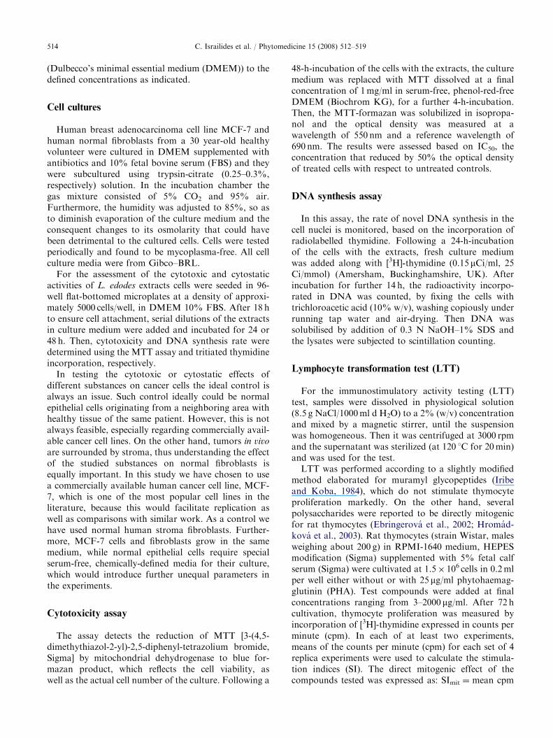
The assay detects the reduction of MTT [3-(4,5-dimethythiazol-2-yl)-2,5-diphenyl-tetrazolium bromide,Sigma] by mitochondrial dehydrogenase to blue for-mazan product, which reflects the cell viability, aswell as the actual cell number of the culture. Following a
48-h-incubation of the cells with the extracts, the culturemedium was replaced with MTT dissolved at a finalconcentration of 1mg/ml in serum-free, phenol-red-freeDMEM (Biochrom KG), for a further 4-h-incubation.Then, the MTT-formazan was solubilized in isopropa-nol and the optical density was measured at awavelength of 550 nm and a reference wavelength of690 nm. The results were assessed based on IC50, theconcentration that reduced by 50% the optical densityof treated cells with respect to untreated controls.
DNA synthesis assay
In this assay, the rate of novel DNA synthesis in thecell nuclei is monitored, based on the incorporation ofradiolabelled thymidine. Following a 24-h-incubationof the cells with the extracts, fresh culture mediumwas added along with [3H]-thymidine (0.15 mCi/ml, 25Ci/mmol) (Amersham, Buckinghamshire, UK). Afterincubation for further 14 h, the radioactivity incorpo-rated in DNA was counted, by fixing the cells withtrichloroacetic acid (10% w/v), washing copiously underrunning tap water and air-drying. Then DNA wassolubilised by addition of 0.3 N NaOH–1% SDS andthe lysates were subjected to scintillation counting.
Lymphocyte transformation test (LTT)
For the immunostimulatory activity testing (LTT)test, samples were dissolved in physiological solution(8.5 g NaCl/1000ml d H2O) to a 2% (w/v) concentrationand mixed by a magnetic stirrer, until the suspensionwas homogeneous. Then it was centrifuged at 3000 rpmand the supernatant was sterilized (at 120 1C for 20min)and was used for the test.
LTT was performed according to a slightly modifiedmethod elaborated for muramyl glycopeptides (Iribeand Koba, 1984), which do not stimulate thymocyteproliferation markedly. On the other hand, severalpolysaccharides were reported to be directly mitogenicfor rat thymocytes (Ebringerova et al., 2002; Hromad-kova et al., 2003). Rat thymocytes (strain Wistar, malesweighing about 200 g) in RPMI-1640 medium, HEPESmodification (Sigma) supplemented with 5% fetal calfserum (Sigma) were cultivated at 1.5� 106 cells in 0.2mlper well either without or with 25 mg/ml phytohaemag-glutinin (PHA). Test compounds were added at finalconcentrations ranging from 3–2000 mg/ml. After 72 hcultivation, thymocyte proliferation was measured byincorporation of [3H]-thymidine expressed in counts perminute (cpm). In each of at least two experiments,means of the counts per minute (cpm) for each set of 4replica experiments were used to calculate the stimula-tion indices (SI). The direct mitogenic effect of thecompounds tested was expressed as: SImit ¼ mean cpm
ARTICLE IN PRESS
Table 1. Analytical data of L. edodes mushroom and
mycelium extracts
Sample Fruit
body
Mycelium
Nitrogen (wt%) 6.81 4.19
Klason lignin (wt%) 12.3 3.1
Ash (wt%) 5.14 4.15
Neutral sugar composition (rel. wt%)
Fucose 0 0.6
Ribose 2.3 0.6
Arabinose 1.5 8.3
Xylose 1.5 1.1
Mannose 28.6 15.0
Glucose 55.9 70.8
Galactose 10.1 3.5
C. Israilides et al. / Phytomedicine 15 (2008) 512–519 515
for test compound/mean cpm for control. The comito-genic effect was expressed as: SIcomit ¼ (mean cpm forPHA+test compound)/mean cpm for PHA. The even-tual contamination with endotoxin was checked bycultivation of the thymocytes in presence of polymyxinB, which inhibits stoichiometrically its biological effectincluding the mitogenic activity. As positive control thecommercial immunomodulator zymosan – a particulateb-glucan from baker’s yeasts (Likospol Ltd., Bratislava,Slovakia) was used. Zymosan gave a fine suspension ofnon-sedimenting particles. Polymyxin B sulfate wasfrom Wellcome (UK) and PHA from Murex BiotechLtd. (UK).
Chemical and FTIR analyses
These were performed on lyophilised samples pre-pared from the fruit body and mycelium of L. edodes asdescribed in a previous paper (Israilides and Philip-poussis, 2003).
Statistical analysis
Multiple extracts from both fruiting bodies andmycelia were prepared and analysed on multipleoccasions. The results for the cytotoxicity assay arepresented as the mean of three independent experimentsperformed in quadruplicate wells. Differences fromcontrol cultures were considered significant whenpp0.01 (Student’s t-test). In Figs. 2 and 3 asterisksabove data points indicate significant differences fromthe control.
In the LTT test, the means of SI in repeated testing ofthe extracts were evaluated by analysis of variance(ANOVA), and calculations were done by the EP16programme.
Results and discussion
Chemical and FTIR analyses
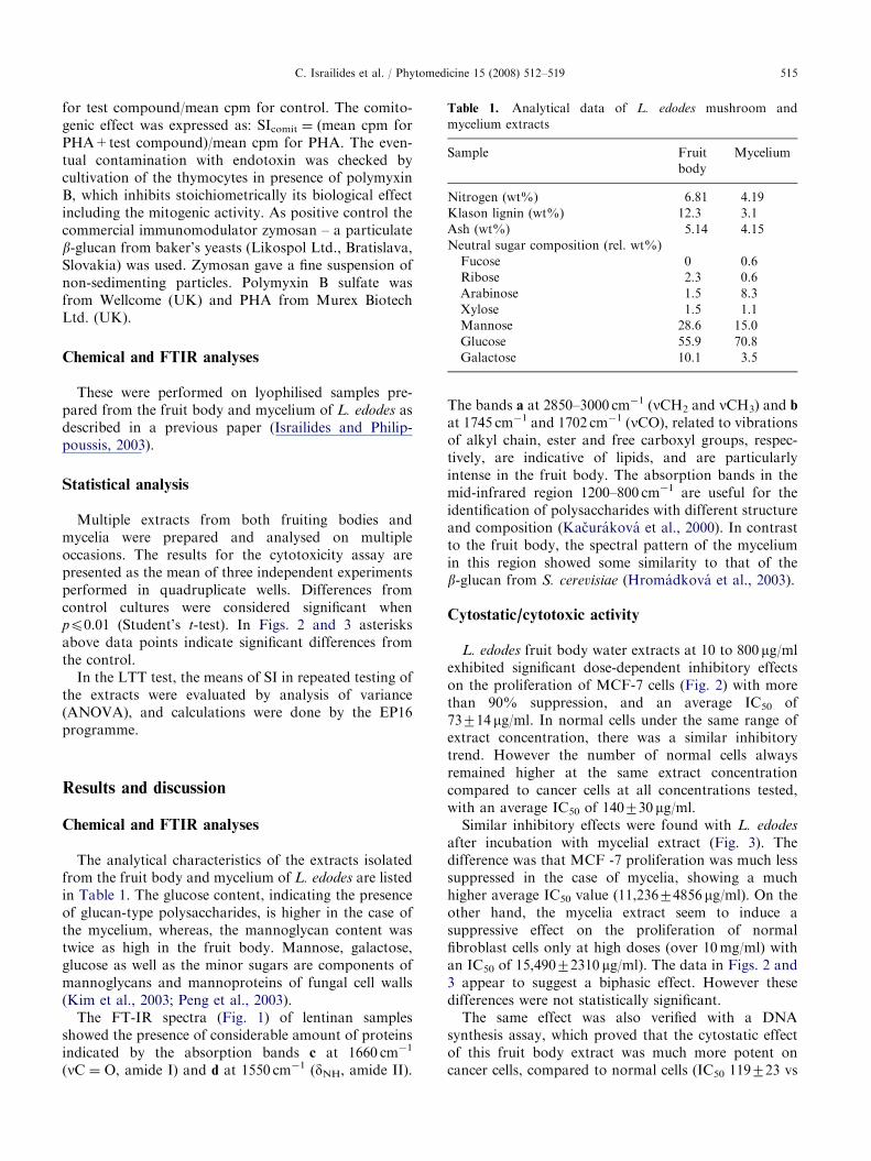
The analytical characteristics of the extracts isolatedfrom the fruit body and mycelium of L. edodes are listedin Table 1. The glucose content, indicating the presenceof glucan-type polysaccharides, is higher in the case ofthe mycelium, whereas, the mannoglycan content wastwice as high in the fruit body. Mannose, galactose,glucose as well as the minor sugars are components ofmannoglycans and mannoproteins of fungal cell walls(Kim et al., 2003; Peng et al., 2003).
The FT-IR spectra (Fig. 1) of lentinan samplesshowed the presence of considerable amount of proteinsindicated by the absorption bands c at 1660 cm�1
(nC ¼ O, amide I) and d at 1550 cm�1 (dNH, amide II).
The bands a at 2850–3000 cm�1 (nCH2 and nCH3) and b
at 1745 cm�1 and 1702 cm�1 (nCO), related to vibrationsof alkyl chain, ester and free carboxyl groups, respec-tively, are indicative of lipids, and are particularlyintense in the fruit body. The absorption bands in themid-infrared region 1200–800 cm�1 are useful for theidentification of polysaccharides with different structureand composition (Kacurakova et al., 2000). In contrastto the fruit body, the spectral pattern of the myceliumin this region showed some similarity to that of theb-glucan from S. cerevisiae (Hromadkova et al., 2003).
Cytostatic/cytotoxic activity
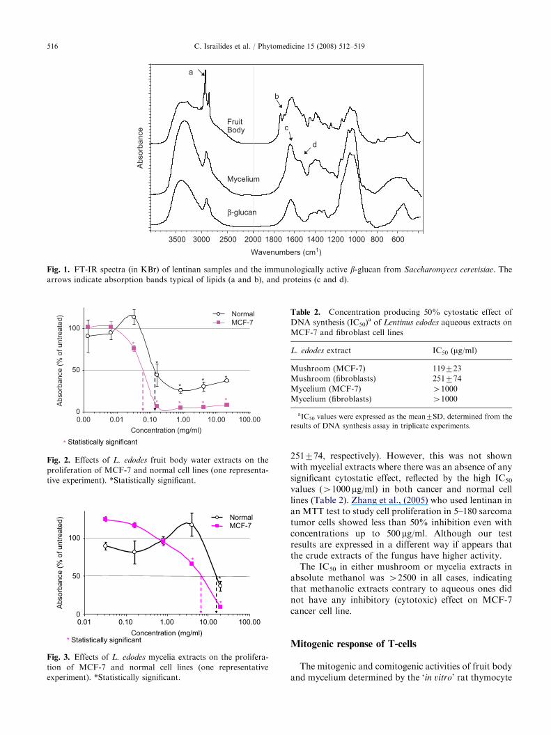
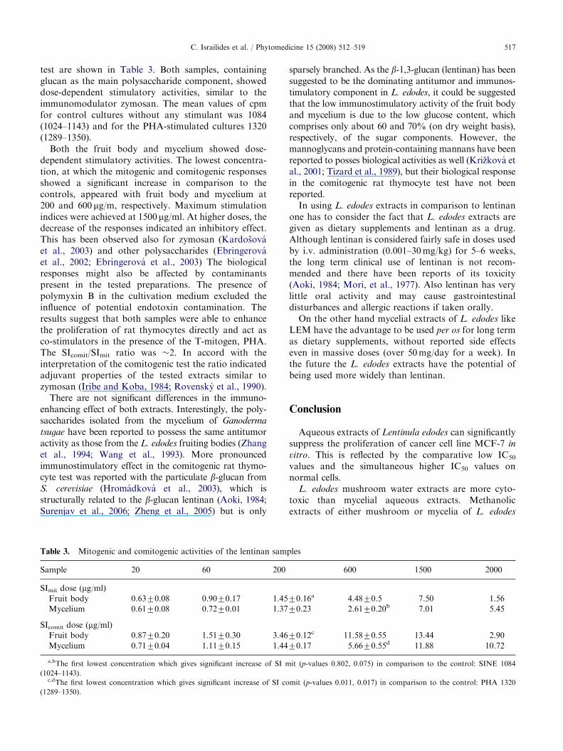
L. edodes fruit body water extracts at 10 to 800 mg/mlexhibited significant dose-dependent inhibitory effectson the proliferation of MCF-7 cells (Fig. 2) with morethan 90% suppression, and an average IC50 of73714 mg/ml. In normal cells under the same range ofextract concentration, there was a similar inhibitorytrend. However the number of normal cells alwaysremained higher at the same extract concentrationcompared to cancer cells at all concentrations tested,with an average IC50 of 140730 mg/ml.
Similar inhibitory effects were found with L. edodes
after incubation with mycelial extract (Fig. 3). Thedifference was that MCF -7 proliferation was much lesssuppressed in the case of mycelia, showing a muchhigher average IC50 value (11,23674856 mg/ml). On theother hand, the mycelia extract seem to induce asuppressive effect on the proliferation of normalfibroblast cells only at high doses (over 10mg/ml) withan IC50 of 15,49072310 mg/ml). The data in Figs. 2 and3 appear to suggest a biphasic effect. However thesedifferences were not statistically significant.
The same effect was also verified with a DNAsynthesis assay, which proved that the cytostatic effectof this fruit body extract was much more potent oncancer cells, compared to normal cells (IC50 119723 vs
ARTICLE IN PRESS
Concentration (mg/ml)
Ab
so
rba
nce
(%
of u
ntr
ea
ted
) Normal
MCF-7
***
*
*
* * * *
0
50
100
0.00 0.01 0.10 1.00 10.00 100.00
* Statistically significant
Fig. 2. Effects of L. edodes fruit body water extracts on the
proliferation of MCF-7 and normal cell lines (one representa-
tive experiment). *Statistically significant.
Concentration (mg/ml)
Absorb
ance (
% o
f untr
eate
d) Normal
MCF-7
*
*
*
0
50
100
0.01 0.10 1.00 10.00 100.00
* Statistically significant
Fig. 3. Effects of L. edodes mycelia extracts on the prolifera-
tion of MCF-7 and normal cell lines (one representative
experiment). *Statistically significant.
Table 2. Concentration producing 50% cytostatic effect of
DNA synthesis (IC50)a of Lentinus edodes aqueous extracts on
MCF-7 and fibroblast cell lines
L. edodes extract IC50 (mg/ml)
Mushroom (MCF-7) 119723
Mushroom (fibroblasts) 251774
Mycelium (MCF-7) 41000
Mycelium (fibroblasts) 41000
aIC50 values were expressed as the mean7SD, determined from the
results of DNA synthesis assay in triplicate experiments.
Fruit Body
Mycelium
β-glucan
Ab
so
rba
nce
600800100012001400160018002000 25003000 3500
Wavenumbers (cm1)
d
c
a
b
Fig. 1. FT-IR spectra (in KBr) of lentinan samples and the immunologically active b-glucan from Saccharomyces cerevisiae. The
arrows indicate absorption bands typical of lipids (a and b), and proteins (c and d).
C. Israilides et al. / Phytomedicine 15 (2008) 512–519516
251774, respectively). However, this was not shownwith mycelial extracts where there was an absence of anysignificant cytostatic effect, reflected by the high IC50
values (41000 mg/ml) in both cancer and normal celllines (Table 2). Zhang et al., (2005) who used lentinan inan MTT test to study cell proliferation in 5–180 sarcomatumor cells showed less than 50% inhibition even withconcentrations up to 500 mg/ml. Although our testresults are expressed in a different way if appears thatthe crude extracts of the fungus have higher activity.
The IC50 in either mushroom or mycelia extracts inabsolute methanol was 42500 in all cases, indicatingthat methanolic extracts contrary to aqueous ones didnot have any inhibitory (cytotoxic) effect on MCF-7cancer cell line.
Mitogenic response of T-cells
The mitogenic and comitogenic activities of fruit bodyand mycelium determined by the ‘in vitro’ rat thymocyte
ARTICLE IN PRESSC. Israilides et al. / Phytomedicine 15 (2008) 512–519 517
test are shown in Table 3. Both samples, containingglucan as the main polysaccharide component, showeddose-dependent stimulatory activities, similar to theimmunomodulator zymosan. The mean values of cpmfor control cultures without any stimulant was 1084(1024–1143) and for the PHA-stimulated cultures 1320(1289–1350).
Both the fruit body and mycelium showed dose-dependent stimulatory activities. The lowest concentra-tion, at which the mitogenic and comitogenic responsesshowed a significant increase in comparison to thecontrols, appeared with fruit body and mycelium at200 and 600 mg/m, respectively. Maximum stimulationindices were achieved at 1500 mg/ml. At higher doses, thedecrease of the responses indicated an inhibitory effect.This has been observed also for zymosan (Kardosovaet al., 2003) and other polysaccharides (Ebringerovaet al., 2002; Ebringerova et al., 2003) The biologicalresponses might also be affected by contaminantspresent in the tested preparations. The presence ofpolymyxin B in the cultivation medium excluded theinfluence of potential endotoxin contamination. Theresults suggest that both samples were able to enhancethe proliferation of rat thymocytes directly and act asco-stimulators in the presence of the T-mitogen, PHA.The SIcomit/SImit ratio was �2. In accord with theinterpretation of the comitogenic test the ratio indicatedadjuvant properties of the tested extracts similar tozymosan (Iribe and Koba, 1984; Rovensky et al., 1990).
There are not significant differences in the immuno-enhancing effect of both extracts. Interestingly, the poly-saccharides isolated from the mycelium of Ganoderma
tsugae have been reported to possess the same antitumoractivity as those from the L. edodes fruiting bodies (Zhanget al., 1994; Wang et al., 1993). More pronouncedimmunostimulatory effect in the comitogenic rat thymo-cyte test was reported with the particulate b-glucan fromS. cerevisiae (Hromadkova et al., 2003), which isstructurally related to the b-glucan lentinan (Aoki, 1984;Surenjav et al., 2006; Zheng et al., 2005) but is only
Table 3. Mitogenic and comitogenic activities of the lentinan samp
Sample 20 60 200
SImit dose (mg/ml)
Fruit body 0.6370.08 0.9070.17 1.45
Mycelium 0.6170.08 0.7270.01 1.37
SIcomit dose (mg/ml)
Fruit body 0.8770.20 1.5170.30 3.46
Mycelium 0.7170.04 1.1170.15 1.44
a,bThe first lowest concentration which gives significant increase of SI m
(1024–1143).c,dThe first lowest concentration which gives significant increase of SI co
(1289–1350).
sparsely branched. As the b-1,3-glucan (lentinan) has beensuggested to be the dominating antitumor and immunos-timulatory component in L. edodes, it could be suggestedthat the low immunostimulatory activity of the fruit bodyand mycelium is due to the low glucose content, whichcomprises only about 60 and 70% (on dry weight basis),respectively, of the sugar components. However, themannoglycans and protein-containing mannans have beenreported to posses biological activities as well (Krizkova etal., 2001; Tizard et al., 1989), but their biological responsein the comitogenic rat thymocyte test have not beenreported.
In using L. edodes extracts in comparison to lentinanone has to consider the fact that L. edodes extracts aregiven as dietary supplements and lentinan as a drug.Although lentinan is considered fairly safe in doses usedby i.v. administration (0.001–30mg/kg) for 5–6 weeks,the long term clinical use of lentinan is not recom-mended and there have been reports of its toxicity(Aoki, 1984; Mori, et al., 1977). Also lentinan has verylittle oral activity and may cause gastrointestinaldisturbances and allergic reactions if taken orally.
On the other hand mycelial extracts of L. edodes likeLEM have the advantage to be used per os for long termas dietary supplements, without reported side effectseven in massive doses (over 50mg/day for a week). Inthe future the L. edodes extracts have the potential ofbeing used more widely than lentinan.
Conclusion
Aqueous extracts of Lentinula edodes can significantlysuppress the proliferation of cancer cell line MCF-7 in
vitro. This is reflected by the comparative low IC50
values and the simultaneous higher IC50 values onnormal cells.
L. edodes mushroom water extracts are more cyto-toxic than mycelial aqueous extracts. Methanolicextracts of either mushroom or mycelia of L. edodes
les
600 1500 2000
70.16a 4.4870.5 7.50 1.56
70.23 2.6170.20b 7.01 5.45
70.12c 11.5870.55 13.44 2.90
70.17 5.6670.55d 11.88 10.72
it (p-values 0.802, 0.075) in comparison to the control: SINE 1084
mit (p-values 0.011, 0.017) in comparison to the control: PHA 1320

ARTICLE IN PRESSC. Israilides et al. / Phytomedicine 15 (2008) 512–519518
do not exhibit any inhibitory (cytostatic) effect onMCF-7 cancer cell line.
Both fruit body and mycelial extracts are able toenhance the proliferation of rat thymocytes directly andact as co-stimulators in the presence of the T-mitogen,PHA. The SIcomit/SImit ratio about 2, indicated adjuvantproperties of the tested extracts.
This paper supports the direct cytostatic/cytotoxicaction of the L. edodes extracts on cancer cells, which isin parallel action with its host-mediated antitumouractivity.
Furthermore it was demonstrated that L. edodes
can act as an immunomodulator to augment theproliferative response of rat thymocytes to T mitogensin vitro, indicating another mechanism for immunosti-mulatory activity. Overall there seems to be a ther-apeutic advantage in using L. edodes extracts orallyadministered instead of a single substance like lentinangiven i.v.
References
Aoki, T., 1984. Lentinan. In: Fenichel, R.L., Chigris, M.A.
(Eds.), Immune Modulation Agents and Their Mechanism.
Marcel Dekker, Inc., New York and Basel, pp. 63–77.
Ebringerova, A., Kardosova, A., Hromadkova, Z., Malovi-
kova, A., Hribalova, V., 2002. Immunostimulatory activity
of acidic xylans in relation to their structural and molecular
properties. Int. J. Biol. Macromol. 30, 1–6.
Ebringerova, A., Kardosova, A., Hromadkova, Z., Hribalova,
V., 2003. Mitogenic and comitogenic activities of poly-
saccharides from some European herbaceous plants.
Fitoterapia 74, 52–61.
Hobbs, Ch., 2003. In: Miovic, Michael (Ed.), Medicinal
Mushrooms: An Exploration Of Tradition, Healing and
Culture. Botanica Press, Williams, OR.
Hromadkova, Z., Ebringerova, A., Sasinkova, V., Sandula, J.,
Hribalova, V., Omelkova, J., 2003. Influence of the drying
method on the physical properties and immunomodulatory
activity of the particulate (1-3)-b-D-glucan from Sacchar-
omyces cerevisiae. Carbohydr. Polym. 51, 9–15.
Iribe, H., Koba, T., 1984. Augmentation of the proliferative
response of thymocytes to phytohemagglutinin by the
muramyl dipeptide. Cell. Immunol. 88, 9–15.
Israilides, C., Philippoussis, A., 2003. Bio-technologies of
recycling agro-industrial wastes for the production of
commercially important fungal polysaccharides and mush-
rooms. Biotechnol. Genet. Eng. Rev. 20, 247–259.
Iizuka, C., 1986. Antiviral Substance and the Manufacturing
Method Thereof. United States Patent 4, 629, 627,
December 16, 1986.
Kacurakova, M., Capek, P., Sasinkova, V., Wellner, N.,
Ebringerova, A., 2000. FT-IR study of plant cell wall model
compounds: pectic polysaccharides and hemicelluloses.
Carbohydr. Polym. 43, 195–203.
Kardosova, A., Ebringerova, A., Alfoldi, J., Nosal’ova, G.,
Franova, S., Hribalova, V., 2003. A biologically active
fructan from the roots of Arctium lappa L, var. Herkules.
Int. J. Biol. Macromol. 33, 135–140.
Kidd, P.M., 2000. The use of mushroom glucans and
proteoglycans in cancer treatment. Altern. Med. Rev. 5,
4–27.
Kim, G.Y., Park, H.S., Nam, B.H., Lee, S.J., Lee, J.D., 2003.
Purification and characterization of acidic proteo-hetero-
glycan from the fruiting body of Phellinus linteus (Berk. &
M.A. Curtis) Teng. Bioresource Technol. 89, 81–87.
Krizkova, L., Durackova, Z., Sandula, J., Sasinkova, V.,
Krajcovic, J., 2001. Antioxidant and anti-mutagenic cell
wall-mannans in vitro. Mut. Res. 497, 213–222.
Meiqin, L., Jianzhong, L., Fanzuo, K., Jiayou, L., Yang, G.,
1998. Induction of immunomodulating cytokines by a new
polysaccharide–peptide complex from culture mycelia of
Lentinus edodes. Immunopharmacology 40, 187–198.
Mori, T., Sakai, T., Itoh, I., 1977. Cancer Immunotherapy
1977. Tokyo Metropolitan Komagome Hospital. Life
Science, August 5, 1977.
Ooio, V.E., Liu, F., 1999. A review of pharmacology activities
of mushroom polysaccharides. Int. J. Med. Mushrooms 1,
195–206.
Peng, Y., Zhang, L., Zeng, F., Xu, Y., 2003. Structure and
antitumour activity of extracellular polysaccharides from
mycelium. Carbohydr. Polym. 54, 297–303.
Philippoussis, A., Diamantopoulou, P., Israilides, C., 2007.
Productivity of agricultural residues used for the cultivation
of medicinal fungus Lentinula edodes. Int. Biodeter.
Biodegr. 59, 216–219.
Rovensky, J., Pekarek, J., Mlynarcik, D., Kasafirek, E.,
Lackovic, V., Hribalova, V., Buc, M., 1990. In: Ram,
B.P., Harris, M.C., Tyle, P. (Eds.), Immunology: Clinical
Fundamental and Therapeutic Aspects. VCH Publishers
Inc., New York, pp. 344–354.
Sugano, N., Hibino, Y., Choji, Y., Maeda, H., 1982. Antic-
arcinogenic actions of water-soluble and alcohol-insoluble
fractions from culture medium of Lentinus edodes mycelia.
Cancer Lett., 109–114.
Sugano, N., Choji, Y., Hibino, Y., Yasumura, S., Maeda, H.,
1985. Anticarcinogenic actions of an alcohol-insoluble
fractions (LAP) from culture medium of Lentinus edodes
mycelia. Cancer Lett., 1–6.
Surenjav, U., Zhang, L., Xu, X., Zhang, X.F., Zeng, F., 2006.
Effects of molecular structure on antitumor activities of
(1,3)-b-D-glucans from different Lentinus edodes. Carbo-
hydr. Polym. 63, 97–104.
Tizard, I.R., Carpenter, R.H., McAnalley, B.H., Kemp, M.C.,
1989. The biological activities of mannans and related
complex carbohydrates. Mol. Biother. 1, 290–296.
Wang, G.Y., Zhang, J., Li, H., Zhang, C., Mizuno, T., Ito, H.,
Mayuzumi, H., Okamoto, H.H., Li, J., 1993. Antitumor
active polysaccharides from the Chinese mushroom Song-
shan Lingzhi, the fruiting body of Ganoderma tsugae.
Biosci. Biotechnol. Biochem. 57, 894–900.
Wasser, S., Weis, A., 1999. Medicinal properties of substances
occurring in higher basidiomycetes mushrooms: current
perspectives (review). Int. J. Med. Mushrooms 1, 31–62.
Yamasaki, K.I., Sone, S., Yamashita, T., Ogura, T., 1989.
Synergistic induction of lymphokine (IL-2)-activated killer
activity by IL-2 and the polysaccharide lentinan, and

ARTICLE IN PRESSC. Israilides et al. / Phytomedicine 15 (2008) 512–519 519
therapy of spontaneous pulmonary metastases. Cancer
Immunol. Immun. 29, 87–92.
Zhang, J., Wang, G.Y., Li, H., Zhang, C., Mizumo, T., Ito,
H., Mayuzumi, H., Okamoto, H., Li, J., 1994. Antitumor
active protein-containing glucans from the Chinese mush-
room Songshan Lingzhi, Ganoderma tsugae mycelium.
Biosci. Biotechnol. Biochem. 58, 1202–1205.
Zhang, L., Li, X., Xu, X., Zeng, F., 2005. Correlation between
antitumor activity, molecular weight, and conformation of
lentinan. Carbohydr. Res. 340 (8), 1515–1521.
Zheng, R., Jie, S., Hanchuan, D., Moucheng, W., 2005.
Characterization and immunomodulating activities of
polysaccharide from Lentinus edodes. Int. Immunopharma-
col., 811–820.