Crystal structures classifier for an evolutionary algorithm structure predictor
8
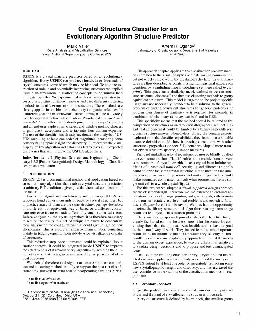
Crystal Structures Classifier for an Evolutionary Algorithm Structure Predictor Mario Valle ∗ Data Analysis and Visualization Services Swiss National Supercomputing Centre (CSCS) Artem R. Oganov † Laboratory of Crystallography, Department of Materials ETH Z ¨ urich ABSTRACT USPEX is a crystal structure predictor based on an evolutionary algorithm. Every USPEX run produces hundreds or thousands of crystal structures, some of which may be identical. To ease the ex- traction of unique and potentially interesting structures we applied usual high-dimensional classification concepts to the unusual field of crystallography. We experimented with various crystal structure descriptors, distinct distance measures and tried different clustering methods to identify groups of similar structures. These methods are already applied in combinatorial chemistry to organic molecules for a different goal and in somewhat different forms, but are not widely used for crystal structures classification. We adopted a visual design and validation method in the development of a library (CrystalFp) and an end-user application to select and validate method choices, to gain users’ acceptance and to tap into their domain expertise. The use of the classifier has already accelerated the analysis of US- PEX output by at least one order of magnitude, promoting some new crystallographic insight and discovery. Furthermore the visual display of key algorithm indicators has led to diverse, unexpected discoveries that will improve the USPEX algorithms. Index Terms: J.2 [Physical Sciences and Engineering]: Chem- istry; I.5.2 [Pattern Recognition]: Design Methodology—Classifier design and evaluation 1 I NTRODUCTION USPEX [20] is a computational method and application based on an evolutionary algorithm that enables crystal structure prediction at arbitrary P-T conditions, given just the chemical composition of the material. Due to the algorithm’s evolutionary nature, every USPEX run produces hundreds or thousands of putative crystal structures, but in practice many of them are the same structure, perhaps described in a different, but equivalent, way or based on a different coordi- nate reference frame or made different by small numerical errors. Before analysis by the crystallographers it is therefore necessary to reduce the results to a set of unique structures to concentrate their analysis on the configurations that could give insight on new phenomena. This is indeed an intensive manual labor, consisting mainly in judging equality from side-by-side visualization of pairs of structures. This reduction step, once automated, could be exploited also in another context. It could be integrated inside USPEX to improve the effectiveness of its evolutionary algorithm by avoiding the dilu- tion of diversity at each generation caused by the presence of iden- tical structures. We decided therefore to design an automatic structure compari- son and clustering method, initially to support the post-run classifi- cation task, but with the final goal of incorporating it inside USPEX. ∗ e-mail: [email protected] † e-mail: [email protected] The approach adopted applies to the classification problem meth- ods common to the visual analytics and data mining communities, but not widely employed in the crystallography field. Crystal struc- tures are thus described as points in a multidimensional space, each identified by a multidimensional coordinate set (here called finger- print). This space has a similarity metric defined so we can mea- sure structure “closeness” and then use clustering methods to group equivalent structures. This model is targeted to the project specific usage and not necessarily intended to be a solution to the general problem of finding equivalent structures for generic molecules or judging their degree of similarity as is required, for example, in combinatorial chemistry (a survey can be found in [18]). This specificity means that the method should be tailored to the comparison of structures as used by crystallographers (see sect. 1.1) and that in general it could be limited to a binary same/different crystal structure answer. Nonetheless, during the domain experts’ exploration of the classifier capabilities, they found that a suitable distance definition could show interesting correlations with other structure’s properties (see sect. 5.1), hence we adopted more usual, but crystal structure-specific, distance measures. Standard multidimensional techniques cannot be blindly applied to crystal structure data. The difficulties stem mainly from the very same structure of crystallographic data: a crystal is an infinite rep- etition of a basic cell (unit cell, see fig. 1) and different unit cells could describe the same crystal structure. Not to mention that small numerical errors in atom positions and unit cell parameters could make automated comparison difficult when progressing from a sin- gle unit cell to a whole crystal (fig. 2). For this project we adopted a visual supported design approach for the classifier design. Therefore we implemented an end-user ap- plication to access the fingerprinting and grouping algorithms mak- ing them immediately usable on real problems and providing inter- active diagnostics on their behavior. We thus had the opportunity to refine the library structure and algorithms starting from usage results on real crystal classification problems. The visual design approach provided also other benefits: first, it greatly facilitated gaining the users support for the project by con- vincing them that the approach was feasible and at least as good as the manual way of work. They indeed feared to miss important results using an automated method for which they see only the final results. Second, a visual exploratory approach simplified the access to the domain expert experience, to explore different alternatives, to validate design decisions and to propose and test unanticipated ideas. The use of the resulting classifier library (CrystalFp) and the re- lated end-user application has already accelerated the analysis of USPEX output by at least one order of magnitude, promoting some new crystallographic insight and discovery, and has increased the user confidence in the viability of the classification methods on real problems. 1.1 Problem Context To put the problem in context we should consider the input data origin and the kind of crystallographic structures processed. A crystal structure is defined by its unit cell, the smallest group 11 IEEE Symposium on Visual Analytics Science and Technology October 21 - 23, Columbus, Ohio, USA 978-1-4244-2935-6/08/$25.00 ©2008 IEEE
-
Upload
institutotecnologicodecelaya -
Category
Documents
-
view
0 -
download
0
Transcript of Crystal structures classifier for an evolutionary algorithm structure predictor
Crystal Structures Classifier for an
Evolutionary Algorithm Structure Predictor
Mario Valle∗
Data Analysis and Visualization ServicesSwiss National Supercomputing Centre (CSCS)
Artem R. Oganov†
Laboratory of Crystallography, Department of MaterialsETH Zurich
ABSTRACT
USPEX is a crystal structure predictor based on an evolutionaryalgorithm. Every USPEX run produces hundreds or thousands ofcrystal structures, some of which may be identical. To ease the ex-traction of unique and potentially interesting structures we appliedusual high-dimensional classification concepts to the unusual fieldof crystallography. We experimented with various crystal structuredescriptors, distinct distance measures and tried different clusteringmethods to identify groups of similar structures. These methods arealready applied in combinatorial chemistry to organic molecules fora different goal and in somewhat different forms, but are not widelyused for crystal structures classification. We adopted a visual designand validation method in the development of a library (CrystalFp)and an end-user application to select and validate method choices,to gain users’ acceptance and to tap into their domain expertise.The use of the classifier has already accelerated the analysis of US-PEX output by at least one order of magnitude, promoting somenew crystallographic insight and discovery. Furthermore the visualdisplay of key algorithm indicators has led to diverse, unexpecteddiscoveries that will improve the USPEX algorithms.
Index Terms: J.2 [Physical Sciences and Engineering]: Chem-istry; I.5.2 [Pattern Recognition]: Design Methodology—Classifierdesign and evaluation
1 INTRODUCTION
USPEX [20] is a computational method and application based onan evolutionary algorithm that enables crystal structure predictionat arbitrary P-T conditions, given just the chemical composition ofthe material.
Due to the algorithm’s evolutionary nature, every USPEX runproduces hundreds or thousands of putative crystal structures, butin practice many of them are the same structure, perhaps describedin a different, but equivalent, way or based on a different coordi-nate reference frame or made different by small numerical errors.Before analysis by the crystallographers it is therefore necessaryto reduce the results to a set of unique structures to concentratetheir analysis on the configurations that could give insight on newphenomena. This is indeed an intensive manual labor, consistingmainly in judging equality from side-by-side visualization of pairsof structures.
This reduction step, once automated, could be exploited also inanother context. It could be integrated inside USPEX to improvethe effectiveness of its evolutionary algorithm by avoiding the dilu-tion of diversity at each generation caused by the presence of iden-tical structures.
We decided therefore to design an automatic structure compari-son and clustering method, initially to support the post-run classifi-cation task, but with the final goal of incorporating it inside USPEX.
∗e-mail: [email protected]†e-mail: [email protected]
The approach adopted applies to the classification problem meth-ods common to the visual analytics and data mining communities,but not widely employed in the crystallography field. Crystal struc-tures are thus described as points in a multidimensional space, eachidentified by a multidimensional coordinate set (here called finger-print). This space has a similarity metric defined so we can mea-sure structure “closeness” and then use clustering methods to groupequivalent structures. This model is targeted to the project specificusage and not necessarily intended to be a solution to the generalproblem of finding equivalent structures for generic molecules orjudging their degree of similarity as is required, for example, incombinatorial chemistry (a survey can be found in [18]).
This specificity means that the method should be tailored to thecomparison of structures as used by crystallographers (see sect. 1.1)and that in general it could be limited to a binary same/differentcrystal structure answer. Nonetheless, during the domain experts’exploration of the classifier capabilities, they found that a suitabledistance definition could show interesting correlations with otherstructure’s properties (see sect. 5.1), hence we adopted more usual,but crystal structure-specific, distance measures.
Standard multidimensional techniques cannot be blindly appliedto crystal structure data. The difficulties stem mainly from the verysame structure of crystallographic data: a crystal is an infinite rep-etition of a basic cell (unit cell, see fig. 1) and different unit cellscould describe the same crystal structure. Not to mention that smallnumerical errors in atom positions and unit cell parameters couldmake automated comparison difficult when progressing from a sin-gle unit cell to a whole crystal (fig. 2).
For this project we adopted a visual supported design approachfor the classifier design. Therefore we implemented an end-user ap-plication to access the fingerprinting and grouping algorithms mak-ing them immediately usable on real problems and providing inter-active diagnostics on their behavior. We thus had the opportunityto refine the library structure and algorithms starting from usageresults on real crystal classification problems.
The visual design approach provided also other benefits: first, itgreatly facilitated gaining the users support for the project by con-vincing them that the approach was feasible and at least as goodas the manual way of work. They indeed feared to miss importantresults using an automated method for which they see only the finalresults. Second, a visual exploratory approach simplified the accessto the domain expert experience, to explore different alternatives,to validate design decisions and to propose and test unanticipatedideas.
The use of the resulting classifier library (CrystalFp) and the re-lated end-user application has already accelerated the analysis ofUSPEX output by at least one order of magnitude, promoting somenew crystallographic insight and discovery, and has increased theuser confidence in the viability of the classification methods on realproblems.
1.1 Problem Context
To put the problem in context we should consider the input dataorigin and the kind of crystallographic structures processed.
A crystal structure is defined by its unit cell, the smallest group
11
IEEE Symposium on Visual Analytics Science and Technology October 21 - 23, Columbus, Ohio, USA 978-1-4244-2935-6/08/$25.00 ©2008 IEEE

of atoms or molecules whose repetition at regular intervals in threedimensions produces the whole infinite structure of a crystal. Forany crystal the unit cell is not unambiguously defined because an in-finite crystal structure could be “cut” in different ways to define theelementary cell (see fig. 1). Each cell thus not necessarily containsthe same number of atoms.
Figure 1: Crystal unit cell (lower left) and its repetitions that build thewhole crystal structure. An alternative unit cell for the same crystalis show on the top right.
Another (incomplete) structure descriptor is the space group,i.e. the set of all symmetry operators in the structure. If the sym-metry for a crystal is known, then it provides a strong constraint onstructure similarity.
A value of internal energy or enthalpy could be associated to astructure. Lower values mean more stable structures. Equal struc-tures, also if represented in different ways, should have equal en-ergy values. But the energy equality alone is not sufficient as acriterion for grouping due to numerical imprecision or because dif-ferent structures could have energies so similar that discriminationis made impossible.
The crystal structures considered in this work differ from struc-tures of interest in other fields, like biochemistry for example,mainly in the number of atoms composing the unit cell and the kindof chemical elements involved. Another important difference is thatbiomolecules have bonds topologies that could be used to constrainthe classification process; instead crystal structures generally do notcarry uniquely defined bonds information.
A typical USPEX run produces 300–3000 structures usually con-taining 6 to 40 atoms of one to four element types. The USPEXoutput data are in concatenated VASP [16] POSCAR file format.This format does not carry the exact atoms’ types and the optionalstructure energy; these information should be provided externally tothe application. The structure symmetries are not available too, andthus are not considered as a possible input to the clustering process.
1.2 Previous Work
In the literature there are plenty of works proposing suitable struc-ture descriptors for organic molecules and distance metrics basedon them, but very few focused on crystal structure descriptors.Some of these few use symmetry criteria or some form of structurestandardization in the comparison phase [1–3, 6, 7, 9, 14, 22]. Forour application these methods seem not sufficiently robust with re-spect to numerical errors. Others use related structure data relevantto the problem they try to solve, like partial charges [31] or powderdiffraction patterns [5]. Furthermore methods based on crystal sym-metries are not applicable because we do not have symmetry data
available and because symmetry is extremely sensitive to small nu-merical errors. The work of Hundt et al. [14] gives a comprehensivesurvey of existing methods with a focus on calculating some formof distance metric between structures.
Chisholm and Motherwell [4] use interatomic distances to in-vestigate molecular packing and inspired one of the methods wetried (see sect. 3.1). Interatomic distances are a good choice for astructure identifier because they are independent from coordinatereference frame and cell choices.
Radial Distribution Function (RDF) is another possible structuredescriptor method based only on local characteristics. Beside inter-atomic distances, it considers also other data associated to the struc-ture. The method of Willighagen et al. [31] computes a RDF usingdistances from a central atom weighted by the atoms partial chargeto include electrostatic interactions that play a major role in crystalpacking. This method then calculates dissimilarities on the basis ofpowder diffraction patterns as in [5]. In our work we use a rapidlyconvergent function based on distance distributions and related toRDF and diffraction spectra, but we focus more on standard multi-dimensional methods for distance computation. Another interestingapplication of RDF is the work of Hemmer et al. [13] that uses RDFto match structures to IR spectra using a counterpropagation neu-ral network. Their goal is indeed different from ours, but they alsofound that RDF’s could be good crystal structures identifiers.
2 VISUAL APPROACH
We approached the design of the classifier from two sides. The firstis the application of multidimensional methods to crystallographicdata (see sect. 3). The second was in retrospect the most importantone: we decided to use a visual supported design and validationapproach for the classifier design. Visual analytics is not only fancygraphics, it is the use of visualizations, also simple ones, that couldfoster scientific insight in the domain expert user.
The visual approach could lead to better scientific ideas and re-sults because the scientists are involved in the design on a levelthey are familiar with, the system could visualize quickly the resultof their suggestions and, last, the visual imagery has the power tosuggest unexpected ideas and correlations not planned in the designphase.
3 CRYSTAL FINGERPRINTING
The proposed method associates a descriptor, called a fingerprint,to each structure. This descriptor is a vector of N real values; eachstructure becomes thus a point in a N-dimensional space. A dis-tance measure between these vectors is then used to cluster theminto groups of “near” fingerprints, that is, groups of similar struc-tures. This is indeed an application to the crystallography field ofconcepts already taken for granted in visual analytics or data min-ing.
To support the classification phase the fingerprint should, asmuch as possible, uniquely identify every structure, tolerate numer-ical errors and enhance contrast between different structures. Thechosen distance between fingerprints measure could help classifi-cation too by increasing contrast between neighbor points and byusing as much as possible the information available. In our highdimensional fingerprints space peculiar phenomena, like distanceconcentration [11], work against this goal. The distance measure-ment method should therefore counteract this effect too. In the laststep, to increase the classification quality, the classifier should cre-ate well separated, but highly internally connected clusters.
3.1 Fingerprint Definition
To support the specific kind of data we consider, the fingerprint as-sociated to a structure should be independent from: 1) translation
12

and rotation of the structure; 2) the choice of unit cell among equiv-alent unit cells; 3) the ordering of cell axis and atoms in the cell; 4)inversions and mirroring of the structure.
Figure 2: The effect of numerical errors on the unit cell is visiblemoving away from the origin along the crystal structure.
Whichever definition we chose for the structure fingerprint, itshould be computed over the “infinite” crystal structure. In prac-tice after a distance of Duc/2, where Duc is the longest unit celldiagonal, everything start repeating in every direction. Thereforein place of the whole crystal structure, a set of unit cell repetitionsthat cover the maximum distance over all structures in all directionsaround the base unit cell is used. We call this the extended unit cell.
We experimented with two fingerprint definitions: 1) per-atomdistance sets and 2) a rapidly convergent function based on distancedistributions and related to RDF and diffraction spectra. The ideabehind these fingerprint definitions is to reduce a global property,like the crystal structure, to a local one, like interatomic distancesor RDF. Both definitions satisfy the criteria stated above for a goodfingerprint.
The per-atom distance sets fingerprint is composed by a sectionfor each atom in the unit cell. Each section is an ascending orderedset of distances from the corresponding atom to all the other atomsof the extended unit cell. All these sets contain the same numberof distances. Furthermore each section is labeled with the elementtype of the corresponding atom. An example is shown in fig. 3.The idea behind this labeling method is this: if two structures are
Figure 3: Local atom distances for a GaAs crystal (top left) are con-catenated to form a fingerprint section (top right). Sections are thenassembled to form the structure fingerprint (bottom).
the same, then at least one atom of the same type in each shouldhave the same set of distances from its neighbors. This definition offingerprint then shifts the burden of corresponding atoms matchingto the distance computation phase (see sect. 3.2).
The second fingerprint definition we have experimented withstarts from the following function based on atoms identities anddistances distribution:
FP(R) =Vuc
NucB∑
i∈uc∑
j∈euc
ZiZ j
4πRi j2
δ (R−Ri j) where i 6= j (1)
Here i runs over the atoms of the unit cell, j over the atoms ofthe extended unit cell; Z is the atomic number; Ri j is the distancebetween atoms i and j; Vuc is the unit cell volume; Nuc is the num-ber of atoms in the unit cell and B is the bin size used to com-pute the value of FP(R) from the discrete peaks. Each peak is thensmoothed using a Gaussian kernel with σ set by the user (usually0.02 A) and accumulated into a histogram with bin size B (usually0.05 A). The resulting function is closely related to the diffractionspectra of the crystal. To remove fingerprint dependency from binsize and cutoff distance, the histogram is normalized:
FPnorm(R) =FP(R)
∑i ∑ j ZiZ jNiN j−1 (2)
Here Ni is the number of atoms in the unit cell with atomic numberZi and the two sums go over all distinct Z values. One example ofnormalized diffraction-like fingerprint is given in fig. 4.
Figure 4: Diffraction like fingerprint.
When atomic numbers Z are used as weights, the fingerprint isrelated to X-ray powder diffraction spectra. However, atoms withvery different properties may have similar atomic numbers (e.g.Z(I) = 53 and Z(Cs) = 55) and will be hard to distinguish them byX-rays or by the above fingerprint. For these cases, instead of Z, wecan use the Chemical Scale χ value or Mendeleev Number m [23]which correctly maps chemical differences between elements. Thissubstitution could increase the discriminating power of the finger-print in realistic situations: for example the relative variation of onefingerprint term due to the exchange of two atoms is proportional to|Z1 −Z2|/Z1Z2 and thus for a Si ↔ O exchange it varies by 5.4%using Z, by 0.2% using m, but using χ it changes by 23.0%.
The decision to use a certain algorithm or a certain set of pa-rameters (e.g. cutoff distance, bin size, Gaussian smoothing width)is made by the domain expert after interacting with the CrystalFpapplication to classify sets of real data that have been previouslymanually analyzed. So the algorithm performance is determined ina rather informal way. The evaluation result is nevertheless compat-ible with the expert’s crystallographic intuition. In the case of thefingerprint definition this experimentation selected the fingerprintdefined by eq. (1) and (2).
3.2 Distance Measurement
To make possible the classification of structures we should definea distance or pseudo-distance (i.e. one distance measure for which
13

triangular inequality does not hold) between fingerprints. We ex-perimented with three distance measures: 1) Cartesian distance; 2)Minkowski norm with fractional exponent and 3) cosine distance.
The Cartesian distance between same type atoms is the morephysically-based measure, and the most obvious one. For eachatom of the first structure we select from the second structure theatom of the same type with the minimum Cartesian distance be-tween the two atoms fingerprint sections. Remember that an atomfingerprint section is composed by the ordered set of distances be-tween this atom and its neighbors up to a certain cutoff distance.After this pairing has been done for all atoms, the sections of thetwo fingerprints are reordered to have corresponding atoms sectionsaligned. Then the usual Cartesian distance between the reorderedfingerprints is computed.
As seen before, we discontinued the use of Cartesian distancemeasure in favor of more crystal-specific measures after experimen-tation. Cartesian distance is thus a good illustration of how blindlyapplying multidimensional techniques is not the best strategy to usethem in the crystallographic domain.
The second proposed distance measure replaces the Cartesiandistance with a more general Minkowski norm with a fractionalexponent 0 < p < 1 as suggested by [11, sect. 2.6]. The distancebetween fingerprints based on the Minkowski norm with exponentp is defined as:
dist(i, j) =
(
∑k
| f pik − f p jk |p
)1p
(3)
Where FPi = ( f pi1 , f pi2 , . . .) is the fingerprint associated to struc-ture i. The Minkowski norm is really a pseudo-distance, but couldalleviate the distance concentration phenomena visible in high di-mensional spaces [11]: in these spaces all pair-wise distances seemto be equal or at least very similar. The reduction of concentration isapplication dependent, so we experimented with various p values;for our data better results have been obtained with p = 1/3.
The cosine distance is a popular norm in the text mining com-munity [25, 26]. Here every text has associated a vector of wordfrequencies and the similarity between texts is based on the dotproduct between these vectors. We use a slightly modified defi-nition of similarity that produces a distance in the [0 . . .1] intervalusing, in place of the word frequency vectors, the fingerprint FPi
associated to structure i:
dist(i, j) =1
2
(
1−FPi ·FPj
‖FPi‖‖FPj‖
)
(4)
To summarize, the choice of metric depends on the kind of dataset under analysis, and the search of the best metric is exactly oneof the goals of our visual design approach. As we will see, we haveobtained better results using the cosine distance metric (eq. 4), notlast for its ability to counteract the distance concentration phenom-ena, as seen in fig. 5, because it spreads distances much more thanthe other methods.
3.3 Structure Clustering
After building the matrix of distances between all pairs of finger-prints, we assign two structures to the same cluster if their distanceis less than a user-defined threshold. This operation transforms thedistance matrix into a binary connection matrix. The graph de-scribed by this matrix is then separated into connected componentsthat are our clusters of similar structures. We explored the fol-lowing methods to extract the connected components (groups) andunconnected entries (singles): 1) Depth First Search (DFS) [15];2) Shared Nearest Neighbor (SNN) [8] and 3) Pseudo SNN.
The first method adds one by one connected structures to a clus-ter doing a Depth First Search on the connection graph. The SNN
Figure 5: Distance distribution for the three distance definitions overthe MgSiO3 Post-perovskite 120GPa dataset. The curves are gen-erated by one of the interactive diagnostics tools (see sect. 4.3) andpresented together after normalizing the distance values.
algorithms are instead density based: they define density as thenumber of nearest neighbors points shared between pairs of con-nected points and confirm this last connection only if the number isat least K. The Pseudo SNN clustering method stops at this point,instead the full SNN algorithm adds a DBSCAN [27] step to refinethe cluster points’ membership.
Figure 6: Ordered distance matrix for: (top left) DFS grouping; (topright) Pseudo SNN with K = 1; (bottom left) Pseudo SNN with K = 5;(bottom right) SNN with K = 5. Inserts show the first 48 structuresgrouped.
Fig. 6 shows the effect of algorithm choice on the grouping struc-ture. Every cell of the matrix is colored from red to blue by the dis-tance between the corresponding row and column structures and thestructures are then ordered to keep the ones pertaining to the samegroup together in decreasing order of group size (see sect. 4.3). Be-side the first group, that has uniformly low distances between struc-tures inside the group for all methods, the differences are in thenumber of groups and in the uniformity of distances inside groups.The the patch color uniformity is visually estimated. DFS groupsstructures that have distances that are not uniformly low. PseudoSNN with K = 5 creates smaller groups, but with the same non uni-formity of distances and it does not consider various similar pairs
14

for grouping as testified by the regularly spaced red points far fromthe diagonal. SNN creates few groups containing similarly low dis-tances. Pseudo SNN with K = 1 seems a good compromise be-tween SNN and DFS. There is another reason for not choosing thefull SNN method even if it is resistant to noise and can create moreconnected clusters: it is well known that its DBSCAN phase doesnot work well with varying cluster densities and high-dimensionaldata. We therefore opted to use the pseudo SNN clustering methodwith K = 1.
4 VISUAL DESIGN AND VALIDATION
To support the visual design of the classifier library we must pro-vide ways to explore algorithm choices and parameters setting andwe must make available visual tools to verify and validate thesesettings.
Figure 7: The CrystalFp end-user application. The control panel(left), the scatterplot and ordered distance matrix (top), one diag-nostic chart and the visual pair-wise structure comparison (bottom).
To make this exploration possible we built an end-user applica-tion around the classifier library to support the USPEX results anal-ysis workflow and to provide interactive visual diagnostics on thebehavior of the algorithms. As we have seen in the previous section,the choices made for the fingerprint definition, the distance metricand the classifier algorithm are direct consequence of the domainexpert exploration and validation of the library during analysis runsmade on real data.
4.1 Analysis Workflow
The analysis starts from the USPEX output that is composed by oneor more files containing crystal data and, optionally, files with thecorresponding energies or enthalpies. These files are then loadedinside the CrystalFp end-user application. Multiple results files areneeded, for example, when investigating the relationship betweendistances and energy differences from a ground-state structure (seesect. 5.1). During load the user should input the element type ofthe atoms, because this information is not contained in the USPEXoutput files.
To focus the analysis over the most stable structures a filteringstep is applied after loading to retain only the lowest energy ones.Then the three phases of the classification –fingerprint computa-tion, distance computation and grouping– are run in sequence. Thissplitting into three independent phases facilitates methods explo-ration and parameters adjustment driven by the visual diagnostictools as we see below.
After classification, to support further analysis, the system couldgenerate two kinds of outputs: 1) a report of the groups’ contenttogether with structure energies and 2) the reduced structure andenergy files, where each group is substituted by its most character-istic structure. This structure is the lowest energy one or, if energieshave not been loaded, the structure with the highest silhouette coef-ficient in the group (see sect. 4.3).
4.2 Previous Analysis Workflow
To appreciate the streamlined workflow made possible by the Crys-talFp end-user application, we take a look at the previous manualstructure classification method.
The USPEX output is loaded inside a visualization tool and thenthe crystallographer has to retrieve on screen all the pairs of struc-tures and visually compare them. Besides being a time consumingmethod, it is worth mentioning its main difficulty: the comparisonin general cannot be limited to a single pair of unit cells, but shouldbe done on the “infinite” crystal structure, and this makes compari-son methods based on naıve superposition strategies difficult to use.
4.3 Interactive Diagnostics
The classification application provides visual representation of keyalgorithm’s quantities so the domain expert could judge the algo-rithm behavior. The visual validation and analysis, plus the user’salgorithm selection and parameters modification, supports a veryeffective exploratory design approach. The visual tools providedare: 1) distance matrix; 2) grouping quality; 3) group evolution dis-play; 4) scatterplot; 5) diagnostic charts and 6) visual structure paircomparison.
Distance matrix The full distance matrix between structuresvisually reveals distance trends and overall distance distribution.When the distance map is sorted to put together structures of thesame group, it becomes the primary method to judge grouping qual-ity. Good grouping produces uniformly red squares on the bottomleft with the rest of the map tending toward green or blue (fig. 8).
Figure 8: Distance matrix (left) and sorted distance matrix (right).
Grouping quality To judge grouping quality we use the pop-ular method of silhouette coefficients [28, p. 541]. This quality
Figure 9: Silhouette coefficients for all grouped structures. Along Yare the groups, along X the elements of the group. Blue: +1, white:0, red: -1.
measure combines cohesion inside a group with separation betweengroups. A value of −1 means the element is probably in the wronggroup, because it has low cohesion with the rest of its group and is
15

too close to another group, whereas a value of +1 means the ele-ment is closer to the other elements of its group than to the elementsof all other ones. The coefficients are visualized in a chart whereevery horizontal slab represents a group with the elements coloredby the respective silhouette coefficient.
Group evolution This visualization shows the various groupsforming and evolving during an USPEX run. Here every structure
Figure 10: Group evolution display.
starts a horizontal line at a position from the left side proportionalto its sequential index. If the structure belongs to a group its line isadded below the ones representing the structures already pertainingto the group, otherwise it is added below all the other lines. Fig. 10shows an example where a big group appears and continues steadilyto grow.
Scatterplot To provide an intuitive view of the fingerprints’multidimensional space, we map it to a 2D space. We use a sim-ple implementation of force-directed 2D point’s placement algo-rithm [12] plus a random perturbation step to let the point config-uration escape from local minima. The scatterplot points could becolored by group with black representing non-grouped structures(fig. 11), this way the user has an intuitive view of the goodness ofthe clustering algorithm. If the points are colored by a measure of
Figure 11: Scatterplot with points colored by group (left) or by tem-perature (center) and the distance mapping fidelity chart (right).
the total forces acting on the point at equilibrium (here called tem-perature), the scatterplot shows if the point configuration has beentrapped in a local minima. This is one of the scatterplot’s own di-agnostic tools; another one is the mapping fidelity chart that showshow multidimensional distances are mapped to 2D distances: themore points lay close to the diagonal, the better the mapping is.
Other diagnostic charts Various charts have been developedto support visual diagnostics: 1) distances distribution charts, tograsp the overall structure of the multidimensional space; 2) fin-gerprint display, to validate the fingerprint computation; 3) energydistribution inside groups, to check that structures with similar en-ergies have been grouped together.
Visual structures comparison The definitive test for the va-lidity of the classification algorithm is the visual comparison andsuperposition of structures assigned to the same group. The user
can select structures in two ways: manually, from the groups’ con-tent listings and graphically, circling a group of points in the scat-terplot. Both structures could be replicated and color coded. One ofthem can be translated and rotated to manually superimpose it overthe other one.
4.4 New Visual Analysis Tools
We added to the above list of interactive diagnostic tools few visu-alizations that are not strictly needed for validation of the classifi-cation schema, but provided new analysis methods of the USPEXresults. We added them because existing algorithms made theirimplementation almost effortless and because in the visual designphase we discovered their usefulness. As we see in sect. 5.1, theseanalysis and visualization tools provided unexpected insight to theresearchers. The first of them is a measure of the degree of order ofthe structures. It is computed from the simulated diffraction spectraand it is defined as:
DegreeO f Order =1
R0
∫
F(x)2 dx (5)
Where R0 is the distance at which the fingerprint first crosses zero(see fig. 4). Order seems to increase and saturate or remain almostconstant during an USPEX run, but exhibits increasing number ofisolated high order peaks at the end of the run (fig. 12). Visualizing
Figure 12: Order evolution in the GaAs (8 atoms/cell) dataset.
energy vs. order and energy differences vs. distances visually re-vealed unexpected correlations also (see sect. 5.1).
5 VISUAL ANALYTICS OUTCOMES
A typical ab-initio evolutionary run samples ≈ 1000 structures(which takes several thousand CPU hours on today’s clusters).Manual analysis of such a dataset, aimed at finding a handful ofthe most promising distinct structures, requires between 2 to 20hours of work. With the CrystalFp end-user application, this can beachieved within ≈ 10 minutes.
To illustrate this, let us look at a rather pathological case, hy-drogen under pressure. The pathology is easy to recognize usingthe CrystalFp visual analysis tools: the number of distinct enthalpyminima is rather small (so that even such a poor global optimizationstrategy as random sampling can find the global minimum [24]),but their enthalpies are extremely close. In such cases, one cannotuse enthalpy as the sole grouping criterion and there will be a largenumber of identical structures found in evolutionary runs. Here weanalyzed a dataset obtained with USPEX for hydrogen at 600 GPa,with 16 atoms in the (super)cell. Our dataset contains 1274 struc-tures, 794 of which had enthalpies within 0.5 eV of the lowest valuefound. The large number of energetically reasonable structuresmakes this dataset difficult to analyze manually. Analyzing thesestructures with CrystalFp, we recognized an unusually high degen-eracy: these 794 structures could be grouped into only 4 uniquestructures. This analysis took only ≈ 5 minutes, but doing the samework manually would take many hours. Among the four structuresone belongs to the Cs-IV structure type (fig. 13a), two are closelyrelated to it, and one belongs to the alpha-Ga type (fig. 13b). The
16

Cs-IV structure is the ground state, while the alpha-Ga type phasehas a 20-30 meV/atom higher enthalpy. Both are atomic phases(i.e. non-molecular), and we confirm the conclusion of Pickard andNeeds [24] that hydrogen adopts non-molecular structures at pres-sures above 500 GPa. The case of compressed hydrogen is ratherrare in the extent of degeneracies, but even in more normal casesthe use of the analysis methods presented here makes data analysismuch easier compared to manual analysis.
Figure 13: Low-enthalpy structures of hydrogen at 600 GPa foundwith USPEX: (a) Cs-IV structure, (b) alpha-Ga structure.
Various other examples of USPEX results with the role playedby CrystalFp in their analysis are collected in the work of Oganovet al. [21]. But more interesting are the unexpected outcomes madepossible by the CrystalFp visual approach. These outcomes wherenot searched for, but the availability of the CrystalFp visualizationshelped pose the right scientific questions. Here below is collecteda quick survey of these outcomes, instead their analysis will appearelsewhere.
5.1 Unexpected Outcomes
Genetic algorithm structure cancer phenomena Standardevolutionary algorithms, when allowed to run indefinitely, tend toconverge to one solution, which tends to create its own replicas.Sometimes this solution may be suboptimal, and such prematureconvergence precludes efficient exploration of other possible solu-tions. CrystalFp visual diagnostics can visualize this phenomenon(see the large blue wedge in fig. 10). A procedure based on finger-printing has be incorporated in the USPEX code, and proved to bevery effective in precluding such “cancer growth” phenomena.
Energy vs. Distance correlation One of the basic assump-tions made in the construction of the structure prediction methodUSPEX is that the energy landscape has an overall shape, wherelow-energy structures are clustered together. Energy-distance cor-relation enables checking this assumption for real systems. In mostcases this assumption is confirmed, but we have found cases wherethere are several “clusters” of low-energy minima separated bylarge distances (see fig. 14). In such cases evolutionary algorithmsare less efficient. Again, it is possible to maximize their efficiencyusing fingerprinting inside the evolutionary algorithm.
Random structures distance distribution Looking at statis-tics of distances between structures in a random set of structures,we discovered a striking Gaussian-type shape of distributions, witha clear peak (fig. 5). In the multidimensional space, fingerprintvectors describing crystal structures form a diffuse spherical shell(lengths of the vectors are similar within an order of magnitude, butdirections differ) and the distance distribution corresponding to thisgeometry is similar to a Gaussian. In some cases we find more thanone peak in the distance distribution, and this is a signal of com-plex chemistry involving different coordination numbers. Interest-ingly, as the number of atoms increases, fingerprints of randomly
Figure 14: GaAs structures exhibits clear energy–distance correla-tion (top). Instead MgNH shows a more complex landscape (bottom).
generated structures (and structures themselves, and their energies)become increasingly close to random (where the fingerprint is −1at small distances and 0 otherwise) and progressively indistinguish-able. This can be viewed as a decrease of the radius of the hyper-dimensional sphere, until it collapses to a point. This picture clar-ifies why Cartesian distances (measuring the distance between twopoints on a sphere) suffer more from the “curse of dimensional-ity” than cosine distances (which measure angular differences) (seesect. 3.2).
Energy vs. Order correlation In many runs we see order in-creasing during the run, and it is correlated with energies: highorder usually means low energies. This is natural, disordered struc-tures are expected to have high energies. Energy-order correlationscan be used to uncover cases of geometric frustration, where less or-dered or more complex structures are made energetically favorableby competition of oppositely directed factors.
6 SYSTEM IMPLEMENTATION
The CrystalFp library has been implemented in C++ with only onedependency on other software1 to make its integration inside appli-cations easier. Its API various areas –fingerprint calculation, dis-tances, grouping and analysis– have been implemented as separateclasses as orthogonal as possible to simplify the addition of otheralgorithms for testing. The library has been tested on Linux, Win-dows and Mac OSX, but potentially can run on any platform.
The end-user application, which supports the library visual de-sign and validation, has been built inside the molecular visualiza-tion toolkit STM4 [29, 30] based on AVS/Express [10]. Each stepin the analysis workflow has been implemented as an AVS/Expressmodule. The complete application has been visually assembled us-ing these modules together with the ones provided by STM4 andAVS/Express itself.
This choice provided us a threefold benefit: first, we don’t haveto implement from scratch functionalities already available, likedata file readers and graphical crystal structure rendering, but wecan concentrate on our problem delegating the application con-trol to the AVS/Express “data flow” architecture [10]. The secondbenefit, which perfectly matches with the project goal of user in-volvement in the visual design, lays in the immediate support ofrapid prototyping provided by the component architecture and vi-sual programming paradigm of AVS/Express. Last, the packag-ing of the CrystalFp functionalities as separate components makethem reusable inside other crystallographic applications built usingSTM4.
1The exception is the use of ANN [19], a library for approximate nearest
neighbor searching.
17

7 DISCUSSION
The main contribution of this work to the crystallographic field isthe reformulation of the crystal structures classification problem us-ing multidimensional analysis and visualization methods. This re-formulation provided flexibility for the exploration of new solutionsand a concrete use case for others to consider visual analytics meth-ods in the crystallographic and chemistry fields.
The visual exploration and diagnosis of key algorithm methodsand parameters has facilitated the involvement of the domain ex-perts in the classifier design. The rapid testing and modificationof the algorithms implementation has been made possible by cou-pling visual design to the fast prototyping capabilities offered bythe STM4 environment. This visual approach made a real differ-ence in the project, also without introducing sophisticated visual-izations, but simply relaying on the grounding ideas of visual ana-lytics. Moreover this approach is not constrained to the crystallo-graphic field, but can be reused also in other contexts.
The algorithms selected for the various library classificationphases have been collected in sections 3.1, 3.2 and 3.3 together withan informal evaluation in section 5. Fortunately nearly-ground-truth datasets were abundantly present and used for this informaltesting and validation of the algorithms.
There are still two open points that we plan to address in the fu-ture. The distance concentration [11] phenomena overshadow everyattempt to define a better fingerprint or a better distance measure.We started experimenting with a weighting of distances that takesinto account this phenomenon that has been introduced by the data-driven high-dimensional scaling (DD-HDS) [17, sect. IV.A] visu-alization method. The second open point concerns too many freeparameters taken by the algorithm. Some of these parameters havebeen fixed based on crystallographic expertise, but the parametersrelated to clustering remains arbitrary.
8 CONCLUSION AND FUTURE WORK
After the visual design and validation of the CrystalFp library, westarted its integration inside the USPEX evolutionary crystal pre-dictor algorithm. This integration will surely raise other interestingproblems beside the open points already listed.
The work on the CrystalFp library and end-user application willcontinue, driven by the domain expert requests elicited by the visualexploration tools put in place during the design phase. For examplewe already have to experiment with a new fingerprinting methodand a request for new functionalities in the end-user application toease the superposition and comparison of crystal structures.
But so far the most important lesson learned in this project is theimportance to have on board domain experts deeply interested inthe project success and to have an end-user application, built usinglanguage and concepts from the crystallographic domain, they canuse and experiment with.
ACKNOWLEDGEMENTS
Calculations were performed at the Joint Russian SupercomputerCentre (Russian Academy of Sciences, Moscow), ETH Zurich andSwiss National Supercomputing Centre (Manno) that the authorsgratefully acknowledge.
REFERENCES
[1] L. C. Andrews and H. J. Bernstein. Bravais lattice invariants. Acta
Crystallogr. Sect. A, 51:413–416, May 1995.
[2] L. C. Andrews, H. J. Bernstein, and G. A. Pelletier. A perturbation
stable cell comparison technique. Acta Crystallogr. Sect. A, 36:248–
252, Mar. 1980.
[3] J. Apostolakis, D. W. M. Hofmann, and T. Lengauer. Derivation of
a scoring function for crystal structure prediction. Acta Crystallogr.
Sect. A, 57:442–450, July 2001.
[4] J. A. Chisholm and S. Motherwell. COMPACK: a program for identi-
fying crystal structure similarity using distances. J. Appl. Crystallogr.,
38:228–231, Feb. 2005.
[5] R. de Gelder. Quantifying the similarity of crystal structures. IUCr
CompComm Newsletter, 7:59–69, Nov. 2006.
[6] A. V. Dzyabchenko. Method of crystal-structure similarity searching.
Acta Crystallogr. Sect. B, 50:414–425, Aug. 1994.
[7] B. P. V. Eijck and J. Kroon. Fast clustering of equivalent structures in
crystal structure prediction. J. Comput. Chem., 18:1036–1042, 1997.
[8] L. Ertoz, M. Steinbach, and V. Kumar. Finding clusters of different
sizes, shapes, and densities in noisy, high dimensional data. In Pro-
ceedings of the 3rd SIAM International Conference on Data Mining
(SDM ’03), Philadelphia, 2003.
[9] L. Fabian and A. Kalman. Volumetric measure of isostructurality. Acta
Crystallogr. Sect. B, 55:1099–1108, Dec. 1999.
[10] J. Favre and M. Valle. AVS and AVS/Express. In C. Hansen and
C. Johnson, editors, The Visualization Handbook, pages 655–672.
Academic Press, Dec. 2004.
[11] D. Francois, V. Wertz, and M. Verleysen. The concentration of frac-
tional distances. IEEE T. Knowl. Data. En., 17(7):873–886, July 2007.
[12] T. M. J. Fruchterman and E. M. Reingold. Graph Drawing by Force-
directed Placement. Software Pract. Exper., 21(11):1129–1164, 1991.
[13] M. C. Hemmer, V. Steinhauer, and J. Gasteiger. Deriving the 3D struc-
ture of organic molecules from their infrared spectra. Vib. Spectrosc.,
19:151–164, Feb. 1999.
[14] R. Hundt, J. C. Schon, and M. Jansen. CMPZ — an algorithm for
the efficient comparison of periodic structures. J. Appl. Crystallogr.,
39:6–16, Feb. 2006.
[15] A. K. Jain, M. N. Murty, and P. J. Flynn. Data clustering: a review.
ACM Comput. Surv., 31(3):264–323, 1999.
[16] G. Kresse and J. Furthmuller. Efficient iterative schemes for ab initio
total-energy calculations using a plane-wave basis set. Phys. Rev. B,
54(16):11169–11186, Oct. 1996.
[17] S. Lespinats, M. Verleysen, A. Giron, and B. Fertil. DD-HDS: A
Method for Visualization and Exploration of High-Dimensional Data.
IEEE T. Neural Networ., 18:1265–1279, 2007.
[18] D. J. Livingstone. The Characterization of Chemical Structures Us-
ing Molecular Properties. A Survey. J. Chem. Inf. Comput. Sci.,
40(2):195–209, 2000.
[19] D. M. Mount and S. Arya. ANN — A Library for Approximate Near-
est Neighbor Searching. http://www.cs.umd.edu/˜mount/
ANN. Online, last checked: Jun. 2008.
[20] A. R. Oganov and C. W. Glass. Crystal structure prediction using ab
initio evolutionary techniques: Principles and applications. J. Chem.
Phys., 124(24):244704, 2006.
[21] A. R. Oganov, Y. Ma, C. W. Glass, and M. Valle. Evolutionary crystal
structure prediction: overview of the USPEX method and some of its
applications. Psi-k Newsletter, 84:1–10, Dec. 2007.
[22] E. Parthe and L. M. Gelato. The standardization of inorganic crystal-
structure data. Acta Crystallogr. Sect. A, 40:169–183, May 1984.
[23] D. G. Pettifor. The structures of binary compounds. I. Phenomenolog-
ical structure maps. J. Phys. C, 19(3):285–313, 1986.
[24] C. J. Pickard and R. J. Needs. Structure of phase III of solid hydrogen.
Nat. Phys., 3:473–476, July 2007.
[25] G. Salton and C. Buckley. Term-weighting approaches in automatic
text retrieval. Inform. Process. Manag., 24(5):513–523, 1988.
[26] G. Salton and M. J. McGill. Introduction to Modern Information Re-
trieval. McGraw-Hill, New York, 1983.
[27] J. Sander, M. Ester, H.-P. Kriegel, and X. Xu. Density-Based Cluster-
ing in Spatial Databases: The Algorithm GDBSCAN and Its Applica-
tions. Data Min. Knowl. Discov., 2(2):169–194, June 1998.
[28] P.-N. Tan, M. Steinbach, and V. Kumar. Introduction to Data Mining.
Addison-Wesley, 2006.
[29] M. Valle. STM4 — the molecular visualization toolkit. http://
www.cscs.ch/˜mvalle/STM4. Online, last checked: Jun. 2008.
[30] M. Valle. STM3: a chemistry visualization platform. Z. Kristallogr.,
220:585–588, 2005.
[31] E. L. Willighagen, R. Wehrens, P. Verwer, R. de Gelder, and L. M. C.
Buydens. Method for the computational comparison of crystal struc-
tures. Acta Crystallogr. Sect. B, 61:29–36, Feb. 2005.
18