
18F-FDG PET/CT Role in Staging of Gastric Carcinomas
8
18 F-FDG PET/CT Role in Staging of Gastric Carcinomas: Comparison with Conventional Contrast Enhancement Computed Tomography Corinna Altini, MD, Artor Niccoli Asabella, MD, PhD, Alessandra Di Palo, MD, Margherita Fanelli, MStat, Cristina Ferrari, MD, Marco Moschetta, MD, PhD, and Giuseppe Rubini, MD Abstract: The purpose of the report was to evaluate the role of fluorine- 18 fluoro-2-deoxy-D-glucose positron emission tomography/computed tomography ( 18 F-FDG PET/CT) in staging gastric cancer comparing it with contrast enhancement computed tomography (CECT). This retrospective study included 45 patients who underwent per- formed whole body CECT and 18 F-FDG PET/CT before any treatment. We calculated CECT and 18 F-FDG PET/CT sensitivity, specificity, accuracy, positive and negative predictive values (PPV and NPV) for gastric, lymphnode, and distant localizations; furthermore, we compared the 2 techniques by McNemar test. The role of 18 F-FDG PET/CT semiquantitative parameters in relation to histotype, grading, and site of gastric lesions were evaluated by ANOVA test. Sensitivity, specificity, accuracy, PPV and NPV of CECT, and 18 F-FDG PET/CT for gastric lesion were, respectively, 92.11%, 57.14%, 86.66%, 92.11%, 57.14% and 81.58%, 85.71%, 82.22%, 96.88%, 46.15%. No differences were identified between the 2 tech- niques about sensitivity and specificity. No statistical differences were observed between PET parameters and histotype, grading, and site of gastric lesion. The results of CECT and 18 F-FDG PET/CT about lymph- node involvement were 70.83%, 61.90%, 66.66%, 68%, 65% and 58.33%, 95.24%, 75.55%, 93.33%, 66.67%. The results of CECT and 18 F-FDG PET/CT about distant metastases were 80%, 62.86%, 66.66%, 38.10%, 91.67% and 60%, 88.57%, 82.22%, 60%, 88.57%. 18 FDG PET/ CT specificity was significantly higher both for lymphnode and distant metastases. The 18 F-FDG PET/CT is a useful tool for the evaluation of gastric carcinoma to detect primary lesion, lymphnode, and distant metastases using 1 single image whole-body technique. Integration of CECT with 18 F-FDG PET/CT permits a more valid staging in these patients. (Medicine 94(20):e864) Abbreviations: CECT = contrast enhancement computed tomography, 18 F-FDG PET/CT = fluorine-18 fluoro-2-deoxy-D- glucose positron emission tomography/computed tomography, MTV = metabolic tumor volume, NPV = negative predictive value, PPV = positive predictive value, Pts = patients, SUV = standardized uptake value, TLG = total lesion glycolysis, VOI = volume of interest. INTRODUCTION G astric cancer is the 4th most common cancer worldwide. 1 Each year, 1 million patients (pts) are diagnosed, account- ing for 12% of all cancer deaths. 2 Although curative surgery remains the mainstay of gastric cancer treatment, surgical morbidity from gastrectomy is signifi- cant. 3 Patients’ selection prior to surgery is essential to avoid the morbidity and mortality of unnecessary surgery in those who will not benefit from the intervention but can be submitted to che- motherapy and radiotherapy. 4 Diagnosis is mainly performed by endoscopy associated with biopsy. Accurate staging of the disease including the local invasion extent, lymphnode involvement, and distant metastases is important for patients management and surgical planning. 3 At the state of art, gastric carcinoma is staged with endo- scopic ultrasonography, contrast enhancement computed tom- ography (CECT), and occasionally laparoscopy. 5–7 Conventionally, morphology-based imaging tools are helpful for preoperative staging, but they have been found incomplete due to their technical limitations. 8,9 In recent decades fluorine-18 fluoro-2-deoxy-D-glucose positron emission tomography/computed tomography ( 18 F-FDG PET/CT) has proven to be useful in the diagnosis and evaluation of malignancies by providing metabolic information and apporting advantages in staging, therapeutic evaluation, and recurrence surveillance. 10,11 Several clinical guidelines, including those of the National Comprehensive Cancer Network and The European Society for Medical Oncology, suggest that 18 F-FDG PET/CT imaging may improve staging. 12,13 Although these recommen- dations, its value in staging gastric carcinomas is still controversial. The purpose of this study was to evaluate the role of 18 F- FDG PET/CT in staging gastric carcinoma comparing it with CECT. Furthermore, we investigated if morphological and func- tional parameters might play a role in staging gastric carcinomas. METHODS Patients Five hundred and seventy-two 18 F-FDG PET/CT were performed from March 2007 to March 2013 for gastric carci- noma evaluation. Fifty-seven of them were performed for staging, 301 for restaging and 214 in follow-up. The 57 18 F-FDG PET/CT performed with staging intent corresponded to 57 pts; all of them had been pathologically diagnosed with gastric carcinoma by endoscopic biopsy. Editor: Jianfeng Li. Received: March 24, 2015; revised and accepted: April 14, 2015. From the Nuclear Medicine Unit (CA, ANA, ADP, MF, CF, GR); Section of Diagnostic Imaging, D.I.M., University of Bari ‘‘Aldo Moro’’, Bari, Italy (MM). Correspondence: Artor Niccoli Asabella, Nuclear Medicine Unit, D.I.M., University of Bari ‘‘Aldo Moro’’, Piazza G. Cesare 11, 70124 Bari, Italy (e-mail: [email protected]). The authors have no funding and conflicts of interest to disclose. Copyright # 2015 Wolters Kluwer Health, Inc. All rights reserved. This is an open access article distributed under the Creative Commons Attribution-NoDerivatives License 4.0, which allows for redistribution, commercial and non-commercial, as long as it is passed along unchanged and in whole, with credit to the author. ISSN: 0025-7974 DOI: 10.1097/MD.0000000000000864 Medicine Volume 94, Number 20, May 2015 www.md-journal.com | 1
-
Upload
independent -
Category
Documents
-
view
0 -
download
0
Transcript of 18F-FDG PET/CT Role in Staging of Gastric Carcinomas

18F-FDG PET/CT Role in Staging of Gastric Carcinomas:Comparison with Conventional Contrast Enhancement
Computed Tomography
ella,
Corinna Altini, MD, Artor Niccoli Asab arcoglucose positron emission tomography/computed tomography,
MTV = metabolic tumor volume, NPV = negative predictive
value, PPV = positive predictive value, Pts = patients, SUV =
The 57 18F-FDGcorresponded to 57 ptdiagnosed with gastric
Editor: Jianfeng Li.Received: March 24, 2015; revised and accepted: April 14, 2015.From the Nuclear Medicine Unit (CA, ANA, ADP, MF, CF, GR); Sectionof Diagnostic Imaging, D.I.M., University of Bari ‘‘Aldo Moro’’, Bari,Italy (MM).Correspondence: Artor Niccoli Asabella, Nuclear Medicine Unit, D.I.M.,
University of Bari ‘‘Aldo Moro’’, Piazza G. Cesare 11, 70124 Bari,Italy (e-mail: [email protected]).
The authors have no funding and conflicts of interest to disclose.Copyright # 2015 Wolters Kluwer Health, Inc. All rights reserved.This is an open access article distributed under the Creative CommonsAttribution-NoDerivatives License 4.0, which allows for redistribution,commercial and non-commercial, as long as it is passed along unchangedand in whole, with credit to the author.ISSN: 0025-7974DOI: 10.1097/MD.0000000000000864
Medicine � Volume 94, Number 20, May 2015
andra Di Palo
Margherita Fanelli, MStat, Cristina Ferrari, MD, MAbstract: The purpose of the report was to evaluate the role of fluorine-
18 fluoro-2-deoxy-D-glucose positron emission tomography/computed
tomography (18F-FDG PET/CT) in staging gastric cancer comparing it
with contrast enhancement computed tomography (CECT).
This retrospective study included 45 patients who underwent per-
formed whole body CECT and 18F-FDG PET/CT before any treatment.
We calculated CECT and 18F-FDG PET/CT sensitivity, specificity,
accuracy, positive and negative predictive values (PPV and NPV) for
gastric, lymphnode, and distant localizations; furthermore, we compared
the 2 techniques by McNemar test. The role of 18F-FDG PET/CT
semiquantitative parameters in relation to histotype, grading, and site
of gastric lesions were evaluated by ANOVA test.
Sensitivity, specificity, accuracy, PPV and NPV of CECT, and18F-FDG PET/CT for gastric lesion were, respectively, 92.11%,
57.14%, 86.66%, 92.11%, 57.14% and 81.58%, 85.71%, 82.22%,
96.88%, 46.15%. No differences were identified between the 2 tech-
niques about sensitivity and specificity. No statistical differences were
observed between PET parameters and histotype, grading, and site of
gastric lesion. The results of CECT and 18F-FDG PET/CT about lymph-
node involvement were 70.83%, 61.90%, 66.66%, 68%, 65% and
58.33%, 95.24%, 75.55%, 93.33%, 66.67%. The results of CECT and18F-FDG PET/CT about distant metastases were 80%, 62.86%, 66.66%,
38.10%, 91.67% and 60%, 88.57%, 82.22%, 60%, 88.57%. 18FDG PET/
CT specificity was significantly higher both for lymphnode and distant
metastases.
The 18F-FDG PET/CT is a useful tool for the evaluation of gastric
carcinoma to detect primary lesion, lymphnode, and distant metastases
using 1 single image whole-body technique. Integration of CECT with18F-FDG PET/CT permits a more valid staging in these patients.
(Medicine 94(20):e864)
Abbreviations: CECT = contrast enhancement computed
tomography, 18F-FDG PET/CT = fluorine-18 fluoro-2-deoxy-D-
MD, PhD, Aless , MD,Moschetta, MD, PhD, and Giuseppe Rubini, MD
volume of interest.
standardized uptake value, TLG = total lesion glycolysis, VOI =
INTRODUCTION
G astric cancer is the 4th most common cancer worldwide.1
Each year, 1 million patients (pts) are diagnosed, account-ing for 12% of all cancer deaths.2
Although curative surgery remains the mainstay of gastriccancer treatment, surgical morbidity from gastrectomy is signifi-cant.3
Patients’ selection prior to surgery is essential to avoid themorbidity and mortality of unnecessary surgery in those who willnot benefit from the intervention but can be submitted to che-motherapy and radiotherapy.4
Diagnosis is mainly performed by endoscopy associatedwith biopsy. Accurate staging of the disease including the localinvasion extent, lymphnode involvement, and distant metastasesis important for patients management and surgical planning.3
At the state of art, gastric carcinoma is staged with endo-scopic ultrasonography, contrast enhancement computed tom-ography (CECT), and occasionally laparoscopy.5–7
Conventionally, morphology-based imaging tools arehelpful for preoperative staging, but they have been foundincomplete due to their technical limitations.8,9
In recent decades fluorine-18 fluoro-2-deoxy-D-glucosepositron emission tomography/computed tomography (18F-FDGPET/CT) has proven to be useful in the diagnosis and evaluation ofmalignancies by providing metabolic information and apportingadvantages in staging, therapeutic evaluation, and recurrencesurveillance.10,11 Several clinical guidelines, including those ofthe National Comprehensive Cancer Network and The EuropeanSociety for Medical Oncology, suggest that 18F-FDG PET/CTimaging may improve staging.12,13 Although these recommen-dations, its value in staging gastric carcinomas is stillcontroversial.
The purpose of this study was to evaluate the role of 18F-FDG PET/CT in staging gastric carcinoma comparing it withCECT. Furthermore, we investigated if morphological and func-tional parameters might play a role in staging gastric carcinomas.
METHODS
PatientsFive hundred and seventy-two 18F-FDG PET/CT were
performed from March 2007 to March 2013 for gastric carci-noma evaluation. Fifty-seven of them were performed forstaging, 301 for restaging and 214 in follow-up.
PET/CT performed with staging intents; all of them had been pathologicallycarcinoma by endoscopic biopsy.
www.md-journal.com | 1

This retrospective study included only 45 pts who under-went whole-body CECT and 18F-FDG PET/CT before anytreatment and within 30 days between them.
The CECT scan was performed on average 18 days beforethe 18FDG-PET/CT scan (range 2–30 days).
Pathological staging was evaluated according to the 7thedition of the American Joint Committee on Cancer Stagingguidelines, and histologic types were classified according to theWorld Health Organization classifications.14
18F-FDG PET/CT and CECT results were compared withthe gold standard, established as histological examination in 29pts who were submitted to surgery, and as very close clinical-instrumental follow-up (specific and/or not specific symptoms,US, CECT, and Magnetic Resonance) in remnant 16 pts.
All patients had already given their consent for the use oftheir data for clinical research.
Patients’ clinical and pathological characteristics aredescribed in Table 1.
CECT TechniqueCT examinations were performed with equipment MDCT
with 16 layers (TSX-1018, Aquilion 16, Toshiba MedicalSystems, Tokyo, Japan), using the following acquisitionparameters: slice thickness 1 mm, pitch 1.75; increment0.6 mm, rotation time 0.5 seconds, kV/mAs 120/250. All exam-inations included contrast enhancer administration. Water wasorally administered immediately prior to CT scanning to obtaingastric distension.
18F-FDG PET/CT TechniqueImages were acquired with a Discovery LSA PET/CT
device (GE Healthcare, Waukesha, WI) that integrates a PET(advance n� I) with 16-slice CT scanner (light speed plus). Allpatients, before 18F-FDG administrations fasted for at least8 hours and had a capillary blood glucose of <160 mg/mL.The image acquisition was obtained 50 minutes after the intra-
Altini et al
venous injection of 4.6 MBq/kg of 18F-FDG.Patients were hydrated by drinking 500 mL of water and
urinated as needed. The CT scan was carried out from the
TABLE 1. Patients’ Clinical and Pathological Characteristics
n¼ 45
SexMale 27 60%Female 18 40%
Median age, years 66 Range 44–86Site
Gastroesophageal junction 10 22.2%Prossimal 8 17.8%Distant 13 28.9%Prossimalþ distant 4 8.9%All 10 22.2%
HistotypeIntestinal type adenocarcinoma 34 75.6%Signet ring cells carcinoma 5 11.1%Moucinous carcinoma 6 13.3%
GradingG1 19 42.2%G2 8 17.8%G3 18 40.0%
2 | www.md-journal.com
external acoustic meatus to the root of the thigh with patientslying on their back with hands above their head. The CTacquisition parameters were 340 mA (auto), 120 kV, slice thick-ness 3.75 mm, tube rotation time 0.8 milliseconds, and collima-tion field of view of 50 cm. The CT images were reconstructedwith a filtered back projection. The CT data were used for theattenuation correction of PET scanning, which was performedimmediately after the acquisition of CT images. The CT scanswere performed without administration of contrast enhancer.The PET acquisition was obtained in caudal-cranial direction;PET was reconstructed with a matrix of 128� 128, orderedsubset expectation maximum iterative reconstruction algorithm(2 iterations, 28 subsets), 8 mm Gaussian filter, and 50 cm fieldof view.
CECT and 18F-FDG PET/CT InterpretationsCECT and 18F-FDG PET/CT blindly and independently
respectively by a radiologist and a nuclear physician with atleast 8 years of experience were evaluated. Both were unawareof the patients medical history.
CECT was considered positive for gastric malignancy incase of description of a polipoid mass with or without ulcerationor of a focal thickening of the wall with irregular mucosal6,15;positive for lymphnode involvement if there was at least onelymphnode enlargement in the abdomen; positive for distantmetastases if there was at least one lesion in sites different fromstomach and lymphnodes.
18F-FDG PET/CT was considered positive for gastricmalignancy in case of any increased 18F-FDG uptake exceedingthat of the adjacent normal gastric wall; positive for lymphnodeinvolvement for any increased 18F-FDG uptake in at least 1lymphnode; positive for distant metastases for at least 1 area ofincreased 18F-FDG uptake in sites different from stomach andlymphnodes. Gastric distension obtained by drinking 500 mL ofwater before images acquisition was used to reduce falsepositive.
Volume of interest (VOI) was drawn semiautomatically onthe high 18F-FDG uptake area, with boundaries drawn largeenough to incorporate each target lesion in the 3 axes ofPET images.
Semiquantitative analysis was performed calculating maxand mean standardized uptake values (SUVmax and SUV-mean), using the maximum and mean activity values withineach VOI with the highest radioactivity concentration, normal-ized to the injected dose, and patient’s body weight.
In order to collect the metabolic tumor volume (MTV) andtotal lesion glycolysis (TLG) a fixed threshold value of 40% ofthe SUVmax uptake was used to determine tumor marginsautomatically, according to the previously published methodof Larson et al16 and Lee.17
SUVmax and SUVmean were collected in all 45 pts, whileMTV and TLG were evaluated in the 32/45 pts in whom 18FDGPET/CT resulted positive for the gastric lesion.
Statistical AnalysisWe calculated CECT and 18F-FDG PET/CT sensitivity,
specificity, accuracy, positive predictive value (PPV) and nega-tive predictive value (NPV) for gastric, lymphnode, and distantlocalizations; furthermore, we compared the performance of the2 techniques by McNemar test.
Medicine � Volume 94, Number 20, May 2015
The role of 18F-FDG PET/CT semiquantitative parametersin relation to histotype, grading, and site of gastric lesions wereevaluated by ANOVA test.
Copyright # 2015 Wolters Kluwer Health, Inc. All rights reserved.
Linear regression was performed to evaluate SUVmax andSUVmean in relation to lesion size.
A P value< 0.05 was considered statistically significant.The analyses were performed using MedCalc software version14.12.0 (MedCalc Software bvba, Ostend, Belgium).
RESULTS
Gastric LesionsCECT resulted positive for gastric localizations in 38/45 pts
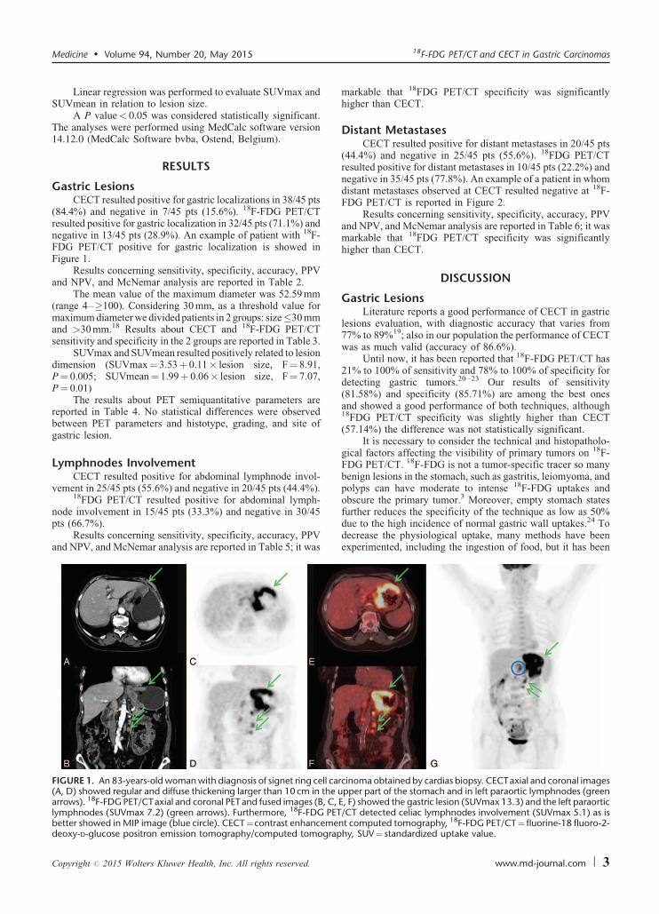
(84.4%) and negative in 7/45 pts (15.6%). 18F-FDG PET/CTresulted positive for gastric localization in 32/45 pts (71.1%) andnegative in 13/45 pts (28.9%). An example of patient with 18F-FDG PET/CT positive for gastric localization is showed inFigure 1.
Results concerning sensitivity, specificity, accuracy, PPVand NPV, and McNemar analysis are reported in Table 2.
The mean value of the maximum diameter was 52.59 mm(range 4–�100). Considering 30 mm, as a threshold value formaximum diameter we divided patients in 2 groups: size�30 mmand >30 mm.18 Results about CECT and 18F-FDG PET/CTsensitivity and specificity in the 2 groups are reported in Table 3.
SUVmax and SUVmean resulted positively related to lesiondimension (SUVmax¼ 3.53þ 0.11� lesion size, F¼ 8.91,P¼ 0.005; SUVmean¼ 1.99þ 0.06� lesion size, F¼ 7.07,P¼ 0.01)
The results about PET semiquantitative parameters arereported in Table 4. No statistical differences were observedbetween PET parameters and histotype, grading, and site ofgastric lesion.
Lymphnodes InvolvementCECT resulted positive for abdominal lymphnode invol-
vement in 25/45 pts (55.6%) and negative in 20/45 pts (44.4%).18FDG PET/CT resulted positive for abdominal lymph-
node involvement in 15/45 pts (33.3%) and negative in 30/45
Medicine � Volume 94, Number 20, May 2015
pts (66.7%).Results concerning sensitivity, specificity, accuracy, PPV
and NPV, and McNemar analysis are reported in Table 5; it was
FIGURE 1. An 83-years-old woman with diagnosis of signet ring cell ca(A, D) showed regular and diffuse thickening larger than 10 cm in the uarrows). 18F-FDG PET/CTaxial and coronal PETand fused images (B, C,lymphnodes (SUVmax 7.2) (green arrows). Furthermore, 18F-FDG PETbetter showed in MIP image (blue circle). CECT¼contrast enhancemedeoxy-D-glucose positron emission tomography/computed tomograp
Copyright # 2015 Wolters Kluwer Health, Inc. All rights reserved.
markable that 18FDG PET/CT specificity was significantlyhigher than CECT.
Distant MetastasesCECT resulted positive for distant metastases in 20/45 pts
(44.4%) and negative in 25/45 pts (55.6%). 18FDG PET/CTresulted positive for distant metastases in 10/45 pts (22.2%) andnegative in 35/45 pts (77.8%). An example of a patient in whomdistant metastases observed at CECT resulted negative at 18F-FDG PET/CT is reported in Figure 2.
Results concerning sensitivity, specificity, accuracy, PPVand NPV, and McNemar analysis are reported in Table 6; it wasmarkable that 18FDG PET/CT specificity was significantlyhigher than CECT.
DISCUSSION
Gastric LesionsLiterature reports a good performance of CECT in gastric
lesions evaluation, with diagnostic accuracy that varies from77% to 89%19; also in our population the performance of CECTwas as much valid (accuracy of 86.6%).
Until now, it has been reported that 18F-FDG PET/CT has21% to 100% of sensitivity and 78% to 100% of specificity fordetecting gastric tumors.20–23 Our results of sensitivity(81.58%) and specificity (85.71%) are among the best onesand showed a good performance of both techniques, although18FDG PET/CT specificity was slightly higher than CECT(57.14%) the difference was not statistically significant.
It is necessary to consider the technical and histopatholo-gical factors affecting the visibility of primary tumors on 18F-FDG PET/CT. 18F-FDG is not a tumor-specific tracer so manybenign lesions in the stomach, such as gastritis, leiomyoma, andpolyps can have moderate to intense 18F-FDG uptakes andobscure the primary tumor.3 Moreover, empty stomach statesfurther reduces the specificity of the technique as low as 50%
18F-FDG PET/CT and CECT in Gastric Carcinomas
due to the high incidence of normal gastric wall uptakes.24 Todecrease the physiological uptake, many methods have beenexperimented, including the ingestion of food, but it has been
rcinoma obtained by cardias biopsy. CECTaxial and coronal imagespper part of the stomach and in left paraortic lymphnodes (green
E, F) showed the gastric lesion (SUVmax 13.3) and the left paraortic/CT detected celiac lymphnodes involvement (SUVmax 5.1) as is
nt computed tomography, 18F-FDG PET/CT¼fluorine-18 fluoro-2-hy, SUV¼ standardized uptake value.
www.md-journal.com | 3

TABLE 2. CECT and 18F-FDG PET/CT Gastric Lesions Diagnostic Performance
CECT 18F-FDG PET/CT P
Sensitivity 92.11% (95%CI: 78.60% to 98.25%) 81.58% (95%CI: 65.67% to 92.22%) 0.34Specificity 57.14% (95%CI: 18.75% to 89.58%) 85.71% (95%CI: 42.23% to 97.63%) 0.62Accuracy 86.66% 82.22%PPV 92.11% (95%CI: 78.60% to 98.25%) 96.88% (95%CI: 83.73% to 99.48%)NPV 57.14% (95%CI: 18.75% to 89.58%) 46.15% (95%CI: 19.33% to 74.78%)
/CTe p
Altini et al Medicine � Volume 94, Number 20, May 2015
demonstrated that simple distention of the stomach using waterimproves the diagnostic performance of 18F-FDG PET/CT indetecting and localizing primary tumors25,26; in our experiencedrinking 500 mL of water before images acquisition was usefulfor a sufficient gastric distension.
The principal factors that can influence gastric lesions’detectability are tumor size, histological type, and localization.3
Tumor size is the major factor; Mukai et al showed that18F-FDG PET/CT had a sensitivity of 76.7% for the detection ofgastric carcinomas >30 mm but only 16.8% for those less than30 mm.18 Because late-stage tumors are usually larger in sizewith deeper invasion, advanced gastric carcinomas tend to yielda higher sensitivity in 18F-FDG PET/CT imaging (from 93% to98%) than early gastric carcinomas (from 26% to 63%).27
According to literature, we choosed 30 mm as a thresholdand observed a markable reduction in sensitivity and negligiblein specificity respect to overall patients results if size �30 mm(33.33% vs 81.58%; 83.33 vs 85.71%), while there werenegligible increase both in sensitivity and specificity if size>30 mm (96.55% vs 81.58%; 100% vs 85.71%). Obviously 18F-FDG PET/CT had a very good performance in larger lesion, butalso in small lesions its specificity was high.
Most studies reported that 18F-FDG PET/CT has signifi-cantly lower sensitivities in detecting mucinous and signet ringcells carcinomas than the intestinal-type adenocarcinoma.4,18,20,28
The lower 18F-FDG uptake in these hystotypes maydepend by the low-density diffuse infiltration of adenocarci-noma cells, the existence of extracellular or intracellular meta-bolically inert mucus content and the low expression level ofglucose transporter 1.20,28
We did not perform a comparison among histotypesbecause our population included 75.5% of intestinal type car-
CECT¼ contrast enhancement computed tomography, 18F-FDG PETcomputed tomography, NPV¼ negative predictive value, PPV¼ positiv
cinomas. However in our population only 1/6 mucinous carci-noma and 1/5 signet ring cells carcinoma (also with 5 mmdiameter) resulted false negative at 18F-FDG PET/CT.
TABLE 3. CECT and 18F-FDG PET/CT Results in the Groups of Pa
Sensitivity
CECT 18F-FDG PET/C
Size� 30 mm(n¼ 15)
80% (95%CI: 44.43%to 96.89%)
33.33% (95%CI: 7.8to 69.93%)
Size> 30 mm(n¼ 30)
88.89% (95%CI: 70.81%to 97.52%)
96.55% (95%CI: 82.to 99.42%)
CECT¼ contrast enhancement computed tomography, 18F-FDG PET/CTcomputed tomography.
4 | www.md-journal.com
Regarding the differentiation, lower 18F-FDG uptake maybe observed in poorly differentiated histotypes, which is likelydue to the low concentration of cancer cells in primary lesions.29
In our population only 2/18 G3 pts resulted false negative at 18F-FDG PET/CT and one of them was the 5 mm signet ringcells carcinoma.
About the tumor’s localization, 18F-FDG PET/CT detec-tion of gastroesophageal junction carcinomas was reported to bemore sensitive than other stomach parts, probably due to thehigher incidence of intestinal types in this site, but someresearchers reported that 18F-FDG PET/CT had a similardetectability for gastric carcinomas independently by site.20,30
In our population only 1/13 pts with gastric lesion localizedin the distant part of the stomach resulted false negative;anyway we did not perform a comparison among localizationsbecause of the variability of size and histotypes.
PET semiquantitative parameters have been investigatedfor their role in 18F-FDG PET/CT interpretations. The com-monly accepted parameters are SUVmax and SUVmean, whileMTV and TLG are not accepted for clinical use. Some authorsrefer these results are affected by many factors including thoseinfluencing gastric lesion detection; others did not find anydifferences in their values respect to histological type andlocalization.4,11,31,32
In our study we analyzed SUVmax, SUVmean, MTV, andTLG of gastric lesions, and we found a positive relation betweenSUVmax and lesion size, and SUVmean and lesion size;otherwise, no differences in none of the PET parameter wereidentified among the influencing factors histotype, grading, andlocalization (Table 3).
¼fluorine-18 fluoro-2-deoxy-D-glucose positron emission tomography/redictive value.
Lymphnode InvolvementThe presence of lymphnode metastases is one of the most
important prognostic factors in gastric carcinoma, and a correct
tients Divided Considering 30 mm as Size Threshold
Specificity
T CECT 18F-FDG PET/CT
8% 80% (95%CI: 28.81%to 96.70%)
83.33% (95%CI: 36.10%to 97.24%)
17% 66.67% (95%CI: 11.55%to 94.53%)
100% (95%CI: 16.55%to 100%)
¼fluorine-18 fluoro-2-deoxy-D-glucose positron emission tomography/
Copyright # 2015 Wolters Kluwer Health, Inc. All rights reserved.

TABLE 4. Semiquantitative Parameters Results
Parameter (Mean; range) Value F P
SUVmax (3.163; 0.9–56) Intestinal type adenocarcinoma 9.50 0.852 0.434Signet ring cells carcinoma 3.92Moucinous carcinoma 9.73G1 8.61 1.562 0.222G2 14.06G3 7.53Gastroesophageal junction 7.74 0.959 0.441Proximal 9.1Distant 6.96Prossimalþ distant 7.7All 13.95
SUVmean (5.08; 0.5–36.6) Intestinal type adenocarcinoma 5.42 0.235 0.791Signet ring cells carcinoma 3.54Moucinous carcinoma 5.45G1 5.52 1.147 0.327G2 7.45G3 3.88Gastroesophageal junction 4.15 1.070 0.380Prossimal 4.92Distant 3.67Prossimalþ distant 4.27All 8.28
MTV (48.69; 4.47–349) Intestinal type adenocarcinoma 41 1.565 0.221Signet ring cells carcinoma 93.5Moucinous carcinoma 55.1G1 93.86 1.782 0.186G2 35.18G3 49.23Gastroesophageal junction 22.99 1.549 0.216Prossimal 34.43Distant 45.97Prossimalþ distant 77.88All 102.51
TLG (547,257.2; 9970–9,916,557) Intestinal type adenocarcinoma 578,846.82 1.631 0.984Signet ring cells carcinoma 2,152,315Moucinous carcinoma 360,035G1 1,443,200.61 1.850 0.175G2 204,775.15G3 194,551.15Gastroesophageal junction 184,039 0.868 0.496Prossimal 235,173Distant 195,602.35Prossimalþ distant 322,548
Medicine � Volume 94, Number 20, May 2015 18F-FDG PET/CT and CECT in Gastric Carcinomas
staging is fundamental to define the necessity and the extensionof the lymphodenectomy.26
To date, CECT effectiveness in predicting lymphnodemetastases has not been satisfactory using any criteria, andthere is still no worldwide consensus. Although there is a clearcorrelation between the lymphnode size and metastases, micro-scopic metastases in normal-size lymphnodes and lymphnodeenlargement resulting from reactive or inflammatory changesare common in gastric cancer patients.33
The overall accuracy, sensitivity, and specificity of lymph-
All
node staging by CECT reported in literature varies from 69% to92%, 78% to 92%, and 73.9%, respectively, and they are strictlydependent on the size of lymphnodes.19
Copyright # 2015 Wolters Kluwer Health, Inc. All rights reserved.
One meta-analysis reported that the sensitivity and speci-ficity of 18F-FDG PET/CT in staging lymphnode involvementranged between 85.7% and 97.0%. Other individual studiesreported that 18F-FDG PET/CT was less sensitive but morespecific compared with commonly used CECT.22,34 The reasonsfor the low sensitivity of 18F-FDG PET/CT are the histologicaltype of the primary tumor and the size of metastatic lymphnodesthat could be smaller than 3 mm.22,35 In spite of the low sensi-tivity, 18F-FDG PET/CT usually showed a higher specificity thanmost other imaging modalities, including CECT because
1,510,828
18F-FDG PET/CT diagnose lymphnode metastases using glucosemetabolism rather than the size change.3 A limit reported for18F-FDG PET/CT is the detection of perigastric lymphnodes
www.md-journal.com | 5

TABLE 5. CECT and 18F-FDG PET/CT Lymphnode Involvement Diagnostic Performance
CECT 18F-FDG PET/CT P
Sensitivity 70.83% (95%CI: 48.91% to 87.33%) 58.33% (95%CI: 36.66% to 77.86%) 0.507Specificity 61.90% (95%CI: 38.45% to 81.84%) 95.24% (95%CI: 76.11% to 99.21%) 0.039Accuracy 66.66% 75.55%PPV 68% (95%CI: 46.50% to 85.01%) 93.33% (95%CI: 67.98% to 98.89%)NPV 65% (95%CI: 40.79% to 84.55%) 66.67% (95%CI: 47.19% to 82.69%)
/CTe p
Altini et al Medicine � Volume 94, Number 20, May 2015
involvement difficult to discriminate because of the radioactive
CECT¼ contrast enhancement computed tomography, 18F-FDG PETcomputed tomography, NPV¼ negative predictive value, PPV¼ positiv
volume effect generated by the nearby primary tumor.36
Our population’s results were lower than literature forCECT such as 18F-FDG PET/CT sensitivity, but they confirmed
FIGURE 2. A 54-years-old man with intestinal type gastric carcinomagreen arrow), a right lung nodule of 15 mm suspected for metastasesdoubtful for herniation pit (C, red arrow). 18F-FDG PET/CTaxial PETandG) but did not showed any 18F-FDG uptake in the lung nodule (E, H) acomputed tomography, 18F-FDG PET/CT¼ fluorine-18 fluoro-2-deoxy-SUV¼ standardized uptake value.
6 | www.md-journal.com
the higher specificity of 18F-FDG PET/CT than CECT (95.24%
¼fluorine-18 fluoro-2-deoxy-D-glucose positron emission tomography/redictive value.
vs 61.90%, P¼ 0.039) probably due to the lymphnodes smallsize and the primitive histological subtype. Localization inperigastric lymphnodes cannot be considered as an influencing
. CECT axial images showed localized gastric wall thickening (A,(B, blue arrow) and a osteolytic lesion in the right femoral headfused images confirmed the gastric lesion with SUVmax of 4.8 (D,nd in the right femoral head (F, I). CECT¼contrast enhancementD-glucose positron emission tomography/computed tomography,
Copyright # 2015 Wolters Kluwer Health, Inc. All rights reserved.

TABLE 6. CECT and 18F-FDG PET/CT Distant Metastases Diagnostic Performance
CECT 18F-FDG PET/CT P
Sensitivity 80% (95%CI: 44.43% to 96.89%) 60% (95%CI: 26.37% to 87.60%) 0.687Specificity 62.86% (95%CI: 44.93% to 78.51%) 88.57% (95%CI: 73.24% to 96.73%) 0.017Accuracy 66.66% 82.22%PPV 38.10% (95%CI: 18.16% to 61.55%) 60% (95%CI: 26.37% to 87.60%)NPV 91.67% (95%CI: 72.96% to 98.73%) 88.57% (95%CI: 73.24% to 96.73%)
/CTe p
Medicine � Volume 94, Number 20, May 2015 18F-FDG PET/CT and CECT in Gastric Carcinomas
factor in our population because they were detected only in6 pts, 5 of them correctly interpreted.
Distant MetastasesThe most frequent sites of distant metastases include liver,
lungs, bones, adrenal glands, and peritoneum.CECT is actually the conventional tool for detecting
distant metastases also from gastric cancer, but it may presentlimits especially for areas such as peritoneum.3
Even if Pan et al37 reported more than 96.6% accuracy ofCECT in distant metastases detection in 350 pts with gastriccancer, a following meta-analysis by Wang and Chen38 reporteda large difference in CECT sensitivities between hepatic metas-tases and peritoneal carcinomatosis (74% vs 33%).
18F-FDG PET/CT showed a good performance in detectingsolid organ metastases with a sensitivity of 95.2% and aspecificity of 100%, even if it resulted less effective in thedetection of bone metastases.29,39
Some studies reported 18F-FDG PET/CT lower sensitivitycompared with CECT for the diagnosis of peritoneal seeding,nevertheless 18F-FDG PET/CT could still be useful for detect-ing peritoneal metastases, especially when the CECT results areequivocal, avoiding unnecessary laparotomy in a considerableportion of patients. Even Lim et al showed its better specificitythan CECT (99% vs 92%).4,36,40,41
Results of our population were quite different from litera-ture; we found lower values for CECT accuracy (66.6%), higherfor CECT sensitivity (80%), and lower for 18F-FDG PET/CTsensitivity (60%). The difference between 18F-FDG PET/CTand CECT specificities resulted statistically significant (88.57vs 62.86%, P¼ 0.017) for distant metastases.
The explanation of these differences is the metastases sitesobserved in our population in only 10/45 pts: liver was involvedin 6 pts, lungs in 2 pts, bones in 3 pts, and adrenal gland andperitoneum in 1 pts, respectively.
The low specificity of CECT can be explained mostly withthe high rate of lung nodules misinterpreted (7/45 pts).
There were some limitations in our study such as theretrospective nature that implies selection bias and the smallnumber of the sample, even if in line with literature.
Furthermore, histological confirmation of lymphnode anddistant metastases should be obtained if feasible but biopsy ofeach lesion is not ethically recommended, so clinical-instru-mental follow-up has also been considered the gold standard.
CONCLUSION
CECT¼ contrast enhancement computed tomography, 18F-FDG PETcomputed tomography, NPV¼ negative predictive value, PPV¼ positiv
18F-FDG PET/CT is a useful tool for the evaluation ofgastric carcinoma; it can detect primary lesion, lymphnode, anddistant metastases using 1 single image whole-body technique.
Copyright # 2015 Wolters Kluwer Health, Inc. All rights reserved.
Our result of higher specificity of 18F-FDG PET/CT forlymphnode and distant metastases detection may suggest theimportant role of this technique in changing the extent oflymphadenectomy or reducing futile laparotomies.
We believe that integration of conventional imaging suchas CECT with 18F-FDG PET/CT permits a more valid whole-body staging in patients with gastric carcinomas.
REFERENCES
1. Jemal A, Bray F, Center MM, et al. Global cancer statistics. CA
Cancer J Clin. 2011;61:69–90.
2. Krejs GJ. Gastric cancer: epidemiology and risk factors. Dig Dis.
2010;28:600–603.
3. Wu CX, Zhu ZH. Diagnosis and evaluation of gastric cancer by
positron emission tomography. World J Gastroenterol.
2014;20:4574–4585.
4. Smyth E, Schoder H, Strong VE, et al. A prospective evaluation of
the utility of 2-deoxy-2-[18F]fluoro-d-glucose positron emission
tomography and computed tomography in staging locally advanced
gastric cancer. Cancer. 2012;15:5481–5488.
5. Coupe NA, Karikios D, Chong S, et al. Metabolic information on
staging FDG-PET-CT as a prognostic tool in evaluation of 97
patients with gastric cancer. Ann Nucl Med. 2014;28:128–135.
6. Moschetta M, Stabile Ianora AA, Anglani A, et al. Preoperative T
staging of gastric carcinoma obtained by MDCT vessel probe
reconstructions and correlations with histological findings. Eur
Radiol. 2010;20:138–145.
7. Moschetta M, Scardapane A, Telegrafo M, et al. Differential
diagnosis between benign and malignant ulcers: 320-row CT virtual
gastroscopy. Abdom Imaging. 2012;37:1066–1073.
8. Kiff RS, Taylor BA. Comparison of computed tomography, endoso-
nography, and intraoperative assessment in TN staging of gastric
carcinoma. Gut. 1994;35:287–288.
9. Lorusso F, Fonio P, Scardapane A. Gatrointestinal imaging with
multidetector CT and MRI. Recenti Prog Med. 2012;103:493–499.
10. Niccoli-Asabella A, Altini C, Notaristefano A, et al. A retrospective
study comparing contrast-enhanced computed tomography with 18F-
FDG-PET/CT in the early follow-up of patients with retroperitoneal
sarcomas. Nucl Med Comm. 2013;34:32–39.
11. Altini C, Niccoli Asabella A, De Luca R, et al. Comparison of 18F-
FDG PET/CT methods of analysis for predicting response to
neoadjuvant chemoradiation therapy in patients with locally
advanced low rectal cancer. Abdom Imaging. 2014(in press).
¼fluorine-18 fluoro-2-deoxy-D-glucose positron emission tomography/redictive value.
12. NCCN Clinical Practice Guidelines in Oncology (NCCN Guide-
lines). Gastric Cancer. 2013. Available from: URL: http://
www.nccn.org/professionals/physician_gls/pdf/. DOI: 10.1007/
s00261-014-0277-8.
www.md-journal.com | 7

13. Waddell T, Verheij M, Allum W, et al. Gastric cancer: ESMO-
ESSO-ESTRO Clinical Practice Guidelines for diagnosis, treatment
and follow-up. Ann Oncol. 2013;24:vi57–vi63.
14. Sobin LH, Gospodarowicz MK, Wittekind C. TNM Classification of
Malignant Tumours. 7th ed. New York: Wiley-Blackwell; 2009.
15. Gore RM, Levine MS. Trattato di Radiologia Gastrointestinale.
Roma: VerduciEditore; 2002.
16. Larson SM, Erdi Y, Akhurst T, et al. Tumor treatment response
based on visual and quantitative changes in global tumor glycolysis
using PET-FDG imaging. The visual response score and the change
in total lesion glycolysis. Clin Positron Imaging. 1999;2:159–171.
17. Lee JA. Segmentation of positron emission tomography images:
some recommendations for target delineation in radiation oncology.
Radiother Oncol. 2010;96:302–307.
18. Mukai K, Ishida Y, Okajima K, et al. Usefulness of preoperative
FDG-PET for detection of gastric cancer. Gastric Cancer.
2006;9:192–196.
19. Hallinan JT, Venkatesh SK. Gastric carcinoma: imaging diagnosis,
staging and assessment of treatment response. Cancer Imaging.
2013;13:212–227.
20. Stahl A, Ott K, Weber WA, et al. FDG PET imaging of locally
advanced gastric carcinomas: correlation with endoscopic and
histopathological findings. Eur J Nucl Med Mol Imaging.
2003;30:288–295.
21. Yun M, Lim JS, Noh SH, et al. Lymph node staging of gastric
cancer using (18)F-FDG PET: a comparison study with CT. J Nucl
Med. 2005;46:1582–1588.
22. Kim SK, Kang KW, Lee JS, et al. Assessment of lymph node
metastases using 18F-FDG PET in patients with advanced gastric
cancer. Eur J Nucl Med Mol Imaging. 2006;33:148–155.
23. Kim EY, Lee WJ, Choi D, et al. The value of PET/CT for
preoperative staging of advanced gastric cancer: comparison with
contrast-enhanced CT. Eur J Radiol. 2011;79:183–188.
24. Kamimura K, Nagamachi S, Wakamatsu H, et al. Role of gastric
distention with additional water in differentiating locally advanced
gastric carcinomas from physiological uptake in the stomach on 18F-
fluoro-2-deoxy-D-glucose PET. Nucl Med Commun. 2009;30:431–
439.
25. Zhu Z, Li F, Zhuang H. Gastric distension by ingesting food is
useful in the evaluation of primary gastric cancer by FDG PET. Clin
Nucl Med. 2007;32:106–109.
26. Yun M. Imaging of gastric cancer metabolism using 18 F-FDG PET/
CT. J Gastric Cancer. 2014;14:1–6.
Altini et al
27. Dassen AE, Lips DJ, Hoekstra CJ, et al. FDGPET has no definite
role in preoperative imaging in gastric cancer. Eur J Surg Oncol.
2009;35:449–455.
8 | www.md-journal.com
28. Ott K, Fink U, Becker K, et al. Prediction of response to
preoperative chemotherapy in gastric carcinoma by metabolic
imaging: results of a prospective trial. J Clin Oncol. 2003;21:4604–
4610.
29. Yoshioka T, Yamaguchi K, Kubota K, et al. Evaluation of 18F-FDG
PET in patients with advanced, metastatic, or recurrent gastric
cancer. J Nucl Med. 2003;44:690–699.
30. Wu AJ, Goodman KA. Positron emission tomography imaging for
gastroesophageal junction tumors. Semin Radiat Oncol. 2013;23:10–
15.
31. Namikawa T, Okabayshi T, Nogami M, et al. Assessment of 18F-
florodeoxyglucose positron emission tomography combined with
computed tomography in the preoperative management of patients
with gastric cancer. Int J Clin Oncol. 2013;19:649–655.
32. Kim J, Lim ST, Na CJ, et al. Pretreatment F-18 FDG PET/CT
parameters to evaluate progression-free survival in gastric cancer.
Nucl Med Mol Imaging. 2014;48:33–40.
33. Choi J, Joo I, Lee JM. State-of-the-art preoperative staging of gastric
cancer by MDCT and magnetic resonance imaging. World J
Gastroenterol. 2014;20:4546–4557.
34. Ha TK, Choi YY, Song SY, et al. F18-fluorodeoxyglucose-positron
emission tomography and computed tomography is not accurate in
preoperative staging of gastric cancer. J Korean Surg Soc.
2011;81:104–110.
35. Monig SP, Zirbes TK, Schroder W, et al. Staging of gastric cancer:
correlation of lymph node size and metastatic infiltration. AJR Am J
Roentgenol. 1999;173:365–367.
36. Chen J, Cheong JH, Yun MJ, et al. Improvement in preoperative
staging of gastric adenocarcinoma with positron emission tomogra-
phy. Cancer. 2005;103:2383–2390.
37. Pan Z, Zhang H, Yan C, et al. Determining gastric cancer
respectability by dynamic MDCT. Eur Radiol. 2010;20:613–620.
38. Wang Z, Chen JQ. Imaging in assessing hepatic and peritoneal
metastases of gastric cancer: a systematic review. BMC Gastro-
enterol. 2011;11:19.
39. Chung HW, Lee EJ, Cho YH, et al. High FDG uptake in PET/CT
predicts worse prognosis in patients with metastatic gastric adeno-
carcinoma. J Cancer Res Clin Oncol. 2010;136:1929–1935.
40. Rubini G, Altini C, Notaristefano A, et al. Peritoneal carcinomatosis
from ovarian cancer: role of 18F-FDG-PET/CT and CA125. Recenti
Prog Med. 2012;103:510–514.
Medicine � Volume 94, Number 20, May 2015
41. Lim JS, Kim MJ, Yun MJ, et al. Comparison of CT and 18F-FDG
pet for detecting peritoneal metastases on the preoperative evaluation
for gastric carcinoma. Korean J Radiol. 2006;7:249–256.
Copyright # 2015 Wolters Kluwer Health, Inc. All rights reserved.


![Usefulness of [18F]-DA and [18F]-DOPA for PET imaging in a mouse model of pheochromocytoma](https://static.fdokumen.com/doc/165x107/6325a7d9852a7313b70e9a7d/usefulness-of-18f-da-and-18f-dopa-for-pet-imaging-in-a-mouse-model-of-pheochromocytoma.jpg)


![CYCLOTRON PRODUCED RADIONUCLIDES:GUIDANCE ON FACILITY DESIGN AND PRODUCTION OF [18F]FLUORODEOXYGLUCOSE (FDG)](https://static.fdokumen.com/doc/165x107/631647d8c32ab5e46f0db468/cyclotron-produced-radionuclidesguidance-on-facility-design-and-production-of-18ffluorodeoxyglucose.jpg)


