FDG PET and the genetics of dementia
12
REVIEW ARTICLE FDG PET and the genetics of dementia Benedetta Nacmias • Valentina Berti • Irene Piaceri • Sandro Sorbi Received: 9 May 2013 / Accepted: 2 July 2013 Ó Italian Association of Nuclear Medicine and Molecular Imaging 2013 Abstract Dementias are the most common neurodegen- erative diseases and they are increasingly becoming a major public health problem. The most common form of dementia, affecting over 20 million people worldwide, is Alzheimer’s disease (AD). AD is a neurodegenerative disease of the central nervous system mainly found in older adults, with an incidence that increases with age. With the development of newer technologies, including genetic screening technologies and PET/MRI scanning, the role of genetic studies and neuroimaging is being redefined; indeed, these approaches are able to provide support not only in the clinical diagnosis of dementia, but also in its presymptomatic evaluation. Many researchers agree that early identification of AD, before abnormal accumulation of amyloid and tau proteins, could provide an opportunity to hinder the progression of the disease. [ 18 F]2-fluoro-2- deoxy-D-glucose (FDG) PET studies have shown that decreased glucose metabolism in AD precedes clinical diagnosis and that the degree of clinical disability in AD correlates closely with the magnitude of the reduction in brain glucose metabolism. Data on presymptomatic muta- tion carriers from families with known early-onset auto- somal dominant AD (familial AD) show reductions in the cerebral metabolic rate of glucose (CMRglc), consistent with the expected AD PET pattern, in the absence of severe atrophy on MRI. These results suggest that PET CMRglc measures have the potential to serve as preclinical bio- markers of dementia, useful also for tracking disease pro- gression. This review highlights the role of genetics and FDG PET in understanding the pathogenesis of dementia. Keywords FDG PET Á Dementia Á Gene Á Alzheimer’s disease Introduction Dementias are the most common neurodegenerative diseases and they are increasingly becoming a major public health problem. These diseases are, in fact, one of the major causes of disability in the general population and it is estimated that their prevalence, currently standing at 35.6 million people worldwide, will increase to 115 million by 2050 [1]. The most common form of dementia, affecting over 20 million people worldwide, is Alzheimer’s disease (AD). AD is a neurodegenerative disease of the central nervous system mainly found in older adults, with an incidence that increases with age. From a clinical point of view, it is characterized predominantly by initial deficits in episodic memory, followed by progressive involvement of semantic memory, attention, executive function and language skills. Disease progression, lasting an average of 10 years, leads to the death of the individual, usually due to intercurrent infections. The key features of the AD brain are neuronal and synapse loss, and the deposition of extracellular plaques composed of amyloid-b (Ab) peptides and intraneuronal neurofibrillary tangles consisting of hyperphosphorylated tau protein. Although the most frequent form is sporadic AD, the rare autosomal and dominant early-onset form of AD B. Nacmias (&) Á I. Piaceri Á S. Sorbi Section of Neuroscience, Department of Neuroscience, Psychology, Drug Research and Child Health, University of Florence, Viale Pieraccini 6, 50139 Florence, Italy e-mail: nacmias@unifi.it V. Berti Nuclear Medicine Unit, Department of Biomedical, Experimental and Clinical Sciences, Viale Morgagni 50, 50134 Florence, Italy 123 Clin Transl Imaging DOI 10.1007/s40336-013-0028-9
-
Upload
independent -
Category
Documents
-
view
0 -
download
0
Transcript of FDG PET and the genetics of dementia

REVIEW ARTICLE
FDG PET and the genetics of dementia
Benedetta Nacmias • Valentina Berti •
Irene Piaceri • Sandro Sorbi
Received: 9 May 2013 / Accepted: 2 July 2013
� Italian Association of Nuclear Medicine and Molecular Imaging 2013
Abstract Dementias are the most common neurodegen-
erative diseases and they are increasingly becoming a
major public health problem. The most common form of
dementia, affecting over 20 million people worldwide, is
Alzheimer’s disease (AD). AD is a neurodegenerative
disease of the central nervous system mainly found in older
adults, with an incidence that increases with age. With the
development of newer technologies, including genetic
screening technologies and PET/MRI scanning, the role of
genetic studies and neuroimaging is being redefined;
indeed, these approaches are able to provide support not
only in the clinical diagnosis of dementia, but also in its
presymptomatic evaluation. Many researchers agree that
early identification of AD, before abnormal accumulation
of amyloid and tau proteins, could provide an opportunity
to hinder the progression of the disease. [18F]2-fluoro-2-
deoxy-D-glucose (FDG) PET studies have shown that
decreased glucose metabolism in AD precedes clinical
diagnosis and that the degree of clinical disability in AD
correlates closely with the magnitude of the reduction in
brain glucose metabolism. Data on presymptomatic muta-
tion carriers from families with known early-onset auto-
somal dominant AD (familial AD) show reductions in the
cerebral metabolic rate of glucose (CMRglc), consistent
with the expected AD PET pattern, in the absence of severe
atrophy on MRI. These results suggest that PET CMRglc
measures have the potential to serve as preclinical bio-
markers of dementia, useful also for tracking disease pro-
gression. This review highlights the role of genetics and
FDG PET in understanding the pathogenesis of dementia.
Keywords FDG PET � Dementia � Gene �Alzheimer’s disease
Introduction
Dementias are the most common neurodegenerative diseases
and they are increasingly becoming a major public health
problem. These diseases are, in fact, one of the major causes
of disability in the general population and it is estimated that
their prevalence, currently standing at 35.6 million people
worldwide, will increase to 115 million by 2050 [1].
The most common form of dementia, affecting over 20
million people worldwide, is Alzheimer’s disease (AD). AD
is a neurodegenerative disease of the central nervous system
mainly found in older adults, with an incidence that
increases with age. From a clinical point of view, it is
characterized predominantly by initial deficits in episodic
memory, followed by progressive involvement of semantic
memory, attention, executive function and language skills.
Disease progression, lasting an average of 10 years, leads to
the death of the individual, usually due to intercurrent
infections. The key features of the AD brain are neuronal
and synapse loss, and the deposition of extracellular plaques
composed of amyloid-b (Ab) peptides and intraneuronal
neurofibrillary tangles consisting of hyperphosphorylated
tau protein.
Although the most frequent form is sporadic AD, the
rare autosomal and dominant early-onset form of AD
B. Nacmias (&) � I. Piaceri � S. Sorbi
Section of Neuroscience, Department of Neuroscience,
Psychology, Drug Research and Child Health, University
of Florence, Viale Pieraccini 6, 50139 Florence, Italy
e-mail: [email protected]
V. Berti
Nuclear Medicine Unit, Department of Biomedical,
Experimental and Clinical Sciences, Viale Morgagni 50,
50134 Florence, Italy
123
Clin Transl Imaging
DOI 10.1007/s40336-013-0028-9
(EOAD) has provided valuable insight into the disease
pathogenesis.
Most dementia cases are late onset and are probably
linked to both genetic and environmental factors. The
most common risk factors include brain injury, diabetes,
hypercholesterolemia and obesity. Moreover, recent
studies seem to indicate an involvement of cerebrovas-
cular risk factors in the development of a substantial
proportion of dementia cases. With regard to the possible
role of these risk factors in dementia, data (spanning 10
or 20 years) from large epidemiological studies conducted
in Minnesota, Illinois and Indiana and from a US national
study showed a substantial reduction in cognitive
impairment in the general population, measured using
neuropsychological tests, and thus justify cautious opti-
mism over the possible impact of reducing cerebrovas-
cular risk factors in dementia [2].
These data are also confirmed by the results of a Rot-
terdam study, which provided evidence of a reduction in
the incidence of dementia between 1990 and 2005 [3].
Many experts agree that identifying AD early, before
abnormal accumulation of amyloid and tau proteins, could
provide an opportunity to hinder the progression of the
disease.
A number of strategies under study were recently dis-
cussed at the 2012 annual meeting of the Society for
Neuroscience. Several neuroimaging techniques have been
included among the biomarkers of AD; however, scientific
evidence is still under development [4].
To develop preventive treatments for AD, it is necessary
to identify early biological markers that are predictive of
AD. To date, the best-recognized in vivo markers are
measures of brain structure and function, as obtained with
neuroimaging. The distribution, in the brain, of the PET
tracer [18F]2-fluoro-2-deoxy-D-glucose (FDG) reflects
regional glucose consumption, correlating with cerebral
synaptic activity [5]. Since neurodegenerative dementias
are characterized by synaptic and neuronal loss, FDG PET
is able to consistently reveal significant hypometabolism in
several cortical regions.
Brain volume loss as assessed on magnetic resonance
imaging (MRI), and reductions in the cerebral metabolic
rate of glucose (CMRglc) as measured with FDG PET are
sensitive to AD-related brain changes. A crucial question is
whether these brain measures can allow preclinical detec-
tion of dementia. To understand how AD starts and pro-
gresses, it is necessary to follow subjects through normal
aging to the onset of clinical symptoms. Several studies
have shown measures of brain atrophy directly assessed
using MRI techniques, capable of capturing presymptom-
atic changes in familial forms of AD [6–8].
FDG PET studies have shown that decreased glucose
metabolism in AD precedes clinical diagnosis and that the
degree of clinical disability in AD correlates closely with
the magnitude of the reduction in brain metabolism.
Recent studies have provided evidence of dynamic
changes in PET amyloid and FDG imaging at different
stages of AD [9]. In order to compare brain atrophy with
hypometabolism as preclinical markers of AD, several
studies have presented data on presymptomatic mutation
carriers from families with known early onset, autosomal
dominant AD, i.e. familial AD (FAD) [10]. Presymptom-
atic FAD individuals have been found to show CMRglc
reductions consistent with the expected AD PET pattern,
but in the absence of severe atrophy on MRI [11]. These
results suggest that PET CMRglc measures could poten-
tially serve as preclinical biomarkers of dementia, useful
also for tracking disease progression.
New diagnostic criteria
A few years ago, the results of many years of research
allowed an update of the clinical research criteria for
dementia and the refinement of guidelines (originally con-
ceived around two and a half decades earlier) for diagnosis
of the various forms of dementia [12]. Meanwhile, 2011
saw the publication of recommendations, from the National
Institute on Aging–Alzheimer’s Association workgroups,
on diagnostic guidelines for Alzheimer’s disease [13].
These new recommendations are the result of acquisitions
obtained through both laboratory and clinical research.
The new criteria proposed a common lexicon for the
clinical and research sectors [12], which has generated
much debate in the scientific community [14–16]. Devel-
opments in the field of biomarkers and the use, alongside
traditional genetics, of new more specific and more sensi-
tive techniques were crucial for the development of these
new criteria, but also for redefining frontotemporal
dementia (FTD) and its subtypes.
Guidelines on the diagnosis and management of
dementia disorders recently developed by a European
Federation of Neurological Societies (EFNS) study group
on dementia, which took into consideration the canonical
syndromes and molecular genetic aspects, identify a
number of different clinical forms of dementia with dif-
ferent ages at onset and/or durations [17].
Moreover, in accordance with recent EFNS-ENS
guidelines on the use of neuroimaging in the diagnosis of
dementia [18], structural imaging should be performed to
exclude other potentially treatable diseases, to detect vas-
cular lesions, and to identify specific findings to help dis-
tinguish between different forms of neurodegenerative
dementia.
Neuroimaging is recommended in cases in which the
diagnosis remains in doubt. Amyloid imaging is likely to
Clin Transl Imaging
123
prove clinically useful in several fields, including the
evaluation of atypical AD presentations [18].
Biomarkers
Recent years have provided some important developments
in the field of dementia, fundamental in furthering under-
standing of the pathogenetic mechanisms of the disease. It is
now believed that AD pathophysiology started years before
cognitive changes are detected and probably before the
onset of clinical signs of dementia [19, 20]. Hence, there has
emerged an urgent need for reliable diagnostic biomarkers
able to detect the disease in a presymptomatic stage. Recent
studies have focused on imaging markers or cerebrospinal
fluid (CSF) and blood biomarkers for early diagnosis.
The biomarkers currently used for AD are both genetic,
such as the pathogenetic mutations in three candidate genes
(amyloid-b precursor protein, APP; presenilin 1, PS1;
presenilin 2, PS2) and biochemical (amyloid-b and tau
proteins in the CSF) [21]. Moreover, the biomarkers of
neurodegeneration include neuroimaging correlates of
hippocampal atrophy on structural MRI and symmetric
decreased metabolism in the temporoparietal regions on
FDG PET.
The 11C Pittsburgh compound (PiB) has always been the
most validated tracer for imaging amyloid pathology.
However, there is now a new tracer, 18F-florebetapir that,
thanks to the longer half-life of the radioactive isotope 18F
(110 versus 20 min for the 11C of 11C-PiB), has the
potential to be used even more extensively both for clinical
and research purposes [22].
In a 2003 study involving a family carrying the T122R
mutation in PS2 and showing an atypical presentation of
dementia, all the biomarkers (the functional neuroimaging
investigations, CSF levels of Ab 1-42, and Tau protein)
provided evidence indicating future dementia development
[23].
Today, FDG PET has moved from the research arena
into clinical practice. Amyloid imaging allows earlier
diagnosis of AD and better differential diagnosis of
dementia and it also provides prognostic information for
mild cognitive impairment (MCI) [24].
The genetics of dementia
The genetic approach to the study of neurodegenerative
diseases is undoubtedly the one that has provided and
continues to provide the most significant contribution to
understand the various aspects of these disorders: their
pathogenesis and diagnosis, the therapeutic options, and
the important ethical implications.
A search of the PubMed database using keywords such as
‘‘dementia’’ and ‘‘genetic’’ reveals, to date, more than
25,000 papers, almost half of which were published in the
past 6 years. The earliest paper, published in 1952, concerns
research into the genetics of AD and Pick’s disease [25];
one of the most recent contributions is a large collaborative
genome-wide association (GWA) study that highlights a
new AD locus on chromosome 10 [26]. The list of genetic
mutations associated with autosomal dominant forms of
familial dementia is getting longer and longer [27].
The extensive study of the genetics of dementia led to
the development of a molecular classification of different
forms of dementia and now many groups are working to
establish the possible diagnostic utility, in dementia, of the
biological and genetic biomarkers developed [28].
The genetics of AD
Alzheimer’s disease is a genetically complex, multifacto-
rial disease involving many candidate genes [29]. From a
genetic point of view, AD can be divided into two different
forms: FAD and sporadic AD. Apart from the fact that
FAD often has onset at an earlier age and can present some
neurological characteristics not generally present in the
sporadic form, the two subtypes are clinically indistin-
guishable. AD can also be divided into two groups by age
at onset: early onset (onset \65 years; EOAD) and late
onset (onset [65 years; LOAD).
In rare early-onset FAD, different disease-causing
mutations have been described. At the time of writing, the
Alzheimer Disease & Frontotemporal Dementia Mutation
Database lists a total of 231 different fully penetrant
autosomal dominant mutations in 517 families around the
world (http://molgen-www.uia.ac.be/ADMutations/).
These mutations are located in three genes (i.e. APP, PS1,
PS2) that have been shown to cause 50 % of all FAD. In
particular, 33 pathogenetic mutations for APP, 185 for PS1
and 13 for PS2 have been discovered [30, 31].
In the more common sporadic form, a locus on chro-
mosome 19, coding for apolipoprotein E (ApoE), has been
associated with increased susceptibility. In particular,
genetic and epidemiological studies have identified the
epsilon 4 (e4) allele as a major risk factor for the disease,
being associated with an increased risk of developing the
disease and with a lower age at onset [32].
The ApoE e4 allele is the only confirmed factor involved
in genetic susceptibility to AD, but it is neither necessary
nor sufficient to cause the disease [33]. Moreover,
according to recent data, the ApoE could be a major gene
with semi-dominant inheritance (individuals with a double
dose of the e4 allele have a 35-fold increased risk of
developing the disease) [34].
Clin Transl Imaging
123
The ApoE e4 isoform is thought to interact with the
mechanisms associated with aging, resulting in deposition
in the brain, of aggregated deposits of abnormal proteins;
this impairs neurotransmission, thereby hastening the dis-
ease process [35, 36]. However, over 70 % of all cases in
the general population are associated with this gene, which
suggests that numerous other factors, in addition to genetic
predisposition, must together contribute to determining the
disease [37].
In recent years, the search for other genetic factors has
shown associations with polymorphisms in an increasing
number of genes, all possibly involved, by interfering with
the deposition and clearance of Ab in the disease patho-
genesis [38].
Recent progress in genomic methodology and the
availability of large sample sizes have ushered in the era of
GWA studies.
From 2005 to the present, many GWA studies have been
published and thanks to these studies; several loci associ-
ated with various neurodegenerative diseases have been
identified.
According to the AlzGene database, GWA studies have
identified ten novel loci associated with an increased risk of
developing AD, i.e. BIN1, CLU, ABCA7, CR1, PICALM,
MS4A6A, MS4A4E, CD33, CD2AP and EPHA1 [39–42].
The role of all the new genes in the pathogenesis of AD
is supported by their involvement in the pathogenetic
processes related to neurodegeneration, including the
amyloidogenic cascade, tau hyperphosphorylation, apop-
tosis, oxidative stress, cholesterol and lipid metabolism,
and immune inflammatory processes.
Despite these successes, this type of approach has many
limitations because these associations do not have a high
predictive value and may also lead to the identification of
false positives and false negatives. For this type of
approach, studying a very large number of subjects is
necessary. In fact, whereas several thousand individuals are
needed to describe an association with a rare, high-impact
variant, the description of a common variant with a low
effect requires many thousands of cases and controls [43].
For this purpose, international research groups have
organized specific genetic consortia, thus making large
collaborative studies feasible.
During 2011, thanks to a group of international
researchers, the First International Collaboration on the
Genetics of Alzheimer’s Disease was established with a
view to perform collaborative studies on large numbers of
subjects (International Genomics of Alzheimer’s Project).
Recent GWA studies have identified CLU, CR1, ABCA7
BIN1, PICALM and MS4A6A/MS4A6E in addition to the
long-established ApoE as loci for AD [39–42, 44].
New findings have recently provided fresh insight into
the genetic mechanisms underlying the development of
AD.
Starting from the hypothesis that low-prevalence vari-
ants showing moderate to high effect may be associated
with AD risk, two independent research groups [45, 46]
have shown that a rare variant (rs 75932628 encoding an
arginine to histidine substitution at residue 47, R47H) in
the TREM2 gene, encoding the triggering receptor
expressed on myeloid cells on chromosome 6 (6p21.1-
q15), is significantly associated with increased suscepti-
bility to LOAD.
Another recent study has provided evidence that a low-
frequency variant of the coding mutation (A673T) in the
APP gene protects against AD and cognitive decline in the
elderly without AD [47, 48]. The discovery of the protec-
tive effect of the A673T substitution against AD provides
proof of the principle that reducing the b-cleavage of APP
may protect against the disease, and supports the clinical
usefulness of the current amyloid-directed therapeutic
research efforts.
The recent important characterizations of the novel risk
factor TREM2 and the novel protective APP-related variant
displaying a statistically significant association with AD
provide grounds for cautious optimism regarding the
results of genetic research.
It has been suggested that families need clinical guide-
lines to help them navigate the complexities of dementia
(in particular AD and FTD) genetics and the processes of
genetic testing and counseling [49].
In fact, the diagnosis of progressive disease in a clin-
ically healthy individual is a new but increasingly fre-
quent scenario for the neurologists and geneticists [50],
and it is one that carries ethical implications [51] and
underlines the importance of genetic counseling. The
genetic approach, being able to shed light on the patho-
genetic mechanisms of the disease and thus indicate
possible therapeutic avenues, is important both from a
research and a clinical perspective. For example, a recent
study aiming to investigate whether treatment with human
anti-Ab monoclonal antibodies leads to a measurable
reduction in the level of Ab in the brain and to elucidate
the mechanism of amyloid reduction provided data on the
use of an amyloid-eating drug that removes plaque
through phagocytosis [52].
As pointed out by a recent editorial in Neurology [53], it
is recommended to start treating patients before the disease
becomes symptomatic, i.e. to exploit the window, in the
preclinical stages of the disease, in which CSF and PET
biomarkers correlated with amyloidogenic and neurode-
generative processes are significantly altered.
Clin Transl Imaging
123

FDG PET and the genetics of AD
The link between genetic susceptibility and AD could be
elucidated by the association of AD candidate genes with
alterations in brain functions, which can be assessed by
FDG PET.
Several studies support the use of FDG PET as an AD
biomarker and, furthermore, as an endophenotype for the
assessment of genetic risk factors for AD.
FDG PET undoubtedly has many advantages; however,
it could also have some limitations. First of all, metabolism
studies are not able to provide information about the
pathophysiological mechanisms linking genetic variability
to AD susceptibility. In addition, CMRglc is also pre-
sumably subject to multiple genetic regulations and
affected by environmental factors.
However, despite these limitations, FDG PET deserves
to be considered a presymptomatic AD endophenotype,
and several studies have already successfully used FDG
PET to reveal CMRglc alterations in normal individuals
who have a genetic risk of developing AD.
FDG PET and presymptomatic early-onset FAD
Studies of individuals from families with FAD with known
genetic mutations have provided unique information about
preclinical AD-related brain impairment and they consti-
tute an ideal resource for exploring the relationship
between genetic and phenotypic expression of the disease.
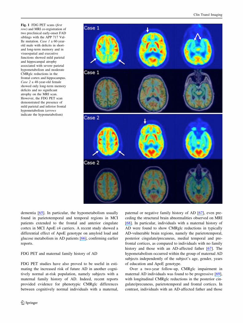
Several FDG PET studies on presymptomatic FAD sub-
jects showed the presence of regional parieto-temporal,
posterior cingulate cortex (PCC) and medial temporal lobe
hypometabolism on a background of wide-spread global
CMRglc impairment (Fig. 1) [11, 54, 55].
In particular, a previous FDG PET study on presymp-
tomatic at-risk individuals carrying the rare APP mutations
(compared to individuals without mutations) reported the
presence of parieto-temporal hypometabolism, with addi-
tional frontal deficits [56]. Similarly, a recent FDG PET
study conducted on carriers of the PS2 mutation demon-
strated CMRglc reductions in parietal and precuneus
regions, showing progression at follow-up in parieto-tem-
poral and frontal lobes [57].
Interestingly, an FDG PET study of presymptomatic
PS1 mutation carriers provided definitive evidence that
CMRglc reductions precede not only clinical symptoms but
also structural brain changes, showing cortical and medial
temporal lobe metabolic reductions, occurring before and
exceeding structural brain atrophy observed on MRI. After
partial volume correction, CMRglc reductions ranged from
13 % (whole brain) to 21 % (PCC), reflecting true reduc-
tions in brain glucose utilization per unit brain volume;
after partial volume correction, the medial temporal lobe
CMRglc values were reduced to 12 % in the hippocampus
and 20 % in the entorhinal cortex [11].
These findings are consistent with and could be
explained by the AD pathological cascade model: follow-
ing the initial deposition of amyloid plaques, the dominant
process is represented by neuronal and synaptic dysfunc-
tion, which could be studied using FDG PET. The neuronal
loss that ultimately causes atrophy measurable with MRI
appears only later [58].
FDG PET and ApoE-associated genetic risk
The e4 allele of the ApoE gene is a widely recognized
genetic risk factor for AD and the association between the
ApoE e4 allele and AD risk has been reported in hundreds
of studies. The ApoE e4 genotype is considered a risk
factor since almost 40 % of LOAD patients have at least
one ApoE e4 allele; moreover, a copy of the e4 allele is
found in about 25 % of Americans and in 33 % of those
with a reported family history of dementia in a first-degree
relative. Two copies of the e4 allele are found in 2–3 % of
Americans and about 5 % of those with a reported history
of dementia in a first-degree relative [59].
To date, many FDG PET studies have been published,
examining the effect of the presence of the ApoE e4 allele
on CMRglc and confirming, study after study, the associ-
ation between metabolic impairment, assessed by FDG
PET, and AD genetic risk factors in cognitively normal
subjects. FDG PET studies in non-demented individuals
report that, as compared to e4 non-carriers, ApoE e4
homozygotes (therefore, with a particularly high risk of
developing AD) have a significantly reduced CMRglc in
the same brain regions as clinically affected AD patients,
including the posterior cingulate/precuneus, parietal, tem-
poral and prefrontal regions [59]. As compared to e4 non-
carriers, ApoE e4 heterozygotes show similar, but milder,
hypometabolism within the same brain regions found to be
affected in AD patients and in ApoE e4 homozygotes [59–
62]. There is evidence that the metabolic reductions in
ApoE e4 carriers are more progressive (25 % decline in
CMRglc over a two-year interval) and correlate with the
reductions in cognitive performance [63].
Moreover, CMRglc abnormalities have been detected
even earlier, in 20- to 40-year-old ApoE e4 carriers [64],
making them the earliest brain abnormalities yet found in
living subjects at risk of AD, detected many years before
the estimated median age of dementia onset in e4
heterozygotes.
The ApoE e4 allele also contributes to heterogeneity in
MCI patients by regionally affecting brain metabolic
activity, so that MCI ApoE e4 carriers present a more
extensive CMRglc impairment than non-carriers, reflecting
the known ApoE e4-related increased vulnerability to
Clin Transl Imaging
123
dementia [65]. In particular, the hypometabolism usually
found in parietotemporal and temporal regions in MCI
patients extended to the frontal and anterior cingulate
cortex in MCI ApoE e4 carriers. A recent study showed a
differential effect of ApoE genotype on amyloid load and
glucose metabolism in AD patients [66], confirming earlier
reports.
FDG PET and maternal family history of AD
FDG PET studies have also proved to be useful in esti-
mating the increased risk of future AD in another cogni-
tively normal at-risk population, namely subjects with a
maternal family history of AD. Indeed, recent reports
provided evidence for phenotypic CMRglc differences
between cognitively normal individuals with a maternal,
paternal or negative family history of AD [67], even pre-
ceding the structural brain abnormalities observed on MRI
[68]. In particular, individuals with a maternal history of
AD were found to show CMRglc reductions in typically
AD-vulnerable brain regions, namely the parietotemporal,
posterior cingulate/precuneus, medial temporal and pre-
frontal cortices, as compared to individuals with no family
history and those with an AD-affected father [67]. The
hypometabolism occurred within the group of maternal AD
subjects independently of the subject’s age, gender, years
of education and ApoE genotype.
Over a two-year follow-up, CMRglc impairment in
maternal AD individuals was found to be progressive [69],
with longitudinal CMRglc reductions in the posterior cin-
gulate/precuneus, parietotemporal and frontal cortices. In
contrast, individuals with an AD-affected father and those
Fig. 1 FDG PET scans (first
row) and MRI co-registration of
two preclinical early-onset FAD
siblings with the APP 717 Val-
Ile mutation. Case 1 a 60-year-
old male with deficits in short-
and long-term memory and in
visuospatial and executive
functions showed mild parietal
and hippocampal atrophy
associated with severe parietal
hypometabolism and moderate
CMRglc reductions in the
frontal cortex and hippocampus.
Case 2 a 48-year-old female
showed only long-term memory
deficits and no significant
atrophy on the MRI scan.
However, the FDG PET scan
demonstrated the presence of
mild parietal and inferior frontal
hypometabolism (arrows
indicate the hypometabolism)
Clin Transl Imaging
123
with no family history of AD showed longitudinal CMRglc
reductions only in the frontal cortex, which is a typical
effect of aging.
FDG PET and the KIBRA gene
KIBRA is a cytoplasmic protein that is highly expressed in
the brain and is a binding partner of dendrin, a putative
modulator of synaptic plasticity. There are two allelic
variants of the KIBRA gene (T and C), resulting in three
possible genotypes: TT, CT and CC. GWA studies and
several replication studies [70, 71] demonstrated that KI-
BRA T allele non-carriers (CC genotype) had lower epi-
sodic memory performance compared to KIBRA T carriers
(CT and TT genotypes).
Since memory impairment is the cardinal clinical fea-
ture of AD, the next step was to investigate the possibility
of an association between AD and KIBRA. Corneveaux and
colleagues [72] tested this hypothesis using data from FDG
PET, as an endophenotype. KIBRA T non-carriers showed
significant CMRglc reductions, compared to KIBRA T
carriers, in the same brain regions preferentially affected
by AD, including the precuneus and PCC.
These FDG PET findings provide a foundation for a
common molecular mechanism contributing not only to
successful episodic memory performance but also to the
predisposition to AD, opening possible future perspectives
for memory-enhancing, AD-modifying and AD risk-
reducing therapies.
Genetics of FTD
First described over 100 years ago, today we can refer to
several clinical forms and at least 16 different pathological
subtypes of FTD [73].
FTD refers to a disease spectrum including the behav-
ioral variant FTD (bvFTD), primary progressive aphasia
(PPA), progressive supranuclear palsy/corticobasal degen-
eration syndrome (PSP/CBDS), and FTD with amyotrophic
lateral sclerosis (FTD-ALS).
The international consortia for the two main syndromes,
bvFTD and PPA, have recently revised the diagnostic cri-
teria and published consensus criteria [74, 75], including
neuroimaging and genetics.
The first evidence of a genetic cause of familial FTD
was provided 18 years ago with the demonstration of a
linkage to chromosome 17 in a familial form with auto-
somal dominant parkinsonism [76]; the most recent evi-
dence is a GGGGCC expansion in C9orf72, a major cause
of FTD and ALS [77, 78].
To date, the familial forms of FTD [79] are explained by
the seven genes (C9orf72, CHMP2B, FUS, GRN, MAPT,
TARDBP, VCP), in which 193 pathogenetic mutations have
been described (http://www.molgen.ua.ac.be/ADMutations).
All the genes code for a protein including C9orf72 [80], an
open reading frame coding for a protein recently character-
ized and located on chromosome 9p21 [77, 78, 80]. The most
revolutionary finding on the genetics of FTD is that these
mutations and expansions concern a very high percentage of
families in the world. Indeed, there are now more than
900 families in the world with mutations associated with
FTD. A flow chart for FTD genetic testing was recently
published [81].
There also exist phenotypic characterizations of FTD
linked to the mutations in GRN and C9orf72 and in which
an important contribution is made by neuroimaging [82]. In
particular, a study in monozygotic twins carrying a 4-bp
deletion (c.388_391delCAGT) in exon 4 of GRN reported
strong clinical, neuroimaging, and serum progranulin level
similarities, demonstrating the importance of shared
genetic profiles beyond environmental influences in the
symptomatic expression of the disease [82]. In a recent
case report, brain FDG PET demonstrated mild hypome-
tabolism involving the medial frontal and lateral temporal
lobes, the left more than the right, which progressed over
time. This case was subsequently confirmed to have the
C9orf72 expansion, thus highlighting the need to consider
mutations in the FTD-associated genes when a familial
disorder is suggested and neuroimaging studies reveal
findings atypical of an AD pathophysiological process,
despite the typical anterograde amnesic syndrome [83].
Different research centers worldwide are involved in
the genetic approach to FTD. The work of GENFI
(http://www.ucl.ac.uk/drc/research/current-studies/genfi), a
multicenter collaborative consortium for tracking the evo-
lution of genetic FTD and its associated disorders from
their earliest stages, is ongoing.
The main aim of GENFI is to recruit and follow a well-
characterized and uniformly studied cohort of individuals
affected with or at risk of developing the major genetic
forms of FTD with mutations in the MAPT or GRN genes
or expansions in the C9orf72 gene.
The innovative aim of the consortium is to derive
sample size estimates and outcome measures for future
natural history studies and clinical trials of disease-modi-
fying therapies.
Conclusions
Several studies describe the contribution made by FDG
PET and genetics to understand the pathogenesis of
dementia. The reported data on presymptomatic mutation
carriers from families with known early-onset autosomal
dominant AD (FAD) show reductions in the cerebral
Clin Transl Imaging
123

glucose metabolism consistent with the typical AD PET
pattern.
These reported results indicate that PET brain metabolic
measures have a potential role to play as preclinical bio-
markers for genetic dementia and for tracking disease
progression.
Acknowledgments This study was supported by grants from the
Cassa di Risparmio di Pistoia e Pescia (Grant 2012) and the Cassa di
Risparmio di Firenze (Grant 2012).
Conflict of interest B. Nacmias, V. Berti, I. Piaceri, S. Sorbi
declare that they have no conflict of interest.
Human and Animal Studies This article does not contain any
studies with human or animal subjects performed by any of the
authors.
References
1. Ballard C, Gauthier S, Corbett A, Brayne C, Aarsland D, Jones E
(2011) Alzheimer’s disease. Lancet 377(9770):1019–1031. doi:
10.1016/S0140-6736(10)61349-9
2. Rocca WA, Petersen RC, Knopman DS, Hebert LE, Evans DA,
Hall KS, Gao S, Unverzagt FW, Langa KM, Larson EB, White
LR (2011) Trends in the incidence and prevalence of Alzheimer’s
disease, dementia, and cognitive impairment in the United States.
Alzheimers Dement 7(1):80–93. doi:10.1016/j.jalz.2010.11.002
3. Schrijvers EM, Verhaaren BF, Koudstaal PJ, Hofman A, Ikram
MA, Breteler MM (2012) Is dementia incidence declining?:
trends in dementia incidence since 1990 in the Rotterdam Study.
Neurology 78(19):1456–1463. doi:10.1212/WNL.0b013e31825
53be6
4. Friedrich MJ (2013) Studies suggest potential approaches for
early detection of Alzheimer disease. JAMA 309(1):18–19. doi:
10.1001/jama.2012.105106
5. Pellerin L, Magistretti PJ (2012) Sweet sixteen for ANLS.
J Cereb Blood Flow Metab 32(7):1152–1166. doi:10.1038/jcbfm.
2011.149
6. Ridha BH, Barnes J, Bartlett JW, Godbolt A, Pepple T, Rossor
MN, Fox NC (2006) Tracking atrophy progression in familial
Alzheimer’s disease: a serial MRI study. Lancet Neurol 5(10):
828–834. doi:10.1016/S1474-4422(06)70550-6
7. Knight WD, Kim LG, Douiri A, Frost C, Rossor MN, FoxNC
(2009) Acceleration of cortical thinning in familial Alzheimer’s
disease. Neurobiol Aging 32(10):1765–73. doi:10.1016/j.neuro
biolaging.2009.11.013
8. Lee GJ, Lu PH, Medina LD, Rodriguez-Agudelo Y, Melchor S,
Coppola G, Braskie MN, Hua X, Apostolova LG, Leow AD,
Thompson PM, Ringman JM (2013) Regional brain volume
differences in symptomatic and presymptomatic carriers of
familial Alzheimer’s disease mutations. J Neurol Neurosurg
Psychiatry 84(2):154–162. doi:10.1136/jnnp-2011-302087
9. Kadir A, Almkvist O, Forsberg A, Wall A, Engler H, Langstrom B,
Nordberg A (2012) Dynamic changes in PET amyloid and FDG
imaging at different stages of Alzheimer’s disease. Neurobiol
Aging 33(1):198.e1–14. doi:10.1016/j.neurobiolaging.2010.06.015
10. Kennedy AM, Frackowiak RS, Newman SK, Bloomfield PM,
Seaward J, Roques P, Lewington G, Cunningham VJ, Rossor MN
(1995) Deficits in cerebral glucose metabolism demonstrated by
positron emission tomography in individuals at risk of familial
Alzheimer’s disease. Neurosci Lett 186:17–20. doi:10.1016/
0304-3940(95)11270-7
11. Mosconi L, Sorbi S, de Leon MJ, Li Y, Nacmias B, Myoung PS,
Tsui W, Ginestroni A, Bessi V, Fayyazz M, Caffarra P, Pupi A
(2006) Hypometabolism exceeds atrophy in presymptomatic
early-onset familial Alzheimer’s disease. J Nucl Med
47:1778–1786
12. Dubois B, Feldman HH, Jacova C, Cummings JL, Dekosky ST,
Barberger-Gateau P, Delacourte A, Frisoni G, Fox NC, Galasko
D, Gauthier S, Hampel H, Jicha GA, Meguro K, O’Brien J,
Pasquier F, Robert P, Rossor M, Salloway S, Sarazin M, de Souza
LC, Stern Y, Visser PJ, Scheltens P (2010) Revising the definition
of Alzheimer’s disease: a new lexicon. Lancet Neurol
9(11):1118–1127. doi:10.1016/S1474-4422(10)70223-4
13. McKhann GM, Knopman DS, Chertkow H, Hyman BT, Jack CR
Jr, Kawas CH, Klunk WE, Koroshetz WJ, Manly JJ, Mayeux R,
Mohs RC, Morris JC, Rossor MN, Scheltens P, Carrillo MC,
Thies B, Weintraub S, Phelps CH (2011) The diagnosis of
dementia due to Alzheimer’s disease: recommendations from the
National Institute on Aging-Alzheimer’s Association workgroups
on diagnostic guidelines for Alzheimer’s disease. Alzheimers
Dement 7(3):263–269. doi:10.1016/j.jalz.2011.03.005
14. Musicco M, Padovani A, Sorbi S, Scarpini E, Caffarra P, Cappa
S, Clerici F, Tabaton M, Caltagirone C, Bonavita V, Bruni AC,
Bruno G, Federico A, Ferrarese C, Marra C, Nacmias B, Parnetti
L, Pettenati C, Sorrentino G, Tagliavini F, Mariani C (2012)
Position paper of the Italian Society for the study of Dementias
(SINDEM) on the proposal of a new lexicon on Alzheimer dis-
ease. Neurol Sci 33(1):201–208. doi:10.1007/s10072-011-0825-8
15. Sohrabi HR, Weinborn M, Badcock J, Bates KA, Clarnette R,
Trivedi D, Verdile G, Sutton T, Lenzo NP, Gandy SE, Martins
RN (2011) New lexicon and criteria for the diagnosis of Alz-
heimer’s disease. Lancet Neurol 10(4):299–300. doi:
10.1016/S1474-4422(11)70056-4
16. Giaccone G, Arzberger T, Alafuzoff I, Al-Sarraj S, Budka H,
Duyckaerts C, Falkai P, Ferrer I, Ironside JW, Kovacs GG,
Meyronet D, Parchi P, Patsouris E, Revesz T, Riederer P, Roz-
emuller A, Schmitt A, Winblad B, Kretzschmar H; BrainNet
Europe consortium (2011) New lexicon and criteria for the
diagnosis of Alzheimer’s disease. Lancet Neurol 10(4):298–299.
doi:10.1016/S1474-4422(11)70055-2
17. Sorbi S, Hort J, Erkinjuntti T, Fladby T, Gainotti G, Gurvit H,
Nacmias B, Pasquier F, Popescu BO, Rektorova I, Religa D,
Rusina R, Rossor M, Schmidt R,Stefanova E, Warren JD,
Scheltens P; EFNS Scientist Panel on Dementia and Cognitive
Neurology (2012) EFNS-ENS Guidelines on the diagnosis and
management of disorders associated with dementia. Eur J Neurol
19(9):1159–1179. doi:10.1111/j.1468-1331.2012.03784.x
18. Filippi M, Agosta F, Barkhof F, Dubois B, Fox NC, Frisoni GB,
Jack CR, Johannsen P, Miller BL, Nestor PJ, Scheltens P, Sorbi
S, Teipel S, Thompson PM, Wahlund LO (2012). EFNS task
force: the use of neuroimaging in the diagnosis of dementia. Eur J
Neurol 19(12):e131–40, 1487–501. doi:10.1111/j.1468-1331.
2012.03859.x
19. Fratiglioni L, Wang HX (2007) Brain reserve hypothesis in
dementia. J Alzheimers Dis 12:11–22
20. Roe CM, Rentz DM (2013) Alzheimer disease: before the diag-
nosis. Neurology 80(13):e148–e149. doi:10.1212/WNL.0b013e
31828cade3
21. Mandal PK (2012) Predictive biomarkers for Alzheimer’s disease
using state-of-the-art brain imaging techniques. J Alzheimers Dis
31(Suppl):S1–3
22. Jack CR Jr (2012) Alzheimer’s disease: new concepts on its
neurobiology and the clinical role imaging will play. Radiology
263:344–361. doi:10.1148/radiol.12110433
Clin Transl Imaging
123
23. Binetti G, Signorini S, Squitti R, Alberici A, Benussi L, Cassetta
E, Frisoni GB, Barbiero L, Feudatari E, Nicosia F, Testa C, Za-
netti O, Gennarelli M, Perani D, Anchisi D, Ghidoni R, Rossini
PM (2003) Atypical dementia associated with a novel presenilin-
2 mutation. Ann Neurol 54(6):832–836. doi:10.1002/ana.10760
24. Rowe CC, Villemagne VL (2013) Amyloid imaging with PET in
early Alzheimer disease diagnosis. Med Clin North Am
97(3):377–398. doi:10.1016/j.mcna.2012.12.017
25. Sjogren T, Sjogren H, Lindgren AG (1952) Morbus Alzheimer
and morbus Pick; a genetic, clinical and patho-anatomical study.
Acta Psychiatr Neurol Scand Suppl 82:1–152
26. Lambert JC, Lambert JC, Grenier-Boley B, Harold D, Zelenika
D, Chouraki V, Kamatani Y, Sleegers K, Ikram MA, Hiltunen
M, Reitz C, Mateo I, Feulner T, Bullido M, Galimberti D,
Concari L, Alvarez V, Sims R, Gerrish A, Chapman J, Deniz-
Naranjo C, Solfrizzi V, Sorbi S, Arosio B, Spalletta G, Si-
ciliano G, Epelbaum J, Hannequin D, Dartigues JF, Tzourio C,
Berr C, Schrijvers EM, Rogers R, Tosto G, Pasquier F, Bettens
K, Van Cauwenberghe C, Fratiglioni L, Graff C, Delepine M,
Ferri R, Reynolds CA, Lannfelt L, Ingelsson M, Prince JA,
Chillotti C, Pilotto A, Seripa D, Boland A, Mancuso M, Bossu
P, Annoni G, Nacmias B, Bosco P, Panza F, Sanchez-Garcia F,
Del Zompo M, Coto E, Owen M, O’Donovan M, Valdivieso F,
Zaffara P, Scarpini E, Combarros O, Buee L, Campion D,
Soininen H, Breteler M, Riemenschneider M, Van Broeckho-
ven C, Alperovitch A, Lathrop M, Tregouet DA, Williams J,
Amouyel P (2013) Genome-wide haplotype association study
identifies the FRMD4A gene as a risk locus for Alzheimer’s
disease. Mol Psychiatry 18(4):461–470. doi:10.1038/mp.
2012.14
27. Rossor MN, Fox NC, Mummery CJ, Schott JM, Warren JD
(2010) The diagnosis of young-onset dementia. Lancet Neurol
9(8):793–806. doi:10.1016/S1474-4422(10)70159-9
28. Honig LS (2012) Translational research in neurology: dementia.
Arch Neurol 69(8):969–977. doi:10.1001/archneurol.2011.2883
29. Piaceri I, Nacmias B, Sorbi S (2013) Genetics of familial and
sporadic Alzheimer’s disease. Front Biosci (Elite Ed) 5:167–177
30. Goate A, Chartier-Harlin MC, Mullan M, Brown J, Crawford F,
Fidani L, Giuffra L, Haynes A, Irving N, James L et al (1991)
Segregation of a missense mutation in the amyloid precursor
protein gene with familial Alzheimer’s disease. Nature 349:704–
706. doi:10.1038/349704a0
31. Goate A, Hardy J (2012) Twenty years of Alzheimer’s disease-
causing mutations. J Neurochem 120(Suppl 1):3–8. doi:10.1111/j.
1471-4159.2011.07575.x
32. Corder EH, Saunders AM, Strittmatter WJ, Schmechel DE,
Gaskell PC, Small GW, Roses AD, Haines JL, Pericak-Vance
MA (1993) Gene dose of apolipoprotein E type 4 allele and the
role of Alzheimer’s disease in late onset families. Science
261:921–923. doi:10.1126/science.8346443
33. Strittmatter WJ, Roses AD (1995) Apolipoprotein E and Alz-
heimer disease. Proc Natl Acad Sci USA 92(11):4725–4727. doi:
10.1073/pnas.92.11.4725
34. Genin E, Hannequin D, Wallon D, Sleegers K, Hiltunen M,
Combarros O, Bullido MJ, Engelborghs S, De Deyn P, Berr C,
Pasquier F, Dubois B, Tognoni G, Fievet N, Brouwers N, Bettens
K, Arosio B, Coto E, Del Zompo M, Mateo I, Epelbaum J, Frank-
Garcia A, Helisalmi S, Porcellini E, Pilotto A, Forti P, Ferri R,
Scarpini E, Siciliano G, Solfrizzi V, Sorbi S, Spalletta G, Val-
divieso F, Vepsalainen S, Alvarez V, Bosco P, Mancuso M,
Panza F, Nacmias B, Bossu P, Hanon O, Piccardi P, Annoni G,
Seripa D, Galimberti D, Licastro F, Soininen H, Dartigues JF,
Kamboh MI, Van Broeckhoven C, Lambert JC, Amouyel P,
Campion D (2011) APOE and Alzheimer disease: a major gene
with semi-dominant inheritance. Mol Psychiatry 16(9):903–907.
doi:10.1038/mp.2011.52
35. Mesulam MM (1999) Neuroplasticity failure in Alzheimer’s
disease: bridging the gap between plaques and tangles. Neuron
24(3):521–529. doi:10.1016/S0896-6273(00)81109-5
36. Isacson O, Seo H, Lin L, Albeck D, Granholm AC (2002) Alz-
heimer’s disease and Down’s syndrome: roles of APP, trophic
factors and ACh. Trends Neurosci 25(2):79–84. doi:
10.1016/S0166-2236(02)02037-4
37. Mill J (2011) Toward an integrated genetic and epigenetic
approach to Alzheimer’s disease. Neurobiol Aging
32(7):1188–1191. doi:10.1016/j.neurobiolaging.2010.10.021
38. Moraes CF, Lins TC, Carmargos EF, Naves JO, Pereira RW,
Nobrega OT (2012) Lessons from genome-wide association
studies findings in Alzheimer’s disease. Psychogeriatrics
12(1):62–73. doi:10.1111/j.1479-8301.2011.00378.x
39. Lambert JC, Heath S, Even G, Campion D, Sleegers K, Hiltunen
M, Combarros O, Zelenika D, Bullido MJ, Tavernier B, Leten-
neur L, Bettens K, Berr C, Pasquier F, Fievet N, Barberger-
Gateau P, Engelborghs S, De Deyn P, Mateo I, Franck A, Heli-
salmi S, Porcellini E, Hanon O; European Alzheimer’s Disease
Iniziative Investigators, de Pancorbo MM, Lendon C, Dufouil C,
Jaillard C, Leveillard T, Alvarez V, Bosco P, Mancuso M, Panza
F, Nacmias B, Bossu P, Piccardi P, Annoni G, Seripa D,
Galimberti D, Hannequin D, Licastro F, Soininen H, Ritchie K,
Blanche H, Dartigues JF, Tzourio C, Gut I, Van Broeckhoven C,
Alperovitch A, Lathrop M, Amouyel P (2009) Genome-wide
association study identifies variants at CLU and CR1 associated
with Alzheimer’s disease. Nat Genet 41(10):1094–1099. doi:
10.1038/ng.439
40. Harold D, Abraham R, Hollingworth P, Sims R, Gerrish A,
Hamshere ML, Pahwa JS, Moskvina V, Dowzell K, Williams A,
Jones N, Thomas C, Stretton A, Morgan AR, Lovestone S, Powell
J, Proitsi P, Lupton MK, Brayne C, Rubinsztein DC, Gill M,
Lawlor B, Lynch A, Morgan K, Brown KS, Passmore PA, Craig
D, McGuinness B, Todd S, Holmes C, Mann D, Smith AD, Love
S, Kehoe PG, Hardy J, Mead S, Fox N, Rossor M, Collinge J,
Maier W, Jessen F, Schurmann B, van den Bussche H, Heuser I,
Kornhuber J, Wiltfang J, Dichgans M, Frolich L, Hampel H, Hull
M, Rujescu D, Goate AM, Kauwe JS, Cruchaga C, Nowotny P,
Morris JC, Mayo K, Sleegers K, Bettens K, Engelborghs S, De
Deyn PP, Van Broeckhoven C, Livingston G, Bass NJ, Gurling
H, McQuillin A, Gwilliam R, Deloukas P, Al-Chalabi A, Shaw
CE, Tsolaki M, Singleton AB, Guerreiro R, Muhleisen TW,
Nothen MM, Moebus S, Jockel KH, Klopp N, Wichmann HE,
Carrasquillo MM, Pankratz VS, Younkin SG, Holmans PA,
O’Donovan M, Owen MJ, Williams J (2009) Genome-wide
association study identifies variants at CLU and PICALM asso-
ciated with Alzheimer’s disease. Nat Genet 41:1088–1093. doi:
10.1038/ng.440
41. Naj AC, Jun G, Beecham GW, Wang LS, Vardarajan BN, Buros
J, Gallins PJ, Buxbaum JD, Jarvik GP, Crane PK, Larson EB,
Bird TD, Boeve BF, Graff-Radford NR, De Jager PL, Evans D,
Schneider JA, Carrasquillo MM, Ertekin-Taner N, Younkin SG,
Cruchaga C, Kauwe JS, Nowotny P, Kramer P, Hardy J, Hu-
entelman MJ, Myers AJ, Barmada MM, Demirci FY, Baldwin
CT, Green RC, Rogaeva E, St George-Hyslop P, Arnold SE,
Barber R, Beach T, Bigio EH, Bowen JD, Boxer A, Burke JR,
Cairns NJ, Carlson CS, Carney RM, Carroll SL, Chui HC, Clark
DG, Corneveaux J, Cotman CW, Cummings JL, DeCarli C,
DeKosky ST, Diaz-Arrastia R, Dick M, Dickson DW, Ellis WG,
Faber KM, Fallon KB, Farlow MR, Ferris S, Frosch MP, Galasko
DR, Ganguli M, Gearing M, Geschwind DH, Ghetti B, Gilbert
JR, Gilman S, Giordani B, Glass JD, Growdon JH, Hamilton RL,
Harrell LE, Head E, Honig LS, Hulette CM, Hyman BT, Jicha
GA, Jin LW, Johnson N, Karlawish J, Karydas A, Kaye JA, Kim
R, Koo EH, Kowall NW, Lah JJ, Levey AI, Lieberman AP,
Lopez OL, Mack WJ, Marson DC, Martiniuk F, Mash DC,
Clin Transl Imaging
123
Masliah E, McCormick WC, McCurry SM, McDavid AN,
McKee AC, Mesulam M, Miller BL, Miller CA, Miller JW, Parisi
JE, Perl DP, Peskind E, Petersen RC, Poon WW, Quinn JF,
Rajbhandary RA, Raskind M, Reisberg B, Ringman JM, Rober-
son ED, Rosenberg RN, Sano M, Schneider LS, Seeley W,
Shelanski ML, Slifer MA, Smith CD, Sonnen JA, Spina S, Stern
RA, Tanzi RE, Trojanowski JQ, Troncoso JC, Van Deerlin VM,
Vinters HV, Vonsattel JP, Weintraub S, Welsh-Bohmer KA,
Williamson J, Woltjer RL, Cantwell LB, Dombroski BA, Beekly
D, Lunetta KL, Martin ER, Kamboh MI, Saykin AJ, Reiman EM,
Bennett DA, Morris JC, Montine TJ, Goate AM, Blacker D,
Tsuang DW, Hakonarson H, Kukull WA, Foroud TM, Haines JL,
Mayeux R, Pericak-Vance MA, Farrer LA, Schellenberg GD
(2011) Common variants at MS4A4/MS4A6E, CD2AP, CD33
and EPHA1 are associated with late-onset Alzheimer’s disease.
Nat Genet 43:436–441. doi:10.1038/ng.801
42. Hollingworth P, Harold D, Sims R, Gerrish A, Lambert JC,
Carrasquillo MM, Abraham R, Hamshere ML, Pahwa JS,
Moskvina V, Dowzell K, Jones N, Stretton A, Thomas C, Rich-
ards A, Ivanov D, Widdowson C, Chapman J, Lovestone S,
Powell J, Proitsi P, Lupton MK, Brayne C, Rubinsztein DC, Gill
M, Lawlor B, Lynch A, Brown KS, Passmore PA, Craig D,
McGuinness B, Todd S, Holmes C, Mann D, Smith AD, Beau-
mont H, Warden D, Wilcock G, Love S, Kehoe PG, Hooper NM,
Vardy ER, Hardy J, Mead S, Fox NC, Rossor M, Collinge J,
Maier W, Jessen F, Ruther E, Schurmann B, Heun R, Kolsch H,
van den Bussche H, Heuser I, Kornhuber J, Wiltfang J, Dichgans
M, Frolich L, Hampel H, Gallacher J, Hull M, Rujescu D, Gie-
gling I, Goate AM, Kauwe JS, Cruchaga C, Nowotny P, Morris
JC, Mayo K, Sleegers K, Bettens K, Engelborghs S, De Deyn PP,
Van Broeckhoven C, Livingston G, Bass NJ, Gurling H,
McQuillin A, Gwilliam R, Deloukas P, Al-Chalabi A, Shaw CE,
Tsolaki M, Singleton AB, Guerreiro R, Muhleisen TW, Nothen
MM, Moebus S, Jockel KH, Klopp N, Wichmann HE, Pankratz
VS, Sando SB, Aasly JO, Barcikowska M, Wszolek ZK, Dickson
DW, Graff-Radford NR, Petersen RC; Alzheimer’s Disease
Neuroimaging Initiative, van Duijn CM, Breteler MM, Ikram
MA, DeStefano AL, Fitzpatrick AL, Lopez O, Launer LJ,
Seshadri S; CHARGE consortium, Berr C, Campion D, Epel-
baum J, Dartigues JF, Tzourio C, Alperovitch A, Lathrop M;
EADI1 consortium, Feulner TM, Friedrich P, Riehle C, Krawc-
zak M, Schreiber S, Mayhaus M, Nicolhaus S, Wagenpfeil S,
Steinberg S, Stefansson H, Stefansson K, Snaedal J, Bjornsson S,
Jonsson PV, Chouraki V, Genier-Boley B, Hiltunen M, Soininen
H, Combarros O, Zelenika D, Delepine M, Bullido MJ, Pasquier
F, Mateo I, Frank-Garcia A, Porcellini E, Hanon O, Coto E,
Alvarez V, Bosco P, Siciliano G, Mancuso M, Panza F, Solfrizzi
V, Nacmias B, Sorbi S, Bossu P, Piccardi P, Arosio B, Annoni G,
Seripa D, Pilotto A, Scarpini E, Galimberti D, Brice A, Hanne-
quin D, Licastro F, Jones L, Holmans PA, Jonsson T, Rie-
menschneider M, Morgan K, Younkin SG, Owen MJ, O’Donovan
M, Amouyel P, Williams J (2011) Common variants at ABCA7,
MS4A6A/MS4A4E, EPHA1, CD33 and CD2AP are associated
with Alzheimer’s disease. Nat Genet 43:429–435. doi:10.1038/
ng.803
43. Pawitan Y, Seng KC, Magnusson PK (2009) How many genetic
variants remain to be discovered? PLoS ONE 4(12):e7969. doi:
10.1371/journal.pone.0007969
44. Holton P, Ryten M, Nalls M, Trabzuni D, Weale ME, Hernandez
D, Crehan H, Gibbs JR, Mayeux R, Haines JL, Farrer LA,
Pericak-Vance MA, Schellenberg GD; Alzheimer’s Disease
Genetics Consortium, Ramirez-Restrepo M, Engel A, Myers AJ,
Corneveaux JJ, Huentelman MJ, Dillman A, Cookson MR, Rei-
man EM, Singleton A, Hardy J, Guerreiro R, Apostolova LG,
Arnold SE, Baldwin CT, Barber R, Barmada MM Beach TG,
Beecham GW, Beekly D, Bennett DA, Bigio EH, Bird TD,
Blacker D, Boeve BF, Bowen JD, Boxer A, Burke JR, Buros J,
Buxbaum JD, Cairns NJ, Cantwell LB, Cao C, Carlson CS,
Carney RM, Carrasquillo MM, Carroll SL, Chui HC, Clark DG,
Cotman CW, Crane PK, Crocco EA, Cruchaga C, Cummings JL,
De Jager PL, DeCarli C, DeKosky ST, Demirci FY, Diaz-Arrastia
R, Dick M, Dickson DW, Duara R, Ellis WG, Ertekin-Taner N,
Evans D, Faber KM, Fallon KB, Farlow MR, Ferris S, Foroud
TM, Frosch MP, Galasko DR, Ganguli M, Gearing M, Gesch-
wind DH, Ghetti B, Gilbert JR, Gilman S, Giordani B, Glass JD,
Goate AM, Graff-Radford NR, Green RC, Growdon JH, Ha-
konarson H, Hamilton RL, Harrell LE, Head E, Honig LS, Hu-
lette CM, Hyman BT, Jarvik GP, Jicha GA, Jin LW, Jun G,
Kamboh MI, Karlawish J, Karydas A, Kauwe JS, Kaye JA, Kim
R, Koo EH, Kowall NW, Kramer P, Kukull WA, Lah JJ, Larson
EB, Levey AI, Lieberman AP, Lopez OL, Lunetta KL, Mack WJ,
Marson DC, Martin ER, Martiniuk F, Mash DC, Masliah E,
McCormick WC, McCurry SM, McDavid AN, McKee AC,
Mesulam M, Miller BL, Miller CA, Miller JW, Montine TJ,
Morris JC, Naj AC, Nowotny P, Parisi JE, Peskind E, Petersen
RC, Poon WW, Potter H, Quinn JF, Raj A, Rajbhandary RA,
Raskind M, Reisberg B, Reitz C, Ringman JM, Roberson ED,
Rogaeva E, Rosenberg RN, Sano M, Saykin AJ, Schneider JA,
Schneider LS, Seeley WW, Shelanski ML, Smith CD, Sonnen JA,
Spina S, St George-Hyslop P, Stern RA, Tanzi RE, Trojanowski
JQ, Troncoso JC, Tsuang DW, Valladares O, Van Deerlin VM,
Vardarajan BN, Vinters HV, Vonsattel JP, Wang LS, Weintraub
S, Welsh-Bohmer KA, Williamson J, Woltjer RL, Wright CB,
Younkin SG (2013) Initial assessment of the pathogenic mecha-
nisms of the recently identified Alzheimer risk loci. Ann Hum
Genet 77(2):85–105. doi:10.1111/ahg.12000
45. Guerreiro R, Wojtas A, Bras J, Carrasquillo M, Rogaeva E,
Majounie E, CruchagaC, Sassi C, Kauwe JS, Younkin S, Hazrati
L, Collinge J, Pocock J, Lashley T,Williams J, Lambert JC,
Amouyel P, Goate A, Rademakers R, Morgan K, Powell J, St
George-Hyslop P, Singleton A, Hardy J; Alzheimer Genetic
Analysis Group (2013) TREM2 variants in Alzheimer’s disease.
N Engl J Med 368(2):117–127. doi:10.1056/NEJMoa1211851
46. Jonsson T, Stefansson H, Steinberg S, Jonsdottir I, Jonsson PV,
Snaedal J, Bjornsson S, Huttenlocher J, Levey AI, Lah JJ, Ru-
jescu D, Hampel H, Giegling I, Andreassen OA, Engedal K,
Ulstein I, Djurovic S, Ibrahim-Verbaas C, Hofman A, Ikram MA,
van Duijn CM, Thorsteinsdottir U, Kong A, Stefansson K (2013)
Variant of TREM2 associated with the risk of Alzheimer’s dis-
ease. N Engl J Med 368(2):107–116. doi:10.1056/NEJMoa
1211103
47. Jonsson T, Atwal JK, Steinberg S, Snaedal J, Jonsson PV,
Bjornsson S, Stefansson H, Sulem P, Gudbjartsson D, Maloney J,
Hoyte K, Gustafson A, Liu Y, Lu Y, Bhangale T, Graham RR,
Huttenlocher J, Bjornsdottir G, Andreassen OA, Jonsson EG,
Palotie A, Behrens TW, Magnusson OT, Kong A, Thorsteins-
dottir U, Watts RJ, Stefansson K (2012) A mutation in APP
protects against Alzheimer’s disease and age-related cognitive
decline. Nature 488(7409):96–99. doi:10.1038/nature11283
48. Kero M, Paetau A, Polvikoski T, Tanskanen M, Sulkava R,
Jansson L, Myllykangas L, Tienari PJ (2013) Amyloid precursor
protein (APP) A673T mutation in the elderly Finnish population.
Neurobiol Aging 34(5):1518.e1–3. doi:10.1016/j.neurobiolaging.
2012.09.017
49. Goldman JS (2012) New approaches to genetic counseling and
testing for Alzheimer’s disease and frontotemporal degeneration.
Curr Neurol Neurosci Rep 12(5):502–510. doi:10.1007/s11910-
012-0296-1
50. Kwon JM, Steiner RD (2011) ‘‘I’m fine; I’m just waiting for
my disease’’: the new and growing class of presymptomatic
patients. Neurology 77(6):522–523. doi:10.1212/WNL.0b013e
318228c15f
Clin Transl Imaging
123
51. Karlawish J (2011) Addressing the ethical, policy, and social
challenges of preclinical Alzheimer disease. Neurology
77(15):1487–1493. doi:10.1212/WNL.0b013e318232ac1a
52. Ostrowitzki S, Deptula D, Thurfjell L, Barkhof F, Bohrmann B,
Brooks DJ, Klunk WE, Ashford E, Yoo K, Xu ZX, Loetscher H,
Santarelli L (2012) Mechanism of amyloid removal in patients
with Alzheimer disease treated with gantenerumab. Arch Neurol
69(2):198–207. doi:10.1001/archneurol.2011.1538
53. Rosenberg RN (2011) Treat Alzheimer disease before it is
symptomatic. Arch Neurol 68(10):1237–1238. doi:10.1001/arch
neurol.2011.135
54. Kennedy AM, Frackowiak RSJ, Newman SK, Bloomfield PM,
Seaward J, Roques P, Lewington G, Cunningham VJ, Rossor MN
(1995) Deficits in cerebral glucose metabolism demonstrated by
positron emission tomography in individuals at risk of familial
Alzheimer’s disease. Neurosci Lett 186:17–20. doi:10.1016/
0304-3940(95)11270-7
55. Kaiser NC, Melrose RJ, Liu C, Sultzer DL, Jimenez E, Su M,
Monserratt L, Mendez MF (2012) Neuropsychological and neu-
roimaging markers in early versus late-onset Alzheimer’s disease.
Am J Alzheimers Dis Other Demen 27(7):520–529. doi:
10.1177/1533317512459798
56. Rossor MN, Kennedy AM, Frackowiak RSJ (1996) Clinical
and neuroimaging features of familial Alzheimer’s disease.
Ann N Y Acad Sci 17:49–56. doi:10.1111/j.1749-6632.1996.
tb34400.x
57. Nikisch G, Hertel A, Kiessling B, Wagner T, Krasz D, Hofmann
E, Wiedemann G (2008) Three-year follow-up of a patient with
early-onset Alzheimer’s disease with presenilin-2 N141I muta-
tion—case report and review of the literature. Eur J Med Res
13:579–584
58. Jack CR Jr, Knopman DS, Jagust WJ, Shaw LM, Aisen PS,
Weiner MW, Petersen RC, Trojanowski JQ (2010) Hypothetical
model of dynamic biomarkers of the Alzheimer’s pathological
cascade. Lancet Neurol 9(1):119–128. doi:10.1016/S1474-4422
(09)70299-6
59. Reiman EM, Caselli RJ, Yun LS, Chen K, Bandy D, Minoshima
S, Thibodeau SN, Osborne D (1996) Preclinical evidence of
Alzheimer’s disease in persons homozygous for the epsilon 4
allele for apolipoprotein E. N Engl J Med 334:752–758. doi:
10.1056/NEJM199603213341202
60. Mosconi L, Herholz K, Prohovnik I, Nacmias B, De Cristofaro
MT, Fayyaz M, Bracco L, Sorbi S, Pupi A (2005) Metabolic
interaction between APOE genotype and onset age in Alzheimer
disease: implications for brain reserve. J Neurol Neurosurg Psy-
chiatry 76:15–23. doi:10.1136/jnnp.2003.030882
61. Mosconi L, De Santi S, Brys M, Tsui WH, Pirraglia E, Glodzik-
Sobanska L, Rich KE, Switalski R, Mehta PD, Pratico D, Zin-
kowski R, Blennow K, de Leon MJ (2008) Hypometabolism and
altered cerebrospinal fluid markers in normal apolipoprotein E4
carriers with subjective memory complaints. Biol Psychiatry
63(6):609–618. doi:10.1016
62. Reiman EM, Caselli RJ, Chen K, Alexander GE, Bandy D, Frost
J (2001) Declining brain activity in cognitively normal apolipo-
protein E epsilon 4 heterozygotes: a foundation for using positron
emission tomography to efficiently test treatments to prevent
Alzheimer’s disease. Proc Natl Acad Sci USA 98(6):3334–3339.
doi:10.1073/pnas.061509598
63. Reiman EM, Caselli RJ, Alexander GE, Chen K (2001b) Tracking
the decline in cerebral glucose metabolism in persons and labo-
ratory animals at genetic risk for Alzheimer’s disease. Clin Neu-
rosci Res 1(3):194–206. doi:10.1016/S1566-2772(01)00006-8
64. Reiman EM, Chen K, Alexander GE, Caselli RJ, Bandy D,
Osborne D, Saunders AM, Hardy J (2004) Functional brain
abnormalities in young adults at genetic risk for late-onset
Alzheimer’s dementia. Proc Natl Acad Sci USA 101(1):284–289.
doi:10.1073/pnas.2635903100
65. Mosconi L, Perani D, Sorbi S, Herholz K, Nacmias B, Holthoff
V, Salmon E, Baron JC, De Cristofaro MT, Padovani A, Borroni
B, Franceschi M, Bracco L, Pupi A (2004) MCI conversion to
dementia and the APOE genotype: a prediction study with FDG-
PET. Neurology 63:2332–2340. doi:10.1212/01.WNL.00001474
69.18313.3B
66. Ossenkoppele R, van der Flier WM, Zwan MD, Adriaanse SF,
Boellaard R, Windhorst AD, Barkhof F, Lammertsma AA,
Scheltens P, van Berckel BN (2013) Differential effect of APOE
genotype on amyloid load and glucose metabolism in AD
dementia. Neurology 80(4):359–365. doi:10.1212/WNL.0b013e
31827f0889
67. Mosconi L, Brys M, Switalski R, Mistur R, Glodzik-Sobanska L,
Pirraglia E, Tsui W, De Santi S, de Leon MJ (2007) Maternal
family history of Alzheimer’s disease predisposes to reduced
brain glucose metabolism. Proc Natl Acad Sci USA 104(48):
19067–19072. doi:10.1073/pnas.0705036104
68. Berti V, Mosconi L, Glodzik L, Li Y, Murray J, De Santi S, Pupi
A, Tsui W, De Leon MJ (2011) Structural brain changes in
normal individuals with a maternal history of Alzheimer’s.
Neurobiol Aging 32(12):2325.e17–26
69. Mosconi L, Mistur R, Glodzik L, Brys M, Switalski R, Pirraglia
E, Tsui W, De Santi S, de Leon MJ (2009) Declining brain
glucose metabolism in normal individuals with a maternal history
of Alzheimer’s. Neurology 72:513–520. doi:10.1212/01.wnl.00
00333247.51383.43
70. Papassotiropoulos A, Stephan DA, Huentelman MJ, Hoerndli FJ,
Craig DW, Pearson JV, Huynh KD, Brunner F, Corneveaux J,
Osborne D, Wollmer MA, Aerni A, Coluccia D, Hanggi J,
Mondadori CR, Buchmann A, Reiman EM, Caselli RJ, Henke K,
de Quervain DJ (2006) Common Kibra alleles are associated with
human memory performance. Science 314:475–478. doi:
/10.1126/science.1129837
71. Nacmias B, Bessi V, Bagnoli S, Tedde A, Cellini E, Piccini C,
Sorbi S, Bracco L (2008) KIBRA gene variants are associated
with episodic memory performance in subjective memory com-
plaints. Neurosci Lett 436(2):145–147. doi:10.1016/j.neulet.
2008.03.008
72. Corneveaux JJ, Liang WS, Reiman EM, Webster JA, Myers AJ,
Zismann VL, Joshipura KD, Pearson JV, Hu-Lince D, Craig DW,
Coon KD, Dunckley T, Bandy D, Lee W, Chen K, Beach TG,
Mastroeni D, Grover A, Ravid R, Sando SB, Aasly JO, Heun R,
Jessen F, Kolsch H, Rogers J, Hutton ML, Melquist S, Petersen
RC, Alexander GE, Caselli RJ, Papassotiropoulos A, Stephan
DA, Huentelman MJ (2010) Evidence for an association between
KIBRA and late-onset Alzheimer’s disease. Neurobiol Aging
31(6):901–909. doi:10.1016/j.neurobiolaging.2008.07.014
73. Mackenzie IR, Rademakers R, Neumann M (2010) TDP-43 and
FUS in amyotrophic lateral sclerosis and frontotemporal
dementia. Lancet Neurol 9(10):995–1007. doi:10.1016/S1474-
4422(10)70195-2
74. Rascovsky K, Hodges JR, Knopman D, Mendez MF, Kramer JH,
Neuhaus J, van Swieten JC, Seelaar H, Dopper EG, Onyike CU,
Hillis AE, Josephs KA, Boeve BF, Kertesz A, Seeley WW,
Rankin KP, Johnson JK, Gorno-Tempini ML, Rosen H, Prioleau-
Latham CE, Lee A, Kipps CM, Lillo P, Piguet O, Rohrer JD,
Rossor MN, Warren JD, Fox NC, Galasko D, Salmon DP, Black
SE, Mesulam M, Weintraub S, Dickerson BC, Diehl-Schmid J,
Pasquier F, Deramecourt V, Lebert F, Pijnenburg Y, Chow TW,
Manes F, Grafman J, Cappa SF, Freedman M, Grossman M,
Miller BL (2011) Sensitivity of revised diagnostic criteria for the
behavioural variant of frontotemporal dementia. Brain 134(Pt
9):2456–2477. doi:10.1093/brain/awr179
Clin Transl Imaging
123
75. Gorno-Tempini ML, Hillis AE, Weintraub S, Kertesz A, Mendez
M, Cappa SF, Ogar JM, Rohrer JD, Black S, Boeve BF, Manes F,
Dronkers NF, Vandenberghe R, Rascovsky K, Patterson K, Miller
BL, Knopman DS, Hodges JR, Mesulam MM, Grossman M
(2011) Classification of primary progressive aphasia and its
variants. Neurology 76(11):1006–1014. doi:10.1212/WNL.0b
013e31821103e6
76. Lynch T, Sano M, Marder KS, Bell KL, Foster NL, Defendini
RF, Sima AA, Keohane C, Nygaard TG, Fahn S et al (1994)
Clinical characteristics of a family with chromosome 17-linked
disinhibition–dementia–parkinsonism–amyotrophy complex.
Neurology 44(10):1878–1884. doi:10.1212/WNL.44.10.1878
77. DeJesus-Hernandez M, Mackenzie IR, Boeve BF, Boxer AL,
Baker M, Rutherford NJ, Nicholson AM, Finch NA, Flynn H,
Adamson J, Kouri N, Wojtas A, Sengdy P, Hsiung GY, Karydas
A, Seeley WW, Josephs KA, Coppola G, Geschwind DH, Wsz-
olek ZK, Feldman H, Knopman DS, Petersen RC, Miller BL,
Dickson DW, Boylan KB (2011) Expanded GGGGCC hexanu-
cleotide repeat in noncoding region of C9ORF72 causes chro-
mosome 9p-linked FTD and ALS. Neuron 72(2):245–256. doi:
10.1016/j.neuron.2011.09.011
78. Renton AE, Majounie E, Waite A, Simon-Sanchez J, Rollinson S,
Gibbs JR, Schymick JC, Laaksovirta H, van Swieten JC, My-
llykangas L, Kalimo H, Paetau A, Abramzon Y,Remes AM,
Kaganovich A, Scholz SW, Duckworth J, Ding J, Harmer DW,
Hernandez DG, Johnson JO, Mok K, Ryten M, Trabzuni D,
Guerreiro RJ, Orrell RW, Neal J, Murray A, Pearson J, Jansen IE,
Sondervan D, Seelaar H, Blake D, Young K, Halliwell N,Callister
JB, Toulson G, Richardson A, Gerhard A, Snowden J, Mann D,
Neary D,Nalls MA, Peuralinna T, Jansson L, Isoviita VM, Ka-
ivorinne AL, Holtta-Vuori M,Ikonen E, Sulkava R, Benatar M,
Wuu J, Chio A, Restagno G, Borghero G, Sabatelli M; ITALS-
GEN Consortium, Heckerman D, Rogaeva E, Zinman L, Roth-
stein JD, Sendtner M, Drepper C, Eichler EE, Alkan C,
Abdullaev Z, Pack SD, Dutra A, Pak E, Hardy J,Singleton A,
Williams NM, Heutink P, Pickering-Brown S, Morris HR, Tie-
nari PJ,Traynor BJ (2011) A hexanucleotide repeat expansion in
C9ORF72 is the cause of chromosome 9p21-linked ALS-FTD.
Neuron 72(2):257–268. doi:10.1016/j.neuron.2011.09.010
79. Rademakers R, Neumann M, Mackenzie IR (2012) Advances in
understanding the molecular basis of frontotemporal dementia.
Nat Rev Neurol 423–434. doi:10.1038/nrneurol.2012.117
80. Mori K, Weng SM, Arzberger T, May S, Rentzsch K, Kremmer
E, Schmid B, Kretzschmar HA, Cruts M, Van Broeckhoven C,
Haass C, Edbauer D (2013) The C9orf72 GGGGCC repeat is
translated into aggregating dipeptide-repeat proteins in FTLD/
ALS. Science 339(6125):1335–1338. doi:10.1126/science.12
32927
81. Le Ber I, Camuzat A, Guillot-Noel L, Hannequin D, Lacomblez
L, Golfier V, Puel M, Martinaud O, Deramecourt V, Rivaud-
Pechoux S, Millecamps S, Vercelletto M, Couratier P, Sellal F,
Pasquier F, Salachas F, Thomas-Anterion C, Didic M, Pariente J,
Seilhean D, Ruberg M, Wargon I, Blanc F, Camu W, Michel BF,
Berger E, Sauvee M, Thauvin-Robinet C, Mondon K, Tournier-
Lasserve E, Goizet C, Fleury M, Viennet G, Verpillat P, Mein-
inger V, Duyckaerts C, Dubois B, Brice A (2013) C9ORF72
repeat expansions in the frontotemporal dementias spectrum of
diseases: a flow-chart for genetic testing. J Alzheimers Dis
34(2):485–499. doi:10.3233/JAD-121456
82. McDade E, Boeve BF, Burrus TM, Boot BP, Kantarci K, Fields J,
Lowe VJ, Peller P, Knopman D, Baker M, Finch N, Rademakers
R, Petersen R (2012) Similar clinical and neuroimaging features
in monozygotic twin pair with mutation in progranulin. Neurol-
ogy 78(16):1245–1249. doi:10.1212/WNL.0b013e318251594c
83. Adeli A, Savica R, Lowe VJ, Vemuri P, Knopman DS, Dejesus-
Hernandez M, Rademakers R, Fields JA, Crum BA, Jack CR,
Petersen RC, Boeve BF (2012) The GGGGCC repeat expansion
in C9ORF72 in a case with discordant clinical and FDG-PET
findings: PET Trumps syndrome. Neurocase (Epub ahead of
print)
Clin Transl Imaging
123