Endoscopic ultrasound‑guided side‑fenestrated needle biopsy ...
ORIGINAL ARTICLE: Clinical Endoscopy
Abbr
FNA;
N3, m
emis
DISC
for
Anne
initia
meth
impl
of th
full
relat
Copy
0016
doi:1
64
Implementation of endoscopic ultrasound for lung cancer staging
eviatio
N2, m
etasta
sion to
LOSUR
Health
ma an
tive o
ods w
emente
e study
access
ionship
right ª-5107/$
0.1016/
GASTR
Jouke T. Annema, MD, PhD, Roman Bohoslavsky, MSc, Sjaak Burgers, MD, PhD, Marianne Smits, MD,Babs Taal, MD, Ben Venmans, MD, PhD, Hans Nabers, MD, PhD, Ben van de Borne, MD, PhD, Rolandvan Balkom, MD, PhD, Tjeerd Haitjema, MD, PhD, Alle Welling, MD, Gerald Staaks, MD, OlafM. Dekkers, MD, PhD, Harm van Tinteren, MD, PhD, Klaus F. Rabe, MD, PhD
Leiden, The Netherlands
Background: EUS-guided FNA is currently advocated in lung cancer staging guidelines as an alternative for sur-gical staging to prove mediastinal metastases. To date, training requirements for chest physicians to obtain com-petency in EUS for lung cancer staging are unknown.
Objective: To test a training and implementation strategy for EUS for the diagnosis and staging of lung cancer.
Design: Prospective national multicenter implementation trial. Nine (chest) physicians from 5 hospitals partic-ipated in a dedicated EUS educational program (investigation of 50 patients) for the diagnosis and staging oflung cancer. EUS outcomes of trainees were compared with those of the training center.
Setting: Four general hospitals, the national cancer center (implementation centers), and a tertiary referral cen-ter (expert center).
Patients: This study involved 551 consecutive patients with (suspected) lung cancer, all candidates for surgicalstaging, who underwent EUS in 1 of the 5 implementation centers (n Z 346) or the single expert center (n Z205). Surgical-pathological staging was the reference standard in case no mediastinal metastases were found.
Results: EUS had a sensitivity of 83% versus 82% and accuracy of 89% versus 88% for mediastinal nodal staging(implementation center vs expert center). Surgery was spared because of EUS findings in 51% versus 54% ofpatients. A single complication occurred in each group.
Limitation: Surgical-pathological verification of mediastinal nodes was not available in all patients staged neg-ative at EUS.
Conclusion: Chest physicians who participate in a dedicated training and implementation program for EUS inlung cancer staging can obtain results similar to those of experts for mediastinal nodal staging. (Gastrointest En-dosc 2010;71:64-70.)
ns: EBUS, endobronchial ultrasound; EUS-FNA, EUS-guided
etastasis in ipsilateral mediastinal/subcarinal lymph nodes;
sis in contralateral mediastinal lymph nodes; PET, positron
mography; T4, mediastinal tumor invasion.
E: Supported by a grant from The Netherlands Organisation
Research and Development (Zon-Mw), provided to Dr
d Dr Rabe. The current study was supported within an
f implementation programs aiming to investigate how
ith proven accuracy and cost-effectiveness could be
d into clinical practice. Zon-Mw had no role in the design
, data collection, analysis, or reporting. All authors had
to the data. All authors disclosed no financial
s relevant to this publication.
2010 by the American Society for Gastrointestinal Endoscopy
36.00
j.gie.2009.07.027
OINTESTINAL ENDOSCOPY Volume 71, No. 1 : 2010
Lung cancer is diagnosed annually in an estimated 1.35million people throughout the world, leading to 1.18 mil-lion deathsdmore than any other cancerdand thereforecauses a huge public health problem.1 In patients withsuspected lung cancer, assessing a tissue diagnosis andthe extent of the disease is crucial for both prognosisand treatment planning. Patients with proven non-smallcell lung cancer without evidence of regional and distantmetastases (stages I and II) are preferentially treatedwith surgical resection of the tumor-containing lobe.2 Inthe presence of mediastinal nodal metastases (stage III),multiple-modality treatment is indicated.3 In the initialwork-up of patients with suspected lung cancer, imagingwith CT and, increasingly, 18-fluorodeoxyglucose positronemission tomography (PET) is performed. Because of
www.giejournal.org
Annema et al Implementation of EUS for lung cancer staging
limitations in the sensitivity and specificity of both CT(51% and 85%, respectively)4 and PET (74% and 85%, re-spectively)4,5 tissue staging is mandatory in patients witheither enlarged (O1 cm short axis) or PET-positivenodes.6
Mediastinoscopy, still regarded as the reference stan-dard for mediastinal tissue staging, has a pooled sensitivityof 78% for nodal staging.7 Limitations of mediastinoscopyare its diagnostic reach, the invasiveness of the procedure,and the requirement for operative facilities and its associ-ated high costs. The use of mediastinoscopy for lung can-cer staging is currently under discussion because of theincreasing clinical availability of minimally invasive echoen-doscopic needle sampling methods.8,9
Transesophageal EUS-guided FNA (EUS-FNA) is a novelmethod for mediastinal tissue staging in patients with lungcancer,10,11 with a pooled sensitivity of 84%.7,12 EUS-FNAhas been shown to reduce the necessity for surgical stag-ing by 49% to 70% because of the detection of nodal me-tastases (N2/N3) or mediastinal tumor invasion (T4).13-15
Additionally, EUS is an ambulatory, minimally invasive pro-cedure, preferred by patients above surgical staging,16 andit is cost-effective in comparison to surgical alternatives.17
In recent guidelines, EUS-FNA is suggested as an alterna-tive for surgical staging to provide tissue proof of nodalmetastases.7,18
EUS-FNA is generally perceived as a complex diagnosticmethod that requires a long learning curve.19 Regardingmediastinal staging of lung cancer, EUS training andimplementation requirements for chest physicians areunknown.
To investigate this, we developed an implementationstrategy for EUS for the diagnosis and staging of lung can-cer and hypothesized that chest physicians would obtainoutcomes similar to those of experts. We aimed to com-pare sensitivity and accuracy of nodal staging as well asspared surgical interventions due to EUS findings betweenimplementation and referral centers.
DESIGN, PATIENTS, AND METHODS
Implementation strategyInitially, several lectures regarding EUS were given at
various lung cancer meetings in The Netherlands, withthe aim being to disseminate information to chest physi-cians and lung surgeons on the indications for EUS-FNAin lung cancer staging. Subsequently, 9 physicians from 5different hospitals were trained to perform EUS. Sevenwere chest physicians who had neither used US (eithertransthoracic or endobronchial US [EBUS]) before in clin-ical practice nor had been exposed to gastroscopy prior tothe training. Two gastroenterologists had been trained inradial EUS and linear EUS (75 procedures each) for GI in-dications. They had no experience in EUS for lung cancerstaging nor specific knowledge on this topic. Chest physi-
www.giejournal.org
Capsule Summary
What is already known on this topic
d Endoscopic ultrasound (EUS-FNA), a novel method formediastinal tissue staging in patients with lung cancer, isperceived as a complex diagnostic method with a longlearning curve.
What this study adds to our knowledge
d Chest physicians participating in an EUS implementationprogram-including investigation of fifty patients-obtained sensitivity and accuracy in mediastinal stagingof lung cancer similar to those of experts withlongstanding experience.
cians learned how to introduce a gastroscope into theesophagus in at least 25 patients from a gastroenterologistin their own hospitals. After following a dedicated EUScourse for a full day, the practical EUS training consistedof 16 half-day sessions in which, on a weekly basis, 3 pa-tients, on average, were investigated by 1 or 2 experts(J.T.A., K.F.R.) in the presence of 2 trainees. During thesesessions, the trainees initially only watched the proce-dures, and in the second half of the training they learnedto find the various nodal stations themselves. Special at-tention was paid to EUS anatomy, interpretation and re-porting of EUS findings, and how to advise referringphysicians. After 12 training sessions, in which 36 patientswere investigated, trainees started to perform EUS in theirown hospitals. From this time, patients were included inthe study. Nine to 12 months after the initial training,trainees returned for 4 additional sessions. In total,trainees participated in the investigation of 50 patients.Hospitals in which EUS was implemented (referred to asimplementation centers) were medium to large, nonuni-versity teaching hospitals (Medical Center Leeuwarden,Medical Center Alkmaar, Catherina Hospital Eindhoven,Meander Medical Center Amersfoort) and the NationalCancer Institute. Trainees were instructed in the LeidenUniversity Medical Centerda university hospital and ter-tiary referral center for EUS/EBUSdwhere EUS-FNA hasbeen performed for the diagnosis and staging of lung can-cer since 1999. Both trainers (J.T.A., K.F.R.) had each per-formed over 1000 EUS-FNA procedures in lung cancerpatients.
PatientsPatients with suspected non-small cell lung cancer who
were candidates for surgical resection of the lung tumorand in whom mediastinal tissue staging was indicated (be-cause of enlarged mediastinal or PET-positive nodes ora centrally located tumor) were eligible for the study. Be-tween January 1, 2005 and January 1, 2007, 644 consecutivepatients (425 from the implementation centers and 219
Volume 71, No. 1 : 2010 GASTROINTESTINAL ENDOSCOPY 65
Implementation of EUS for lung cancer staging Annema et al
from the expert center) who underwent EUS for mediasti-nal assessment, as an alternative to surgical staging, were in-cluded in this trial. Not considered for analysis were the 93patients (79 from the implementation centers and 14 fromthe expert center) who underwent EUS for indicationsother than diagnosis or staging of lung cancer (sarcoidosis,n Z 17; tuberculosis, n Z 3; lymphoma, n Z 12; mesothe-lioma, n Z 22; restaging of lung cancer after neoadjuvantchemotherapy, n Z 25; staging of extrathoracic tumors, nZ 14). Therefore, a total of 551 patients were includedfor the present analysis.
ProceduresEUS-FNA was performed in a standardized way as pre-
viously described16 at all 6 study sites. Systematically, allmediastinal regions that can be detected from the esoph-agus were visualized. Nodes that were suspicious for ma-lignancy on EUS images, because of their size (short axisO1 cm), echo texture (hypoechoic), shape (round), orUS borders (sharp), were aspirated. In most cases inboth study cohorts, EUS samples were judged on sitefor adequacy by either the EUS investigators or cytotech-nicians. Surgical staging, mostly mediastinoscopy, was per-formed in patients in whom no mediastinal metastaseswere found by EUS-FNA. Minimally, biopsy specimenswere taken from stations 4L, 7, and 4R. In patients who un-derwent thoracotomy, mediastinal exploration involvedsystematic lymph node sampling or dissection. Study find-ings were entered in a Web-based database designed forthis trial.
OutcomesPrimary outcomes of the implementation study were
(1) sensitivity and accuracy of EUS-FNA for the detectionof mediastinal metastases and (2) spared surgical proce-dures because of EUS findings. Outcomes of implementa-tion centers were compared with outcomes of the expertcenter. Surgical-pathological staging of the mediastinumwas regarded as the reference standard for the assessmentof nodal metastases. EUS-FNA aspirates demonstratinglymph node metastases were regarded as true positives.Surgical confirmation of these tumor-positive nodes wasregarded as unethical. Major outcomes of implementationcenters of the first and the second year after introductionwere compared to test for a period effect. Secondary out-comes were complications and proportion of aspiratesthat demonstrated adequate material.
We hypothesized that EUS implementation would besuccessful if the sensitivity of EUS-FNA in the implementa-tion group differed less than 15% from those of the con-trol group. We estimated that the prevalence ofmediastinal metastases would be 50%. Further, the ratioof the number of patients diagnosed by the implementa-tion centers with respect to the expert center was esti-mated to be 1.5. With a total sample size of 428evaluable patients, a test of proportions with a 1-sided
66 GASTROINTESTINAL ENDOSCOPY Volume 71, No. 1 : 2010
0.05 significance level would have 85% power to rejectthe null hypothesis that the test and the reference accu-racy are not equivalent. To compensate for patients whowere not able to be evaluated, we planned to enroll 550patients.
StatisticsSensitivity, negative predictive value, and accuracy were
calculated for both the implementation and controlgroups (for definitions, see Appendix, available online atwww.giejournal.org). In addition, we performed a stratifiedcomparison of sensitivity, negative predictive value, andaccuracy to adjust for differences in lymph node size(!1 cm vs O1 cm) and PET scan use (PET vs no PET)to exclude the possibility that differences between the im-plementation and control groups were missed because ofimbalance in these characteristics. Baseline characteristicswere compared with a chi-square test for categorized dataand a t test for continuous variables. Differences in pro-portions were expressed as difference � 90% confidenceintervals.
EthicsThe ethical committee of the Leiden University Medical
Center as well as those of the 5 implementation hospitalsapproved this study. Oral and written informed consentfor study participants was obtained before study inclusion.
RESULTS
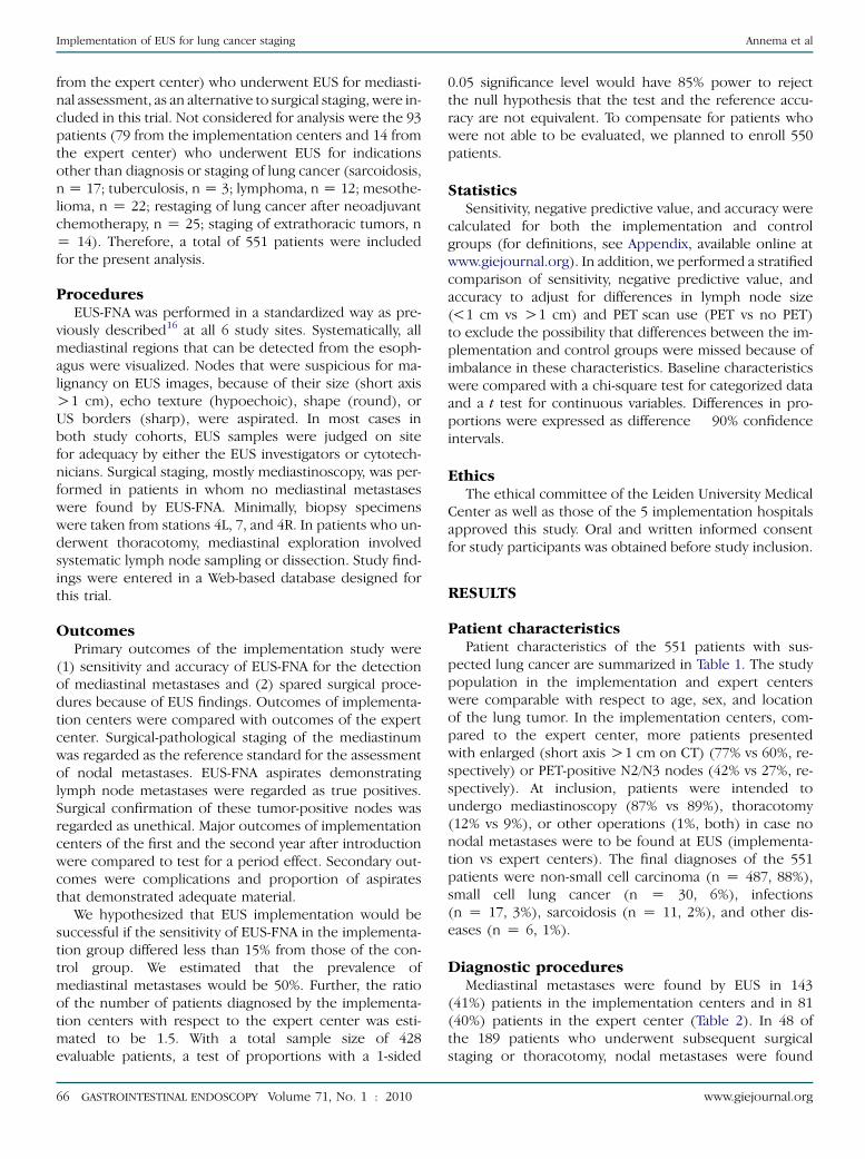
Patient characteristicsPatient characteristics of the 551 patients with sus-
pected lung cancer are summarized in Table 1. The studypopulation in the implementation and expert centerswere comparable with respect to age, sex, and locationof the lung tumor. In the implementation centers, com-pared to the expert center, more patients presentedwith enlarged (short axis O1 cm on CT) (77% vs 60%, re-spectively) or PET-positive N2/N3 nodes (42% vs 27%, re-spectively). At inclusion, patients were intended toundergo mediastinoscopy (87% vs 89%), thoracotomy(12% vs 9%), or other operations (1%, both) in case nonodal metastases were to be found at EUS (implementa-tion vs expert centers). The final diagnoses of the 551patients were non-small cell carcinoma (n Z 487, 88%),small cell lung cancer (n Z 30, 6%), infections(n Z 17, 3%), sarcoidosis (n Z 11, 2%), and other dis-eases (n Z 6, 1%).
Diagnostic proceduresMediastinal metastases were found by EUS in 143
(41%) patients in the implementation centers and in 81(40%) patients in the expert center (Table 2). In 48 ofthe 189 patients who underwent subsequent surgicalstaging or thoracotomy, nodal metastases were found
www.giejournal.org
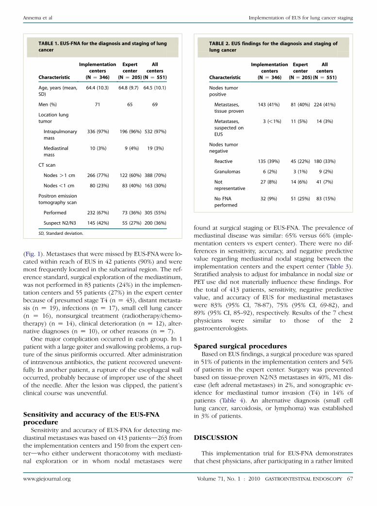
TABLE 1. EUS-FNA for the diagnosis and staging of lung
cancer
Characteristic
Implementation
centers
(N Z 346)
Expert
center
(N Z 205)
All
centers
(N Z 551)
Age, years (mean,
SD)
64.4 (10.3) 64.8 (9.7) 64.5 (10.1)
Men (%) 71 65 69
Location lung
tumor
Intrapulmonary
mass
336 (97%) 196 (96%) 532 (97%)
Mediastinal
mass
10 (3%) 9 (4%) 19 (3%)
CT scan
Nodes O1 cm 266 (77%) 122 (60%) 388 (70%)
Nodes !1 cm 80 (23%) 83 (40%) 163 (30%)
Positron emission
tomography scan
Performed 232 (67%) 73 (36%) 305 (55%)
Suspect N2/N3 145 (42%) 55 (27%) 200 (36%)
SD, Standard deviation.
TABLE 2. EUS findings for the diagnosis and staging of
lung cancer
Characteristic
Implementation
centers
(N Z 346)
Expert
center
(N Z 205)
All
centers
(N Z 551)
Nodes tumor
positive
Metastases,
tissue proven
143 (41%) 81 (40%) 224 (41%)
Metastases,
suspected on
EUS
3 (!1%) 11 (5%) 14 (3%)
Nodes tumor
negative
Reactive 135 (39%) 45 (22%) 180 (33%)
Granulomas 6 (2%) 3 (1%) 9 (2%)
Not
representative
27 (8%) 14 (6%) 41 (7%)
No FNA
performed
32 (9%) 51 (25%) 83 (15%)
Annema et al Implementation of EUS for lung cancer staging
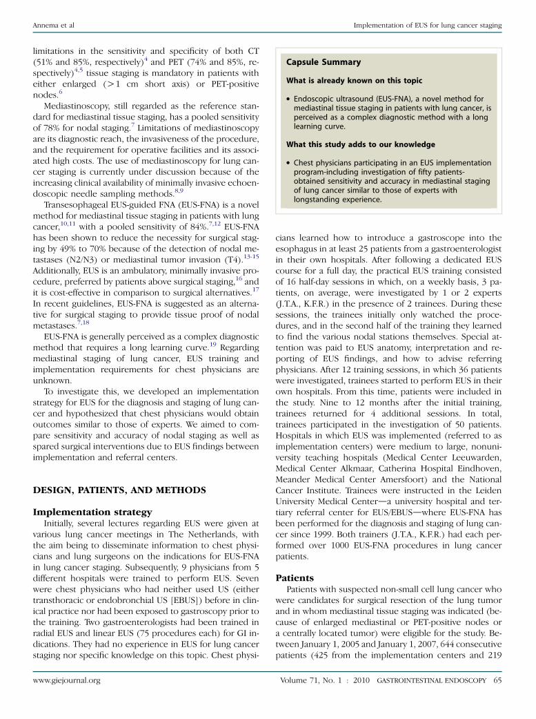
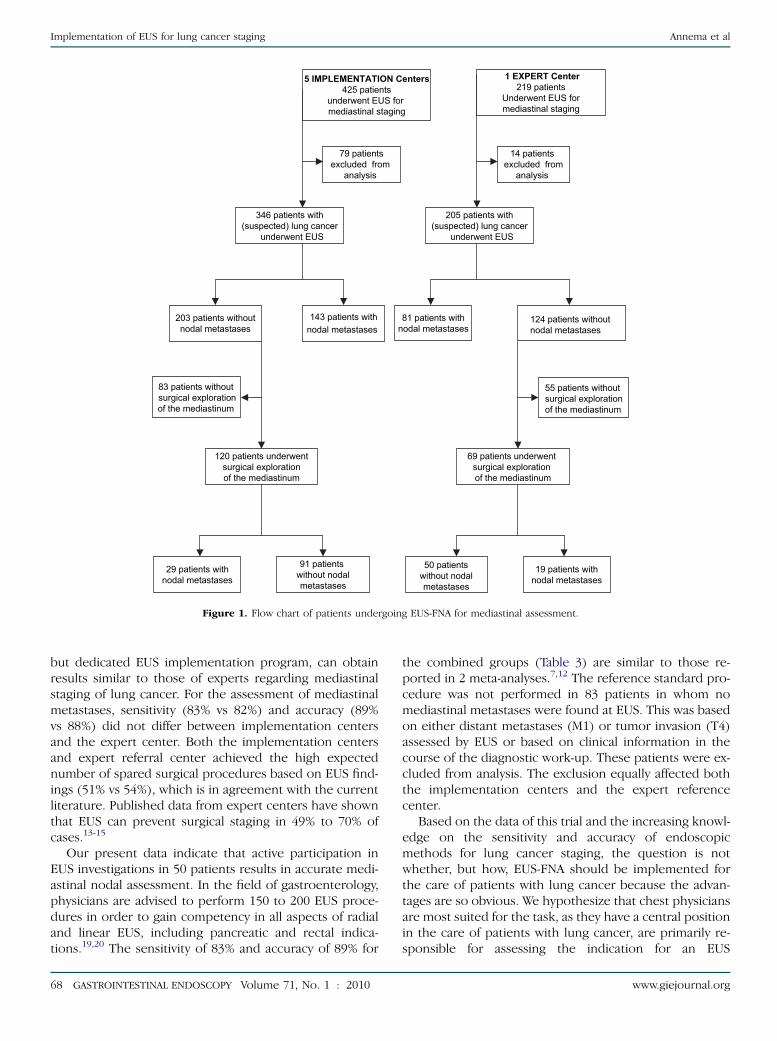
(Fig. 1). Metastases that were missed by EUS-FNA were lo-cated within reach of EUS in 42 patients (90%) and weremost frequently located in the subcarinal region. The ref-erence standard, surgical exploration of the mediastinum,was not performed in 83 patients (24%) in the implemen-tation centers and 55 patients (27%) in the expert centerbecause of presumed stage T4 (n Z 43), distant metasta-sis (n Z 19), infections (n Z 17), small cell lung cancer(n Z 16), nonsurgical treatment (radiotherapy/chemo-therapy) (n Z 14), clinical deterioration (n Z 12), alter-native diagnoses (n Z 10), or other reasons (n Z 7).
One major complication occurred in each group. In 1patient with a large goiter and swallowing problems, a rup-ture of the sinus piriformis occurred. After administrationof intravenous antibiotics, the patient recovered unevent-fully. In another patient, a rupture of the esophageal walloccurred, probably because of improper use of the sheetof the needle. After the lesion was clipped, the patient’sclinical course was uneventful.
Sensitivity and accuracy of the EUS-FNAprocedure
Sensitivity and accuracy of EUS-FNA for detecting me-diastinal metastases was based on 413 patientsd263 fromthe implementation centers and 150 from the expert cen-terdwho either underwent thoracotomy with mediasti-nal exploration or in whom nodal metastases were
www.giejournal.org
found at surgical staging or EUS-FNA. The prevalence ofmediastinal disease was similar: 65% versus 66% (imple-mentation centers vs expert center). There were no dif-ferences in sensitivity, accuracy, and negative predictivevalue regarding mediastinal nodal staging between theimplementation centers and the expert center (Table 3).Stratified analysis to adjust for imbalance in nodal size orPET use did not materially influence these findings. Forthe total of 413 patients, sensitivity, negative predictivevalue, and accuracy of EUS for mediastinal metastaseswere 83% (95% CI, 78-87), 75% (95% CI, 69-82), and89% (95% CI, 85–92), respectively. Results of the 7 chestphysicians were similar to those of the 2gastroenterologists.
Spared surgical proceduresBased on EUS findings, a surgical procedure was spared
in 51% of patients in the implementation centers and 54%of patients in the expert center. Surgery was preventedbased on tissue-proven N2/N3 metastases in 40%, M1 dis-ease (left adrenal metastases) in 2%, and sonographic ev-idence for mediastinal tumor invasion (T4) in 14% ofpatients (Table 4). An alternative diagnosis (small celllung cancer, sarcoidosis, or lymphoma) was establishedin 3% of patients.
DISCUSSION
This implementation trial for EUS-FNA demonstratesthat chest physicians, after participating in a rather limited
Volume 71, No. 1 : 2010 GASTROINTESTINAL ENDOSCOPY 67
5 IMPLEMENTATION Centers
425 patientsunderwent EUS formediastinal staging
203 patients withoutnodal metastases
69 patients underwent surgical exploration of the mediastinum
143 patients withnodal metastases
81 patients withnodal metastases
120 patients underwentsurgical explorationof the mediastinum
29 patients withnodal metastases
91 patients without nodalmetastases
50 patientswithout nodalmetastases
19 patients withnodal metastases
83 patients withoutsurgical explorationof the mediastinum
124 patients withoutnodal metastases
55 patients withoutsurgical explorationof the mediastinum
346 patients with(suspected) lung cancer
underwent EUS
1 EXPERT Center
219 patientsUnderwent EUS formediastinal staging
79 patientsexcluded from
analysis
14 patients excluded from
analysis
205 patients with(suspected) lung cancer
underwent EUS
Figure 1. Flow chart of patients undergoing EUS-FNA for mediastinal assessment.
Implementation of EUS for lung cancer staging Annema et al
but dedicated EUS implementation program, can obtainresults similar to those of experts regarding mediastinalstaging of lung cancer. For the assessment of mediastinalmetastases, sensitivity (83% vs 82%) and accuracy (89%vs 88%) did not differ between implementation centersand the expert center. Both the implementation centersand expert referral center achieved the high expectednumber of spared surgical procedures based on EUS find-ings (51% vs 54%), which is in agreement with the currentliterature. Published data from expert centers have shownthat EUS can prevent surgical staging in 49% to 70% ofcases.13-15
Our present data indicate that active participation inEUS investigations in 50 patients results in accurate medi-astinal nodal assessment. In the field of gastroenterology,physicians are advised to perform 150 to 200 EUS proce-dures in order to gain competency in all aspects of radialand linear EUS, including pancreatic and rectal indica-tions.19,20 The sensitivity of 83% and accuracy of 89% for
68 GASTROINTESTINAL ENDOSCOPY Volume 71, No. 1 : 2010
the combined groups (Table 3) are similar to those re-ported in 2 meta-analyses.7,12 The reference standard pro-cedure was not performed in 83 patients in whom nomediastinal metastases were found at EUS. This was basedon either distant metastases (M1) or tumor invasion (T4)assessed by EUS or based on clinical information in thecourse of the diagnostic work-up. These patients were ex-cluded from analysis. The exclusion equally affected boththe implementation centers and the expert referencecenter.
Based on the data of this trial and the increasing knowl-edge on the sensitivity and accuracy of endoscopicmethods for lung cancer staging, the question is notwhether, but how, EUS-FNA should be implemented forthe care of patients with lung cancer because the advan-tages are so obvious. We hypothesize that chest physiciansare most suited for the task, as they have a central positionin the care of patients with lung cancer, are primarily re-sponsible for assessing the indication for an EUS
www.giejournal.org
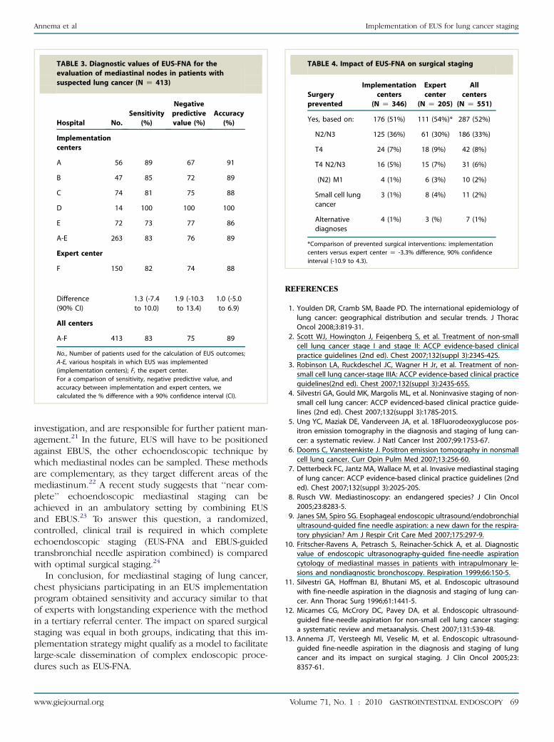
TABLE 3. Diagnostic values of EUS-FNA for the
evaluation of mediastinal nodes in patients with
suspected lung cancer (N Z 413)
Hospital No.
Sensitivity
(%)
Negative
predictive
value (%)
Accuracy
(%)
Implementation
centers
A 56 89 67 91
B 47 85 72 89
C 74 81 75 88
D 14 100 100 100
E 72 73 77 86
A-E 263 83 76 89
Expert center
F 150 82 74 88
Difference
(90% CI)
1.3 (-7.4
to 10.0)
1.9 (-10.3
to 13.4)
1.0 (-5.0
to 6.9)
All centers
A-F 413 83 75 89
No., Number of patients used for the calculation of EUS outcomes;
A-E, various hospitals in which EUS was implemented
(implementation centers); F, the expert center.
For a comparison of sensitivity, negative predictive value, and
accuracy between implementation and expert centers, we
calculated the % difference with a 90% confidence interval (CI).
TABLE 4. Impact of EUS-FNA on surgical staging
Surgery
prevented
Implementation
centers
(N Z 346)
Expert
center
(N Z 205)
All
centers
(N Z 551)
Yes, based on: 176 (51%) 111 (54%)* 287 (52%)
N2/N3 125 (36%) 61 (30%) 186 (33%)
T4 24 (7%) 18 (9%) 42 (8%)
T4 N2/N3 16 (5%) 15 (7%) 31 (6%)
(N2) M1 4 (1%) 6 (3%) 10 (2%)
Small cell lung
cancer
3 (1%) 8 (4%) 11 (2%)
Alternative
diagnoses
4 (1%) 3 (%) 7 (1%)
*Comparison of prevented surgical interventions: implementation
centers versus expert center Z -3.3% difference, 90% confidence
interval (-10.9 to 4.3).
Annema et al Implementation of EUS for lung cancer staging
investigation, and are responsible for further patient man-agement.21 In the future, EUS will have to be positionedagainst EBUS, the other echoendoscopic technique bywhich mediastinal nodes can be sampled. These methodsare complementary, as they target different areas of themediastinum.22 A recent study suggests that ‘‘near com-plete’’ echoendoscopic mediastinal staging can beachieved in an ambulatory setting by combining EUSand EBUS.23 To answer this question, a randomized,controlled, clinical trail is required in which completeechoendoscopic staging (EUS-FNA and EBUS-guidedtransbronchial needle aspiration combined) is comparedwith optimal surgical staging.24
In conclusion, for mediastinal staging of lung cancer,chest physicians participating in an EUS implementationprogram obtained sensitivity and accuracy similar to thatof experts with longstanding experience with the methodin a tertiary referral center. The impact on spared surgicalstaging was equal in both groups, indicating that this im-plementation strategy might qualify as a model to facilitatelarge-scale dissemination of complex endoscopic proce-dures such as EUS-FNA.
www.giejournal.org
REFERENCES
1. Youlden DR, Cramb SM, Baade PD. The international epidemiology of
lung cancer: geographical distribution and secular trends. J Thorac
Oncol 2008;3:819-31.
2. Scott WJ, Howington J, Feigenberg S, et al. Treatment of non-small
cell lung cancer stage I and stage II: ACCP evidence-based clinical
practice guidelines (2nd ed). Chest 2007;132(suppl 3):234S-42S.
3. Robinson LA, Ruckdeschel JC, Wagner H Jr, et al. Treatment of non-
small cell lung cancer-stage IIIA: ACCP evidence-based clinical practice
guidelines(2nd ed). Chest 2007;132(suppl 3):243S-65S.
4. Silvestri GA, Gould MK, Margolis ML, et al. Noninvasive staging of non-
small cell lung cancer: ACCP evidenced-based clinical practice guide-
lines (2nd ed). Chest 2007;132(suppl 3):178S-201S.
5. Ung YC, Maziak DE, Vanderveen JA, et al. 18Fluorodeoxyglucose pos-
itron emission tomography in the diagnosis and staging of lung can-
cer: a systematic review. J Natl Cancer Inst 2007;99:1753-67.
6. Dooms C, Vansteenkiste J. Positron emission tomography in nonsmall
cell lung cancer. Curr Opin Pulm Med 2007;13:256-60.
7. Detterbeck FC, Jantz MA, Wallace M, et al. Invasive mediastinal staging
of lung cancer: ACCP evidence-based clinical practice guidelines (2nd
ed). Chest 2007;132(suppl 3):202S-20S.
8. Rusch VW. Mediastinoscopy: an endangered species? J Clin Oncol
2005;23:8283-5.
9. Janes SM, Spiro SG. Esophageal endoscopic ultrasound/endobronchial
ultrasound-guided fine needle aspiration: a new dawn for the respira-
tory physician? Am J Respir Crit Care Med 2007;175:297-9.
10. Fritscher-Ravens A, Petrasch S, Reinacher-Schick A, et al. Diagnostic
value of endoscopic ultrasonography-guided fine-needle aspiration
cytology of mediastinal masses in patients with intrapulmonary le-
sions and nondiagnostic bronchoscopy. Respiration 1999;66:150-5.
11. Silvestri GA, Hoffman BJ, Bhutani MS, et al. Endoscopic ultrasound
with fine-needle aspiration in the diagnosis and staging of lung can-
cer. Ann Thorac Surg 1996;61:1441-5.
12. Micames CG, McCrory DC, Pavey DA, et al. Endoscopic ultrasound-
guided fine-needle aspiration for non-small cell lung cancer staging:
a systematic review and metaanalysis. Chest 2007;131:539-48.
13. Annema JT, Versteegh MI, Veselic M, et al. Endoscopic ultrasound-
guided fine-needle aspiration in the diagnosis and staging of lung
cancer and its impact on surgical staging. J Clin Oncol 2005;23:
8357-61.
Volume 71, No. 1 : 2010 GASTROINTESTINAL ENDOSCOPY 69
Implementation of EUS for lung cancer staging Annema et al
14. Larsen SS, Krasnik M, Vilmann P, et al. Endoscopic ultrasound guided
biopsy of mediastinal lesions has a major impact on patient manage-
ment. Thorax 2002;57:98-103.
15. Tournoy KG, De Ryck F, Vanwalleghem LR, et al. Endoscopic ultra-
sound reduces surgical mediastinal staging in lung cancer: a random-
ized trial. Am J Respir Crit Care Med 2008;177:531-5.
16. Annema JT, Versteegh MI, Veselic M, et al. Endoscopic ultrasound
added to mediastinoscopy for preoperative staging of patients with
lung cancer. JAMA 2005;294:931-6.
17. Kramer H, van Putten JW, Post WJ, et al. Oesophageal endoscopic ul-
trasound with fine needle aspiration improves and simplifies the stag-
ing of lung cancer. Thorax 2004;59:596-601.
18. De Leyn P, Lardinois D, Van Schil PE, et al. ESTS guidelines for preop-
erative lymph node staging for non-small cell lung cancer. Eur J Car-
diothorac Surg 2007;32:1-8.
19. Rosch T. State of the art lecture: endoscopic ultrasonography: training
and competence. Endoscopy 2006;38(suppl 1):S69-72.
20. Eisen GM, Dominitz JA, Faigel DO, et al. Guidelines for credentialing
and granting privileges for endoscopic ultrasound. Gastrointest En-
dosc 2001;54:811-4.
21. Annema JT, Rabe KF. Why respiratory physicians should learn and im-
plement EUS-FNA. Am J Respir Crit Care Med 2007;176:99.
22. Herth FJ, Lunn W, Eberhardt R, et al. Transbronchial vs. transesopha-
geal ultrasound-guided aspiration of enlarged mediastinal lymph no-
des. Am J Respir Crit Care Med 2005;171:1164-7.
23. Wallace MB, Pascual JM, Raimondo M, et al. Minimally invasive
endoscopic staging of suspected lung cancer. JAMA 2008;299:
540-6.
70 GASTROINTESTINAL ENDOSCOPY Volume 71, No. 1 : 2010
24. Annema JT, Tournoy KG, Rabe KF. Lung cancer staging with minimally
invasive endoscopic techniques. JAMA 2008;299:2510-1.
Received March 11, 2009. Accepted July 15, 2009.
Current affiliations: Divisions of Pulmonary Medicine (J.T.A., K.F.R.) and
Clinical Epidemiology (O.M.D.), Leiden University Medical Center, Leiden,
Biometrics Department (R.B., H.v.T.), Pulmonary Medicine (S.B.), Gastero-
enterology (M.S., B.T.), Netherlands Cancer Institute Amsterdam,
Pulmonary Medicine Medical Center, Leeuwarden (B.V., H.N.), Pulmonary
Medicine, St. Catherine’s Hospital Eindhoven (B.v.d.B., R.v.B.), Pulmonary
Medicine, Medical Center Alkmaar (T.H., A.W.), Pulmonary Medicine,
Meander Medical Center Amersfoort (G.S.), The Netherlands.
Presented in part at Digestive Disease Week, May 22, 2007, Chicago
Illinois, European Respiratory Society, September 16, 2007, Stockholm,
Sweden, American Thoracic Society, May 20, 2008, Toronto, Ontario,
Canada, and American Society of Clinical Oncology, May 20, 2008,
Chicago, Illinois.
Reprint requests: Jouke T. Annema, MD, PhD, Department of
Pulmonology C3 P, Albinusdreef 2, PO box 9600, 2300 RC, Leiden
University Medical Center, Leiden, The Netherlands.
If you would like to chat with an author of this article, you may contact Dr.
Annema at [email protected].
www.giejournal.org
APPENDIX
Definition of diagnostic valuesSensitivity was defined as the total number of patients
with tissue-proven N2-N3 metastases by EUS, divided bythe total number of patients with proven N2-N3 metastases.
Negative predictive value was defined as the total num-ber of patients in whom EUS correctly assessed no N2-N3metastases, divided by the total number of patients inwhom EUS assessed no N2/-N3 metastases.
Accuracy was defined as the total number of patients inwhom EUS correctly assessed no N2-N3 metastases plus
the total number of patients in whom EUS correctly as-sessed N2-N3 metastases, divided by the total number ofevaluated patients.
Participating hospitalsMCL Z Medical Center LeeuwardenMCA Z Medical Center AlkmaarCZE Z Catherin Hospital EindhovenMMC Z Meander Medical Center Amersfoort
NCI Z National Cancer InstituteLUMC Z Leiden University Medical Center
www.giejournal.org Volume 71, No. 1 : 2010 GASTROINTESTINAL ENDOSCOPY 70.e1
Annema et al Implementation of EUS for lung cancer staging
Copyright © 2022 FDOKUMEN