Inactivating mutations of caspase-8 gene in colorectal carcinomas
Upload
independentCategory
view
1download
0
780
Significance of Cyclin D1 Overexpression inTransitional Cell Carcinomas of the Urinary Bladderand Its Correlation with Histopathologic Features
BACKGROUND. Genetic alterations leading to neoplastic transformation of the uro-Chyi Chia R. Lee, M.D.1
thelium are likely to involve the activation of oncogenes and loss of functionalShinji Yamamoto, M.D.1
tumor suppressor genes. Cyclin D1 has been implicated as a putative protoonco-Keiichirou Morimura, M.D.1
gene whereas mutations of the p53 gene occur frequently in invasive transitionalHideki Wanibuchi, M.D.1
cell carcinomas (TCCs) of the urinary bladder. In this study, cyclin D1 overex-Nobuyasu Nishisaka, M.D.2
pression and nuclear accumulation of p53 were evaluated and the results correlatedShinichi Ikemoto, M.D.2
with histopathologic features.Tatsuya Nakatani, M.D.2METHODS. TCCs of the urinary bladder from 161 surgical procedures were evalu-Seiji Wada, M.D.2ated for cyclin D1 overexpression and nuclear accumulation of p53. Results were
Taketoshi Kishimoto, M.D.2correlated with tumor grade, T classification, and papillary status. Topologic distri-
Shoji Fukushima, M.D.1butions of cyclin D1, p53, and proliferating cellular nuclear antigen (PCNA) were
evaluated. Northern blot analysis was performed on selected specimens.1 First Department of Pathology, Osaka City Uni- RESULTS. Overexpression of cyclin D1 was observed in 47% (24 of 51) of Grade 1versity Medical School, Osaka, Japan.
TCCs and 20% (13 of 65) of Grade 2 TCCs but in no Grade 3 TCCs. Approximately
34% (14 of 41) of Ta classified TCCs and 21% (13 of 63) of T1 classified TCCs2 Department of Urology, Osaka City UniversityMedical School, Osaka, Japan. were immunoreactive for cyclin D1 whereas none of the TCCs beyond T1 was
immunoreactive. Overexpression of cyclin D1 was observed only in papillary type
TCCs. Results of Northern blot analysis for cyclin D1 were comparable to those of
immunohistochemistry.
CONCLUSIONS. The observed significant relation between cyclin D1 overexpression
and tumor grade/T classification suggests that cyclin D1 may be a useful biologic
marker for biopsied materials or urine cytology specimens. The prognostic signifi-
cance of cyclin D1 overexpression in TCCs remains to be determined. Cancer 1997;
79:780–9. q 1997 American Cancer Society.
KEYWORDS: Cyclin D1, p53, proliferating cell nuclear antigen, transitional cell carci-noma, urinary bladder, cell cycle.
Transitional cell carcinomas (TCCs) of the urinary bladder are themost common cancers of the urinary tract. Although the genetic
alterations leading to neoplastic transformation of the urothelium arenot completely understood, they are likely to involve the activationof oncogenes and loss of functional tumor suppressor genes.1 Cellcycle regulators exert their effects by governing cell cycle progressionat various checkpoints and alterations in their expression are likely
Address for reprints: Chyi Chia R. Lee, M.D., to contribute to tumorigenesis. The restriction point situated in lateFirst Department of Pathology, Osaka City Uni- G1 just before the G1/S-phase transition is perhaps the most importantversity Medical School, 1-4-54 Asahi-machi,
checkpoint in the mammalian cell cycle.2 In mammalian cells, the DAbeno-ku, Osaka 545, Japan.type cyclins, through their interactions with the cyclin dependentkinases, are primarily responsible for driving the cell cycle throughReceived August 1, 1996; revision received Oc-
tober 10, 1996; accepted October 28, 1996. the restriction point.3 Cyclin D1 is a member of the G1 cyclins involved
q 1997 American Cancer Society
/ 7b4c$$0883 01-23-97 11:16:36 cana W: Cancer
Cyclin D1 in TCC of Urinary Bladder/Lee et al. 781
TABLE 1in regulating the transition through G1 .4,5 It was ini-Characteristics of the 161 Patients With Transitional Cell Carcinomastially implicated as an oncogene in parathyroid adeno-of the Urinary Bladder
mas6 and B-cell lymphomas7 and experiments intransgenic mice have provided support for a role of Variable No. %cyclin D1 in the neoplastic transformation of mam-
Age (yrs)mary cells.8 In addition, overexpression of cyclin D1Median (range) 60.5 (38–83)has been reported in several types of human cancers,
Genderincluding esophageal carcinomas,9 breast carcino- Male 123 76.4mas,10,11 hepatocellular carcinomas,12 and nonsmall Female 38 23.6
Type of surgerycell lung carcinomas,13 with prognostic significance inBiopsy 64 39.8most of these cases. However, to the authors’ knowl-TURBT 61 37.9edge, no report has addressed the significance ofCystectomy 35 21.7
cyclin D1 overexpression in TCCs of the urinary tract. Partial cystectomy 1 0.6Tumor gradeThe p53 gene product has been proposed to play
G1 51 31.7a role in cell cycle regulation at the G1/S-phase check-G2 65 40.4point.14 In vitro experiments using cancer cell linesG3 45 28
indicate that cyclin D1 may be involved in p53-medi- Tumor classificationated growth suppression.15 However, relatively few pTis 0 0
pTa 41 25.5studies have been concerned with the involvement ofpT1 63 39.1cyclin D1 in p53-mediated growth suppression in hu-pT2 5 3.1
man cancers. The current investigation was therefore pT3a 7 4.3aimed at determining whether expression of cell cycle pT3b 7 4.3regulatory proteins changes during the underlying ma- pT4 1 0.6
Unclassifieda 37 23lignant transformation processes in the urothelium.Papillary statusThe authors examined surgical specimens of TCCs
Papillary 111 68.9of the urinary bladder from 161 patients for overex- Nonpapillary 20 12.4pression of cyclin D1 and nuclear accumulation of p53 Unclassifiedb 30 18.6using immunohistochemical techniques. Double
TURBT: transurethral resection of bladder tumor.staining was employed to compare the topologic dis-a The biopsied specimen did not contain sufficient adjacent normal tissue to tumor.
tributions of cyclin D1 and p53 in relation to proliferat- b Definite classification of papillary status was not possible.ing cellular nuclear antigen (PCNA), a marker for cellproliferation. The results of cyclin D1 and p53 immu-nohistochemistry were compared with tumor grade,stage, and papillary status. Histologic Methods
Histologic grading and pathologic staging of TCCswere performed according to the criteria defined in
MATERIALS AND METHODS General Rules for Clinical and Pathological Studies onThe study population was comprised of patients with Bladder Cancer published by The Japanese UrologicalTCCs of the urinary bladder or other disease entities Association and The Japanese Pathological Society.16
who underwent surgical resection. Paraffin embedded Histologic grading was consistent with the criteria de-specimens were obtained from the archives at Osaka fined by the World Health Organization’s (WHO) Inter-City University Hospital from 1983 to 1989 for 149 national Classification of Tumors. The system forTCCs of the urinary bladder and 10 non-TCC lesions. pathologic staging employed was compatible with theIn addition, surgical specimens from 20 patients with staging system recommended by International UnionTCCs who underwent surgery between July 1995 and Against Cancer. Without knowing the results of theMay 1996 were processed and frozen material was immunohistochemical analyses, two pathologists (S.available for RNA extraction. The surgical procedures Y. and C. L.) reviewed all histologic slides to makewere eight nephroureterectomies, nine total cystecto- definite diagnosis of the tumors. Definite pathologicmies, and three transurethral resections. Only TCCs of staging was not possible in 37 tumors because somethe urinary bladder were included in the statistical biopsied specimens did not contain sufficient normalanalysis. The characteristics of the 161 patients with adjacent tissue. A papillary status could not be as-TCCs of the urinary bladder selected for this study are signed to 30 tumors due to their small size or tissue
destruction in some of the biopsied specimens.summarized in Table 1.
/ 7b4c$$0883 01-23-97 11:16:36 cana W: Cancer
782 CANCER February 15, 1997 / Volume 79 / Number 4
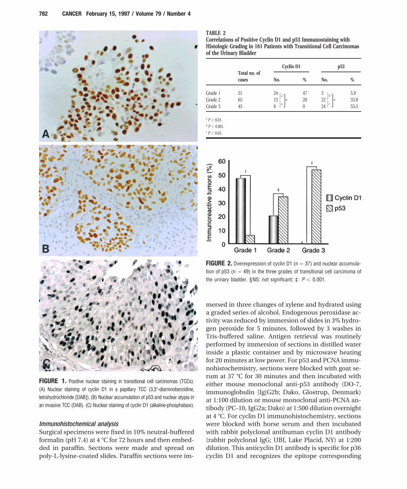
TABLE 2Correlations of Positive Cyclin D1 and p53 Immunostaining withHistologic Grading in 161 Patients with Transitional Cell Carcinomasof the Urinary Bladder
Cyclin D1 p53Total no. ofcases No. % No. %
Grade 1 51 24 47 3 5.9G aGbG bGbGrade 2 65 13 20 22 33.8G a G c
Grade 3 45 0 0 24 53.3
a P õ 0.01.b P õ 0.001.c P õ 0.05.
FIGURE 2. Overexpression of cyclin D1 (n Å 37) and nuclear accumula-tion of p53 (n Å 49) in the three grades of transitional cell carcinoma ofthe urinary bladder. §NS: not significant; ‡ : P õ 0.001.
mersed in three changes of xylene and hydrated usinga graded series of alcohol. Endogenous peroxidase ac-tivity was reduced by immersion of slides in 3% hydro-gen peroxide for 5 minutes, followed by 3 washes inTris-buffered saline. Antigen retrieval was routinelyperformed by immersion of sections in distilled waterinside a plastic container and by microwave heatingfor 20 minutes at low power. For p53 and PCNA immu-nohistochemistry, sections were blocked with goat se-rum at 37 7C for 30 minutes and then incubated with
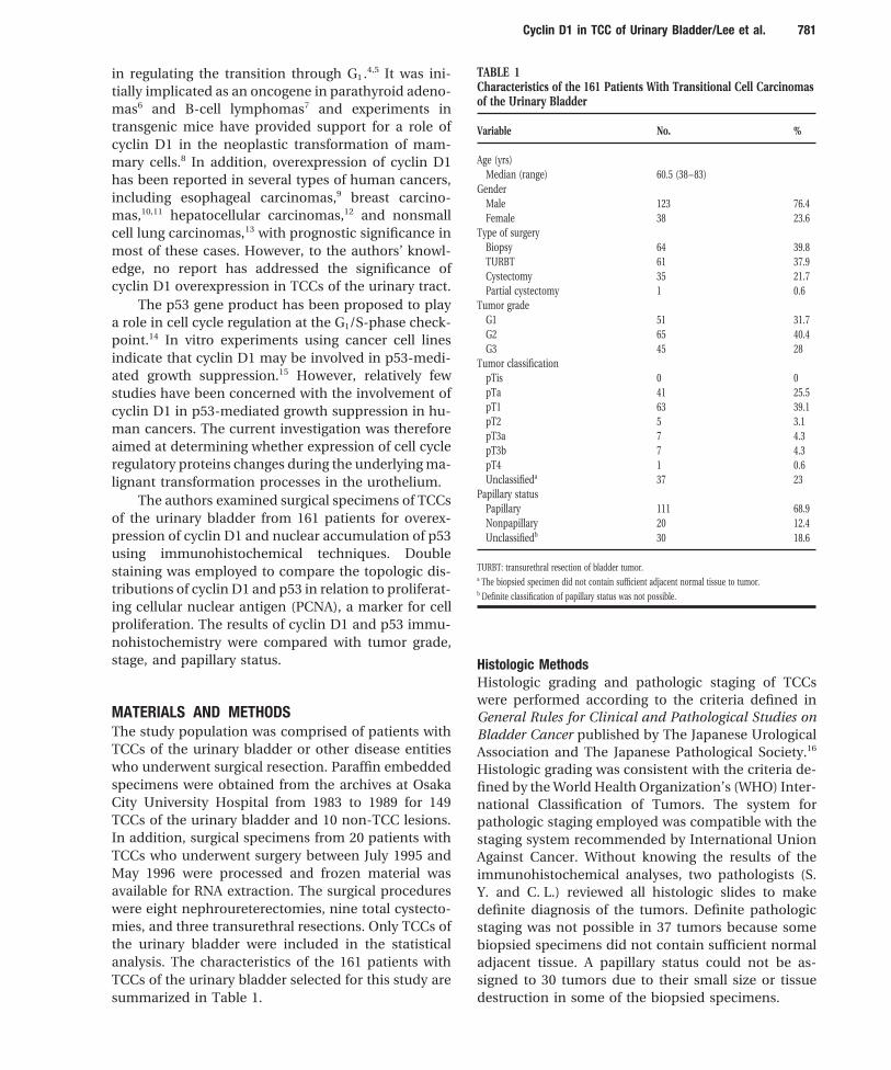
FIGURE 1. Positive nuclear staining in transitional cell carcinomas (TCCs). either mouse monoclonal anti-p53 antibody (DO-7,(A) Nuclear staining of cyclin D1 in a papillary TCC (3,3*-diaminobenzidine, immunoglobulin [Ig]G2b; Dako, Glostrup, Denmark)tetrahydrochloride [DAB]). (B) Nuclear accumulation of p53 and nuclear atypia in at 1:100 dilution or mouse monoclonal anti-PCNA an-an invasive TCC (DAB). (C) Nuclear staining of cyclin D1 (alkaline-phosphatase). tibody (PC-10, IgG2a; Dako) at 1:500 dilution overnight
at 4 7C. For cyclin D1 immunohistochemistry, sectionswere blocked with horse serum and then incubatedImmunohistochemical analysis
Surgical specimens were fixed in 10% neutral-buffered with rabbit polyclonal antihuman cyclin D1 antibody(rabbit polyclonal IgG; UBI, Lake Placid, NY) at 1:200formalin (pH 7.4) at 4 7C for 72 hours and then embed-
ded in paraffin. Sections were made and spread on dilution. This anticyclin D1 antibody is specific for p36cyclin D1 and recognizes the epitope correspondingpoly-L-lysine-coated slides. Paraffin sections were im-
/ 7b4c$$0883 01-23-97 11:16:36 cana W: Cancer
Cyclin D1 in TCC of Urinary Bladder/Lee et al. 783
TABLE 3Results for Positive Cyclin D1 and p53 Immunostaining in Transitional Cell Carcinomas in Relation toHistopathologic Staging
Cyclin D1 p53Pathologic Total no. ofclassification cases No. % No. %
pTa 41 14 34.1 6 14.6pT1 63 13 20.6 21 33.3pT2 5 0 0 3 60pT3a 7 0 0 4 57.1pT3b 7 0 0 6 85.7pT4 1 0 0 1 100pTa to pT1a 104 27 26 27 26G c G dpT2 to pT4b 20 0 0 14 70
a Tumor invasion was limited to the submucosa.b Tumor invasion reached the muscle layer.c P õ 0.01.d P õ 0.001.
TABLE 4Correlation of Cyclin D1 and p53 Immunoreactivity with the Papillary Status in 161 Patients with TransitionalCell Carcinomas of the Urinary Bladder
Cyclin D1 p53
Total No. % No. %
Papillary 111 33 29.7 29 26.1G a G bNonpapillary 20 0 0 10 50
Cyclin D1: Cyclin D1 immunoreactive; p53: p53 immunoreactive.a P õ 0.01.b P õ 0.05.
to the 11 C-terminal amino acids (residues 285-295) Northern blot analysisSpecimens for Northern blot analysis were snap frozenof human cyclin D1. Staining was achieved using the
Vectastaint ABC-PO kit (Vector Laboratories, Burlin- in liquid nitrogen and stored at 080 7C until analyzed.Total RNA was isolated using Isogen (Nippon Gene,game, CA) and developed with the 3,3*-diaminobenzi-
dine, tetrahydrochloride (DAB) chromogen (Wako, Toyama, Japan). Thirty microgram samples of totalRNA per lane were electrophoresed through 1% dena-Tokyo, Japan). A Histostain-DSTM kit (Zymed Labora-
tory, Inc., South San Francisco, CA) was used for dou- turing agarose gel and then transferred to HybondTM-N/ nylon membranes (Amersham, Amersham, Unitedble staining. Sections were first probed with anticyclin
D1 antibody overnight and developed with the alka- Kingdom). Examination of agarose gels under ultravio-let light at the end of electrophoresis revealed the posi-line-phosphatase method to produce a black color.
After incubation with double staining enhancer tions of 28S and 18S rRNA migration and confirmedthe integrity of RNA. RNA was immobilized to the ny-(Zymed) for 30 minutes, sections were reprobed with
the anti-PCNA antibody for 2 hours and developed lon membrane by baking for 2 hours at 80 7C. Afterprehybridization at 42 7C for 5 hours, 32P-labeled cyclinwith horseradish peroxidase to produce a red color.
Sections were counterstained for 10 minutes in 0.5% D1 specific DNA probes were added and hybridizationwas performed overnight at 42 7C in a hybridizationmethyl green. Negative control staining was included
with each staining procedure by omitting the primary oven. Washes were performed to a final stringency of0.1X standard saline citrate and 0.1% sodium dodecylantibody. Tumors with intense staining for either
cyclin D1 or p53 were included with each staining sulfate at 65 7C. Membranes were exposed to KodakXAR film (Eastman Kodak, Rochester, NY) for 48 to 72procedure and served as positive controls.
/ 7b4c$$0883 01-23-97 11:16:36 cana W: Cancer

784 CANCER February 15, 1997 / Volume 79 / Number 4
plate for cDNA synthesis. The cyclin D1 probe wasgenerated by polymerase chain reaction (PCR) ampli-fication of cDNA with the following pair of cyclin D1specific primers for 35 cycles: 5*-CTGGAGCCCGTG-AAAAAGAGC-3*; and 5*-CTGGAGAGGAAGCGTGTG-AGG-3*.9 The PCR product was electrophoresedthrough a 1% agarose gel to confirm its identity andthen recovered with a Genecleant SPIN kit (BIO 101,La Jolla, CA). The GAPDH probe was purchased fromCosmo-Bio (Osaka, Japan). DNA probes were 32P-la-beled with a Bca-BestTM random-primer labeling kit(TaKaRa Biomedicals; Otsu, Japan) and unincorpo-rated nucleotides were removed by spin-column chro-matography.
FIGURE 3. Overexpression of cyclin D1 (n Å 33) and nuclear accumula-tion of p53 (n Å 39) in papillary and nonpapillary transitional cell carcino- Statistical Analysismas of the urinary bladder. † : P õ 0.01. Unless otherwise indicated, the chi-square test was
used for statistical comparisons. A result was only con-sidered significant if the P value was õ 0.05.
RESULTSAssessment of Cyclin D1 and p53 ImmunohistochemicalStainingAll specimens were initially examined by immuno-staining with the avidin-biotin complex technique anddevelopment with the DAB chromogen. Histologicallynormal bladder specimens showed absence of nuclearstaining in urothelial cells. A tumor was consideredpositively stained for cyclin D1 (Fig. 1A) or p53 only ifdefinite nuclear staining was present (Fig. 1B). Tumorspositive for cyclin D1 were confirmed by staining withthe alkaline-phosphatase technique (Fig. 1C). All spec-imens were assessed by one of the authors (C.L.) andthe results confirmed by another (S.Y.). The stainingwas divided into two categories, positive or negative,by estimating the percentage of stained nuclei. CyclinD1 positive tumors had ú5% of their cells demonstra-FIGURE 4. Correlation between cyclin D1 overexpression and nuclearting positive nuclear staining.17 p53 positive specimensaccumulation of p53 in 161 cases of transitional cell carcinomas of thehad ú20% of tumor cells positive for nuclear staining.urinary bladder. j: immunoreactive for both cyclin D1 and p53; Ω: posi-
tively stained for cyclin D1 but not for p53; º: positively stained for p53Correlation between Cyclin D1 or p53 Expression andbut not for cyclin D1; h: negative staining for both cyclin D1 and p53.*,*:Histologic GradingP õ 0.05.Results for positive cyclin D1 and p53 immunohisto-chemical staining in TCCs of the urinary bladder corre-lated to WHO histologic grading are summarized inhours at 080 7C between 2 sheets of intensifying
screens. After obtaining the results of cyclin D1 hybrid- Table 2. Positive cyclin D1 staining was strongly corre-lated with the differentiation status. Cyclin D1 overex-izations, membranes were stripped and rehybridized
to a glyceraldehyde-3-phosphate dehydrogenase pression was most frequent in Grade 1 TCCs (47%),followed by Grade 2 tumors (20%), whereas none of the(GAPDH) probe to confirm equivalent loading of RNA
to all lanes. Grade 3 tumors were cyclin D1 positive. Conversely,positive p53 staining was most frequent in Grade 3 tu-mors (53.3%), followed by Grade 2 (33.8%) and GradeProbes for Northern blot analysis
Five micrograms of total RNA, extracted from a histo- 1 lesions (5.9%). Statistical comparison showed signifi-cant differences between all three grades of TCCs forlogically normal human kidney cortex, was the tem-
/ 7b4c$$0883 01-23-97 11:16:36 cana W: Cancer

Cyclin D1 in TCC of Urinary Bladder/Lee et al. 785
FIGURE 5. Topologic distribution of cyclin D1 and proliferating cell nuclear antigen (PCNA) in papillary transitional cell carcinoma (TCC) (A) H & E.(B) Cyclin D1 (black). (C) PCNA (red). (D) Double staining of cyclin D1 (black) and PCNA (red). Intense staining of PCNA was limited to the basal celllayers and the highly proliferative regions. However, cyclin D1 positive nuclei were randomly distributed and not limited to the proliferative compartments.
positive cyclin D1 staining. In addition, an inverse rela- ated were negative and the association was statisticallysignificant (P õ 0.01). Conversely, nuclear accumula-tionship between cyclin D1 and p53 immunohisto-
chemical staining was observed (Fig. 2). tion of p53 was observed in both papillary and non-papillary TCCs and the frequency within the nonpapil-
Correlation of Cyclin D1 and p53 to Pathologic Staging lary category (50%) was significantly higher thanResults of positive cyclin D1 and p53 immunostaining within the papillary category (26.1%) (P õ 0.05). Thein TCCs in relation to histopathologic staging are sum- difference between cyclin D1 overexpression and nu-marized in Table 3. Positive cyclin D1 staining was clear accumulation of p53 was statistically significantlimited to Ta and T1 tumors; none of those beyond (P õ 0.01) (Fig. 3).T1 were positive. Comparison of positive cyclin D1
Inverse Relation between Cyclin D1 and p53 Expressionstaining between the two categories, (Ta-T1 vs. T2-T4)
in TCCsrevealed a statistically significant difference (Põ 0.01).
Only a very small percentage of tumors (3.7%) wereThe difference in positive rates for p53 staining be-
positive for both cyclin D1 and p53 (Fig. 4). Cyclin D1tween the Ta-T1 and T2-T4 categories was also statisti-
positive tumors were generally p53 negative whereascally significant (P õ 0.001).
p53 positive tumors were generally cyclin D1 negative(P õ 0.05).Correlation of Cyclin D1 and p53 Immunoreactivity to
Papillary Status Topologic Distribution of Cyclin D1 and PCNA in TCCsCyclin D1 has been reported to be associated with multi-Positive cyclin D1 staining was observed only in papil-
lary TCCs (Table 4). All the nonpapillary tumors evalu- ple protein kinases and PCNA.18 To assess the topologic
/ 7b4c$$0883 01-23-97 11:16:36 cana W: Cancer
786 CANCER February 15, 1997 / Volume 79 / Number 4
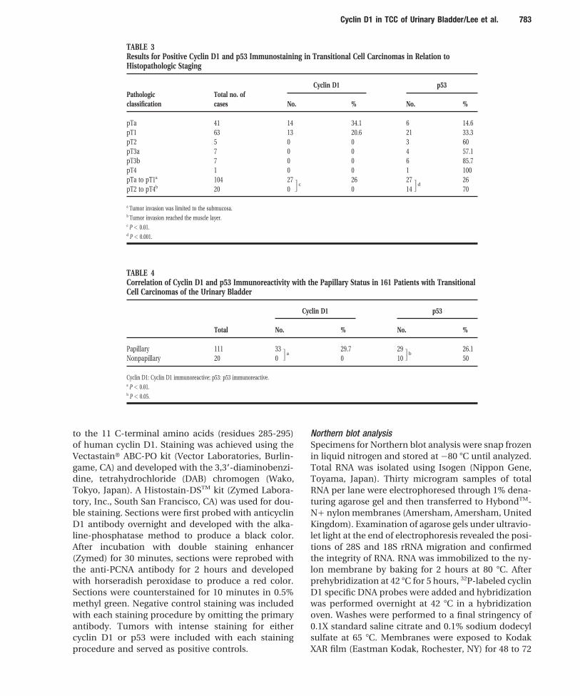
FIGURE 6. Topologic distribution of cyclin D1 and proliferating cell nuclear antigen (PCNA) in a Grade 2 transitional cell carcinoma (TCC) with glandularmetaplasia (TCC with well differentiated adenocarcinoma). (A) H & E. (B) Cyclin D1 (black). (C) PCNA (red). (D) Double staining of cyclin D1 (black)and PCNA (red). PCNA positive nuclei are confined to the highly proliferative regions whereas cyclin D1 positive nuclei are located within the welldifferentiated regions.
distribution of cyclin D1, p53, and PCNA in TCCs, immu- to have only few cells stained with the anti-PCNA anti-body. Double staining of these regions provided evi-nohistochemistry for each of these antigens was per-
formed separately in serial sections. Double staining was dence of an inverse relationship between p53 andPCNA (Fig. 7). This phenomenon was not observed inalso performed on selected specimens to allow precise
localization of cyclin D1 positive cells relative to PCNA, any of the nonpapillary TCCs.a marker for cellular proliferation in TCCs.19 Cyclin D1 Overexpression of Cyclin D1 mRNA in TCCspositive nuclei were randomly distributed and did not
The authors used Northern blot analysis to examinecoincide with cellular proliferation (Fig. 5B). However,
whether overexpression of cyclin D1 in TCC was due toPCNA positive nuclei were largely confined to the known
changed mRNA expression. Transcripts correspondingproliferative region near the basal cell layer (Fig. 5C). The
to 4.8 and 1.7 kilobases were detected by probing withintensity of cyclin D1 and PCNA staining was regionally
the cyclin D1 DNA probe20 as expected from alternativevariable. Highly proliferative regions were intensely
splicing of the cyclin D1 transcript (Fig. 8). The result ofstained positive for PCNA; areas of glandular formation
Northern blot analysis thus indicates alteration at thedemonstrating tumor differentiation, were intensely
cyclin D1 mRNA levels was responsible for the increase.stained positive for cyclin D1 (Fig. 6).
Cyclin D1 Immunohistochemistry in Non-TCC Lesions ofthe Urinary BladderTopologic Distribution of p53 and PCNA in TCCs
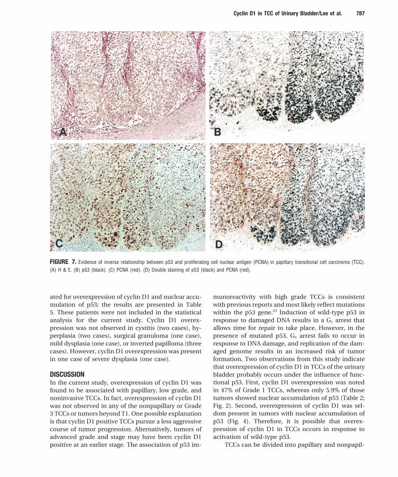
Regional variation of p53 immunoreactivity was noted Ten surgical specimens from the urinary bladders ofpatients with conditions other than TCCs were evalu-in some papillary TCCs. p53 positive regions tended
/ 7b4c$$0883 01-23-97 11:16:36 cana W: Cancer
Cyclin D1 in TCC of Urinary Bladder/Lee et al. 787
FIGURE 7. Evidence of inverse relationship between p53 and proliferating cell nuclear antigen (PCNA) in papillary transitional cell carcinoma (TCC).(A) H & E. (B) p53 (black). (C) PCNA (red). (D) Double staining of p53 (black) and PCNA (red).
ated for overexpression of cyclin D1 and nuclear accu- munoreactivity with high grade TCCs is consistentwith previous reports and most likely reflect mutationsmulation of p53; the results are presented in Table
5. These patients were not included in the statistical within the p53 gene.23 Induction of wild-type p53 inresponse to damaged DNA results in a G1 arrest thatanalysis for the current study. Cyclin D1 overex-
pression was not observed in cystitis (two cases), hy- allows time for repair to take place. However, in thepresence of mutated p53, G1 arrest fails to occur inperplasia (two cases), surgical granuloma (one case),
mild dysplasia (one case), or inverted papilloma (three response to DNA damage, and replication of the dam-aged genome results in an increased risk of tumorcases). However, cyclin D1 overexpression was present
in one case of severe dysplasia (one case). formation. Two observations from this study indicatethat overexpression of cyclin D1 in TCCs of the urinarybladder probably occurs under the influence of func-DISCUSSIONtional p53. First, cyclin D1 overexpression was notedIn the current study, overexpression of cyclin D1 wasin 47% of Grade 1 TCCs, whereas only 5.9% of thosefound to be associated with papillary, low grade, andtumors showed nuclear accumulation of p53 (Table 2;noninvasive TCCs. In fact, overexpression of cyclin D1Fig. 2). Second, overexpression of cyclin D1 was sel-was not observed in any of the nonpapillary or Gradedom present in tumors with nuclear accumulation of3 TCCs or tumors beyond T1. One possible explanationp53 (Fig. 4). Therefore, it is possible that overex-is that cyclin D1 positive TCCs pursue a less aggressivepression of cyclin D1 in TCCs occurs in response tocourse of tumor progression. Alternatively, tumors ofactivation of wild-type p53.advanced grade and stage may have been cyclin D1
positive at an earlier stage. The association of p53 im- TCCs can be divided into papillary and nonpapil-
/ 7b4c$$0883 01-23-97 11:16:36 cana W: Cancer

788 CANCER February 15, 1997 / Volume 79 / Number 4
of cyclin D1 and PCNA staining in these tumors im-plies that cyclin D1 overexpression may play a rolein negatively controlling cellular proliferation in TCCsand allow tumor differentiation. Alternatively, cyclinD1 may be complexed to other cell cycle regulatoryproteins such as the cyclin dependent kinase inhibi-tors to exhibit such effects. In a recent report, restora-tion of wild-type p53 protein conformation in cell linescarrying the temperature-sensitive p53 mutant genewas shown to cause an increase in the levels of cyclinD1 and p21,15 and induction of high levels of cyclin D1at the G1/S-phase transition period can functionallyantagonize cellular proliferation by preventing S-phase entry.
To evaluate whether cyclin D1 overexpression isthe result of mRNA overexpression, TCCs positivelyand negatively stained for cyclin D1 were comparedFIGURE 8. Northern blot analysis of cyclin D1. A transitional cell carci-with histologically normal urothelium and a closenoma (TCC) with nuclear staining of cyclin D1 (lane 1) expresses higherrelationship was found between immunohistochem-level of mRNA than the TCC without nuclear staining of cyclin D1 (laneical and Northern blot findings. Therefore, overex-2). Two transcripts of 4.8 kilobase (kb) and 1.7 kb were detected and thepression of cyclin D1 in TCCs could be due to ele-high molecular weight form was the most abundant.vated mRNA levels. Furthermore, the presence oftwo distinct bands in the Northern blots suggeststhat alternative splicing of cyclin D1 transcripts oc-TABLE 5curs in TCCs.Immunohistochemical Status of Cyclin D1 and p53 in Noncancerous
Lesions of the Urinary Bladder From a clinical standpoint, the significant corre-lation of cyclin D1 overexpression with low grade,
No. of Cyclinsuperficial, papillary TCCs suggest that cyclin D1cases D1 P53may be a useful biologic marker for prediction of
Hyperplasia 2 0 0 histopathologic grading and staging using biopsiedCystitis 2 0 0 material or urine cytology specimens. In the currentSurgical granuloma 1 0 0
study, overexpression of cyclin D1 allowed clear dis-Mild dysplasia 1 0 0tinction of tumors of T1 and earlier from those ofSevere dysplasia 1 / 0
Inverted papilloma 3 0 0 T2 and beyond. Such information is potentially valu-able for planning in patient treatment. However, the
0: negative; /: positive. prognostic significance of cyclin D1 overexpressionin TCCs remains to be determined. From the bio-logic standpoint, the significant correlation of cyclinD1 to pathologic features and the reciprocal rela-lary types. Potentially, two divergent pathways of tu-tionship to p53 nuclear staining suggests themor progression exist and may be closely associatedinvolvement of this protein in the induction and pro-with p53 mutations.24 The current evidence of an in-gression of bladder tumors. A detailed examinationverse relationship between p53 and PCNA staining ininto the mechanisms underlying the overexpressionpapillary but not nonpapillary TCCs (Fig. 7) providesof cyclin D1 in TCCs of the urinary bladder shouldfurther support for the importance of p53 gene alter-generate a better understanding of the biologic char-ation for tumor progression. However, the observationacteristics of this important tumor type.of cyclin D1 overexpression only in papillary TCCs and
In conclusion, the current study results showed ex-not the flat type TCCs suggests that cyclin D1 may alsocellent correlation between cyclin D1 immunohisto-affect tumor progression.chemistry and favorable pathologic parameters in TCCsThe authors examined the topologic distributionof the urinary bladder. Future studies should be concen-of cyclin D1 and its effect on cell proliferation. Intensetrated on the mechanisms of cyclin D1 overexpressioncyclin D1 staining was noted in differentiated regionsin TCCs and their significance for clinical parametersof TCCs whereas highly proliferative regions were only
weakly positive (Fig. 6). The mutually exclusive pattern such as recurrence, progression, and survival rates.
/ 7b4c$$0883 01-23-97 11:16:36 cana W: Cancer

Cyclin D1 in TCC of Urinary Bladder/Lee et al. 789
WDJ, Cerny T, et al. Prognostic significance of CCND1REFERENCES(cyclin D1) overexpression in primary resected non-small-1. Carrol PR. Urothelial carcinoma: cancers of the bladder, ure-cell lung cancer. Br J Cancer 1996;73:294–300.ter, & renal pelvis. In: Tanagho EA, McAninch JW, editors.
14. Mercer WE, Shields MT, Amin M, Sauve GJ, Appella E, Ro-Smith’s general urology. 14th edition. Norwalk, CT: Apple-mano JW, et al. Negative growth regulation in a glioblastomaton & Lange, 1995:353–71.tumor cell line that conditionally expresses human wild-2. Pardee AB. G1 events and regulation of cell proliferation.type p53. Proc Natl Acad Sci USA 1990;87:6166–70.Science 1989;246:603–8.
15. Giannino DS, Murphy M, Ruaro EM, Lazarevic D, Levine AJ,3. Hunter T, Pines J. Cyclins and cancer II: cyclin D and CDKSchneider C. Cyclin D1 and p21/waf1 are both involved ininhibitors come of age. Cell 1994;79:573–82.p53 growth suppression. Oncogene 1996;12:177–85.4. Matsushime H, Roussel MF, Ashmun RA, Sherr CJ. Colony-
16. Ohshima M, Fukushima S, Okamura T, Asamoto M, Okajimastimulating factor 1 regulates novel cyclins during the G1E, Washida H, et al. Histopathological analysis of 98 urinaryphase of the cell cycle. Cell 1991;65:701–13.bladder carcinoma cases by the serial section whole-organ5. Motokura T, Bloom T, Kim HG, Juppner H, Ruderman JV,approach. J Urol Pathol 1995;3:1–15.Kronenberg HM, et al. A novel cyclin encoded by a bcl 1-
17. Frierson HF, Gaffey MJ, Zukerberg LR, Arnold A, Williamslinked candidate oncogene. Nature 1991;350:512–5.ME. Immunohistochemical detection and gene amplifica-6. Motokura T, Arnold A. Cyclin D1 and oncogenesis. Currtion of cyclin D1 in mammary infiltrating ductal carcinoma.Opin Genet Dev 1993;3:5–10.Mod Pathol 1996;9:723–30.7. Rosenberg CL, Wong E, Petty EM, Bale AE, Tsujimoto Y,
18. Xiong Y, Zhang H, Beach D. D type cyclins associate withHarris NL, et al. PRAD1, a candidate BCL1 oncogene: map-multiple protein kinases and the DNA replication and repairping and expression in centrocytic lymphoma. Proc Natlfactor PCNA. Cell 1992;71:505–14.Acad Sci USA 1991;88:9638–42.
19. Lipponen PK, Eskelinen MJ. Cell proliferation of transitional8. Wang TC, Cardiff RD, Zukerberg LR, Lees E, Arnold A,cell bladder tumours determined by PCNA/cyclin immuno-Schmidt EV. Mammary hyperplasia and carcinoma instaining and its prognostic value. Br J Cancer 1992;66:171–6.MMTV-cyclin D1 (PRAD-1) transgenic mice. Nature 1994;
20. Jiang W, Zhang YJ, Kahn SM, Hollstein MC, Santella RM, Lu369:669–71.SH, et al. Altered expression of the cyclin D1 and retinoblas-9. Naitoh H, Shibata J, Kawaguchi A, Kodama M, Hattori T.toma genes in human esophageal cancer. Proc Natl AcadOverexpression and localization of cyclin D1 mRNA and an-Sci USA 1993;90:9026–30.tigen in esophageal cancer. Am J Pathol 1995;146:1161–9.
21. Xiong Y, Connolly T, Futcher B, Beach D. Human D-type10. Zhang SY, Caamano J, Cooper F, Guo X, Klein-Szanto AJP.cyclin. Cell 1991;65:691–9.Immunohistochemistry of cyclin D1 in human breast can-
22.cer. Am J Clin Pathol 1994;102:695–8. Betticher DC, Thatcher N, Altermatt HJ, Hoban P, RyderWDJ, Heighway J. Alternate splicing produces a novel cyclin11. McIntosh GG, Anderson JJ, Milton I, Steward M, Parr AH,D1 transcript. Oncogene 1995;11:1005–11.Thomas MD, et al. Determination of the prognostic value
of cyclin D1 overexpression in breast cancer. Oncogene 23. Esrig D, Spruck CH III, Nichols PW, Chaiwun B, Steven K,1995;11:885–91. Groshen S, et al. p53 nuclear protein accumulation corre-
lates with mutations in the p53 gene, tumor grade, and stage12. Nishida N, Fukuda Y, Komeda T, Kita R, Sando T, Furukawain bladder cancer. Am J Pathol 1993;143:1389–97.M, et al. Amplification and overexpression of the cyclin D1
gene in aggressive human hepatocellular carcinoma. Cancer 24. Spruck CH III, Schmutte C, Yane AS, Cote R, Dubeau L,Res 1994;54:3107 –10. Nichols PW, et al. Two molecular pathways to transitional
cell carcinoma of the bladder. Cancer Res 1994;54:784–8.13. Betticher DC, Heighway J, Hasleton PS, Altermatt HJ, Ryder
/ 7b4c$$0883 01-23-97 11:16:36 cana W: Cancer
Copyright © 2022 FDOKUMEN