Prevalence and genetic diversity of simian immunodeficiency virus infection in wild-living red...
14
Prevalence and genetic diversity of simian immunodeficiency virus infection in wild-living red colobus monkeys (Piliocolobus badius badius) from the Taı ¨ forest, Co ˆte d’Ivoire SIVwrc in wild-living western red colobus monkeys § Sabrina Locatelli a , Florian Liegeois a , Be ´ne ´dicte Lafay b , Amy D. Roeder c , Michael W. Bruford c , Pierre Formenty d , Ronald Noe ¨ e , Eric Delaporte a , Martine Peeters a, * a UMR 145, Institut de Recherche pour le De ´veloppement (IRD), and University of Montpellier 1, Montpellier, France b UMR CNRS-IRD 2724, Montpellier, France c Cardiff School of Biosciences, Museum Avenue, Cardiff CF10 3TL, UK d Ebola Taı ¨ Forest Project, Abidjan, Co ˆte d’Ivoire; World Health Organisation (WHO), Department of Communicable Diseases, Surveillance and Response (CDS/CSR), Geneva, Switzerland e Ethologie des Primates (DEPE-IPHC-CNRS UMR 7178) and University of Louis Pasteur, Strasbourg, France Received 7 June 2007; received in revised form 29 August 2007; accepted 30 August 2007 Available online 4 September 2007 Abstract Numerous African primates are infected with simian immunodeficiency viruses (SIVs). It is now well established that the clade of SIVs infecting west-central African chimpanzees (Pan troglodytes troglodytes) and western gorillas (Gorilla gorilla gorilla) represent the progenitors of human immunodeficiency virus type 1 (HIV-1), whereas HIV-2 results from different cross-species transmissions of SIVsmm from sooty mangabeys (Cercocebus atys atys). We present here the first molecular epidemiological survey of simian immunodeficiency virus (SIVwrc) in wild-living western red colobus monkeys (Piliocolobus badius badius) which are frequently hunted by the human population and represent a favourite prey of western chimpanzees (Pan troglodytes verus). We collected faecal samples (n = 88) and we assessed individual discrimination by microsatellite analyses and visual observation. We tested the inferred 53 adult individuals belonging to two neighbouring habituated groups for presence of SIVwrc infection by viral RNA (vRNA) detection. We amplified viral polymerase ( pol) (650 bp) and/or envelope (env) (570 bp) sequences in 14 individuals, resulting in a minimal prevalence of 26% among the individuals sampled, possibly reaching 50% when considering the relatively low sensitivity of viral RNA detection in faecal samples. With a few exceptions, phylogenetic analysis of pol and env sequences revealed a low degree of intragroup genetic diversity and a general viral clustering related to the social group of origin. However, we found a higher intergroup diversity. Behavioural and demographic data collected previously from these communities indicate that red colobus monkeys live in promiscuous multi-male societies, where females leave their natal group at the sub-adult stage of their lives and where extra-group copulations or male immigration have been rarely observed. The phylogenetic data we obtained seem to reflect these behavioural characteristics. Overall, our results indicate that wild-living red colobus represent a substantial reservoir of SIVwrc. Moreover, because of their frequent association with other monkey species, the predation pressure exerted by chimpanzees www.elsevier.com/locate/meegid Infection, Genetics and Evolution 8 (2008) 1–14 § Nucleotide sequence data reported in this paper are available in the GenBank, EMBL and DDBJ databases under the accession numbers listed in parentheses: for the pol gene: SIVwrc-04CI-196 (AM743109), SIVwrc-04CI-116 (AM743110), SIVwrc-04CI-299 (AM743111), SIVwrc-04CI-110 (AM743112), SIVwrc-04CI-115 (AM743113), SIVwrc-04CI-112 (AM743114), SIVwrc-04CI-223 (AM743115), SIVwrc-04CI-52 (AM743116), SIVwrc-04CI-237 (AM743117), for the env gene: SIVwrc-04CI-268 (AM743118) SIVwrc-04CI-52 (AM743119), SIVwrc-04CI-116 (AM743120), SIVwrc-04CI-110 (AM743121), SIVwrc-04CI-237 (AM743122), SIVwrc-04CI-175 (AM743123), SIVwrc-04CI-296 (AM743124), SIVwrc-04CI-223 (AM743125), SIVwrc-04CI-299 (AM743126), SIVwrc-04CI-280 (AM743127), SIVwrc-04CI-32 (AM743128), SIVwrc-04CI-196 (AM743129), SIVwrc-04CI-112 (AM743130). * Corresponding author at: UMR 145, Institut de Recherche pour le De ´veloppement (IRD), 911 Avenue Agropolis, 34394 Montpellier, France. Tel.: +33 467416297; fax: +33 467416146. E-mail address: [email protected] (M. Peeters). 1567-1348/$ – see front matter # 2007 Elsevier B.V. All rights reserved. doi:10.1016/j.meegid.2007.08.004
Transcript of Prevalence and genetic diversity of simian immunodeficiency virus infection in wild-living red...
www.elsevier.com/locate/meegid
Infection, Genetics and Evolution 8 (2008) 1–14
Prevalence and genetic diversity of simian immunodeficiency virus infection
in wild-living red colobus monkeys (Piliocolobus badius badius)
from the Taı forest, Cote d’Ivoire
SIVwrc in wild-living western red colobus monkeys§
Sabrina Locatelli a, Florian Liegeois a, Benedicte Lafay b, Amy D. Roeder c,Michael W. Bruford c, Pierre Formenty d, Ronald Noe e,
Eric Delaporte a, Martine Peeters a,*a UMR 145, Institut de Recherche pour le Developpement (IRD), and University of Montpellier 1, Montpellier, France
b UMR CNRS-IRD 2724, Montpellier, Francec Cardiff School of Biosciences, Museum Avenue, Cardiff CF10 3TL, UK
d Ebola Taı Forest Project, Abidjan, Cote d’Ivoire; World Health Organisation (WHO), Department of Communicable Diseases,
Surveillance and Response (CDS/CSR), Geneva, Switzerlande Ethologie des Primates (DEPE-IPHC-CNRS UMR 7178) and University of Louis Pasteur, Strasbourg, France
Received 7 June 2007; received in revised form 29 August 2007; accepted 30 August 2007
Available online 4 September 2007
Abstract
Numerous African primates are infected with simian immunodeficiency viruses (SIVs). It is now well established that the clade of SIVs
infecting west-central African chimpanzees (Pan troglodytes troglodytes) and western gorillas (Gorilla gorilla gorilla) represent the progenitors of
human immunodeficiency virus type 1 (HIV-1), whereas HIV-2 results from different cross-species transmissions of SIVsmm from sooty
mangabeys (Cercocebus atys atys).
We present here the first molecular epidemiological survey of simian immunodeficiency virus (SIVwrc) in wild-living western red
colobus monkeys (Piliocolobus badius badius) which are frequently hunted by the human population and represent a favourite prey of
western chimpanzees (Pan troglodytes verus). We collected faecal samples (n = 88) and we assessed individual discrimination by
microsatellite analyses and visual observation. We tested the inferred 53 adult individuals belonging to two neighbouring habituated
groups for presence of SIVwrc infection by viral RNA (vRNA) detection. We amplified viral polymerase ( pol) (650 bp) and/or envelope
(env) (570 bp) sequences in 14 individuals, resulting in a minimal prevalence of 26% among the individuals sampled, possibly reaching
50% when considering the relatively low sensitivity of viral RNA detection in faecal samples. With a few exceptions, phylogenetic analysis
of pol and env sequences revealed a low degree of intragroup genetic diversity and a general viral clustering related to the social group of
origin. However, we found a higher intergroup diversity. Behavioural and demographic data collected previously from these communities
indicate that red colobus monkeys live in promiscuous multi-male societies, where females leave their natal group at the sub-adult stage
of their lives and where extra-group copulations or male immigration have been rarely observed. The phylogenetic data we obtained seem
to reflect these behavioural characteristics. Overall, our results indicate that wild-living red colobus represent a substantial reservoir
of SIVwrc. Moreover, because of their frequent association with other monkey species, the predation pressure exerted by chimpanzees
§ Nucleotide sequence data reported in this paper are available in the GenBank, EMBL and DDBJ databases under the accession numbers listed in parentheses: for
the pol gene: SIVwrc-04CI-196 (AM743109), SIVwrc-04CI-116 (AM743110), SIVwrc-04CI-299 (AM743111), SIVwrc-04CI-110 (AM743112), SIVwrc-04CI-115
(AM743113), SIVwrc-04CI-112 (AM743114), SIVwrc-04CI-223 (AM743115), SIVwrc-04CI-52 (AM743116), SIVwrc-04CI-237 (AM743117), for the env gene:
SIVwrc-04CI-268 (AM743118) SIVwrc-04CI-52 (AM743119), SIVwrc-04CI-116 (AM743120), SIVwrc-04CI-110 (AM743121), SIVwrc-04CI-237 (AM743122),
SIVwrc-04CI-175 (AM743123), SIVwrc-04CI-296 (AM743124), SIVwrc-04CI-223 (AM743125), SIVwrc-04CI-299 (AM743126), SIVwrc-04CI-280
(AM743127), SIVwrc-04CI-32 (AM743128), SIVwrc-04CI-196 (AM743129), SIVwrc-04CI-112 (AM743130).
* Corresponding author at: UMR 145, Institut de Recherche pour le Developpement (IRD), 911 Avenue Agropolis, 34394 Montpellier, France.
Tel.: +33 467416297; fax: +33 467416146.
E-mail address: [email protected] (M. Peeters).
1567-1348/$ – see front matter # 2007 Elsevier B.V. All rights reserved.
doi:10.1016/j.meegid.2007.08.004
S. Locatelli et al. / Infection, Genetics and Evolution 8 (2008) 1–142
(Pan troglodytes verus) and by poachers around and inside the park, simian to simian and simian to human SIVwrc cross-species
transmission cannot be excluded.
# 2007 Elsevier B.V. All rights reserved.
Keywords: SIV; HIV; Red colobus; Faecal samples; Microsatellites; Non-invasive sample collection; Cote d’Ivoire; Polyspecific associations; Bushmeat; Taı forest
1. Introduction
Serological evidence for simian immunodeficiency virus
(SIV) infection has been identified, to date, in 39 different
nonhuman primate (NHP) species in sub-Saharan Africa. SIV
infection has been molecularly confirmed in 32 NHP species
and in 19, full length SIV sequences were obtained (van de
Woude and Apetrei, 2006). High genetic diversity is observed
among the known SIVs but, generally, each primate species is
infected with a species-specific virus that forms monophyletic
lineages in phylogenetic trees (Bibollet-Ruche et al., 2004;
Courgnaud et al., 2001). In addition, several ‘‘major SIV
lineages’’ have been identified which represent groups of SIVs
from different primate species that are more closely related to
one another than they are to other SIVs. For some of these SIV
lineages, virus and host phylogenies seem to match, suggesting
virus/host co-speciation, but there are also numerous examples
of cross-species transmissions and recombination (Bailes et al.,
2003; Beer et al., 1999; Bibollet-Ruche et al., 1997; Bibollet-
Ruche et al., 1996; Hirsch et al., 1999; Hu et al., 2003; Jin et al.,
1994a, 1994b; Salemi et al., 2003; van Rensburg et al., 1998).
Interestingly, it has also been shown that one primate species
can be infected with two different SIVs (Aghokeng et al., 2007;
Souquiere et al., 2001). One of the most striking examples of
cross-species transmission, followed by recombination, is
SIVcpz in chimpanzees from central Africa (Pan troglodytes
troglodytes), with its 50 end being closest to that of SIVrcm
from red capped mangabeys and the 30 end most closely related
to those of the SIVgsn/SIVmus/SIVmon lineage from greater
spot-nosed, mustached and mona monkeys (Bailes et al., 2003;
Beer et al., 2001; Courgnaud et al., 2002; Santiago et al., 2002).
Apparently, chimpanzees acquired their SIV infection through
hunting other NHP species (Mitani and Watts, 1999; Sharp
et al., 2005). With the exception of SIVcpz from chimpanzees
and SIVgor from gorillas, all SIVs identified so far originate
from primates belonging to the family Cercopithecidae, or Old
World monkeys, which is subdivided into two subfamilies:
Colobinae and Cercopithecinae (Disotell, 1996). The colobinae
subfamily comprises three genera, Colobus, Procolobus and
Piliocolobus (Groves, 2001). The first primate lentivirus
identified in the Colobinae subfamily, SIVcol from mantled
guerezas (Colobus guereza) in Cameroon, forms a separate
divergent lineage in the phylogenetic tree analysis (Courgnaud
et al., 2001). Subsequently, SIVwrc and SIVolc were identified
in western red (Piliocolobus badius badius) and in olive
colobus (Procolobus verus), respectively, in Taı National Park,
Cote d’Ivoire. Phylogenetic analyses of a 2000 bp fragment in
the pol region showed that SIVwrc and SIVolc sequences each
formed species-specific monophyletic lineages but were not at
all related to the SIVcol strain obtained from a mantled guereza
in Cameroon (Courgnaud et al., 2003).
We know today that handling of infected NHP carcasses
exposes the human population to a risk of transmission of
different viruses, including SIV (Peeters et al., 2002). Today,
apart from SIVsmm in sooty mangabeys (Cercocebus atys
atys), which is recognised as the progenitor of human
immunodeficiency virus type 2 (HIV-2) (Chen et al., 1995;
Clavel et al., 1986), we know of SIVcpz and SIVgor from
chimpanzees (Pan troglodytes troglodytes) from western
gorillas (Gorilla gorilla gorilla) inhabiting west-central Africa
to have given rise to human immunodeficiency virus type 1
(HIV-1), group M, N and O (Gao et al., 1999; Keele et al., 2006;
Sharp et al., 2005; Van Heuverswyn et al., 2006). Moreover, it
has been shown that SIVs isolated in sooty mangabeys from the
Taı forest are closely related to certain HIV-2 variants playing a
role in the west African HIV-2 epidemic (Santiago et al., 2005).
The Taı forest is home to nine diurnal primate species,
including chimpanzees, western red colobus, olive colobus,
western black and white colobus, different species of guenons
(diana monkeys, campbell monkeys, lesser spot-nosed and
greater spot-nosed monkeys) and sooty mangabeys (McGraw
et al., 2007). The red colobus (Piliocolobus badius badius) and
the diana monkey (Cercopithecus diana diana) are the primate
species best represented in Taı National Park, but, after the
antelopes, they also are the most extensively hunted by the
human population (Refisch and Kone, 2005). In addition,
chimpanzees (Pan troglodytes verus) of the Taı forests are also
known to hunt western red colobus monkeys frequently
(Boesch and Boesch-Achermann, 1989). More than 1400
chimpanzees issued from U.S. research centers and zoos
(18.16% were African born) have been tested for HIV cross-
reactive antibodies, yet no SIV infection has been identified in
the chimpanzee subspecies P.t.verus (Switzer et al., 2005).
However, in analogy to the origin of SIVcpz in chimpanzees
from central Africa, it cannot be excluded that western
chimpanzees became infected with a SIV harboured by their
prey. It is important to note that these animals have been
screened with HIV assays, which may not be sensitive enough
to detect infections with divergent SIVs. Moreover, another
retrovirus, the simian T-cell leukaemia virus type 1 (STLV-1), is
already circulating in both western red colobus and chimpan-
zees in the Taı forest (Leendertz et al., 2004).
In order to investigate the likelihood of west African
chimpanzees becoming infected with SIVwrc from western red
colobus monkeys and to estimate the risk of SIVwrc
transmission into the human population, it is important to
gain more information on SIV infection in this primate species
in its natural habitat. The Taı National Park provided a unique
S. Locatelli et al. / Infection, Genetics and Evolution 8 (2008) 1–14 3
opportunity to investigate SIVwrc infection in wild-living
western red colobus (Piliocolobus badius badius). Their
ecological and behavioural characteristics have been studied
extensively since 1992 by researchers who habituated several
social groups to the presence of human observers (McGraw
et al., 2007). In this study, we collected faecal samples from
two habituated free-ranging red colobus groups to study the
prevalence and genetic diversity of SIVwrc in his natural
hosts.
2. Materials and methods
2.1. Animals and sample collection
The present study was carried out on two groups of
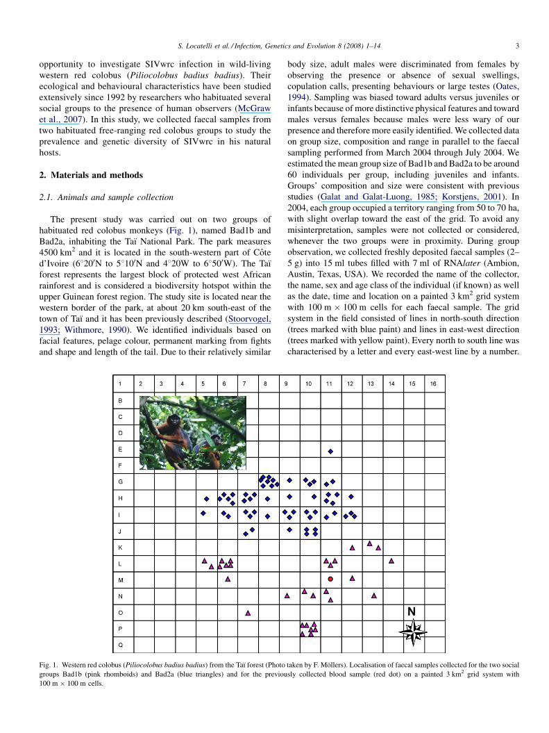
habituated red colobus monkeys (Fig. 1), named Bad1b and
Bad2a, inhabiting the Taı National Park. The park measures
4500 km2 and it is located in the south-western part of Cote
d’Ivoire (68200N to 58100N and 4820W to 68500W). The Taı
forest represents the largest block of protected west African
rainforest and is considered a biodiversity hotspot within the
upper Guinean forest region. The study site is located near the
western border of the park, at about 20 km south-east of the
town of Taı and it has been previously described (Stoorvogel,
1993; Withmore, 1990). We identified individuals based on
facial features, pelage colour, permanent marking from fights
and shape and length of the tail. Due to their relatively similar
Fig. 1. Western red colobus (Piliocolobus badius badius) from the Taı forest (Photo
groups Bad1b (pink rhomboids) and Bad2a (blue triangles) and for the previou
100 m � 100 m cells.
body size, adult males were discriminated from females by
observing the presence or absence of sexual swellings,
copulation calls, presenting behaviours or large testes (Oates,
1994). Sampling was biased toward adults versus juveniles or
infants because of more distinctive physical features and toward
males versus females because males were less wary of our
presence and therefore more easily identified. We collected data
on group size, composition and range in parallel to the faecal
sampling performed from March 2004 through July 2004. We
estimated the mean group size of Bad1b and Bad2a to be around
60 individuals per group, including juveniles and infants.
Groups’ composition and size were consistent with previous
studies (Galat and Galat-Luong, 1985; Korstjens, 2001). In
2004, each group occupied a territory ranging from 50 to 70 ha,
with slight overlap toward the east of the grid. To avoid any
misinterpretation, samples were not collected or considered,
whenever the two groups were in proximity. During group
observation, we collected freshly deposited faecal samples (2–
5 g) into 15 ml tubes filled with 7 ml of RNAlater (Ambion,
Austin, Texas, USA). We recorded the name of the collector,
the name, sex and age class of the individual (if known) as well
as the date, time and location on a painted 3 km2 grid system
with 100 m � 100 m cells for each faecal sample. The grid
system in the field consisted of lines in north-south direction
(trees marked with blue paint) and lines in east-west direction
(trees marked with yellow paint). Every north to south line was
characterised by a letter and every east-west line by a number.
taken by F. Mollers). Localisation of faecal samples collected for the two social
sly collected blood sample (red dot) on a painted 3 km2 grid system with
S. Locatelli et al. / Infection, Genetics and Evolution 8 (2008) 1–144
This way, every cell can be identified by a precise code (for
example L 8) (Fig. 1). Samples were stored at camp for 30–60
days at 4 8C and subsequently shipped to the laboratory in
Montpellier, France. Upon receipt samples were stored at
�80 8C.
2.2. Detection of HIV cross-reactive antibodies in red
colobus faecal samples
We recovered IgGs after dialyses of faecal samples by
applying the methods previously described for antibody
detection in faecal samples of gorillas and chimpanzees (Keele
et al., 2006; Van Heuverswyn et al., 2006). About 1.5 ml of
faecal sample was filtered, potential viruses were inactivated
and then the samples were dialysed to eliminate the presence of
salt contained in the RNAlater medium. We then tested the
resulting sample for HIV cross-reactive antibodies by the
INNO-LIA HIV confirmation test (Innogenetics, Ghent,
Belgium). This assay has proved to be useful in identifying
new SIVs, notably SIVwrc and SIVolc, in infected blood
samples (Courgnaud et al., 2003). This test includes HIV-1 and
HIV-2 recombinant proteins and synthetic peptides that are
coated as discrete lines on a nylon strip. The five HIV-1 antigens
are synthetic peptides for the exterior envelope glycoprotein
(sgp 120), as well as recombinant proteins for the transmem-
brane envelope glycoprotein (gp41), integrase (p31), core
(p24), and matrix (p17) proteins. HIV-1 group O envelope
peptides are included in the HIV-1 sgp120 band. The HIV-2
antigens include synthetic peptides for sgp105, as well as
recombinant gp36 protein. In addition to these HIV antigens,
each strip has control lines: one sample addition line (3+)
containing anti-human immunoglobulin (IgG) and two test
performance lines (1+ and +/�) containing human IgG. We
performed all assays according to manufacturer’s instructions,
with alkaline phosphatase-labelled goat anti-human IgG as the
secondary antibody. Samples should be scored as INNO-LIA
positive when they react with at least one HIVantigen and have
a band intensity equal to or greater than the assay cutoff (+/�)
lane; samples that react less strongly but still visibly with two or
more HIV antigens should be classified as indeterminate; and
samples reacting with no bands or only one band with less than
+/� intensity should be classified as negative.
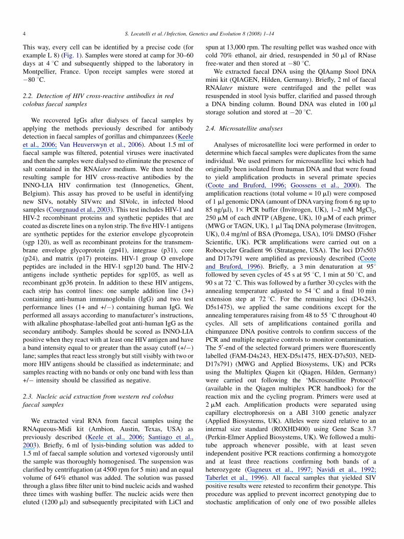
2.3. Nucleic acid extraction from western red colobus
faecal samples
We extracted viral RNA from faecal samples using the
RNAqueous-Midi kit (Ambion, Austin, Texas, USA) as
previously described (Keele et al., 2006; Santiago et al.,
2003). Briefly, 6 ml of lysis-binding solution was added to
1.5 ml of faecal sample solution and vortexed vigorously until
the sample was thoroughly homogenised. The suspension was
clarified by centrifugation (at 4500 rpm for 5 min) and an equal
volume of 64% ethanol was added. The solution was passed
through a glass fibre filter unit to bind nucleic acids and washed
three times with washing buffer. The nucleic acids were then
eluted (1200 ml) and subsequently precipitated with LiCl and
spun at 13,000 rpm. The resulting pellet was washed once with
cold 70% ethanol, air dried, resuspended in 50 ml of RNase
free-water and then stored at �80 8C.
We extracted faecal DNA using the QIAamp Stool DNA
mini kit (QIAGEN, Hilden, Germany). Briefly, 2 ml of faecal
RNAlater mixture were centrifuged and the pellet was
resuspended in stool lysis buffer, clarified and passed through
a DNA binding column. Bound DNA was eluted in 100 ml
storage solution and stored at �20 8C.
2.4. Microsatellite analyses
Analyses of microsatellite loci were performed in order to
determine which faecal samples were duplicates from the same
individual. We used primers for microsatellite loci which had
originally been isolated from human DNA and that were found
to yield amplification products in several primate species
(Coote and Bruford, 1996; Goossens et al., 2000). The
amplification reactions (total volume = 10 ml) were composed
of 1 ml genomic DNA (amount of DNA varying from 6 ng up to
85 ng/ml), 1� PCR buffer (Invitrogen, UK), 1–2 mM MgCl2,
250 mM of each dNTP (ABgene, UK), 10 mM of each primer
(MWG or TAGN, UK), 1 ml Taq DNA polymerase (Invitrogen,
UK), 0.4 mg/ml of BSA (Promega, USA), 10% DMSO (Fisher
Scientific, UK). PCR amplifications were carried out on a
Robocycler Gradient 96 (Stratagene, USA). The loci D7s503
and D17s791 were amplified as previously described (Coote
and Bruford, 1996). Briefly, a 3 min denaturation at 958followed by seven cycles of 45 s at 95 8C, 1 min at 50 8C, and
90 s at 72 8C. This was followed by a further 30 cycles with the
annealing temperature adjusted to 54 8C and a final 10 min
extension step at 72 8C. For the remaining loci (D4s243,
D5s1475), we applied the same conditions except for the
annealing temperatures raising from 48 to 55 8C throughout 40
cycles. All sets of amplifications contained gorilla and
chimpanzee DNA positive controls to confirm success of the
PCR and multiple negative controls to monitor contamination.
The 50-end of the selected forward primers were fluorescently
labelled (FAM-D4s243, HEX-D5s1475, HEX-D7s503, NED-
D17s791) (MWG and Applied Biosystems, UK) and PCRs
using the Multiplex Qiagen kit (Qiagen, Hilden, Germany)
were carried out following the ‘Microsatellite Protocol’
(available in the Qiagen multiplex PCR handbook) for the
reaction mix and the cycling program. Primers were used at
2 mM each. Amplification products were separated using
capillary electrophoresis on a ABI 3100 genetic analyzer
(Applied Biosystems, UK). Alleles were sized relative to an
internal size standard (ROXHD400) using Gene Scan 3.7
(Perkin-Elmer Applied Biosystems, UK). We followed a multi-
tube approach whenever possible, with at least seven
independent positive PCR reactions confirming a homozygote
and at least three reactions confirming both bands of a
heterozygote (Gagneux et al., 1997; Navidi et al., 1992;
Taberlet et al., 1996). All faecal samples that yielded SIV
positive results were retested to reconfirm their genotype. This
procedure was applied to prevent incorrect genotyping due to
stochastic amplification of only one of two possible alleles
S. Locatelli et al. / Infection, Genetics and Evolution 8 (2008) 1–14 5
(allelic dropout). We extracted DNA from the hair of two
researchers involved in this study and genotyped at each locus
to detect possible human contamination at both the sample
collection and PCR stage (Allen et al., 1998; Vigilant, 1999).
We also relied on a few high-quality DNA samples as a species
control mostly to verify whether the two sources had
comparable allele sizes.
2.5. Amplification of SIVwrc sequences from faecal RNA
We performed RT-PCR amplification of faecal virion using
two sets of primers specific for SIVwrc pol and env sequences.
We designed the pol primers on the basis of the sequence
alignment of five previously published sequences, four SIVwrc
from western red colobus and one SIVolc from an olive colobus
(Courgnaud et al., 2003). In turn, the env primers were designed
only on the basis of two SIVwrc sequences that have been fully
molecularly characterised (Liegeois et al., manuscript in
preparation). The regions amplified correspond to the 30 end
of the pol gene and the gp41 region of the env gene. cDNA was
synthetised using the wrcpolR1/wrcenvR1 primers followed by
nested PCRs using primers F1/R1 and F2/R2 as inner and outer
primers, respectively. The pol primers included wrcpolF1 (50-TAGGGACAGAAAGTATAGTAATHTGG-30) and wrcpolR1
(50-GCCATWGCYAATGCTGTTTC-30) as outer primers and
wrcpolF2 (50AGAGACAGTAAGG AAGGGAAAGCAGG-30)and wrcpolR2 (50-GTTCWATTCCTAACCACCAGCADA-30)as inner primers for the second PCR round. The env primers
included wrcenvF1 (50-TGGCAGTGGGACAAAAATATA-
AAC-30), wrcenvR1 (50-CTGGCAGTCCCTCTTCCAAGT-
TGT-30), wrcenvF2 (50-TGATAGGGMTGGCTCCTGGTG-
ATG-30) and wrcenvR2 (50-AATCCCCATTTYAACCAGT-
TCCA-30).We performed PCRs using a Long Expand PCR kit (Roche
Molecular Biochemicals, Manheim, Germany) under the
following conditions: a hot start at 94 8C for 2 min followed
by 10 cycles of denaturation at 92 8C for 20 s, annealing at
45 8C for 45 s, extension at 72 8C for 1.5 min, and 20 cycles
with the annealing temperature increased to 50 8C with
extension at 72 8C for 1.5 min. Amplification was completed
by a final extension at 72 8C for 5 min. PCR conditions for the
second PCR round were the same except that the extension time
during cycling was 45 s. RT-PCR products from pol (�650 bp)
and env (�570 bp) regions were purified (Q-Biogene, Illkirch,
France), and directly sequenced using the inner (F2/R2)
primers on an ABI 3130xl Genetic Analyser (Applied
Biosystem, Courtaboeuf, France). We then checked and
assembled the sequences using the software package Lasergene
(DNASTAR Inc. Madison, USA)).
2.6. Phylogenetic analysis
We aligned the SIVwrc sequences in pol and env with
previously published SIVwrc sequences and a set of reference
sequences from different SIV lineages. Sites that could not be
unambiguously aligned were excluded from the analyses.
Neither pol nor env sequences showed substitution saturation as
evidenced by the comparison of transitional versus transver-
sional distances between all possible sequence pairs. The model
of evolution for pol and env (general time reversible model of
evolution with a gamma distribution of rates) was selected
under the Akaike information criterion using Modeltest v3.7
(Posada and Crandall, 1998). We performed Bayesian
inferences using MrBayes v3.1.2 (Ronquist and Huelsenbeck,
2003). The tree-space was explored using four chains over
1,000,000 generations sampled every 100. Burn-in value was
fixed at 10% of the total generation number after empirical
determination of the convergence. We examined Bayesian
parameters with the Tracer program (http://evolve.zoo.ox.a-
c.uk/software.html?id=tracer) and all estimated sample sizes
were greater than 1621. Pol and env phylogenies of red colobus
monkeys’ SIV were rooted using sequences of corresponding
regions in SIVlho from l’Hoest monkeys (Cercopithecus
lhoesti) (AF188114), SIVsun from sun-tailed monkeys
(Cercopithecus solatus) (AF131870) and SIVmnd-1 from
mandrills (Mandrillus sphinx) (M27470) as well as SIVolc
from olive colobus (Procolobus verus) (accession and manu-
script in preparation).
2.7. Nucleotide sequence accession numbers
EMBL accession numbers (in parentheses) for the sequences
determined in this study are as follows for the pol gene: SIVwrc-
04CI-196 (AM743109), SIVwrc-04CI-116 (AM743110),
SIVwrc-04CI-299 (AM743111), SIVwrc-04CI-110
(AM743112), SIVwrc-04CI-115 (AM743113), SIVwrc-04CI-
112 (AM743114), SIVwrc-04CI-223 (AM743115), SIVwrc-
04CI-52 (AM743116), SIVwrc-04CI-237 (AM743117); and
for the env gene: SIVwrc-04CI-268 (AM743118), SIVwrc-
04CI-52 (AM743119), SIVwrc-04CI-116 (AM743120),
SIVwrc-04CI-110 (AM743121), SIVwrc-04CI-237
(AM743122), SIVwrc-04CI-175 (AM743123), SIVwrc-04CI-
296 (AM743124), SIVwrc-04CI-223 (AM743125), SIVwrc-
04CI-299 (AM743126), SIVwrc-04CI-280 (AM743127),
SIVwrc-04CI-32 (AM743128) , SIVwrc-04CI-196
(AM743129), SIVwrc-04CI-112 (AM743130).
3. Results
3.1. Samples and animal identification
We collected a total of 88 faecal samples between March and
July 2004, 30 from the Bad1b social group and 58 from Bad2a.
Although several individuals were visually recognized in both
groups, we used microsatellite analysis for definitive individual
identification. Unlike several other primate species in which
multiple microsatellite loci are well characterized, there are no
published microsatellite data for red colobus monkeys. Due to
the lack of DNA of suitable quality to construct microsatellites
libraries, cross-specific amplification was determined to be the
most suitable method to find polymorphic microsatellites loci
in these species. Nine human microsatellite loci that were found
to yield amplification products in other primate species were
screened for amplification in red colobus (Locatelli et al. in
S. Locatelli et al. / Infection, Genetics and Evolution 8 (2008) 1–146
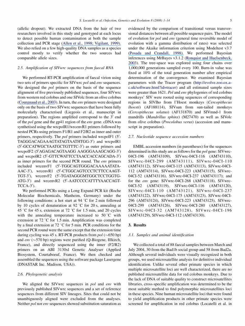
preparation). We subsequently chose four highly polymorphic
markers (D4s243, D5s1475, D7s503, D17s791). Amplicon
sizes for each of the loci are as follows: D4s243 (141–201 bp),
D5s1475 (110–142 bp), D7s503 (130–154 bp), D17s791 (141–
173 bp). The loci provided enough information to exclude
sample misidentification. The microsatellite analysis results are
summarised in Table 1. Twelve out of 88 samples yielded poor
or no microsatellite data and three samples were not genotyped
due to insufficient material. Among the remaining 73 samples,
54 were confirmed to originate from different individuals.
However, for one individual (sample 211) we were not able to
perform RNA extraction due to insufficient material available,
therefore we considered the number of individuals tested to be
53. Twelve samples from 12 different individuals were
confirmed to have been collected in duplicate and three
samples to have been collected three times from the same
individual at different times of the day or on different days. We
collected samples from 24 individuals from the Bad1b group,
which by visual inspection corresponded to 11 males, 8
females, and 5 individuals with undetermined gender.
Similarly, in the Bad2a group we collected samples from 21
males, 4 females and 5 individuals with undetermined gender
(30 individuals in total). Given the average size of each group to
Table 1
Genotype results at four loci and SIV results for all samples collected
aFaecal
spl. N8Social
group
bSex cLocus
D4s243
Locus
D5s1457
Locus
D7s503
Lo
D1
26 Bad1b nd ns ns ns ns
27 Bad1b nd 157157 ns 142146 16
28 Bad1b nd 145169 122126 146154 15
29 Bad1b nd ns 118126 138146 15
30 Bad1b nd 145157 ns ns 14
31 Bad1b nd 145157 122126 148148 14
32 Bad1b nd 161161 126130 134140 15
52 Bad1b M 145161 126134 142144 14
94 Bad2a M 161173 126130 140140 15
95 Bad2a M 161201 122126 140150 15
96 Bad2a M ns 122130 ns 15
97 Bad2a M 173201 ns 140152 15
102 Bad2a M ns ns ns ns
103 Bad2a M ns ns 146150 ns
107 Bad2a nd 153165 122130 144150 15
108 Bad2a F nd nd nd nd
108b Bad2a nd ns ns ns ns
109 Bad2a M 161165 122126 146150 15
110 Bad2a nd 153161 122126 150152 15
111 Bad2a M 161201 122126 140150 15
112 Bad2a nd 145173 ns 142150 14
115 Bad2a M 153161 122126 140150 15
116 Bad2a M 145165 126134 140150 15
118 Bad2a M 189197 ns 138152 ns
119 Bad2a M 161165 122126 146150 15
120 Bad2a M 201201 126130 140146 16
126 Bad2a M ns 122130 ns ns
127 Bad2a M 173185 126130 140148 15
128 Bad2a nd 145161 114118 140154 16
129 Bad2a M 161201 126134 ns 15
130 Bad2a nd 141161 ns 140150 15
167 Bad2a M 145165 126134 140150 15
be around 60 individuals, we estimated that between 68% and
83% of the adult population had been sampled.
3.2. Detection of SIVwrc antibodies
The first SIVwrc positive animals were identified by using the
Innolia HIV confirmation assay (Courgnaud et al., 2003). Blood
samples from red colobus showed clear IgG bands and cross-
reacted with HIVantigens for the core protein p24 or the matrix
protein p17. Consequently, and in analogy with studies on wild-
living chimpanzees and gorillas, we also tested 67 faecal samples
(24 samples corresponding to 22 individuals from group Bad1b
and 43 samples corresponding to 28 individuals from group
Bad2a) from 50 out of 54 individuals with the same assay, the
remaining four individuals were not tested because of insufficient
material availability. Only one sample revealed a clear presence
of IgG, but did not react with any of the HIV antigens. The
samples from the remaining 49 individuals had to be considered
‘not interpretable’, since the baseline results did not fit the
requirements, i.e., the anti-human IgG upper line was absent
(n = 22) or gave only a weaker (n = 27) signal than that of the
lower human IgG line on the strips, and no reactivity with any
HIV antigens was observed (Table 1).
cus
7s791
dAssigned
indiv. N8
eFaecal antibody
detection
fFaecal vRNA detection
pol env
bs nd nd
1165 1 nd � �3163 2 NI � �5161 3 NI � �1157 4 nd � �1157 4 NI � �7163 5 NI � +
7161 13 NI + +
3171 47 nd � �3161 25 nd � �1165 bs nd nd
3171 26 nd � �bs NI nd
27 NI � �1159 41 NI � �
nd nd nd
bs NI nd
1153 27 nd � �3165 42 NI + +
3161 25 NI � �9167 43 NI + +
3165 49 NI + �3161 28 nd + +
33 NI � �1153 27 NI � �1171 29 nd � �
bs NI nd
9171 50 nd � �5171 44 NI � �3161 25 NI � �3161 45 NI � �3161 28 NEG � �
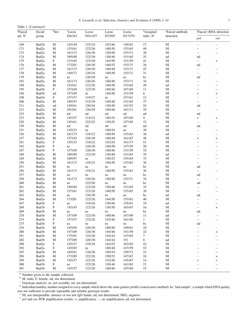
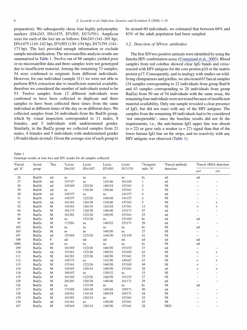
Table 1 (Continued )aFaecal
spl. N8Social
group
bSex cLocus
D4s243
Locus
D5s1457
Locus
D7s503
Locus
D17s791
dAssigned
indiv. N8
eFaecal antibody
detection
fFaecal vRNA detection
pol env
169 Bad1b M 145149 122134 142146 149163 17 NI � �171 Bad2a M 153161 122126 140150 153165 49 NI � �172 Bad2a M 161173 126130 130140 153171 30 NI � �174 Bad2a M 189189 122130 138140 151165 35 nd nd
175 Bad2a F 153165 122130 144150 151159 41 NI � +
176 Bad2a M 173201 126130 140152 153171 26 NI nd
177 Bad2a M 161173 126130 140148 153171 47 NI � �178 Bad2a M 149173 126134 140150 153171 31 NI � �179 Bad2a M ns 126130 ns ns bs NI nd
182 Bad2a M 161173 126126 140140 153171 34 NI � �190 Bad2a M 153161 122126 140150 153165 49 nd � �195 Bad1b F 157169 122130 140146 167169 11 NI � �196 Bad1b nd 157169 nt 144148 151159 6 NI + +
198 Bad1b F 157157 119127 ns 157161 12 NI � �206 Bad2a M 189197 122130 140148 151165 37 NI � �211 Bad2a nd 149161 126144 138140 141153 54 NI nd
213 Bad2a M 201201 126130 140146 161171 29 NI � �215 Bad2a M nd nd nd nd nd NI nd
223 Bad1b M 145157 114122 146154 167169 9 NI + +
229 Bad2a M 145161 122122 138152 157165 32 NI � �230 Bad2a M nd nd nd nd nd nd nd
231 Bad2a M 145153 ns 140154 ns 38 NI � �236 Bad2a M 161173 118122 140150 153163 36 nd � �237 Bad2a M 157165 126130 140148 161167 48 NI + +
241 Bad2a F 145153 118122 142154 161173 51 NI � �243 Bad1b F ns 126130 140150 147159 20 NI � �247 Bad1b F 157169 126130 140146 151159 18 NI � �248 Bad2a M 189189 122130 138140 151165 35 nd � �249 Bad2a M 189197 ns 138152 155165 33 NI � �250 Bad2a M 161173 118122 140150 153163 36 NI � �251 Bad2a M ns ns ns ns bs NI nd
256 Bad2a M 161173 118122 140150 153163 36 NI � �257 Bad2a M ns ns ns ns bs NI nd
258 Bad2a M 161173 126126 140140 153171 34 NI � �260 Bad2a M ns 122126 ns ns bs NI nd
261 Bad2a M 189189 122130 138140 151165 35 NI � �262 Bad2a M 157161 122126 140150 153165 39 NI � �263 Bad2a M ns 126130 ns ns bs nd nd
264 Bad2a M 173201 122126 144150 153161 40 NI � �267 Bad1b F ns 118126 138146 155161 19 nd � �268 Bad1b F 145145 122126 138150 163167 16 NI � +
269 Bad1b M ns ns ns ns bs NI nd
270 Bad1b M 157169 122130 140146 167169 11 nd � �274 Bad1b F 157157 122126 142146 161165 1 NI � �275 Bad1b F ns ns ns ns bs NI nd
276 Bad1b M 145169 126130 140150 149161 10 NI � �280 Bad1b M 157169 126130 144146 151159 22 NI � +
281 Bad1b M 175191 122130 144144 147165 7 NI � �282 Bad1b M 157169 126130 144144 151 8 nd � �290 Bad2a F 145157 110118 142152 163165 52 NI � �292 Bad2a F 145185 ns 140148 147159 53 NI � �293 Bad1b M 169181 126130 150154 159173 23 NI � �296 Bad1b M 173189 122126 150152 147167 24 NI � +
299 Bad1b M 145157 122126 142148 145167 14 NI + +
300 Bad1b F ns 122126 138146 161165 21 NI � �301 Bad1b M 145157 122130 140146 167169 15 NI � �a Number given to the sample collected.b M, male; F, female; nd, not determined.c Genotype analysis: ns, not scorable; nd, not determined.d Individual number, number assigned to every sample which shows the same genetic profile (conservative method). bs, ‘bad sample’, a sample which DNA quality
was not sufficient to provide repeatable and reliable genotype results.e NI, not interpretable: absence or too low IgG bands; nd, not determined; NEG, negative.f pol and env PCR amplification results: +, amplification; �, no amplification; nd, not determined.
S. Locatelli et al. / Infection, Genetics and Evolution 8 (2008) 1–14 7
S. Locatelli et al. / Infection, Genetics and Evolution 8 (2008) 1–148
3.3. Viral RNA detection of SIVwrc
We extracted RNA from 73 faecal samples belonging to 53
distinct individuals: 24 were members of the Bad1b group and
29 were members of the Bad2a group. RNAwas then subjected
to RT-PCR analysis using two sets of consensus primers which
amplified a �650-bp fragment in pol and a �570-bp fragment
in the env regions. This analysis identified 14 SIVwrc infected
individuals, eight in group Bad1b and six in group Bad2a using
one or both primer sets (Tables 1 and 2). Sequence analysis of
the respective amplification products confirmed infection, and
showed that the animals were infected with genetically
different SIVwrc strains. We were able to amplify the env
fragment in 13 samples and the pol fragment in 9 out of 14
samples, which corresponds in this sample set to a PCR
efficacy of 93% and 64%, respectively. Altogether, 26% of the
individuals tested were SIVwrc positive, 30% in Bad1b and
21% in Bad2a. RNA extraction was performed in duplicate,
when there was enough material available (N = 19) and for one
sample, RNA extraction was repeated three times. In 16
samples, PCRs from repeated RNA extractions gave con-
sistent results for both pol and env fragments (14 SIVwrc
negative and 2 positive). The remaining 4 samples, which
proved to be SIVwrc positive in the first RNA extraction, gave
negative PCR results in the second extraction performed four
months later, illustrating the frailty of RNA when faecal
samples undergo several thaw and freeze procedures. In
addition, samples belonging to the same individuals but
collected at different time points did not always provide
consistent results, i.e., among the 12 individuals which
samples were available in duplicate, 10 were initially negative
and reconfirmed to be negative in the corresponding additional
10 samples. For the two remaining individuals the first samples
were positive, but their duplicates were not. Three individuals
were represented by three samples each: two were confirmed
all three times to be negative but the third individual was
positive one out of three times.
Table 2
Characteristics of SIVwrc positive samples
Sample n8 Agea Sexb Social Group pol envc
32 A nd Bad1b � +
52 A M Bad1b + +
196 A nd Bad1b + +
223 A M Bad1b + +
268 A F Bad1b � +
280 A M Bad1b � +
296 A M Bad1b � +
299 A M Bad1b + +
110 A nd Bad2a + +
112 A nd Bad2a + +
115 A M Bad2a + �116 A M Bad2a + +
175 A F Bad2a � +
237 A M Bad2a + +
a Age group at the time of sample collection: A, adult.b M, male; F, female.c Fragments PCR amplified.
3.4. Genetic diversity of SIVwrc in the two Taı red colobus
communities
To determine the relationships between the newly identified
SIVwrc viruses to one another as well as to previously
characterised SIVwrc strains from the Taı forest, we
constructed phylogenetic trees from partial pol and env
sequences using a Bayesian inference approach. The previously
published SIVwrc sequences obtained from four red colobus
samples and the SIVolc sequence from one olive colobus
collected during the Ebola study conducted in Taı National Park
during the late nineties (Courgnaud et al., 2003) were also
included in this analysis. Samples SIVwrc-97CI-12, SIVwrc-
96CI-M001 and SIVwrc-97CI-14 were collected in a region
north of our study area, SIVwrc-00CI-04 was collected in the
vicinity of our study groups and SIVwrc-98CI-04 was collected
in the home range of our study groups (Fig. 2). Corresponding
SIV sequences from the SIVlho lineage (SIVmnd-1, SIVsun
and SIVlho) were included in the analysis to serve as outgroups.
Although we could not obtain pol and env fragments for every
sample, the phylogenies yielded some intriguing results (Fig. 3a
and b).
Previously as well as newly collected red colobus SIVs
clustered together and formed a ‘‘SIVwrc’’ monophyletic
group. Overall, within that group, we could observe two
main well supported clades, each consisting of virus strains
belonging to individuals coming from the same social group
in both pol and env phylogenetic trees. Thus, all strains but
one ( pol phylogeny) or two (env phylogeny) fell into either
Bad2a or Bad1b clusters. SIVwrc-04CI-116 did not group
with the other Bad2a samples in both pol and env
phylogenies. Its grouping within the Bad2a cluster in pol
phylogeny was not supported at a significant level (posterior
probability <91%; (Zander, 2004) whereas its basal position
within the Bad1b cluster in the env tree was. Strain SIVwrc-
04CI-116 thus appears to be more closely related to Bad1b
samples than to Bad2a. Similarly, SIVwrc-04CI-299 and
SIVwrc-04CI-296 (Bad1b) branched independently from
their group of origin (for SIVwrc-04CI-296, only the env
fragment could be amplified). As observed in the phyloge-
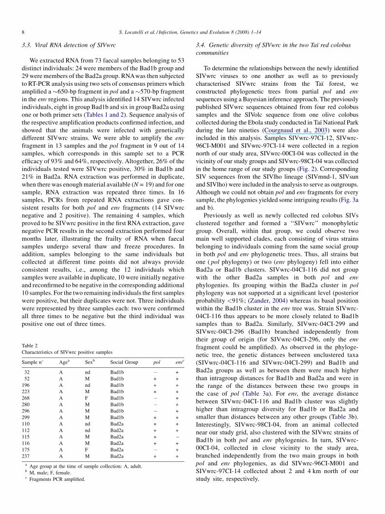
netic tree, the genetic distances between unclustered taxa
(SIVwrc-04CI-116 and SIVwrc-04CI-299) and Bad1b and
Bad2a groups as well as between them were much higher
than intragroup distances for Bad1b and Bad2a and were in
the range of the distances between these two groups in
the case of pol (Table 3a). For env, the average distance
between SIVwrc-04CI-116 and Bad1b cluster was slightly
higher than intragroup diversity for Bad1b or Bad2a and
smaller than distances between any other groups (Table 3b).
Interestingly, SIVwrc-98CI-04, from an animal collected
near our study grid, also clustered with the SIVwrc strains of
Bad1b in both pol and env phylogenies. In turn, SIVwrc-
00CI-04, collected in close vicinity to the study area,
branched independently from the two main groups in both
pol and env phylogenies, as did SIVwrc-96CI-M001 and
SIVwrc-97CI-14 collected about 2 and 4 km north of our
study site, respectively.
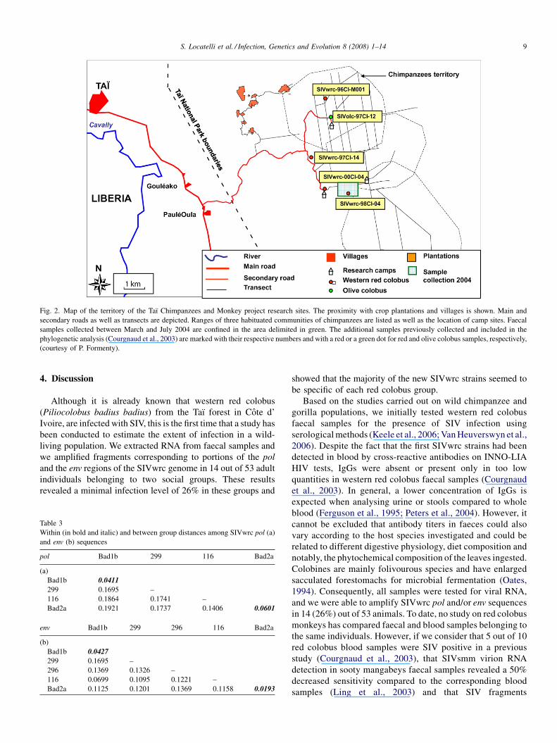
Fig. 2. Map of the territory of the Taı Chimpanzees and Monkey project research sites. The proximity with crop plantations and villages is shown. Main and
secondary roads as well as transects are depicted. Ranges of three habituated communities of chimpanzees are listed as well as the location of camp sites. Faecal
samples collected between March and July 2004 are confined in the area delimited in green. The additional samples previously collected and included in the
phylogenetic analysis (Courgnaud et al., 2003) are marked with their respective numbers and with a red or a green dot for red and olive colobus samples, respectively,
(courtesy of P. Formenty).
S. Locatelli et al. / Infection, Genetics and Evolution 8 (2008) 1–14 9
4. Discussion
Although it is already known that western red colobus
(Piliocolobus badius badius) from the Taı forest in Cote d’
Ivoire, are infected with SIV, this is the first time that a study has
been conducted to estimate the extent of infection in a wild-
living population. We extracted RNA from faecal samples and
we amplified fragments corresponding to portions of the pol
and the env regions of the SIVwrc genome in 14 out of 53 adult
individuals belonging to two social groups. These results
revealed a minimal infection level of 26% in these groups and
Table 3
Within (in bold and italic) and between group distances among SIVwrc pol (a)
and env (b) sequences
pol Bad1b 299 116 Bad2a
(a)
Bad1b 0.0411
299 0.1695 –
116 0.1864 0.1741 –
Bad2a 0.1921 0.1737 0.1406 0.0601
env Bad1b 299 296 116 Bad2a
(b)
Bad1b 0.0427
299 0.1695 –
296 0.1369 0.1326 –
116 0.0699 0.1095 0.1221 –
Bad2a 0.1125 0.1201 0.1369 0.1158 0.0193
showed that the majority of the new SIVwrc strains seemed to
be specific of each red colobus group.
Based on the studies carried out on wild chimpanzee and
gorilla populations, we initially tested western red colobus
faecal samples for the presence of SIV infection using
serological methods (Keele et al., 2006; Van Heuverswyn et al.,
2006). Despite the fact that the first SIVwrc strains had been
detected in blood by cross-reactive antibodies on INNO-LIA
HIV tests, IgGs were absent or present only in too low
quantities in western red colobus faecal samples (Courgnaud
et al., 2003). In general, a lower concentration of IgGs is
expected when analysing urine or stools compared to whole
blood (Ferguson et al., 1995; Peters et al., 2004). However, it
cannot be excluded that antibody titers in faeces could also
vary according to the host species investigated and could be
related to different digestive physiology, diet composition and
notably, the phytochemical composition of the leaves ingested.
Colobines are mainly folivourous species and have enlarged
sacculated forestomachs for microbial fermentation (Oates,
1994). Consequently, all samples were tested for viral RNA,
and we were able to amplify SIVwrc pol and/or env sequences
in 14 (26%) out of 53 animals. To date, no study on red colobus
monkeys has compared faecal and blood samples belonging to
the same individuals. However, if we consider that 5 out of 10
red colobus blood samples were SIV positive in a previous
study (Courgnaud et al., 2003), that SIVsmm virion RNA
detection in sooty mangabeys faecal samples revealed a 50%
decreased sensitivity compared to the corresponding blood
samples (Ling et al., 2003) and that SIV fragments
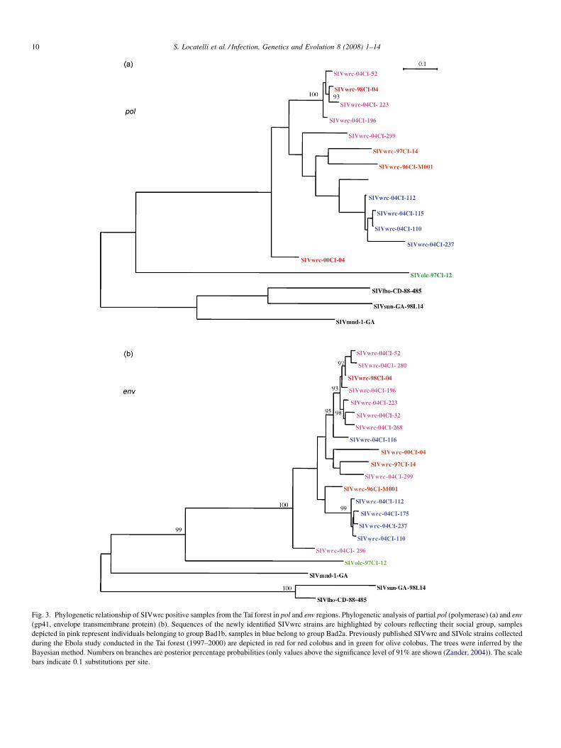
Fig. 3. Phylogenetic relationship of SIVwrc positive samples from the Taı forest in pol and env regions. Phylogenetic analysis of partial pol (polymerase) (a) and env
(gp41, envelope transmembrane protein) (b). Sequences of the newly identified SIVwrc strains are highlighted by colours reflecting their social group, samples
depicted in pink represent individuals belonging to group Bad1b, samples in blue belong to group Bad2a. Previously published SIVwrc and SIVolc strains collected
during the Ebola study conducted in the Tai forest (1997–2000) are depicted in red for red colobus and in green for olive colobus. The trees were inferred by the
Bayesian method. Numbers on branches are posterior percentage probabilities (only values above the significance level of 91% are shown (Zander, 2004)). The scale
bars indicate 0.1 substitutions per site.
S. Locatelli et al. / Infection, Genetics and Evolution 8 (2008) 1–1410
S. Locatelli et al. / Infection, Genetics and Evolution 8 (2008) 1–14 11
amplifications are sometimes difficult to obtain from RNA
extracted from faecal samples (93% and 64% in env and pol
fragments, respectively, in this study), we estimate that the
percentage of SIVinfection in the adult population living in Taı
National Park should be at least 26% and could possibly reach
50%. Therefore, we conclude that wild-living red colobus
monkeys represent a substantial reservoir of SIVwrc.
The relatively high SIV prevalence could partly be explained
by the promiscuous social system in which red colobus
monkeys live (Korstjens et al., 2002). With the exception of a
few cases, the grouping of the SIVwrc (Taı) viruses identified in
both pol and env phylogenies, paralleled the host distribution
into two social groups. This suggests that the infections within
either group are epidemiologically linked. Long-term beha-
vioural studies reported that red colobus monkeys live in large
multi-male groups, with adult males having a lifelong breeding
tenure in the group and male immigration being extremely rare.
Dispersal is strongly female-biased and occurs when females
reach the sub-adult stage of life, therefore before entering
sexual maturity. Accordingly, extra-group copulations have
rarely been observed (Korstjens et al., 2002). This could explain
the close relationship between the SIVwrc viruses harboured by
the two distinct social groups in our study. In order to make sure
that the faecal samples containing near identical SIVwrc
sequences were not inadvertently collected from the same
individual but were instead derived from horizontal or vertical
transmission, we relied on observational data, repeated
microsatellite testing and SIV PCR results to confirm the
different origins of the positive samples as well as the absence
of contaminations by closely related strains from individuals of
the same group.
However, routes of SIV transmission are extremely
challenging to determine, especially when observing an almost
exclusively arboreal primate species, in comparison to ground
dwelling primates which can be more easily monitored and
where juveniles and infants can be better identified and ascribed
to their respective mothers and their faecal samples more
confidently detected. Most of the newly identified SIVwrc
viruses fell into two clusters corresponding to the social group
of origin of their host. Furthermore, SIVwrc-98CI-04, collected
in 1998 within the range area of our two 2004 study groups,
groups with the Bad1b viruses, thus supporting the hypothesis
that those viruses parallel the host distribution. SIV infection
has also been studied in wild-living mangabeys from the Taı
forest, and nearly identical SIVsmm sequences have been
reported in both related and unrelated individuals belonging to
a single-community suggesting vertical as well as horizontal
routes of SIV transmission. However, highly divergent as well
as recombinant virus strains were also identified, supporting the
notion that the natural history of SIVsmm is one of a virus
spread both within and between wild communities (Santiago
et al., 2005). The fact that, unlike red colobus females, sooty
mangabey females have stable dominance ranks (Range and
Noe, 2002), that there is a statistically significant association
between high rank and faecal viral RNA positivity and that
males are the gender which disperses (possibly to more than
one group in a lifetime), could account for a higher virus
divergence compared to that observed in the two red colobus
communities under study. In our study, a few SIVwrc viruses
had intermediary positions between the two major groups in our
phylogenies. SIVwrc-04CI-296 and SIVwrc-04CI-299 branch
independently from their group of origin. Groups Bad1b and
Bad2a are of course surrounded by other non-habituated red
colobus groups and these individuals could have been infected
with SIV during agonistic behaviours with individuals from a
neighbouring group. An interesting case is that of SIVwrc-
04CI-116, which branches in the pol phylogeny apart from the
SIV strains from the social group to which it belongs, and is
closer to strains from the group Bad1b in the env phylogeny.
Several hypotheses can be formulated a posteriori regarding the
reasons for these results. Individual 116 could have been
infected by getting wounded in a fight with males from the
neighbouring group Bad1b or by a recently emigrated female,
who had already contracted the infection in her natal group by
vertical transmission. Our long-term field studies identified
SIVwrc-04CI-116 to belong to an old male named ‘Adam’. In
1992 the territory under study was inhabited by two groups of
about 90 individuals each, Bad1 and Bad2, which were
followed until 1999. Between 1994 and 1998, each group
started to split up into two sister fractions (van Oirschot, 1999),
that shared the same home range for 2–3 years during and after
the splitting process. The smaller sister fractions slowly moved
out of the main home range area and groups Bad 1b and Bad 2a
became the major targets of observation (Korstjens, 2001).
Adam was observed moving between the two groups during the
period when the fission of the larger Bad1 and Bad2 groups
occurred. At the beginning of the fission process males moved
between groups; the younger males were never observed in
their non-natal sister group, but the older males were often
going between groups until 1997–1998. Moreover, when well
established in the Bad2a group, Adam was observed to suffer
from impotency (A.H. Korstjens, personal communication) and
therefore would have been unable to pass on his SIV infection,
and indeed, among the individuals tested in the group Bad2a,
none were identified with a closely related SIVwrc strain. If
Adam was still a sexually active individual, we can speculate
that individuals expressing double infections or a recombinant
virus from the two neighbouring groups would have been
observed. Nevertheless, data from more individuals, in
particular females, would be required to confirm such a
hypothesis.
Chimpanzees of the Taı forest are strongly specialised in
hunting colobus monkeys. It has been shown that the annual
killing rate of colobus is 125 in 250 hunts, where 80% of kills
are represented by red colobus and 13% by black and white
colobus monkeys (Boesch, 1994). In light of the frequency of
Taı chimpanzees predation on red colobus and considering the
significant SIVwrc prevalence, we cannot exclude that this
subspecies of chimpanzees might have been as well infected by
SIV, which could have resulted in a low level of SIVwrc
infection or the emergence of a recombinant SIV strain so far
undetected by the current tools available. Moreover, the data
obtained on STLV type 1 viruses isolated from chimpanzees in
the Taı forest evokes the potential for interspecies transmission
S. Locatelli et al. / Infection, Genetics and Evolution 8 (2008) 1–1412
of retroviruses through predatory relationships. Notably, one of
the chimpanzee’s isolates clustered with the STLV-1 isolated
from the red colobus monkey 1497 (Leendertz et al., 2003;
Leendertz et al., 2004).
Chimpanzees do not represent the only threat for red colobus
monkeys. Red colobus monkeys are also one of the most
frequent catch of bushmeat hunters because they are found in
large numbers and are easily detected by their loud
vocalisations. Between April 1998 and March 1999, 2351
monkey carcasses were sold around the Taı National Park and
of those 28% were red colobus (Caspary et al., 2001). Today,
civil war and political instability also contributes to increasing
poaching pressure. Additionally, SIVwrc has been detected in
the same territory where sooty mangabeys have been found to
harbour SIVsmm variants which are the ancestors of different
groups of HIV-2, including those playing a major role in HIV-2
epidemic in West Africa. Given the significant frequency of
SIVwrc infection in the wild, the relative abundance of red
colobus, their cohabitation with other monkey species carrying
genetically different SIVs and given the relatively high
handling and consumption of their meat by chimpanzees and
by the human population, there are many prerequisites for
potential cross-species transmissions. In order better to
document SIVwrc and their evolution among the Colobinae
subfamily, full genomes need to be characterised among
individuals representative of the genus Piliocolobus in west,
central and east Africa and subsequently compared to SIVs
isolated from Procolobus verus as well as Colobus species
across Africa. A thorough study of the SIVwrc genome could
provide us with new tools for the search of SIV in Pan
troglodytes verus. In the Taı forest, screening should be
extended not only to chimpanzees but also to other sympatric
species, such as Colobus polykomos and especially to species
associating with red colobus, e.g. Cercopithecus diana.
Acknowledgements
We thank the ‘Ministere d’Enseignement Superieur et
Recherche Scientifique’, the ‘Ministere d’Agriculture et
Resources Animales’, the ‘Centre Suisse de Recherche
Scientifiques’ (CSRS) in Abidjan, the ‘P.A.C.P.N.T.’ and the
‘Centre de Recherche en Ecologie’ in Cote d’Ivoire for support
and permission to conduct research in the Taı National Park. We
thank the Taı Monkey Project and in particular Ferdinand Bele,
Cecile Benetton and Bertin Diero, for helping out in samples
collection; Christelle Butel, Fran Van Heuverswyn and Nicole
Vidal for technical advice in the laboratory in Montpellier;
Fabian Leendertz and Johannes Refisch, for involvement in the
early phase of the project, Marcel Tanner and Jakob Zinsstag
for critical support outside the field. The virology section of this
study was financially supported by the Institut de Recherche
pour le Developpement (IRD) and the Agence Nationale de
Recherches pour le SIDA (ANRS).
Sabrina Locatelli was supported by grants from the
Commission for Research Partnerships with Developing
Countries, Bern (KFPE) to conduct field research and by the
Messerli foundation, Zurich and the Guggenheim-Schnurr
Foundation, Basel, Switzerland to conduct laboratory analysis
on the monkey genetics.
References
Aghokeng, A.F., Bailes, E., Loul, S., Courgnaud, V., Mpoudi-Ngolle, E., Sharp,
P.M., Delaporte, E., Peeters, M., 2007. Full-length sequence analysis of
SIVmus in wild populations of mustached monkeys (Cercopithecus cephus)
from Cameroon provides evidence for two co-circulating SIVmus lineages.
Virology 360 (2), 407–418.
Allen, M., Engstrom, A.S., Meyers, S., Handt, O., Saldeen, T., von Haeseler, A.,
Paabo, S., Gyllensten, U., 1998. Mitochondrial DNA sequencing of shed
hairs and saliva on robbery caps: sensitivity and matching probabilities. J.
Forensic Sci. 43 (3), 453–464.
Bailes, E., Gao, F., Bibollet-Ruche, F., Courgnaud, V., Peeters, M., Marx, P.A.,
Hahn, B.H., Sharp, P.M., 2003. Hybrid origin of SIV in chimpanzees.
Science 300 (5626), 1713.
Beer, B.E., Bailes, E., Goeken, R., Dapolito, G., Coulibaly, C., Norley, S.G.,
Kurth, R., Gautier, J.P., Gautier-Hion, A., Vallet, D., Sharp, P.M., Hirsch,
V.M., 1999. Simian immunodeficiency virus (SIV) from sun-tailed monkeys
(Cercopithecus solatus): evidence for host-dependent evolution of SIV
within the C. lhoesti superspecies. J. Virol. 73 (9), 7734–7744.
Beer, B.E., Foley, B.T., Kuiken, C.L., Tooze, Z., Goeken, R.M., Brown, C.R.,
Hu, J., St Claire, M., Korber, B.T., Hirsch, V.M., 2001. Characterization of
novel simian immunodeficiency viruses from red-capped mangabeys from
Nigeria (SIVrcmNG409 and -NG411). J. Virol. 75 (24), 12014–12027.
Bibollet-Ruche, F., Galat-Luong, A., Cuny, G., Sarni-Manchado, P., Galat, G.,
Durand, J.P., Pourrut, X., Veas, F., 1996. Simian immunodeficiency virus
infection in a patas monkey (Erythrocebus patas): evidence for cross-
species transmission from African green monkeys (Cercopithecus aethiops
sabaeus) in the wild. J. Gen. Virol. 77 (Pt. 4), 773–781.
Bibollet-Ruche, F., Brengues, C., Galat-Luong, A., Galat, G., Pourrut, X., Vidal,
N., Veas, F., Durand, J.P., Cuny, G., 1997. Genetic diversity of simian
immunodeficiency viruses from West African green monkeys: evidence of
multiple genotypes within populations from the same geographical locale. J.
Virol. 71 (1), 307–313.
Bibollet-Ruche, F., Bailes, E., Gao, F., Pourrut, X., Barlow, K.L., Clewley, J.P.,
Mwenda, J.M., Langat, D.K., Chege, G.K., McClure, H.M., Mpoudi-Ngole,
E., Delaporte, E., Peeters, M., Shaw, G.M., Sharp, P.M., Hahn, B.H., 2004.
New simian immunodeficiency virus infecting De Brazza’s monkeys (Cer-
copithecus neglectus): evidence for a cercopithecus monkey virus clade. J.
Virol. 78 (14), 7748–7762.
Boesch, C., 1994. Chimpanzees-red Colobus monkeys: a predator-prey system.
Behaviour 47, 1135–1148.
Boesch, C., Boesch-Achermann, H., 1989. Hunting behavior of wild chimpan-
zees in the Tai National Park. Am. J. Phys. Anthropol. 78 (4), 547–573.
Caspary, H. U., Kone, I., Prouot, C., De Pauw, M., 2001. La chasse et la filiere
viande de brousse dans l’espace Taı, Cote-d’Ivoire. Tropenbos Cote-
d’Ivoire. Abidjan: Tropenbos Cote-d’Ivoire. In series 2, p. 170.
Chen, Z., Telfer, P., Reed, P., Zhang, L., Getti, A., Ho, D.D., Marx, P.A., 1995.
Isolation and characterization of the first simian immunodeficiency virus
from a feral sooty mangabey (Cercocebus atys) in West Africa. J. Med.
Primatol. 24 (3), 108–115.
Clavel, F., Brun-Vezinet, F., Guetard, D., Chamaret, S., Laurent, A., Rouzioux,
C., Rey, M., Katlama, C., Rey, F., Champelinaud, J.L., et al., 1986. LAV
type II: a second retrovirus associated with AIDS in West Africa. C R Acad.
Sci. III 302 (13), 485–488.
Coote, T., Bruford, M.W., 1996. Human microsatellites applicable for analysis
of genetic variation in apes and Old World monkeys. J. Hered. 87 (5), 406–
410.
Courgnaud, V., Pourrut, X., Bibollet-Ruche, F., Mpoudi-Ngole, E., Bourgeois,
A., Delaporte, E., Peeters, M., 2001. Characterization of a novel simian
immunodeficiency virus from guereza colobus monkeys (Colobus guereza)
in Cameroon: a new lineage in the nonhuman primate lentivirus family. J.
Virol. 75 (2), 857–866.
Courgnaud, V., Salemi, M., Pourrut, X., Mpoudi-Ngole, E., Abela, B., Auzel, P.,
Bibollet-Ruche, F., Hahn, B., Vandamme, A.M., Delaporte, E., Peeters, M.,
S. Locatelli et al. / Infection, Genetics and Evolution 8 (2008) 1–14 13
2002. Characterization of a novel simian immunodeficiency virus with a vpu
gene from greater spot-nosed monkeys (Cercopithecus nictitans) provides
new insights into simian/human immunodeficiency virus phylogeny. J.
Virol. 76 (16), 8298–8309.
Courgnaud, V., Formenty, P., Akoua-Koffi, C., Noe, R., Boesch, C., Delaporte,
E., Peeters, M., 2003. Partial molecular characterization of two simian
immunodeficiency viruses (SIV) from African colobids: SIVwrc from
western red colobus (Piliocolobus badius) and SIVolc from olive colobus
(Procolobus verus). J. Virol. 77 (1), 744–748.
Disotell, T.R., 1996. The phylogeny of Old World monkeys. Evol. Anthropol. 5,
18–24.
Ferguson, A., Humphreys, K.A., Croft, N.M., 1995. Technical report: results of
immunological tests on faecal extracts are likely to be misleading. Clin.
Exp. Immunol. 99, 70–75.
Gagneux, P., Boesch, C., Woodruff, D.S., 1997. Microsatellite scoring errors
associated with noninvasive genotyping based on nuclear DNA amplified
from shed hair. Mol. Ecol. 6 (9), 861–868.
Galat, G., Galat-Luong, A., 1985. La communaute de primates diurnes de la
foret de Taı, Cote d’Ivoire. Rev. Ecol. (Terre Vie) (40), 3–32.
Gao, F., Bailes, E., Robertson, D.L., Chen, Y., Rodenburg, C.M., Michael, S.F.,
Cummins, L.B., Arthur, L.O., Peeters, M., Shaw, G.M., Sharp, P.M., Hahn,
B.H., 1999. Origin of HIV-1 in the chimpanzee Pan troglodytes troglodytes.
Nature 397 (6718), 436–441.
Goossens, B., Chikhi, L., Utami, S., de Ruiter, J.R., Bruford, M.W., 2000. A
multisamples, multi-extracts approach for microsatellite analysis of faecal
samples in an arboreal ape. Conservation Genet. 1, 157–162.
Groves, C., 2001. Primate Taxonomy. Smithsonian Institution Press, Washing-
ton.
Hirsch, V.M., Campbell, B.J., Bailes, E., Goeken, R., Brown, C., Elkins, W.R.,
Axthelm, M., Murphey-Corb, M., Sharp, P.M., 1999. Characterization of a
novel simian immunodeficiency virus (SIV) from L’Hoest monkeys (Cer-
copithecus l’hoesti): implications for the origins of SIVmnd and other
primate lentiviruses. J. Virol. 73 (2), 1036–1045.
Hu, J., Switzer, W.M., Foley, B.T., Robertson, D.L., Goeken, R.M., Korber,
B.T., Hirsch, V.M., Beer, B.E., 2003. Characterization and comparison of
recombinant simian immunodeficiency virus from drill (Mandrillus leuco-
phaeus) and mandrill (Mandrillus sphinx) isolates. J. Virol. 77 (8), 4867–
4880.
Jin, M.J., Hui, H., Robertson, D.L., Muller, M.C., Barre-Sinoussi, F., Hirsch,
V.M., Allan, J.S., Shaw, G.M., Sharp, P.M., Hahn, B.H., 1994a. Mosaic
genome structure of simian immunodeficiency virus from west African
green monkeys. EMBO. J. 13 (12), 2935–2947.
Jin, M.J., Rogers, J., Phillips-Conroy, J.E., Allan, J.S., Desrosiers, R.C., Shaw,
G.M., Sharp, P.M., Hahn, B.H., 1994b. Infection of a yellow baboon with
simian immunodeficiency virus from African green monkeys: evidence for
cross-species transmission in the wild. J. Virol. 68 (12), 8454–8460.
Keele, B.F., Van Heuverswyn, F., Li, Y., Bailes, E., Takehisa, J., Santiago, M.L.,
Bibollet-Ruche, F., Chen, Y., Wain, L.V., Liegeois, F., Loul, S., Ngole, E.M.,
Bienvenue, Y., Delaporte, E., Brookfield, J.F., Sharp, P.M., Shaw, G.M.,
Peeters, M., Hahn, B.H., 2006. Chimpanzee reservoirs of pandemic and
nonpandemic HIV-1. Science 313 (5786), 523–526.
Korstjens, A.H., 2001. PhD Thesis. Utrecht University, Utrecht.
Korstjens, A.H., Sterck, E.H.M., Noe, R., 2002. How adaptive or phylogen-
etically inert is primate social behaviour? A Test with two sympatric
colobines. Behaviour 139, 203–225.
Leendertz, F.H., Boesch, C., Junglen, S., Pauli, G., Ellerbrok, H., 2003.
Characterization of a new simian T-lymphocyte virus type 1 (STLV-1) in
a wild living chimpanzee (Pan troglodytes verus) from Ivory Coast:
evidence of a new STLV-1 group? AIDS Res. Hum. Retroviruses 19
(3), 255–258.
Leendertz, F.H., Junglen, S., Boesch, C., Formenty, P., Couacy-Hymann, E.,
Courgnaud, V., Pauli, G., Ellerbrok, H., 2004. High variety of different
simian T-cell leukemia virus type 1 strains in chimpanzees (Pan troglo-
dytes verus) of the Tai National Park, Cote d’Ivoire. J. Virol. 78 (8),
4352–4356.
Ling, B., Santiago, M.L., Meleth, S., Gormus, B., McClure, H.M., Apetrei, C.,
Hahn, B.H., Marx, P.A., 2003. Noninvasive detection of new simian
immunodeficiency virus lineages in captive sooty mangabeys: ability to
amplify virion RNA from fecal samples correlates with viral load in plasma.
J. Virol. 77 (3), 2214–2226.
McGraw, W.S., Zuberbuhler, K., Noe, R., 2007. Monkeys of the Taı Forest. An
African Monkey Community. Cambridge University Press, Cambridge.
Mitani, J.C., Watts, D.P., 1999. Demographic influences on the hunting behavior
of chimpanzees. Am. J. Phys. Anthropol. 109 (4), 439–454.
Navidi, W., Arnheim, N., Waterman, M.S., 1992. A multiple-tubes approach for
accurate genotyping of very small DNA samples by using PCR: statistical
considerations. Am. J. Hum. Genet. 50, 347–359.
Oates, J.F., 1994. The natural history of African colobines. In: Davies, A.G.,
Oates, J.F. (Eds.), Colobine monkeys. Cambridge University Press,
Cambridge, pp. 75–128.
Peeters, M., Courgnaud, V., Abela, B., Auzel, P., Pourrut, X., Bibollet-Ruche, F.,
Loul, S., Liegeois, F., Butel, C., Koulagna, D., Mpoudi-Ngole, E., Shaw,
G.M., Hahn, B.H., Delaporte, E., 2002. Risk to human health from a
plethora of simian immunodeficiency viruses in primate bushmeat. Emerg.
Infect. Dis. 8 (5), 451–457.
Peters, I.R., Calvert, E.L., Hall, E.J., Day, M.J., 2004. Measurement of
immunoglobulin concentrations in the feces of healthy dogs. Clin. Diagn.
Lab. Immunol. 11 (5), 841–848.
Posada, D., Crandall, K.A., 1998. MODELTEST: testing the model of DNA
substitution. Bioinformatics 14 (9), 817–818.
Range, F., Noe, R., 2002. Familiarity and dominance relations among
female sooty mangabeys in the Tai National Park. Am. J. Primatol. 56
(3), 137–153.
Refisch, J., Kone, I., 2005. Impact of commercial hunting on monkey
populations in the Taı region, Cote d’Ivoire. Biotropica 37 (1),
136–144.
Ronquist, F., Huelsenbeck, J.P., 2003. MrBayes 3: Bayesian phylogenetic
inference under mixed models. Bioinformatics 19 (12), 1572–1574.
Salemi, M., De Oliveira, T., Courgnaud, V., Moulton, V., Holland, B., Cassol, S.,
Switzer, W.M., Vandamme, A.M., 2003. Mosaic genomes of the six major
primate lentivirus lineages revealed by phylogenetic analyses. J. Virol. 77
(13), 7202–7213.
Santiago, M.L., Rodenburg, C.M., Kamenya, S., Bibollet-Ruche, F., Gao, F.,
Bailes, E., Meleth, S., Soong, S.J., Kilby, J.M., Moldoveanu, Z., Fahey, B.,
Muller, M.N., Ayouba, A., Nerrienet, E., McClure, H.M., Heeney, J.L.,
Pusey, A.E., Collins, D.A., Boesch, C., Wrangham, R.W., Goodall, J.,
Sharp, P.M., Shaw, G.M., Hahn, B.H., 2002. SIVcpz in wild chimpanzees.
Science 295 (5554), 465.
Santiago, M.L., Lukasik, M., Kamenya, S., Li, Y., Bibollet-Ruche, F., Bailes, E.,
Muller, M.N., Emery, M., Goldenberg, D.A., Lwanga, J.S., Ayouba, A.,
Nerrienet, E., McClure, H.M., Heeney, J.L., Watts, D.P., Pusey, A.E.,
Collins, D.A., Wrangham, R.W., Goodall, J., Brookfield, J.F., Sharp,
P.M., Shaw, G.M., Hahn, B.H., 2003. Foci of endemic simian immunode-
ficiency virus infection in wild-living eastern chimpanzees (Pan troglodytes
schweinfurthii). J. Virol. 77 (13), 7545–7562.
Santiago, M.L., Range, F., Keele, B.F., Li, Y., Bailes, E., Bibollet-Ruche, F.,
Fruteau, C., Noe, R., Peeters, M., Brookfield, J.F., Shaw, G.M., Sharp, P.M.,
Hahn, B.H., 2005. Simian immunodeficiency virus infection in free-ranging
sooty mangabeys (Cercocebus atys atys) from the Tai forest, Cote d’Ivoire:
implications for the origin of epidemic human immunodeficiency virus type
2. J. Virol. 79 (19), 12515–12527.
Sharp, P.M., Shaw, G.M., Hahn, B.H., 2005. Simian immunodeficiency virus
infection of chimpanzees. J. Virol. 79 (7), 3891–3902.
Souquiere, S., Bibollet-Ruche, F., Robertson, D.L., Makuwa, M., Apetrei, C.,
Onanga, R., Kornfeld, C., Plantier, J.C., Gao, F., Abernethy, K., White, L.J.,
Karesh, W., Telfer, P., Wickings, E.J., Mauclere, P., Marx, P.A., Barre-
Sinoussi, F., Hahn, B.H., Muller-Trutwin, M.C., Simon, F., 2001. Wild
Mandrillus sphinx are carriers of two types of lentivirus. J. Virol. 75 (15),
7086–7096.
Stoorvogel, J.J., 1993. Gross inputs and outputs of nutrients in undisturbed
forest, Taı Area, Cote d’Ivoire. Veenman Drukkers, Wageningen.
Switzer, W.M., Parekh, B., Shanmugam, V., Bhullar, V., Phillips, S., Ely, J.J.,
Heneine, W., 2005. The epidemiology of simian immunodeficiency virus
infection in a large number of wild- and captive-born chimpanzees:
evidence for a recent introduction following chimpanzee divergence. AIDS
Res. Hum. Retroviruses 21 (5), 335–342.
S. Locatelli et al. / Infection, Genetics and Evolution 8 (2008) 1–1414
Taberlet, P., Griffin, S., Goossens, B., Questiau, S., Manceau, V., Escaravage,
N., Waits, L.P., Bouvet, J., 1996. Reliable genotyping of samples with very
low DNA quantities using PCR. Nucleic Acids Res. 24 (16), 3189–3194.
van de Woude, S., Apetrei, C., 2006. Going wild: lessons from naturally
occurring T-lymphotropic lentiviruses. Clin. Microbiol. Rev. 19 (4), 728–
762.
Van Heuverswyn, F., Li, Y., Neel, C., Bailes, E., Keele, B.F., Liu, W., Loul, S.,
Butel, C., Liegeois, F., Bienvenue, Y., Ngolle, E.M., Sharp, P.M., Shaw,
G.M., Delaporte, E., Hahn, B.H., Peeters, M., 2006. Human immunodefi-
ciency viruses: SIV infection in wild gorillas. Nature 444 (7116), 164.
van Oirschot, B., 1999. M.Sc. Thesis. Universiteit Leiden, Leiden.
van Rensburg, E.J., Engelbrecht, S., Mwenda, J., Laten, J.D., Robson, B.A.,
Stander, T., Chege, G.K., 1998. Simian immunodeficiency viruses (SIVs)
from eastern and southern Africa: detection of a SIVagm variant from a
chacma baboon. J. Gen. Virol. 79 (Pt. 7), 1809–1814.
Vigilant, L., 1999. An evaluation of techniques for the extraction and amplifica-
tion of DNA from naturally shed hairs. Biol. Chem. 380 (11), 1329–1331.
Withmore, T.C., 1990. An Introduction to Tropical Rain Forests. Clarendon
Press, Oxford.
Zander, R.H., 2004. Minimal values for reliability of bootstrap and jacknife
proportions, decay index, and bayesian posterior probabilities. PhyloInfor-
matics 2, 1–13.