Attenuation of pathogenic immune responses during infection with human and simian immunodeficiency...
15
Attenuation of Pathogenic Immune Responses during Infection with Human and Simian Immunodeficiency Virus (HIV/SIV) by the Tetracycline Derivative Minocycline Julia L. Drewes 1 , Gregory L. Szeto 1¤ , Elizabeth L. Engle 1 , Zhaohao Liao 1 , Gene M. Shearer 2 , M. Christine Zink 1 *, David R. Graham 1,3 * 1 Department of Molecular and Comparative Pathobiology, Johns Hopkins University School of Medicine, Baltimore, Maryland, United States of America, 2 Experimental Immunology Branch, Center for Cancer Research, National Cancer Institute, National Institutes of Health, Bethesda, Maryland, United States of America, 3 Johns Hopkins Bayview Proteomics Center, Department of Medicine, Division of Cardiology, Johns Hopkins School of Medicine Bayview Campus, Baltimore, Maryland, United States of America Abstract HIV immune pathogenesis is postulated to involve two major mechanisms: 1) chronic innate immune responses that drive T cell activation and apoptosis and 2) induction of immune regulators that suppress T cell function and proliferation. Both arms are elevated chronically in lymphoid tissues of non-natural hosts, which ultimately develop AIDS. However, these mechanisms are not elevated chronically in natural hosts of SIV infection that avert immune pathogenesis despite similarly high viral loads. In this study we investigated whether minocycline could modulate these pathogenic antiviral responses in non-natural hosts of HIV and SIV. We found that minocycline attenuated in vitro induction of type I interferon (IFN) and the IFN-stimulated genes indoleamine 2,3-dioxygenase (IDO1) and TNF-related apoptosis inducing ligand (TRAIL) in human plasmacytoid dendritic cells and PBMCs exposed to aldrithiol-2 inactivated HIV or infectious influenza virus. Activation- induced TRAIL and expression of cytotoxic T-lymphocyte antigen 4 (CTLA-4) in isolated CD4+ T cells were also reduced by minocycline. Translation of these in vitro findings to in vivo effects, however, were mixed as minocycline significantly reduced markers of activation and activation-induced cell death (CD25, Fas, caspase-3) but did not affect expression of IFNb or the IFN-stimulated genes IDO1, FasL, or Mx in the spleens of chronically SIV-infected pigtailed macaques. TRAIL expression, reflecting the mixed effects of minocycline on activation and type I IFN stimuli, was reduced by half, but this change was not significant. These results show that minocycline administered after infection may protect against aspects of activation-induced cell death during HIV/SIV immune disease, but that in vitro effects of minocycline on type I IFN responses are not recapitulated in a rapid progressor model in vivo. Citation: Drewes JL, Szeto GL, Engle EL, Liao Z, Shearer GM, et al. (2014) Attenuation of Pathogenic Immune Responses during Infection with Human and Simian Immunodeficiency Virus (HIV/SIV) by the Tetracycline Derivative Minocycline. PLoS ONE 9(4): e94375. doi:10.1371/journal.pone.0094375 Editor: Rafael Aldabe, Centro de Investigacio ´ n en Medicina Aplicada (CIMA), Spain Received November 19, 2013; Accepted March 15, 2014; Published April 14, 2014 This is an open-access article, free of all copyright, and may be freely reproduced, distributed, transmitted, modified, built upon, or otherwise used by anyone for any lawful purpose. The work is made available under the Creative Commons CC0 public domain dedication. Funding: This work was supported by the following NIH grants: R1MH087233, R1MH085554, and R1NS055648, as well as the National Center for Research Resources and the Office of Research Infrastructure Programs (ORIP) of the National Institutes of Health through Grant Number P40RR019995. The funders had no role in study design, data collection and analysis, decision to publish, or preparation of the manuscript. Competing Interests: The authors have declared that no competing interests exist. * E-mail: [email protected] (DRG); [email protected] (MCZ) ¤ Current address: Departments of Materials Science and Engineering, Biological Engineering, and the David H. Koch Institute for Integrative Cancer Research at the Massachusetts Institute of Technology, and the Ragon Institute of Massachusetts General Hospital, Massachusetts Institute of Technology, and Harvard, Cambridge, Massachusetts, United States of America Introduction In the absence of antiretroviral therapy, the majority of HIV- infected patients develop chronic immune activation, immuno- suppression and eventually AIDS in a disease process represen- tative of HIV/SIV infection in non-natural hosts (NNH). In contrast, most natural hosts (NH) of SIV such as African green monkeys, gorillas, and sooty mangabeys avert immune pathogen- esis despite high viral loads [1–4]. One hypothesis for the different outcomes in NH versus NNH focuses on host immune responses to the virus. Documented immune differences include differential induction of type I IFN and IFN-stimulated genes (ISGs) such as TNF-related apoptosis inducing ligand (TRAIL), as well as the immune regulators cytotoxic T-lymphocyte antigen-4 (CTLA-4) and indoleamine 2,3-dioxygenase (IDO), all of which contribute to T cell dysfunction and potentially the development of AIDS [5– 12]. IDO activity and TRAIL (both soluble and membrane- associated) remain elevated even in patients on suppressive cART [13–15]. In addition to their purported role in chronic HIV/SIV infection, many of these genes are also thought to be involved in the ‘‘cytokine storm’’ that contributes to pervasive inflammation, lymphocytopenia, and apoptosis during highly pathogenic influ- enza infections [16–19]. These immune responses therefore represent potential therapeutic targets for not only HIV infection but also other pathogenic viral infections such as influenza. PLOS ONE | www.plosone.org 1 April 2014 | Volume 9 | Issue 4 | e94375
Transcript of Attenuation of pathogenic immune responses during infection with human and simian immunodeficiency...

Attenuation of Pathogenic Immune Responses duringInfection with Human and Simian ImmunodeficiencyVirus (HIV/SIV) by the Tetracycline DerivativeMinocyclineJulia L. Drewes1, Gregory L. Szeto1¤, Elizabeth L. Engle1, Zhaohao Liao1, Gene M. Shearer2, M.
Christine Zink1*, David R. Graham1,3*
1 Department of Molecular and Comparative Pathobiology, Johns Hopkins University School of Medicine, Baltimore, Maryland, United States of America, 2 Experimental
Immunology Branch, Center for Cancer Research, National Cancer Institute, National Institutes of Health, Bethesda, Maryland, United States of America, 3 Johns Hopkins
Bayview Proteomics Center, Department of Medicine, Division of Cardiology, Johns Hopkins School of Medicine Bayview Campus, Baltimore, Maryland, United States of
America
Abstract
HIV immune pathogenesis is postulated to involve two major mechanisms: 1) chronic innate immune responses that drive Tcell activation and apoptosis and 2) induction of immune regulators that suppress T cell function and proliferation. Botharms are elevated chronically in lymphoid tissues of non-natural hosts, which ultimately develop AIDS. However, thesemechanisms are not elevated chronically in natural hosts of SIV infection that avert immune pathogenesis despite similarlyhigh viral loads. In this study we investigated whether minocycline could modulate these pathogenic antiviral responses innon-natural hosts of HIV and SIV. We found that minocycline attenuated in vitro induction of type I interferon (IFN) and theIFN-stimulated genes indoleamine 2,3-dioxygenase (IDO1) and TNF-related apoptosis inducing ligand (TRAIL) in humanplasmacytoid dendritic cells and PBMCs exposed to aldrithiol-2 inactivated HIV or infectious influenza virus. Activation-induced TRAIL and expression of cytotoxic T-lymphocyte antigen 4 (CTLA-4) in isolated CD4+ T cells were also reduced byminocycline. Translation of these in vitro findings to in vivo effects, however, were mixed as minocycline significantlyreduced markers of activation and activation-induced cell death (CD25, Fas, caspase-3) but did not affect expression of IFNbor the IFN-stimulated genes IDO1, FasL, or Mx in the spleens of chronically SIV-infected pigtailed macaques. TRAILexpression, reflecting the mixed effects of minocycline on activation and type I IFN stimuli, was reduced by half, but thischange was not significant. These results show that minocycline administered after infection may protect against aspects ofactivation-induced cell death during HIV/SIV immune disease, but that in vitro effects of minocycline on type I IFN responsesare not recapitulated in a rapid progressor model in vivo.
Citation: Drewes JL, Szeto GL, Engle EL, Liao Z, Shearer GM, et al. (2014) Attenuation of Pathogenic Immune Responses during Infection with Human and SimianImmunodeficiency Virus (HIV/SIV) by the Tetracycline Derivative Minocycline. PLoS ONE 9(4): e94375. doi:10.1371/journal.pone.0094375
Editor: Rafael Aldabe, Centro de Investigacion en Medicina Aplicada (CIMA), Spain
Received November 19, 2013; Accepted March 15, 2014; Published April 14, 2014
This is an open-access article, free of all copyright, and may be freely reproduced, distributed, transmitted, modified, built upon, or otherwise used by anyone forany lawful purpose. The work is made available under the Creative Commons CC0 public domain dedication.
Funding: This work was supported by the following NIH grants: R1MH087233, R1MH085554, and R1NS055648, as well as the National Center for ResearchResources and the Office of Research Infrastructure Programs (ORIP) of the National Institutes of Health through Grant Number P40RR019995. The funders had norole in study design, data collection and analysis, decision to publish, or preparation of the manuscript.
Competing Interests: The authors have declared that no competing interests exist.
* E-mail: [email protected] (DRG); [email protected] (MCZ)
¤ Current address: Departments of Materials Science and Engineering, Biological Engineering, and the David H. Koch Institute for Integrative Cancer Research atthe Massachusetts Institute of Technology, and the Ragon Institute of Massachusetts General Hospital, Massachusetts Institute of Technology, and Harvard,Cambridge, Massachusetts, United States of America
Introduction
In the absence of antiretroviral therapy, the majority of HIV-
infected patients develop chronic immune activation, immuno-
suppression and eventually AIDS in a disease process represen-
tative of HIV/SIV infection in non-natural hosts (NNH). In
contrast, most natural hosts (NH) of SIV such as African green
monkeys, gorillas, and sooty mangabeys avert immune pathogen-
esis despite high viral loads [1–4]. One hypothesis for the different
outcomes in NH versus NNH focuses on host immune responses to
the virus. Documented immune differences include differential
induction of type I IFN and IFN-stimulated genes (ISGs) such as
TNF-related apoptosis inducing ligand (TRAIL), as well as the
immune regulators cytotoxic T-lymphocyte antigen-4 (CTLA-4)
and indoleamine 2,3-dioxygenase (IDO), all of which contribute to
T cell dysfunction and potentially the development of AIDS [5–
12]. IDO activity and TRAIL (both soluble and membrane-
associated) remain elevated even in patients on suppressive cART
[13–15]. In addition to their purported role in chronic HIV/SIV
infection, many of these genes are also thought to be involved in
the ‘‘cytokine storm’’ that contributes to pervasive inflammation,
lymphocytopenia, and apoptosis during highly pathogenic influ-
enza infections [16–19]. These immune responses therefore
represent potential therapeutic targets for not only HIV infection
but also other pathogenic viral infections such as influenza.
PLOS ONE | www.plosone.org 1 April 2014 | Volume 9 | Issue 4 | e94375
In the first arm of HIV pathogenesis, hyperactive innate
immune responses that are driven by viral stimulation of Toll-like
receptors (TLRs) and other pattern recognition receptors lead to
chronic ISG expression, cellular activation, and apoptosis. Type I
IFN responses are critical in the control of many viral infections, as
ISGs can directly inhibit viral replication [20] and also stimulate
adaptive immune responses [21]. Although type I IFN can inhibit
HIV replication in vitro, administration of exogenous type I IFN
has only a moderate effect on HIV viral load and disease
progression in vivo [22–26]. Studies comparing NH and NNH
have corroborated the hypothesis that hyperactive innate immune
responses contribute to pathogenesis, as both NH and NNH
express robust levels of ISGs during acute infection in vivo [5–7],
but NH control this response within weeks while levels of IFNamRNA and ISGs remain elevated in NNH throughout chronic
infection [5–9]. These ISGs include TRAIL and Fas ligand (FasL),
which can induce apoptosis of uninfected CD4+ T cells expressing
the cognate receptors death receptor 5 (DR5) and Fas, and
programmed death ligand 1 (PDL1), which can induce apoptosis
or exhaustion of CD8+ T cells expressing the corresponding
receptor programmed cell death 1 (PD1) [27–30]. Thus, despite
the need for an early phase of IFN expression to stimulate adaptive
immune responses and assist in the control of virus replication,
chronic expression of type I IFN and ISGs likely does more harm
than good in HIV/SIV infection of NNH [31].
In the second arm of HIV pathogenesis, induction of the
immune regulators CTLA-4 and IDO suppresses the ability of T
cells to proliferate and respond to antigen, further compromising
immune responses already damaged by chronic ISGs. CTLA-4 is
expressed on activated CD4+ T cells and CD4+Foxp3+ regulatory
T cells (Tregs). These CTLA-4-expressing T cells convert DCs
into regulatory DCs by ligating with B7 molecules and inducing
IDO expression [32,33]. IDO can also be upregulated in
plasmacytoid DCs (pDCs) by stimulation with pro-inflammatory
cytokines such as IFNa, b, and c, TNFa, or directly by HIV [34].
Alternatively, long-term, chronic expression of IDO in regulatory
pDCs may be mediated by the anti-inflammatory cytokine TGFb[35,36]. IDO-expressing pDCs mediate suppressive effects on T
cells via 1) degradation/depletion of local tryptophan which
prevents T cell proliferation and 2) generation of kynurenine and
downstream metabolites that block T cell proliferation [37],
induce T cell apoptosis [38,39], and convert naıve CD4+ cells and
Th17 cells into Tregs [40,41]. Thus, CTLA-4 and IDO work in
tandem to increase the number and regulatory function of
suppressive DCs and Tregs in lymphoid tissues. Notably, NNH
with progressive infections have higher levels of CTLA-4, Foxp3,
and IDO mRNA in their lymphoid tissues compared to non-
progressors and uninfected individuals [10–12]. These data
suggest that CTLA-4+Foxp3+ Tregs accumulate in lymphoid
tissues during progressive infection of NNH, where they can
influence the function of DCs and other T cells, and thereby
represent an important therapeutic target in preventing immune
suppression in HIV [42].
Minocycline, a semi-synthetic tetracycline derivative, amelio-
rates the severity of a number of inflammatory diseases, including
rheumatoid arthritis [43] and animal models of multiple sclerosis
[44], amyotrophic lateral sclerosis [45], Huntington’s disease [46],
Parkinson’s disease [47], allergy/asthma [48,49], Japanese en-
cephalitis virus [50], and SIV-associated neurological disease
[51,52]. Minocycline’s effects have been primarily attributed to its
ability to decrease activation of a variety of immune cell types,
including monocytes/macrophages, microglia, and T cells [53–
56]. Additionally, many inflammatory cytokines can be downreg-
ulated by minocycline, including IL-6 [52,57–59], IL-1b [58],
TNFa [57–60], and IFNc [59–61]. However, minocycline’s effects
on type I IFN responses and IDO have not been explored.
Here we show that minocycline prevented the pathogenic
upregulation of type I IFN and IDO in pDCs following in vitro
exposure to aldrithiol-2-inactivated (AT-2) HIV and prevented
activation of CD4+ T cells after exposure to anti-CD3 and type I
IFN, culminating in decreased surface TRAIL expression on both
cell types. The AT-2 inactivated form of HIV was used in order to
dissociate innate immune signaling from factors associated with
productive infection because pDCs can be infected by HIV.
Reductions in type I IFN and TRAIL with minocycline treatment
were also observed in PBMCs exposed to either AT-2 HIV or
infectious influenza virus. A trend towards reduced TRAIL
expression was also seen in spleens from minocycline treated,
SIV-infected pigtailed macaques and was accompanied by
downregulation of the cellular activation marker CD25 and the
apoptosis-promoting genes Fas and caspase-3. Notably, these
changes in CD25, Fas, and caspase-3 upon minocycline treatment
were not significantly different from the changes seen in animals
treated with combination antiretroviral therapy (cART). However,
minocycline did not reduce expression of ISGs in the spleens of
SIV-infected macaques. Taken together, our data suggest that
minocycline is capable of attenuating aspects of pathogenic
immune responses during HIV/SIV infection and may be useful
as an adjunct immunotherapy against hyperactive immune
responses in diverse viral infections.
Materials and Methods
Ethics Statement on Human SubjectsFor in vitro experiments, blood was drawn from healthy human
donors following their informed oral consent in accordance with
the Johns Hopkins Medicine Institutional Review Board protocol
NA 00014329 or via anonymous donor leukopaks from the Johns
Hopkins University Outpatient Center or New York Blood Bank.
The Johns Hopkins Medicine Institutional Review Board
approved all experiments involving healthy human donors and
approved the use of informed oral consent in place of written
consent.
Ethics Statement on Animal SubjectsAll animal studies were approved by the Johns Hopkins Animal
Care and Use Committee (IACUC protocol #PR12M310); all
animals were humanely treated in accordance with federal
guidelines and institutional policies. All juvenile/adult pigtailed
macaques (Macaca nemestrina) used in this study were obtained from
nonhuman primate breeding facilities within the United States. A
total of thirty-four pigtailed macaques were studied. Median age
was 3 years (1.4–9.5 years); median weight was 3.5 kg (2.4–
14.5 kg). Of the 34 animals, 7 were used as uninfected controls
and 27 were inoculated intravenously with SIV/DeltaB670 and
SIV/17E-Fr, as previously described [62]. All animals were male
with the exception of 2 procedural controls that were female.
Eleven of the SIV-infected macaques received no drug treatment.
Eleven of the SIV-infected macaques received minocycline at a
dose of 4 mg/kg per day divided over two doses and administered
orally starting at 21 days p.i. as previously described [51]. No
adverse effects due to minocycline treatment were observed in
these animals. Five of the SIV-infected macaques received
combination antiretroviral therapy (cART) beginning at day 12
p.i. which suppressed plasma viremia to ,100 SIV RNA copy
eq./mL by day 70 p.i. [63]. The 4-drug cART regimen consisted
of 270 mg/kg of the protease inhibitor atazanavir (Bristol-Myers
Squibb), 10 mg/kg of the integrase inhibitor L-870812 (Merck),
Attenuation of Viral Pathogenesis by Minocycline
PLOS ONE | www.plosone.org 2 April 2014 | Volume 9 | Issue 4 | e94375
and 205 mg/kg of the protease inhibitor saquinavir (Roche)
administered orally twice/day; as well as 30 mg/kg of the
nucleotide reverse transcriptase inhibitor PMPA (Gilead) admin-
istered subcutaneously once daily.
Macaques were housed in Johns Hopkins University facilities
which are fully accredited by the association for the Assessment
and Accreditation of Laboratory Animal Care, International,
(AALAC). Macaques were housed in pairs or groups prior to
inoculation, then housed singly after inoculation to prevent
repeated infection events, in stainless steel cages providing at least
6 square feet of space per animal and with visual and auditory
contact with conspecifics. All housing met or exceeded guidelines
of the National Institutes of Health ‘‘Guide for the Care and Use
of Laboratory Animals’’ and the United States Department of
Agriculture Animal Welfare Act. Macaques were fed a balanced,
commercial macaque chow (Purina Mills, Gray Summit, MO,
USA) once a day supplemented with a variety of food enrichment.
Water was provided ad libitum and a light:dark cycle of 14:10 hours
was maintained. Macaques were provided with environmental
enrichment including manipulanda, novel foodstuffs and movies/
radio under the supervision of an enrichment specialist. Macaques
were observed 1–2 times daily by trained laboratory and/or
veterinary staff, and attitude, appetite and fecal consistency noted;
macaques were weighed and plasma and CSF viral loads
monitored at least once every two weeks. If abnormalities were
noted, veterinary personnel were consulted and treatment initiated
if necessary. Euthanasia was performed under veterinary super-
vision using an overdose of intravenous sodium pentobarbital
while under deep ketamine sedation (10 mg/kg intramuscular),
followed by perfusion with 1X PBS prior to tissue harvest. All
infected, untreated macaques were euthanized during late-stage
infection at approximately 84 days p.i., a timepoint at which the
majority of infected animals develop encephalitis [64], or prior to
this timepoint if macaques presented with clinical symptoms as
previously described [65]. Minocycline-treated animals were also
euthanized at approximately 84 days p.i., while cART-treated
animals were euthanized at approximately 180 days p.i., after
approximately 100 days of plasma virus suppression. ARRIVE
guidelines for animal research are reported in Checklist S1.
Minocycline Dosage In vitroA dose of 20 mM minocycline was chosen based on in vitro dose-
response testing on pDC viability as well as a review of
pharmacokinetic studies of patients given a commonly prescribed
dose of minocycline (oral 200 mg/day), where plasma concentra-
tions of minocycline reached 3.0–3.6 mg/L, roughly equivalent to
6–7 mM [66]. Since minocycline is absorbed into tissues at a high
rate (up to 10 times the amount in plasma) [67], an in vitro dose of
20 mM minocycline is a physiologically relevant concentration that
could be feasibly obtained in tissue microenvironments.
PBMC IsolationWhole blood from healthy human donors was obtained in
accordance with the Johns Hopkins IRB protocol NA 00014329 or
via leukopaks from the Johns Hopkins University Outpatient
Center or New York Blood Bank. PBMCs were isolated from
whole blood via Ficoll-Hypaque density gradient centrifugation
and red blood cell lysis (10 minute incubation at 37uC with 1X
solution of the following 10X buffer: 4.15 g NH4Cl, 0.5 g
KHCO3, 0.15 g EDTA dissolved in water and adjusted to pH
7.2–7.3 in a final volume of 500 mL). PBMCs were subsequently
washed with HBSS with EDTA before further use.
pDC Isolation and CulturepDCs were isolated from healthy human PBMCs via the
Diamond pDC Isolation Kit (Miltenyi). Purity was consistently .
95% as determined by BDCA-2 and CD123 flow cytometry
staining. pDCs were plated at a density of 50,000 cells/well in flat-
bottom 96-well plates in 150 mL RPMI 1640 supplemented with
10% FBS, 2 mM L-glutamine, 1 mM HEPES buffer, and 1%
Pen-Strep (Invitrogen; final concentration 100 U/mL penicillin
and 100 mg/mL streptomycin). Ten ng/mL IL-3 (R&D Systems)
was added to the media daily. pDCs were co-treated with 20 mM
minocycline hydrochloride (Sigma) dissolved in warm media and
300 ng/mL p24 equivalents of AT-2 HIV-1MN (X4-tropic, strain
P3935, AIDS and Cancer Vaccine Program, SAIC-Frederick, a
gift of Dr. Jeffery Lifson and Julian Bess). After 18 hours, cell-free
supernatants were collected and stored at 280uC and cells were
either analyzed by flow cytometry or lysed for RNA extraction.
CD4+ T Cell Isolation and CultureCD4+ T cells were isolated from PBMCs by the Dynabeads
FlowComp Human CD4 kit (Invitrogen). Purity was consistently
.95% of live cells as determined by CD3 and CD4 staining by
flow cytometry. CD4+ T cells were grown in 96-well plates or anti-
CD3 coated 96-well plates (BD) with or without 20 mM
minocycline hydrochloride (Sigma) in 100 ml RPMI 1640 supple-
mented with 10% FBS, 2 mM L-glutamine, 1 mM HEPES buffer,
and 2 mg/mL gentamicin. After 24 hours, a 50 mL aliquot
containing 60 mM minocycline was added to replenish minocy-
cline at a final concentration of 20 mM in the 150 mL well volume.
Some wells were also stimulated with 1,000 U/mL each of IFNa-
2a and IFNb-1a (PBL). All wells had a final volume of 150 mL and
were cultured for an additional 24 hours (total 48 hours) before
flow cytometry analysis.
PBMC Treatment with Virus and MinocyclinePBMCs were seeded in 24-well plates at a density of 4–5 million
cells/mL in RPMI 1640 supplemented with 10% FBS, 2 mM L-
glutamine, 1 mM HEPES buffer, and 10 U/mL recombinant IL-
2 (BD Biosciences). Cells were treated with varying doses of
minocycline hydrochloride (Sigma; 0, 20, or 40 mM) dissolved in
warm media for 2 hours before adding varying doses of either AT-
2 HIV-1MN or infectious influenza virus (A/Hong Kong/68-X-31
(H3N2)). AT-2 HIV concentrations are shown as ng/mL p24
equivalents while influenza concentrations are shown as ng/mL
nucleoprotein, as measured by the Influenza A Nucleoprotein
Antigen Capture ELISA (Virusys). After overnight culture, cells
were harvested for flow cytometry.
Flow CytometryCells were removed from plates with gentle pipetting, washed
with 1 x PBS, and stained for 1 hour at room temperature in the
dark. PBMCs were stained with TRAIL antibody and gated on
lymphocytes by forward and side scatter profiles. Isolated pDCs
were stained with Annexin V, TRAIL, and 7AAD antibodies to
discriminate viable cells. Isolated CD4+ T cells were stained with
CD3 and TRAIL antibodies and gated on the lymphocyte
morphology gate as determined by forward and side scatter. All
antibodies were from BD. Samples were washed with 1 x PBS to
remove excess antibodies and run on a BD FACSCalibur machine
(pDC, CD4+ T cell experiments) with appropriate isotype controls
or on a BD LSRFortessa (PBMC experiments). Data were
analyzed by FlowJo software.
Attenuation of Viral Pathogenesis by Minocycline
PLOS ONE | www.plosone.org 3 April 2014 | Volume 9 | Issue 4 | e94375
IFN ELISAsCell-free supernatants from pDCs were analyzed by ELISA for
IFNa (PBL Interferon Source #41100, which primarily recognizes
7/16 subtypes) and IFNb (Invitrogen, with an extended initial
incubation of 18 hours shaking at 4uC instead of 1 hour). Cell-free
supernatants from PBMCs were analyzed by ELISA for IFNa(PBL Interferon Source) and IFNb (PBL Interferon Source). All
plates were read on a microplate reader (BioRad) at the
recommended wavelengths for each respective ELISA assay.
RNA Extraction from pDCsRNA from human pDCs was isolated using the RNeasy Plus kit
(Qiagen) followed by treatment with RQ1 DNase (Promega) for
30 min at 37uC and inactivation with Stop Solution (Promega) for
10 min at 65uC.
RNA Extraction from Macaque SpleenRNA was harvested from 25 mg of snap-frozen macaque spleen
tissue by RNA STAT-60 (Isotex Diagnostics) extraction, DNase
treatment with RQ1 DNase (Promega) for 1 hour at 37uC, and
finally purification with miRVana Kit (Invitrogen). RNA concen-
tration was determined by NanoDrop (ThermoFisher) and
subsequently diluted to 40 ng/mL for Nanostring nCounter gene
expression analysis. All samples underwent RNA integrity testing,
and samples with RNA integrity scores less than 5 were excluded.
qRT-PCRRNA was reverse transcribed into cDNA with SuperScript III
(Invitrogen). PCR amplification of cDNA was performed in a
Chromo4 or CFX96 thermal cycler (both from BioRad) with
Multiplex NoRox PCR Mix (Qiagen) and gene-specific primers
and probes. Results were analyzed via the DDCt method [68] with
normalization to both 18S rRNA and negative controls. The
following primers and probes were used for the in vitro human
samples: IDO1 (59- TGCTTTGACGTCCTGCTGG, 59-
TTCCTGTGAGCTGGTGGCA, and 59- TXR - ATGCTGCT-
CAGTTCCTCCAGGACA - IAbRQs); Mx (59- AG-
GAGTTGCCCTTCCCAGA, 59- TCGTTCACAAGTTTCTT-
CAGTTTCA, and 59- Hex -
ACCAGCGGGCATCTGGTCACGA - BHQ1); 18S rRNA (59-
TAGAGGGACAAGTGGCGTTC, 59- CGCTGAGCCAGT-
CAGTGT, and 59- Cy5 - AGCAATAACAGGTCTGTGATG -
BHQ2 or Crimson); IFNb (59- GCCTCAAGGACAGGAT-
GAACTT, 59- GCGTCCTCCTTCTGGAACTG, and 59- Cy5
- CATCCCTGAGGAAATTAAGCAGCCGC - BHQ2). For
some in vitro human samples, negative controls for IFNb and
IDO1 mRNA were not detectable and were assigned a Ct of 45
(limit of detection). The above sequences for IFNb, Mx, and 18S
as well as identical thermal cycling conditions were used for
analysis of pigtailed macaque spleen RNA. Because IFNb is an
intronless gene, RNA samples underwent an additional DNase
step with Turbo DNase (Invitrogen Life Technologies) for 30 min
at 37uC to eliminate any residual genomic DNA immediately prior
to IFNb testing. A subset of the macaque samples were normalized
in a second reaction to the lymphocyte gene CD2 (Invitrogen Life
Technologies TaqMan Assay Rh02839718_m1), which was one of
the most stable transcripts in the Nanostring nCounter assay
across the samples used in this study. Normalization to CD2
yielded similar results to those from 18S normalization.
Nanostring nCounter Gene Expression AnalysisNanostring analysis allows for the highly sensitive and
reproducible detection of mRNA molecules without the need for
enzymatic amplification [69]. As part of a larger collaborative
project, a CodeSet for 116 macaque genes (including the 7
transcripts analyzed in this manuscript as well as 11 putative
housekeeping genes) for 96 samples were designed according to
NanoString specifications based on rhesus macaque (Macaca
mulata) and human sequences. Two hundred ng of each RNA
sample were hybridized for 16 hours with the CodeSet, and genes
were quantitated using the nCounter Digital Analyzer. Thirty-four
of the 96 samples were examined in this study; the rest were
excluded from the present analysis. Data were first normalized by
the geometric mean of six spiked positive controls to correct for
assay efficiency. Background was assessed as the average of eight
spiked negative controls plus two standard deviations following
positive control correction. Any genes with values below this
threshold of 30 counts were excluded from the present analysis,
with the exception of SIV/17E-Fr for which below threshold
values were expected and obtained for uninfected and cART-
treated animals. As no single housekeeping gene has been proven
to be consistently expressed across different cell types, cell
maturation states, tissue types, or disease models [70–74], we
used the Kruskal-Wallis non-parametric analysis of variance
(ANOVA) test on all positive control normalized genes to find
the three most stably expressed genes across our spleen samples
[70,75]. We found that the lymphocyte markers CD7, CD2, and
CD5 were the least varying genes between infection groups (p
values = 0.953, 0.937, 0.828, respectively). The geometric mean of
these three genes was used to normalize the positive control
corrected data [74]. Alternative normalization to lymphocyte
genes such as CD4 in HIV/SIV infected lymphoid tissue has been
used in other studies as well [10,11,76]. Similar results were
obtained when our data was normalized to the most stably
expressed housekeeping gene, ribosomal gene RPS9 (Fig. S1). All
other traditional housekeeping genes in our panel (ACTB, B2M,
GAPDH, HPRT1, HMBS, RPL13A, SDHA, TBP, UBC, and
YWHAZ) had Kruskal-Wallis p-values,0.100. Raw data as well as
positive and negative controls are shown in Table S1. Trends in
SIV viral load and IDO1 mRNA expression in spleen have been
independently verified for subsets of the animals by qRT-PCR and
kynurenine/tryptophan metabolite ratios, respectively (data not
shown).
StatisticsTRAIL and CTLA-4 in isolated pDCs and CD4+ T cells passed
the Kolmogorov-Smirnov test for Gaussian distribution and were
analyzed by paired t-test. ELISAs were analyzed by Wilcoxon
paired t-test for nonparametric data. qRT-PCR data on in vitro
experiments were analyzed by Wilcoxon paired t-test to compare
different treatments. PBMC data on dose-response curves of
minocycline with either AT-2 HIV or influenza were analyzed by
two-way repeated measures ANOVA. In vivo spleen RNA data
were analyzed by the Mann-Whitney test for nonparametric,
unpaired data. All tests were performed with Prism software
(version 5 or 6).
Results
Minocycline Attenuated IFN Responses in pDCsAs diagrammed in Fig. 1A, the elevated expression of type I
IFNs and downstream ISGs such as TRAIL, FasL, and PDL1 in
chronic infection of NNH compared to NH suggests that IFN
responses are a point of divergence between pathogenic and
nonpathogenic HIV/SIV infection and represent a potential
target for immunotherapy [5–9]. To test whether minocycline
could modulate these hyperactive innate immune responses, we
Attenuation of Viral Pathogenesis by Minocycline
PLOS ONE | www.plosone.org 4 April 2014 | Volume 9 | Issue 4 | e94375
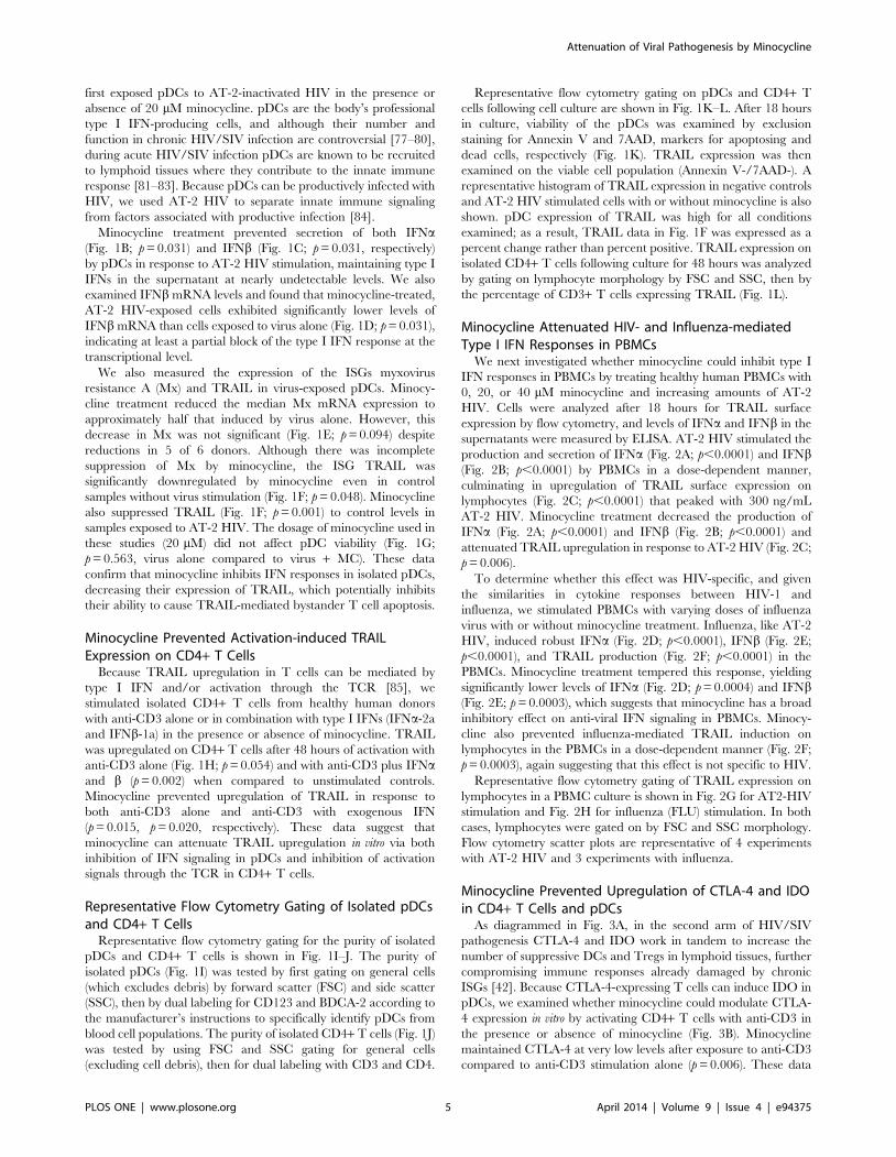
first exposed pDCs to AT-2-inactivated HIV in the presence or
absence of 20 mM minocycline. pDCs are the body’s professional
type I IFN-producing cells, and although their number and
function in chronic HIV/SIV infection are controversial [77–80],
during acute HIV/SIV infection pDCs are known to be recruited
to lymphoid tissues where they contribute to the innate immune
response [81–83]. Because pDCs can be productively infected with
HIV, we used AT-2 HIV to separate innate immune signaling
from factors associated with productive infection [84].
Minocycline treatment prevented secretion of both IFNa(Fig. 1B; p = 0.031) and IFNb (Fig. 1C; p = 0.031, respectively)
by pDCs in response to AT-2 HIV stimulation, maintaining type I
IFNs in the supernatant at nearly undetectable levels. We also
examined IFNb mRNA levels and found that minocycline-treated,
AT-2 HIV-exposed cells exhibited significantly lower levels of
IFNb mRNA than cells exposed to virus alone (Fig. 1D; p = 0.031),
indicating at least a partial block of the type I IFN response at the
transcriptional level.
We also measured the expression of the ISGs myxovirus
resistance A (Mx) and TRAIL in virus-exposed pDCs. Minocy-
cline treatment reduced the median Mx mRNA expression to
approximately half that induced by virus alone. However, this
decrease in Mx was not significant (Fig. 1E; p = 0.094) despite
reductions in 5 of 6 donors. Although there was incomplete
suppression of Mx by minocycline, the ISG TRAIL was
significantly downregulated by minocycline even in control
samples without virus stimulation (Fig. 1F; p = 0.048). Minocycline
also suppressed TRAIL (Fig. 1F; p = 0.001) to control levels in
samples exposed to AT-2 HIV. The dosage of minocycline used in
these studies (20 mM) did not affect pDC viability (Fig. 1G;
p = 0.563, virus alone compared to virus + MC). These data
confirm that minocycline inhibits IFN responses in isolated pDCs,
decreasing their expression of TRAIL, which potentially inhibits
their ability to cause TRAIL-mediated bystander T cell apoptosis.
Minocycline Prevented Activation-induced TRAILExpression on CD4+ T Cells
Because TRAIL upregulation in T cells can be mediated by
type I IFN and/or activation through the TCR [85], we
stimulated isolated CD4+ T cells from healthy human donors
with anti-CD3 alone or in combination with type I IFNs (IFNa-2a
and IFNb-1a) in the presence or absence of minocycline. TRAIL
was upregulated on CD4+ T cells after 48 hours of activation with
anti-CD3 alone (Fig. 1H; p = 0.054) and with anti-CD3 plus IFNaand b (p = 0.002) when compared to unstimulated controls.
Minocycline prevented upregulation of TRAIL in response to
both anti-CD3 alone and anti-CD3 with exogenous IFN
(p = 0.015, p = 0.020, respectively). These data suggest that
minocycline can attenuate TRAIL upregulation in vitro via both
inhibition of IFN signaling in pDCs and inhibition of activation
signals through the TCR in CD4+ T cells.
Representative Flow Cytometry Gating of Isolated pDCsand CD4+ T Cells
Representative flow cytometry gating for the purity of isolated
pDCs and CD4+ T cells is shown in Fig. 1I–J. The purity of
isolated pDCs (Fig. 1I) was tested by first gating on general cells
(which excludes debris) by forward scatter (FSC) and side scatter
(SSC), then by dual labeling for CD123 and BDCA-2 according to
the manufacturer’s instructions to specifically identify pDCs from
blood cell populations. The purity of isolated CD4+ T cells (Fig. 1J)
was tested by using FSC and SSC gating for general cells
(excluding cell debris), then for dual labeling with CD3 and CD4.
Representative flow cytometry gating on pDCs and CD4+ T
cells following cell culture are shown in Fig. 1K–L. After 18 hours
in culture, viability of the pDCs was examined by exclusion
staining for Annexin V and 7AAD, markers for apoptosing and
dead cells, respectively (Fig. 1K). TRAIL expression was then
examined on the viable cell population (Annexin V-/7AAD-). A
representative histogram of TRAIL expression in negative controls
and AT-2 HIV stimulated cells with or without minocycline is also
shown. pDC expression of TRAIL was high for all conditions
examined; as a result, TRAIL data in Fig. 1F was expressed as a
percent change rather than percent positive. TRAIL expression on
isolated CD4+ T cells following culture for 48 hours was analyzed
by gating on lymphocyte morphology by FSC and SSC, then by
the percentage of CD3+ T cells expressing TRAIL (Fig. 1L).
Minocycline Attenuated HIV- and Influenza-mediatedType I IFN Responses in PBMCs
We next investigated whether minocycline could inhibit type I
IFN responses in PBMCs by treating healthy human PBMCs with
0, 20, or 40 mM minocycline and increasing amounts of AT-2
HIV. Cells were analyzed after 18 hours for TRAIL surface
expression by flow cytometry, and levels of IFNa and IFNb in the
supernatants were measured by ELISA. AT-2 HIV stimulated the
production and secretion of IFNa (Fig. 2A; p,0.0001) and IFNb(Fig. 2B; p,0.0001) by PBMCs in a dose-dependent manner,
culminating in upregulation of TRAIL surface expression on
lymphocytes (Fig. 2C; p,0.0001) that peaked with 300 ng/mL
AT-2 HIV. Minocycline treatment decreased the production of
IFNa (Fig. 2A; p,0.0001) and IFNb (Fig. 2B; p,0.0001) and
attenuated TRAIL upregulation in response to AT-2 HIV (Fig. 2C;
p = 0.006).
To determine whether this effect was HIV-specific, and given
the similarities in cytokine responses between HIV-1 and
influenza, we stimulated PBMCs with varying doses of influenza
virus with or without minocycline treatment. Influenza, like AT-2
HIV, induced robust IFNa (Fig. 2D; p,0.0001), IFNb (Fig. 2E;
p,0.0001), and TRAIL production (Fig. 2F; p,0.0001) in the
PBMCs. Minocycline treatment tempered this response, yielding
significantly lower levels of IFNa (Fig. 2D; p = 0.0004) and IFNb(Fig. 2E; p = 0.0003), which suggests that minocycline has a broad
inhibitory effect on anti-viral IFN signaling in PBMCs. Minocy-
cline also prevented influenza-mediated TRAIL induction on
lymphocytes in the PBMCs in a dose-dependent manner (Fig. 2F;
p = 0.0003), again suggesting that this effect is not specific to HIV.
Representative flow cytometry gating of TRAIL expression on
lymphocytes in a PBMC culture is shown in Fig. 2G for AT2-HIV
stimulation and Fig. 2H for influenza (FLU) stimulation. In both
cases, lymphocytes were gated on by FSC and SSC morphology.
Flow cytometry scatter plots are representative of 4 experiments
with AT-2 HIV and 3 experiments with influenza.
Minocycline Prevented Upregulation of CTLA-4 and IDOin CD4+ T Cells and pDCs
As diagrammed in Fig. 3A, in the second arm of HIV/SIV
pathogenesis CTLA-4 and IDO work in tandem to increase the
number of suppressive DCs and Tregs in lymphoid tissues, further
compromising immune responses already damaged by chronic
ISGs [42]. Because CTLA-4-expressing T cells can induce IDO in
pDCs, we examined whether minocycline could modulate CTLA-
4 expression in vitro by activating CD4+ T cells with anti-CD3 in
the presence or absence of minocycline (Fig. 3B). Minocycline
maintained CTLA-4 at very low levels after exposure to anti-CD3
compared to anti-CD3 stimulation alone (p = 0.006). These data
Attenuation of Viral Pathogenesis by Minocycline
PLOS ONE | www.plosone.org 5 April 2014 | Volume 9 | Issue 4 | e94375
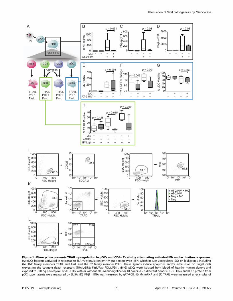
Figure 1. Minocycline prevents TRAIL upregulation in pDCs and CD4+ T cells by attenuating anti-viral IFN and activation responses.(A) pDCs become activated in response to TLR7/9 stimulation by HIV and secrete type I IFN, which in turn upregulates ISGs on leukocytes, includingthe TNF family members TRAIL and FasL and the B7 family member PDL1. These ligands induce apoptosis and/or exhaustion on target cellsexpressing the cognate death receptors (TRAIL/DR5; FasL/Fas; PDL1/PD1). (B–G) pDCs were isolated from blood of healthy human donors andexposed to 300 ng p24 eq./mL of AT-2 HIV with or without 20 mM minocycline for 18 hours (n = 6 different donors). (B, C) IFNa and IFNb protein frompDC supernatants were measured by ELISA. (D) IFNb mRNA was measured by qRT-PCR. (E) Mx mRNA and (F) TRAIL were measured as examples of
Attenuation of Viral Pathogenesis by Minocycline
PLOS ONE | www.plosone.org 6 April 2014 | Volume 9 | Issue 4 | e94375

are in agreement with previous studies that demonstrated that
minocycline attenuates CTLA-4 expression [86] and other
markers of CD4+ T cell activation [53,54]. However, IDO can
also be induced in pDCs directly by virus or by cytokines such as
type I IFN [34,87]. Therefore, we stimulated pDCs with AT-2
HIV in the presence or absence of minocycline and analyzed
IDO1 mRNA expression. pDCs that were treated with virus and
minocycline expressed significantly less IDO1 mRNA than with
virus alone (p = 0.031). IDO2, a variant of IDO1 that also
catabolizes tryptophan, was not detected in any of the samples
(data not shown).
Representative flow cytometry gating of CTLA-4 expression on
isolated CD4+ T cells for all four treatment conditions is shown in
Fig. 3D. Cells were gated on by FSC and SSC morphology.
Minocycline Downregulated Activation-induced GenesFas, CD25, and Caspase-3 but not IFN-stimulated Genesin Spleens of Chronically SIV-infected Pigtailed Macaques
To determine whether our in vitro findings could be recapitu-
lated in vivo, we examined whether minocycline had an effect on
IFN, IDO, and apoptosis-inducing factors TRAIL, FasL, and Fas
in archived spleen samples from SIV-infected pigtailed macaques
[62]. RNA was harvested from spleens from uninfected macaques,
SIV-infected macaques in late stage infection, or SIV-infected
macaques treated with either minocycline or cART and analyzed
for viral RNA and various markers of immune activation.
We first measured the viral loads in the spleen because
minocycline has previously been shown to reduce viral replication
both in vitro and in vivo [51,52] and can also inhibit HIV
reactivation from latency [88]. Minocycline reduced median viral
loads in the spleens of SIV-infected animals by 50%, but this
decrease was not significant (Fig. 4A; p = 0.240). Despite high
levels of virus in the chronically SIV-infected animals, IFNbexpression in spleen was not significantly elevated above uninfect-
ed controls during chronic infection (Fig. 4B; p = 0.157, Uninf vs.
SIV). This may have been due to the wide spread in IFNbexpression levels, with one control animal in particular having very
high levels. Though IFNb was not significantly increased during
chronic infection, we also measured the ISG Mx as a surrogate
marker of total type I IFN responses because even small amounts
of type I IFN can induce robust downstream responses. Mx was
significantly elevated in chronically SIV infected animals com-
pared to uninfected controls (Fig. 4B; p,0.0001). Minocycline
treatment had no effect on IFNb (Fig. 4B; p = 0.638) or Mx
(Fig. 4C; p.0.999) transcriptional expression compared to SIV
alone. Similarly, minocycline did not significantly reduce expres-
sion of IDO1 (Fig. 4D; p = 0.552) or the death ligands TRAIL
(Fig. 4E; p = 0.170) and FasL (Fig. 4F; p = 0.881) in vivo.
In contrast to the lack of effect on the death ligands TRAIL and
FasL, minocycline potently downregulated expression of the death
receptor Fas (Fig. 4G; p = 0.003). Minocycline also significantly
downregulated expression of the activation marker CD25 (Fig. 4H;
p = 0.040) in the SIV-infected spleens. Finally, we examined
expression of caspase-3, an important molecule in both extrinsic
and intrinsic apoptosis signaling pathways, because several studies
in a variety of disease models have demonstrated that minocycline
alters caspase-3 transcription and activation [46,89–91]. In our
SIV-infected macaques minocycline potently reduced caspase-3
mRNA levels (Fig. 4I; p = 0.003).
Comparison of Minocycline with CombinationAntiretroviral Therapy (cART)
Given the significant effects of minocycline on various markers
of activation-induced cell death in the spleens of SIV-infected
macaques, we next determined how minocycline treatment
compared to cART, the standard of care for HIV-infected
individuals. cART treatment reduced the median expression levels
of all genes back to uninfected levels, with the exception of IFNband FasL which were unchanged with either minocycline or cART
treatment compared to spleens from SIV infected animals. As
expected, cART treatment was significantly more effective than
minocycline at reducing SIV viral load and the ISGs Mx and
TRAIL in the macaque spleens (Fig. 4; MC vs. cART, p = 0.0005
for SIV, p = 0.0005 for Mx, p = 0.027 for TRAIL). Although
cART medians were consistently lower than minocycline’s across
all inflammatory genes, cART was not significantly more effective
than minocycline at reducing expression of IDO1 (Fig. 4D;
p = 0.171), FasL (Fig. 4F; p = 0.908), Fas (Fig. 4G; p = 0.208),
CD25 (Fig. 4H; p = 0.110), or caspase-3 (Fig. 4I; p = 0.411).
Discussion
Differences between NH and NNH suggest that chronic
induction of the IFN and IDO pathways are at the root of
immune pathogenesis seen in HIV infection [42]. In this study,
minocycline had potent activity against IFN, IDO, and activation
pathways in an acute model of HIV infection in vitro, which
culminated in reductions in TRAIL expression on both pDCs and
CD4+ T cells. We also observed reductions in IFN responses and
TRAIL expression in minocycline-treated PBMCs exposed to
either infectious influenza virus or AT-2 HIV. However, in spleens
from chronically SIV-infected pigtailed macaques, minocycline
did not affect IFNb, Mx, IDO, TRAIL, or FasL but did
significantly reduce activation-induced genes Fas, CD25, and
caspase-3. Overall, these data suggest that minocycline attenuates
markers of activation-induced cell death, a major component of
HIV pathogenesis, but that testing for inhibition of type I IFN
responses is more complex than can be discerned from our in vitro
model.
In our in vitro model of acute infection, minocycline blocked
both IFN- and activation-induced TRAIL, as seen by inhibition of
AT-2 HIV-triggered IFNa and IFNb responses in pDCs as well as
prevention of TRAIL upregulation on CD4+ T cells activated
with aCD3 antibody. The suppression of pDC IFN production
could also be linked to modulation of pDC activation by
minocycline; this will need to be confirmed in future studies.
Importantly, minocycline also prevented TRAIL upregulation on
CD4+ T cells stimulated with both aCD3 antibody and exogenous
IFNa and IFNb. These data showed that minocycline suppressed
ISGs by qRT-PCR and flow cytometry, respectively. (G) Viability of pDCs was determined by Annexin V/7AAD staining. (H) CD4+ T cells were isolatedfrom blood of healthy human donors and activated with anti-CD3 with or without 20 mM minocycline (n = 6 different donors). After 24 hours,minocycline was replenished and some wells were additionally stimulated with IFNa and IFNb. TRAIL was measured by flow cytometry after anadditional 24 hours. (I) Representative flow cytometry gating of pDC purity by BDCA2+/CD123+ double staining immediately following isolation. (J)Representative gating of CD4+ T cell purity by CD4+/CD3+ double staining immediately following isolation. (K) Representative gating of pDC viability(Annexin V-/7AAD-) and TRAIL expression after 18 hours of stimulation in culture with virus and/or minocycline. (L) Representative gating of TRAILexpression in CD4+ T cells following 48 hours in culture. Parametric data were analyzed by paired t-test (TRAIL flow cytometry), and nonparametricdata were analyzed by Wilcoxon signed-rank test (IFN ELISAs and IFNb, Mx qRT-PCR).doi:10.1371/journal.pone.0094375.g001
Attenuation of Viral Pathogenesis by Minocycline
PLOS ONE | www.plosone.org 7 April 2014 | Volume 9 | Issue 4 | e94375
Figure 2. Minocycline attenuates type I IFN production and TRAIL expression in lymphocytes. PBMCs were isolated from the blood ofhealthy human donors, pretreated for two hours in vitro with 0, 20, or 40 mM minocycline, and exposed to increasing amounts of either AT-2inactivated HIV (n = 4 different donors) or infectious influenza virus (n = 3 different donors). After overnight culture, supernatants were analyzed for
Attenuation of Viral Pathogenesis by Minocycline
PLOS ONE | www.plosone.org 8 April 2014 | Volume 9 | Issue 4 | e94375
TRAIL even in a complex immune environment consisting of
both TCR activation and IFN signals and led us to examine
TRAIL in the chronically infected spleens.
Minocycline reduced TRAIL expression in the SIV-infected
spleens by 50% but this change was not significant. This partial
inhibition of TRAIL is consistent with our data that showed
minocycline blocked only one of the two modes of TRAIL
induction in vivo; i.e., minocycline reduced cellular activation (Fas
and CD25) but did not reduce type I IFN responses (neither IFNbnor the ISG Mx). Mechanistically, TRAIL upregulation on T cells
through TCR activation/signaling is dependent on protein kinase
C (PKC) translocation and Ca2+ mobilization [92]. Minocycline
has previously been shown to inhibit these two mechanisms in
other cell types and in isolated mitochondria [93–95], suggesting
that the effects we observed on activation-induced TRAIL in T
cells might be due to inhibition of PKC and/or Ca2+ mobilization
by minocycline.
Despite not finding evidence for blockade of type I IFN
signaling in the SIV-infected spleens, our experiments with
influenza virus showed that minocycline’s inhibitory effects on
type I IFN and TRAIL in in vitro PBMCs were not specific for
inactivated viruses nor were they specific for HIV. These results
are compelling because a cytokine storm is widely associated with
influenza pathogenesis. Additionally, two groups recently reported
that pDCs may participate in the cytokine storm observed in
pathogenic influenza infections [16,96], which posits a need for
therapeutics that could modulate pDC cytokine responses in
pathogenic influenza as well as HIV infection. Future studies must
address whether minocycline’s inhibition of influenza-induced IFN
responses in vitro can be recapitulated in an in vivo model or suffer
from the same pitfalls as translation of HIV-induced IFN responses
from in vitro to in vivo models.
In contrast to the lack of an effect on type I IFN responses in the
SIV-infected spleens, minocycline treatment showed a trend
towards reduced SIV RNA levels in spleen compared to infected,
untreated animals, reducing viral loads by approximately 50%.
Previous studies from our group and others have demonstrated
that minocycline reduces viral replication in macaques and in a
humanized mouse model of HIV infection, as well as in vitro in
human and macaque primary macrophages and lymphocytes
[51,52,86,88]. However, in a recent pilot study of seven HIV-
infected individuals not on anti-retroviral therapy, viral loads in
both CSF and plasma were unchanged with an eight-week course
of minocycline treatment [97]. Thus our findings of a trend
towards reduction of viral loads in the spleens of SIV-infected
animals may reflect a broader picture that is emerging of a more
marginal effect of minocycline on viral loads during chronic
infection, particularly in patients, than previously proposed.
Given the trend towards reduced splenic viral loads in vivo and
minocycline’s robust inhibition of type I IFN responses in our
in vitro model of acute infection, our finding that minocycline had
no effect on IFNb, Mx, or IDO transcript levels in the chronically
infected spleen samples was surprising. IDO is induced by several
other cytokines, including IFNc and TFNa, but these have also
been shown to be reduced by minocycline in in vitro studies
[57,58,61,98] as well as in a mouse model of vaginal mucosal
inflammation [60]. However, in a pilot study in HIV-infected
patients, minocycline did not affect plasma or CSF levels of
neopterin, a marker of macrophage/microglial activation that is
also induced by IFNc [97]. It is possible that a higher dose of
minocycline would be required to recapitulate in vitro findings for
IFN and ISGs such as IDO in vivo.
Alternatively, it was recently reported that HIV-infected cells
may serve as a more potent stimulator of type I IFN in PBMCs
than cell-free virions [99]. It is conceivable that minocycline may
not have as strong of an effect on IFN signaling mediated through
contact with infected cells as opposed to free virus. In a pilot
experiment with 3 donors using HIV-infected peripheral blood
leukocytes co-cultured with autologous target PBMCs, minocy-
cline inhibited TRAIL induction in pDCs but not in T cells as
examined by flow cytometry (unpublished data). In contrast,
minocycline reduced TRAIL induction in both pDCs and T cells
from the same 3 donors after exposure to the TLR3 agonist poly
I:C, suggesting that minocycline’s efficacy against type I IFN
signaling may be dependent on both the cell type and source of
immune stimuli.
Additionally, there may be differences in the mechanisms for
IFN and ISG production during acute versus chronic SIV/HIV
infection. For example, while pDCs are known to be major
contributors to IFN responses in acute infection [81–83], their
number and function in chronic infection are controversial, with
some studies reporting higher numbers and/or higher IFN-
producing capabilities of pDCs in chronically infected tissues [77–
79,82,100], while others have reported depletion and/or dysfunc-
tion of pDCs in chronically infected tissues [80,101]. Chronically
infected animals also tend to have a smaller type I IFN fold
induction compared to acutely infected animals [102–104],
perhaps in part because of pDC dysfunction. In addition to
different cellular sources of IFN, other variables potentially
explaining the discordant findings on minocycline’s effectiveness
against type I IFN responses in vitro versus in vivo include tissue
selection, stage of disease, virus subtype, noncanonical IFN
signaling [105] or alternative signaling pathways leading to ISG
expression such as the TGFb/IDO axis [35,106].
We focused on TRAIL, a TNF family death ligand, as a
representative downstream IFN effector molecule in this study
because it is consistently elevated in SIV/HIV infection of NNH
[9,28,107–111]. However, there is controversy over the expression
of TRAIL’s death receptors, DR4 and DR5. Stary et al. and Kim et
al. reported elevated DR4, but not DR5, by flow cytometry of
circulating CD4+ T cells from HIV-infected patients [28] and
SIV-infected rhesus macaques [111]. Herbeuval et al. demonstrat-
ed elevated DR5 mRNA expression in tonsillar lymphoid tissues
from HIV progressors compared to nonprogressors [9]. In
contrast, Chehimi et al. were unable to detect significant changes
in DR5 by flow cytometry of CD4+ T cells from viremic
individuals [13].
Because of these controversies surrounding DR4 and DR5
expression, we also examined expression of Fas and FasL in the
SIV-infected spleens. Similar to TRAIL induction, Fas and FasL
are highly regulated at the transcriptional level by either TCR
activation or type I IFN signaling [92,104,112,113], although
in vivo data in mice suggest that FasL expression is more
susceptible to IFN signaling than Fas [114]. We found that
minocycline potently suppressed Fas (p = 0.003) but did not change
FasL expression, consistent with our findings that minocycline
secreted IFNa (A, D) and IFNb protein (B, E) by ELISA. (C, F) Lymphocytes were analyzed by flow cytometry for TRAIL expression. (G) Representativeflow cytometry gating of lymphocyte TRAIL expression in PBMC mixed cultures following AT-2 HIV stimulation. (H) Representative gating oflymphocyte TRAIL expression in PBMC mixed cultures following influenza stimulation. A two-way repeated measures ANOVA was used to comparethe effect of different doses of minocycline (p-value shown on graph) and varying levels of AT-2 HIV or influenza on levels of TRAIL, IFNa, and IFNb.doi:10.1371/journal.pone.0094375.g002
Attenuation of Viral Pathogenesis by Minocycline
PLOS ONE | www.plosone.org 9 April 2014 | Volume 9 | Issue 4 | e94375

Figure 3. Minocycline prevents CTLA-4 and IDO expression in CD4+ T cells and pDCs. (A) IDO can be induced in pDCs by engagement ofB7 receptors with CTLA-4 on CD4+ T cells or by stimulation with IFNa, b, c, TNFa, TGFb, or HIV. IDO converts the amino acid tryptophan (TRP) into L-formylkynurenine, initiating the production of a cascade of TRP metabolites that block T cell proliferation, induce T cell apoptosis, and convert CD4+cells into Tregs. KYN: kynurenine; PIC: picolinic acid; TRP: tryptophan; 3HAA: 3-hydroxy anthranilic acid. (B) CD4+ T cells were isolated from blood ofhealthy human donors and activated with anti-CD3 with or without 20 mM minocycline (n = 6 different donors). After 24 hours minocycline wasreplenished and some wells were stimulated with IFNa and IFNb. After 48 hours total cells were analyzed by flow cytometry for CTLA-4. (C) pDCswere isolated from blood of healthy human donors and exposed to AT-2 HIV with or without 20 mM minocycline (n = 6 different donors). After 18hours RNA was harvested for IDO1 qRT-PCR. CTLA-4 data were analyzed by paired t-test. IDO mRNA was analyzed by Wilcoxon signed-rank test. (D)Representative flow cytometry gating of CTLA-4 expression on isolated CD4+ T cells.doi:10.1371/journal.pone.0094375.g003
Attenuation of Viral Pathogenesis by Minocycline
PLOS ONE | www.plosone.org 10 April 2014 | Volume 9 | Issue 4 | e94375

reduced activation genes such as CD25 but not type I IFN in vivo.
These data also support a greater role for IFN in the induction of
death ligands than death receptors as others have found [114].
Finally, we also examined expression of caspase-3, a down-
stream mediator of both intrinsic and extrinsic apoptosis pathways
that has been shown to be elevated in HIV/SIV infection
[115,116]. Minocycline’s protective effects against apoptosis in
neurodegenerative diseases [45,46] and fulminant hepatitis [89]
have recently been attributed, in part, to reductions in caspase-3
expression and activation [46,89–91]. We reproduced those
findings in SIV-infected pigtailed macaques, demonstrating that
minocycline-treated animals had significantly lower caspase-3
expression than infected, untreated animals (p = 0.003). By
inhibiting caspase-3 expression, minocycline may attenuate not
only TRAIL-mediated apoptosis but also other mechanisms of cell
death. However, caspase-3 is also regulated at the protein level,
and minocycline treatment did not completely reduce caspase-3
expression to uninfected control levels, so some degree of apoptosis
could continue.
In comparison to minocycline, cART-treated animals consis-
tently had lower expression of inflammatory and apoptotic genes,
although only SIV/17E-Fr, Mx, and TRAIL mRNA levels were
significantly different between the two treatments. The difference
between splenic viral loads is not surprising (p = 0.0005, MC vs
Figure 4. Minocycline attenuates activation but not IFN responses in spleens of SIV-infected pigtailed macaques. (A–I) The RNAexpression of virus and 8 genes putatively involved in HIV/SIV pathogenesis were analyzed in RNA from archived spleens of uninfected controls(Uninf; n = 7), chronically SIV-infected pigtailed macaques (SIV; n = 11), chronically SIV-infected pigtailed macaques treated with minocycline startingat day 21 p.i. (MC; n = 11), and SIV-infected pigtailed macaques treated with cART starting at day 12 p.i. (cART; n = 5). All genes were analyzed byNanostring nCounter analysis with the exception of IFNb (B) and Mx (C), which were analyzed by qRT-PCR. Nanostring data are represented ascounts; qRT-PCR data are represented as fold change over the median of 7 uninfected animals, although statistics were performed on delta Ct valuesprior to transformation. Solid lines denote medians; dashed lines represent the medians of uninfected controls. Data were analyzed by Mann-Whitneytest.doi:10.1371/journal.pone.0094375.g004
Attenuation of Viral Pathogenesis by Minocycline
PLOS ONE | www.plosone.org 11 April 2014 | Volume 9 | Issue 4 | e94375
cART), because cART inhibits virus replication directly, whereas
minocycline mediates its anti-viral effects indirectly by reducing
cellular activation and thereby reducing the susceptibility of cells
to infection. However, minocycline did reduce Fas, CD25, and
caspase-3 to levels that were not significantly different from those
of cART-treated animals. The overall modulation of the genes in
this study, combined with other published mechanisms in HIV/
SIV models [51,52,55,86,88,117–119], suggests that immuno-
modulatory drugs such as minocycline might provide an important
adjunct to cART [88].
However, despite the promising results obtained with minocy-
cline in several animal models of inflammatory disorders, pilot
studies in HIV-infected individuals have yet to yield significant
changes in inflammatory markers such as neopterin, CCL2, or
cognitive impairment [97,120,121]. Longer trials and perhaps
earlier dosing with minocycline may be necessary, as the
importance of early versus late initiation of minocycline treatment
has been previously demonstrated [117]. In the present study,
minocycline treatment was initiated at 21 days p.i., an asymp-
tomatic timepoint occurring after the acute phase of infection but
before the establishment of chronic infection and emergence of
clinical symptoms. This is in contrast to the recent clinical trials, in
which treatment was given to patients already displaying evidence
of advanced, chronic infection (indicated by low CD4 counts, time
since HIV diagnosis, and/or presence of neurocognitive impair-
ment) [97,120,121]. Thus, particular attention should be paid
towards whether minocycline can prevent immune pathogenesis as
compared to reversing it. Future studies should also determine
which of the myriad properties of minocycline are critical for its
beneficial effects on HIV/SIV pathogenesis in animal models;
these findings could then assist in discovery of drugs with
enhanced pharmacological properties to improve efficacy in
human trials.
The need for an adjunct for cART stems from evidence that,
despite robust suppression of viral replication, small elevations in
genes such as TRAIL [13,108,122], DR4 [28,122], DR5 [108],
Fas [108], FasL [108], caspase-3 [115], and IDO activity [15]
persist in patients on cART, although these differences are not
always significant and vary between cell and tissue types
examined. Remarkably, in this study we found that cART
treatment restored inflammatory gene expression back to unin-
fected control levels in spleen, with the exception of IFNb (which
was not significantly different between uninfected animals and
chronically infected animals) and FasL. Additionally, despite viral
suppression, one of the five cART treated animals consistently had
higher gene activation than the rest of his group and may have
been a candidate for adjunct therapy. The lingering immune
activation seen in this animal may be analogous to the residual
immune activation seen in some patients on cART, activation that
is thought to contribute to comorbidities such as HIV-associated
neurological disease. We propose the further study of immuno-
modulatory agents as adjuncts to cART, with the goal of reducing
chronic immune activation back to uninfected control levels and
improving the quality of life of HIV-infected individuals. Finally,
our results with influenza, in conjunction with a recent report on
the protective anti-inflammatory effects of doxycycline during
influenza-associated pneumonia in mice [123], suggest that the
tetracycline class of compounds may be useful in other viral
diseases as well in which robust inflammatory cytokine responses
and immune hyperactivation are associated with pathogenesis.
Supporting Information
Figure S1 In vivo spleen data normalized to RPS9housekeeping gene. In an alternative analysis to the one
shown in Figure 4, spleen Nanostring nCounter data were
normalized to the geometric mean of positive controls and then
against the ribosomal gene RPS9, which was the traditional
housekeeping gene that showed the least variance in expression
between groups of animals.
(TIFF)
Table S1 Raw Nanostring nCounter data. RNA from
spleens of SIV-infected pigtailed macaques were analyzed for
expression of genes putatively involved in HIV/SIV pathogenesis.
The genes listed were part of a larger CodeSet of 116 genes.
Because of the high variability in expression of the 11
housekeeping genes despite equal amounts of RNA being loaded,
the 3 genes that we found to be most stably expressed in our
Nanostring nCounter CodeSet (CD2, CD5, and CD7) were also
included because they were used for normalization purposes.
(TIFF)
Checklist S1 ARRIVE guidelines checklist. Information on
the study design of the animal studies is reported in this checklist.
(DOC)
Acknowledgments
We would like to thank Bristol-Myers Squibb for the gift of atazanavir,
Merck for the gift of integrase inhibitor L-870812, Hoffman-La Roche for
the gift of saquinavir, and Gilead for the gift of PMPA.
We would like to acknowledge Kenneth Witwer and Lucio Gama for
assistance in designing the probes for the spleen Nanostring panel, and we
thank Ceereena Ubaida Mohien for her expertise in the analysis of the
spleen Nanostring data. Finally, we would also like to thank Suzanne
Queen, Brandon Bullock, Chris Bartizal, Elizabeth Engle, Veronica
Aquino, and Erin Shirk for their excellent technical assistance in the
pigtailed macaque studies.
Author Contributions
Conceived and designed the experiments: JLD GLS ELE ZL GMS MCZ
DRG. Performed the experiments: JLD ELE ZL. Analyzed the data: JLD
ZL. Wrote the paper: JLD GLS ELE ZL GMS MCZ DRG.
References
1. Kraus G, Werner A, Baier M, Binniger D, Ferdinand FJ, et al. (1989) Isolation
of human immunodeficiency virus-related simian immunodeficiency viruses
from African green monkeys. Proc Natl Acad Sci U S A 86: 2892–2896.
2. Chahroudi A, Bosinger SE, Vanderford TH, Paiardini M, Silvestri G (2012)
Natural SIV hosts: showing AIDS the door. Science 335: 1188–1193.
3. Rey-Cuille MA, Berthier JL, Bomsel-Demontoy MC, Chaduc Y, Montagnier
L, et al. (1998) Simian immunodeficiency virus replicates to high levels in sooty
mangabeys without inducing disease. J Virol 72: 3872–3886.
4. Silvestri G, Sodora DL, Koup RA, Paiardini M, O’Neil SP, et al. (2003)
Nonpathogenic SIV infection of sooty mangabeys is characterized by limited
bystander immunopathology despite chronic high-level viremia. Immunity 18:
441–452.
5. Jacquelin B, Mayau V, Targat B, Liovat AS, Kunkel D, et al. (2009)Nonpathogenic SIV infection of African green monkeys induces a strong but
rapidly controlled type I IFN response. J Clin Invest 119: 3544–3555.
6. Harris LD, Tabb B, Sodora DL, Paiardini M, Klatt NR, et al. (2010)Downregulation of robust acute type I interferon responses distinguishes
nonpathogenic simian immunodeficiency virus (SIV) infection of natural hostsfrom pathogenic SIV infection of rhesus macaques. J Virol 84: 7886–7891.
7. Bosinger SE, Li Q, Gordon SN, Klatt NR, Duan L, et al. (2009) Global
genomic analysis reveals rapid control of a robust innate response in SIV-infected sooty mangabeys. J Clin Invest 119: 3556–3572.
8. Herbeuval JP, Grivel JC, Boasso A, Hardy AW, Chougnet C, et al. (2005)CD4+ T-cell death induced by infectious and noninfectious HIV-1: role of type
1 interferon-dependent, TRAIL/DR5-mediated apoptosis. Blood 106: 3524–
3531.
Attenuation of Viral Pathogenesis by Minocycline
PLOS ONE | www.plosone.org 12 April 2014 | Volume 9 | Issue 4 | e94375
9. Herbeuval JP, Nilsson J, Boasso A, Hardy AW, Kruhlak MJ, et al. (2006)
Differential expression of IFN-alpha and TRAIL/DR5 in lymphoid tissue ofprogressor versus nonprogressor HIV-1-infected patients. Proc Natl Acad
Sci U S A 103: 7000–7005.
10. Nilsson J, Boasso A, Velilla PA, Zhang R, Vaccari M, et al. (2006) HIV-1-driven regulatory T-cell accumulation in lymphoid tissues is associated with
disease progression in HIV/AIDS. Blood 108: 3808–3817.
11. Boasso A, Vaccari M, Hryniewicz A, Fuchs D, Nacsa J, et al. (2007) Regulatory
T-cell markers, indoleamine 2,3-dioxygenase, and virus levels in spleen and gutduring progressive simian immunodeficiency virus infection. J Virol 81: 11593–
11603.
12. Bandera A, Ferrario G, Saresella M, Marventano I, Soria A, et al. (2010) CD4+T cell depletion, immune activation and increased production of regulatory T
cells in the thymus of HIV-infected individuals. PLoS One 5: e10788.
13. Chehimi J, Papasavvas E, Tomescu C, Gekonge B, Abdulhaqq S, et al. (2010)
Inability of plasmacytoid dendritic cells to directly lyse HIV-infected autologousCD4+ T cells despite induction of tumor necrosis factor-related apoptosis-
inducing ligand. J Virol 84: 2762–2773.
14. Hansjee N, Kaufmann GR, Strub C, Weber R, Battegay M, et al. (2004)Persistent apoptosis in HIV-1-infected individuals receiving potent antiretro-
viral therapy is associated with poor recovery of CD4 T lymphocytes. J AcquirImmune Defic Syndr 36: 671–677.
15. Jenabian MA, Patel M, Kema I, Kanagaratham C, Radzioch D, et al. (2013)
Distinct Tryptophan Catabolism and Th17/Treg Balance in HIV Progressors
and Elite Controllers. PLoS One 8: e78146.
16. Sandbulte MR, Boon AC, Webby RJ, Riberdy JM (2008) Analysis of cytokinesecretion from human plasmacytoid dendritic cells infected with H5N1 or low-
pathogenicity influenza viruses. Virology 381: 22–28.
17. Peiris JS, Cheung CY, Leung CY, Nicholls JM (2009) Innate immune responsesto influenza A H5N1: friend or foe? Trends Immunol 30: 574–584.
18. Geiler J, Michaelis M, Sithisarn P, Cinatl J, Jr. (2011) Comparison of pro-
inflammatory cytokine expression and cellular signal transduction in human
macrophages infected with different influenza A viruses. Med MicrobiolImmunol 200: 53–60.
19. Wurzer WJ, Ehrhardt C, Pleschka S, Berberich-Siebelt F, Wolff T, et al. (2004)
NF-kappaB-dependent induction of tumor necrosis factor-related apoptosis-inducing ligand (TRAIL) and Fas/FasL is crucial for efficient influenza virus
propagation. J Biol Chem 279: 30931–30937.
20. Samuel CE (2001) Antiviral actions of interferons. Clin Microbiol Rev 14: 778–
809, table of contents.
21. Gonzalez-Navajas JM, Lee J, David M, Raz E (2012) Immunomodulatoryfunctions of type I interferons. Nat Rev Immunol 12: 125–135.
22. Fernandez-Cruz E, Lang JM, Frissen J, Furner V, Chateauvert M, et al. (1995)
Zidovudine plus interferon-alpha versus zidovudine alone in HIV-infectedsymptomatic or asymptomatic persons with CD4+ cell counts.150610(6)/L:
results of the Zidon trial. Zidon Study Group. AIDS 9: 1025–1035.
23. Lane HC, Davey V, Kovacs JA, Feinberg J, Metcalf JA, et al. (1990) Interferon-
alpha in patients with asymptomatic human immunodeficiency virus (HIV)infection. A randomized, placebo-controlled trial. Ann Intern Med 112: 805–
811.
24. Rivero J, Limonta M, Aguilera A, Fraga M, Lopez Saura P (1994) Use ofrecombinant interferon-alpha in human immunodeficiency virus (HIV)-
infected individuals. Biotherapy 8: 23–31.
25. Skillman DR, Malone JL, Decker CF, Wagner KF, Mapou RL, et al. (1996)
Phase I trial of interferon alfa-n3 in early-stage human immunodeficiency virustype 1 disease: evidence for drug safety, tolerance, and antiviral activity. J Infect
Dis 173: 1107–1114.
26. Katabira ET, Sewankambo NK, Mugerwa RD, Belsey EM, Mubiru FX, et al.(1998) Lack of efficacy of low dose oral interferon alfa in symptomatic HIV-1
infection: a randomised, double blind, placebo controlled trial. Sex Transm
Infect 74: 265–270.
27. Jeremias I, Herr I, Boehler T, Debatin KM (1998) TRAIL/Apo-2-ligand-induced apoptosis in human T cells. Eur J Immunol 28: 143–152.
28. Stary G, Klein I, Kohlhofer S, Koszik F, Scherzer T, et al. (2009) Plasmacytoid
dendritic cells express TRAIL and induce CD4+ T-cell apoptosis in HIV-1viremic patients. Blood 114: 3854–3863.
29. Dockrell DH, Badley AD, Algeciras-Schimnich A, Simpson M, Schut R, et al.
(1999) Activation-induced CD4+ T cell death in HIV-positive individuals
correlates with Fas susceptibility, CD4+ T cell count, and HIV plasma viralcopy number. AIDS Res Hum Retroviruses 15: 1509–1518.
30. Meier A, Bagchi A, Sidhu HK, Alter G, Suscovich TJ, et al. (2008)
Upregulation of PD-L1 on monocytes and dendritic cells by HIV-1 derivedTLR ligands. AIDS 22: 655–658.
31. Herbeuval JP, Shearer GM (2007) HIV-1 immunopathogenesis: how good
interferon turns bad. Clin Immunol 123: 121–128.
32. Grohmann U, Orabona C, Fallarino F, Vacca C, Calcinaro F, et al. (2002)
CTLA-4-Ig regulates tryptophan catabolism in vivo. Nat Immunol 3: 1097–1101.
33. Munn DH, Sharma MD, Mellor AL (2004) Ligation of B7-1/B7-2 by human
CD4+ T cells triggers indoleamine 2,3-dioxygenase activity in dendritic cells.J Immunol 172: 4100–4110.
34. Boasso A, Herbeuval JP, Hardy AW, Anderson SA, Dolan MJ, et al. (2007)
HIV inhibits CD4+ T-cell proliferation by inducing indoleamine 2,3-
dioxygenase in plasmacytoid dendritic cells. Blood 109: 3351–3359.
35. Pallotta MT, Orabona C, Volpi C, Vacca C, Belladonna ML, et al. (2011)
Indoleamine 2,3-dioxygenase is a signaling protein in long-term tolerance by
dendritic cells. Nat Immunol 12: 870–878.
36. Belladonna ML, Orabona C, Grohmann U, Puccetti P (2009) TGF-beta and
kynurenines as the key to infectious tolerance. Trends Mol Med 15: 41–49.
37. Frumento G, Rotondo R, Tonetti M, Damonte G, Benatti U, et al. (2002)
Tryptophan-derived catabolites are responsible for inhibition of T and natural
killer cell proliferation induced by indoleamine 2,3-dioxygenase. J Exp Med
196: 459–468.
38. Fallarino F, Grohmann U, Vacca C, Bianchi R, Orabona C, et al. (2002) T cell
apoptosis by tryptophan catabolism. Cell Death Differ 9: 1069–1077.
39. Lee SM, Lee YS, Choi JH, Park SG, Choi IW, et al. (2010) Tryptophan
metabolite 3-hydroxyanthranilic acid selectively induces activated T cell death
via intracellular GSH depletion. Immunol Lett 132: 53–60.
40. Favre D, Mold J, Hunt PW, Kanwar B, Loke P, et al. (2010) Tryptophan
catabolism by indoleamine 2,3-dioxygenase 1 alters the balance of TH17 to
regulatory T cells in HIV disease. Sci Transl Med 2: 32ra36.
41. Fallarino F, Grohmann U, You S, McGrath BC, Cavener DR, et al. (2006)
The combined effects of tryptophan starvation and tryptophan catabolites
down-regulate T cell receptor zeta-chain and induce a regulatory phenotype in
naive T cells. J Immunol 176: 6752–6761.
42. Boasso A, Shearer GM (2008) Chronic innate immune activation as a cause of
HIV-1 immunopathogenesis. Clin Immunol 126: 235–242.
43. Tilley BC, Alarcon GS, Heyse SP, Trentham DE, Neuner R, et al. (1995)
Minocycline in rheumatoid arthritis. A 48-week, double-blind, placebo-
controlled trial. MIRA Trial Group. Ann Intern Med 122: 81–89.
44. Brundula V, Rewcastle NB, Metz LM, Bernard CC, Yong VW (2002)
Targeting leukocyte MMPs and transmigration: minocycline as a potential
therapy for multiple sclerosis. Brain 125: 1297–1308.
45. Zhu S, Stavrovskaya IG, Drozda M, Kim BY, Ona V, et al. (2002) Minocycline
inhibits cytochrome c release and delays progression of amyotrophic lateral
sclerosis in mice. Nature 417: 74–78.
46. Chen M, Ona VO, Li M, Ferrante RJ, Fink KB, et al. (2000) Minocycline
inhibits caspase-1 and caspase-3 expression and delays mortality in a transgenic
mouse model of Huntington disease. Nat Med 6: 797–801.
47. Du Y, Ma Z, Lin S, Dodel RC, Gao F, et al. (2001) Minocycline prevents
nigrostriatal dopaminergic neurodegeneration in the MPTP model of
Parkinson’s disease. Proc Natl Acad Sci U S A 98: 14669–14674.
48. Naura AS, Kim H, Ju J, Rodriguez PC, Jordan J, et al. (2013) Minocycline
blocks asthma-associated inflammation in part by interfering with the t cell
receptor-nuclear factor kappaB-GATA-3-IL-4 axis without a prominent effect
on poly(ADP-ribose) polymerase. J Biol Chem 288: 1458–1468.
49. Joks R, Smith-Norowitz T, Nowakowski M, Bluth MH, Durkin HG (2010)
Tetracycline-mediated IgE isotype-specific suppression of ongoing human and
murine IgE responses in vivo and murine memory IgE responses induced
in vitro. Int Immunol 22: 281–288.
50. Dutta K, Kumawat KL, Nazmi A, Mishra MK, Basu A (2010) Minocycline
differentially modulates viral infection and persistence in an experimental
model of Japanese encephalitis. J Neuroimmune Pharmacol 5: 553–565.
51. Zink MC, Uhrlaub J, DeWitt J, Voelker T, Bullock B, et al. (2005)
Neuroprotective and anti-human immunodeficiency virus activity of minocy-
cline. JAMA 293: 2003–2011.
52. Campbell JH, Burdo TH, Autissier P, Bombardier JP, Westmoreland SV, et al.
(2011) Minocycline inhibition of monocyte activation correlates with neuronal
protection in SIV neuroAIDS. PLoS One 6: e18688.
53. Szeto GL, Pomerantz JL, Graham DR, Clements JE (2011) Minocycline
suppresses activation of nuclear factor of activated T cells 1 (NFAT1) in human
CD4+ T cells. J Biol Chem.
54. Kloppenburg M, Verweij CL, Miltenburg AM, Verhoeven AJ, Daha MR, et
al. (1995) The influence of tetracyclines on T cell activation. Clin Exp Immunol
102: 635–641.
55. Follstaedt SC, Barber SA, Zink MC (2008) Mechanisms of minocycline-
induced suppression of simian immunodeficiency virus encephalitis: inhibition
of apoptosis signal-regulating kinase 1. J Neurovirol 14: 376–388.
56. Nikodemova M, Duncan ID, Watters JJ (2006) Minocycline exerts inhibitory
effects on multiple mitogen-activated protein kinases and IkappaBalpha
degradation in a stimulus-specific manner in microglia. J Neurochem 96:
314–323.
57. Kloppenburg M, Brinkman BM, de Rooij-Dijk HH, Miltenburg AM, Daha
MR, et al. (1996) The tetracycline derivative minocycline differentially affects
cytokine production by monocytes and T lymphocytes. Antimicrob Agents
Chemother 40: 934–940.
58. Pang T, Wang J, Benicky J, Saavedra JM (2012) Minocycline ameliorates LPS-
induced inflammation in human monocytes by novel mechanisms including
LOX-1, Nur77 and LITAF inhibition. Biochim Biophys Acta 1820: 503–510.
59. Tai K, Iwasaki H, Ikegaya S, Ueda T (2012) Minocycline modulates cytokine
and chemokine production in lipopolysaccharide-stimulated THP-1 monocytic
cells by inhibiting IkappaB kinase alpha/beta phosphorylation. Transl Res.
60. Li L, Ben Y, Zhu Z, Li W, Xu J, et al. (2012) Minocycline down-regulates
topical mucosal inflammation during the application of microbicide candidates.
PLoS One 7: e43211.
61. Enose-Akahata Y, Matsuura E, Tanaka Y, Oh U, Jacobson S (2012)
Minocycline modulates antigen-specific CTL activity through inactivation of
Attenuation of Viral Pathogenesis by Minocycline
PLOS ONE | www.plosone.org 13 April 2014 | Volume 9 | Issue 4 | e94375
mononuclear phagocytes in patients with HTLV-I associated neurologic
disease. Retrovirology 9: 16.
62. Zink MC, Suryanarayana K, Mankowski JL, Shen A, Piatak M, Jr., et al.
(1999) High viral load in the cerebrospinal fluid and brain correlates with
severity of simian immunodeficiency virus encephalitis. J Virol 73: 10480–
10488.
63. Dinoso JB, Rabi SA, Blankson JN, Gama L, Mankowski JL, et al. (2009) A
simian immunodeficiency virus-infected macaque model to study viral
reservoirs that persist during highly active antiretroviral therapy. J Virol 83:
9247–9257.
64. Zink MC, Clements JE (2002) A novel simian immunodeficiency virus model
that provides insight into mechanisms of human immunodeficiency virus
central nervous system disease. J Neurovirol 8 Suppl 2: 42–48.
65. Weed MR, Hienz RD, Brady JV, Adams RJ, Mankowski JL, et al. (2003)
Central nervous system correlates of behavioral deficits following simian
immunodeficiency virus infection. J Neurovirol 9: 452–464.
66. Agwuh KN, MacGowan A (2006) Pharmacokinetics and pharmacodynamics of
the tetracyclines including glycylcyclines. J Antimicrob Chemother 58: 256–
265.
67. Macdonald H, Kelly RG, Allen ES, Noble JF, Kanegis LA (1973)
Pharmacokinetic studies on minocycline in man. Clin Pharmacol Ther 14:
852–861.
68. Livak KJ, Schmittgen TD (2001) Analysis of relative gene expression data using
real-time quantitative PCR and the 2(-Delta Delta C(T)) Method. Methods 25:
402–408.
69. Geiss GK, Bumgarner RE, Birditt B, Dahl T, Dowidar N, et al. (2008) Direct
multiplexed measurement of gene expression with color-coded probe pairs. Nat
Biotechnol 26: 317–325.
70. Lee PD, Sladek R, Greenwood CM, Hudson TJ (2002) Control genes and
variability: absence of ubiquitous reference transcripts in diverse mammalian
expression studies. Genome Res 12: 292–297.
71. Chege D, Chai Y, Huibner S, McKinnon L, Wachihi C, et al. (2010)
Evaluation of a quantitative real-time PCR assay to measure HIV-specific
mucosal CD8+ T cell responses in the cervix. PLoS One 5: e13077.
72. Harman AN, Wilkinson J, Bye CR, Bosnjak L, Stern JL, et al. (2006) HIV
induces maturation of monocyte-derived dendritic cells and Langerhans cells.
J Immunol 177: 7103–7113.
73. Radonic A, Thulke S, Mackay IM, Landt O, Siegert W, et al. (2004) Guideline
to reference gene selection for quantitative real-time PCR. Biochem Biophys
Res Commun 313: 856–862.
74. Vandesompele J, De Preter K, Pattyn F, Poppe B, Van Roy N, et al. (2002)
Accurate normalization of real-time quantitative RT-PCR data by geometric
averaging of multiple internal control genes. Genome Biol 3: RE-
SEARCH0034.
75. Gubern C, Hurtado O, Rodriguez R, Morales JR, Romera VG, et al. (2009)
Validation of housekeeping genes for quantitative real-time PCR in in-vivo and
in-vitro models of cerebral ischaemia. BMC Mol Biol 10: 57.
76. Andersson J, Boasso A, Nilsson J, Zhang R, Shire NJ, et al. (2005) The
prevalence of regulatory T cells in lymphoid tissue is correlated with viral load
in HIV-infected patients. J Immunol 174: 3143–3147.
77. Reeves RK, Evans TI, Gillis J, Wong FE, Kang G, et al. (2012) SIV infection
induces accumulation of plasmacytoid dendritic cells in the gut mucosa. J Infect
Dis 206: 1462–1468.
78. Lehmann C, Lafferty M, Garzino-Demo A, Jung N, Hartmann P, et al. (2010)
Plasmacytoid dendritic cells accumulate and secrete interferon alpha in lymph
nodes of HIV-1 patients. PLoS One 5: e11110.
79. Lehmann C, Harper JM, Taubert D, Hartmann P, Fatkenheuer G, et al. (2008)
Increased interferon alpha expression in circulating plasmacytoid dendritic cells
of HIV-1-infected patients. J Acquir Immune Defic Syndr 48: 522–530.
80. Brown KN, Trichel A, Barratt-Boyes SM (2007) Parallel loss of myeloid and
plasmacytoid dendritic cells from blood and lymphoid tissue in simian AIDS.
J Immunol 178: 6958–6967.
81. Brown KN, Wijewardana V, Liu X, Barratt-Boyes SM (2009) Rapid influx and
death of plasmacytoid dendritic cells in lymph nodes mediate depletion in acute
simian immunodeficiency virus infection. PLoS Pathog 5: e1000413.
82. Malleret B, Maneglier B, Karlsson I, Lebon P, Nascimbeni M, et al. (2008)
Primary infection with simian immunodeficiency virus: plasmacytoid dendritic
cell homing to lymph nodes, type I interferon, and immune suppression. Blood
112: 4598–4608.
83. Kader M, Smith AP, Guiducci C, Wonderlich ER, Normolle D, et al. (2013)
Blocking TLR7- and TLR9-mediated IFN-alpha Production by Plasmacytoid
Dendritic Cells Does Not Diminish Immune Activation in Early SIV Infection.
PLoS Pathog 9: e1003530.
84. Rossio JL, Esser MT, Suryanarayana K, Schneider DK, Bess JW, Jr., et al.
(1998) Inactivation of human immunodeficiency virus type 1 infectivity with
preservation of conformational and functional integrity of virion surface
proteins. J Virol 72: 7992–8001.
85. Kayagaki N, Yamaguchi N, Nakayama M, Eto H, Okumura K, et al. (1999)
Type I Interferons (IFNs) Regulate Tumor Necrosis Factor–related Apoptosis-
inducing Ligand (TRAIL) Expression on Human T Cells: A Novel Mechanism
for the Antitumor Effects of Type I IFNs. The Journal of Experimental
Medicine 189: 1451–1460.
86. Singh M, Singh P, Vaira D, Amand M, Rahmouni S, et al. (2014) Minocycline
Attenuates HIV-1 Infection and Suppresses Chronic Immune Activation in
Humanized NOD/LtsZ-scidIL-2Rgamma mice. Immunology.
87. Mellor AL, Baban B, Chandler PR, Manlapat A, Kahler DJ, et al. (2005)
Cutting edge: CpG oligonucleotides induce splenic CD19+ dendritic cells to
acquire potent indoleamine 2,3-dioxygenase-dependent T cell regulatory
functions via IFN Type 1 signaling. J Immunol 175: 5601–5605.
88. Szeto GL, Brice AK, Yang HC, Barber SA, Siliciano RF, et al. (2010)
Minocycline attenuates HIV infection and reactivation by suppressing cellular
activation in human CD4+ T cells. J Infect Dis 201: 1132–1140.
89. Chu HC, Lin YL, Sytwu HK, Lin SH, Liao CL, et al. (2005) Effects of
minocycline on Fas-mediated fulminant hepatitis in mice. Br J Pharmacol 144:
275–282.
90. Wei X, Zhao L, Liu J, Dodel RC, Farlow MR, et al. (2005) Minocycline
prevents gentamicin-induced ototoxicity by inhibiting p38 MAP kinase
phosphorylation and caspase 3 activation. Neuroscience 131: 513–521.
91. Noble W, Garwood C, Stephenson J, Kinsey AM, Hanger DP, et al. (2009)
Minocycline reduces the development of abnormal tau species in models of
Alzheimer’s disease. FASEB J 23: 739–750.
92. Wang R, Zhang L, Zhang X, Moreno J, Luo X, et al. (2001) Differential
regulation of the expression of CD95 ligand, receptor activator of nuclear
factor-kappa B ligand (RANKL), TNF-related apoptosis-inducing ligand
(TRAIL), and TNF-alpha during T cell activation. J Immunol 166: 1983–1990.
93. Fernandez-Gomez FJ, Galindo MF, Gomez-Lazaro M, Gonzalez-Garcia C,
Cena V, et al. (2005) Involvement of mitochondrial potential and calcium
buffering capacity in minocycline cytoprotective actions. Neuroscience 133:
959–967.
94. Garcia-Martinez EM, Sanz-Blasco S, Karachitos A, Bandez MJ, Fernandez-
Gomez FJ, et al. (2010) Mitochondria and calcium flux as targets of
neuroprotection caused by minocycline in cerebellar granule cells. Biochem
Pharmacol 79: 239–250.
95. Nikodemova M, Watters JJ, Jackson SJ, Yang SK, Duncan ID (2007)
Minocycline down-regulates MHC II expression in microglia and macrophages
through inhibition of IRF-1 and protein kinase C (PKC)alpha/betaII. J Biol
Chem 282: 15208–15216.
96. Bel M, Ocana-Macchi M, Liniger M, McCullough KC, Matrosovich M, et al.
(2011) Efficient sensing of avian influenza viruses by porcine plasmacytoid
dendritic cells. Viruses 3: 312–330.
97. Ho EL, Spudich SS, Lee E, Fuchs D, Sinclair E, et al. (2011) Minocycline fails
to modulate cerebrospinal fluid HIV infection or immune activation in chronic
untreated HIV-1 infection: results of a pilot study. AIDS Res Ther 8: 17.
98. Tai K, Iwasaki H, Ikegaya S, Ueda T (2013) Minocycline modulates cytokine
and chemokine production in lipopolysaccharide-stimulated THP-1 monocytic
cells by inhibiting IkappaB kinase alpha/beta phosphorylation. Transl Res 161:
99–109.
99. Lepelley A, Louis S, Sourisseau M, Law HK, Pothlichet J, et al. (2011) Innate
sensing of HIV-infected cells. PLoS Pathog 7: e1001284.
100. Campillo-Gimenez L, Laforge M, Fay M, Brussel A, Cumont MC, et al. (2010)
Nonpathogenesis of simian immunodeficiency virus infection is associated with
reduced inflammation and recruitment of plasmacytoid dendritic cells to lymph
nodes, not to lack of an interferon type I response, during the acute phase.
J Virol 84: 1838–1846.
101. Reeves RK, Fultz PN (2007) Disparate effects of acute and chronic infection
with SIVmac239 or SHIV-89.6P on macaque plasmacytoid dendritic cells.
Virology 365: 356–368.
102. Ravimohan S, Gama L, Engle EL, Zink MC, Clements JE (2012) Early
emergence and selection of a SIV-LTR C/EBP site variant in SIV-infected
macaques that increases virus infectivity. PLoS One 7: e42801.
103. Abel K, Alegria-Hartman MJ, Rothaeusler K, Marthas M, Miller CJ (2002)
The relationship between simian immunodeficiency virus RNA levels and the
mRNA levels of alpha/beta interferons (IFN-alpha/beta) and IFN-alpha/beta-
inducible Mx in lymphoid tissues of rhesus macaques during acute and chronic
infection. J Virol 76: 8433–8445.
104. Fraietta JA, Mueller YM, Yang G, Boesteanu AC, Gracias DT, et al. (2013)
Type I Interferon Upregulates Bak and Contributes to T Cell Loss during
Human Immunodeficiency Virus (HIV) Infection. PLoS Pathog 9: e1003658.
105. Rani MR, Ransohoff RM (2005) Alternative and accessory pathways in the
regulation of IFN-beta-mediated gene expression. J Interferon Cytokine Res
25: 788–798.
106. Manches O, Fernandez MV, Plumas J, Chaperot L, Bhardwaj N (2012)
Activation of the noncanonical NF-kappaB pathway by HIV controls a
dendritic cell immunoregulatory phenotype. Proc Natl Acad Sci U S A 109:
14122–14127.
107. Barblu L, Machmach K, Gras C, Delfraissy JF, Boufassa F, et al. (2012)
Plasmacytoid dendritic cells (pDCs) from HIV controllers produce interferon-
alpha and differentiate into functional killer pDCs under HIV activation.
J Infect Dis 206: 790–801.
108. Herbeuval JP, Nilsson J, Boasso A, Hardy AW, Vaccari M, et al. (2009)
HAART reduces death ligand but not death receptors in lymphoid tissue of
HIV-infected patients and simian immunodeficiency virus-infected macaques.
AIDS 23: 35–40.
109. Gasper-Smith N, Crossman DM, Whitesides JF, Mensali N, Ottinger JS, et al.
(2008) Induction of plasma (TRAIL), TNFR-2, Fas ligand, and plasma
Attenuation of Viral Pathogenesis by Minocycline
PLOS ONE | www.plosone.org 14 April 2014 | Volume 9 | Issue 4 | e94375
microparticles after human immunodeficiency virus type 1 (HIV-1) transmis-
sion: implications for HIV-1 vaccine design. J Virol 82: 7700–7710.110. Gibellini D, Borderi M, De Crignis E, Cicola R, Vescini F, et al. (2007)
RANKL/OPG/TRAIL plasma levels and bone mass loss evaluation in
antiretroviral naive HIV-1-positive men. J Med Virol 79: 1446–1454.111. Kim N, Dabrowska A, Jenner RG, Aldovini A (2007) Human and simian
immunodeficiency virus-mediated upregulation of the apoptotic factor TRAILoccurs in antigen-presenting cells from AIDS-susceptible but not from AIDS-
resistant species. J Virol 81: 7584–7597.
112. Kaser A, Nagata S, Tilg H (1999) Interferon alpha augments activation-induced T cell death by upregulation of Fas (CD95/APO-1) and Fas ligand
expression. Cytokine 11: 736–743.113. Xu G, Shi Y (2007) Apoptosis signaling pathways and lymphocyte homeostasis.
Cell Res 17: 759–771.114. Fujikura D, Chiba S, Muramatsu D, Kazumata M, Nakayama Y, et al. (2013)
Type-I interferon is critical for FasL expression on lung cells to determine the
severity of influenza. PLoS One 8: e55321.115. Ronquillo RE, Desai SN, Norris PJ, Golub ET, Greenblatt RM, et al. (2010)
Elevated caspase-3 expression and T-cell activation in elite suppressors. J AcquirImmune Defic Syndr 54: 110–111.
116. Liegler TJ, Yonemoto W, Elbeik T, Vittinghoff E, Buchbinder SP, et al. (1998)
Diminished spontaneous apoptosis in lymphocytes from human immunodefi-ciency virus-infected long-term nonprogressors. J Infect Dis 178: 669–679.
117. Meulendyke KA, Pletnikov MV, Engle EL, Tarwater PM, Graham DR, et al.(2012) Early minocycline treatment prevents a decrease in striatal dopamine in
an SIV model of HIV-associated neurological disease. J Neuroimmune
Pharmacol 7: 454–464.
118. Ratai EM, Bombardier JP, Joo CG, Annamalai L, Burdo TH, et al. (2010)
Proton magnetic resonance spectroscopy reveals neuroprotection by oral
minocycline in a nonhuman primate model of accelerated NeuroAIDS. PLoS
One 5: e10523.
119. Si Q, Cosenza M, Kim MO, Zhao ML, Brownlee M, et al. (2004) A novel
action of minocycline: inhibition of human immunodeficiency virus type 1
infection in microglia. J Neurovirol 10: 284–292.
120. Nakasujja N, Miyahara S, Evans S, Lee A, Musisi S, et al. (2013) Randomized
trial of minocycline in the treatment of HIV-associated cognitive impairment.
Neurology 80: 196–202.
121. Sacktor N, Miyahara S, Deng L, Evans S, Schifitto G, et al. (2011) Minocycline
treatment for HIV-associated cognitive impairment: results from a randomized
trial. Neurology 77: 1135–1142.
122. Hansjee N, Kaufmann GR, Strub C, Weber R, Battegay M, et al. (2004)
Persistent apoptosis in HIV-1-infected individuals receiving potent antiretro-
viral therapy is associated with poor recovery of CD4 T lymphocytes. J Acquir
Immune Defic Syndr 36: 671–677.
123. Ng HH, Narasaraju T, Phoon MC, Sim MK, Seet JE, et al. (2012) Doxycycline
treatment attenuates acute lung injury in mice infected with virulent influenza
H3N2 virus: involvement of matrix metalloproteinases. Exp Mol Pathol 92:
287–295.
Attenuation of Viral Pathogenesis by Minocycline
PLOS ONE | www.plosone.org 15 April 2014 | Volume 9 | Issue 4 | e94375