Embryonic stem cell differentiation models: cardiogenesis, myogenesis, neurogenesis, epithelial and...
16
Cytotechnology 30: 211–226, 1999. © 1999 Kluwer Academic Publishers. Printed in the Netherlands. 211 Embryonic stem cell differentiation models: cardiogenesis, myogenesis, neurogenesis, epithelial and vascular smooth muscle cell differentiation in vitro Kaomei Guan, Jürgen Rohwedel * & Anna M. Wobus “In Vitro Differentiation” Group, IPK Gatersleben, D-06466 Gatersleben, Germany Received 14 April 1998; accepted 23 July 1998 Key words: cardiogenesis, cell differentiation, gene expression, mouse embryonic stem cells, myogenesis, neurogenesis Abstract Embryonic stem cells, totipotent cells of the early mouse embryo, were established as permanent cell lines of undifferentiated cells. ES cells provide an important cellular system in developmental biology for the manipulation of preselected genes in mice by using the gene targeting technology. Embryonic stem cells, when cultivated as embryo-like aggregates, so-called ‘embryoid bodies’, are able to differentiate in vitro into derivatives of all three primary germ layers, the endoderm, ectoderm and mesoderm. We established differentiation protocols for the in vitro development of undifferentiated embryonic stem cells into differentiated cardiomyocytes, skeletal muscle, neuronal, epithelial and vascular smooth muscle cells. During differentiation, tissue-specific genes, proteins, ion channels, receptors and action potentials were expressed in a developmentally controlled pattern. This pattern closely recapitulates the developmental pattern during embryogenesis in the living organism. In vitro, the controlled developmental pattern was found to be influenced by differentiation and growth factor molecules or by xenobiotics. Furthermore, the differentiation system has been used for genetic analyses by ‘gain of function’ and ‘loss of function’ approaches in vitro. Abbreviations: ES cells – embryonic stem cells; EBs – embryoid bodies; ECC – embryonic carcinoma cells; FCS – fetal calf serum; FCS-DCC – dextran-coated charcoal-treated FCS; RA – retinoic acid; VSM – vascular smooth muscle Introduction Permanent lines of totipotent mouse embryonic stem (ES) cells have been used intensively in developmental biology during the last years, since their establishment from undifferentiated embryonic cells of blastocyst stage embryos by Evans and Kaufman (1981) and Martin (1981). ES cells injected into a host blastocyst may be integrated into the inner cell mass and partic- ipate in the embryonic development. After retransfer of these blastocysts into pseudopregnant foster moth- ers, the in vitro cultivated ‘donor’ ES cells are able to * Present address: Department of Medical Molecular Biology, Medical University of Lübeck, D-23538 Lübeck, FRG generate cells of all lineages including the germ line and build up chimaeric animals in vivo (Bradley et al., 1984). Therefore, ES cells provide an important cellu- lar system for manipulating preselected genes in mice by the gene targeting technology (Thomas and Capec- chi, 1987). It enabled the generation of numerous genetically manipulated ‘knock out’ mice (Brandon et al., 1995), some of them serve as mouse models for heritable human diseases that are attributable to mutations at single genetic loci (Clarke, 1994). Permanent ES cell lines are routinely cultivated from the inner cell mass (ICM) of mouse blastocysts (Evans and Kaufman, 1981; Martin, 1981; Wobus et al., 1984; Figure 1), from single blastomeres of 8-cell-stages (Wobus et al., 1991) or morulae stage
-
Upload
telkomuniversity -
Category
Documents
-
view
4 -
download
0
Transcript of Embryonic stem cell differentiation models: cardiogenesis, myogenesis, neurogenesis, epithelial and...

Cytotechnology30: 211–226, 1999.© 1999Kluwer Academic Publishers. Printed in the Netherlands.
211
Embryonic stem cell differentiation models:cardiogenesis, myogenesis, neurogenesis, epithelial and vascular smoothmuscle cell differentiation in vitro
Kaomei Guan, Jürgen Rohwedel∗ & Anna M. Wobus“In Vitro Differentiation” Group, IPK Gatersleben, D-06466 Gatersleben, Germany
Received 14 April 1998; accepted 23 July 1998
Key words: cardiogenesis, cell differentiation, gene expression, mouse embryonic stem cells, myogenesis,neurogenesis
Abstract
Embryonic stem cells, totipotent cells of the early mouse embryo, were established as permanent cell lines ofundifferentiated cells. ES cells provide an important cellular system in developmental biology for the manipulationof preselected genes in mice by using the gene targeting technology. Embryonic stem cells, when cultivated asembryo-like aggregates, so-called ‘embryoid bodies’, are able to differentiatein vitro into derivatives of all threeprimary germ layers, the endoderm, ectoderm and mesoderm. We established differentiation protocols for theinvitro development of undifferentiated embryonic stem cells into differentiated cardiomyocytes, skeletal muscle,neuronal, epithelial and vascular smooth muscle cells. During differentiation, tissue-specific genes, proteins, ionchannels, receptors and action potentials were expressed in a developmentally controlled pattern. This patternclosely recapitulates the developmental pattern during embryogenesis in the living organism.In vitro, the controlleddevelopmental pattern was found to be influenced by differentiation and growth factor molecules or by xenobiotics.Furthermore, the differentiation system has been used for genetic analyses by ‘gain of function’ and ‘loss offunction’ approachesin vitro.
Abbreviations:ES cells – embryonic stem cells; EBs – embryoid bodies; ECC – embryonic carcinoma cells; FCS– fetal calf serum; FCS-DCC – dextran-coated charcoal-treated FCS; RA – retinoic acid; VSM – vascular smoothmuscle
Introduction
Permanent lines of totipotent mouse embryonic stem(ES) cells have been used intensively in developmentalbiology during the last years, since their establishmentfrom undifferentiated embryonic cells of blastocyststage embryos by Evans and Kaufman (1981) andMartin (1981). ES cells injected into a host blastocystmay be integrated into the inner cell mass and partic-ipate in the embryonic development. After retransferof these blastocysts into pseudopregnant foster moth-ers, thein vitro cultivated ‘donor’ ES cells are able to
∗ Present address: Department of Medical Molecular Biology,Medical University of Lübeck, D-23538 Lübeck, FRG
generate cells of all lineages including the germ lineand build up chimaeric animalsin vivo (Bradley et al.,1984). Therefore, ES cells provide an important cellu-lar system for manipulating preselected genes in miceby the gene targeting technology (Thomas and Capec-chi, 1987). It enabled the generation of numerousgenetically manipulated ‘knock out’ mice (Brandonet al., 1995), some of them serve as mouse modelsfor heritable human diseases that are attributable tomutations at single genetic loci (Clarke, 1994).
Permanent ES cell lines are routinely cultivatedfrom the inner cell mass (ICM) of mouse blastocysts(Evans and Kaufman, 1981; Martin, 1981; Wobuset al., 1984; Figure 1), from single blastomeres of8-cell-stages (Wobus et al., 1991) or morulae stage

212
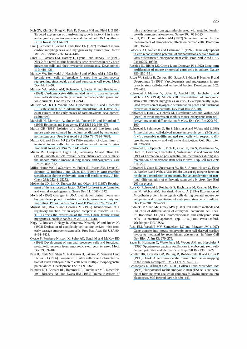
.Figure 1. ES cell technologyin vitro. Permanent embryonic stem cell lines (ESC) were cultivated from the inner cell mass (ICM) of mouseblastocysts, and embryonic germ cell lines (EGC) were cultivated from primordial germ cells (PGC). These pluripotent ESC or EGC are ableto differentiate via “embryoid bodies” (EBs) into derivatives of the endodermal, ectodermal and mesodermal lineagesin vitro

213
embryos (Eistetter, 1989). Besides ES cells, two othertypes of undifferentiated embryonic cells have beenestablished as permanent cell lines, the embryonalcarcinoma (EC) cells, malignant stem cells of terato-carcinomas derived by extrauterine transfer of earlyembryos (Martin and Evans, 1975; Stevens, 1984),and the embryonic germ (EG) cells cultivated fromprimordial germ (PG) cells (Figure 1; Resnick et al.,1992; Stewart et al., 1994). Both totipotent cell types,ES and EG cells, have been shown to participate innormal development when injected into blastocysts(Gardner and Brook, 1997).
ES, EG and EC cell lines exhibit characteristics ofundifferentiated embryonic cellsin vitro: (i) pluripo-tent differentiation capacity (Figure 1; Doetschman etal., 1985; Keller, 1995; Rohwedel et al., 1994, 1996;Maltsev et al., 1993, 1994; Strübing et al., 1995; Bainet al., 1995; Dani et al., 1997; Bagutti et al., 1996;Risau et al., 1988; Drab et al., 1997; Wobus et al.,1991, 1994b, 1997a; Wobus and Guan, 1998), expres-sion of (ii) endogenous alkaline phosphatase (Resnicket al., 1992), (iii) stage-specific embryonic antigenSSEA-1 (Solter and Knowles, 1978; Resnick et al.,1992), and (iv) germline-specific transcription factorOct-4 (Schöler et al., 1990), (v) hypomethylation ofDNA (Monk, 1990), and (vi) a short G1 phase of thecell cycle (Rohwedel et al., 1996).
To mimic the differentiation of totipotent stemcells in the embryo,in vitro cultivated ES cells weredifferentiated as embryo-like aggregates, so-called‘embryoid bodies’ (EBs). Within these EBs, cellu-lar derivatives of all three primary germ layers ofendodermal, ectodermal and mesodermal origin aredifferentiated. The pluripotent/totipotent ES cell linesdevelop from an undifferentiated stage resemblingcells of the early embryo into terminally differenti-ated stages of the cardiogenic (Wobus et al., 1991,1997a, 1997b; Maltsev et al., 1993, 1994; Hescheleret al., 1997; Miller-Hance et al., 1993; Wobus andGuan, 1998), myogenic (Miller-Hance et al., 1993;Rohwedel et al., 1994; Rose et al., 1994), neurogenic(Strübing et al., 1995; Bain et al., 1995; Fraichard etal., 1995; Okabe et al., 1996), hematopoietic (Keller,1995; Wiles and Keller, 1991; Hole and Smith, 1994)or adipogenic (Dani et al., 1997) lineage, as well asinto endodermal (Sauer, 1998), epithelial (Bagutti etal., 1996), endothelial (Risau et al., 1988), vascularsmooth muscle (VSM, Risau et al., 1988; Weitzer etal., 1995; Drab et al., 1997) and chondrogenic cells(Rohwedel, unpublished data).
We found that the terminally differentiated cellsshowed pharmacological and physiological propertiesof specialized cells:in vitro differentiated cardiomy-ocytes resemble characteristics of atrial-, ventricle-,purkinje- and pacemaker-like cells (Maltsev et al.,1993, 1994; Wobus et al., 1997b; Hescheler et al.,1997), neuronal cells are characterized by inhibitoryand excitatory synapses (Strübing et al., 1995; Okabeet al., 1996), and cardiac, myogenic, neuronal and vas-cular smooth muscle (VSM) cells express ion channelsand tissue-specific functional receptors (Wobus et al.,1991; Strübing et al., 1995; Rohwedel et al., 1998a;Drab et al., 1997; Wobus et al., 1997b). Further-more, the interaction of neuronal and skeletal musclecells resulted in the formation of postsynaptic-likemembranes (Rohwedel et al., 1998a).
In the following review, the ES cell differenti-ation systems of cardiogenesis, myogenesis, neuro-genesis, epithelial and VSM cell differentiation aresummarized with respect to the expression pattern ofgenes, proteins and to their functional properties (forhaematopoietic differentiation, see Keller, 1995, foradipogenic differentiation, see Dani et al., 1997). Fur-thermore, a short overview about the possibilities tomodulate thein vitro differentiation pattern by exoge-nous compounds, and to use ES cells for ‘gain offunction’ and ‘loss of function’ approaches are given.
Material and methods
Culture of undifferentiated ES cells and embryoidbody differentiation
ES cells of lines D3 (Doetschman et al., 1985), R1(Nagy et al., 1993) or CCE (Wiles and Keller, 1991),were cultivated on a feeder layer of primary mouseembryonic fibroblasts (Wobus et al., 1991) in Dul-becco’s modified Eagle’s medium (DMEM, GibcoBRL, Life Technologies, Eggenstein, Germany) sup-plemented with 15% heat-inactivated fetal calf serum(FCS, selected batches, Gibco), L-glutamine (2 mM,Gibco), β-mercaptoethanol (β-ME, final concentra-tion 5× 10⁻⁵M; Serva, Heidelberg, Germany) andnon-essential amino acids (NEAA, stock solution di-luted 1:100, Gibco) as described (Wobus et al., 1991;Maltsev et al., 1993) to keep ES cells in the undiffer-entiated stage.
In addition, ES cell lines may be grown with-out feeder layer in media supplemented with 10–20 ng/ml recombinant leukemia inhibitory factor (LIF;

214
Figure 2. ES cell differentiation protocol. ES cells (ESC) are routinely cultivated on mouse embryonic feeder layer to keep the cells in theundifferentiated stage (a). Forin vitro differentiation, ES cells were cultivated as embryoid bodies (EBs) in hanging drops for two days,and after suspension culture for 3–5 days. EBs are plated between days 5–7, depending on the differentiation lineage. EBs attach to tissueculture plates and differentiated cells develop in the EB outgrowths. Shown are the quantitative estimations of ES cell differentiation intospontaneously beating cardiac cells (b), skeletal muscle cells (c), neuronal cells (d) without (open symbols) and with induction by retinoicacid (RA; filled symbols), endodermal/ epithelial cells (e) and vascular smooth muscle cells (f) without (open symbols) and with induction byRA and dibutyryl-cAMP (db-cAMP; filled symbols). The cells were characterized by morphological analysis of EBs, i.e., the number of EBscontaining differentiated cells was estimated as percentage.
for preparation, see Rohwedel et al., 1996) for growthin the undifferentiated stage, or on both, a feeder layerand LIF-supplemented media.
The ES cell differentiation protocols shown in Fig-ure 2 have been described in detail (see Wobus et al.,1991, 1997a). In principal, for the development ofES cells into differentiated phenotypes, the pluripo-tent cells were cultivated as EBs by the ‘hanging drop’method (Wobus et al., 1991; Rudnicki and McBurney,1987) or by ‘mass culture’ (Doetschman et al., 1985).After plating the EBs at day 5 or 7, the differentiatedcells are grown out and the number of EB outgrowthswith the specific differentiated cell type was calculatedas a percentage (Figure 2b–f). The process of morpho-
logical differentiation in the EBs is accompanied bychanges in the pattern of gene expression as well asprotein formation, development of ion channels andtissue-specific receptors (Figure 2b–f, Figure 3b–f).
The different ES cell lines show pluripotent devel-opmental capacitiesin vitro, i.e., they differentiate inthe EB outgrowths into many differentiated cell types.But, to obtain maximal differentiation of a defined celltype, specific cell lines and cultivation conditions wereemployed.

215
Figure 3. Immunocytological analysis of tissue-specific proteins. a) Undifferentiated ES cells of line D3 immunostained with a monoclonalantibody against the stage-specific embryonic antigen, SSEA-1, and ES cell-derived differentiated cellular phenotypes are shown (b–f). Car-diomyocytes are immunostained by monoclonal antibodies against titin (Z-band; b), myotubes by nebulin (c), neuronal cells by neurofilamentproteins NF160 kDa (d), epithelial cells by cytokeratin K19 (e) and VSM cells by smooth muscleα-actin (f), respectively. Undifferentiated EScells immunostained by SSEA-1 (a) were scanned in single sections by the confocal laser scanning microscope, EB outgrowths immunostainedfor cytokeratin 19 (e), and smooth muscleα-actin-positive cells (f) were scanned in 8 sections (1µm; e) and 4 sections (0.5µm; f), respectively.Bars represent 10µm.

216
Detection of tissue-specific genes bysemi-quantitative RT-PCR analysis
The expression of tissue-specific genes in EBs andoutgrowths is analyzed by semi-quantitative RT-PCRusing the “primer-dropping” method according toWong et al. (1994) as described (Wobus et al., 1997b).EBs or outgrowths were collected at several stages af-ter plating at day 5 or 7. The total RNA was isolated bythe single step extraction method according to Chom-czynski and Sacchi (1987) and mRNA was reversetranscribed using Oligo d(T)16 primer (Perkin-Elmer,Überlingen, Germany) and amplified using oligonu-cleotide primers complementary and identical to targetgenes (see Wobus et al., submitted).
Expression of tissue-specific proteins analyzed byimmunofluorescence
The formation of tissue-specific proteins in EB out-growths is analyzed by immunofluorescence analysis.EBs plated on cover slips were rinsed twice withPBS, fixed with methanol: acetone (7:3) at –20◦Cfor 10 min and processed for immunofluorescencemicroscopy (Maltsev et al., 1993). Antibodies forthe analysis of tissue-specific proteins are, for ex-ample, the titin (Z-band)-specific mAb T12 (Fürst etal., 1988) for cardiomyocytes and skeletal myocytes(a cardiomyocyte is shown in Figure 3b), a nebulin-specific mAb Nb2 (Rohwedel et al., 1998a) for skele-tal muscle cells (Figure 3c), a neurofilament protein160kDa-specific mAb NN18 (Strübing et al., 1995) forneuronal cells (Figure 3d), a cytokeratin K19-specificmAb (TROMA III, a gift of Dr. Kemler, Freiburg) forendodermal and epithelial cells (Figure 3e; see Baguttiet al., 1996) and a smooth muscleα-actin-specificmAb 1A4 for vascular smooth muscle cells (Figure 3f;see Drab et al., 1997).
Cardiomyocytes were isolated as single beatingcells from EB outgrowths (see Maltsev et al., 1993;1994) and were used for immunostaining (Figure 3b)and for the characterization of action potentials andion channels by patch-clamp analysis.
Pharmacological and physiological analyses
To demonstrate the functional properties ofin vitrodifferentiated excitable cardiac, skeletal muscle, neu-ronal or VSM cells, electrophysiological and pharma-cological techniques were employed. The expressionof ion channels on ES cell differentiated cardiomy-ocytes (Maltsev et al., 1993; 1994), skeletal muscle
(Rohwedel et al., 1994), neuronal (Strübing et al.,1995; Fraichard et al., 1995) and VSM cells (Drabet al., 1997) was analyzed by the whole-cell config-uration of the patch-clamp technique (Hamill et al.,1981). Action potentials of isolated cardiomyocyteswere analyzed by the same technique (Maltsev et al.,1993; Fässler et al., 1996; Hescheler et al., 1997).
The spontaneous beating capacity of ES and ECcell-derived cardiomyocytes enabled to investigatechronotropic effects of cardioactive substances bymeasuring the beating frequencies of cardiac cells(Wobus et al., 1991, 1994b). An inverted microscopeDiaphot-TMD (Nikon) equipped with a 37◦C and5% CO2 incubation chamber was used. Cardioactivedrugs were cumulatively added to pulsating clustersof EB outgrowths, the frequencies were measuredfrom control (= basal level of beating frequency)and agonist-treated variants to make up dose-responsecurves. By using the LUZIA imaging device (Nikon),a semi-automatic computer-assisted imaging systemfor a routine screening of cardioactive drugs wasestablished (Pich et al., 1997; Wobus et al., submitted).
The receptor activity of ES cell-derived VSM cellswas analyzed by measuring the increase of intracel-lular [Ca2+] transients by confocal laser scanningmicroscopy of fluo-3-labelled VSM cells. Vasoactiveagonists were able to evoke intracellular free [Ca2+]transients in VSM cells differentiated from ES cells(Drab et al., 1997).
Results
ES cells develop into differentiated phenotypes
Depending on the specific differentiated cellular phe-notypes, distinct protocols with different ES cell lineswere established. The ‘hanging drop’ method gen-erated EBs of a defined cell number and size. Thistechnique has been used for developmental studies be-cause the differentiation pattern is dependent on thenumber of ES cells which differentiate within the EBs.It was found that the following variables influencedthe developmental potency of ES cells in culture: (1)number of cells differentiating in the EBs, (2) medium,quality of fetal calf serum, growth factors and mediumadditives, (3) ES cell lines used, and (4) the time ofEB plating.
Mouse ES cells can differentiate into cardiogenic,myogenic, neuronal-, epithelial- and vascular smoothmuscle-like cellsin vitro, and the expression of tissue-

217
specific genes, proteins and ion channels is devel-opmentally controlled during differentiation. In thefollowing chapter a short overview about the ES cell-derived phenotypes is given with respect to tissue-specific genes, proteins and ion channels (Figure 4).
Cardiogenesis
ES cells of lines D3, R1 and CCE differentiatedviaEBs into clusters of spontaneously beating cardiomy-ocytes. Optimal cardiac differentiation was achievedby using 400 cells of line D3 or R1 for the prepa-ration of EBs. We used different media for EB de-velopment and cellular differentiation: DMEM sup-plemented with 20% FCS, L-glutamine,β-ME andNEAA (‘DMEM differentiation medium’), or Is-cove’s modification of DMEM (IMDM, Gibco) sup-plemented with 20% FCS, L-glutamine, NEAA andα-monothioglycerol 3-mercapto-1,2-propandiol (MTG,final concentration 450µM; Sigma; ‘IMDM differ-entiation medium’). EBs were plated between days 5and 7.
First beating clusters in EBs can already be seenin 7 day old EBs or 1 to 2 days after plating (Wobuset al., 1991; Maltsev et al., 1993, 1994), but maximaldifferentiation of beating cardiomyocytes is achievedseveral days (7 to 10 d) after EB plating at days 5 to 7(Figure 2b).
At the level of gene expression, a characteristic se-quence of cardiac-specific gene expression was found.The cardiac-specific transcription factor Nkx 2.5 wasweakly expressed in ES cells of line D3 and in EBs atday 3, but maximally in EBs between days 5 and 5 +12 (Wobus and Guan, 1998). The gene encoding theα1 subunit of the L-type Ca2+ channel (α1 CaCh) wasfirst expressed in EBs two days before beating cellsappeared, followed byα- andβ-cardiac myosin heavychain (MHC) around the day when first beating cellswere observed. Finally, genes expressed in specializedcardiac cell types, such as those encoding atrial natri-uretic factor (ANF, expressed mainly in atrial cells)and myosin light chain isoform 2v (MLC-2v, spe-cific for ventricular cells; Fässler et al., 1996; Wobuset al., 1997b; Hescheler et al., 1997) were detected(Figure 4).
Cardiomyocytes differentiated from EB out-growths revealed a specific sequence of expression ofthe sarcomeric proteins during cardiogenesis as fol-lows: titin (Z-band-specific; Figure 3b),α-actinin, my-omesin, titin (M-band-specific), sarcomeric MHC andα-actin in early cardiomyocytes. M-protein was only
expressed at terminal differentiation stages (Figure 4;Wobus and Guan, 1998).
During differentiation, ES cells developed intomesodermal progenitor cells and early cardiomyocyteswhich show pacemaker-like action potentials at theearly stage (day 7). They further specialize into atrial-(38%), ventricular- (48%) and sinusnodal-like (14%)cells, based on electrophysiological determination ofaction potentials at the terminal differentiation stage(between days 14 to 26 after plating; Maltsev etal., 1993; Fässler et al., 1996). Recently, a fourthtype of action potential was described that repre-sented Purkinje-like cells (Hescheler et al., 1997;Wobus et al., 1997b). The various types of actionpotentials measured in cardiomyocytes of different de-velopmental stages correlated with the expression ofspecialized types of ion channels (Figure 4; Malt-sev et al., 1994; Hescheler et al., 1997; Wobus andGuan, 1998). Recently (Maltsev et al., submitted),the modulation of ICa was used as a functional as-say to test different components of theβ-adrenergicsignaling cascade during cardiomyocyte development.Maltsev et al. found that the uncoupling and/or lowexpression of Gs-protein accounted for the ICa in-sensitivity toβ-adrenergic stimulation in very early-developmental stage cardiomyocytes. But in early-developmental stage cells, the uncoupling is due, atleast in part, to a high intrinsic activity of phospho-diesterases. These results together with previous data(Maltsev et al., 1993, 1994) indicate the normal courseof development for ES cell-derived cardiomyocytessimilar to embryonic cardiomyocytes. Furthermore,we found that the differentiated cardiomyocytes re-sponded with characteristic chronotropic responsesto cardiotropic drugs comparable to cardiomyocytesfrom living organisms (Wobus et al., 1991; Pich et al.,1997).
MyogenesisDifferentiation of ES cells into skeletal muscle cellswas shown by the formation of myoblasts which fusedinto multinucleated myotubes during terminal differ-entiation. Optimal development into skeletal musclecells was achieved by using 800 cells of line D3(Rohwedel et al., 1998a) or 600 cells of line BLC6(Rohwedel et al., 1994) in DMEM containing 15%dextran-coated charcoal -treated FCS (DCC-FCS, Ro-hwedel et al., 1998), L-glutamine, NEAA,β-ME,sodium selenite (final concentration: 3× 10⁻⁸ M;Sigma), bovine serum albumine (BSA, final concen-tration: 0.1875%; Gibco) and transferrin (final con-

218
Figure 4. Developmentally controlled expression of tissue-specific genes, proteins and ion channels after differentiation of ES cells into cardiac,skeletal muscle, neuronal, epithelial and VSM cellsin vitro. Cardiogenesis: Genes encoding the cardiac transcription factor Nkx 2.5, theα1subunit of the L-type Ca2+ channel (α1CaCh),α- andβ-cardiac myosin heavy chain (α-, β-MHC), atrial natriuretic factor (ANF) and theventricular isoform 2 of myosin light chain (MLC-2v) were analyzed by RT-PCR. The expression of sarcomeric proteins was determined byimmunofluorescence using the antibodies against: titin (Z-band), nonmuscleα-actinin, myomesin, sarcomeric MHC,α-actin and M-protein.The cardiac-specific ion currents ICa (L-type Ca2+ current), Ik,to (transient K+ current), IK,ATP (ATP-modulated K+ current), IK (outwardlyrectifying K+ current), INa (inward Na+ current), If (hyperpolarization-activated pacemaker current), IK1 (inwardly rectifying K+ current),IK,ACh (muscarinic acetylcholine-activated K+ current) were found during ES cell-derived cardiogenesis.Myogenesis: Myogenic determi-nation genes myf5, myogenin, MyoD and myf6, the muscle-specific cell adhesion molecule M-cadherin, and theγ - and ε-subunit of thenicotinic acetylcholine receptor (nAChR) were analyzed. The myocytes expressed muscle-specific proteins such as titin, nebulin, M-cadherin,myogenin, sarcomeric MHC and slow C-protein, the latter only found to be expressed at terminal stages. Skeletal muscle-specific L-typeand T-type Ca2+ channels (ICa,L ; ICa,T) and functional nicotinic acetylcholine receptor-operated channels (nAChR) were expressed duringES cell-derived myogenesis.Neurogenesis: A developmentally controlled expression pattern of genes encoding 68 kDa (NFL), 160 kDa(NFM) and 200 kDa (NFH) neurofilament proteins, coding for the synaptic vesicle protein synaptophysin, the neuron-specific proteoglycanneurocan, and the embryonic splice variant of the microtubule-associated protein tau was found during ES cell-derived neurogenesis. Neuron-and glial cell-specific proteins NFL, NFM, NFH, SNAP-25, synaptophysin and glial fibrillary acidic protein (GFAP) were determinated byimmunofluorescence studies. Neuron-specific voltage-dependent ion currents: K+ current (IK ), Na+ current (INa) and Ca2+ current (ICa),and neuron-specific receptors:γ -aminobutyric acid (GABAA), Glycin (Gly), Kainate (Kai) and N-methyl-D-aspartate (NMDA) were found inneuronal cells by electrophysiological analyses.Epithelial differentiation:Epithelial cells expressed cytokeratins 8, 18, 19 (K8, K18 and K19)at early stages, and, cytokeratins K8, K18, K10, K14 and involucrin (inv) at later stages.Vascular smooth muscle (VSM) cell differentiation:VSM cells derived from ES cells preponderantly expressed vascular smooth muscle myosin heavy chain A (V-SM-MHC-A) and smooth muscleα-actin (SM-α-Actin), the intestinal smooth muscle myosin heavy chain B (I-SM-MHC-B) was only slightly expressed at later stages. Threevoltage-sensitive ion channels, the smooth muscle-specific Ca2+ channel (IK,Ca), the calcium-activated maxi K+ channel (IK,Ca), and delayedrectifying K+ channel (IKv) were expressed in VSM cells. Receptors for angiotensin II (AII-R), bradykinin, histamin, platelet-derived growthfactor AB (PDGF AB), thrombin and endothelin-1 were functionally expressed.

219
centration: 0.01 mg/ml; Gibco) (‘DMEM-DCC dif-ferentiation medium’), or in ‘IMDM differentiationmedium’. EBs were plated at day 5.
The first myoblasts/ myocytes appeared 4 (lineBLC6) or 5 to 7 days (D3) after EB plating (Fig-ure 2b). Skeletal muscle cells fused into myotubes inthe EB outgrowths 1 or 2 days later (Figure 3c). Dur-ing myogenic development, muscle-specific genes,proteins and ion channels were time-dependently ex-pressed as summarized in Figure 4.
During ES cell differentiation, genes encoding themyogenic regulatory factors Myf5, myogenin, MyoDand Myf6, the muscle-specific cell adhesion mole-cule M-cadherin as well as theγ - and ε-subunit ofthe nicotinic acetylcholine receptor (nAChR) wereexpressed in a sequence closely resembling myoge-nesis in vivo (Rohwedel et al., 1994; Rose et al.,1994; Rohwedel et al., 1998a). The myogenic cellsexpressed muscle-specific proteins such as sarcom-eric MHC, myogenin and M-cadherin (Rohwedel etal., 1994; Rohwedel et al., 1995) as well as titin,nebulin (Rohwedel et al., 1998a),α-actinin and slowC-protein (Guan, unpublished). In addition, electro-physiological studies using the patch-clamp techniquedemonstrated the expression of functional nicotiniccholinoceptors and T-type Ca2+ channels with de-creasing density, as well as L-type Ca2+ channelswith increasing density on ES cell-derived musclecells comparable to developing muscle cellsin vivo(Figure 4; Rohwedel et al., 1994). Interactions be-tween muscle cells and neuronal cells which bothdifferentiate in the same EBs resulted in the formationof neuromuscular junctions. We demonstrated thatmultinucleated contracting myotubes which appear ata terminal differentiation stage formed postsynaptic-like membranes exhibiting a clustering of nAChRscolocalized with agrin and synaptophysin. A coex-pression of the myogenic regulatory gene Myf6 andthe AChRε-subunit gene was found, which paralleledtheir expression in adult muscle cells (Rohwedel et al.,1998a).
NeurogenesisDifferent ES cell lines and protocols were establishedfor efficient differentiation of neuronal and glial cells(Strübing et al., 1995; Bain et al., 1995; Fraichard etal., 1995; Okabe et al., 1996).
D3 cells cultivated by the hanging drop method fortwo days followed by suspension culture in the pres-ence of 10−7 M retinoic acid (RA) between days 2 and5 and plating at day 7 resulted in efficient neuronal
differentiation (Wobus et al., 1994a). In addition, EBsprepared from 400 cells of line BLC6 cultivated inDMEM-DCC differentiation medium in the presenceof 10−7 M RA during the first two days of EB culturefollowed by 2 days of suspension culture (without RA)and plating at day 4 showed an efficient differentiationof functional neuronal cells (Strübing et al., 1995).
The frequency of spontaneous differentiation ofBLC6 cells into neuronal cells amounted to 15 to 30%.After differentiation induction by RA (Strübing et al.,1995), the differentiation rate of neuronal cells wasincreased to nearly 100% of the EBs (Figure 2d).
ES cells of line BLC6 differentiated after RA-induction into neuronal cells which expressed neuron-specific genes and were characterized by the complexelectrophysiological and immunocytochemical prop-erties of postmitotic nerve cells (Strübing et al., 1995).Similar to ES cell-derived cardiac and myogenic dif-ferentiation, also neurogenic differentiation preceedsfrom progenitor cells to specialized cell types reflect-ing a characteristic sequence of expression of neuron-specific genes, proteins and ion-channels (Figure 3d;Figure 4). Genes encoding the low (NFL) and middle(NFM) molecular mass neurofilament proteins and thesynaptic vesicle protein synaptophysin were expressedat an early stage of ES cell- derived neurogenesis (Ro-hwedel et al., 1998a) in parallel to the expressionof voltage-gated ion channels, Ca2+, Na+ and K+channels (Strübing et al., 1995). Further differentia-tion was characterized by an increase in the densityof voltage-gated ion channels, the onset of expres-sion of receptor-operated ion channels in parallel tothe expression of genes and proteins characteristic formature neurons (Strübing et al., 1995; Rohwedel et al.,1998a).
In addition to the expression of specific neuronalreceptors, neuronal cells generated Na+-driven actionpotentials and were functionally coupled by inhibitory(GABAergic) and excitatory (glutamatergic) synapsesas revealed by measurements of postsynaptic currents(Figure 4; Strübing et al., 1995; Wobus et al., 1997a).
Bain et al. (1995) used a four-day mass cultureof ES cells to prepare EBs followed by a four-daysuspension culture in the presence of 5× 10⁻⁷ MRA and plating at day 8. Another method describeddifferentiation factors (insulin, transferrin, selenium),growth factors (bFGF) and extracellular matrix pro-teins (fibronectin, laminin, polyornithine) as efficientneuronal differentiation inducers (Okabe et al., 1996).

220
Epithelial cell differentiationDifferentiation of ES cells as EBs cultivated by thehanging drop or the mass culture method in DMEMor IMDM differentiation medium resulted in epithe-lial differentiation and expression of epithelial-specificgenes and proteins (Bagutti et al., 1996).
Epithelial-like cells are one of the most prominentcell types in the ES cell-derived EB outgrowths whichwere found in all EB outgrowths without any spe-cial differentiation induction (Figure 2d; Figure 3e).Epithelial-specific genes and proteins characteristicfor early (cytokeratins K8, K18, K19), intermediate(cytokeratin K14) and terminal (involucrin) differenti-ation were expressed during EB developmentin vitro(Figure 4; Bagutti et al., 1996).
Vascular smooth muscle (VSM) cell differentiationA complex cell type which differentiated from EScells, are spontaneously contracting VSM cells. Weestablished a specific differentiation protocol by us-ing RA and db-cAMP for the induction of VSM cellsduring EB differentiation (Drab et al., 1997). For dif-ferentiation of smooth muscle cells, ES cells of lineD3 were cultivated in hanging drops for 2 days andafter suspension culture for five days. EBs were platedat day 7 followed by treatment with 10−8 M RA and0.5× 10−3 M db-cAMP between days 7 and 11 (Drabet al., 1997). During differentiation of the EB out-growths, the medium was changed every second dayand the first spontaneously contracting smooth musclecells appeared around day 14 after plating.
Addition of RA and db-cAMP increased the dif-ferentiation rate of VSM cells from a control level ofonly 5–10% to more than 60% of the EBs 28 daysafter plating (Figure 2e; Drab et al., 1997). DuringES cell differentiationin vitro, VSM-specific MHCwas preponderantly expressed between days 7 and 14,which is in temporal agreement with the observationin the dorsal aorta on day 10 postcoitumin vivo (Mi-ano et al., 1994). The intestinal splice variant of thesmooth muscle MHC was only sligthly expressed ata rather terminal stage of EB differentiation (Drab etal., 1997). In addition, three distinct voltage-sensitiveion channels: the calcium-activated maxi K+ channel,(IKca) the “delayed rectifier” K+ channel (IKv) andthe dihydropyridine-sensitive (L-type) Ca2+ channel(ICa) were expressed in VSM cells differentiated fromES cells (Drab et al., 1997). ES cell-derived VSMcells were functionally characterized by [Ca2+] tran-sients in response to the VSM cell-specific agonistsangiotensin II, bradykinin, histamine, endothelin-1,
PDGF AB, thrombin and vasopressin with an in-creased intracellular Ca2+ release (Figure 4; Drab etal., 1997).
Another protocol for VSM cell differentiation usedES cells of lines AB1 and AB2.1 cultivated as EBs inhanging drops of M15 medium (DMEM supplementedby 15% FCS and 2 mM glutamine) for 4.5 days.After plating of the EBs, the medium was partiallyexchanged by fresh medium every third day (Weitzeret al., 1995).
Modulation of differentiation
Modulation of differentiation by retinoic acid (RA)The establishment of the ES cell differentiationmodels allowed the study of cellular differentiationprocesses during embryonic developmentin vitro. Thesystems permitted the analysis of undifferentiated em-bryonic cells via progenitor cells into highly differ-entiated and specialized cells of the cardiovascular,myogenic and neurogenic lineages.
The controlled developmental process in the EBsin vitro offered the possibility of modulating the de-velopmental pattern by differentiation factors, growthfactors, or extracellular matrix (ECM) proteins. Oneof the most effective differentiation factors is retinoicacid (RA) which influenced time- and concentration-dependently the differentiation of EC and ES cellsinto cardiogenic (Wobus et al., 1994a, 1997b), myo-genic (Wobus et al., 1994a), neurogenic (Wobus et al.,1994a; Strübing et al., 1995, Bain et al., 1995) or VSMcell (Drab et al., 1997) lineages.
It has been clearly shown that RA exerts its spe-cific differentiation-inducing effect on ES cells in aconcentration- and time-dependent manner during EBdevelopment. Treatment with high concentrations ofRA (10−7 M) during the first 2 days of EB differ-entiation increased the differentiation frequency ofneuronal cells and accelerated the neuronal differen-tiation without changing the functional cell fates ofthe differentiating neurons (Strübing et al., 1995), butsignificantly inhibited cardiac differentiation (Wobuset al., 1994a). Incubation of EBs with RA (10−8 and10−7 M) between days 2 and 5 of EB developmentresulted in an induction of both neurogenesis and myo-genesis, but in an inhibition of cardiogenesis (Wobuset al., 1994a). The RA-induced skeletal myocytesfunctionally expressed tissue-specific Ca2+ channelsand nicotinic cholinoceptors. In contrast, treatmentwith 10−9 and 10−8 M RA, respectively, of EBs begin-ning on day 5 resulted in an increased and accelerated

221
differentiation into the cardiogenic lineage and es-pecially into ventricular cells (Wobus et al., 1994aand 1997b). RA, both in the all-trans and in the 9-cis configuration, accelerated cardiac differentiationthrough induction of expression of the cardiac-specificα-cardiac MHC and MLC-2v genes, events whichresulted in an enhanced development of ventricularcardiomyocytes (Wobus et al., 1997b).
RA treatment (10−8 M RA in combination with 0.5× 10⁻³ M db-cAMP) between days 7 and 11 of EBdevelopment induced the differentiation of VSM cells(Drab et al., 1997). Smooth muscle cells were fullydifferentiated and expressed vascular-specific genes,proteins, ion channels and receptors (Drab et al.,1997).
The mechanisms whereby RA induces differenti-ation are not known. However, as shown by variousgroups (reviewed by Marshall et al., 1996), RA ex-erts a wide variety of profound effects on vertebratedevelopment and cellular differentiation by activationof homoeotic genes and other transcription factors.Hogan et al. (1992) described the endogenous synthe-sis of RA in Hensen’s node during a defined time win-dow of embryonic development in chicken. Therefore,RA was suspected to be one of the most importantmorphogens during vertebrate embryogenesis.
In vitro, RA induced MHox expression during thedifferentiation of smooth muscle cells (Blank et al.,1995) and the expression of transcription factors HNF-3 (Jacob et al., 1997), MASH-1, MATH-1, neuroDand NSCL-2 (Itoh et al., 1997) during neuronal differ-entiation of mouse P19 cells. Furthermore, retinoidspromoted terminal muscle differentiation via activa-tion of the muscle-specific myoD gene family (myoD,myogenin, myf-5 and MRF-4) of transcription fac-tors (Muscat et al., 1995). A novel RA-inducible geneof the basic HLH family, Stra13, was found to beexpressed during mouse embryogenesis in neuroec-toderm and in several mesodermal and endodermalderivatives. Overexpression of Stra13 resulted in neu-ronal differentiation of P19 cells, which without RAinduction undergo mesodermal and endodermal dif-ferentiation indicating that Stra13 might be one of theearliest RA target genes (Boudjelal et al., 1998).
With respect to cardiogenesis, homoeotic genesand transcription factors, such as the tinman-relatedNkx 2.5 (Lints et al., 1993), the muscle-specificMEF2C (Lin et al., 1997), the cardiac-specificdHAND (Srivastava et al., 1995) or GATA4 genes(Molkentin et al., 1997) might be candidate RA-responsive genes.
Modulation of differentiation by ‘gain of function’
Overexpression of a tissue-specific gene which is spa-tially and temporally regulated during developmenthelps to reveal its function. Since the work of Palmiteret al. (1982), ‘gain of function’ studies have been suc-cessfully carried out by generating transgenic animalscarrying genes under the control of inducible or con-stitutively active promoters. An alternative approach isthe overexpression of genes duringin vitro differenti-ation of ES cells (Figure 5). We used the transcriptionfactor M-twist, a negative regulator of muscle differ-entiation in a ‘gain of function’ approach. ES cellclones stably transfected with the M-twist cDNA un-der the control of a modified SV40 promoter showeddelayed differentiation of myogenic cells and skele-tal muscle-specific gene expression depending on thelevel of exogenous M-twist expression (Rohwedel etal., 1995). Using the same approach, overexpressionof the homeobox gene HOXB4 during ES cell differ-entiationin vitro resulted in enhanced differentiationof erythroid progenitor cells (Helgason et al., 1996).Furthermore, it was demonstrated that ES cells whichconstitutively express the myogenic regulatory factorMyoD preferentially differentiated into the myogeniclineage (Dinsmore et al., 1996). Thus,in vitro ‘gain offunction’ studies using ES cells provide an alternativestrategy to transgenic animals.
Modulation of differentiation by ‘loss of function’
The establishment of optimal conditions for the devel-opment of ES cellsin vitro into cardiac, myogenic,neuronal, epithelial and VSM cells enabled us to studydifferentiation of genetically altered ES cells. Aftergene inactivation by homologous recombination, EScells can be retransferred into blastocysts to regen-erate chimaeric micein vivo (Thomas and Capecchi,1987). Null mutations could lead to homozygous ani-mals which show (i) no mutant phenotypes, (ii) mutantphenotypes, or (iii) result in embryonic lethality (Fig-ure 5). In cases, in which the mutation leads to earlyembryonic death, the differentiation of targeted EScells via EBsin vitro represents a new supplemen-tary technique to analyze the effects of the specificmutation on cellular differentiation (Figure 5).
We successfully applied this approach to the analy-sis of loss ofβ1 integrin function on differentiation.Invivo, a lack ofβ1 integrin resulted in embryonic deathshortly after implantation (Fässler and Meyer, 1995).By using the ES cell differentiation approachin vitro,we showed that cell-matrix interaction viaβ1 integrin

222
Figure 5. Modulation of embryonic differentiation by “gain of function” or “loss of function” strategiesin vitro and the results of gene targetingexperimentsin vivo (for explanation, see text).
is important for normal cardiogenesis, myogenesis,neurogenesis, epithelial and VSM cell differentiation.
We found that duringin vitro differentiation ofβ1-null ES cells, the development into cardiomyocytes,skeletal muscle and epithelial cells was severely im-paired (Fässler et al., 1996; Bagutti et al., 1996;Rohwedel, et al., 1998b; our unpublished data). Ingeneral, mesodermal cell types such as cardiac andmyogenic cells developed with a delay, whereas dif-ferentiation of neuronal cells was accelerated in theabsence ofβ1 integrin. Usually, ES cells differenti-ated into cardiomyocytes of atrial-, ventricular- andpacemaker-like types, whereas a large number ofpacemaker-like cells was found during the differen-
tiation of β1-null ES cells, and only few atrial andventricular cells developed transiently (Fässler et al.,1996). Also the development of sarcomeric proteinswas retarded inβ1-null cardiomyocytes (Fässler et al.,1996). The formation of myotubes was significantlydelayed and reduced, although some myocytes fusedand sarcomeres were formed. In the absence ofβ1 in-tegrin, ES cell-derived neurogenic differentiation wasaccelerated, but the outgrowth of neuronal cells wasretarded (our unpublished data). We found an alteredexpression pattern of regulatory genes involved inmesodermal and neuroectodermal differentiation pos-sibly due to an important regulatory function ofβ1integrins during early cell lineage determination (our

223
unpublished data). It has been shown that the capacityof theβ1-null cells to differentiate into keratinocytesin vitro was severely impaired since the differentiatingβ1-null cells expressed the simple epithelial keratins,but not K14, K10, and involucrin was detected onlyoccasionally (Bagutti et al., 1996).
In vitro differentiation of ES cells was also usedto demonstrate the disruption of visceral endodermdifferentiation after homozygous inactivation of theGATA-4 (Soudais et al., 1995), and the inhibition ofmyogenic differentiation after inactivation of desmingenes (Weitzer et al., 1995). In conclusion,in vitro dif-ferentiation of mutant ES cells served as an excellentalternative strategy in all those cases where the genedefect resulted in early embryonic lethality to unrevealthe function of those genes which are indispensableinvivo.
Summary and future prospects
The ES cellin vitro technology allows the study of cel-lular differentiation processes during early embryonicdevelopment in culture. Thein vitro systems permittedus to analyze the differentiation of early embryoniccells via progenitor cells into highly differentiated andspecialized cells of the cardiovascular, myogenic andneurogenic lineages, as well as into epithelial andVSM cells. From other laboratories,in vitro modelsfor hematopoietic (see Keller et al., 1995) and adi-pogenic (Dani et al., 1997) differentiation of mouseES cells were successfully established.
During the last years, in addition to mice, otherorganisms were successfully used to establish ES celllines, and living chimaeras have been obtained so far,as for example, from chicken (Pain et al., 1996), fish(Hong et al. 1996), rabbit (Schoonjans et al. 1996),and pig (Wheeler, 1994).
These embryonic stem cell lines may be used toinvestigate early developmental processesin vitro. Inaddition, the effects of growth and differentiation fac-tors (Wobus et al., 1994a, b; 1997b; Johansson andWiles, 1995) or extracellular matrix proteins on thedifferentiation of embryonal cells may be investigated.Furthermore, xenobiotics acting as embryotoxic andteratogenic agents may be screened for their capacityto modulate differentiationin vitro (Spielmann et al.,1997). In addition, transgenic ES cell lines contain-ing reporter genes fused to tissue-specific promotersmay be used to prescreen for embryotoxic/teratogeniccompounds which induce stage-specific developmen-
tal alterationsin vitro. These embryotoxicologicalapproaches together with pharmacological and elec-trophysiological analyses of differentiated ES cell-derived cardiomyocytes (for analysis of cardioactivedrugs, see Wobus et al., 1991, 1994b, 1997b; Pich etal., 1997) might help to reduce the use of laboratoryanimals in pharmacotoxicology.
In addition, the differentiation of genetically modi-fied cells by “gain of function” (Rohwedel et al., 1995)and “loss of function” (Fässler et al., 1996; Wobus andGuan, 1998) using totipotent ES cellsin vitro is an ex-cellent alternative to and substitute forin vivo studieswith transgenic animals to analyse the consequencesof mutations during early embryogenesis. Induciblegene trap experiments with ES cells will further helpto understand key events in early embryogenesis. Byusing RA as differentiation inducer and transgenic EScell lines which express reporter genes, i.e. LacZ orgreen fluorescent protein (GFP; Cormack et al., 1996),it is possible to screen selectively for genes which areinduced or repressed in a defined lineage (Forrester etal., 1996).
In the future, the ES cellin vitro technology may beof special interest to obtain homogeneous populationsof differentiated cells to be used for transplantations.For example, ES cells differentiated into GABAer-gic (Strübing et al., 1995; Dinsmore et al., 1996) ordopaminergic (Dinsmore et al., 1998) neurons, cardiacventricular cells (Wobus et al., 1997b) or hematopoi-etic cells (Hole and Smith, 1994; Potocnik et al. 1997)might be used as a novel source of cells for somatictherapy and transplantation. The construction of effi-cient vector systems and selection strategies will openfurther prospects forin vivo gene expression strate-gies (Koh et al., 1995; Rust et al., 1997). TransgenicES cell lines carrying tissue-specific promoters fusedto selectable marker genes can be differentiated intothe specific lineagesin vitro, and after selectioninvitro, the differentiated cell population might be usedin grafting experiments and transplantations to recon-stitute defective tissues (Koh et al., 1995; Klug et al.,1996; Dinsmore et al., 1996; Rust et al., 1997).
Acknowledgements
We wish to thank Mrs. S. Sommerfeld, K. Meierand O. Weiß for skilful technical assistance, Drs. D.Fürst, University of Potsdam, and R. Kemler, MPI forImmunobiology, Freiburg, for providing monoclonalantibodies, R. Fässler, University of Lund, forβ1

224
integrin-null ES cells, and U. Pich for help with theconfocal scanning microscopy analysis. We are grate-ful to all colleagues, especially to Dr. J. Hescheler,University of Köln, for continuous collaboration. Thework was supported by the Deutsche Forschungsge-meinschaft (Wo 503/1-3, SFB 366/YE1) and Fondsder Chemischen Industrie (FCI).
References
Bagutti C, Wobus AM, Fässler R and Watt FM (1996) Differen-tiation of embryonal stem cells into keratinocytes: Comparisonof wild-type and β1 integrin-deficient cells. Dev Biol 179:184–196.
Bain G, Kitchens D, Yao M, Huettner JE and Gottlieb DI (1995)Embryonic stem cells express neuronal propertiesin vitro. DevBiol 168: 342–357.
Blank RS, Swartz EA, Thompson MM, Olson EN and Owens GK(1995) A retinoic acid-induced clonal cell line derived frommultipotential P19 embryonal carcinoma cells expresses smoothmuscle characteristics. Circ Res 76: 742–749.
Boudjelal M, Taneja R, Matsubara S, Bouillet P, Dolle P andChambon P (1997) Overexpression of Stra13, a novel retinoicacid-inducible gene of the basic helix-loop-helix family, inhibitsmesodermal and promotes neuronal differentiation of P19 cells.Genes Dev 11: 2052–2065.
Bradley A, Evans M, Kaufman MH and Robertson E (1984) For-mation of germ-line chimaeras from embryo-derived teratocarci-noma cell lines. Nature 309: 255–256.
Brandon EP, Idzerda RL and McKnight GS (1995) Targeting themouse genome: a compendium of knockouts (part I – III). CurrBiol 5: 625–634, 758–765, 873–881.
Chomczynski P and Sacchi N (1987) Single-step method of RNAisolation by acid guanidinium thiocyanate-phenol-chloroformextraction. Anal Biochem 162: 156–159.
Clarke AR (1994) Murine genetic models of human disease. CurrOpin Genet Dev 4: 453–460.
Cormack BP, Valdivia RH and Falkow S (1996) FACS-optimizedmutants of the green fluorescent protein (GFP). Gene 173: 33–38.
Dani C, Smith AG, Dessolin S, Leroy P, Staccini L, Villageois P,Darimont C and Ailhaud G (1997) Differentiation of embryonicstem cells into adipocytesin vitro. J Cell Sci 110: 1279–1285.
Dinsmore J, Ratliff J, Deason T, Pakzaban P, Jacoby D, Galpern Wand Isacson O (1996) Embryonic stem cells differentiatedin vitroas a novel source of cells for transplantation. Cell Transplant 5:131–143.
Dinsmore J, Ratcliff J, Jacoby D, Wunderlich M and Lindberg C(1998) Embryonic stem cells as a model for studying regulationof cellular differentiation. Theriogenology 49: 145–151.
Doetschman TC, Eistetter H, Katz M, Schmidt W and Kemler R(1985) Thein vitro development of blastocyst-derived embryonicstem cell lines: formation of visceral yolk sac, blood islands andmyocardium. J Embryol Exp Morphol 87: 27–45.
Drab M, Haller H, Bychkow R, Erdmann B, Lindschau C, Haase H,Morano I, Luft FC and Wobus AM (1997) From totipotent em-bryonic stem cells to spontaneously contracting vascular smoothmuscle cells: a retinoic acid and db-cAMPin vitro differentiationmodel. FASEB J 11: 905–915.
Eistetter HR (1989) Pluripotent embryonal stem cell lines can be es-tablished from disaggregated mouse morulae. Dev GrowthDiffer31: 275–282.
Evans MJ and Kaufman MH (1981) Establishment in culture ofpluripotential cells from mouse embryos. Nature 292: 154–156.
Fässler R and Meyer M (1995) Consequences of lack ofβ1 integringene expression in mice. Genes Dev 9: 1896–1908.
Fässler R, Rohwedel J, Maltsev V, Bloch W, Lentini S, Guan K,Gullberg D, Hescheler J, Addicks K and Wobus AM (1996) Dif-ferentiation and integrity of cardiac muscle cells are impaired inthe absence ofβ1 integrin. J Cell Sci 109: 2989–2999.
Forrester LM, Nagy A, Sam W, Watt A, Stevenson L, Bernstein A,Joyner AL and Wurst W (1996) An induction of gene trap screenin embryonic stem cells – identification of genes that respond toretinoic acidin vitro. Proc Natl Acad Sci USA 93: 1677–1682.
Fraichard A, Chassande O, Bilbaut G, Dehay C, Savatier P andSamarut J (1995)In vitro differentiation of embryonic stemcells into glial cells and functional neurons. J Cell Sci 108:3181–3188.
Fürst DO, Osborn M, Nave R and Weber K (1988) The organizationof titin filaments in the half-sarcomere revealed by monoclonalantibodies in immunelectron microscopy: a map of ten nonrepet-itive epitopes starting at the Z line extends close to the M line. JCell Biol 106: 1563–1572.
Gardner RL and Brook FA (1997) Reflections on the biology ofembryonic stem (ES) cells. Int J Dev Biol 41: 235–243.
Hamill OP, Marty A, Neher E, Sakmann B and Sigworth FJ (1981)Improved patch-clamp techniques for high-resolution currentrecording from cells and cell-free membrane patches. PflügersArch 391: 85–100.
Helgason CD, Sauvageau G, Lawrence HJ, Largman C andHumphries RK (1996) Overexpression of HOXB4 enhances thehematopoietic potential of embryonic stem cells differentiatedinvitro. Blood 87: 2740–2749.
Hescheler J, Fleischmann BK, Lentini S, Maltsev VA, RohwedelJ, Wobus AM and Addicks K (1997) Embryonic stem cells: amodel to study structural and functional properties in cardiomyo-genesis. Cardiovasc Res 36: 149–162.
Hogan BLM, Thaller C and Eichele G (1992) Evidence thatHensen’s node is a site of retinoic acid synthesis. Nature 359:237–241.
Hole N and Smith AG (1994) Embryonic stem cells andhematopoiesis. In: Culture of hematopoietic cells, (pp. 235–249)Wiley-Liss, Inc.
Hong Y, Winkler C and Schartl M (1996) Pluripotentcy and dif-ferentiation of embryonic stem cell lines from the medakafish(Oryzias latipes). Mech Dev 60: 33–44.
Itoh F, Nakane T and Chiba S (1997) Gene expression of MASH-1,MATH-1, neuroD and NSCL-2, basic helix-loop-helix proteins,during neural differentiation in P19 embryonal carcinoma cells.Tohoku J Exp Med 182: 327–336.
Jacob A, Budhiraja S and Reichel RR (1997) Differential inductionof HNF-3 transcription factors during neuronal differentiation.Exp Cell Res 234: 277–284.
Johansson BM and Wiles MW (1995) Evidence for involvementof Activin A and bone morphogenetic protein 4 in mammalianmesoderm and hematopoietic development. Mol Cell Biol 15:141–151.
Keller GM (1995)In vitro differentiation of embryonic stem cells.Curr Opin Cell Biol 7: 862–869.
Klug MG, Soonpa MH, Koh GY and Field LJ (1996) Genetically se-lected cardiomyocytes from differentiating embryonic stem cellsform stable intracardiac grafts. J Clin Invest 98: 216–224.
225
Koh GY, Kim S-J, Klug M, Park K, Soonpa MH and Field L (1995)Targeted expression of transforming growth factor-ß1 in intrac-ardiac grafts promotes vascular endothelial cell DNA synthesis.J Clin Invest 95: 114–121.
Lin Q, Schwarz J, Bucana C and Olson EN (1997) Control of mousecardiac morphogenesis and myogenesis by transcription factorMEF2C. Science 276: 1404–1407.
Lints TJ, Parsons LM, Hartley L, Lyons I and Harvey RP (1993)Nkx-2.5: a novel murine homeobox gene expressed in early heartprogenitor cells and their myogenic descendants. Development119: 419–431.
Maltsev VA, Rohwedel J, Hescheler J and Wobus AM (1993) Em-bryonic stem cells differentiatein vitro into cardiomyocytesrepresenting sinusnodal, atrial and ventricular cell types. MechDev 44: 41–50.
Maltsev VA, Wobus AM, Rohwedel J, Bader M and Hescheler J(1994) Cardiomyocytes differentiatedin vitro from embryonicstem cells developmentally express cardiac-specific genes andionic currents. Circ Res 75: 233–244.
Maltsev VA, Ji GJ, Wobus AM, Fleischmann BK and HeschelerJ: Establishment ofβ-adrenergic modulation of L-type cal-cium current in the early stages of cardiomyocyte development(submitted)
Marshall H, Morrison A, Studer M, Pöpperl H and Krumlauf R(1996) Retinoids and Hox genes. FASEB J 10: 969–978.
Martin GR (1981) Isolation of a pluripotent cell line from earlymouse embryos cultured in medium conditioned by teratocarci-noma stem cells. Proc Nat Acad Sci USA 78: 7634–7638.
Martin GR and Evans MJ (1975) Differentiation of clonal lines ofteratocarcinoma cells: formation of embryoid bodiesin vitro.Proc Natl Acad Sci USA 72: 1441–1445.
Miano JM, Cserjesi P, Ligon KL, Periasamy M and Olson EN(1994) Smooth muscle myosin heavy chain exclusively marksthe smooth muscle lineage during mouse embryogenesis. CircRes 75: 803–812.
Miller-Hance WC, LaCorbiere M, Fuller SJ, Evans SM, Lyons G,Schmidt C, Robbins J and Chien KR (1993)In vitro chamberspecification during embryonic stem cell cardiogenesis. J BiolChem 268: 25244–25252.
Molkentin JD, Lin Q, Duncan SA and Olson EN (1997) Require-ment of the transcription factor GATA4 for heart tube formationand ventral morphogenesis. Genes Dev 11: 1061–1072.
Monk M (1990) Changes in DNA methylation during mouse em-bryonic development in relation to X-chromosome activity andimprinting. Philos Trans R Soc Lond B Biol Sci 326: 299–312.
Muscat GE, Rea S and Downes M (1995) Identification of aregulatory function for an orphan receptor in muscle: COUP-TF II affects the expression of the myoD gene family duringmyogenesis. Nucleic Acids Res 23: 1311–1318.
Nagy A, Rossant J, Nagy R, Abramow-Newerly W and Roder JC(1993) Derivation of completely cell culture-derived mice fromearly passage embryonic stem cells. Proc Natl Acad Sci USA 90:8424–8428.
Okabe S, Forsberg-Nilsson K, Spiro AC, Segal M and McKay RD(1996) Development of neuronal precursor cells and functionalpostmitotic neurons from embryonic stem cellsin vitro. MechDev 59: 89–102.
Pain B, Clark ME, Shen M, Nakazawa H, Sakurai M, Samarut J andEtches RJ (1996) Long-termin vitro culture and characteriza-tion of avian embryonic stem cells with multiple morphogeneticpotentialities. Development 122: 2339–2348.
Palmiter RD, Brinster RL, Hammer RE, Trumbauer ME, RosenfeldMG, Birnberg NC and Evans RM (1982) Dramatic growth of
mice that develop from eggs microinjected with metallothionein-growth hormone fusion genes. Nature 300: 611–615.
Pich U, Pütz D and Wobus AM (1997) Screening method for themeasurement of chronotropic effects on cardiac cells. Bioforum20: 536–540.
Potocnik AJ, Kohler H and Eichmann K (1997) Hemato-lymphoidin vivo reconstitution potential of subpopulations derived frominvitro differentiated embryonic stem cells. Proc Natl Acad USA94: 10295–10300.
Resnick JL, Bixler LS, Cheng L and Donovan PJ (1992) Long-termproliferation of mouse primordial germ cells in culture. Nature359: 550–551.
Risau W, Sariola H, Zerwes HG, Sasse J, Ekblom P, Kemler R andDoetschman T (1988) Vasculogenesis and angiogenesis in em-bryonic stem cell-derived embryoid bodies. Development 102:471–478.
Rohwedel J, Maltsev V, Bober E, Arnold HH, Hescheler J andWobus AM (1994) Muscle cell differentiation of embryonicstem cells reflects myogenesisin vivo: Developmentally regu-lated expression of myogenic determination genes and functionalexpression of ionic currents. Dev Biol 164: 87–101.
Rohwedel J, Horak V, Hebrok M, Füchtbauer EM and Wobus AM(1995) M-twist expression inhibits mouse embryonic stem cell-derived myogenic differentiationin vitro. Exp Cell Res 220: 92–100.
Rohwedel J, Sehlmeyer U, Jin S, Meister A and Wobus AM (1996)Primordial germ cell-derived mouse embryonic germ (EG) cellsin vitro resemble undifferentiated stem cells with respect to dif-ferentiation capacity and cell cycle distribution. Cell Biol Inter20: 579–587.
Rohwedel J, Kleppisch T, Pich U, Guan K, Jin S, Zuschratter W,Hopf C, Hoch W, Hescheler J, Witzemann V and Wobus AM(1998a) Formation of postsynaptic-like membranes during dif-ferentiation of embryonic stem cellsin vitro. Exp Cell Res 239:214–225.
Rohwedel J, Guan K, Zuschratter W, Jin S, Ahnert-Hilger G, FürstD, Fässler R and Wobus AM (1998b) Loss ofβ1 integrin functionresults in a retardation of myogenic, but an acceleration of neu-ronal differentiation of embryonic stem cellsin vitro. Dev Biol201 (in press).
Rose O, Rohwedel J, Reinhardt S, Bachmann M, Cramer M, Rot-ter M, Wobus AM, Starzinski-Powitz A (1994) Expression ofM-cadherin protein in myogenic cells during prenatal mouse de-velopment and differentiation of embryonic stem cells in culture.Dev Dyn 201: 245–259.
Rudnicki MA and McBurney MW (1987) Cell culture methods andinduction of differentiation of embryonal carcinoma cell lines.In: Robertson EJ (ed.) Teratocarcinomas and embryonic stemcells – a practical approach, (pp. 19–49) IRL Press Oxford,Washington DC, USA.
Rust EM, Westfall MV, Samuelson LC and Metzger JM (1997)Gene transfer into mouse embryonic stem cell-derived cardiacmyocytes mediated by recombinant adenovirus. In Vitro CellDev Biol, Anim 33: 270–276.
Sauer H, Hofmann C, Wartenberg M, Wobus AM and Hescheler J(1998) Spontaneous calcium oscillations in embryonic stem cell-derived primitive endodermal cells. Exp Cell Res 238: 13–22.
Schöler HR, Dressler GR, Balling R, Rohdewohld H and Gruss P(1990) Oct-4: A germline-specific transcription factor mappingto the mouse t-complex. EMBO J 9: 2185–2195.
Schoonjans L, Albright GM, Li JL, Collen D and Moreadith RW(1996) Pluripotential rabbit embryonic stem (ES) cells are capa-ble of forming overt coat color chimeras following injection intoblastocysts. Mol Reprod Dev 45: 439–443.

226
Solter D and Knowles BB (1978) Monoclonal antibody defininga stage-specific mouse embryonic antigen (SSEA-1). Proc NatlAcad Sci USA 75: 5565–5569.
Soudais C, Bielinska M, Heikinheimo M, MacArthur CA, NaritaN, Saffitz JE, Simon MC, Leiden JM and Wilson DB (1995)Targeted mutagenesis of the transcription factor GATA-4 genein mouse embryonic stem cells disrupts visceral endoderm dif-ferentiationin vitro. Development 121: 3877–3888.
Spielmann H, Pohl I, Döring B, Liebsch M and Moldenhauer F(1997) The embryonic stem cell test (EST), anin vitro em-bryotoxicity test using two permanent mouse cell lines: 3T3fibroblasts and embryonic stem cells. In: Van Zutphen LFMand Balls M (eds) Animal Alternatives, Welfare and Ethics,(pp. 663–669) Elsevier Science Amsterdam.
Srivastava D, Cserjesi P and Olson EN (1995) A subclass of bHLHproteins required for cardiac morphogenesis. Science 270: 1995–1999.
Stevens LC (1984) Germ cell origin of testicular and ovarianteratomas. Transplant Proc 16: 502–504.
Stewart CL, Gadi I and Bhatt H (1994) Stem cells from primordialgerm cells can reenter the germ line. Dev Biol 161: 626–628.
Strübing C, Ahnert-Hilger G, Jin S, Wiedenmann B, Hescheler Jand Wobus AM (1995) Differentiation of pluripotent embryonicstem cells into the neuronal lineagein vitro gives rise to matureinhibitory and excitatory neurons. Mech Dev 53: 275–287.
Thomas KR and Capecchi MR (1987) Site-directed mutagenesis bygene targeting in mouse embryo-derived stem cells. Cell 51 503–512.
Weitzer G, Milner DJ, Kim JU, Bradley A and Capetanaki Y(1995) Cytoskeletal control of myogenesis: a desmin null mu-tation blocks the myogenic pathway during embryonic stem celldifferentiation. Dev Biol 172: 422–439.
Wheeler MB (1994) Development and validation of swine embry-onic stem cells: a review. Reprod Fertil Dev 6: 563–568.
Wiles MV and Keller G (1991) Multiple hematopoietic lineages de-velop from embryonic stem (ES) cells in culture. Development111: 259–267.
Wobus AM, Holzhausen H, Jäkel P and Schöneich J (1984) Char-acterization of a pluripotent stem cell line derived from a mouseembryo. Exp Cell Res 152: 212–219.
Wobus AM, Wallukat G and Hescheler J (1991) Pluripotent mouseembryonic stem cells are able to differentiate into cardiomy-ocytes expressing chronotropic responses to adrenergic and
cholinergic agents and Ca2+ channel blockers. Differentiation48: 173–182.
Wobus AM, Rohwedel J, Maltsev V and Hescheler J (1994a)In vitrodifferentiation of embryonic stem cells into cardiomyocytes orskeletal muscle cells is specifically modulated by retinoic acid.Roux’s Arch Dev Biol 204: 36–45.
Wobus AM, Kleppisch T, Maltsev V and Hescheler J (1994b)Cardiomyocyte-like cells differentiatedin vitro from embryoniccarcinoma cells P19 are characterized by functional expressionof adrenoceptors and Ca2+ channels. In Vitro Cell Dev Biol,Anim 30A: 425–434.
Wobus AM, Rohwedel J, Maltsev V and Hescheler J (1995) De-velopment of cardiomyocytes expressing cardiac-specific genes,action potentials, and ionic channels during embryonic stemcell-derived cardiogenesis. Ann N Y Acad Sci 752: 460–469.
Wobus AM, Rohwedel J, Strübing C, Jin S, Adler K, Maltsev V andHescheler J (1997a)In vitro differentiation of embryonic stemcells. In: Klug S and Thiel R (eds) Methods in DevelopmentalToxicology and Biology, (pp. 1–17) Blackwell Science BerlinVienna.
Wobus AM, Guan K, Shan J, Wellner MC, Rohwedel J, Ji G,Fleischmann B, Katus HA, Hescheler J and Franz WM (1997b)Retinoic acid accelerates embryonic stem cell-derived cardiacdifferentiation and enhances development of ventricular car-diomyocytes. J Mol Cell Cardiol 29: 1525–1539.
Wobus AM and Guan K (1998) Embryonic stem cell-derived car-diac differentiation: Modulation of differentiation and ‘loss offunction’ analysisin vitro. Trends Cardiovasc Med 8: 64–74.
Wobus AM, Guan K and Pich U:In vitro differentiation of em-bryonic stem cells and analysis of cellular phenotypes, “Geneknockout protocols”, In: Methods in Molecular Biology, Hu-mana Press, Inc. (submitted).
Wong H, Anderson WD, Cheng T and Riabowol KT (1994) Mon-itoring mRNA expression by polymerase chain reaction: the“primer-dropping” method. Anal Biochem 223: 251–258.
Address for correspondence:Priv.-Doz. Dr. Anna M. Wobus, InVitro Differentiation Group, Institute of Plant Genetics and CropPlant Research (IPK) Gatersleben, Corrensstr. 3, D - 06466 Gater-sleben, Germany, Phone: +49-39482-5256; Fax: +49-39482-5500.E-mail: [email protected]