Factors contributing to work satisfaction for employees with exceptional care responsibilities
Upload
independentCategory
view
6download
0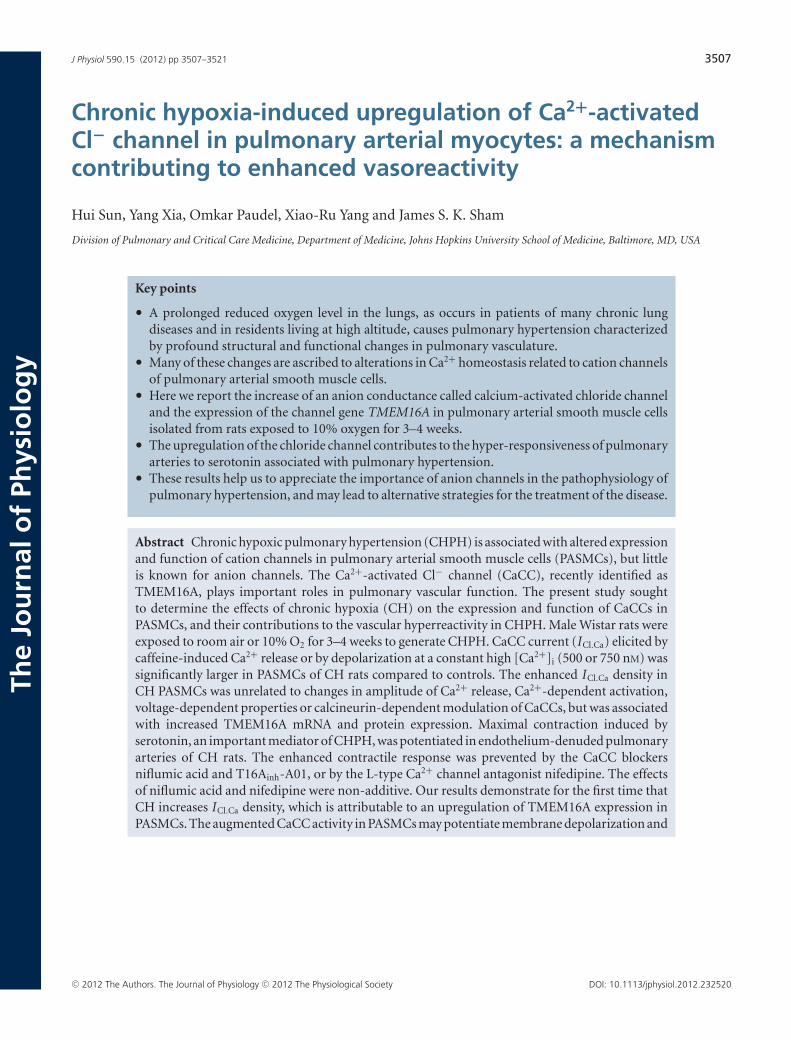
J Physiol 590.15 (2012) pp 3507–3521 3507
The
Jou
rnal
of
Phys
iolo
gy
Chronic hypoxia-induced upregulation of Ca2+-activatedCl− channel in pulmonary arterial myocytes: a mechanismcontributing to enhanced vasoreactivity
Hui Sun, Yang Xia, Omkar Paudel, Xiao-Ru Yang and James S. K. Sham
Division of Pulmonary and Critical Care Medicine, Department of Medicine, Johns Hopkins University School of Medicine, Baltimore, MD, USA
Key points
• A prolonged reduced oxygen level in the lungs, as occurs in patients of many chronic lungdiseases and in residents living at high altitude, causes pulmonary hypertension characterizedby profound structural and functional changes in pulmonary vasculature.
• Many of these changes are ascribed to alterations in Ca2+ homeostasis related to cation channelsof pulmonary arterial smooth muscle cells.
• Here we report the increase of an anion conductance called calcium-activated chloride channeland the expression of the channel gene TMEM16A in pulmonary arterial smooth muscle cellsisolated from rats exposed to 10% oxygen for 3–4 weeks.
• The upregulation of the chloride channel contributes to the hyper-responsiveness of pulmonaryarteries to serotonin associated with pulmonary hypertension.
• These results help us to appreciate the importance of anion channels in the pathophysiology ofpulmonary hypertension, and may lead to alternative strategies for the treatment of the disease.
Abstract Chronic hypoxic pulmonary hypertension (CHPH) is associated with altered expressionand function of cation channels in pulmonary arterial smooth muscle cells (PASMCs), but littleis known for anion channels. The Ca2+-activated Cl− channel (CaCC), recently identified asTMEM16A, plays important roles in pulmonary vascular function. The present study soughtto determine the effects of chronic hypoxia (CH) on the expression and function of CaCCs inPASMCs, and their contributions to the vascular hyperreactivity in CHPH. Male Wistar rats wereexposed to room air or 10% O2 for 3–4 weeks to generate CHPH. CaCC current (ICl.Ca) elicited bycaffeine-induced Ca2+ release or by depolarization at a constant high [Ca2+]i (500 or 750 nM) wassignificantly larger in PASMCs of CH rats compared to controls. The enhanced ICl.Ca density inCH PASMCs was unrelated to changes in amplitude of Ca2+ release, Ca2+-dependent activation,voltage-dependent properties or calcineurin-dependent modulation of CaCCs, but was associatedwith increased TMEM16A mRNA and protein expression. Maximal contraction induced byserotonin, an important mediator of CHPH, was potentiated in endothelium-denuded pulmonaryarteries of CH rats. The enhanced contractile response was prevented by the CaCC blockersniflumic acid and T16Ainh-A01, or by the L-type Ca2+ channel antagonist nifedipine. The effectsof niflumic acid and nifedipine were non-additive. Our results demonstrate for the first time thatCH increases ICl.Ca density, which is attributable to an upregulation of TMEM16A expression inPASMCs. The augmented CaCC activity in PASMCs may potentiate membrane depolarization and
C© 2012 The Authors. The Journal of Physiology C© 2012 The Physiological Society DOI: 10.1113/jphysiol.2012.232520

3508 H. Sun and others J Physiol 590.15
L-type channel activation in response to vasoconstrictors and enhance pulmonary vasoreactivityin CHPH.
(Received 16 March 2012; accepted after revision 1 June 2012; first published online 6 June 2012)Corresponding author J. S. K. Sham: Division of Pulmonary and Critical Care Medicine, 5501, Hopkins Bayview Circle,Baltimore, MD 21224, USA. Email: [email protected]
Abbreviations BKCa, large conductance Ca2+-activated K+ channel; CaCC, Ca2+-activated Cl− channel; CH, chronichypoxia; CHPH, chronic hypoxic pulmonary hypertension; CsA, cyclosporine A; ECl, Cl− equilibrium potential; Emax,maximal contraction; ICl.Ca, Ca2+-activated Cl− current; I inst, instantaneous current; I ss, steady-state current; MCT,monocrotaline; PA, pulmonary artery; PASMC, pulmonary arterial smooth muscle cell; PH, pulmonary hypertension;SR, sarcoplasmic reticulum; TTP, time to peak; TMEM16A, transmembrane protein 16A; VDCC, voltage-dependentL-type Ca2+ channel; VSMCs, vascular smooth muscle cells.
Introduction
Chronic exposure to alveolar hypoxia, as occurs in patientsof many chronic lung diseases and in residents living athigh altitude, causes progressive and sustained increase inpulmonary arterial pressure, right heart hypertrophy andultimately leads to a worsened prognosis of the under-lying disease. Chronic hypoxic pulmonary hypertension(CHPH) is characterized by persistent constriction andprofound remodelling of pulmonary arteries (PAs), andaltered pulmonary vasoreactivity (Karamsetty et al. 1995;Stenmark et al. 2006). The pathogenesis of CHPH is ahighly complex process, involving biochemical, functionaland structural changes in and interactions between end-othelial cells, smooth muscle cells, adventitial fibroblastsand the recruited circulating progenitor cells in PAtissue (Stenmark et al. 2006). A hallmark feature ofpulmonary arterial smooth muscle cells (PASMCs) iso-lated from animals exposed to chronic hypoxia (CH)is the altered homeostasis of [Ca2+]i (Shimoda et al.2000; Platoshyn et al. 2001; Lin et al. 2004), a keydeterminant of the contractile and proliferative stateof PASMCs, and thereby enhanced pulmonary vasculartone and vascular remodelling. Yet the mechanismsunderlying the alterations in [Ca2+]i are not fullyelucidated.
Altered expression and function of ion channelscontribute critically to the elevated [Ca2+]i in PASMCsassociated with CHPH (Moudgil et al. 2006; Weir& Olschewski, 2006). Current evidence indicates thatCH causes down-regulation of voltage-dependent K+
channels (Kv) and reduction in Kv currents (Platoshynet al. 2001; Pozeg et al. 2003) as well as upregulation ofstore-operated, receptor-operated, and stretch-activatedCa2+-permeable non-selective cation channels in PASMCs(Lin et al. 2004; Wang et al. 2006; Ducret et al.2010; Yang et al. 2012) resulting in the increase involtage-dependent and voltage-independent Ca2+ influxand in resting [Ca2+]i. In contrast to the extensive studieson CH-induced cation channel remodelling (Moudgilet al. 2006; Weir & Olschewski, 2006), little is knownabout whether anion channels of PASMCs are affected
by and contribute to CHPH; except for a report showingan increase in swelling-activated Ca2+-independent Cl−
currents in PASMCs of rat exposed to hypoxia for 7 days(Liang et al. 2009). In monocrotaline (MCT)-induced PHmodel, several studies have suggested a possible role of Cl−
channels in PH. For example, the volume-sensitive ClC3Cl− channel was upregulated in PAs from MCT-treatedrats (Dai et al. 2005). Application of Cl− channelblockers abolished the elevation in basal PA myo-genic tone (Nakazawa et al. 2001) and suppressed thenoradrenaline-induced PA constriction to a greater extentin MCT-treated rats (Oriowo, 2004).
Ca2+-activated Cl− channels (CaCCs) are widelyexpressed in many cell types and exert a varietyof important functions, including the regulation ofvascular tone under physiological conditions (Large &Wang, 1996; Hartzell et al. 2005). The recent discoveryof TMEM16A and TMEM16B, two members of themammalian TMEM16 family, as CaCC subunits (Caputoet al. 2008; Schroeder et al. 2008; Yang et al. 2008)has led to the identification of TMEM16A as the maincomponents of CaCCs in rat PASMCs (Manoury et al.2010). Because of active intracellular Cl− accumulation,the Cl− equilibrium potential (ECl) has a more positivevalue than the resting potential of vascular smoothmuscle cells (VSMCs), including PASMCs (Chipperfield &Harper, 2000). Activation of CaCCs by agonist-induced orspontaneous Ca2+ release from the sarcoplasmic reticulum(SR) (Large & Wang, 1996) elicits Cl− efflux, leading tomembrane depolarization (Klockner & Isenberg, 1991;Bakhramov et al. 1996; Yuan, 1997), activation ofvoltage-dependent Ca2+ channels (VDCCs), as well asincrease in [Ca2+]i and vascular tone (Criddle et al.1997; Guibert et al. 1997; Wang et al. 1997; Yuan, 1997;Lamb & Barna, 1998). Indeed, the involvement of CaCCsin the agonist-induced PA constriction has been welldocumented in normoxic animals (Guibert et al. 1997;Yuan, 1997). No information is available, however, onwhether CH alters the expression and activity of CaCCs inPA smooth muscle; and whether these channels contributeto the enhanced pulmonary vasoconstriction associatedwith CHPH. The present study aimed to address these
C© 2012 The Authors. The Journal of Physiology C© 2012 The Physiological Society

J Physiol 590.15 Upregulation of Ca2+-activated Cl− channels in chronic hypoxic pulmonary hypertension 3509
issues with the use of a well-characterized chronic hypoxicrat model of pulmonary hypertension.
Methods
Ethical approval
All animal procedures conform to the Guide for the Careand Use of Laboratory Animals published by the UnitedStates National Institutes of Health and were approvedby the Johns Hopkins University Animal Care and UseCommittee, as well as conforming to UK regulations, asdescribed in Drummond (2009).
Animal model
The CHPH was generated by placing male Wistar rats(150–200 g) in a hypoxic chamber and exposed tonormobaric hypoxia (10 ± 0.5% O2) for 3–4 weeks, aspreviously described and characterized in our laboratory(Shimoda et al. 1999, 2000; Lin et al. 2004). TheO2 level inside the chamber was servo-controlled andcontinuously monitored using PRO:OX 110 gas analysers(Reming Bioinstruments Co., Redfield, NY, USA). Ratswere exposed to room air for 10 min twice a week toclean the cages, and to replenish food and water supplies.Age-matched animals housed in room air were used asnormoxic controls.
Cell isolation
Rats were anaesthetized with sodium pentobarbital(130 mg kg−1, I.P.). Lungs were removed quickly andimmersed in ice-cold Hepes-buffered salt solution (HBSS)containing (in mM) 130 NaCl, 5 KCl, 1.2 MgCl2, 1.5CaCl2, 10 Hepes and 10 glucose (pH 7.2 with NaOH).The third generation of intralobar PAs (outer diameter:300–800 μm) were dissected out and cleaned of connectivetissue. After removal of endothelium by rubbing theluminal surface with a cotton swab, the arteries weredigested at 37◦C for 20 min in 20 μmol-Ca2+ HBSScontaining type I collagenase (1750 U ml−1), papain(9.6 U ml−1), bovine serum albumin (2 mg ml−1) andDTT (1 mmol l−1). After washing with nominal Ca2+-freeHBSS, the PASMCs were mechanically dispersed and keptat 4◦C in the Ca2+-free HBSS for use within 6 h after iso-lation. For some experiments, the cells were kept in Ham’sF-12 medium (with 0.5% of fetal calf serum) overnightbefore use under 4% O2–5% CO2 and 21% O2–5% CO2
(37◦C) for cells isolated from hypoxic and normoxic rats,respectively.
Electrophysiology
Perforated and conventional whole-cell patch clamptechniques were used to record ICl.Ca evoked bycaffeine-induced Ca2+ release and by pipette solutioncontaining high Ca2+, respectively. Membrane currentswere sampled at 5–10 kHz and filtered at 2–5 kHzusing an Axopatch 200B amplifier driven by pCLAMP10 software (Molecular Devices, Sunnyvale, CA, USA).Contamination by other currents was minimized byreplacing K+ ions with Cs+ and by adding TEA chloridein both pipette and bath solutions. For perforatedpatch clamp experiments, the pipette solution contained(in mM): 125 caesium methanesulfonate, 20 CsCl, and10 Hepes (pH 7.2 adjusted with CsOH). Freshly pre-pared amphotericin B was added to the pipette solution(300 μg ml−1) before experiments. For conventionalwhole-cell patch clamp experiments, the pipette solutioncontained (mM): 110 CsCl, 20 TEA-Cl, 2 MgATP, 10 EGTA,5 Hepes and 0.16 MgCl2 (pH 7.2 adjusted with CsOH).CaCl2 at 8.095 or 7.475 mM was also included to set thefree Ca2+ concentration at 750 or 500 nM, respectively, asestimated using the MaxC program (Stanford University).The current–voltage (I–V ) relationships, constructed bydepolarizing the cell from a holding potential (V h) of−70 mV to voltages ranging from −60 to +120 mV in20 mV increments, were assessed every 3–5 min afterwhole-cell formation. The current amplitude usuallyincreased progressively and stabilized after 15–30 minof intracellular dialysis with the Ca2+-buffered pipettesolution. The bath solution for both types of experimentscontained (in mM): 135 NaCl, 5.4 CsCl, 1 MgCl2, 1 CaCl2,0.33 NaH2PO4, 5 TEA-Cl, 10 Hepes and 10 glucose (pH7.35 adjusted with NaOH). The calculated ECl is −50.8 mVfor experiments using amphotericin B-perforated patchclamp method, and −0.5 mV for conventional whole-cellpatch clamp experiments. All experiments were performedat room temperature.
[Ca2+]i measurement
Caffeine-induced Ca2+ transients and ICl.Ca were recordedsimultaneously from PASMCs loaded with fluo-3 or indo1 acetoxymethyl ester (fluo-3/AM or indo-1/AM). Theextracellular dye was washed after incubation of cells with5 μmol l−1 of either indicator for 30–45 min at roomtemperature. The cells were then superfused with bathsolution for ∼20 min to allow for complete de-esterizationof intracellular dye. The intracellular fluo-3 was excited at480 nm and the emitted fluorescence detected at 535 nm.The amplitude of Ca2+ signals was expressed as the ratioof fluorescence change (F) over the resting fluorescence(F0). The intracellular indo-1 was excited at 365 nm andthe emitted fluorescence detected at 405 and 485 nm. The
C© 2012 The Authors. The Journal of Physiology C© 2012 The Physiological Society

3510 H. Sun and others J Physiol 590.15
ratio of fluorescence emitted at 405 nm over that at 485 nm(R405/485) was used as an index of [Ca2+]i.
RT-PCR and quantitative real-time PCR
Total RNA was extracted from endothelium-denudedPAs, and first-strand cDNA was synthesized.Gene-specific primers were used to amplifyTMEM16A (5′-TGACGAGGATACCAAAATCCA-3′ and5′-CGGGTCTCACTGATGTGGT-3′) and 18S rRNA(5′-CGGCTACCACATCCAAGGAA-3′ and 5′-AGCTGGAATTACCGCGGC-3′). Real-time PCR reactions wereperformed with SYBR Green PCR Master Mix on an iQ5Real-time PCR detection System. For the internal control,18S rRNA was used. Standard curves for each gene weregenerated for each real-time PCR reaction. The absolutecopy number of mRNA for each gene was determinedby interpolation of their respective standard curves withthe threshold cycle value of each sample. The TMEM16AmRNA levels were expressed as their respective copynumber relative to that of 18S rRNA for each sampleobtained in the same run.
Western blot
Endothelium-denuded PAs were homogenized andresuspended in ice-cold lysis buffer containing 50 mM
Tris-Cl (pH 7.4), 150 mM NaCl, 1% deoxycholic acid,0.1% SDS, 0.5% NP-40 and protease inhibitor cocktail(Roche, Mannheim, Germany). The homogenate wascentrifuged at 4◦C at 1000 g for 5 min, the supernatantwas collected, and the total amounts of proteins werequantified with bicinchoninic acid assay. The proteins(5 μg) were separated on SDS-polyacrylamide gels (10%),transferred onto polyvinylidene difluoride membranes,immunoblotted with a goat polyclonal antibody againstTMEM16A (sc-69343, Santa Cruz Biotechnology, Inc.,Santa Cruz, CA, USA) at 1:200 dilution and visualizedwith Pierce ECL (Thermo Scientific). Competition studiesusing blocking peptide supplied with the antibody wereperformed according to manufacturer’s instruction toverify the specificity of results. Actin, used as internalcontrol, was immunodetected using a goat polyclonalantibody (sc-1615, Santa Cruz Biotechnology, Inc.) foreach experiment. The intensity of interested bands wasquantified using the Gel Logic 200 image system (Kodak,New Haven, CT, USA).
Isometric tension measurements in PAs
Intralobar PAs (o.d.: 300–800 μm) were isolated, cleanedof connective tissue and cut into ring segments 4 mmin length. Endothelium was disrupted by gently rubbingthe lumen with a small wooden stick. The PA rings
were suspended between two stainless steel stirrups andmounted in organ chambers filled with modified Krebssolution containing (in mM): 118.3 NaCl, 4.7 KCl, 1.2MgSO4, 25 NaHCO3, 11.1 glucose, 1.2 KH2PO4 and 2CaCl2. The solution was gassed with 16% O2–5% CO2
to maintain a pH of 7.4 at 37◦C. The isometric tensionwas measured using a strain gauge (Grass Instruments,Quincy, MA, USA) connected to a PowerLab amplifierdriven by the Chart software (ADInstruments Inc.,Colorado Springs, CO, USA). After equilibrium undera resting tension of 0.8 g for 60 min, the arteries wereexposed to 80 mM KCl 3 times with 30 min of washout toestablish viability and maximum contraction. Serotoninconcentration-dependent responses were assessed byadding the agonist cumulatively (0.5 log unit increments)to the bath in the presence of the vehicle solution (DMSO),or different inhibitors added 15 min before application ofserotonin. The concentration–response curves obtainedin the presence DMSO were used as controls. In somecases as indicated, the artery rings were first challengedwith a maximum dose of serotonin (10−4 M), and theeffects of different drugs were tested when the contractileresponse reached the plateau. At the end of experiments,the PA ring was exposed to phenylephrine (3 × 10⁻⁷ M)followed by acetylcholine (10−6 M) to verify completedisruption of endothelium. The rings that relaxedwith acetylcholine or did not contract with KCl werediscarded. The isometric tension response to serotonin wasnormalized to the maximal tension generated by 80 mM ofKCl.
Chemicals and drugs
Fluo-3/AM and Indo-1/AM (Molecular Probes) stockswere prepared in 20% pluronic acid F-127 (MolecularProbes) solution prepared with DMSO. The stocksolution of serotonin (Sigma) was prepared inminipore water. Niflumic acid (Sigma), nifedipine(Research Biochemicals International) and T16Ainh-A01(Asinex) stock solutions were prepared in DMSO atconcentrations at least 1000 times of that used in theexperiments.
Statistical analysis
Student’s t tests or analyses of variance were performed toevaluate the statistical significance of differences betweentwo means, multiple means or two means at multiple testvoltages as indicated. Tukey’s test as a post hoc analysiswas performed for multiple pairwise comparisons. Pooleddata were expressed as means ± standard error ofmean.
C© 2012 The Authors. The Journal of Physiology C© 2012 The Physiological Society

J Physiol 590.15 Upregulation of Ca2+-activated Cl− channels in chronic hypoxic pulmonary hypertension 3511
Results
Caffeine-induced ICl.Ca density is enhanced in PASMCsof CH rats
To investigate whether CH alters CaCC activity in ratintralobar PASMCs, we first characterized the ICl.Ca
evoked by caffeine-induced Ca2+ release from the SR influo-3/AM-loaded normoxic PASMCs using perforatedpatch clamp technique. Rapid application of 10 mM
caffeine evoked a robust outward current accompaniedby a large Ca2+ signal in a cell held at 0 mV (Fig. 1A).A second caffeine application immediately following thefirst did not induce further Ca2+ release or detectablecurrent, suggesting that the caffeine-activated current isCa2+-dependent. When sufficient time (5–10 min) wasallowed for refilling SR with Ca2+, repetitive caffeineapplication (5 mM) at different holding potentials evoked
Ca2+ transients of similar amplitude accompanied bya membrane current changing progressively from alarge inward current at −80 mV to a large outwardcurrent at 0 mV, with no significant current beingrecorded at −35 mV (Fig. 1B). The current–voltage (I–V )relation exhibited linear voltage dependence (Fig. 1B).The current reversed at −49.8 ± 0.51 mV (n = 6 cells)after correction for junction potential, a value similar tothe predicted ECl (−50.8 mV) set for these experiments.The Cl− selectivity of this current was further verifiedby changing the transmembrane Cl− gradient. When[Cl−]o was lowered from 150 to 20 mM (equal to[Cl−]i), the outward current recorded at 0 mV wasabolished despite a comparable caffeine-induced Ca2+
transient (Fig. 1C). The caffeine-induced current wasalso inhibited by niflumic acid (100 μM, Fig. 1D). Takentogether, these results are consistent with ICl.Ca evoked bycaffeine-induced Ca2+ release in PASMCs.
Figure 1. Characterization of caffeine-induced ICl.Ca in rat PASMCsA, typical caffeine-induced current and Ca2+ transient measured with perforated patch clamp technique from acell at a holding potential (Vh) of 0 mV. B, representative caffeine-induced currents and Ca2+ transients recordedfrom a cell at different Vh, and plot of peak I–V relation obtained from the same cell. The values of Vh were notcorrected for junction potential, which was −14.7 mV ([Cl−]i/[Cl−]o = 20/150 mM). C, representative recordingsand mean data (n = 6 cells) showing the effects of reducing [Cl−]o on caffeine-induced currents and Ca2+signals (Vh = 0 mV). ∗P < 0.05 (Student’s paired t test). D, representative recordings and mean data (n = 5 cells,Vh = 0 mV) showing the effects of 100 μM niflumic acid (NFA) on caffeine-induced ICl.Ca and Ca2+ transients.∗P < 0.05 (Student’s paired t test).
C© 2012 The Authors. The Journal of Physiology C© 2012 The Physiological Society

3512 H. Sun and others J Physiol 590.15
The effects of CH on caffeine-induced ICl.Ca wereexamined at 0 and −80 mV. Figure 2A and B showstypical caffeine-induced ICl.Ca and Ca2+ signal obtainedfrom single normoxic and hypoxic PASMCs. The ICl.Ca
density in CH cells was significantly higher than thatof normoxic PASMCs, without apparent difference inthe peak amplitude of Ca2+ release between the twogroups. The magnitude of increase in ICl.Ca density inCH PASMCs was similar in both inward and outwarddirection (Fig. 2C). To confirm that the increase in ICl.Ca
density in CH PASMCs was not due to changes inthe amplitude of caffeine-induced Ca2+ release, ICl.Ca
and Ca2+ transient elicited by rapid caffeine applicationwere simultaneously measured in the ratiometric Ca2+
indicator indo-1/AM-loaded PASMCs held at 0 mV. Asshown in Fig. 2D, while the ICl.Ca density was significantlyincreased in CH cells compared to normoxic cells, thecaffeine-induced changes in indo-1 ratio (�R405/485),reflecting the amplitude of Ca2+ transients, were notdifferent between two groups. These results reveal forthe first time that CH enhances ICl.Ca activated by Ca2+
release from the ryanodine receptor-gated Ca2+ stores in
PASMCs, and this increased current density is not due toan increase in the amplitude of Ca2+ release under ourexperimental conditions.
Effects of CH on Ca2+-dependent properties ofcaffeine-induced ICl.Ca
To examine whether Ca2+-dependent properties ofcaffeine-induced ICl.Ca were altered by CH, the kineticsof simultaneously recorded Ca2+ transient (by fluo-3) andICl.Ca evoked by caffeine were analysed. The activationrates of ICl.Ca and Ca2+ release, measured as time to peak(TTP) of the signals, were not different between normoxicand hypoxic groups at either 0 or −80 mV (Fig. 3A). Thespeed of activation of ICl.Ca correlated strongly with that ofcaffeine-induced Ca2+ signals in both normoxic and hypo-xic cells at both 0 and −80 mV, with the slope of linearregression derived from CH cells slightly reduced at 0 mV(Fig. 3B). The positive correlation of TTP of ICl.Ca andCa2+ transients confirms the Ca2+-dependent activationof ICl.Ca, and the absence of change in the relationship in
Figure 2. Effects of CH on caffeine-induced ICl.Ca and Ca2+ transientsA and B, representative caffeine-induced ICl.Ca and Ca2+ release simultaneously recorded at 0 and −80 mVfrom normoxic and hypoxic cells loaded with fluo-3/AM, respectively, using perforated patch clamp technique.Recordings were obtained from different cells. C, average amplitudes of caffeine-induced ICl.Ca density andCa2+ transients measured from fluo 3-loaded normoxic and hypoxic PASMCs at 0 and −80 mV. n = 13–16for each group. ∗P < 0.05 (Student’s t test). Membrane capacitance (Cm): 10.0 ± 1.2 pF and 10.6 ± 1.1 pF fornormoxic cells held at 0 and −80 mV, respectively; 9.8 ± 1.4 pF and 10.1 ± 1.1 pF for CH cells held at 0 and−80 mV. D, representative recordings (top) and mean amplitude (bottom, n = 10 each group) of caffeine-inducedICl.Ca and Ca2+ release recorded from normoxic and hypoxic cells held at 0 mV and loaded with indo−1/AM.Cm = 13.4 ± 1.6 pF for normoxic and 13.2 ± 0.9 pF for CH group. ∗P < 0.05 (Student’s t test).
C© 2012 The Authors. The Journal of Physiology C© 2012 The Physiological Society

J Physiol 590.15 Upregulation of Ca2+-activated Cl− channels in chronic hypoxic pulmonary hypertension 3513
the two groups of cells indicate that the Ca2+ dependenceand time course of CaCC activation were unalteredby CH.
In normoxic PASMCs, the current exhibited fasterdeactivation at −80 mV than at 0 mV, while the relaxationkinetics of concomitant Ca2+ signals was not different atboth voltages (Fig. 3C). No correlation was found betweenthe ICl.Ca deactivation and Ca2+ transient relaxationkinetics at either 0 or −80 mV (Fig. 3D). Similar fasterdeactivation of ICl.Ca at the negative voltage was foundin hypoxic cells. Compared to the normoxic control,however, the deactivation of ICl.Ca was significantly slowerat −80 mV accompanied by a slower relaxation of Ca2+
signals (Fig. 3C). A similar tendency in ICl.Ca deactivationwas also observed at 0 mV (P = 0.057). Plotting the 50%deactivation time of ICl.Ca against the 50% relaxation timeof Ca2+ transients revealed a fair correlation of these twoprocesses in hypoxic cells (R = 0.723, P = 0.012 at 0 mV;and R = 0.703, P = 0.023 at −80 mV) (Fig. 3D). Theseresults suggest that the relaxation rate of Ca2+ transients is
decreased in hypoxic PASMCs, which could be responsiblefor the slower decline of caffeine-induced ICl.Ca observedin these cells.
Effects of CH on voltage-dependent properties ofICl.Ca in PASMCs
To further investigate whether the voltage-dependentproperties of CaCCs were altered by CH and contributedto enhanced ICl.Ca, we examined the current evokedby depolarization at a high [Ca2+]i (750 nM) set bya Ca2+-buffered pipette solution under conventionalwhole-cell configuration in freshly isolated cells.Depolarization evoked slowly activating outward currentsfollowed by inward tail currents upon repolarizationin both normoxic and hypoxic PASMCs. The currentamplitude was significantly higher in CH cells (Fig. 4A).Niflumic acid (100 μM) almost completely eliminatedthe time-dependent outward currents and tail currentsin both cell types. The I–V relationships of steady-state
Figure 3. Effects of CH on the kinetics ofcaffeine-induced ICl.Ca and Ca2+ transientsmeasured from fluo-3-loaded cellsA, average time to peak of caffeine-induced ICl.Ca(TTP-ICl.Ca) and Ca2+ transients (TTP-CaT) obtainedfrom normoxic and hypoxic PASMCs held at 0 (n = 13for normoxic, 11 for hypoxic group) and −80 mV(n = 14 for normoxic, 11 for hypoxic group). B, scatterplots of TTP-ICl.Ca against TTP-CaT measured at 0 and−80 mV from normoxic and hypoxic cells. Thecorrelation coefficient (r) = 0.83 for normoxic and 0.8for hypoxic group at 0 mV (P < 0.05 for slopecomparison), while r = 0.74 for normoxic and 0.86 forhypoxic group at −80 mV. C, average time for 50%relaxation of ICl.Ca (R50-ICl.Ca) and Ca2+ transients(R50-CaT) measured from the peak of the signals fornormoxic and hypoxic cells held at 0 (n = 13 fornormoxic, 11 for hypoxic group) and −80 mV (n = 14for normoxic, 10 for hypoxic group). ∗P < 0.05compared to 0 mV. ∗∗P < 0.05 between normoxic andhypoxic groups (Student’s t test). D, scatter plots ofR50-ICl.Ca against R50-CaT measured at 0 and −80 mVfrom normoxic and hypoxic cells. r = 0.06 for normoxicand 0.72 for hypoxic group (0 mV). r = 0.4 fornormoxic and 0.7 for hypoxic group (−80 mV).
C© 2012 The Authors. The Journal of Physiology C© 2012 The Physiological Society

3514 H. Sun and others J Physiol 590.15
currents (I ss), measured at the end of 1 s pulses, exhibiteda strong outward rectification (Fig. 4B, left). Despite amarked increase in the current density in CH cells, theratio of current amplitude measured at +60 mV overthat at −60 mV (i.e. rectification index) was not differentbetween normoxic and hypoxic groups (8.7 ± 0.5, n = 10vs. 8.2 ± 0.8, n = 10, P > 0.05 by Student’s t test). Theinstantaneous current (I inst) measured at the end ofdepolarization-induced capacitance transients, reflectingthe portion of channels activated at the holding potential,was also elevated in CH cells (Fig. 4B, middle). The ratioof I inst/ISS was not, however, altered by CH over theexamined voltage range (Fig. 4B, right). The activationtime constants, obtained by fitting the outward currentdeveloped during depolarization to a monoexponentialfunction, were similar between the two groups and
independent of membrane potential at voltages between+40 and +120 mV (Fig. 4C). These results suggestthat the activation kinetics of ICl.Ca during sustaineddepolarization, which is sensitive to Ca2+ (Kuruma &Hartzell, 2000), remained unchanged in hypoxic cells.The steady-state activation curves of ICl.Ca of normoxicand hypoxic PASMCs were generated by fitting thechord conductance measured at the end of 1 s pulsesand plotted against the depolarizing voltages (Fig. 4D).CH significantly augmented the maximal conductance(2.06 ± 0.32 vs. 0.87 ± 0.07 nS pF−1, P < 0.01 by Student’st test), but did not affect the V 1/2 (141.2 ± 12.7 vs.153.7 ± 12.2 mV, P > 0.05). These results indicate thatCH enhanced the ICl.Ca density without altering therectification of current, and voltage-dependent activationof the channels.
Figure 4. Effects of CH on ICl.Ca elicited by depolarization of PASMCs buffered at a [Ca2+]i of 750 nM
with conventional whole-cell patch clamp techniqueA, representative recordings from a normoxic and a hypoxic cells in the absence or presence of 100 μM
niflumic acid. B, plots of mean Iss, Iinst and the ratio of Iinst/ISS against the voltages for normoxic (n = 10,Cm = 14.3 ± 1.0 pF) and hypoxic (n = 10, Cm = 15.2 ± 1.6 pF) groups. [Cl−]i/[Cl−]o = 146.5/149.4 mmol l−1.Predicted ECl = −0.5 mV. C, mean activation time constants plotted against depolarizing voltages (Vm) fornormoxic and CH cells. D, mean steady-state activation curves generated by plotting the chord conductance(G = I/(Vm − ECl)) obtained at the end of depolarization against Vm. The continuous and dashed lines representthe fits of data points obtained from normoxic and hypoxic cells (n = 10 each group), respectively, to theBoltzmann function: G = Gmax/(1 + exp(−(Vm − V1/2)/k)), where Gmax represents the maximal conductance, V1/2
the voltage at which 50% of activation occurs and k the slope. E, representative tail currents (left) and plot of meandeactivation time constants (τdeactivation) against repolarizing voltages (right) obtained from normoxic and hypoxiccells (n = 6 each group). τdeactivation was obtained by fitting the decay phase of tail currents to a monoexponentialfunction.∗P < 0.0001 (two-way ANOVA).
C© 2012 The Authors. The Journal of Physiology C© 2012 The Physiological Society

J Physiol 590.15 Upregulation of Ca2+-activated Cl− channels in chronic hypoxic pulmonary hypertension 3515
To investigate whether the slower deactivation ofcaffeine-induced ICl.Ca in CH cells was entirely due tothe slower relaxation of Ca2+ transients, the deactivationproperties of ICl.Ca evoked at constant high [Ca2+]i
(750 nM) were examined. Figure 4E shows the tail currentselicited by repolarization to different voltages from astrong depolarization in a normoxic and a CH cell. Thedecay of tail currents accelerated as the repolarizing voltagebecame more negative in both cell types, a well-knownproperty of classical ICl.Ca (Kuruma & Hartzell, 2000).The time constants of deactivation were slightly, butsignificantly greater in hypoxic PASMCs at all voltagesexamined (Fig. 4E, right). These results revealed a genuinealteration in ICl.Ca deactivation kinetics (independent ofCa2+ changes) in hypoxic PASMCs.
Calcineurin inhibitor cyclosporine A did not affectCH-induced increase in ICl.Ca
Several recent studies suggest that the CaCC activityin vascular smooth muscle cells is modulated byCa2+–calmodulin-dependent kinase II (CaMKII) and theCa2+-dependent phosphatase calcineurin. Inhibition ofCaMKII or intracellular dialysis of constitutively activecalcineurin enhanced ICl.Ca and altered the activation anddeactivation kinetics (Ledoux et al. 2003; Greenwoodet al. 2004). Moreover, CH-induced increased activationof nuclear factor of activated T cells, which can beprevented by calcineurin inhibitor cyclosporine A, hasbeen reported in mouse PASMCs, suggesting increasedcalcineurin activity in CHPH (de Frutos et al. 2011).We therefore examined whether the enhanced ICl.Ca
density and slower channel deactivation observed in CHcells are due to altered channel function by calcineurin.Application of 10 μM cyclosporine A in both bath andpipette solutions did not alter significantly the densityof ICl.Ca, the I–V relation or activation kinetics, butaccelerated the deactivation in both normoxic and hypo-xic PASMCs dialysed with 500 nM of [Ca2+]i (Fig. 5).These results suggest that the CH-induced alterations inICl.Ca were likely not the result of a change in channelmodulation mediated by calcineurin.
Increased TMEM16A mRNA and protein expression inPAs of chronically hypoxic rats
To determine whether the enhanced ICl.Ca density observedin CH PASMCs resulted from the upregulation ofCaCC channel expression, TMEM16A mRNA and proteinexpression in endothelium-denuded PAs were examined.Quantitative RT-PCR analysis revealed a significantincrease in the TMEM16A mRNA levels in PA samplesfrom CH rats (Fig. 6A). Western blot analysis detecteda band of ∼114 kDa (the predicted size of TMEM16A)
in PA protein lysates from both normoxia and CHrats. The signal was much stronger in the hypoxicPA samples (Fig. 6B). The specificity of the antibodyagainst TMEM16A was verified with the blocking peptide,which significantly reduced the intensity of the 114 kDaband. Pooled data showed a significant increase inthe TMEM16A protein level in hypoxic PAs (Fig. 6C).These results indicate that CH enhances the TMEM16Aexpression in PAs, and that the CH-induced increase inTMEM16A expression occurs at both mRNA and proteinlevels.
Figure 5. Effects of cyclosporine A (CsA) on ICl.Ca elicited bydepolarization at 500 nM [Ca2+]iMean I–V relations (A), voltage dependence of activation (B) anddeactivation (C) time constants were obatained from normoxic (Nx)and CH PASMCs in the absence (Ctl) and presence of 10 μM of CsA.n = 6 for each group. Cm = 14.9 ± 1.8 pF for normoxic and15.7 ± 1.7 pF for CH group. ∗P < 0.05 (two-way ANOVA),∗∗P < 0.001 (three-way ANOVA).
C© 2012 The Authors. The Journal of Physiology C© 2012 The Physiological Society

3516 H. Sun and others J Physiol 590.15
Contribution of upregulated CaCCs to enhanced PAcontractile response to serotonin in CH rats
To explore the pathophysiological function of enhancedCaCC activity in CHPH, we examined the contributionof CaCCs to the contractile response of PAs isolatedfrom normoxic and CH rats to serotonin, a vaso-constrictor and mitogen that plays an important rolein the pathogenesis of CHPH (MacLean & Dempsie,2009). The maximal serotonin-induced contraction (Emax,relative to KCl-induced contraction) was enhanced inCH PAs compared to normoxic controls with anincreased sensitivity to the agonist (EC50 = 4.68 ± 0.65vs. 11.20 ± 0.84 μM, P < 0.05, Student’s t test), consistentwith previous reports (Fig. 7A) (MacLean et al. 1996).The amplitude of KCl-induced PA contraction wasnot different between normoxic and CH groups(0.813 ± 0.026 g, n = 66 vs. 0.808 ± 0.025 g, n = 83,P > 0.05, Student’s t test). The CaCC blocker niflumic acid(30 μM, a concentration showing no effect on KCl-inducedPA contraction; Yuan, 1997) or the VDCC antagonistnifedipine (1 μM) attenuated the contractile response toserotonin to a similar extent (Fig. 7B) without affectingthe EC50 (Fig. 7C) in both normoxic and CH rats.Combined application of niflumic acid and nifedipinedid not produce additive inhibitory effects. Furthermore,the enhanced serotonin-induced maximal contractionobserved in PAs of CH rats disappeared in the presence ofthese agents when applied individually or simultaneously(Fig. 7D). The inhibition of serotonin response by niflumicacid was not due to the stimulation of Ca2+-activatedK+ channels (BKca), since the pretreatment of PArings with 1 mM TEA did not alter the magnitude ofconstriction induced by serotonin (100 μM), nor theinhibitory effects of niflumic acid in both normoxicand CH rats (Fig. 7E). Application of T16Ainh-A01, a
newly identified TMEM16A channel-specific inhibitorthat is structurally different from niflumic acid(Namkung et al. 2011), also eliminated the increasein serotonin-induced contraction in CH PAs (Fig. 7F).Collectively, these results suggest that the augmentedCaCC activity found in CH PASMCs contributes tothe enhanced pulmonary vasoconstriction in responseto serotonin observed in CH rats via the activation ofVDCCs.
Discussion
The development of CHPH significantly worsens theprognosis of patients suffering from many chroniclung diseases. Better understanding of the underlyingpathophysiological processes may lead to novel therapiesfor more effective management of this clinical syndrome.Membrane depolarization and increased [Ca2+]i inPASMCs of CH animals (Shimoda et al. 2000; Bonnet et al.2001; Platoshyn et al. 2001; Lin et al. 2004; Rodat et al.2007) are known to play a pivotal role in the inductionof PASMC proliferation and the increase of PA toneassociated with CHPH (Platoshyn et al. 2000; Moudgilet al. 2006; Cogolludo et al. 2007). Extensive studies on therole of cation channels in CHPH have reached the currentconsensus that the altered Ca2+ homeostasis in PASMCsassociated with CHPH is the result of down-regulationof Kv channels and upregulation of Ca2+ permeablecation channels caused by CH (Moudgil et al. 2006;Weir & Olschewski, 2006). In the present study, we havedemonstrated for the first time that the ICl.Ca activity andTMEM16A expression are enhanced in PA smooth muscleof CH rats. The increased CaCC activity, as a result ofupregulation of TMEM16A expression, contributes to theenhanced pulmonary vasoreactivity to agonist associated
Figure 6. Effects of CH on TMEM16A mRNA and protein expressionA, the amount of TMEM16A mRNA, relative to 18S RNA, measured in normoxic and CH PAs. n = 6 animals foreach group, ∗P < 0.05 (Student’s t test). B, representative immunoblots of protein lysates prepared from normoxicand hypoxic PAs, respectively, using antibody targeting TMEM16A or actin (left), and from a normoxic PA in theabsence and presence of an anti-TMEM16A blocking peptide (right). C, mean TMEM16A protein levels normalizedto actin obtained from normoxic and hypoxic PAs (n = 6 rats for each group). ∗P < 0.05 (Student’s t test).
C© 2012 The Authors. The Journal of Physiology C© 2012 The Physiological Society

J Physiol 590.15 Upregulation of Ca2+-activated Cl− channels in chronic hypoxic pulmonary hypertension 3517
with CHPH through a mechanism involving the activationof voltage-dependent Ca2+ influx. A possible role forCaCCs in mediating the ex vivo hypoxia-induced increasein resting [Ca2+]i in PASMCs has also been proposedby Yang et al. (2006) based on their observation thatthe presence of Cl− channel blockers niflumic acid orindaryloxyacetic acid in culture medium prevented theincrease in [Ca2+]i and proliferation of PASMCs exposedto hypoxia for 48 h.
CH-induced increased ICl.Ca in PASMCs is attributableto an upregulation of TMEM16A expression
Our results show that a rapid rise in the [Ca2+]i caused bycaffeine-induced Ca2+ release, and steady state elevation of[Ca2+]i by intracellular dialysis of buffered Ca2+ solutionboth activated robust ICl.Ca in rat PASMCs. This currentshares similar biophysical and pharmacological properties,such as voltage and Ca2+ dependence of activation,voltage-dependent deactivation, selectivity to Cl−, and
Figure 7. Effects of Cl− channel blockers and VDCC inhibitor on serotonin-induced contractile responseof PA rings from normoxic and CH ratsA, cumulative concentration–response curves obtained from normoxic (n = 33) and CH (n = 26) PAs by fittingthe mean data points to a sigmoidal function. Inset: normalized curves showing the leftward shift of EC50 in CHrats. B, cumulative concentration–response curves obtained from normoxic and CH rings in the absence (Control,n = 33 for normoxic, 26 for hypoxic group) or presence of 30 μM niflumic acid (NFA 30, n = 9 for both groups),or 1 μM nifedipine (Nif 1, n = 13 for normoxic, 9 for hypoxic group) or both niflumic acid and nifedipine (Nif+ NFA, n = 9 for normoxic, 8 for hypoxic group). Different curves represent fitting of mean data points of eachgroup to the sigmoidal function. C, mean values of EC50 derived from experiments shown in panel B. ∗P < 0.05compared to normoxic group for each treatment (Student’s t test). n.s.: P > 0.05 (one-way ANOVA). D, averageEmax values obtained from experiments shown on panel B. Emax was determined for each experiment by fitting thedata points to the sigmoidal function. ∗P < 0.001 compared to normoxic group; #P < 0.001 compared to Controlwithin normoxic or CH group (one-way ANOVA). E, bar graph showing the contractile response of normoxic andhypoxic PAs to 10−4 M serotonin and inhibitory effects of niflumic acid (NFA, 30 μM) in the absence or presence ofTEA (1 mM). n = 10 and 12 for normoxic group with and without TEA treatment, respectively. n = 11 and 12 forhypoxic group with and without TEA treatment, respectively. ∗P < 0.05 compared to normoxic groups (Student’s ttest). #P < 0.01, †P < 0.001 compared to controls (absence of NFA) within normoxic and CH groups, respectively(one-way ANOVA). F, representative recordings (top) and bar graph (bottom) showing the amplitude of contractileresponse of normoxic and hypoxic PAs to serotonin (10−4 M) and inhibitory effects of TMEM16 channel blockerT16Aihn-A01 (10 μM). Normoxic: n = 8. Hypoxic: n = 11. ∗P < 0.001 compared to normoxic group; #P < 0.001compared to respective control (one-way ANOVA).
C© 2012 The Authors. The Journal of Physiology C© 2012 The Physiological Society

3518 H. Sun and others J Physiol 590.15
sensitivity to niflumic acid with the previously reportedclassical ICl.Ca (Large & Wang, 1996; Leblanc et al. 2005).This current also bears the signature feature of the classicalICl.Ca that the current–voltage relation is different whenrecorded at different [Ca2+]i (Kuruma & Hartzell, 2000;Xiao et al. 2011), e.g. the strong outward rectificationof ICl.Ca steady-state I–V relationship at submicromolar[Ca2+]i (750 nM), and the linear relationship at high[Ca2+]i caused by caffeine-induced Ca2+ release observedin this study. The ICl.Ca evoked by caffeine or by bufferedCa2+ solution was significantly increased in PASMCs ofCH rats. We did not observe, however, a significant changein time-independent baseline Cl− currents, as reportedpreviously in PASMCs of rats exposed to 1 week of hypo-xia (Liang et al. 2009), in the CH myocytes under ourrecording conditions.
Detailed analyses suggest that the CH-induced increasein ICl.Ca density is not due to an increase in Ca2+
sensitivity of the channel or alterations in voltage- andCa2+-dependent activation. First, our results showed thatthe rectification index of ICl.Ca was not different betweennormoxic and hypoxic myocytes at a given [Ca2+]i, andthe voltage dependence of steady-state activation wasunchanged by CH despite a marked increase in themaximal conductance of the channel. This is incompatiblewith an increase in CaCC Ca2+ sensitivity, which is knownto associate with a reduction in outward rectificationof ICl.Ca and a shift of steady-state activation curveto the negative potentials (Kuruma & Hartzell, 2000;Xiao et al. 2011). Second, the activation kinetics ofICl.Ca during sustained depolarizations, which is voltageindependent and Ca2+ sensitive (Kuruma & Hartzell,2000), remained unchanged in CH cells as demonstratedby similar activation time constants and ratios of I inst/I ss
found in two groups of cells over a wide range ofvoltages. Third, CH did not affect the Ca2+ dependenceof ICl.Ca activation, as correlations between the rate ofcaffeine-induced rise in [Ca2+]i and the rate of ICl.Ca
activation were similar in normoxic and hypoxic cells.Fourth, inhibition of calcineurin by cyclosporine A hadno effects on the ICl.Ca density despite the acceleratedchannel deactivation in both normoxic and CH myo-cytes. The lack of effects of calcineurin inhibition on ICl.Ca
density in normoxic PASMCs is inconsistent with pre-vious reports (Greenwood et al. 2004), and it is currentlyunclear whether this is due to a difference in the basalphosphorylation states of the channels, or in the species(rat vs. rabbit) or PA regions (intralobar vs. extralobar)used in different studies. Finally, analyses of TMEM16Aexpression revealed a significant increase in mRNA andprotein levels in PAs from CH rats. Taken together, itseems reasonable to suggest that the CH-induced increasein ICl.Ca density in PASMCs is mainly attributable to theupregulation of CaCC protein TMEM16A expression byCH.
Another observation in this study is that CH altersthe deactivation kinetics of ICl.Ca in PASMCs, and theunderlying mechanism is not entirely clear. The slowerdecay of caffeine-induced ICl.Ca observed in hypoxic cellsmay be due in part to a slower relaxation of Ca2+ trans-ients because sustained increase in [Ca2+] slows downCaCC deactivation due to a reduced rate of dissociationof Ca2+ from the channel (Kuruma & Hartzell, 2000; Xiaoet al. 2011). Slower relaxation of caffeine-induced Ca2+
transients has also been reported by others in PASMCsof CH rats and attributed to impaired SR Ca2+ reuptake(Bonnet et al. 2001). However, similar observation in CHPASMCs at constant [Ca2+]i suggests other additionalmechanisms might be involved. The finding that cyclo-sporine A treatment accelerated the deactivation of ICl.Ca
to the same extent in normoxic and CH cells rules out thepossibility that this alteration in kinetics was mediatedby calcineurin. Regardless the underlying mechanism,the slower deactivation of CaCCs together with slowerrelaxation of agonist-induced Ca2+ signals would favoursustained membrane depolarization in PASMCs and maycontribute to maintaining the elevation in vascular toneunder the influence of vasoconstrictors in CH rats.
Enhanced CaCC activity contributes to potentiatedagonist-induced pulmonary vasoconstriction in CHPH
Altered pulmonary vasoreactivity to vasoactive agents isone of the salient features of CHPH (Karamsetty et al.1995; MacLean et al. 1996; Shimoda et al. 2000; Cogolludoet al. 2007). Enhanced PA constriction in response to vaso-constrictors such as serotonin and endothelin-1 has beenwell documented in CH animals (Karamsetty et al. 1995;MacLean et al. 1996; Shimoda et al. 2000). The contra-ctile response evoked by agonist is primarily mediatedby a rise in [Ca2+]i resulting from increased Ca2+ influxand SR Ca2+ release, as well as an increase in myo-filament Ca2+ sensitivity. In contrast to the dominant roleof SR Ca2+ release in providing Ca2+ for striated musclecontraction, Ca2+ released from SR in arterial SMCs playsan important regulatory role in controlling membranepotential by targeting Ca2+-sensitive ion channels on theplasma membrane (Thorneloe & Nelson, 2005). CaCCsand BKCa channels are two major types of Ca2+-activatedchannels in VSMCs including PASMCs. Activation ofCaCC depolarizes the membrane potential, leading to theactivation of VDCCs, increase in [Ca2+]i and contraction(positive feedback), whereas activation of BKCa channelsresults in opposite effects leading to cell relaxation(negative feedback) (Wellman & Nelson, 2003). It is welldocumented that CaCCs in PASMCs can be activated byspontaneous or agonist-evoked Ca2+ release from the SRat physiological membrane potentials, and their activationcontributes to agonist-induced PA constriction by means
C© 2012 The Authors. The Journal of Physiology C© 2012 The Physiological Society

J Physiol 590.15 Upregulation of Ca2+-activated Cl− channels in chronic hypoxic pulmonary hypertension 3519
of the positive feedback mechanism (Large & Wang, 1996;Guibert et al. 1997; Yuan, 1997). Increased TMEM16Aexpression and ICl.Ca density in PASMCs of CH ratsfound in the present study may lead to more robustmembrane depolarization for Ca2+ influx via VDCCsand enhance vascular reactivity to agonists. Consistentwith this hypothesis, our study demonstrated that twostructurally different CaCC blockers, niflumic acid andT16Ainh-A01, or VDCC antagonist nifedipine attenuatedthe contractile response of PAs to serotonin in bothnormoxic and CH rats, and abolished the enhancedpulmonary vasoconstriction to serotonin observed inCH rats. Moreover, combined use of niflumic acid andnifedipine did not produce further inhibition.
The markedly enhanced PA vasoconstriction toserotonin with significant increase in both efficacy andsensitivity observed in CH rats in this study is consistentwith previous reports (MacLean et al. 1996; Homma et al.2007; Rodat et al. 2007). It has been proposed that theenhanced responsiveness to serotonin is due to the increasein the activity of 5-HT1B or 5-HT2B receptors (MacLeanet al. 1996; MacLean & Dempsie, 2009) and reactive oxygenspecies production (Liu et al. 2006), as well as related tothe enhanced activation of RhoA/Rho kinase (Hommaet al. 2007) and voltage-independent Ca2+ influx (Rodatet al. 2007). The inhibitory effect of niflumic acid on theefficacy but not on the sensitivity of the serotonin-inducedresponse observed in the present study suggests that CaCCis a major downstream effector in the serotonin signallingpathway for enhancing PA contraction in CH rats. Similarconclusion has been reached in pulmonary hypertensiverats associated with hyperthyroidism (Oriowo et al. 2011).
Our finding that pretreatment of PA rings with TEAdid not alter the magnitude of their contractile responseto serotonin, which is consistent with previous report(Yuan, 1997), suggests that BKCa channels may not play asignificant role in modulating the vasoreactivity of intra-lobar PAs to serotonin. Furthermore, the observation thatthe inhibitory effect of niflumic acid on serotonin-inducedcontractile response was unaltered by TEA in bothnormoxic and CH PAs indicates that the suppressionof contraction by niflumic acid resulted from CaCCinhibition rather than stimulation of the BKCa channels.
In conclusion, CaCC activity and expression areupregulated in pulmonary arterial smooth muscleassociated with CHPH. Upregulation of CaCCs,in conjunction with well-documented elevations incirculating and local levels of vasoconstrictors andincreased agonist receptors in PASMCs (Karamsetty et al.1995; Stenmark et al. 2006; MacLean & Dempsie, 2009)may provide a powerful mechanism for increasing andsustaining the elevated vascular tone in CHPH. Furtherstudies on the role of CaCCs in the development andregulation of CH-induced pulmonary vasoconstrictionand remodelling should add insight into our under-
standing of CHPH pathophysiology with ultimate goalof developing new therapeutic strategies.
References
Bakhramov A, Hartley SA, Salter KJ & Kozlowski RZ (1996).Contractile agonists preferentially activate Cl− over K+currents in arterial myocytes. Biochem Biophys Res Commun227, 168–175.
Bonnet S, Belus A, Hyvelin JM, Roux E, Marthan R & SavineauJP (2001). Effect of chronic hypoxia on agonist-induced toneand calcium signaling in rat pulmonary artery. Am J PhysiolLung Cell Mol Physiol 281, L193–201.
Caputo A, Caci E, Ferrera L, Pedemonte N, Barsanti C, SondoE, Pfeffer U, Ravazzolo R, Zegarra-Moran O & Galietta LJ(2008). TMEM16A, a membrane protein associated withcalcium-dependent chloride channel activity. Science 322,590–594.
Chipperfield AR & Harper AA (2000). Chloride in smoothmuscle. Prog Biophys Mol Biol 74, 175–221.
Cogolludo A, Moreno L & Villamor E (2007). Mechanismscontrolling vascular tone in pulmonary arterialhypertension: implications for vasodilator therapy.Pharmacology 79, 65–75.
Criddle DN, de Moura RS, Greenwood IA & Large WA (1997).Inhibitory action of niflumic acid on noradrenaline- and5-hydroxytryptamine-induced pressor responses in theisolated mesenteric vascular bed of the rat. Br J Pharmacol120, 813–818.
Dai YP, Bongalon S, Hatton WJ, Hume JR & Yamboliev IA(2005). ClC-3 chloride channel is upregulated byhypertrophy and inflammation in rat and canine pulmonaryartery. Br J Pharmacol 145, 5–14.
de Frutos S, Diaz JM, Nitta CH, Sherpa ML & Bosc LV (2011).Endothelin-1 contributes to increased NFATc3 activation bychronic hypoxia in pulmonary arteries. Am J Physiol CellPhysiol 301, C441–450.
Drummond GB (2009). Reporting ethical matters in TheJournal of Physiology: standards and advice. J Physiol 587,713–719.
Ducret T, El Arrouchi J, Courtois A, Quignard JF, Marthan R &Savineau JP (2010). Stretch-activated channels in pulmonaryarterial smooth muscle cells from normoxic and chronicallyhypoxic rats. Cell Calcium 48, 251–259.
Greenwood IA, Ledoux J, Sanguinetti A, Perrino BA & LeblancN (2004). Calcineurin Aα but not Aβ augments ICl(Ca) inrabbit pulmonary artery smooth muscle cells. J Biol Chem279, 38830–38837.
Guibert C, Marthan R & Savineau JP (1997). Oscillatory Cl−current induced by angiotensin II in rat pulmonary arterialmyocytes: Ca2+ dependence and physiological implication.Cell Calcium 21, 421–429.
Hartzell C, Putzier I & Arreola J (2005). Calcium-activatedchloride channels. Annu Rev Physiol 67, 719–758.
Homma N, Nagaoka T, Morio Y, Ota H, Gebb SA, Karoor V,McMurtry IF & Oka M (2007). Endothelin-1 and serotoninare involved in activation of RhoA/Rho kinase signaling inthe chronically hypoxic hypertensive rat pulmonarycirculation. J Cardiovasc Pharmacol 50, 697–702.
C© 2012 The Authors. The Journal of Physiology C© 2012 The Physiological Society

3520 H. Sun and others J Physiol 590.15
Karamsetty VS, Kane KA & Wadsworth RM (1995). The effectsof chronic hypoxia on the pharmacological responsiveness ofthe pulmonary artery. Pharmacol Ther 68, 233–246.
Klockner U & Isenberg G (1991). Endothelin depolarizesmyocytes from porcine coronary and human mesentericarteries through a Ca-activated chloride current. PflugersArch 418, 168–175.
Kuruma A & Hartzell HC (2000). Bimodal control of aCa2+-activated Cl− channel by different Ca2+ signals. J GenPhysiol 115, 59–80.
Lamb FS & Barna TJ (1998). Chloride ion currents contributefunctionally to norepinephrine-induced vascularcontraction. Am J Physiol Heart Circ Physiol 275, H151–160.
Large WA & Wang Q (1996). Characteristics and physiologicalrole of the Ca2+-activated Cl− conductance in smoothmuscle. Am J Physiol Cell Physiol 271, C435–454.
Leblanc N, Ledoux J, Saleh S, Sanguinetti A, Angermann J,O’Driscoll K, Britton F, Perrino BA & Greenwood IA (2005).Regulation of calcium-activated chloride channels in smoothmuscle cells: a complex picture is emerging. Can J PhysiolPharmacol 83, 541–556.
Ledoux J, Greenwood I, Villeneuve LR & Leblanc N (2003).Modulation of Ca2+-dependent Cl− channels by calcineurinin rabbit coronary arterial myocytes. J Physiol 552, 701–714.
Liang W, Ray JB, He JZ, Backx PH & Ward ME (2009).Regulation of proliferation and membrane potential bychloride currents in rat pulmonary artery smooth musclecells. Hypertension 54, 286–293.
Lin MJ, Leung GP, Zhang WM, Yang XR, Yip KP, Tse CM &Sham JSK (2004). Chronic hypoxia-induced upregulation ofstore-operated and receptor-operated Ca2+ channels inpulmonary arterial smooth muscle cells: a novel mechanismof hypoxic pulmonary hypertension. Circ Res 95, 496–505.
Liu JQ, Zelko IN, Erbynn EM, Sham JSK & Folz RJ (2006).Hypoxic pulmonary hypertension: role of superoxide andNADPH oxidase (gp91phox). Am J Physiol Lung Cell MolPhysiol 290, L2–10.
MacLean MR & Dempsie Y (2009). Serotonin and pulmonaryhypertension – from bench to bedside? Curr Opin Pharmacol9, 281–286.
MacLean MR, Sweeney G, Baird M, McCulloch KM, HouslayM & Morecroft I (1996). 5-Hydroxytryptamine receptorsmediating vasoconstriction in pulmonary arteries fromcontrol and pulmonary hypertensive rats. Br J Pharmacol119, 917–930.
Manoury B, Tamuleviciute A & Tammaro P (2010).TMEM16A/anoctamin 1 protein mediates calcium-activatedchloride currents in pulmonary arterial smooth muscle cells.J Physiol 588, 2305–2314.
Moudgil R, Michelakis ED & Archer SL (2006). The role of K+channels in determining pulmonary vascular tone, oxygensensing, cell proliferation, and apoptosis: implications inhypoxic pulmonary vasoconstriction and pulmonary arterialhypertension. Microcirculation 13, 615–632.
Nakazawa H, Hori M, Murata T, Ozaki H & Karaki H (2001).Contribution of chloride channel activation to the elevatedmuscular tone of the pulmonary artery inmonocrotaline-induced pulmonary hypertensive rats. Jpn JPharmacol 86, 310–315.
Namkung W, Phuan PW & Verkman AS (2011). TMEM16Ainhibitors reveal TMEM16A as a minor component ofcalcium-activated chloride channel conductance in airwayand intestinal epithelial cells. J Biol Chem 286, 2365–2374.
Oriowo MA (2004). Chloride channels andα1-adrenoceptor-mediated pulmonary artery smoothmuscle contraction: effect of pulmonary hypertension. Eur JPharmacol 506, 157–163.
Oriowo MA, Oommen E & Khan I (2011). Hyperthyroidismenhances 5-HT-induced contraction of the rat pulmonaryartery: role of calcium-activated chloride channel activation.Eur J Pharmacol 669, 108–114.
Platoshyn O, Golovina VA, Bailey CL, Limsuwan A, Krick S,Juhaszova M, Seiden JE, Rubin LJ & Yuan JX (2000).Sustained membrane depolarization and pulmonary arterysmooth muscle cell proliferation. Am J Physiol Cell Physiol279, C1540–1549.
Platoshyn O, Yu Y, Golovina VA, McDaniel SS, Krick S, Li L,Wang JY, Rubin LJ & Yuan JX (2001). Chronic hypoxiadecreases KV channel expression and function in pulmonaryartery myocytes. Am J Physiol Lung Cell Mol Physiol 280,L801–812.
Pozeg ZI, Michelakis ED, McMurtry MS, Thebaud B, Wu XC,Dyck JR, Hashimoto K, Wang S, Moudgil R, Harry G,Sultanian R, Koshal A & Archer SL (2003). In vivo genetransfer of the O2-sensitive potassium channel Kv1.5 reducespulmonary hypertension and restores hypoxic pulmonaryvasoconstriction in chronically hypoxic rats. Circulation 107,2037–2044.
Rodat L, Savineau JP, Marthan R & Guibert C (2007). Effect ofchronic hypoxia on voltage-independent calcium influxactivated by 5-HT in rat intrapulmonary arteries. PflugersArch 454, 41–51.
Schroeder BC, Cheng T, Jan YN & Jan LY (2008). Expressioncloning of TMEM16A as a calcium-activated chloridechannel subunit. Cell 134, 1019–1029.
Shimoda LA, Sham JSK, Shimoda TH & Sylvester JT (2000).L-type Ca2+ channels, resting [Ca2+]i, and ET-1-inducedresponses in chronically hypoxic pulmonary myocytes. Am JPhysiol Lung Cell Mol Physiol 279, L884–894.
Shimoda LA, Sylvester JT & Sham JSK (1999). Chronic hypoxiaalters effects of endothelin and angiotensin on K+ currentsin pulmonary arterial myocytes. Am J Physiol Lung Cell MolPhysiol 277, L431–439.
Stenmark KR, Fagan KA & Frid MG (2006). Hypoxia-inducedpulmonary vascular remodeling: cellular and molecularmechanisms. Circ Res 99, 675–691.
Thorneloe KS & Nelson MT (2005). Ion channels in smoothmuscle: regulators of intracellular calcium and contractility.Can J Physiol Pharmacol 83, 215–242.
Wang J, Weigand L, Lu W, Sylvester JT, Semenza GL &Shimoda LA (2006). Hypoxia inducible factor 1 mediateshypoxia-induced TRPC expression and elevated intracellularCa2+ in pulmonary arterial smooth muscle cells. Circ Res 98,1528–1537.
Wang Q, Wang YX, Yu M & Kotlikoff MI (1997).Ca2+-activated Cl− currents are activated by metabolicinhibition in rat pulmonary artery smooth muscle cells. Am JPhysiol Cell Physiol 273, C520–530.
C© 2012 The Authors. The Journal of Physiology C© 2012 The Physiological Society

J Physiol 590.15 Upregulation of Ca2+-activated Cl− channels in chronic hypoxic pulmonary hypertension 3521
Weir EK & Olschewski A (2006). Role of ion channels in acuteand chronic responses of the pulmonary vasculature tohypoxia. Cardiovasc Res 71, 630–641.
Wellman GC & Nelson MT (2003). Signaling between SR andplasmalemma in smooth muscle: sparks and the activationof Ca2+-sensitive ion channels. Cell Calcium 34,211–229.
Xiao Q, Yu K, Perez-Cornejo P, Cui Y, Arreola J & Hartzell HC(2011). Voltage- and calcium-dependent gating ofTMEM16A/Ano1 chloride channels are physically coupledby the first intracellular loop. Proc Natl Acad Sci U S A 108,8891–8896.
Yang XR, Lin AH, Hughes JM, Flavahan NA, Cao YN, LiedtkeW & Sham JSK (2012). Upregulation ofosmo-mechanosensitive TRPV4 channel facilitates chronichypoxia induced myogenic tone and pulmonaryhypertension. Am J Physiol Lung Cell Mol Physiol 302,L555–565.
Yang YD, Cho H, Koo JY, Tak MH, Cho Y, Shim WS, Park SP,Lee J, Lee B, Kim BM, Raouf R, Shin YK & Oh U (2008).TMEM16A confers receptor-activated calcium-dependentchloride conductance. Nature 455, 1210–1215.
Yang Z, Zhang Z, Xu Y, Li Y & Ye T (2006). Relationship ofintracellular free Ca2+ concentration and calcium-activatedchloride channels of pulmonary artery smooth muscle cellsin rats under hypoxic conditions. J Huazhong Univ SciTechnolog Med Sci 26, 172–174, 191.
Yuan XJ (1997). Role of calcium-activated chloride current inregulating pulmonary vasomotor tone. Am J Physiol LungCell Mol Physiol 272, L959–968.
Author contributions
H.S. and J.S.K.S.: conception and design of the experiments; H.S.,Y.X., O.P., and X.R.Y.: collection, analysis and interpretation ofdata; H.S. and J.S.K.S.: drafting the article and/or revising itcritically for important intellectual content. The authors haveno conflicts of interest to declare. All authors approved the finalversion for publication.
Acknowledgements
This work is supported by the National Institutes of Health(HL-071835 and HL-075134 to J.S.K.S.); and the AmericanHeart Association (11SDG7440069 to H.S.).
C© 2012 The Authors. The Journal of Physiology C© 2012 The Physiological Society
Copyright © 2022 FDOKUMEN