Induced-fit docking of mometasone furoate and further evidence for glucocorticoid receptor 17α...
10
Induced-fit docking of mometasone furoate and further evidence for glucocorticoid receptor 17a pocket flexibility Hongwu Wang a, *, Robert Aslanian b , Vincent S. Madison a a Department of Structural Chemistry, Schering-Plough Research Institute, 2015 Galloping Hill Road, Kenilworth, NJ 07033, USA b Department of Chemical Research, Schering-Plough Research Institute, 2015 Galloping Hill Road, Kenilworth, NJ 07033, USA 1. Introduction Since Hench’s pioneering research in the 1950s using hydro- cortisone for the treatment of rheumatoid arthritis, glucocorticoid (GC) compounds have become the foundation of management of many inflammatory diseases, including those of the lung and upper respiratory tract such as asthma and allergic rhinitis [1–5]. Older oral GCs such as dexamethasone (Dex) and prednisolone are effective at suppressing inflammation, but produce negative side effects such as hyperglycemia and osteoporosis, particularly with long-term administration [6]. The more recent development of GCs administered by inhalation or intranasal application marked a major advance in the management of upper respiratory inflam- matory diseases by providing local drug delivery with minimal systemic absorption. Pharmaceutical research has continued to search for high-affinity GC compounds with minimal toxicity, and the newest GCs for inhalation/intranasal administration (i.e., mometasone furoate [MF], fluticasone furoate [FF], fluticasone propionate [FP] and ciclesonide) have greatly improved benefit- risk ratio. Glucocorticoid pharmacologic activity is mediated through interaction with the glucocorticoid receptor (GR), a member of the nuclear-receptor family of ligand-activated transcription factors that has been shown to suppress the inflammatory response in the context of asthma [7]. Similar to other members of this family, GR is characterized by three major domains: an N-terminal activation function-1 domain (AF-1), a central DNA-binding domain, and a C- terminal ligand-binding domain (LBD) [8]. An accurate under- standing of the structure of the LBD could have tremendous ramifications for glucocorticoid research and pharmaceutical development, but unfortunately this domain is difficult to express in recombinant form and is not easily purified or crystallized [8,9]. To date, three human GR-LBD structures have been documented in the Protein Data Bank (PDB) [8–10]. These structures reveal that GR has a unique side pocket bounded by helices 3, 6, and 7. This pocket can accommodate large substituents at position C17a of GCs that are characteristic of clinically effective compounds. Experimental evidence shows that the GR binding site is extremely flexible and adaptive in its interactions with GCs. Most modeling of ligand–receptor interactions has been done via docking methods that use rigid receptor structures obtained either from crystallography or homology modeling. Ligands that Journal of Molecular Graphics and Modelling 27 (2008) 512–521 ARTICLE INFO Article history: Received 4 April 2008 Received in revised form 22 August 2008 Accepted 2 September 2008 Available online 10 September 2008 Keywords: Induced-fit docking Ligand–protein interaction Protein flexibility Glucocorticoids Glucocorticoid receptor Mometasone furoate ABSTRACT An induced-fit docking method was used to characterize the interactions of the glucocorticoid receptor binding-site with mometasone furoate, a glucocorticoid with a lipophilic ester at the C17a position. Two validation studies demonstrated that the protocol can reproduce crystal structures of nuclear receptors, and is appropriate for modeling ligand binding to the glucocorticoid receptor. Key hydrogen bonding interactions between mometasone furoate and the glucocorticoid receptor, as well as favorable hydrophobic interactions between the furoate group and the 17a pocket, contribute to high affinity and specificity of this ligand for the receptor. Using the glucocorticoid des-ciclesonide, which has an even larger moiety at the 16,17a position, induced-fit docking demonstrates the ability of the 17a pocket of the receptor to expand even further to accommodate the ligand. ß 2008 Elsevier Inc. All rights reserved. * Corresponding author at: Schering-Plough Research Institute, Mailstop K15-L- 0300, 2015 Galloping Hill Road, Kenilworth, NJ 07033, USA. Tel.: +1 908 740 2924; fax: +1 908 740 4640. E-mail address: [email protected] (H. Wang). Abbreviations: des-CIC, desisobutyryl-ciclesonide; Dex, dexamethasone; FF, fluti- casone furoate; FP, fluticasone propionate; GC, glucocorticoid; GR, glucocorticoid receptor; IFD, induced-fit docking; LBD, ligand-binding domain; MF, mometasone furoate; PDB, Protein Data Bank; PR, progesterone receptor; RBA, relative binding affinity; RMSD, root-mean-square deviation; vdW, van der Waals. Contents lists available at ScienceDirect Journal of Molecular Graphics and Modelling journal homepage: www.elsevier.com/locate/JMGM 1093-3263/$ – see front matter ß 2008 Elsevier Inc. All rights reserved. doi:10.1016/j.jmgm.2008.09.002
Transcript of Induced-fit docking of mometasone furoate and further evidence for glucocorticoid receptor 17α...
Journal of Molecular Graphics and Modelling 27 (2008) 512–521
Induced-fit docking of mometasone furoate and further evidence forglucocorticoid receptor 17a pocket flexibility
Hongwu Wang a,*, Robert Aslanian b, Vincent S. Madison a
a Department of Structural Chemistry, Schering-Plough Research Institute, 2015 Galloping Hill Road, Kenilworth, NJ 07033, USAb Department of Chemical Research, Schering-Plough Research Institute, 2015 Galloping Hill Road, Kenilworth, NJ 07033, USA
A R T I C L E I N F O
Article history:
Received 4 April 2008
Received in revised form 22 August 2008
Accepted 2 September 2008
Available online 10 September 2008
Keywords:
Induced-fit docking
Ligand–protein interaction
Protein flexibility
Glucocorticoids
Glucocorticoid receptor
Mometasone furoate
A B S T R A C T
An induced-fit docking method was used to characterize the interactions of the glucocorticoid receptor
binding-site with mometasone furoate, a glucocorticoid with a lipophilic ester at the C17a position. Two
validation studies demonstrated that the protocol can reproduce crystal structures of nuclear receptors,
and is appropriate for modeling ligand binding to the glucocorticoid receptor. Key hydrogen bonding
interactions between mometasone furoate and the glucocorticoid receptor, as well as favorable
hydrophobic interactions between the furoate group and the 17a pocket, contribute to high affinity and
specificity of this ligand for the receptor. Using the glucocorticoid des-ciclesonide, which has an even
larger moiety at the 16,17a position, induced-fit docking demonstrates the ability of the 17a pocket of
the receptor to expand even further to accommodate the ligand.
� 2008 Elsevier Inc. All rights reserved.
Contents lists available at ScienceDirect
Journal of Molecular Graphics and Modelling
journa l homepage: www.e lsev ier .com/ locate /JMGM
1. Introduction
Since Hench’s pioneering research in the 1950s using hydro-cortisone for the treatment of rheumatoid arthritis, glucocorticoid(GC) compounds have become the foundation of management ofmany inflammatory diseases, including those of the lung and upperrespiratory tract such as asthma and allergic rhinitis [1–5]. Olderoral GCs such as dexamethasone (Dex) and prednisolone areeffective at suppressing inflammation, but produce negative sideeffects such as hyperglycemia and osteoporosis, particularly withlong-term administration [6]. The more recent development of GCsadministered by inhalation or intranasal application marked amajor advance in the management of upper respiratory inflam-matory diseases by providing local drug delivery with minimalsystemic absorption. Pharmaceutical research has continued tosearch for high-affinity GC compounds with minimal toxicity, and
* Corresponding author at: Schering-Plough Research Institute, Mailstop K15-L-
0300, 2015 Galloping Hill Road, Kenilworth, NJ 07033, USA. Tel.: +1 908 740 2924;
fax: +1 908 740 4640.
E-mail address: [email protected] (H. Wang).
Abbreviations: des-CIC, desisobutyryl-ciclesonide; Dex, dexamethasone; FF, fluti-
casone furoate; FP, fluticasone propionate; GC, glucocorticoid; GR, glucocorticoid
receptor; IFD, induced-fit docking; LBD, ligand-binding domain; MF, mometasone
furoate; PDB, Protein Data Bank; PR, progesterone receptor; RBA, relative binding
affinity; RMSD, root-mean-square deviation; vdW, van der Waals.
1093-3263/$ – see front matter � 2008 Elsevier Inc. All rights reserved.
doi:10.1016/j.jmgm.2008.09.002
the newest GCs for inhalation/intranasal administration (i.e.,mometasone furoate [MF], fluticasone furoate [FF], fluticasonepropionate [FP] and ciclesonide) have greatly improved benefit-risk ratio.
Glucocorticoid pharmacologic activity is mediated throughinteraction with the glucocorticoid receptor (GR), a member of thenuclear-receptor family of ligand-activated transcription factorsthat has been shown to suppress the inflammatory response in thecontext of asthma [7]. Similar to other members of this family, GRis characterized by three major domains: an N-terminal activationfunction-1 domain (AF-1), a central DNA-binding domain, and a C-terminal ligand-binding domain (LBD) [8]. An accurate under-standing of the structure of the LBD could have tremendousramifications for glucocorticoid research and pharmaceuticaldevelopment, but unfortunately this domain is difficult to expressin recombinant form and is not easily purified or crystallized [8,9].To date, three human GR-LBD structures have been documented inthe Protein Data Bank (PDB) [8–10]. These structures reveal thatGR has a unique side pocket bounded by helices 3, 6, and 7. Thispocket can accommodate large substituents at position C17a ofGCs that are characteristic of clinically effective compounds.Experimental evidence shows that the GR binding site is extremelyflexible and adaptive in its interactions with GCs.
Most modeling of ligand–receptor interactions has been donevia docking methods that use rigid receptor structures obtainedeither from crystallography or homology modeling. Ligands that
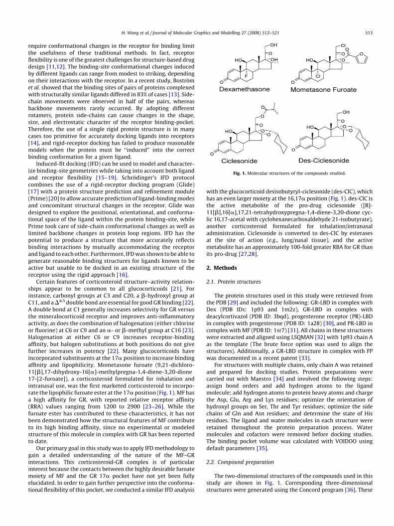
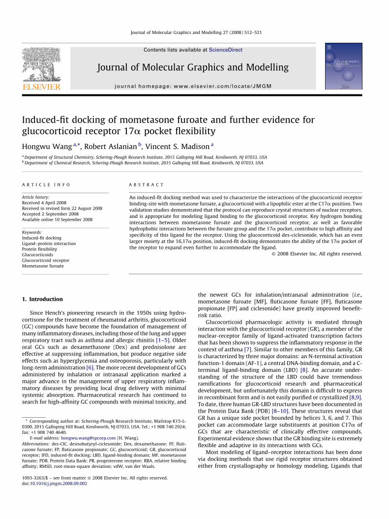
Fig. 1. Molecular structures of the compounds studied.
H. Wang et al. / Journal of Molecular Graphics and Modelling 27 (2008) 512–521 513
require conformational changes in the receptor for binding limitthe usefulness of these traditional methods. In fact, receptorflexibility is one of the greatest challenges for structure-based drugdesign [11,12]. The binding-site conformational changes inducedby different ligands can range from modest to striking, dependingon their interactions with the receptor. In a recent study, Bostromet al. showed that the binding sites of pairs of proteins complexedwith structurally similar ligands differed in 83% of cases [13]. Side-chain movements were observed in half of the pairs, whereasbackbone movements rarely occurred. By adopting differentrotamers, protein side-chains can cause changes in the shape,size, and electrostatic character of the receptor binding-pocket.Therefore, the use of a single rigid protein structure is in manycases too primitive for accurately docking ligands into receptors[14], and rigid-receptor docking has failed to produce reasonablemodels when the protein must be ‘‘induced’’ into the correctbinding conformation for a given ligand.
Induced-fit docking (IFD) can be used to model and character-ize binding-site geometries while taking into account both ligandand receptor flexibility [15–19]. Schrodinger’s IFD protocolcombines the use of a rigid-receptor docking program (Glide)[17] with a protein structure prediction and refinement module(Prime) [20] to allow accurate prediction of ligand-binding modesand concomitant structural changes in the receptor. Glide wasdesigned to explore the positional, orientational, and conforma-tional space of the ligand within the protein binding-site, whilePrime took care of side-chain conformational changes as well aslimited backbone changes in protein loop regions. IFD has thepotential to produce a structure that more accurately reflectsbinding interactions by mutually accommodating the receptorand ligand to each other. Furthermore, IFD was shown to be able togenerate reasonable binding structures for ligands known to beactive but unable to be docked in an existing structure of thereceptor using the rigid approach [16].
Certain features of corticosteroid structure–activity relation-ships appear to be common to all glucocorticoids [21]. Forinstance, carbonyl groups at C3 and C20, a b-hydroxyl group atC11, and a D4,5 double bond are essential for good GR binding [22].A double bond at C1 generally increases selectivity for GR versusthe mineralocorticoid receptor and improves anti-inflammatoryactivity, as does the combination of halogenation (either chlorineor fluorine) at C6 or C9 and an a- or b-methyl group at C16 [23].Halogenation at either C6 or C9 increases receptor-bindingaffinity, but halogen substitutions at both positions do not givefurther increases in potency [22]. Many glucocorticoids haveincorporated substituents at the 17a position to increase bindingaffinity and lipophilicity. Mometasone furoate (9,21-dichloro-11[b],17-dihydroxy-16[a]-methylpregna-1,4-diene-3,20-dione17-[2-furoate]), a corticosteroid formulated for inhalation andintranasal use, was the first marketed corticosteroid to incorpo-rate the lipophilic furoate ester at the 17a position (Fig. 1). MF hasa high affinity for GR, with reported relative receptor affinity(RRA) values ranging from 1200 to 2900 [23–26]. While thefuroate ester has contributed to these characteristics, it has notbeen demonstrated how the structural features of MF contributeto its high binding affinity, since no experimental or modeledstructure of this molecule in complex with GR has been reportedto date.
Our primary goal in this study was to apply IFD methodology togain a detailed understanding of the nature of the MF–GRinteractions. This corticosteroid-GR complex is of particularinterest because the contacts between the highly desirable furoatemoiety of MF and the GR 17a pocket have not yet been fullyelucidated. In order to gain further perspective into the conforma-tional flexibility of this pocket, we conducted a similar IFD analysis
with the glucocorticoid desisobutyryl-ciclesonide (des-CIC), whichhas an even larger moiety at the 16,17a position (Fig. 1). des-CIC isthe active metabolite of the pro-drug ciclesonide ([R]-11[b],16[a],17,21-tetrahydroxypregna-1,4-diene-3,20-dione cyc-lic 16,17-acetal with cyclohexanecarboxaldehyde 21-isobutyrate),another corticosteroid formulated for inhalation/intranasaladministration. Ciclesonide is converted to des-CIC by esterasesat the site of action (e.g., lung/nasal tissue), and the activemetabolite has an approximately 100-fold greater RBA for GR thanits pro-drug [27,28].
2. Methods
2.1. Protein structures
The protein structures used in this study were retrieved fromthe PDB [29] and included the following: GR-LBD in complex withDex (PDB IDs: 1p93 and 1m2z), GR-LBD in complex withdeacylcortivazol (PDB ID: 3bqd), progesterone receptor (PR)-LBDin complex with progesterone (PDB ID: 1a28) [30], and PR-LBD incomplex with MF (PDB ID: 1sr7) [31]. All chains in these structureswere extracted and aligned using LSQMAN [32] with 1p93 chain Aas the template (The brute force option was used to align thestructures). Additionally, a GR-LBD structure in complex with FPwas documented in a recent patent [33].
For structures with multiple chains, only chain A was retainedand prepared for docking studies. Protein preparations werecarried out with Maestro [34] and involved the following steps:assign bond orders and add hydrogen atoms to the ligandmolecule; add hydrogen atoms to protein heavy atoms and chargethe Asp, Glu, Arg and Lys residues; optimize the orientation ofhydroxyl groups on Ser, Thr and Tyr residues; optimize the sidechains of Gln and Asn residues; and determine the state of Hisresidues. The ligand and water molecules in each structure wereretained throughout the protein preparation process. Watermolecules and cofactors were removed before docking studies.The binding pocket volume was calculated with VOIDOO usingdefault parameters [35].
2.2. Compound preparation
The two-dimensional structures of the compounds used in thisstudy are shown in Fig. 1. Corresponding three-dimensionalstructures were generated using the Concord program [36]. These
H. Wang et al. / Journal of Molecular Graphics and Modelling 27 (2008) 512–521514
structures were then subjected to energy minimization usingMacroModel [37] with Merck Molecular Force Field (MMFFs) and aconstant dielectric of 1.0. Energy minimization was terminatedwhen the gradient dropped below 0.05, and the minimizedstructures were used as the starting points for docking.
2.3. Rigid docking
A rigid docking of MF to 1p93 was carried out using Glide XPmethodology set at default parameters, unless otherwise specified.Chain A of 1p93, prepared as described above, was used to generatethe energy grids for docking. Water molecules and the TIF2cofactor molecule in the crystal structure were removed, but noenergy minimization of the protein structure was conducted priorto grid calculation. The receptor binding-site was represented bythe energy grids using a cubic box centered on the centroid ofthe crystal ligand. Dimensions for the bounding box (withinwhich the centroid of a docked pose is confined) were set to12 A � 12 A � 12 A. No geometric or hydrogen-bonding con-straints were imposed on any of the protein binding-site atoms.A maximum of 10 poses were saved.
2.4. Induced-fit docking
The IFD protocol used in this study was carried out in threeconsecutive steps [16]. First, the ligand was docked into a rigid-receptor model with scaled-down van der Waals (vdW) radii. AvdW scaling of 0.5 was used for both the protein and ligand non-polar atoms. A constrained energy minimization was carried out onthe protein structure, keeping it close to the original crystalstructure while removing bad steric contacts. Energy minimizationwas carried out using the OPLS-2001 force field with implicitsolvation model until default criteria were met. The centroid andsize of the crystal ligand were used to define the location of thebinding site and the dimension of the energy grids for initialdocking. The Glide SP mode was used for the initial docking, and 20ligand poses were retained for protein structural refinements.
In the second step, Prime was used to generate the induced-fitprotein–ligand complexes. Each of the 20 structures from theprevious step was subjected to side-chain and backbone refine-ments. All residues with at least one atom located within 4.0 A ofeach corresponding ligand pose were included in the Primerefinement. All steroidal ligands used in this study had a C3-carbonyl group in their A-rings that forms identical interactions asin the crystal structures. Since we do not expect any significantconformational changes in this region, two of the binding-siteresidues that make hydrogen bonds with this carbonyl group(Gln570 and Arg611 in GR; Gln725 and Arg766 in PR) were fixedduring side-chain refinement. In both the GR and PR crystalstructures, these two residues also make extensive hydrogenbonds with water molecules in a nearby pocket. Since watermolecules were removed in the IFD studies, fixing the conforma-tion of these residues would also reduce any artifacts caused by thewater cavity. The refined complexes were ranked by Prime energy,and the receptor structures within 30 kcal/mol of the minimumenergy structure were passed through for a final round of Glidedocking and scoring.
In the final step, each ligand was redocked into every refinedlow-energy receptor structure produced in the second step usingGlide XP at default settings. An IFD score that accounts for both theprotein–ligand interaction energy and the total energy of thesystem was calculated and used to rank the IFD poses. The best-ranked IFD structures were used for comparison with theavailable crystal structures or as a model for new GCs. Theroot-mean-square deviations (RMSDs) between the docked
ligands and the corresponding crystal structures were calculatedafter aligning protein Ca atoms, and only heavy atoms were usedfor the calculations.
2.5. Hydrophobic interaction map
A hydrophobic interaction map of the binding site of the GR-LBD, which allows the visualization of the locations of thehydrophobic regions, was generated using Maestro. The prefer-ential binding of ligand hydrophobic groups in these regions canenhance receptor-binding affinity. The contour surface was plottedat a level of �1.40 kcal/mol.
3. Results
3.1. Nuclear receptor binding-site flexibility
Nuclear receptors such as the GR and the PR are known for theirability to adopt large conformational changes to accommodatestructurally diverse ligands. In many cases, they bind agonists andantagonists with distinctive conformations. We superimposed allavailable GR-LBD agonist-bound structures to get a preliminaryidea of the scope of binding-site flexibility. The two GR-LBD/Dexcomplex structures obtained from the PDB were solved indepen-dently by two separate groups under slightly different conditions.The 1m2z structure has two chains in the asymmetric unit, whilethe 1p93 has four. The GR-LBD/deacylcortivazol structure, 3bqd,has one chain in its asymmetric unit. The seven chains wereextracted from the PDB structures and aligned using 1p93 chain Aas template. Fig. 2 shows the overlay of these structures. Theprotein backbone among the structures aligns very well, and itsmovement around the agonist binding site is limited. Receptorside-chain movements, on the other hand, are relatively extensive;many side chains that line the binding site can adopt severalconformations, particularly those residues forming the 17a pocket.
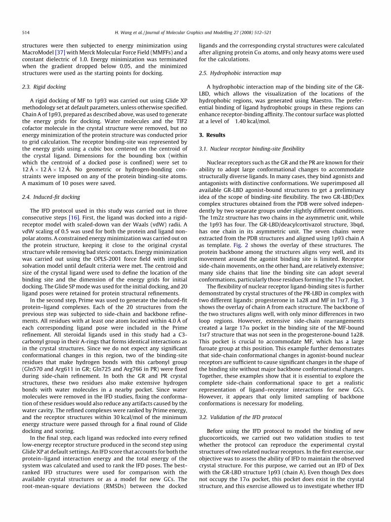
The flexibility of nuclear receptor ligand-binding sites is furtherdemonstrated by crystal structures of the PR-LBD in complex withtwo different ligands: progesterone in 1a28 and MF in 1sr7. Fig. 3shows the overlay of chain A from each structure. The backbone ofthe two structures aligns well, with only minor differences in twoloop regions. However, extensive side-chain rearrangementscreated a large 17a pocket in the binding site of the MF-bound1sr7 structure that was not seen in the progesterone-bound 1a28.This pocket is crucial to accommodate MF, which has a largefuroate group at this position. This example further demonstratesthat side-chain conformational changes in agonist-bound nuclearreceptors are sufficient to cause significant changes in the shape ofthe binding site without major backbone conformational changes.Together, these examples show that it is essential to explore thecomplete side-chain conformational space to get a realisticrepresentation of ligand–receptor interactions for new GCs.However, it appears that only limited sampling of backboneconformations is necessary for modeling.
3.2. Validation of the IFD protocol
Before using the IFD protocol to model the binding of newglucocorticoids, we carried out two validation studies to testwhether the protocol can reproduce the experimental crystalstructures of two related nuclear receptors. In the first exercise, ourobjective was to assess the ability of IFD to maintain the observedcrystal structure. For this purpose, we carried out an IFD of Dexwith the GR-LBD structure 1p93 (chain A). Even though Dex doesnot occupy the 17a pocket, this pocket does exist in the crystalstructure, and this exercise allowed us to investigate whether IFD
Fig. 2. Overlay of all chains in the three available agonist bound GR-LBD structures
(1m2z:AB; 1p93:ABCD; 3bqd). O atoms are colored red, N blue, S yellow, F light
green and Cl green throughout this paper. Carbon atoms are colored by molecules.
(a) The backbone conformation is consistent among the structures. Each colored
ribbon represents a unique chain from the crystal structures (cyan: 1p93A; brown:
1p93B; green: 1p93C; yellow: 1p93D; pink: 1m2zA; blue: 1m2zB; lime green:
3bqd). (b) Side-chain flexibility around the ligand-binding site. Side chains with at
least one atom within 5 A of the ligand are displayed (some residues were removed
for clarity).
H. Wang et al. / Journal of Molecular Graphics and Modelling 27 (2008) 512–521 515
would cause it to artificially collapse. As shown in Fig. 4, IFD closelyreproduced the Dex-bound GR-LBD crystal structure, and no majordistortions in side-chain conformations were generated. When thebest-ranked IFD structure is overlaid on its crystal counterpartusing the Ca atoms, the resulting RMSD is only 0.12 A. Whenlimited to the binding site, the RMSD for the backbone heavy atomsof residues within 5 A of the docked Dex is 0.21 A, while the RMSDfor the side-chain atoms of these residues is 1.17 A. Mostimportantly, the 17a pocket does not collapse and the generalshape of the ligand-binding site is preserved.
For the second IFD validation study, we used PR-LBD structuressince we could not find an appropriate example from the limitednumber of available GR-LBD crystal structures. The LBD of PR,which also belongs to the glucocorticoid-like nuclear-receptor
family, shares 54% sequence identity with the GR-LBD. While thereis no 17a pocket in the receptor binding-site when PR-LBD binds toits endogenous ligand progesterone (i.e., 1a28), the pocket iscreated when the protein is bound to MF, as in 1sr7. Since it wouldtherefore be impossible to find a binding pose for MF in the 1a28structure using rigid docking, we assessed whether a reasonablepocket would be generated when using IFD to model MF to 1a28.
As seen in Fig. 5, the IFD protocol did indeed induce a 17apocket in the PR-LBD binding site. The model of MF binding to PR-LBD obtained by IFD is very similar to the crystal structure of 1sr7,including side-chain conformations. Since there is a gap betweenAsn705 and Pro708 in the PR-MF crystal structure, the top-rankingIFD structure was superimposed on the 1sr7 structure using Caatoms of residues 712–930. The RMSD of the Ca atoms of the IFDstructure compared to the crystal structure is only 0.32 A. In thebinding site, the RMSD of the backbone atoms for the residueswithin 5 A of the docked MF is 0.40 A, while the RMSD for the side-chain atoms of these residues is 1.35 A. Furthermore, the RMSD forMF heavy atoms is only 1.13 A when the protein Ca atoms aresuperimposed. The volume of the PR-LBD binding site in the crystalstructures increases from 493 A3 when progesterone is bound to635 A3 when MF is bound. In comparison, the binding site of thePR-LBD/MF structure generated by IFD has a volume of 623 A3.Additionally, IFD of another marketed glucocorticoid with a 17afuroate group, FF, shows that this molecule can also induce asimilar 17a pocket in the PR-LBD binding site (data not shown).This agrees with a recent experimental finding that FF hassubnanomolar affinity for PR (pEC50 = 9.05) [38].
These two validation studies demonstrate that IFD is capable ofboth maintaining the experimental structure and inducing areasonable binding pocket in nuclear receptors in the presence of alarger ligand. By exhaustively sampling the conformational spacefor protein side-chains involved in the binding of ligand molecules,the IFD protocol can cover many accessible receptor binding-siteconfigurations. At the same time, limited backbone flexibility wasexplored, through constrained energy minimization. In thefollowing sections, we report our modeling results for two potentGCs with large 17a groups, MF and des-CIS.
3.3. Modeling of MF
Mometasone furoate is the first marketed steroid to incorporatea furoate group at the 17a position to achieve a very high affinityfor GR (Table 1). No crystal structure or computer model for thiscompound in complex with the GR-LBD has been reported. A rigiddocking of MF to the GR-LBD was carried out using the 1p93 chainA with a vdW scaling of 0.8 for both the protein and ligand non-polar atoms. In this model, MF fits at the binding site with thefuroate group docked into the 17a pocket. The typical hydrogen-bonding patterns seen in the crystal structure of Dex aremaintained in this rigid docking model, e.g., the C3-carbonylgroup with Gln576 and Arg611, and the 11b-hydroxyl group withAsn564. But, many short vdW contacts (defined as when thedistance between a pair of non-bound atoms is less than 80% of thesum of their vdW radii) are observed between the furoate groupand the protein side-chain atoms in the 17a region. Most of thesteric conflicts came from the close contacts of MF with two Metresidues (Met560 and Met601). The long flexible side chains ofthese amino acids make them prone to adopting differentconformations that can enlarge the pocket observed in the crystalstructure. Hence, we would expect GR to adopt conformationalchanges to better accommodate the large 17a furoate group of MF.
An IFD was then carried out using the same protein structure toget a more realistic picture of MF and GR-LBD interactions.Conformations of side chains within 4 A of the docked ligand were
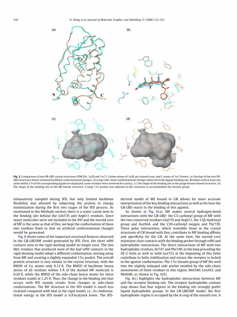
Fig. 3. Comparison of two PR-LBD crystal structures (PDB IDs: 1a28 and 1sr7). Carbon atoms of 1a28 are colored cyan, and C atoms of 1sr7 brown. (a) Overlay of the two PR-
LBD structures shows minimal backbone conformational changes. (b) Large side-chain conformational changes observed in the ligand-binding site. Residues with at least one
atom within 5 A of the corresponding ligand are displayed (some residues were removed for clarity). (c) The shape of the binding site in the progesterone-bound structure. (d)
The shape of the binding site in the MF-bound structure. A large 17a pocket was induced in this structure to accommodate the furoate group.
H. Wang et al. / Journal of Molecular Graphics and Modelling 27 (2008) 512–521516
exhaustively sampled during IFD, but only limited backboneflexibility was allowed by subjecting the protein to energyminimization during the first two stages of the IFD process. Asmentioned in the Methods section, there is a water cavity next tothe binding site behind the Gln570 and Arg611 residues. Sincewater molecules were not included in the IFD and the steroid coreof MF is the same as that of Dex, we kept the conformation of thesetwo residues fixed so that no artificial conformational changeswould be generated.
Fig. 6 shows some of the important structural features observedin the GR-LBD/MF model generated by IFD. First, the short vdWcontacts seen in the rigid-docking model no longer exist. The twoMet residues that produced most of the bad vdW contacts in therigid-docking model adopt a different conformation, moving awayfrom MF and creating a slightly expanded 17a pocket. The overallprotein structure is very similar to the crystal structure, with theRMSD of Ca atoms only 0.12 A. The RMSD of backbone heavyatoms of all residues within 5 A of the docked MF molecule is0.20 A, while the RMSD of the side-chain heavy atoms for theseresidues stands at 1.25 A. Thus, the change in the binding site thatoccurs with IFD mainly results from changes in side-chainconformations. The MF structure in the IFD model is much lessstrained compared with that in the rigid model, i.e., its conforma-tional energy in the IFD model is 6.8 kcal/mol lower. The IFD-
derived model of MF bound to GR allows for more accurateinterpretation of the key binding interactions as well as for how theGR-LBD reacts to the binding of this agonist.
As shown in Fig. 6(a), MF makes several hydrogen-bondinteractions with the GR-LBD: the C3-carbonyl group of MF withthe two conserved residues Glu570 and Arg611, the 11b-hydroxylgroup and Asn564, and the C20-carbonyl oxygen and Thr739.These polar interactions, which resemble those in the crystalstructures of GR bound with Dex, contribute to MF binding affinityand specificity for the GR. At the same time, the steroid coremaintains close contacts with the binding pocket through vdW andhydrophobic interactions. The direct interactions of MF with twohydrophobic residues, Ile747 and Phe749, in the loop preceding theAF-2 helix as well as with Leu753 at the beginning of this helixcontribute to helix stabilization and ensure the receptor is lockedin the agonist conformation. The 17a furoate group of MF fits wellinto the slightly enlarged side pocket enabled by the side-chainmovements of three residues in this region, Met560, Leu563, andMet646, as shown in Fig. 6(b).
Fig. 6(c) highlights the hydrophobic interactions between MFand the receptor binding-site. The receptor hydrophobic contourmap shows that four regions in the binding site strongly preferligand hydrophobic groups. In the GR-LBD/MF model, the firsthydrophobic region is occupied by the A-ring of the steroid core. A
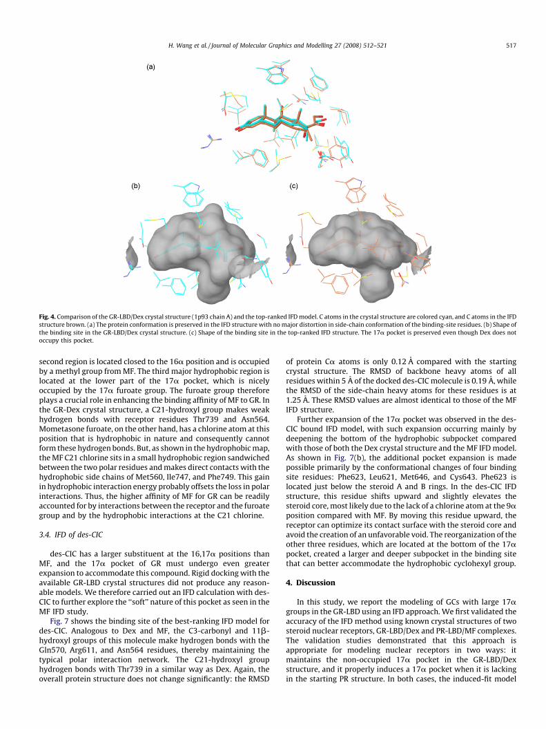
Fig. 4. Comparison of the GR-LBD/Dex crystal structure (1p93 chain A) and the top-ranked IFD model. C atoms in the crystal structure are colored cyan, and C atoms in the IFD
structure brown. (a) The protein conformation is preserved in the IFD structure with no major distortion in side-chain conformation of the binding-site residues. (b) Shape of
the binding site in the GR-LBD/Dex crystal structure. (c) Shape of the binding site in the top-ranked IFD structure. The 17a pocket is preserved even though Dex does not
occupy this pocket.
H. Wang et al. / Journal of Molecular Graphics and Modelling 27 (2008) 512–521 517
second region is located closed to the 16a position and is occupiedby a methyl group from MF. The third major hydrophobic region islocated at the lower part of the 17a pocket, which is nicelyoccupied by the 17a furoate group. The furoate group thereforeplays a crucial role in enhancing the binding affinity of MF to GR. Inthe GR-Dex crystal structure, a C21-hydroxyl group makes weakhydrogen bonds with receptor residues Thr739 and Asn564.Mometasone furoate, on the other hand, has a chlorine atom at thisposition that is hydrophobic in nature and consequently cannotform these hydrogen bonds. But, as shown in the hydrophobic map,the MF C21 chlorine sits in a small hydrophobic region sandwichedbetween the two polar residues and makes direct contacts with thehydrophobic side chains of Met560, Ile747, and Phe749. This gainin hydrophobic interaction energy probably offsets the loss in polarinteractions. Thus, the higher affinity of MF for GR can be readilyaccounted for by interactions between the receptor and the furoategroup and by the hydrophobic interactions at the C21 chlorine.
3.4. IFD of des-CIC
des-CIC has a larger substituent at the 16,17a positions thanMF, and the 17a pocket of GR must undergo even greaterexpansion to accommodate this compound. Rigid docking with theavailable GR-LBD crystal structures did not produce any reason-able models. We therefore carried out an IFD calculation with des-CIC to further explore the ‘‘soft’’ nature of this pocket as seen in theMF IFD study.
Fig. 7 shows the binding site of the best-ranking IFD model fordes-CIC. Analogous to Dex and MF, the C3-carbonyl and 11b-hydroxyl groups of this molecule make hydrogen bonds with theGln570, Arg611, and Asn564 residues, thereby maintaining thetypical polar interaction network. The C21-hydroxyl grouphydrogen bonds with Thr739 in a similar way as Dex. Again, theoverall protein structure does not change significantly: the RMSD
of protein Ca atoms is only 0.12 A compared with the startingcrystal structure. The RMSD of backbone heavy atoms of allresidues within 5 A of the docked des-CIC molecule is 0.19 A, whilethe RMSD of the side-chain heavy atoms for these residues is at1.25 A. These RMSD values are almost identical to those of the MFIFD structure.
Further expansion of the 17a pocket was observed in the des-CIC bound IFD model, with such expansion occurring mainly bydeepening the bottom of the hydrophobic subpocket comparedwith those of both the Dex crystal structure and the MF IFD model.As shown in Fig. 7(b), the additional pocket expansion is madepossible primarily by the conformational changes of four bindingsite residues: Phe623, Leu621, Met646, and Cys643. Phe623 islocated just below the steroid A and B rings. In the des-CIC IFDstructure, this residue shifts upward and slightly elevates thesteroid core, most likely due to the lack of a chlorine atom at the 9aposition compared with MF. By moving this residue upward, thereceptor can optimize its contact surface with the steroid core andavoid the creation of an unfavorable void. The reorganization of theother three residues, which are located at the bottom of the 17apocket, created a larger and deeper subpocket in the binding sitethat can better accommodate the hydrophobic cyclohexyl group.
4. Discussion
In this study, we report the modeling of GCs with large 17agroups in the GR-LBD using an IFD approach. We first validated theaccuracy of the IFD method using known crystal structures of twosteroid nuclear receptors, GR-LBD/Dex and PR-LBD/MF complexes.The validation studies demonstrated that this approach isappropriate for modeling nuclear receptors in two ways: itmaintains the non-occupied 17a pocket in the GR-LBD/Dexstructure, and it properly induces a 17a pocket when it is lackingin the starting PR structure. In both cases, the induced-fit model
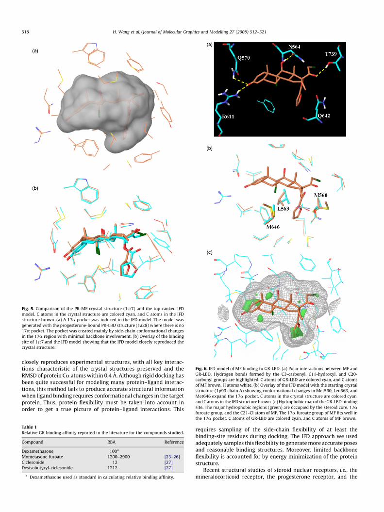
Fig. 5. Comparison of the PR-MF crystal structure (1sr7) and the top-ranked IFD
model. C atoms in the crystal structure are colored cyan, and C atoms in the IFD
structure brown. (a) A 17a pocket was induced in the IFD model. The model was
generated with the progesterone-bound PR-LBD structure (1a28) where there is no
17a pocket. The pocket was created mainly by side-chain conformational changes
in the 17a region with minimal backbone involvement. (b) Overlay of the binding
site of 1sr7 and the IFD model showing that the IFD model closely reproduced the
crystal structure.
Fig. 6. IFD model of MF binding to GR-LBD. (a) Polar interactions between MF and
GR-LBD. Hydrogen bonds formed by the C3-carbonyl, C11-hydroxyl, and C20-
carbonyl groups are highlighted. C atoms of GR-LBD are colored cyan, and C atoms
of MF brown, H atoms white. (b) Overlay of the IFD model with the starting crystal
structure (1p93 chain A) showing conformational changes in Met560, Leu563, and
Met646 expand the 17a pocket. C atoms in the crystal structure are colored cyan,
and C atoms in the IFD structure brown. (c) Hydrophobic map of the GR-LBD binding
site. The major hydrophobic regions (green) are occupied by the steroid core, 17a
H. Wang et al. / Journal of Molecular Graphics and Modelling 27 (2008) 512–521518
closely reproduces experimental structures, with all key interac-tions characteristic of the crystal structures preserved and theRMSD of protein Ca atoms within 0.4 A. Although rigid docking hasbeen quite successful for modeling many protein–ligand interac-tions, this method fails to produce accurate structural informationwhen ligand binding requires conformational changes in the targetprotein. Thus, protein flexibility must be taken into account inorder to get a true picture of protein–ligand interactions. This
Table 1Relative GR binding affinity reported in the literature for the compounds studied.
Compound RBA Reference
Dexamethasone 100a
Mometasone furoate 1200–2900 [23–26]
Ciclesonide 12 [27]
Desisobutyryl-ciclesonide 1212 [27]
a Dexamethasone used as standard in calculating relative binding affinity.
furoate group, and the C21-Cl atom of MF. The 17a furoate group of MF fits well in
the 17a pocket. C atoms of GR-LBD are colored cyan, and C atoms of MF brown.
requires sampling of the side-chain flexibility of at least thebinding-site residues during docking. The IFD approach we usedadequately samples this flexibility to generate more accurate posesand reasonable binding structures. Moreover, limited backboneflexibility is accounted for by energy minimization of the proteinstructure.
Recent structural studies of steroid nuclear receptors, i.e., themineralocorticoid receptor, the progesterone receptor, and the
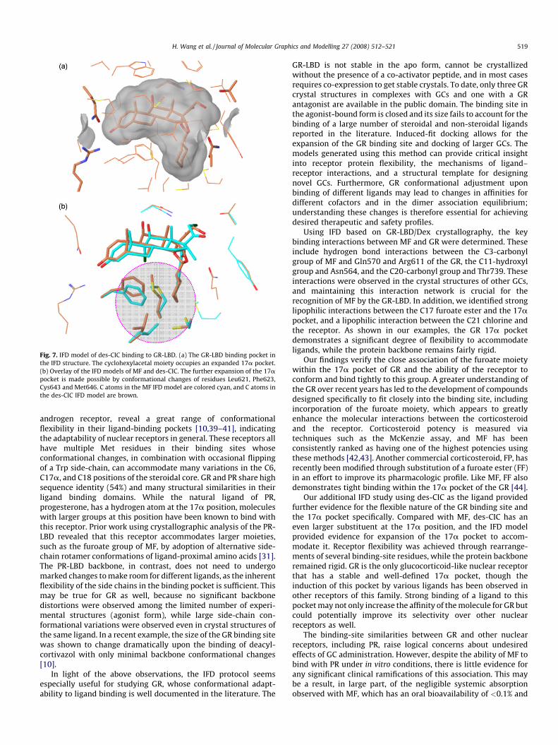
Fig. 7. IFD model of des-CIC binding to GR-LBD. (a) The GR-LBD binding pocket in
the IFD structure. The cyclohexylacetal moiety occupies an expanded 17a pocket.
(b) Overlay of the IFD models of MF and des-CIC. The further expansion of the 17apocket is made possible by conformational changes of residues Leu621, Phe623,
Cys643 and Met646. C atoms in the MF IFD model are colored cyan, and C atoms in
the des-CIC IFD model are brown.
H. Wang et al. / Journal of Molecular Graphics and Modelling 27 (2008) 512–521 519
androgen receptor, reveal a great range of conformationalflexibility in their ligand-binding pockets [10,39–41], indicatingthe adaptability of nuclear receptors in general. These receptors allhave multiple Met residues in their binding sites whoseconformational changes, in combination with occasional flippingof a Trp side-chain, can accommodate many variations in the C6,C17a, and C18 positions of the steroidal core. GR and PR share highsequence identity (54%) and many structural similarities in theirligand binding domains. While the natural ligand of PR,progesterone, has a hydrogen atom at the 17a position, moleculeswith larger groups at this position have been known to bind withthis receptor. Prior work using crystallographic analysis of the PR-LBD revealed that this receptor accommodates larger moieties,such as the furoate group of MF, by adoption of alternative side-chain rotamer conformations of ligand-proximal amino acids [31].The PR-LBD backbone, in contrast, does not need to undergomarked changes to make room for different ligands, as the inherentflexibility of the side chains in the binding pocket is sufficient. Thismay be true for GR as well, because no significant backbonedistortions were observed among the limited number of experi-mental structures (agonist form), while large side-chain con-formational variations were observed even in crystal structures ofthe same ligand. In a recent example, the size of the GR binding sitewas shown to change dramatically upon the binding of deacyl-cortivazol with only minimal backbone conformational changes[10].
In light of the above observations, the IFD protocol seemsespecially useful for studying GR, whose conformational adapt-ability to ligand binding is well documented in the literature. The
GR-LBD is not stable in the apo form, cannot be crystallizedwithout the presence of a co-activator peptide, and in most casesrequires co-expression to get stable crystals. To date, only three GRcrystal structures in complexes with GCs and one with a GRantagonist are available in the public domain. The binding site inthe agonist-bound form is closed and its size fails to account for thebinding of a large number of steroidal and non-steroidal ligandsreported in the literature. Induced-fit docking allows for theexpansion of the GR binding site and docking of larger GCs. Themodels generated using this method can provide critical insightinto receptor protein flexibility, the mechanisms of ligand–receptor interactions, and a structural template for designingnovel GCs. Furthermore, GR conformational adjustment uponbinding of different ligands may lead to changes in affinities fordifferent cofactors and in the dimer association equilibrium;understanding these changes is therefore essential for achievingdesired therapeutic and safety profiles.
Using IFD based on GR-LBD/Dex crystallography, the keybinding interactions between MF and GR were determined. Theseinclude hydrogen bond interactions between the C3-carbonylgroup of MF and Gln570 and Arg611 of the GR, the C11-hydroxylgroup and Asn564, and the C20-carbonyl group and Thr739. Theseinteractions were observed in the crystal structures of other GCs,and maintaining this interaction network is crucial for therecognition of MF by the GR-LBD. In addition, we identified stronglipophilic interactions between the C17 furoate ester and the 17apocket, and a lipophilic interaction between the C21 chlorine andthe receptor. As shown in our examples, the GR 17a pocketdemonstrates a significant degree of flexibility to accommodateligands, while the protein backbone remains fairly rigid.
Our findings verify the close association of the furoate moietywithin the 17a pocket of GR and the ability of the receptor toconform and bind tightly to this group. A greater understanding ofthe GR over recent years has led to the development of compoundsdesigned specifically to fit closely into the binding site, includingincorporation of the furoate moiety, which appears to greatlyenhance the molecular interactions between the corticosteroidand the receptor. Corticosteroid potency is measured viatechniques such as the McKenzie assay, and MF has beenconsistently ranked as having one of the highest potencies usingthese methods [42,43]. Another commercial corticosteroid, FP, hasrecently been modified through substitution of a furoate ester (FF)in an effort to improve its pharmacologic profile. Like MF, FF alsodemonstrates tight binding within the 17a pocket of the GR [44].
Our additional IFD study using des-CIC as the ligand providedfurther evidence for the flexible nature of the GR binding site andthe 17a pocket specifically. Compared with MF, des-CIC has aneven larger substituent at the 17a position, and the IFD modelprovided evidence for expansion of the 17a pocket to accom-modate it. Receptor flexibility was achieved through rearrange-ments of several binding-site residues, while the protein backboneremained rigid. GR is the only glucocorticoid-like nuclear receptorthat has a stable and well-defined 17a pocket, though theinduction of this pocket by various ligands has been observed inother receptors of this family. Strong binding of a ligand to thispocket may not only increase the affinity of the molecule for GR butcould potentially improve its selectivity over other nuclearreceptors as well.
The binding-site similarities between GR and other nuclearreceptors, including PR, raise logical concerns about undesiredeffects of GC administration. However, despite the ability of MF tobind with PR under in vitro conditions, there is little evidence forany significant clinical ramifications of this association. This maybe a result, in large part, of the negligible systemic absorptionobserved with MF, which has an oral bioavailability of <0.1% and
H. Wang et al. / Journal of Molecular Graphics and Modelling 27 (2008) 512–521520
undergoes almost complete first-pass metabolism [45]. Even whenadministered at 20 times the recommended dose for treatment ofallergic rhinitis, MF administered intranasally was not shown toadversely affect urinary free-cortisol or plasma levels, nor wasthere evidence of hypothalamic–pituitary–gland axis suppression[46].
5. Conclusions
In this paper, we report the modeling of glucocorticoids withlarge 17a group, MF and des-CIC, to GR-LBD using an IFD protocol.With the limited number of available crystal structures, the large17a groups make it hard to model these compounds using rigiddocking. Close examination of the available agonist-bound crystalstructures of the steroid nuclear receptors shows that their bindingsite is very flexible, and the change in size and shape of the bindingsite comes mainly from the side-chain conformational changes.Our validation studies show that the IFD protocol is appropriate forthe study of GR and other nuclear receptors. The MF IFD modelhighlights the crucial molecular features for its high GR affinity, i.e.,the hydrogen bonding network and especially the favorablehydrophobic interactions between the furoate group and the GR17a pocket. IFD model with des-CIC demonstrates the ability of the17a pocket to expand even further to accommodate ligands withbulkier groups. In both models, the protein side-chains in thispocket provide the flexibility to adapt to the ligands, while thebackbone demonstrates minimal movement. Our findings clearlydemonstrate the benefits of IFD techniques for structural modelingwhen receptor conformational change is required.
Acknowledgement
The authors thank Adelphi Inc. for editorial support.
References
[1] Global Initiative for Asthma, Global strategy for asthma management and pre-vention, NHLBI/WHO Workshop Report, 2006, pp. 1–96, Available at: www.gi-nasthma.com.
[2] M.S. Dykewicz, S. Fineman, Executive summary of joint task force practiceparameters on diagnosis and management of rhinitis, Ann. Allergy Asth. Immunol.81 (1998) 463–468.
[3] P. van Cauwenberge, C. Bachert, G. Passalacqua, J. Bousquet, G.W. Canonica, S.R.Durham, W.J. Fokkens, P.H. Howarth, V. Lund, H.J. Malling, N. Mygind, D. Passali,G.K. Scadding, D.Y. Wang, Consensus statement on the treatment of allergicrhinitis, Eur. Acad. Allergol. Clin. Immunol., Allergy 55 (2000) 116–134.
[4] J. Bousquet, P. Van Cauwenberge, N. Khaltaev, Aria Workshop Group, WorldHealth Organization, Allergic rhinitis and its impact on asthma, J. Allergy Clin.Immunol. 108 (2001) S147–S334.
[5] S.P. Umland, R.P. Schleimer, S.L. Johnston, Review of the molecular and cellularmechanisms of action of glucocorticoids for use in asthma, Pulm. Pharmacol. Ther.15 (2002) 35–50.
[6] H. Schacke, W.D. Docke, K. Asadullah, Mechanisms involved in the side effects ofglucocorticoids, Pharmacol. Ther. 96 (2002) 23–43.
[7] L. Ramamurthy, R.P. Trump, Nuclear receptor ligands in inflammatory lungdiseases, Drug Dis. Today: Dis. Mech. 3 (2006) 85–90.
[8] R.K. Bledsoe, V.G. Montana, T.B. Stanley, C.J. Delves, C.J. Apolito, D.D. McKee, T.G.Consler, D.J. Parks, E.L. Stewart, T.M. Willson, M.H. Lambert, J.T. Moore, K.H.Pearce, H.E. Xu, Crystal structure of the glucocorticoid receptor ligand bindingdomain reveals a novel mode of receptor dimerization and coactivator recogni-tion, Cell 110 (2002) 93–105.
[9] B. Kauppi, C. Jakob, M. Farnegardh, J. Yang, H. Ahola, M. Alarcon, K. Calles, O.Engstrom, J. Harlan, S. Muchmore, A.K. Ramqvist, S. Thorell, L. Ohman, J. Greer, J.A.Gustafsson, J. Carlstedt-Duke, M. Carlquist, The three-dimensional structures ofantagonistic and agonistic forms of the glucocorticoid receptor ligand-bindingdomain: RU-486 induces a transconformation that leads to active antagonism, J.Biol. Chem. 278 (2003) 22748–22754.
[10] K. Suino-Powell, Y. Xu, C. Zhang, Y.-G. Tao, W.D. Tolbert, S.S. Simons Jr., H.E. Xu,Doubling the size of the glucocorticoid receptor ligand binding pocket by dea-cylcortivazol, Mol. Cell. Biol. 28 (2008) 1915–1923.
[11] A.R. Leach, B.K. Shoichet, C.E. Peishoff, Prediction of protein–ligand interactions.Docking and scoring: successes and gaps, J. Med. Chem. 49 (2006) 5851–5855.
[12] S.F. Sousa, P.A. Fernandes, M.J. Ramos, Protein–ligand docking: current status andfuture challenges, Proteins 65 (2006) 15–26.
[13] J. Bostrom, A. Hogner, S. Schmitt, Do structurally similar ligands bind in a similarfashion? J. Med. Chem. 49 (2006) 6716–6725.
[14] C.N. Cavasotto, R.A. Abagyan, Protein flexibility in ligand docking and virtualscreening to protein kinases, J. Mol. Biol. 337 (2004) 209–225.
[15] N. Moitessier, E. Therrien, S. Hanessian, A method for induced-fit docking, scoring,and ranking of flexible ligands. Application to peptidic and pseudopeptidic beta-secretase (BACE 1) inhibitors, J. Med. Chem. 49 (2006) 5885–5894.
[16] W. Sherman, T. Day, M.P. Jacobson, R.A. Friesner, R. Farid, Novel procedure formodeling ligand/receptor induced fit effects, J. Med. Chem. 49 (2006) 534–553.
[17] R.A. Friesner, J.L. Banks, R.B. Murphy, T.A. Halgren, J.J. Klicic, D.T. Mainz, M.P.Repasky, E.H. Knoll, M. Shelley, J.K. Perry, D.E. Shaw, P. Francis, P.S. Shenkin, Glide:a new approach for rapid, accurate docking and scoring. 1. Method and assess-ment of docking accuracy, J. Med. Chem. 47 (2004) 1739–1749.
[18] H. Claussen, C. Buning, M. Rarey, T. Lengauer, FlexE: efficient molecular dockingconsidering protein structure variations, J. Mol. Biol. 308 (2001) 377–395.
[19] H.A. Carlson, Protein flexibility and drug design: how to hit a moving target, Curr.Opin. Chem. Biol. 6 (2002) 447–452.
[20] K. Zhu, M.R. Shirts, R.A. Friesner, Improved methods for side chain and looppredictions via the protein local optimization program: variable dielectric modelfor implicitly improving the treatment of polarization effects, J. Chem. TheoryComput. 3 (2007) 2108–2119.
[21] M.A. Avery, J.R. Woolfrey, Anti-inflammatory steroids, in: D.J. Abraham (Ed.),Burger’s Medicinal Chemistry & Drug Discovery, John Wiley and Sons, Hoboken,NJ, 2003, pp. 747–851.
[22] J. Bikowski, R. Pillai, B. Shroot, The position not the presence of the halogen incorticosteroids influences potency and side effects, J. Drugs Dermatol. 5 (2006)125–130.
[23] P. Hogger, Current concepts for optimizing the therapeutic index of glucocorti-coid receptor ligands for oral and inhalative use: basic considerations andclinical reality, Curr. Med. Chem.-Anti-Inflam. Anti-Allergy Agents 2 (2003)395–408.
[24] A. Valotis, P. Hogger, Human receptor kinetics and lung tissue retention of theenhanced-affinity glucocorticoid fluticasone furoate, Respir. Res. 8 (2007) 54.
[25] C.L. Smith, W. Kreutner, In vitro glucocorticoid receptor binding and transcrip-tional activation by topically active glucocorticoids, Arzneim. Forsch. 48 (1998)956–960.
[26] M. Issar, S. Sahasranaman, P. Buchwald, G. Hochhaus, Differences in the gluco-corticoid to progesterone receptor selectivity of inhaled glucocorticoids, Eur.Respir. J. 27 (2006) 511–516.
[27] M. Stoeck, R. Riedel, G. Hochhaus, D. Hafner, J.M. Masso, B. Schmidt, A. Hatzel-mann, D. Marx, D.S. Bundschuh, In vitro and in vivo anti-inflammatory activity ofthe new glucocorticoid ciclesonide, J. Pharmacol. Exp. Ther. 309 (2004) 249–258.
[28] E. Mutch, R. Nave, N. McCracken, K. Zech, F.M. Williams, The role of esterases inthe metabolism of ciclesonide to desisobutyryl-ciclesonide in human tissue,Biochem. Pharmacol. 73 (2007) 1657–1664.
[29] H.M. Berman, J. Westbrook, Z. Feng, G. Gilliland, T.N. Bhat, H. Weissig, I.N.Shindyalov, P.E. Bourne, The Protein Data Bank, Nucleic Acids Res. 28 (2000)235–242.
[30] S.P. Williams, P.B. Sigler, Atomic structure of progesterone complexed with itsreceptor, Nature 393 (1998) 392–396.
[31] K.P. Madauss, S.J. Deng, R.J. Austin, M.H. Lambert, I. McLay, J. Pritchard, S.A. Short,E.L. Stewart, I.J. Uings, S.P. Williams, Progesterone receptor ligand binding pocketflexibility: crystal structures of the norethindrone and mometasone furoatecomplexes, J. Med. Chem. 47 (2004) 3381–3387.
[32] G.J. Kleywegt, Experimental assessment of differences between related proteincrystal structures, Acta Crystallogr., Sect. B 55 (1999) 1878–1884.
[33] R.K. Bledsoe, M.H. Lambert, V.G. Montana, E.L. Stewart, E.H. Xu, Structure of aglucocorticoid receptor ligand binding domain comprising an expanded bindingpocket, and methods using nuclear receptors structure for drug design, Eur. Pat.Appl. 1375517 (2004).
[34] Maestro Software, Scherodinger, LLC, 120 West 45th Street, New York, NY 10036,USA.
[35] G.J. Kleywegt, T.A. Jones, Detection, delineation, measurement and display ofcavities in macromolecular structures., Acta Crystallogr., Sect. D 50 (1994) 178–185.
[36] Concord Software Distributed by Tripos Inc., St. Louis, Missouri, MO 63144, USA.[37] MacroModel Software, Scherodinger, LLC, 120 West 45th Street, New York, NY
10036, USA.[38] M. Salter, K. Biggadike, J.L. Matthews, M.R. West, M.V. Haase, S.N. Farrow, I.J.
Uings, D.W. Gray, Pharmacological properties of the enhanced-affinity gluco-corticoid fluticasone furoate in vitro and in an in vivo model of respiratoryinflammatory disease, Am. J. Physiol. Lung Cell. Mol. Physiol. 293 (2007) L660–L667.
[39] J. Huyet, G.M. Pinon, M.R. Fay, J. Fagart, M.-E. Rafestin-Oblin, Structural basis ofspirolactone recognition by the mineralocorticoid receptor, Mol. Pharmacol. 72(2007) 563–571.
[40] C.E. Bohl, Z. Wu, D.D. Miller, C.E. Bell, J.T. Dalton, Crystal structure of the T877Ahuman androgen receptor ligand-binding domain complexed to cyproteroneacetate provides insight for ligand-induced conformational changes and struc-ture-based drug design, J. Biol. Chem. 282 (2007) 13648–13655.
[41] L. Cantin, F. Faucher, J.-F. Couture, K.P. de Jesus-Tran, P. Legrand, L.C. Ciobanu, Y.Frechette, R. Labrecque, S.M. Singh, F. Labrie, R. Breton, Structural characteriza-tion of the human androgen receptor ligand-binding domain complexed withEM5744, a rationally designed steroidal ligand bearing a bulky chain directedtoward helix 12, J. Biol. Chem. 282 (2007) 30910–30919.
H. Wang et al. / Journal of Molecular Graphics and Modelling 27 (2008) 512–521 521
[42] C. Roumestan, C. Henriquet, J. Bousquet, M. Mathieu, Fluticasone propionate andmometasone furoate have equivalent transcriptional potencies, Clin. Exp. Allergy33 (2003) 895–901.
[43] C. Crim, L.N. Pierre, P.T. Daley-Yates, A review of the pharmacology and pharma-cokinetics of inhaled fluticasone propionate and mometasone furoate, Clin. Ther.23 (2001) 1339–1354.
[44] K. Biggadike, R. Bledsoe, A. Hassell, S. Hughes, L. Shewchuk, GW685698X-enhancedaffinity for the glucocorticoid receptor: receptor crystal structure and route of
metabolic inactivation, in: Proceedings of the XXVth Congress of the EuropeanAcademy of Allergology and Clinical Immunology, Vienna, Austria, 2006.
[45] C. van Drunen, E.O. Meltzer, C. Bachert, J. Bousquet, W.J. Fokkens, Nasal allergiesand beyond: a clinical review of the pharmacology, efficacy, and safety ofmometasone furoate, Allergy 80 (2005) 5–19.
[46] M.D. Brannan, M. Seiberling, D.L. Cutler, F.M. Cuss, M.B. Affrime, Lack ofsystemic activity with intranasal mometasone furoate, J. Allergy Clin. Immu-nol. 97 (1996) 198.