Consensus document for Management of Pediatric Lymphoma.
153
-
Upload
khangminh22 -
Category
Documents
-
view
1 -
download
0
Transcript of Consensus document for Management of Pediatric Lymphoma.
Coordinated byDivision of Non Communicable Diseases
Indian Council of Medical Research, Ansari Nagar, New Delhi – 110029
2017
CONSENSUS DOCUMENTFOR MANAGEMENT
OF PEDIATRIC LYMPHOMAS AND SOLID TUMORS
Prepared as an outcome of ICMR Subcommittee on Pediatric Lymphomas & Solid Tumours
Disclaimer
This consensus document represents the current thinking of experts on the topic based on available evidence. This has been developed by national experts in the field and does not in any way bind a clinician to follow this guideline. One can use an alternate mode of therapy based on discussions with the patient and institution, national or international guidelines. The mention of pharmaceutical drugs for therapy does not constitute endorsement or recommendation for use but will act only as a guidance for clinicians in complex decision –making.
Dr. Soumya Swaminathan Secretary, Department of Health Research and Director General, ICMR
Published in 2017
Dr. Neeraj Tandon : Head (Publication & Information)
Compiled & Edited by Dr. Tanvir Kaur Scientist ‘F’
Production Controller : JN Mathur
Published by the Division of Publication and Information on behalf of the Secretary DHR & DG, ICMR, New Delhi.
Designed & Printed at M/s Aravali Printers & Publishers (P) Ltd., W-30, Okhla Industrial Area, Phase-II, New Delhi-110020 Phone: 47173300, 26388830-32

Foreword
I am glad to write this foreword for consensus document for management of Pediatric Lymphomas & Solid Tumors. The ICMR had constituted sub-committees to prepare consensus document for management of various cancer sites. This document is the result of the hard work of various experts across the country working in the area of oncology.
This consensus document on management of Pediatric Lymphomas & Solid Tumors summarizes the modalities of treatment including the site-specific anti-cancer therapies, supportive and palliative care and molecular markers and research questions. It also interweaves clinical, biochemical and epidemiological studies.
The various subcommittees constituted under Task Force project on Review of Cancer Management Guidelines worked tirelessly in formulating site-specific guidelines. Each member of the subcommittee’s contribution towards drafting of these guidelines deserves appreciation and acknowledgement for their dedicated research, experience and effort for successful completion. Hope that this document would provide guidance to practicing doctors and researchers for the management of patients suffering from Pediatric Lymphomas & Solid Tumors and also focusing their research efforts in Indian context.
It is understood that this document represents the current thinking of national experts on subject based on available evidence. Mention of drugs and clinical tests for therapy do not imply endorsement or recommendation for their use, these are examples to guide clinicians in complex decision making. We are confident that this first edition of Consensus Document on Management of Pediatric Lymphoas & Solid Tumors would serve the desired purpose.
(Dr.Soumya Swaminathan)Secretary, Department of Health Research
and Director-General, ICMR
MessageI take this opportunity to thank Indian Council of Medical Research and all
the expert members of the subcommittees for having faith and considering me as chairperson of ICMR Task Force project on guidelines for management of cancer.
The Task Force on management of cancers has been constituted to plan various research projects. Two sub-committees were constituted initially to review the literature on management practices. Subsequently, it was expanded to include more sub-committees to review the literature related to guidelines for management of various sites of cancer. The selected cancer sites are lung, breast, oesophagus, cervix, uterus, stomach, gall bladder, soft tissue sarcoma and osteo-sarcoma, tongue, acute myeloid leukemia, acute lymphoblastic leukaemia, CLL, Non Hodgkin’s Lymphoma-high grade, Non Hodgkin’s Lymphoma-low grade, Hodgkin’s Disease, Multiple Myeloma, Myelodysplastic Syndrome and Pediatric Lymphoma. All aspects related to management were considered including, specific anti-cancer treatment, supportive care, palliative care, molecular markers, epidemiological and clinical aspects. The published literature till December 2012 was reviewed while formulating consensus document and accordingly recommendations are made.
Now, that I have spent over a quarter of a century devoting my career to the fight against cancer, I have witnessed how this disease drastically alters the lives of patients and their families. The theme behind designing of the consensus document for management of cancers associated with various sites of body is to encourage all the eminent scientists and clinicians to actively participate in the diagnosis and treatment of cancers and provide educational information and support services to the patients and researchers. The assessment of the public-health importance of the disease has been hampered by the lack of common methods to investigate the overall; worldwide burden. ICMR’s National Cancer Registry Programme (NCRP) routinely collects data on cancer incidence, mortality and morbidity in India through its co-ordinating activities across the country since 1982 by Population Based and Hospital Based Cancer Registries and witnessed the rise in cancer cases. Based upon NCRP’s three year report of PBCR’s (2009-2011) and time trends on Cancer Incidence rates report, the burden of cancer in the country has increased many folds.
In summary, the Consensus Document for management of various cancer sites integrates diagnostic and prognostic criteria with supportive and palliative care that serve our three part mission of clinical service, education and research. Widespread use of the consensus documents will further help us to improve the document in future and thus overall optimizing the outcome of patients. I thank all the eminent faculties and scientists for the excellent work and urge all the practicing oncologists to use the document and give us valuable inputs.
(Dr. G.K. Rath)Chairperson
ICMR Task Force Project

PrefacePediatric tumors constitute 6-8% of all cancers. India has a proportionately
larger paediatric and adolescent population and thus, India has approximately one-fifth of the world’s pediatric cancer load. A lot of cancers in India in children present in advanced stage, poor performance status and thus it would be pertinent to develop management guidelines which are specific to our population.
In view of the same, this effort was made and guidelines on paediatric lymphomas and common solid tumors (Wilms tumor, Neuroblastoma, Germ Cell Tumor, Rhabdomyosarcoma and Hepatoblastoma) have been developed. These guidelines have included an extensive literature review of literature from the West and India, and then tailored for our population needs.
I am thankful to each and every committee members for their efforts who completed this task timely. I would like to thank Professor G. K. Rath for his inspiration and Dr. Tanvir Kaur for her continued assistance. I would also like to thank my resident Dr. Akash Tiwari who assisted in proof reading the entire document.
I would urge all the practicing oncologists to use this documents and give us feedback on the same.
(Dr.Sameer Bakhshi) Chairperson
Subcommittee on PL & ST
PrefaceCancer is a leading cause of death worldwide. Globally Cancer of various types
effect millions of population and leads to loss of lives. According to the available data through our comprehensive nationwide registries on cancer incidence, prevalence and mortality in India among males cancers of lung, mouth, oesophagus and stomach are leading sites of cancer and among females cancer of breast, cervix are leading sites. Literature on management and treatment of various cancers in west is widely available but data in Indian context is sparse. Cancer of gallbladder and oesophagus followed by cancer of breast marks as leading site in North-Eastern states. Therefore, cancer research and management practices become one of the crucial tasks of importance for effective management and clinical care for patient in any country. Hence, the need to develop a nationwide consensus for clinical management and treatment for various cancers was felt.
The consensus document is based on review of available evidence about effective management and treatment of cancers in Indian setting by an expert multidisciplinary team of oncologists whose endless efforts, comments, reviews and discussions helped in shaping this document to its current form. This document also represents as first leading step towards development of guidelines for various other cancer specific sites in future ahead. Development of these guidelines will ensure significant contribution in successful management and treatment of cancer and best care made available to patients.
I hope this document would help practicing doctors, clinicians, researchers and patients in complex decision making process in management of the disease. However, constant revision of the document forms another crucial task in future. With this, I would like to acknowledge the valuable contributions of all members of the Expert Committee in formulating, drafting and finalizing these national comprehensive guidelines which would bring uniformity in management and treatment of disease across the length and breadth of our country.
(Dr. R.S. Dhaliwal)Head, NCD Division
AcknowledgementThe Consensus Document on Management of Pediatric Lymphomas & Solid
Tumors is a concerted outcome of effort made by experts of varied disciplines of oncology across the nation. The Indian Council of Medical Research has constituted various sub committees to formulate the document for management of different cancer sites. The Task Force on Management of Cancers has been constituted to formulate the guidelines for management of cancer sites. The sub-committees were constituted to review to review the literature related to management and treatment practices being adopted nationally and internationally of different cancer sites. The selected cancer sites are that of lung, breast, oesophagus, cervix, uterus, stomach, gallbladder, soft tissue sarcoma and osteo-sarcoma, tongue, acute myeloid leukaemia, ALL, CLL, NHL-high grade, NHL-low grade, HD, MM, MDS, and paediatric lymphoma. All aspects related to treatment were considered including, specific anti-cancer treatment, supportive care, palliative care, molecular markers, epidemiological and clinical aspects.
This document represents a joint effort of large effort of large number of individuals and it is my pleasure to acknowledge the dedication and determination of each member who worked tirelessly in completion of the document.
I would like to take this opportunity to thank Dr. GK Rath, chairperson, ICMR Task Force on Guidelines for Management of Cancer for his constant guidance and review in drafting the consensus document. The chairperson of subcommittee is specially acknowledged in getting the members together, organizing the meetings and drafting the document.
I would like to express gratitude to Dr. Soumya Swaminathan, Secretary, Department of Health Research and Director General, Indian Council of Medical Research, for taking her special interest and understanding the need of formulating the guidelines which are expected to benefit the cancer patients.
I would like to acknowledge here the initiative undertaken with the able guidance of Dr. Bela Shah. I would like to thank Dr. RS Dhaliwal for his support and coordination in finalizing this document. I would like to acknowledge the assistance provided by administrative staff. This document is the result of the deliberations by subcommittees constituted for this purpose. The guidelines were further ratified by circulation to extended group of researchers and practitioners drawn from all over the country. It is hoped that these guidelines will help the practicing doctors to treat cancer patients effectively and thus help them to lead a normal and healthy life.
The ICMR appreciatively acknowledges the valuable contribution of the members for extending their support in formulating these guidelines. The data inputs provided by National Cancer Registry Programme are gratefully acknowledged
(Dr.Tanvir Kaur)Programme Officer & Coordinator
Members of the Subcommittee
ChairmanDr. Sameer Bakhshi
Department of Medical Oncology, Dr. B.R.A. Institute Rotary Cancer Hospital,
All India Institute of Medical Sciences, New Delhi.
Members
Dr.G.K.Rath ChiefDr. B.R.A. IRCHAll India Institute of Medical SciencesAnsari NagarNew Delhi
Dr Maya PrasadAssistant ProfessorDept of Medical Oncology (Pediatric)Tata Memorial Hospital, Parel, Mumbai 400012
Prof. Brijesh AroraDepartment of Pediatric OncologyTata Memorial Hospital, Parel, Mumbai 400012
Prof. Sandeep AgarwalaDepartment of Pediatric SurgeryAll India Institute of Medical Sciences,New Delhi 110029
Prof. Deepak BansalPediatric Hematology Oncology UnitDept. of Pediatrics, Advanced Pediatric CenterPostgraduate Institute of Medical Education and ResearchChandigarh - 160 012
Prof. Siddharth Laskar Department of Radiation OncologyTata Memorial Hospital, Parel, Mumbai 400012
Dr Gauri KapoorDirector and Head Department of Pediatric Hematology & Oncology Rajiv Gandhi Cancer Institute & Research Center, Delhi
Dr Tushar VoraAssociate ProfessorDepartment of Pediatric OncologyTata Memorial Hospital, Parel, Mumbai 400012
Dr. Girish Chinnaswamy Associate Professor(Pediatric Oncology)Dept of Medical OncologyTata Memorial Hospital, Parel, Mumbai 400012.
Dr Venkatraman RadhakrishnanAssociate ProfessorDepartment of Medical Oncology and Pediatric OncologyCancer Institute (W.I.A)38, Sardar Patel Road, Adyar, Chennai, 600036
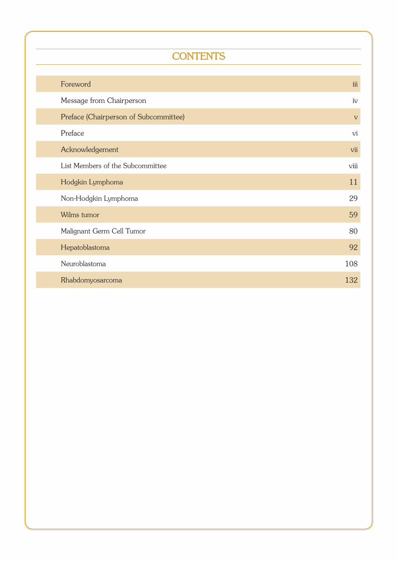
CONTENTS
Foreword iii
Message from Chairperson iv
Preface (Chairperson of Subcommittee) v
Preface vi
Acknowledgement vii
List Members of the Subcommittee viii
Hodgkin Lymphoma 11
Non-Hodgkin Lymphoma 29
Wilms tumor 59
Malignant Germ Cell Tumor 80
Hepatoblastoma 92
Neuroblastoma 108
Rhabdomyosarcoma 132

11 Consensus Document for Management of Pediatric Lymphomas and Solid Tumors
Introduction
Pediatric Hodgkin lymphoma is a highly curable malignancy. The emphasis of treatment in pediatric Hodgkin lymphoma has shifted towards risk stratified approach, so that long-term side effects of chemotherapy and radiotherapy can be reduced. The age standardized rates (ASR) of Hodgkin lymphoma in India is 0.4/100000 population, whereas the global ASR varies between 0.3/100000 in less developed countries and 0.6/100000 in developed countries1. Hodgkin lymphoma is more common in boys than in girls with the gender gap being wider in developing countries than developed countries2. Children with Hodgkin lymphoma in India present at a younger age when compared to Western patients2. Long-term outcomes reported from various centers in India are comparable to outcomes reported from western centers.
Review of literature
Management of pediatric Hodgkin lymphoma has evolved over the last 5-6 decades. Multiple prospective randomized controlled trials in pediatric Hodgkin lymphoma have been conducted in North America and Europe. The majority of the data on Hodgkin lymphoma management from India has been retrospective in nature. The current guidelines therefore will be based mainly on the results of prospective RCT data from the western countries.
The optimum treatment of Hodgkin lymphoma in children is not clearly defined. There is wide variation among the treatment protocols used in various centers in India and abroad. Although protocols using the ABVD regimen are standard for treating adults, their use in children is limited due to the cumulative toxicity of the regimen.
Treatment Philosophy for Pediatric Hodgkin Lymphoma3
Treatment is risk-adapted •
Treatment is response-based •
Age and Gender are important factors when deciding treatment •
Chemotherapy is required for treating all patients with classical Hodgkin lymphoma. The dose and •cycles of chemotherapy are determined by the stage, risk group, initial response, age, disease bulk, B symptoms and gender
Radiotherapy in low doses is an integral part of treatment regimens for early stage favorable-risk •Hodgkin lymphoma. Radiotherapy is incorporated to reduce the chemotherapy cycles delivered, thereby decreasing the long-term chemotherapy toxicities
The goal is to minimize treatment in patients with favorable Hodgkin lymphoma and in those with •good response to initial chemotherapy
CHAPTER
1 HODGKIN LYMPHOMA
12 Consensus Document for Management of Pediatric Lymphomas and Solid Tumors
Chemotherapy Principles3
Combination chemotherapy is preferred over single agent drugs •
Alkylating agents like procarbazine, and cyclophosphamide can cause sterility especially in males •
Anthracylines like doxorubicin in higher cumulative doses can cause cardiac dysfunction •
Etoposide can cause secondary leukemia •
Bleomycin is associated with pulmonary toxicity •
Therefore, it is essential to limit the cumulative doses of the above drugs in chemotherapy regimens •used for treating pediatric Hodgkin Lymphoma.
Radiotherapy Principles3
Emphasis is shifting from involved-field radiation therapy (IFRT) to involved nodal RT (INRT). •
The radiation field in IFRT will depend on the location of the nodes •
The radiotherapy dose used varies from 20-36 Gy depending upon the response to chemotherapy •
Pre-treatment nodal size needs to be irradiated. •
Risk Stratification4
There is considerable variation in risk stratification among various trials and treatment groups •
The risk stratification has also evolved over the last few decades •
Therefore, it is difficult to compare trials •
The general risk stratification followed by various groups are given below �
Favorable � : Stage I or II without adverse prognostic factors
Intermediate � : Stage I or II with adverse prognostic factors (presence of “B” symptoms, bulky lymphadenopathy,extranodal extension to contiguous structures, involvement of three or more nodal areas)
Advanced � : Stage II BE, II BX, IIIAE, IIIAX IIIB-IV
Summary of important trials
North American Trials
Pediatric Oncology Group: Response-based risk-adapted therapy1.
a. Favorable low-stage patient (IA, IB, IIA, IIIA): 2 cycles ABVE with IFRT (25.5 Gy) was equivalent to four cycles of ABVE with IFRT (25.5 Gy) in patients who achieved complete remission (CR) after 2 cycles5.
b. Unfavorable advanced disease: patient who achieved a rapid response after 3 cycles of dose-dense ABVE-PC had outcomes comparable to patients who achieved rapid response and received 5 cycles of dose-dense ABVE-PC. All patients received 21 Gy IFRT6.
Childrens Cancer Group Trial:2.
a. COPP/ABV hybrid chemotherapy followed by randomization to IFRT or no IFRT in patients achieving CR. Event-free survival (EFS) was inferior in patients in whom IFRT was omitted7.
13 Consensus Document for Management of Pediatric Lymphomas and Solid Tumors
b. Response-adapted de-escalation treatment in patients with stage IIB, IIIB and stage IV. Patients with rapid early response after four cycles of dose-intensive BEACOPP could be de-escalated to four cycles of COPP/ABV without IFRT in girls and two cycles of ABVD followed by IFRT in boys8.
Stanford, St Jude and Boston Consortium trials:3.
a. Patients with favorable Hodgkin lymphoma who achieve early CR with four cycles VAMP chemotherapy have outcomes similar to patients who receive 4 cycles VAMP with 25.5 Gy IFRT9.
German Trials4.
a. Omission of radiotherapy in intermediate or high-risk patients who achieve CR leads to inferior outcome. However the omission of radiotherapy in favorable-risk group patients did not result in inferior outcome. All patients received OEPA or OPPA/COPP chemotherapy10.
5. Euronet Trial: Ongoing multi-center trial in Europe. Final results have not been published, the interim analysis has revealed the following
a. COPP and COPDAC are similarly efficacious and therefore procarbazine (COPP) can be eliminated in boys thereby decreasing sterility.
b. EFS of all patients did not differ whether they received radiotherapy or not.
c. Favorable-risk patients with bulky disease (200ml) and/or an ESR≥30mm/hr at presentation should be treated as an intermediate-risk group11.
6. Indian Experience
a. Trehan et al12 have reported on outcomes of 206 children with Hodgkin lymphoma treated at PGI, Chandigarh. The 5-year overall survival (OS) and EFS were 92.7% and 77.75%, respectively. Children with early stage disease and the absence of B symptoms had a better OS of 97.7% each, as compared with 87.2% and 88.2% in those with late-stage disease and B symptoms, respectively. Only 3/206 patients received radiotherapy, various chemotherapy protocols like ABVD, VAEP, ABVD+COPP were used during different time periods of the study. This retrospective study highlighted that good outcomes comparable to western data can be achieved with multi-agent chemotherapy alone, omitting radiotherapy.
b. Arya et al13 have published the outcomes of 148 children with Hodgkin lymphoma treated with chemotherapy alone. Patients received 4 cycles COPP alternating with four cycles ABVD. The 5-year OS and EFS are 91.5 and 87.9%, respectively. Advanced stage, B symptoms, anemia, spleen, and marrow involvement were adverse prognostic factors for survival. Late toxicities were minimal.
c. Chandra et al14 reported a 5 year OS of 80% in 36 patients with Hodgkin lymphoma treated with six cycles of COPP. 70% of patients in the study had advanced disease.
d. Sagar et al15 reported 5 year OS of 85% in stage III and IV patients and 92% in stage I Hodgkin lymphoma patient treated with 6-8 cycles of COPP/ABV chemotherapy. There were 134 patients in this retrospective analysis and 60% of patients had advanced stage disease. Only 5% of the patients received radiotherapy for residual disease after completion of chemotherapy.
14 Consensus Document for Management of Pediatric Lymphomas and Solid Tumors
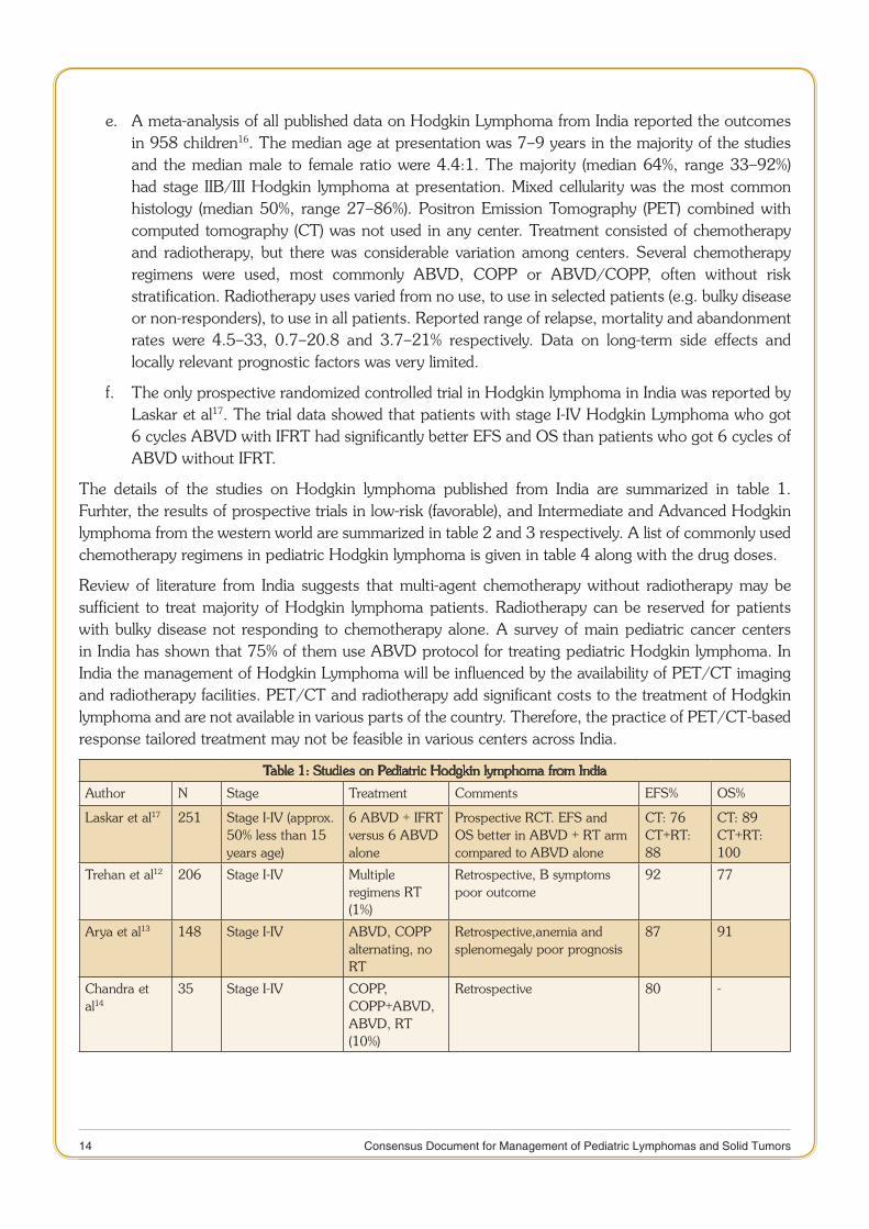
e. A meta-analysis of all published data on Hodgkin Lymphoma from India reported the outcomes in 958 children16. The median age at presentation was 7–9 years in the majority of the studies and the median male to female ratio were 4.4:1. The majority (median 64%, range 33–92%) had stage IIB/III Hodgkin lymphoma at presentation. Mixed cellularity was the most common histology (median 50%, range 27–86%). Positron Emission Tomography (PET) combined with computed tomography (CT) was not used in any center. Treatment consisted of chemotherapy and radiotherapy, but there was considerable variation among centers. Several chemotherapy regimens were used, most commonly ABVD, COPP or ABVD/COPP, often without risk stratification. Radiotherapy uses varied from no use, to use in selected patients (e.g. bulky disease or non-responders), to use in all patients. Reported range of relapse, mortality and abandonment rates were 4.5–33, 0.7–20.8 and 3.7–21% respectively. Data on long-term side effects and locally relevant prognostic factors was very limited.
f. The only prospective randomized controlled trial in Hodgkin lymphoma in India was reported by Laskar et al17. The trial data showed that patients with stage I-IV Hodgkin Lymphoma who got 6 cycles ABVD with IFRT had significantly better EFS and OS than patients who got 6 cycles of ABVD without IFRT.
The details of the studies on Hodgkin lymphoma published from India are summarized in table 1. Furhter, the results of prospective trials in low-risk (favorable), and Intermediate and Advanced Hodgkin lymphoma from the western world are summarized in table 2 and 3 respectively. A list of commonly used chemotherapy regimens in pediatric Hodgkin lymphoma is given in table 4 along with the drug doses.
Review of literature from India suggests that multi-agent chemotherapy without radiotherapy may be sufficient to treat majority of Hodgkin lymphoma patients. Radiotherapy can be reserved for patients with bulky disease not responding to chemotherapy alone. A survey of main pediatric cancer centers in India has shown that 75% of them use ABVD protocol for treating pediatric Hodgkin lymphoma. In India the management of Hodgkin Lymphoma will be influenced by the availability of PET/CT imaging and radiotherapy facilities. PET/CT and radiotherapy add significant costs to the treatment of Hodgkin lymphoma and are not available in various parts of the country. Therefore, the practice of PET/CT-based response tailored treatment may not be feasible in various centers across India.
Table 1: Studies on Pediatric Hodgkin lymphoma from India
Author N Stage Treatment Comments EFS% OS%
Laskar et al17 251 Stage I-IV (approx. 50% less than 15 years age)
6 ABVD + IFRT versus 6 ABVD alone
Prospective RCT. EFS and OS better in ABVD + RT arm compared to ABVD alone
CT: 76CT+RT:88
CT: 89CT+RT: 100
Trehan et al12 206 Stage I-IV Multiple regimens RT (1%)
Retrospective, B symptoms poor outcome
92 77
Arya et al13 148 Stage I-IV ABVD, COPP alternating, no RT
Retrospective,anemia and splenomegaly poor prognosis
87 91
Chandra et al14
35 Stage I-IV COPP, COPP+ABVD, ABVD, RT (10%)
Retrospective 80 -
15 Consensus Document for Management of Pediatric Lymphomas and Solid Tumors
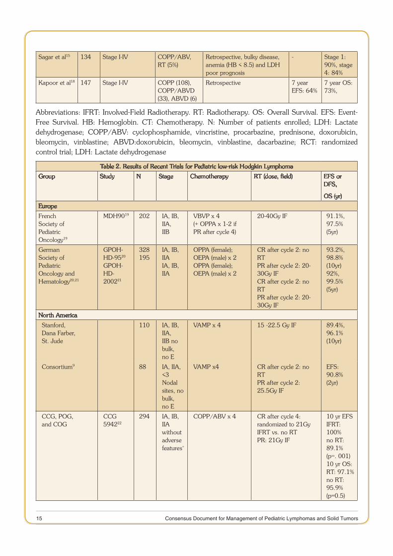
Sagar et al15 134 Stage I-IV COPP/ABV, RT (5%)
Retrospective, bulky disease, anemia (HB < 8.5) and LDH poor prognosis
- Stage 1: 90%, stage 4: 84%
Kapoor et al18 147 Stage I-IV COPP (108), COPP/ABVD (33), ABVD (6)
Retrospective 7 year EFS: 64%
7 year OS: 73%,
Abbreviations: IFRT: Involved-Field Radiotherapy. RT: Radiotherapy. OS: Overall Survival. EFS: Event-Free Survival. HB: Hemoglobin. CT: Chemotherapy. N: Number of patients enrolled; LDH: Lactate dehydrogenase; COPP/ABV: cyclophosphamide, vincristine, procarbazine, prednisone, doxorubicin, bleomycin, vinblastine; ABVD:doxorubicin, bleomycin, vinblastine, dacarbazine; RCT: randomized control trial; LDH: Lactate dehydrogenase
Table 2. Results of Recent Trials for Pediatric low-risk Hodgkin Lymphoma
Group Study N Stage Chemotherapy RT (dose, field) EFS or DFS,
OS (yr)
Europe
FrenchSociety ofPediatricOncology19
MDH9019 202 IA, IB, IIA,IIB
VBVP x 4(+ OPPA x 1-2 ifPR after cycle 4)
20-40Gy IF 91.1%,97.5%(5yr)
GermanSociety ofPediatricOncology andHematology20,21
GPOH-HD-9520 GPOH-HD-200221
328195
IA, IB, IIAIA, IB, IIA
OPPA (female);OEPA (male) x 2OPPA (female);OEPA (male) x 2
CR after cycle 2: no RTPR after cycle 2: 20-30Gy IFCR after cycle 2: no RTPR after cycle 2: 20-30Gy IF
93.2%,98.8%(10yr)92%,99.5%(5yr)
North America
Stanford,Dana Farber,St. Jude
110 IA, IB, IIA,IIB nobulk, no E
VAMP x 4 15 -22.5 Gy IF 89.4%,96.1%(10yr)
Consortium9 88 IA, IIA, <3Nodalsites, nobulk, no E
VAMP x4 CR after cycle 2: no RTPR after cycle 2:25.5Gy IF
EFS:90.8%(2yr)
CCG, POG,and COG
CCG 594222
294 IA, IB, IIAwithoutadversefeatures+
COPP/ABV x 4 CR after cycle 4:randomized to 21GyIFRT vs. no RTPR: 21Gy IF
10 yr EFSIFRT: 100%no RT: 89.1%(p=. 001)10 yr OS:RT: 97.1%no RT: 95.9%(p=0.5)
16 Consensus Document for Management of Pediatric Lymphomas and Solid Tumors
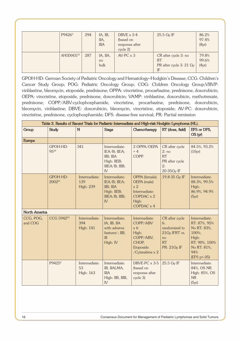
P94265 294 IA, IB, IIA,IIIA
DBVE x 2-4(based onresponse aftercycle 2)
25.5 Gy IF 86.2%97.4%(8yr)
AHOD043123 287 IA, IIA, nobulk
AV-PC x 3 CR after cycle 3: no RTPR after cycle 3: 21 GyIF
79.8%99.6%(4yr)
GPOH-HD: German Society of Pediatric Oncology and Hematology–Hodgkin’s Disease; CCG: Children’s Cancer Study Group; POG: Pediatric Oncology Group; COG: Children Oncology Group;VBVP: vinblastine, bleomycin, etoposide, prednisone; OPPA: vincristine, procarbazine, prednisone, doxorubicin; OEPA: vincristine, etoposide, prednisone, doxorubicin; VAMP: vinblastine, doxorubicin, methotrexate, prednisone; COPP/ABV:cyclophosphamide, vincristine, procarbazine, prednisone, doxorubicin, bleomycin, vinblastine; DBVE: doxorubicin, bleomycin, vincristine, etoposide; AV-PC: doxorubicin, vincristine, prednisone, cyclophosphamide; DFS: disease-free survival; PR: Partial remission
Table 3. Results of Recent Trials for Pediatric Intermediate and High-risk Hodgkin Lymphoma (HL).
Group Study N Stage Chemotherapy RT (dose, field) EFS or DFS, OS (yr)
Europe
GPOH-HD-9520
341 Intermediate:IEA/B; IIEA;IIB; IIIAHigh: IIEB;IIIEA/B; IIIB;IV
2 OPPA/OEPA + 4COPP.
CR after cycle 2: noRTPR after cycle 2:20-35Gy IF
84.5%, 93.2%(10yr)
GPOH-HD-200221
Intermediate:139High: 239
Intermediate:IEA/B; IIEA;IIB; IIIAHigh: IIEB;IIIEA/B; IIIB;IV
OPPA (female);OEPA (male) x 2Intermediate:COPDAC x 2High:COPDAC x 4
19.8-35 Gy IF Intermediate:88.3%, 99.5%High:86.9%, 94.9%(5yr)
North America
CCG, POG,and COG
CCG 594222 Intermediate:394High: 141
Intermediate;IA, IB, IIAwith adversefeatures+; IIB,IIIHigh: IV
Intermediate:COPP/ABV x 6High:COPP/ABV,CHOP, Etoposide/Cytarabine x 2
CR after cycle 6:randomized to21Gy IFRT vs. noRTPR: 21Gy IF
Intermediate:RT: 87%, 95%No RT: 83%, 100%;High:RT: 90%, 100%No RT: 81%, 94%(EFS p<.05)
P94256 Intermediate:53High: 163
Intermediate:IB, IIALMA, IIIAHigh: IIB, IIIB,IV
DBVE-PC x 3-5(based onresponse aftercycle 3)
25.5 Gy IF Intermediate:84%, OS NRHigh: 85%, OS NR(5yr)

17 Consensus Document for Management of Pediatric Lymphomas and Solid Tumors
C597048 99 IIB/IIIB + bulk,IV
BEACOPPx 4M RER: ABVD x 2F RER :COPP/ABV x 4SER: BEACOPP x4
M RER: 21 Gy IFF RER: No RTSER: 21 Gy IF
94%, 97%(5 yr)
aAHOD003124 1712 IA, IIA + bulk,IB, IIB, IIIA,IVA
ABVE-PC x 4SER: RandomizedDECA x 2
Randomized RERafter cycle two andCR after cycle 4: noRTAll others: 21 Gy IF
85.6%, 98.2% (3 yr)
VBVP: vinblastine, bleomycin, etoposide, prednisone, OPPA: vincristine, procarbazine, prednisone, doxorubicin, OEPA: vincristine, etoposide, prednisone, doxorubicin,VAMP: vinblastine, doxorubicin, methotrexate, prednisone,COPP/ABV: cyclophosphamide, vincristine, procarbazine, prednisone, doxorubicin, bleomycin, vinblastine, DBVE: doxorubicin, bleomycin, vincristine, etoposide, AV-PC: doxorubicin, vincristine, prednisone, cyclophosphamide,COPDac: cyclophosphamide, vincristine, prednisone, dacarbazine,CHOP: cyclophosphamide, doxorubicin, vincristine, prednisone, DBVE-PC: doxorubicin, bleomycin, vincristine, etoposide, prednisone, cyclophosphamide, BEACOPP: bleomycin, etoposide, doxorubicin, cyclophosphamide, vincristine, prednisone, procarbazine, ABVE-PC: doxorubicin, bleomycin, vincristine, etoposide, prednisone, cyclophosphamide, DECA: dexamethasone, etoposide, cisplatin, cytarabine, IF: involved-field, RT: radiation therapy, M: male,F: female, RER: rapid early responder, SER: slow early responder, CR: complete remission, PR: partial remission
Table 4: Commonly used Chemotherapy Regimens and Doses in Pediatric Hodgkin Lymphoma
Name Drugs Dose Route Days Schedule
COPP21 Cyclophosphamide 600 mg/m2 IV 1,8 Repeat every 28 days
Vincristine 1.4 mg/m2 IV 1,8
Procarbazine 100 mg/m2 PO 1-15
Prednisone 40 mg/m2 PO 1-15
COPDAC21 Dacarbazine substituted for procarbazine in COPP
250 mg/m2 IV 1-3 Repeat every 28 days
OPPA21 Vincristine 1.5 mg/m2 IV 1,8,15 Repeat every 28 days
Prednisone 100 mg/m2 PO 1-15
Procarbazine 60 mg/m2 PO 1-15
Adriamycin 40 mg/m2 IV 1, 15
OEPA21 Vincristine 1.5 mg/m2 IV 1,8,15 Repeat every 28 days
Etoposide 125 mg/m2 IV 3-6
Procarbazine 60 mg/m2 PO 1-15
Adriamycin 40 mg/m2 IV 1, 15

18 Consensus Document for Management of Pediatric Lymphomas and Solid Tumors
ABVD25 Adriamycin 25 mg/m2 IV 1, 15 Repeat every 28 days
Bleomycin 10 U/m2 IV 1, 15
Vinblastine 6 mg/m2 IV 1, 15
Dacarbazine 375 mg/m2 IV 1, 15
COPP/ABV7 Cyclophosphamide 600 mg/m2 IV 0 Repeat every 28 days
Vincristine 1.4 mg/m2 IV 0
Procarbazine 100 mg/m2 PO 0-6
Prednisone 40 mg/m2 PO 0-13
Adriamycin 35 mg/m2 IV 7
Bleomycin 10 U/m2 IV 7
Vinblastine 6 mg/m2 IV 7
VAMP26 Vinblastine 6 mg/m2 IV 1, 15 Repeat every 28 days
Adriamycin 25 mg/m2 IV 1, 15
Methotrexate 20 mg/m2 IV 1, 15
Prednisone 40 mg/m2 PO 1-14
DBVE5 Doxorubicin 25 mg/m2 IV 1, 15 Repeat every 28 days
Bleomycin 10 U/m2 IV 1, 15
Vincristine 1.5 mg/m2 IV 1, 15
Etoposide 100 mg/m2 IV 1-5
ABVE-PC6 Doxorubicin 30 mg/m2 IV 0,1 Repeat every 21 days
Bleomycin 10 U/m2 IV 0,7
Vincristine 1.4 mg/m2 IV 0,7
Etoposide 75 mg/m2 IV 0-4
Prednisone 40 mg/m2 PO 0-9
Cyclophosphamide 800 mg/m2 IV 0
BEACOPP8 Bleomycin 10 U/m2 IV 7 Repeat every 21 days
Etoposide 200 mg/m2 IV 0-2
Doxorubicin 35 mg/m2 IV 0
Cyclophosphamide 1200 mg/m2 IV 1,8
Vincristine 2 mg/m2 IV 7
Prednisone 40 mg/m2 PO 0-13
Procarbazine 100 mg/m2 PO 0-6
CVP27 Cyclophosphamide 500 mg/m2 IV 1 Repeat every 21 days
Vincristine 6 mg/m2 IV 1,8
Prednisolone 40 mg/m2 PO 1-8
Response assessment
Further refinement of risk classification may be performed through assessment of response after initial cycles of chemotherapy or at the completion of chemotherapy.
Interim response assessment
Assessment of response to treatment after completing 2-3 cycles of chemotherapy has been found to be useful in de-escalating treatment in patients with good response or escalating treatment in patients with poor response. The interim assessment can be performed using CT scans or PET/CT scan. There is no standard definition of a good response or poor response and various protocols have used their own definitions to define response. Clinical findings and laboratory investigations have also been incorporated

19 Consensus Document for Management of Pediatric Lymphomas and Solid Tumors
along with the radiological findings to define response. The Lugano classification is the most widely accepted classification for response assessment (Table 5)28.
Diagnostic work-up
Guidelines for histopathology29
Lymph node biopsy for confirming the diagnosis
Wherever possible excisional lymph node biopsy is strongly recommended over core needle biopsy. 1. However, in inaccessible sites like retroperitoneum and mediastinum, core needle biopsy will be acceptable.
Fine-needle aspiration is usually not sufficient for diagnosis of lymphoma in children and not 2. recommended
For histological diagnosis and subtyping, immunohistochemistry is 3. recommended where feasible. Immunostaining for CD15, CD30, CD3, CD20, and CD45 are ideal for classical HL (cHL) but a limited profile with CD15 and CD30 may be adequate if histopathology is classical. For nodular lymphocyte-predominant Hodgkin Lymphoma (NLPHL), CD20 is recommended.
Pathological diagnosis should be made according to the World Health Organization (WHO) classification from a sufficiently large surgical specimen or excisional lymph node biopsy to provide enough material for fresh frozen and formalin-fixed samples. In cHL, the presence of Hodgkin and Reed–Sternberg (HRS) cells is disease-defining while the detection of lymphocyte-predominant (LP) cells is required for the diagnosis of NLPHL. The immunophenotype of the malignant cells in cHL and NLPHL differs significantly. In contrast to HRS cells that stain consistently positive for CD30 and CD15, occasionally positive for CD20 and negative for CD45, LP cells are characterized by the expression of CD20 and CD45 but they lack CD15 and CD30.
2008 WHO classification of Lymphoid Neoplasms
Nodular Lymphocyte-Predominant •
Classical Hodgkin Lymphoma •
Nodular sclerosis classical Hodgkin Lymphoma �
Lymphocyte-rich classical Hodgkin Lymphoma �
Mixed cellularity classical Hodgkin Lymphoma �
Lymphocyte- depleted classical Hodgkin Lymphoma �
Staging
It is essential that every patient undergoes staging investigations prior to starting disease directed therapy. The stage is determined by anatomic evidence of disease using CT scanning in conjunction with functional imaging (wherever possible) and bone marrow biopsy. The staging classification used for Hodgkin lymphoma was adopted at the Ann Arbor Conference held in 1971 and revised in 1989.
Ann Arbor Staging classification of Hodgkin Lymphoma30
Stage I
Involvement of a single lymphatic site (i.e., nodal region, Waldeyer’s ring, thymus, or spleen) (I); or localized involvement of a single extralymphatic organ or site in the absence of any lymph node involvement (IE).
20 Consensus Document for Management of Pediatric Lymphomas and Solid Tumors
Stage II
Involvement of two or more lymph node regions on the same side of the diaphragm (II); or or localized involvement of a single extralymphatic organ or site in association with regional lymph node involvement with or without involvement of other lymph node regions on the same side of the diaphragm (IIE).
Stage III
Involvement of lymph node regions on both sides of the diaphragm (III), which may also be accompanied by extralymphatic extension in association with adjacent lymph node involvement (IIIE) or by involvement of the spleen (IIIS) or both (IIIE,S).
Stage IV
Diffuse or disseminated involvement of one or more extralymphatic organs, with or without associated lymph node involvement; or isolated extralymphatic organ involvement in the absence of adjacent regional lymph node involvement, but in conjunction with disease in distant site(s). Stage IV includes any involvement of the liver or bone marrow, lungs (other than by direct extension from another site), or cerebrospinal fluid.
Annotations of Stage
HL will be subclassified into A and B categories. Patients with any of the following specific symptoms will be classified as B:
Unexplained loss of more than 10% of body weight in the 6 months before diagnosis. •
Unexplained fever with temperatures above 38° C for more than 3 days. •
Drenching night sweats. •
Definition of bulky disease
Bulky mediastinal disease is defined as a mediastinal mass with a horizontal tumor diameter> 1/3 the thoracic diameter (measured transversely at the level of the dome of the diaphragm on a 6 foot upright posterior-anterior chest x-ray. In the presence of hilar nodal disease the maximal mediastinal tumor measurement may be taken at the level of the hilum. This should be measured as the maximum mediastinal width (at a level containing the tumor and any normal mediastinal structures at the level) over the maximum thoracic ratio.
Bulky disease outside the mediastinum is defined as a single node or continuous aggregate of nodal tissue that measures > 6 cm in the longest diameter in any nodal area.
Diagnostic work-up
Clinical Evaluation: The work-up should include a thorough history and physical examination, including 1. B symptoms (unexplained fever, more than 10% weight loss and/or drenching night sweats).
Physical Examination should be careful and complete:2.
a. Common lymph node areas to be palpated
b. Number of sites / lymph node regions are to be noted
c. Measurement of largest mass (bulky disease)
d. The size of liver / spleen in cm below costal margin
e. Baseline pubertal status
21 Consensus Document for Management of Pediatric Lymphomas and Solid Tumors
Essential laboratory investigations:3.
a. Complete blood counts (CBC) & differential leukocyte counts and erythrocyte sedimentation rate (ESR)
b. Lactate dehydrogenase (LDH), liver function tests (LFT) and serum creatinine
c. Contrast-enhanced computed tomography (CECT) neck, chest and whole abdomen are mandatory
d. Adequate bilateral (B/L) bone marrow (BM) biopsy should be performed on patients who have stage III or IV disease or B symptoms
e. Pleural cytology if there is pleural effusion
Other investigations
a. HBV, HCV and HIV screening
b. Baseline echocardiography and pulmonary function test
c. PET/CT scan should be done wherever feasible
d. Bone scan: indicated in case of bone pain, elevated alkaline phosphatase; it is not needed if PET/CT scan has been done
e. Reproductive counselling (in younger patients) and semen preservation for older male patients and serum pregnancy test (in female patients)
Recommendations regarding PET/CT scan:
1. PET Scan is a preferable modality for staging and response assessment.
2. Any sub-centimeter lymph node regardless of Fleuro-deoxy-glucose (FDG) avidity should be taken as negative.
3. PET/CT response should be reported according to Deauville criteria. Score 1,2, 3 should be considered ‘negative’ and 4,5 considered ‘positive’.
4. Interim scans should be performed as long after the last chemotherapy administration as possible, to avoid false positive uptake.
The five-point scale also referred to as the ‘Deauville criteria’ has been used for reporting in response guided trials and has published a high interobserver agreement and improved predictive value when compared with earlier International Harmonization criteria. The response scan is compared with the baseline scan and scored according to the level of highest residual FDG uptake using the five point score as follows:
Score 1 no uptake •
Score 2 uptake less than or equal to the mediastinum •
Score 3 uptake greater than the mediastinum, but less than the liver •
Score 4 uptake moderately higher than the liver •
Score 5 uptake markedly higher than the liver •

22 Consensus Document for Management of Pediatric Lymphomas and Solid Tumors
After chemotherapy, stimulation of normal BM may result in diffusely increased uptake which is higher than normal liver. The uptake in sites of Initial marrow involvement should then be compared to uptake within normal marrow to assess the presence/absence of residual disease.
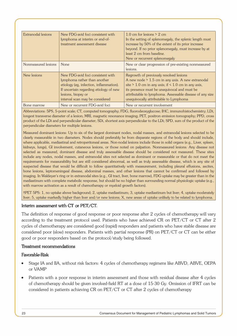
Table 5 below defines CT-based and PET/CT-based response for interim and end of treatment assessment in Hodgkin lymphoma28.
Table 5. Response assessment criteria with CT or PET/CT
Response and Site PET-CT–Based Response CT-Based Response
Complete Complete metabolic response Complete radiologic response (all of the following)
Lymph nodes andextralymphatic sites
Score 1, 2, or 3 with or without a residual mass on 5PS†
Target nodes/nodal masses must regress to 1.5 cm in LDi. No extralymphatic sites of disease
Nonmeasured lesion Not applicable Absent
Organ enlargement Not applicable Regress to normal
New lesions None None
Bone marrow No evidence of FDG-avid disease in marrow
Normal by morphology; if indeterminate, IHC negative
Partial Partial metabolic response Partial remission (all of the following)
Lymph nodes and extralymphatic sites
Score 4 or 5† with reduced uptake compared with baseline and residual mass(es) of any size. At interim, these findings suggest responding disease. At the end of treatment, these findings indicate residual disease
A 50 % decrease in the SPD of up to six target measurable nodes and extranodal sites.
Nonmeasured lesions Not applicable Absent/normal, regressed, but no increase
Organ enlargement Not applicable The spleen must have regressed by 50% in length beyond normal
New lesions None None
Bone marrow Residual uptake higher than uptake in normal marrow, but reduced compared with baseline
Not applicable
No response or stable disease
No metabolic response Stable disease
Target nodes/nodal masses,extranodal lesions
Score 4 or 5 with no significant change in FDG uptake from baseline in interim or end of treatment
A 50 % decrease from baseline in the SPD of up to six dominant, measurable nodes and extranodal sites; no criteria for progressive disease are met
Nonmeasured lesions Not applicable No increase consistent with progression
Organ enlargement Not applicable No increase consistent with progression
New lesions None None
Bone marrow No change from baseline Not applicable
Progressive disease Progressive metabolic Progressive disease requires at least one of the following PPD progression:
Individual target nodes/nodal masses
Score 4 or 5 with an increase in intensity of uptake from baseline and/or
An individual node/lesion must be abnormal with: LDi 1.5 cm and Increase by 50% from PPD nadir and An increase in LDi or SDi from nadir 0.5 cm for lesions > 2 cm
23 Consensus Document for Management of Pediatric Lymphomas and Solid Tumors
Extranodal lesions New FDG-avid foci consistent with lymphoma at interim or end-of-treatment assessment disease
1.0 cm for lesions > 2 cmIn the setting of splenomegaly, the splenic length must increase by 50% of the extent of its prior increase beyond. If no prior splenomegaly, must increase by at least 2 cm from baseline.New or recurrent splenomegaly
Nonmeasured lesions None New or clear progression of pre-existing nonmeasured lesions.
New lesions New FDG-avid foci consistent with lymphoma rather than another etiology (eg, infection, inflammation). If uncertain regarding etiology of new lesions, biopsy orinterval scan may be considered
Regrowth of previously resolved lesionsA new node > 1.5 cm in any axis. A new extranodal site > 1.0 cm in any axis; if < 1.0 cm in any axis, its presence must be unequivocal and must be attributable to lymphoma. Assessable disease of any size unequivocally attributable to Lymphoma
Bone marrow New or recurrent FDG-avid foci New or recurrent involvement
Abbreviations: 5PS, 5-point scale; CT, computed tomography; FDG, fluorodeoxyglucose; IHC, immunohistochemistry; LDi, longest transverse diameter of a lesion; MRI, magnetic resonance imaging; PET, positron emission tomography; PPD, cross product of the LDi and perpendicular diameter; SDi, shortest axis perpendicular to the LDi; SPD, sum of the product of the perpendicular diameters for multiple lesions.
Measured dominant lesions: Up to six of the largest dominant nodes, nodal masses, and extranodal lesions selected to be clearly measurable in two diameters. Nodes should preferably be from disparate regions of the body and should include, where applicable, mediastinal and retroperitoneal areas. Non-nodal lesions include those in solid organs (e.g., Liver, spleen, kidneys, lungs), GI involvement, cutaneous lesions, or those noted on palpation. Nonmeasured lesions: Any disease not selected as measured, dominant disease and truly assessable disease should be considered not measured. These sites include any nodes, nodal masses, and extranodal sites not selected as dominant or measurable or that do not meet the requirements for measurability but are still considered abnormal, as well as truly assessable disease, which is any site of suspected disease that would be difficult to follow quantitatively with measurement, including pleural effusions, ascites, bone lesions, leptomeningeal disease, abdominal masses, and other lesions that cannot be confirmed and followed by imaging. In Waldeyer’s ring or in extranodal sites (e.g., GI tract, liver, bone marrow), FDG uptake may be greater than in the mediastinum with complete metabolic response, but should be no higher than surrounding normal physiologic uptake (e.g., with marrow activation as a result of chemotherapy or myeloid growth factors).
†PET 5PS: 1, no uptake above background; 2, uptake mediastinum; 3, uptake mediastinum but liver; 4, uptake moderately liver; 5, uptake markedly higher than liver and/or new lesions; X, new areas of uptake unlikely to be related to lymphoma.
Interim assessment with CT or PET/CT.
The definition of response of good response or poor response after 2 cycles of chemotherapy will vary according to the treatment protocol used. Patients who have achieved CR on PET/CT or CT after 2 cycles of chemotherapy are considered good (rapid) responders and patients who have stable disease are considered poor (slow) responders. Patients with partial response (PR) on PET/CT or CT can be either good or poor responders based on the protocol/study being followed.
Treatment recommendations
Favorable-Risk
Stage IA and IIA, without risk factors: 4 cycles of chemotherapy regimens like ABVD, ABVE, OEPA •or VAMP
Patients with a poor response in interim assessment and those with residual disease after 4 cycles •of chemotherapy should be given involved-field RT at a dose of 15-30 Gy. Omission of IFRT can be considered in patients achieving CR on PET/CT or CT after 2 cycles of chemotherapy
24 Consensus Document for Management of Pediatric Lymphomas and Solid Tumors
Intermediate-Risk
Good initial response to two cycles of chemotherapy
4 cycles of ABVD with IFRT (20-26 Gy) •
4 cycles ABVD + 2 cycles COPP •
4 cycles of ABVE-PC +/- IFRT (20-26 Gy) •
2 cycles O (E/P) PA + 2 COP (P/Dac) + 20–35 Gy IFRT •
Poor Initial response to two cycles of chemotherapy
5 cycles of ABVE-PC +/- IFRT (20-26 Gy) •
2 cycles O (E/P) PA + 4 COP (P/Dac) + 20–35 Gy IFRT •
6-8 cycles of BEACOPP •
Advanced Stage
Good initial response to two cycles of chemotherapy •
2 cycles O (E/P) PA + 4 COP (P/Dac) + 20–35 Gy IFRT •
5 cycles ABVE-PC+ 20–26 Gy IFRT •
6 cycles ABVD+ 20–26 Gy IFRT •
8 cycles BEACOPP+ 20–26 Gy IFRT •
Treatment of Relapse/ Refractory disease
Approximately 10 - 20% of patients with advanced stage HL relapse after frontline treatment. Most relapses in patients with HL occur within the first three years, and response. Response to retrieval (salvage) therapy is directly related to duration of an initial response. Progression during induction therapy or within 12 months of completion of treatment has a dismal prognosis with a 5-year disease-free survival rates of 0% and 20% respectively. Relapses occurring 12 months or greater have better outcomes with salvage chemotherapy followed by autologous stem cell transplant.
Adverse prognostic factors after relapse include the following:
The presence of B symptoms (fever, weight loss, and night sweats) and extranodal disease •
Early relapse (occurring between 3–12 months from the end of therapy). •
Inadequate response to initial second-line therapy. •
There is no uniform strategy for choice of second line regimen in Hodgkins lymphoma. The patients with late relapse and good response to initial two cycles of chemotherapy can be salvaged in 40-50% of cases with high-dose chemotherapy and autologous stem cell transplant (SCT)31,32. Various relapse regimens include are listed in Table 6, the most popular among them being ifosfamide, carboplatin, etoposide (ICE)33. The commonly used conditioning regimen for autologous SCT is BEAM (BCNU, etoposide, cytosine arabinoside, and melphalan)33. Results of published literature are listed in Table 7. Allogenic SCT represents an option in a small subset of highest risk patients in whom there are probably no other realistic options for cure at present. However, treatment-related toxicity and relapse rates are very high.
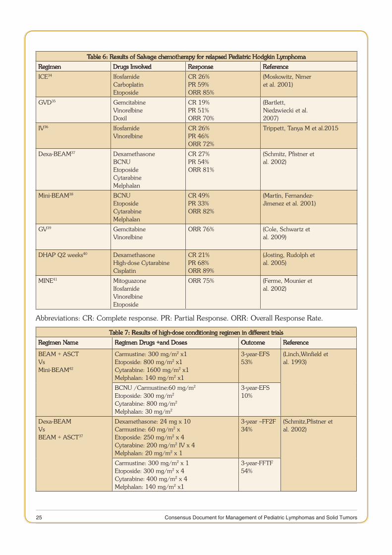
25 Consensus Document for Management of Pediatric Lymphomas and Solid Tumors
Table 6: Results of Salvage chemotherapy for relapsed Pediatric Hodgkin Lymphoma
Regimen Drugs Involved Response Reference
ICE34 IfosfamideCarboplatinEtoposide
CR 26%PR 59%ORR 85%
(Moskowitz, Nimeret al. 2001)
GVD35 GemcitabineVinorelbineDoxil
CR 19%PR 51%ORR 70%
(Bartlett,Niedzwiecki et al.2007)
IV36 IfosfamideVinorelbine
CR 26%PR 46%ORR 72%
Trippett, Tanya M et al.2015
Dexa-BEAM37 DexamethasoneBCNUEtoposideCytarabineMelphalan
CR 27%PR 54%ORR 81%
(Schmitz, Pfistner etal. 2002)
Mini-BEAM38 BCNUEtoposideCytarabineMelphalan
CR 49%PR 33%ORR 82%
(Martín, Fernandez-Jimenez et al. 2001)
GV39 GemcitabineVinorelbine
ORR 76% (Cole, Schwartz etal. 2009)
DHAP Q2 weeks40 DexamethasoneHigh-dose CytarabineCisplatin
CR 21%PR 68%ORR 89%
(Josting, Rudolph etal. 2005)
MINE41 MitoguazoneIfosfamideVinorelbineEtoposide
ORR 75% (Ferme, Mounier etal. 2002)
Abbreviations: CR: Complete response. PR: Partial Response. ORR: Overall Response Rate.
Table 7: Results of high-dose conditioning regimen in different trials
Regimen Name Regimen Drugs +and Doses Outcome Reference
BEAM + ASCTVsMini-BEAM42
Carmustine: 300 mg/m2 x1Etoposide: 800 mg/m2 x1Cytarabine: 1600 mg/m2 x1Melphalan: 140 mg/m2 x1
3-year-EFS 53%
(Linch,Winfield etal. 1993)
BCNU /Carmustine:60 mg/m2
Etoposide: 300 mg/m2
Cytarabine: 800 mg/m2
Melphalan: 30 mg/m2
3-year-EFS 10%
Dexa-BEAMVsBEAM + ASCT37
Dexamethasone: 24 mg x 10Carmustine: 60 mg/m2 xEtoposide: 250 mg/m2 x 4Cytarabine: 200 mg/m2 IV x 4Melphalan: 20 mg/m2 x 1
3-year –FF2F34%
(Schmitz,Pfistner etal. 2002)
Carmustine: 300 mg/m2 x 1Etoposide: 300 mg/m2 x 4Cytarabine: 400 mg/m2 x 4Melphalan: 140 mg/m2 x1
3-year-FFTF54%
26 Consensus Document for Management of Pediatric Lymphomas and Solid Tumors
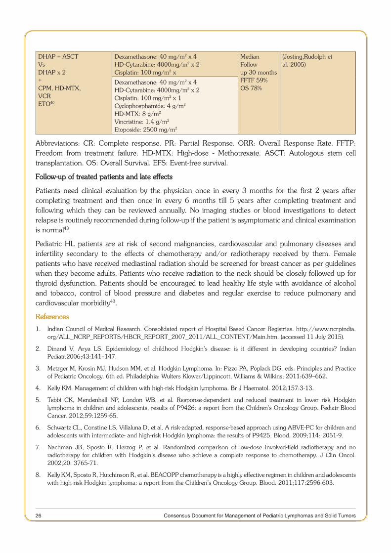
DHAP + ASCTVsDHAP x 2+CPM, HD-MTX,VCRETO40
Dexamethasone: 40 mg/m2 x 4HD-Cytarabine: 4000mg/m2 x 2Cisplatin: 100 mg/m2 x
Median Followup 30 monthsFFTF 59%OS 78%
(Josting,Rudolph etal. 2005)
Dexamethasone: 40 mg/m2 x 4HD-Cytarabine: 4000mg/m2 x 2Cisplatin: 100 mg/m2 x 1Cyclophosphamide: 4 g/m2
HD-MTX: 8 g/m2
Vincristine: 1.4 g/m2
Etoposide: 2500 mg/m2
Abbreviations: CR: Complete response. PR: Partial Response. ORR: Overall Response Rate. FFTP: Freedom from treatment failure. HD-MTX: High-dose - Methotrexate. ASCT: Autologous stem cell transplantation. OS: Overall Survival. EFS: Event-free survival.
Follow-up of treated patients and late effects
Patients need clinical evaluation by the physician once in every 3 months for the first 2 years after completing treatment and then once in every 6 months till 5 years after completing treatment and following which they can be reviewed annually. No imaging studies or blood investigations to detect relapse is routinely recommended during follow-up if the patient is asymptomatic and clinical examination is normal43.
Pediatric HL patients are at risk of second malignancies, cardiovascular and pulmonary diseases and infertility secondary to the effects of chemotherapy and/or radiotherapy received by them. Female patients who have received mediastinal radiation should be screened for breast cancer as per guidelines when they become adults. Patients who receive radiation to the neck should be closely followed up for thyroid dysfunction. Patients should be encouraged to lead healthy life style with avoidance of alcohol and tobacco, control of blood pressure and diabetes and regular exercise to reduce pulmonary and cardiovascular morbidity43.
References
Indian Council of Medical Research. Consolidated report of Hospital Based Cancer Registries. http://www.ncrpindia.1. org/ALL_NCRP_REPORTS/HBCR_REPORT_2007_2011/ALL_CONTENT/Main.htm. (accessed 11 July 2015).
Dinand V, Arya LS. Epidemiology of childhood Hodgkin’s disease: is it different in developing countries? Indian 2. Pediatr.2006;43:141–147.
Metzger M, Krosin MJ, Hudson MM, et al. Hodgkin Lymphoma. In: Pizzo PA, Poplack DG, eds. Principles and Practice 3. of Pediatric Oncology. 6th ed. Philadelphia: Wulters Klower/Lippincott, Williams & Wilkins; 2011:639–662.
Kelly KM: Management of children with high-risk Hodgkin lymphoma. Br J Haematol. 2012;157:3-13. 4.
Tebbi CK, Mendenhall NP, London WB, et al. Response-dependent and reduced treatment in lower risk Hodgkin 5. lymphoma in children and adolescents, results of P9426: a report from the Children’s Oncology Group. Pediatr Blood Cancer. 2012;59:1259-65.
Schwartz CL, Constine LS, Villaluna D, et al. A risk-adapted, response-based approach using ABVE-PC for children and 6. adolescents with intermediate- and high-risk Hodgkin lymphoma: the results of P9425. Blood. 2009;114: 2051-9.
Nachman JB, Sposto R, Herzog P, et al. Randomized comparison of low-dose involved-field radiotherapy and no 7. radiotherapy for children with Hodgkin’s disease who achieve a complete response to chemotherapy. J Clin Oncol. 2002;20: 3765-71.
Kelly KM, Sposto R, Hutchinson R, et al. BEACOPP chemotherapy is a highly effective regimen in children and adolescents 8. with high-risk Hodgkin lymphoma: a report from the Children’s Oncology Group. Blood. 2011;117:2596-603.

27 Consensus Document for Management of Pediatric Lymphomas and Solid Tumors
Metzger ML, Weinstein HJ, Hudson MM, et al. Association between radiotherapy vs no radiotherapy based on early 9. response to VAMP chemotherapy and survival among children with favorable-risk Hodgkin lymphoma. JAMA. 2012;307:2609-16.
Dörffel W, Lüders H, Rühl U, et al. Preliminary results of the multicenter trial GPOH-HD 95 for the treatment of 10. Hodgkin’s disease in children and adolescents: analysis and outlook. Klin Padiatr. 2003;215:139-45.
Euronet Hodgkin lymphoma protocol available at. 11. https://www.skion.nl/workspace/uploads/EuroNet-PHL-Interim-Treatment-Guidelines-2012-12-3v0-2.pdf. Accessed on 2 July 2015.
Trehan 12. A, Singla S, Marwaha RK, Bansal D, Srinivasan R.Hodgkin lymphoma in children: experience in a tertiary care center in India.J Pediatr Hematol Oncol. 2013;35:174-9.
Arya LS, Thavaraj V, Dawar R, et al. Hodgkin’s disease in Indian children: outcome with chemotherapy alone. Pediatr 13. Blood Cancer. 2006;46:26–34.
Chandra J, Naithan R, Singh V, et al. Developing anticancer chemotherapy services in a developing country: Hodgkin 14. lymphoma experience. Pediatr Blood Cancer. 2008;17:485–488.
Sagar 15. TG, Chandra A, Raman SG. Childhood Hodgkin disease treated with COPP/ABV hybrid chemotherapy: a progress report.Med Pediatr Oncol. 2003;40:66-9.
Aabideen K, Kulkarni KP, Arora RS. Current outcomes of Hodgkin’s disease among children in India: A systematic 16. analysis. 44th Congress of the International Society of Paediatric Oncology (SIOP) 2012, London, United Kingdom, 5th–8th October, 2012 SIOP abstracts. Pediatr. Blood Cancer, 59: 965–1152.
Laskar S, Gupta T, Vimal S, Muckaden MA, Saikia TK, Pai SK, et al. Consolidation radiation after complete remission 17. in Hodgkin’s disease following six cycles of doxorubicin, bleomycin, vinblastine, and dacarbazine chemotherapy: is there a need? J Clin Oncol. 2004;;22:62–8.
Kapoor G, Advani SH, Dinshaw KA, Muckaden MA, Soman CS, Saikia TK, et al. Treatment results of Hodgkin’s disease 18. in Indian children. Pediatr Hematol Oncol. 1995 Dec;12:559–69.
Landman-Parker J, Pacquement H, Leblanc T, Habrand JL, Terrier-Lacombe MJ, Bertrand Y, et al. Localized childhood 19. Hodgkin’s disease: response-adapted chemotherapy with etoposide, bleomycin, vinblastine, and prednisone before low-dose radiation therapy-results of the French Society of Pediatric Oncology Study MDH90. J Clin Oncol. 2000;18:1500-7.
Dörffel W, Lüders H, Rühl U, et al. Preliminary results of the multicenter trial GPOH-HD 95 for the treatment of 20. Hodgkin’s disease in children and adolescents: analysis and outlook. Klin Padiatr. 2003;215:139-45.
Mauz-Körholz C, Hasenclever D, Dörffel W, et al. Procarbazine-free OEPA-COPDAC chemotherapy in boys and standard 21. OPPA-COPP in girls have comparable effectiveness in pediatric Hodgkin’s lymphoma: the GPOH-HD-2002 study. J Clin Oncol. 2010;28:3680-6.
Wolden SL, Chen L, Kelly KM, et al. Long-term results of CCG 5942: a randomized comparison of chemotherapy with 22. and without radiotherapy for children with Hodgkin’s lymphoma--a report from the Children’s Oncology Group. J Clin Oncol. 2102;30:3174-80.
Keller FG, Castellino SM, Nachman JB: What is the best treatment for children with limited-stage Hodgkin lymphoma? 23. Curr Hematol Malig Rep. 2009;4:129-135.
Friedman DL, Chen L, Wolden S, et al. Dose-intensive response-based chemotherapy and radiation therapy for children 24. and adolescents with newly diagnosed intermediate-risk hodgkin lymphoma: a report from the Children’s Oncology Group Study AHOD0031. J Clin Oncol. 2104;32:3651-8.
Bonadonna G, Santoro A: ABVD chemotherapy in the treatment of Hodgkin’s disease. Cancer Treat Rev. 1982;9 21-25. 35.
Donaldson SS, Link MP, Weinstein HJ, et al. Final results of a prospective clinical trial with VAMP and low-dose involved-26. field radiation for children with low-risk Hodgkin’s disease. J Clin Oncol. 2007;25:332-7.
Shankar A, Hall GW, Gorde-Grosjean S, et al.: Treatment outcome after low intensity chemotherapy [CVP] in children 27. and adolescents with early stage nodular lymphocyte-predominant Hodgkin’s lymphoma - an Anglo-French collaborative report. Eur J Cancer. 2012;48:1700-6.
28 Consensus Document for Management of Pediatric Lymphomas and Solid Tumors
Cheson BD, Fisher RI, Barrington SF, et al. Recommendations for initial evaluation, staging, and response assessment of 28. Hodgkin and non-Hodgkin lymphoma: the Lugano classification. J Clin Oncol. 2014;32:3059-68.
Pileri SA, Ascani S, Leoncini L, et al. Hodgkin’s lymphoma: the pathologist’s viewpoint. J Clin Pathol.2002;55:162-29. 76.
Lister TA, Crowther D, Sutcliffe SB, et al. Report of a committee convened to discuss the evaluation and staging of 30. patients with Hodgkin’s disease: Cotswolds meeting. J Clin Oncol. 1989;7:1630-6.
Schellong G, Dörffel W, Claviez A, Korholz D, Mann G, Scheel-Walter HG, et al. Salvage therapy of progressive and 31. recurrent Hodgkin’s disease: results from a multicenter study of the pediatric DAL/GPOH-HD study group. J Clin Oncol. 2005; 23:6181–9.
Monika L. Metzger et al. Initial Response to Salvage Therapy Determines Prognosis in Relapsed Pediatric Hodgkin 32. Lymphoma Patients. Cancer. 2010;116: 4376–4384.
Daw S, Wynn R, Wallace H. Management of relapsed and refractory classical Hodgkin lymphoma in children and 33. adolescents. British Journal of Hematology. 2010;152:249–260.
Moskowitz CH, Nimer SD, Zelenetz AD, Trippett T, Hedrick EE, Filippa DA, et al. A 2-step comprehensive high-dose 34. chemoradiotherapy second-line program for relapsed and refractory Hodgkin disease: analysis by intent to treat and development of a prognostic model.Blood. 2001;97:616-23.
Bartlett NL, Niedzwiecki D, Johnson JL, Friedberg JW, Johnson KB, van Besien K, et al.Gemcitabine, vinorelbine, and 35. pegylated liposomal doxorubicin (GVD), a salvage regimen in relapsed Hodgkin’s lymphoma: CALGB 59804. Ann Oncol. 2007; 18:1071-9.
Trippett TM, Schwartz CL, Guillerman RP, Gamis AS, Gardner S, Hogan S, et al.Ifosfamide and vinorelbine is an effective 36. re-induction regimen in children with refractory/relapsed Hodgkin lymphoma, AHOD00P1: a children’s oncology group report.Pediatr Blood Cancer. 2015;62:60-4.
Schmitz N, Pfistner B, Sextro M, Sieber M, Carella AM, Haenel M, et al; German Hodgkin’s Lymphoma Study Group; 37. Lymphoma Working Party of the European Group for Blood and Marrow Transplantation.Aggressive conventional chemotherapy compared with high-dose chemotherapy with autologous haemopoietic stem cell transplantation for relapsed chemosensitive Hodgkin’s disease: a randomized trial.Lancet. 2002;359:2065-71.
Martín A, Fernández-Jiménez MC, Caballero MD, Canales MA, Pérez-Simón JA, García de Bustos J, et al.Long-term 38. follow-up in patients treated with Mini-BEAM as salvage therapy for relapsed or refractory Hodgkin’s disease.Br J Haematol. 2001;113:161-71.
Cole PD, Schwartz CL, Drachtman RA, de Alarcon PA, Chen L, Trippett TM. Phase II study of weekly gemcitabine and 39. vinorelbine for children with recurrent or refractory Hodgkin’s disease: a children’s oncology group report. J Clin Oncol. 2009;27:1456-61.
Josting A, Rudolph C, Mapara M, Glossmann JP, Sieniawski M, Sieber M, et al. Cologne high-dose sequential chemotherapy 40. in relapsed and refractory Hodgkin lymphoma: results of a large multicenter study of the German Hodgkin Lymphoma Study Group (GHSG).Ann Oncol. 2005;16:116-23.
Ferme C, Mounier N, Diviné M; Groupe d’Etudes des Lymphomes de l’Adulte. Current clinical trials for the treatment of 41. adult advanced stage Hodgkin’s disease: GELA experiences. Groupe d’Etudes des Lymphomes de l’Adulte.Ann Oncol. 2002;13 Suppl 1:96-7.
Linch DC, Winfield D, Goldstone AH, Moir D, Hancock B, McMillan A, et al. Dose intensification with autologous 42. bone marrow transplantation in relapsed and resistant Hodgkin’s disease: results of a BNLI randomized trial.Lancet. 1993;341:1051-4.
Ng AK. Current survivorship recommendations for patients with Hodgkin lymphoma: focus on late effects.43. Blood. 2014;124:3373-9.
29 Consensus Document for Management of Pediatric Lymphomas and Solid Tumors
CHAPTER
2 NON-HODGKIN LYMPHOMA
Introduction:
Progress in therapy of childhood Non-Hodgkin Lymphoma (NHL) is one of the greatest success stories of the pediatric oncology in past two decades. More than 80% of children with NHL can now be cured with modern therapy. Between 1975 and 2010, the 5-year survival rate has increased from 45% to 87% in children younger than 15 years and from 48% to 82% for adolescents aged 15 to 19 years1. These extraordinary advances in treatment have resulted from an enhanced understanding of the biology, immunology, and molecular biology of the NHL; improvements in imaging and staging systems; advances in supportive care; and more rational application of risk-adapted chemotherapy by cooperative group trials. Consequent to such high cure rates, the current focus is on optimization of therapy to reduce the acute and long-term consequences of treatment.
However, the progress in treatment & outcome of NHL in children in India has not kept pace with international advances sans few tertiary care centers. A recent systematic review of outcome of childhood NHL in India showed dismal outcome; the EFS was 19-72% (median 31%) with variable follow-up. Mortality, progression/relapse rates and treatment, abandonment rates (TRA) were 7-39% (median 30%), 9-20% (median 20%) and 0-49% (median 29%) respectively2 (Table-1).
Table 1: Treatment Outcome of Pediatric NHL in India2
Disease EFS (%) Mortality Relapse/progression TRA
B-NHL 42-78 7-39% 9-20% 0-49%
Lymphoblastic lym-phoma
30-60
Anaplastic large-cell lymphoma
0-84
Overall 19-72 30% (median) 20% (median) 29% (median)
The key barriers to optimal outcome of childhood NHL in India include patient related factors such as late presentation with advanced disease, malnutrition, failure to complete treatment and infrastructure limitations, including inadequate manpower, and inadequate facilities both for the specific cancer treatment as well as for supportive care. In the ensuing sections, we outline the current progress in diagnosis, risk stratification and treatment of NHL in high-income countries as well as in India and recommend the adaptable guidelines for India depending upon the patient factors and institutional resources.
Existing guidelines:
There is a paucity of international guidelines in Pediatric NHL. Two guidelines are currently available online;
National Cancer Institute (NCI), USA guidelines on “Childhood Non-Hodgkin Lymphoma Treatment 1. for health professionals (PDQ®)”3.
30 Consensus Document for Management of Pediatric Lymphomas and Solid Tumors
Tata Memorial Hospital (TMH) text book on evidence based management of “Aggressive Non-Hodgkin 2. lymphoma”4.
Of these guidelines, the NCI guidelines for staging, diagnostic work-up and management are most commonly followed all over the world. These guidelines are regularly updated based on recent good quality evidence from multicenter randomized controlled trials and the evidence is assigned levels based on its quality.
Pathologic classification:
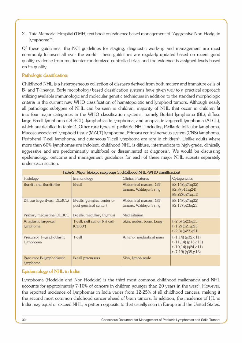
Childhood NHL is a heterogeneous collection of diseases derived from both mature and immature cells of B- and T-lineage. Early morphology based classification systems have given way to a practical approach utilizing available immunologic and molecular genetic techniques in addition to the standard morphologic criteria in the current new WHO classification of hematopoietic and lymphoid tumors. Although nearly all pathologic subtypes of NHL can be seen in children; majority of NHL that occur in children fit into four major categories in the WHO classification systems, namely Burkitt lymphoma (BL), diffuse large B-cell lymphoma (DLBCL), lymphoblastic lymphoma, and anaplastic large-cell lymphoma (ALCL), which are detailed in table-2. Other rare types of pediatric NHL including Pediatric follicular lymphoma, Mucosa-associated lymphoid tissue (MALT) lymphoma, Primary central nervous system (CNS) lymphoma, Peripheral T-cell lymphoma, and cutaneous T-cell lymphoma are rare in children5. Unlike adults where more than 60% lymphomas are indolent; childhood NHL is diffuse, intermediate to high-grade, clinically aggressive and are predominantly multifocal or disseminated at diagnosis5. We would be discussing epidemiology, outcome and management guidelines for each of these major NHL subsets separately under each section.
Table-2: Major biologic subgroups in childhood NHL (WHO classification)
Histology Immunology Clinical Features Cytogenetics
Burkitt and Burkitt-like B-cell Abdominal masses, GIT tumors, Waldeyer's ring
t(8;14)(q24;q32)t(2;8)(p11;q24)t(8;22)(q24;q11)
Diffuse large B-cell (DLBCL)
Primary mediastinal DLBCL
B-cells (germinal center or post germinal center)
B-cells( medullary thymus)
Abdominal masses, GIT tumors, Waldeyer's ring
Mediastinum
t(8;14)(q24;q32)t(2;17)(p23;q23)
Anaplastic large-celllymphoma
T-cell, null cell or NK cell (CD30+)
Skin, nodes, bone, Lung t (2;5) (p23;q35)t (1;2) (q21;p23)t (2;3) (p23;q21)
Precursor T-lymphoblasticLymphoma
T-cell Anterior mediastinal mass t (1;14) (p32;q11)t (11;14) (p13;q11)t (10;14) (q24;q11)t (7;19) (q35;p13)
Precursor B-lymphoblastic lymphoma
B-cell precursors Skin, lymph node
Epidemiology of NHL in India:
Lymphoma (Hodgkin and Non-Hodgkin) is the third most common childhood malignancy and NHL accounts for approximately 7-10% of cancers in children younger than 20 years in the west5. However, the reported incidence of lymphomas in India varies from 12-25% of all childhood cancers, making it the second most common childhood cancer ahead of brain tumors. In addition, the incidence of HL in India may equal or exceed NHL, a pattern opposite to that usually seen in Europe and the United States.
31 Consensus Document for Management of Pediatric Lymphomas and Solid Tumors
Although, some of these differences are secondary to the high incidence of HL reported in male children in North India, reporting bias cannot be excluded. NHL occurs most commonly in the second decade of life, and occurs less frequently in children younger than 3-years5.
The incidence and relative frequency of various subtypes of lymphoma in children varies considerably in different world regions. In India, the estimated incidence is between 1.9-5.6 / million/ year in girls and 9.2-15.7/ million/ year in boys in major cancer registries (Mumbai, Bengaluru, Chennai, and Delhi) similar to the incidence in the developed world6. It is estimated based on this incidence that over 3000 children develop NHL each year in India. Also, there is no population-based study with sufficient immunohistochemical backup to allow subgroup assignment according to the WHO classification in India. However, data collated by lymphoma registry at TMH in 2001 suggested an almost equal distribution of B and T-cell tumors with B-cell lymphomas constituting 48.1% of NHL whereas T-cell lymphomas 44.3% of all the lymphomas. Of B-cell, DLBCL was the commonest (22.9%) followed by BL (15.3%) and in T-Cell, LL was the commonest (31.5%) followed by ALCL seen in 11.1% cases. Overall, there seemed to be a higher prevalence of DLBCL and LL and lower frequency of BL compared to western countries7. However, impact of referral bias cannot be ruled out. In a recent multicentric survey of six large centers from across India, this distribution seems to be changing and the current subtype distribution is not significantly different from west (Arora B et al, unpublished data).
Diagnosis & Staging:
A quick and accurate diagnosis is the key to management and a successful outcome of NHL in children as these are fast-growing and delay may be fatal. Also, since most patients present with advanced disease associated with metabolic and tumor related complications in India, supportive care may need to be provided even as the diagnostic work-up is underway. In clinical presentations suggestive of NHL, such as anterior mediastinal mass with or without a pleural effusion, firm non-tender progressive lymphadenopathy, or an unexplained intra-abdominal mass with or without ascites, diagnostic material should be obtained expeditiously. Imaging may help clarify the dimensions of any primary mass and the best site to obtain a surgical biopsy. In all cases, pathology or cytology specimens obtained should be reviewed by an experienced hematopathologist because of the rarity of childhood NHL.
EVALUATION OF CHILDREN WITH NHL
Blood Investigations: •
Complete Blood Count: It is usually normal, but pancytopenia may be present with blasts in o peripheral blood if there is BM involvement
Biochemistry: Uric acid, Calcium, Phosphate, Electrolytes (Tumor lysis syndrome [TLS] parameters), o Renal and LFT (deranged if hepato-biliary or renal system is involved), LDH (tumor burden and prognostic marker)
Diagnostic Investigations: • Most children present with advanced stage disease, including BM invasion or/and malignant effusions. In such cases, the correct diagnosis can be made by cytology and immunophenotyping by flow cytometry. If this is not possible, diagnosis is based on biopsy.
Tissue Diagnosis: Biopsy (open/image guided) is planned depending on the site of involvement o (abdominal mass, extranodal site, lymph node). If the patient’s clinical condition is unstable such as in cases of superior vena cava syndrome; the diagnosis should be made with the use of less-invasive procedures (examination of pleural, or peritoneal fluid or bone marrow aspirate (BMA),
32 Consensus Document for Management of Pediatric Lymphomas and Solid Tumors
percutaneous fine-needle aspiration or biopsy of a peripheral lymph node or a large abdominal mass)
Immunophenotype: Identify the subtype of NHL as shown in table 1. It can be done byo
Immunohistochemistry on the fixed tissue: • DLBCL demonstrates a mature B-cell phenotype with B-cell lineage markers such as CD19, CD20, CD22, and CD79a. Expression of CD10 is seen in approximately half of the cases. CD30 is most commonly expressed in primary mediastinal DLBCL. Immunophenotypic features of both the Burkitt and atypical BLs are nearly identical. Both are composed of mature B-cells that express cell surface CD19, CD20, CD22, CD10, and cell surface immunoglobulin. Neither BL nor atypical Burkitt lymphomas express anti-apoptotic protein BCL2, which is very helpful in distinguishing BL from DLBCL, where BCL2 expression is more commonly seen. Immunohistochemical staining for cMYC protein is positive in BL, but may also be seen in DLBCL. Immunohistochemical stains with proliferation markers, such as Ki-67 or MIB-1, will show staining in excess of 99% of the tumor cells in BL. ALCL expresses the CD30 (Ki-1) antigen in virtually all cases. The majority of tumors have a T-cell phenotype and express a wide range of T-cell antigens on paraffin-embedded tissues (including CD2, CD3, CD4, CD5, CD7, CD8). Expression of the ALK protein by immunohistochemistry is extremely common which is strongly associated with systemic disease and is characteristically absent in primary cutaneous ALCL.Epithelial membrane antigen (EMA) is also very frequently seen, but CD45 expression may vary from strong to weak or absent and may be focally expressed.Precursor T-LL express some combination of CD1a, CD2, CD5, and CD7 along with expression of CD4 and/or CD8. CD10 is expressed in 15−40% of cases. Precursor B-LL expresses CD19, CD10, TdT and variably CD20, CD22, and HLA-DR. Precursor B-LL often expresses BCL2, helping to distinguish these cells from BL. TdT is seen in most cases of precursor B- or T-LL Demonstration of TdT is an extremely helpful finding in making a diagnosis of LL.
Flow cytometry on pleural fluid, ascitic fluid or involved bone marrow can also be done •
Cytogenetic studies: • Only in certain ambiguous cases, cytogenetics is also required for diagnosis, such as variant BL/BL-like lymphomas. Fluorescence in situ hybridization (FISH), which can be performed on tumor touch preparations, or paraffin sections, is a standard method for confirming most of the chromosomal translocations. FISH of BL will demonstrate characteristic translocations involving the cMYC oncogene locus on chromosome 8q24 in most cases which is required by WHO classification in order to make a definitive diagnosis of BL. Roughly 80% of BL contain a t(8;14)(q24;q32) rearrangement in which translocation of cMYC, normally on chromosome 8, occurs to the immunoglobulin heavy-chain gene locus on chromosome 14. The remaining cases have either a t(2;8)(p12;q24) (found in 15% of cases) or a t(8;22)(q24;q11) (5% of cases) involving cMYC and either the kappa or lambda immunoglobulin light-chain gene loci on chromosomes 2 or 22, respectively.
Staging Investigations: •
Bone marrow studies: Bilateral BM biopsy and aspiration to look for involvement by tumor cells.o
Cerebro-spinal Fluid (CSF) studies: CSF cytology and cytomorphology to look for involvement by o tumor cells.
Imaging: To see the extent of disease and response assessment.o
Whole-body contrast enhanced CT: It is the imaging modality of choice to determine tumor •extent and stage the disease. Baseline CT also serves a baseline for comparison to determine
33 Consensus Document for Management of Pediatric Lymphomas and Solid Tumors
response to treatment. However, in centers with limited infrastructure, an ultrasound of the abdomen with chest X- ray may be used in place of CT scan.
Magnetic Resonance Imaging (MRI): It can be done in children having a mass in paraspinal •area or CNS for accurate evaluation of the extent of intraspinal/CNS extent.
Investigational tests:
Positron emission tomography (PET) scan: PET-CT scan of the whole body for staging and response evaluation in children is currently investigational and being evaluated in many current studies. Although PET-CT is recognized to be advantageous in the primary staging of adult NHL, this has not been demonstrated in childhood NHL. This may due to the fact that the majority of children present with advanced disease (stages III or IV) which is easily detectable by CT scan. However, PET-CT appears to have a higher level of sensitivity for extranodal disease. It is better than BM biopsy in the detection of BM infiltration and hence may be useful as a non-invasive modality for detecting BM involvement in pediatric NHL.
Although, early response assessment to chemotherapy with an interim PET is now routine done in the management of adults with NHL; this is not regarded as standard practice in children due to limited data. However, PET-CT may be potentially useful for assessing the speed of response and confirmation of post-therapy remission (CR) since PET provides information on the size as well as the activity of residual masses. PET-CT for an interim or end of the therapy response evaluation has good negative predictive value but poor positive predictive value. Hence, residual masses should be biopsied to distinguish between residual or relapsed disease and other masses (e.g, fibrosis, thymus) before proceeding to salvage therapy.
In a prospective study from India, 34 children with non-lymphoblastic NHL underwent imaging with PET/CT and conventional contrast material-enhanced CT at baseline, after two cycles of chemotherapy, and after completion of chemotherapy. Baseline PET/CT and conventional CT were concordant in 112 disease sites, while PET/CT depicted eighteen more disease sites and two fewer disease sites resulting in disease upstaging in five patients, but had no impact on treatment. There was 100 percent concordance regarding BM involvement between PET/CT and BM biopsy. Interim imaging did not predict progression-free or OS. Both post-treatment PET/CT and CT could predict progression-free survival, but only post-treatment contrast enhanced CT could predict OS8.
Staging and risk stratification (Table-2, 3,4):
The Ann Arbor staging classification used for HL does not adequately reflect prognosis in childhood NHL because of the unique biology, clinical behavior and outcome of the four major subtypes of NHL seen in children. Murphy’s Staging (St. Jude Children’s Research Hospital) devised in 1980 is the most widely used staging scheme for childhood NHL which takes into consideration increased extranodal involvement, metastatic spread to the BM or CNS and noncontiguous spread of disease in this group (table-3)9. However, the St. Jude staging system is primarily based on clinicopathologic features of childhood BL and LL. Further, over the last 35 years, there has been the identification of new pathologic entities; improvements in cytogenetic, molecular, and immunophenotypic characterizations of disease; new diagnostic methods for the detection of minimal disseminated disease (MDD) or residual disease (MRD); and major advances in imaging. Hence, St Jude staging has been recently revised to account for these new distinct biologic entities & to incorporate some modifications and more explicit indications on peculiar sites of disease. Also, an additional staging information section, with the aim of encouraging clinicians and researchers to collect information on selected items related to marrow and CNS involvement including MDD has been introduced. (Table-4)
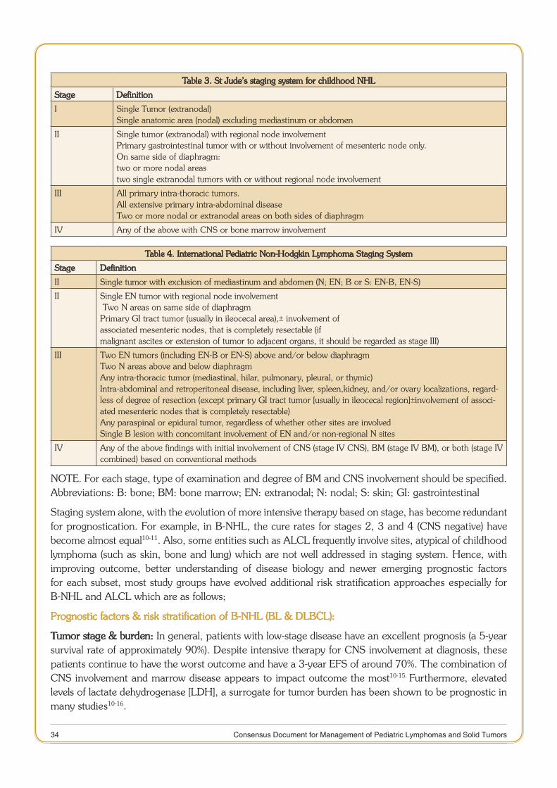
34 Consensus Document for Management of Pediatric Lymphomas and Solid Tumors
Table 3. St Jude’s staging system for childhood NHL
Stage Definition
I Single Tumor (extranodal)Single anatomic area (nodal) excluding mediastinum or abdomen
II Single tumor (extranodal) with regional node involvementPrimary gastrointestinal tumor with or without involvement of mesenteric node only.On same side of diaphragm:two or more nodal areastwo single extranodal tumors with or without regional node involvement
III All primary intra-thoracic tumors.All extensive primary intra-abdominal diseaseTwo or more nodal or extranodal areas on both sides of diaphragm
IV Any of the above with CNS or bone marrow involvement
Table 4. International Pediatric Non-Hodgkin Lymphoma Staging System
Stage Definition
I I Single tumor with exclusion of mediastinum and abdomen (N; EN; B or S: EN-B, EN-S)
II Single EN tumor with regional node involvement≥Two N areas on same side of diaphragmPrimary GI tract tumor (usually in ileocecal area),± involvement ofassociated mesenteric nodes, that is completely resectable (ifmalignant ascites or extension of tumor to adjacent organs, it should be regarded as stage III)
III Two EN tumors (including EN-B or EN-S) above and/or below diaphragmTwo N areas above and below diaphragmAny intra-thoracic tumor (mediastinal, hilar, pulmonary, pleural, or thymic)Intra-abdominal and retroperitoneal disease, including liver, spleen,kidney, and/or ovary localizations, regard-less of degree of resection (except primary GI tract tumor [usually in ileocecal region]±involvement of associ-ated mesenteric nodes that is completely resectable)Any paraspinal or epidural tumor, regardless of whether other sites are involvedSingle B lesion with concomitant involvement of EN and/or non-regional N sites
IV Any of the above findings with initial involvement of CNS (stage IV CNS), BM (stage IV BM), or both (stage IV combined) based on conventional methods
NOTE. For each stage, type of examination and degree of BM and CNS involvement should be specified. Abbreviations: B: bone; BM: bone marrow; EN: extranodal; N: nodal; S: skin; GI: gastrointestinal
Staging system alone, with the evolution of more intensive therapy based on stage, has become redundant for prognostication. For example, in B-NHL, the cure rates for stages 2, 3 and 4 (CNS negative) have become almost equal10-11. Also, some entities such as ALCL frequently involve sites, atypical of childhood lymphoma (such as skin, bone and lung) which are not well addressed in staging system. Hence, with improving outcome, better understanding of disease biology and newer emerging prognostic factors for each subset, most study groups have evolved additional risk stratification approaches especially for B-NHL and ALCL which are as follows;
Prognostic factors & risk stratification of B-NHL (BL & DLBCL):
Tumor stage & burden: In general, patients with low-stage disease have an excellent prognosis (a 5-year survival rate of approximately 90%). Despite intensive therapy for CNS involvement at diagnosis, these patients continue to have the worst outcome and have a 3-year EFS of around 70%. The combination of CNS involvement and marrow disease appears to impact outcome the most10-15. Furthermore, elevated levels of lactate dehydrogenase [LDH], a surrogate for tumor burden has been shown to be prognostic in many studies10-16.
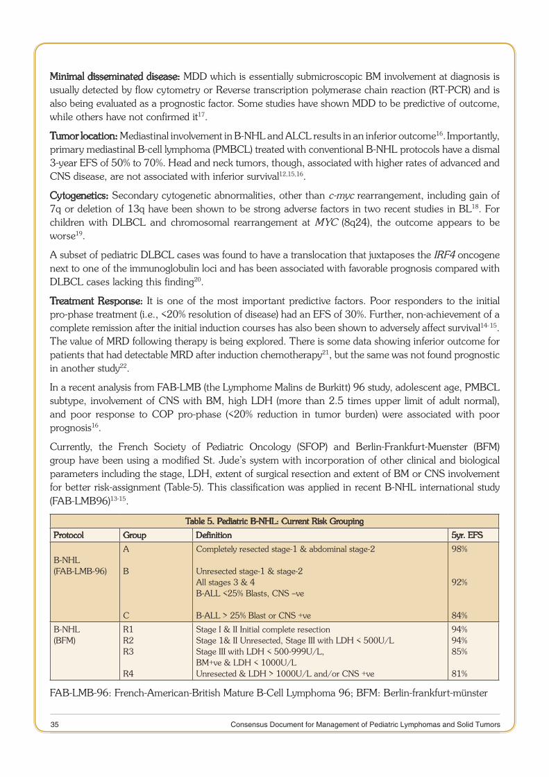
35 Consensus Document for Management of Pediatric Lymphomas and Solid Tumors
Minimal disseminated disease: MDD which is essentially submicroscopic BM involvement at diagnosis is usually detected by flow cytometry or Reverse transcription polymerase chain reaction (RT-PCR) and is also being evaluated as a prognostic factor. Some studies have shown MDD to be predictive of outcome, while others have not confirmed it17.
Tumor location: Mediastinal involvement in B-NHL and ALCL results in an inferior outcome16. Importantly, primary mediastinal B-cell lymphoma (PMBCL) treated with conventional B-NHL protocols have a dismal 3-year EFS of 50% to 70%. Head and neck tumors, though, associated with higher rates of advanced and CNS disease, are not associated with inferior survival12,15,16.
Cytogenetics: Secondary cytogenetic abnormalities, other than c-myc rearrangement, including gain of 7q or deletion of 13q have been shown to be strong adverse factors in two recent studies in BL18. For children with DLBCL and chromosomal rearrangement at MYC (8q24), the outcome appears to be worse19.
A subset of pediatric DLBCL cases was found to have a translocation that juxtaposes the IRF4 oncogene next to one of the immunoglobulin loci and has been associated with favorable prognosis compared with DLBCL cases lacking this finding20.
Treatment Response: It is one of the most important predictive factors. Poor responders to the initial pro-phase treatment (i.e., <20% resolution of disease) had an EFS of 30%. Further, non-achievement of a complete remission after the initial induction courses has also been shown to adversely affect survival14- 15.
The value of MRD following therapy is being explored. There is some data showing inferior outcome for patients that had detectable MRD after induction chemotherapy21, but the same was not found prognostic in another study22.
In a recent analysis from FAB-LMB (the Lymphome Malins de Burkitt) 96 study, adolescent age, PMBCL subtype, involvement of CNS with BM, high LDH (more than 2.5 times upper limit of adult normal), and poor response to COP pro-phase (<20% reduction in tumor burden) were associated with poor prognosis16.
Currently, the French Society of Pediatric Oncology (SFOP) and Berlin-Frankfurt-Muenster (BFM) group have been using a modified St. Jude’s system with incorporation of other clinical and biological parameters including the stage, LDH, extent of surgical resection and extent of BM or CNS involvement for better risk-assignment (Table-5). This classification was applied in recent B-NHL international study (FAB-LMB96)13-15.
Table 5. Pediatric B-NHL: Current Risk Grouping
Protocol Group Definition 5yr. EFS
B-NHL(FAB-LMB-96)
A
B
C
Completely resected stage-1 & abdominal stage-2
Unresected stage-1 & stage-2All stages 3 & 4 B-ALL <25% Blasts, CNS –ve
B-ALL > 25% Blast or CNS +ve
98%
92%
84%
B-NHL(BFM)
R1R2R3
R4
Stage I & II Initial complete resection Stage 1& II Unresected, Stage III with LDH < 500U/LStage III with LDH < 500-999U/L, BM+ve & LDH < 1000U/LUnresected & LDH > 1000U/L and/or CNS +ve
94%94%85%
81%
FAB-LMB-96: French-American-British Mature B-Cell Lymphoma 96; BFM: Berlin-frankfurt-münster

36 Consensus Document for Management of Pediatric Lymphomas and Solid Tumors
Risk Stratification of ALCL
Disease extent: ALCL frequently involves sites, atypical of childhood lymphoma (such as skin, bone and lung). Le Deley evaluated prognostic factors for ALCL in culled data from BFM, SFOP and United Kingdom Children’s Cancer Study Group (UKCCSG) studies and found that, mediastinal involvement, lung, spleen and/or hepatic disease and skin lesions were associated with a significantly poorer outcome. Based on this, two risk groups could be delineated: standard (EFS-87%), and high-risk (skin, mediastinal and/or visceral disease; EFS 61%) which is detailed in table-623.
Table 6: Risk stratification currently used for Pediatric ALCL in relation to EFS
Low-risk
Standard-Risk
High-risk
Stage 1 completely excised
No skin, or mediastinal, or visceral involvement
Biopsy proven skin, or mediastinal, or liver, spleen, lung involvement.
100%
90%
60%
Histology: Unlike adults, the difference in outcome between ALK-positive and ALK-negative disease has not been demonstrated in children24. However, a small-cell or lymphohistiocytic variety of ALCL, which is observed in 32% of patients, is independently associated with a high-risk of relapse and poor survival25.
MDD & MRD: MDD detected by RT-PCR for the NPM-ALK gene has been evaluated in children with ALCL recently. It was detected in 57% of children at diagnosis and correlated with clinical stage and uncommon histologic subtypes containing small-cell and/or lymphohistiocytic components26. The presence of MDD was associated with a 46% chance of relapse compared with a 15% incidence of relapse in the MDD-negative patients. Further, children with MDD who achieved MRD-negative status before their second course of therapy had an intermediate EFS (69%) compared with MDD-negative patients (82%) and compared with patients with both MDD and positive MRD status (19%)26. A retrospective analysis of a collaborative European study showed that after induction, MRD-negative patients had a relapse risk of approximately 20% and an OS rate of approximately 90%. By contrast, MRD-positive patients (> 10 copies of NPM-ALK/10,000 copies abl in BM or blood) had a relapse risk of 81% and an OS rate of 65% (P <.001). Quantitative PCR in BM or blood allowed identification of 20% of patients experiencing 60% of all relapses. The presence of MRD was significantly associated with uncommon histologic subtypes containing small-cell and/or lymphohistiocytic components27.
Immune response to tumor: High level of anti-ALK antibody titer has been shown to correlate with lower clinical stage and predict lower relapse risk, but not survival28. A recent EICNHL study demonstrated that newly diagnosed ALCL patients could be reliably stratified into three risk groups (low, intermediate, and all remaining patients), with a PFS of 28%, 68% and 93%, respectively, based on the combined level of anti-ALK antibody with MDD (P <.001)29.
T-Lymphoblastic Lymphoma:
Response to therapy: The response to therapy is the most powerful prognostic factor in LL. In a recent European Organisation for Research and Treatment of Cancer (EORTC) Children's Leukaemia Group (CLG) 58881 trial, patients with complete response (n = 16) to the 7-day pro-phase had an EFS rate at 6 years of 100% versus 14% for patients with no response (n=7)30. Although, the presence of a residual mediastinal mass at day 33 or at the end of induction was not found to be associated with a decreased survival in the BFM 90-95 studies, but all patients with less than 70% reduction at end induction had therapy intensified31.
37 Consensus Document for Management of Pediatric Lymphomas and Solid Tumors
Cytogenetics: For pediatric patients with T-cell LL, the BFM group reported that loss of heterozygosity at chromosome 6q was observed in 12% of patients (25 of 217) and was associated with unfavorable prognosis (probability of EFS [pEFS], 27% vs. 86%, P<.0001)32. NOTCH1 mutations were seen in 60% of patients (70 of 116) and were associated with favorable prognosis (pEFS, 84% vs. 66%; P = .021). NOTCH1 mutations were rarely seen in patients with loss of heterozygosity in 6q1633.
MDD & MRD: A COG study demonstrated the 2-year EFS for patients who had an MDD level by flow cytometry of less than 1% was 91% compared with 68% if the MDD level was more than 1%, and 52% if the MDD was 5% and greater34. There is limited data on the role of MRD in LL. In a small study, one of ten patients had measurable MRD at end induction and was the only one to relapse in follow-up35.
Management of NHL in children:
Principles of management:
Childhood NHL are extremely chemosensitive tumors. Surgery plays a very limited role, mainly for arriving at a diagnosis or for emergency management of obstruction or perforation. Localized abdominal tumors diagnosed at the time of emergency laparotomy are often easily resected, and the prognosis is excellent with a short course (6 weeks) of chemotherapy13. Surgery should not be performed for the purpose of resection or for debulking of tumors, and surgical interventions that delay the onset of chemotherapy should be avoided. Radiation of primary sites is used very rarely in emergency situations such as a large mediastinal mass causing airway obstruction. Multi-agent chemotherapy directed to the histologic subtype and stage of the disease remains the cornerstone of therapy.
Emergency management:
Pediatric NHL usually has a very high growth fraction and short doubling time, sometimes as short as 24 hours seen with BL. Life-threatening complications may develop as a result of physical compression of tumor masses on vital structures or because of high cell turnover in a large tumor with resultant biochemical disturbances (TLS), which need to be anticipated and promptly addressed.
There are two potentially life-threatening clinical situations that are often seen in children with NHL at presentation; (1) superior vena cava syndrome (or mediastinal tumor with airway obstruction), most often seen in LL; and (2) TLS, most often seen in LL and BL.
Superior vena cava syndrome: Patients with large mediastinal masses, especially with superior vena cava compression, and or large pleural and pericardial effusions are at risk of cardiac or respiratory arrest during general anesthesia or heavy sedation due to tracheal compression, or right and left ventricular outflow compression especially if the patient is put in a supine position. If peripheral blood counts are normal, the least invasive procedure should be used to establish the diagnosis of lymphoma such as pleural tap, BM examination, a lymph node biopsy or a CT–guided core needle biopsy under local anesthesia. These children should be closely monitored in the intensive care unit in a propped-up lateral position and may be started on steroids if it is unsafe to perform a diagnostic biopsy because of the risk of anesthesia or sedation. Prednisone given for up to 48 hours (40 to 60 mg/m2/day) may result in rapid clinical improvement with minimal loss of diagnostic tissue. Biopsy should be obtained as soon as the patient is able to undergo the procedure safely.
Tumor lysis syndrome: This results from the rapid breakdown of malignant cells, resulting in a number of metabolic abnormalities, most notably hyperuricemia, hyperkalemia, and hyperphosphatemia. Hyperhydration and allopurinol or rasburicase (urate oxidase) are essential components of therapy. High-volume fluids to establish good urine flow and allopurinol to prevent urate production and urate
38 Consensus Document for Management of Pediatric Lymphomas and Solid Tumors
oxidase to cause urate breakdown should be started immediately. In the presence of significant pleural or pericardial effusions, right ventricular outflow, or superior vena caval obstruction, hyperhydration may need to be given in the setting of an intensive care unit. It is important to correct pre-existing abnormalities before the initiation of chemotherapy, and hemodialysis may be necessary even before therapy is started. Establishment of good urine flow is essential to prevent potentially fatal hyperkalemia, and potassium should be avoided in intravenous solutions until the period of risk for TLS is over. Rasburicase, a recombinant urate oxidase rapidly lowers serum uric acid levels and prevents the metabolic problems associated with TLS. Use of Rasburicase (0.05 to 0.1 mg /Kg IV [Max 1.5 mg] for 3-5 days) preserves renal function and allows early administration of planned therapy. In India, studies from few centers have shown that low-dose rasburicase( single dose of 0.05 to 0.1 mg /Kg IV [Max 1.5 mg]) is quite effective and cost-efficacious36. The use of Rasburicase has dramatically reduced the requirement for dialysis in this population. Gastrointestinal bleeding, obstruction, and (rarely) perforation may also occur during the initial phase of therapy in B-NHL with gut involvement. A cytoreductive pro-phase added to many regimens, helps achieve tumor control and without increasing the risk of clinical deterioration during initiation of therapy, especially in sick patients and reduces the initial morbidity and mortality of therapy. The pro-phase should be used in children with NHL in India who usually present in a poor general condition with large disease burden and metabolic obstructive complications except the children with fully resected disease.
Definitive management:
Many studies including the seminal Children’s Oncology Group (COG) trial that randomized all children with NHL to be treated with short-duration pulse-intensive COMP regimen (cyclophosphamide, vincristine, methotrexate, and prednisone) or to a long duration modified LSA
2L
2 regimen (used for
acute lymphoblastic leukemia) have shown that LL fare better when treated with a leukemia-like regimen whereas short-duration COMP was better for patients with B-cell NHL37. Similarly, POG demonstrated that lymphoma like protocol (CHOP) followed by maintenance chemotherapy with mercaptopurine and methotrexate resulted in long-term EFS of only 65% in children with early LL38. In the following sections we present the evidence for therapeutic strategies in each subset of pediatric NHL from various international study groups;
Mature B-NHL (DLBCL & BL): Despite the histologic differences, DLBL, Burkitt and Burkitt-like lymphoma/leukemia are clinically very aggressive and are treated together on similar protocols with very aggressive regimens and have similar outcomes. When the BM is involved, the distinction between BL and Burkitt leukemia is somewhat arbitrary; If more than 25% of the marrow is replaced by abnormal lymphocytes, the patient is considered to have leukemia; if involvement is less than 25%, the patient is considered to have an advanced - stage NHL with marrow involvement.
Principles of treatment:
B-NHL are characterized by very high growth fraction and very short doubling time. Hence, treatment protocols involve an intensive short multi-agent chemotherapy given in courses of 3-5 days with a schedule characterized by fractionation or continuous infusion of alkylators and antimetabolites. The aim is to maintain a cytotoxic level of high-dose S-phase drugs that cross the blood-brain-barrier over a period of 48-72 hours, during which every malignant cell should have a chance to enter the cell cycle. Also, because of the rapid doubling time of tumor cells, and the potential for tumor re-growth before a BM recovers, the courses have to be administered with the shortest intervals in between10-11,39.
39 Consensus Document for Management of Pediatric Lymphomas and Solid Tumors
Over the past 20 years the different pediatric oncology groups have incorporated above principles in the treatment of pediatric NHL. Several conclusions have become evident on review of multiple cooperative group protocols (SFOP, BFM, POG and CCG) and published and preliminary results of ongoing trials. The important trials are summarized below;
1. Societe Francaise Oncologie Pediatrique (SFOP/LMB):
The LMB 84 (1984–87) study randomized 216 CNS-negative patients with advanced B-cell lymphomas and leukemias to 4 months vs. 7 months of intensive therapy. The EFS was 78% with equivalent survival between treatment arms. Patients with B-ALL or stage IV disease had a 67% 4 year EFS. The study confirmed the previous French studies of high survival without radiotherapy or debulking.
In LMB-89 the study goals were to deliver chemotherapy stratified according to tumor burden (stage, resection status, BM and CNS involvement) and response to chemotherapy. The dose of MTX was increased to 3 gm/m2 in group B patients and 8 gm/m2 in group C patients and added high-dose ara-C plus VP-16 (CYVE) to group C patients. The 5 year EFS and OS in group B patients were 94 % and 92 %, respectively. Group C patients also had improved survival from previous LMB regimens with 5 year EFS and OS of 85 % and 84 % respectively. The toxicity in this trial was also substantial.This protocol resulted in an OS of 91 percent at 5 years, 87 percent for stage IV, 88 percent for B-ALL, and an improvement in DFS for CNS-positive patients to 79 percent from 19 percent in the earlier LMB 84 study10.
International FAB-LMB-96 study (CCG, SFOP, UKCCSG):
To build upon the excellent results of LMB-89, the SFOP, the CCG, and the United Kingdom CCG, combined to conduct the French-American-British FAB/LMB 96 trial aimed at reducing therapy and minimizing toxicity while maintaining efficacy of LMB89. In the FAB-LMB-96 study, the outcome of group B patients, who had a greater than 20% response to cytoreductive pro-phase, was not affected by a reduction of the total dose of cyclophosphamide by 50% and elimination of one cycle of maintenance. The 3-year EFS was 98%, 90%, and 86% for stage I/II, stage III, and stage IV (CNS negative) patients, respectively, while patients with PMBCL had a 3-year EFS of 70%14. However, in high-risk CNS+ group C patients, reduction of therapy resulted in inferior outcome. Patients with leukemic disease only, and no CNS disease, had a 3-year EFS of 90%, while patients with CNS disease at presentation had a 70% 3-year EFS and those with combined marrow and CNS disease at diagnosis had an EFS of only 61%15.
Patients with a lactate dehydrogenase (LDH) level more than twice the upper limit of normal had an EFS of 86% compared with 96% in those with lower LDH levels16. Importantly CNS-positive patients showed similar outcome to LMB-89 (EFS 75%) after high-dose methotrexate (8gm/m2) and extra intrathecal chemotherapy without cranial irradiation15. Lastly, delay in therapy of >21 days between courses one and two significantly impacted survival suggesting early treatment intensity has a major prognostic impact in the childhood B-cell lymphoma and treatment should be delivered without delays40.
2. BFM
In NHL-BFM 90 trial, patients received a cytoreductive pre-phase and then were stratified into three treatment groups. R1 patients (completely resected tumors) received 2 courses of multi-agent therapy with ID-MTX (500mg/m2). R2 patients (extra-abdominal primary and LDH < 500 U/L) received 4 courses of multi-agent chemotherapy with HD-MTX (5 gm/m2). R3 patients (most advanced patients, including CNS +) received 6 courses of therapy. Incomplete responders after two cycles of therapy received an added intensification containing high-dose ara-C and VP-16. The 6 year EFS was 89% and OS was 100%, 96% and 78% in R1, R2, and R3 patients, respectively11.
40 Consensus Document for Management of Pediatric Lymphomas and Solid Tumors
In the BFM-95 trial, it was shown that reducing the infusion time of methotrexate from 24 hours to 4 hours resulted in inferior outcomes for R3 and R4 group but not R1 & R2 patients. EFS with the best therapy in BFM-95 was more than 95% for R1 and R2 group patients and was 93% for R3 and R4 group patients. Inferior outcome was observed in patients with CNS disease at presentation (70% 3-year EFS)12.
Overall, BFM-NHL 86/90 and 95 studies confirmed the safe omission of cranial radiation, even for CNS-positive disease and the importance of high-dose methotrexate and high-dose cytarabine (ara-C) in advanced disease. They also confirmed that toxicity could be reduced and efficacy maintained by shortening intravenous methotrexate to 4 hours versus 24 hours in those with limited-stage B-NHL but not in patients with advanced disease11-12.
3. POG
The POG has had a similar overall strategy of dose intensification and aggressive non-radiation CNS directed therapy in advance B-cell disease.
In POG 8617, 133 children with Murphy stage IV small non-cleaved-cell lymphoma (SNCCL) or B-ALL were treated with fractionated cyclophosphamide, doxorubicin and vincristine followed by methotrexate (1 gm/m2), high-dose (3 gm/m2) ara-C and IT therapy. At 4 years, the estimated EFS rate was 65% and 79% in patients with B-ALL and stage IV SNCCL, respectively41.
The POG 9317 study (which also included stage III patients) randomized CNS-negative patients to additional therapy with VP-16 and Ifosfamide. In this trial all CNS-positive patients were non-randomly assigned the VP-16/Ifosfamide arm and achieved a two year EFS of 79%, which was better than the 58% EFS for CNS-positive patients on POG 8617. In the Pediatric Oncology Group (POG) 9219 trial, patients with stage I and II non-lymphoblastic disease treated with 9 weeks of cyclophosphamide–doxorubicin-vincristine–prednisone (CHOP) -based chemotherapy achieved an EFS close to 90 percent and demonstrated that radiation therapy can be safely omitted even for bone disease. It is noteworthy that unlike FABLMB9640, POG trial enrolled patients with stage I and II resected and unresected disease, whereas the FAB study enrolled only patients with resected disease (stage I or stage II) in group A; unresected disease was assigned to Group B37.
4. Modified MCP-842 protocol: To improve the outcome of childhood NHL in India, the MCP 842 Protocol, a short-duration pulse-intensive chemotherapy protocol was initiated in 1987. 160 previously untreated patients <≤24 years of age with B-NHL (BL:107 and DLBCL:53) were enrolled between 1987 and 2006. Treatment consisted of eight alternating cycles of two regimens, A (Cyclophosphamide, Adriamycin, Vincristine and Cytosine arabinoside) and B (Etoposide, Vincristine, Methotrexate, and Ifosfamide). Intrathecal methotrexate and cytosine arabinoside were administered in the first four cycles. No radiotherapy or high-dose methotrexate was given42. The protocol was modified in 2003 with the addition of COP pro-phase, low-dose rasburicase in patients with clinical TLS and optimization of dose intensity with granulocyte colony stimulating factors43.
Recent analysis has shown that 10 year EFS analyzed stage wise is 76% and 73% for localized stages (I & II) and advanced stages (III & IV) respectively with an overall EFS of 74% and OS of 82.7%. The EFS has improved from 68% to 86% and relapse rate decreased from 17% to 3% after modification of protocol in 2003. The toxic death rate with this protocol is less than 5%. The average cost of the protocol is $400 (Rs 2,00,000) for the entire therapy43.
41 Consensus Document for Management of Pediatric Lymphomas and Solid Tumors
Treatment Response assessment & treatment adaptation:
Treatment response should be assessed after COP pro-phase since those who respond poorly to the COP pro-phase and have less than 20% response have a dismal EFS of 30% compared to 78% in good responders. These children should receive the intensified therapy such as FAB-LMB group C or BFM R4 and should be reevaluated after induction courses14-15.
The place of surgery in the assessment of residual tumors post-chemotherapy is controversial. SFOP demonstrated that two-thirds of residual abdominal masses were necrotic and suggested that second-look surgery was necessary to define remission status. Both the BFM and LMB groups showed that patients with residual disease following three courses of chemotherapy could be salvaged with high-dose chemotherapy and ASCT. These results suggest that surgery to confirm residual tumor is necessary, as high-dose therapy may be successful for tumors that respond slowly but remain chemotherapy-sensitive10- 11.
CNS directed therapy
Intensive CNS directed therapy with preventive and curative intent is necessary. HD-MTX and HD ara-C besides their clear systemic effect are essential because of their passage into CNS39,44. It is now admitted that cranial irradiation is not necessary even if there is CNS +ve disease12,15. This has been supported by studies from both the BFM and FAB/LMB groups. Intensive intrathecal therapy is however required, though it is still unclear whether intraventricular therapy is superior to intrathecal, more so because insertion of an Omaya reservoir may not be feasible everywhere9. CNS prophylaxis is no longer recommended for patients with localized abdominal lymphoma, including those with BL histology.
Thus, based on the above results, the guidelines for pediatric mature B-NHL in India can be summarized as below:
Supportive care & Initial Stabilization: Most children in India with mature B-NHL present in advanced stages with high disease burden, metabolic complications, and comorbidities such as malnutrition and infections including Tuberculosis. Hence, following precautions should be taken during treatment;
Sick children should be initially cared for in intensive care/high-dependency unit for intensive 1. monitoring and timely intervention in case of complications.
Children should be stabilized first, especially with aggressive tumor lysis prevention & management. 2. In this proactive & timely use of low-dose rasburicase can prevent clinical tumor lysis and need for dialysis as well as may reduce early deaths.
Aggressive nutrition support, including enteral/parenteral nutrition, especially in children with 3. moderate to severe malnutrition and/or gut involvement is critical. Due to large abdominal masses with effusions, arm anthropometry (mid upper arm circumference and triceps skin fold thickness) is a preferred tool for judging the severity of malnutrition instead of weight which may be falsely normal. Pre-existing micronutrient deficiencies should be checked and corrected immediately.
All children with clinically unresected disease (group B & C) especially bulky tumors should receive 4. a COP pro-phase for gradual cytoreduction and stabilization of children to prepare for aggressive chemotherapy. COP pro-phase may be repeated in children with persistent poor general condition.
In children with significant third space collections, IT methotrexate/IV methotrexate should be 5. delayed till the resolution of effusions.
Drug doses of all drugs, e6. specially protein bound drugs should be modified in children with severe malnutrition and/or weight < 12 kg.
42 Consensus Document for Management of Pediatric Lymphomas and Solid Tumors
Children with transmural gut involvement should be closely watched for signs of gut perforation and 7. peritonitis.
All efforts should be made to deliver the cycles in time, preferably within 21 days and as soon as the 8. blood counts recover with use of growth factors.
Early response should be assessed after COP pro-phase and children with less than 20% response 9. should be treated with intensive therapy (FAB-LMB group C/ BFM R4). These children should be again reassessed after 2 cycles and if have significant biopsy proven residual disease, may be taken up for salvage therapy including ASCT.
Children who have presented with recurrent disease after initial surgery at local centers or some 10. chemotherapy should be treated with aggressive protocols such as (FAB-LMB group C/ BFM R4).
All children receiving methotrexate of more than 1gm/m11. 2 should preferably have methotrexate levels done to minimize toxicity and prevent loss of efficacy due to leucoverin over-rescue or overhydration.
Treatment of limited-stage disease (table-7)
Children with limited-stage B-cell NHL (St. Jude stage I and II, CCG limited-stage, BFM R1 or FAB group A) have a good prognosis with an estimated five-year EFS of 90-95% with minimal chemotherapy (range 6 weeks to 6 months). There are several multi-agent chemotherapy regimens that have resulted in this excellent outcome, including CHOP (9 weeks; POG)38, COPAD (6 weeks; FAB)13, MCP-842 (6 cycles-4 months)43 or cyclophosphamide and prednisone followed by dexamethasone/ifosfamide/Ara-C/VP-16/methotrexate and dexamethasone/cyclophosphamide/methotrexate/doxorubicin (12 weeks; BFM)12.
All completely resected stage I and abdominal stage II (group A) can be treated with two cycles of multi-agent chemotherapy without intrathecal chemotherapy as well as COP pre-phase either with FAB-LMB-96, or BFM-95 protocol or three cycles of CHOP as an outpatient. For unresected stage I/II disease (group B), reduced duration therapy of four cycles of chemotherapy following a cytoreduction phase and reduced cumulative doses of cyclophosphamide and doxorubicin can be used as per FAB-LMB-96 or 4 cycles of BFM-95 in centers with good supportive care or CHOP chemotherapy in centers with limited supportive care and inpatient facility.
Treatment of advanced stage disease (table-7)
Patients with disseminated BL have an 80% to 90% long-term survival and can be treated as per FAB-LMB-96 or BFM-95 in centers with very good supportive care and facility for methotrexate levels as well as experience with delivery of methotrexate12,14. All patients receive a cytoreductive prophase.In centers with limited supportive care infrastructure, and/or facility/experience with methotrexate delivery or cost-constraints, MCP-842 (8 cycles-6 months) is a good option with excellent outcomes43.
Children with CNS disease should be treated with aggressive protocols with high-dose methotrexate & cytarabine such as (FAB-LMB group C/ BFM R4)12,15.
Treatment of relapse
The outcome of relapsed patients with BL is dismal because most relapses tend to occur early during active chemotherapy, and drug resistance is a major obstacle to successful salvage. Multi-agent salvage regimens that have been studied include DHAP (dexamethasone, high-dose cytarabine, and platinum), VIPA (etoposide, ifosfamide, and high-dose cytarabine), ICE45, MIED (high-dose methotrexate, ifosfamide,

43 Consensus Document for Management of Pediatric Lymphomas and Solid Tumors
etoposide, and dexamethasone) and DECAL (dexamethasone, etoposide, cisplatin, cytarabine, and L-asparaginase)46. Rituximab has been reported to be active in the relapse setting. A COG study of twenty patients (70% with BL/leukemia) using rituximab, ifosfamide, carboplatin, and etoposide (R-ICE) showed a complete remission/partial remission rate of 60%45. Most protocols combine chemotherapy with rituximab followed by allogeneic or autologous SCT if remission can be achieved. Many retrospective studies have not shown any difference using either autologous or allogeneic stem cell transplantation with 2-year EFS of approximately 50% for DLBCL and 30% for BL/leukemia patients47. Usually, allogeneic transplantation results in a lower relapse rate due to putative graft-versus-lymphoma effect, which is offset by the higher treatment-related mortality. The outcome is dismal for patients who do not achieve a second remission before proceeding to SCT and these children should be offered best supportive care48.
Primary Mediastinal DLBCL (PMBCL): The response to chemotherapy is slow and outcome is poor in PMBL. In one CCG series of 20 children with PMBCL, where almost half received local irradiation, the 5-year EFS was only 75%. In a BFM report of 30 children, the 5-year EFS was 70% using chemotherapy alone49. In FAB-LMB96 study of stage III PMBCL, the 5-year EFS was 66%, versus 85% for adolescents with non-mediastinal DLBCL50. Recently, a single-arm study in adults showed excellent EFS utilizing the DA-EPOCH-R regimen (dose-adjusted etoposide, doxorubicin, cyclophosphamide, vincristine, prednisone, and rituximab; usually six cycles) with filgrastim and no radiation therapy. The 5-year EFS was 93% and OS was 97%51. This study also showed that majority of PET-positive residual masses at the end of therapy are necrotic. However, early mediastinal irradiation in biopsy proven incomplete initial responders may be considered. A concern for using this regimen is the significantly higher cumulative doses of alkylating agents and anthracyclines for children51. Hence, modified DA-EPOCH-R (six cycles with filgrastim, no radiation therapy, doxorubicin dose at 360 mg/m2 and intrathecal chemotherapy) was evaluated by the BFM group, which showed a promising 2-year OS of 92% among the 15 consecutive pediatric patients treated which is significantly better than previous results with FAB-LMB96 or BFM-95 protocols52.
Emerging strategies:
Role of Rituximab (Anti-CD 20)
Rituximab is a mouse/human chimeric monoclonal antibody targeting the CD20 antigen. Among the lymphomas in children, both DLBCL and BL express high levels of CD20. Adult clinical trials have demonstrated that rituximab is mainly active against bcl-6–negative DLBCL patients53. Rituximab has been safely combined with standard CHOP chemotherapy and an intensive chemotherapy regimen for BL. There are an increasing number of case reports describing complete responses to rituximab in relapsed B-cell lymphoma/ leukemia in children, but most children with B-NHL, in particular BL and DLBCL, are bcl-6 positive and hence its precise place in current strategies remains unclear. In a recent BFM phase II window study in patients with newly diagnosed B-NHL and B-ALL, the overall response rate of one course of rituximab was 41% with tolerable toxicity, suggesting that the antibody has efficacy even in very high-grade pediatric B-NHL54. A recently closed COG study demonstrated that rituximab can be added to Group B and Group C LMB-type therapy without increased toxicity. Whether the addition of rituximab to the successful intensive pediatric protocols will add to survival, however, remains to be proven55. The COG is currently exploring the use of rituximab in combination with the intensive chemotherapy regimen in the most recent multinational cooperative study, and in combination with cyclophosphamide and prednisone for patients with post-transplant lymphoproliferative disease56. The preliminary interim analysis of A-NHLO1P1 study shows that addition of Rituximab to FAB LMB-96 backbone improves survival for high-risk group (stage 3 with high LDH or stage 4)
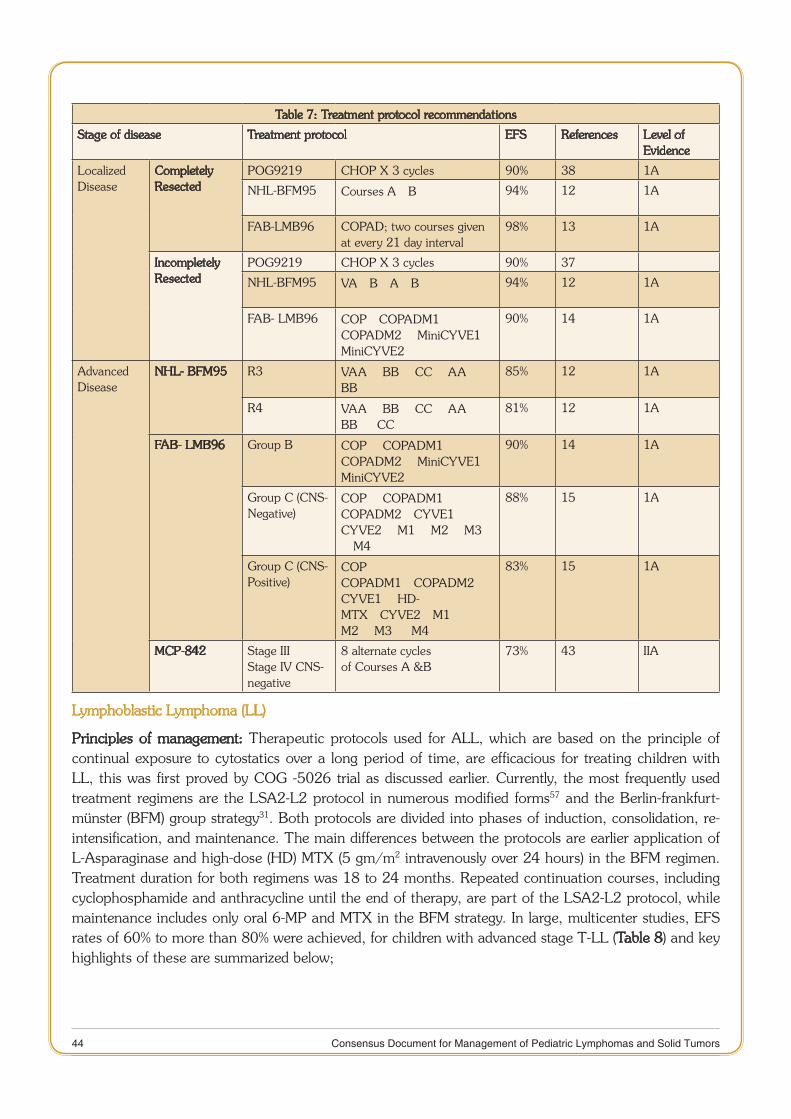
44 Consensus Document for Management of Pediatric Lymphomas and Solid Tumors
Table 7: Treatment protocol recommendations
Stage of disease Treatment protocol EFS References Level of Evidence
Localized Disease
Completely Resected
POG9219 CHOP X 3 cycles 90% 38 1A
NHL-BFM95 Courses A®B 94% 12 1A
FAB-LMB96 COPAD; two courses given at every 21 day interval
98% 13 1A
Incompletely Resected
POG9219 CHOP X 3 cycles 90% 37
NHL-BFM95 VA®B®A®B 94% 12 1A
FAB- LMB96 COP®COPADM1® COPADM2® MiniCYVE1® MiniCYVE2
90% 14 1A
Advanced Disease
NHL- BFM95 R3 VAA® BB® CC® AA ® BB
85% 12 1A
R4 VAA® BB® CC® AA ® BB ® CC
81% 12 1A
FAB- LMB96 Group B COP® COPADM1® COPADM2® MiniCYVE1® MiniCYVE2
90% 14 1A
Group C (CNS-Negative)
COP® COPADM1® COPADM2®CYVE1® CYVE2® M1® M2® M3 ®M4
88% 15 1A
Group C (CNS-Positive)
COP® COPADM1®COPADM2® CYVE1® HD-MTX®CYVE2®M1® M2® M3 ® M4
83% 15 1A
MCP-842 Stage IIIStage IV CNS-negative
8 alternate cyclesof Courses A &B
73% 43 IIA
Lymphoblastic Lymphoma (LL)
Principles of management: Therapeutic protocols used for ALL, which are based on the principle of continual exposure to cytostatics over a long period of time, are efficacious for treating children with LL, this was first proved by COG -5026 trial as discussed earlier. Currently, the most frequently used treatment regimens are the LSA2-L2 protocol in numerous modified forms57 and the Berlin-frankfurt-münster (BFM) group strategy31. Both protocols are divided into phases of induction, consolidation, re-intensification, and maintenance. The main differences between the protocols are earlier application of L-Asparaginase and high-dose (HD) MTX (5 gm/m2 intravenously over 24 hours) in the BFM regimen. Treatment duration for both regimens was 18 to 24 months. Repeated continuation courses, including cyclophosphamide and anthracycline until the end of therapy, are part of the LSA2-L2 protocol, while maintenance includes only oral 6-MP and MTX in the BFM strategy. In large, multicenter studies, EFS rates of 60% to more than 80% were achieved, for children with advanced stage T-LL (Table 8) and key highlights of these are summarized below;
45 Consensus Document for Management of Pediatric Lymphomas and Solid Tumors
NHL-BFM-LL studies:
The BFM 90–LL study has shown that with intensive ALL-type chemotherapy, including standard •BFM induction, a consolidation phase (protocol M) consisting of four doses of high-dose (5g/m2) Methotrexate, and a re-induction followed by maintenance therapy for 2 years, including moderate cumulative doses of anthracyclines at 240 mg/m2 with cyclophosphamide (3 g/m2) and moderate dose prophylactic cranial irradiation without local radiotherapy, an EFS rate of 90% can be achieved in childhood T-LL. Further, the EFS of 90 percent can be achieved using the standard arm of the BFM T-cell protocol without re-induction therapy or local or cranial radiation for stage I and II patients31.
The BFM-90 protocol also assessed the relevance of persistent thoracic disease at the end of induction. •The study showed EFS at 5 years of 95% ± 2% for the 80 patients with complete tumor response at the end of induction, and 89% ± 5% for 19 patients with tumor remnants after induction. All those who had tumor remnants underwent surgery and only necrotic material was documented. Of 19 patients with tumor residues after induction, two relapsed as compared to 4 of 80 patients with complete tumor regression. Hence, initial therapy predicts outcome irrespective of persistent abnormalities on CXR. Most often residual mass is necrotic and there is no role for local surgery or local RT in the consolidation for such patients to prevent local relapse31.
The BFM-90 study also confirmed that even in patients with testicular disease at diagnosis, testicular •radiation is only indicated for residual biopsy confirmed disease after high-dose MTX31.
No additive effect on outcome was observed for high-dose cytarabine in the consolidation phase. •
The length of the maintenance therapy as well as the optimal maintenance drugs remains unclear, but •most current protocols utilize ALL-like therapy for 24 months. Although 24 months of therapy was used in the BFM-90 study, the relapse pattern suggests that the duration of therapy can be reduced to 18 months31.
In NHL-BFM-95, prophylactic cranial radiation was omitted, and the intensity of induction therapy •was modified (reduction of L-Asparaginase and/or doxorubicin)58. There was no significant increase in CNS relapses, suggesting cranial radiation may be reserved for patients with CNS disease at diagnosis. However, survival was worse in BFM-95 than in BFM-90 (90% vs. 82%), possibly due to reduced intensity induction and increased number of secondary malignancies in BFM-95[58].
POG studies:
In the POG 9219 trial, among patients with stage I and II LL treated with 9 weeks of CHOP-based •chemotherapy EFS was inferior in comparison with non-LL (63% vs. 88%, P<0.001). It became apparent that among patients with LL, those who receive eight months of chemotherapy (including six months of continuation chemotherapy with or without radiotherapy) have a better outcome than those who receive nine weeks of chemotherapy without continuation chemotherapy. Thus a 24-week maintenance in addition to a 9-week induction was found beneficial for patients with LL10. This confirmed that biological similarity to ALL is more important for LL than their low tumor burden and that they might, therefore, benefit from an ALL-type treatment, including maintenance37.
POG-8704 trial showed the benefit of L-Asparaginase in T-LBL by randomizing patients to receive •or not to receive weekly L-Asparaginase after induction. Complete remission rate was 78 % for those who received L-Asparaginase versus 64 % for those who did not59.
46 Consensus Document for Management of Pediatric Lymphomas and Solid Tumors
POG 9404 trial tested th • e effectiveness of the addition of high-dose methotrexate in T-cell ALL and T-cell LL. In the lymphoma patients, high-dose methotrexate did not demonstrate benefit. In the small cohort (n = 66) of lymphoma patients who did not receive high-dose methotrexate, the 5-year EFS was 88%. Of note, all of these patients received prophylactic craniospinal radiation therapy, which has been demonstrated not to be required in T-cell lymphoblastic lymphoma patients60.
St Jude studies:
In the St. Jude NHL13 study, 41 children with advanced stage LL were treated with a regimen used for T-cell ALL that featured intensive intrathecal chemotherapy rather than prophylactic cranial irradiation. With a median follow-up of 9.3 years, the estimated five-year survival rate was 90 percent61.
COG studies:
In the COG-A5971 trial, 60 children with stage I or II lymphoblastic lymphoma (75 percent B-cell) •were treated with a two-year ALL regimen without prophylactic cranial radiation. At a median follow-up of 5.9 years, the estimated survival at five years was 96 percent. For children and adolescents with stage III/IV disease without CNS involvement, the A5971 evaluated two strategies for CNS prophylaxis, without the use of CNS irradiation. Patients were randomly assigned to high-dose methotrexate in interim maintenance (BFM-95) or intrathecal chemotherapy throughout the maintenance (CCG-BFM). The overall incidence of CNS relapse was 1.2% and there was no difference between arms for CNS relapse, DFS, or OS. In contrast, this same trial reported that the 12 children with disseminated CNS disease at the time of diagnosis had estimated EFS, OS, and relapse rates of 63, 81, and 25 percent at five years, respectively62-63.
The benefit of intensifying induction therapy with increased doses of daunomycin and the addition •of cyclophosphamide was also studied in a randomized fashion. Intensification of induction did not improve DFS or OS, but increased grade III and grade IV toxicities. Taken together with BFM and St. Jude data, these results suggest that in the setting of BFM-like chemotherapy, cranial radiation therapy can be omitted for CNS-negative T-LL provided the patients receive either high-dose MTX or intensified IT MTX62-63.
International BFM studies (I-BFM-NHL-2009 study):
A recent I-BFM study replaced high-dose methotrexate with Capizzi methotrexate in interim maintenance and CNS prophylaxis was accomplished with 13 intrathecal injections without irradiation. Radiotherapy was restricted to CNS-positive patients only. This recent study showed the probability of OS and EFS at 3 years for the whole population of 90.8% and 90.7% respectively, which is equivalent to BFM-95 results. This approach without the use of high-dose methotrexate has a lot of relevance for low-middle income countries like India who have limited facilities for delivery for high-dose methotrexate. (Dr Jaroslav Sterba- personal communication)
Indian studies:
A single-arm study of modified BFM-90 protocol from TMH, Mumbai with replacement of 5gm/m2 high-dose methotrexate with 3gm/m2 methotrexate in interim maintenance showed an EFS of more than 90% (Dr Shripad Banavali - personal communication). Also, modified MCP-841 protocol with the addition of two pulses of high-dose cytarabine (8gm/m2) in consolidation phase has shown more than 80% survival in T-cell ALL64. Both of the above are good choices for centers with limited /no facility for methotrexate delivery. Further, a retrospective audit of feasibility of delivery of high-dose methotrexate at 3gm/m2 without therapeutic drug monitoring revealed that treatment naïve children with normal levels in first methotrexate cycle can be safely given further high-dose methotrexate without levels65.
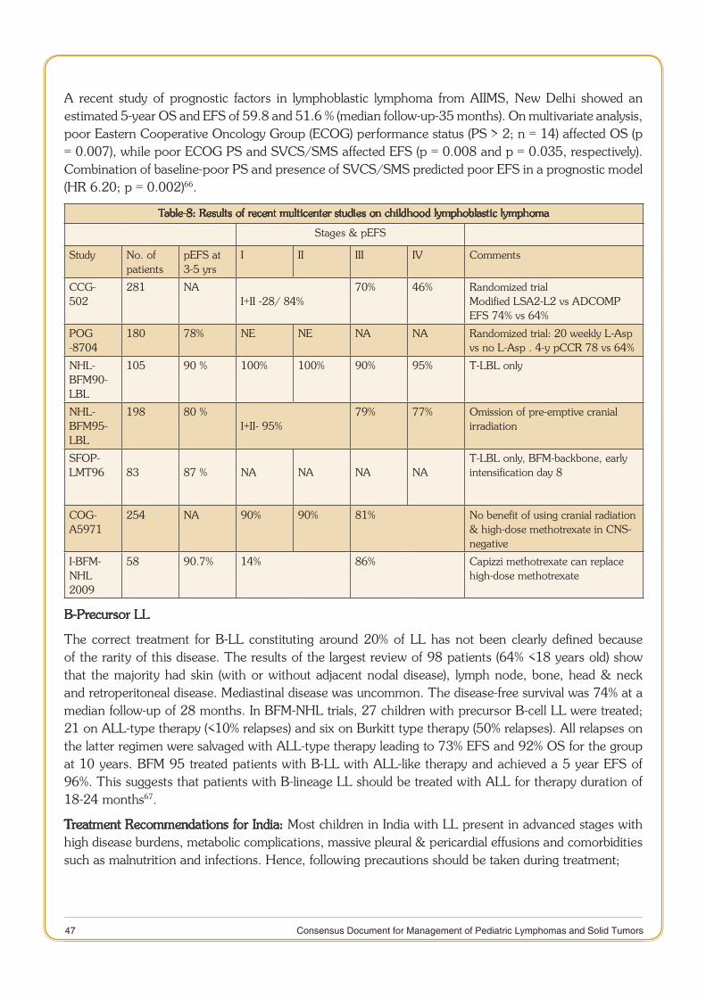
47 Consensus Document for Management of Pediatric Lymphomas and Solid Tumors
A recent study of prognostic factors in lymphoblastic lymphoma from AIIMS, New Delhi showed an estimated 5-year OS and EFS of 59.8 and 51.6 % (median follow-up-35 months). On multivariate analysis, poor Eastern Cooperative Oncology Group (ECOG) performance status (PS > 2; n = 14) affected OS (p = 0.007), while poor ECOG PS and SVCS/SMS affected EFS (p = 0.008 and p = 0.035, respectively). Combination of baseline-poor PS and presence of SVCS/SMS predicted poor EFS in a prognostic model (HR 6.20; p = 0.002)66.
Table-8: Results of recent multicenter studies on childhood lymphoblastic lymphoma
Stages & pEFS
Study No. of patients
pEFS at 3-5 yrs
I II III IV Comments
CCG- 502
281 NAI+II -28/ 84%
70% 46% Randomized trialModified LSA2-L2 vs ADCOMP EFS 74% vs 64%
POG -8704
180 78% NE NE NA NA Randomized trial: 20 weekly L-Asp vs no L-Asp . 4-y pCCR 78 vs 64%
NHL-BFM90-LBL
105 90 % 100% 100% 90% 95% T-LBL only
NHL-BFM95-LBL
198 80 %I+II- 95%
79% 77% Omission of pre-emptive cranial irradiation
SFOP-LMT96 83 87 % NA NA NA NA
T-LBL only, BFM-backbone, early intensification day 8
COG-A5971
254 NA 90% 90% 81% No benefit of using cranial radiation & high-dose methotrexate in CNS-negative
I-BFM-NHL 2009
58 90.7% 14% 86% Capizzi methotrexate can replace high-dose methotrexate
B-Precursor LL
The correct treatment for B-LL constituting around 20% of LL has not been clearly defined because of the rarity of this disease. The results of the largest review of 98 patients (64% <18 years old) show that the majority had skin (with or without adjacent nodal disease), lymph node, bone, head & neck and retroperitoneal disease. Mediastinal disease was uncommon. The disease-free survival was 74% at a median follow-up of 28 months. In BFM-NHL trials, 27 children with precursor B-cell LL were treated; 21 on ALL-type therapy (<10% relapses) and six on Burkitt type therapy (50% relapses). All relapses on the latter regimen were salvaged with ALL-type therapy leading to 73% EFS and 92% OS for the group at 10 years. BFM 95 treated patients with B-LL with ALL-like therapy and achieved a 5 year EFS of 96%. This suggests that patients with B-lineage LL should be treated with ALL for therapy duration of 18-24 months67.
Treatment Recommendations for India: Most children in India with LL present in advanced stages with high disease burdens, metabolic complications, massive pleural & pericardial effusions and comorbidities such as malnutrition and infections. Hence, following precautions should be taken during treatment;
48 Consensus Document for Management of Pediatric Lymphomas and Solid Tumors
Sick children, especially with SVCS/superior mediastinal syndrome should be initially cared for in 1. intensive care/high-dependency unit in propped-up lateral position for intensive monitoring and timely intervention in case of complications.
The least invasive procedure should be used to establish the diagnosis of lymphoma such as blood 2. smear/flow, pleural tap, BM examination, a lymph node biopsy under local anesthesia in sitting/prone position.
If it is unsafe to perform a diagnostic biopsy because of the risk of anesthesia or sedation, Prednisone 3. given for up to 48 and biopsy should be obtained as soon as the patient is able to undergo the procedure safely.
Aggressive tumor lysis prevention & management should include proactive & timely use of low-dose 4. rasburicase but in the presence of significant pleural or pericardial effusions, right ventricular outflow, or superior vena cava obstruction, hydration may need to be given very carefully in the setting of an intensive care unit preferably using lower limb venous access.
The pro-phase steroids should be used in children who usually present in a poor general condition 5. with large disease burden and metabolic obstructive complications.
Patients should be followed for response and children with no response to the 7-day pro-phase or 6. presence of a residual mediastinal mass at day 33 or at the end of induction (with less than 70% reduction) should preferably get intensified therapy.
Guidelines for localized LL
For localized LL (stage I/II disease) patients, BFM-95 protocol can be used. The centers with limited/ no availability of methotrexate levels can either use modified MCP-841 or modified BFM-90 protocol with reduced dose methotrexate (3gm/m2) or I-BFM 2009 protocol with Capizzi methotrexate in interim maintenance or COG-A5971 standard arm for localized LL.
Guidelines for advanced stage LL
For advanced LL (stage III/IV disease) patients, advanced LL BFM-95 protocol can be used with re-induction. The centers with limited/ no availability of methotrexate levels can use either modified MCP-841, modified BFM-90 protocol with reduced dose methotrexate (3gm/m2) or I-BFM 2009 protocol with Capizzi methotrexate in interim maintenance or COG-A5971 standard arm for advanced LL.
Extra-compartmental therapy:
CNS-negative patients: For CNS negative patients, treatment that includes either intensified intrathecal MTX or systemic HD-MTX (5 g/m²) without CRT is sufficient CNS protection.
CNS-positive patients: 18 to 24 Gy cranial irradiation (CRT), in addition to LSA2-L2 or BFM chemotherapy, are highly effective in preventing CNS recurrences. Children under 3 years of age are not irradiated.
Testicular and mediastinal disease: BFM-90 study also confirmed that even in patients with testicular disease at diagnosis, testicular radiation is only indicated for residual biopsy confirmed disease after high-dose MTX. There is no role of mediastinal RT.
Management of relapsed disease: Relapse is a significant obstacle to long-term survival for children with advanced stage LL. Most relapses occur within 2 years of diagnosis, but occasional late relapse is observed. In contrast to relapse for early stage disease, the outcome after salvage chemotherapy is poor for children with advanced stage disease at initial presentation68. However, survival rates of 14% to 40%

49 Consensus Document for Management of Pediatric Lymphomas and Solid Tumors
have been reported after allogeneic stem cell transplantation (SCT)69. CIBMTR audit demonstrated that EFS was significantly worse using an autologous (4%) versus allogeneic (40%) SCT47.
The salvage protocols include ICE70, DECAL45 and MIED etc. Recently a COG phase II study of nelarabine as a single agent demonstrated a response rate of 40%71 and nelarabine in combination with cyclophosphamide and etoposide has shown promising responses in children with relapsed T-LL72. Despite the associated CNS toxicity, nelarabine is currently being evaluated for newly diagnosed intermediate and high-risk T-ALL patients in the open COG trial and may prove to be of value in relapsed T-NHL.
Management of ALCL
Systemic ALCL, like BL, has a high growth fraction (>90%) and hence has aggressive clinical behavior. A substantial majority (75%) have B symptoms and extranodal involvement (60%). Intriguingly, BM (< 10%) and CNS (<5%) involvement is relatively uncommon. Hence, most study groups treat ALCL like B-NHL, however, some treat like lymphoblastic lymphoma73.
Meaningful conclusions regarding management of ALCL are difficult to draw from published series due to a small number of patients, significant heterogeneity in inclusion criteria, the variable staging system used and diverse treatment approaches in various series. The therapy of ALCL differs depending on whether a patient has the cutaneous or systemic variant.
Primary Cutaneous ALCL (PCALCL)
PCALCL is an indolent disease; treatment should focus on minimally invasive local therapies. Systemic therapies should be reserved for patients with disseminated disease or disease that is refractory to local measures. There is essentially no large series examining the efficacy of local therapy in PCALCL, but anecdotal observations and small case reports suggest that long-term remissions can be achieved with surgical excision and/or localized radiotherapy74. Low-dose, single-agent methotrexate is an effective therapy for PCALCL in patients with widespread cutaneous disease or in those whom radiation and surgery have failed75.
Systemic ALCL:
Results of different national protocols (Table-9)
BFM Studies73,76
The result of the BFM studies on 87 patients treated with the protocols NHL-BFM-90 have been reported by Reiter et al76. All the patients received a pre-phase with vincristine, cyclophosphamide and dexamethasone, then the treatment was stratified according to stage:
Stage 1 & 2 resected received 3 courses: ¾ one course ‘a’(Methotrexate 500mg/m2 in continuous infusion over 24 hours, Ifosfamide 800 mg/m2 X 5, VP-16 100 mg/m2 X 2, Cytarabine 150 mg/m2 X 2, Dexamethasone 10 mg/m2 X 5 and triple intrathecal treatment Prednisolone, Methotrexate & Cytarabine) and a course ‘b’ (Dexamethasone, Methotrexate and Cytarabine, Cyclophosphamide 200 mg/m2 X 5, Adriamycin 25 mg/m2 X 2) followed by a second course of ‘a’.
Stage 2 not resected and stage 3 received ¾ 6 courses (3 courses ‘a’ and three courses ‘b’ given alternately, the duration of treatment being 4 months. A total dose of Adriamycin 150 mg/m2, of Cyclophosphamide 3.4 gm/m2, and of Ifosfamide 12 gm/m2 was given.
50 Consensus Document for Management of Pediatric Lymphomas and Solid Tumors
Stage 4 ¾ defined by the existence of multifocal bone disease and/or BM disease and/or CNS involvement received 2 courses AA (identical to course ‘a’ but with a dose of methotrexate of 5 gm/m2 and an injection of vincristine), 2 courses BB (identical to ‘b’ but with a dose of methotrexate of 5 gm/m2 and an injection of vincristine) and a two courses of ‘CC’ ( Dexamethasone, Vincristine, Cytarabine 2 gm/m2 X 4, Etoposide 150 mg/m X 3 and intrathecal therapy)
This study reported the best results in ALCL to date with a DFS of 83 percent at 2.5 years and a ¾9-year duration of EFS of 81 ± 5 percent. Duration of therapy was 2-5 months, depending on risk grouping and relapses tended to occur with a mean time of 8 months after achieving remission73.
SFOP studies77
SFOP conducted two consecutive studies for ALCL: HM89 & HM91
Study HM89: ¾ This study used intensive induction treatment of 1 COP and 2 COPADM and maintenance treatment consisting of four cycles of two courses VEM (VP-16, Cyclophosphamide, Methotrexate) and VAD (Vincristine, Adriamycin) for a duration of 8 months77.
Study HM 91: ¾ This study used intensive induction treatment of 1 COP and 2 COPADM, then maintenance treatment consisting of four cycles of two courses: VEBBP (Vinblastine, VP-16, Bleomycin, Prednisone) and sequence 1 (Methotrexate, Vincristine, Doxorubicin, Cyclophosphamide and Prednisone) for a duration of 7 months77.
Italian study78
The AIEOP protocol-LNH consisted of an induction and consolidation similar to those of the protocol LSA
2L
2, followed by a maintenance treatment consisting of seven cycles of four weekly courses given
alternatively using Cyclophosphamide, 6MP, Cytarabine-VP16 and Vincristine-Dexamethasone
Stage 3 & 4 received, in addition, triple intrathecal treatment every 6 weeks. Duration of treatment was 24 months. At 19 months follow-up, the EFS was 65%, and although the duration of first remission was prolonged by the longer therapy, several relapses occurred later reducing the EFS78.
UK studies79
Intensive induction treatment with COP and 2 COPADM, followed by 2 CYM (Cytarabine, Methotrexate) and a final COPADM is used. Duration of treatment is 5 months79.
POG & CCG studies
In stage I and stage II ALCL, 9 weeks of CHOP chemotherapy were given on an outpatient basis as per POG 9219 described earlier, and achieved a 5-year EFS of 84% and OS of 100% which is a very cost-effective and applicable option for India37.
The CCG-5941 study tested an approach of compressed T-cell directed regimen similar to LNH-92, with more intensive induction, consolidation and maintenance for 1 year and achieved a similar 5-year EFS of 68% but similar significant increase in hematologic toxicity80.
The POG treated all ALCL patients with the APO regimen (doxorubicin 75mg/m2 day 1 and 22, vincristine 1.5mg/m2 day 1 and 22, prednisone 40 mg/m2 daily for 28 days) and 15 courses of consolidation therapy, lasting about 12 months. No alkylator therapy was given. This regimen can be administered in the outpatient setting. A subsequent POG trial (POG-9317) demonstrated no benefit of adding methotrexate and high-dose cytarabine to 52 weeks of the APO regimen81.
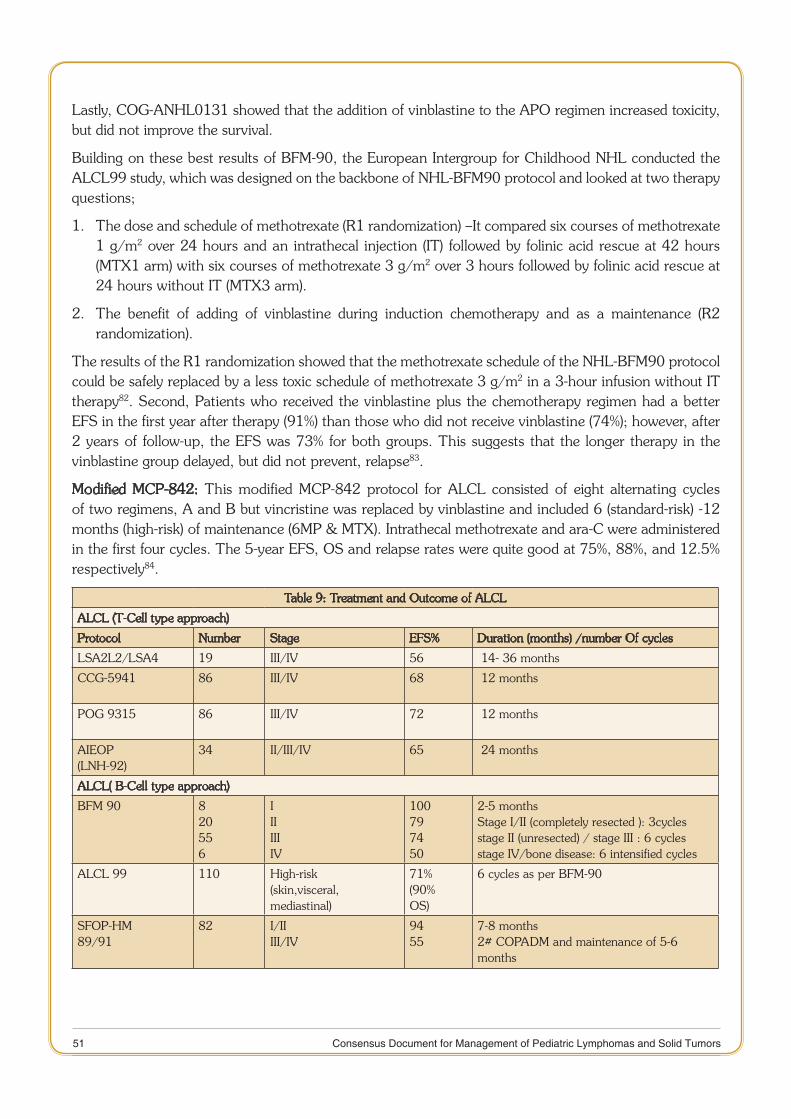
51 Consensus Document for Management of Pediatric Lymphomas and Solid Tumors
Lastly, COG-ANHL0131 showed that the addition of vinblastine to the APO regimen increased toxicity, but did not improve the survival.
Building on these best results of BFM-90, the European Intergroup for Childhood NHL conducted the ALCL99 study, which was designed on the backbone of NHL-BFM90 protocol and looked at two therapy questions;
1. The dose and schedule of methotrexate (R1 randomization) –It compared six courses of methotrexate 1 g/m2 over 24 hours and an intrathecal injection (IT) followed by folinic acid rescue at 42 hours (MTX1 arm) with six courses of methotrexate 3 g/m2 over 3 hours followed by folinic acid rescue at 24 hours without IT (MTX3 arm).
2. The benefit of adding of vinblastine during induction chemotherapy and as a maintenance (R2 randomization).
The results of the R1 randomization showed that the methotrexate schedule of the NHL-BFM90 protocol could be safely replaced by a less toxic schedule of methotrexate 3 g/m2 in a 3-hour infusion without IT therapy82. Second, Patients who received the vinblastine plus the chemotherapy regimen had a better EFS in the first year after therapy (91%) than those who did not receive vinblastine (74%); however, after 2 years of follow-up, the EFS was 73% for both groups. This suggests that the longer therapy in the vinblastine group delayed, but did not prevent, relapse83.
Modified MCP-842: This modified MCP-842 protocol for ALCL consisted of eight alternating cycles of two regimens, A and B but vincristine was replaced by vinblastine and included 6 (standard-risk) -12 months (high-risk) of maintenance (6MP & MTX). Intrathecal methotrexate and ara-C were administered in the first four cycles. The 5-year EFS, OS and relapse rates were quite good at 75%, 88%, and 12.5% respectively84.
Table 9: Treatment and Outcome of ALCL
ALCL (T-Cell type approach)
Protocol Number Stage EFS% Duration (months) /number Of cycles
LSA2L2/LSA4 19 III/IV 56 14- 36 months
CCG-5941 86 III/IV 68 12 months
POG 9315 86 III/IV 72 12 months
AIEOP (LNH-92)
34 II/III/IV 65 24 months
ALCL( B-Cell type approach)
BFM 90 820556
IIIIIIIV
1007974 50
2-5 months Stage I/II (completely resected ): 3cyclesstage II (unresected) / stage III : 6 cyclesstage IV/bone disease: 6 intensified cycles
ALCL 99 110 High-risk(skin,visceral, mediastinal)
71%(90%OS)
6 cycles as per BFM-90
SFOP-HM89/91
82 I/IIIII/IV
9455
7-8 months 2# COPADM and maintenance of 5-6 months
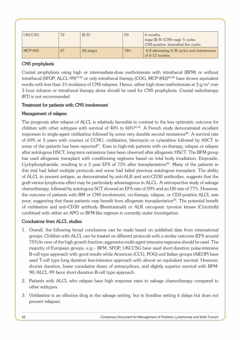
52 Consensus Document for Management of Pediatric Lymphomas and Solid Tumors
UKCCSG 72 III/IV 59 6 months stage III/IV (CNS–neg): 5 cyclesCNS-positive: Intensified five cycles
MCP-842 27 All stages 78% 6-8 alternating A/B cycles and maintenance of 6-12 months
CNS prophylaxis
Cranial prophylaxis using high or intermediate-dose methotrexate with intrathecal (BFM) or without intrathecal (SFOP, ALCL-99)79,82 or only intrathecal therapy (COG, MCP-842)80,84 have shown equivalent results with less than 1% incidence of CNS relapses. Hence, either high-dose methotrexate at 3 g/m2 over 3 hour infusion or intrathecal therapy alone should be used for CNS prophylaxis. Cranial radiotherapy (RT) is not recommended
Treatment for patients with CNS involvement
Management of relapse:
The prognosis after relapse of ALCL is relatively favorable in contrast to the less optimistic outcome for children with other subtypes with survival of 40% to 60%68,85. A French study demonstrated excellent responses to single-agent vinblastine followed by some very durable second remissions86. A survival rate of 69% at 3 years with courses of CCNU, vinblastine, bleomycin or cytarabine followed by ASCT in some of the patients has been reported87. Even in high-risk patients with on-therapy, relapse or relapse after autologous HSCT, long-term remissions have been observed after allogeneic HSCT. The BFM group has used allogeneic transplant with conditioning regimens based on total body irradiation, Etoposide, Cyclophosphamide, resulting in a 3 year EFS of 75% after transplantation88. Many of the patients in this trial had failed multiple protocols and some had failed previous autologous transplant. The ability of ALCL to present antigen, as demonstrated by anti-ALK and anti-CD30 antibodies, suggests that the graft-versus-lymphoma effect may be particularly advantageous in ALCL. A retrospective study of salvage chemotherapy, followed by autologous SCT showed an EFS rate of 59% and an OS rate of 77%. However, the outcome of patients with BM or CNS involvement, on-therapy, relapse, or CD3-positive ALCL was poor, suggesting that these patients may benefit from allogeneic transplantation85. The potential benefit of vinblastine and anti-CD30 antibody (Brentuximab) or ALK oncogenic tyrosine kinase (Crizotinib) combined with either an APO or BFM-like regimen is currently under investigation.
Conclusions from ALCL studies
Overall, the following broad conclusions can be made based on published data from international 1. groups; Children with ALCL can be treated on different protocols with a similar outcome (EFS around 75%) In view of the high growth fraction; aggressive multi-agent intensive regimens should be used. The majority of European groups, e.g.:- BFM, SFOP, UKCCSG have used short-duration pulse-intensive B-cell type approach with good results while American (CCG, POG) and Italian groups (AIEOP) have used T-cell type long duration less-intensive approach with almost an equivalent survival. However, shorter duration, lower cumulative doses of antracyclines, and slightly superior survival with BFM-90/ALCL-99 favor short-duration B-cell type approach.
Patients with ALCL who relapse have high response rates to salvage chemotherapy compared to 2. other subtypes.
Vinblastine is an effective drug in the salvage setting, but in frontline setting it delays but does not 3. prevent relapses.
53 Consensus Document for Management of Pediatric Lymphomas and Solid Tumors
Management guidelines of Systemic ALCL for India:-
Standard Risk: - Stage I/II completely resected with no high-risk features including involvement of skin, mediastinum, visceral organs (Liver, spleen, and lung), CNS or BM.
These can be managed with three cycles (10-12 weeks) of pulse-intensive B-cell type regimen (like ALCL-99). In centers without the experience of high-dose methotrexate administration or limited in-patient facility, CHOP protocol may be considered.
High-Risk: - All non-standard risk patients qualify for high-risk group. These can be managed by six cycles (6 months) of short-duration B-cell type regimen (ALCL-99). In centers without the experience of high-dose methotrexate administration, Modified MCP-842 may be considered.
Response Evaluation: - All patients should undergo response evaluation with clinical examination and conventional imaging at the end of two cycles for response evaluation. Patients with suboptimal response (stable disease/progression) should be considered for salvage treatment. Patients with good response should complete the treatment and undergo post-treatment revaluation.
Novel therapeutic approaches: Two targeted therapies have demonstrated significant initial activity in relapsed ALCL:
Brentuximab vedotin: • It is an immunotoxin with a CD-30 directed antibody linked to the antitubulin agent monomethylauristatin E. Brentuximab vedotin is approved by the US FDA for the treatment of adults with systemic ALCL after failure of at least one prior multi-agent chemotherapy regimen. A phase II trial in adults with relapsed ALCL has shown CR rates of approximately 55% to 60% and PR rates of 29%89.
Crizotinib: • is a small molecule inhibitor of the ALK tyrosine kinase that has demonstrated activity in a subset of patients with non-small cell lung cancer. Case reports and a phase I study have described responses in patients with multiply relapsed ALK-positive ALCL. Phase 1 study in pediatric patients with ALCL has shown a response rate of 88 % in ALCL (ALK+) patients90.
The use of these targeted therapies in children with newly diagnosed stage II-IV ALCL is being evaluated in a COG randomized phase II trial (NCT01979536) using six months infusional chemotherapy plus Brentuximab vedotin in one arm and Crizotinib in the other arm. Outcomes for each arm will be compared with historical outcomes of the ALCL99 trial.
Future directions:
Refinements in systemic chemotherapy fuelled by better understanding of NHL biology in children have led to cure in approximately 80% to 85% of all patients. This improved outlook for childhood NHL, however, has come with a certain price. The use of intense chemotherapy has resulted in long hospitalizations, severe hematopoietic as well as non-hematopoietic toxicity and late effects, such as sterility, cardiomyopathy, and secondary malignancies. Consequently, the emphasis in the near future is to decrease the therapy in good risk patients as well as better identification & development of new therapeutic approaches for high-risk cases. In the future, as the molecular pathogenesis of the malignant lymphomas is better elucidated using molecular diagnostic tools, new targets for therapy will emerge. Also, it is likely that targeted therapy will substitute for some of the toxic chemotherapy and thereby minimize the chemotherapy related morbidity. This novel molecular biologic information will also be valuable for developing more sensitive diagnostic tools, measurement of early response to therapy as well as submicroscopic disease and for identifying new prognostic subgroups. Superior risk-adapted therapy based on these advances would maximize the chance for cure while avoiding both acute and chronic toxicities of treatment.
54 Consensus Document for Management of Pediatric Lymphomas and Solid Tumors
References:
Smith MA, Altekruse SF, Adamson PC, et al.: Declining childhood and adolescent cancer mortality. Cancer 120 (16): 1. 2497-506, 2014.
Kulkarni KP, Arora RS, Arora B. Current outcomes of childhood Non-Hodgkin lymphoma in india: results of a systematic 2. review. Proceedings of the SIOP 2013 meeting abstract no. PJ 23.
http://www.cancer.gov/types/lymphoma/hp/child-nhl-treatment-pd3. q. Last accessed June 4, 2015.
https://tmc.gov.in/clinicalguidelines/EBM/Vol9/NHL_for_Part_B.pdf4. . Last accessed June 4, 2015.
Arora RS, Eden TO, Kapoor G. Epidemiology of childhood cancer in India. Indian J Cancer. 2009 Oct-Dec;46(4):264-5. 73.
Three year report of the population based cancer registries 2009-2011: Report of 25 PBCRs; National Cancer Registry 6. Programme, Indian Council Medical Research, Bangalore 2013. Available from: URL: http://ncrpindia.org/Reports/PBCR_2009_2011.aspx .Accessed 14th July 2015.
K. N. Naresh, V. Srinivas and C. S. Soman. Distribution of various subtypes of non-Hodgkin’s lymphoma in India:A 7. study of 2773 lymphomas using R.E.A.L. and WHO Classifications. Annals of Oncology.2000 11 (Suppl. I): S63-S67.
Bakhshi S, Radhakrishnan V, Sharma P, et al Pediatric non-lymphoblastic non-Hodgkin lymphoma: baseline, interim, 8. and post-treatment PET/CT versus contrast enhanced CT for evaluation—a prospective study. Radiology. 2012 Mar;262(3):956-68.
Murphy SB, Fairclough DL, Hutchison RE, et al.: Non-Hodgkin’s lymphomas of childhood: an analysis of the histology, 9. staging, and response to treatment of 338 cases at a single institution. J ClinOncol 7 (2): 186-93, 1989.
Patte C, Auperin A, Michon J, et al. The Societe Francaised Oncologie Pediatrique LMB89 protocol: highly effective 10. multi-agent chemotherapy tailored to the tumor burden and initial response in 561 unselected children with B-cell lymphomas and L3 leukemia. Blood. 2001; 97:3370–3379.
Reiter A, Schrappe M, Tiemann M, et al. Improved treatment results in childhood B-cell neoplasms with tailored 11. intensification of therapy: a report of the Berlin-frankfurt-münster Group Trial NHL-BFM 90. Blood. 1999; 94:3294–3306.
Woessmann W, Seidemann K, Mann G, et al. The impact of the methotrexate administration schedule and dose in the 12. treatment of children and adolescents with B-cell neoplasms: a report of the BFM Group Study NHL-BFM95. Blood. 2005; 105:948–958.
Gerrard M, Cairo MS, Weston C, et al. Results of the FAB-LMB 96 international study in children and adolescents 13. (C+A) with localized, resected B-cell lymphoma (large-cell [LCL], Burkitt’s [BL] and Burkitt-like [BLL]). J ClinOncol. 2003;22:795.
Patte C, Auperin A, Gerrard M, et al. Results of the randomized international FAB/LMB96 trial for intermediate-risk 14. B-cell non-Hodgkin lymphoma in children and adolescents: it is possible to reduce treatment for the early responding patients. Blood. 2007;109:2773–2780.
Cairo MS, Gerrard M, Sposto R, et al. Results of a randomized international study of high-risk central nervous system 15. B non-Hodgkin lymphoma and B acute lymphoblastic leukemia in children and adolescents. Blood. 2007; 109:2736–2743.
Cairo MS, Sposto R, Gerrard M, et al.: Advanced stage, increased lactate dehydrogenase, and primary site, but not 16. adolescent age (≥ 15 years), are associated with an increased risk of treatment failure in children and adolescents with mature B-cell non-Hodgkin’s lymphoma: results of the FAB-LMB 96 study. J ClinOncol 30 (4): 387-93, 2012.
Mussolin L, Pillon M, d’Amore ES, et al.: Minimal disseminated disease in high-risk Burkitt’s lymphoma identifies 17. patients with different prognosis. J ClinOncol 29 (13): 1779-84, 2011.
Nelson M, Perkins SL, Dave BJ, et al.: An increased frequency of 13q deletions detected by fluorescence in situ 18. hybridization and its impact on survival in children and adolescents with Burkitt lymphoma: results from the Children’s Oncology Group study CCG-5961. Br J Haematol 148 (4): 600-10, 2010.
Poirel HA, Heerema NA, Swansbury J, et al. Cytogenetic analysis of 238 pediatric mature B-cell non-Hodgkin lymphoma 19. (NHL) cases from the randomized international FAB LMB96 trial identities several patterns of chromosomal abnormality and new prognostic factors. Pediatr Blood Cancer. 2006;46:835-842.
55 Consensus Document for Management of Pediatric Lymphomas and Solid Tumors
Salaverria I, Philipp C, Os20. chlies I, et al.: Translocations activating IRF4 identify a subtype of germinal center-derived B-cell lymphoma affecting predominantly children and young adults. Blood 118 (1): 139-47, 2011.
Mussolin L, Pillon M, Conter V, et al.: Prognostic role of minimal residual disease inmature B-cell acute lymphoblastic 21. leukemia of childhood. J ClinOncol 25 (33): 5254-61, 2007.
Shiramizu B, Goldman S, Kusao I, et al.: Minimal disease assessment in the treatment of children and adolescents with 22. intermediate-risk (Stage III/IV) B-cell non-Hodgkin lymphoma: a children’s oncology group report. Br J Haematol 153 (6): 758-63,2011.
Le Deley MC, Reiter A, Williams D, et al.: Prognostic factors in childhood anaplastic large-cell lymphoma: results of a 23. large European intergroup study. Blood 111 (3): 1560-6, 2008.
Stein H, Foss HD, Dürkop H, et al.: CD30(+) anaplastic large-cell lymphoma: a review of its histopathologic, genetic, 24. and clinical features. Blood 96 (12): 3681-95,2000.
Lamant L, McCarthy K, d’Amore E, et al.: Prognostic impact of morphologic and phenotypic features of childhood 25. ALK-positive anaplastic large-cell lymphoma:results of the ALCL99 study. J ClinOncol 29 (35): 4669-76, 2011.
Damm-Welk C, Busch K, Burkhardt B, et al.: Prognostic significance of circulating tumor cells in bone marrow or 26. peripheral blood as detected by qualitative and quantitative PCR in pediatric NPM-ALK-positive anaplastic large-cell lymphoma. Blood110 (2): 670-7, 2007.
Damm-Welk C, Mussolin L, Zimmermann M, et al.: Early assessment of minimal residual disease identifies patients at 27. very high relapse risk in NPM-ALK-positive anaplastic large-cell lymphoma. Blood 123 (3): 334-7, 2014.
Ait-Tahar K, Damm-Welk C, Burkhardt B, et al.: Correlation of the auto antibody response to the ALK oncoantigen in 28. pediatric anaplastic lymphoma kinase-positive anaplastic large-cell lymphoma with tumor dissemination and relapse risk. Blood 115(16): 3314-9, 2010.
Mussolin L, Damm-Welk C, Pillon M, et al.: Use of minimal disseminated disease and immunity to NPM-ALK antigen to 29. stratify ALK-positive ALCL patients with different prognosis. Leukemia 27 (2): 416-22, 2013.
Uyttebroeck A, Suciu S, Laureys G, Robert A, et al; Children’s Leukaemia Group (CLG) of the European Organisation 30. for Research and Treatment of Cancer (EORTC). Treatment of childhoodT-cell lymphoblastic lymphoma according to the strategy for acute lymphoblasti cleukaemia, without radiotherapy: long-term results of the EORTC CLG 58881 trial. Eur J Cancer. 2008 Apr;44(6):840-6.
Reiter A, Schrappe M, Ludwig WD, et al.: Intensive ALL-type therapy without local radiotherapy provides a 90% EFS 31. for children with T-cell lymphoblastic lymphoma: a BFM group report. Blood 95 (2): 416-21, 2000.
Bonn BR, Rohde M, Zimmermann M, et al.: Incidence and prognostic relevance of genetic variations in T-cell 32. lymphoblastic lymphoma in childhood and adolescence. Blood 121 (16): 3153-60, 2013.
Burkhardt B, Moericke A, Klapper W, et al.: Pediatric precursor T lymphoblastic leukemia and lymphoblastic lymphoma: 33. Differences in the common regions with lossof heterozygosity at chromosome 6q and their prognostic impact. Leuk Lymphoma 49 (3): 451-61, 2008.
Coustan-Smith E, Sandlund JT, Perkins SL, et al.: Minimal disseminated disease in childhood T-cell lymphoblastic 34. lymphoma: a report from the children’s oncology group. J Clin Oncol 27 (21): 3533-9, 2009.
Stark B, Avigad S, Luria D, et al.: Bone marrow minimal disseminated disease (MDD) and minimal residual disease 35. (MRD) in childhood T-cell lymphoblastic lymphoma stage III, detected by flow cytometry (FC) and real-time quantitative polymerase chain reaction (RQ-PCR). Pediatr Blood Cancer 52 (1): 20-5, 2009.
Jayabose S, Kumar V, Dhanabalan R, Rajan P, Rathnam K, Viswanathan TK. Low-dose rasburicase in hematologic 36. malignancies. Indian J Pediatr. 2015 May;82(5):458-61.
Anderson JR, Jenkin RD, Wilson JF, et al. Long-term follow-up of patients treated with COMP or LSA2L2 therapy 37. for childhood non-Hodgkin’s lymphoma: a report of CCG-551 from the Childrens Cancer Group. J Clin Oncol. 1993 Jun;11(6):1024-32.
Link MP, Shuster JJ, Donaldson SS, Berard CW, Murphy SB. Treatment of children and young adults with early stage 38. non-Hodgkin’s lymphoma. N Engl J Med. 1997;337:1259–1266.
56 Consensus Document for Management of Pediatric Lymphomas and Solid Tumors
Patte C: Treatment of mature B-ALL and high-grade B-NHL in children. Best Pract Res Clin Haematol 15:695-711, 39. 2003.
Patte C, Gerrard M, Auperin A, et al. Early treatment intensity has a major prognostic impact in the “intermediate-40. risk” childhood and adolescent B-cell lymphoma: results of the international FAB-LMB 96 trial. SIOP abstract book 2008,#O119,pg 52.
Sullivan MP, Brecher M, Ramirez I, et al. High-dose cyclophosphamide-high-dose methotrexate with coordinated 41. intrathecal therapy for advanced non-lymphoblastic lymphoma of childhood: results of a Pediatric Oncology Group study. Am J Pediatr Hematol Oncol. 1991 Fall;13(3):288-95.
Advani S, Pai S, Adde M, et al. Preliminary report of an intensified, short duration chemotherapy protocol for the 42. treatment of pediatric non-Hodgkin’s lymphoma inIndia. Ann Oncol. 1997 Sep;8(9):893-7.
Arora B, Kaur U, Gulia S et al. Modified MCP-842: A novel highly efficacious, safe and cost-effective protocol for B-NHL 43. treatment in resource poor countries. Proceedings of the third international symposium on childhood, adolescent and young adult Non-Hodgkin Lymphoma, June 2009, Frankfurt, Germany.
Vassal G, Valteau D, Bonnay M, et al: Cerebrospinal fluid and plasma methotrexate levels following high-dose regimen 44. given as a 3-hour intravenous infusion in children with non Hodgkin’s lymphoma. Pediatr Hematol Oncol 7:71-7, 1990.
Kobrinsky NL, Sposto R, Shah NR, et al.: Outcomes of treatment of children and adolescents with recurrent non-45. Hodgkin’s lymphoma and Hodgkin’s disease with dexamethasone, etoposide, cisplatin, cytarabine, and l-asparaginase, maintenance chemotherapy, and transplantation: Children’s Cancer Group Study CCG-5912. J ClinOncol 19 (9): 2390-6, 2001.
Griffin TC, Weitzman S, Weinstein H, et al.: A study of rituximab and ifosfamide, carboplatin, and etoposide chemotherapy 46. in children with recurrent/refractory B-cell (CD20+) non-Hodgkin lymphoma and mature B-cell acute lymphoblastic leukemia: areport from the Children’s Oncology Group. Pediatr Blood Cancer 52 (2): 177-81,2009.
Gross TG, Hale GA, He W, et al.: Hematopoietic stem cell transplantation for refractory or recurrent non-Hodgkin 47. lymphoma in children and adolescents. Biol Blood Marrow Transplant 16 (2): 223-30, 2010.
Fujita N, Mori T, Mitsui T, et al.: The role of hematopoietic stem cell transplantation with relapsed or primary refractory 48. childhood B-cell non-Hodgkin lymphoma and mature B-cell leukemia: a retrospective analysis of enrolled cases in Japan. Pediatr Blood Cancer 51 (2): 188-92, 2008.
Seidemann K, Tiemann M, Lauterbach I, et al. Primary mediastinal large B-cell lymphoma with sclerosis in pediatric 49. and adolescent patients: treatment and results from three therapeutic studies of the Berlin-frankfurt-münster Group. J Clin Oncol. 2003;21:1782–1789.
Gerrard M, Waxman IM, Sposto R, et al.: Outcome and pathologic classification of children and adolescents with 50. mediastinal large B-cell lymphoma treated with FAB/LMB96 mature B-NHL therapy. Blood 121 (2): 278-85, 2013.
Dunleavy K, Pittaluga S, Maeda LS, et al.: Dose-adjusted EPOCH-rituximab therapy in primary mediastinal B-cell 51. lymphoma. N Engl J Med 368 (15): 1408-16,2013.
Woessmann W, Lisfeld J, Burkhardt B, et al.: Therapy in primary mediastinal B-celllymphoma. N Engl J Med 369 (3): 52. 282, 2013.
Feugier P, Van Hoof A, Sebban C, et al: Long-term results of the R-CHOP study in the treatment of elderly patients with 53. diffuse large B-cell lymphoma: a study by the Groupe d’Etude des Lymphomes de l’Adulte. J Clin Oncol 23:4117-26, 2005.
Meinhardt A, Burkhardt B, Zimmermann M, et al.: Phase II window study on rituximab in newly diagnosed pediatric 54. mature B-cell non-Hodgkin’s lymphoma and Burkitt leukemia. J Clin Oncol 28 (19): 3115-21, 2010.
Barth MJ, Goldman S, Smith L, et al.: Rituximab pharmacokine55. tics in children and adolescents with de novo intermediate and advanced mature B-cell lymphoma/leukaemia: a Children’s Oncology Group report. Br J Haematol 162 (5):678-83, 2013.
Goldman S, S56. mith L, Galardy P, et al.: Rituximab with chemotherapy in children and adolescents with central nervous system and/or bone marrow-positive Burkitt lymphoma/leukaemia: a Children’s Oncology Group Report. Br J Haematol 167 (3):394-401, 2014.
57 Consensus Document for Management of Pediatric Lymphomas and Solid Tumors
Patte 57. C, Kalifa C, Flamant F et al. Results of the LMT81 protocol, a modified LSA2L2 protocol with high-dose methotrexate, on 84 children with non-B-cell (lymphoblastic) lymphoma. Med Pediatr Oncol. 1992; 20(2):105-13.
Burkhardt B, Woessmann W, Zimmermann M et al. Impact of cranial radiotherapy on central nervous system prophylaxis 58. in children and adolescents with central nervous system-negative stage III or IV lymphoblastic lymphoma. J Clin Oncol 2006; 24: 491 – 499.
Amylon MD59. , Shuster J, Pullen J, at al. Intensive high-dose asparaginase consolidation improves survival for pediatric patients with T-cell acute lymphoblastic leukemia and advanced stage lymphoblastic lymphoma: a Pediatric Oncology Group study. Leukemia. 1999 Mar; 13(3):335-4.
Asselin BL, Devidas M, Wang C, et al. Effectiveness of high-dose methotrexate in T-cell lymphoblastic leukemia and 60. advanced stage lymphoblastic lymphoma: a randomized study by the Children‘s Oncology Group (POG 9404). Blood. 2011 Jul 28;118(4):874-83.
Sandlund JT, Pui CH, Zhou Y, et al. Effective treatment of advanced stage childhood lymphoblastic lymphoma without 61. prophylactic cranial irradiation: results of St Jude NHL13 study. Leukemia 2009; 23:1127.
Termuhlen AM, Smith LM, Perkins SL, et al. Disseminated lymphoblastic lymphoma in children and adolescents: results 62. of the COG A5971 trial: a report from the Children’s Oncology Group. Br J Haematol 2013; 162:792.
Termuhlen AM, Smith LM, Perkins SL, et al. Outcome of newly diagnosed children and adolescents with localized 63. lymphoblastic lymphoma treated on Children’s Oncology Group trial A5971: a report from the Children’s Oncology Group. Pediatr Blood Cancer 2012; 59:1229.
Banavali S, Arora B, Kurkure PA, Pai S, Gujral S, Laskar S, Kolhatkar B, Tambe B, Adde M, Magrath I, Parikh P. 64. Improved Outcome Of Children & Young Adults With T-Cell Acute Lymphoblastic Leukemia (T-All) With Addition Of High-Dose Ara-C (Hdac). 40th SIOP Annual Meeting Abs D039; Pg.121.
Arora B, Shah N, Narula G et al. Serum methotrexate levels, Methotexate toxicity and its predictors in a large cohort of 65. Pediatric ALL patients on intermediate-dose (ID) methotrexate (3mg/m2) and its relevance to adopting ID-methotrexate based interim maintenance in low-middle income countries. abstract no-8, Proceedings of 9th SIOP-Asia 2015, Amman, Jordan;PP 58.
Tilak TV, Raina V, Kumar L, et al. Superior vena cava syndrome and poor performance status at presentation affect 66. survival in mediastinal T-lymphoblastic lymphoma--a single institute experience from India. Ann Hematol. 2013;92:917-23.
Neth O, Seidemann K, Jansen P, et al.: Precursor B-cell lymphoblastic lymphoma in childhood and adolescence: clinical 67. features, treatment, and results in trials NHL-BFM 86 and 90. Med Pediatr Oncol 35 (1): 20-7, 2000.
Attarbaschi A, Dworzak M, Steiner M, et al.: Outcome of children with primary Resistant or relapsed non-Hodgkin 68. lymphoma and mature B-cell leukemia afterintensive first-line treatment: a population-based analysis of the Austrian Cooperative Study Group. Pediatr Blood Cancer 44 (1): 70-6, 2005.
Burkhardt B, Reiter A, Landmann E, et al.: Poor outcome for children and adolescents with progressive disease or relapse 69. of lymphoblastic lymphoma: a report from the berlin-frankfurt-muenster group. J Clin Oncol 27 (20): 3363-9, 2.
Kung FH, Harris MB, Krischer JP: Ifosfamide/carboplatin/etoposide, an effective salvaging therapy for recurrent 70. malignant non-Hodgkin lymphoma of childhood: a Pediatric Oncology Group phase II study. Med Pediatr Oncol 32 (3): 225-6,1999.
Berg SL, Blaney SM, Devidas M, et al.: Phase II study of nelarabine (compound 506U78) in children and young adults 71. with refractory T-cell malignancies: a reportfrom the Children’s Oncology Group. J Clin Oncol 23 (15): 3376-82, 2005.
Commander LA, Seif AE, Insogna IG, Rheingold SR. Salvage therapy with nelarabine, etoposide, and cyclophosphamide 72. in relapsed/refractory paediatricT-cell lymphoblastic leukaemia and lymphoma. Br J Haematol. 2010Aug;150(3):345-51.
Seidemann K, Tiemann M, Schrappe M, et al. Short-pulse B-non-Hodgkin lymphoma-type chemotherapy is efficacious 73. treatment for pediatric anaplastic large-cell lymphoma: a report of the Berlin-frankfurt-münster Group Trial NHL-BFM 90. Blood. 2001;97:3699–3706.
Kaufmann TP, Coleman M, Nisce LZ. Ki-1 skin lymphoproliferative disorders: management with radiation therapy. 74. Cancer Invest 1997;15: 91–97.

58 Consensus Document for Management of Pediatric Lymphomas and Solid Tumors
Vonderheid EC, Sajjadian A, Kadin ME. Methotrexate is effective therapy for lymphomatoidpapulosis and other primary 75. cutaneous CD30-positive lymphoproliferative disorders. J Am Acad Dermatol 1996;34:470–481.
Reiter A, Schrappe M, Tiemann M, et al. Successful treatment strategy for Ki-1 anaplastic large-cell lymphoma of 76. childhood: a prospective analysis of 62 patients enrolled in three consecutive Berlin-frankfurt-münster group studies. J Clin Oncol. 1994;12:899–908.
Bugieres L, Deley MC, Pacquement H et al. CD30 (+) anaplastic large-cell lymphoma in children: analysis of T- and NK 77. cell lymphomas in children and adolescents of 82 patients enrolled in two consecutive studies of the French Society of Pediatric Oncology. Blood 1998; 92: 3591 -- 3598.
Rosolen A, Pillon M, Garaventa A, et al.: Anaplastic large-cell lymphoma treated with a leukemia-like therapy: report 78. of the Italian Association of Pediatric Hematology and Oncology (AIEOP) LNH-92 protocol. Cancer 2005;104 (10): 2133-40.
Williams DM, Hobson R, Imeson J, et al. Anaplastic large-cell lymphoma in childhood: analysis of 72 patients treated on 79. The United Kingdom Children’s Cancer Study Group chemotherapy regimens. Br J Haematol. 2002;117:812-820.
Lowe EJ, Sposto R, Perkins SL, et al;Children’s Cancer Group Study 5941. Intensive chemotherapy for systemic 80. anaplastic large-cell lymphoma in children and adolescents: final results of Children’s Cancer Group Study 5941. Pediatr Blood Cancer. 2009 Mar;52(3):335-9.
Laver JH, Kraveka JM, Hutchison RE, et al.: Advanced stage large-cell lymphoma in children and adolescents: results of 81. a randomized trial incorporating intermediate-dose methotrexate and high-dose cytarabine in the maintenance phase of the APO regimen: a Pediatric Oncology Group phase III trial. J ClinOncol 2005; 23 (3): 541-7.
Brugieres L, Le Deley MC, Rosolen A, et al. Anaplastic large-cell lympoma (ALCL) in children: equal efficacy but greater 82. toxicity of chemotherapy including methotrexate (MTX) 1g/ m2 in 24-hour infusion with intrathecal injection (IT) than chemotherapy with MTX 3g/m2 in 3-hour infusion without IT: results of the ALCL99–R1 randomized trial [abstract]. Blood. 2006;108:122a.
Marie-Ce´cile Le Deley, Angelo Rosolen, Denise M. Williams et al. Vinblastine in Children and Adolescents With 83. High-Risk Anaplastic Large-Cell Lymphoma: Results of the Randomized ALCL99-Vinblastine Trial. J Clin Oncol 2010; 28:3987-3993.
Banavali SD, Arora B, Vora T, Bansal S, Hingmire S, Pai SK, Parikh PM, Kolhatkar B, Adde M, Magrath I. Vinblastine 84. improves outcome in children with anaplastic large-cell lymphoma (alcl) treated with a uniform short-duration intensive protocol. SIOP XXXIX meeting Abstracts PJ038. Pediatr Blood Cancer, 2007; 49:532.
Woessmann W, Zimmermann M, Lenhard M, et al.: Relapsed or refractory anaplastic large-cell lymphoma in children 85. and adolescents after Berlin-Frankfurt-Muenster (BFM)-type first-line therapy: a BFM group study. J Clin Oncol 29 (22): 3065-71,2011.
Brugieres L, Pacquement H, Le Deley MC, et al. Single-Drug Vinblastine As Salvage Treatment for Refractory or 86. Relapsed Anaplastic Large-Cell Lymphoma: A Report From the French Society of Paediatric Oncology J. Clin. Oncol., Oct 20, 2009; 27(30): 5056 – 5061.
Brugières L, Quartier P, LeDeley MC, et al.: Relapses of childhood anaplastic large-cell lymphoma: treatment results in 87. a series of 41 children--a report from the French Society of Pediatric Oncology. Ann Oncol 2000;11 (1): 53-8.
Woessmann W, Peters C, Lenhard M, et al.: Allogeneic hematopoietic stem cell transplantation in relapsed or refractory 88. anaplastic large-cell lymphoma of children and adolescents--a Berlin-Frankfurt-Münster group report. Br J Haematol 133 (2):176-82, 2006.
Pro B, Advani R, Brice P, et al.: Brentuximab vedotin (SGN-35) in patients with relapsed or refractory systemic anaplastic 89. large-cell lymphoma: results of a phase II study. J Clin Oncol 30 (18): 2190-6, 2012.
Yael P Mossé90. , Megan S Lim b, Stephan D Voss MD et al. Safety and activity of crizotinib for paediatric patients with refractory solid tumors or anaplastic large-cell lymphoma: a Children’s Oncology Group phase 1 consortium study.Lancet Oncology; 2013 : 472 – 480.
59 Consensus Document for Management of Pediatric Lymphomas and Solid Tumors
CHAPTER
3 WILMS TUMOR
Introduction
Wilmstumor is the most common renal tumor of childhood1. With the advent of multidisciplinary care including surgery, chemotherapy and radiotherapy, great advances have been made in the treatment of Wilms tumor. For children younger than 15 years with Wilms tumor, the 5-year survival rate in developed countries is over 90%2. The key to this achievement is early detection and optimal multidisciplinary care. But in India and other developing countries, it has been an uphill task and reported OS ranges from 50% to 85%3-9. The reasons for lower survival are late presentations, malnutrition, lack of access to appropriate treatment and supportive care, treatment refusal and abandonment, faith in alternative medicines and illiteracy10. Lack of social and financial support also plays a role. In these circumstances, along with capacity building, improved supportive care and social support, locally adaptable guidelines for management of Wilms tumor would play a crucial role in improving survival.
Epidemiology and literature from India
The National Cancer Registry program (NCRP) report of hospital based cancer registries (2007-2011), which collated information from seven centers across India, indicated that among all childhood cancers, renal tumors constituted 0.9% to 5.5% in boys and 1.9 % to 6.8% in girls11.
There are few Indian studies which give information on the types of renal tumors and their outcome. An earlier report in 1998 from Sen at el described the outcome of 87 patients with Wilms tumor from two tertiary care hospitals in Asia. The overall disease-free survival in stages I-IV was reported as 81%, 75%, 42% and 50%, respectively; Overall disease-free survival was 69% for Wilms' tumor of favorable histology and 50% for anaplastic tumors3. More recently, Appaji et al reported the outcome of 61 children with Wilms tumor. The age and sex distribution in this study were similar to large published western series. At presentation, 80.3% cases had favorable histology and 19.7% of cases had unfavorable histology. At a median follow-up of 48 months (range 6 – 84 months), the estimated event-free (EFS) and OS reported by them was 83.3% and 85.2% respectively. Interestingly, the majority (37.7%) presented with stage III disease in this study. In this series, diffuse anaplasia and not focal anaplasia had poor outcome (p<0.0001)4. In another recent publication, Trehan et al reported the outcome of children treated with a modified International Society of Pediatric Oncology (SIOP) protocol. The 5-year EFS was 70%, with a 10% morbidity and 5% mortality during treatment6.
In 2012, a systematic analysis on outcomes of Wilms tumor in India was reported at the annual meeting of Société Internationale d’Oncologie Pédiatrique (SIOP) by Koddiyedath et al. This analysis included a comprehensive review of published and gray literature after the year 2000 and abstracts presented at SIOP and ASCO annual congresses (2000-2011). Of the initial 331 studies identified, only nine single center studies (2 papers and seven other abstracts) fulfilled inclusion criteria. The review included a total of 446 patients. Apart from noting the heterogeneity in presenting features, staging and treatment approaches,
60 Consensus Document for Management of Pediatric Lymphomas and Solid Tumors
the review indicated that intra-operative spill of tumor was more likely with upfront surgery. The DFS reported ranged from 44% to 77% at 5 years. Relapse rates reported in centers using neoadjuvant chemotherapy were 5-15% and in those which used upfront surgery were 15-30%. Abandonment rates reported ranged from 4 to 37%. The review highlighted the need for a large multicenter study with appropriate and uniform staging and treatment approaches to validate the findings7. Bhagwat et al. reported their experience from TMH, Mumbai at the SIOP congress in 2005. A large proportion of patients were operated outside, and staging was suboptimal. Hence, the standard institutional protocol included a three-drug chemotherapy (vincristine, dactinomycin and doxorubicin) for all the patients to compensate for lacunae in staging. The OS and relapse-free survival of 118 Wilms Tumor (WT) patients treated over a 10-year period was 77.6% and 73.4% at 10 years respectively. The OS for Stages I-IV were 83%, 81%, 47% and 75%, respectively8. Rastogi et al at the SIOP Congress in 2014 presented the outcome of a more recent cohort of 100 patients, who also received 3 drug chemotherapy based on a similar premise as above, with improved outcomes as compared to the above abstract - DFS and OS of 84% and 89% respectively with acceptable toxicity9.
The following can be used as guidelines for approach and investigations into a suspected case of Wilms tumor (adapted from reference12).
BASELINE INVESTIGATIONS AND EVALUATION:
Physical examination:
Nutritional status •
Side and size of the tumor •
Size of the liver •
Blood Pressure •
Suspect lymph nodes or other masses •
Congenital anomalies and syndromic features, if any. •
Laboratory investigations and imaging:
Complete Hemogram •
Biochemistry: Liver and Renal Function tests •
Contrast-Enhanced CT scan of Chest, Abdomen and Pelvis •
Optional: Fine-needle aspiration cytology or Tru-cut biopsy of the tumor may be done as per the •individual institutional practice. Needle biopsy should be from a retroperitoneal approach without more than 2-3 attempts, and Image Guided when possible. Biopsy is not recommended in bilateral tumors if the radiological picture is consistent.
Needle Biopsy is to be considered in following situations:
Inoperable Renal Mass - Pre-chemotherapy - since 5-10% of renal masses may be non-WT. •
Unusual clinical presentations: Age > 5-6 years •
Urinary infection, Septicemia, Psoas inflammation •
Unusual findings on imaging: Calcification, Voluminous adenopathies, Renal parenchyma not visible •or almost totally extrarenal process
61 Consensus Document for Management of Pediatric Lymphomas and Solid Tumors
Abdominal Radiology should mention the following points:
Size of tumor in maximum dimension •
Laterality with a comment on contralateral kidney •
Presence of thrombus •
Lymph node status •
Liver nodules: Number, size, site •
Relationship with aorta and inferior vena cava: • pushed, engulfed, none
Origin of tumor: Upper pole, lower pole or hilum •
Chest Radiology should mention the following points:
Chest radiograph/ CT Chest (ideally) Metastases
Metastasis present/absent •
Unilateral/Bilateral •
Number on each side: up to five or >5 •
CT scan of the chest is recommended for detection of pulmonary metastasis. �
If Chest radiograph is showing a doubtful lesion, then a CT Chest would be desirable. �
PATHOLOGY AND RISK STRATIFICATION:
The current SIOP 2001 study places tumors into revised risk categories based on histology13.
A. For Post-chemotherapy cases (Table 1)
Table 1: Risk categories based on post-chemotherapy histology as per SIOP
Low-risk Mesoblasticnephroma, Cystic partially differentiated nephroblastoma, Completely necrotic nephroblastoma
Intermediate-risk Nephroblastoma - epithelial type, Nephroblastoma - stromal type, Nephroblastoma - mixed type, Nephroblastoma - regressive type, Nephroblastoma - focal anaplasia
High-risk Nephroblastoma - blastemal type, Nephroblastoma - diffuse anaplasia, Clear cell sarcoma of the kidney, Rhabdoid tumor of the kidney
B. For Primary Nephrectomy specimen (Table 2)
Table 2: Risk categories based on pre-chemotherapy histology as per SIOP
Low-risk Mesoblasticnephroma, Cystic partially differentiated nephroblastoma
Intermediate-risk Non-anaplastic nephroblastoma and its variants, Nephroblastoma - focal anaplasia
High-risk Nephroblastoma – diffuse anaplasia, Clear cell sarcoma of the kidney, Rhabdoid tumor of the kidney
For the post-chemotherapy specimen, the percentage necrosis (response to treatment) should be evaluated carefully. Only if the percentage of necrosis is < 66% can the risk stratification to blastemal predominance can be ascertained. If the percentage necrosis is 66-99%, it can only be stratified as regressive type and risk group should not be changed.
As per the North American NWTS /COG group, histology is broadly classified as favorable and unfavorable with prognostic implications14:
62 Consensus Document for Management of Pediatric Lymphomas and Solid Tumors
Favorable histology (FH) • : Refers to triphasic WT (blastemal, epithelial, and stromal cell types), biphasic or monophasic (either of the three above components).
Anaplastic histology • : Anaplastic histology accounts for about 10% of WT, and is considered the single most important histologic predictor of response and survival in patients with WT. Depending on the extent of anaplasia, these can be further subdivided into focal (FA) and diffuse anaplasia (DA. Tumors with diffuse anaplasia are associated with advanced stages, resistance to chemotherapy and poor prognosis. Focal anaplasia does not confer as poor a prognosis as diffuse anaplasia.
Nephrogenic rests and Nephroblastomatosis:
Persistent metanephric tissue in the kidney after the 36th week of gestation known as Nephrogenic Rests, can be found in 30–40% of kidneys removed for WT and are considered as precursor of WT. There are two types of NRs: (a) Perilobar, which are limited to the periphery of the renal cortex, usually multiple, and contain predominantly blastema and tubules. (b) Intralobar NRs occur randomly deep within the renal lobe, are usually solitary and contain predominantly stroma. The presence of nephrogenic rests is termed as nephroblastomatosis.
STAGING:
The stage is one of the most important therapeutic and prognostic criteria for renal tumors. It has been shown in all multicenter trials that accuracy of staging still represents a major problem. This is partly because of the fact that renal tumors are usually very large at nephrectomy and it is often very difficult to assess their relationship with normal renal anatomical structures such as the renal capsule and the renal sinus. It is absolutely critical to take blocks from all sites that are important for staging and to carefully document the site from which each block is. The presence/absence of metastases is evaluated on presentation, on the basis of imaging studies and finding after nephrectomy.
Table 3: Criteria for staging as per SIOP12:
Stage I
a) The tumor is limited to the kidney or surrounded with a fibrous pseudocapsule if outside of the normal contours of the kidney. The renal capsule or pseudocapsule may be infiltrated with the tumor, but it does not reach the outer surface, and it is completely resected
b) The tumor may be protruding into the pelvic system and ‘dipping’ into the ureter (but it is not infiltrating their walls)c) The vessels of the renal sinus are not involved d) Intrarenal vessel involvement may be present Fine-needle aspiration or percutaneous core needle biopsy (‘Tru-cut’) does not upstage the tumorThe presence of necrotic tumor or chemotherapy-induced change in the renal sinus and/or within the perirenal fat should not be regarded as a reason for upstaging a tumor providing it is completely excised and does not reach the resection margins.
Stage II
a) The tumor extends beyond kidney or penetrates through the renal capsule and/or fibrous pseudocapsule into peri-renal fat but is completely resected
b) Tumor infiltrates the renal sinus and/or invades blood and lymphatic vessels outside the renal parenchyma but it is completely resected
c) Tumor infiltrates adjacent organs or vena cava but is completely resected
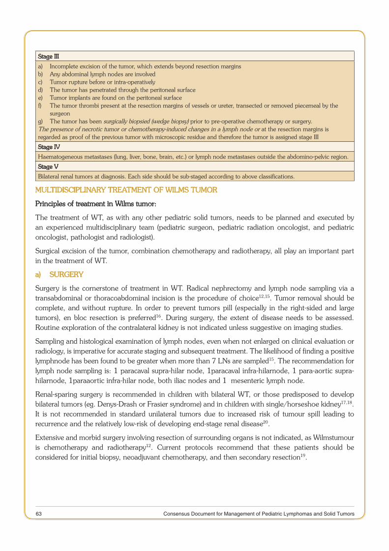
63 Consensus Document for Management of Pediatric Lymphomas and Solid Tumors
Stage III
a) Incomplete excision of the tumor, which extends beyond resection margins b) Any abdominal lymph nodes are involved c) Tumor rupture before or intra-operatively d) The tumor has penetrated through the peritoneal surface e) Tumor implants are found on the peritoneal surface f) The tumor thrombi present at the resection margins of vessels or ureter, transected or removed piecemeal by the
surgeong) The tumor has been surgically biopsied (wedge biopsy) prior to pre-operative chemotherapy or surgery. The presence of necrotic tumor or chemotherapy-induced changes in a lymph node or at the resection margins is regarded as proof of the previous tumor with microscopic residue and therefore the tumor is assigned stage III
Stage IV
Haematogeneous metastases (lung, liver, bone, brain, etc.) or lymph node metastases outside the abdomino-pelvic region.
Stage V
Bilateral renal tumors at diagnosis. Each side should be sub-staged according to above classifications.
MULTIDISCIPLINARY TREATMENT OF WILMS TUMOR
Principles of treatment in Wilms tumor:
The treatment of WT, as with any other pediatric solid tumors, needs to be planned and executed by an experienced multidisciplinary team (pediatric surgeon, pediatric radiation oncologist, and pediatric oncologist, pathologist and radiologist).
Surgical excision of the tumor, combination chemotherapy and radiotherapy, all play an important part in the treatment of WT.
a) SURGERY
Surgery is the cornerstone of treatment in WT. Radical nephrectomy and lymph node sampling via a transabdominal or thoracoabdominal incision is the procedure of choice12,15. Tumor removal should be complete, and without rupture. In order to prevent tumors pill (especially in the right-sided and large tumors), en bloc resection is preferred16. During surgery, the extent of disease needs to be assessed. Routine exploration of the contralateral kidney is not indicated unless suggestive on imaging studies.
Sampling and histological examination of lymph nodes, even when not enlarged on clinical evaluation or radiology, is imperative for accurate staging and subsequent treatment. The likelihood of finding a positive lymphnode has been found to be greater when more than 7 LNs are sampled15. The recommendation for lymph node sampling is: 1 paracaval supra-hilar node, 1paracaval infra-hilarnode, 1 para-aortic supra-hilarnode, 1paraaortic infra-hilar node, both iliac nodes and 1 mesenteric lymph node.
Renal-sparing surgery is recommended in children with bilateral WT, or those predisposed to develop bilateral tumors (eg. Denys-Drash or Frasier syndrome) and in children with single/horseshoe kidney17,18. It is not recommended in standard unilateral tumors due to increased risk of tumour spill leading to recurrence and the relatively low-risk of developing end-stage renal disease20.
Extensive and morbid surgery involving resection of surrounding organs is not indicated, as Wilmstumour is chemotherapy and radiotherapy12. Current protocols recommend that these patients should be considered for initial biopsy, neoadjuvant chemotherapy, and then secondary resection19.

64 Consensus Document for Management of Pediatric Lymphomas and Solid Tumors
b) CHEMOTHERAPY
WT is an extremely chemo sensitive tumor. The timing of chemotherapy has been contentious, with the North American National WT Study/Children’s Oncology Group (NWTS/COG) groups advocating initial nephrectomy followed by chemotherapy (+/- radiation therapy), and the SIOP protocols preferring pre-operative chemotherapy before definitive resection for patients21-23. The details of treatment guidelines and recommendations are detailed below.
The advantages of pre-nephrectomy chemotherapy as recommended by SIOP include decreasing the size and vascular supply of the tumor, and reduction in the frequency of surgical complications such as intra-operative tumor ruptureandhaemorrhage. Furthermore, metastases may disappear or become resectable, vascular extension may regress and partial nephrectomy may become possible. However, this approach could lead to inaccurate staging of the tumor and potentially under- or over-treatment. Most studies do not show a difference in survival between the two groups.
In resource-limited settings, the majority of patients present with large tumors, which may either be unresectable or risky to resect; making pre-operative chemotherapy followed by delayed surgery the preferred approach.
The commonly used chemotherapy drugs in protocols worldwide are vincristine, dactinomycin, and doxorubicin; other drugs are cyclophosphamide, carboplatin and etoposide. The doses and toxicities of drugs are mentioned towards the end of this write up.
c) RADIOTHERAPY (RT)
WT is an extremely radiosensitive tumor. Indications for radiation therapy and details are as follows:
All stage III tumors receive either flank irradiation or whole abdomen irradiation • 12,23.
Irradiation of Lung Metastasis: The treatment approach varies with the treating group. In the NWTS/ •COG protocols, patients with favorable histology tumours whose lung lesions do not show a complete response to chemotherapy at week 6 receive whole-lung irradiation24. As per SIOP approach, patients with pulmonary metastasis were reassessed after 6 weeks of vincristine, dactinomycin, and epirubicin or doxorubicin. If pulmonary complete remission (CR) was not obtained, metastatectomy was considered. Patients in CR received three-drug post-operative chemotherapy, whereas patients not in CR were switched to a high-risk regimen with an assessment at week 11. If CR was not obtained at week 11, pulmonary RT was mandatory25.
The indications for RT in Wilmstumor as well as doses and portals are given below in the recommendations.
CURRENT TREATMENT PROTOCOLS FOR WILMS TUMOR
Traditionally, the treatment of WT depends on the stage and histology of the tumor. A retrospective analysis of the SIOP 93-01 data looking for prognostic factors in localizedtumors showed that the histological classification of the tumor as low, intermediate or high-risk is more important than tumor stage13. The risk stratification based on histology is mentioned above in Table 1.
Current treatment guidelines are based largely on studies conducted over the past four decades by the North American groups: NWTS/COG and groups from Europe: International Society of Paediatric Oncology (SIOP) and United Kingdom Children's Cancer Study Group (UK CCSG).
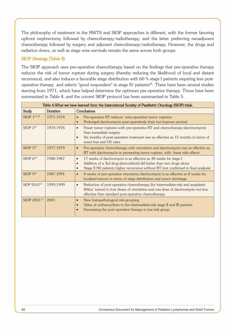
65 Consensus Document for Management of Pediatric Lymphomas and Solid Tumors
The philosophy of treatment in the NWTS and SIOP approaches is different, with the former favoring upfront nephrectomy followed by chemotherapy/radiotherapy, and the latter preferring neoadjuvant chemotherapy followed by surgery and adjuvant chemotherapy/radiotherapy. However, the drugs and radiation doses, as well as stage wise survivals remain the same across both groups.
SIOP Strategy (Table 4)
The SIOP approach uses pre-operative chemotherapy based on the findings that pre-operative therapy reduces the risk of tumor rupture during surgery (thereby reducing the likelihood of local and distant recurrence), and also induces a favorable stage distribution with 60 % stage I patients requiring less post-operative therapy and selects "good responders" in stage IV patients26. There have been several studies starting from 1971, which have helped determine the optimum pre-operative therapy. These have been summarized in Table 4, and the current SIOP protocol has been summarized in Table 5.
Table 4.What we have learned from the International Society of Paediatric Oncology (SIOP) trials.
Study Duration Conclusions
SIOP 127,28 1971-1974 Pre-operative RT reduces intra-operative tumor ruptures •Prolonged dactinomycin post-operatively does not improve survival •
SIOP 227 1974-1976 Fewer tumor ruptures with pre-operative RT and chemotherapy (dactinomycin) •than immediate surgery Six months of post-operative treatment was as effective as 15 months in terms of •event-free and OS rates
SIOP 529 1977-1979 Pre-operative chemotherapy with vincristine and dactinomycin was as effective as •RT with dactinomycin in preventing tumor rupture, with lower side effects
SIOP 630 1980-1987 17 weeks of dactinomycin is as effective as 38 weeks for stage I •Addition of a 3rd drug (doxorubicin) did better than two drugs alone •Stage II N0 patients higher recurrence without RT (not confirmed in final analysis) •
SIOP 931 1987-1991 4 weeks of pre-operative vincristine/dactinomycin is as effective as 8 weeks for •localized tumors in terms of stage distribution and tumor shrinkage
SIOP 93-0132 1993-1999 Reduction of post-operative chemotherapy (for intermediate-risk and anaplastic •Wilms’ tumor) to four doses of vincristine and one dose of dactinomycin not less effective than standard post-operative chemotherapy
SIOP 200112 2001 New histopathological risk-grouping •Value of anthracyclines in the intermediate-risk stage II and III patients •Decreasing the post-operative therapy in low-risk group •
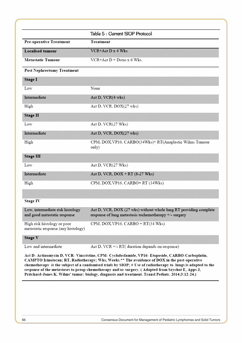
66 Consensus Document for Management of Pediatric Lymphomas and Solid Tumors
Table 5 : Current SIOP Protocol
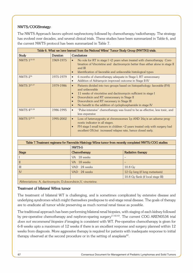
67 Consensus Document for Management of Pediatric Lymphomas and Solid Tumors
NWTS/COGStrategy:
The NWTS Approach favors upfront nephrectomy followed by chemotherapy/radiotherapy. The strategy has evolved over decades, and several clinical trials. These studies have been summarized in Table 6, and the current NWTS protocol has been summarized in Table 7.
Table 6. What we have learned from the National Wilms' Tumor Study Group (NWTSG) trials.
Study Duration Conclusions
NWTS 133-35 1969-1975 No role for RT in stage I <2 years when treated with chemotherapy Com- •bination of Vincristine and dactinomycin better than either alone in stage II and IIIIdentification of favorable and unfavorable histological types •
NWTS 236 1975-1979 6 months of chemotherapy adequate in Stage I; RT unnecessary •Addition of Adriamycin improved outcome in Stage II-IV •
NWTS 323,37 1979-1986 Patients divided into two groups based on histopathology: favorable (FH) •and unfavorable11 weeks of vincristine and dactinomycin sufficient in stage I •Doxorubicin and RT unnecessary in Stage II •Doxorubicin and RT necessary in Stage III •No benefit to the addition of cyclophosphamide in stage IV •
NWTS 423,38 1986-1995 “Pulse-intensive” chemotherapy was found to be as effective, less toxic, and •less expensive
NWTS 539-41 1995-2002 Loss of heterozygosity at chromosomes 1p AND 16q is an adverse prog- •nostic indicator in all stages. FH stage I small tumors in children <2 years treated only with surgery had •excellent OS,but increased relapse rate, hence closed early.
Table 7.Treatment regimens for Favorable Histology Wilms tumor from recently completed NWTS/COG studies
NWTS-5
Stage Chemotherapy Radiation therapy
I VA × 18 weeks –
II VA × 18 weeks –
III VAD × 24 weeks 10.8 Gy
IV VAD × 24 weeks 12 Gy lung (if lung metastasis)
10.8 Gy flank (if local stage III)
Abbreviations: A, dactinomycin; D,doxorubicin,V, vincristine.
Treatment of bilateral Wilms tumor
The treatment of bilateral WT is challenging, and is sometimes complicated by extensive disease and underlying syndromes which might themselves predispose to end-stage renal disease. The goals of therapy are to eradicate all tumor while preserving as much normal renal tissue as possible.
The traditional approach has been performing bilateral renal biopsies, with staging of each kidney followed by pre-operative chemotherapy and nephron-sparing surgery17,42,43. The current COG AREN0534 trial does not recommend biopsies if imaging is consistent with WT. Pre-operative chemotherapy is given for 6-8 weeks upto a maximum of 12 weeks if there is an excellent response and surgery planned within 12 weeks from diagnosis. More aggressive therapy is required for patients with inadequate response to initial therapy observed at the second procedure or in the setting of anaplasia42.
68 Consensus Document for Management of Pediatric Lymphomas and Solid Tumors
Renal transplantation for children with stage V Wilms or with underlying Denys-Drash syndrome tumor is usually delayed until 1 to 2 years have passed without evidence of malignancy44.
TREATMENT OF RECURRENT DISEASE
Studies conducted by SIOP and NWTS have identified three risk groups in relapsed disease depending on the time to relapse, frontline therapy and histology at relapse.
Standard risk: • Favorable histology WT relapsing after 2 drug chemotherapy (Vincristine and/or dactinomycin). These patients can be treated with surgery (when feasible), radiation therapy, and alternating courses of vincristine, doxorubicin, and cyclophosphamide; and etoposide and cyclophosphamide, and have an expected EFS of 70-80%45.
High-risk: • Favorable Histology WT relapsing after 3 or more drug chemotherapy (usually includes doxorubicin). These patients can be treated with alternating courses of cyclophosphamide/etoposide and carboplatin/etoposide, surgery, and radiation therapy and have survival rates of around 40% to 50%46.
Very high-risk: • Recurrent anaplastic or blastemal-predominant WT. These patients have extremely poor prognosis, with survival rates in the 10% range41. The treatment options tried in this group include combination chemotherapy with ifosfamide, etoposide, and carboplatin, surgery and radiation therapy, as well as high-dose chemotherapy with autologous stem cell rescue47,48.
ISSUES SPECIFIC TO RESOURCE POOR SETTINGS:
Although most centers in India treating WT and other pediatric cancers have a reasonable level of infrastructure, there are still several challenges in management. A significant proportion of children present with advanced disease and severe malnutrition, due to both underlying malnutrition as well as due to disease. Patients face financial, social and logistical issues, and treating centers sometimes lack the appropriate infrastructure and trained personnel. Some children present to referral centers with partially or inadequately treated tumors, which might be then refractory to treatment. SIOP guidelines for Paediatric Oncology in Developing Countries (PODC) have made several minimum recommendations10 which include basic laboratory and radiology services, with provisions for essential chemotherapy drugs and facilities for safe administration, trained surgeon and supportive care as well as social support. Most oncology centers in India more than fulfil these criteria, except possibly financial and social support. Recommendations pertinent to our scenario include administration of pre-operative chemotherapy in large tumors, starting with a lower dosage of drugs (2/3rd) in severely acutely malnourished children and reduction of doxorubicin dose to 30 mg/m2in case of neutropenia.
LATE EFFECTS FOLLOWING THE TREATMENT OF WILMS TUMOR:
Children treated for WT have an increased risk of developing certain late effects: approximately 25% of survivors have serious chronic health conditions 25 years from diagnosis49. Of concern is the fact that most children present and are treated at a young age.
Cardiomyopathy and Congestive heart failure • 50: due to anthracyclines, and mediastinal RT.Long-term monitoring of cardiac function needs to be done depending on age at treatment, the dose of anthracycline received and clinical and cardiac status.
End-stage renal disease • 20: The cumulative incidence of end-stage renal disease at 20 years is less than 1% in unilateral WT and around 3% for bilateral WT.
Second malignant neoplasms • 51

69 Consensus Document for Management of Pediatric Lymphomas and Solid Tumors
Fortunately, there has been a significant reduction in late mortality due to cardiac late effects as well as second malignancy in the recent years49.
RESEARCH22,52
The questions currently being studied in WT focus on improving outcomes in high-risk groups, and in reducing late effects in low-risk groups. Specifically, these are:
Identification of molecular prognostic factors followed by augmented therapy in high-risk groups, and •reduction of therapy (and late effects) in lower risk groups
Identification of molecular targets in order to improve outcomes. •
The role of nephrectomy alone in young children with small, stage I/FH WT •
The need for whole-lung radiation therapy for the management of children with pulmonary •metastasis
RECOMMENDATIONS FOR THE MANAGEMENT OF WILMS TUMOR (BY STAGE AND HISTOLOGY)
In resource-limited settings, the majority of patients present with large tumors, which may either be unresectable or risky to resect; making pre-operative chemotherapy followed by delayed surgery the preferred approach.
1. PRE-OPERATIVE CHEMOTHERAPY: To be given in all cases, when possible
A. Pre-operative Chemotherapy for Non-Metastatic WT: 4 weeks of Vincristine(VCR)/Actinomycin d (ACD)
Wk 1 VCR ACD
Wk 2 VCR
Wk 3 VCR ACD
Wk 4 VCR
B. Pre-operative Chemotherapy for Metastatic WT: 6 weeks of VCR/ACD/ADR
Wk 1VCR ADRACD
Wk 2VCR
Wk 3VCR ACD
Wk 4VCR
Wk 5VCR ADRACD
Wk 6 VCR
2. RE-EVALUATION:
Week 4 in Non-Metastatic WT •
Week 6 in Metastatic WT •
Investigation of Choice: CECT of Thorax/ Abdomen and Pelvis
Abdominal radiology should mention (as in the baseline CT scan) •
1. Size of tumor in maximum dimension
2. Presence of necrosis: <25%, 25-50%, 50-75%, >75%
70 Consensus Document for Management of Pediatric Lymphomas and Solid Tumors
3. Presence of thrombus
4. Lymph node status
5. Liver nodules: Number, size, site
6. Relationship with aorta and inferior vena cava: pushed, engulfed, none.
Chest radiology should mention (as in baseline Radiology) •
Chest Metastases*: Resolution/ Stable Disease/Progression
1. Present/absent
2. Unilateral/Bilateral
3. Number on each side: up to five or >5
3. SURGERY
A. Timing of Surgery: Week 5 in non-metastatic WT
Week 7 in metastatic WT
B. Abdominal Surgery
Proceed to definitive surgery: Radical Nephroureterectomy and lymph node sampling is the procedure •of choice
Approach should be transabdominal/transperitoneal. Mention whether done outside or within the •Gerota’s fascia (as per the institutional practice)
Sampling and histological examination of lymph nodes, even when not enlarged on clinical evaluation •or radiology.
The minimum of seven lymph nodes required to be sampled are one paracaval supra-hilar node, 1 •paracaval infra-hilarnode, 1 para-aortic supra-hilar node, 1 para-aortic infra-hilar node, both iliac nodes and 1 mesenteric lymph node.
Examination of the liver: If any nodules, the same have to be biopsied. •
Tumor removal should be complete and en bloc, without rupture. • Comment on spillage, even if absent.
Presence/Absence of renal vein or inferior vena caval thrombus: comment whether thrombus is adherent •or non-adherent. Further, removal of thrombus performed by cavotomy or partial cavectomy.
Evaluation of contralateral kidney not needed with CT imaging {If ultrasound (USG) is used as imaging •alone, then this may be incorporated for those centers}
C. Pulmonary Metastectomy
After 9 weeks of pre-operative chemotherapy, if there is a doubtful lesion on CT scan, then the same •should be biopsied.
In cases where lung nodules persist after chemotherapy and radiotherapy, then the same should be •removed at the end of therapy
71 Consensus Document for Management of Pediatric Lymphomas and Solid Tumors
4. PATHOLOGICAL EVALUATION in all Resected Specimens
The Whole Specimen to be examined (Not just a biopsy), ideally one block per cm of tissue.
Points to include in the Pathology Report:
Percentage of tumor necrosis/chemotherapy-induced changes 1.
Within the viable tumor, an actual percentage of residual blastemal or anaplasia. (If percentage necrosis 2. <66%)
Ideally, also assess and record percentages of other tumor components (epithelial and stromal). 3.
4. Subtype of Wilms tumor:
Identify the subtype of WT and allocate a risk category based on histology as per Table 1 a.
5. Classify as Stage 1-3 based on (as per Table 3):
Involvement of renal capsule, sinuses, perirenal fata.
Intra-operative spillageb.
Transectedtumor thrombusc.
Lymph node involvement as evidenced by the presence of tumor or necrosisd.
5. POST-OPERATIVE CHEMOTHERAPY:
To be started as soon as ileus subsides after surgery, and is decided based on the post-operative histology as well as stage (as per tables 1 and 3). Although the SIOP protocol stratifies completely necrotic post-operative tumors to be ‘low-risk’ with Stage I Low-Risk tumors requiring no further treatment, we do not recommend stopping treatment in such cases due to limitations in the interpretation of histopathology.
A. Stage I Intermediate-Risk:
4 weeks of VCR-ACD (will include only a single dose of ACD) •
B. Stage II and III Low-Risk:
27 weeks of VCR, and ACD •
VCR given weekly, ACD given 3 –weekly •
C. Stage II-IV Intermediate-Risk, Stage I High-Risk:
27 weeks of VCR, ACD and ADR; •
VCR given weekly, ACD given 3 –weekly and ADR given 6-weekly. •
Additionally, in • stage IV: Surgery+ RT to metastatic sites
D. Stage II-IV High-Risk Unfavorable Histology:
Alternating cycles of CARBO/VP-16 and CYCLO-ADR* to be given three-weekly •
6 cycles each of CARBO/VP-16 and CYCLO-ADR* •
Total du • ration of 34 weeks
Stage II High-Risk, Stage III Intermediate and High-Risk and all Stage IV tumors require RT as per doses mentioned below.
72 Consensus Document for Management of Pediatric Lymphomas and Solid Tumors
Chemotherapy Doses
VCR: Vincristine 1.5 mg/m1. 2 IV push (Max 2mg) (0.05 mg/kg for weight <30 kg)
ACD : Actinomycin D45 mcg/kg IV push(Max 2mg) 2.
ADR: Adriamycin/ Doxorubicin 50mg/m3. 2 in a 4-6 hours infusion (ideally) or as slow IV push (1.5 mg/kg for weight <30 kg)
ADR*: Adriamycin/ Doxorubicin 50 mg/m4. 2 in a 4-6 hours infusion (1.5 mg/kg for weight <30 kg)
CYCLO: Cyclophosphamide 450mg/m5. 2as IV infusion for 3consecutive days
CARBO: Carboplatin 200 mg/m6. 2 as IV infusion over 1 hour for three consecutive days
VP-16: Etoposide 150mg/m7. 2as IV infusion over one hour for three consecutive days
All above doses are for children weighing above 12 kg. According to SIOP protocols, patients below 12 kg or with acute malnutrition should have a 2/3rd dose reduction of chemotherapeutic agents. These regimens must be modified according to hematological tolerance. In case of neutropenia, the dose of drugs (actinomycin, doxorubicin) can be reduced or frequency of administration lengthened. NWTS protocols recommend that newborns and all infants younger than 12 months require a 50% reduction in chemotherapy dose.
6. RECOMMENDATIONS FOR RADIOTHERAPY (Table 8):
Radiotherapy to be started within 9 - 14 day of surgery unless medically contraindicated.1.
Radiation therapy is given to the Flank, except when Whole abdominal irradiation (WAI) is indicated 2. in the following conditions:
Diffuse tumor spillagea.
Pre-operative/ intra-operative tumor spillageb.
Intraperitoneal tumor rupturec.
Peritoneal tumor seeding/ hemorrhagic or cytology positive ascitesd.
Gross residual disease after surgery – additional RT Boost of 10.8Gy/6# @ 1.8Gy/ Fraction after 3. Flank or WAI
Whole vertebral body to be included in the irradiated volume4.
For WAI Bilateral Femoral Heads & Acetabulum should be shielded5.
If both Whole-lung irradiation (WLI) & abdominal RT required – should include both in one field (avoid 6. field junction)
WLI indicated for all patients with pulmonary lesions (CXR or CT detected)7.
Whole liver RT: indicated for patients with liver metastasis if the metastatic lesions are not completely 8. resected.
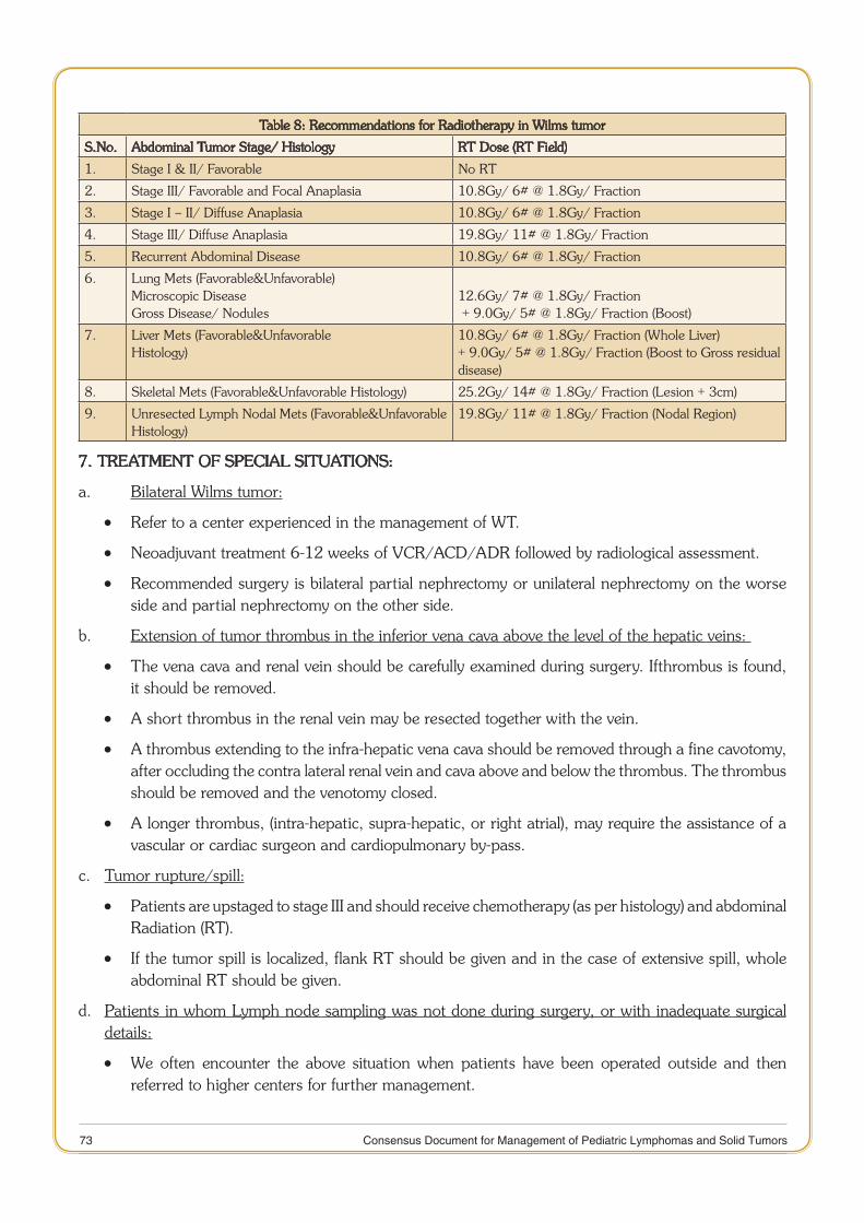
73 Consensus Document for Management of Pediatric Lymphomas and Solid Tumors
Table 8: Recommendations for Radiotherapy in Wilms tumor
S.No. Abdominal Tumor Stage/ Histology RT Dose (RT Field)
1. Stage I & II/ Favorable No RT
2. Stage III/ Favorable and Focal Anaplasia 10.8Gy/ 6# @ 1.8Gy/ Fraction
3. Stage I – II/ Diffuse Anaplasia 10.8Gy/ 6# @ 1.8Gy/ Fraction
4. Stage III/ Diffuse Anaplasia 19.8Gy/ 11# @ 1.8Gy/ Fraction
5. Recurrent Abdominal Disease 10.8Gy/ 6# @ 1.8Gy/ Fraction
6. Lung Mets (Favorable&Unfavorable) Microscopic DiseaseGross Disease/ Nodules
12.6Gy/ 7# @ 1.8Gy/ Fraction + 9.0Gy/ 5# @ 1.8Gy/ Fraction (Boost)
7. Liver Mets (Favorable&UnfavorableHistology)
10.8Gy/ 6# @ 1.8Gy/ Fraction (Whole Liver)+ 9.0Gy/ 5# @ 1.8Gy/ Fraction (Boost to Gross residual disease)
8. Skeletal Mets (Favorable&Unfavorable Histology) 25.2Gy/ 14# @ 1.8Gy/ Fraction (Lesion + 3cm)
9. Unresected Lymph Nodal Mets (Favorable&Unfavorable Histology)
19.8Gy/ 11# @ 1.8Gy/ Fraction (Nodal Region)
7. TREATMENT OF SPECIAL SITUATIONS:
a. Bilateral Wilms tumor:
Refer to a center experienced in the management of WT. •
Neoadjuvant treatment 6-12 weeks of VCR/ACD/ADR followed by radiological assessment. •
Recommended surgery is bilateral partial nephrectomy or unilateral nephrectomy on the worse •side and partial nephrectomy on the other side.
b. Extension of tumor thrombus in the inferior vena cava above the level of the hepatic veins:
The vena cava and renal vein should be carefully examined during surgery. Ifthrombus is found, •it should be removed.
A short thrombus in the renal vein may be resected together with the vein. •
A thrombus extending to the infra-hepatic vena cava should be removed through a fine cavotomy, •after occluding the contra lateral renal vein and cava above and below the thrombus. The thrombus should be removed and the venotomy closed.
A longer thrombus, (intra-hepatic, supra-hepatic, or right atrial), may require the assistance of a •vascular or cardiac surgeon and cardiopulmonary by-pass.
c. Tumor rupture/spill:
Patients are upstaged to stage III and should receive chemotherapy (as per histology) and abdominal •Radiation (RT).
If the tumor spill is localized, flank RT should be given and in the case of extensive spill, whole •abdominal RT should be given.
d. Patients in whom Lymph node sampling was not done during surgery, or with inadequate surgical details:
We often encounter the above situation when patients have been operated outside and then •referred to higher centers for further management.
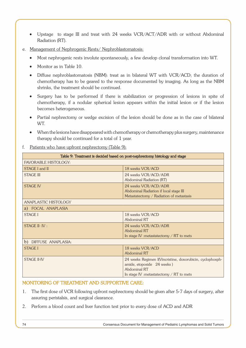
74 Consensus Document for Management of Pediatric Lymphomas and Solid Tumors
Upstage to stage III and treat with 24 weeks VCR/ACT/ADR with or without Abdominal •Radiation (RT).
e. Management of Nephrogenic Rests/ Nephroblastomatosis:
Most nephrogenic rests involute spontaneously, a few develop clonal transformation into WT. •
Monitor as in Table 10. •
Diffuse nephroblastomatosis (NBM): treat as in bilateral WT with VCR/ACD; the duration of •chemotherapy has to be geared to the response documented by imaging. As long as the NBM shrinks, the treatment should be continued.
Surgery has to be performed if there is stabilization or progression of lesions in spite of •chemotherapy, if a nodular spherical lesion appears within the initial lesion or if the lesion becomes heterogeneous.
Partial nephrectomy or wedge excision of the lesion should be done as in the case of bilateral •WT.
When the lesions have disappeared with chemotherapy or chemotherapy plus surgery, maintenance •therapy should be continued for a total of 1 year.
f. Patients who have upfront nephrectomy (Table 9):
Table 9: Treatment is decided based on post-nephrectomy histology and stage
FAVORABLE HISTOLOGY:
STAGE I and II 18 weeks VCR/ACD
STAGE III 24 weeks VCR/ACD/ADRAbdominal Radiation (RT)
STAGE IV 24 weeks VCR/ACD/ADRAbdominal Radiation if local stage IIIMetastatectomy / Radiation of metastasis
ANAPLASTIC HISTOLOGY
FOCAL ANAPLASIAa) STAGE I 18 weeks VCR/ACD
Abdominal RT
STAGE II- IV : 24 weeks VCR/ACD/ADRAbdominal RTIn stage IV :metastatectomy / RT to mets
DIFFUSE ANAPLASIA: b) STAGE I 18 weeks VCR/ACD
Abdominal RT
STAGE II-IV 24 weeks Regimen I(Vincristine, doxorubicin, cyclophosph-amide, etoposide × 24 weeks )Abdominal RT In stage IV :metastatectomy / RT to mets
MONITORING OF TREATMENT AND SUPPORTIVE CARE:
The first dose of VCR following upfront nephrectomy should be given after 5-7 days of surgery, after 1. assuring peristalsis, and surgical clearance.
Perform a blood count and liver function test prior to every dose of ACD and ADR2.
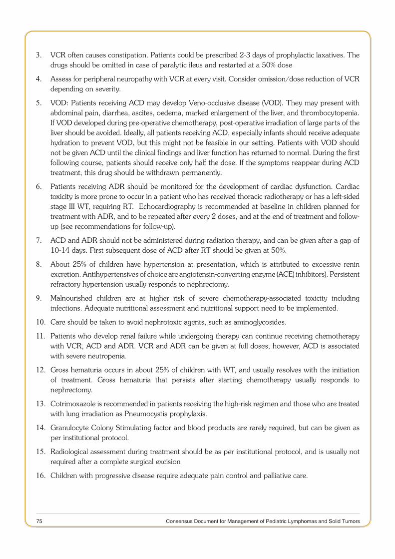
75 Consensus Document for Management of Pediatric Lymphomas and Solid Tumors
VCR often causes constipation. Patients could be prescribed 2-3 days of prophylactic laxatives. The 3. drugs should be omitted in case of paralytic ileus and restarted at a 50% dose
Assess for peripheral neuropathy with VCR at every visit. Consider omission/dose reduction of VCR 4. depending on severity.
VOD: Patients receiving ACD may develop Veno-occlusive disease (VOD). They may present with 5. abdominal pain, diarrhea, ascites, oedema, marked enlargement of the liver, and thrombocytopenia. If VOD developed during pre-operative chemotherapy, post-operative irradiation of large parts of the liver should be avoided. Ideally, all patients receiving ACD, especially infants should receive adequate hydration to prevent VOD, but this might not be feasible in our setting. Patients with VOD should not be given ACD until the clinical findings and liver function has returned to normal. During the first following course, patients should receive only half the dose. If the symptoms reappear during ACD treatment, this drug should be withdrawn permanently.
Patients receiving ADR should be monitored for the development of cardiac dysfunction. Cardiac 6. toxicity is more prone to occur in a patient who has received thoracic radiotherapy or has a left-sided stage III WT, requiring RT. Echocardiography is recommended at baseline in children planned for treatment with ADR, and to be repeated after every 2 doses, and at the end of treatment and follow-up (see recommendations for follow-up).
ACD and ADR should not be administered during radiation therapy, and can be given after a gap of 7. 10-14 days. First subsequent dose of ACD after RT should be given at 50%.
About 25% of children have hypertension at presentation, which is attributed to excessive renin 8. excretion. Antihypertensives of choice are angiotensin-converting enzyme (ACE) inhibitors). Persistent refractory hypertension usually responds to nephrectomy.
Malnourished children are at higher risk of severe chemotherapy-associated toxicity including 9. infections. Adequate nutritional assessment and nutritional support need to be implemented.
Care should be taken to avoid nephrotoxic agents, such as aminoglycosides.10.
Patients who develop renal failure while undergoing therapy can continue receiving chemotherapy 11. with VCR, ACD and ADR. VCR and ADR can be given at full doses; however, ACD is associated with severe neutropenia.
Gross hematuria occurs in about 25% of children with WT, and usually resolves with the initiation 12. of treatment. Gross hematuria that persists after starting chemotherapy usually responds to nephrectomy.
Cotrimoxazole is recommended in patients receiving the high-risk regimen and those who are treated 13. with lung irradiation as Pneumocystis prophylaxis.
Granulocyte Colony Stimulating factor and blood products are rarely required, but can be given as 14. per institutional protocol.
Radiological assessment during treatment should be as per institutional protocol, and is usually not 15. required after a complete surgical excision
Children with progressive disease require adequate pain control and palliative care.16.
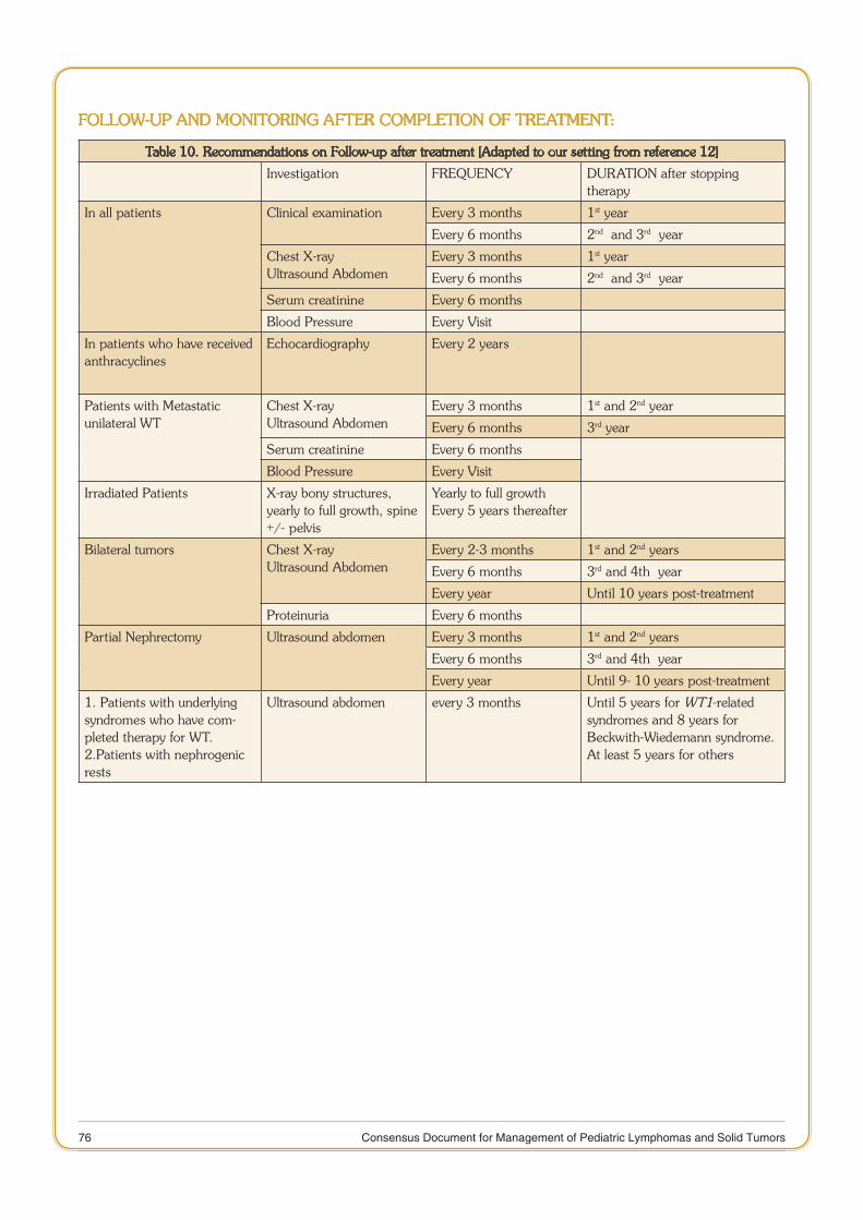
76 Consensus Document for Management of Pediatric Lymphomas and Solid Tumors
FOLLOW-UP AND MONITORING AFTER COMPLETION OF TREATMENT:
Table 10. Recommendations on Follow-up after treatment [Adapted to our setting from reference 12]
Investigation FREQUENCY DURATION after stopping therapy
In all patients Clinical examination Every 3 months 1st year
Every 6 months 2nd and 3rd year
Chest X-rayUltrasound Abdomen
Every 3 months 1st year
Every 6 months 2nd and 3rd year
Serum creatinine Every 6 months
Blood Pressure Every Visit
In patients who have received anthracyclines
Echocardiography Every 2 years
Patients with Metastatic unilateral WT
Chest X-rayUltrasound Abdomen
Every 3 months 1st and 2nd year
Every 6 months 3rd year
Serum creatinine Every 6 months
Blood Pressure Every Visit
Irradiated Patients X-ray bony structures, yearly to full growth, spine +/- pelvis
Yearly to full growthEvery 5 years thereafter
Bilateral tumors Chest X-rayUltrasound Abdomen
Every 2-3 months 1st and 2nd years
Every 6 months 3rd and 4th year
Every year Until 10 years post-treatment
Proteinuria Every 6 months
Partial Nephrectomy Ultrasound abdomen Every 3 months 1st and 2nd years
Every 6 months 3rd and 4th year
Every year Until 9- 10 years post-treatment
1. Patients with underlying syndromes who have com-pleted therapy for WT.2.Patients with nephrogenic rests
Ultrasound abdomen every 3 months Until 5 years for WT1-related syndromes and 8 years for Beckwith-Wiedemann syndrome.At least 5 years for others
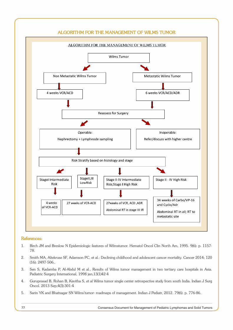
77 Consensus Document for Management of Pediatric Lymphomas and Solid Tumors
ALGORITHM FOR THE MANAGEMENT OF WILMS TUMOR
References:
Birch JM and Breslow N Epidemiologic features of Wilmstumor. Hematol Oncol Clin North Am, 1995. 9(6): p. 1157-1. 78.
Smith MA, Altekruse SF, Adamson PC, et al.: Declining childhood and adolescent cancer mortality. Cancer 2014; 120 2. (16): 2497-506,.
Sen S, Kadamba P, Al-Abdul M et al., Results of Wilms tumor management in two tertiary care hospitals in Asia. 3. Pediatric Surgery International, 1998 jan;13(1)42-4
Guruprasad B, Rohan B, Kavitha S, et al Wilms tumor single center retrospective study from south India. Indian J Surg 4. Oncol. 2013 Sep;4(3):301-4
Sarin YK and Bhatnagar SN Wilms’tumor- roadmaps of management. Indian J Pediatr, 2012. 79(6): p. 776-86.5.

78 Consensus Document for Management of Pediatric Lymphomas and Solid Tumors
Trehan A, Chowdhary SK and Marwaha RK. Wilms tumor: five-year tumor-free survival on a modified SIOP protocol 6. from an Indian university hospital.” Journal of pediatric hematology/oncology 34.1 2012: 57-62.
Koodiyedath B, Kulkarni KP, and Arora RS. “Outcomes of Wilms Tumor in India: Findings from a Systematic Analysis 7. Pediatric Blood and Cancer Vol. 59. No. 6. Wiley Periodicals Inc 2012.
Bhagwat R, Kurkure P, Iyer K, et al. Is anthracycline based chemotherapy for all stages of Wilms Tumor (WT) a practical 8. compromise for better outcome in developing countries? SIOP XXXVI meeting Abstracts PJ. Pediatr Blood Cancer 2005;45:530.
Rastogi S, Qureshi S, Vora T et al. Is Three-Drug Chemotherapy protocol for all stages of Wilms Tumor a practical 9. compromise for suboptimal staging in Developing Country? Is it worth and Safe? Pediatric Blood and Cancer Vol. 61. No. Wiley Periodicals Inc 2014.
Israels T, Moreira C, Scanlan T, et al. SIOP PODC: recommendations for supportive care of children with cancer in a 10. low-income setting. Pediatr Blood Cancer, 2013. 60(6): p. 899-904.
National Cancer Registry Program, Indian council of medical research, Consolidated report of hospital based cancer 11. registries – 2007-2011
De Kraker J, Graf N, Pritchard-Jones K, Pein F. Nephroblastoma clinical trial and study SIOP 2001, Protocol. SIOP 12. RTSG. 2001
VujanićGM Sandstedt B, Harms D et al Revised International Society of Paediatric Oncology (SIOP) working classification 13. of renal tumors of childhood. Med. Pediatr. Oncol. (2002), 38: 79–82
Perlman EJ: Pediatric renal tumors: practical updates for the pathologist. Pediatr Dev Pathol 8 (3): 320-38, 2005 May-14. Jun.
Kieran K, Anderson JR, Dome JS, et al.: Lymph node involvement in Wilmstumor: results from National WilmsTumor 15. Studies 4 and 5. J PediatrSurg 47 (4): 700-6, 2012.
Gow KW, Barnhart DC, Hamilton TE, et al.: Primary nephrectomy and intra-operative tumor spill: report from the 16. Children’s Oncology Group (COG) renal tumors committee. J PediatrSurg 48 (1): 34-8, 2013.
Davidoff AM, Giel DW, Jones DP, et al. The feasibility and outcome of nephron-sparing surgery for children with 17. bilateral Wilmstumor. The St Jude Children’s Research Hospital experience: 1999–2006. Cancer 2008;112:2060-70.
Auber F, Jeanpierre C, Denamur E, et al.: Management of Wilmstumors in Drash and Frasier syndromes. Pediatr Blood 18. Cancer 52 (1): 55-9, 2009.
Ritchey ML, Kelalis PP, Breslow N, et al.: Surgical complications after nephrectomy for Wilms’ tumor. SurgGynecolObstet 19. 175 (6): 507-14, 1992.
Interiano R B, Delos Santos N, et al (2015), Renal function in survivors of nonsyndromicWilmstumor treated with 20. unilateral radical nephrectomy Cancer. doi: 10.1002/cncr.29373
Metzger ML, Dome JS. Current therapy for Wilms’ tumor. Oncologist 2005;10:815-26.21.
Green DM The evolution of treatment for Wilmstumor J PediatrSurg, 48 (2013), pp. 14–1922.
Green DM The treatment of stages I–IV favorable histology Wilms tumor J ClinOncol, 22 (2004), pp. 1366–137223.
Grundy PE, Green DM et al. Clinical significance of pulmonary nodules detected by CT and Not CXR in patients treated 24. for favorable histology Wilmstumor on national Wilmstumor studies-4 and -5: a report from the Children’s Oncology Group. Pediatr Blood Cancer 2012;59:631-5.
Smets, A.M., et al., The contribution of chest CT scan at diagnosis in children with unilateral Wilms’ tumor. Results of 25. the SIOP 2001 study. Eur J Cancer, 2012. 48(7): p. 1060-5.
Godzinski J. The current status of treatment of Wilms’ tumor as per the SIOP trials. Journal of Indian Association of 26. Pediatric Surgeons. 2015;20(1):16-20. doi:10.4103/0971-9261.145439.
Burger D, Moorman-Voestermans CG, Mildenberger H et al. The advantages of pre-operative therapy in Wilms’ 27. tumor. A summarized report on clinical trials conducted by the International Society of Paediatric Oncology (SIOP). Z Kinderchir 1985;40:170–175.
Lemerle J, Voute PA, Tournade MF et al. Pre-operative versus post-operative radiotherapy, single versus multiple courses 28. of actinomycin D, in the treatment of Wilms’ tumor.Preliminary results of a controlled clinical trial conducted by the International Society of Paediatric Oncology (S.I.O.P.). Cancer 1976;38:647–654.
Lemerle J, Voute PA, Tournade MF et al. Effectiveness of pre-operative chemotherapy in Wilms’ tumor: results of an 29. International Society of Paediatric Oncology (SIOP) clinical trial. J ClinOncol 1983;1:604–609.
79 Consensus Document for Management of Pediatric Lymphomas and Solid Tumors
Tournade MF, Com-Nougue C, Voute PA et al. Results of the Sixth International Society of Pediatric Oncology Wilms’ 30. Tumor Trial and Study: a risk-adapted therapeutic approach in Wilms’ tumor. J ClinOncol1993;11:1014–1023.
Tournade MF, Com-Nougue C, de Kraker J et al. Optimal duration of pre-operative therapy in unilateral and nonmetastatic 31. Wilms’ tumor in children older than 6 months: results of the Ninth International Society of Pediatric Oncology Wilms’ Tumor Trial and Study. J ClinOncol2001;19:488–500.
deKraker J, Graf N, van Tinteren H et al. Reduction of post-operative chemotherapy in children with stage I intermediate-32. risk and anaplastic Wilms’ tumor (SIOP 93-01 trial): a randomized controlled trial. Lancet 2004;364:1229–1235.
Wolff, G. D’Angio, J. Hartmann, et al. Long-term evaluation of single versus multiple courses of actinomycin D therapy 33. of Wilms’ tumor New Engl J Med, 290 (1974), pp. 84–86J.
D’Angio G, Evans AE , Breslow N, etal.The treatment of Wilms’ tumor: results of the National Wilms’ Tumor Study 34. Cancer, 38 (1976), pp. 633–646
Breslow NE, Palmer NF, Hill LR, et al.Wilms’ tumor: prognostic factors for patients without metastases at diagnosis: 35. results of the National Wilms’ Tumor Study Cancer, 41 (1978), pp. ;41:1577–1589
D’Angio GJ, Evans A, Breslow N, et al The treatment of Wilms’ tumor: results of the Second National Wilms’ Tumor 36. Study Cancer, 47 (1981), pp. 2302–2311
D’Angio GJ, Breslow N, Beckwith JB et al. Treatment of Wilms’ tumor. Results of the Third National Wilms’ Tumor 37. Study Cancer, 64 (1989), pp. 349–360
Breslow NE, Ou SS, Beckwith JB, et al.: Doxorubicin for favorable histology, Stage II-III Wilmstumor: results from the 38. National WilmsTumor Studies. Cancer 101 (5): 1072-80, 2004.1577–1589
Shamberger RC, Anderson JR, Breslow NE, et al. Long-term outcomes for infants with very low-risk Wilmstumor 39. treated with surgery alone in National WilmsTumor Study-5 Ann Surg, 251 (2010), pp. 555–558
P.E. Grundy, N.E. Breslow, S. Li, et al. Loss of heterozygosity for chromosomes 1p and 16q is an adverse prognostic 40. factor in favorable histology Wilmstumor: a report from the National Wilms Tumor Study Group J Clin Oncol, 23 (2005), pp. 7312–7321
Dome JS, Cotton CA, Perlman EJ (2006) Treatment of anaplastic histology Wilms’ tumor: Results from the fifth National 41. Wilms’ Tumor Study. J ClinOncol 24:2352–2358.
Hamilton TE, Ritchey ML, Haase GM, et al.: The management of synchronous bilateral Wilmstumor: a report from the 42. National Wilms Tumor Study Group. Ann Surg 253 (5): 1004-10, 2011.
Ehrlich PF. Bilateral Wilms’ tumor: the need to improve outcomes. Expert Rev Anticancer Ther 2009;9:963-73.43.
Kist-van Holthe JE, Ho PL, Stablein D, et al.: Outcome of renal transplantation for Wilms tumor and Denys-Drash 44. syndrome: a report of the North American Pediatric Renal Transplant Cooperative Study. Pediatr Transplant 9 (3): 305-10, 2005.
Green DM, Cotton CA, Malogolowkin M, et al.: Treatment of Wilmstumor relapsing after initial treatment with vincristine 45. and actinomycin D: a report from the National Wilms Tumor Study Group. Pediatr Blood Cancer 48 (5): 493-9, 2007.
Malogolowkin M, Cotton CA, Green DM, et al.: Treatment of Wilmstumor relapsing after initial treatment with vincristine, 46. actinomycin D, and doxorubicin. A report from the National WilmsTumor Study Group.Pediatr Blood Cancer 50 (2): 236-41, 2008.
Abu-Ghosh AM, Krailo MD, Goldman SC, et al.: Ifosfamide, carboplatin and etoposide in children with poor-risk 47. relapsed Wilms’ tumor: a Children’s Cancer Group report. Ann Oncol 13 (3): 460-9, 2002
Spreafico F, Bisogno G, Collini P, et al.: Treatment of high-risk relapsed Wilmstumor with dose-intensive chemotherapy, 48. marrow-ablative chemotherapy, and autologous hematopoietic stem cell support: experience by the Italian Association of PediatricHematology and Oncology. Pediatr Blood Cancer 51 (1): 23-8, 2008.
Cotton CA, Peterson S, Norkool PA , et al. Early and late mortality after diagnosis of Wilmstumor J ClinOncol, 27 49. (2009), pp. 1304–1309
Green DM, Grigoriev YA, Nan B et al. Congestive heart failure after treatment for Wilms tumor: a report from the 50. National Wilms Tumor Study group. J ClinOncol 2001;19:1926–1934.
N.E. Breslow, J.R. Takashima, J.A. Whitton, et al. Second malignant neoplasms following treatment for Wilmstumor: a 51. report from the National Wilms’ Tumor Study Group J ClinOncol, 13 (1995), pp. 1851–1859
Dome, J. S., Fernandez, C. V., Mullen, et al and on behalf of the COG Renal Tumors Committee (2013), Children’s 52. Oncology Group’s 2013 blueprint for research: Renal tumors. Pediatr. Blood Cancer, 60: 994–1000
80 Consensus Document for Management of Pediatric Lymphomas and Solid Tumors
CHAPTER
4 MALIGNANT GERM CELL TUMORS
Introduction
The germ cell tumors of childhood represent a very heterogeneous group of tumors with a variety of manifestations because of the sheer number of potential sites of origin. The morbid outlook of the past has changed dramatically with the advent of platinum based chemotherapy and the concomitant improvement diagnostics, surgical and post-operative care. Although surgical excision remains the cornerstone of treatment of these tumors, the extent is now defined by the staging and response to neoadjuvant chemotherapy. This customized surgical approach is responsible for the lesser morbidity seen in the survivors of this particular group of tumors.
Accurate national statistics regarding the incidence and survival data of these tumors are lacking. The few papers found in literature are essentially institutional reviews1 and do not represent a collation of data of the caliber of the American or European cooperative studies. This consensus document pools established knowledge and current literature in order to provide a foundation for framing treatment protocols for pediatric malignant germ cell tumors.
EXISTING GUIDELINES
POG guideline1.
European germ cell cancer consensus guidelines (EGCCCG)2.
German Testicular Cancer Study Group (GTCSG)3.
UKCCSG Germ cell tumors Protocol (GC 2005 04)4.
Review of Published literature
DIAGNOSIS
Malignant germ cell tumors (MGCT) account for 3% of pediatric cancer2. There is a bimodal age distribution. The MGCT of adolescents and adults presents mainly as gonadal seminomas, nonseminomatous tumors and spermatocytic seminomas3. An extragonadal site of origin is more common in pediatric MGCT with yolk sac tumor being the most common histopathological finding4. The origin from a totipotent cell account for the wide variety of tumors encountered5 and the plethora of anatomic sites, including gonadal, sacrococcygeal, mediastinal, retroperitoneal, and other para-axial locations. The different histologic types including endodermal sinus (yolk sac tumor), germinoma (dysgerminoma and seminoma), embryonal carcinoma and choriocarcinoma may co-exist in a single tumor accounting for 25% of the MGCT6.
Tumor markers important for the clinician in this group of malignancies are alpha fetoprotein (aFP), secreted by the yolk sac tumors and beta human chorionic gonadotropin (b- hCG), produced by choriocarcinoma.
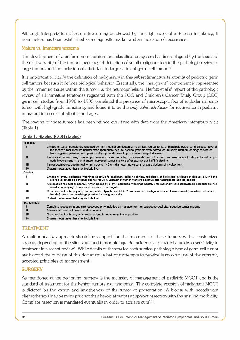
81 Consensus Document for Management of Pediatric Lymphomas and Solid Tumors
Although interpretation of serum levels may be skewed by the high levels of aFP seen in infancy, it nonetheless has been established as a diagnostic marker and an indicator of recurrence.
Mature vs. Immature teratoma
The development of a uniform nomenclature and classification system has been plagued by the issues of the relative rarity of the tumors, accuracy of detection of small malignant foci in the pathologic review of large tumors and the inclusion of adult data in large series of germ cell tumors6.
It is important to clarify the definition of malignancy in this subset (immature teratoma) of pediatric germ cell tumors because it defines biological behavior. Essentially, the “malignant” component is represented by the immature tissue within the tumor i.e. the neuroepithelium. Heifetz et al’s7 report of the pathologic review of all immature teratomas registered with the POG and Children’s Cancer Study Group (CCG) germ cell studies from 1990 to 1995 correlated the presence of microscopic foci of endodermal sinus tumor with high-grade immaturity and found it to be the only valid risk factor for recurrence in pediatric immature teratomas at all sites and ages.
The staging of these tumors has been refined over time with data from the American intergroup trials (Table 1).
Table 1. Staging (COG staging)
TREATMENT
A multi-modality approach should be adopted for the treatment of these tumors with a customized strategy depending on the site, stage and tumor biology. Schneider et al provided a guide to sensitivity to treatment in a recent review8. While details of therapy for each surgico-pathologic type of germ cell tumor are beyond the purview of this document, what one attempts to provide is an overview of the currently accepted principles of management.
SURGERY
As mentioned at the beginning, surgery is the mainstay of management of pediatric MGCT and is the standard of treatment for the benign tumors e.g. teratoma8. The complete excision of malignant MGCT is dictated by the extent and invasiveness of the tumor at presentation. A biopsy with neoadjuvant chemotherapy may be more prudent than heroic attempts at upfront resection with the ensuing morbidity. Complete resection is mandated eventually in order to achieve cure9,10.
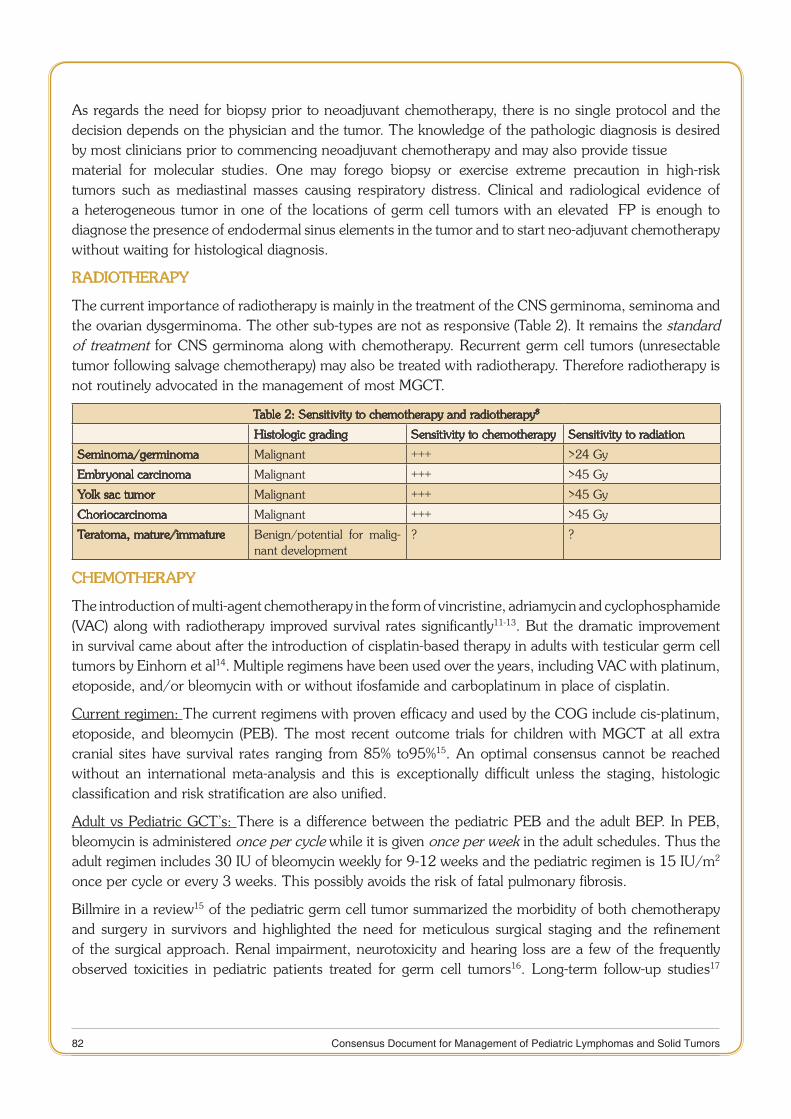
82 Consensus Document for Management of Pediatric Lymphomas and Solid Tumors
As regards the need for biopsy prior to neoadjuvant chemotherapy, there is no single protocol and the decision depends on the physician and the tumor. The knowledge of the pathologic diagnosis is desired by most clinicians prior to commencing neoadjuvant chemotherapy and may also provide tissue material for molecular studies. One may forego biopsy or exercise extreme precaution in high-risk tumors such as mediastinal masses causing respiratory distress. Clinical and radiological evidence of a heterogeneous tumor in one of the locations of germ cell tumors with an elevated αFP is enough to diagnose the presence of endodermal sinus elements in the tumor and to start neo-adjuvant chemotherapy without waiting for histological diagnosis.
RADIOTHERAPY
The current importance of radiotherapy is mainly in the treatment of the CNS germinoma, seminoma and the ovarian dysgerminoma. The other sub-types are not as responsive (Table 2). It remains the standard of treatment for CNS germinoma along with chemotherapy. Recurrent germ cell tumors (unresectable tumor following salvage chemotherapy) may also be treated with radiotherapy. Therefore radiotherapy is not routinely advocated in the management of most MGCT.
Table 2: Sensitivity to chemotherapy and radiotherapy8
Histologic grading Sensitivity to chemotherapy Sensitivity to radiation
Seminoma/germinoma Malignant +++ >24 Gy
Embryonal carcinoma Malignant +++ >45 Gy
Yolk sac tumor Malignant +++ >45 Gy
Choriocarcinoma Malignant +++ >45 Gy
Teratoma, mature/immature Benign/potential for malig-nant development
? ?
CHEMOTHERAPY
The introduction of multi-agent chemotherapy in the form of vincristine, adriamycin and cyclophosphamide (VAC) along with radiotherapy improved survival rates significantly11-13. But the dramatic improvement in survival came about after the introduction of cisplatin-based therapy in adults with testicular germ cell tumors by Einhorn et al14. Multiple regimens have been used over the years, including VAC with platinum, etoposide, and/or bleomycin with or without ifosfamide and carboplatinum in place of cisplatin.
Current regimen: The current regimens with proven efficacy and used by the COG include cis-platinum, etoposide, and bleomycin (PEB). The most recent outcome trials for children with MGCT at all extra cranial sites have survival rates ranging from 85% to95%15. An optimal consensus cannot be reached without an international meta-analysis and this is exceptionally difficult unless the staging, histologic classification and risk stratification are also unified.
Adult vs Pediatric GCT’s: There is a difference between the pediatric PEB and the adult BEP. In PEB, bleomycin is administered once per cycle while it is given once per week in the adult schedules. Thus the adult regimen includes 30 IU of bleomycin weekly for 9-12 weeks and the pediatric regimen is 15 IU/m2 once per cycle or every 3 weeks. This possibly avoids the risk of fatal pulmonary fibrosis.
Billmire in a review15 of the pediatric germ cell tumor summarized the morbidity of both chemotherapy and surgery in survivors and highlighted the need for meticulous surgical staging and the refinement of the surgical approach. Renal impairment, neurotoxicity and hearing loss are a few of the frequently observed toxicities in pediatric patients treated for germ cell tumors16. Long-term follow-up studies17
83 Consensus Document for Management of Pediatric Lymphomas and Solid Tumors
of men with testicular cancer have noted a two-fold increase in the risk of cardiovascular diseases and second malignancies (a rate of 50% by age 75).
Surgery only, is now accepted standard of care for stage I testicular tumors. An 85% success rate has been demonstrated and even those who relapse can be salvaged successfully with chemotherapy18-20. Those who relapse can be effectively treated with PEB regime.
To see whether the same standard can be applied to ovarian tumors, the most recent COG protocol (AGCT0132) was developed in 2003. It utilized the results of the last intergroup pediatric germ cell trial, INT-001621, to define the risk categories in pediatric germ cell tumors. Low-risk patients with stage I ovarian and testicular germ cell tumors were followed by surveillance after surgery. Intermediate-risk patients included stage I to II extragonadal, stage III ovarian, or stage III to IV testicular tumors and were given three courses of PEB. High-risk patients were those with stage IV ovarian tumors, stage III and IV extragonadal tumors and these got 4 courses of PEB. AGCT0132 was opened in November 2003 but enrollment into the low-risk stratum was stopped in January 2010 because of a significantly evidence lower 3 year EFS at less than 70% evidence with surveillance alone22. This was lower than expected. The standard surgical guidelines (Table 3) had been followed. The authors argued that a more thorough surgical approach (as for adults) would have detected occult disease and altered the assignment of stage I status. In this study, the salvage rate with chemotherapy was equivalent to the OS with upfront chemotherapy, and surgical morbidity was minimized. Therefore, it is now suggested that low-risk should include only stage I testicular tumors and not ovarian tumors. Stage I ovarian tumor should be regarded as intermediate-risk and treated as such.
The role of high-dose cisplatin: A few pediatric studies in the late 80’s used high-dose cisplatin and etoposide and reported acceptable toxicity23,24. This was supported by successful trials of high-dose cisplatin in adult testicular tumors25-28. A subsequent study29 failed to demonstrate a significant effect on outcome in those with advanced testicular tumors and showed a significant increase in toxicity. This established standard-dose cisplatin combined with bleomycin and either vinblastine or etoposide30-36 for adult testicular tumors.
As there are a multitude of differences between the adult and pediatric MGCT, a randomized trial to test the efficacy of cisplatin dose intensification on the outcome of high- risk patients was carried out by the POG and CCG. High-Dose PEB (HDPEB) significantly improved EFS for children with high-risk MGCT although the OS was similar in both regimen. Unfortunately, excessive toxic deaths and significant ototoxicity21 limited its utility. The audiograms of the patients in this study were subsequently reviewed by Li et al37 and graded using the NCI Common Toxicity Criteria and the Brock criteria38. They confirmed a higher incidence of ototoxicity in HDPEB-treated patients (67% of patients with HDPEB and 10.5% treated with PEB).
Cisplatin vs Carboplatin: It is established that the use of cisplatin carries with it a risk of late effects, including nephrotoxicity, neurotoxicity, ototoxicity, cardiovascular disease, and second malignancies39. A systematic review by Shaikh et al investigated the role of risk-adapted therapy. They hypothesized that optimal outcomes could be achieved by an approach that utilizes carboplatin in the setting of a young child with low-intermediate-risk features, and cisplatin in older children or those with high-risk features. They also acknowledged that due to the rarity of childhood MGCT’s, a randomized trial of this hypothesis would be difficult40. The UKCCSG has been using carboplatin instead of cisplatin (JEB regime) since 1989 with very good outcomes. However, they also suggest the use of standard PEB regime, instead of JEB, in adolescents.
84 Consensus Document for Management of Pediatric Lymphomas and Solid Tumors
Perinatal germ cell tumors
Most of the tumors found in this population are histologically benign and yolk sac tumor occurs alone or in combination with a teratoma very infrequently. The overall frequency of neonatal sacrococcygeal teratomas with yolk sac tumor is approximately 2.5% to 25%41. Despite the presence of immature neuroglial elements, patients with neonatal teratoma generally have a favorable outcome. The incidence of malignancy in the neonate is approximately 10% and approaches almost 100% by 3 years of age41,
42. As per POG study, surgery alone is curative in children and adolescents with immature teratomas of any grade, and that chemotherapy should be reserved for cases of relapse that are proven to be malignant7,43,44.
A fetus with a sacrococcygeal teratoma may develop hydrops. In the event that it happens after pulmonary maturity, delivery and standard postnatal resection are recommended. If occurring prior to lung maturity, fetal surgical intervention may be indicated45,46. Antenatal resection debulks and devascularizes the tumor, thus eliminating the large arteriovenous shunt through the tumor and effectively reversing the hydrops47,
48. Currently, there are no definite chemotherapy guidelines for neonates with teratomas49. Aziz Khan et al50 recommend chemotherapy only in infants with disseminated metastases (that have not differentiated) and those with invasive tumors and residual tumor after resection. Long-term follow-up with imaging and aFP monitoring is recommending.
Other presentations include epignathus and nasopharyngeal teratomas13, gastric teratomas51-53, intrapericardial and intra cardiac teratomas54-56. The common theme of management is contraindication of radical and disfiguring surgery, prevention of inappropriate intervention in those with poor prognosis and a regular, monitored follow-up.
Intraspinal extension
Neurological involvement due to an intraspinal extension in sacrococcygeal MGCT has rarely been reported and is usually representative of advanced disease. These patients have been found to respond to chemotherapy and surgical resection and most have complete neurological improvement57. A recent case series supported the need for neoadjuvant chemotherapy for all patients of MGCT with intraspinal extension and mandatory coccygectomy to reduce the recurrence rate.
RECURRENCE AND SALVAGE THERAPY
The treatment of recurrent pediatric GCT is based primarily on anecdotal reports and is yet to be studied systematically. Recurrence may be benign or malignant and local or distant and the first-line treatment has a bearing on the choice of salvage therapy8. The Children Cancer Group GCT study showed an 11% tumor recurrence rate with mature sacrococcygeal teratomas and a 4% recurrence with immature ones58. These should be treated with surgery as chemotherapy does not have much of an effect in benign tumors. Recurrence was detected at even 34 months post-resection and underscored the need for long-term follow-up. aFP determinations and imaging studies are recommended for at least 3 years after diagnosis.
A combined POG/CCG intergroup germ cell tumor study recommended treating all infants with recurrent teratomas containing yolk sac tumor on the high-risk protocol with cisplatin, etoposide, and bleomycin28. They demonstrated an OS and EFS of 90% and 84%, respectively, after surgical resection and a high-dose cisplatinum regimen. The 5-year overall OS rate in the pediatric population is estimated to be higher than 80%, all stages and locations being considered59,60. It is impossible to apply the principles learned
85 Consensus Document for Management of Pediatric Lymphomas and Solid Tumors
from adult studies to children as the genetic anomalies are different61. One study focussing on recurrent pediatric NSGCT has been published so far by the German group of MAKEI. In this study conducted in 22 relapsing patients with sacrococcygeal NSGCTs, the latest relapse occurred 26 months after initial diagnosis and at least one local tumor recurred in 20 patients62. They showed that the complete aggressive resection of a recurrent tumor had prognostic value and accounted for the better 5-year EFS and OS (30% and 42%, respectively). In case only partial resection is possible due to anatomic limitations, local radiotherapy should be added.
Chemotherapy protocols for recurrent or refractory MGCT are yet to be established. Some data is available on the use of paclitaxel to treat pediatric MGCT and a phase II trial of the Children Oncology Group is currently testing paclitaxel, ifosfamide and carboplatin in combination for recurrent or refractory MGCT in patients younger than 21 years (COG-AGCT0521). A French study recommended that overall, as long as the initial cumulative dose is in the safe range, a cisplatin- based regimen may still be used for salvage. UKCCSG also has recommended using vinblastine+ Ifosfamide+ cisplatin (VIP) for relapsed cases. The other regimen suggested ICE (Ifosfamide+carboplatin+ etoposide). The renal and otological toxicity due to a high cumulative dose of cisplatin must always be kept in mind63. They also suggested a consideration of high-dose cisplatin chemotherapy in select patients, but stressed the need for multi-institutional and international randomized trials.
Retroperitoneal lymph node dissection (RPLND) is not a part of standard pediatric MGCT treatment, while it is de-rigueur in the therapy of adolescent and adult GCT. An awareness of the differences between the pediatric, adolescent and adult germ cell tumors64 is essential to guide salvage therapy.
RECOMMENDATIONS
Investigations for diagnostic work-up and follow-up:
Clinical work-up: history and examination1.
Imaging: ultrasound of the involved site, X-ray chest, CECT of the involved site and chest. MRI in 2. cases with suspected intraspinal extension, especially tumors in the mediastinum and sacrococcygeal region.
Tumor markers: αaFP, 3. bhCG (remember to look at the aFP normogram for infants: Table 3). LDH is a non-specific marker of tumor burden.
Pathology: Fine-needle aspiration cytology (FNAC) or core needle biopsy (required only if aFP is not 4. elevated and neoadjuvant chemotherapy is being planned). Cases undergoing upfront resection would have enough material for pathological study. In intra-abdominal primaries, such as those abutting the liver, a biopsy may be essential to differentiate from hepatoblastoma. At all other sites, when the aFP is elevated, it indicates the presence of yolk sac elements and so a biopsy is not mandatory.
Follow-up with aFP and imaging (ultrasound alternating with CECT).5.
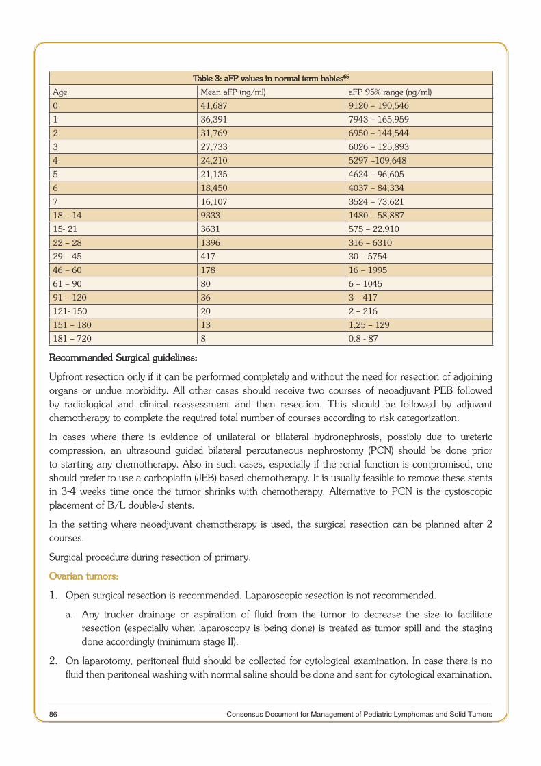
86 Consensus Document for Management of Pediatric Lymphomas and Solid Tumors
Table 3: aFP values in normal term babies65
Age Mean aFP (ng/ml) aFP 95% range (ng/ml)
0 41,687 9120 – 190,546
1 36,391 7943 – 165,959
2 31,769 6950 – 144,544
3 27,733 6026 – 125,893
4 24,210 5297 –109,648
5 21,135 4624 – 96,605
6 18,450 4037 – 84,334
7 16,107 3524 – 73,621
18 – 14 9333 1480 – 58,887
15- 21 3631 575 – 22,910
22 – 28 1396 316 – 6310
29 – 45 417 30 – 5754
46 – 60 178 16 – 1995
61 – 90 80 6 – 1045
91 – 120 36 3 – 417
121- 150 20 2 – 216
151 – 180 13 1,25 – 129
181 – 720 8 0.8 - 87
Recommended Surgical guidelines:
Upfront resection only if it can be performed completely and without the need for resection of adjoining organs or undue morbidity. All other cases should receive two courses of neoadjuvant PEB followed by radiological and clinical reassessment and then resection. This should be followed by adjuvant chemotherapy to complete the required total number of courses according to risk categorization.
In cases where there is evidence of unilateral or bilateral hydronephrosis, possibly due to ureteric compression, an ultrasound guided bilateral percutaneous nephrostomy (PCN) should be done prior to starting any chemotherapy. Also in such cases, especially if the renal function is compromised, one should prefer to use a carboplatin (JEB) based chemotherapy. It is usually feasible to remove these stents in 3-4 weeks time once the tumor shrinks with chemotherapy. Alternative to PCN is the cystoscopic placement of B/L double-J stents.
In the setting where neoadjuvant chemotherapy is used, the surgical resection can be planned after 2 courses.
Surgical procedure during resection of primary:
Ovarian tumors:
Open surgical resection is recommended. Laparoscopic resection is not recommended.1.
Any trucker drainage or aspiration of fluid from the tumor to decrease the size to facilitate a. resection (especially when laparoscopy is being done) is treated as tumor spill and the staging done accordingly (minimum stage II).
On laparotomy, peritoneal fluid should be collected for cytological examination. In case there is no 2. fluid then peritoneal washing with normal saline should be done and sent for cytological examination.
87 Consensus Document for Management of Pediatric Lymphomas and Solid Tumors
This is a must for the accurate staging. Ovarian primaries with un-sampled ascites are treated as stage III.
Para-aortic lymph nodes should be biopsied, if enlarged. RPLN dissection is not required.3.
Any peritoneal or omental deposits should be biopsied. Omentectomy is not required, only portions 4. of omentum adherent to the tumor need to be resected.
Contralateral ovarian biopsy should be performed only in cases of suspicious nodule as seen on pre-5. operative imaging (ultrasound or CECT) or when detected intra-operatively.
In cases of obvious bilateral ovarian tumors, wedge biopsy of one should be performed and the patient 6. should have a second-look surgery after neo-adjuvant chemotherapy when conservative surgery may be possible. If the patient is a known case of XY Disorders of sexual differentiation (DSD), bilateral oophorectomy should be performed.
Testicular tumors:
The recommended procedure is high ligation and removal of testis through an inguinal route. 1. Proper RPLN and pulmonary evaluation radiologically is a must for the appropriate staging of the disease.
Trans-scrotal violation is to be avoided. 2.
In cases where the trans - scrotal biopsy has been performed, these should then be treated as a. stage II (intermediate-risk) and therefore get three courses of PEB. There is no indication for hemiscrotectomy.
If trans-scrotal orchiectomy has been done and spermatic cord is free of tumor, then treat b. as stage I; however, if the spermatic cord shows tumor infiltration, then perform an inguinal incision and remove the remaining spermatic cord and treat as minimum stage II. Hemi-scrotectomy is not required.
Retroperitoneal LN dissection is not required:3.
All retroperitoneal lymph nodes that are >1cm on the pre-chemotherapy CECT scan should a. be regarded as positive and patients staged as stage III. Therefore, should get adjuvant chemotherapy as for intermediate-risk (3 courses of PEB).
If RPLN are <1 cm and baseline aFP is not raised, then these require to be biopsied for b. appropriate staging.
If RPLN are <1 cm and the baseline aFP is raised, then post-operative follow-up with aFP. If c. these fail to decline, according to log fall table, then these should be treated as stage III and requisite chemotherapy given without resorting to RPLN biopsy.
Sacrococcygeal tumors:
Neonates: Complete surgical excision with coccygectomy should be performed. As most are 1. mature/immature teratomas, no adjuvant chemotherapy is required. For very large tumors consider laparotomy and control median sacral vessels and resection through a combination of laparotomy and posterior routes. Follow-up with regular serum αaFP every 2-3 months for at least 3 years as nearly 15% may recur. Radiological follow-up with ultrosound and/or CT is also required as the recurrence may not be secreting aFP.
88 Consensus Document for Management of Pediatric Lymphomas and Solid Tumors
Older children: These will invariably be malignant and most may not be resectable at presentation 2. (without mutilating surgery). If aFP is elevated, initial biopsy is not mandatory. Chemotherapy should be initiated and surgery planned at the end of 2nd or 3rd course, when mutilating surgery can be avoided. In cases where the tumor completely disappears following chemotherapy, a coccygectomy should anyway be done.
Mediastinal tumors:
The risk of general anesthesia should be properly assessed for mediastinal tumors. If high-risk, the 1. diagnostic biopsy should be performed under local anesthesia with ultrasound or under CT guidance. In cases where tumor markers are raised, biopsy should be avoided.
Upfront resection should only be attempted in cases with normal serum markers or if it can be 2. performed completely and without the need for resection of adjoining organs or undue morbidity. Regional nodes should be biopsied.
All other cases (with raised markers) should receive two courses of neoadjuvant PEB followed by 3. radiological and clinical reassessment and then resection of residual tumor. The UKCCSG suggests resection of the residual tumor at the end of planned chemotherapy.
Recommended Chemotherapy guidelines:
Staging and risk categorization should be done to decide regarding the chemotherapy.1.
PEB is recommended as the standard regimen. However JEB may also be used as an alternative, 2. especially in cases with deranged renal functions.
Recommended risk categorization and chemotherapy are as follows*:3.
Low-Risk: Stage I testicular tumors and all immature teratomasa.
Intermediated Risk: Stage II-IV testicular tumors, Stage I-III Ovarian tumors and Stage I-II b. extragonadal tumors. Stage I testicular tumors showing rising aFP in post-operative period.
High-Risk: Stage IV ovarian tumors, Stage III-IV extragonadal tumors.c.
*Risk categorization used in UKCCSG is slightly different.
Chemotherapy recommended is as follows:4.
Low-Risk: No chemotherapya.
Intermediate-Risk: PEB x 3 courses (4 courses of JEB in UKCCSG)b.
High-Risk: PEB x 4 courses # (6 courses of JEB in UKCCSG)c.
#- additional two courses may of PE (without bleomycin) to be considered for sacrococcygeal tumors and RP tumors.
Administration of chemotherapy:
PEB: Day 1: Cisplatin 35 mg/m2 IV infusion over 1 hr* + Etoposide 120mg/m2 IV over 1 hr
Day 2: same as day 1
Day 3: Same as day 1 + Bleomycin# 15 mg/m2 IV over 30 minutes
*ensure per hydration and post hydration with 125ml/m2 of N/2 saline

89 Consensus Document for Management of Pediatric Lymphomas and Solid Tumors
JEB: Day 1: Etoposide 120 mg/m2 in 5% dextrose normal saline over 1 hour
Day 2: Etoposide as above + Carboplatin 600mg/m2*
Day 3: Etoposide as above + Bleomycin# 15mg/m2 IV over 30 minutes
As Bleomycin toxicity is enhanced in patients with renal impairment (Glomerular Filtration Rate (GFR) < 60/min/1.73m2), it should be omitted and replaced with vincristine 1.5 mg/ m2 IV bolus (max 2 mg) on day 3. Once renal impairment recovers, Bleomycin can be reintroduced.
For children < 6months of age the dose should be reduced to 50% of the calculated and for those between 6 months and 1 year it should be 75% of the calculated in both PEB and JEB regimen.
Refrence:
Jain V, Agarwala S, Bakhshi et al. Malignant germ cell tumors in children: Management and outcomes from the AIIMS-1. MGCT 94 trial. (Abstract). Pediatr Blood Cancer 2009; 53: 732.
Lo Curto M, Lumia F, Alaggio R et al. Malignant germ cell tumors in childhood: results of the first Italian cooperative 2. study “TCG 91.” Med PediatrOncol 2003;41:417-25.
Oosterhuis JW, Looijenga LH. Testicular germ cell tumors in a broader perspective. Nat Rev Cancer 2005; 5:210–22. 3.
Schneider DT, Calaminus G, Koch S et al. Epidemiologic analysis of 1,442 children and adolescents registered in the 4. German germ cell tumor protocols. Pediatr Blood Cancer 200; 42:169–175.
Teilum G. Classification of endodermal sinus tumor (mesoblastomavitellinum) and so-called “embryonal carcinoma” of 5. the ovary. ActaPatholMicrobiolScand 1965;64:407-29.
Göbel U, Schneider DT, Calaminus G et al. Germ cell tumors in childhood and adolescence. Ann Oncol 2000;11:263-6. 71.
Heifetz SA, Cushing B, Giller R et al. Immature teratomas in children: pathologic considerations: a report from the 7. combined Pediatric Oncology Group/Children’s Cancer Group. Am J SurgPathol1998;22:1115e24.
Schneider, Dominik T et al. Gonadal and extragonadal germ cell tumors, sex cord stromal and rare gonadal tumors.8. Rare TumorsIn Children and Adolescents. Springer Berlin Heidelberg, 2012. 327-402.
Schneider, Dominik T et al. Primary mediastinal germ cell tumors in children and adolescents: results of the German 9. cooperative protocols MAKEI 83/86, 89, and 96. J ClinOncol 2000; 18.4: 832.
Göbel U, Schneider DT, Calaminus G et al. Multimodal treatment of malignant sacrococcygeal germ cell tumors: a 10. prospective analysis of 66 patients of the German cooperative protocols MAKEI 83/86 and 89. J ClinOncol2001;19(7), 1943-50.
Mulligan RM. Pathogenesis of teratoidtumors of the ovary and testis. PatholAnnu1975; 10:271-98.11.
Kurman RJ, Norris HJ. Endodermal sinus tumor of the ovary: A clinical and pathologic analysis of 71 cases. Cancer 12. 1976; 38:2404-19.
Chretien PB, Milam JD, Foote FW, et al. Embryonal adenocarcinomas (a type of malignant teratoma) of the sacrococcygeal 13. region: Clinical and pathologic aspects of 21 cases. Cancer 1970; 26:522-35.
EinhornLH, Donohue J.Cisdiamminedichloroplatinum, vinblastine, and bleomycin combination chemotherapy in 14. disseminated testicular cancer. Ann Intern Med 1977; 87:293-8.
Billmire DF (2006, February). Malignant germ cell tumors in childhood. In 15. Seminars in pediatric surgery (Vol. 15, No. 1, pp. 30-36). WB Saunders.
Hale GA, Marina NM, Jones-Wallace D et al. Late effects of treatment for germ cell tumors during childhood and 16. adolescence. J PediatrHematolOncol 1999; 21:115-122.
Travis LB, Beard C, Allan JM, et al: Testicular cancer survivorship: Research strategies and recommendations. J Natl 17. Cancer Inst2010; 102:1114-30.
Mann JR, Raafat F, Robinson K et al. The United Kingdom Children’s Cancer Study Group’s second germ cell tumor 18. study: carboplatin, etoposide, and bleomycin are effective treatment for children with malignant extracranial germ cell tumors, with acceptable toxicity. J ClinOncol 2000;18:3809-18.
90 Consensus Document for Management of Pediatric Lymphomas and Solid Tumors
Schmidt P, Haas RJ, Göbel U et al. Results of the German studies (MAHO) for treatment of testicular germ cell tumors 19. in children-an update. KlinPadiatr 2002;214:167-72.
Schlatter M, Rescorla F, Giller R et al. Excellent outcome in patients with stage I germ cell tumors of the testes: a study 20. of the Children’s Cancer Group/Pediatric Oncology Group. J PediatrSurg 2003;38:319-24.
Cushin21. g, B, Giller R, Cullen JW et al. Randomized comparison of combination chemotherapy with etoposide, bleomycin, and either high-dose or standard-dose cisplatin in children and adolescents with high-risk malignant germ cell tumors: a pediatric intergroup study—Pediatric Oncology Group 9049 and Children's Cancer Group 8882.J ClinOncol 2004; 22(13): 2691-700.
Billmire DF, Cullen JW, Rescorla FJ et al. Surveillance after initial surgery for pediatric and adolescent girls with stage I 22. ovarian germ cell tumors: Report from the Children's Oncology Group. J ClinOncol 2014; 32(5): 465-70.
Hartmann O, Pinkerton CR, Philip T et al. Very-high-dose cisplatin and etoposide in children with untreated advanced 23. neuroblastoma. J ClinOncol 1988;6:44-50.
Perin G, Dallorso S, Stura M et al. High-dose cisplatin and etoposide in advanced malignancies of childhood. 24. PediatrHematolOncol1987; 4:329-36.
Ozols RF, Deisseroth AB, Javadpour N et al. Treatment of poor prognosis nonseminomatous testicular cancer with a 25. “high-dose” platinum combination chemotherapy regimen. Cancer 1983; 51:1803-7.
Ozols RF, Ihde DC, Linehan WM et al. A randomized trial of standard chemotherapy v a high-dose chemotherapy 26. regimen in the treatment of poor prognosis nonseminomatous germ cell tumors. J ClinOncol1988; 6:1031-40.
Hayes DM, Cvitkovic E, Golbey RB, et al. High-dose cis-platinum diammine dichloride: Amelioration of renal toxicity by 27. mannitol diuresis.Cancer 1977; 39:1372-81.
Ghosn M, Droz JP, Theodore C et al. Salvage chemotherapy in refractory germ cell tumors with etoposide (VP-16) plus 28. ifosfamide plus high-dose cisplatin: A VIhP regimen. Cancer 1988; 62:24-7.
Nichols CR, Williams SD, Loehrer PJ et al. Randomized study of cisplatin dose intensity in poor-risk germ cell tumors: 29. A Southeastern Cancer Study Group and Southwest Oncology Group protocol. J ClinOncol1991; 9:1163-72.
Einhorn LH, Williams SD: Chemotherapy of disseminated testicular cancer. West J Med 1979; 131:1-3.30.
Williams SD, Blessing JA, Moore DH et al. Cisplatin, vinblastine, and bleomycin in advanced and recurrent ovarian germ 31. cell tumors: A trial of the Gynecologic Oncology Group. Ann Intern Med 1989; 111:22-7.
Einhorn LH. Chemotherapy of disseminated germ cell tumors. Cancer 1987; 60:570-3.32.
Israel A, Bosl GJ, Golbey RB et al. The results of chemotherapy for extragonadalgermcelltumors in the cisplatin era: The 33. Memorial Sloan-Kettering Cancer Center experience (1975 to 1982). J ClinOncol1985; 3:1073-8.
Pizzocaro G, Salvioni R, Pasi M et al. Early resection of residual tumor during cisplatin, vinblastine, bleomycin combination 34. chemotherapy in stage III and bulky stage II nonseminomatous testicular cancer. Cancer 1985; 56:249-55.
Carlson RW, Sikic BI, Turbow MM et al. Combination cisplatin, vinblastine, and bleomycin chemotherapy (PVB) for 35. malignant germ cell tumors of the ovary. J ClinOncol1983; 1:645-51.
Logothetis CJ, Samuels ML, Selig DE et al. Chemotherapy of extragonadal germ cell tumors. J ClinOncol1985; 3:316-36. 25.
Li Y, Womer RB, Silber JH. Predicting cisplatin ototoxicity in children, the influence of age and the cumulative dose. Eur 37. J Cancer. Online article. http://ejconline.com.
Brock PR, Bellman SC, Yeomans EC et al. Cisplatin ototoxicity in children: A practical grading system. Med 38. PediatrOncol1991; 19:295-300.
Bertolini P, Lassalle M, Mercier G et al. Platinum compound-related ototoxicity in children: Long-term follow-up reveals 39. continuous worsening of hearing loss. J PediatrHematolOncol 2004; 26:649–55.
Shaikh F, Nathan PC, Hale F. Is There a Role for Carboplatin in the Treatment of Malignant Germ Cell Tumors? A 40. Systematic Review of Adult and Pediatric Trials. Pediatr Blood Cancer 2013;60:587–92.
Isaacs H Jr: Germ cell tumors. Tumors of the Fetus and Newborn. Vol. 35 in the series Major Problems in Pathology. 41. Philadelphia, PA, Saunders, 1997, pp 15-38.
91 Consensus Document for Management of Pediatric Lymphomas and Solid Tumors
Dehner LP: Neoplasms of the fetus and neonate, in Naeye RL, Kissane JM, Kaufman N (eds): Perinatal Diseases, 42. International Academy of Pathology, Monograph number 22. Baltimore, MD, Williams and Wilkins, 1981, pp 286-345.
Cushing B, Giller R, Ablin A, et al.43. Surgical resection alone is effective treatment for ovarian immature teratoma in children and adolescents: a report of the Pediatric Oncology Group and the Children’s Cancer Group. Am J ObstetGynecol1999;181:353e8.
Marina NM, Cushing B, Giller R, et al. Complete surgical excision is effective treatment for children with immature 44. teratomas with or without malignant elements: a Pediatric Oncology Group/Children’s Cancer Group Intergroup Study. J ClinOncol1999;17:2137e43.
Adzick NS, Crombleholme TM, Morgan MA et al. A rapidly growing fetalteratoma. Lancet 1997; 349:538. 45.
Graf JL, Albanese CT, Jennings RW et al. Successful fetalsacrococcygealteratoma resection in a hydropicfetus. 46. J PediatrSurg 2000;35:1489-91.
Isaacs H Jr: Tumors, in Gilbert-Barness E (ed): Potter’s Pathology of the Fetus and Infant, Vol 2. St Louis, MO, Mosby, 47. 1997, pp1242-1339.
Isaacs H. Perinatal (fetaland neonatal) germ cell tumors.JPedSurg 2004;39.7: 1003-13.48.
Flake AW: Fetalsacrococcygealteratoma. SeminPediatrSurg1993; 2:113-120.49.
Azizkhan RG, Haase GM, Applebaum H et al. Diagnosis, management, and outcome of cervicofacialteratomas in 50. neonates: A Childrens Cancer Group Study. J PediatrSurg1995; 30:312-6.
Esposito G, Cigliano B, PaludettoR.Abdominothoracic gastric teratoma in a female newborn infant. J PediatrSurg1983; 51. 18:304-5.
Haley T, Dimler M, Hollier P. Gastric teratoma with gastrointestinal bleeding. J PediatrSurg1986; 21:949-50. 52.
Gengler JS, Ashcraft JW, Slattery P. Gastric teratoma: The sixth reported case in a female infant. J PediatrSurg1995; 53. 30:889-90.
Van der Hauwaert LG. Cardiac tumors in infancy and childhood. Br Heart J 1971; 33:125-32.54.
Costas C, Williams RL, Fortune RL.Intracardiacteratoma in an infant. PediatrCardiol1986; 7:179-81.55.
Bruch SW, Adzick NS, Reiss R et al. Prenatal therapy for pericardial teratomas. J PediatrSurg1997; 32:1113-5. 56.
Garg R, AgarwalaS et al. Sacrococcygeal malignant germ cell tumor (SC-MGCT) with intraspinalextension.J 57. PedatrSurg2014; 49.7: 1113- 5.
Rescoria F, Sawin RS, Coran AG et al. Long-term outcome for infants and children with sacrococcygealteratoma: A 58. report from the Childrens Cancer group. J PediatrSurg 1998; 33:171-6.
Göbel U, Schneider DT, Calaminus G et al. Germ cell tumors in childhood and adolescence. GPOH MAKEI and the 59. MAHO study groups. Ann Oncol 2000; 11:263–71.
Marina N, London WB, Frazier AL et al. Prognostic factors in children with extragonadal malignant germ cell tumors: A 60. pediatric intergroup study. J ClinOncol 2006; 24:2544-8.
Bussey KJ, Lawce HJ, Olson SB et al. Chromosome abnormalities of eighty one pediatric germ cell tumors: Sex-, age-, 61. site-, and histopathology-related differences—A Children’s Cancer Group study. Genes Chromosomes Cancer 1999; 25:134–46.
Schneider DT, Wessalowski R, Calaminus G et al. Treatment of recurrent malignant sacrococcygeal germ cell tumors: 62. Analysis of 22 patients registered in the German protocols MAKEI 83/86, 89 and 96. J ClinOncol 2001; 19:1951–60.
Faure‐Conter63. C, Orbach D, Cropet C et al.Salvage therapy for refractory or recurrent pediatric germ cell tumors: The french SFCE experience. Pediatr Blood Cancer 2014;61(2), 253-59.
Cost NG, Lubahn JD, Adibi M et al. A comparison of pediatric, adolescent, and adult testicular germ cell malignancy. 64. Pediatr Blood Cancer 2014; 61(3), 446-51.
Blohm, M. E. G., Vesterling-Hörner D, Calaminus G, Göbel U. Alpha1-fetoprotein (AFP) as a reference values in infants 65. up to 2 years of age. Pediatr Hematol Oncol 1998;15(2):135-142.

92 Consensus Document for Management of Pediatric Lymphomas and Solid Tumors
CHAPTER
5 HEPATOBLASTOMA
Introduction
In the past three decades astonishing progress has been made in the management of children with hepatoblastoma (HB). These include highly effective neoadjuvant chemotherapeutic agents, better understanding of the surgical anatomy of the liver leading to less risky resections, better anesthetic and post- operative care and the availability of liver transplantation (LTx) for these patients. The outlook of these children has improved from a 100% mortality rate to an approximately 60% 5-year OS. Although improvement in survival is multifactorial, complete resection remains fundamental in optimizing outcome. A thoroughly planned complete surgical resection should be strongly considered in the absence of metastatic disease. First and foremost credit of this “catching up” survival in hepatoblastoma goes to the various cooperative groups active in various parts of the world.
The context in developing countries like India is slightly off this track. In India, many children present late, following the diagnostic delay or difficult access to the appropriate medical facility. Associated comorbidities such as malnutrition often further compromise treatment. To overcome these issues, an attempt has been made by the SIOPEL group (SIOPEL RCN, described later) to cater to the needs of these resources-challenged nations.
Existing guidelines
The existing guidelines are basically based on the recommendations of one of the following cooperative groups:
International society of pediatric oncology – liver tumor strategy group – SIOPEL guidelines. •
Children Oncology Group-COG guidelines •
German Pediatric Hematology Oncology Group – GPOH guidelines •
Japanese Pediatric liver tumor study group – JPLT guidelines •
Of all these guidelines, the SIOPEL guidelines for staging, diagnostic work-up and management are the ones that are most extensively followed all over the world. These guidelines have been made and refined over the past two decades through well conducted multicenter, randomized controlled trials.
Review of Existing literature
All the advances made in improving the outlook for children with hepatoblastoma have stemmed from contributions by mainly four cooperative groups which have conducted trials on a large scale to come to certain conclusions. Recently, all four of these multicenter groups collaborated into an international group CHIC (Children’s Hepatic Tumors International Collaboration) to focus on international global cooperation for investigations of pediatric malignant hepatic tumors, including hepatoblastoma. Risk stratification in these trials was based on individual special classification of stage, metastasis and histology
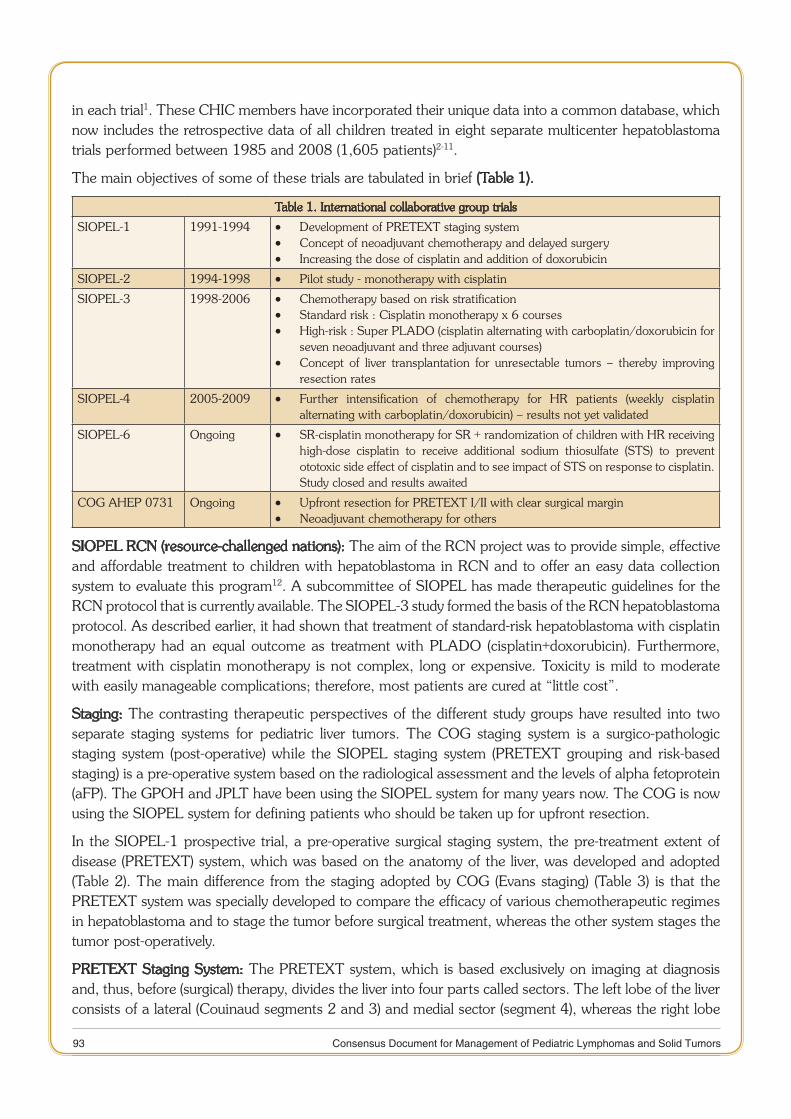
93 Consensus Document for Management of Pediatric Lymphomas and Solid Tumors
in each trial1. These CHIC members have incorporated their unique data into a common database, which now includes the retrospective data of all children treated in eight separate multicenter hepatoblastoma trials performed between 1985 and 2008 (1,605 patients)2-11.
The main objectives of some of these trials are tabulated in brief (Table 1).
Table 1. International collaborative group trials
SIOPEL-1 1991-1994 Development of PRETEXT staging system •Concept of neoadjuvant chemotherapy and delayed surgery •Increasing the dose of cisplatin and addition of doxorubicin •
SIOPEL-2 1994-1998 Pilot study - monotherapy with cisplatin •SIOPEL-3 1998-2006 Chemotherapy based on risk stratification •
Standard risk : Cisplatin monotherapy x 6 courses •High-risk : Super PLADO (cisplatin alternating with carboplatin/doxorubicin for •seven neoadjuvant and three adjuvant courses)Concept of liver transplantation for unresectable tumors – thereby improving •resection rates
SIOPEL-4 2005-2009 Further intensification of chemotherapy for HR patients (weekly cisplatin •alternating with carboplatin/doxorubicin) – results not yet validated
SIOPEL-6 Ongoing SR-cisplatin monotherapy for SR + randomization of children with HR receiving •high-dose cisplatin to receive additional sodium thiosulfate (STS) to prevent ototoxic side effect of cisplatin and to see impact of STS on response to cisplatin. Study closed and results awaited
COG AHEP 0731 Ongoing Upfront resection for PRETEXT I/II with clear surgical margin •Neoadjuvant chemotherapy for others •
SIOPEL RCN (resource-challenged nations): The aim of the RCN project was to provide simple, effective and affordable treatment to children with hepatoblastoma in RCN and to offer an easy data collection system to evaluate this program12. A subcommittee of SIOPEL has made therapeutic guidelines for the RCN protocol that is currently available. The SIOPEL-3 study formed the basis of the RCN hepatoblastoma protocol. As described earlier, it had shown that treatment of standard-risk hepatoblastoma with cisplatin monotherapy had an equal outcome as treatment with PLADO (cisplatin+doxorubicin). Furthermore, treatment with cisplatin monotherapy is not complex, long or expensive. Toxicity is mild to moderate with easily manageable complications; therefore, most patients are cured at “little cost”.
Staging: The contrasting therapeutic perspectives of the different study groups have resulted into two separate staging systems for pediatric liver tumors. The COG staging system is a surgico-pathologic staging system (post-operative) while the SIOPEL staging system (PRETEXT grouping and risk-based staging) is a pre-operative system based on the radiological assessment and the levels of alpha fetoprotein (aFP). The GPOH and JPLT have been using the SIOPEL system for many years now. The COG is now using the SIOPEL system for defining patients who should be taken up for upfront resection.
In the SIOPEL-1 prospective trial, a pre-operative surgical staging system, the pre-treatment extent of disease (PRETEXT) system, which was based on the anatomy of the liver, was developed and adopted (Table 2). The main difference from the staging adopted by COG (Evans staging) (Table 3) is that the PRETEXT system was specially developed to compare the efficacy of various chemotherapeutic regimes in hepatoblastoma and to stage the tumor before surgical treatment, whereas the other system stages the tumor post-operatively.
PRETEXT Staging System: The PRETEXT system, which is based exclusively on imaging at diagnosis and, thus, before (surgical) therapy, divides the liver into four parts called sectors. The left lobe of the liver consists of a lateral (Couinaud segments 2 and 3) and medial sector (segment 4), whereas the right lobe
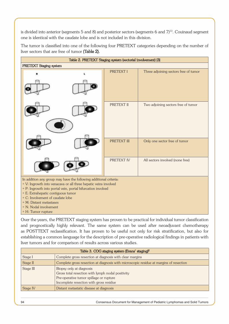
94 Consensus Document for Management of Pediatric Lymphomas and Solid Tumors
is divided into anterior (segments 5 and 8) and posterior sectors (segments 6 and 7)13. Couinaud segment one is identical with the caudate lobe and is not included in this division.
The tumor is classified into one of the following four PRETEXT categories depending on the number of liver sectors that are free of tumor (Table 2).
Table 2. PRETEXT Staging system (sectorial involvement) [3]
PRETEXT Staging system
PRETEXT I Three adjoining sectors free of tumor
PRETEXT II Two adjoining sectors free of tumor
PRETEXT III Only one sector free of tumor
PRETEXT IV All sectors involved (none free)
In addition any group may have the following additional criteria:+ V: Ingrowth into venacava or all three hepatic veins involved+ P: Ingrowth into portal vein, portal bifurcation involved+ E: Extrahepatic contiguous tumor+ C: Involvement of caudate lobe+ M: Distant metastases+ N: Nodal involvement+ H: Tumor rupture
Over the years, the PRETEXT staging system has proven to be practical for individual tumor classification and prognostically highly relevant. The same system can be used after neoadjuvant chemotherapy as POSTTEXT reclassification. It has proven to be useful not only for risk stratification, but also for establishing a common language for the description of pre-operative radiological findings in patients with liver tumors and for comparison of results across various studies.
Table 3. COG staging system (Evans’ staging)2
Stage I Complete gross resection at diagnosis with clear margins
Stage II Complete gross resection at diagnosis with microscopic residue at margins of resection
Stage III Biopsy only at diagnosisGross total resection with lymph nodal positivityPre-operative tumor spillage or ruptureIncomplete resection with gross residue
Stage IV Distant metastatic disease at diagnosis

95 Consensus Document for Management of Pediatric Lymphomas and Solid Tumors
Risk stratification
Risk-based stratification for management as well as prognostication has been adopted by all the study groups as tabulated (Table 4).
Table 4. Current risk stratification systems of different study groups
COG Very low-risk Stage I, PFH
Low-risk Stage I, non PFH/non SCUStage II, non SCU
Intermediate-risk Stage I/II + SCUStage III
High-risk Stage IVAny stage + aFP<100ng/ml
SIOPEL/GPOH Standard risk PRETEXT I/II/III + aFP > 100 ng/ml + no additional criteria
High-risk PRETEXT IVAny PRETEXT + aFP < 100ng/mlAny PRETEXT + additional criteria (E,V,P,M,N,H)
JPLT PRETEXT I, no additional criteria
PRETEXT II, no additional criteria
PRETEXT III/IV or any PRETEXT + additional criteria (E,V,P,M,H,N)
PFH=pure fetal histology ; SCU=small cell undifferentiated histology ; E=extrahepatic contiguous spread ; V=vena cava or all three hepatic vein involvement ; P=portal vein bifurcation, main portal vein or both portal vein involvement ; M=distant metastasis ; N=positive lymph nodes ; H=tumor rupture
Management: In the United States, the protocol of the current COG study AHEP 0731 recommends initial surgery for all children with a liver tumor. A primary resection should be undertaken for limited PRETEXT I and II tumors with at least 1 cm of clear margins, whereas those tumors with a larger extension (PRETEXT III, IV), vascular invasion or distant metastases should be treated with neoadjuvant chemotherapy. The result of primary surgery determines the tumor’s stage, according to the Evans system. All patients are treated with adjuvant chemotherapy, according to POSTTEXT classification. The only exemptions are patients with a completely resected (stage I) hepatoblastoma with pure fetal histology who do not receive any chemotherapy, because these were found to have a 100% cure rate with surgery alone. Thus, the COG study AHEP 0731 intends to differentially adapt treatment by stratifying patients into four different risk groups, reducing the intensity of chemotherapy in approximately 30% of the patients.
In contrast to the United States, the international SIOPEL group and the German GPOH group do not recommend primary surgery and resection of hepatoblastoma. This is followed in Europe, South America and most of Asia-Pacific region. These recommendations are based on the response rate of approximately 90% for these tumors to neoadjuvant therapy, which not only makes the tumors smaller and less risky to resect but potentially can also suppress occult micro-metastases without delay. When one uses this strategy, resections of hepatoblastoma become easier and safer in a setting in which many centers with different expertise treat these patients. Finally, the GPOH group observed that hepatic surgery alone with induction of liver regeneration may also promote the growth of residual tumor and metastases not pre-treated with chemotherapy by secretion of so-called liver growth factors14. Therefore, neo-adjuvant chemotherapy is recommended for more or less all hepatoblastoma’s.
In the Japanese JPLT group, PRETEXT I hepatoblastoma’s without other additional PRETEXT criteria are primarily resected, and all others are pre-treated with 2-3 courses of chemotherapy according to tumor extension and response5.

96 Consensus Document for Management of Pediatric Lymphomas and Solid Tumors
Thus far, no controlled comparison has been performed between these two treatment strategies. However, the present survival rates of patients with tumors not eligible for up front resection reported by the different study groups are comparable regardless of the first therapeutic modality used.
Chemotherapy: The evolution of various chemotherapy regimens for the treatment of hepatoblastoma has been governed by differing principles over time. The initial focus was on finding drugs which effectively decreased tumor size, making it amenable to resection. Once such drugs were identified, controversies regarding the actual need for such high doses arose. The identification and development of new prognostic stratifications have led to novel treatments for high-risk patients and treatment reduction in low-risk patients, who do not need therapy intensification but need to avoid the delayed effects and unnecessary toxicities associated with treatment.
That hepatoblastoma is a chemosensitive tumor was realised in the early 1970’s when responses were seen to combinations of cyclophosphamide, vincristine, 5-fluorouracil and actinomycin D15. Introduction of cisplatin and doxorubicin containing regimes in the 1980’s had a major impact on survival. Over 30 years later, cisplatin still remains the backbone of the chemotherapy regimen16,17. Doxorubicin is the second most commonly administered agent. Table 5 tabulates the current chemotherapy recommendations of the various hepatoblastoma study groups.
Table 5. Current chemotherapy recommendations for the HB study groups
Study group Risk group Neoadjuvant chemotherapy Adjuvant chemotherapy
COG Very low-risk Nil Nil
Low-risk Nil C5V x 2
Intermediate-risk C5V-Doxo x 4-6 C5V-Doxo x 2
High-risk VCR-Irino x 2 + C5V-Doxo x 6
SIOPEL Standard risk CDDP x 4 CDDP x 2
High-risk CDDP x 4 alternating with carbo/doxo x 3
CDDP x 1 alternating with car-bo/doxo x 2
GPOH Standard risk PLADO x 2-3 or IPA PLADO x 1
High-risk CDDP x 4 alternating with carbo/doxo x 3 or carbo/etoposide
CDDP x 1 alternating with car-bo/doxo x 2
JPLT PRETEXT I - CITA x 4 (50% dose)
PRETEXT II CITA x 2 CITA x 4 (50% dose)
PRETEXT III/IV or EVPH+
CITA x 4 CITA x 2
M+ CITA x 4 + high-dose etoposide/ carbo/melphalan +/- HACE
CITA x 2
C5V=cisplatin+5-fluorouracil+vincristine ; CDDP=cisplatin ; Carbo=carboplatin ; Doxo=Doxorubicin ; PLADO=cisplatin+doxorubicin ; CITA=cisplatin+pirarubicin ; EVPHM=extrahepatic/venous involvement/portal vein inva-sion/tumor rupture/metastasis ; IPA=ifosphamide/cisplatin/doxorubicin ; HACE=hepatic artery chemo-embolization
The current COG trial, AHEP 0731 is testing whether a reduction in therapy from 4 to 2 cycles can maintain the excellent outcome with less acute toxicity and less cost. The COG INT0098 trial used six cycles of cisplatin-only chemotherapy for tumors with pure fetal histology. Their subsequent trial, COG 9645, used observation alone for these upfront resected tumors with pure fetal histology. An EFS and OS of 100% was seen for this sub-group in both these trials and hence they concluded that only surgical resection with follow-up was enough treatment for this group, thereby avoiding the chemotherapy related morbidity18. In contrast, tumors with small cell undifferentiated histology (SCUD) had worse outcomes and warranted more intensive chemotherapy regimes.
97 Consensus Document for Management of Pediatric Lymphomas and Solid Tumors
In the SIOPEL-3 study, high-risk patients received cisplatin alternating with carboplatin/doxorubicin for a total of ten cycles19. Patients with standard-risk hepatoblastoma were randomized to receive cisplatin alone versus cisplatin/doxorubicin4. Three-year EFS for both these random groups was similar at 83% versus 85%. Hence, it is being investigated now to use cisplatin monotherapy for those with standard-risk with less potential for side effects, particularly ototoxicity. Sodium thiosulfate has demonstrated otoprotective effects, as well as potential tumor protective effects, and is being studied in the SIOPEL-6 trial and in a COG trial for several solid tumor types treated with cisplatin.
Chemotherapeutic toxicity monitoring has been an important part of the SIOPEL-4 protocol and includes:-
1. Ototoxicity monitoring: The grading system for hearing loss proposed by Brock et al is a useful adjunct for hearing evaluation in a cisplatin-treated child. To monitor ototoxicity in infants, distortion product otoacoustic emissions, when available, are useful as a prospective method and are a preferable technique to brainstem evoked auditory response. Pure tone audiometry is the method of choice in children older than 3 years of age. Brock grade 3 should be considered as adverse event.
2. Renal toxicity monitoring: Nephrotoxicity of cisplatin in children (as in adults) is dose-related and sometimes severe. Renal monitoring should be carried out carefully during and at the end of treatment, and at follow-up. Plasma creatinine measurements and creatinine clearances are not reliable guides to the degree of cisplatin-induced renal damage, particularly in children. Careful measurement of GFR by isotope clearance or other clearance method is essential for accurate monitoring of renal status. A GFR of < 60 ml/min/1.73 m2 should be reported as an AE.
Tubular toxicity: Renal loss of Magnesium is expected in nearly all children and oral Magnesium supplementation is recommended for all. Thus, careful electrolyte monitoring is essential in all children exposed to cisplatin treatment.
3. Cardiotoxicity monitoring: Due to the risk of acute or long-term cardiotoxicity of doxorubicin, careful monitoring is essential during and after treatment. The recommended investigation is 2D and M mode echocardiogram, with measurement of the shortening fraction and ejection fraction (EF). Dexrazoxane is a metal ion chelator and acts as a cardio-protectant. It is commercially available in two forms Cardioxane and Zinecard. The two medications need to be given at different dose ratios to Doxorubicin. These cardioprotective agents are currently not recommended in hepatoblastoma treatment. Left ventricular dysfunction (resting EF < 50-40% or shortening fraction SF < 24-15%) should be reported as an AE.
Surgery: Complete surgical resection provides the only realistic chance of long-term disease-free survival in children with hepatoblastoma, yet less than 50% of patients with hepatoblastoma have resectable tumors at diagnosis20. SIOPEL has defined complete surgical resection as - ‘Total macroscopic removal of the tumor as reported by the surgeon and pathologist’. SIOPEL advocates at least 2, if not 4, courses of neoadjuvant chemotherapy followed by re-assessment of tumor extent and delayed resection/LT.
COG (AHEP-0731) surgical guidelines recommend:-
1. Lobectomy or segmentectomy at diagnosis for PRETEXT I and II if a margin-free resection is anticipated. If not, percutaneous, laparoscopic, or open biopsy is performed.
2. Lobectomy or trisegmentectomy after neoadjuvant chemotherapy for POST-TEXT II or III which do not have macroscopic venous involvement.

98 Consensus Document for Management of Pediatric Lymphomas and Solid Tumors
3. Extreme/complex resection or LT after neoadjuvant chemotherapy for POST-TEXT III with a macroscopic venous involvement or POST-TEXT IV.
Only in the setting of an experienced surgical liver team with transplant capability should the decision to perform an extreme/complex resection rather than a LT be made21-23.
Incomplete tumor resection and macroscopic tumor residual have been associated with a worse outcome. Whenever there is any doubt, and particularly when one suspects macroscopic residual, the surgeon should biopsy and re-resect the margin by taking an extra slice, or if necessary an additional segment, of the liver. If a resection free margin, obtained safely and without danger to the inflow/outflow vasculature, cannot be anticipated with a high degree of confidence, LT is preferred.
Atypical, non-anatomic, or wedge resections are not recommended. In two consecutive GPOH multicenter trials, HB89 and HB94, 38% of the patients with an atypical resection were found to have post-resection residual tumor and this was associated with a worse outcome8.
Role of Liver Transplantation: While most agree that ‘‘extreme’’ resection of tumors without LTx will avoid the need for long-term immunosuppressive therapy, hazardous attempts at partial hepatectomy in children with major venous involvement or with extensive multifocal tumors should be discouraged.
Recently, the study groups COG, SIOPEL, and GPOH have developed common guidelines for LTx in HB24-26. These indications are:-
1. Multifocal HB in all four liver sections (PRETEXT IV)
2. Patients with solitary PRETEXT IV HB that are not clearly downstaged to PRETEXT III
3. HB with portal vein involvement
4. HB with involvement of all three hepatic veins (V3)
5. Central HB (if a conventional resection does not seem feasible)
General indications for referral to a center for LTx also include (1) insufficient tumor regression after a variable number of cycles of neoadjuvant chemotherapy to render the tumor resectable as determined by imaging; (2) attempted unsuccessful resection by a surgeon; or (3) recurrence in the native liver after initial resection.
Pre-transplantation assessment protocol includes detailed radiographic imaging including CT or magnetic resonance scans to evaluate the extent of tumor and exclude metastatic disease. Pulmonary extension of disease at time of initial diagnosis is not a contraindication for LTx as long as it is considered resolved with either surgical resection or chemotherapy before transplant.
Rescue LTx for recurrent hepatoblastoma after previous resection has a poor survival outcome and should be considered a relative contraindication. In a series, children who receive these so-called “rescue transplants” had a 1-year survival rate of only 25% compared with 90% survival in those whose hepatectomy and transplant constituted the first hepatic resection. Resection margins were negative for tumor in the primary resection specimen in all patients27. It could be argued that tumors that recur after adequate chemotherapy and expert surgical resection represent a more aggressive type of tumor within the spectrum of biological behavior, and therefore, transplantation is no more likely to succeed than the initial resection. Post-transplantation chemotherapy has been observed to improve survival and hence, should be administered even at the cost of increased toxicity.
99 Consensus Document for Management of Pediatric Lymphomas and Solid Tumors
To learn more about children with LTx for liver tumors, an international electronic registry, the Pediatric Liver Unresectable Tumor Observatory (PLUTO) has been established, which collects detailed clinical data of these patients28.
Surgery for lung metastases: Recent data confirm that in patients with hepatoblastoma, presenting with initial lung metastases and locally resectable hepatic tumor, reasonable survival can be achieved with cisplatin-based chemotherapy and aggressive surgery of the metastatic lesions29. Lung metastases generally respond sufficiently to initial chemotherapy leading in most cases to complete disappearance of the lung disease. In a few cases, however, some residual disease remains visible in the lungs making surgical removal necessary. When metastatic tumors become refractory to chemotherapy, their active removal should be attempted, rather than sticking to further chemotherapy. Subsequent to the successful pulmonary metastectomy, hepatic tumor resection must be completed.
There is no clear limit to the number of metastases that is reasonable and justified to attempt to resect. Despite adequate imaging, manual lung palpation is indispensable for the detection of metastases during the operation. Not infrequently the number of metastases detected manually can be higher than shown by imaging studies. Wedge resection is a preferred technique for the removal of pulmonary deposits.
Alternative therapies: The most promising alternative approach is hepatic artery chemoembolization (HACE, also called transarterial chemoembolization—TACE), which has been performed in the last several decades in single patients or small series. Most of the liver (75-80%) derives its supply from the portal vein, whereas the tumor derives its supply from the hepatic artery. The dual blood supply of the liver makes it an appropriate site for intra-arterial drug treatment. Different cytotoxic drugs, mostly cisplatin and doxorubicin, have been mixed either with water-soluble radiographic contrast media or with ethiodized oil (Lipoidol). The procedure is completed by embolization of the feeding arteries of the tumor with gelatine foam or stainless steel coils. Hepatoblastoma often respond well to this therapy, in addition to systemically pre-treated tumors. However, this technique is feasible only in cases in which both branches of the hepatic artery are not involved. The rate of complications is substantial with pain, nausea, and fever in most patients, sometimes TLS or Lipoidol embolization into the lungs, which may be fatal. Taken together, indications for HACE would be:-
To increase resectability in tumors, which remain unresectable even after neoadjuvant chemo, thereby 1. decreasing the need for LTx.
As a bridge to LTx.2.
Palliation in unresectable tumors in children who are unfit as LTx candidates.3.
Percutaneous tumor ablation with radiofrequency, ethanol injection, cryoablation, laser or microwave ablation, which are commonly applied in adults, are rarely indicated for hepatoblastoma. These techniques work only in small lesions and often do not completely eradicate all tumor cells, so they are appropriate only in a palliative scenario or in case of limited local recurrence.
Outcomes: Treatment outcomes of children with hepatoblastoma in India were analyzed in a comprehensive review of the published literature30. There were a total of 157 patients with a median age of 12 to 24 months. None of the studies stratified patients for treatment based on stage or risk group. In all the studies, the majority of patients received pre-operative chemotherapy mainly with PLADO followed by surgical resection (75-100% of patients). Surgery was followed by additional chemotherapy. The main causes of, treatment failure were the progression of disease (0-30%) and treatment-related mortality (0-50%).
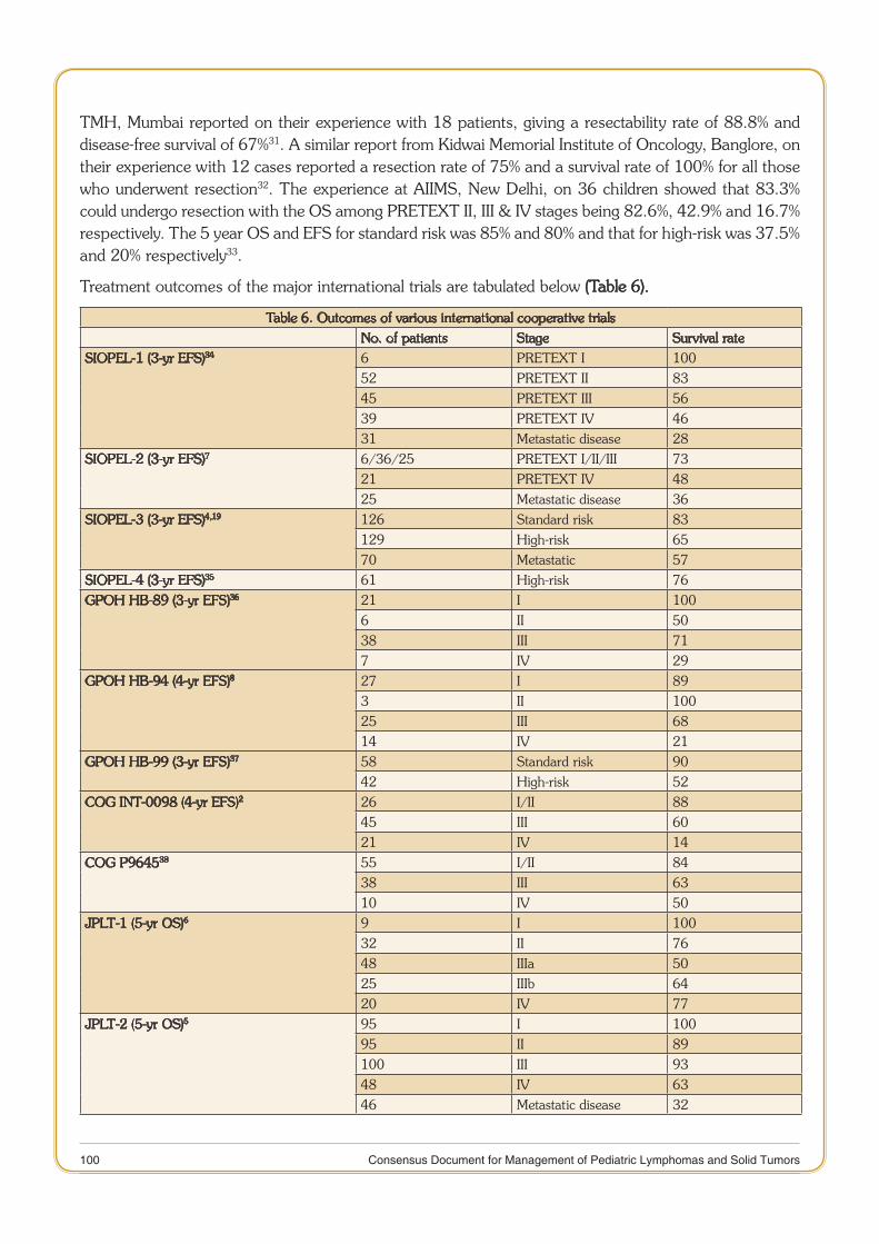
100 Consensus Document for Management of Pediatric Lymphomas and Solid Tumors
TMH, Mumbai reported on their experience with 18 patients, giving a resectability rate of 88.8% and disease-free survival of 67%31. A similar report from Kidwai Memorial Institute of Oncology, Banglore, on their experience with 12 cases reported a resection rate of 75% and a survival rate of 100% for all those who underwent resection32. The experience at AIIMS, New Delhi, on 36 children showed that 83.3% could undergo resection with the OS among PRETEXT II, III & IV stages being 82.6%, 42.9% and 16.7% respectively. The 5 year OS and EFS for standard risk was 85% and 80% and that for high-risk was 37.5% and 20% respectively33.
Treatment outcomes of the major international trials are tabulated below (Table 6).
Table 6. Outcomes of various international cooperative trialsNo. of patients Stage Survival rate
SIOPEL-1 (3-yr EFS)34 6 PRETEXT I 10052 PRETEXT II 8345 PRETEXT III 5639 PRETEXT IV 4631 Metastatic disease 28
SIOPEL-2 (3-yr EFS)7 6/36/25 PRETEXT I/II/III 7321 PRETEXT IV 4825 Metastatic disease 36
SIOPEL-3 (3-yr EFS)4,19 126 Standard risk 83129 High-risk 6570 Metastatic 57
SIOPEL-4 (3-yr EFS)35 61 High-risk 76GPOH HB-89 (3-yr EFS)36 21 I 100
6 II 5038 III 717 IV 29
GPOH HB-94 (4-yr EFS)8 27 I 893 II 10025 III 6814 IV 21
GPOH HB-99 (3-yr EFS)37 58 Standard risk 9042 High-risk 52
COG INT-0098 (4-yr EFS)2 26 I/II 8845 III 6021 IV 14
COG P964538 55 I/II 8438 III 6310 IV 50
JPLT-1 (5-yr OS)6 9 I 10032 II 7648 IIIa 5025 IIIb 6420 IV 77
JPLT-2 (5-yr OS)5 95 I 10095 II 89100 III 9348 IV 6346 Metastatic disease 32
101 Consensus Document for Management of Pediatric Lymphomas and Solid Tumors
Recurrent disease: Very little definitive data exist regarding treatment for relapsed hepatoblastoma. Numbers of patients with recurrent hepatoblastoma are small owing to the rarity of the disease per se and the success of initial treatment, especially in the setting of localized disease.
Success of the treatment of recurrent hepatoblastoma relies largely on surgical resection. When tumors are responsive, chemotherapy can be used to render the tumor resectable. Various chemotherapeutic regimes studied in small numbers of patients in phase I/II trials have shown few responses.
The SIOPEL group has segregated its data on children with relapsed hepatoblastoma (defined as recurrence after complete remission with normal aFP values for at least 4 weeks after completion of treatment) from SIOPEL – 1, 2 & 3 studies2. Out of a total of 695 children, 59 had recurrent disease (8.4%). The median time from the initial diagnosis to relapse was 12 months. The site of relapse was most commonly lung followed by liver, both liver and lung and others. All but nine patients had an aFP level >10 ng/ml at the time of relapse. Treatment of the relapse included chemotherapy and surgery in 42%, chemotherapy alone in 35%, surgery alone in 12% and only palliative treatment in 8%. Overall, 52% achieved a second remission. Three-year EFS and OS were 34% and 43% respectively. The main factors associated with a good outcome were PRETEXT group I–III at diagnosis, a high aFP level at relapse and relapse treatment including both chemotherapy and surgery. The relapses with no aFP elevation underline the fact that follow-up should include not only aFP measurement, but also chest X-rays and an abdominal ultrasound as recommended by most guidelines. Above data also suggest that combined chemotherapy and surgery should be offered to all patients at relapse even in patients with apparently resectable lesions.
The best available data indicate that doxorubicin, if not given during initial treatment, and irinotecan is the most active agents in recurrent hepatoblastoma. A multi-center, prospective, phase II trial of SIOPEL group evaluated the clinical activity of irinotecan as single drug in children with refractory or recurrent hepatoblastoma39. Response to irinotecan was associated with a low early progression rate (17%). In 30% of the patients, a tumor-free status was achieved. Patients with recurrent disease had a better response rate than those with refractory/progressive disease. No patients with a low aFP level showed response.
Recommended Guidelines
Laboratory evaluation: Appropriate laboratory evaluation for suspected hepatoblastoma include total blood counts, LFT, lactate dehydrogenase, aFP. aFP is the most important marker for hepatoblastoma; it is increased in 90% of patients with the tumor. Several investigators have shown that most hepatoblastoma’s with low aFP levels (<100 ng/ml) are aggressive and associated with a poor prognosis. In neonates the interpretation of aFP measurements is more difficult because of the naturally high serum levels in infants (Table 7). Also, aFP may be elevated in some patients with benign liver tumors, including mesenchymal hamartoma and adenoma. aFP has been shown to be a reliable predictor of treatment response during the neoadjuvant chemotherapy and ultimate outcome. It is therefore used in disease monitoring to identify poor treatment responders, relapse, or metastatic disease, indicating the need for change in treatment strategy.
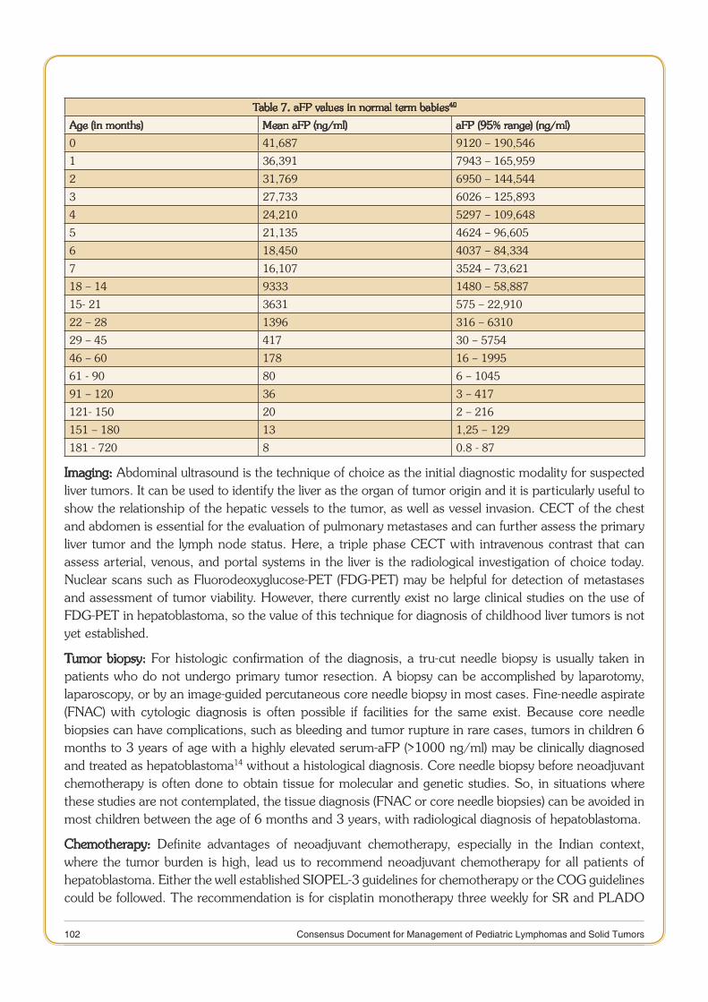
102 Consensus Document for Management of Pediatric Lymphomas and Solid Tumors
Table 7. aFP values in normal term babies40
Age (in months) Mean aFP (ng/ml) aFP (95% range) (ng/ml)
0 41,687 9120 – 190,546
1 36,391 7943 – 165,959
2 31,769 6950 – 144,544
3 27,733 6026 – 125,893
4 24,210 5297 – 109,648
5 21,135 4624 – 96,605
6 18,450 4037 – 84,334
7 16,107 3524 – 73,621
18 – 14 9333 1480 – 58,887
15- 21 3631 575 – 22,910
22 – 28 1396 316 – 6310
29 – 45 417 30 – 5754
46 – 60 178 16 – 1995
61 - 90 80 6 – 1045
91 – 120 36 3 – 417
121- 150 20 2 – 216
151 – 180 13 1,25 – 129
181 - 720 8 0.8 - 87
Imaging: Abdominal ultrasound is the technique of choice as the initial diagnostic modality for suspected liver tumors. It can be used to identify the liver as the organ of tumor origin and it is particularly useful to show the relationship of the hepatic vessels to the tumor, as well as vessel invasion. CECT of the chest and abdomen is essential for the evaluation of pulmonary metastases and can further assess the primary liver tumor and the lymph node status. Here, a triple phase CECT with intravenous contrast that can assess arterial, venous, and portal systems in the liver is the radiological investigation of choice today. Nuclear scans such as Fluorodeoxyglucose-PET (FDG-PET) may be helpful for detection of metastases and assessment of tumor viability. However, there currently exist no large clinical studies on the use of FDG-PET in hepatoblastoma, so the value of this technique for diagnosis of childhood liver tumors is not yet established.
Tumor biopsy: For histologic confirmation of the diagnosis, a tru-cut needle biopsy is usually taken in patients who do not undergo primary tumor resection. A biopsy can be accomplished by laparotomy, laparoscopy, or by an image-guided percutaneous core needle biopsy in most cases. Fine-needle aspirate (FNAC) with cytologic diagnosis is often possible if facilities for the same exist. Because core needle biopsies can have complications, such as bleeding and tumor rupture in rare cases, tumors in children 6 months to 3 years of age with a highly elevated serum-aFP (>1000 ng/ml) may be clinically diagnosed and treated as hepatoblastoma14 without a histological diagnosis. Core needle biopsy before neoadjuvant chemotherapy is often done to obtain tissue for molecular and genetic studies. So, in situations where these studies are not contemplated, the tissue diagnosis (FNAC or core needle biopsies) can be avoided in most children between the age of 6 months and 3 years, with radiological diagnosis of hepatoblastoma.
Chemotherapy: Definite advantages of neoadjuvant chemotherapy, especially in the Indian context, where the tumor burden is high, lead us to recommend neoadjuvant chemotherapy for all patients of hepatoblastoma. Either the well established SIOPEL-3 guidelines for chemotherapy or the COG guidelines could be followed. The recommendation is for cisplatin monotherapy three weekly for SR and PLADO
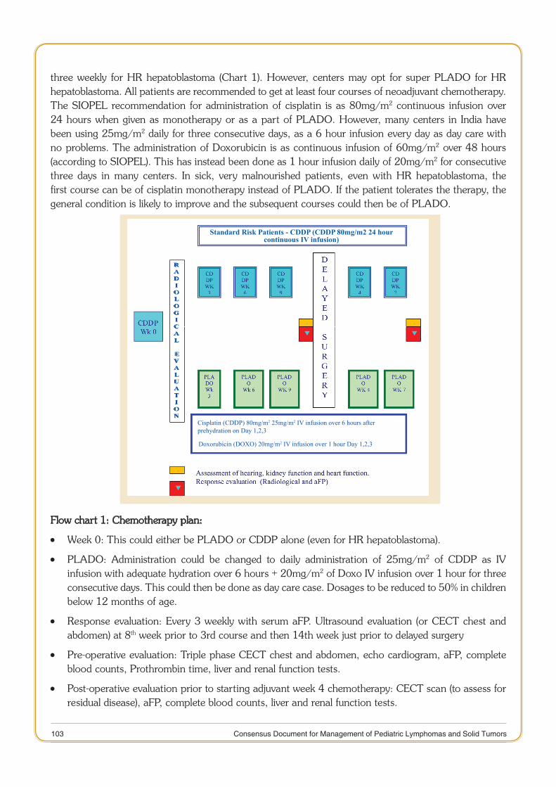
103 Consensus Document for Management of Pediatric Lymphomas and Solid Tumors
three weekly for HR hepatoblastoma (Chart 1). However, centers may opt for super PLADO for HR hepatoblastoma. All patients are recommended to get at least four courses of neoadjuvant chemotherapy. The SIOPEL recommendation for administration of cisplatin is as 80mg/m2 continuous infusion over 24 hours when given as monotherapy or as a part of PLADO. However, many centers in India have been using 25mg/m2 daily for three consecutive days, as a 6 hour infusion every day as day care with no problems. The administration of Doxorubicin is as continuous infusion of 60mg/m2 over 48 hours (according to SIOPEL). This has instead been done as 1 hour infusion daily of 20mg/m2 for consecutive three days in many centers. In sick, very malnourished patients, even with HR hepatoblastoma, the first course can be of cisplatin monotherapy instead of PLADO. If the patient tolerates the therapy, the general condition is likely to improve and the subsequent courses could then be of PLADO.
Standard Risk Patients - CDDP (CDDP 80mg/m2 24 hour continuous IV infusion)
RaDIologICal
eValuatIon
Cisplatin (CDDP) 80mg/m2 25mg/m2 IV infusion over 6 hours after prehydration on Day 1,2,3
Doxorubicin (DOXO) 20mg/m2 IV infusion over 1 hour Day 1,2,3
Flow chart 1: Chemotherapy plan:
Week 0: This could either be PLADO or CDDP alone (even for HR hepatoblastoma). •
PLADO: Administration could be changed to daily administration of 25mg/m • 2 of CDDP as IV infusion with adequate hydration over 6 hours + 20mg/m2 of Doxo IV infusion over 1 hour for three consecutive days. This could then be done as day care case. Dosages to be reduced to 50% in children below 12 months of age.
Response evaluation: Every 3 weekly with serum aFP. Ultrasound evaluation (or CECT chest and •abdomen) at 8th week prior to 3rd course and then 14th week just prior to delayed surgery
Pre-operative evaluation: Triple phase CECT chest and abdomen, echo cardiogram, aFP, complete •blood counts, Prothrombin time, liver and renal function tests.
Post-operative evaluation prior to starting adjuvant week 4 chemotherapy: CECT scan (to assess for •residual disease), aFP, complete blood counts, liver and renal function tests.
104 Consensus Document for Management of Pediatric Lymphomas and Solid Tumors
Response evaluation during neoadjuvant chemotherapy should be carried out with aFP monitoring in conjunction with the age-related nomogram available (Table 7). Inadequate response may help upgrading from monotherapy to PLADO. Response evaluation by imaging is recommended with ultrasound of the abdomen after two courses of neoadjuvant chemotherapy. The ultrasound should look for the reduction in the tumor volume, the status of inferior venacava (IVC) or portal vein, especially if these were involved earlier.
Pre-operative evaluation: Immediate pre-operative evaluation should include serum aFP level, triple phase CECT of the chest and abdomen to clearly delineate the extent of resection required and the vascular anatomy, echocardiography to evaluate the cardiac functions, complete blood counts, prothrombin time, liver and renal function tests.
Surgery: An anatomical surgical resection is recommended in all cases of hepatoblastoma. Non-anatomical resections should be avoided. If anatomical resection is not feasible, one must consider the possibility of LTx. Without complete tumor resection survival is not feasible. Patients with pulmonary metastases at the end of four courses of neoadjuvant chemotherapy should undergo resection of these pulmonary metastases before liver resection is undertaken. Liver resection is only useful if the patient achieves a CR (complete remission) at the end of the surgical treatment.
Radiotherapy: Radiotherapy has very little defined role in the management of hepatoblastoma. However, some centers have occasionally used adjuvant RT in cases with positive surgical margins. It has also been described for non-responsive unresectable hepatoblastoma.
Follow-up: The FU protocol, subsequent to completion of all treatment, should include three monthly radiological evaluations (alternating ultrasound abdomen + chest x-ray with a CECT of chest and abdomen) and αFP levels for at least one year. It is important to evaluate the chest as most of the recurrences take place in the lungs (besides the local recurrences). After the first year of FU, the frequency of these evaluations should be reduced to six monthly intervals for at least two more years and then annually. The centers may opt for ultrasound as the only imaging modality in the follow-up. The survivors should also be evaluated for long-term side effects such as ototoxicity by pure tone audiometry, nephrotoxicity by a radio-nuclide GFR estimation, and cardiotoxicity by echocardiogram or a Multi Gated Acquisition (MUGA) scan.
Future roadmap
New cytotoxic drugs are required for successful treatment of resistant or recurrent hepatoblastoma. In preclinical tests topotecan and paclitaxel have shown some activity against hepatoblastoma, but they have not been thoroughly tested in clinical trials. Another approach for high-risk hepatoblastoma patients to prevent recurrent disease would be to apply long-term maintenance chemotherapy over 1-2 years after achieving remission. Irinotecan has been successfully used for this in single patients, so this approach should be evaluated in future multi-institutional trials41. Other agents which are being targeted against hepatoblastoma include sorafenib (tyrosine kinase inhibitor), transtuzumab (against erbB2 receptor), sirolimus, retinoic acid and arsenic trioxide. A new risk-based cooperative international trial, the Pediatric hepatoblastoma International Therapeutic Trial (PHITT), a joint venture of global collaboration that includes SIOPEL, COG, and JPLT is being planned to have an in-depth insights into the actual tumor biology and to find new therapeutic roadways to achieve a cure in most.
Suggested research topics
6 hour vs 48 hour infusion of Doxorubicin.1.
105 Consensus Document for Management of Pediatric Lymphomas and Solid Tumors
aFP, Ultrasound abdomen and chest X-ray every 3 months vs aFP, CECT chest and abdomen 2. alternating with ultrasound and chest X-ray every 3 monthly as follow-up schedule
3-D CECT reconstruction as pre-operative imaging in assisting, surgical planning and decision making3.
The feasibility of using only monotherapy for HR hepatoblastoma in extremely resource-challenged 4. states
Newer molecular and genetic markers for risk stratification and prognostication in hepatoblastoma 5.
Role of nutritional enhancement in the overall outcome and reduction of complications in 6. hepatoblastoma
References
Czauderna P, Lopez-Terrada D, Hiyama E et al. Hepatoblastoma state of the art: pathology, genetics, risk stratification, 1. and chemotherapy. Curr Opin Pediatr. 2014 Feb;26(1):19-28
Ortega JA, Douglass EC, Feusner JH et al. Randomized comparison of cisplatin/vincristine/ fluorouracil and cisplatin/2. continuous infusion doxorubicin for treatment of pediatric hepatoblastoma: A report from the Children's Cancer Group and the Pediatric Oncology Group. J Clin Oncol. 2000 Jul;18(14):2665-75.
Pritchard J, Brown J, Shafford E et al. Cisplatin, doxorubicin, and delayed surgery for childhood hepatoblastoma: a 3. successful approach--results of the first prospective study of the International Society of Pediatric Oncology. J Clin Oncol. 2000 Nov 15;18(22):3819-28
Perilongo G, Maibach R, Shafford E et al. Cisplatin versus cisplatin plus doxorubicin for standard-risk hepatoblastoma. 4. N Engl J Med. 2009 Oct 22;361(17):1662-70
Hishiki T, Matsunaga T, Sasaki F et al. Outcome of hepatoblastomas treated using the Japanese Study Group for 5. Pediatric Liver Tumor (JPLT) protocol-2: report from the JPLT. Pediatr Surg Int. 2011 Jan;27(1):1-8
Sasaki F, Matsunaga T, Iwafuchi M et al. (Japanese Study Group for Pediatric Liver Tumor). Outcome of hepatoblastoma 6. treated with the JPLT-1 (Japanese Study Group for Pediatric Liver Tumor) Protocol-1: A report from the Japanese Study Group for Pediatric Liver Tumor. J Pediatr Surg. 2002 Jun;37(6):851-6.
Perilongo G, Shafford E, Maibach R et al. International Society of Paediatric Oncology-SIOPEL 2. Risk-adapted treatment 7. for childhood hepatoblastoma. final report of the second study of the International Society of Paediatric Oncology-SIOPEL 2. Eur J Cancer. 2004 Feb;40(3):411-21
Fuchs J, Rydzynski J, Von Schweinitz D et al. Study Committee of the Cooperative Pediatric Liver Tumor Study HB 94 8. for the German Society for Pediatric Oncology and Hematology. Pre-treatment prognostic factors and treatment results in children with hepatoblastoma: a report from the German Cooperative Pediatric Liver Tumor Study HB 94. Cancer. 2002 Jul 1;95(1):172-82
Häberle B, Bode U, von Schweinitz D. [Differentiated treatment protocols for high- and standard-risk hepatoblastoma--an 9. interim report of the German Liver Tumor Study HB99]. Klin Padiatr. 2003 May-Jun;215(3):159-65
Katzenstein HM, Rigsby C, Shaw PH et al. Novel therapeutic approaches in the treatment of children with hepatoblastoma. 10. J Pediatr Hematol Oncol. 2002 Dec;24(9):751-5
Katzenstein HM, Chang KW, Krailo M et al. Children's Oncology Group. Amifostine does not prevent platinum-induced 11. hearing loss associated with the treatment of children with hepatoblastoma: a report of the Intergroup Hepatoblastoma Study P9645 as a part of the Children's Oncology Group. Cancer. 2009 Dec 15;115(24):5828-35
Aronson DC, Czauderna P, Maibach R et al. The treatment of hepatoblastoma: Its evolution and the current status as per 12. the SIOPEL trials. J Indian Assoc Pediatr Surg. 2014 Oct;19(4):201-7
Couinaud C. [Liver lobes and segments: notes on the anatomical architecture and surgery of the liver ]. Presse Med. 13. 1954 May 5;62(33):709-12
von Schweinitz D. Management of liver tumors in childhood. Semin Pediatr Surg. 2006 Feb;15(1):17-2414.
Evans AE, Land VJ, Newton WA et al. Combination chemotherapy (vincristine, adriamycin, cyclophosphamide, and 15. 5-fluorouracil) in the treatment of children with malignant hepatoma. Cancer. 1982 Sep 1;50(5):821-6

106 Consensus Document for Management of Pediatric Lymphomas and Solid Tumors
Black CT, Cangir A, Choroszy M, Andrassy RJ. Marked response to pre-operative high-dose cis-platinum in children 16. with unresectable hepatoblastoma. J Pediatr Surg. 1991 Sep;26(9):1070-3
Douglass EC, Green AA, Wrenn E et al. Effective cisplatin (DDP) based chemotherapy in the treatment of hepatoblastoma. 17. Med Pediatr Oncol. 1985;13(4):187-90
Malogolowkin MH, Katzenstein HM, Meyers RL et al. Complete surgical resection is curative for children with 18. hepatoblastoma with pure fetal histology: a report from the Children's Oncology Group. J Clin Oncol. 2011 Aug 20;29(24):3301-6
Zsíros J, Maibach R, Shafford E et al. Successful treatment of childhood high-risk hepatoblastoma with dose-intensive 19. multi-agent chemotherapy and surgery: final results of the SIOPEL-3HR study. J Clin Oncol. 2010 May 20;28(15):2584-90
Katzenstein HM, London WB, Douglass EC et al. Treatment of unresectable and metastatic hepatoblastoma: a pediatric 20. oncology group phase II study. J Clin Oncol. 2002 Aug 15;20(16):3438-44
Meyers RL, Tiao GM, Dunn SP, Langham MR Jr. Liver transplantation in the management of unresectable hepatoblastoma 21. in children. Front Biosci (Elite Ed). 2012 Jan 1;4:1293-302
Lautz TB, Ben-Ami T, Tantemsapya N et al. Successful nontransplant resection of POST-TEXT III and IV hepatoblastoma. 22. Cancer. 2011 May 1;117(9):1976-83
Guérin F, Gauthier F, Martelli H et al. Outcome of central hepatectomy for hepatoblastomas. J Pediatr Surg. 2010 23. Mar;45(3):555-63
Meyers RL, Aronson DC, von Schweinitz D, et al. Pediatric liver tumors, in Pizzo PA, Poplack DG (eds): Principles and 24. Practice in Pediatric Oncology. Philadelphia, PA, WLTers Kluwer, Lippincott. Williams Wilkins, 2011, 838-60.
Meyers RL, Otte J-B. Liver transplantation for unresectable liver tumors in children, in Zimmermann A, Perilongo G, 25. Malogolowkin M, von Schweinitz D (eds): Pediatric Liver Tumors. Heidelberg, Springer, 2011, 133-52.
Gupta AA, Gerstle JT, Ng V et al. Critical review of controversial issues in the management of advanced pediatric liver 26. tumors. Pediatr Blood Cancer. 2011 Jul 1;56(7):1013-8
Browne M, Sher D, Grant D et al. Survival after liver transplantation for hepatoblastoma: a 2-center experience. J Pediatr 27. Surg. 2008 Nov;43(11):1973-81
Otte JB, Meyers R. PLUTO first report. Pediatr Transplant. 2010 Nov;14(7):830-528.
Meyers RL, Katzenstein HM, Krailo M et al. Surgical resection of pulmonary metastatic lesions in children with 29. hepatoblastoma. J Pediatr Surg. 2007 Dec;42(12):2050-6
Arora RS. Outcomes of hepatoblastoma in the Indian context. Indian Pediatr. 2012 Apr;49(4):307-930.
Shukla PJ, Barreto SG, Qureshi SS et al. Hepatoblastoma: a single institutional experience of 18 cases. Pediatr Surg 31. Int. 2008 Jul;24(7):799-802
Singh T, Satheesh CT, Appaji L et al. Hepatoblastoma: experience from a single center. Indian J Cancer. 2010 Jul-32. Sep;47(3):314-6
Agarwala S, Bakshi S, Bajpai M et al. Validation of PRETEXT staging system and risk categorization for prognostication 33. and outcome in hepatoblastoma- Results from AIIMS-HB 94 trial. Pediatr Blood Cancer. 2007;49:401.
Brown J, Perilongo G, Shafford E et al. Pre-treatment prognostic factors for children with hepatoblastoma-- results from 34. the International Society of Paediatric Oncology (SIOP) study SIOPEL 1. Eur J Cancer. 2000 Jul;36(11):1418-25
Zsiros J, Brugieres L, Brock P et al. International Childhood Liver Tumors Strategy Group (SIOPEL). Dose-dense 35. cisplatin-based chemotherapy and surgery for children with high-risk hepatoblastoma (SIOPEL-4): a prospective, single-arm, feasibility study. Lancet Oncol. 2013 Aug;14(9):834-42
von Schweinitz D, Byrd DJ, Hecker H et al. Efficiency and toxicity of ifosfamide, cisplatin and doxorubicin in the 36. treatment of childhood hepatoblastoma. Study Committee of the Cooperative Paediatric Liver Tumor Study HB89 of the German Society for Paediatric Oncology and Hematology. Eur J Cancer. 1997 Jul;33(8):1243-9
Haeberle B, Schweinitz Dv. Treatment of hepatoblastoma in the German cooperative pediatric liver tumor studies. Front 37. Biosci (Elite Ed). 2012 Jan 1;4:493-8
107 Consensus Document for Management of Pediatric Lymphomas and Solid Tumors
Malogolowkin MH, Katzenstein H, Krailo MD et al. Intensified platinum therapy is an ineffective strategy for improving 38. outcome in pediatric patients with advanced hepatoblastoma. J Clin Oncol. 2006 Jun 20;24(18):2879-84
Zsíros J, Brugières L, Brock P et al. Efficacy of irinotecan single drug treatment in children with refractory or 39. recurrent hepatoblastoma--a phase II trial of the childhood liver tumor strategy group (SIOPEL). Eur J Cancer. 2012 Dec;48(18):3456-64
Blohm ME, Vesterling-Hörner D, Calaminus G, Göbel U. Alpha 1-fetoprotein (AFP) reference values in infants up to 2 40. years of age. Pediatr Hematol Oncol. 1998 Mar-Apr;15(2):135-42.
Qayed M, Powell C, Morgan ER et al. Irinotecan as maintenance therapy in high-risk hepatoblastoma. Pediatr Blood 41. Cancer. 2010 May;54(5):761-3
108 Consensus Document for Management of Pediatric Lymphomas and Solid Tumors
CHAPTER
6 NEUROBLASTOMA
Introduction
Neuroblastoma (NBL) is the most common extracranial solid tumor in childhood in developed countries where it accounts for 10% of pediatric cancers1. However, in India, its precise incidence is unknown. Approximately 2000 new cases of NBL may be expected to be diagnosed per year in India2. As per the ‘ICMR - NCRP’, the relative proportion of NBL (and ganglioneuroblastoma) in childhood (0-14 years) in seven hospital based cancer registries across India varied from 2.4% to 7.5% during 2007-113. In a retrospective compilation of all childhood cancers, at PGIMER, Chandigarh, NBL was the second most common solid tumor diagnosed following retinoblastoma; of 3568 cases over a 14 year period, 223 (6.3%) patients were diagnosed with NBL4.
NBL is considered to have one of the least favorable outcomes among pediatric cancers. At a Govt-run tertiary care center in India, NBL would likely account for at least 20 new cases annually. The cure rate of the high - risk NBL in the developed countries does not typically exceed 40%1,5. The outcome of the high - risk NBL in India is widely perceived to be dismal. The factors contributing to a poor outcome of the high - risk NBL in India, include late diagnosis, poor nutrition with resultant higher treatment-related mortality, limited availability of an ASCT and treatment abandonment. With optimal risk stratification, judicious administration of a management protocol and good supportive care, the outcome of children with NBL in India can hopefully be improved.
Neuroblastoma: Outcome, Indian data
In contrast to the number of patients with NBL, the number of studies with outcome data from India is very limited. It plausibly reflects a poor outcome of patients with NBL. The centers would be less likely to publish retrospective series of a cancer with a poor survival.
a) AIIMS, New Delhi study
One hundred and forty four children from 1996 through 2009 were included in the study6. Thirty eight (26%) patients were under 12 months; 112 (78%) of the tumors were abdominal and 32 (22%) were extra-abdominal. Stage distribution was stage 1 and stage 2 in 6 (4%); 3 in 58 (40%); 4 in 68 (47%); 4s in 12 (8%). All children included in the study received chemotherapy and radiation therapy, appropriate for the stage. Tumor resection was done when feasible. The OS was 70% for those under 12 months of age. For stage 3 patients the complete remission (CR) was 57%, with an OS of 72%. The OS (36%) and CR (18%) for stage 4 patients were significantly less (p=0. 001)6.
b) PGIMER, Chandigarh study
The outcome of 103 children, older than one year was reported7. Seventy four had stage IV, 27 Stage III and one patient each had Stage I or II disease. Treatment included chemotherapy followed by surgical resection/debulking. Radiotherapy was administered to those with residual tumor. Chemotherapy
109 Consensus Document for Management of Pediatric Lymphomas and Solid Tumors
consisted of ‘OPEC’ (vincristine, cyclophosphamide, cisplatin and etoposide). The caretakers of 54 (52%) children either did not opt for or defaulted therapy, while three patients died before chemotherapy could be initiated. Of the remaining 46 patients, the tumor progressed during therapy in 19 (41%). Relapse of the disease was documented in 22 (48%) cases. Merely 4 (9%) children were disease-free for a period of 16.5±6.7 months. The majority of children presented with advanced disease and the outcomes were dismal with conventional non-myeloablative chemotherapy7.
Kulkarni et al performed a comprehensive search of reported outcomes of NBL in India2. Advanced disease was present in 75% to greater than 90% patients reported from Indian centers. Survival varied widely from 8.7% to 80%. There was a high rate of progression/PR, treatment, abandonment, relapse or death2.
Literature review
Major groups which have published results of trials conducted in NBL include POG, COG, CCG, SIOPEN, Children’s Cancer and Leukemia Group (CCLG) and GPOH. Recently the International Society of Pediatric Oncology-Committee on Developing Countries (SIOP-PODC) has published recommendations for risk stratification and treatment of the NBL in low/middle-income countries8. The SIOP-PODC clinical practice guidelines have been used as the backbone for this document. Treatment of patients with low and intermediate-risk NBL has been adapted from the CCLG March 2015 guidelines9.
Clinical features
NBL is the commonest cancer in infancy. Up to 90% patients are less than five years of age at diagnosis and almost all are less than 10 years of age1. As per western data, there is a slight male preponderance with a male: female ratio of 1.2:11,5. In a study of 103 patients (> 1 year) from PGIMER, Chandigarh, the male: female ratio was 2.8:17. NBL can originate from anywhere along the sympathetic chain. It is considered in the differential diagnosis of a mass arising in neck, mediastinum, abdomen or pelvis. Nearly two-thirds of primary tumors occur within the abdomen1,5. Adrenal NBL is more frequent in older children than infants (40% vs. 25%)1. Thoracic and cervical primary tumors are more common in infants1. Metastasis can occur to regional lymph nodes, and by hematogenous spread to distant sites; predominantly BM, cortical bone, liver and skin. Overall, metastatic disease is observed in 50% patients1. It is frequent in older children as compared to infants (60% vs. 40%)1.
Paraspinal NBL can result in compression of nerve roots and spinal cord. Occasionally NBL may be associated with paraneoplastic syndromes such as opsoclonus-myoclonus-ataxia syndrome and intractable watery diarrhea1,5. Hypertension is common; it can be managed with long-acting ACE inhibitor (e.g. enalapril) or calcium channel blocker (e.g. amlodipine)1. It typically resolves with surgical resection/chemo-reduction of the tumor following which the anti-hypertensive drugs can be stopped.
Diagnosis
Biopsy
An unequivocal pathological diagnosis from tumor tissue is made by light microscopy, with or without: Immunohistology (IHC) or raised urine/serum homovanillic (HVA) or vanillylmandelic acid (VMA)1. IHC aids in distinguishing from other small round blue cell tumors. IHC markers for NBL classically include CD56, synaptophysin, tyrosine hydroxylase and neuron-specific enolase8. In India, biopsy is often performed with a Tru-Cut® needle under image guidance. An open biopsy in operation-theatre under general anesthesia may often be done in developed countries to obtain adequate material for experimental biological studies. Surgical procedures performed at presentation are highlighted in Table 1.
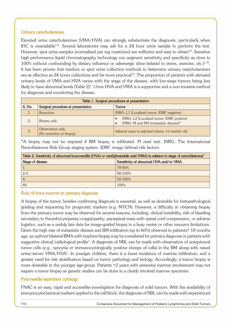
110 Consensus Document for Management of Pediatric Lymphomas and Solid Tumors
Urinary catecholamines
Elevated urine catecholamines (VMA/HVA) can strongly substantiate the diagnosis, particularly when IHC is unavailable1,8. Several laboratories may ask for a 24 hour urine sample to perform the test. However, spot urine samples (normalized per mg creatinine) are sufficient and easy to obtain10. Sensitive high-performance liquid chromatography technology can augment sensitivity and specificity as close to 100% without confounding by dietary influence or adrenergic drive (related to stress, exercise, etc.)1,10. It has been proven that random or spot urine collection methods to determine urinary catecholamines are as effective as 24 hours collections and far more practical10. The proportion of patients with elevated urinary levels of VMA and HVA varies with the stage of the disease, with low-stage tumors being less likely to have abnormal levels (Table 2)1. Urine HVA and VMA is a supportive and a non-invasive method for diagnosis and monitoring the disease.
Table 1. Surgical procedures at presentation
S. No. Surgical procedure at presentation Tumor
1. Resection INRG: L1 (Localized tumor; IDRF negative)
2. Biopsy onlyINRG: L2 (Localized tumor; IDRF positive) •INRG: M and MS (metastatic disease)* •
3.Observation only(No resection or biopsy)
Adrenal mass in selected infants <3 months old
*A biopsy may not be required if BM biopsy is infiltrated. Pl read text. INRG, The International Neuroblastoma Risk Group staging system. IDRF: image defined risk factors.
Table 2. Sensitivity of abnormal homovanillic (HVA) or vanillylmandelic acid (VMA) in relation to stage of neuroblastoma1
Stage of disease Sensitivity of abnormal HVA and/or VMA
1 78-85%
2-3 80-100%
4 92-100%
4S 100%
Role of bone marrow in primary diagnosis
A biopsy of the tumor, besides confirming diagnosis is essential, as well as desirable for histopathological grading and requesting for prognostic markers (e.g. MYCN). However, a difficulty in obtaining biopsy from the primary tumor may be observed for several reasons, including, clinical instability, risk of bleeding secondary to thrombocytopenia/coagulopathy, paraspinal mass with spinal cord compression, or adverse logistics, such as a unduly late date for image-guided biopsy in a busy center or other resource limitations. Given the high rate of metastatic disease and BM infiltration (up to 60%) observed in patients> 18 months age, an upfront bilateral BMA with trephine biopsy may be considered for primary diagnosis in patients with suggestive clinical/radiological profile1. A diagnosis of NBL can be made with observation of unequivocal tumor cells (e.g., syncytia or immunocytologically positive clumps of cells) in the BM along with raised urine/serum VMA/HVA1. In younger children, there is a lower incidence of marrow infiltration, and a greater need for risk stratification based on tumor pathology and biology. Accordingly, a tumor biopsy is more desirable in the younger age-group. Patients >2 years with extensive marrow involvement may not require a tumor biopsy as genetic studies can be done in a clearly involved marrow specimen.
Fine-needle aspiration cytology
FNAC is an easy, rapid and accessible investigation for diagnosis of solid tumors. With the availability of immunocytochemical markers applied to the cell block, the diagnosis of NBL can be made with experienced
111 Consensus Document for Management of Pediatric Lymphomas and Solid Tumors
hands. However, a biopsy is encouraged, particularly in patients in whom MYCN amplification (MYCN-A) status will make a critical difference in treatment approach (Pl read the later section titled MYCN).
Staging
Radiological staging of the primary tumor is commonly performed with a contrast-enhanced CT scan in tumors, which primarily arise in the chest, abdomen or pelvis1,5,8. MRI is a superior modality for paraspinal lesions, particularly when associated with nerve root/cord compression1,8. Either CT or MRI may be used for a cervical mass8. Metastatic evaluation classically includes bilateral BMA and trephine biopsy and an MIBG (meta-iodobenzyl guanidine) scan1,5,8. The SIOP-PODC guidelines for NBL recommend obtaining a CT of the neck, chest, abdomen and pelvis in all patients8. We suggest that in patients who undergo adequate metastatic work-up with MIBG or FDG-PET and a BM examination, CT of the primary tumor site alone should suffice.
MIBG /Bone scan/ FDG-PET: Which one to choose?
MIBG is the most sensitive metastatic investigation for skeletal and soft tissue1. MIBG can be labeled with either 131I or 123I11. 123I is considered the radiopharmaceutical of choice as it has a more favorable dosimetry and provides better image quality, allowing accurate anatomical localization11. Nevertheless, 131I is what is commonly available in major centers of India. Moreover, the iodine tracer is sourced from elsewhere by most Indian centers, making it available intermittently, often on a periodic (say, monthly) basis. When unavailable, or non-MIBG avid (in up to 10% patients), a TC-99-diphosphonate scintigraphy (bone scans) is performed1,8. 18FDG-PET is another alternative in this situation8. The advantage of 18FDG-PET over a bone scan is that FDG-PET can be used for re-evaluation as well12.
A recent study was conducted at AIIMS, New Delhi to compare the diagnostic value of FDG-PET/CT with 131I-MIBG scintigraphy in 40 pediatric neuroblastoma patients13. On a patient-based comparison, there was no significant difference between FDG-PET/CT and I-MIBG (p=1. 00), however, FDG-PET/CT was superior to I-MIBG on a lesion-based comparison (p<0.0001). Although no difference was noted for primary lesions (p=1. 00), PET/CT was superior to I-MIBG scintigraphy for the detection of lymph nodal (p=0. 001) and bone/BM lesions (p=0. 007)13.
Response evaluation following chemotherapy is recommended with MIBG. FDG-PET is a suitable alternative for response evaluation, in the absence of MIBG8. A bone scan is not reliable for re-evaluation12. Any detectable response on bone scan may be evident for up to 6-8 months after therapy. On the contrary, response to therapy in the first 4-12 weeks may result in “flare phenomenon” and increased uptake related to the process of healing12.
A BM examination is an essential staging investigation in the NBL and cannot be replaced by an imaging modality. The essential and desirable investigations to be obtained in a patient with NBL prior to treatment are listed in Table 3.
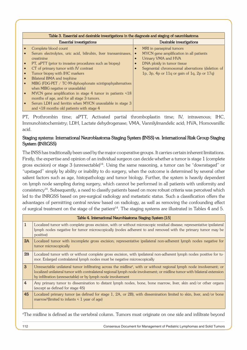
112 Consensus Document for Management of Pediatric Lymphomas and Solid Tumors
Table 3. Essential and desirable investigations in the diagnosis and staging of neuroblastoma
Essential investigations Desirable investigations
Complete blood count •Serum electrolytes, uric acid, bilirubin, liver transaminases, •creatininePT, aPTT (prior to invasive procedures such as biopsy) •CT of primary tumor with IV contrast •Tumor biopsy with IHC markers •Bilateral BMA and trephine •MIBG (FDG-PET / TC-99-diphosphonate scintigraphyalternatives •when MIBG negative or unavailable)MYCN gene amplification in stage 4 tumor in patients <18 •months of age, and for all stage 3 tumors.Serum LDH and ferritin when MYCN unavailable in stage 3 •and <18 months old patients with stage 4
MRI in paraspinal tumors •MYCN gene amplification in all patients •Urinary VMA and HVA •DNA ploidy in tumor tissue •Segmental chromosomal aberrations (deletion of •1p, 3p, 4p or 11q or gain of 1q, 2p or 17q)
PT, Prothrombin time; aPTT, Activated partial thromboplastin time; IV, intravenous; IHC, Immunohistochemistry; LDH, Lactate dehydrogenase; VMA, Vannilylmandelic acid; HVA, Homovanillic acid.
Staging systems: International Neuroblastoma Staging System (INSS) vs. International Risk Group Staging System (INRGSS)
The INSS has traditionally been used by the major cooperative groups. It carries certain inherent limitations. Firstly, the expertise and opinion of an individual surgeon can decide whether a tumor is stage 1 (complete gross excision) or stage 3 (unresectable)14. Using the same reasoning, a tumor can be “downstaged” or “upstaged” simply by ability or inability to do surgery, when the outcome is determined by several other salient factors such as age, histopathology and tumor biology. Further, the system is heavily dependent on lymph node sampling during surgery, which cannot be performed in all patients with uniformity and consistency14. Subsequently, a need to classify patients based on more robust criteria was perceived which led to the INRGSS based on pre-surgical radiology and metastatic status. Such a classification offers the advantages of permitting central review based on radiology, as well as removing the confounding effect of surgical treatment on the stage of the patient14. The staging systems are illustrated in Tables 4 and 5.
Table 4. International Neuroblastoma Staging System [15]
1 Localized tumor with complete gross excision, with or without microscopic residual disease; representative ipsilateral lymph nodes negative for tumor microscopically (nodes adherent to and removed with the primary tumor may be positive)
2A Localized tumor with incomplete gross excision; representative ipsilateral non-adherent lymph nodes negative for tumor microscopically
2B Localized tumor with or without complete gross excision, with ipsilateral non-adherent lymph nodes positive for tu-mor. Enlarged contralateral lymph nodes must be negative microscopically
3 Unresectable unilateral tumor infiltrating across the midlinea, with or without regional lymph node involvement; or localized unilateral tumor with contralateral regional lymph node involvement; or midline tumor with bilateral extension by infiltration (unresectable) or by lymph node involvement
4 Any primary tumor to dissemination to distant lymph nodes, bone, bone marrow, liver, skin and/or other organs (except as defined for stage 4S)
4S Localized primary tumor (as defined for stage 1, 2A, or 2B), with dissemination limited to skin, liver, and/or bone marrowb(limited to infants < 1 year of age)
aThe midline is defined as the vertebral column. Tumors must originate on one side and infiltrate beyond
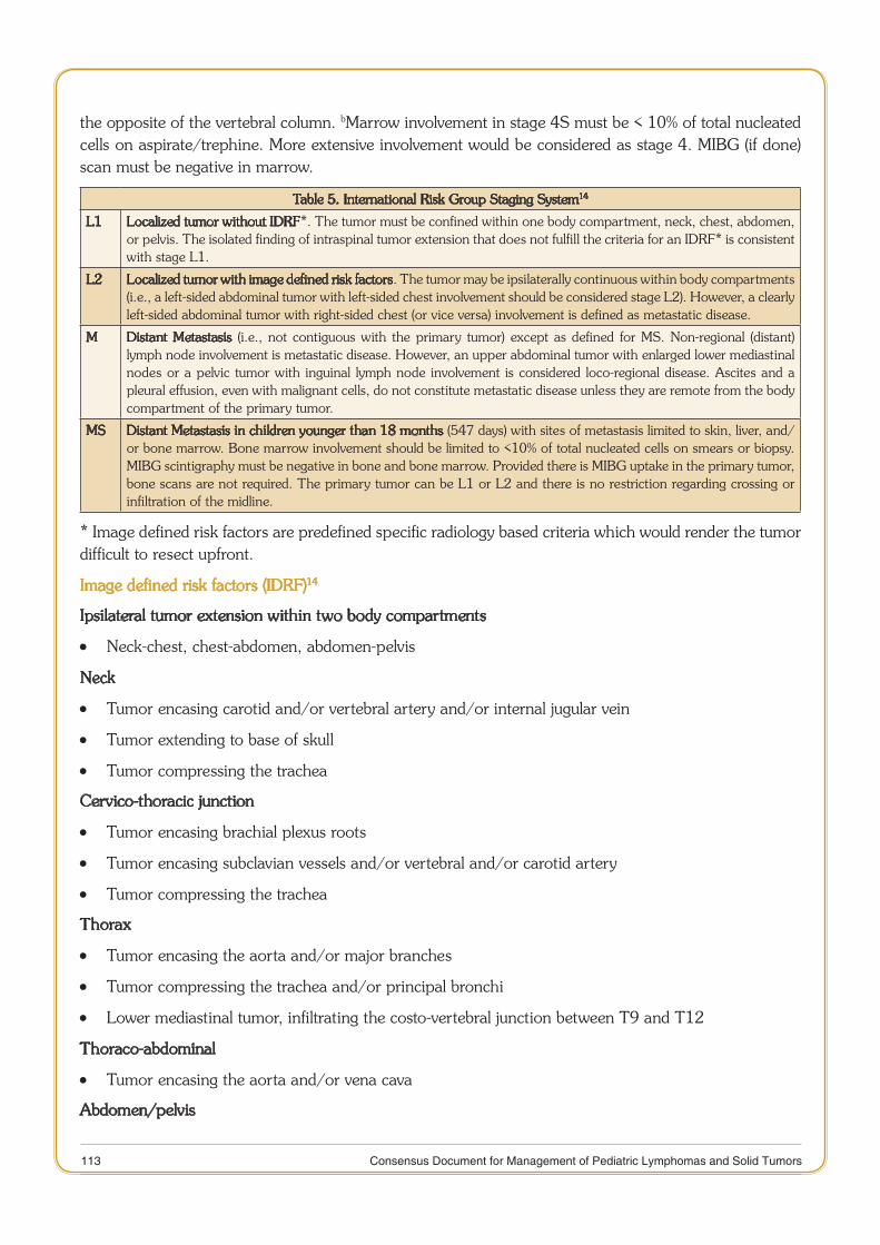
113 Consensus Document for Management of Pediatric Lymphomas and Solid Tumors
the opposite of the vertebral column. bMarrow involvement in stage 4S must be < 10% of total nucleated cells on aspirate/trephine. More extensive involvement would be considered as stage 4. MIBG (if done) scan must be negative in marrow.
Table 5. International Risk Group Staging System14
L1 Localized tumor without IDRF*. The tumor must be confined within one body compartment, neck, chest, abdomen, or pelvis. The isolated finding of intraspinal tumor extension that does not fulfill the criteria for an IDRF* is consistent with stage L1.
L2 Localized tumor with image defined risk factors. The tumor may be ipsilaterally continuous within body compartments (i.e., a left-sided abdominal tumor with left-sided chest involvement should be considered stage L2). However, a clearly left-sided abdominal tumor with right-sided chest (or vice versa) involvement is defined as metastatic disease.
M Distant Metastasis (i.e., not contiguous with the primary tumor) except as defined for MS. Non-regional (distant) lymph node involvement is metastatic disease. However, an upper abdominal tumor with enlarged lower mediastinal nodes or a pelvic tumor with inguinal lymph node involvement is considered loco-regional disease. Ascites and a pleural effusion, even with malignant cells, do not constitute metastatic disease unless they are remote from the body compartment of the primary tumor.
MS Distant Metastasis in children younger than 18 months (547 days) with sites of metastasis limited to skin, liver, and/or bone marrow. Bone marrow involvement should be limited to <10% of total nucleated cells on smears or biopsy. MIBG scintigraphy must be negative in bone and bone marrow. Provided there is MIBG uptake in the primary tumor, bone scans are not required. The primary tumor can be L1 or L2 and there is no restriction regarding crossing or infiltration of the midline.
* Image defined risk factors are predefined specific radiology based criteria which would render the tumor difficult to resect upfront.
Image defined risk factors (IDRF)14
Ipsilateral tumor extension within two body compartments
Neck-chest, chest-abdomen, abdomen-pelvis •
Neck
Tumor encasing carotid and/or vertebral artery and/or internal jugular vein •
Tumor extending to base of skull •
Tumor compressing the trachea •
Cervico-thoracic junction
Tumor encasing brachial plexus roots •
Tumor encasing subclavian vessels and/or vertebral and/or carotid artery •
Tumor compressing the trachea •
Thorax
Tumor encasing the aorta and/or major branches •
Tumor compressing the trachea and/or principal bronchi •
Lower mediastinal tumor, infiltrating the costo-vertebral junction between T9 and T12 •
Thoraco-abdominal
Tumor encasing the aorta and/or vena cava •
Abdomen/pelvis
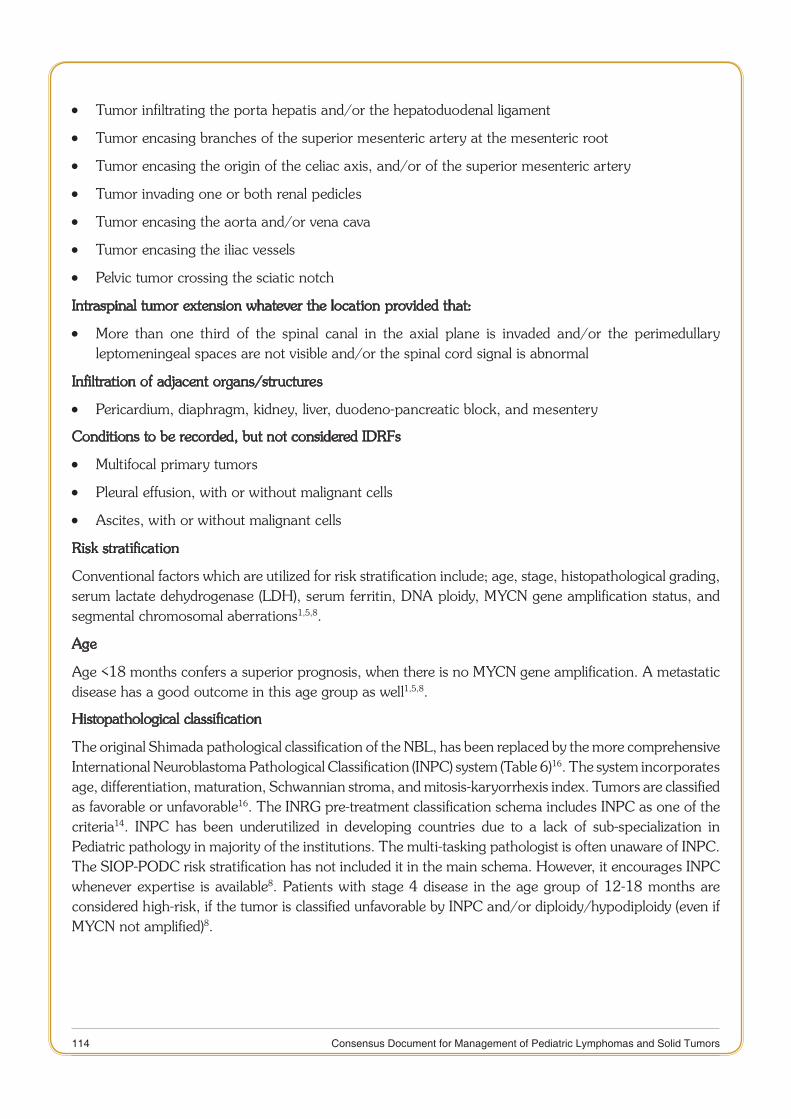
114 Consensus Document for Management of Pediatric Lymphomas and Solid Tumors
Tumor infiltrating the porta hepatis and/or the hepatoduodenal ligament •
Tumor encasing branches of the superior mesenteric artery at the mesenteric root •
Tumor encasing the origin of the celiac axis, and/or of the superior mesenteric artery •
Tumor invading one or both renal pedicles •
Tumor encasing the aorta and/or vena cava •
Tumor encasing the iliac vessels •
Pelvic tumor crossing the sciatic notch •
Intraspinal tumor extension whatever the location provided that:
More than one third of the spinal canal in the axial plane is invaded and/or the perimedullary •leptomeningeal spaces are not visible and/or the spinal cord signal is abnormal
Infiltration of adjacent organs/structures
Pericardium, diaphragm, kidney, liver, duodeno-pancreatic block, and mesentery •
Conditions to be recorded, but not considered IDRFs
Multifocal primary tumors •
Pleural effusion, with or without malignant cells •
Ascites, with or without malignant cells •
Risk stratification
Conventional factors which are utilized for risk stratification include; age, stage, histopathological grading, serum lactate dehydrogenase (LDH), serum ferritin, DNA ploidy, MYCN gene amplification status, and segmental chromosomal aberrations1,5,8.
Age
Age <18 months confers a superior prognosis, when there is no MYCN gene amplification. A metastatic disease has a good outcome in this age group as well1,5,8.
Histopathological classification
The original Shimada pathological classification of the NBL, has been replaced by the more comprehensive International Neuroblastoma Pathological Classification (INPC) system (Table 6)16. The system incorporates age, differentiation, maturation, Schwannian stroma, and mitosis-karyorrhexis index. Tumors are classified as favorable or unfavorable16. The INRG pre-treatment classification schema includes INPC as one of the criteria14. INPC has been underutilized in developing countries due to a lack of sub-specialization in Pediatric pathology in majority of the institutions. The multi-tasking pathologist is often unaware of INPC. The SIOP-PODC risk stratification has not included it in the main schema. However, it encourages INPC whenever expertise is available8. Patients with stage 4 disease in the age group of 12-18 months are considered high-risk, if the tumor is classified unfavorable by INPC and/or diploidy/hypodiploidy (even if MYCN not amplified)8.

115 Consensus Document for Management of Pediatric Lymphomas and Solid Tumors
Table 6. International Neuroblastoma Pathology Classification16
Category and subtype Prognostic group
Neuroblastoma (Schwannian stroma-poor)
UndifferentiatedPoorly differentiatedDifferentiating
All UH< 1.5 years with MKI < 4% FH, rest UH< 1.5 years with MKI < 4% and 1.5-5 years with MKI < 2% FH, rest UH
Ganglioneuroblastoma, intermixed (Schwannian stroma-rich) FH
Ganglioneuroma (Schwannian stroma-dominant)
MaturingMature
FH
Ganglioneuroblastoma, nodular (Schwannian
stroma-rich/stroma-dominant and stroma poor)
UH
FH, Favorable histology; UH, Unfavorable histology; MKI, Mitosis-karyorrhexis index.
MYCN
Amplification of the MYCN (MYCN-A) gene is the single most important biological marker for risk stratification and treatment assignment in NBL1,8. MYCN-A correlates with advanced stage, unfavorable tumor biology and poor outcome1. Additionally, it predicts a poor outcome even among apparently favorable groups such as infants, stage 4S and lower stages. The frequency of MYCN-A was reported as 3%, 4%, 25%, 32% and 8% in INSS 1,2,3,4 and 4S, respectively (overall 18%), in a large COG cohort1.
The MYCN status is conventionally determined from the tumor sample at diagnosis by FISH or PCR17. The International consensus for NBL molecular diagnostics recommends FISH as the preferred technique, especially since quality control is better ensured17. MYCN-A is defined as greater than four fold increase in the number of MYCN copies (FISH signals), when compared to a control 2p probe1,17. In the circumstance of diagnosis being established from trephine biopsy, adequate sample must be preserved for performing MYCN analysis.
FISH for MYCN is not available in-house in the majority of hospitals in India. It is a critical investigation in two categories of patients: 1) All patients with stage 3 disease, and 2) Patients < 18 months of age with stage 4 disease, where amplification confers a high-risk status for the purpose of treatment8. Among a large cohort of 958 patients (<18 months old) with stage 4 disease the incidence of MYCN-A was 12% vs. 48% in the age group of 0-12 vs. 12-18 months, respectively18. In stage 1 and 2, MYCN-A is observed in < 5% patients; hence, if unavailable, it can be assumed to be non-amplified. In a large cohort of 717 patients with stage 4S disease, the incidence of MYCN-A was 8% and 39% in patients 0-12 and 12-18 months old, respectively18. Status of amplification in stage 4S will determine the decision for intensive therapy versus merely observation or administration of intermediate-risk chemotherapy1,8,9. Attempts should be made to obtain MYCN in stage 4S as well, particularly in the age group of 12-18 months.
Status of MYCN in patients > 18 months with stage 4 disease will not alter therapy, as are high-risk otherwise. MYCN need not be actively sought for in this group of patients. The prognostic relevance of MYCN may emerge in this group as well, as the survival improves1.
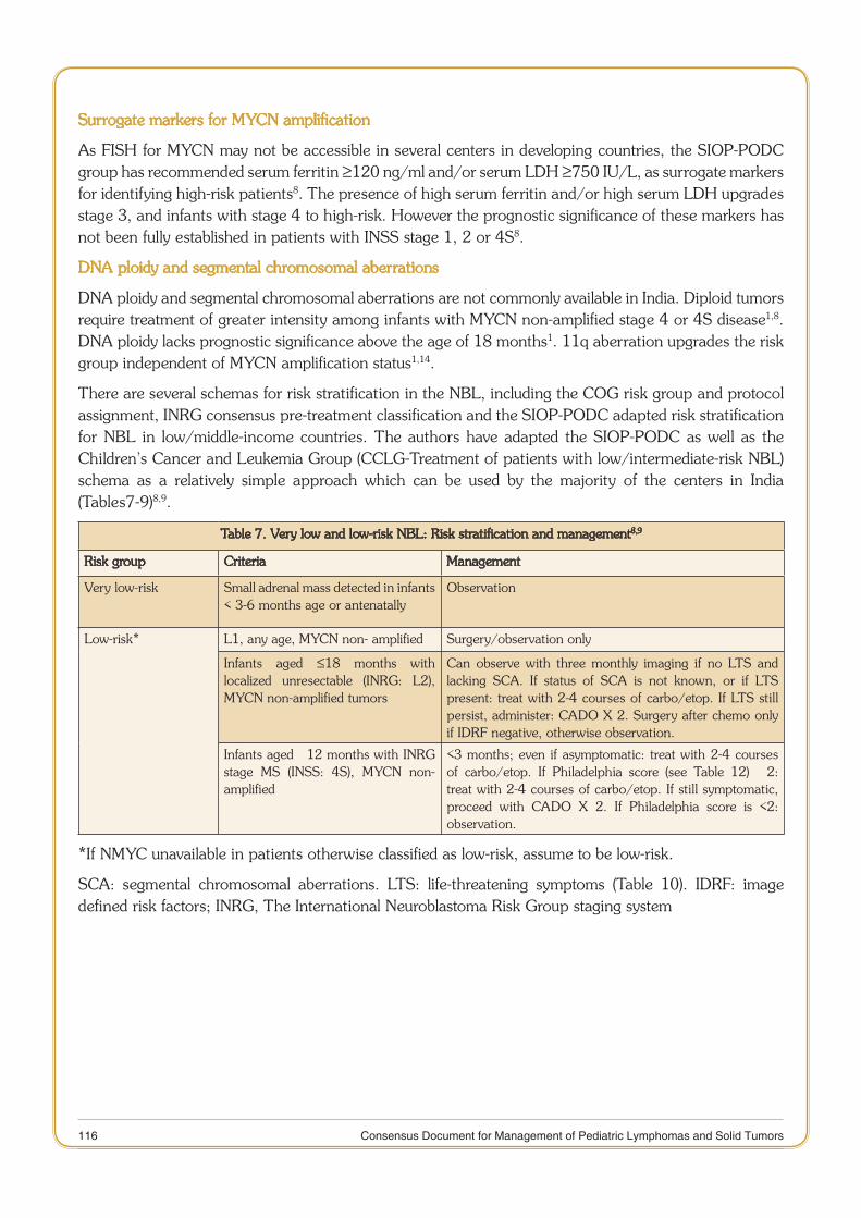
116 Consensus Document for Management of Pediatric Lymphomas and Solid Tumors
Surrogate markers for MYCN amplification
As FISH for MYCN may not be accessible in several centers in developing countries, the SIOP-PODC group has recommended serum ferritin ≥120 ng/ml and/or serum LDH ≥750 IU/L, as surrogate markers for identifying high-risk patients8. The presence of high serum ferritin and/or high serum LDH upgrades stage 3, and infants with stage 4 to high-risk. However the prognostic significance of these markers has not been fully established in patients with INSS stage 1, 2 or 4S8.
DNA ploidy and segmental chromosomal aberrations
DNA ploidy and segmental chromosomal aberrations are not commonly available in India. Diploid tumors require treatment of greater intensity among infants with MYCN non-amplified stage 4 or 4S disease1,8. DNA ploidy lacks prognostic significance above the age of 18 months1. 11q aberration upgrades the risk group independent of MYCN amplification status1,14.
There are several schemas for risk stratification in the NBL, including the COG risk group and protocol assignment, INRG consensus pre-treatment classification and the SIOP-PODC adapted risk stratification for NBL in low/middle-income countries. The authors have adapted the SIOP-PODC as well as the Children’s Cancer and Leukemia Group (CCLG-Treatment of patients with low/intermediate-risk NBL) schema as a relatively simple approach which can be used by the majority of the centers in India (Tables7-9)8,9.
Table 7. Very low and low-risk NBL: Risk stratification and management8,9
Risk group Criteria Management
Very low-risk Small adrenal mass detected in infants < 3-6 months age or antenatally
Observation
Low-risk* L1, any age, MYCN non- amplified Surgery/observation only
Infants aged ≤18 months with localized unresectable (INRG: L2), MYCN non-amplified tumors
Can observe with three monthly imaging if no LTS and lacking SCA. If status of SCA is not known, or if LTS present: treat with 2-4 courses of carbo/etop. If LTS still persist, administer: CADO X 2. Surgery after chemo only if IDRF negative, otherwise observation.
Infants aged ≤ 12 months with INRG stage MS (INSS: 4S), MYCN non-amplified
<3 months; even if asymptomatic: treat with 2-4 courses of carbo/etop. If Philadelphia score (see Table 12) ≥ 2: treat with 2-4 courses of carbo/etop. If still symptomatic, proceed with CADO X 2. If Philadelphia score is <2: observation.
*If NMYC unavailable in patients otherwise classified as low-risk, assume to be low-risk.
SCA: segmental chromosomal aberrations. LTS: life-threatening symptoms (Table 10). IDRF: image defined risk factors; INRG, The International Neuroblastoma Risk Group staging system

117 Consensus Document for Management of Pediatric Lymphomas and Solid Tumors
Table 8. Intermediate-risk NBL: Risk stratification and management8,9
Criteria Management
Age >18 months with localized unre-sectable (INRG: L2) non-MYCN ampli-fied tumors(or) low serum ferritin and/or LDH when MYCN unknown^
Histology: INPC differentiatingRx 4 courses of chemotherapy VP/Carbo x 2 – if tumor responds continue with further VP/Carbo x 2, if no response continue with CADO x 2 then: If IDRF negative, proceed for surgical resection
Histology: INPC undifferentiated or poorly differentiated <5 yrs of ageRx 6 courses of chemotherapy, surgery, radiotherapy & cis-retinoic acid VP/Carbo x 2, CADO x 2;Followed by:
If tumor response & IDRF negative then: surgical resection followed by VP/ •Carbo x 1, CADO x 1 if there was response to initial VP/Carbo, or CADO x 2 if no initial response to VP/CarboIf no tumor response & IDRF persist: surgical resection followed by CADO •x 2 If tumor response but IDRF persist: VP/Carbo x 1, CADO x 1 if there was •response to initial VP/Carbo, or CADO x 2 if no initial response to VP/Carbo then surgical resection Surgery should be followed by radiotherapy + 6 courses of 13 cis-retinoic •acid
If > 5 years with undifferentiated or poorly differentiated tumor histology, consider treatment according to high-risk protocol.
Stage INRG: L1, MYCN amplified age ≤10 years
Rx 6 courses of chemotherapy:VP/Carbo x 2, CADO x 2, VP/Carbo x 1, CADO x 1 Surgery Radiotherapy + 6 courses of 13 cis-retinoic acid.
Age ≤12 months with stage 4(INRG: M) disease, MYCN not amplified (or) low serum ferritin &/or LDH when MYCN unknown^
VP/Carbo x 2 then If response shown, proceed for further VP/Carbo x 2,& if metastatic remis- •sion achieved proceed for surgical resection of primary, if metastatic remis-sion not achieved continue with CADO x 2-4 courses to achieve metastatic remission If no response after initial VP/Carbo proceed for CADO x 2-4 to achieve •metastatic remission.Note: metastatic remission is all sites other than the liver. Surgical resection •when metastatic remission achieved & no further chemotherapy
INPC, International Neuroblastoma Pathological Classification system.SIOP-PODC, International Society of Pediatric Oncology- Committee on Developing Countries; INRG, The International Neuroblastoma Risk Group staging system, LDH, Lactate dehydrogenase. IDRF: image defined risk factors.
^If NMYC unavailable in INSS3 and infants with INSS4, serum LDH ≥750 IU/L and/or serum ferritin ≥ ≥120 ng/ml can be used as surrogate markers to classify the patient as high-risk.
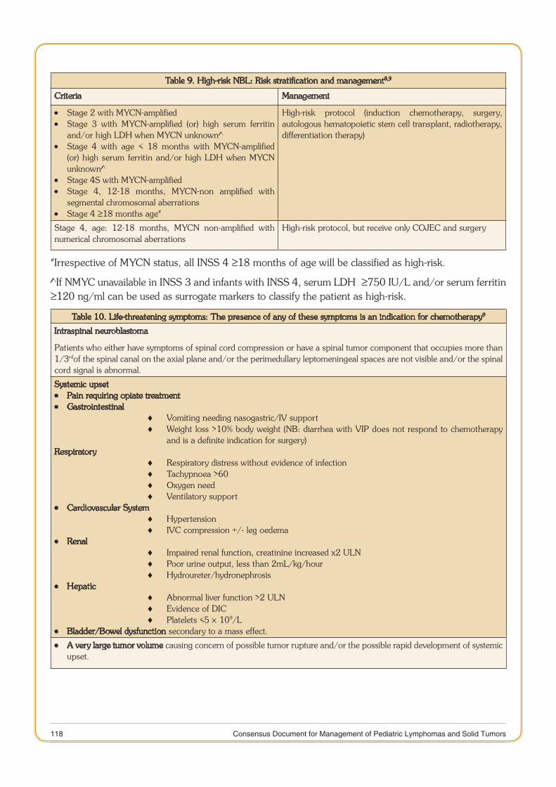
118 Consensus Document for Management of Pediatric Lymphomas and Solid Tumors
Table 9. High-risk NBL: Risk stratification and management8,9
Criteria Management
Stage 2 with MYCN-amplified •Stage 3 with MYCN-amplified (or) high serum ferritin •and/or high LDH when MYCN unknown^Stage 4 with age < 18 months with MYCN-amplified •(or) high serum ferritin and/or high LDH when MYCN unknown^Stage 4S with MYCN-amplified •Stage 4, 12-18 months, MYCN-non amplified with •segmental chromosomal aberrationsStage 4 • ≥18 months age#
High-risk protocol (induction chemotherapy, surgery, autologous hematopoietic stem cell transplant, radiotherapy, differentiation therapy)
Stage 4, age: 12-18 months, MYCN non-amplified with numerical chromosomal aberrations
High-risk protocol, but receive only COJEC and surgery
#Irrespective of MYCN status, all INSS 4 ≥18 months of age will be classified as high-risk.
^If NMYC unavailable in INSS 3 and infants with INSS 4, serum LDH ≥≥750 IU/L and/or serum ferritin ≥120 ng/ml can be used as surrogate markers to classify the patient as high-risk.
Table 10. Life-threatening symptoms: The presence of any of these symptoms is an indication for chemotherapy9
Intraspinal neuroblastoma
Patients who either have symptoms of spinal cord compression or have a spinal tumor component that occupies more than 1/3rdof the spinal canal on the axial plane and/or the perimedullary leptomeningeal spaces are not visible and/or the spinal cord signal is abnormal.
Systemic upset Pain requiring opiate treatment •Gastrointestinal •
Vomiting needing nasogastric/IV support ♦Weight loss >10% body weight (NB: diarrhea with VIP does not respond to chemotherapy ♦and is a definite indication for surgery)
RespiratoryRespiratory distress without evidence of infection ♦Tachypnoea >60 ♦Oxygen need ♦Ventilatory support ♦
Cardiovascular System •Hypertension ♦IVC compression +/- leg oedema ♦
Renal •Impaired renal function, creatinine increased x2 ULN ♦Poor urine output, less than 2mL/kg/hour ♦Hydroureter/hydronephrosis ♦
Hepatic •Abnormal liver function >2 ULN ♦Evidence of DIC ♦Platelets <5 ♦ ´ 109/L
Bladder/Bowel dysfunction • secondary to a mass effect.
A very large tumor volume • causing concern of possible tumor rupture and/or the possible rapid development of systemic upset.
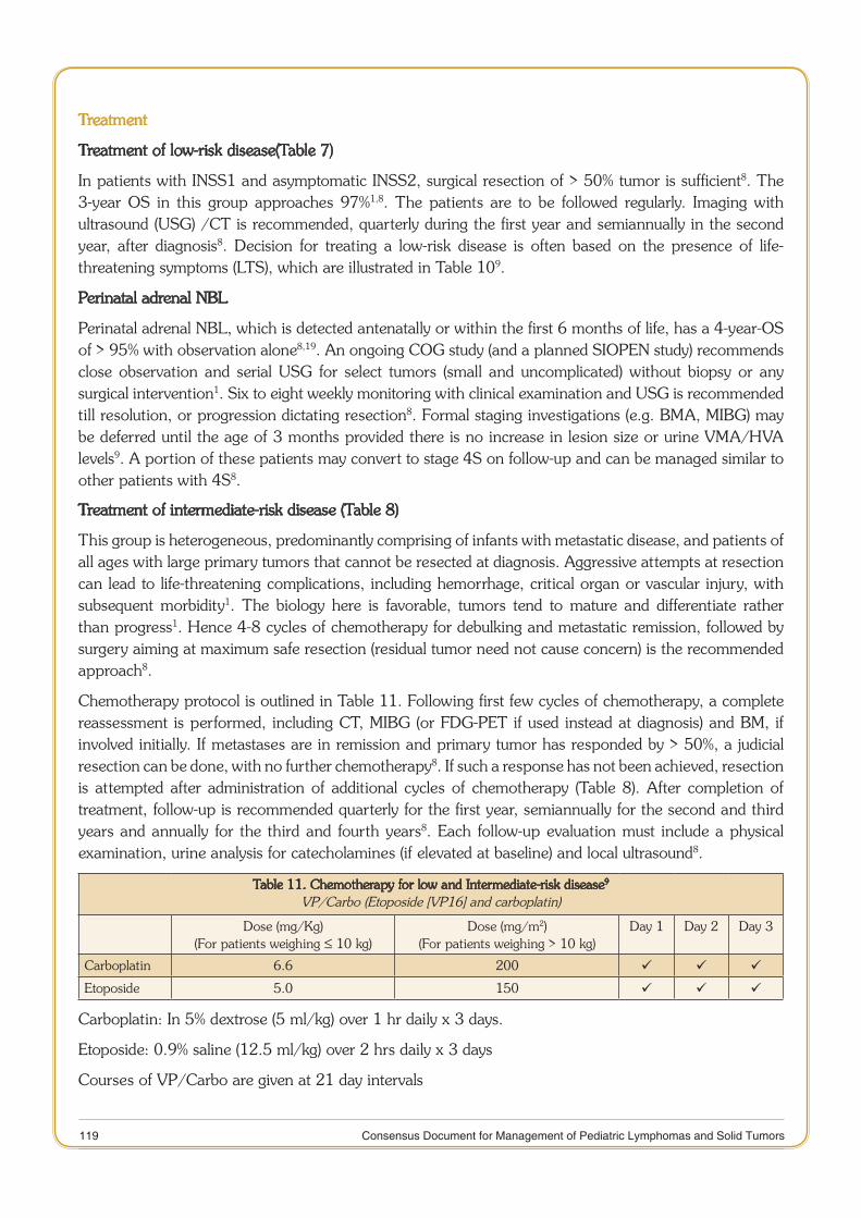
119 Consensus Document for Management of Pediatric Lymphomas and Solid Tumors
Treatment
Treatment of low-risk disease(Table 7)
In patients with INSS1 and asymptomatic INSS2, surgical resection of > 50% tumor is sufficient8. The 3-year OS in this group approaches 97%1,8. The patients are to be followed regularly. Imaging with ultrasound (USG) /CT is recommended, quarterly during the first year and semiannually in the second year, after diagnosis8. Decision for treating a low-risk disease is often based on the presence of life-threatening symptoms (LTS), which are illustrated in Table 109.
Perinatal adrenal NBL
Perinatal adrenal NBL, which is detected antenatally or within the first 6 months of life, has a 4-year-OS of > 95% with observation alone8,19. An ongoing COG study (and a planned SIOPEN study) recommends close observation and serial USG for select tumors (small and uncomplicated) without biopsy or any surgical intervention1. Six to eight weekly monitoring with clinical examination and USG is recommended till resolution, or progression dictating resection8. Formal staging investigations (e.g. BMA, MIBG) may be deferred until the age of 3 months provided there is no increase in lesion size or urine VMA/HVA levels9. A portion of these patients may convert to stage 4S on follow-up and can be managed similar to other patients with 4S8.
Treatment of intermediate-risk disease (Table 8)
This group is heterogeneous, predominantly comprising of infants with metastatic disease, and patients of all ages with large primary tumors that cannot be resected at diagnosis. Aggressive attempts at resection can lead to life-threatening complications, including hemorrhage, critical organ or vascular injury, with subsequent morbidity1. The biology here is favorable, tumors tend to mature and differentiate rather than progress1. Hence 4-8 cycles of chemotherapy for debulking and metastatic remission, followed by surgery aiming at maximum safe resection (residual tumor need not cause concern) is the recommended approach8.
Chemotherapy protocol is outlined in Table 11. Following first few cycles of chemotherapy, a complete reassessment is performed, including CT, MIBG (or FDG-PET if used instead at diagnosis) and BM, if involved initially. If metastases are in remission and primary tumor has responded by > 50%, a judicial resection can be done, with no further chemotherapy8. If such a response has not been achieved, resection is attempted after administration of additional cycles of chemotherapy (Table 8). After completion of treatment, follow-up is recommended quarterly for the first year, semiannually for the second and third years and annually for the third and fourth years8. Each follow-up evaluation must include a physical examination, urine analysis for catecholamines (if elevated at baseline) and local ultrasound8.
Table 11. Chemotherapy for low and Intermediate-risk disease9
VP/Carbo (Etoposide [VP16] and carboplatin)
Dose (mg/Kg)(For patients weighing ≤ 10 kg)
Dose (mg/m2)(For patients weighing > 10 kg)
Day 1 Day 2 Day 3
Carboplatin 6.6 200
Etoposide 5.0 150
Carboplatin: In 5% dextrose (5 ml/kg) over 1 hr daily x 3 days.
Etoposide: 0.9% saline (12.5 ml/kg) over 2 hrs daily x 3 days
Courses of VP/Carbo are given at 21 day intervals
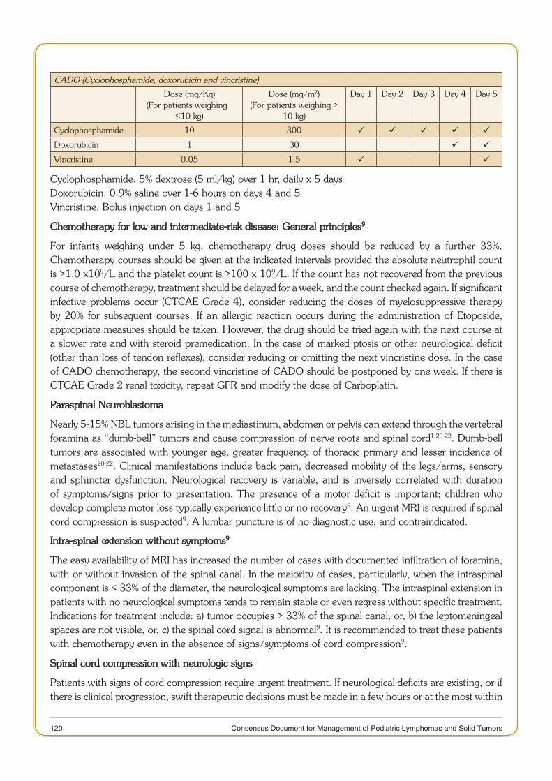
120 Consensus Document for Management of Pediatric Lymphomas and Solid Tumors
CADO (Cyclophosphamide, doxorubicin and vincristine)
Dose (mg/Kg)(For patients weighing ≤
≤10 kg)
Dose (mg/m2)(For patients weighing >
10 kg)
Day 1 Day 2 Day 3 Day 4 Day 5
Cyclophosphamide 10 300
Doxorubicin 1 30
Vincristine 0.05 1.5
Cyclophosphamide: 5% dextrose (5 ml/kg) over 1 hr, daily x 5 daysDoxorubicin: 0.9% saline over 1-6 hours on days 4 and 5Vincristine: Bolus injection on days 1 and 5
Chemotherapy for low and intermediate-risk disease: General principles9
For infants weighing under 5 kg, chemotherapy drug doses should be reduced by a further 33%. Chemotherapy courses should be given at the indicated intervals provided the absolute neutrophil count is >1.0 x10⁹/L and the platelet count is >100 x 109/L. If the count has not recovered from the previous course of chemotherapy, treatment should be delayed for a week, and the count checked again. If significant infective problems occur (CTCAE Grade 4), consider reducing the doses of myelosuppressive therapy by 20% for subsequent courses. If an allergic reaction occurs during the administration of Etoposide, appropriate measures should be taken. However, the drug should be tried again with the next course at a slower rate and with steroid premedication. In the case of marked ptosis or other neurological deficit (other than loss of tendon reflexes), consider reducing or omitting the next vincristine dose. In the case of CADO chemotherapy, the second vincristine of CADO should be postponed by one week. If there is CTCAE Grade 2 renal toxicity, repeat GFR and modify the dose of Carboplatin.
Paraspinal Neuroblastoma
Nearly 5-15% NBL tumors arising in the mediastinum, abdomen or pelvis can extend through the vertebral foramina as “dumb-bell” tumors and cause compression of nerve roots and spinal cord1,20-22. Dumb-bell tumors are associated with younger age, greater frequency of thoracic primary and lesser incidence of metastases20-22. Clinical manifestations include back pain, decreased mobility of the legs/arms, sensory and sphincter dysfunction. Neurological recovery is variable, and is inversely correlated with duration of symptoms/signs prior to presentation. The presence of a motor deficit is important; children who develop complete motor loss typically experience little or no recovery9. An urgent MRI is required if spinal cord compression is suspected9. A lumbar puncture is of no diagnostic use, and contraindicated.
Intra-spinal extension without symptoms9
The easy availability of MRI has increased the number of cases with documented infiltration of foramina, with or without invasion of the spinal canal. In the majority of cases, particularly, when the intraspinal component is < 33% of the diameter, the neurological symptoms are lacking. The intraspinal extension in patients with no neurological symptoms tends to remain stable or even regress without specific treatment. Indications for treatment include: a) tumor occupies > 33% of the spinal canal, or, b) the leptomeningeal spaces are not visible, or, c) the spinal cord signal is abnormal9. It is recommended to treat these patients with chemotherapy even in the absence of signs/symptoms of cord compression9.
Spinal cord compression with neurologic signs
Patients with signs of cord compression require urgent treatment. If neurological deficits are existing, or if there is clinical progression, swift therapeutic decisions must be made in a few hours or at the most within
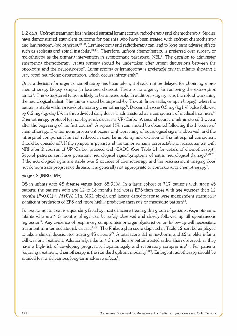
121 Consensus Document for Management of Pediatric Lymphomas and Solid Tumors
1-2 days. Upfront treatment has included surgical laminectomy, radiotherapy and chemotherapy. Studies have demonstrated equivalent outcome for patients who have been treated with upfront chemotherapy and laminectomy/radiotherapy20-22. Laminectomy and radiotherapy can lead to long-term adverse effects such as scoliosis and spinal instability21,22. Therefore, upfront chemotherapy is preferred over surgery or radiotherapy as the primary intervention in symptomatic paraspinal NBL1. The decision to administer emergency chemotherapy versus surgery should be undertaken after urgent discussions between the oncologist and the neurosurgeon9. Laminectomy or laminotomy is preferable only in infants showing a very rapid neurologic deterioration, which occurs infrequently9.
Once a decision for urgent chemotherapy has been taken, it should not be delayed for obtaining a pre-chemotherapy biopsy sample (in localized disease). There is no urgency for removing the extra-spinal tumor9. The extra-spinal tumor is likely to be unresectable. In addition, surgery runs the risk of worsening the neurological deficit. The tumor should be biopsied (by Tru-cut, fine-needle, or open biopsy), when the patient is stable within a week of initiating chemotherapy9. Dexamethasone 0.5 mg/kg I.V. bolus followed by 0.2 mg/kg/day I.V. in three divided daily doses is administered as a component of medical treatment9. Chemotherapy protocol for non-high-risk disease is VP/Carbo. A second course is administered 3 weeks after the beginning of the first course9. A repeat MRI scan should be obtained following the 1stcourse of chemotherapy. If either no improvement occurs or if worsening of neurological signs is observed, and the intraspinal component has not reduced in size, laminotomy and excision of the intraspinal component should be considered9. If the symptoms persist and the tumor remains unresectable on reassessment with MRI after 2 courses of VP/Carbo, proceed with CADO (See Table 11 for details of chemotherapy)9. Several patients can have persistent neurological signs/symptoms of initial neurological damage9,20,21. If the neurological signs are stable over 2 courses of chemotherapy and the reassessment imaging does not demonstrate progressive disease, it is generally not appropriate to continue with chemotherapy9.
Stage 4S (INRG: MS)
OS in infants with 4S disease varies from 85-92%1. In a large cohort of 717 patients with stage 4S pattern, the patients with age 12 to 18 months had worse EFS than those with age younger than 12 months (P<0.01)18. MYCN, 11q, MKI, ploidy, and lactate dehydrogenase were independent statistically significant predictors of EFS and more highly predictive than age or metastatic pattern18.
To treat or not to treat is a quandary faced by most clinicians treating this group of patients. Asymptomatic infants who are > 3 months of age can be safely observed and closely followed up till spontaneous regression8. Any evidence of respiratory compromise or organ dysfunction on follow-up will necessitate treatment as intermediate-risk disease1,8,9. The Philadelphia score depicted in Table 12 can be employed to take a clinical decision for treating 4S disease23. A total score ≥≥1 in newborns and ≥2 in older infants will warrant treatment. Additionally, infants < 3 months are better treated rather than observed, as they have a high-risk of developing progressive hepatomegaly and respiratory compromise1,8. For patients requiring treatment, chemotherapy is the standard upfront modality1,8,9. Emergent radiotherapy should be avoided for its deleterious long-term adverse effects1.

122 Consensus Document for Management of Pediatric Lymphomas and Solid Tumors
Table 12. Philadelphia score for infants with stage 4S disease23
Clinical parameter Score 0 Score 1 Score 2
Emesis Absent >10% intake Severe requiring intravenous fluids
Respiratory involvement Absent Respiratory rate >60, O2support
requirementCPAP, Ventilation
Edema(Obstruction of Venous return) Absent Pedal Sacral and scrotal
Renal involvement Absent Oliguria < 2ml/kg/hr urea/creatinine above age appropri-ate level
Liver involvement Absent - Evidence of DIC/ Platelet count < 50,000/µL
CPAP, Continuous positive airway pressure; DIC, Disseminated intravascular coagulation. A total score ≥ ≥1 in newborns and ≥2 in older infants will warrant treatment in 4S disease.
A diagnostic biopsy is indicated for evaluation of tumor biology, importantly MYCN amplification status. If subcutaneous nodules are present, they can be safely biopsied as compared to liver or primary site1. When indicated, treat with 2-4 courses of carbo/etop9. Following carbo/etop if symptoms resolve, proceed with the observation, otherwise administer CADO X 2 (Table 11)9. Resection of the primary tumor does not seem to influence the outcome.
High-risk NBL
Among patients with NBL, patients with high-risk disease constitute the majority in day to day practice. This sub-group is a challenge for the pediatric oncologist. Treatment typically is spread over a period of one year and includes five distinct phases1,8:
a) Induction chemotherapy: The aim is to achieve maximum reduction of tumor burden with intensive chemotherapy, including reduction of metastatic disease within a timeframe that will minimize the risk of developing resistant tumor clones and clinical progression.
b) Surgery of the primary tumor
c) Myeloablative chemotherapy with autologous hematopoietic stem cell rescue: For treatment of potentially resistant residual tumor
d) Radiotherapy: To primary tumor bed and residual metastatic sites
e) Treatment of minimal residual disease (MRD)
Induction chemotherapy
No single chemotherapy induction regimen has proven to be superior for induction response and EFS in high-risk NBL8. The several regimens reported administer multi-agent chemotherapy with an alkylator therapy such as cyclophosphamide, platinum agent, topoisomerase, usually etoposide or less frequently, doxorubicin and vincristine.
The treating team may decide to offer upfront palliative care to a patient with high-risk NBL due to restricted supportive care or lack of resources. One option suggested by SIOP-PODC is a regimen with low-dose metronomic Oral cyclophosphamide 25 mg/m2 daily (max 50 mg) or oral etoposide 50 mg/m2/day for palliation of pain and improvement of quality of life8. An alternate non-curative therapy would involve administering 1-2 cycles of intermediate-risk chemotherapy and local radiation to the primary tumor bed to allow for some tumor reduction, pain relief and improved quality of life8.
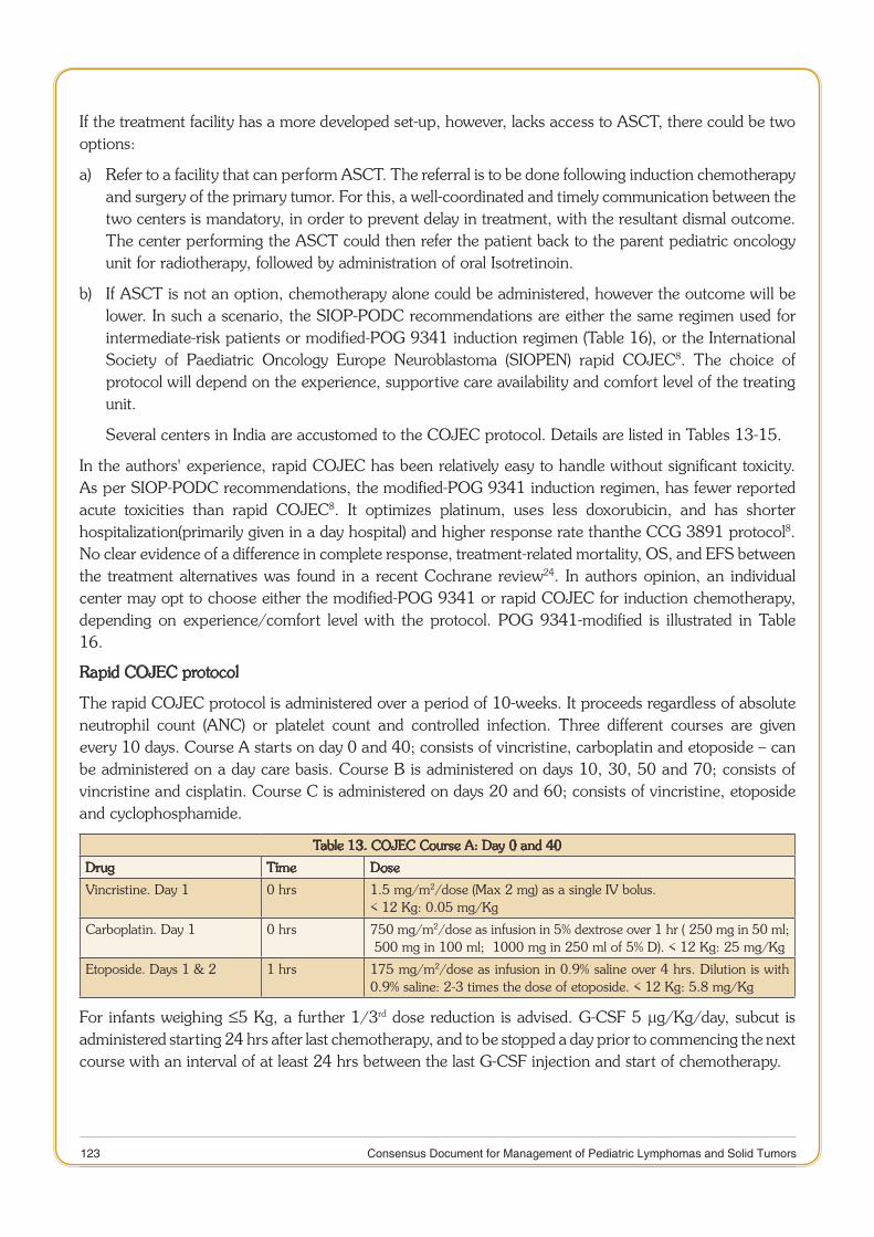
123 Consensus Document for Management of Pediatric Lymphomas and Solid Tumors
If the treatment facility has a more developed set-up, however, lacks access to ASCT, there could be two options:
Refer to a facility that can perform ASCT. The referral is to be done following induction chemotherapy a) and surgery of the primary tumor. For this, a well-coordinated and timely communication between the two centers is mandatory, in order to prevent delay in treatment, with the resultant dismal outcome. The center performing the ASCT could then refer the patient back to the parent pediatric oncology unit for radiotherapy, followed by administration of oral Isotretinoin.
If ASCT is not an option, chemotherapy alone could be administered, however the outcome will be b) lower. In such a scenario, the SIOP-PODC recommendations are either the same regimen used for intermediate-risk patients or modified-POG 9341 induction regimen (Table 16), or the International Society of Paediatric Oncology Europe Neuroblastoma (SIOPEN) rapid COJEC8. The choice of protocol will depend on the experience, supportive care availability and comfort level of the treating unit.
Several centers in India are accustomed to the COJEC protocol. Details are listed in Tables 13-15.
In the authors' experience, rapid COJEC has been relatively easy to handle without significant toxicity. As per SIOP-PODC recommendations, the modified-POG 9341 induction regimen, has fewer reported acute toxicities than rapid COJEC8. It optimizes platinum, uses less doxorubicin, and has shorter hospitalization(primarily given in a day hospital) and higher response rate thanthe CCG 3891 protocol8. No clear evidence of a difference in complete response, treatment-related mortality, OS, and EFS between the treatment alternatives was found in a recent Cochrane review24. In authors opinion, an individual center may opt to choose either the modified-POG 9341 or rapid COJEC for induction chemotherapy, depending on experience/comfort level with the protocol. POG 9341-modified is illustrated in Table 16.
Rapid COJEC protocol
The rapid COJEC protocol is administered over a period of 10-weeks. It proceeds regardless of absolute neutrophil count (ANC) or platelet count and controlled infection. Three different courses are given every 10 days. Course A starts on day 0 and 40; consists of vincristine, carboplatin and etoposide – can be administered on a day care basis. Course B is administered on days 10, 30, 50 and 70; consists of vincristine and cisplatin. Course C is administered on days 20 and 60; consists of vincristine, etoposide and cyclophosphamide.
Table 13. COJEC Course A: Day 0 and 40
Drug Time Dose
Vincristine. Day 1 0 hrs 1.5 mg/m2/dose (Max 2 mg) as a single IV bolus.< 12 Kg: 0.05 mg/Kg
Carboplatin. Day 1 0 hrs 750 mg/m2/dose as infusion in 5% dextrose over 1 hr (≤250 mg in 50 ml; ≤500 mg in 100 ml; ≤1000 mg in 250 ml of 5% D). < 12 Kg: 25 mg/Kg
Etoposide. Days 1 & 2 1 hrs 175 mg/m2/dose as infusion in 0.9% saline over 4 hrs. Dilution is with 0.9% saline: 2-3 times the dose of etoposide. < 12 Kg: 5.8 mg/Kg
For infants weighing ≤5 Kg, a further 1/3rd dose reduction is advised. G-CSF 5 µg/Kg/day, subcut is administered starting 24 hrs after last chemotherapy, and to be stopped a day prior to commencing the next course with an interval of at least 24 hrs between the last G-CSF injection and start of chemotherapy.
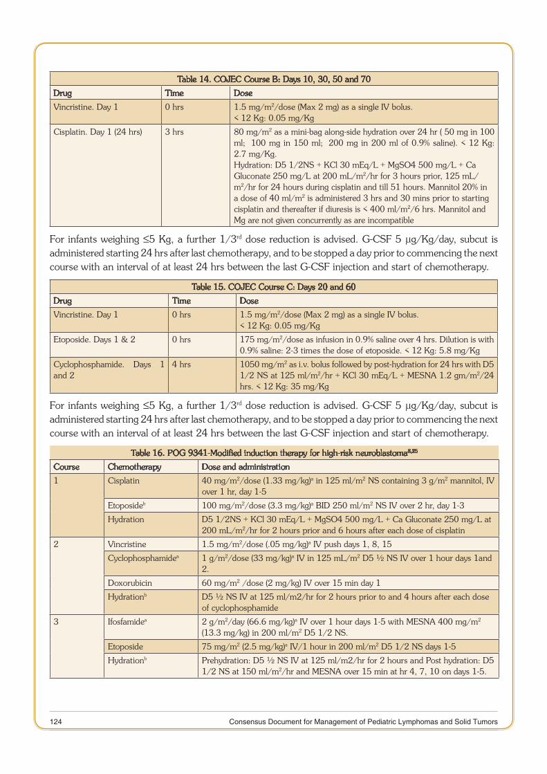
124 Consensus Document for Management of Pediatric Lymphomas and Solid Tumors
Table 14. COJEC Course B: Days 10, 30, 50 and 70
Drug Time Dose
Vincristine. Day 1 0 hrs 1.5 mg/m2/dose (Max 2 mg) as a single IV bolus.< 12 Kg: 0.05 mg/Kg
Cisplatin. Day 1 (24 hrs) 3 hrs 80 mg/m2 as a mini-bag along-side hydration over 24 hr (≤50 mg in 100 ml; ≤100 mg in 150 ml; ≤200 mg in 200 ml of 0.9% saline). < 12 Kg: 2.7 mg/Kg.Hydration: D5 1/2NS + KCl 30 mEq/L + MgSO4 500 mg/L + Ca Gluconate 250 mg/L at 200 mL/m2/hr for 3 hours prior, 125 mL/m2/hr for 24 hours during cisplatin and till 51 hours. Mannitol 20% in a dose of 40 ml/m2 is administered 3 hrs and 30 mins prior to starting cisplatin and thereafter if diuresis is < 400 ml/m2/6 hrs. Mannitol and Mg are not given concurrently as are incompatible
For infants weighing ≤5 Kg, a further 1/3rd dose reduction is advised. G-CSF 5 µg/Kg/day, subcut is administered starting 24 hrs after last chemotherapy, and to be stopped a day prior to commencing the next course with an interval of at least 24 hrs between the last G-CSF injection and start of chemotherapy.
Table 15. COJEC Course C: Days 20 and 60
Drug Time Dose
Vincristine. Day 1 0 hrs 1.5 mg/m2/dose (Max 2 mg) as a single IV bolus.< 12 Kg: 0.05 mg/Kg
Etoposide. Days 1 & 2 0 hrs 175 mg/m2/dose as infusion in 0.9% saline over 4 hrs. Dilution is with 0.9% saline: 2-3 times the dose of etoposide. < 12 Kg: 5.8 mg/Kg
Cyclophosphamide. Days 1 and 2
4 hrs 1050 mg/m2 as i.v. bolus followed by post-hydration for 24 hrs with D5 1/2 NS at 125 ml/m2/hr + KCl 30 mEq/L + MESNA 1.2 gm/m2/24 hrs. < 12 Kg: 35 mg/Kg
For infants weighing ≤5 Kg, a further 1/3rd dose reduction is advised. G-CSF 5 µg/Kg/day, subcut is administered starting 24 hrs after last chemotherapy, and to be stopped a day prior to commencing the next course with an interval of at least 24 hrs between the last G-CSF injection and start of chemotherapy.
Table 16. POG 9341-Modified induction therapy for high-risk neuroblastoma8,25
Course Chemotherapy Dose and administration
1 Cisplatin 40 mg/m2/dose (1.33 mg/kg)a in 125 ml/m2 NS containing 3 g/m2 mannitol, IV over 1 hr, day 1-5
Etoposideb 100 mg/m2/dose (3.3 mg/kg)a BID 250 ml/m2 NS IV over 2 hr, day 1-3
Hydration D5 1/2NS + KCl 30 mEq/L + MgSO4 500 mg/L + Ca Gluconate 250 mg/L at 200 mL/m2/hr for 2 hours prior and 6 hours after each dose of cisplatin
2 Vincristine 1.5 mg/m2/dose (.05 mg/kg)a IV push days 1, 8, 15
Cyclophosphamidea 1 g/m2/dose (33 mg/kg)a IV in 125 mL/m2 D5 ½ NS IV over 1 hour days 1and 2.
Doxorubicin 60 mg/m2 /dose (2 mg/kg) IV over 15 min day 1
Hydrationb D5 ½ NS IV at 125 ml/m2/hr for 2 hours prior to and 4 hours after each dose of cyclophosphamide
3 Ifosfamidea 2 g/m2/day (66.6 mg/kg)a IV over 1 hour days 1-5 with MESNA 400 mg/m2 (13.3 mg/kg) in 200 ml/m2 D5 1/2 NS.
Etoposide 75 mg/m2 (2.5 mg/kg)a IV/1 hour in 200 ml/m2 D5 1/2 NS days 1-5
Hydrationb Prehydration: D5 ½ NS IV at 125 ml/m2/hr for 2 hours and Post hydration: D5 1/2 NS at 150 ml/m2/hr and MESNA over 15 min at hr 4, 7, 10 on days 1-5.
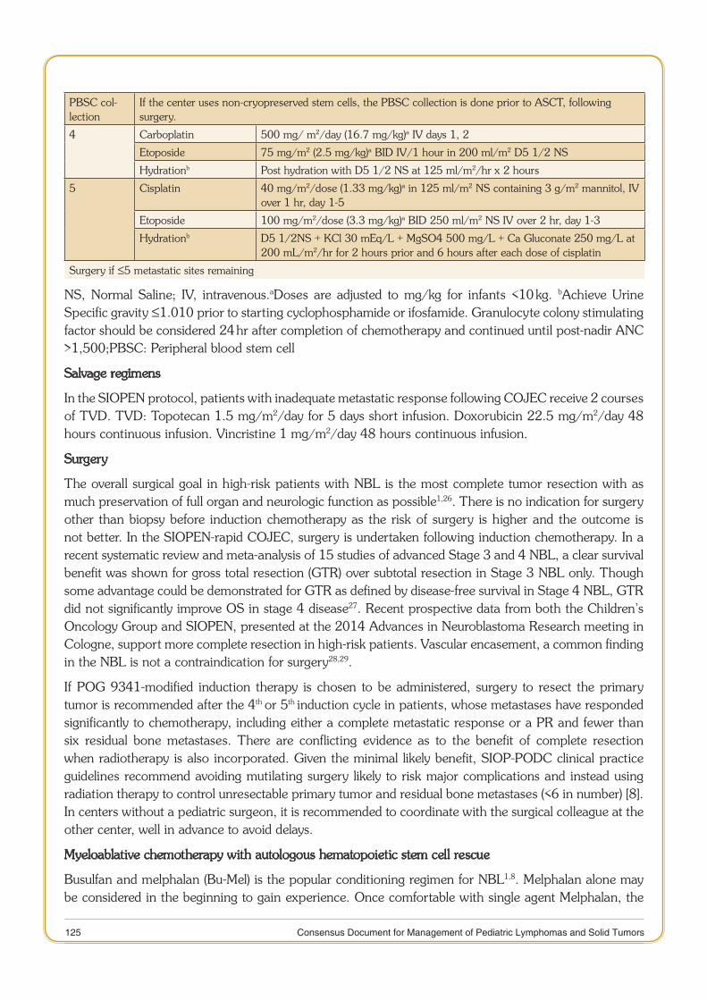
125 Consensus Document for Management of Pediatric Lymphomas and Solid Tumors
PBSC col-lection
If the center uses non-cryopreserved stem cells, the PBSC collection is done prior to ASCT, following surgery.
4 Carboplatin 500 mg/ m2/day (16.7 mg/kg)a IV days 1, 2
Etoposide 75 mg/m2 (2.5 mg/kg)a BID IV/1 hour in 200 ml/m2 D5 1/2 NS
Hydrationb Post hydration with D5 1/2 NS at 125 ml/m2/hr x 2 hours
5 Cisplatin 40 mg/m2/dose (1.33 mg/kg)a in 125 ml/m2 NS containing 3 g/m2 mannitol, IV over 1 hr, day 1-5
Etoposide 100 mg/m2/dose (3.3 mg/kg)a BID 250 ml/m2 NS IV over 2 hr, day 1-3
Hydrationb D5 1/2NS + KCl 30 mEq/L + MgSO4 500 mg/L + Ca Gluconate 250 mg/L at 200 mL/m2/hr for 2 hours prior and 6 hours after each dose of cisplatin
Surgery if ≤5 metastatic sites remaining
NS, Normal Saline; IV, intravenous.aDoses are adjusted to mg/kg for infants <10 kg. bAchieve Urine Specific gravity ≤1.010 prior to starting cyclophosphamide or ifosfamide. Granulocyte colony stimulating factor should be considered 24 hr after completion of chemotherapy and continued until post-nadir ANC >1,500;PBSC: Peripheral blood stem cell
Salvage regimens
In the SIOPEN protocol, patients with inadequate metastatic response following COJEC receive 2 courses of TVD. TVD: Topotecan 1.5 mg/m2/day for 5 days short infusion. Doxorubicin 22.5 mg/m2/day 48 hours continuous infusion. Vincristine 1 mg/m2/day 48 hours continuous infusion.
Surgery
The overall surgical goal in high-risk patients with NBL is the most complete tumor resection with as much preservation of full organ and neurologic function as possible1,26. There is no indication for surgery other than biopsy before induction chemotherapy as the risk of surgery is higher and the outcome is not better. In the SIOPEN-rapid COJEC, surgery is undertaken following induction chemotherapy. In a recent systematic review and meta-analysis of 15 studies of advanced Stage 3 and 4 NBL, a clear survival benefit was shown for gross total resection (GTR) over subtotal resection in Stage 3 NBL only. Though some advantage could be demonstrated for GTR as defined by disease-free survival in Stage 4 NBL, GTR did not significantly improve OS in stage 4 disease27. Recent prospective data from both the Children’s Oncology Group and SIOPEN, presented at the 2014 Advances in Neuroblastoma Research meeting in Cologne, support more complete resection in high-risk patients. Vascular encasement, a common finding in the NBL is not a contraindication for surgery28,29.
If POG 9341-modified induction therapy is chosen to be administered, surgery to resect the primary tumor is recommended after the 4th or 5th induction cycle in patients, whose metastases have responded significantly to chemotherapy, including either a complete metastatic response or a PR and fewer than six residual bone metastases. There are conflicting evidence as to the benefit of complete resection when radiotherapy is also incorporated. Given the minimal likely benefit, SIOP-PODC clinical practice guidelines recommend avoiding mutilating surgery likely to risk major complications and instead using radiation therapy to control unresectable primary tumor and residual bone metastases (<6 in number) [8]. In centers without a pediatric surgeon, it is recommended to coordinate with the surgical colleague at the other center, well in advance to avoid delays.
Myeloablative chemotherapy with autologous hematopoietic stem cell rescue
Busulfan and melphalan (Bu-Mel) is the popular conditioning regimen for NBL1,8. Melphalan alone may be considered in the beginning to gain experience. Once comfortable with single agent Melphalan, the

126 Consensus Document for Management of Pediatric Lymphomas and Solid Tumors
center could switch to Bu-Mel. In the rapid COJEC, the first option of PBSC harvest is following recovery from aplasia after Day 70 of COJEC chemotherapy. In the POG 9341-Modified induction therapy, a harvest could be performed after 2–5 cycles of induction. An earlier timing of apheresis generally leads to improved feasibility inobtaining sufficient PBSC numbers8. If apheresis is not available,autologous BM could be collected and stored providing that metastases have cleared8. If the center lacks cryopreservation facilities, the PBSC collection is done just prior to ASCT. The non-cryopreserved stem cell collection can be safely stored at 40C for upto 7-9 days.
If resources to conduct ASCT are not available, SIOP-PODC recommends consolidation using outpatient-based cyclophosphamide/topotecan for six cycles at 3–4 week intervals8. This combination was successful in obtaining a response in 30% of resistant or relapsed patients. An alternative therapy option would be to continue induction therapy (POG 9341) for another four cycles. It is reasonable to continue such chemotherapy with curative intent, as there was still a > 20% EFS for patients treated with intensive consolidation chemotherapy on the randomized CCG protocol8.
Intravenous vs. oral Busulfan for ASCT
Oral Bu was introduced 30 years ago. Since its introduction, it has had a mixed reputation. Oral, Bu has always carried with it the shadow of significant intra- and inter-patient variability in absorption and first pass metabolism that contributes to risk of venous-occlusive disease/sinusoidal obstruction syndrome (VOD/SOS). To address these metabolic limitations, an intravenous (IV) formulation of the drug was developed and approved for use in allogeneic transplantation in 1999. Compared to oral Bu, the IV formulation of Bu resulted in less patient-to-patient variability in metabolism, drug exposure, and early toxicities30.
Although more expensive, the perceived advantages and convenience with IV Bu has led many programs to switch from oral to IV Bu30. With time, however, one has learned that it is not as simple as one had hoped. There remains patient-to-patient variability in metabolism and this is marked in pediatrics. The hope for a simple weight-based dose, as originally introduced, has not been borne out in practice. Therapeutic drug monitoring (TDM) based on the pharmacokinetics of the first therapeutic dose or test dose has led to a better understanding of the therapeutic range for Bu (AUC 900 to 1500 micromole x min), above which the risk of toxicity, particularly VOD/SOS, increases30.
Each center has to consider the local variables and chose one form of Bu over the other. In a personal communication with Dr. Biju George – Dept. of Hematology, CMC Vellore, in regards to IV vs. oral Bu: In their experience with Indian IV Bu, in up to 90% cases, a dose adjustment (either an increase or decrease) had to be made, based on TDM. In a recent large study of oral vs. IV Bu from Japan in allogeneic stem cell transplant for pediatric acute myeloid leukemia (AML), no significant survival advantage with IV Bu was observed31. For centers that choose oral over IV Bu, the result of Japanese study is reassuring that perhaps the results with oral Bu may be acceptable31. The most important thing about the mechanics of transplantation is to do a limited number of things, understand what one does, and to do them well. This applies equally to IV vs. oral Bu30.
Supportive care for ASCT
Limited number of Pediatric Oncology centers in India are offering a regular service of myeloablative chemotherapy with autologous stem cell rescue for the high - risk NBL. The likely contributory factors for the restricted availability include: a) A low priority for the cancer with a lower cure rate, b) Preference of adult transplant units for allogeneic over autologous transplants for the NBL, c) Lack of experience for apheresis in a younger patient, d) Apprehension for performing the procedure outside of a transplant unit.
127 Consensus Document for Management of Pediatric Lymphomas and Solid Tumors
In the authors' experience, ASCT for NBL can be safely performed in the general Hematology/Oncology ward (preferably side or isolation room). Non-availability of a transplant unit or a (High-efficiency particulate arrestance) HEPA filtered room should not be considered a limitation. A central or a peripherally inserted central line is ideal and desirable, but not necessary. A pediatric oncology unit that can handle patients with AML, should be able to comfortably manage an ASCT for NBL. In the authors' experience, prevention of hospital acquired infection is a critical step for success of ASCT in India. Hospital acquired infections can be best prevented with hand hygiene, judicious and restricted use of intravenous fluids, preference of oral over IV medications when feasible, restricted use of indwelling catheters and administration of enteral over parenteral feeding.
Stem cell apheresis
G-CSF10 µg/kg/day single dose is administered for mobilizing the stem cells. The apheresis is typically done on day 4-5. In the rare case of poor mobilization, Injection Plerixaforcan be considered (Dose: 0.24 mg/kg subcutaneous, 10 hrs prior to apheresis, expensive – Rs ~ 62,000 for a vial of 1.2 ml = 24 mg)32. A femoral dialysis catheter size eight Fr (available as double lumen: Arrow/Wygon/Medcomp) is typically used for apheresis in patients below 5yrs of age.
Conditioning regimen: IV or Oral Busulfan + Melphalan8
1. Busulfan
IV Busulfan. < 10 kg: 3.2 mg/kg/dose every 24 hours x 4 doses. a. ≥10 kg and ≤4 years old: 4 mg/kg/dose every 24 hours x 4 doses. > 4 years: 3.2 mg/kg/dose every 24 hours x 4 doses. Administer at a final conc. of 0.5 mg/mL in D5W or 0.9% NaCl over 3 hrs. (Should ideally adjust doses based on therapeutic drug monitoring, if available). Or
Tab Busulfan (1 bottle = 100 tablets of 2 mg each; also available as a strip): Total dose: > 12 Kg: b. 600 mg/m2 in 16 divided doses q 6 hourly. < 12 kg: 480 mg/m2. Fasting 2 hours before and 30 minutes after Bu. Avoid acetaminophen 72 hours before and during Bu administration. Drugs that may interact with Bu include acetaminophen, itraconazole, metronidazole or phenytoin.
2. Injection Melphalan (50 mg/vial): > 12 Kg: Dose: 140 mg/m2. < 12 kg: 120 mg/m2 or 4 mg/kg. Administer at least 24 hours after last dose of Bu. Administer melphalan in 100 ml NS over 15 minutes as an IV infusion immediately after reconstitution. Final concentration: < 2 mg/mL for central line or 0.45 mg/mL for peripheral line. Give within 1 hour of reconstitution; if late discard and ask for a new vial. Monitoring vitals with cardiac monitor: diluent can cause arrhythmia/hypotension. Stem cells are infused at least 12 hours after Melphalan.
3. I.V. fluid: Prehydration for melphalan: NS 125 mL/m2/hr for ≥3 hours. Continue hydration 125 mL/m2/ hr with N/2 5% Dx till ≥≥12 hours after Melphalan. Must ensure urine output 4 mL/kg/hr: Can increase IVF or add Lasix.
4. G-CSF 5 mcg/kg/dose subcut: Daily beginning on Day +4. Continue until post-nadir ANC > 2,000/µL for three consecutive days. Alternative: Peg-G-CSF 100 mcg/kg subcut single dose on Day +4 (Available as 6 mg prefilled syringe; approx. cost Rs 3800).
Supportive drugs
Tab Ursodiol 150 mg/m1. 2/dose BD. (Tablet: 75 mg). From Day -8 to day +80, for prevention of VOD.
128 Consensus Document for Management of Pediatric Lymphomas and Solid Tumors
Tab Clonazepam: Day -6 to -1. 0.025 to 0.1 mg/kg/day in three div doses PO. Start at least 12 hours 2. before and till 24 hours after Bu. Tablet is available as 0.25, 0.5 mg.
Conditioning regimen: Single agent: IV Melphalan
This may be considered by the Pediatric Oncology team in the beginning of an ASCT program to gain experience, before switching to BuMel. Dose: 180-200 mg/m2.
Radiotherapy
Radiotherapy is administered following ASCT and prior to therapy with 13-cis-retinoic acid. In the SIOPEN HR-NBL-1 protocol, radiotherapy is administered to the initial tumor site, irrespective of the extent or the result of surgery. SIOP-PODC recommends administration of 21.6 Gy radiation to the primary tumor bed and to residual bone metastases (fewer than six sites)7. In other words, while the primary site is always irradiated, radiation is also given to those metastatic sites with persistent active disease (a positive MIBG or if not available, a positive FDG-PET) demonstrated at the time of evaluation prior to ASCT. Radiotherapy is typically administered 60 days after Bu to avoid the risk of Bu enhanced-radiotoxicity. 13-Cis RA is not co-administered with radiotherapy as a negative interaction has been described8.
Administration of 13-cis-retinoic acid
Isotretinoin is a naturally occurring analogue of Vitamin A. Growth arrest and differentiation in response to isotretinoin have been observed in NBL cell lines. The exact mechanism of RA-induced maturation of tumor cells is not known. It has been shown to enhance EFS in high-risk NBL33. 13-cis RA is administered following the completion of radiotherapy. It is not indicated for gross residual disease. Dose: 160 mg/m2/day orally in two divided doses for 14 days, followed by 14 days rest, for a total of six cycles (6 months). For patients ≤12 Kg, the dose is 5.33 mg/Kg/day in two divided doses. Isotretinoin toxicities are generally mild; consisting primarily of chelitis, dry skin, and hypertriglyceridemia, with hypercalcemia seen at higher doses8. Topical vitamin E should be applied to lips twice a day if chelitis develops. Patients should avoid direct sun exposure while on RA. Criteria to be fulfilled prior to each cycle of 13-cis-RA: a) ALT < 5 times normal, b) Skin toxicity ≤ Grade 1, c) Serum TG < 300 mg/dL, d) Serum creatinine < 1.5, e) No hematuria or proteinuria [SIOPEN HR-NBL-1 protocol].
Anti-gangliosidase (GD2) monoclonal antibody ch14.18
Despite the use of isotretinoin, greater than 40% of children will develop recurrent NBL. To further improve outcome, the efficacy of novel, anti-NBL targeted immunotherapy to eliminate MRD has been evaluated. Immunotherapy consisting of ch14.18 with GM-CSF and IL2 significantly improves outcome for high-risk NBL patients34,35. An improvement in 2-year EFS of up to 20% has been demonstrated. However the long-term benefit of this approach remains to be established as late relapses have been described and long-term survival outcomes have not been published yet. It is not commercially available.
Follow-up of high-risk NBL
As cure is currently unrealistic for relapsed high-risk NBL, an early detection of relapse is unlikely to be rewarding. Follow-up in developing countries may be reasonable with periodic history/examination. A spot urinary VMA (if elevated at diagnosis) and/or USG of primary site may be done as per the discretion of the treating physician. As per SIOPEN HR-NBL-1 protocol, following completion of therapy, imaging of primary site (USG or CT scan as appropriate) is recommended every 6 months for the first three years, and subsequently annually till five years. Metastatic re-assessment is indicated if there is residual skeletal MIBG positivity at the end of treatment. In such a scenario, MIBG scan should be performed every 3 months until negative or till progression. If stable over 1 year, the scan can be repeated annually for up
129 Consensus Document for Management of Pediatric Lymphomas and Solid Tumors
to 5 years. Appropriate investigations for toxicity such as pure tone audiometry and GFR estimation are performed at the end of therapy, and repeated if abnormal.
Recurrent neuroblastoma
Salvage may be possible for localized recurrence of low and intermediate-risk disease using additional surgery, chemotherapy and radiotherapy9. However, the recurrent high-risk disease has a dismal prognosis with 5-year OS of less than 10%9. More than 50% patients with high-risk NBL relapse, despite upfront multimodal intensive therapy1,9. Long-term survival after relapse of high-risk NBL is uncommon (even in the developed countries)36. Treatment intent should be palliative. Oral metronomic therapy may be considered. Re-induction regimens such as cyclophosphamide/topotecan and irinotecan/temozolomide have shown response rates of 15-30%. 131I-MIBG therapy has been demonstrated to show up to 30% response rate, though availability is restricted to a few centers. MIBG therapy can be utilized in MIBG avid tumors to induce metastatic remission, halt progression and relieve the symptoms37. Administration of 131I-MIBG requires a patient to be isolated in a special lead-lined treatment room until such time as the radioactive iodine has been excreted from the body, and their radiation level is low enough for them to be around other people36. The place of 131I-MIBG therapy in the treatment of the NBL, however, remains unclear and further trials are warranted36. Serious adverse events include myelosuppression which may necessitate hematopoietic SCT, and VOD of the liver. Late toxicities include hypothyroidism and secondary malignancies37.
References
Brodeur GM, Hogarty MD, Mosse YP, et al. In: Pizzo PA, Poplack DG, eds. Principles and practice of pediatric oncology. 1. 6thed. Philadelphia: Williams and Wilkins; 2011. pp. 886-922.
Kulkarni KP, Marwaha RK. Outcome of neuroblastoma in India. Indian J Pediatr. 2013;80:832-7.2.
Indian Council of Medical Research. Consolidated report of Hospital Based Cancer Registries 2007-11. 3. http://www.ncrpindia.org/ALL_NCRP_REPORTS/HBCR_REPORT_2007_2011/ALL_CONTENT/Main.htm(accessed 11 July 2015).
Velu S, Trehan A, Marwaha RK, Bansal D. Epidemiology of childhood cancer presenting to a tertiary care pediatric 4. center. MD, Pediatrics thesis 2014. PGIMER, Chandigarh.
Park JR, Eggert A, Caron H. Neuroblastoma: biology, prognosis, and treatment. Hematol Oncol Clin North Am. 5. 2010;24:65-86.
Agarwala S, Mandelia A, Bakhshi S, et al. Neuroblastoma: outcome over a 14 year period from a tertiary care referral 6. center in India. J Pediatr Surg 2014;49:1280-5.
Bansal D, Marwaha RK, Trehan A, Rao KL, Gupta V. Profile and outcome of neuroblastoma with conventional 7. chemotherapy in children older than one year: a 15-year’s experience. Indian Pediatr 2008;45:135-9.
Parikh NS, Howard SC, Chantada G, et al. SIOP-PODC adapted risk stratification and treatment guidelines: 8. Recommendations for neuroblastoma in low- and middle-income settings. Pediatr Blood Cancer. 2015;62:1305-16.
Treatment of patients with low/intermediate-risk neuroblastoma. Children’s Cancer and Leukaemia Group (CCLG). 9. Neuroblastoma special interest group. January 2015. http://www.cclg.org.uk/clinical-information/treatment-guidelines.
Erdelyi DJ, Elliott M, Phillips B. Urine catecholamines in paediatrics. Arch Dis Child Educ Pract Ed. 2011;96:107-11.10.
Bombardieri E, Giammarile F, Aktolun C, et al; European Association for Nuclear Medicine. 131I/123I-11. metaiodobenzylguanidine (mIBG) scintigraphy: procedure guidelines for tumor imaging. Eur J Nucl Med Mol Imaging. 2010;37:2436-46.
Vassiliou V, Polyviou P, Andreopolos D, et al. In: Vassiliou V, Chow E, Kardamakis D, eds. Bone Metastases: A translational 12. and Clinical Approach. Volume 21 of Cancer Metastases – Biology and Treatment. 2nded. Springer Science & Business Media; 2013. pp. 383-406.
130 Consensus Document for Management of Pediatric Lymphomas and Solid Tumors
Dhull VS, Sharma P, Patel C, et al. Diagnostic value of 18F-FDG-PET/CT in paediatric neuroblastoma: comparison with 13. 131I-MIBG scintigraphy. Nucl Med Commun. 2015 Jun 5. [Epub ahead of print].
Monclair T, Brodeur GM, Ambros PF, et al. The International Neuroblastoma Risk Group (INRG) staging system: an INRG 14. Task Force report. J Clin Oncol 2009:27:298-303.
Brodeur GM, Pritchard J, Berthold F, et al. Revisions of the international criteria for neuroblastoma diagnosis, staging, 15. and response to treatment. J Clin Oncol. 1993;11:1466-77.
Shimada H, Umehara S, Monobe Y, et al. International neuroblastoma pathology classification for prognostic evaluation 16. of patients with peripheral neuroblastic tumors: a report from the Children's Cancer Group. Cancer. 2001;92:2451-61.
Ambros PF, Ambros IM, Brodeur GM, et al. International consensus for neuroblastoma molecular diagnostics: report from 17. the International Neuroblastoma Risk Group (INRG) Biology Committee. Br J Cancer. 2009;100:1471-82.
Taggart DR, London WB, Schmidt ML, et al. Prognostic value of the stage 4S metastatic pattern and tumor biology in 18. patients with metastatic neuroblastoma diagnosed between birth and 18 months of age. J ClinOncol 2011;29:4358-64.
Hero B, Simon T, Spitz R, et al. Localized infant neuroblastomas often show spontaneous regression: results of the 19. prospective trials NB95-S and NB97. J Clin Oncol. 2008;26:1504-10.
Katzenstein HM, Kent PM, London WB, Cohn SL. Treatment and outcome of 83 children with intraspinal neuroblastoma: 20. the Pediatric Oncology Group experience. J Clin Oncol. 2001;19:1047-55.
De Bernardi B, Pianca C, Pistamiglio P, et al. Neuroblastoma with symptomatic spinal cord compression at diagnosis: 21. treatment and results with 76 cases. J Clin Oncol. 2001;19:183-90.
Fawzy M, El-Beltagy M, Shafei ME, et al. Intraspinal neuroblastoma: Treatment options and neurological outcome of 22. spinal cord compression. Oncol Lett. 2015;9:907-911.
Hsu LL, Evans AE, D'Angio GJ. Hepatomegaly in neuroblastoma stage 4s: criteria for treatment of the vulnerable 23. neonate. Med Pediatr Oncol. 1996;27:521-8.
Peinemann F, Tushabe DA, van Dalen EC, Berthold F. Rapid COJEC versus standard induction therapies for high-risk 24. neuroblastoma. Cochrane Database Syst Rev. 2015;5:CD010774.
Zage PE, Kletzel M, Murray K, et al. Outcomes of the POG 9340/9341/9342 trials for children with high-risk 25. neuroblastoma: A report from the Children's Oncology Group.Pediatr Blood Cancer 2008:51:747-753.
La Quaglia MP. The role of primary tumor resection in neuroblastoma: When and how much? Pediatr Blood Cancer. 26. 2015;62:1516-7.
Mullassery D, Farrelly P, Losty PD. Does aggressive surgical resection improve survival in advanced stage 3 and 4 27. neuroblastoma? A systematic review and meta-analysis. Pediatr Hematol Oncol 2014;31:703-16.
Von Allmen D, Davidoff AM, London W, et al. Influence of extent of resection on Survival in high-risk neuroblastoma 28. patients: a report from the COG A3973 Study. Abstract OR076. Advances in Neuroblastoma Research (ANR) Congress Information Book. Frechen, Germany: Welcome Veranstaltungs GmbH 2014:131. http://www.anrmeeting.org/dl/ANR2014/ANR_2014_Information_Book_2014- 05–08.pdf.
Holmes K, Sarnacki S, Poetschger U, et al. Influence of surgical incision on survival of patients with high-risk neuroblastoma. 29. Report from Study 1 of SIOP Europe (SIOPEN). Abstract PL012. Advances in Neuroblastoma Research (ANR) Congress Information Book. Frechen, Germany: Welcome Veranstaltungs GmbH 2014:107–108. http://www.anrmeeting.org/dl/ANR2014/ANR_2014_Information_- Book_2014-05-08.pdf.
Bredeson C. Intravenous versus oral busulfan-based conditioning for pediatric allogeneic hematopoietic cell transplantations: 30. did the pendulum swing too far, too fast? Biol Blood Marrow Transplant 2013;19:1657-8.
Kato M, Takahashi Y, Tomizawa D, et al. Comparison of intravenous with oral busulfan in allogeneic hematopoietic 31. stem cell transplantation wit myeloablative conditioning regimens for pediatric leukemia. Biol Blood Marrow Transplant. 2013;19:1690-1694.
Giralt S, Costa L, Schriber J, et al. Optimizing autologous stem cell mobilization strategies to improve patient outcomes: 32. consensus guidelines and recommendations. Biol Blood Marrow Transplant. 2014;20:295-308.
MatthayKK, Villablanca JG, Seeger RC, et al. Treatment of high-risk neuroblastoma with intensive chemotherapy, 33.
131 Consensus Document for Management of Pediatric Lymphomas and Solid Tumors
radiotherapy, autologous bone marrow transplantation, and 13-cis-retinoic acid. Children's Cancer Group. N Engl J Med. 1999;341:1165-73.
Yu AL, Gilman AL, Ozkaynak MF, et al; Children's Oncology Group. Anti-GD2 antibody with GM-CSF, interleukin-2, and 34. isotretinoin for neuroblastoma. N Engl J Med. 2010;363:1324-34.
Simon T, Hero B, Faldum A, et al. Long-term outcome of high-risk neuroblastoma patients after immunotherapy with 35. antibody ch14.18 or oral metronomic chemotherapy. BMC Cancer. 2011;11:21.
DA Morgenstern, G Barone, L Moreno, et al. Options for the treatment of patients with relapsed/progressive high-risk 36. neuroblastoma. Children’s cancer and leukaemia group, Neuroblastoma special interest group, March 2015. http://www.cclg.org.uk/clinical-information/treatment-guidelines.
Kayano D, Kinuya S. Iodine-131 metaiodobenzylguanidine therapy for neuroblastoma: reports so far and future 37. perspective. ScientificWorldJournal. 2015;2015:189135. doi: 10.1155/2015/189135. Epub 2015 Mar 22.
132 Consensus Document for Management of Pediatric Lymphomas and Solid Tumors
CHAPTER
7 RHABDOMYOSARCOMA
Introduction
Rhabdomyosarcoma (RMS) is the commonest soft tissue sarcoma in childhood and accounts for 3.5% of childhood cancers seen in the 0-14 year age group. This tumor, which also occurs in adolescents and young adults has an incidence of 2% in the 15-19 year age group1, 2. This malignancy, which is of a mesenchymal origin, has a reported incidence of 4.5 per million children. More than 50% of these tumors occur in the first decade of life3. Significant improvements in survival have been noted over decades with 5-year survival rates in children (less than 15 years) having improved from 53% to 67%4. This has largely been possible with the improvements in the multimodality treatment of RMS. Needless to say that RMS therapy is multidisciplinary and should ideally be undertaken in dedicated centers where multimodality therapy can be optimally delivered.
Outcome and data from India
There are no systematic published reports regarding the population based incidence in India, but unpublished reports from large centers in India report, 3% of all malignancies seen in children (less than 15 years of age) to be RMS. Additionally, data from the hospital based cancer registries across seven cities in India have revealed an incidence of 1-4.5% of all childhood malignancies to be RMS5.
Most of the reports from India are institutional retrospective case series, often with a limited number of patients. One of the earliest case series by Kamat et al (6) described fifteen children who were treated for RMS of the bladder and the prostate, between 1976 and 1985. A 5-year survival of 40% was reported. Yet another series by Mehta et al7 studied 24 cases of RMS involving the head and neck region, of which 83% were of embryonal variety. With combined chemotherapy and radiotherapy, a short follow-up of 6 months revealed 50% of cases to be tumor-free.
In the past few years, there have been a few institutional case series, which were presented at the various International Society of Paediatric Oncology (SIOP) conferences.
Bhasker et al8 presented a retrospective analysis of 40 patients; the most common site was orbital (40%), followed by para-meningeal head and neck site (27%). Embryonal subtype constituted 65% of the cases. The majority (75%) belonged to group III disease. All 40 patients were treated with combination therapy, with a median OS of 9.5 months. Padmanabhan et al9 also presented an institutional series of 40 children in whom the primary tumor sites were head-neck, genitourinary and extremity followed by others. Most of the survivors had embryonal histology. The median time to progression was 42 months, and the OS was 87% for Stage I, 83% for Stage II, 50% for Stage III, with no survivors with Stage IV disease. The most recent, and largest case series was presented by Radhakrishnan et al10, which reported 162 cases over a five-year period. The male to female (M/F) ratio was 3.1:1, and he most common histology was embryonal in 122 patients (74.5%). The primary site of disease was head and neck in 71 cases (43.8%).
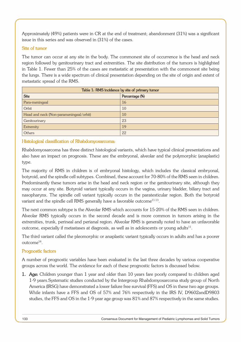
133 Consensus Document for Management of Pediatric Lymphomas and Solid Tumors
Approximately (49%) patients were in CR at the end of treatment; abandonment (31%) was a significant issue in this series and was observed in (31%) of the cases.
Site of tumor
The tumor can occur at any site in the body. The commonest site of occurrence is the head and neck region followed by genitourinary tract and extremities. The site distribution of the tumors is highlighted in Table 1. Fewer than 25% of the cases are metastatic at presentation with the commonest site being the lungs. There is a wide spectrum of clinical presentation depending on the site of origin and extent of metastatic spread of the RMS.
Table 1: RMS incidence by site of primary tumor
Site Percentage (%)
Para-meningeal 16
Orbit 10
Head and neck (Non-parameningeal/orbit) 10
Genitourinary 23
Extremity 19
Others 22
Histological classification of Rhabdomyosarcoma:
Rhabdomyosarcoma has three distinct histological variants, which have typical clinical presentations and also have an impact on prognosis. These are the embryonal, alveolar and the polymorphic (anaplastic) type.
The majority of RMS in children is of embryonal histology, which includes the classical embryonal, botyroid, and the spindle cell subtypes. Combined, these account for 70-80% of the RMS seen in children. Predominantly these tumors arise in the head and neck region or the genitourinary site, although they may occur at any site. Botyroid variant typically occurs in the vagina, urinary bladder, biliary tract and nasopharynx. The spindle cell variant typically occurs in the paratesticular region. Both the botyroid variant and the spindle cell RMS generally have a favorable outcome11-13.
The next common subtype is the Alveolar RMS which accounts for 15-20% of the RMS seen in children. Alveolar RMS typically occurs in the second decade and is more common in tumors arising in the extremities, trunk, perineal and perianal region. Alveolar RMS is generally noted to have an unfavorable outcome, especially if metastases at diagnosis, as well as in adolescents or young adults11.
The third variant called the pleomorphic or anaplastic variant typically occurs in adults and has a poorer outcome14.
Prognostic factors
A number of prognostic variables have been evaluated in the last three decades by various cooperative groups across the world. The evidence for each of these prognostic factors is discussed below.
1. Age: Children younger than 1 year and older than 10 years fare poorly compared to children aged 1-9 years.Systematic studies conducted by the Intergroup Rhabdomyosarcoma study group of North America (IRSG) have demonstrated a lower failure free survival (FFS) and OS in these two age groups. While infants have a FFS and OS of 57% and 76% respectively in the IRS IV, D9602andD9803 studies, the FFS and OS in the 1-9 year age group was 81% and 87% respectively in the same studies.
134 Consensus Document for Management of Pediatric Lymphomas and Solid Tumors
The poor prognosis has been attributed to the reluctance in using aggressive local therapy in infants due to predicted late morbidity of therapy15.
Similarly, in children aged more than 10 years the 5 year FFS and OS is reported to be 68% and 76%. The predominance of alveolar histology, tumor primarily occurring at unfavorable sites (mainly extremities), a higher metastatic disease at presentation and enhanced toxicity of chemotherapy in this age group have been the factors attributed to poorer outcomes in this age group16,17. Adults with RMS tend to fare much poorly with a 5 year OS of 27%14,18.
2. Site of primary tumor:
The sites of occurrence of the primary tumor have been classified as favorable and unfavorable sites based on outcome.
Favorable sites: Non-parameningeal head and neck, orbit, genitourinary (non-bladder, non-prostate, non-kidney) and biliary tract.
Unfavorable sites: All sites of primary tumor other than the above listed under favorable sites. These commonly include extremity, para-meningeal, bladder and prostate RMS19,20.
3. Metastases and lymph node involvement
Metastasis is one of the strongest predictors of outcome in RMS. Children with metastatic disease fare poorly and outcomes are also dependent on the histology of the primary, site of metastases and a number of sites of metastases. Embryonal primarily with a single site of metastasis fares better compared to Alveolar RMS with comparable staging21-23. Additionally, it is critical to assess regional lymph node involvement accurately since children with regional nodal disease fare worse compared to patients without regional nodal involvement24.
4. Extent of resection
Early IRSG studies identified the extent of resection done up front as a significant prognostic factor. In the IRS III study, the extent of disease after the primary resection-called the surgical-pathologic group or the clinical group -correlated with outcome. Patients with gross residual disease after initial surgery had a worse outcome (5 year OS 70%-clinical group III) compared to patients who had no residual disease (clinical group I-5 year OS-90%)20.
5. Histopathological subtype
Alveolar histology has a significantly poor outcome compared to similar patients with embryonal rhabdomyosarcoma. The IRSG studies I and II have demonstrated that alveolar histology was associated with unfavorable outcome even in clinical group I disease25. However, subsequently the outcome was similar in localized disease when alveolar histology was treated with more intense therapy20. Additionally, in metastatic RMS-alveolar subtype favored much worse compared to the embryonal subtype23.
Other factors which have been evaluated but not consistently proven to be prognostic are tumor size and response to therapy (as determined by imaging in patients who have undergone irradiation as the primary modality of local therapy).
Molecular subclassification
RMS has distinct molecular characteristics that have been utilized for diagnostic purposes as well as for predicting outcome in certain situations. They may also in the future play a role for monitoring residual disease post-therapy, especially in metastatic disease which is infiltrating the BM.
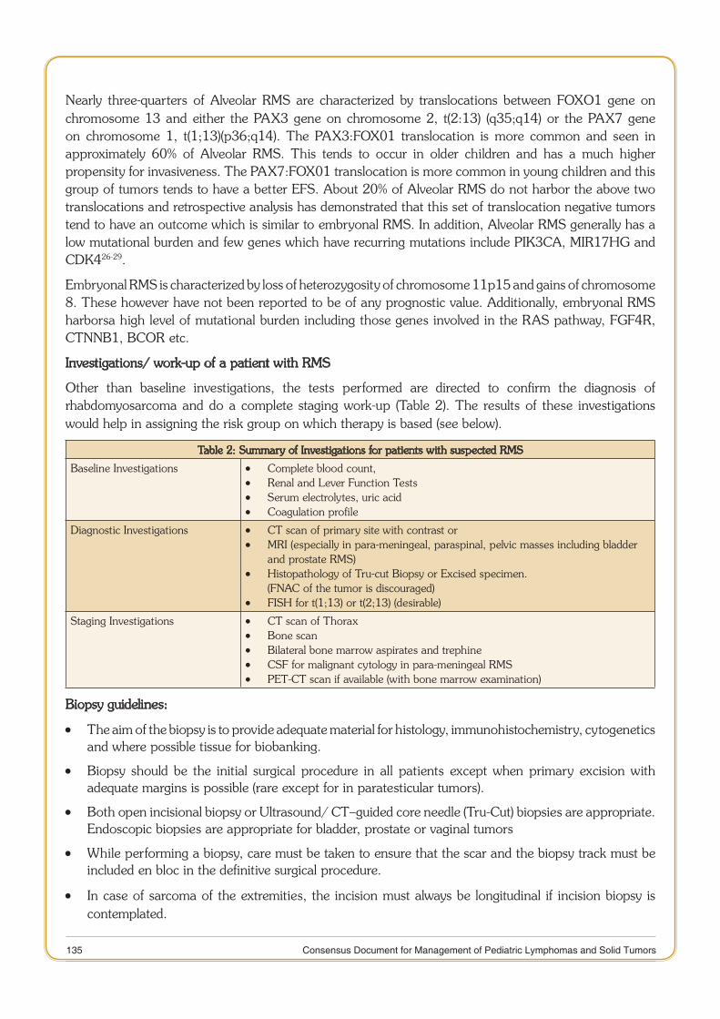
135 Consensus Document for Management of Pediatric Lymphomas and Solid Tumors
Nearly three-quarters of Alveolar RMS are characterized by translocations between FOXO1 gene on chromosome 13 and either the PAX3 gene on chromosome 2, t(2:13) (q35;q14) or the PAX7 gene on chromosome 1, t(1;13)(p36;q14). The PAX3:FOX01 translocation is more common and seen in approximately 60% of Alveolar RMS. This tends to occur in older children and has a much higher propensity for invasiveness. The PAX7:FOX01 translocation is more common in young children and this group of tumors tends to have a better EFS. About 20% of Alveolar RMS do not harbor the above two translocations and retrospective analysis has demonstrated that this set of translocation negative tumors tend to have an outcome which is similar to embryonal RMS. In addition, Alveolar RMS generally has a low mutational burden and few genes which have recurring mutations include PIK3CA, MIR17HG and CDK426-29.
Embryonal RMS is characterized by loss of heterozygosity of chromosome 11p15 and gains of chromosome 8. These however have not been reported to be of any prognostic value. Additionally, embryonal RMS harborsa high level of mutational burden including those genes involved in the RAS pathway, FGF4R, CTNNB1, BCOR etc.
Investigations/ work-up of a patient with RMS
Other than baseline investigations, the tests performed are directed to confirm the diagnosis of rhabdomyosarcoma and do a complete staging work-up (Table 2). The results of these investigations would help in assigning the risk group on which therapy is based (see below).
Table 2: Summary of Investigations for patients with suspected RMS
Baseline Investigations Complete blood count, •Renal and Lever Function Tests •Serum electrolytes, uric acid •Coagulation profile •
Diagnostic Investigations CT scan of primary site with contrast or •MRI (especially in para-meningeal, paraspinal, pelvic masses including bladder •and prostate RMS)Histopathology of Tru-cut Biopsy or Excised specimen. •(FNAC of the tumor is discouraged)FISH for t(1;13) or t(2;13) (desirable) •
Staging Investigations CT scan of Thorax •Bone scan •Bilateral bone marrow aspirates and trephine •CSF for malignant cytology in para-meningeal RMS •PET-CT scan if available (with bone marrow examination) •
Biopsy guidelines:
The aim of t • he biopsy is to provide adequate material for histology, immunohistochemistry, cytogenetics and where possible tissue for biobanking.
Biopsy should be the initial surgical procedure in all patients except when primary excision with •adequate margins is possible (rare except for in paratesticular tumors).
Both open incisional biopsy or Ultrasound/ CT–guided core needle (Tru-Cut) biopsies are appropriate. •Endoscopic biopsies are appropriate for bladder, prostate or vaginal tumors
While performing a biopsy, care must be taken to ensure that the scar and the biopsy track must be •included en bloc in the definitive surgical procedure.
In case of sarcom • a of the extremities, the incision must always be longitudinal if incision biopsy is contemplated.
136 Consensus Document for Management of Pediatric Lymphomas and Solid Tumors
If Tru-cut bio • psy is performed, 18 or 16 G needles should be used with 4 to 6 cores performed. The biopsy tract must only contaminate the anatomical compartment in which the tumor is situated, avoiding major neurovascular bundles.
The fixative recommended is formalin. However, if biological studies are contemplated tissue should •always be sent fresh to the laboratory.
There is now emerging evidence that regional lymph nodes should be subjected to biopsy or FNAC •especially in primary tumors of the limb even when there is no clinic-radiological evidence of lymph nodal spread
Pathology guidelines:
The diagnosis and histological subtyping of RMS is carried out by the local pathologist. This would •include classifying tumors as classical Embryonal, botyroid, spindle cell type or alveolar subtype. To be classified as alveolar subtype, the tumor must have greater than 50% alveolar histology.
All suspected RMS specimens should be subject to immunohistochemistry which should include •Desmin, Vimentin, MyoD1, Myogenin, S100, EMA, LCA, Fli1 and Mic2.
The pathology report must routinely include the RMS histologic subtype, percentage of necrosis (in •resected specimens post-chemotherapy), margins (in tumor excisions) and also comment on vascular/lymphatic invasion and involvement of regional lymph nodes (if sampled).
Where required, the local pathologist must coordinate sample storage for biobanking •
The pathologist should coordinate with molecular biology laboratory so that appropriate molecular •classification of the RMS is performed. This may involve performing FISH or PCR.
Radiology Guidelines:
First loco-regional evaluation should be done by either CT scan or MRI depending on the availability •or site of Primary.
MRI is preferable in head and neck tumors, paraspinal tumors, limb and genitourinary primary •tumors.
CT scan is recommended for tumors in the chest and abdomen. CT scan would also be useful for •assessing bony erosions in head and neck primary
Both MRI and CT scan should be carried out with the use of IV contrast when evaluating the primary •tumor.
Tumor dimensions should be recorded in three diameters choosing as far as possible the three •maximum diameters (sagittal, coronal and axial)
Imaging of the primary site should be performed (again) after surgical excision biopsy if a significant •volume has been resected.
CT chest ( non-contrast) is recommended for evaluation of lung metastases. •
Craniospinal MRI is recommended if intraspinal extension or meningeal involvement is suspected. •
Paratesticular tumors must have an evaluation of regional (para-aortic) lymph nodes by CT/MRI and •ultrasound.
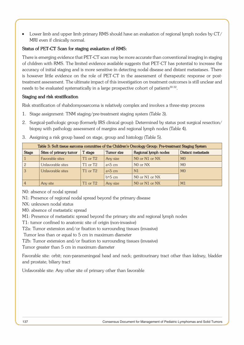
137 Consensus Document for Management of Pediatric Lymphomas and Solid Tumors
Lower limb and upper limb primary RMS should have an evaluation of regional lymph nodes by CT/ •MRI even if clinically normal.
Status of PET-CT Scan for staging evaluation of RMS:
There is emerging evidence that PET-CT scan may be more accurate than conventional imaging in staging of children with RMS. The limited evidence available suggests that PET-CT has potential to increase the accuracy of initial staging and is more sensitive in detecting nodal disease and distant metastases. There is however little evidence on the role of PET-CT in the assessment of therapeutic response or post-treatment assessment. The ultimate impact of this investigation on treatment outcomes is still unclear and needs to be evaluated systematically in a large prospective cohort of patients30-32.
Staging and risk stratification
Risk stratification of rhabdomyosarcoma is relatively complex and involves a three-step process
Stage assignment: TNM staging/pre-treatment staging system (Table 3).1.
Surgical-pathologic group (formerly IRS clinical group): Determined by status post surgical resection/2. biopsy with pathology assessment of margins and regional lymph nodes (Table 4).
Assigning a risk group based on stage, group and histology (Table 5).3.
Table 3: Soft tissue sarcoma committee of the Children’s Oncology Group: Pre-treatment Staging System
Stage Sites of primary tumor T stage Tumor size Regional lymph nodes Distant metastasis
1 Favorable sites T1 or T2 Any size N0 or N1 or NX M0
2 Unfavorable sites T1 or T2 a<5 cm N0 or NX M0
3 Unfavorable sites T1 or T2 a<5 cm N1 M0
b>5 cm N0 or N1 or NX
4 Any site T1 or T2 Any size N0 or N1 or NX M1
N0: absence of nodal spreadN1: Presence of regional nodal spread beyond the primary diseaseNX: unknown nodal statusM0: absence of metastatic spreadM1: Presence of metastatic spread beyond the primary site and regional lymph nodesT1: tumor confined to anatomic site of origin (non-invasive)T2a: Tumor extension and/or fixation to surrounding tissues (invasive) Tumor less than or equal to 5 cm in maximum diameterT2b: Tumor extension and/or fixation to surrounding tissues (invasive)Tumor greater than 5 cm in maximum diameter
Favorable site: orbit; non-parameningeal head and neck; genitourinary tract other than kidney, bladder and prostate; biliary tract
Unfavorable site: Any other site of primary other than favorable
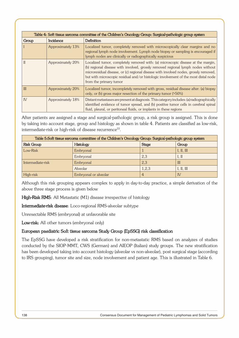
138 Consensus Document for Management of Pediatric Lymphomas and Solid Tumors
Table 4: Soft tissue sarcoma committee of the Children’s Oncology Group: Surgical-pathologic group system
Group Incidence Definition
I Approximately 13% Localized tumor, completely removed with microscopically clear margins and no regional lymph node involvement. Lymph node biopsy or sampling is encouraged if lymph nodes are clinically or radiographically suspicious
II Approximately 20% Localized tumor, completely removed with: (a) microscopic disease at the margin, (b) regional disease with involved, grossly removed regional lymph nodes without microresidual disease, or (c) regional disease with involved nodes, grossly removed, but with microscopic residual and/or histologic involvement of the most distal node from the primary tumor
III Approximately 20% Localized tumor, incompletely removed with gross, residual disease after: (a) biopsy only, or (b) gross major resection of the primary tumor (>50%)
IV Approximately 18% Distant metastases are present at diagnosis. This category includes: (a) radiographically identified evidence of tumor spread, and (b) positive tumor cells in cerebral spinal fluid, pleural, or peritoneal fluids, or implants in these regions
After patients are assigned a stage and surgical-pathologic group, a risk group is assigned. This is done by taking into account stage, group and histology as shown in table 4. Patients are classified as low-risk, intermediate-risk or high-risk of disease recurrence33.
Table 5:Soft tissue sarcoma committee of the Children’s Oncology Group: Surgical-pathologic group system
Risk Group Histology Stage Group
Low-Risk Embryonal 1 I, II, III
Embryonal 2,3 I, II
Intermediate-risk Embryonal 2,3 III
Alveolar 1,2,3 I, II, III
High-risk Embryonal or alveolar 4 IV
Although this risk grouping appears complex to apply in day-to-day practice, a simple derivation of the above three stage process is given below
High-Risk RMS: All Metastatic (M1) disease irrespective of histology
Intermediate-risk disease: Loco-regional RMS-alveolar subtype
Unresectable RMS (embryonal) at unfavorable site
Low-risk: All other tumors (embryonal only)
European paediatric Soft tissue sarcoma Study Group (EpSSG) risk classification
The EpSSG have developed a risk stratification for non-metastatic RMS based on analyzes of studies conducted by the SIOP-MMT, CWS (German) and AIEOP (Italian) study groups. The new stratification has been developed taking into account histology (alveolar vs non-alveolar), post surgical stage (according to IRS grouping), tumor site and size, node involvement and patient age. This is illustrated in Table 6.
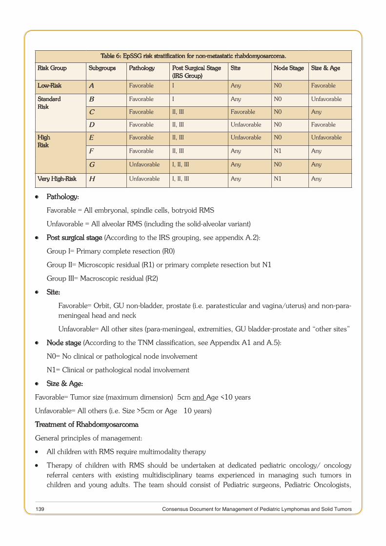
139 Consensus Document for Management of Pediatric Lymphomas and Solid Tumors
Table 6: EpSSG risk stratification for non-metastatic rhabdomyosarcoma.
Risk Group Subgroups Pathology Post Surgical Stage(IRS Group)
Site Node Stage Size & Age
Low-Risk A Favorable I Any N0 Favorable
StandardRisk
B Favorable I Any N0 Unfavorable
C Favorable II, III Favorable N0 Any
D Favorable II, III Unfavorable N0 Favorable
HighRisk
E Favorable II, III Unfavorable N0 Unfavorable
F Favorable II, III Any N1 Any
G Unfavorable I, II, III Any N0 Any
Very High-Risk H Unfavorable I, II, III Any N1 Any
Pathology: •
Favorable = All embryonal, spindle cells, botryoid RMS
Unfavorable = All alveolar RMS (including the solid-alveolar variant)
Post surgical stage • (According to the IRS grouping, see appendix A.2):
Group I= Primary complete resection (R0)
Group II= Microscopic residual (R1) or primary complete resection but N1
Group III= Macroscopic residual (R2)
Site: •
Favorable= Orbit, GU non-bladder, prostate (i.e. paratesticular and vagina/uterus) and non-para-meningeal head and neck
Unfavorable= All other sites (para-meningeal, extremities, GU bladder-prostate and “other sites”
Node stage • (According to the TNM classification, see Appendix A1 and A.5):
N0= No clinical or pathological node involvement
N1= Clinical or pathological nodal involvement
Size & Age: •
Favorable= Tumor size (maximum dimension) ≤5cm and Age <10 years
Unfavorable= All others (i.e. Size >5cm or Age ≥ 10 years)
Treatment of Rhabdomyosarcoma
General principles of management:
All children with RMS require multimodality therapy •
Therapy of children with RMS should be undertaken at dedicated pediatric oncology/ oncology •referral centers with existing multidisciplinary teams experienced in managing such tumors in children and young adults. The team should consist of Pediatric surgeons, Pediatric Oncologists,
140 Consensus Document for Management of Pediatric Lymphomas and Solid Tumors
radiation oncologists specialized in management of children, palliative care specialists, pathologists, radiologists, molecular biologists and other supportive staff involved in the care of children.
Systemic chemotherapy is to be administered to all RMS patients in conjunction with local therapy •
Local therapy :Surgery, radiotherapy or both modalities may need to be utilized to maximize local •tumor control.
Local therapy has to be individualized based on expertise available at each center. The decision for •local therapy should be taken upfront in multidisciplinary team meetings where the therapy for each patient is planned.
Primary surgical resection may be performed before chemotherapy if it results in no substantial •functional compromise or organ dysfunction or disfigurement. This should only be undertaken if the tumor is deemed resectable with negative margins. Generally, this is possible in only a minority of cases like paratesticular tumors.
The majority of patients fall into group III (gross residual disease or biopsy only) and would need •radiotherapy as treatment modality for local control.
In some patients with initially unresectable tumor, a second-look surgery (delayed primary excision) •may be undertaken for removal of the residual tumor. This is advocated in cases where delayed excision is deemed feasible , and without significantly compromising organ function or cosmesis. This is especially recommended when there is likelihood of reduction in radiation dose which would significantly reduce late effects of therapy.
RT is recommended for clinically or radiologically suspicious lymph nodes unless the nodes are •biopsied and shown to be free of tumor.
As discussed above, rhabdomyosarcoma should be meticulously risk stratified into low–intermediate •and high-risk groups. The therapy for each of these groups is outlined below.
Low-risk rhabdomyosarcoma management
The IRS V and the COG soft tissue sarcoma committee estimate 35% of all RMS to fall under the category of low-risk category. These have excellent survival rates with long-term survival reaching over 90% with limited chemotherapy (Table 7 and Table 8). Low-risk group RMS consist of
Non-metastatic embryonal /botryoid tumors •
Favorable sites (Stage 1, Group I-III) •
Unfavorable sites if completely resected (Stage 2-3, Group I- II) •
The Low-risk are further subgrouped into subgroup A and B
Sub-group A •
Histology: Embryonal / Botryoid •
Stage 1, Groups I, II (N0) •
Stage 1, Group III (N0) Orbit only •
Stage 2, Group I (N0) •
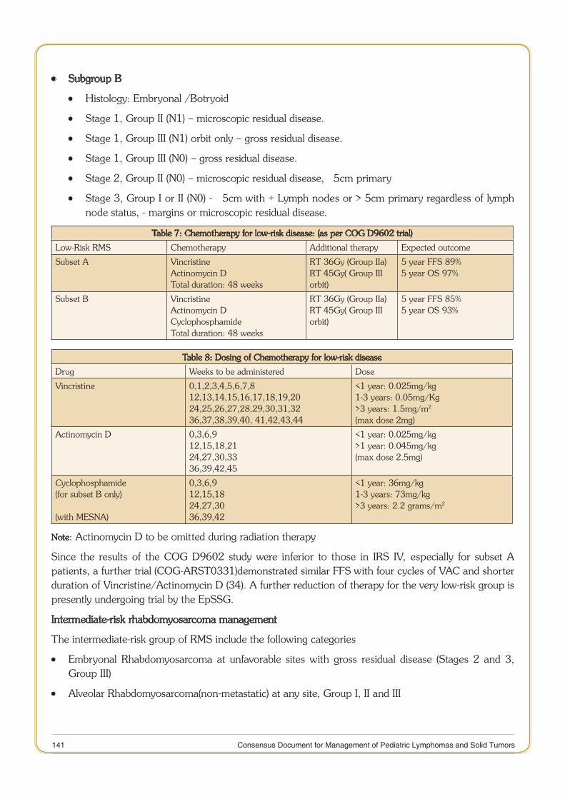
141 Consensus Document for Management of Pediatric Lymphomas and Solid Tumors
Subgroup B •
Histology: Embryonal /Botryoid •
Stage 1, Group II (N1) – microscopic residual disease. •
Stage 1, Group III (N1) orbit only – gross residual disease. •
Stage 1, Group III (N0) – gross residual disease. •
Stage 2, Group II (N0) – microscopic residual disease, ≤ 5cm primary •
Stage 3, Group I or II (N0) - ≤ 5cm with + Lymph nodes or > 5cm primary regardless of lymph •node status, - margins or microscopic residual disease.
Table 7: Chemotherapy for low-risk disease: (as per COG D9602 trial)
Low-Risk RMS Chemotherapy Additional therapy Expected outcome
Subset A VincristineActinomycin DTotal duration: 48 weeks
RT 36Gy (Group IIa)RT 45Gy( Group III orbit)
5 year FFS 89%5 year OS 97%
Subset B VincristineActinomycin DCyclophosphamideTotal duration: 48 weeks
RT 36Gy (Group IIa)RT 45Gy( Group III orbit)
5 year FFS 85%5 year OS 93%
Table 8: Dosing of Chemotherapy for low-risk disease
Drug Weeks to be administered Dose
Vincristine 0,1,2,3,4,5,6,7,812,13,14,15,16,17,18,19,2024,25,26,27,28,29,30,31,3236,37,38,39,40, 41,42,43,44
<1 year: 0.025mg/kg1-3 years: 0.05mg/Kg>3 years: 1.5mg/m2
(max dose 2mg)
Actinomycin D 0,3,6,912,15,18,2124,27,30,3336,39,42,45
<1 year: 0.025mg/kg>1 year: 0.045mg/kg(max dose 2.5mg)
Cyclophosphamide(for subset B only)
(with MESNA)
0,3,6,912,15,1824,27,3036,39,42
<1 year: 36mg/kg1-3 years: 73mg/kg>3 years: 2.2 grams/m2
Note: Actinomycin D to be omitted during radiation therapy
Since the results of the COG D9602 study were inferior to those in IRS IV, especially for subset A patients, a further trial (COG-ARST0331)demonstrated similar FFS with four cycles of VAC and shorter duration of Vincristine/Actinomycin D (34). A further reduction of therapy for the very low-risk group is presently undergoing trial by the EpSSG.
Intermediate-risk rhabdomyosarcoma management
The intermediate-risk group of RMS include the following categories
Embryonal Rhabdomyosarcoma at unfavorable sites with gross residual disease (Stages 2 and 3, •Group III)
Alveolar Rhabdomyosarcoma(non-metastatic) at any site, Group I, II and III •
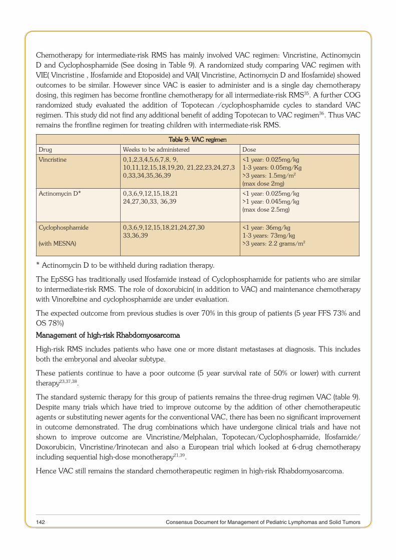
142 Consensus Document for Management of Pediatric Lymphomas and Solid Tumors
Chemotherapy for intermediate-risk RMS has mainly involved VAC regimen: Vincristine, Actinomycin D and Cyclophosphamide (See dosing in Table 9). A randomized study comparing VAC regimen with VIE( Vincristine , Ifosfamide and Etoposide) and VAI( Vincristine, Actinomycin D and Ifosfamide) showed outcomes to be similar. However since VAC is easier to administer and is a single day chemotherapy dosing, this regimen has become frontline chemotherapy for all intermediate-risk RMS35. A further COG randomized study evaluated the addition of Topotecan /cyclophosphamide cycles to standard VAC regimen. This study did not find any additional benefit of adding Topotecan to VAC regimen36. Thus VAC remains the frontline regimen for treating children with intermediate-risk RMS.
Table 9: VAC regimen
Drug Weeks to be administered Dose
Vincristine 0,1,2,3,4,5,6,7,8, 9, 10,11,12,15,18,19,20, 21,22,23,24,27,30,33,34,35,36,39
<1 year: 0.025mg/kg1-3 years: 0.05mg/Kg>3 years: 1.5mg/m2
(max dose 2mg)
Actinomycin D* 0,3,6,9,12,15,18,2124,27,30,33, 36,39
<1 year: 0.025mg/kg>1 year: 0.045mg/kg(max dose 2.5mg)
Cyclophosphamide
(with MESNA)
0,3,6,9,12,15,18,21,24,27,3033,36,39
<1 year: 36mg/kg1-3 years: 73mg/kg>3 years: 2.2 grams/m2
* Actinomycin D to be withheld during radiation therapy.
The EpSSG has traditionally used Ifosfamide instead of Cyclophosphamide for patients who are similar to intermediate-risk RMS. The role of doxorubicin( in addition to VAC) and maintenance chemotherapy with Vinorelbine and cyclophosphamide are under evaluation.
The expected outcome from previous studies is over 70% in this group of patients (5 year FFS 73% and OS 78%)
Management of high-risk Rhabdomyosarcoma
High-risk RMS includes patients who have one or more distant metastases at diagnosis. This includes both the embryonal and alveolar subtype.
These patients continue to have a poor outcome (5 year survival rate of 50% or lower) with current therapy23,37,38.
The standard systemic therapy for this group of patients remains the three-drug regimen VAC (table 9). Despite many trials which have tried to improve outcome by the addition of other chemotherapeutic agents or substituting newer agents for the conventional VAC, there has been no significant improvement in outcome demonstrated. The drug combinations which have undergone clinical trials and have not shown to improve outcome are Vincristine/Melphalan, Topotecan/Cyclophosphamide, Ifosfamide/Doxorubicin, Vincristine/Irinotecan and also a European trial which looked at 6-drug chemotherapy including sequential high-dose monotherapy21,39.
Hence VAC still remains the standard chemotherapeutic regimen in high-risk Rhabdomyosarcoma.
143 Consensus Document for Management of Pediatric Lymphomas and Solid Tumors
High-dose chemotherapy with autologous stem cell rescue has been evaluated in a small number of patients, but this intense therapy has not been shown to improve outcome both in newly diagnosed and recurrent Rhabdomyosarcoma40,41&42.
The prognosis of metastatic RMS is dependent on several adverse factors. Age <1 year and more than 10 years, unfavorable primary site, bone and/or bone marrow involvement, and three or more sites of metastases, all have adverse outcomes. The EFS is 50% for patients with none of the adverse factors, 42% for one adverse factor, 18% for two adverse factors, 12% for three adverse factors and 5% for four adverse factors38.
Treatment of recurrent Rhabdomyosarcoma
Outcomes of recurrent rhabdomyosarcoma are generally poor even though secondary remission can sometimes be achieved with salvage therapy. The outcome from salvage therapy of a recurrent RMS is dependent on various factors, as has been identified in retrospective studies. Unfavorable prognostic factors include short time to recurrence (<18 months), metastatic (as opposed to local) recurrence, previous radiotherapy and previous tumor size >5cm. The prognosis is favorable for children who initially presented with Stage I/ Group I disease, embryonal histology and have a loco-regional recurrence only43- 46. The chemotherapy regimens that have been used for salvage in a recurrent setting are listed below.
Carboplatin/Etoposide •
Ifosfamide/carboplatin/etoposide •
Vincristine/Irinotecan •
Cyclophosphamide/Topotecan •
Vinorelbine/Cyclophosphamide. •
The role of high-dose chemotherapy with autologous stem cell rescue has not been established in a recurrent setting.
Radiotherapy guidelines
Principles for radiotherapy
Techniques like three dimensional conformal radiation therapy (3D-CRT) & intensity-modulated radiation therapy (IMRT) are useful to deliver optimal doses to the tumor while sparing the surrounding normal tissues. Interstitial & intracavitary brachytherapy should be used for appropriate indications when the expertise is available. Chemotherapy should be modified to avoid the radiosensitizing agents like dactinomycin and doxorubicin during radiotherapy.
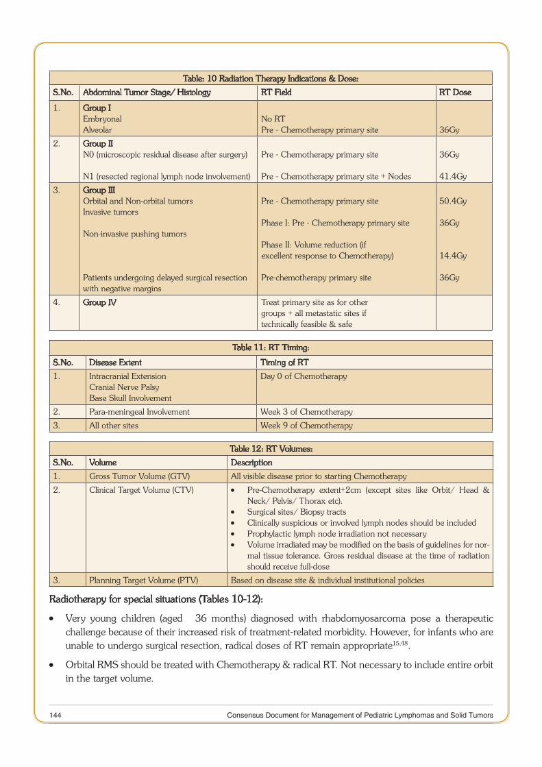
144 Consensus Document for Management of Pediatric Lymphomas and Solid Tumors
Table: 10 Radiation Therapy Indications & Dose:
S.No. Abdominal Tumor Stage/ Histology RT Field RT Dose
1. Group I Embryonal Alveolar
No RTPre - Chemotherapy primary site 36Gy
2. Group II N0 (microscopic residual disease after surgery)
N1 (resected regional lymph node involvement)
Pre - Chemotherapy primary site
Pre - Chemotherapy primary site + Nodes
36Gy
41.4Gy
3. Group III Orbital and Non-orbital tumors Invasive tumors
Non-invasive pushing tumors
Patients undergoing delayed surgical resection with negative margins
Pre - Chemotherapy primary site
Phase I: Pre - Chemotherapy primary site
Phase II: Volume reduction (if excellent response to Chemotherapy)
Pre-chemotherapy primary site
50.4Gy
36Gy
14.4Gy
36Gy
4. Group IV Treat primary site as for othergroups + all metastatic sites iftechnically feasible & safe
Table 11: RT Timing:
S.No. Disease Extent Timing of RT
1. Intracranial ExtensionCranial Nerve PalsyBase Skull Involvement
Day 0 of Chemotherapy
2. Para-meningeal Involvement Week 3 of Chemotherapy
3. All other sites Week 9 of Chemotherapy
Table 12: RT Volumes:
S.No. Volume Description
1. Gross Tumor Volume (GTV) All visible disease prior to starting Chemotherapy
2. Clinical Target Volume (CTV) Pre-Chemotherapy extent+2cm (except sites like Orbit/ Head & •Neck/ Pelvis/ Thorax etc).Surgical sites/ Biopsy tracts •Clinically suspicious or involved lymph nodes should be included •Prophylactic lymph node irradiation not necessary •Volume irradiated may be modified on the basis of guidelines for nor- •mal tissue tolerance. Gross residual disease at the time of radiation should receive full-dose
3. Planning Target Volume (PTV) Based on disease site & individual institutional policies
Radiotherapy for special situations (Tables 10-12):
Very young children (aged ≤ 36 months) diagnosed with rhabdomyosarcoma pose a therapeutic •challenge because of their increased risk of treatment-related morbidity. However, for infants who are unable to undergo surgical resection, radical doses of RT remain appropriate15,48.
Orbital RMS should be treated with Chemotherapy & radical RT. Not necessary to include entire orbit •in the target volume.
145 Consensus Document for Management of Pediatric Lymphomas and Solid Tumors
Non-Orbital Head & Neck: •
Para-meningeal: do not require whole-brain irradiation unless tumor cells are present in the CSF •at diagnosis49. Patients should receive RT to the site of the primary tumor with a 1.5 cm margin to include the meninges adjacent to the primary tumor and the region of intracranial extension, if present, with a 1.5 cm margin50.
Non-parameningeal head and neck: Surgical resection should be done only if a wide local •excision is feasible without causing significant morbidity. Most patients should be treated with Chemotherapy & definitive RT.
Extremity: •
Complete primary tumor removal from the hand or foot may not be feasible in all cases without •significant functional impairment51. Children in COG studies presenting with a primary tumor of the hands or feet have shown 100% 10-year local control using RT along with chemotherapy, avoiding amputation in these children52.
Intra-abdominal/ Intra-thoracic: •
Post-operative RT improves EFS. Patients with peritoneal disease & ascites benefit with whole •abdominal RT53.
Biliary Tree/ Anus & Perineum: •
Chemotherapy & definitive RT should be offered if surgical resection is not feasible or associated •with significant morbidity.
Paratesticular: •
For patients requiring adjuvant RT, testicular transposition into the adjacent thigh should be •considered.
Bladder/ Prostate: •
Except for patients with lesions exclusively involving the bladder dome who can undergo adequate •surgical resection, the rest of the patients should be treated with Chemotherapy & RT.
Vulva/ Vagina/ Uterus: •
Radical surgery should be avoided at these sites. Patients treated with chemotherapy & radical RT •(External RT & Brachytherapy) achieve good outcomes with function preservation. The COG-ARST0331 study reported an unacceptably high rate of local recurrences in girls with Group III vaginal tumors who did not receive RT54.
Post-treatment surveillance
All patients, post-treatment should be followed up for 5 years for possible tumor relapse and until adulthood for treatment side effects. A suggested surveillance schedule is summarized below (Table 13).
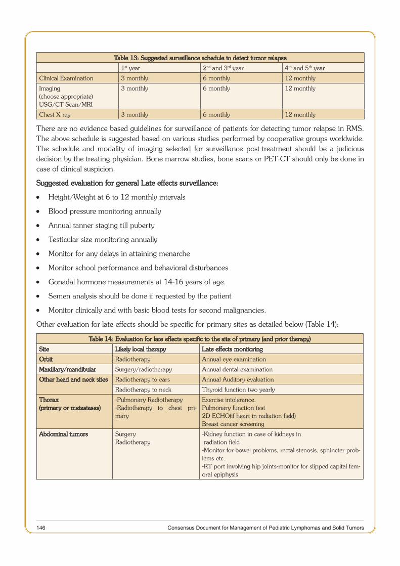
146 Consensus Document for Management of Pediatric Lymphomas and Solid Tumors
Table 13: Suggested surveillance schedule to detect tumor relapse
1st year 2nd and 3rd year 4th and 5th year
Clinical Examination 3 monthly 6 monthly 12 monthly
Imaging (choose appropriate)USG/CT Scan/MRI
3 monthly 6 monthly 12 monthly
Chest X ray 3 monthly 6 monthly 12 monthly
There are no evidence based guidelines for surveillance of patients for detecting tumor relapse in RMS. The above schedule is suggested based on various studies performed by cooperative groups worldwide. The schedule and modality of imaging selected for surveillance post-treatment should be a judicious decision by the treating physician. Bone marrow studies, bone scans or PET-CT should only be done in case of clinical suspicion.
Suggested evaluation for general Late effects surveillance:
Height/Weight at 6 to 12 monthly intervals •
Blood pressure monitoring annually •
Annual tanner staging till puberty •
Testicular size monitoring annually •
Monitor for any delays in attaining menarche •
Monitor school performance and behavioral disturbances •
Gonadal hormone measurements at 14-16 years of age. •
Semen analysis should be done if requested by the patient •
Monitor clinically and with basic blood tests for second malignancies. •
Other evaluation for late effects should be specific for primary sites as detailed below (Table 14):
Table 14: Evaluation for late effects specific to the site of primary (and prior therapy)
Site Likely local therapy Late effects monitoring
Orbit Radiotherapy Annual eye examination
Maxillary/mandibular Surgery/radiotherapy Annual dental examination
Other head and neck sites Radiotherapy to ears Annual Auditory evaluation
Radiotherapy to neck Thyroid function two yearly
Thorax(primary or metastases)
-Pulmonary Radiotherapy-Radiotherapy to chest pri-mary
Exercise intolerance.Pulmonary function test2D ECHO(if heart in radiation field)Breast cancer screening
Abdominal tumors SurgeryRadiotherapy
-Kidney function in case of kidneys in radiation field-Monitor for bowel problems, rectal stenosis, sphincter prob-lems etc.-RT port involving hip joints-monitor for slipped capital fem-oral epiphysis
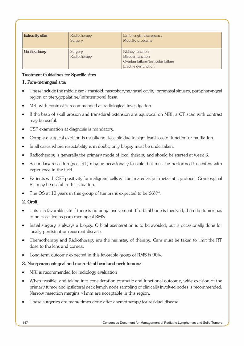
147 Consensus Document for Management of Pediatric Lymphomas and Solid Tumors
Extremity sites RadiotherapySurgery
Limb length discrepancyMobility problems
Genitourinary SurgeryRadiotherapy
Kidney functionBladder functionOvarian failure/testicular failureErectile dysfunction
Treatment Guidelines for Specific sites
1. Para-meningeal site:
These include the middle ear / mastoid, nasopharynx/nasal cavity, paranasal sinuses, parapharyngeal •region or pterygopalatine/infratemporal fossa.
MRI with contrast is recommended as radiological investigation •
If the base of skull erosion and transdural extension are equivocal on MRI, a CT scan with contrast •may be useful.
CSF examination at diagnosis is mandatory. •
Complete surgical excision is usually not feasible due to significant loss of function or mutilation. •
In all cases where resectability is in doubt, only biopsy must be undertaken. •
Radiotherapy is generally the primary mode of local therapy and should be started at week 3. •
Secondary resection (post RT) may be occasionally feasible, but must be performed in centers with •experience in the field.
Patients with CSF positivity for malignant cells will be treated as per metastatic protocol. Craniospinal •RT may be useful in this situation.
The OS at 10 years in this group of tumors is expected to be 66% • 47.
2. Orbit:
This is a favorable site if there is no bony involvement. If orbital bone is involved, then the tumor has •to be classified as para-meningeal RMS.
Initial surgery is always a biopsy. Orbital exenteration is to be avoided, but is occasionally done for •locally persistent or recurrent disease.
Chemotherapy and Radiotherapy are the mainstay of therapy. Care must be taken to limit the RT •dose to the lens and cornea.
Long-term outcome expected in this favorable group of RMS is 90%. •
3. Non-parameningeal and non-orbital head and neck tumors:
MRI is recommended for radiology evaluation •
When feasible, and taking into consideration cosmetic and functional outcome, wide excision of the •primary tumor and ipsilateral neck lymph node sampling of clinically involved nodes is recommended. Narrow resection margins <1mm are acceptable in this region.
These surgeries are many times done after chemotherapy for residual disease. •
148 Consensus Document for Management of Pediatric Lymphomas and Solid Tumors
If the primary tumor is considered unresectable, then RT is the primary modality of local therapy and •there is usually no role for primary debulking surgery.
4. Bladder/Prostate:
MRI is the radiological investigation of choice. •
Cystoscopic evaluation should be done at diagnosis and at follow-up. •
A biopsy is usually performed upfront and secondary surgery can be contemplated post neoadjuvant •chemotherapy. A conservative surgery (partial cystectomy and/orpartial prostatectomy) can be considered in conjunction with brachytherapy.
If conservative surgery is not feasible, then the choice of local therapy is between radiotherapy and •total cystectomy and /or prostatectomy.
Occasionally, initial resection can be done only in case of very small tumors arising from the fundus •and away from the trigone.
5. Vagina/uterus:
Examination under anesthesia is usually recommended for defining the local extent of the tumor •
Surgery should be contemplated for removal of residual tumor post neo-adjuvant chemotherapy. In •case of residual disease, partial vaginectomy with or without brachytherapy is preferred. Occasionally RMS of the vagina with favorable histology may go into complete remission and may not warrant any local therapy.
In case of alveolar histology, radiotherapy is warranted as local therapy. •
Oophoropexy has to be considered in order to avoid irradiation. •
6. Paratesticular:
Suspected paratesticular tumors should have a scrotal ultrasound, •
It is mandatory to evaluate regional lymph nodes by CT scan (thin-cut). •
Complete surgical resection-high inguinal orchidectomy should be performed up front. •
Retroperitoneal lymph node sampling or lymphadenectomy is not recommended in children less than •10 years and CT scan shows no evidence of retroperitoneal lymphadenopathy.
For patients who have a positive CT scan for retroperitoneal lymphadenopathy or suspicious lymph •nodes, retroperitoneal lymph node sampling is recommended and treatment planned accordingly.
For children more than 10 years, a staging ipsilateral lymph node dissection is currently recommended •by the COG but not in Europe.
If the initial surgery was through the scrotum, then a primary operation should be done. In case of •any suspected contamination, then hemiscrotectomy should be performed.
Incompletely resected paratesticular RMS needs radiotherapy. Orchidopexy of the other testis is •recommended in this situation.
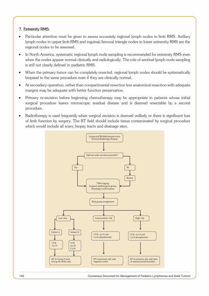
149 Consensus Document for Management of Pediatric Lymphomas and Solid Tumors
7. Extremity RMS:
Particular attention must be given to assess accurately regional lymph nodes in limb RMS. Axillary •lymph nodes in upper limb RMS and inguinal/femoral triangle nodes in lower extremity RMS are the regional nodes to be assessed.
In North America, systematic regional lymph node sampling is recommended for extremity RMS even •when the nodes appear normal clinically and radiologically. The role of sentinel lymph node sampling is still not clearly defined in pediatric RMS.
When the primary tumor can be completely resected, regional lymph nodes should be systematically •biopsied in the same procedure even if they are clinically normal.
At secondary operation, rather than compartmental resection less anatomical resection with adequate •margins may be adequate with better function preservation.
Primary re-excision before beginning chemotherapy may be appropriate in patients whose initial •surgical procedure leaves microscopic residual disease and is deemed resectable by a second procedure.
Radiotherapy is used frequently when surgical excision is deemed unlikely or there is significant loss •of limb function by surgery. The RT field should include tissue contaminated by surgical procedure which would include all scars, biopsy tracts and drainage sites.
VCRAct D
VCRAct DCyclo
VCR, Act D and Cyclo phosphamide
VCR, Act D and Cyclo phosphamide

150 Consensus Document for Management of Pediatric Lymphomas and Solid Tumors
References:
Gurney JG, Severson RK, Davis S, Robison et al. Incidence of cancer in children in the United States. Sex-, race-, and 1. 1-year age- specific rates by histological type. Cancer. 1995;75 (8):2186-95.
Linet MS, Ries LA, Smith MA et al. Cancer surveillance series: recent trends in childhood cancer incidence and mortality 2. in the United States.. J Natl Cancer Inst. 1999 ;91(12):1051-8.
Ognjanovic S, Linabery AM, Charbonneau B et al.Trends in childhood rhabdomyosarcoma incidence and survival in the 3. United States, 1975-2005. Cancer. 2009;115(18):4218-26.
Smith MA, Altekruse SF, Adamson PC et al. Declining childhood and adolescent cancer mortality.Cancer. 2014 4. ;120(16):2497-506.
Cancers in childhood. Consolidated report of hospital based cancer registries, 2007-2011. NCDIR-NCRP, Bangalore. 5. India. 2013: 19-25.
Kamat MR,6. Kulkarni JN, Tongaonkar HB, Ravi R. Rhabdomyosarcoma of the bladder and prostate in children. J Surg Oncol. 199;48(3):180-2.
Mehta S, Verma A, Mann SB et al. Rhabdomyosarcoma of head and neck-an analysis of 24 cases. Indian J Cancer. 7. 1996;33(1):37-42.
Suman B, Jaura MS, Mohanti B K , Agarwala S, and Bakshi S. “Childhood rhabdomyosarcoma: An AIIMS study.” In 8. Pediatric Blood and Cancer, vol. 49, no. 4, pp. 585-58
Padmanabhan, Ranjani, and Kusuma Kumary. “Survival analysis of rhabdomysarcoma cases at the Regional Cancer 9. Center Trivandrum, India.” In Pediatric Blood and Cancer, vol. 49, no. 4, pp. 481-481
Radhakrishnan S, Vora T, Chinnaswamy G, Dwivedi P, Patil V, Avinash P, Laskar S et al. “Clinical Profile and Outcomes 10. of Rhabdomyosarcoma patients treated at Tata Memorial Hospital : A Retrospective Analysis “ In Pediatric Blood and Cancer, vol. 59, no. 6, pp. 1067-1067.
Parham DM, Ellison DA. Rhabdomyosarcomas in adults and children: an update.Arch Pathol Lab Med. 2006;130 11. (10):1454-65.
Newton WA Jr, Gehan EA, Webber BL et al. Classification of rhabdomyosarcomas and related sarcomas. Pathologic 12. aspects and proposal for a new classification-an Intergroup Rhabdomyosarcoma Study.Cancer. 1995;76(6):1073-85.
Leuschner I, Harms D, Mattke A et al. Rhabdomyosarcoma of the urinary bladder and vagina: a clinicopathologic study 13. with emphasis on recurrent disease: a report from the Kiel Pediatric Tumor Registry and the German CWS Study. Am J Surg Pathol. 2001 J;25 (7):856-64.
Sultan I, Qaddoumi I, Yaser S et al Comparing adult and pediatric rhabdomyosarcoma in the surveillance, epidemiology 14. and end results program, 1973 to 2005: an analysis of 2,600 patients.J Clin Oncol. 2009;27(20):3391-7.
Malempati S, Rodeberg DA, Donaldson SS et al. Rhabdomyosarcoma in infants younger than 1 year: a report from the 15. Children’s Oncology Group. Cancer. 2011 ;117(15):3493-501.
Joshi D, Anderson JR, Paidas C et al.Age is an independent prognostic factor in rhabdomyosarcoma: a report from the 16. Soft Tissue Sarcoma Committee of the Children’s Oncology Group.Pediatr Blood Cancer. 2004;42(1):64-73.
Gupta AA, Anderson JR, Pappo AS et al. Patterns of chemotherapy-induced toxicities in younger children and 17. adolescents with rhabdomyosarcoma: a report from the Children’s Oncology Group Soft Tissue Sarcoma Committee. Cancer. 2012;118 (4):1130-7.
Dumont SN, Araujo DM, Munsell MF et al. Management and outcome of 239 adolescent and adult rhabdomyosarcoma 18. patients.Cancer Med. 2013;2(4):553-63.
Andrassy RJ, Corpron CA, Hays D et al. Extremity sarcomas: an analysis of prognostic factors from the Intergroup 19. Rhabdomyosarcoma Study III. J Pediatr Surg. 1996 ;31(1):191-6.
Crist W, Gehan EA, Ragab AH et al.The Third Intergroup Rhabdomyosarcoma Study. J Clin Oncol. 1995;13(3):610-20. 30.
McDowell HP, Foot AB, Ellershaw C et al. Outcomes in paediatric metastatic rhabdomyosarcoma: results of The 21. International Society of Paediatric Oncology (SIOP) study MMT-98.Eur J Cancer. 2010;46(9):1588-95.
151 Consensus Document for Management of Pediatric Lymphomas and Solid Tumors
Dantonello TM, Winkler P, Boelling T et al. Embryonal rhabdomyosarcoma with metastases confined to the lungs: report 22. from the CWS Study Group.Pediatr Blood Cancer. 2011;56(5):725-32.
Breneman JC, Lyden E, Pappo AS et al .Prognostic factors and clinical outcomes in children and adolescents with 23. metastatic rhabdomyosarcoma--a report from the Intergroup Rhabdomyosarcoma Study IV.J Clin Oncol. 2003;21(1): 78-84.
Mandell L, Ghavimi F, LaQuaglia M et al. Prognostic significance of regional lymph node involvement in childhood 24. extremity rhabdomyosarcoma. Med Pediatr Oncol. 1990;18(6):466-71.
Crist WM, Garnsey L, Beltangady MS et al. Prognosis in children with rhabdomyosarcoma: a report of the intergroup 25. rhabdomyosarcoma studies I and II. Intergroup Rhabdomyosarcoma Committee.J Clin Oncol. 1990;8(3):443-52.
Parham DM, Barr FG. Classification of rhabdomyosarcoma and its molecular basis.Adv Anat Pathol. 2013;20(6): 26. 387-97
Barr FG, Smith LM, Lynch JC et al. Examination of gene fusion status in archival samples of alveolar rhabdomyosarcoma 27. entered on the Intergroup Rhabdomyosarcoma Study-III trial: a report from the Children’s Oncology Group.J Mol Diagn. 2006;8(2):202-8.
Missiaglia E, Williamson D, Chisholm J et al. PAX3/FOXO1 fusion gene status is the key prognostic molecular marker 28. in rhabdomyosarcoma and significantly improves current risk stratification. J Clin Oncol. 2012;30 (14):1670-7.
Williamson D, Missiaglia E, de Reyniès A et al. Fusion gene-negative alveolar rhabdomyosarcoma is clinically and 29. molecularly indistinguishable from embryonal rhabdomyosarcoma. J Clin Oncol. 2010;28(13):2151-8
Norman G, Fayter D, Lewis-Light K et al. An emerging evidence base for PET-CT in the management of childhood 30. rhabdomyosarcoma: systematic review. BMJ Open. 2015;5(1).
Federico SM, Spunt SL, Krasin MJ et al. Comparison of PET-CT and conventional imaging in staging pediatric 31. rhabdomyosarcoma. Pediatr Blood Cancer. 2013;60(7):1128-34.
Tateishi U, Hosono A, Makimoto A et al. Comparative study of FDG PET/CT and conventional imaging in the staging 32. of rhabdomyosarcoma. Ann Nucl Med. 2009 ;23(2):155-61.
Raney RB, Maurer HM, Anderson JR et al. The Intergroup Rhabdomyosarcoma Study Group (IRSG): Major Lessons 33. From the IRS-I Through IRS-IV Studies as Background for the Current IRS-V Treatment Protocols.Sarcoma. 2001;5(1): 9-15.
Walterhouse DO, Pappo AS, Meza JL et al.Shorter-duration therapy using vincristine, dactinomycin, and lower-dose 34. cyclophosphamide with or without radiotherapy for patients with newly diagnosed low-risk rhabdomyosarcoma: a report from the Soft Tissue Sarcoma Committee of the Children’s Oncology Group. J Clin Oncol. 2014;32(31):3547-52
Crist WM, Anderson JR, Meza JL et al. Intergroup rhabdomyosarcoma study-IV: results for patients with nonmetastatic 35. disease. J Clin Oncol. 2001;19(12):3091-102.
Arndt CA, Stoner JA, Hawkins DS et al. Vincristine, actinomycin, and cyclophosphamide compared with vincristine, 36. actinomycin, and cyclophosphamide alternating with vincristine, topotecan, and cyclophosphamide for intermediate-risk rhabdomyosarcoma: children’s oncology group study D9803.J Clin Oncol. 2009 ;27(31):5182-8.
Rodeberg D, Arndt C, Breneman J et al. Characteristics and outcomes of rhabdomyosarcoma patients with isolated lung 37. metastases from IRS-IV. J Pediatr Surg. 2005;40(1):256-62.
Oberlin O, Rey A, Lyden E. Prognostic factors in metastatic rhabdomyosarcomas: results of a pooled analysis from 38. United States and European cooperative groups. J Clin Oncol. 2008;26 (14):2384-9.
Pappo AS, Lyden E, Breitfeld P et al. Two consecutive phase II window trials of irinotecan alone or in combination 39. with vincristine for the treatment of metastatic rhabdomyosarcoma: the Children’s Oncology Group. J Clin Oncol. 2007;25(4):362-9.
Thiel U, Koscielniak E, Blaeschke F et al. Allogeneic stem cell transplantation for patients with advanced 40. rhabdomyosarcoma: a retrospective assessment. Br J Cancer. 2013;109 (10):2523-32.
Klingebiel T, Boos J, Beske F et al. Treatment of children with metastatic soft tissue sarcoma with oral maintenance 41. compared to high dose chemotherapy: report of the HD CWS-96 trial. Pediatr Blood Cancer. 2008 ;50(4):739-45.
152 Consensus Document for Management of Pediatric Lymphomas and Solid Tumors
Admiraal R, van der Paardt M, Kobes J et al. High-dose chemotherapy for children and young adults with stage IV 42. rhabdomyosarcoma. Cochrane Database Syst Rev. 2010 8;(12).
Pappo AS, Anderson JR, Crist WM et al.Survival after relapse in children and adolescents with rhabdomyosarcoma: 43. A report from the Intergroup Rhabdomyosarcoma Study Group. J Clin Oncol. 1999;17(11):3487-93.
Mazzoleni S, Bisogno G, Garaventa A et al. Outcomes and prognostic factors after recurrence in children and adolescents 44. with nonmetastatic rhabdomyosarcoma. Cancer. 2005;104 (1):183-90.
Raney B, Huh W, Hawkins D et al. Outcome of patients with localized orbital sarcoma who relapsed following treatment 45. on Intergroup Rhabdomyosarcoma Study Group (IRSG) Protocols-III and -IV, 1984-1997: a report from the Children’s Oncology Group. Pediatr Blood Cancer. 2013;60 (3):371-6.
Chisholm JC, Marandet J, Rey A et al. Prognostic factors after relapse in nonmetastatic rhabdomyosarcoma: a nomogram 46. to better define patients who can be salvaged with further therapy.J Clin Oncol. 2011;29(10):1319-25.
Merks JH, De Salvo GL, Bergeron C et al. Parameningeal rhabdomyosarcoma in pediatric age: results of a pooled 47. analysis from North American and European cooperative groups.Ann Oncol. 2014;25(1):231-6.
Puri DR, Wexler LH, Meyers PA, et al.: The challenging role of radiation therapy for very young children with 48. rhabdomyosarcoma. Int J Radiat Oncol Biol Phys 65 (4): 1177-84, 2006.
Raney RB, Meza J, Anderson JR, et al.: Treatment of children and adolescents with localized parameningeal sarcoma: 49. experience of the Intergroup Rhabdomyosarcoma Study Group protocols IRS-II through -IV, 1978-1997. Med Pediatr Oncol 38 (1): 22-32, 2002.
Michalski JM, Meza J, Breneman JC, et al.: Influence of radiation therapy parameters on outcome in children treated 50. with radiation therapy for localized parameningeal rhabdomyosarcoma in Intergroup Rhabdomyosarcoma Study Group trials II through IV. Int J Radiat Oncol Biol Phys 59 (4): 1027-38, 2004.
La TH, Wolden SL, Su Z, et al.: Local therapy for rhabdomyosarcoma of the hands and feet: is amputation necessary? 51. A report from the Children’s Oncology Group. Int J Radiat Oncol Biol Phys 80 (1): 206-12, 2011.
Neville HL, Andrassy RJ, Lobe TE, et al.: Preoperative staging, prognostic factors, and outcome for extremity 52. rhabdomyosarcoma: a preliminary report from the Intergroup Rhabdomyosarcoma Study IV (1991-1997). J Pediatr Surg 35 (2): 317-21, 2000.
Casey DL, Wexler LH, LaQuaglia MP, et al.: Favorable outcomes after whole abdominopelvic radiation therapy for 53. pediatric and young adult sarcoma. Pediatr Blood Cancer 61 (9): 1565-9, 2014.
Walterhouse DO, Meza JL, Breneman JC, et al.: Local control and outcome in children with localized vaginal 54. rhabdomyosarcoma: a report from the Soft Tissue Sarcoma committee of the Children’s Oncology Group. Pediatr Blood Cancer 57 (1): 76-83, 2011