Radiation Oncology Learning Outcomes - RANZCR
147
Radiation Oncology Learning Outcomes RANZCR Radiation Oncology Training Program © The Royal Australian and New Zealand College of Radiologists July 2021 v1.1
-
Upload
khangminh22 -
Category
Documents
-
view
0 -
download
0
Transcript of Radiation Oncology Learning Outcomes - RANZCR

Radiation OncologyLearning OutcomesRANZCR Radiation Oncology Training Program© The Royal Australian and New Zealand College of Radiologists July 2021 v1.1

Page 2 of 147
Radiation Oncology Learning OutcomesSpecialty Training Unit© The Royal Australian and New Zealand College of RadiologistsJuly 2021 v1.1
CONTENTS
Introduction 4
Section One – Oncology Sciences 5Radiation Oncology Physics 7Radiation and Cancer Biology 15Anatomy 22
Section Two – Care of the Oncology Patient 23Principles of Care 25Tailoring Care for Oncology Patients from Specific Populations 29
Section Three – Treatment Modalities 31Radiation Therapy 33Other Treatment Modalities 35
Section Four – Symptom Control and Palliative Care 37Cancer Related Symptoms and Treatment Side-Effects 39Palliative Care 46
Section Five – Care of the Oncology Patient Applied to Specific Tumour Sites 49Breast 51Lung and Mediastinum 54Head and Neck 60Skin 65Male Reproductive System 68Female Reproductive System 74Urinary Tract 81Gastrointestinal Tract 84Central Nervous System (CNS) 95Haematology 106Musculoskeletal and Connective Tissue 110Paediatric 116Endocrine 120Metastatic Disease 122Non Malignant 124

Page 3 of 147
Radiation Oncology Learning OutcomesSpecialty Training Unit© The Royal Australian and New Zealand College of RadiologistsJuly 2021 v1.1
Section Six – Intrinsic Roles 126Communicator 127Collaborator 128Leader (and Manager) 129Health Advocate 131Professional 132Scholar 133
Appendix 135Appendix 1 – Anatomy Tables 136

Page 4 of 147
Radiation Oncology Learning OutcomesSpecialty Training Unit© The Royal Australian and New Zealand College of RadiologistsJuly 2021 v1.1
INTRODUCTIONThe Radiation Oncology Curriculum was first released in 2008. Subsequent reviews of curriculum components resulted in some minor changes to subject content and scope and the second edition was published in 2012.
In December 2014, the College commissioned a full review of its assessment and examination processes for Fellowship training, across both faculties. The intention of the review was to ensure that the College is an exemplar of current best practice in medical education, well prepared for the future and confident that all its processes involving assessment and examination are defendable, transparent and fair to all trainees.
To implement the recommendations from the review, the College initiated the Training and Assessment Reform (TAR), which includes the development and implementation of the enhanced training program, including:• Determining learning outcomes• Reviewing the curriculum to bring it up to date with current practice• Evaluating existing assessments aligned to the program• Developing the most appropriate assessment tools to measure performance against the learning outcomes.
While some sections of the curriculum have minor updates (e.g. oncology sciences), other sections have been transformed or additional content added. The content layout differs from previous iterations in order to reflect patient-centred care.
The Radiation Oncology Central Knowledge and Skills Summary (ROCKSS) from the second edition has been expanded into Section Two – Care of the Oncology Patient and Section Three – Treatment Modalities. Section Two includes learning outcomes relevant to applied anatomy, pathology, clinical assessment, management, symptom control and treatment side-effects, outcome and continuing care, and screening and prevention. Learning outcomes on symptom control and treatment side-effects are a new addition. There is also more emphasis on follow-up after therapy and at recurrence, with the inclusion of the concept of survivorship.
Subheadings from Section Two are then used as a basis for Section Five – Care of the Oncology Patient Applied to Specific Tumour Sites. A sub section ‘Tailoring Care for Oncology Patients from Specific Populations’ has been added to articulate the special needs of paediatric patients, adolescent or young patients, pregnant or lactating patients, and the elderly.
In Section Three – Treatment Modalities, radiation therapy has a focus in this section and learning outcomes on stereotactic radiation therapy have been added.
The introduction of Section Four – Symptom Control and Palliative Care is the key change to the revised curriculum. Learning outcomes with regard to the assessment and management of cancer related symptoms and side-effects have been added, with a variety of symptoms listed as subheadings in alphabetical order. A separate subsection titled ‘Palliative Care’ focuses on prognostication in the palliative setting and management of the terminally ill patient and their family.
Section Five – Care of the Oncology Patient Applied to Specific Tumour Sites essentially replaces the medical expert supplement topics of the previous version. All conditions have been reviewed and accompany the more general learning outcomes within Section Two.
Section Six – Intrinsic Roles incorporates roles 2-7 of the previous curriculum. Learning outcomes for each role have been extensively reviewed and updated to incorporate changes made to the CanMEDS framework in 2015, upon which the roles are based. Cultural competency is a new inclusion.
We trust you will find the Radiation Oncology Learning Outcomes a comprehensive guide to education and training for the specialty. The learning outcomes form the basis of the structured learning activities, formative work-based assessments and assessable content for examinations within the training program.

Section OneONCOLOGY SCIENCES

Page 6 of 147
Radiation Oncology Learning OutcomesSpecialty Training Unit© The Royal Australian and New Zealand College of RadiologistsJuly 2021 v1.1
SECTION ONE ONCOLOGY SCIENCES
OverviewCompetencies articulated in this section focus on the ability of a radiation oncologist to demonstrate foundation knowledge in the following subjects in oncology sciences: • Radiation oncology physics • Radiation and cancer biology• Anatomy.
Please note, not all learning outcomes need to be achieved to the same level. The level of achievement for learning outcomes in this section are as follows:
[D] = A detailed level of knowledge, and ability to apply this knowledge in clinical settings is required.
[G] = A more general knowledge, and minimal application of this knowledge, is required.
The level may be specified for a group of outcomes, although if any individual learning outcome within that group differs, a notation is placed next to the individual outcome.

Page 7 of 147
Radiation Oncology Learning OutcomesSpecialty Training Unit© The Royal Australian and New Zealand College of RadiologistsJuly 2021 v1.1
RADIATION ONCOLOGY PHYSICSPHASE 1
1 Radiation and interactions with matter [D]The trainee is able to:1.1 Describe the fundamentals of an atom in terms of:
1.1.1 Structure – nucleus, orbital shells, energy levels, binding energy1.1.2 Particles – proton, neutron, electron, positron – ionising vs. non-ionising1.1.3 Description – atomic number, atomic weight, isotope, isomer1.1.4 Energy – radiant, kinetic, thermal, potential, conservation of mass and energy, mass-energy
conversion1.1.5 Units of energy – coulomb, volt, joule, electronvolt.
1.2 Describe the processes involved in photon absorption, scattering processes and electron interactions in terms of:1.2.1 Photon interactions, i.e. coherent (elastic) scattering, photoelectric effect, Compton scattering,
pair production, annihilation radiation, characteristic radiation, photonuclear reactions1.2.2 Processes of attenuation – exponential attenuation, energy transfer, energy absorption [G]1.2.3 Interaction coefficients – coefficients of attenuation, energy transfer and absorption (in relation
to relative importance of interactions in photon beam therapy) [G]1.2.4 Electron interactions – ionisation, excitation, heat production, radiative interaction
(bremsstrahlung), relative rate of energy loss and directional changes through collisional and radiative processes, stopping power, range, scattering power, linear energy transfer.
1.3 Describe the basic principles of X-ray production in terms of:1.3.1 Bremsstrahlung and characteristic radiation production by electron bombardment1.3.2 Efficiency of X-ray production and its dependence on electron energy and target atomic
number.
2 Fundamental radiation quantities and units [D]The trainee is able to define and give units for:2.1 Absorbed dose, kinetic energy released per unit mass (kerma), relative biological effectiveness,
equivalent dose, effective dose, attenuation coefficient.
3 Principles of image production and use in radiation therapy [D]The trainee is able to:3.1 Describe the principles and sources of uncertainty for imaging modalities used for treatment planning:
3.1.1 Computed tomography (CT) scanning, including 4D-CT3.1.2 Magnetic resonance imaging (MRI), nuclear medicine imaging, including positron emission
tomography (PET)3.1.3 Image registration and fusion.
3.2 Describe imaging techniques used to verify treatment accuracy (e.g. electronic portal imaging, on-board kV, cone beam CT, ultrasound (US), infrared (IR) tracking systems, MRI).

Page 8 of 147
Radiation Oncology Learning OutcomesSpecialty Training Unit© The Royal Australian and New Zealand College of RadiologistsJuly 2021 v1.1
4 Kilovoltage (kV) photon beam radiation therapy [D]The trainee is able to:4.1 Describe the construction of a kV therapy unit and explain how a treatment beam is generated:
4.1.1 Discuss how the beam aperture is collimated using applicators and lead cut-outs4.1.2 Discuss the use of filters to alter beam parameters.
4.2 Describe the characteristics of a kV photon beam in terms of:4.2.1 Intensity and angular distribution, including the heel effect4.2.2 Beam quality (e.g. energy spectra, effective energy, half value layer)4.2.3 Beam variation (e.g. change in characteristics with maximum electron energy, voltage, current
and filtration as applicable)4.2.4 Beam edges and penumbra and their relation to beam energy.
4.3 Describe, with the aid of diagrams, the dose distribution in tissue produced by kV photon radiation in terms of:4.3.1 Radiation components i.e. primary and scattered radiation4.3.2 Descriptors of dose distribution i.e. percentage depth dose, beam profile, isodose charts,
flatness and symmetry, penumbra, surface dose (entrance and exit) and skin sparing4.3.3 Factors affecting dose distribution and beam output, i.e. effects of applicator size, lead cut-out
size and shape, stand-off, obliquity and beam quality or energy/filtration on dose distribution and beam output
4.3.4 Effects of tissue heterogeneity and patient irregularity, i.e. effects on dose distribution of patient contour, bone, lung, air cavities, dose within bone cavities, interface effects, effects of electronic disequilibrium.
5 Megavoltage (MV) photon beam radiation therapy [D]The trainee is able to:5.1 Describe the construction of a linear accelerator and explain how an MV photon beam can be generated:
5.1.1 Discuss how the beam aperture can be altered using Cerrobend blocking, multi-leaf collimators, independent jaws and stereotactic cones
5.1.2 Discuss design and function of multi-leaf collimators, including awareness of issues related to leakage and transmission
5.1.3 Describe the different types of wedge filters (e.g. physical wedges and dynamic wedging)5.1.4 Compare flattening filter (FF) and flattening filter free (FFF) modes.
5.2 Describe the characteristics of MV photon beams in terms of:5.2.1 Intensity and angular distribution5.2.2 Beam quality (e.g. energy spectra, effective energy)5.2.3 Beam variation (e.g. change in characteristics with maximum electron energy)5.2.4 Beam edges and penumbra and their relation to beam energy.
5.3 Describe, with the aid of diagrams, the dose distribution in tissue produced by external beam photon radiation in terms of:5.3.1 Radiation components, i.e. primary and scattered radiation5.3.2 Descriptors of dose distribution, i.e. percentage depth dose, beam profile, isodose charts,
flatness and symmetry, penumbra, surface dose (entrance and exit) and skin sparing5.3.3 Factors affecting dose distribution and beam output, i.e. effects of field size and shape, source-
skin distance, beam quality and beam modifying devices on dose distribution and beam output5.3.4 Effects of tissue heterogeneity and patient irregularity, i.e. effects on dose distribution of patient
contour, bone, lung, air cavities and prostheses; and dose within bone cavities, interface effects, effects of electronic disequilibrium.
5.4 Describe the effects on dose distribution of irregular or offset fields and the associated clinical implications of changes in beam aperture: 5.4.1 compare and contrast the use of Cerrobend blocking, multi-leaf collimators and independent jaws.

Page 9 of 147
Radiation Oncology Learning OutcomesSpecialty Training Unit© The Royal Australian and New Zealand College of RadiologistsJuly 2021 v1.1
5.5 Discuss dose modification techniques in terms of:5.5.1 Methods of compensation for patient contour variation and/or tissue inhomogeneity, including
wedging and compensating filters5.5.2 Shielding of dose-limiting tissues5.5.3 The use of bolus and build-up material.
5.6 Describe and contrast the physical aspects of the following treatment techniques:5.6.1 Fixed SSD and isocentric techniques5.6.2 Simple techniques – parallel opposed fields, multiple fields5.6.3 3D-conformal radiation therapy (3D-CRT), including field-in-field techniques.
5.7 Describe the principles of intensity modulated radiation therapy and be able to distinguish features of step-and-shoot and dynamic deliveries, including dynamic arcs (VMAT).
6 Electron beam radiation therapy [D]The trainee is able to:6.1 Explain how an electron beam can be generated from a linear accelerator6.2 Describe the characteristics of an electron beam – energy spectra, energy specification, variation of
mean energy with depth, photon contamination6.3 Demonstrate a basic understanding of the difference between electron interaction with matter and
interaction with a heavy, charged particle (in particular the proton)6.4 Describe, with the aid of diagrams, the dose distribution in tissue from an electron beam in terms of:
6.4.1 Dose distribution, i.e. percentage depth dose, beam profiles, isodose charts, flatness and symmetry, penumbra, surface dose
6.4.2 Effects of field size and shape, source-skin distance, energy, beam collimation on dose distribution and beam output
6.4.3 Effects of heterogeneity and patient irregularity, i.e. effect on dose distribution of surface obliquity, air gaps, lung, bone, air filled cavities, external and internal shielding, stand-off and stand-in.
6.5 Discuss methods of field shaping and the effect on surface dose.
7 Treatment planning and delivery for photon and electron beams [D]The trainee is able to:7.1 Discuss different equipment and methods for patient simulation7.2 Describe the principles of immobilisation and methods/equipment used7.3 Discuss current International Commission on Radiation Units and Measurements recommendations
(ICRU reports 50, 62 and 83), including definitions of the terms used in these documents and choice of prescription points or areas
7.4 Describe methods of determining gross tumour volume (GTV), clinical target volume (CTV), planning target volume (PTV), internal target volume (ITV), organs at risk (OAR) and planning organ at risk volume (PRV)
7.5 Discuss the choice of beam energy, field size, beam arrangement and the use of bolus7.6 Discuss the use of, and problems associated with, field junctions in terms of:
7.6.1 Photon-photon junctions7.6.2 Photon-electron junctions7.6.3 Electron-electron junctions.
7.7 Discuss the process involved in calculation of monitor units and/or treatment time [G]7.8 Discuss dose calculation algorithms to enable inhomogeneity corrections, including superposition/
convolution (e.g. collapsed cone), Monte Carlo and pencil beam methods, their comparative advantages and limitations for different clinical treatment sites and delivery techniques [G]
7.9 Describe the principles of intensity modulated radiation therapy and inverse treatment planning [G]

Page 10 of 147
Radiation Oncology Learning OutcomesSpecialty Training Unit© The Royal Australian and New Zealand College of RadiologistsJuly 2021 v1.1
7.10 Describe plan evaluation methods (e.g. dose-volume metrics, dose-volume histogram, conformity index, dose-gradient index, homogeneity index) and the advantages and disadvantages of each
7.11 Describe treatment verification in terms of:7.11.1 Methods of patient monitoring and ensuring reproducibility of patient positioning throughout
treatment and planning, including immobilisation methods, treatment set-up, lasers, portal imaging, respiratory monitoring systems
7.11.2 Image-guided radiation therapy, including the use of cone beam CT and fiducial markers7.11.3 Tolerance levels for field shift7.11.4 Consistency of patient contour and position of normal and tumour tissues during the course of
treatment7.11.5 Accuracy of calibration, stability of beam parameters, accuracy of isodose calculation [G]7.11.6 Determination of mechanical and radiation accuracy of treatment machines and simulators
including the light field, cross-wire images, optical distance indicators [G]7.11.7 Systematic and random errors and how they are used to calculate size of PTV margins7.11.8 Avoidance and detection of dose delivery errors, including record and verify systems, select
and confirm procedures, and interlocks7.11.9 Potential errors arising from computer control of set-up and treatment machine operation [G]7.11.10 In-vivo dosimetry techniques (e.g. diodes, thermoluminescent dosimeter (TLDs), electronic
portal imaging device (EPID), metal oxide semiconductor field effect transistors (MOSFETs), optically stimulated luminescence dosimetry (OSLD), scintillators, electron paramagnetic resonance (EPR), diamond detectors, and radiochromic film). [G]
7.12 Describe the principles of off-line and on-line adaptive radiation therapy techniques, including plan library ‘plan of the day’ approaches, patient-specific margins from initial treatments, daily plan adjustments and adaptive re-planning based on verification imaging. [D]
8 Measurement of radiation [G]The trainee is able to:8.1 Describe measurements of treatment beams, including choice of suitable radiation detector, specifically:
8.1.1 Radiation phantoms and other dosimetry tools8.1.2 Beam measurement – radiation quality, output and inverse square law8.1.3 Nationally recommended absolute dosimetry protocols8.1.4 Dose distribution – kV and MV photon and electron beam profiles, depth dose curves,
construction of isodose charts.8.2 Recognise and describe the principles of operation of radiation measuring devices, including:
8.2.1 Ionisation chambers, radiochromic film, semi-conductor detectors (e.g. diodes and MOSFETs, thermoluminescent and optically stimulated luminescence dosimeters (TLDs and OSLDs) and EPIDs)
8.2.2 Geiger-Muller counters, ion chamber survey meters, scintillation counters, environmental survey dosimeters.
9 Radioactivity [D]The trainee is able to:9.1 Describe radioactivity in terms of:
9.1.1 Radionuclide decay processes (e.g. alpha, beta, positron, gamma, electron capture, internal conversion)
9.1.2 Radionuclide production (e.g. natural and artificial radioactivity) [G]9.1.3 Exponential radioactive decay (e.g. decay constant, half-life (physical, biological, effective),
mean life, daughter products, radioactive equilibrium).9.2 Define the term and give units for: [G]
9.2.1 Activity, specific activity and reference air kerma rate.

Page 11 of 147
Radiation Oncology Learning OutcomesSpecialty Training Unit© The Royal Australian and New Zealand College of RadiologistsJuly 2021 v1.1
10 Fundamentals of sealed source radionuclides and brachytherapy [D]The trainee is able to:10.1 Discuss the radioactive sources used in sealed source brachytherapy in terms of:
10.1.1 Construction – source construction, including filtration [G]10.1.2 Properties of an ideal source – type, energy and range of radiation emitted, half-life, usual
specific activity10.1.3 Commonly used – iridium-192, iodine-125, strontium-9010.1.4 Historical and less commonly used – caesium-137, radium-226, cobalt-60, yttrium-90,
palladium-106 [G]10.1.5 Clinical decision-making – compare the advantages of radionuclides in various clinical
circumstances10.1.6 Measurement of source strength and reference air kerma rate, choice of suitable detectors and
calibration methods [G]10.1.7 Management: handling, sterilisation, inspection, storage and transport. [G]
10.2 Describe sealed source brachytherapy in terms of:10.2.1 Types of procedures – surface applications, eye plaques, interstitial implants, intracavitary
techniques10.2.2 Source dose rate – low, high and pulsed dose rate10.2.3 Remote afterloading and safety features10.2.4 ICRU dose specification system: current ICRU recommendations for interstitial and
gynaecological treatment specifications (ICRU report 89)10.2.5 Dosage systems – Paris and Manchester systems, production of conformal dose distributions
using a single, stepping source.
11 Unsealed source radionuclide therapy [G]The trainee is able to:11.1 Discuss the concepts of uptake, distribution and elimination11.2 Define physical, biological and effective half-life11.3 Discuss methods of dose estimation: the medical internal radiation dose (MIRD) and other methods of
estimating dose to target tissues and critical organs11.4 Discuss the radioactive sources used in unsealed source therapy in terms of: [D]
11.4.1 Properties – type, energy and range of radiation emitted, half-life, daughter products, physical form and technique of delivery to patient and use in clinical practice
11.4.2 Measurement of activity and dose rates11.4.3 Commonly used – iodine-131, strontium-89, radium-223, lutetium-17711.4.4 Less commonly used – phosphorus-32, yttrium-90, samarium-153 [G]11.4.5 Management – safe handling, storage, transport, cleaning up spills.
12 Radiation protection [D]The trainee is able to describe and demonstrate understanding of:12.1 The ‘as low as reasonably achievable’ (ALARA) principle12.2 International Commission on Radiological Protection (ICRP) recommended dose limits, the basis for
international recommended limits, specific ICRP and national radiation protection standards12.3 Regulatory frameworks in Australia and New Zealand (as applicable)12.4 Practical dose minimisation practices and procedures (time, dose, distance, shielding)12.5 Typical environmental dose levels and doses from diagnostic medical exposures12.6 Medical exposure in contrast to exposure of the public and occupational exposure (justification,
optimisation and dose limits)

Page 12 of 147
Radiation Oncology Learning OutcomesSpecialty Training Unit© The Royal Australian and New Zealand College of RadiologistsJuly 2021 v1.1
12.7 Evaluate the practice of radiation protection in terms of:12.7.1 Working procedures for use with radiation sources, including simulators, CT, external beam
therapy, brachytherapy and unsealed sources12.7.2 Minimisation of dose to patients, staff and general public, including safety procedures for staff,
control of areas and radiation sources, radiation protection surveys, personal monitoring, area monitoring, construction of rooms to house sources and radiation generators.
12.8 Recommended dose limits for fetal exposure and the human data from which these have been derived12.9 Emergency procedures for safety incidents (e.g. brachytherapy source suspected stuck inside patient,
lost or stolen brachytherapy source)12.10 Documentation and reporting requirements relating to radiation incidents.

Page 13 of 147
Radiation Oncology Learning OutcomesSpecialty Training Unit© The Royal Australian and New Zealand College of RadiologistsJuly 2021 v1.1
PHASE 2
1 Applied external photon beam radiation therapy [D]The trainee is able to:1.1 Discuss the clinical advantages and disadvantages of intensity-modulated radiation therapy (IMRT)
compared with 3D-CRT1.2 Discuss the differences between stationary field and rotational (arc) IMRT – the latter usually referred to
as VMAT1.3 Discuss and compare image-guided radiation therapy techniques:
1.3.1 Radiation-based methods – kV, CBCT, MV, fan-beam kV, fan-beam MV or hybrid systems1.3.2 Non-radiation-based methods – ultrasound-based tracking, camera-based tracking,
electromagnetic tracking, MRI-guided.1.4 Compare the advantages and clinical uses of FF and FFF linear modes1.5 Interpret 3D rendering and dose-volume histograms1.6 Describe the physical aspects, including limitations, of stereotactic radiosurgery and fractionated
stereotactic radiation therapy in terms of:1.6.1 The hardware and software components of stereotactic equipment1.6.2 Stereotactic planning principles, i.e. the steps involved in quality assurance for stereotactic
treatments, achievable target dose homogeneity and peripheral dose fall-off.1.7 Discuss the physical aspects of total body irradiation (TBI).
2 Applied electron beam radiation therapy [D]The trainee is able to:2.1 Select, compare and describe the physical aspects of treatment techniques in terms of:
2.1.1 Simple techniques – single fields, multiple adjacent fields, multiple energies2.1.2 Specialised techniques – electron arc therapy, total skin electron irradiation, modulated electron
radiotherapy (MERT) techniques. [G]
3 Applied sealed source radionuclides and brachytherapy [D]The trainee is able to:3.1 Discuss sealed source brachytherapy in terms of:
3.1.1 Clinical uses treating various anatomical sites – surface applications, eye plaques, interstitial implants, intracavitary techniques
3.1.2 Selection of source dose rate – low, high and pulsed dose rate3.1.3 Dose distributions – compare isodoses surrounding ideal sources and clinical sources3.1.4 Planning – methods of reconstruction and dosage calculation using radiography, CT, MRI and US3.1.5 Procedures for beta emitters – surface and ophthalmic applications, intravascular techniques,
techniques of delivery – unique applicators and methods of use. [G]
4 Advanced technologies [G]The trainee is able to:4.1 Describe the clinical applications, principles of use, advantages and disadvantages of:
4.1.1 Gamma Knife®
4.1.2 CyberKnife® linear accelerators4.1.3 Tomotherapy4.1.4 MRI-based linear accelerators.
Page 14 of 147
Radiation Oncology Learning OutcomesSpecialty Training Unit© The Royal Australian and New Zealand College of RadiologistsJuly 2021 v1.1
4.2 Describe the following for proton beams:4.2.1 Clinical proton beam production, including the key principles and advantages of the cyclotron
and synchrotron particle beam generation systems.4.2.2 The dose distribution in tissue produced by proton beam radiation in terms of:
4.2.2.1 Beam profile and percentage depth dose4.2.2.2 Clinical modification of Bragg peak and beam collimation4.2.2.3 Beams produced by passive scattering foils and active scanning.
4.2.3 Biological advantages and disadvantages of proton therapy vs. conventional therapy.
5 Selection of an appropriate modality and technique to solve clinical problems [D]The trainee is able to: 5.1 Select and justify the choice of treatment modality and technique for specific clinical circumstances,
including choice of photons vs. electrons, external beam vs. brachytherapy, selection of beam arrangements and energies and choice of other technical parameters
5.2 Discuss the modifications to technique and dosimetry, and quality assurance issues which may apply to pregnant patients receiving radiation to non-abdominal sites.
6 Commissioning and quality assurance of radiation therapy techniques [D]The trainee is able to:6.1 Discuss the process of commissioning a treatment technique, including data acquisition and
establishment of baseline values for quality management6.2 Discuss methods of verifying the actual delivery of dose as modelled by the planning system6.3 Exhibit an understanding of the concept of set-up tolerance levels and action levels in relation to quality
assurance measures6.4 Describe specific issues related to the introduction of new techniques (e.g. literature review, risk
assessments, internal and external audit, staggered implementation with strictly audited initial patient cohort, analysis and dissemination of initial findings, quality improvement cycle).

Page 15 of 147
Radiation Oncology Learning OutcomesSpecialty Training Unit© The Royal Australian and New Zealand College of RadiologistsJuly 2021 v1.1
RADIATION AND CANCER BIOLOGYPHASE 1
1 Historical background [G] The trainee is able to:1.1 Describe the early empirical observations of the effects of ionising radiation, including:
1.1.1 The anti-proliferative effect1.1.2 The relationship between radiosensitivity and cellular reproductive activity – law of Bergonié
and Tribondeau.1.2 Describe the developments leading to the understanding of the therapeutic ratio, including:
1.2.1 The recognition and quantification of normal tissue injury1.2.2 The distinction between acute and late reactions1.2.3 The notion of treating to ‘tolerance’.
1.3 Describe the evolution of fractionated treatment, including:1.3.1 The ram’s testis experiment1.3.2 The demonstration of clinical cures with fractionated external radiation therapy1.3.3 The adverse effect of treatment protraction1.3.4 The evolution of ‘standard’ fractionation schedules.
2 Normal cell structure and functions relevant to neoplasia [G]The trainee is able to discuss:2.1 Normal cell structure2.2 The structure of eukaryotic genes (e.g. open reading frame, untranslated regions, introns, exons,
regulatory elements)2.3 Chromosome packaging2.4 DNA replication and the importance of maintaining genomic integrity2.5 RNA transcription and translation2.6 Epigenetic effects on gene expression2.7 The cell cycle:
2.7.1 Cell cycle phases and functions2.7.2 Checkpoints and their molecular controls2.7.3 Major cell cycle regulators (e.g. pRB, p53, cyclins, cyclin-dependent kinases)2.7.4 Cell cycle kinetic parameters.
2.8 Physiological controls of the cell cycle:2.8.1 Extra cellular agents affecting cell growth / survival (e.g. growth factors, hormones)2.8.2 Growth factor receptors2.8.3 Signal transduction (e.g. MAPK/ERK, RAS, RAF pathways).
3 Mechanisms of malignant cell transformation and progression [D]The trainee is able to discuss:3.1 Dysregulation of cancer-associated genes
3.1.1 Mechanisms, including sequence-level (e.g. point mutation) and chromosome-level (e.g. translocation) genetic changes, epigenetic/telomeric changes and polymorphisms
3.1.2 Methods of quantification, including comparative genomic hybridisation, in situ hybridisation and spectral karyotyping
3.1.3 Stable and unstable aberrations.

Page 16 of 147
Radiation Oncology Learning OutcomesSpecialty Training Unit© The Royal Australian and New Zealand College of RadiologistsJuly 2021 v1.1
3.2 Knudson’s ‘2-hit hypothesis’3.3 Carcinogenesis:
3.3.1 Initiation and promotion3.3.2 Molecular basis of multi-step carcinogenesis, including the Vogelstein model of colorectal
carcinogenesis3.3.3 Chemical, radiation and microbial carcinogenesis3.3.4 Malignant transformation3.3.5 Precursor lesions and field effect.
3.4 In vitro characteristics of transformed and malignant cells3.5 Hallmark traits and enabling characteristics of human cancers:
3.5.1 Self-sufficiency in growth signals3.5.2 Insensitivity to growth inhibition3.5.3 Evasion of apoptosis3.5.4 Limitless replicative potential3.5.5 Sustained angiogenesis3.5.6 Tissue invasion and metastasis3.5.7 Genomic instability3.5.8 Deregulating cellular energetics3.5.9 Avoiding immune destruction, including tumour antigens, anti-tumour effector mechanism and
immune surveillance3.5.10 Tumour-promoting inflammation.
3.6 Stromal microenvironment3.7 Cancer stem cells3.8 Intra-tumour heterogeneity.
4 Tumour growth [D]The trainee is able to discuss:4.1 Gompertzian growth of untreated cancers, including the concepts of:
4.1.1 Tumour doubling time (Td)4.1.2 Potential doubling time (Tpot).
4.2 Determinants of tumour growth rate, including:4.2.1 Cell cycle time (Tc)4.2.2 Growth fraction (GF)4.2.3 Cell loss factor.
4.3 The effect of tumour microenvironment on growth rate4.4 The concept and mechanism of accelerated repopulation following radiation.
5 Radiation-induced cellular damage [D] The trainee is able to discuss:5.1 The evidence for DNA being the clinically relevant target for cell killing [G]5.2 Other targets of radiation damage5.3 Types of DNA lesions caused by ionising radiation, including double strand breaks (DSB), single strand
breaks (SSB), cross links and base damage5.4 DNA damage repair mechanisms, including sub-lethal damage and potentially lethal damage5.5 AssaysforDNAdamage,includingƴ-H2AX,Cometassay,pulsedfieldelectrophoresis,plasmid-based
assay, micronucleus assay and chromosomal aberrations [G]5.6 Radiation sensitivity in different phases of the cell cycle

Page 17 of 147
Radiation Oncology Learning OutcomesSpecialty Training Unit© The Royal Australian and New Zealand College of RadiologistsJuly 2021 v1.1
5.7 Modes of cell death, including timing of cell death and relative importance following ionising radiation, mitotic catastrophe, apoptosis, radiation-induced senescence, necrotic death and autophagy
5.8 Concepts of reproductive death and clonogenicity5.9 The bystander effect.
6 DNA double-strand break repair [D] The trainee is able to discuss:6.1 Processes involved in the DNA double-strand break (DSB) repair response, including:
6.1.1 Sensing DNA DSB6.1.2 Cell cycle arrest6.1.3 Histone modifications6.1.4 Recruitment of DNA DSB repair proteins6.1.5 DNA DSB repair pathways6.1.6 Homologous recombination and non-homologous end-joining.
6.2 The link between single strand breaks and double strand breaks:6.2.1 The concept of synthetic lethality (e.g. efficacy of poly (ADP-ribose) polymerase (PARP)
inhibitors in BRCA-deficient cell).6.3 The genetic diseases that affect DNA repair/clinically apparent radiosensitivity (e.g. ataxia telangiectasia).
7 Quantifying cell survival following irradiation [D]The trainee is able to discuss:7.1 The concept of cell survival curves7.2 In-vitro and in-vivo techniques to generate survival curves [G]7.3 Dose rate effects on cell survival7.4 The linear quadratic formula in terms of:
7.4.1 Thebiophysicalbasisofαandβinthelinearquadraticformula7.4.2 Clinically-derivedα/βratiosfordifferenttypesofcancerandacuteandlaterespondingnormal
tissues7.4.3 Limitations of the linear quadratic formula (e.g. do not apply to high radio-ablative doses per
fraction).
8 Fractionation and the ‘5 Rs’ [D]The trainee is able to discuss:8.1 The ‘5 Rs’ of fractionation and their significance in clinical practice:
8.1.1 Intrinsic radiosensitivity8.1.2 Repair8.1.3 Reoxygenation8.1.4 Redistribution8.1.5 Repopulation.
8.2 The definition and rationale for non-standard fractionation schedules, including:8.2.1 Hyperfractionation8.2.2 Accelerated fractionation8.2.3 Hypofractionation.
8.3 The reasons behind the differences in ‘typical’ fraction schedules employed for curative non-curative (palliative treatments), including differences in treatment aim, differing concern regarding late side-effects, dose required to achieve an effect
8.4 Relevance of the ‘5 Rs’ and other putative mechanisms to the response to very high dose per fraction (stereotactic) radiation therapy.

Page 18 of 147
Radiation Oncology Learning OutcomesSpecialty Training Unit© The Royal Australian and New Zealand College of RadiologistsJuly 2021 v1.1
9 Thelinear-quadraticdoseresponseandtheα/βratioinclinicalpractice[D]The trainee is able to discuss:9.1 Relationshipbetweendose/fractionandtissueα/βratio9.2 Effect of incomplete repair between fractions9.3 Biologically effective dose (BED) and equivalent dose in 2-Gy fractions (EQD2)
9.3.1 Method of calculation9.3.2 Distinction between BED and EQD2, including preference for use of EQD2 in most clinical
situations.9.4 Use of appropriate formulae to:
9.4.1 Calculate iso-effective doses for different fractionation schedules9.4.2 Calculate partial/residual tolerance of normal tissues9.4.3 Correct BED for tumour cell proliferation.
9.5 The meaning and impact of the ‘double trouble’ phenomenon9.6 Dose rate effects in brachytherapy.
10 Hypoxia and the oxygen effect [D] The trainee is able to discuss:10.1 Modification of radiation-induced DNA damage by oxygen10.2 The oxygen enhancement ratio (OER)10.3 Evidence supporting the clinical significance of tumour hypoxia10.4 Methods used to overcome the effect of tumour hypoxia, including their rationale (e.g. fractionation,
hypoxic cell sensitisers, hypoxic cell cytotoxins, hyperbaric oxygen, high linear energy transfer (LET) radiation, and hyperthermia)
10.5 Tumour responses to hypoxia occurring at the molecular level, including the role of transcription factorHIF1-αanditseffectontumourmetabolism,pHoftumourmicroenvironment,vasculatureandangiogenesis, and increased propensity for metastasis and genetic instability. [G]
11 Radiation quality [D]The trainee is able to discuss:11.1 Types of ionising radiation11.2 LET and its relationship to direct and indirect DNA damage, free radicals and free radical scavengers11.3 Relative biological effectiveness (RBE)11.4 The relationship between LET and OER.
12 Dose response for tumour control [D]The trainee is able to discuss:12.1 Shape of the dose-response curve12.2 The determinants of the steepness of the dose-response curve12.3 The concept and significance of the therapeutic ratio12.4 Concepts of radiocurability and radiation responsiveness12.5 Major factors influencing tumour control:
12.5.1 Physical factors, including dose, dose rate, radiation quality and temperature12.5.2 Chemical factors, including oxygen, radio-sensitisers and radio-protectors12.5.3 Biological factors, including cell type and radiosensitivity, clonogen number and host factors12.5.4 Technical factors, including geographic miss.
12.6 Tumour control probability curves.

Page 19 of 147
Radiation Oncology Learning OutcomesSpecialty Training Unit© The Royal Australian and New Zealand College of RadiologistsJuly 2021 v1.1
13 Effects of radiation on normal tissues [D]The trainee is able to discuss:13.1 Acute, sub-acute and late side-effects from radiation13.2 The meaning of latency with regard to normal tissue effects13.3 Functional sub-units and the volume effect on:
13.3.1 Parallel arrangement of functional sub-units13.3.2 Series arrangement of functional sub-units.
13.4 Flexible and hierarchical kinetic models [G]13.5 The abscopal effect [G]13.6 Post-radiation regeneration of normal tissues13.7 The concept of normal tissue/organ tolerance13.8 How the relationship between tolerance dose and irradiated volume was is determined13.9 The mechanism of effect and consequences of radiation on:
13.9.1 Parenchymal tissues13.9.2 Connective tissue13.9.3 Vascular systems13.9.4 Immune system.
13.10 The pathogenesis and clinical manifestations of radiation injury to normal tissues and organs, including neural tissue, skin, mucosa, bone, eye, thyroid, lung, heart, bowel, kidney, liver, testis and ovary
13.11 Patient-related factors that affect normal tissue damage from radiation13.12 Acute syndromes following high doses of total body radiation:
13.12.1 Acute radiation syndrome, including prodromal period, latent period, manifest illness (critical phase) and recovery or death
13.12.2 Cerebrovascular syndrome13.12.3 Haematological syndrome13.12.4 Gastrointestinal syndrome.
13.13 Methods of biological dosimetry for unplanned or uncontrolled radiation exposure, including blood counts, chromosomeaberrationsinperipheralbloodlymphocytes(dicentricassay,translocationassay),ƴ-H2AX,mitotic index, micronucleus and comet assays. [G]
14 Effects of radiation on the human embryo and foetus [D]The trainee is able to discuss:14.1 The major phases of fetal development, including CNS growth and corresponding gestational age14.2 The nature of and reasons for effects caused in utero14.3 Factors influencing effect type and risk, including dose and stage of gestation14.4 The definition of doubling dose.
15 Quantification of radiation effects on normal tissues [G]The trainee is able to discuss:15.1 Principles of toxicity scoring systems used in current clinical practice, including selection of appropriate
endpoints and quantification15.2 Examples of toxicity scoring systems used in current clinical practice (e.g. RTOG, common toxicity
criteria, LENT/SOMA)15.3 Tolerance doses of normal tissues/organs, including the QUANTEC data and its limitations.

Page 20 of 147
Radiation Oncology Learning OutcomesSpecialty Training Unit© The Royal Australian and New Zealand College of RadiologistsJuly 2021 v1.1
16 Radiation carcinogenesis [D]The trainee is able to describe:16.1 The shape of the dose-response curve for this effect, including the peaks for leukaemia but not for solid
tumours16.2 Threshold vs. non-threshold uncertainty16.3 Assumptions and recommendations for dose limits in radiation protection16.4 Relevance of integral dose in radiation therapy to second cancer induction risk.
17 Combination of radiation with other therapies [D]The trainee is able to discuss:17.1 The radiobiological basis and rationale for combining surgery and radiation in the preoperative,
postoperative and intra-operative settings:17.1.1 The titration of radiation dose according to tumour cell ‘burden’, including macroscopic vs.
microscopic disease.17.2 Combining systemic therapies with radiation, including the rationale and sequencing of therapies17.3 Mechanisms of cytotoxic enhancement by chemotherapy (e.g. independent action, additive and
synergistic interactions)17.4 Biological cooperation (e.g. hypoxic cell sensitisers and cytotoxins)17.5 Temporal modulation (e.g. EGFR blockade, endocrine agents)17.6 Spatial cooperation, including the concept of ‘sanctuary sites’17.7 Normal tissue protection17.8 Combination of radiation therapy with immunotherapy, including mechanisms by which radiation may
enhance anti-tumour immunity17.9 The impact on acute and late side-effects arising from combining radiation with other treatments.
18 Retreatment with radiation therapy [D]The trainee is able to discuss:18.1 The radiobiological principles for consideration of re-treatment, including initial radiation therapy dose
(EQD2), volume, volume of overlap, and technique used18.2 The effect of radiation modifiers used in treatment of the first tumour (e.g. concurrent chemotherapy)18.3 Time interval between therapy courses and concept of forgotten dose18.4 The re-irradiation tolerance of normal tissues derived from experimental and clinical studies for both early
and late effects.

Page 21 of 147
Radiation Oncology Learning OutcomesSpecialty Training Unit© The Royal Australian and New Zealand College of RadiologistsJuly 2021 v1.1
PHASE 2
19 Specialised radiation therapy techniques [D]The trainee is able to describe:19.1 The radiobiological principles as they relate to brachytherapy, including low dose rate, high dose rate and
pulsed treatments19.2 The radiobiological principles and implications of specialised methods for external beam radiation
delivery, including ablative stereotactic radiosurgery and radiation therapy, tomotherapy, intensity-modulated radiation therapy (IMRT) and particle therapy.
20 Further application of cancer biology to systemic therapy [G]The trainee is able to discuss:20.1 A classification of systemic agents, including cytotoxic chemotherapy, endocrine therapy, cell membrane
receptor blockers, cell signalling pathway inhibitors, immunotherapy, radio-sensitisers and radio-protectors
20.2 The mechanism of action of commonly used systemic therapies, including phase-specific and cell cycle specific agents and immune checkpoint inhibitors
20.3 Common molecular targets for therapy (e.g. angiogenesis, signal transduction, DNA repair, apoptosis, immune checkpoints and examples of therapies directed at these targets)
20.4 Individualisation of systemic treatments based on molecular features or other biomarkers (e.g. PD-L1).
21 Molecular analysis in oncology [G]The trainee is able to discuss in general terms the methods for and examples of usage of:21.1 Nucleic acid hybridisation, including northern and southern blot analysis, DNA microarrays and
comparative genomic hybridisation (CGH)21.2 Protein analysis – Western blot analysis, immunoprecipitation, immunohistochemistry, proteomics (two-
dimensional gels, mass spectrometry)21.3 Polymerase chain reaction (PCR) and quantitative RT-PCR21.4 DNA sequencing21.5 Detection of single nucleotide polymorphisms (SNPs), mutations in tumours21.6 Knock-in, knock-out and transgenic mice21.7 RNA interference (RNAi)21.8 Tissue microarrays (TMAs)21.9 In-situ hybridisation (e.g. fluorescent in situ hybridisation (FISH), chromogenic in situ hybridisation (CISH))21.10 Systems biology and bioinformatics, including definitions and application to DNA microarrays and
proteomics.
22 Treatment interruptions [D]The trainee is able to discuss:22.1 Common causes of treatment interruptions22.2 Impact of treatment interruptions on tumour control22.3 Methods to prevent treatment interruptions22.4 Options to compensate for treatment interruptions, including advantages and disadvantages22.5 Factors that may affect management of interruptions (e.g. tumour type, length and timing of interruption,
fractionation schedule, treatment intent)22.6 The importance of departmental protocols for prevention and management of treatment interruptions.
Page 22 of 147
Radiation Oncology Learning OutcomesSpecialty Training Unit© The Royal Australian and New Zealand College of RadiologistsJuly 2021 v1.1
ANATOMYFor each anatomical site or organ listed (refer to Appendix 1), the trainee is able to:1. Discuss the macroscopic and microscopic appearance2. Describe the location, including definition of boundaries and contents, and special landmarks3. Discuss the:
3.1 Lymphatic drainage, including major nodal stations3.2 Vascular supply/drainage3.3 Neurological pathways.
4. Discuss important deviations from normal, either developmental or arising from iatrogenic causes5. Identify the site or organ on specified imaging modalities, and, where relevant, identify anatomical sub-
parts, major nodal stations, vascular supply/drainage, neurological pathways and adjacent anatomical relations
6. Describe routes of cancer spread, including:6.1 Local planes / direct spread6.2 Lymphatic spread6.3 Haematogenous6.4 Transcoelomic6.5 Neurological6.6 Iatrogenic.

Section TwoCARE OF THE ONCOLOGY PATIENT

Page 24 of 147
Radiation Oncology Learning OutcomesSpecialty Training Unit© The Royal Australian and New Zealand College of RadiologistsJuly 2021 v1.1
SECTION TWO CARE OF THE ONCOLOGY PATIENT
OverviewCompetencies articulated in this section focus on the ability of a radiation oncologist to:• Apply oncology science knowledge in clinical situations and clinical decision-making• Apply an understanding of the pathological basis of disease to the diagnosis of malignancy, management plan
and prognosis for the patient• Demonstrate clinical anatomy knowledge, particularly during the treatment planning process• Acquire relevant information about the patient’s condition from a history, physical examination and
investigations• Develop a management plan for the patient before, during and after treatment• Diagnose, investigate and manage cancer-related symptoms and treatment-related side-effects• Appreciate the differing needs of oncology patients from specific populations and tailor their care accordingly• Evaluate the role of screening and cancer prevention strategies and refer patients as indicated.
Section Five – Care of the Oncology Patient Applied to Specific Tumour Sites builds upon the general principles of care detailed in this section.
Page 25 of 147
Radiation Oncology Learning OutcomesSpecialty Training Unit© The Royal Australian and New Zealand College of RadiologistsJuly 2021 v1.1
PRINCIPLES OF CARE1 Applied anatomyThe trainee is able to clinically apply anatomy knowledge and:1.1 Perform an appropriate physical examination in order to identify the primary tumour site and potential
sites of tumour spread1.2 Discuss how surgical intervention can interfere with normal anatomy1.3 Interpret diagnostic imaging1.4 Define tumour and normal tissue volumes during the radiation therapy planning process.
2 Pathology The trainee is able to:2.1 Discuss the patient and tumour factors which influence tumour and normal tissue outcomes2.2 Evaluate the optimal methods of obtaining a diagnosis of malignancy, including the advantages and
disadvantages of biopsy methods for various types of cancer2.3 Discuss staging and classification systems:
2.3.1 Tumour node metastasis (TNM) principles and terminology2.3.2 Commonly-applied staging systems2.3.3 Commonly-applied classification systems2.3.4 Histological and genetically defined classification systems. [G]
2.4 Discuss the relationship between histological features, classification systems, grading systems, staging systems and predicted biological behaviour
2.5 Work with pathologists as part of the multidisciplinary team:2.5.1 Describe the role of pathologists in the cancer management team2.5.2 Communicate effectively with pathologists regarding the pathological features which may
influence diagnosis and treatment2.5.3 Discuss how to interpret a pathology report, including pathologic prognostic and predictive
factors2.5.4 Evaluate the advantages and disadvantages of synoptic reporting2.5.5 Discuss when second review of pathology specimen may be appropriate.
2.6 Interpret a pathology report and integrate findings into a management plan2.7 Discuss the difficulties and uncertainties of pathological diagnosis2.8 Interpret blood test results and discuss the significance of anaemia, electrolyte disturbance, liver enzyme
abnormalities, thromboembolic abnormalities and tumour markers.
For each tumour site and type listed in Section Five, the trainee is able to discuss:
2.9 Epidemiology (population statistics):2.9.1 Incidence (in population)2.9.2 Age (of onset)2.9.3 Gender predilection2.9.4 Geographical distribution.
2.10 Aetiology (individual causative factors) and pathogenesis:2.10.1 Genetic and chromosomal abnormalities2.10.2 Environmental2.10.3 Non-hereditary predisposing conditions2.10.4 Associated familial cancer syndromes.

Page 26 of 147
Radiation Oncology Learning OutcomesSpecialty Training Unit© The Royal Australian and New Zealand College of RadiologistsJuly 2021 v1.1
2.11 Natural history:2.11.1 Precursor lesions2.11.2 Field effect2.11.3 Patterns of spread and biological behaviour.
2.12 Clinical presentation:2.12.1 Common symptoms and signs (link to pathology)2.12.2 Effects of the tumour on the host, including paraneoplastic syndromes, cancer cachexia, local
effects (e.g. destructions of adjacent tissues, obstruction or compression of hollow structures) and hormonal effects
2.12.3 Characteristic imaging findings (link to pathology).2.13 Laboratory diagnosis of malignancy:
2.13.1 Biopsy methods2.13.2 Handling of specimens2.13.3 Macroscopic appearance and growth patterns2.13.4 Microscopic appearance and growth patterns:
2.13.4.1 Histological subtypes and classification2.13.4.2 Link to predicted biological behaviour2.13.4.3 Laboratory methods, including frozen section analysis and electron microscopy2.13.4.4 Characteristic cellular appearance and architecture2.13.4.5 Grading systems2.13.4.6 Immunohistochemistry2.13.4.7 Molecular techniques, chromosomal changes, flow cytometry2.13.4.8 Uncertainty in microscopic diagnosis.
2.13.5 Normal histology and structure of tissue of origin, i.e. distinguish normal from pathological areas2.13.6 Differential diagnosis, i.e. other histologies to consider in relation to anatomical location.
2.14 Prognostic and predictive factors.
3 Clinical assessment The trainee is able to:3.1 Obtain accurate and relevant information from history and physical examination to determine probable
diagnosis, extent of disease and other relevant factors (e.g. comorbidity, fertility) that inform clinical decision-making
3.2 Identify and explore patient issues, concerns and beliefs within the scope of a focused consultation3.3 Interpret investigations and select and interpret further diagnostic and staging investigations, if required3.4 Discuss the role of genomic and/or molecular tumour profiling to enrich diagnosis and treatment selection3.5 Describe patient, tumour and treatment factors which influence prognosis and inform treatment decisions3.6 Arrange appropriate referrals prior to treatment (e.g. dentist).
4 Management The trainee is able to:4.1 Identify indications for treatment based on available evidence and national or international guidelines4.2 Determine and justify the intent of treatment (e.g. cure, local control, prolongation of life, symptom
palliation)4.3 Select modality(ies) of treatment relevant to the patient’s needs and treatment intent4.4 Explain the rationale of sequencing of treatment modalities4.5 Assess risks and benefits of different treatment options for the individual patient incorporating the views
of colleagues from various disciplines4.6 Discuss and agree upon a management plan with the patient and significant others, which may include
no anti-cancer treatment

Page 27 of 147
Radiation Oncology Learning OutcomesSpecialty Training Unit© The Royal Australian and New Zealand College of RadiologistsJuly 2021 v1.1
4.7 Recognise when to refer to other oncology specialists, other medical specialists (e.g. surgeons), or allied health professionals
4.8 Demonstrate an understanding of the use of complementary and alternative therapies in cancer patients and the potential impact on conventional treatment.
Also refer to Section 3 – Treatment Modalities.
5 Symptom control and treatment side-effectsSymptom Control5.1 Recognise and manage common paraneoplastic syndromes in patients with confirmed malignancy5.2 Manage cancer symptoms and symptoms which arise as a result of cancer treatment, in order to
maximise quality of life and minimise the burden of disease for the patient and significant othersSide-effects5.3 Demonstrate an awareness of acceptable radiation therapy side-effects in the curative and the
palliative settings5.4 Describe the pathophysiology of acute and late radiation induced symptoms5.5 Estimate the risk of radiation side-effects, taking into account patient, tumour and treatment factors5.6 Describe the expected timing and duration of radiation-induced side-effects5.7 Grade symptoms of acute side-effects according to commonly-used toxicity scoring symptoms, i.e.
common toxicity criteria, RTOG toxicity scoring system, LENT-SOMA scale5.8 Select and utilise patient reported outcome tools to evaluate symptoms and side-effects.
Also refer to Symptom Control within Section 4 – Symptom Control and Palliative Care.
6 Outcome and continuing careThe trainee is able to:Outcome6.1 Discuss patient and cancer-specific factors which may influence prognosis6.2 Discuss the various site-specific and general models available to help predict prognosis in patients6.3 Determine the likelihood of tumour control, progression free survival and overall survival following treatmentFollow-up after therapy (and survivorship care where relevant)6.4 Devise and explain the rationale for a suitable follow-up program, including patient specific rehabilitation6.5 Assess the patient’s response to treatment according to response evaluation criteria in solid tumours
(RECIST)6.6 Identify common psychological sequelae following cancer diagnosis and treatment, and manage or refer
appropriately6.7 Re-evaluate the patient’s condition and disease status on an ongoing basis6.8 Identify, counsel and monitor patients at higher risk of subsequent malignancies6.9 Discuss the role of surgery in improving function, ameliorating deformities and improving cosmesis,
including treatment for long-term side-effects from radiation therapy6.10 Discuss the importance of involving the general practitioner for continuity of care and in the coordination
of lifelong follow-up care At recurrence 6.11 Take a focused history, perform a physical examination and request relevant investigations to diagnose
recurrent disease6.12 Discuss the role of surgery, systemic therapy, radiation therapy and/or palliative care in patients with
recurrent disease6.13 Assess suitability of further treatments for the patient, making referrals as appropriate6.14 Quantify how likely further treatment will meet patient need in terms of salvage (i.e. cure) and/or symptom relief.
Also refer to Palliative Care within Section 4 – Symptom Control and Palliative Care

Page 28 of 147
Radiation Oncology Learning OutcomesSpecialty Training Unit© The Royal Australian and New Zealand College of RadiologistsJuly 2021 v1.1
7 Screening and preventionThe trainee is able to:7.1 Discuss the various cancer screening programs available for the general population and in high risk
groups7.2 Describe the clinical relevance of common predisposing conditions, including BRCA1, BRCA2, multiple
endocrine neoplasia (MEN), Li-Fraumeni, Peutz-Jeghers syndrome and Lynch syndrome7.3 Identify patients with a personal or family history indicating a high probability of a genetic basis to their
disease and refer for assessment at a familial cancer clinic7.4 Describe the role of familial cancer clinics and indications for referral.

Page 29 of 147
Radiation Oncology Learning OutcomesSpecialty Training Unit© The Royal Australian and New Zealand College of RadiologistsJuly 2021 v1.1
TAILORING CARE FOR ONCOLOGY PATIENTS FROM SPECIFIC POPULATIONSIn addition to content outlined in this section, when arriving at a management plan, the trainee should be able to:
1 Paediatric patientsThe spectrum of paediatric cancer is unique because it is not defined by histological entity but by age (generally accepted as under 16 years). When arriving at a management plan the trainee should be able to:1.1 Discuss the importance of age and developmental status1.2 Consider the use of genetic testing and involvement of a familial cancer service1.3 Discuss the impact of treatment on fertility, tissue growth, neuropsychological development and second
malignancy risk1.4 Demonstrate ability to tailor communication to the needs the paediatric patient1.5 Communicate the benefits and risks of treatment with the patient and their family1.6 Consider legal issues relating to delegated consent1.7 Discuss the rationale for long-term follow-up and ‘late effects clinics’1.8 Be aware of late effects follow-up guidelines (e.g. International Late Effects of Childhood Cancer
Guideline Harmonization Group (IGHG)).
2 Adolescent or young adult patientsA patient aged 15 to 25 years may be referred to as an adolescent and young adult (AYA). The issues experienced by AYA patients with cancer differ from both paediatric and older adults, given the critical time in physical and psychosocial maturation that the illness occurs.
2.1 Discuss factors that can lead to delays in diagnosis in AYA patients2.2 Consider the patient’s age-specific physical, psychosocial, sexual and practical needs2.3 Discuss the impact of treatment on fertility, tissue growth, neuropsychological development and second
malignancy risk2.4 Consider legal issues relating to consent and assent for treatment, sharing medical information with
family members2.5 Recognise the need for an AYA-tailored survivorship approach and transition of care.
3 Pregnant or lactating patients3.1 Discuss principles of cancer management with respect to the stage of disease and gestation at
diagnosis, including:3.1.1 Adaptations to staging investigations3.1.2 Treatment choices and appropriate modification, if recommended3.1.3 Risks to the mother if treatment is initiated or delayed, and potential effect of pregnancy on
tumour3.1.4 Risks to the foetus if treatment is initiated, and potential for transplacental spread3.1.5 Social, cultural, religious and psychological factors to consider when discussing termination of a
pregnancy in this situation.3.2 Discuss the potential impact of radiation dose on the foetus at different stages of gestation3.3 Describe the different ways in which radiation delivery can be adapted to minimise dose to the foetus.

Page 30 of 147
Radiation Oncology Learning OutcomesSpecialty Training Unit© The Royal Australian and New Zealand College of RadiologistsJuly 2021 v1.1
4 The older person with cancer4.1 Demonstrate an awareness of the global trend of population ageing4.2 Explain the role and rationale of radiation therapy and its risks and benefits, comparing it to potential
alternatives (e.g. surgery, chemotherapy) in an individual clinical situation for an older person with cancer4.3 Describe the clinical, social and logistical factors which may make it more difficult for older people to
receive radiation therapy4.4 Apply current internationally-recognised guidelines and recommendations regarding best practice and
specific treatment approaches for older people with cancer4.5 Define frailty in geriatric medicine and discuss the clinical and biological features of frailty:
4.5.1 List the domains of a comprehensive geriatric assessment (CGA)4.5.2 Discuss the purpose of a comprehensive geriatric assessment (CGA).
4.6 Explain how different features within the CGA can impact on the oncology management plan in an older person with cancer
4.7 Integrate the findings of the geriatric assessment into oncological decision-making and treatment recommendations
4.8 Demonstrate collaboration with geriatricians and allied healthcare workers to optimise care for older individuals with cancer
4.9 Discuss how characteristics specific to older people affect prognosis and treatment decisions4.10 Discuss the impact of geriatric syndromes and frailty on morbidity, mortality, tolerance of illness and
intervention and treatments associated with cancer diagnosis.
For competencies in relation to recognising and respecting the differing needs of patients, promoting cultural safety and Indigenous populations refer to Communicator and Health Advocate within Section 6 – Intrinsic Roles
Section ThreeTREATMENT MODALITIES

Page 32 of 147
Radiation Oncology Learning OutcomesSpecialty Training Unit© The Royal Australian and New Zealand College of RadiologistsJuly 2021 v1.1
SECTION THREE TREATMENT MODALITIES
OverviewCompetencies articulated in this section focus on the ability of a radiation oncologist to:• Supervise the radiation therapy planning process, including appropriate completion of planning request form,
selecting appropriate patient positioning, simulation processes and techniques for a broad range of treatment planning scenarios
• Consider motion management strategies where appropriate• Define and delineate appropriate target volumes and OARs for a broad range of treatment planning scenarios• Prescribe appropriate radiation dose and fractionation schedule, including treatment modality and technique,
OAR constraints, and image guidance• Critically appraise radiation therapy plans and suggest improvements where necessary• Incorporate other treatment modalities in the overall management plan of the patient, providing a rationale for
their inclusion.
All competencies within Section Three require a detailed knowledge and the ability to apply this knowledge in clinical settings [D]. A [G] indicates more general knowledge, and minimal application of this knowledge, is required.

Page 33 of 147
Radiation Oncology Learning OutcomesSpecialty Training Unit© The Royal Australian and New Zealand College of RadiologistsJuly 2021 v1.1
RADIATION THERAPY The following competencies relate to where radiation therapy is part of the management plan.
1 External beam radiation therapy (kilovoltage and megavoltage) The trainee is able to:1.1 Supervise the patient treatment planning process, in particular to:
1.1.1 Recommend patient positioning, including the use of any immobilisation, positioning or shielding devices
1.1.2 Choose an appropriate mode of simulation and the use of contrast media and/or markers which may aid in planning
1.1.3 Choose appropriate strategies for motion management1.1.4 Recommend imaging modalities contributing to volume delineation on planning systems and
techniques for image fusion1.1.5 Review relevant histopathology, surgical reports and imaging, and use the information to devise
an appropriate GTV/CTV/PTV/ITV or field size and identify relevant organs at risk1.1.6 Define the target volumes to receive specific doses1.1.7 Define the organ at risk volumes to receive specific doses1.1.8 Consider the likely treatment field arrangement and the need for build-up material or beam
modifiers (e.g. bolus)1.1.9 Communicate with radiation therapists involved in simulating and planning the patient, with
regard to planning information.1.2 Prescribe a course of radiation therapy and in doing so:
1.2.1 Choose the appropriate treatment technique, including choosing the appropriate a beam type and energy
1.2.2 Decide a prescription dose, dose per fraction, number of fractions, and overall treatment time1.2.3 Critically evaluate select and approve the final treatment plan1.2.4 Communicate with radiation therapists involved in planning the patient with regard to planning
goals.1.3 Supervise a course of radiation therapy, including:
1.3.1 Verification of treatment, including set-up and choice of image verification modality1.3.2 Review of patient during treatment and management of acute side-effects.
Stereotactic Radiation Therapy 1.4 List the clinical applications of intracranial stereotactic radiation therapy/surgery and extracranial or body
stereotactic radiation therapy1.5 Describe and explain the physical, logistic and radiobiological differences between stereotactic therapy
and external beam radiation therapy1.6 Define the target volumes to receive specific doses1.7 Prescribe and justify the doses used for stereotactic treatments and the dose limitations to relevant
normal tissues1.8 Discuss how and why dose prescription differs between external beam and stereotactic treatments.

Page 34 of 147
Radiation Oncology Learning OutcomesSpecialty Training Unit© The Royal Australian and New Zealand College of RadiologistsJuly 2021 v1.1
2 BrachytherapyIn addition to the learning outcomes pertaining to external beam radiation therapy, the trainee is able to:2.1 List the clinical applications of intracavitary, surface and interstitial brachytherapy2.2 Describe and explain the physical, logistic and radiobiological differences between manual loading, low
dose rate (LDR) afterloading and high dose rate (HDR) afterloading methods2.3 Define and select the target volumes for brachytherapy treatments2.4 Prescribe and justify the dose for brachytherapy treatments and dose limitations to relevant normal
tissues2.5 Apply relevant ICRU reporting principles to brachytherapy2.6 Explain, compare and contrast the different methods of dose prescription for brachytherapy2.7 Explain the techniques of intracavitary, surface and interstitial brachytherapy, including quality assurance
and safety checks2.8 List and justify the steps in preparation for brachytherapy treatment, including patient set-up, sedation if
required, management during the implantation period and radiation protection issues.
3 Radioisotope therapyThe trainee is able to:3.1 List the common clinical applications of radioisotope therapy and provide evidence for their efficacy3.2 Describe the techniques of radioisotope administration and explain the importance of radiation protection
for unsealed sources3.3 Describe potential side-effects of isotope treatment and their management.
4 Principles of re-irradiationThe trainee is able to:4.1 Identify the indications for re-irradiation of normal and tumour tissue4.2 Provide evidence to support a decision to re-irradiate in the presence of persistence or recurrent disease4.3 Quantify the likely outcomes and side-effects of re-irradiation.

Page 35 of 147
Radiation Oncology Learning OutcomesSpecialty Training Unit© The Royal Australian and New Zealand College of RadiologistsJuly 2021 v1.1
OTHER TREATMENT MODALITIES1 Surgery Where surgery is part of the management plan, the trainee is able to:1.1 Evaluate the role of surgery in diagnosis and staging1.2 Describe the principles of cancer surgery1.3 List common complications of cancer surgery in the curative and palliative settings1.4 Evaluate appropriate surgical options and understand the impact on individual patients1.5 Describe the rationale of sequencing of surgery in relation to other treatment modalities1.6 Describe the impact and timing of surgical procedures on radiation planning and delivery for individual
patientsIn the situation where either surgery or radiation therapy is a reasonable management option, the trainee is able
to:1.7 Describe the indications for one modality over another1.8 Discuss the patient, tumour and treatment factors to be considered when choosing one modality1.9 Describe the advantages and disadvantages of each treatment modality.
2 Systemic therapyWhere systemic therapy is part of the management plan, the trainee is able to:2.1 Discuss the use of systemic therapies, such as chemotherapy, targeted agents, immunotherapy and
hormonal therapy, including:2.1.1 Mechanism of action2.1.2 Use in the neoadjuvant, definitive, adjuvant and palliative settings2.1.3 Common side-effects (acute and late) and the general principles of management of these side-
effects.2.2 Describe the rationale for sequencing of systemic therapy in relation to other treatment modalities2.3 Explain the potential interactions between systemic therapy and radiation therapy in concurrent and
sequential settings and how these affect radiation therapy dose and delivery2.4 Discuss with the patient the role and implications of concurrent systemic therapy and radiation therapy in
a management plan.
3 Interventional radiology [G]Where interventional radiology is part of the management plan, the trainee is able to:3.1 For diagnostic procedures:
3.1.1 Evaluate the role in the diagnosis of cancer3.1.2 Identify and manage common complications after such procedures3.1.3 Select patients better suited for surgical diagnostic approaches.
3.2 For therapeutic procedures:3.2.1 Evaluate the role of endovascular and non-vascular procedures in the treatment of patients in
curative and palliative settings3.2.2 Describe the rationale of sequencing with other treatment modalities3.2.3 Identify and manage common complications after procedures such as post-embolisation
syndrome3.2.4 Understand issues around interpreting post-treatment imaging and expected sequelae.

Page 36 of 147
Radiation Oncology Learning OutcomesSpecialty Training Unit© The Royal Australian and New Zealand College of RadiologistsJuly 2021 v1.1
4 Other therapies [G]Where other therapies are part of the management plan, the trainee is able to: 4.1 Evaluate the role of other therapies, such as cryotherapy, phototherapy and topical agents, used for
specific tumour sites, including:4.1.1 Mechanism of action4.1.2 Advantages and disadvantages as compared with radiation therapy.
4.2 Discuss the role of hyperbaric oxygen treatment in the management of severe late radiation side-effects.

Section FourSYMPTOM CONTROL AND PALLIATIVE CARE

Page 38 of 147
Radiation Oncology Learning OutcomesSpecialty Training Unit© The Royal Australian and New Zealand College of RadiologistsJuly 2021 v1.1
SECTION FOUR SYMPTOM CONTROL AND PALLIATIVE CARE
OverviewCompetencies articulated in this section focus on the ability of a radiation oncologist to:• Manage common symptoms and conditions that occur in patients with cancer• Provide holistic management to the terminally ill patient.
All competencies within Section Four require a detailed knowledge and the ability to apply this knowledge in clinical settings [D]. A [G] indicates more general knowledge, and minimal application of this knowledge, is required.

Page 39 of 147
Radiation Oncology Learning OutcomesSpecialty Training Unit© The Royal Australian and New Zealand College of RadiologistsJuly 2021 v1.1
CANCER RELATED SYMPTOMS AND TREATMENT SIDE-EFFECTS1 Anxiety and depressionThe trainee is able to:1.1 List the risk factors for cancer patients developing depression or anxiety1.2 Discuss the difficulties associated with the diagnosis of depression in the cancer patient1.3 Describe the cognitive and physical symptoms of depression that aid in diagnosis1.4 Recognise that depression and anxiety in the cancer patient is best managed by a combination of drugs,
supportive psychotherapy and cognitive behavioural techniques1.5 Refer patients requiring urgent and routine psychiatric assessment1.6 Prescribe appropriate antidepressants and anxiolytics in the context of disease burden, recognising that
drug selection is dependent on the patient’s symptoms, intercurrent medical problems, the drug side-effect profile and potential interactions with other medications. [G]
2 Bleeding The trainee is able to:2.1 Describe risk factors and common causes of bleeding in patients with malignancy2.2 Perform a focused history and examination of patients with suspected bleeding2.3 Determine the severity of bleeding and potential impact on prognosis and quality of life2.4 Describe pharmacological, surgical, radiation and supportive options for patients with uncontrolled
bleeding2.5 Formulate a management plan and liaise with appropriate medical specialities2.6 Devise an action plan for the management of patients at risk of catastrophic bleeding where intervention
other than comfort measures is not warranted.
3 Bowel obstruction The trainee is able to:3.1 Describe the aetiology of malignant and treatment-related small and large bowel obstruction3.2 Describe the clinical features of sub-acute bowel obstruction3.3 Select and interpret radiological investigations to ascertain the level and degree of obstruction3.4 Manage patients using conservative therapy including nasogastric tube, gut rest, IV fluids, correction of
electrolyte imbalance, analgesia and antiemetics, as indicated3.5 Refer patients for surgical opinion, including palliative procedures such as venting gastrostomies or
endoscopic stents3.6 Discuss the use of antisecretory agents and steroids in the palliative patient who is unsuitable for surgery. [G]
4 Cancer pain The trainee is able to:4.1 Discuss the pathophysiology and the different types of cancer pain, i.e. somatic, visceral, neuropathic
and psychological [G]4.2 Take a pain history eliciting the quality/severity, precipitating factors, temporal factors and site and/or
radiation of pain4.3 Describe common pain syndromes (e.g. plexopathy, spinal cord compression)4.4 Describe the location of dermatomes4.5 Select or review investigations in order to determine the likely causes and severity of the pain4.6 Identify and use validated pain assessment tools4.7 Discuss the importance of a multidisciplinary approach in achieving adequate analgesia

Page 40 of 147
Radiation Oncology Learning OutcomesSpecialty Training Unit© The Royal Australian and New Zealand College of RadiologistsJuly 2021 v1.1
4.8 Pharmacological management:4.8.1 Describe the World Health Organisation (WHO) analgesic ladder4.8.2 Prescribe non-opiate analgesia, anticipate and manage related side-effects4.8.3 Prescribe common opiate analgesics, including selection of most appropriate drug and route
of administration, differentiation of the needs of long-acting and breakthrough analgesia, demonstration of an understanding of equianalgesic dosing; and anticipation and management of potential side-effects
4.8.4 Demonstrate an understanding of the use of syringe drivers and subcutaneous access in the palliative patient
4.8.5 Describe the role of adjuvant analgesics (e.g. steroids, anti-epileptics, antidepressants, bisphosphonates)
4.8.6 Assess patient response to a chosen analgesic regimen and refer patient to a palliative care or pain specialist, if required.
4.9 Non-pharmacological management: [G]4.9.1 Discuss the role of non-pharmacological physical approaches (e.g., splinting or immobilisation,
transcutaneous electrical nerve stimulation (TENS), acupuncture, physiotherapy)4.9.2 Describe common anaesthetic and palliative surgical procedures that may be used to control
cancer-related pain4.9.3 Discuss the psychological interventions that may be used for pain management.
4.10 Radiation therapy4.10.1 Describe clinical settings where radiation is used to control pain and for each setting ascribe an
expected response rate after treatment4.10.2 Determine suitable dose fractionation schedule to manage pain, dependent on the patient’s
prognosis and tumour characteristics.
5 Constipation The trainee is able to:5.1 Identify the common causes of constipation in cancer patients, including pharmacological causes such as
opiates5.2 Recognise that the cause of constipation is often multifactorial5.3 Perform an abdominal and rectal assessment5.4 Differentiate the clinical symptoms and radiologic signs of constipation vs. bowel obstruction5.5 Describe the modes of action of different aperients (e.g. oral softeners, peristaltic stimulators, per rectum
laxatives)5.6 Manage a patient with constipation, prescribing appropriate aperients and follow up accordingly to
monitor effectiveness.
6 Delirium and agitation The trainee is able to:6.1 Describe the clinical features of acute delirium6.2 Describe common causes of acute delirium in the cancer patient6.3 Differentiate between acute delirium and terminal restlessness6.4 Address any reversible causes of acute delirium6.5 Discuss the use of non-pharmacological strategies for re-orientating and calming a delirious or agitated
patient6.6 Discuss the use of antipsychotics, sedatives and hypnotics to patients experiencing acute delirium and
agitation.

Page 41 of 147
Radiation Oncology Learning OutcomesSpecialty Training Unit© The Royal Australian and New Zealand College of RadiologistsJuly 2021 v1.1
7 Diarrhoea The trainee is able to:7.1 Identify the common causes of diarrhoea in the cancer patient7.2 Distinguish between true diarrhoea and tenesmus7.3 Assess patient hydration status and determine whether patients require intravenous rehydration/admission7.4 Identify situations where patients require stool and blood workup7.5 Prescribe pharmacological intervention as appropriate, and describe regimens commonly used to
manage radiation-induced diarrhoea7.6 In the setting of severe treatment-related or tumour-related diarrhoea, prescribe parenteral antisecretory
agents such as octreotide.
8 Dyspnoea The trainee is able to:8.1 Discuss the causes of cancer-related dyspnoea8.2 Assess the severity of dyspnoea and identify situations where urgent inpatient management is required8.3 Select or review investigations in order to identify potentially reversible causes of dyspnoea8.4 Provide information to patients in relation to the risks and benefits of an intervention in the palliative
setting to assist them to make informed decisions with regard to treatment8.5 Recognise that the cause of dyspnoea is often multifactorial, and that treatment is dictated by the
suspected cause(s)8.6 Pharmacological management:
8.6.1 Prescribe medication (e.g. oxygen therapy, steroids, opiates, anticoagulants and benzodiazepines, as indicated).
8.7 Non-pharmacological management8.7.1 Arrange blood transfusion, pleural tap, pleurodesis, and paracentesis, as indicated8.7.2 Discuss palliative strategies to decrease the sensation of dyspnoea in patients with non-reversible
aetiology (e.g. relaxation, cognitive behavioural therapies and environmental strategies).
9 Endocrine dysfunction For the hypothalamic-pituitary axis, thyroid, pancreatic and adrenal endocrine systems, the trainee is able to:9.1 Discuss risk factors for dysfunction after cancer treatment9.2 Describe clinical signs and symptoms of dysfunction in adult and paediatric populations9.3 Request and interpret appropriate investigations to confirm the diagnosis and assess progress9.4 Discuss the role of replacement hormone therapies and refer for endocrinological follow-up as required. [G]
10 Fatigue, cachexia and anorexiaThe trainee is able to:10.1 Describe their multifactorial aetiology in the cancer patient10.2 Discuss the pathophysiology of the anorexia-cachexia syndrome10.3 Assess the severity of symptoms and identify potentially reversible factors10.4 Evaluate these symptoms in the context of prognosis and end-of-life care10.5 Non-pharmacological management:
10.5.1 Discuss the role of exercise in cancer-related fatigue10.5.2 Refer patients to relevant allied health professionals for physical and psychological therapy or
dietary advice and/or supplementation.10.6 Pharmacological management:
10.6.1 Review current medications that may be contributing to symptoms and amend as required10.6.2 Prescribe medications to improve sleep, including psychostimulants, steroids, analgesia and
transfusion, as indicated.

Page 42 of 147
Radiation Oncology Learning OutcomesSpecialty Training Unit© The Royal Australian and New Zealand College of RadiologistsJuly 2021 v1.1
11 Fever and neutropenic sepsisThe trainee is able to:11.1 Describe the aetiology of fever in patients with cancer11.2 Identify patients at risk of infection and consider prophylactic measures11.3 Identify patients at risk of developing febrile neutropenia (FN), particularly with respect to timing and
delivery of cytotoxic chemotherapy11.4 Identify the unwell patient and those at risk of rapid deterioration11.5 Prescribe appropriate intravenous antibiotic regimens and supportive measures for patients with FN
according to local and national management guidelines11.6 Monitor patient progress and response to therapy.
12 Genitourinarysymptoms–obstructive/irritativesymptomsandbleedingThe trainee is able to: 12.1 Assess urinary function and use specific tools (e.g. the international prostate symptom score (IPSS))12.2 Review patient medications and assess impact on urinary symptoms12.3 Evaluate a patient for suspected urinary retention and define the cause of obstruction12.4 Discuss the aetiology of genitourinary symptoms in cancer patients, select investigations and refer for
surgical evaluation, when appropriate12.5 Discuss the pharmacological management of obstructive and irritative urinary symptoms12.6 Discuss the role of urethral and suprapubic catheters in the management of outlet obstruction and urinary
incontinence12.7 Discuss the treatment of tumour-related haematuria, including the management of pre-existing
anticoagulation12.8 Discuss the role of surgery in the management of late radiation genitourinary side-effects, i.e. fistulae,
strictures, hydronephrosis and haematuria.
13 Gonadal dysfunctionThe trainee is able to:13.1 Discuss risk factors for treatment-related infertility13.2 Discuss risk factors for loss of function after treatment13.3 Describe effects of reduced sex hormone production on the body13.4 Describe fertility-preservation options according to the age and sex of the patient [G]13.5 Discuss the role of replacement therapies according to age and sex of the patient, recognising situations
where replacement is contraindicated. [G]
114 Headache and raised intracranial pressureTrainee is able to:14.1 Differentiate between benign and malignant causes of headache14.2 Elicit the signs and symptoms associated with raised intracranial pressure14.3 Identify patients with potentially life-threatening causes of headache (e.g. acute haemorrhage, obstructive
hydrocephalus at risk of cerebral herniation) for inpatient assessment and management14.4 Prescribe steroid therapy and anticipate side-effects, as indicated14.5 Prescribe a suitable analgesic and antiemetic regimen depending on severity of headache14.6 Manage patients experiencing tumour or treatment-related seizures14.7 Describe surgical procedures available to manage raised intracranial pressure, and refer patients, as
appropriate.

Page 43 of 147
Radiation Oncology Learning OutcomesSpecialty Training Unit© The Royal Australian and New Zealand College of RadiologistsJuly 2021 v1.1
15 HypercalcaemiaThe trainee is able to:15.1 Describe the causes and pathophysiology of hypercalcaemia in the setting of malignancy15.2 Recognise the symptoms and signs of hypercalcaemia15.3 Identify common medications that may exacerbate hypercalcaemia15.4 Assess hydration and renal function15.5 Describe the prognostic significance of hypercalcaemia in the metastatic setting15.6 Formulate a management plan for a patient with hypercalcaemia15.7 Appropriately prescribe denosumab or bisphosphonate therapy15.8 Monitor patient progress and evaluate response to therapy.
16 Lymphoedema The trainee is able to:16.1 Describe the clinical symptoms and signs of lymphoedema16.2 Discuss the tumour and treatment-related aetiology of lymphoedema16.3 Assess the degree of lymphoedema as it relates to physical, functional and psychosocial impacts on the
patient and refer early to allied health services16.4 Describe the different management strategies available (e.g. manual lymphatic drainage, compression
bandaging/garments, exercise [G])16.5 Educate the patient on preventative strategies to reduce the risk of lymphoedema [G]16.6 Educate patients on preventative skin care and manage skin infections. [G]
17 Metabolic dysfunction The trainee is able to: 17.1 Describe clinical and laboratory features of metabolic syndrome and the relation to treatment and non-
treatment-related risk factors for cardiovascular disease17.2 Discuss treatment and non-treatment-related determinants of bone metabolism17.3 Define osteoporosis and osteopaenia, and explain their clinical relevance in terms of fracture risk17.4 Outline various preventative strategies to reduce the risk of cardiovascular events in patients with
treatment-related metabolic syndrome and devise an appropriate strategy for the patient17.5 Define osteoporosis and osteopenia, and explain their clinical relevance in terms of fracture risk17.6 Manage patients to restore metabolic function, including the use of pharmacological, non-
pharmacological and lifestyle interventions to reduce fracture risk associated with endocrine therapies.
18 Nausea and vomiting The trainee is able to:18.1 Discuss the causes of cancer-related and treatment-related nausea and vomiting18.2 Predict the risk of radiation-induced emesis according to anatomical site and dose prescription18.3 Pharmacological management:
18.3.1 Discuss the multifactorial nature of nausea and vomiting and the mode of action of commonly used anti-emetic agents
18.3.2 Prescribe appropriate anti-emetic medication (e.g. hydroxytryptamine receptor blockers (5-HT3), dopamine antagonists, haloperidol and steroids).
18.4 Non-pharmacological management18.4.1 Determine the suitability of surgical intervention for patients who require treatment for
uncontrolled nausea and vomiting.

Page 44 of 147
Radiation Oncology Learning OutcomesSpecialty Training Unit© The Royal Australian and New Zealand College of RadiologistsJuly 2021 v1.1
19 Oral cavity and pharyngeal mucositis, oesophagitis and dysphagiaThe trainee is able to:19.1 Discuss the impact of radiation on the upper aerodigestive tract, including expected timing and duration
of symptoms19.2 Discuss the impact of different systemic therapy agents on the upper aerodigestive tract19.3 Assess the patient presenting with suspected oropharyngeal and oesophageal fungal/viral infection19.4 Refer patients to a dietician and speech therapy, as appropriate19.5 Determine the suitability of percutaneous endoscopic gastroscopy (PEG) insertion19.6 Apply evidence-based clinical practice guidelines for patients with oral and gastrointestinal mucositis
(e.g. MASCC/ISOO guidelines)19.7 Prescribe medications such as analgesia, antifungals, antivirals, medicated mouthwashes, topical
anaesthetics and proton pump inhibitors, as indicated19.8 Suggest supportive non-pharmacological interventions such as standard oral care protocols and
humidified air to patients, as appropriate19.9 Refer patients for endoscopic dilatation or stenting to manage malignant and treatment-related
oesophageal stricture, as appropriate.
20 Proctitis The trainee is able to:20.1 Assess the risk of radiation proctitis based on patient and treatment factors20.2 Prescribe pharmacological interventions for patients with acute and chronic radiation-induced proctitis
(e.g. hydrocortisone suppositories and retention enemas, sucralfate enemas)20.3 Discuss the risks and benefits of the different techniques used to treat rectal bleeding secondary to
chronic radiation proctitis (e.g. endoscopic argon plasma coagulation and topical formaldehyde).
21 Sexual dysfunction after cancer treatment The trainee is able to: 21.1 Identify patients at risk of treatment-related sexual dysfunction21.2 Discuss the utility of screening tools for sexual dysfunction in at-risk populations [G]21.3 Identify the endocrine, physical and psychological impact that cancer treatment may have on sexual
dysfunction21.4 Describe psychological, pharmacological and surgical approaches to treatment depending on the
underlying cause21.5 Discuss the role of vaginal dilators after radiation therapy.
22 Skin complications The trainee is able to:22.1 Describe the factors that predispose a patient to radiation therapy skin reactions and/or increase the
severity of a skin reaction22.2 Describe the possible skin reactions that may result from systemic cancer treatment [G]22.3 Assess and institute appropriate skincare management during a course of radiation therapy or systemic
therapy22.4 Identify patients experiencing an unexpected skin reaction and investigate and manage appropriately22.5 Discuss the role of medications in the treatment of skin complications (e.g. oral and topical antibiotics,
analgesia and antifungals)22.6 Refer patients to wound care specialist nurses, community nurses and surgeons.

Page 45 of 147
Radiation Oncology Learning OutcomesSpecialty Training Unit© The Royal Australian and New Zealand College of RadiologistsJuly 2021 v1.1
23 Tumour lysis syndrome [G and S]The trainee is able to:23.1 Explain the pathophysiology of tumour lysis syndrome23.2 Describe the symptoms, signs and possible complications of tumour lysis syndrome23.3 Identify patients at high risk for the development of tumour lysis syndrome23.4 Discuss a regimen for the prophylaxis of tumour lysis syndrome23.5 List the biochemical parameters that should be monitored in patients at risk for tumour lysis syndrome23.6 Assess severity of established tumour lysis syndrome and describe management.
24 Venous Thromboembolism (VTE) The trainee is able to:24.1 Identify patients at high risk of developing VTE and discuss preventative measures in the context of
established guidelines24.2 Describe the symptoms and signs of VTE and perform a focused examination to assess severity24.3 Select appropriate investigations to confirm or exclude a diagnosis of VTE24.4 Counsel patients on the associated risks of VTE and anticoagulation therapy24.5 Prescribe therapeutic anticoagulation where appropriate and liaise with other medical/interventional
specialties as needed for complex cases24.6 Monitor patient progress and duration of treatment.
25 Xerostomia The trainee is able to:25.1 Assess the risk of developing xerostomia based on radiation therapy dosimetry25.2 Assess the impact of xerostomia on nutritional status and dental hygiene25.3 Be aware of medications that may exacerbate xerostomia, such as anticholinergics, antihistamines and
antipsychotics25.4 Manage patients with xerostomia, including the use of pharmacological and non-pharmacological
gustatory stimulants, artificial saliva and dietary change25.5 Refer patients to a speech pathologist, dietician and/or dentist for review, as appropriate.

Page 46 of 147
Radiation Oncology Learning OutcomesSpecialty Training Unit© The Royal Australian and New Zealand College of RadiologistsJuly 2021 v1.1
PALLIATIVE CARE 1 GeneralThe trainee is able to:1.1 Define the concept of palliative care, including the WHO definition1.2 Provide a holistic treatment approach by involving palliative care, nursing care and allied health early in
the course of illness for patients with metastatic disease or a high symptom burden1.3 Discuss community and inpatient palliative care services and the eligibility criteria to receive care1.4 Prescribe palliative radiation, taking into account the time to clinical response and the patient’s life
expectancy, utilising short fractionation schedules, where possible1.5 Discuss end-of-life issues with patients and their families, including the benefits of advanced health
directives and do-not-resuscitate (DNR) orders1.6 Establish goals of care that are in keeping with patient values and preferences1.7 Acknowledge patients’ need for spiritual experience and observance of religious beliefs and assist with
access to pastoral care if requested.
2 Prognostication in the palliative setting The trainee is able to:2.1 Discuss the importance of prognosis with regard to decision-making around delivery of potential
therapies for the palliative patient2.2 Discuss the patient and cancer-specific factors that influence prognosis2.3 Discuss the site-specific and general models available to help predict prognosis in patients with
advanced cancer2.4 Be aware of tendency among physicians to overestimate prognosis2.5 Discuss the benefit/side-effects ratio of commonly-delivered palliative radiation therapy treatments with
respect to impact on quality of life (QOL)2.6 Understand the concept of futile treatment and potential for harm to patients with limited life expectancy2.7 Use validated prognostic tools to estimate predicted survival when devising a management plan for a
palliative patient2.8 Establish the patient’s wishes in the setting of poor prognosis with regard to potential treatments and
impact on QOL2.9 Involve the patient in the decision-making process and respect their wishes in the setting of refusal of
treatment.

Page 47 of 147
Radiation Oncology Learning OutcomesSpecialty Training Unit© The Royal Australian and New Zealand College of RadiologistsJuly 2021 v1.1
3 Management of the terminally ill patient and their family The trainee is able to:3.1 Identify the symptoms and signs of the terminal phase of patients with advanced cancer3.2 Outline the holistic care needs for dying patients and their families with respect to:
3.2.1 Physical care3.2.2 Emotional support3.2.3 Cultural/social aspects3.2.4 Spiritual/religious aspects.
3.3 Identify potential barriers/conflicts that may result in a sub-optimal experience of the dying process:3.3.1 Conflict/misunderstanding regarding DNR orders3.3.2 Requests for euthanasia (requires awareness of relevant state and national legislation)3.3.3 Conflict regarding withdrawing or withholding ‘active’ interventions3.3.4 Religious and cultural beliefs of the patient and family different from those of the treating
physician.3.4 Locate and initiate appropriate local ‘care of the dying’ pathways for patients and liaise with palliative care
specialists, as necessary3.5 Describe the management of commonly-occurring symptoms, including common medications, at the end
of life, including:3.5.1 Progressive swallowing difficulties3.5.2 Mouth dryness/soreness3.5.3 ‘Death rattle’ and airway secretions3.5.4 Agitation and terminal restlessness.
3.6 Exhibit compassion and empathy for dying patients and their families and provide information regarding relevant duties for families around the time of death
3.7 Identify criteria for the pronouncement of death3.8 Be aware of the legal requirements for death certification, including reporting of death to the coroner,
when appropriate.

Section FiveCARE OF THE ONCOLOGY PATIENT APPLIED TO SPECIFIC TUMOUR SITES

Page 49 of 147
Radiation Oncology Learning OutcomesSpecialty Training Unit© The Royal Australian and New Zealand College of RadiologistsJuly 2021 v1.1
SECTION FIVE CARE OF THE ONCOLOGY PATIENT APPLIED
TO SPECIFIC TUMOUR SITESOverviewCompetencies articulated in this section focus on the ability of a radiation oncologist to:• Develop knowledge pertaining to the individual tumour sub-types• Apply tumour-site specific knowledge to the care of each patient to optimise:
o Clinical assessmento Management, including the use of treatment modalities which will be most effectiveo Cancer-related symptom control and the minimisation of acute and late side-effectso Clinical outcome and the patient’s continuing care.
It is acknowledged that radiation therapy plays a more significant role in the management of some tumour sub-types compared with others. Division of tumour categories into major and minor focus areas reflect this.
All competencies within Section Five require a detailed knowledge and the ability to apply this knowledge in clinical settings [D]. A [G] indicates that more general knowledge, and minimal application of this knowledge, is required.
In relation to staging systems, specific staging systems are referred to for many tumour sites. It is acknowledged that staging systems are updated frequently and in day-to-day clinical practice, radiation oncologists are not expected to commit required detail to memory. While the learning outcomes refer to the trainee being able to discuss the various staging systems, more specifically trainees are expected to know of the relevant staging system; how to access the current version, determine when it was updated, and apply it to the assessment of a patient’s condition.

Page 50 of 147
Radiation Oncology Learning OutcomesSpecialty Training Unit© The Royal Australian and New Zealand College of RadiologistsJuly 2021 v1.1
MAJOR AND LESSER FOCUS TOPICSMajor Focus Topics Lesser Focus Topics
Breast Breast cancerDuctal carcinoma in situ (DCIS)
Lung and mediastinum Non-small cell lung cancer small cell lung cancerSuperior vena caval (SVC) obstruction
MesotheliomaTumours of the mediastinum
Head and neck Mucosal and salivary gland tumoursSkin Non-melanomatous skin cancer
MelanomaKaposi’s sarcoma (KS)
Male reproductive system Prostate cancer Penile cancer
Seminoma of the testisNon-seminomatous testis germ cell tumours (NSGST)
Female reproductive system Cervical cancer Uterine cancer Vulvar cancerVaginal cancer
Ovarian cancerGestational trophoblastic disease
Urinary tract Urothelial cancers – bladder and upper tractsKidney cancer
Gastrointestinal tract Oesophageal cancer Gastric cancer Rectal cancerAnal cancer Pancreatic cancer Hepatocellular cancerLiver metastases
Biliary tract and gall bladder Cancers gastrointestinal stromal tumoursColon cancer
Central nervous system Adult gliomaMeningioma Pituitary tumoursMedulloblastoma and primitive neuroectodermal tumourCerebral metastasesMalignant spinal cord compression
Other CNS tumours Acoustic neuromaCerebral arteriovenous malformations
Haematology Hodgkin lymphomaNon-Hodgkin lymphomaPlasmacytoma / multiple myeloma
Leukaemia
Musculoskeletal and connective tissue
Soft tissue sarcoma Bone metastases
Primary tumours of the boneDesmoid tumour (aggressive fibromatosis)
Paediatric Paediatric cancersEndocrine Thyroid cancer Adrenal primary tumoursMetastatic disease Metastatic cancer of unknown
primary siteMetastases at sites not above
Non-malignant disease Non-malignant disease treated with radiation therapy

Page 51 of 147
Radiation Oncology Learning OutcomesSpecialty Training Unit© The Royal Australian and New Zealand College of RadiologistsJuly 2021 v1.1
BREASTBreast cancer and DCIS
1 Anatomy The trainee is able to describe the:1.1 Anatomy of the internal mammary chain, axilla, supraclavicular fossa and the interpectoral (Rotter’s
node)1.2 Potential alterations to lymphatic drainage following surgery.
Refer to Appendix 1 – Breast Anatomy
2 Pathology The trainee is able to describe the:2.1 Histological typing and grading of breast cancer2.2 Key features of a breast cancer synoptic report2.3 Known prognostic and predictive factors for local recurrence and distant relapse after treatment of pre-
invasive (ductal carcinoma in situ (DCIS)) and invasive breast cancers2.4 Use of immunohistochemistry, in-situ hybridisation assays and micro-array predictive testing for ER/PR
and Her2 status, and different prognostic multi-gene assays (e.g. Oncotype DX, mammaPrint, PAM50)2.5 The pathological findings of inflammatory breast cancer (T4d)2.6 Clinico-pathological findings of triple-negative, luminal A/B, Her2+ breast cancers and their prognoses2.7 Union for International Cancer Control (UICC) staging, including an understanding of the definition and
implication of micro-metastases and isolated tumour cells (ITC)2.8 Known genetic mutations and associations, including BRCA1 and BRCA2.
3 Clinical assessment The trainee is able to:3.1 Take a relevant family history and refer appropriately for genetic assessment3.2 Discuss the clinical examination findings which may impact on radiation therapy delivery (e.g. shoulder
movement, breast size and ptosis), suitability for deep inspiration breath-hold (DIBH) and prone position3.3 Evaluate routine diagnostic tests (e.g. mammogram and ultrasound)3.4 Select patients for further staging investigations (e.g. breast MRI, CT scan, bone scan and PET)3.5 Understand the limitations that pregnancy imposes on maternal staging investigations.
4 Management, including treatment modalitiesThe trainee is able to:General 4.1 Advise patients regarding fertility management and family planning in relation to their cancer, including
the role of ovarian suppression for fertility preservation4.2 In the pregnant patient, discuss the impact of pregnancy (on mother and foetus) on the management
process, including delaying or embarking on therapy based on fetal age, tumour stage and other prognostic features
4.3 Discuss the impact on and risk to a foetus from surgery and systemic therapy at different stages of gestation
4.4 Discuss the role of multidisciplinary team/meeting/clinic in management of breast cancer
Also refer to Tailoring Care for Specific Populations, within Section 2 – Care of the Oncology Patient.

Page 52 of 147
Radiation Oncology Learning OutcomesSpecialty Training Unit© The Royal Australian and New Zealand College of RadiologistsJuly 2021 v1.1
Radiation Therapy 4.5 Discuss the indications for radiation therapy following breast conservation surgery or mastectomy for
DCIS, early stage and advanced breast cancer4.6 Discuss the management of regional nodes4.7 Discuss the role of a breast boost4.8 Discuss the use of different dose fractionation regimens4.9 Discuss the use of partial breast irradiation4.10 Discuss locoregional control in setting of known metastatic disease4.11 Discuss the use of radiation therapy as palliative therapy in breast cancer managementSurgery [G]4.12 Describe the indications for breast conservation, re-excision and mastectomy for DCIS, early stage and
advanced breast cancer4.13 Discuss the role of sentinel lymph node biopsy axillary dissection4.14 Describe the types of mastectomy and reconstructive procedures, including sequencing4.15 Describe oncoplastic breast conserving surgery, including an awareness of the impact on delineating the
tumour bedSystemic Therapy [D]4.16 Discuss the systemic therapy agents used in the management of breast cancer, including method of
administration and mechanism of action4.17 Discuss the use and sequencing of chemotherapy in early and advanced breast cancer, including the use
of neoadjuvant systemic therapy4.18 Describe the various ‘generations’ of chemotherapy, including their risks and benefits, using available
prognostic calculators4.19 Discuss the different types of hormonal therapy, including ovarian suppression/ablation and their
indications, side-effects and cautions4.20 Discuss the role of biological therapy and sequencing with radiation therapy4.21 Describe the use of immunotherapy and its role in managing breast cancer4.22 Discuss the role of CDK4/6 agents in hormone-positive breast cancer4.23 Discuss the role of PARP-inhibitors in BRCA-mutated triple negative tumoursOther Therapies [G]4.24 Discuss the use of bisphosphonates in the setting of breast cancer4.25 Discuss the role of exercise in breast cancer management.
5 Symptom control and treatment side-effectsThe trainee is able to discuss:5.1 The potential acute and late side-effects of radiation therapy and their management5.2 The effect of radiation therapy on cosmetically-augmented or reconstructed breasts5.3 Screen for and manage side-effects of systemic therapy5.4 Discuss the risk factors for the development of lymphedema and management of lymphoedema.
6 Outcome and continuing care The trainee is able to discuss:6.1 The risk of relapse and typical time-course, including range, for relapse dependent on risk factors for
failure6.2 An appropriate follow-up schedule, including investigations and the role of the specialist a general
practitioner in the process.

Page 53 of 147
Radiation Oncology Learning OutcomesSpecialty Training Unit© The Royal Australian and New Zealand College of RadiologistsJuly 2021 v1.1
7 Screening and prevention [G] The trainee is able to: 7.1 Discuss the role of chemoprevention7.2 Describe the role of prophylactic surgery, including prophylactic mastectomy and prophylactic
oophorectomy7.3 Select appropriate patients for referral to specialist genetic clinics7.4 Discuss the role of breast cancer screening7.5 Discuss the risks associated with smoking and breast cancer7.6 Screen for risk factors for cardiovascular disease and make appropriate referrals.

Page 54 of 147
Radiation Oncology Learning OutcomesSpecialty Training Unit© The Royal Australian and New Zealand College of RadiologistsJuly 2021 v1.1
LUNG AND MEDIASTINUMNon-Small Cell Lung Cancer (NSCLC))
1 AnatomyThe trainee is able to describe the:1.1 Structures within the mediastinum, including nodal stations and the course of important nervous
structures (e.g. recurrent laryngeal nerve, vagus nerve, phrenic nerve, sympathetic chain)1.2 Brachial plexus.
Refer to Appendix 1 – Thoracic Anatomy
2 Pathology The trainee is able to describe the:2.1 Changing incidence of lung cancer2.2 WHO histological classification of malignant epithelial lung tumours2.3 TNM staging system for lung cancer2.4 Molecular pathology of adenocarcinoma – epidermal growth factor receptor and KRAS mutations, ALK
and ROS1 re-arrangements and PDL1 expression, including the incidence and significance.
3 Clinical assessmentThe trainee is able to:3.1 Describe the influence of performance status, comorbidity, weight loss and lung function on treatment
choices3.2 Recognise, demonstrate, where applicable, and understand the significance of the signs of finger
clubbing, vocal cord paralysis, SVC obstruction, lobar collapse, pleural or pericardial effusion3.3 Select suitable tests and staging investigations, including:
3.3.1 Conventional radiology, FDG-PET scanning and brain imaging3.3.2 Invasive procedures (e.g. bronchoscopy, endobronchial ultrasound, endoscopic (oesophageal)
ultrasound, percutaneous biopsy, mediastinoscopy, and video-assisted thoracoscopic surgery (VATS))
3.3.3 Bloods – calcium, neutrophil to lymphocyte ratio3.3.4 Respiratory function tests.
4 Management including treatment The trainee is able to:Radiation Therapy 4.1 Select patients for curative or palliative intent radiation therapy4.2 Describe indications and contraindications for stereotactic ablative body radiation therapy (SABR) and
stereotactic body radiation therapy (SBRT)4.3 Describe the rationale and indications for postoperative or preoperative radiation therapy4.4 Evaluate the role of different dose and fractionation schedules for potentially curative and palliative
radiation therapy, including SABR4.5 Describe technical aspects of planning: Motion management (e.g. 4D-CT), hybrid PET/CT4.6 Describe technical aspects of treatment: 3D-conformal, IMRT, image guidance, including motion tracking4.7 Describe organs at risk and constraints for conventionally fractionated and stereotactic schedulesSurgery [G]4.8 Discuss the indications for and evaluate surgical options, including wedge resection, lobectomy and
pneumonectomy4.9 Describe the indications for systematic mediastinal node dissection

Page 55 of 147
Radiation Oncology Learning OutcomesSpecialty Training Unit© The Royal Australian and New Zealand College of RadiologistsJuly 2021 v1.1
4.10 Describe the indications for bronchoscopic laser resection and stents4.11 Discuss the indications for and methods of performing pleurodesisSystemic Therapy [G] 4.12 Discuss the systemic chemotherapy agents commonly used in the management of non-small cell lung
cancer (NSCLC)4.13 Discuss the use of neoadjuvant chemotherapy4.14 Select patients for concurrent chemo-radiation for both radical and palliative treatment4.15 Evaluate the use of adjuvant chemotherapy following definitive local treatment (surgery or radiation therapy)4.16 Describe the clinical use of targeted therapies, including EGFR, ALK and vascular endothelial growth
factor (VEGF) inhibitors and immunotherapies (anti-PD-1 and anti-PD-L1 agents)4.17 Discuss the common side-effects associated with systemic chemotherapy agents, targeted therapies and
immunotherapy.
5 Symptom control and treatment side-effectsThe trainee is able to discuss:5.1 Discuss the complications of thoracic radiation therapy and their management5.2 Explain how and why side-effects are affected by different radiation fractionation protocols5.3 Describe the additional side-effects when using concurrent systemic therapy for NSCLC
6 Outcome and continuing careThe trainee is able to: 6.1 Describe the likelihood of different symptoms (e.g. cough, haemoptysis) responding to palliative radiation
therapy to the primary tumour6.2 Discuss the likelihood of tumour control, progression free survival and overall survival following curative,
palliative or stereotactic radiation therapy schedules for each stage of disease.
7 Screening and prevention [G] The trainee is able to:7.1 Describe the impact of community-based programs (especially smoking cessation) and clinical trials to
reduce the incidence of lung cancer7.2 Evaluate the evidence for lung cancer screening.
Small Cell Lung Cancer (SCLC)
1 AnatomyRefer to Appendix 1 – Thoracic Anatomy
2 Pathology The trainee is able to describe the:2.1 TNM staging2.2 Veterans Affairs Lung Study Group (VALSG) staging system2.3 Relationship of SCLC to other neuroendocrine tumours.
3 Clinical assessment The trainee is able to:3.1 Describe the presenting symptoms and signs of SCLC3.2 Select suitable tests and staging investigations including conventional imaging, FDG-PET, brain imaging,
relevant blood tests3.3 Assess relevant prognostic factors for survival and suitability for combined modality treatment.

Page 56 of 147
Radiation Oncology Learning OutcomesSpecialty Training Unit© The Royal Australian and New Zealand College of RadiologistsJuly 2021 v1.1
4 Management, including treatment modalitiesThe trainee is able to:Radiation Therapy 4.1 Discuss the use and timing of thoracic radiation therapy in all stages of SCLC4.2 Evaluate the relative benefits of different dose and fractionation schedules for the treatment of thoracic
disease in all stages of SCLC4.3 Describe the technique of thoracic radiation therapy, including a discussion of the clinical target volume4.4 Describe technical aspects of planning – 4D-CT, hybrid PET/CT4.5 Describe technical aspects of treatment – 3D-conformal, IMRT, volumetric modulated arc therapy
(VMAT), image guidance4.6 Describe organs at risk and constraints4.7 Discuss the role of prophylactic cranial irradiation in all stages of SCLC4.8 Describe the technique of prophylactic cranial irradiation, including a discussion of dose4.9 Discuss palliative radiation therapy in SCLCSurgery [G]4.10 Discuss the use of surgery in SCLCSystemic Therapy [G]4.11 Describe the commonly-used chemotherapy regimens for the treatment of SCLC, including duration of
treatment and side-effects of commonly-used regimens.
5 Symptom control and treatment side-effectsThe trainee is able to discuss:5.1 Discuss the complications of thoracic radiation therapy and their management5.2 Describe the additional side-effects when using concurrent systemic chemotherapy in the management
of SCLC5.3 Discuss the sequelae of prophylactic cranial irradiation (PCI).
6 Outcome and continuing care [D]The trainee is able to:6.1 Discuss the likelihood of tumour control, progression-free survival and overall survival for each stage of
disease6.2 Discuss the impact of PCI on brain metastasis-free survival and overall survival for each stage of
disease.
7 Screening and prevention [G] The trainee is able to discuss the:7.1 Benefits of smoking cessation as a preventive measure and to increase therapeutic response in
established disease.
Superior Vena Caval Obstruction (SVCO)
1 AnatomyThe trainee is able to describe the:1.1 Relationship of surrounding structures1.2 Bony landmarks relevant to the mediastinal region.
Refer to Appendix 1 – Thoracic Anatomy

Page 57 of 147
Radiation Oncology Learning OutcomesSpecialty Training Unit© The Royal Australian and New Zealand College of RadiologistsJuly 2021 v1.1
2 Pathology The trainee is able to describe the:2.1 Common and uncommon histologies associated with presentation of SVCO, including lung cancer,
lymphoma, thymoma and germ cell tumours2.2 Differences in presentation of primary disease metastatic spread.
3 Clinical assessment The trainee is able to:3.1 Elicit symptoms of SVCO and underlying malignancy3.2 Discuss signs of SVCO and to distinguish rapid slower development of SVCO3.3 Select and evaluate the timing and method of biopsy.
4 Management, including treatment modalitiesThe trainee is able to:Radiation Therapy4.1 Discuss options for patient positioning (e.g. the treatment of severely dyspnoeic patients)4.2 Describe methods of delineation of the target volume where CT is not possible Surgery [G]4.3 Explain the role of open biopsySystemic Therapy [G]4.4 Discuss the indications for initial chemotherapy as an alternative to initial radiation therapy4.5 Describe the role of chemotherapy in conjunction with radiation therapy in the curative and palliative
settings, including consideration of sequencing and timingOther Therapies [D]4.6 Discuss the use of oxygen, corticosteroids and analgesia as supportive measures4.7 Evaluate the indications for stenting.
Mesothelioma
1 AnatomyThe trainee is able to:1.1 Describe the extent of pleural lining in hemithorax.
Refer to Appendix 1 – Thoracic Anatomy
2 Pathology The trainee is able to describe:2.1 The importance of asbestos in aetiology2.2 Subtypes, classification and immunohistochemistry
3 Clinical assessment The trainee is able to:3.1 Describe the presenting symptoms of mesothelioma.

Page 58 of 147
Radiation Oncology Learning OutcomesSpecialty Training Unit© The Royal Australian and New Zealand College of RadiologistsJuly 2021 v1.1
4 Management, including treatment modalitiesThe trainee is able to:Radiation Therapy 4.1 Discuss the place of radiation therapy in the adjuvant setting following extra-pleural pneumonectomy4.2 Discuss the use of radiation therapy in the palliative settingSurgery [G]4.3 Describe the indications for, and surgical techniques of pleurodesis, pleurectomy and extra-pleural
pneumonectomy Systemic Therapy [G]4.4 Discuss systemic therapy agents commonly used in the management of mesothelioma.
5 Symptom control and treatment side-effects
6 Outcome and continuing careThe trainee is able to:6.1 Discuss response rates to common systemic therapy agents in the management of mesothelioma6.2 Discuss response rates to radiation therapy in the management of mesothelioma.
7 Screening and prevention [G]The trainee is able to:7.1 Discuss the utility of surveillance imaging in the at-risk group.
Tumours of the MediastinumNote: Topics that overlap with this category are covered in other sections (e.g. lymphoma, germ cell tumour, and neuroendocrine tumour).
1 AnatomyThe trainee is able to:1.1 Describe the division of the mediastinum and the structures within.
Refer to Appendix 1 – Thoracic Anatomy
2 Pathology The trainee is able to describe the:2.1 Common histologies in relation to anatomical location2.2 Spectrum of thymus tumours – thymoma through to thymic carcinoma2.3 Masaoka staging system for thymic epithelial tumours2.4 Patterns of spread of thymic tumours.
3 Clinical assessment The trainee is able to:3.1 Recognise the association between myasthenia gravis and thymoma3.2 Select suitable tests for diagnosis and staging – chest X-ray, CT, mediastinoscopy and
metaiodobenzylguanidine (MIBG)3.3 Discuss factors impacting on suitability for treatment, including lung function.

Page 59 of 147
Radiation Oncology Learning OutcomesSpecialty Training Unit© The Royal Australian and New Zealand College of RadiologistsJuly 2021 v1.1
4 Management including treatment modalitiesThe trainee is able to:Radiation Therapy 4.1 Discuss the indications for radiation therapy4.2 Describe the technique of thoracic radiation therapy, including a discussion of the clinical target volume4.3 Describe technical aspects of planning – 4D-CT, hybrid PET/CT4.4 Describe technical aspects of treatment – 3D-conformal, IMRT, image guidance4.5 Describe organs at risk and constraintsSurgery [G]4.6 Discuss the role of surgery, especially thymic tumours4.7 Describe the importance of the extent of resectionSystemic Therapy [G]4.8 Discuss the use of neoadjuvant, adjuvant and palliative chemotherapy for thymic tumours, including
advantages and disadvantagesOther Therapies [G]4.9 Evaluate the use of octreotide-labelled antibodies in thymic tumours.
5 Symptom control and treatment side-effectsThe trainee is able to discuss the:5.1 Side-effects of radiation therapy and their management.
6 Outcome and continuing care The trainee is able to discuss the:6.1 Impact of local therapy on the symptoms of paraneoplastic syndrome6.2 The impact of radiation therapy on local control, progression-free survival and overall survival6.3 Morbidity of surgery.

Page 60 of 147
Radiation Oncology Learning OutcomesSpecialty Training Unit© The Royal Australian and New Zealand College of RadiologistsJuly 2021 v1.1
HEAD AND NECKMucosal and Salivary Gland TumoursThe important common features of these tumours are outlined below. Following the general headings are specific features relating to individual sites.
1 AnatomyThe trainee is able to describe the:1.1 Pathway of each of the cranial nerves, and demonstrate an understanding of how this anatomy affects
the various treatment techniques used to treat head and neck cancer1.2 Predominant lymphatic drainage patterns for various primary mucosal and salivary gland tumour sites,
including laterality1.3 Location of the brachial plexus on CT and MRI.
Refer to Appendix 1 – Head and Neck
2 PathologyThe trainee is able to describe the:2.1 Macroscopic features of a tumour (i.e. exophytic . endophytic) and the implications for choice of
treatment modality2.2 Importance of field change, precursors, risk factors and the incidence of synchronous primaries, including
those located within the head and neck and other areas of the body (e.g. lung)2.3 Prognostic and clinical significance of histological variants2.4 Importance of p16/HPV status in oropharyngeal carcinoma, its potential implications for treatment and
impacts on prognosis when smoking history is also considered2.5 Relevance and importance of p16/HPV status in non-oropharyngeal head and neck cancers and the
implication of p16/HPV status in squamous cell carcinoma (SCC) in cervical lymph nodes with an unknown primary
3 Clinical assessment The trainee is able to:3.1 Describe the relevance of synchronous primaries occurring in the aerodigestive tract3.2 Elicit relevant history that affects the approach to management –
3.2.1 Related to tumour – e.g. trismus, cranial nerve deficits, dysphagia, hoarse voice, aspiration3.2.2 Nodal status – e.g. size and location of nodes, presence of extracapsular extension, fixity to
skin or muscle3.2.3 Metastases – e.g. loss of weight, shortness of breath (SOB), musculoskeletal symptoms, liver
dysfunction3.2.4 Other – Performance status, smoking history, prior therapy, hearing / kidney impairment,
peripheral neuropathy, pre-existing cardiac disease.3.3 Elicit the history of referred pain (e.g. otalgia and understand its significance)3.4 Perform nasoendoscopy and indirect laryngoscopy3.5 Elicit signs of cranial nerve palsies3.6 Assess a patient for impending airway obstruction3.7 Elicit signs of trismus and understand its significance3.8 Describe the tumour location in relation to normal anatomy3.9 Discuss the limitations of CT imaging for staging head and neck malignancies and the role of MRI and
FDG-PET/CT imaging and emerging imaging modalities

Page 61 of 147
Radiation Oncology Learning OutcomesSpecialty Training Unit© The Royal Australian and New Zealand College of RadiologistsJuly 2021 v1.1
3.10 Synthesise panendoscopy, biopsy and operative reports, including assessment of factors (e.g. number and size of involved nodes, presence of extracapsular extension) that influence radiation therapy recommendation
3.11 Describe the importance of pre-treatment dental assessment and select patients for dental referral3.12 Refer patients to allied health professionals, as appropriate.
4 Management, including treatment modalitiesThe trainee is able to:General4.1 Discuss the importance of pre-treatment multidisciplinary team assessment, including dietician and
speech therapist4.2 Discuss the situation of metastatic lymph node involvement from unknown primary, including appropriate
investigations and different management pathways for each of the possible primary sites and histologiesRadiation Therapy 4.3 Select patients for definitive radiation therapy and justify radiation therapy as the preferred modality4.4 Select patients for postoperative radiation therapy4.5 Discuss the indications for elective nodal irradiation4.6 Discuss the rationale and selection of patients for altered fractionation schedules4.7 List the indications for including the tracheal stoma within the treatment field4.8 Discuss parotid-sparing techniques4.9 Select patients suitable for brachytherapy4.10 Discuss the role and risks of re-irradiation, including brachytherapy and stereotactic radiation therapy, for
local recurrence4.11 Discuss the use of PET in evaluation of post-treatment responseSurgery [G]4.12 Select patients for whom surgery is the preferred initial management modality and justify surgery as the
preferred modality4.13 Describe the impact of definitive and salvage surgical procedures on anatomical function, quality of life
and cosmesis4.14 Discuss the types of neck dissection and the implications for postoperative radiation therapy techniques4.15 Discuss the role of salvage surgerySystemic Therapy [G]4.16 Discuss the evidence and indications for combined chemo-radiation in the definitive and adjuvant settings4.17 Define the risks associated with combined chemo-radiation and discuss ways to support a patient through
chemo-radiation to avoid breaks in treatment4.18 Discuss the systemic therapy agents employed in the concurrent, and adjuvant setting, including
biological modifiers and other anti-hypoxic agents4.19 Discuss the use of neoadjuvant chemotherapy particularly in the management of nasopharyngeal cancer.
5 Symptom control and treatment side-effects5.1 Describe the potential acute and late radiation therapy (RT)-related side-effects and the management of these5.2 Describe the additional side-effects due to the concurrent administration of systemic therapy agents,
including biological agents5.3 Manage mucositis with allied health team5.4 Discuss the advantages and disadvantages of prophylactic percutaneous endoscopic gastronomy (PEG)
or radiological implanted gastronomy (RIG) vs. reactive nasogastric tube placement5.5 Select patients for PEG or RIG insertion5.6 Describe the management of xerostomia5.7 List the risk factors for development of osteoradionecrosis of the jaw and describe the management of
osteoradionecrosis of the jaw.

Page 62 of 147
Radiation Oncology Learning OutcomesSpecialty Training Unit© The Royal Australian and New Zealand College of RadiologistsJuly 2021 v1.1
6 Outcome and continuing care The trainee is able to:6.1 Discuss the likelihood of local tumour control, progression-free survival and overall survival following
curative or palliative treatment6.2 Discuss the radiobiological concepts underlying the effect of extending overall treatment time on tumour
control in the definitive and postoperative setting6.3 Discuss the impact on tumour control (and side-effects) of continued smoking during treatment6.4 Discuss the impact of RT on the carotid artery and the long-term risk of cerebrovascular accident.
7 Screening and prevention [G]The trainee is able to discuss:7.1 Anti-smoking advice aimed at individuals and the community7.2 The role of HPV vaccination.
Specific Head and Neck Cancer sitesThe trainee is able to:
A. Nasal cavity and paranasal sinusesA.1 Pathology
A.1.1 Describe the prognostic significance and natural history of the common and less common histologies such as sinonasal undifferentiated tumours, adenoid cystic carcinomas, inverted papillomas and esthesioneuroblastoma
A.1.2 Assess and understand the prognostic significance of intracranial extension.A.2 Anatomy
A.2.1 Identify and describe the paranasal sub-sites.A.3 Management
A.3.1 List the indications for orbital exenterationA.3.2 Describe the possible complications of base of skull and dural surgery (e.g. cerebrospinal fluid
(CSF) leak)A.3.2 Describe dental prostheses (e.g. obturator)A.3.3 Describe ocular-sparing radiation techniquesA.3.4 Discuss the indications for elective management of the neck.
A.4 OutcomesA.4.1 Assess the risk of and manage acute and late ocular side-effects.
B. NasopharynxB.1 Pathology
B.1.1 List the endemic areas and discuss the associated dietary and viral aetiological factorsB.1.2 Describe the WHO classification systemB.1.3 Describe the natural history with regard to local, nodal and distant spread.
B.2 Anatomy B.2.1 Describe the boundaries of the nasopharynxB.2.2 Identify and describe the cranial nerves most commonly involved by neural spread of
nasopharyngeal carcinoma (NPC).

Page 63 of 147
Radiation Oncology Learning OutcomesSpecialty Training Unit© The Royal Australian and New Zealand College of RadiologistsJuly 2021 v1.1
B.3 ManagementB.3.1 Discuss the indications for and role of adjuvant or neoadjuvant, concurrent and adjuvant
chemotherapyB.3.2 Describe the potential areas of tumour spread that need to be covered in the prophylactic
radiation therapy volume of the primary (e.g. pterygopalatine fossa, foramen rotundum, foramen ovale, anterior clivus, cavernous sinus, carotid artery)
B.3.3 Discuss the management of a local nasopharyngeal recurrence.B.4 Outcomes
B.4.1 Assess the risk of and manage late effects to the pituitary, thyroid, temporal lobe and hearing apparatus
B.4.2 Describe the time course and most frequently involved cranial nerves which are affected as a consequence of a late radiation injury.
C. LarynxC.1 Anatomy
C.1.1 Describe sub-sites of larynx and components of each.C.2 Clinical assessment
C.2.1 Assess for vocal cord movement, supra-glottic and sub-glottic extension and thyroid cartilage involvement.
C.3 ManagementC.3.1 For early-stage laryngeal cancer, compare and contrast treatment outcomes of radiation,
surgery and endoscopic laser resection, in particular for functional outcome and cure.C.3.2 For advanced laryngeal cancer, discuss the indications for and role of radiation therapy, chemo-
radiation therapy and laryngectomyC3.3 Discuss the indications for elective nodal irradiation in the clinically node negative neck.
D. Oral cavity D.1 Anatomy:
D.1.1 List the sub-sites of the oral cavityD.1.2 Describe the vascular supply of the tongue and the implications for surgical managementD.1.3 Describes the distribution of nodal spread in tumours of the oral cavity and how this differs from
tumours of the oropharynx.D.2 Physical examination and investigationsD.3 Management:
D.3.1 Discuss the treatment implications of involvement of the mandibleD.3.2 Compare and contrast the functional and cosmetic results of surgery vs. radiation therapy for
early oral cavity tumoursD.3.4 Discuss the functional impact and quality of life implications for partial and total glossectomy.
E. OropharynxE.1 Anatomy
E.1.1 List the subsites of the oropharynxE.1.2 Describe the distribution of nodal spread of oropharyngeal tumours and how this differs from
tumours of the oral cavity.E.2 Physical examination and Investigations
E.2.1 Assess the patient for involvement of the pharyngeal wall and for retropharyngeal node involvement
E.2.2 Define the well-lateralised tonsillar tumour.
Page 64 of 147
Radiation Oncology Learning OutcomesSpecialty Training Unit© The Royal Australian and New Zealand College of RadiologistsJuly 2021 v1.1
E.3 Management E.3.3 Discuss the pros and cons of robotic surgery and neck dissection vs. radiation +/-
chemotherapy for oropharyngeal carcinomaE.3.4 Discuss the use of ipsilateral radiation therapy for tonsillar tumours.
F. HypopharynxF.1 Anatomy
F.1.1 List the sub-sites of the hypopharynx and describe the anatomical boundaries of the pyriform fossa.
F.2 Physical Examination and InvestigationsF.2.1 Assess the patient for involvement of the posterior pharyngeal wall and for retropharyngeal
node involvement.F.3 Management
F.3.1 Radiation TherapyF.3.1.1 Discuss the importance of neck and shoulder positioningF.3.1.2 Discuss the indications and the treatment techniques available for including the
superior mediastinum.F.3.2 Surgery
F.3.2.1 List the indications for pharyngolaryngectomy and the surgical options for reconstruction.
F.3.3 OutcomeF.3.3.1 Describe the long-term swallowing problems in patients treated with definitive
(chemo)radiotherapy for hypopharynx cancersF.3.3.2 Describe the long-term prognosis and risk of second malignancies in patients with
hypopharyngeal malignancies.
G. Salivary gland tumoursG.1 Pathology
G.1.1 Describe the prognostic significance and natural history of common and less common histologies such as pleomorphic adenoma, Warthin’s tumour, mucoepidermoid carcinoma, acinic cell carcinoma and adenoid cystic carcinoma.
G.2 ManagementG.2.1 Discuss the implications of perineural invasion or adenoid cystic pathology on radiation therapy
fields for parotid, submandibular and lingual primariesG.2.2 Contrast the management of primary and secondary tumours in the parotid.

Page 65 of 147
Radiation Oncology Learning OutcomesSpecialty Training Unit© The Royal Australian and New Zealand College of RadiologistsJuly 2021 v1.1
SKINNon-melanomatous Skin Cancer, including Merkel Cell Carcinoma
1 AnatomyThe trainee is able to describe the:1.1 Cranial nerve pathways that might be affected by perineural spread1.2 Lymphatic drainage of regions of the skin.
2 Pathology The trainee is able to describe the:2.1 Importance of Gorlin syndrome, albinism and racial susceptibility2.2 Role of ultraviolet (UV) light exposure2.3 Effect of immunosuppression (e.g. transplant patients)2.4 Emerging potential role of polyomavirus status as prognostic factor for Merkel cell carcinoma.
3 Clinical assessment The trainee is able to: 3.1 Recognise the patient, tumour and treatment factors that may impact on decisions regarding treatment3.2 Recognise the presentations of differing skin lesions3.3 Accurately assess tumour extent3.4 Select suitable means of obtaining histological diagnosis (e.g. excision vs. incisional biopsy)3.5 Discuss indications and rationale for imaging the primary tumour, draining lymph nodes and potential
metastatic disease3.6 Discuss the indications and relative benefits of sentinel lymph node biopsy and PET/CT in staging of
Merkel cell carcinoma.
4 Management, including treatment modalitiesThe trainee is able to:Radiation Therapy4.1 Evaluate the relative merits of definitive treatment with radiation therapy vs. surgery4.2 Select and explain the use of various treatment modalities and techniques (e.g. electrons, superficial /
orthovoltage photons and megavoltage photons)4.3 Identify indications for postoperative radiation therapy to primary site4.4 Identify indications for radiation therapy to draining nodal regions and potential high-risk cranial nerve
pathwaysSurgery [G]4.5 Discuss the indication for, and possible complications of, the various skin surgical techniques, including
split skin grafts, flap repair, lymph node dissection and Mohs’ technique4.6 Evaluate the relative merits of definitive treatment with surgery vs. radiation therapy Systemic Therapy [G]4.7 Discuss the uses of chemotherapy and targeted therapy in the management of non-melanomatous skin
cancer and Merkel cell carcinomaOther Therapies [G]4.8 Explain the role of photodynamic therapy, laser and cryotherapy4.9 Discuss the role of topical agents.

Page 66 of 147
Radiation Oncology Learning OutcomesSpecialty Training Unit© The Royal Australian and New Zealand College of RadiologistsJuly 2021 v1.1
5 Symptom control and treatment side-effectsThe trainee is able to:5.1 Recognise factors that influence the development of late effects, including the expected cosmetic outcome.
6 Outcome and continuing care The trainee is able to:6.1 Discuss the likelihood of tumour control, progression-free survival and overall survival following curative
or palliative treatment.
7 Screening and prevention The trainee is able to:7.1 Explain principles of prevention, including:
7.1.1 Identify people with genetic predisposition7.1.2 Identify and advise individual and community education on UV protection, environmental and
occupational exposure to carcinogenic pollutants.
Melanoma
1 AnatomyThe trainee is able to describe the:1.1 Lymphatic drainage of regions of the skin.
2 Pathology The trainee is able to describe the:2.1 Growth patterns and histological subtypes, specificity and sensitivity of immunohistochemical markers2.2 Molecular profile of actionable mutations (e.g. BRAF, cKit, NRAS)2.3 Natural history of non-cutaneous sites of melanoma (e.g. ocular, mucosal).
3 Clinical assessment The trainee is able to: 3.1 Recognise the patient, tumour and treatment factors that may impact on decisions regarding treatment3.2 Discuss relative indications and benefits of sentinel node biopsy, PET, MRI and CT in staging assessment.
4 Management, including treatment modalitiesThe trainee is able to:Radiation Therapy 4.1 Discuss the role and use of postoperative radiation therapy to the primary site and nodal regions4.2 Discuss the role and use of radiation therapy in the management of locally advanced, recurrent, in-transit
and metastatic diseaseSurgery [G]4.3 Describe the importance of excision margins4.4 Describe the role of surgery in the management of nodal disease, including the place of sentinel node
biopsy4.5 Describe the role of surgery in the management of locally recurrent, in-transit and metastatic disease Systemic Therapy [G]4.6 Describe the indications for, and possible benefits of, regional chemotherapyOther therapy [G]4.7 Discuss the indications for, and possible benefits of, targeted therapy and immunotherapy.

Page 67 of 147
Radiation Oncology Learning OutcomesSpecialty Training Unit© The Royal Australian and New Zealand College of RadiologistsJuly 2021 v1.1
5 Symptom control and treatment side-effectsThe trainee is able to:5.1 Discuss the potential acute and late side-effects of curative or palliative therapy.
6 Outcome and continuing care The trainee is able to:6.1 Discuss the likelihood of tumour control, progression-free survival and overall survival following curative
or palliative therapy.
7 Screening and prevention [G]The trainee is able to:7.1 Advise individuals and the community regarding effective skin protection from UV radiation7.2 Identify potential familial malignant melanoma and refer for appropriate genetic counselling.
Kaposi’s Sarcoma (KS)
1 Anatomy
2 Pathology The trainee is able to describe the:2.1 Four epidemiological subtypes2.2 Differentiation from angiosarcoma.
3 Clinical assessment 3.1 Elicit a history of risk factors for KS3.2 Recognise the clinical appearance of KS and the typical anatomical sites of origin.
4 Management, including treatment modalitiesThe trainee is able to:Radiation Therapy 4.1 Discuss a suitable range of radiation doses, fractionation schedules and techniques for treatment (e.g.
superficial X-ray therapy (SXRT) and electrons for solitary superficial lesions, and megavoltage photons for lesions covering the majority of a limb)
4.2 Discuss the effectiveness of re-treatmentSurgery [G]4.3 Describe the limited role of surgery in biopsy confirmation/palliationSystemic Therapy [G]4.4 Discuss the systemic treatment of lesions refractory to radiation therapy Other Therapies [G]4.5 Describe other potentially effective treatments, including antiviral treatments in HIV/AIDS patients, and
liposomal anthracyclines.
5 Symptom control and side-effects The trainee is able to:5.1 Be aware of increased mucosal side-effects from radiation therapy in HIV-related KS.
6 Outcome and continuing care The trainee is able to:6.1 Discuss the likelihood of tumour response to therapy6.2 Recognise the appearance of KS post-treatment (residual pigmentation common).

Page 68 of 147
Radiation Oncology Learning OutcomesSpecialty Training Unit© The Royal Australian and New Zealand College of RadiologistsJuly 2021 v1.1
MALE REPRODUCTIVE SYSTEMProstate Cancer
1 AnatomyThe trainee is able to describe the:1.1 Location and relations of the apex, and the neurovascular bundle1.2 Zonal anatomy of the prostate, including appearances on MRI1.3 Lymphatic drainage.
Refer to Appendix 1 – GU Anatomy
2 Pathology [D]The trainee is able to describe the:2.1 Prostate specific antigen (PSA) function and clinical use, including free-to-total ratio, median values, age
adjusted PSA and PSA density2.2 Gleason and International Society of Urological Pathology (ISUP) grading and scoring and the TNM
staging system2.3 Histological variants (e.g. ductal adenocarcinoma, sarcoma, small cell and neuroendocrine tumours).
3 Clinical assessment The trainee is able to:3.1 Assess urinary symptoms, erectile function, bowel habit, including the use of standard scoring systems
and patient reported outcomes (e.g. EPIC), where appropriate3.2 Select and evaluate relevant diagnostic tests, including CT scan, bone scan, MRI and PET scan3.3 Describe prognostic factors (e.g. stage, risk group, Gleason score/IUSP, initial PSA, PSA doubling time,
volume of disease and number of positive biopsies).
4 Management, including treatment modalitiesThe trainee is able to:General [G]4.3 Discuss the role, timing and technique of radiation therapy as curative treatment for intact prostate
cancer, and the potential combination with systemic therapy4.4 Discuss the use of elective pelvic nodal radiation, dose escalation, IMRT/VMAT and hypofractionated
schedules, for definitive irradiation4.5 Discuss the role, selection criteria, technique and procedure for HDR and LDR brachytherapy4.6 Discuss the role, timing and technique of post-prostatectomy radiation therapy and the potential
combination with systemic therapy4.7 Discuss the role of elective pelvic nodal radiation, dose escalation, IMRT/VMAT and hypofractionated
schedules for post-prostatectomy radiation therapy4.8 Discuss the role of local radiotherapy to the prostate in the setting of metastatic disease4.9 Discuss variation in target position and methods to manage this (e.g. bladder/bowel protocols, fiducial
markers, CBCT, tracking)4.10 Discuss the role of rectal displacement during definitive radiation therapy (e.g. hydrogel spacer)4.11 Discuss the principles of management of relapsed prostate cancer following definitive local treatment,
including biochemical relapse, local relapse, locoregional or distant relapse4.12 Discuss the principles of management of oligometastatic nodal or distant metastases both in the setting
of relapse and de novo presentation4.13 Describe the use of external beam and radiopharmaceuticals (e.g. strontium-89, radium-223 and PSMA
lutetium-177) and actinium

Page 69 of 147
Radiation Oncology Learning OutcomesSpecialty Training Unit© The Royal Australian and New Zealand College of RadiologistsJuly 2021 v1.1
Surgery [G] 4.14 Describe the role and indications for surgery in biopsy, prostatectomy (e.g. nerve sparing), and in the
salvage setting after brachytherapy4.15 Describe the use of surgical techniques in the palliative setting (e.g. trans-urethral resection of the
prostate (TURP) and stenting)Systemic Therapy [G]4.16 Discuss the role for, and evidence supporting, the use of concurrent and adjuvant androgen deprivation
with radiation therapy4.17 Describe the role, timing, and technique (continuous vs. intermittent) of androgen deprivation in the
palliative setting4.18 Discuss the role and timing of chemotherapy and identify the commonly-used agents4.19 Describe indications for the use of anti-androgen therapy in the palliative, adjuvant and neoadjuvant
setting, including the use of abiraterone, enzalutamide and other novel antiandrogensOther Therapies [G]4.20 Describe the rationale for, and mechanism of action of, bisphosphonates and RANKL inhibition4.21 Describe, in simple terms, the role of other local treatments such as cryotherapy and high-intensity
focused ultrasound (HiFU) and focal therapy.
5 Symptom control and treatment side-effectsThe trainee is able to:5.1 Describe the presentation, incidence and management of acute and late radiation side-effects (e.g.
impotence, radiation cystitis, radiation proctitis)5.2 Discuss the side-effects of androgen deprivation therapy.
6 Outcome and continuing care The trainee is able to:6.1 Discuss the likelihood of tumour control, progression-free survival and overall survival following curative
or palliative treatment.
7 Screening and prevention The trainee is able to:7.1 Discuss serum PSA measurement as a screening tool and for case identification in high-risk populations
(e.g. men with a family history of prostate cancer)7.2 Explain indications and rationale for referral to genetic counselling (e.g. BRCA 1/2 testing).
Penile Cancer
1 AnatomyThe trainee is able to:1.1 Describe anatomy of the penis, surrounding structures and organs, and regional nodes.
Refer to Appendix 1 – GU Anatomy

Page 70 of 147
Radiation Oncology Learning OutcomesSpecialty Training Unit© The Royal Australian and New Zealand College of RadiologistsJuly 2021 v1.1
2 Pathology The trainee is able to describe:2.1 The importance of risk factors (e.g. phimosis, presence of foreskin and human papillomavirus (HPV))2.2 The prognostic importance of the depth of invasion of the primary and nodal involvement.
3 Clinical assessment The trainee is able to:3.1 Assess degree of urethral involvement using information from urethroscopy/cystoscopy report3.2 Radiologically assess the inguinal regions and pelvis for nodal involvement3.3 Assess the clinical stage of penile cancer from stage 0 to stage IV.
4 Management, including treatment modalitiesThe trainee is able to:General [G]4.1 Discuss the choice of treatment in terms of tumour control and the preservation of sexual and urinary
function, based on stage and location4.2 Describe the prognostic significance of tumour size, invasiveness and nodal involvementRadiation Therapy4.3 Discuss the role of nodal irradiation following the surgical treatment of the primary lesion4.4 Discuss the role of palliative radiotherapy for locoregional disease4.5 Discuss the relative benefits of radiation as definitive treatment to the primary tumourSurgery [G]4.6 Evaluate the place of observation, sentinel node biopsy or superficial nodal dissection for clinically node
negative patientsSystemic Therapy [G]4.7 Evaluate the use of chemotherapy in locally advanced disease treated with curative radiation therapy4.8 Outline the role of neoadjuvant chemotherapy for node positive diseaseOther Therapies [G]4.9 Evaluate the potential role for various non-surgical techniques for in-situ disease (e.g. topical
5-fluorouracil, photodynamic therapy and laser treatment).
5 Symptom control and treatment side-effectsThe trainee is able to:5.1 Describe the potential acute and late RT-related side-effects and the management of these5.2 Discuss the psychosocial and quality of life impact of penectomy.
6 Outcome and continuing care The trainee is able to:6.1 Discuss the likelihood of tumour control, progression-free survival and overall survival following curative
or palliative therapy for each stage of disease6.2 Describe a suitable follow-up program for patients.

Page 71 of 147
Radiation Oncology Learning OutcomesSpecialty Training Unit© The Royal Australian and New Zealand College of RadiologistsJuly 2021 v1.1
Seminoma of the Testis
1 AnatomyThe trainee is able to:1.1 Describe the drainage of lymphatics to retroperitoneum, and circumstances in which this is disrupted.
Refer to Appendix 1 – GU Anatomy
2 Pathology The trainee is able to describe:2.1 Pathological classification of testicular tumours, including in-situ disease2.2 The rationale for non-TNM staging systems (e.g. International Germ Cell Cancer Collaborative Group
(IGCCCG) and Marsden systems)2.3 Tumour markers associated with seminoma and their significance2.4 The relevance of undescended testes2.5 The significance and interpretation of elevated serum tumour markers in seminoma.
3 Clinical AssessmentThe trainee is able to: 3.1 Elicit a history, including prior inguinal surgery, fertility status and the intention for future children3.2 Select and interpret investigations, including tumour markers, CT scans3.3 Discuss the controversy over contralateral biopsy.
4 Management, including treatment modalitiesThe trainee is able to:General [G]4.1 Describe the differences between seminoma and non-seminomatous germ cell tumourRadiation Therapy 4.2 Discuss the role of adjuvant or definitive radiation treatment to lymph node groups (stages I and II
seminoma) and palliative treatment4.3 Describe suitable radiation therapy doses and techniques4.4 Describe rationale for selecting para-aortic or para-aortic plus pelvic (dog-leg) nodal irradiation4.5 Discuss the indications for radiation therapy in the situation of post-chemotherapy residual nodal diseaseSurgery [G]4.6 Discuss the approach and procedure of radical inguinal orchidectomy4.7 Discuss the risk of scrotal and nodal recurrence resulting from trans-scrotal incision and/or exploration4.8 Discuss the limited role of retroperitoneal lymph node dissectionSystemic Therapy [G] 4.9 Evaluate the evidence for adjuvant chemotherapy for stage I disease4.10 Evaluate the role of chemotherapy vs. radiation therapy for stage II disease4.11 Discuss typical chemotherapy agents and combinationsOther Therapies 4.12 Discuss the rationale for a surveillance program for stage I seminoma and discuss the relative
advantages and disadvantages of this approach4.13 Describe a suitable follow-up program for patients opting for surveillance.

Page 72 of 147
Radiation Oncology Learning OutcomesSpecialty Training Unit© The Royal Australian and New Zealand College of RadiologistsJuly 2021 v1.1
5 Symptom control and treatment side-effectsThe trainee is able to discuss:5.1 Common side-effects of all standard treatments, including bleomycin5.2 The risk, management and prevention of infertility, including cryopreservation5.3 The evidence for and against iatrogenic carcinogenesis5.4 The comparative side-effects of prophylactic radiation therapy vs. chemotherapy at relapse for initial stage I
disease.
6 Outcome and continuing care The trainee is able to discuss:6.1 The 5-year relapse-free rates for men with stage one seminoma managed with surveillance, radiation
therapy and single agent chemotherapy6.2 The long-term risk of contralateral testicular tumour6.3 Management of persistent elevation of tumour markers and residual masses6.4 Describe a suitable follow-up program for patients, including in those opting for surveillance.
7 Screening and prevention [G]The trainee is able to:7.1 Explain the role, if any, of screening.
Non-Seminomatous Germ Cell Tumours (NSGCT)
1 AnatomyThe trainee is able to:1.1 Describe the drainage of lymphatics to retroperitoneum, and circumstances in which this is disrupted.
Refer to Appendix 1 – GU Anatomy
2 Pathology The trainee is able to describe:2.1 Pathological classification of testicular tumours2.2 Staging systems (e.g. Royal Marsden Hospital, International Germ Cell Cancer Collaborative Group
(IGCCCG))2.3 The differences between NSGCTs and seminomas in terms of histology, natural history, mode of spread
and biological behaviour2.4 Serum tumour markers associated with NSGCT.
3 Clinical assessment The trainee is able to:3.1 Elicit clinical features (e.g. prior pelvic surgery, scrotal involvement) affecting patterns of potential nodal
spread3.2 Describe the endocrine manifestations of testicular tumours3.3 Describe the clinical presentation of extra-gonadal germ cell tumours, including retroperitoneal,
mediastinal and cerebral manifestations3.4 Select and interpret investigations, including tumour markers, CT scans and PET scanning3.5 Demonstrate an understanding of the International Germ Cell Cancer Collaborative Group (IGCCCG)
staging system and the impact on treatment options.

Page 73 of 147
Radiation Oncology Learning OutcomesSpecialty Training Unit© The Royal Australian and New Zealand College of RadiologistsJuly 2021 v1.1
4 Management, including treatment modalitiesThe trainee is able to:General [G]4.1 Describe the impact of therapy (including active surveillance) on follow-up imaging choices and
schedules4.2 Describe a suitable ‘surveillance’ program for men under observation for stage I disease4.3 Describe the significance and natural history of a diagnosis of teratoma at presentation and in a residual
mass post-chemotherapy4.4 Describe the management of reduced fertility4.5 Describe the clinical use of serum tumour markersRadiation Therapy 4.6 Discuss the use of radiation therapy to metastatic sites (e.g. brain) in potentially curative and palliative
settings Surgery [G]4.7 Outline the approach for inguinal orchidectomy4.8 Describe the utility of contralateral testicular biopsy4.9 Describe the indications for, and the surgical approach to, retroperitoneal lymph node dissection4.10 Discuss the role of surgery for post-chemotherapy residual masses (nodal) and/or other sites of
metastatic diseaseSystemic Therapy [G]4.11 Outline typical chemotherapy regimens used for non-seminoma4.12 Explain the evidence for and against the use of high-dose chemotherapy with stem cell rescue.
5 Symptom control and treatment side-effectsThe trainee is able to:5.1 Describe the pathophysiology and morbidity of bleomycin-induced pulmonary fibrosis.
6 Outcome and continuing care The trainee is able to:6.1 Recognise situations at presentation and relapse which are still amenable to potentially curative therapy
despite widespread metastatic disease.
7 Screening and prevention The trainee is able to discuss:7.1 The value of self-examination of the testes7.2 The risk of second testis cancer (seminoma or NSGCT) and to give an opinion on the value of tumour
marker testing for early diagnosis.
Page 74 of 147
Radiation Oncology Learning OutcomesSpecialty Training Unit© The Royal Australian and New Zealand College of RadiologistsJuly 2021 v1.1
FEMALE REPRODUCTIVE SYSTEMCervical Cancer
1 Anatomy [D]The trainee is able to describe the:1.1 Borders and contents of the parametrium1.2 Potential alterations to lymphatic drainage following surgery.
Refer to Appendix 1 – Gynaecological Anatomy
2 Pathology The trainee is able to describe the:2.1 Pathogenesis, including the role of HPV, subtypes and vaccines2.2 Significance of the natural history of cervical intraepithelial neoplasia (CIN) 1-32.3 The International Federation of Gynaecology and Obstetrics (FIGO) staging system.
3 Clinical assessment The trainee is able to: 3.1 Perform an EUA to clinically stage cervical cancer with a gynaecological oncologist, and accurately
document EUA findings3.2 List the relative advantages and disadvantages of the FIGO and TNM staging systems for cervical cancer3.3 List the prognostic factors that influence local control and survival in cervical cancer (e.g. stage, anaemia,
nodal positivity, treatment time)3.4 Discuss the relative benefits of various imaging modalities (e.g. CT, MRI, PET).
4 Management, including treatment modalitiesThe trainee is able to: General [D]4.1 Discuss measures that ma be performed prior to treatment to optimise patient care (e.g. blood
transfusion, ureteric stents, ovarian transposition, fertility consultation)4.2 Discuss the impact on management decisions in the situation of a pregnant patient in all stages of
pregnancyRadiation Therapy 4.3 Discuss timing of external beam radiation and brachytherapy4.4 Discuss selection of brachytherapy devices (intracavitary and interstitial)4.5 Discuss HDR vs. LDR vs. pulsed dose rate (PDR) brachytherapy, including procedure, dose and
fractionation4.6 Apply relevant ICRU reporting principles to brachytherapy and discuss potential limitations4.7 Discuss the issues associated with extended field radiation therapySurgery [G]4.8 Identify patients suitable for definitive surgery4.9 Explain the different types of surgery, including fertility-preserving surgery4.10 Describe a management plan for cervical cancer in a pregnant woman4.11 Evaluate the role of surgery for recurrence after primary radiation4.12 Discuss the role of surgical transposition of the ovaries Systemic Therapy [G]4.13 Discuss the rationale and select patients for concurrent chemoradiation.

Page 75 of 147
Radiation Oncology Learning OutcomesSpecialty Training Unit© The Royal Australian and New Zealand College of RadiologistsJuly 2021 v1.1
5 Symptom control and treatment side-effectsThe trainee is able to: 5.1 Identify and manage the endocrine and sexual morbidities of treatment (e.g. vaginal effects, menopause,
and altered body image)5.2 Describe the potential acute and late RT-related side-effects and the management of these.
6 Outcome and continuing careThe trainee is able to: 6.1 Discuss the likelihood of tumour control, progression free and overall survival following curative or
palliative therapy for each stage of disease6.2 Describe a suitable follow-up program for patients.
7 Screening and prevention [G]The trainee is able to: 7.1 Discuss cervical cancer screening programs (e.g. evidence, rationale, outcomes, and structure)7.2 Describe the evidence, rationale and structure of the HPV vaccine program.
Uterine Cancer
1 Anatomy The trainee is able to describe:1.1 The route of spread of endometrial epithelial carcinoma vs. other non-epithelial histology, including
serous carcinoma1.2 Borders and contents of parametrium.
Refer to Appendix 1 – Gynaecological Anatomy
2 Pathology The trainee is able to describe:2.1 Type 1 and type 2 endometrial cancer (based on microscopic appearance, clinical behaviour, and
epidemiology)2.2 Grading system used for endometrioid tumours2.3 Pathological features and subtypes of uterine carcinomas and sarcomas2.4 List the pathological risk factors that may be used to stratify women with endometrial cancer into low risk,
intermediate (low and high-intermediate) and high risk2.5 List the relative advantages and disadvantages of the FIGO and TNM staging systems for endometrial
cancer.
3 Clinical assessment The trainee is able to: 3.1 Recognise commonly-associated comorbidities (hypertension, obesity, diabetes mellitus, and previous
pelvic radiation therapy) and their impact on management3.2 Perform a pelvic examination in women under anaesthetic or as an outpatient, including obtaining
cervical smears3.3 Describe how to determine the appropriate vaginal brachytherapy applicator size based on clinical
examination3.4 Discuss the relative benefits of various imaging modalities.

Page 76 of 147
Radiation Oncology Learning OutcomesSpecialty Training Unit© The Royal Australian and New Zealand College of RadiologistsJuly 2021 v1.1
4 Management, including treatment modalitiesThe trainee is able to:Radiation Therapy 4.1 Discuss the prognostic factors for recurrence following pelvic surgery and the indications for
postoperative radiation therapy4.2 Discuss the indications for definitive radiation therapy, including where surgery is inappropriate4.3 Describe the indications for brachytherapy in the management of uterine cancer4.4 Describe the radiobiological and physical aspects of brachytherapy – LDR, HDR, and PDR afterloading
equipment4.5 Perform brachytherapy insertions using vaginal ovoids and vaginal applicators or vaginal cylinder4.6 Describe factors influencing decision to use pelvic radiation therapy or vault brachytherapy4.7 Discuss the role of radiation therapy in the setting of recurrent disease following surgerySurgery [G]4.8 Describe the general techniques for surgical resection (total abdominal hysterectomy and bilateral
salpingo-oophorectomy – TAH BSO)4.9 Describe modifications to surgical procedures – extra-fascial/radical hysterectomy or vaginal
hysterectomy in case of prolapse4.10 Describe the indications for surgical staging and components of the procedure (exploration, cytology,
TAH BSO, omental biopsy and selective lymphadenectomy)Systemic Therapy [G]4.11 Discuss the role of chemotherapy (concurrent, adjuvant or palliative)4.12 Describe the use of hormones in uterine cancer.
5 Symptom control and treatment side-effectsThe trainee is able to:5.1 Describe the potential acute and late RT-related side-effects (for both pelvic radiation therapy and vaginal
vault brachytherapy) and the management of these.
6 Outcome and continuing care 6.1 Discuss the likelihood of tumour control, progression-free survival and overall survival following curative
or palliative therapy for each stage of disease6.2 Describe a suitable follow-up program for patients.
Vulval Cancer
1 AnatomyThe trainee is able to:1.1 Describe the lymphatic drainage of the vulva.
Refer to Appendix 1 – Gynaecological Anatomy
2 PathologyThe trainee is able to:2.1 Discuss the natural history of vulvar intraepithelial neoplasia (VIN)2.2 Discuss the pathogenesis of vulval cancers.

Page 77 of 147
Radiation Oncology Learning OutcomesSpecialty Training Unit© The Royal Australian and New Zealand College of RadiologistsJuly 2021 v1.1
3 Clinical assessment The trainee is able to: 3.1 Perform a clinical examination (under anaesthetic if required)3.2 List the surgical and non-surgical prognostic factors that influence local control and survival in vulval
cancer (e.g. depth of invasion, lymphovascular space invasion, dermal lymphatic invasion nodal status, excision margins, tumour size)
3.3 Discuss the imaging modalities used for local and distant staging.
4 Management, including treatment modalitiesThe trainee is able to:Radiation Therapy 4.1 Compare and contrast surgery vs. definitive radiation therapy for early-stage disease with emphasis on
indications, morbidity of treatment and outcomes4.2 Select appropriate patients for adjuvant radiation therapy to the primary tumour and to the inguinal and
pelvic lymph nodes4.3 List the relative advantages and disadvantages of the different treatment techniques (3D-CRT photons,
photons and electrons, IMRT/VMAT) used to treat the vulva and inguinal lymph nodes in continuity4.4 Discuss controversies surrounding elective treatment of unilateral or bilateral inguinal lymph nodes, with
either surgery or radiation therapy4.5 Discuss controversies surrounding central shielding of the vulva during elective treatment of inguinal and
pelvic lymph nodesSurgery 4.6 Explain the different types of surgery (wide local excision, hemi-vulvectomy, and vulvectomy) and their
indications [G]4.7 Select appropriate patients for sentinel node biopsy or inguinal lymph node dissection (unilateral vs.
bilateral)Systemic Therapy [G]4.8 Discuss the use of concurrent chemotherapy during radiation therapy for vulval cancers.
5 Symptom control and treatment side-effectsThe trainee is able to describe:5.1 Potential acute and late RT-related side-effects and the management of these, including the management
of acute skin reactions5.2 Effect that treatment has on sexual function (vaginal effects, menopause, and altered body image) and
the available management options5.3 Risk of lymphedema and its management.
6 Outcome and continuing careThe trainee is able to describe:6.1 Discuss the likelihood of tumour control, progression-free survival and overall survival following curative
or palliative therapy for each stage of disease.

Page 78 of 147
Radiation Oncology Learning OutcomesSpecialty Training Unit© The Royal Australian and New Zealand College of RadiologistsJuly 2021 v1.1
Vaginal Cancer
1 Anatomy The trainee is able to:1.1 Describe the differences in lymphatic drainage of the upper two-thirds and lower third of the vagina.
Refer to Appendix 1 – Gynaecological Anatomy
2 Pathology The trainee is able to:2.1 Discuss multi-centricity and clinical importance2.2 Discuss the natural history of vaginal cancer.
3 Clinical assessment The trainee is able to: 3.1 Perform an evaluation under anaesthesia (EUA) to stage vaginal cancer using the FIGO clinical staging
system3.2 Describe the prognostic factors that influence local control and survival in vaginal cancer3.3 Diagnose the presence of a rectovaginal or vesicovaginal fistula3.4 Synthesise information regarding the tumour’s involvement of and proximity to local structures and the
psychosocial needs of the patient, to arrive at a management approach3.5 Discuss appropriate imaging modalities for staging.
4 Management, including treatment modalitiesThe trainee is able to:Radiation Therapy [D and I]4.1 Describe the use of external beam, intracavitary and interstitial brachytherapy in the definitive and
adjuvant treatment settingSurgery [G]4.2 Describe the indications for surgery and definitive surgical techniques4.3 Describe surgical management of fistulaeSystemic Therapy [G]4.4 Discuss the indications for concurrent, platinum-based chemotherapyOther therapies [G]4.5 Discuss the use of intra-vaginal chemotherapy or immune modifiers for stage 0 disease.
5 Symptom control and treatment side-effectsThe trainee is able to: 5.1 Describe the potential acute and late RT-related side-effects and the management of these5.2 Describe the effect that treatment may have on sexual function (vaginal effects, menopause, and altered
body image) and the management of these side-effects.
6 Outcome and continuing care The trainee is able to: 6.1 Describe a management plan for local and/or distant recurrence depending on the previous treatment
approach6.2 Discuss the likelihood of tumour control, progression-free survival and overall survival following curative
or palliative therapy for each stage of disease.

Page 79 of 147
Radiation Oncology Learning OutcomesSpecialty Training Unit© The Royal Australian and New Zealand College of RadiologistsJuly 2021 v1.1
Ovarian Cancer
1 Anatomy The trainee is able to:1.1 Describe the route of transcoelomic spread.
Refer to Appendix 1 – Gynaecological Anatomy
2 Pathology The trainee is able to describe the:2.1 Importance of BRCA 1 and 22.2 Natural history of ovarian carcinoma, borderline tumours, and germ cell tumours.
3 Clinical assessment The trainee is able to:3.1 Select and interpret suitable tests for diagnosis and staging, including Ca125, CT and PET scan for
recurrence.
4 Management, including treatment modalitiesThe trainee is able to:Radiation Therapy 4.1 Discuss the use of radiation therapy in ovarian cancer4.2 Discuss doses and normal tissue constraints for pelvic and para-aortic radiation therapySurgery [G]4.3 Describe indications and benefits of surgical staging and second-look laparotomy4.4 Describe advantages and disadvantages of palliative surgical proceduresSystemic Therapy [G]4.5 Discuss the use of chemotherapy (including intra-peritoneal chemotherapy) in ovarian cancer4.6 Select appropriate patients for adjuvant chemotherapy.
5 Symptom control and treatment side-effects
6 Outcome and continuing care
7 Screening and prevention The trainee is able to:7.1 Evaluate the screening options in BRCA 1 and 2 carriers7.2 Discuss the advantages and disadvantages of prophylactic salpingo-oophorectomy in BRCA 1 and 2
carriers.

Page 80 of 147
Radiation Oncology Learning OutcomesSpecialty Training Unit© The Royal Australian and New Zealand College of RadiologistsJuly 2021 v1.1
Gestational Trophoblastic Disease (GTD)
1 Anatomy Refer to Appendix 1 – Gynaecological Anatomy
2 Pathology The trainee is able to:2.1 Discuss and define the heterogeneous group of lesions arising from abnormal proliferation of
trophoblastic epithelium of the placenta:2.1.1 Benign trophoblastic lesions2.1.2 Hydatidiform mole (partial, complete and invasive)2.1.3 Gestational trophoblastic neoplasia: choriocarcinoma and placental site trophoblastic tumour.
2.2 Describe the pathogenesis of GTD (i.e. from foetal tissue)2.3 Describe the significance of beta-HCG2.4 Discuss the pathology and natural history of malignant GTD after a non-molar pregnancy.
3 Clinical assessment The trainee is able to:3.1 Describe the post-partum symptoms that warrant investigation3.2 Discuss routine HCG monitoring post-partum and the levels of HCG seen in presence of GTD3.3 Discuss the use of investigations to assess uterine contents and look for metastatic disease.
4 Management, including treatment modalitiesThe trainee is able to:General [G]4.1 Identify patients at high risk of recurrenceRadiation Therapy [D and I]4.2 Demonstrate an awareness of the role of RT in the palliative settingSurgery [G]4.3 Discuss the indications for surgerySystemic Therapy [G] 4.4 Discuss the indications for chemotherapy and the specific agents used.
5 Symptom control and treatment side-effects
6 Outcome and continuing care The trainee is able to: 6.1 Discuss the importance of extent of disease at presentation, age, interval between the gestational event
and persistent disease and serum beta-HCG levels6.2 Discuss issues related to contraception and future pregnancy during and following treatment for GTD6.3 Describe an appropriate follow-up schedule.

Page 81 of 147
Radiation Oncology Learning OutcomesSpecialty Training Unit© The Royal Australian and New Zealand College of RadiologistsJuly 2021 v1.1
URINARY TRACTUrothelial Cancers – Bladder and Upper Tracts
1 Anatomy The trainee is able to describe:1.1 Rare variations (e.g. hypotonic bladder, diverticula, cystocoeles).
Refer to Appendix 1 – GU Anatomy
2 Pathology The trainee is able to describe:2.1 The concept and relevance of urothelial field change2.2 Prognostic factors for risk of recurrence and progression for superficial bladder cancer2.3 Comparison between primary urothelial vs. other primary tumour in the bladder or ureters.
3 Clinical assessment The trainee is able to describe the importance of:3.1 Previous intravesical therapy3.2 Irritable bladder symptoms3.3 Pre-treatment bladder capacity and function. 3.4 Evaluate the results of EUA, cystoscopy, bi-manual palpation and transurethral resection of bladder
tumour (TURBT) with bladder mapping and ‘random’ biopsies3.5 Explain the importance of extent of carcinoma in situ (CIS) and presence of hydronephrosis3.6 Outline the use of ureteroscopy in evaluation of transitional cell carcinoma (TCC) of the ureter.
4 Management, including treatment modalitiesThe trainee is able to:Radiation Therapy 4.1 Select patients for bladder-conserving therapy4.2 Describe the indications for palliative radiation therapy for bladder cancer4.3 Discuss the dose fractionation regimens available and the factors to consider when choosing dose
fractionation regimens for both radical and palliative treatment4.4 Describe variations in target position and methods to manage this, including adaptive strategies4.5 Describe situations where you would consider differential dosing to whole bladder and tumour bed4.6 Discuss the controversies associated with elective pelvic nodal irradiation4.7 Evaluate the utility of radiation therapy in treating high-risk, T1 grade 3 disease4.8 Describe historical data concerning the use of radiation therapy in the pre and post-operative settings
and bladder brachytherapySurgery [G] 4.9 Discuss the management options for hydronephrosis4.10 Discuss the use of TURBT for superficial TCC/CIS4.11 List the indications for partial cystectomy4.12 List the indications for radical cystectomy4.13 Describe the procedure of radical cystectomy in general terms (for males and females) and options for
urinary diversion4.14 Discuss the importance of complete/maximal TURBT when organ-conserving approach employed

Page 82 of 147
Radiation Oncology Learning OutcomesSpecialty Training Unit© The Royal Australian and New Zealand College of RadiologistsJuly 2021 v1.1
4.15 Evaluate the role of completion/salvage cystectomy following persistence or recurrence of muscle-invasive TCC after radiation treatment
4.16 Describe the role of (nephro) ureterectomy for TCC of the ureterSystemic Therapy [G] The trainee is able to describe:4.17 Describe the use of intravesical Bacillus Calmette-Guérin (BCG) and chemotherapy for superficial
disease4.18 Discuss the relative benefits of concurrent chemotherapy in bladder-conserving therapy [D]4.19 Describe the cytotoxic agents used in the management of bladder cancer, including rationale, scheduling,
mode of delivery and mechanism of action4.20 Describe the current status and evidence for neoadjuvant and adjuvant chemotherapy and
immunotherapy4.21 Discuss the benefits and limitations of palliative chemotherapy and immunotherapy.
5 Symptom control and treatment side-effectsThe trainee is able to:5.1 Describe the morbidities and impact on quality of life of surgical procedures, including urinary diversion5.2 Describe comparative QOL outcomes for radical cystectomy vs. bladder conservation5.3 Describe the potential acute and late RT-related side-effects and the management of these.
6 Outcome and continuing care The trainee is able to describe the:6.1 Importance of close follow-up with regular urine cytology and check cystoscopies for superficial disease6.2 Risk of recurrence with superficial TCC/CIS following bladder conserving therapy and its further
management6.3 Recommended follow-up schedule for patients having bladder conservation approach for muscle-
invasive TCC, including the rationale, timing of biopsies and salvage operation, if required6.4 Describe the oncological outcomes following bladder conservation, including the likelihood of local
tumour control and overall survival6.5 Describe the oncological outcomes following palliative therapy.
Kidney Cancer
1 AnatomyThe trainee is able to describe the:1.1 Relevance of the horseshoe kidney.
Refer to Appendix 1 – GU Anatomy
2 Pathology The trainee is able to describe the:2.1 Highly variable natural history of renal cell carcinoma2.2 Natural history of small renal masses.
3 Clinical assessment The trainee is able to:3.1 Recognise the role of sequential CT scan assessment of size and appearance of renal lesions in

Page 83 of 147
Radiation Oncology Learning OutcomesSpecialty Training Unit© The Royal Australian and New Zealand College of RadiologistsJuly 2021 v1.1
predicting the diagnosis of renal cell carcinoma, need for intervention and risk of metastasis.
4 Management, including treatment modalitiesThe trainee is able to:General [G]4.1 Describe the rationale for observation of selected small renal massesRadiation Therapy 4.2 Describe the evidence for and against radiation therapy to the renal bed following nephrectomy4.3 Discuss the potential role of stereotactic body radiation therapy for renal cancer (primary or metastatic)4.4 Discuss the variations in radiation therapy dose fractionation regimens given for metastatic renal cell
carcinoma and the factors which influence the choice of regimen. solitary metastases and long disease-free interval
Surgery [G]4.5 Describe the primary role of surgery (radical nephrectomy – open and laparoscopic) in the management
of renal cell carcinoma4.6 Discuss the indications and evidence for nephrectomy in a patient with metastatic disease4.7 Discuss the role of ablative therapies such as radiofrequency ablation (RFA)Systemic Therapy [G]4.8 Describe the targeted agents used in renal cell cancer4.9 Demonstrate awareness of the immunotherapy agents used in renal cell carcinoma.
5 Symptom control and treatment side-effects
6 Outcome and continuing careThe trainee is able to:6.1 Describe the highly variable life expectancy and behaviour of renal cell carcinoma with and without
treatment.

Page 84 of 147
Radiation Oncology Learning OutcomesSpecialty Training Unit© The Royal Australian and New Zealand College of RadiologistsJuly 2021 v1.1
GASTROINTESTINAL TRACTOesophageal Cancer
1 Anatomy The trainee is able to:1.1 Describe the lymphatic drainage of parts of oesophagus.
Refer to Appendix 1 – Gastrointestinal Anatomy
2 Pathology The trainee is able to:2.1 Compare and contrast the epidemiology, aetiologic factors, and pathobiology of adenocarcinoma and
squamous cell carcinoma of the oesophagus2.2 Discuss the association with synchronous or metachronous head and neck primaries2.3 List the common and uncommon histologies, including sarcoma, lymphoma, mucosal melanoma2.4 Describe Siewert staging of gastro-oesophageal junction cancers.
3 Clinical assessment The trainee is able to:3.1 Describe appropriate imaging modalities for staging3.2 Discuss the limitations of staging investigations (e.g. difficulty in assessing depth of tumour invasion).
4 Management, including treatment modalitiesThe trainee is able to:Radiation Therapy 4.1 Discuss the indications and relative benefits of combining radiation therapy with surgery – preoperative
or postoperative4.2 Discuss the indications for the use of radiation therapy as definitive treatment, alone or with systemic
therapy4.3 Discuss the role of palliative radiation therapy vs. stenting4.4 Discuss the role of tri-modality therapies for squamous cell carcinomas and adenocarcinomasSurgery [G]4.5 Discuss the indications and rationale for definitive surgery with aim of cure4.6 Discuss the importance of preservation of function4.7 Describe common surgical procedures for attempted cure or palliationSystemic Therapy [G]4.8 Describe the cytotoxic agents used in oesophageal cancer, including mode of delivery and mechanism of
action4.9 Discuss the use of chemotherapy alone or in combination with radiation, with the aim of cure or palliation4.10 Discuss differences between systemic treatment options when used either alone or concurrently with
radiation therapy.
5 Symptom control and treatment side-effectsThe trainee is able to:5.1 Management of oesophageal stricture

Page 85 of 147
Radiation Oncology Learning OutcomesSpecialty Training Unit© The Royal Australian and New Zealand College of RadiologistsJuly 2021 v1.1
5.2 Describe the potential acute and late RT- related side-effects and the management of these.
6 Outcome and continuing care The trainee is able to:6.1 Discuss management of loco-regionally recurrent disease6.2 Discuss the likelihood of tumour control, progression-free survival and overall survival following curative
or palliative therapy for each stage of disease.
Gastric Cancer
1 AnatomyRefer to Appendix 1 – Gastrointestinal Anatomy
2 Pathology The trainee is able to describe the:2.1 WHO classification system of gastric tumours2.2 Epidemiology and risk factors2.3 Diffuse type and e-cadherin association2.4 Other histologies – primary lymphoma, gastrointestinal stromal tumours (GIST).
3 Clinical assessment The trainee is able to:3.1 Describe the relative importance of localising symptoms (e.g. haematemesis, back pain, gastric outlet
obstruction and constitutional symptoms such as weight loss and performance status)3.2 Discuss appropriate staging investigations (e.g. endoscopy, CT scan, PET, laparoscopy).
4 Management, including treatment modalitiesThe trainee is able to:Radiation Therapy 4.1 Discuss the indications for radiation therapy preoperatively and postoperatively4.2 Discuss the use of radiation therapy for palliation of inoperable local diseaseSurgery [G]4.3 Describe the indications for definitive surgical treatment4.4 Discuss the different gastric and nodal resections and implications for staging4.5 Discuss the role of palliative surgerySystemic Therapy [G]4.6 Discuss the controversies surrounding the use of concurrent chemo-radiation4.7 Debate the pros and cons of neoadjuvant and adjuvant chemotherapy regimensOther Therapies [G]4.8 Describe the potential application of evolving therapies (e.g. EGFR inhibitors).
5 Symptom control and treatment side-effects
6 Outcome and continuing care
7 Screening and prevention [G]The trainee is able to:7.1 Evaluate the effectiveness of the Japanese gastric cancer screening program.

Page 86 of 147
Radiation Oncology Learning OutcomesSpecialty Training Unit© The Royal Australian and New Zealand College of RadiologistsJuly 2021 v1.1
Pancreatic Cancer
1 AnatomyRefer to Appendix 1 – Gastrointestinal Anatomy
2 PathologyThe trainee is able to describe:2.1 The differences between exocrine and endocrine tumours and subtypes2.2 Molecular pathogenesis of pancreatic cancer.
3 Clinical assessment The trainee is able to:3.1 Describe variations in presentation and distinguish from other causes of jaundice3.2 Discuss appropriate investigations, including liver function tests (LFTs), endoscopic retrograde
cholangiopancreatography (ERCP), pancreatic protocol CT and MRI.
4 Management, including treatment modalitiesThe trainee is able to:General [G]4.1 Discuss issues associated with tissue confirmation of suspected pancreatic cancer4.2 Describe management of coeliac pain (e.g. nerve blocks)Radiation Therapy 4.3 Discuss the controversies in the use of radiation therapy, and variations in practice4.4 Discuss the role of stereotactic radiation therapy in the management of pancreatic cancerSurgery [G]4.4 Describe the operation of pancreaticoduodenectomy (Whipple procedure)4.5 Define operable, borderline resectable and locally-advanced inoperable cancer4.6 Describe the indications for, and complications of, stentingSystemic Therapy [G]4.7 Discuss the relative benefits of the addition of concurrent chemotherapy to radiation therapy4.8 Discuss indications for the use of chemotherapy alone as palliative therapy4.9 Discuss the different systemic options used with radiation therapy or in the postoperative setting.
5 Symptom control and treatment side-effectsThe trainee is able to:5.1 Identify and manage malabsorption syndrome.
6 Outcome and continuing care The trainee is able to:6.1 Describe the expected rates of palliation in relation to specific symptoms.

Page 87 of 147
Radiation Oncology Learning OutcomesSpecialty Training Unit© The Royal Australian and New Zealand College of RadiologistsJuly 2021 v1.1
Rectal Cancer
1 AnatomyThe trainee is able to describe:1.1 The variability of recto-sigmoidal anatomy1.2 The importance of the anterior peritoneal reflection in determining indication for radiation therapy.
Refer to Appendix 1 – Gastrointestinal Anatomy
2 PathologyThe trainee is able to:2.1 Describe colorectal carcinogenesis – morphological, molecular and genetic changes genetic testing and
role of inflammation2.2 Describe the Dukes’, Modified Astler-Coller (MAC) and AJCC (TNM) staging systems2.3 Assess pathological risk factors associated with recurrence (e.g. extra-mural venous invasion, extra-
mural tumour extension and pathological margins).2.4 Discuss the role of the immune system in prognosis (e.g. tumour infiltrating lymphocytes).
3 Clinical assessment The trainee is able to: 3.1 Identify patients with limited metastatic disease in whom treatment with curative intent may be considered3.2 Discuss appropriate investigations (e.g. endoluminal ultrasound, CT, pelvic MRI, PET).
4 Management, including treatment modalitiesThe trainee is able to:Radiation Therapy 4.1 Evaluate radiation therapy regimens in the preoperative and postoperative setting4.2 Explain the relative advantages of short– and long-course neoadjuvant radiation therapy4.3 Explain the benefits of radiation therapy in palliative treatment4.4 Discuss the potential role of chemo-radiation therapy alone4.5 Describe the role of re-irradiation for rectal cancer and intraoperative radiation therapySurgery [G]4.6 Describe the various operations performed with curative intent4.7 Understand the role of surgery in palliation (e.g. diversion techniques) Systemic Therapy [G]4.8 Describe the cytotoxic agents used in rectal cancer, including rationale, scheduling, mode of delivery
(e.g. 5-fluorouracil vs. capecitabine) and mechanism of action4.9 Describe the rationale for sequencing of treatment for locally-advanced metastatic rectal cancerOther Therapies [G]4.10 Discuss the various local therapies used to treat liver metastases (e.g., radiofrequency/microwave
ablation, transarterial radioembolisation, radioactive microspheres, stereotactic radiation therapy)4.11 Discuss the role of biological therapies.
5 Symptom control and treatment side-effectsThe trainee is able to:5.1 Describe the potential acute and late RT-related side-effects and the management of these.

Page 88 of 147
Radiation Oncology Learning OutcomesSpecialty Training Unit© The Royal Australian and New Zealand College of RadiologistsJuly 2021 v1.1
6 Outcome and continuing care The trainee is able to:6.1 Discuss the likelihood of tumour control, progression-free survival and overall survival following curative
or palliative therapy for each stage of disease6.2 Describe approaches to the management of local recurrence, including the potential for salvage therapies.
7 Screening and prevention [G]The trainee is able to:7.1 Discuss screening programs for colorectal cancer in the general population and in high-risk groups7.2 Explain the relative merits of various screening tools – stool-testing, sigmoidoscopy and colonoscopy7.3 Explain the function of familial cancer clinics7.4 Counsel patients and their families regarding risk reduction in colorectal cancer.
Anal Cancer
1 AnatomyThe trainee is able to describe:1.1 The lymphatic drainage of the anal canal.
Refer to Appendix 1 – Gastrointestinal Anatomy
2 PathologyThe trainee is able to:2.1 Describe the importance of human papilloma virus (HPV) and immunosuppression2.2 Describe differences between anal margin and anal canal cancer.
3 Clinical assessment The trainee is able to:3.1 Assess sphincter function3.2 Discuss appropriate investigations including EUA, biopsy, endorectal ultrasound (EUS), MRI and PET3.3 Describe the investigation of suspected malignant lymphadenopathy (e.g. enlarged inguinal nodes)3.4 Recognise the importance of gynaecological assessment in female patients.
4 Management, including treatment modalitiesThe trainee is able to:Radiation Therapy 4.1 Describe the relative benefits of radiation therapy (as compared to surgery) for definitive treatment of the
primary tumour4.2 Discuss the role of prophylactic nodal irradiation4.3 Describe the role of intensity modulated radiation therapy for treatment of anal cancer4.4 Discuss the advantages and disadvantages of split-course techniques4.5 Discuss the use(s) of brachytherapy4.6 Discuss the use of radiation therapy in the palliative settingSurgery [G]4.7 Appreciate the principles of surgery where it is used in treating early disease, the importance of margins
and the preservation of sphincter function4.8 Describe the issues related to surgery for more advanced disease, including stoma formation and care4.9 Describe the surgical management of inguinal lymphadenopathy4.10 Discuss the use of surgery as salvage treatment for persistent or recurrent disease

Page 89 of 147
Radiation Oncology Learning OutcomesSpecialty Training Unit© The Royal Australian and New Zealand College of RadiologistsJuly 2021 v1.1
Systemic Therapy [G]4.11 Describe the cytotoxic agents used in anal cancer, including rationale, scheduling, mode of delivery and
mechanism of action.
5 Symptom control and treatment side-effectsThe trainee is able to:5.1 Describe the potential acute and late RT-related side-effects and the management of these5.2 Discuss measures to prevent late effects of radiation therapy (e.g. dilators for vaginal fibrosis)5.3 Describe the management of early menopause5.4 Appreciate how side-effects might be modified by HIV infection5.5 Discuss the benefits and increased side-effects associated with chemotherapy delivered concurrently
with radiation therapy.
6 Outcome and continuing care The trainee is able to:6.1 Discuss the likelihood of tumour control, progression-free survival and overall survival following curative
or palliative therapy for each stage of disease6.2 Describe principles of follow-up to detect salvageable loco-regional recurrence6.3 Discuss the role and timing of biopsy in follow-up after non-surgical treatment6.4 Discuss treatment options for recurrent disease6.5 Discuss the principles of follow-up to detect salvageable loco-regional recurrence6.6 Discuss the role and timing of biopsy in follow-up after non-surgical treatment.
Biliary Tract and Gall Bladder Cancers
1 Anatomy Refer to Appendix 1 – Gastrointestinal Anatomy
2 Pathology The trainee is able to:2.1 Describe the diffusely invasive pattern of adenocarcinoma.
3 Clinical assessment The trainee is able to:3.1 Select and interpret appropriate investigations, including ERCP and digital subtraction angiography3.2 Describe the limitations of tissue biopsy.
4 Management, including treatment modalitiesThe trainee is able to:Radiation Therapy 4.1 Discuss the circumstances when external beam would be used definitively4.2 Describe the varied roles, techniques and doses of brachytherapy, i.e. boost in definitive treatment, and
as a bile duct implant to prevent stenosis or restenosisSurgery [G]4.3 Describe the role of surgery, including the indications and techniques for stenting.

Page 90 of 147
Radiation Oncology Learning OutcomesSpecialty Training Unit© The Royal Australian and New Zealand College of RadiologistsJuly 2021 v1.1
Hepatocellular Cancer (HCC)
1 AnatomyThe trainee is able to describe the:1.1 Blood supply of the liver1.2 Anatomical relationship to other organs at risk1.3 Effect of respiratory motion.
Refer to Appendix 1 – Gastrointestinal Anatomy
2 Pathology The trainee is able to describe the:2.1 Differential diagnosis for a solitary liver lesion (adult and children)2.2 Risk factors for HCC2.3 Propensity for multifocal disease and vascular invasion2.4 Causes of elevated serum alpha-fetoprotein (AFP).
3 Clinical assessment The trainee is able to describe the importance of:3.1 Performance status and co-morbidities3.2 Extent of extrahepatic tumour burden and presence of extrahepatic disease3.3 Co-existing liver disease3.4 Previous liver directed treatment (e.g. surgery, RFA, transarterial chemoembolisation (TACE), transarterial
radioembolisation (TARE))3.5 Evidence of hepatic decompensation (e.g. ascites, jaundice and encephalopathy). 3.6 General assessments of baseline liver function such as the Child-Pugh-Turcotte classification3.7 Characteristic imaging appearances of HCC on CT/MRI Liver Imaging Reporting and Data System
(LI-RADS) 3.8 Relative benefits of CT and MRI for imaging liver lesions3.9 Appropriate imaging for extra-hepatic disease.
4 Management, including treatment modalitiesThe trainee is able to:General [G] 4.1 Discuss curative treatment options for early-stage disease and palliative treatment options for advanced
disease4.2 Discuss the balance between preventing vs. accelerating liver failure with liver-directed treatments,
highlighting the relatively high proportion of death due to intrahepatic progression4.3 Discuss the risk of hepatitis B reactivation with chemotherapy or radiation therapyRadiation Therapy 4.4 Describe the indications, relative contraindications and technique for SBRT4.5 Describe the indications and technique for palliative radiation therapySurgery [G]4.6 Describe indications for surgical resection4.7 Describe reasons why only a minority of patients are suitable for resection4.8 Describe the role of liver transplantationSystemic Therapy [G]4.9 Outline systemic therapy options in advanced disease, common side-effects and the survival benefit

Page 91 of 147
Radiation Oncology Learning OutcomesSpecialty Training Unit© The Royal Australian and New Zealand College of RadiologistsJuly 2021 v1.1
Other Therapies [G]4.10 Discuss the role of thermal ablation (e.g. RFA, microwave ablation (MWA))4.11 Discuss the role of palliative transarterial regional therapies such as TACE and TARE.
5 Symptom control and treatment side-effectsThe trainee is able to:5.1 Describe pathogenesis and predictive factors for radiation-induced liver disease.
6 Outcome and continuing care The trainee is able to describe:6.1 Local control rates with surgery, RFA/MWA and SBRT6.2 Survival rates for early stage and advanced disease based on Barcelona Clinic Liver Cancer (BCLC)
staging system, incorporating tumour extent and liver function [G]6.3 Response, local control and survival rates with radical and palliative treatments, including palliative whole
liver radiation therapy.
7 Screening and prevention [G]The trainee is able to:7.1 Discuss screening for HCC.
Liver Metastases
1 AnatomyThe trainee is able to describe the:1.1 Portal circulation and the preference of gastrointestinal tract tumours to metastasise to the liver1.2 Division of the liver into lobes and sub-segments.
Refer to Appendix 1 – Gastrointestinal Anatomy
2 Pathology The trainee is able to describe the:2.1 Common and uncommon primary sites associated with liver metastases2.2 Mechanisms of metastasis to the liver.
3 Clinical assessment The trainee is able to describe the importance of:3.1 Performance status and comorbidities, including coexisting liver disease3.2 Primary tumour histology, prior treatments, disease-free interval3.3 Extent of intrahepatic burden and presence of extra-hepatic disease3.4 Imaging to define extent of intra-hepatic disease and relationship to vascular structures to determine
resectability or gastrointestinal structures if considering SBRT3.5 Appropriate imaging for extra-hepatic disease.
4 Management, including treatment modalitiesThe trainee is able to:General [G]4.1 Discuss common histologies where aggressive resection or ablation of oligometastatic liver metastases
may be of benefit4.2 Discuss the level of evidence supporting the radical treatment of oligometastatic liver disease

Page 92 of 147
Radiation Oncology Learning OutcomesSpecialty Training Unit© The Royal Australian and New Zealand College of RadiologistsJuly 2021 v1.1
Radiation Therapy 4.3 Discuss situations where treatment with curative intent SBRT may be considered e.g. patient: medically
inoperable, patient preference; tumour – primary tumour controlled by radiation therapy, treatment – technically difficult resection eg abutting major vascular branch
4.4 Select patients for palliative liver irradiation4.5 Discuss the limited role of radio-isotope therapy (e.g. yttrium microspheres)Surgery [G]4.6 Describe the indications for partial liver resection with curative intentSystemic Therapy [G]4.7 Discuss the integration of systemic therapy for colorectal liver oligometastasesOther Therapies [G]4.8 Evaluate other therapeutic options (e.g. arterial embolisation, cryotherapy, RFA).
5 Symptom control and treatment side-effectsThe trainee is able to:5.1 Describe the common side-effects and the uncommon but potentially severe side-effects associated with
liver SBRT in the non-cirrhotic patient.
6 Outcome and continuing care The trainee is able to:6.1 Local control rates with surgery and SBRT6.2 Survival rates following radical treatment of liver oligomets for colorectal histology vs. other histologies
[G]6.3 Symptomatic response rates for palliative treatments.
Gastrointestinal Stromal Tumours (GIST)
1 AnatomyRefer to Appendix 1 – Gastrointestinal Anatomy
2 Pathology The trainee is able to describe the:2.1 Proto-oncogene c-KIT mutation2.2 Pattern of recurrence.
3 Clinical assessment The trainee is able to:3.1 Describe prognostic features – site, size, mitotic rate, extent, imaging characteristics.
4 Management, including treatment modalitiesThe trainee is able to:Systemic Therapy [G]4.1 Discuss the limited response to doxorubicin, dacarbazine, mitomycin-C and cisplatinOther Therapies [D]4.2 Describe mechanism of action of imatinib mesylate.

Page 93 of 147
Radiation Oncology Learning OutcomesSpecialty Training Unit© The Royal Australian and New Zealand College of RadiologistsJuly 2021 v1.1
5 Symptom control and treatment side-effects
6 Outcome and continuing careThe trainee is able to:6.1 Discuss the unreliability of survival data for this disease prior to 2000.
Neuroendocrine Tumour
1 AnatomyThe trainee is able to:1.1 Describe presentations in non-gastrointestinal tract sites (lungs, mediastinum, thymus, liver, pancreas,
bronchus, and ovaries).
2 PathologyThe trainee is able to describe:2.1 The importance of malignant carcinoid syndrome2.2 The association with other familial or genetic disorders, such as multiple endocrine neoplasia type 1
(MEN1) and Peutz-Jeghers syndrome.
3 Clinical assessment The trainee is able to:3.1 Biogenic amines and metabolite measurements3.2 Scintigraphy with MIBG and octreotide scanning3.3 Radiotracers – indium-111-labelled diethylenetriaminepentaacetic acid (DTPA)3.4 DOTATATE PET scan.
4 Management, including treatment modalitiesThe trainee is able to discuss:Surgery [G]4.1 Indications for surgery with curative intent4.2 The palliative value of de-bulking surgeryOther Therapies [G]4.3 Somatostatin analogues (octreotide) for systemic and targeted treatment4.4 Interferon alpha4.5 Hepatic arterial embolisation.
5 Symptom control and treatment side-effects
6 Outcome and continuing care The trainee is able to:6.1 Describe carcinoid crisis.

Page 94 of 147
Radiation Oncology Learning OutcomesSpecialty Training Unit© The Royal Australian and New Zealand College of RadiologistsJuly 2021 v1.1
Colon Cancer
1 AnatomyThe trainee is able to:1.1 Describe relationship of the colon to the peritoneum and retroperitoneum in its different segments.
Refer to Appendix 1 – Gastrointestinal Anatomy
2 Pathology The trainee is able to describe:2.1 Colorectal carcinogenesis – morphological, molecular and genetic changes and genetic testing2.2 The Dukes’, MAC and AJCC (TNM) staging systems.
3 Clinical assessment The trainee is able to discuss: 3.1 The significance of family history3.2 Appropriate investigations (e.g. colonoscopy, CT scans, blood tests, including carcinoembryonic antigen
(CEA)).
4 Management, including treatment modalitiesThe trainee is able to:Radiation Therapy 4.1 Describe the situations where radiation therapy may be used as an adjunct to surgerySurgery [G]4.2 Outline the procedures of and describe the indications for:
4.2.1 Colonoscopic polypectomy 4.2.2 Right-sided, transverse and distal colon resections4.2.3 Total colectomy in high risk patients – familial adenomatosis polyposis (FAP) or ulcerative colitis
with invasive cancer4.2.4 Metastasectomy (e.g. hepatic resection for liver metastases, pulmonary metastasectomy in
highly selected cases).Systemic Therapy [G] 4.3 Demonstrate understanding of the use of EGFR inhibitors and their mechanisms of action.
5 Symptom control and treatment side-effects
6 Outcome and continuing care
7 Screening and prevention The trainee is able to:7.1 Identify family members at risk of colon cancer and refer them for genetic counselling7.2 Demonstrate awareness that the patient may be the index case in a familial syndrome7.3 Describe the rationale for the general population screening program.

Page 95 of 147
Radiation Oncology Learning OutcomesSpecialty Training Unit© The Royal Australian and New Zealand College of RadiologistsJuly 2021 v1.1
CENTRAL NERVOUS SYSTEM (CNS)Adult Gliomas
1 AnatomyThe trainee is able to:1.1 Describe neuroanatomy in terms of the functional consequences of lesion location and relationships with
other critical CNS structures.
Refer to Appendix 1 – Neuroanatomy
2 Pathology The trainee is able to describe:2.1 Integrated clinico-pathological WHO classification system of gliomas2.2 Molecular alterations in glioma which are used for classification, prognosis and treatment planning.
3 Clinical assessment The trainee is able to:3.1 Elicit signs and symptoms of raised intracranial pressure3.2 Elicit potential symptoms based on location of intracranial mass lesion3.3 Select and interpret appropriate investigations (e.g. CT, MRI, MRS and PET)3.4 Assess clinical prognostic factors for outcome – Medical Research Council (MRC) and Radiation Therapy
Oncology Group (RTOG) prognostic groups and explain how they aid treatment decision-making3.5 Describe how molecular information aids prognosis and response to treatment in patients with glioma.
4 Management, including treatment modalitiesThe trainee is able to:General [D]4.1 Explain the advantages and disadvantages of observation vs. immediate therapy in patients with low-
grade gliomas, describe features that influence the timing of an intervention4.2 Recognise the role of supportive care alone for selected patients with high-grade glioma and the
palliative measures that may alleviate symptoms and optimise quality of life4.3 Discuss the management of raised intracranial pressureRadiation Therapy 4.4 Select patients with glioma for treatment with the aim of improving survival4.3 Select appropriate radiation dose fractionation regimens based on patient, tumour and treatment factors.4.4 Describe potential indications for specialised radiation therapy techniques (e.g. stereotactic radiosurgery
and fractionated SRT)4.5 Discuss situations in which re-irradiation may be indicatedSurgery 4.6 Describe the indications for surgery and the factors that influence the extent of surgery (i.e. biopsy vs.
resection)4.7 Describe indications for other surgical procedures such as stereotactic biopsy and CSF shunting4.8 Recognise situations where re-resection may be employed4.9 Recognise situations where palliative surgery may be employed

Page 96 of 147
Radiation Oncology Learning OutcomesSpecialty Training Unit© The Royal Australian and New Zealand College of RadiologistsJuly 2021 v1.1
Systemic Therapy [G]4.10 Describe the systemic agents used in gliomas, including rationale, mode of delivery and mechanism of
action4.11 Explain how agents may be integrated with radiation therapy in the treatment of gliomas [D]4.12 Describe the potential side-effects of common chemotherapeutic regimens in the treatment of gliomas4.13 Recognise the potential role of emerging systemic agents (e.g. targeted therapies)Other Therapies 4.14 Describe the use of corticosteroids in managing the symptoms of raised intracranial pressure, including
appropriate weaning after treatment.
5 Symptom control and treatment side-effectsThe trainee is able to:5.1 Describe the potential acute and late RT-related side-effects and the management of these, including but
not limited to radionecrosis and neurocognitive effects5.2 Describe the importance of late effects of radiation therapy in the management of patients with low grade
glioma.
6 Outcome and continuing care The trainee is able to:6.1 Interpret diagnostic investigations such as CT, MRI and functional imaging after radiation therapy,
including differentiation of recurrence from necrosis6.2 Discuss the likelihood of tumour control, progression-free survival and overall survival following curative
or palliative therapy6.3 Identify patients suitable for rehabilitation.
Meningioma
1 AnatomyThe trainee is able to:1.1 Describe the common anatomical sites at which meningiomas arise and related critical structures.
Refer to Appendix 1 – Neuroanatomy
2 Pathology [D]The trainee is able to:2.1 Describe the WHO classification system2.2 Explain the clinical significance of different pathological grades of meningioma2.3 Describe the common associations (e.g. gender, pregnancy).
3 Clinical assessment The trainee is able to:3.1 Elicit clinical symptoms and describe focal deficits caused by meningiomas in specific locations3.2 Conduct a neurological examination to elicit signs of meningiomas in specific locations3.3 Discuss imaging techniques to define disease extent and plan treatment.

Page 97 of 147
Radiation Oncology Learning OutcomesSpecialty Training Unit© The Royal Australian and New Zealand College of RadiologistsJuly 2021 v1.1
4 Management, including treatment modalitiesThe trainee is able to:General 4.1 Discuss indications for treatment vs. observationRadiation Therapy4.2 Discuss radiation therapy in definitive, adjuvant and recurrent settings4.3 Describe potential indications for specialised radiation therapy techniques (e.g. stereotactic radiosurgery,
fractionated stereotactic radiation therapy and particle therapy)Surgery [G]4.4 Discuss the circumstances for maximal surgical resection4.5 Describe situations where complete surgical resection is difficult, in particular, skull base and parasagittal
meningiomasSystemic Therapy [G]4.6 Demonstrate an awareness of treatments.
5 Symptom control and treatment side-effectsThe trainee is able to:5.1 Describe potential long-term side-effects associated with radiation therapy and surgery.
6 Outcome and continuing care The trainee is able to:6.1 Discuss the likelihood of local control with surgery alone (complete resection, incomplete resection),
surgery and adjuvant radiation therapy, and radiation therapy alone6.2 Discuss response assessment after treatment (e.g. structural response vs. disease stability).
Pituitary Tumours
1 AnatomyThe trainee is able to:1.1 Describe the structure of the pituitary gland1.2 Discuss the importance of adjacent structures.
Refer to Appendix 1 – Neuroanatomy
2 Pathology The trainee is able to describe the:2.1 Differentiation between secretory and non-secretory tumours2.2 Differentiation between macro and micro adenomas and their clinical significance2.3 Differential diagnosis for a sellar mass (adult and paediatric).
3 Clinical AssessmentThe trainee is able to: 3.1 Discuss the typical presenting features for the different pathological subtypes, including malignant
tumours3.2 Assess the visual pathways to determine their possible involvement by tumour3.3 Describe the macroscopic growth and neuro-endocrine effects of the tumour using a combination of
clinical examination, blood tests and neuro-imaging, including involvement of the cavernous sinus(es) and optic chiasm
3.4 Describe the role of petrosal venous sampling in evaluation of secretory tumours. [G]

Page 98 of 147
Radiation Oncology Learning OutcomesSpecialty Training Unit© The Royal Australian and New Zealand College of RadiologistsJuly 2021 v1.1
4 Management, including treatment modalitiesThe trainee is able to:Radiation Therapy 4.1 Discuss indications for radiation therapy, including pituitary carcinomas4.2 Evaluate the suitability of individual patients for stereotactic radiosurgery Surgery [G]4.3 Recognise indications for urgent surgical decompression4.4 List the contraindications to trans-sphenoidal surgeryOther Therapies [G]4.5 Discuss the appropriate use of medical management options for secretory tumours.
5 Symptom control and treatment side-effectsThe trainee is able to:5.1 Identify patients for whom post-treatment visual and endocrine assessment is appropriate5.2 Identify and refer patients with radiation-induced hypopituitarism.
6 Outcome and continuing careThe trainee is able to:6.1 Formulate an appropriate plan for clinical, biochemical and imaging follow-up.
7 Screening and prevention The trainee is able to refer:7.1 Patients with relevant family history for genetic counselling.
Medulloblastoma and Primitive Neuroectodermal Tumours (PNET)
1 AnatomyThe trainee is able to describe the:1.1 Structures in and adjacent to the cerebrum, cerebellum, brainstem, spinal cord and spinal canal1.2 Anatomical extent of the cerebrospinal fluid spaces, including the optic nerves, meninges, spine and
sacrum.
Refer to Appendix 1 – Neuroanatomy
2 Pathology The trainee is able to describe the:2.1 Sporadic and familial occurrence of medulloblastoma and primitive neuroectodermal tumours (including
Turcot syndrome)2.2 Molecular pathogenesis and subgroups, clinical significance and associated prognosis.
3 Clinical assessment The trainee is able to:3.1 Perform a neurological examination to assess mental state, cranial nerves, cerebellar function, motor and
sensory function3.2 Describe appropriate investigations, including lumbar puncture, CT, MRI brain and spine, and PET
imaging3.3 Assess prognostic factors for outcome, including age and histology3.4 Explain how prognostic factor groupings aid treatment decision-making.

Page 99 of 147
Radiation Oncology Learning OutcomesSpecialty Training Unit© The Royal Australian and New Zealand College of RadiologistsJuly 2021 v1.1
4 Management, including treatment modalitiesThe trainee is able to:Radiation Therapy 4.1 Discuss the relationship between radiation dose, fractionation and outcomes4.2 Describe strategies for regions at risk of underdosage (e.g. the cribriform plate)4.3 Explain junction techniques of coplanar and non-coplanar beams for craniospinal irradiationSurgery [G]4.4 Communicate with neurosurgical colleagues regarding optimal management of individual patients4.5 Explain the advantages and disadvantages of surgical interventionSystemic Therapy [G]4.6 Explain how, and which, drugs are commonly integrated with radiation therapy.
5 Symptom control and treatment side-effects5.1 Explain clinical consequences of radiation damage to the brain, brainstem, and growing tissues5.2 Select and refer patients for rehabilitation.
6 Outcome and continuing care
7 Screening and prevention [G]The trainee is able to:7.1 Recognise a family history typical of Turcot syndrome7.2 Explain the screening procedures for patients with Turcot syndrome.
Cerebral Metastases
1 AnatomyThe trainee is able to:1.1 Describe the relationship of site(s) of brain involvement to symptoms.
Refer to Appendix 1 – Neuroanatomy
2 Pathology The trainee is able to describe:2.1 Common primary sites associated with brain metastases2.2 Pathophysiology of brain metastases and leptomeningeal metastases.
3 Clinical assessment The trainee is able to:3.1 Assess for symptoms of increased intracranial pressure3.2 Assess for symptoms related to location of lesion3.3 Identify factors which impact on treatment recommendations (e.g. disease-free interval, previous treatment)3.4 Identify signs of increased intracranial pressure3.5 Perform fundoscopy to evaluate for papilloedema3.6 Undertake comprehensive neurological examination3.7 Describe the role of MRI if stereotactic radiosurgery or surgical resection is being considered, or if
leptomeningeal involvement is suspected3.8 Evaluate factors which impact on treatment recommendations, including the extent of systemic disease,
and the available systemic treatment options.
Page 100 of 147
Radiation Oncology Learning OutcomesSpecialty Training Unit© The Royal Australian and New Zealand College of RadiologistsJuly 2021 v1.1
4 Management, including treatment modalitiesThe trainee is able to: General [G]4.1 Discuss variations in management and the factors which influence management decisions (e.g. patient
performance status, the primary tumour type (including carcinoma of unknown primary), extent of brain metastases, extent of systemic disease and availability of systemic treatment options)
4.2 Recognise situations where supportive care alone is most appropriateRadiation Therapy 4.3 Discuss the potential techniques to mitigate radiation therapy related toxicity4.4 Evaluate the indications for radiation therapy in patients with single and multiple brain metastases (with
and without surgery), including stereotactic techniques4.5 Describe a general understanding of the principles of stereotactic radiosurgery and radiotherapy,
including potential technical limitations of treatment on a standard linear acceleratorSurgery [G]4.6 Describe surgical options for diagnosis (e.g. stereotactic biopsy, open brain biopsy, total or partial
resection of metastases)4.7 Discuss the contribution of surgery to the management of solitary and multiple brain metastases4.8 Recognise clinical situations for which urgent decompression surgery is requiredSystemic Therapy [G]4.9 Discuss the relevance of the blood-brain barrier in certain clinical situations4.10 Discuss the potential side-effects of systemic therapy with radiation therapy for brain metastases, and
appropriate interval between treatments4.11 Discuss the role of targeted therapies and immunotherapy in management of brain metastases, and their
integration with local therapiesOther Therapies4.12 Prescribe steroids, analgesia and anticonvulsants, as required4.13 Manage weaning of steroids after treatment4.14 Involve palliative care services4.15 Evaluate the role of neuroprotective agents in brain radiation therapy.
5 Symptom control and treatment side-effectsThe trainee is able to:5.1 Discuss the time course and appropriate evaluation of neurological side-effects from whole-brain
radiation therapy.
6 Outcome and continuing care The trainee is able to:6.1 Outline an appropriate plan for clinical and imaging follow-up after management of brain metastases6.2 Discuss appropriate use of CT, MRI and/or functional imaging after radiation therapy, and techniques to
differentiate tumour progression from post-treatment change6.3 Discuss potential treatments in the event of relapsed or recurrent brain metastases.

Page 101 of 147
Radiation Oncology Learning OutcomesSpecialty Training Unit© The Royal Australian and New Zealand College of RadiologistsJuly 2021 v1.1
Malignant Spinal Cord Compression (MSCC)
1 AnatomyThe trainee is able to:1.1 Describe the implications of the position of the tumour within the vertebra (e.g. body vs. posterior elements).
Refer to Appendix 1 – Neuroanatomy
2 Pathology The trainee is able to describe the:2.1 Classification systems for the causes of cord compression, including neoplastic (primary and secondary)
and non-malignant causes2.2 Pathophysiology of the clinical manifestations of cord compression, including cord oedema2.3 Acute and late radiation therapy changes in the spinal cord and research relating to spinal cord tolerance
and recovery.
3 Clinical assessment The trainee is able to:3.1 Describe the chronology of symptoms associated with MSCC3.2 Recognise and justify the emergency status of established MSCC and importance of impending MSCC3.3 Elicit signs of early and more advanced MSCC3.4 Select suitable diagnostic tests and investigations for:
3.4.1 Situations where diagnosis of malignancy is known3.4.2 Situations where there is no previous diagnosis of malignancy3.4.3 Clarification of full extent of disease.
3.5 Describe factors (patient, tumour, and treatment factors) influencing the approach to treatment.
4 Management, including treatment modalitiesThe trainee is able to:General 4.1 Distinguish malignant spinal cord compression from cauda equina compression in terms of diagnosis and
treatment4.2 Describe general supportive measures such as analgesia, physiotherapy, bladder and bowel care4.3 Describe the purpose and prescription of steroid therapyRadiation Therapy4.4 Discuss radiation therapy in MSCC management (alone and postoperative), including stereotactic
techniques4.5 Recognise special cases of potentially curable or long-term controllable disease, including plasmacytoma
and lymphoma4.6 Discuss and justify appropriate treatment options, including surgery or re-irradiation, in previously
irradiated patientsSurgery [G]4.7 Evaluate the use of surgery in diagnosis4.8 Discuss the indications for laminectomy and vertebrectomy and/or stabilisation procedures4.9 Demonstrate an awareness of validated prognostic scores used in surgical decision-making (e.g. spinal
instability neoplastic score (SINS))Systemic Therapy [G]4.10 Discuss the limited use of chemotherapy in MSCC as an alternative to radiation therapy or surgery (e.g.
lymphoma, paediatric tumours).

Page 102 of 147
Radiation Oncology Learning OutcomesSpecialty Training Unit© The Royal Australian and New Zealand College of RadiologistsJuly 2021 v1.1
5 Symptom control and treatment side-effects
6 Outcome and continuing care The trainee is able to:6.1 Describe expected outcomes and goals of treatment6.2 Explain options if there is neurological deterioration following initial therapy6.3 Integrate other components of palliative care, including rehabilitation, where appropriate.
7 Screening and prevention [G]The trainee is able to:7.1 Educate high-risk patients about early warning symptoms and signs of MSCC.
Other CNS tumours: ependymoma, pineal and germ cell tumours
1 AnatomyThe trainee is able to:1.1 Describe the anatomical extent of the cerebrospinal fluid space.
Refer to Appendix 1 – Neuroanatomy
2 PathologyThe trainee is able to describe:2.1 Histological classification of ependymoma and pineal tumours (WHO grading system)2.2 Molecular classification of ependymoma [G]2.3 Histology of pure germinoma and non-germinomatous germ cell tumours2.4 Difference in histologic entities occurring in children and adults, and by location (pineal, supratentorial,
posterior fossa, spinal).
3 Clinical assessment The trainee is able to:3.1 Perform a neurological examination to assess mental state, the cranial nerves, and cerebellar motor and
sensory function3.2 Select and interpret appropriate investigations such as lumbar puncture, CT, MRI of the brain and spine
and PET3.3 Define prognostic factor grouping for a particular patient for the purposes of treatment decision-making.
4 Management, including treatment modalitiesThe trainee is able to:General 4.1 Discuss the methods by which a diagnosis can be obtained and potential advantages and disadvantages
of planning treatment without a tissue diagnosisRadiation Therapy 4.2 Explain the indications for localised, ventricular and craniospinal irradiation4.3 Explain the relationship between radiation dose, fractionation, control rates and the risk of complications4.4 Demarcate the CSF spaces and develop strategies for regions at risk of under dosage, such as the
cribriform plate4.5 Explain junction techniques of coplanar and non-coplanar beams for craniospinal irradiation
Page 103 of 147
Radiation Oncology Learning OutcomesSpecialty Training Unit© The Royal Australian and New Zealand College of RadiologistsJuly 2021 v1.1
Surgery [G]4.6 Communicate with neurosurgeons regarding the optimal management of individual patients4.7 List the advantages and disadvantages of surgical interventionSystemic Therapy [G]4.8 Explain how agents may be integrated with radiation therapy.
5 Symptom control and treatment side-effectsThe trainee is able to:5.1 Explain clinical consequences of radiation damage to the brain, brainstem, and growing tissues5.2 Explain risk of radiation-induced malignancy5.3 Identify patients suitable for rehabilitation and refer appropriately.
6 Outcome and continuing care The trainee is able to:6.1 Explain the special definition of local control, i.e. lack of progression.
AcousticNeuroma(AN)/VestibularSchwannoma
1 AnatomyThe trainee is able to describe the:1.1 Structures in, and adjacent to, cerebello-pontine angle and inner ear and identify these structures on CT
and MRI scans (transverse, sagittal and coronal slices)1.2 Anatomical pathways and functions of cranial nerves.
Refer to Appendix 1 – Neuroanatomy
2 Pathology The trainee is able to:2.1 Describe the sporadic and familial occurrence of AN.
3 Clinical assessment The trainee is able to: 3.1 Perform a neurological examination to assess mental state, cranial nerves V, VII, VIII, cerebellar function,
motor and sensory function3.2 Order and interpret appropriate investigations (e.g. MRI, hearing tests).
4 Management, including treatment modalitiesThe trainee is able to:General [G]4.1 Select patients for active treatment vs. observation4.2 Compare and contrast radiation therapy vs. surgery in the management of AN, including indications,
morbidity and outcomesRadiation Therapy 4.3 Explain the concept and rationale of stereotactic radiosurgery4.4 Select patients for stereotactic vs. conventional external beam therapy4.5 Explain the relationships between radiation dose, fractionation (single vs. fractionated) and technique,
incidence of control of AN and risk of complications (e.g. cochlear dose, body dose and second malignancy risk)

Page 104 of 147
Radiation Oncology Learning OutcomesSpecialty Training Unit© The Royal Australian and New Zealand College of RadiologistsJuly 2021 v1.1
Surgery [G]4.6 Communicate with neuro/ENT surgeon regarding optimal management of individual patients4.7 List the advantages and disadvantages of surgical intervention4.8 Describe the surgical approaches to acoustic neuroma, indications for each and resultant effects on
hearing.
5 Symptom control and treatment side-effectsThe trainee is able to:5.1 Explain clinical consequences of radiation damage to the brainstem, and cranial nerves V–VIII5.2 Explain risk of radiation-induced malignancy5.3 Refer patients with radiation-induced cranial nerve injury for surgical management.
6 Outcome and continuing care The trainee is able to:6.1 Explain the special definition of local control, i.e. lack of progression.
Cerebral Arteriovenous Malformations (AVMs)
1 AnatomyThe trainee is able to describe the:1.1 Cerebral vasculature and sketch a diagram or schema1.2 Correlation of areas of brain parenchyma to vasculature.
Refer to Appendix 1 – Neuroanatomy
2 Pathology The trainee is able to describe the:2.1 Natural history of AVMs in relation to the risk of haemorrhage2.2 Gross and microscopic structure of AVMs, including nidus and associated afferent and efferent vessels2.3 Pathogenesis of luminal obliteration following radiation therapy.
3 Clinical assessment The trainee is able to:3.1 Discuss the importance of MRI and angiogram findings, with respect to treatment options and side-
effects of treatment.
4 Management, including treatment modalitiesThe trainee is able to demonstrate an understanding of:General [G]4.1 Compare and contrast the use of stereotactic radiosurgery vs. surgery for the use of AVMs, including
indications, morbidity and outcomesRadiation Therapy 4.2 The concepts behind, and rationale for, stereotactic radiosurgery4.3 The relationships between radiation dose, incidence of AVM ablation and risk of complicationsSurgery [G]4.4 The principles of, and indications for, treatment of AVMs with a surgical or endovascular approach.

Page 105 of 147
Radiation Oncology Learning OutcomesSpecialty Training Unit© The Royal Australian and New Zealand College of RadiologistsJuly 2021 v1.1
5 Symptom control and treatment side-effectsThe trainee is able to describe:5.1 Clinical consequences of radiation necrosis or cerebral oedema of specific areas of the brain5.2 The risk of radiation-induced malignancy.
6 Outcome and continuing careThe trainee is able to:6.1 Describe the time course of ablation following radiosurgery, and how it is assessed.
Page 106 of 147
Radiation Oncology Learning OutcomesSpecialty Training Unit© The Royal Australian and New Zealand College of RadiologistsJuly 2021 v1.1
HAEMATOLOGYHodgkin Lymphoma (HL)
1 Anatomy
2 Pathology The trainee is able to describe the:2.1 WHO classification system2.2 Implication of Epstein-Barr virus (EBV) in aetiology2.3 Differences in clinical presentation and, histological and immunohistochemical features in comparison to
non-Hodgkin lymphoma2.4 Describe the staging systems used.
3 Clinical assessment The trainee is able to: 3.1 Identify B symptoms and alcohol intolerance3.2 Describe appropriate investigations, including laboratory and imaging studies3.3 Describe the Cotswolds staging classification, i.e. modified Ann Arbor staging and German Hodgkin
Study Group (GHSG) site definitions3.4 Stratify patients according to prognostic groupings, including Ann Arbor stage, bulk and other prognostic
factors such as definitions of favourable and unfavourable in early disease3.5 Explain the clinical significance of pathological subtypes of HL3.6 Describe the PET response assessment criteria (Deauville score).
4 Management, including treatment modalitiesThe trainee is able to:General [D]4.1 Distinguish the utility of fine needle biopsy vs. core biopsy vs. excision biopsy4.2 Discuss the impact of pregnancy on management decisions, including the significance of gestational
stage and special psychosocial issues4.3 Advise patients regarding fertility management and family planning in relation to their cancer4.4 Discuss the use of PET-adapted therapies and relative recurrence rates if radiation therapy is omitted in
early-stage disease Radiation Therapy 4.5 Demonstrate an awareness of the historical changes in the role of radiation therapy in the curative
treatment of HL, including mantle field, extended field, subtotal or total nodal irradiation4.6 Describe the differences between involved field, ‘involved site’ and involved nodal radiation therapy for
the various Ann Arbor sites, including the planning principles of the post-chemotherapy radiation volume4.7 Determine the role of salvage radiation therapy in the case of chemotherapy treatment failureSystemic Therapy [G]4.8 Discuss the role of chemotherapy alone and combined with radiation therapy, and the common regimens
used, in the curative and palliative settings4.9 Discuss the use of high-dose chemotherapyOther Therapies [G]4.10 Discuss the role of autologous and allogeneic bone marrow transplant.

Page 107 of 147
Radiation Oncology Learning OutcomesSpecialty Training Unit© The Royal Australian and New Zealand College of RadiologistsJuly 2021 v1.1
5 Symptom control and treatment side-effectsThe trainee is able to:5.1 Describe the potential acute and late RT-related side-effects and the management of these5.2 Discuss how late effects, including secondary malignancy, cardiac and pulmonary effects, influence the
evolution of treatment strategies5.3 Demonstrate an awareness of potential strategies to decrease radiation-related morbidity.
6 Outcome and continuing care The trainee is able to:6.1 Discuss likelihood of progression-free survival and overall survival for each stage of disease6.2 Contrast, in prognostic terms, post-radiation treatment failures and post-chemotherapy treatment failures.
Non-Hodgkin Lymphoma (NHL)
1 AnatomyThe trainee is able to describe:1.1 Lymph node regions as related to Ann Arbor staging system.
2 Pathology The trainee is able to:2.1 Describe WHO classification schemes and the prognostic significance of subtypes related to cell of
origin, gene expression and translocations2.2 Describe features distinguishing special sites, i.e. orbital lymphoma, testicular lymphoma, MALT-type
(extra-nodal marginal zone) lymphoma, primary bone lymphoma, primary cutaneous lymphoma (primary cutaneous B cell lymphomas (PBCL) / primary cutaneous T cell lymphomas, PTCL/mycosis fungoides), primary CNS lymphoma, breast implant-associated and HIV-related lymphoma
2.3 List infection associated subtypes of lymphoma, including Epstein Barr virus (EBV) and Helicobacter pylori.
3 Clinical assessment The trainee is able to:3.1 Elicit a history and risk profile for immunosuppression (e.g. HIV/AIDS)3.2 Evaluate molecular and cytogenetic studies indicating B or T-cell origin3.3 Evaluate staging investigations including role of PET (impact on treatment and prognosis), and role of
bone marrow biopsy and trephine3.4 Evaluate prognostic factors, including performance status, associated comorbidity, grade of disease, bulk
of disease, Ann Arbor staging system and international prognostic index.
4 Management, including treatment modalitiesThe trainee is able to:General [G]4.1 Discuss the impact of pregnancy on management decisions, including the significance of gestational
stage and special psychosocial issuesRadiation Therapy 4.2 Describe the differences between ‘involved site,’ involved field and involved nodal radiation therapy for
the various Ann Arbor sites, including the planning principles of the post-chemotherapy radiation volume4.3 Describe the indications and techniques for applying involved site or field radiation therapy, sub-total
nodal irradiation, total nodal irradiation, craniospinal radiation therapy and total body irradiation, including practical considerations of these techniques

Page 108 of 147
Radiation Oncology Learning OutcomesSpecialty Training Unit© The Royal Australian and New Zealand College of RadiologistsJuly 2021 v1.1
Surgery [G] 4.4 Discuss the role of surgery in diagnostic biopsy and definitive treatment of NHL (e.g. MALT-type (extra-
nodal marginal zone) lymphoma and primary CNS lymphoma)4.5 Discuss the relative roles of chemotherapy and radiation therapy in early and late-stage low-grade NHL4.6 Describe commonly-used chemotherapy agents and regimens and their integration with radiation therapy4.7 Discuss current indications for high-dose chemotherapy, autologous and allogeneic bone marrow transplantOther Therapies [G]4.8 Discuss the current and evolving role of biologicals, including checkpoint inhibitors and radioimmunotherapy4.9 Discuss the role of triple therapy for H.pylori-related gastric MALT-type (extra-nodal marginal zone)
lymphoma and antibiotic therapy in other antigen-driven lymphomas4.10 Evaluate the use of intrathecal therapy for CNS prophylaxis.
5 Symptom control and treatment side-effectsThe trainee is able to:5.1 Describe the potential acute and late RT-related side-effects and the management of these5.2 Risk of second malignancies and other late side-effects associated with NHL and its treatments.
6 Outcome and continuing care The trainee is able to describe the:6.1 The likelihood of progression-free survival and overall survival for each stage of disease.
7 Screening and prevention [G]The trainee is able to:7.1 Explain the role of H. pylori screening and eradication in MALT-type (extra-nodal marginal zone) lymphoma.
Plasmacytoma/MultipleMyeloma
1 Anatomy
2 PathologyThe trainee is able to describe the:2.1 Differences between monoclonal gammopathy of undetermined significance (MGUS), solitary
plasmacytoma, smouldering multiple myeloma and active multiple myeloma2.2 Criteria for establishing diagnosis, staging and prognosis2.3 Discuss the wide variation in clinical presentation of plasma cell neoplasms.
3 Clinical assessment The trainee is able to:3.1 Select and evaluate diagnostic tests, including serum IgG and IgA, creatinine, calcium, urinary protein
excretion, and radiology (skeletal survey, CT, PET and MRI).
4 Management, including treatment modalitiesThe trainee is able to:Radiation Therapy 4.1 Discuss the differences in CTV and prescription for solitary plasmacytoma compared to multiple myelomaSurgery [G]4.2 Discuss the indications for spinal surgerySystemic Therapy [G]4.3 Discuss the use of chemotherapy as the primary treatment modality, including the most effective
regimens and the timing of interventions with systemic therapies

Page 109 of 147
Radiation Oncology Learning OutcomesSpecialty Training Unit© The Royal Australian and New Zealand College of RadiologistsJuly 2021 v1.1
Other Therapies [G]4.4 Describe the use of bisphosphonates and receptor activator of nuclear factor ligand (RANKL) inhibitors in
multiple myeloma4.5 Describe the evolving role of immunotherapy and biological agents4.6 Describe the role of transplantation.
5 Symptom control and treatment side-effects
6 Outcome and continuing care The trainee demonstrates knowledge of:6.1 The measurement of response to systemic therapies6.2 The impact of site of plasmacytoma on risk of progression to multiple myeloma.
7 Screening and prevention [G]The trainee demonstrates knowledge of:7.1 The importance of regular monitoring of patients with solitary plasmacytoma.
Leukaemia
1 Anatomy
2 Pathology The trainee is able to describe the:2.1 Infective precursors2.2 Mutagenic therapies and agents associated with development of leukaemia.
3 Clinical assessment The trainee is able to:3.1 Evaluate relevant diagnostic tests, including molecular and cytogenetic studies (e.g. indicating Ph+
disease)3.2 Describe significance of minimal residual disease and the duration of disease remission3.3 Describe the technique of bone marrow biopsy and its importance.
4 Management, including treatment modalitiesThe trainee is able to:Radiation Therapy4.1 Describe uses of radiation therapy, including indications for cranial, craniospinal or total body irradiation
or in palliation of tissue deposits and symptomatic lymphadenopathy4.2 Explain the practical considerations of cranial, craniospinal or total body irradiationOther Therapies [G]4.3 Discuss the evolving role of biological and targeted therapies4.4 Discuss the different roles of radiation therapy in allogeneic, myeloablative, non-myeloablative and
haploidentical transplantation.
5 Symptom control and treatment side-effects
6 Outcome and continuing care [G]The trainee is able to:6.1 Discuss the importance of close follow-up and the measurement of minimum residual disease.

Page 110 of 147
Radiation Oncology Learning OutcomesSpecialty Training Unit© The Royal Australian and New Zealand College of RadiologistsJuly 2021 v1.1
MUSCULOSKELETAL AND CONNECTIVE TISSUESoft Tissue Sarcoma (STS)
1 AnatomyThe trainee is able:1.1 Describe the location of major neurovascular bundles and fascial planes in relation to muscle
compartments.
Refer to Appendix 1 – Musculoskeletal
2 Pathology The trainee is able to describe:2.1 The spectrum of malignancy from benign to malignant and the differential diagnosis between these
entities2.2 The main histological subgroups of sarcomas and their specific immunohistochemical features and
chromosome abnormalities2.3 Common histological subtypes for adult soft tissue extremity (retroperitoneal, gastrointestinal) and
paediatric soft tissue sarcomas2.4 Common indolent sarcomas (e.g. desmoids, haemangioendotheliomas)2.5 Hereditary predispositions2.6 Immunohistochemistry –markers that aid classification of an undifferentiated malignancy into sarcoma /
mesenchymal lineage2.7 Cytogenetics – sarcomas with diagnostic cytogenetic markers (e.g. Ewing’s, synovial, gastrointestinal
stromal tumours (GIST), dermatofibrosarcoma protuberans (DFSP), well differentiated and de-differentiated liposarcomas)
2.8 The AJCC TNM staging system2.9 Patterns of spread – local (extent of microscopic spread), nodal, haematogenous (lung, bone, other).
3 Clinical assessment The trainee is able to evaluate:3.1 Common modes of presentation3.2 Functional deficits (mechanical, neural, vascular) caused by the tumour or treatment3.3 Biopsy techniques, including the consideration of subsequent surgical approach3.4 Imaging, including CT, MRI/MRA, bone scan and PET3.5 Survivorship planning, including optimising functional outcomes and fertility.
4 Management, including treatment modalitiesThe trainee is able to describe:General [G]4.1 The rationale for specialist sarcoma services and networks4.2 The rationale for combined modality treatments in sarcoma4.3 The role of active surveillance in indolent STS4.4 Subtypes where chemotherapy plays an essential role in the curative setting4.5 Subtypes where definitive radiation therapy may be preferred over surgical resection.4.6 Survivorship planning

Page 111 of 147
Radiation Oncology Learning OutcomesSpecialty Training Unit© The Royal Australian and New Zealand College of RadiologistsJuly 2021 v1.1
Radiation Therapy 4.7 The role of radiation therapy in curative (including definitive, preoperative, postoperative and salvage)
and palliative situations4.8 Indications for incorporating RT into the management of adult-extremity STS4.9 Special considerations for target delineation, specifically:
4.9.1 Patient set-up for the treatment of various limb compartments4.9.2 Individualised use of bolus, including rationale and considerations with regard to surgical
approach4.9.3 Use of appropriate image fusion4.9.4 Recommended clinical target volumes for extremity STS (preoperative vs. postoperative)
and extrapolation of these principles to STS outside usual limb muscle compartments (e.g. subcutaneous, head and neck region)
4.9.5 Recommended clinical target volumes for preoperative RT for retroperitoneal STS4.9.6 Indications for elective nodal irradiation.
4.10 The role of intraoperative radiation treatment and brachytherapy [G]Surgery [G]4.11 The role and indications for surgery for biopsy or resection (wide excision, compartmental resection or
amputation)4.12 The importance of wide surgical margins4.13 The potential morbidity and impact on quality of life following surgery4.14 Scenarios where functional considerations may take priority over pursuing a margin negative resection
(e.g. for indolent sarcomas such as desmoids or planned positive margins against major neurovascular bundles after preoperative RT)
Systemic Therapy [G]4.15 The role of systemic therapy in standard adult STSs (curative and palliative setting)4.16 The use of systemic therapy in specific subtypes sensitive to chemotherapy or targeted therapies.
5 Symptom control and treatment side-effectsThe trainee is able to:5.1 Measure functional outcome following treatment and contribute to planning a suitable rehabilitation program.
6 Outcome and continuing care The trainee is able to:6.1 Describe expected local control rates with and without radiation therapy and risk of distant relapse6.2 Appropriate surveillance strategy post-treatment and the rationale for each component of the follow-up
protocol (e.g. early detection for salvage therapy).
Bone Metastases
1 AnatomyThe trainee is able to:1.1 Appreciate anatomical implications for the position of metastases (e.g. neck of femur, spine, adjacent
nerves)1.2 Describe the typical distribution of bone metastases within the skeleton and the reasons for this (e.g.
prostate cancer metastasising to vertebral bodies and pelvis).
Refer to Appendix 1 – Musculoskeletal

Page 112 of 147
Radiation Oncology Learning OutcomesSpecialty Training Unit© The Royal Australian and New Zealand College of RadiologistsJuly 2021 v1.1
2 Pathology The trainee is able to:2.1 List common and less common primary sites2.2 Recognise the appearance and natural history of metastases according to primary site2.3 Describe the mechanisms of bone metastasis.
3 Clinical assessment The trainee is able to:3.1 Describe different types of bone pain (e.g. uncomplicated vs. complicated or neuropathic)3.2 Describe measures and scales of the severity of pain3.3 Select suitable investigations for suspected bony metastases3.4 Describe the mechanism by which bone scans detect lesions and recognise diseases where a bone scan
may not show bone metastases (e.g. myeloma)3.5 Describe the principles of obtaining pathological diagnosis if bone metastases are the first presentation of
cancer3.6 Describe validated tools to categorise risk of pathological fracture (Mirel’s score), spinal instability (SINS)
and grade of epidural spinal cord compression (Bilsky scale).
4 Management, including treatment modalitiesThe trainee is able to:General 4.1 Demonstrate an understanding of treatment intent and likely outcomes4.2 Integrate methods of symptom management of bone metastases with other components of palliative care4.3 Select patients at higher risk for spinal cord compression and pathological fractures, discuss preventative
or prophylactic measures4.4 Demonstrate knowledge of the prevention of pathological fracturesRadiation Therapy 4.5 Discuss the rationale for single fraction vs. multi-fraction schedules for uncomplicated bone pain,
neuropathic pain and cord compression4.6 Recognise situations where typical radiation therapy schedules may be altered and discuss factors
affecting decision to alter radiation schedules (e.g. higher dose or SRT for solitary lesions)4.7 Identify patients for re-irradiation and discuss factors to consider when undertaking re-irradiationSurgery [G] 4.8 Describe the role of surgery in diagnosis4.9 Describe the role of surgery in the prevention and treatment of pathological fracture4.10 Describe the role of stabilisation and separation surgery prior to SRTSystemic Therapy [G]4.11 Describe the potential benefit and use of radioisotope therapyOther Therapies 4.12 Demonstrate an ability to manage pain with pharmacological agents4.13 Demonstrate an understanding of the mechanism of action, indications for and evidence for use of
bisphosphonates and RANK ligand inhibitors4.14 Demonstrate an understanding of other measures to monitor and maintain bone health.
5 Symptom control and treatment side-effectsThe trainee is able to:5.1 Incidence and management of pain flare5.2 Side-effects of palliative radiation therapy at various anatomical sites.

Page 113 of 147
Radiation Oncology Learning OutcomesSpecialty Training Unit© The Royal Australian and New Zealand College of RadiologistsJuly 2021 v1.1
6 Outcome and continuing care6.1 Describe expected outcomes of palliative radiation therapy for bone pain.
Primary Tumours of the Bone
1 AnatomyThe trainee is able to describe the:1.1 Limb – relation to neurovascular structures – functional considerations (e.g. above vs. below knee
amputation)1.2 Pelvic girdle – mechanical and neurological impact of sacrificing sacrum/sacral nerves, pelvic bones1.3 Skull base and spine.
Refer to Appendix 1 – Musculoskeletal
2 PathologyThe trainee is able to describe the:2.1 Classification of primary bone tumours, including benign vs. malignant2.2 Common sites of the various bone tumours – axial/girdle vs. long bones, including relation to epiphysis,
metaphysis and diaphysis2.3 Age distribution of osteosarcoma and Ewing’s sarcoma2.4 Subtypes of osteosarcoma2.5 Immunohistochemistry (IHC) and cytogenetic markers for Ewing’s sarcoma2.6 Patterns of spread for osteosarcoma, Ewing’s sarcoma, chondrosarcoma and chordoma.
3 Clinical assessment The trainee is able to discuss the:3.1 Importance of pathology review3.2 Plain radiographic appearances (e.g. sunburst, onion skin)3.3 Imaging extent of local disease with CT/MRI3.4 Staging – CT, MRI, bone and PET scans, bone marrow aspirate and trephine (BMAT).
4 Management, including treatment modalitiesThe trainee is able to describe:General [G]4.1 The rationale for specialist sarcoma services and networks4.2 The rationale for combined modality treatments in bone sarcomas4.3 Subtypes in which chemotherapy plays an essential role in the curative setting4.4 Subtypes in which definitive radiation therapy may be preferred over surgical resection (e.g. axial
Ewing’s, skull base)4.5 Timing of local therapies for osteosarcoma and Ewing’s4.6 Pros and cons of surgery, radiation or combined modality treatment for axial Ewing’s, chondrosarcoma/
chordoma4.7 Management of oligometastatic presentations4.8 Survivorship planning (e.g. fertility, late effects counselling, prostheses)

Page 114 of 147
Radiation Oncology Learning OutcomesSpecialty Training Unit© The Royal Australian and New Zealand College of RadiologistsJuly 2021 v1.1
Radiation Therapy 4.9 The role of radiation therapy in:
4.9.1 Ewing’s sarcoma, including definitive, preoperative, postoperative, consolidation, oligometastatic and palliative situations
4.9.2 Chondrosarcoma/chordoma – evidence of dose response, role of particle therapies4.9.3 Osteosarcoma4.9.4 Giant cell tumour (GCT).
Surgery [G]4.10 Describe indications for limb conservation and amputation, considering advantages and disadvantagesSystemic Therapy [G]4.11 Have an understanding of the role of multi-agent chemotherapy in the management of Ewing’s sarcoma,
the timing of local therapies and how radiation therapy is incorporated to the treatment regimen4.12 Role of chemotherapy in osteosarcoma and chondrosarcoma4.13 Use of RANK ligand inhibitors in GCT.
5 Symptom control and treatment side-effectsThe trainee is able to describe:5.1 Functional and cosmetic outcomes5.2 Specific issues for paediatric patients (e.g. growth potential, second malignancy, fertility).
6 Outcome and continuing care The trainee is able to describe:6.1 Expected 5-year survival for Ewing’s sarcoma, including for patients with localised disease, lung-only
metastases or non-lung metastases6.2 Local control rates with definitive radiation therapy for Ewing’s sarcoma6.3 Local control rates with radiation (definitive, postoperative) for chondrosarcoma/chordoma6.4 Survival outcomes for chondrosarcoma, chordoma, osteosarcoma and GCT.
Desmoid Tumours (Aggressive Fibromatosis)
1 Anatomy The trainee is able to discuss:1.1 The most common anatomical sites of disease.
2 Pathology The trainee is able to discuss:2.1 The relationship to familial adenomatous polyposis, antecedent trauma, pregnancy, Gardner syndrome2.2 Natural history and variations in clinical course2.3 Factors felt to influence disease recurrence (anatomic sites of disease, size, gender and age)2.4 The relationship between margin status and recurrence rate.
3 Clinical assessment The trainee is able to discuss:3.1 Elicit the functional impact of the disease and potential treatment3.2 Elicit a history of previous treatment3.3 Elicit a family history, particularly of colon cancer.

Page 115 of 147
Radiation Oncology Learning OutcomesSpecialty Training Unit© The Royal Australian and New Zealand College of RadiologistsJuly 2021 v1.1
4 Management, including treatment modalitiesThe trainee is able to describe:Radiation Therapy 4.1 The indications for definitive and adjuvant therapy4.2 The role in the management of recurrent disease4.3 The importance of large margins on CTV Surgery 4.4 The indications for biopsy and local excisionObservation 4.5 The indications for observation4.6 The role of observation when postoperative margins are involvedSystemic Therapy and other agents4.7 The indications for systemic therapy (cytotoxic chemotherapy) or other therapy (e.g. tyrosine kinase
inhibitor (TKIs), non-steroidal anti-inflammatory drugs (NSAIDs) and hormonal therapy)4.8 The reported responses to systemic therapy or other agents.
5 Symptom control and treatment side-effectsThe trainee is able to describe:5.1 The potential functional and cosmetic morbidity of surgery5.2 The potential morbidity of definitive or adjuvant radiation therapy5.3 The potential functional outcomes of therapy and requirement for rehabilitation strategy.
6 Outcome and continuing care The trainee is able to describe:6.1 The expected local control rates with and without radiation therapy6.2 An appropriate surveillance strategy post-treatment.
7 Screening and prevention

Page 116 of 147
Radiation Oncology Learning OutcomesSpecialty Training Unit© The Royal Australian and New Zealand College of RadiologistsJuly 2021 v1.1
PAEDIATRICPaediatric CancersThe spectrum of paediatric cancer is unique because it is not defined by histological entity but by age (generally accepted as <16 years).
Although paediatric patients and tumours are infrequently seen in a radiation oncology practice, the fundamental reasons for all trainees achieving a good level of knowledge in this area are so that trainees:• Develop an appreciation of managing malignancy in the developing child, focusing on the long-term
consequences of treatment choices, in particular ‘survivorship’• Consider issues related to delegated consent.
The management of children with differing cancers follows a basic pattern of minimising radiation with its potent long-term consequences whilst maintaining or improving cure rates. This is well illustrated by the changes in the management of acute lymphocytic leukemia (ALL) and Hodgkin lymphoma, particularly when contrasted with the adult setting.
The specific malignancies that trainees should have most detailed knowledge of are:• ‘Developmental’ malignancies – retinoblastoma, neuroblastoma, Wilms’ tumour• Haematological malignancies – ALL, Hodgkin lymphoma• Musculoskeletal malignancies – rhabdomyosarcoma, Ewing’s sarcoma, osteosarcoma• CNS malignancies – medulloblastoma, ependymoma, brain stem glioma, other gliomas, CNS germ cell
tumours.
At the end of this section, following the general headings, are the key learning objectives relating to each of these malignancies.
1 AnatomyThe trainee is able to:1.1 Describe anatomical variations from adult (e.g. skeleton).
2 Pathology The trainee is able to describe:2.1 The retinoblastoma (RB) gene and the syndromes associated with Wilms’ tumour2.2 Variations in differentiation of ‘small round blue cell tumours’, and differences in natural history and
modes of spread and features of histopathology, IHC and cytogenetics that may allow differentiation between ‘small round blue cell tumours’
2.3 Molecular subgroups of medulloblastoma2.4 Key molecular subtyping/markers in paediatric brain tumours – HK27M mutation, IDH mutations, 1p/19q-
co-deleted, BRAFV600E, MYC.
3 Clinical assessment The trainee is able to describe the importance of:3.1 Familial cancers3.2 Developmental status in management decisions3.3 Second malignancies

Page 117 of 147
Radiation Oncology Learning OutcomesSpecialty Training Unit© The Royal Australian and New Zealand College of RadiologistsJuly 2021 v1.1
The trainee is able to evaluate relevant diagnostic tests, specifically:3.4 Discuss the role of CNS evaluation in ALL, parameningeal RMS, and CNS tumours3.5 Discuss the roles of MRI and PET in staging sarcomas and brain tumours3.6 Discuss the impact of treatment on fertility, tissue growth and neuropsychological development3.7 Describe the relevant prognostic factors and risk groupings3.8 Describe the role of tumour molecular analysis in the identification of potential therapeutic targets,
particularly in the setting of relapsed disease (e.g. V600E mutations in CNS and other malignancies).
4 Management, including treatment modalitiesThe trainee is able to describe:Radiation Therapy 4.1 Immobilisation of children and infants, including anaesthesia4.2 Considerations for planning target volume4.3 Importance of tolerance of organs at risk4.4 Standard doses and fractionation schedules4.5 Dose escalation and altered fractionation4.6 Use of special techniques – IMRT, brachytherapy, stereotactic, TBI4.7 Use of particle therapy in paediatric malignancies – patient selection, advantages and disadvantages
when compared to photon therapy (e.g. dosimetric advantages in normal tissue sparing, particularly in paediatric patients) (key reference is RANZCR position paper on particle therapy)
4.8 Use of radiation for palliationSurgery [G]4.9 Role and extent of surgery, as part of overall managementSystemic Therapy [G]4.10 Role and timing of systemic therapy in conjunction with other treatments4.11 Important interactions between radiation therapy and systemic agents in paediatric treatment protocols
(e.g. anthracyclines, busulphan)4.12 Common drugs used for the specific malignancies listedOther Therapies [G]4.13 Use of agents such as retinoic acid in neuroblastoma, and steroids in haematological and CNS
malignancies4.14 Basic knowledge of the role of immunotherapy particularly in relapsed disease (e.g. anti-GD2 in
neuroblastoma).
5 Symptom control and treatment side-effects5.1 Discuss the risks, management and prevention of acute and late morbidity of radiation and other
treatment – in particular, growth and developmental issues (including neuropsychological), fertility and second malignancy
5.2 Discuss treatment side-effects with the patient and family.
6 Outcome and continuing care
7 Screening and prevention [G]The trainee is able to describe:7.1 The use of genetic testing, and involvement of the familial cancer service7.2 The rationale for long-term follow-up and ‘late effects clinics’Paediatric Tumour-specific PointsIn reference to the following specific tumour histologies, the trainee is able to describe the significance of:

Page 118 of 147
Radiation Oncology Learning OutcomesSpecialty Training Unit© The Royal Australian and New Zealand College of RadiologistsJuly 2021 v1.1
Retinoblastoma • The RB gene and the impact of Knudson’s ‘two-hit hypothesis’ in understanding carcinogenesis• The implications of optic nerve and/or scleral involvementNeuroblastoma • Embryonic neural crest cell maturation and consequent limitations of infant screening• The impact of N-Myc amplification and the use of biological markers to guide therapy• The use of MIBG for staging and therapyWilms’ • Differences between the Société Internationale d’Oncologie Pédatrique (SIOP) and National Wilms Tumor
Study (NWTS) approach to management• ‘Favourable’ vs. ‘unfavourable’ histologyAcute Lymphocytic Leukaemia • Defining children in the ‘high-risk’ category• CNS preventive therapy, including craniospinal irradiation (CSI)Hodgkin Lymphoma • PET in defining involved fields• Issues specific to adolescent patientsRhabdomyosarcoma (RMS) • Nodal spread• So-called ‘favourable’ sites of RMS• Differences in prognosis and behaviour of different histology (e.g. alveolar vs. embyronal, and the role of PAX3-
FOX01 fusion gene• Sequencing therapy for parameningeal RMS• Differences in European (SIOP) and North American (Children’s Oncology Group (COG)) management
approachesEwing’s Sarcoma • Distinguishing this disease from PNETs• Associated chromosomal abnormalitiesOsteosarcoma • The association with the RB gene• The Cade technique (historical) and how this has evolved to and contrasts with current treatment approaches• Isolated metastases (e.g. to lung and management thereof)Medulloblastoma • Degree of anaplasia• Risk categories and influence on management, molecular risk-directed radiation therapy – escalation/
de- escalation of therapy• Hydrocephalus and relationship to neurocognitive functionEpendymoma • Degree of anaplasia• Baby brain study outcomes and controversy surrounding benefit of radiation therapy• Choice of local vs. craniospinal irradiationBrain Stem Glioma • The classical diffuse infiltrating pontine glioma (diffuse midline glioma with H3 K27M mutation) in paediatric
setting• Risks of obtaining histological confirmation vs. importance of obtaining histological confirmation, particularly for
clinical trials• This diagnosis in relation to urgency of treatment• Temozolamide as part of treatment regimen

Page 119 of 147
Radiation Oncology Learning OutcomesSpecialty Training Unit© The Royal Australian and New Zealand College of RadiologistsJuly 2021 v1.1
Other Gliomas • The option of observation for NF-1 and visual pathway gliomas• Juvenile pilocytic astrocytoma (JPA) in relation to timing and dose of radiation therapy• High-grade gliomas and management issues in the paediatric setting for Li-Fraumeni syndrome in connection
with gliomasIntracranial Germ Cell Tumours • Differentiating lesions from other suprasellar pathologies• Historical ‘test’ dose radiation therapy (to confirm germ cell origin)• Employing whole-ventricle treatment in comparison with CSI• In relation to systemic therapy options.

Page 120 of 147
Radiation Oncology Learning OutcomesSpecialty Training Unit© The Royal Australian and New Zealand College of RadiologistsJuly 2021 v1.1
ENDOCRINEThyroid Cancer
1 AnatomyRefer to Appendix 1 – Head and Neck
2 Pathology The trainee is able to:2.1 Compare and contrast the epidemiology, risk factors, presentation, histology and the biological behaviour
of the different pathological subtypes (papillary carcinoma, follicular carcinoma, medullary carcinoma, and anaplastic carcinoma)
2.2 Discuss the role of fine-needle aspirate (FNA) vs. core biopsies in diagnosis of thyroid malignancies.
3 Clinical assessment The trainee is able to:3.1 Elicit prognostic determinants in particular age groups3.2 Select and interpret diagnostic tests and staging investigations, including:
3.2.1 Blood investigations (e.g. thyroid function tests, thyroglobulin, calcitonin and CEA)3.2.2 Imaging (e.g. radionuclide scan, CT scan, and PET scan).
4 Management, including treatment modalitiesThe trainee is able to:General [G]4.1 Describe different treatment approaches for different pathological subtypes of thyroid cancer4.2 Discuss the impact on treatment decisions for the pregnant patientRadiation Therapy 4.3 Discuss the use of radioactive iodine and involvement of the endocrinologist in papillary and follicular
carcinoma [G]4.4 Describe external-beam radiation therapy techniquesSurgery [G]4.5 Describe surgical techniques for the relief or prevention of respiratory obstruction4.6 Discuss the indications for total vs. partial thyroidectomy4.7 Describe the role of neck dissectionSystemic Therapy [G]4.8 Discuss the potential role of TKIs.
5 Symptom control and treatment side-effectsThe trainee is able to discuss:5.1 Side-effects of radiation therapy treatment, radioactive iodine and surgery5.2 Use of medical therapy for hormone deficiency. [G]
6 Outcome and continuing careThe trainee is able to:6.1 Describe differences in prognosis for the different pathological subtypes of thyroid cancer.
7 Screening and prevention [G]The trainee is able to: 7.1 Demonstrate knowledge of situations where prophylactic thyroidectomy and screening for
pheochromocytoma in MEN II families would be recommended.

Page 121 of 147
Radiation Oncology Learning OutcomesSpecialty Training Unit© The Royal Australian and New Zealand College of RadiologistsJuly 2021 v1.1
Adrenal Primary Tumours
1 AnatomyRefer to Appendix 1 – Gastrointestinal
2 PathologyThe trainee is able to describe the:2.1 Importance of MEN syndromes and associations.
3 Clinical assessment The trainee is able to:3.1 Identify families at risk of MEN and refer appropriately for genetic counselling3.2 Describe the following – Cushing’s syndrome and Conn’s syndrome 3.3 Interpret blood and urine tests to assess the adrenal status 3.4 Describe the radiological features of benign and malignant tumours3.5 Discuss the importance of nodal involvement and inferior vena cava (IVC) involvement.
4 Management, including treatment modalitiesThe trainee is able to:General [G]4.1 Discuss the management of electrolyte and hormone imbalances4.2 Discuss the control of blood pressureRadiation Therapy 4.3 Discuss the indications for adjuvant radiation therapy4.4 Discuss the use of radiation for palliationSurgery [G]4.5 Describe the utility of surgery in early stage disease and palliation.
5 Symptom control and treatment side-effects
6 Outcome and continuing care
7 Screening and prevention [G]The trainee is able to:7.1 Identify families at risk of MEN and refer appropriately for genetic counselling.
Page 122 of 147
Radiation Oncology Learning OutcomesSpecialty Training Unit© The Royal Australian and New Zealand College of RadiologistsJuly 2021 v1.1
METASTATIC DISEASEMetastatic Carcinoma of Unknown Primary Site
1 AnatomyRefer to Appendix 1
2 PathologyThe trainee is able to describe:2.1 Relevant epidemiology, i.e. age, geographical distribution, etc. to indicate possible primary sites2.2 Common patterns of lymphatic and haematogenous spread of various malignancies, which may help
identification of the likely primary2.3 Cytological, histological and immunohistochemical features that assist in determining the primary site2.4 The role of tumour markers.
3 Clinical assessment The trainee is able to:3.1 Elicit clinical features that may aid in identification of the likely primary3.2 Identify where specialised examination techniques may be required (e.g. nasendoscopy)3.3 Select and interpret appropriate investigations, including biopsy techniques, aimed at identification
of subtypes of malignancy (e.g. carcinoma vs. lymphoma, and adenocarcinoma vs. squamous cell carcinoma)
3.4 Explain the importance of histological confirmation of malignancy.
4 Management, including treatment modalitiesThe trainee is able to discuss:Radiation Therapy 4.1 The need, on rare occasions, to commence treatment prior to the completion of a full diagnostic work-up,
even potentially without histological confirmation (e.g. spinal cord compression, airway compromise)4.2 Situations where radiation therapy management of an occult primary may be undertaken with
curative intent (e.g. squamous cell carcinoma metastatic to parotid gland or cervical lymph nodes, adenocarcinoma metastatic to axillary lymph nodes).
5 Symptom control and treatment side-effects
6 Outcome and continuing care The trainee is able to:6.1 Describe the differing prognoses dependent on the probable primary.

Page 123 of 147
Radiation Oncology Learning OutcomesSpecialty Training Unit© The Royal Australian and New Zealand College of RadiologistsJuly 2021 v1.1
Metastases at Sites Not Otherwise Specified The rationale for this as a separate topic is to emphasise the principles of palliation employing non-pharmacological modalities. Note that management of metastases is also dealt with in the tumour site topics and the separate sections on bone, brain and liver metastases.
1 AnatomyRefer to Appendix 1 – Anatomy
2 Pathology The trainee is able to:2.1 Describe the unusual patterns of metastatic spread that prompt need for histological confirmation (e.g.
isolated lung tumour in the setting of an otherwise low-risk previous breast cancer, cerebral tumours following previous prostate cancer).
3 Clinical assessment The trainee is able to: 3.1 Recognise clinical features that assist in assessing performance status3.2 Select and interpret appropriate investigations, including biopsy techniques to confirm the presence of
disease, histology and define disease extent3.3 Explain the importance of histological confirmation of metastases in certain situations (as above).
4 Management, including treatment modalitiesThe trainee is able to:General [G]4.1 Recognise the entity of oligometastatic disease, its clinical features and the impact on managementRadiation Therapy 4.2 Recognise the impact of Eastern Cooperative Oncology Group (ECOG) performance status, weight loss,
histology, and volume of disease on radiation schedules4.3 Recognise the potential role, benefits and limitations of stereotactic body radiation therapy in
management of oligometastatic diseaseSurgery [G]4.4 Identify situations where metastectomy may be appropriate4.5 Evaluate palliative surgical interventions (e.g. stenting)Systemic Therapy [G]4.6 List agents, regimens and modes of delivery that are most efficacious for palliation, according to histology
and site of metastatic spread4.7 Liaise with medical oncology team regarding appropriate sequencing of therapy.
5 Symptom control and treatment side-effects
6 Outcome and continuing care The trainee is able to discuss:6.1 The likelihood of particular symptoms responding to local palliative therapy6.2 The differing prognoses dependent on cancer type, disease-free interval, site of metastasis and how this
influences therapy and follow-up.

Page 124 of 147
Radiation Oncology Learning OutcomesSpecialty Training Unit© The Royal Australian and New Zealand College of RadiologistsJuly 2021 v1.1
NON MALIGNANTNon Malignant Disease Treated with Radiation TherapyIt is expected that the following benign diseases are studied in detail: heterotopic bone ossification, keloid scarring, Grave’s ophthalmopathy (hyperthyroid eye disease) and pterygium.
1 Anatomy
2 Pathology The trainee is able to discuss:2.1 Pathophysiology of benign processes that are treated with radiation therapy.
3 Clinical assessment
4 Management, including treatment modalitiesThe trainee is able to:Radiation Therapy 4.1 Discuss the indications for radiation therapy4.2 Discuss the timing of radiation therapy in relation to other treatment modalitiesOther Therapies [G]4.3 Alternative options for management and their effectiveness.
5 Symptom control and treatment side-effectsThe trainee is able to discuss:5.1 The potential side-effects of treatment5.2 Incidence, histopathology, natural history and features of radiation-induced malignancies.
6 Outcome and continuing care The trainee is able to discuss:6.1 Potential benefit of radiation therapy in terms of symptom relief, local control and cure.
7 Screening and prevention

Section SixINTRINSIC ROLES

Page 126 of 147
Radiation Oncology Learning OutcomesSpecialty Training Unit© The Royal Australian and New Zealand College of RadiologistsJuly 2021 v1.1
SECTION SIX INTRINSIC ROLES
OverviewCompetencies articulated in this section focus on the ability of a radiation oncologist to:• Embrace their intrinsic roles as a communicator, collaborator, leader, health advocate, professional and scholar
to help reverse the under-utilisation of radiation therapy as a major cancer treatment, and in doing so reduce unnecessary deaths and needless suffering caused by cancer
• Establish professional therapeutic relationships with patients in order to elicit information, develop a patient-centred management plan and navigate challenging communication scenarios
• Document and share patient information in an effective manner, including in written and electronic formats, to optimise clinical decision making, patient safety, confidentiality and privacy
• Develop and maintain working relationships with other health professionals, engaging in respectful, shared decision-making and ensuring continuity of care
• Display leadership in local and wider healthcare systems, initiating and carrying out quality improvements, and exhibiting responsible stewardship of cancer care resources
• Manage elements of professional practice, career development and personal life to balance wellbeing with optimal patient care
• Apply expertise and influence, individually or as part of a collective, to advance cancer care outcomes on behalf of individual patients, groups of people with cancer and the general community
• Promote cultural safety and tailor care according to patients’ diverse needs, including religious and personal beliefs and values
• Consistently demonstrate professional behaviour, in accordance with the RANZCR Code of Ethics, reflecting the values of the specialty and medical profession in general
• Critically appraise scientific literature and adapt clinical practice according to the best available evidence• Design and engage in research to address a clinical question and disseminate findings to contribute to the
advancement of radiation oncology as a specialty• Apply a lifelong learning approach to professional development and participate in the education of students,
peers, patients and other health professionals.

Page 127 of 147
Radiation Oncology Learning OutcomesSpecialty Training Unit© The Royal Australian and New Zealand College of RadiologistsJuly 2021 v1.1
COMMUNICATORThe trainee is able to:
1 Establishing professional therapeutic relationships1.1 Optimise the physical environment for patient* comfort, dignity, privacy and safety1.2 Describe potential barriers to effective cross-cultural communication1.3 Demonstrate self-awareness and sensitivity to the patient’s personal circumstances, beliefs, cultural
background and religion1.4 Recognise the need to use an interpreter, indigenous health worker or cultural support staff to facilitate
communication with patients from culturally and linguistically diverse backgrounds, especially in relation to obtaining informed consent and education about medication safety
1.5 Demonstrate empathy and compassion.
[* references to ‘patient’ are intended to include the patient’s family, carers and those who are significant to the patient and involved in their care.]
2 Eliciting information and developing a patient-centred management plan2.1 Use patient-centred interviewing skills to obtain accurate and relevant information from the patient to
compile a salient history2.2 Explore the patient’s perspectives, including their anxieties or fears, perceptions of their illness and its
impact on their life and previous healthcare experiences2.3 Recognise the impact of language, literacy and cultural considerations on the patient’s participation in
their care2.4 Utilise resources to enhance the patient’s understanding of their illness (e.g. information translated into a
different language, or via phone apps or images)2.5 Assist patients to identify, access, and make use of information to support their care and manage their
health, including from support groups, websites and social media2.6 Recognise and respect the differing needs of patients with regard to the type or amount of information
they would like to know, and the degrees of participation they would like to have in management decisions
2.7 Share information and explanations regarding diagnosis and management plans in a clear, accurate and non-judgemental manner
2.8 Assess the patient’s comprehension of information provided and answer queries in an empathic and sensitive manner
2.9 Explain and manage issues that arise from therapeutic uncertainty2.10 Encourage patients to ask questions and seek guidance about alternative treatments.
3 Documenting and sharing patient information 3.1 Document clinical encounters in an accurate, contemporaneous, thorough and accessible manner3.2 Communicate effectively using a written health record, electronic medical record or other digital technology3.3 Utilise clinical photography appropriately, adhering to local institutional policy3.4 Share patient information in a manner that respects privacy and confidentiality.
4 Challenging scenarios4.1 Utilise an established framework to deliver difficult news to a patient (e.g. SPIKES six-step protocol or
NURSE mnemonic)4.2 Disclose an adverse event to a patient, accurately and appropriately, using open disclosure principles4.3 Demonstrate high-level communication skills required to manage emotional reactions of the patient and
family (e.g. denial, anger).

Page 128 of 147
Radiation Oncology Learning OutcomesSpecialty Training Unit© The Royal Australian and New Zealand College of RadiologistsJuly 2021 v1.1
COLLABORATORThe trainee is able to:
1 Effective collaboration1.1 Develop and maintain effective working relationships with other health professionals to arrive at
respectful, shared decision-making1.2 Convey the patient’s problems succinctly and accurately to colleagues and other health professionals1.3 Demonstrate respect for different opinions and approaches, negotiating and challenging when
appropriate1.4 Negotiate overlapping and shared responsibilities with other health professionals in episodic and ongoing
care1.5 Share perspectives and seek advice from other health professionals to achieve better patient outcomes1.6 Implement strategies to promote understanding, manage differences and prevent or resolve conflicts1.7 Build networks and engage colleagues to participate together on research projects.
2 Multidisciplinary patient care2.1 Discuss the rationale and advantages of multidisciplinary care of cancer patients2.2 Apply the principles of multidisciplinary approach to patient care at all times and in a variety of contexts2.3 Build and use referrals, across community health and allied health sectors, to support the provision of
quality and safe health care2.4 Describe framework and core requirements of multidisciplinary team (MDT) meetings in oncology2.5 Review performance indicators of the MDT meeting regularly and implement strategies to improve these2.6 Provide accurate, contemporaneous documentation of MDT meeting proceedings, and prepare a
summary as correspondence to referrers and/or general practitioners2.7 Facilitate discussion of various management options, taking into account the patient’s views,
psychosocial factors and available evidence or clinical guidelines to achieve an optimal patient-centred management plan
2.8 Implement strategies to promote good teamwork and culture, including demonstrating respect, trust and equality.
3 Continuity of care3.1 Determine when care should be transferred to another radiation oncologist or health professional3.2 Demonstrate safe handover of care, through both verbal and written communication, during a transition
to and from a different healthcare professional, setting, or stage of care.

Page 129 of 147
Radiation Oncology Learning OutcomesSpecialty Training Unit© The Royal Australian and New Zealand College of RadiologistsJuly 2021 v1.1
LEADER (AND MANAGER)The trainee is able to:
1 Improvement of cancer care delivery in teams and wider health systems1.1 Demonstrate knowledge of the roles and responsibilities of radiation therapists, medical physicists,
oncology nurses and other team members in the context of improving patient care1.2 Describe key quality indicators for monitoring service performance in radiation oncology1.3 Identify where quality improvements might be initiated in the work environment1.4 Demonstrate knowledge of the steps and tools that may be applied to quality improvement processes,
including the use of data in driving change1.5 Describe radiation oncology incident reporting and monitoring systems1.6 Participate in the development and implementation of patient safety initiatives1.7 Participate in the investigation of a radiation-related adverse event, ‘near miss’ or system error1.8 Demonstrate knowledge of radiation therapy utilisation rates in Australia and New Zealand and around
the world1.9 Describe current major challenges in health care, including how these impact on oncology.
2 Leadership in practice2.1 Discuss the conceptual difference between the radiation oncologist as a manager and a leader2.2 Describe leadership theories and styles, and how these may apply in practice2.3 Engage in developing self-awareness – strengths, weaknesses, values, drivers, behaviours and impact
on others2.4 Delegate clinical activities safely to colleagues and other members of the healthcare team2.5 Run effective and efficient meetings2.6 Discuss the key steps in managing change and initiate effective communication with regard to the
implementation of new policies or processes2.7 Engage and support team members to bring them through a change process2.8 Demonstrate the ability to negotiate with other team members and solve problems 2.9 Demonstrate awareness of the roles, structure and priority work areas of the Royal Australian and New
Zealand College of Radiologists and its Faculty of Radiation Oncology and how trainees and radiation oncologists contribute
2.10 Outline the process, costs involved and potential challenges in establishing a new radiation oncology centre, including staffing, equipment and facility components.
3 Technology and innovation3.1 Demonstrate understanding of information technology (IT) systems essential to modern radiation therapy
departments, including IT pertaining to medical, simulation and planning, records and verification, treatment delivery and data storage
3.2 Define algorithms, machine learning, convoluted neural networks and artificial intelligence (AI), highlighting differences between these concepts
3.3 Understand the requirement for the algorithms to be sound and unbiased3.4 Outline the current state and possible future trajectory of development and deployment of machine
learning within oncology3.5 List key ethical principles pertaining to AI relevant to medical imaging and radiation oncology.

Page 130 of 147
Radiation Oncology Learning OutcomesSpecialty Training Unit© The Royal Australian and New Zealand College of RadiologistsJuly 2021 v1.1
4 Stewardship of cancer care resources4.1 Describe local and international guidelines and initiatives to promote resource stewardship (e.g.
Choosing Wisely)4.2 Discuss factors involved with resource stewardship, including financial and other costs of cancer patient
care4.3 Discuss funding arrangements for radiation oncology service delivery in Australia and New Zealand4.4 Select investigations and treatment for individual patients responsibly, with consideration of controlling
costs of health care.
5 Improving cancer care in the setting of sub-optimal resources5.1 Outline differences in the approach to work-up and management of cancer patients, including the
importance of clinical staging systems and resource-stratified guidelines5.2 Describe differences in access to radiation therapy, including utilisation rates, as well as geographical,
economic and social and cultural barriers5.3 Discuss methods to improve utilisation and maintain safety of radiation therapy treatment in the setting of
suboptimal resources (e.g. developing countries)5.4 Discuss priority setting, healthcare rationing and funding for oncology and oncology-related research.
6 Management of self6.1 Identify signs of stress and burn-out (in self and others)6.2 Manage professional practice, including effective time management and prioritisation6.3 Show awareness of working within own physical and mental capacity6.4 Demonstrate strategies and techniques to manage stress and maintain personal health and wellness.
Page 131 of 147
Radiation Oncology Learning OutcomesSpecialty Training Unit© The Royal Australian and New Zealand College of RadiologistsJuly 2021 v1.1
HEALTH ADVOCATEThe trainee is able to:
1 Individual patients1.1 Demonstrate knowledge of the determinants of health and assist in ensuring appropriate care is provided
to all patients1.2 Recognise and help overcome barriers to radiation therapy1.3 Promote health and lifestyle change in interactions with individual patients:
1.3.1 Prior to treatment to increase chance of tumour responses and cope with acute side-effects1.3.2 To enable them to cope optimally with late side-effects due to previous treatment and present
medication1.3.3 To reduce the risk of patients developing further cancers.
1.4 Discuss and facilitate access to support services and resources1.5 Promote all aspects of patients’ physical, mental and cultural safety1.6 Advocate for patients, in multidisciplinary clinics and meetings, ensuring management plans are patient-
focused1.7 Recognise situations where continued curative treatment is not in the best interests of the patient and
when the goals of care should be palliative1.8 Recognise the psychosocial impact of a cancer diagnosis and arrange support for the patient and their
family and carers1.9 Assist patients with documenting their preferences for care (e.g. through advanced care directives and
do-not-resuscitate orders).
2 The community2.1 Discuss the global epidemiology and burden of cancer2.2 Discuss the role and components of a National Cancer Care Plan2.3 Discuss cancer-related public health policy in Australia and New Zealand (e.g. community screening
programs)2.4 Engage with cancer-related community and consumer organisations (e.g. Cancer Council, Cancer
Voices) for the purposes of developing patient resources and building relationships2.5 Take part in promoting awareness of radiation therapy through engagement with the Targeting Cancer
campaign or similar community advocacy programs2.6 Promote recent developments related to radiation therapy and the profession, to patients, medical
specialists and other health professionals2.7 Advocate for additional services for communities in need2.8 Assist in campaigning for education and increased funding for treatment modalities that are evidence-
based, and assist in the addition of current and emerging treatments to the Pharmaceutical Benefits Scheme
2.9 Describe ways that radiation oncology staff may help departments from low– and middle-income countries, including through local education and training initiatives.
3 Cultural competency3.1 Discuss how conscious and unconscious bias of health professionals may influence the care of patients3.2 Describe how the history of Aboriginal and Torres Strait Islander peoples (Australian) and Maori and
Pacific peoples (New Zealand) may affect their health status, perception of medical services and interactions with health professionals
3.3 Discuss how cultural beliefs and perspectives may affect emotional responses to cancer and treatment3.4 Discuss varying perceptions of illness, dying and death across cultural settings and apply this knowledge
to individual patient care3.5 Apply knowledge of a patient’s cultural, social and religious background, and individual beliefs in
developing, communicating and carrying out management plans3.6 Recognise the family and community context of patients from different cultural backgrounds and its
impact on consent, treatment and follow-up3.7 Partner with aboriginal liaison officers and other cultural support staff to promote cultural safety and tailor
care for patients from all cultural backgrounds.

Page 132 of 147
Radiation Oncology Learning OutcomesSpecialty Training Unit© The Royal Australian and New Zealand College of RadiologistsJuly 2021 v1.1
PROFESSIONALThe trainee is able to:
1 Commitment to patients1.1 Discuss and practice the principles and elements of the RANZCR Code of Ethics, including but not
limited to:1.1.1 Respecting the right to self-determination and the humanity and dignity of every patient1.1.2 Providing the best attainable and most appropriate care for patients1.1.3 Maintaining confidentiality of patients and their families1.1.4 Obtaining valid informed consent from patients before undertaking any procedure or treatment,
including fully-informed financial consent1.1.5 Using professional knowledge and skills responsibly1.1.6 Declaring any relevant conflict of interest.
1.2 Consistently demonstrate professional behaviours and apply ethical principles to practice, guided by the RANZCR Code of Ethics
1.3 Show understanding and apply the principles of duty of care1.4 Behave reliably and honestly, instilling trust in others1.5 Acknowledge professional limitations and seek help from colleagues, when required.
2 Commitment to the profession and community2.1 Adheres to regulations, standards or practice and laws relating to practice, including but not limited to:
2.1.1 Informed consent and assessment of competence2.1.2 Statutory notification (e.g. new cancer, communicable diseases)2.1.3 Impaired colleagues2.1.4 Employment2.1.5 Occupational health and safety2.1.6 Privacy and confidentiality2.1.7 The use of ionising radiation by radiation oncologists.
2.2 Speaks respectfully of other health professionals2.3 Holds self and others accountable for ethical practice, recognising and responding to unprofessional
behaviour appropriately2.4 Advocate for the profession, seeking to educate others, including those from other specialities about the
importance and benefits of radiation therapy2.5 Contribute responsibly to public forums (e.g. public debates, media (including social media) for the
purposes of patient advocacy)2.6 Promote a culture that recognises, supports and responds effectively to colleagues in need.

Page 133 of 147
Radiation Oncology Learning OutcomesSpecialty Training Unit© The Royal Australian and New Zealand College of RadiologistsJuly 2021 v1.1
SCHOLARThe radiation oncologists able to:
1 Lifelong learning1.1 Identify opportunities to improve knowledge and skills, through reflection and evaluation of performance1.2 Seek feedback from patients, other health professionals and colleagues in relation to potential areas of
improvement1.3 Develop and explore special areas of interest (e.g. tumour sites, research, education)1.4 Actively participate in continuing professional development in order to maintain currency in and enhance
knowledge, skills and practice in radiation oncology, and other areas of medicine1.5 Undertake quality assurance activities, including audit, to improve personal and departmental practice1.6 Participate in peer review of self and colleagues using validated tools.
2 Research Concepts2.1 Discuss the key principles and advantages and disadvantages of common clinical trial designs (e.g.
randomised controlled trials, case-control studies, historical and concurrent controls, and blind and double-blind studies)
2.2 Define phases [I–III] of clinical trials2.3 Compare and contrast the aims of qualitative and quantitative research2.4 Explain common research terminology (e.g. hypotheses, endpoints, outcomes, incidence, prevalence,
biases)2.5 Discuss common statistical methods and tests and their application2.6 Discuss levels of significance, types of errors and power calculations2.7 Describe and select appropriate outcome measures (e.g. overall survival, disease-free survival, time to
progression, quality of life)2.8 Discuss the aims of translational research and the continuum from T0 (pre-clinical studies) through to T4
(translation to population health)2.9 Demonstrate knowledge of other types of research relevant to radiation oncology (e.g. laboratory, health
economics and education research)2.10 Develop a sound research proposal, including a clear research question methodology and ethics
requirements2.11 Participate in identifying and recruiting suitable patients to clinical trials, including obtaining informed
consent2.12 Demonstrate awareness of the importance of ethics in human experimentation, as defined by National
Health and Medical Research Council (NHMRC) national statement, the Helsinki declaration, and Good Clinical Practice Guidelines (GCP)
2.13 Comply with national standards for research ethics2.14 Respect intellectual property rights and take a strong stand against plagiarism2.15 Disseminate research findings through publication, peer review journals or at scientific meetings.
3 Evidence appraisal and application to practice 3.1 Demonstrate knowledge of principles of the peer-review process for publications3.2 Employ a systematic process to keep up to date with current literature3.3 Define and describe levels of evidence and the principles of defining levels of evidence (e.g. NHMRC)3.4 Critically appraise published literature and other research-related documents, and discuss challenges
involved in applying evidence to clinical practice3.5 Contribute to the development of departmental and/or national protocols and guidelines.
Page 134 of 147
Radiation Oncology Learning OutcomesSpecialty Training Unit© The Royal Australian and New Zealand College of RadiologistsJuly 2021 v1.1
4 Education4.1 Assess the learning needs of a group of learners, including patients, students, junior colleagues or other
health professionals4.2 Plan and deliver a teaching session using effective methods targeted to the audience and context4.3 Contribute to the development of teaching sessions and educational programs4.4 Encourage and mentor students and junior colleagues, including providing constructive feedback to
learners on their performance.

APPENDIX

Page 136 of 147
Radiation Oncology Learning OutcomesSpecialty Training Unit© The Royal Australian and New Zealand College of RadiologistsJuly 2021
APPENDIX 1 – ANATOMY TABLES
Macroscopic structure
Microscopic Structure
Special landmarks
Location/relations/
boundaries /contents
(where applicable)
Lymphatic drainage inc. major nodal
stations
Vascular supply/
drainage
Neurological pathways
Important deviations
from ‘normal’
Imaging appearance
– identify and delineate
Important routes of
cancer spread
Breast Anatomy
Breast (D) D D DBorders D D D D N/A
D(MMG, US, CT MRI)
D
Chest wall anatomy (D) D D D D D D N/A D(CT, MRI) D
Axilla (D) D D DBorders
DBrachial plexus D D D N/A D
(CT, MRI) N/A
Supraclavicular fossa (D) D D D
BordersDBrachial plexus D D D N/A D
(CT, MRI) N/A
Internal mammary chain lymph nodes (D) N/A N/A D D D N/A N/A N/A D
(CT, MRI) N/A
Pericardium (G) G G N/A G G G G N/A G (CT, X-RAY) G

Page 137 of 147
Radiation Oncology Learning OutcomesSpecialty Training Unit© The Royal Australian and New Zealand College of RadiologistsJuly 2021
Macroscopic structure
Microscopic Structure
Special landmarks
Location/relations/
boundaries /contents
(where applicable)
Lymphatic drainage inc. major nodal
stations
Vascular supply/
drainage
Neurological pathways
Important deviations
from ‘normal’
Imaging appearance
– identify and delineate
Important routes of
cancer spread
GU AnatomyBladder (D) D D Trigone and
bladder neck D D D D Presence of diverticula
CTMRI D
Male and female urethra (D) D D
Prostatic, membranous, bulbar and penile
D D G G CTMRI N/A
Prostate (D) D D
Insertion of seminal vesicles/ relationship to genitourinary diaphragm, location of apex peripheral/ transitional and anterior zones on MRI
D D D G Median lobe CT MRI D
Penis (D) D D N/A D D G D N/A CT G
Kidneys (D) D D N/A D D D G Horseshoe kidney CT G
Ureters (D) G D Insertion into tirogne D D G G Duplex systems CT G
Testes, epidydimis, vas deferens, seminal vesicles (G)
G G N/A D D G G N/A CTMRI G

Page 138 of 147
Radiation Oncology Learning OutcomesSpecialty Training Unit© The Royal Australian and New Zealand College of RadiologistsJuly 2021
Macroscopic structure
Microscopic Structure
Special landmarks
Location/relations/
boundaries /contents
(where applicable)
Lymphatic drainage inc. major nodal
stations
Vascular supply/
drainage
Neurological pathways
Important deviations
from ‘normal’
Imaging appearance
– identify and delineate
Important routes of
cancer spread
Gynaecological Anatomy
Uterus (D) D D N/A D D G G N/A MRI, CTD (direct, lymphatic and haematogenous)
Cervix (D) D D N/A D D G GAnteverted vs. retroverted uterus for brachytherapy
MRI, CT and PET.Ultrasound for brachytherapy
D (direct, lymphatic, and haematogenous)
Vagina (G) G G N/A D D G G N/A MRI, CTD (direct, lymphatic, and haematogenous)
Vulva (G) G G N/A D D G G G
D (direct, dermal lymphatic, lymphatic, and haematogenous)
Ovaries (G) G G N/A D G G G N/AUltrasound (diagnostic)G
D (transcoelomic to peritoneal cavity)
Fallopian tubes (G) G G N/A G G G G N/A G G

Page 139 of 147
Radiation Oncology Learning OutcomesSpecialty Training Unit© The Royal Australian and New Zealand College of RadiologistsJuly 2021
Macroscopic structure
Microscopic Structure
Special landmarks
Location/relations/
boundaries /contents
(where applicable)
Lymphatic drainage inc. major nodal
stations
Vascular supply/
drainage
Neurological pathways
Important deviations
from ‘normal’
Imaging appearance
– identify and delineate
Important routes of
cancer spread
Thoracic Anatomy
Mediastinum (D) G G N/A
DMediastinal Lymphnode stations
D GNerves traversing
G mediastinal shift from cancer or treatment
G D (CT, XRAY)
DRoutes of cancer spread to mediastinal LN e.g. from lobes of lung
Trachea and main bronchi (D) D G CARINA D G G N/A
GDue to prior lung surgery
D(CT, XRAY) N/A
Lung (D) D D Lobes of lung, Hilar lymph nodes D D G N/A
GDue to prior lung surgery
D(CT, XRAY) D
Oesophagus (D) D D N/A D D G G N/A D (CT) D
Thoracic course of the thoracic duct (G) N/A N/A N/A G N/A N/A N/A N/A G
(CT) N/A
Pericardium (G) G G N/A G G G G N/A G(CT, XRAY) G
Pleura and pleural cavities (G) G G N/A G G N/A N/A N/A G
(CT) G
Heart and great vessels (G) G G
AortaSVCIVCPulmonary trunkHeart
D N/A N/A N/A G D (CT) N/A
Azygos vein (G) N/A N/A N/A G N/A N/A N/A N/A G (CT) N/A
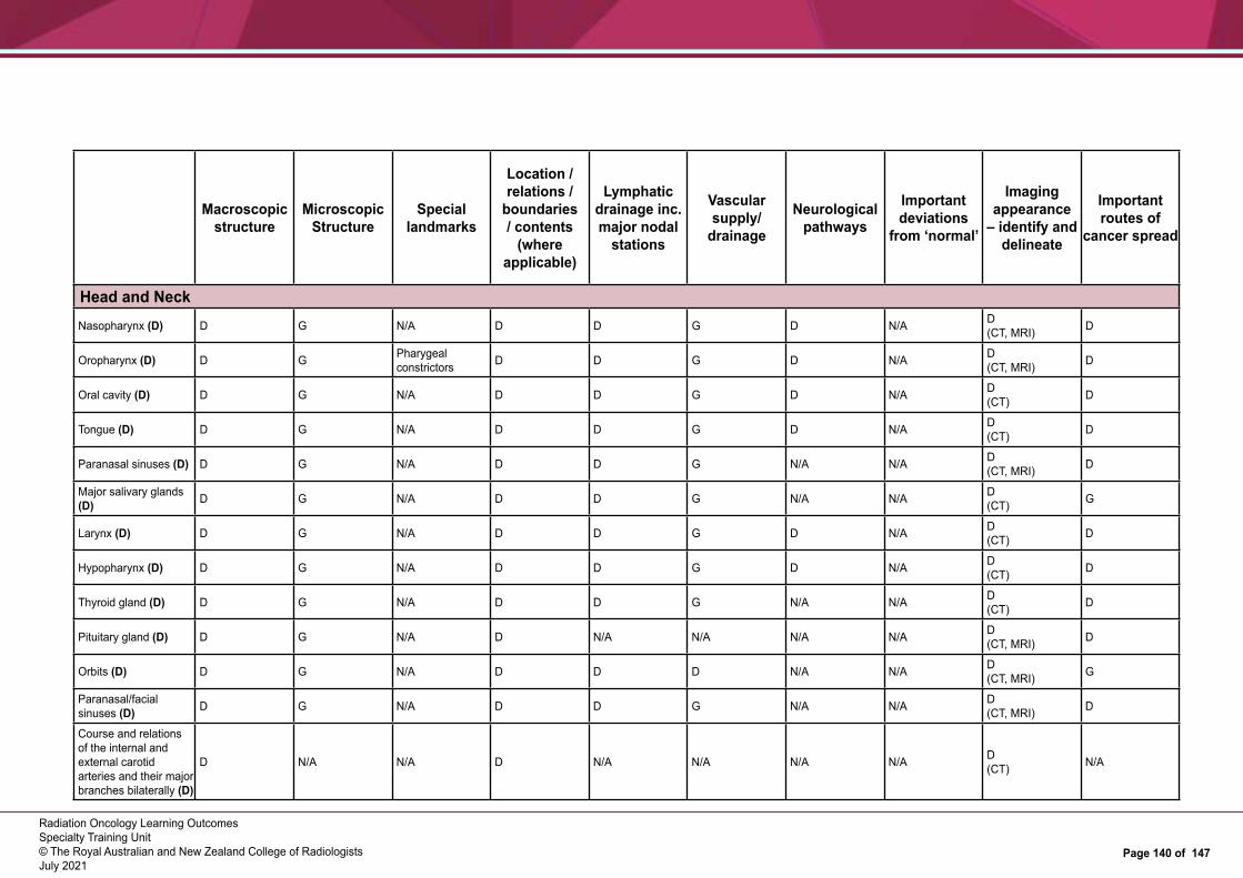
Page 140 of 147
Radiation Oncology Learning OutcomesSpecialty Training Unit© The Royal Australian and New Zealand College of RadiologistsJuly 2021
Macroscopic structure
Microscopic Structure
Special landmarks
Location/relations/
boundaries /contents
(where applicable)
Lymphatic drainage inc. major nodal
stations
Vascular supply/
drainage
Neurological pathways
Important deviations
from ‘normal’
Imaging appearance
– identify and delineate
Important routes of
cancer spread
Head and NeckNasopharynx (D) D G N/A D D G D N/A D
(CT, MRI) D
Oropharynx (D) D G Pharygeal constrictors D D G D N/A D
(CT, MRI) D
Oral cavity (D) D G N/A D D G D N/A D (CT) D
Tongue (D) D G N/A D D G D N/A D (CT) D
Paranasal sinuses (D) D G N/A D D G N/A N/A D (CT, MRI) D
Major salivary glands (D) D G N/A D D G N/A N/A D
(CT) G
Larynx (D) D G N/A D D G D N/A D(CT) D
Hypopharynx (D) D G N/A D D G D N/A D(CT) D
Thyroid gland (D) D G N/A D D G N/A N/A D(CT) D
Pituitary gland (D) D G N/A D N/A N/A N/A N/A D(CT, MRI) D
Orbits (D) D G N/A D D D N/A N/A D(CT, MRI) G
Paranasal/facial sinuses (D) D G N/A D D G N/A N/A D
(CT, MRI) D
Course and relations of the internal and external carotid arteries and their major branches bilaterally (D)
D N/A N/A D N/A N/A N/A N/A D(CT) N/A

Page 141 of 147
Radiation Oncology Learning OutcomesSpecialty Training Unit© The Royal Australian and New Zealand College of RadiologistsJuly 2021
Macroscopic structure
Microscopic Structure
Special landmarks
Location/relations/
boundaries /contents
(where applicable)
Lymphatic drainage inc. major nodal
stations
Vascular supply/
drainage
Neurological pathways
Important deviations
from ‘normal’
Imaging appearance
– identify and delineate
Important routes of
cancer spread
Head and NeckAnterior and posterior triangles of the neck (including surgical nodal levels) (D)
D N/A N/ADSurgical nodal stations
N/A N/A N/A N/A D(CT) N/A
Supraclavicular fossa (D) D D N/A D
Brachial plexus D D D N/A D(CT) N/A
Pterygopalatine fossa (D) D D N/A D D D D N/A D
(CT, MRI) N/A
Temporal and infratemporal fossae (D)
D D N/A D D D D N/A D(CT, MRI) N/A
Base of skull including pituitary fossa, cavernous sinus, Meckel’s cave, Rathke’s pouch, clivus. (D)
D N/A N/A D N/A N/A D N/A D(CT, MRI) N/A
All vascular and neural foramina for major vessels, cranial nerves and their branches traversing the base of skull (D)
D N/A D N/A N/A D N/A D(CT, MRI) N/A
Parathyroid glands (G) G G G G N/A N/A N/A N/A G
Cochlear (G) G G G N/A N/A N/A N/A D (CT, MRI)
Note: Head and Neck contouring requires a knowledge of other important structures listed elsewhere including: brachial plexus, brainstem, optic nerve, optic chiasm and cervical oesophagus)

Page 142 of 147
Radiation Oncology Learning OutcomesSpecialty Training Unit© The Royal Australian and New Zealand College of RadiologistsJuly 2021
Macroscopic structure
Microscopic Structure
Special landmarks
Location/relations/
boundaries /contents
(where applicable)
Lymphatic drainage inc. major nodal
stations
Vascular supply/
drainage
Neurological pathways
Important deviations
from ‘normal’
Imaging appearance
– identify and delineate
Important routes of
cancer spread
NeuroanatomyFunctional and anatomical compartments of the cerebrum and cerebellum (D)
D G N/A G N/A G (CT, MRI) G G (CT, MRI) N/A
Brainstem(Detailed knowledge of individual nuclei not required) (D)
D G N/ADInsertion of cranial nerves
N/A G D (CT, MRI) G D
(CT, MRI) N/A
Cranial nerves including their origin and distribution (intra and extra cranial) (D)
D V/VII, others G N/A D N/A N/A N/A N/A D V/VII
(MRI) N/A
Spinal cord and cauda equina (D) Note: Only sensory and motor tracts in detail; clinical and surface anatomy skills important; knowledge of dermatomes and myotomes expected
DSensory and motor tracts
G N/A D N/A G N/A N/A G (CT,MRI) N/A
Meninges (G) G G N/A G N/A N/A N/A N/A G (MRI) N/A
Brachial plexus (D) D N/A N/A D N/A N/A N/A N/A D (CT, MRI) N/A
Innervation of the upper and lower limbs, course and distribution of major motor and sensory nerves (G)
G N/A N/A G N/A N/A G N/A N/A N/A

Page 143 of 147
Radiation Oncology Learning OutcomesSpecialty Training Unit© The Royal Australian and New Zealand College of RadiologistsJuly 2021
Macroscopic structure
Microscopic Structure
Special landmarks
Location/relations/
boundaries /contents
(where applicable)
Lymphatic drainage inc. major nodal
stations
Vascular supply/
drainage
Neurological pathways
Important deviations
from ‘normal’
Imaging appearance
– identify and delineate
Important routes of
cancer spread
NeuroanatomyAutonomic nervous system, specifically location of ganglions (G)
G N/A N/A G N/A N/A G N/A N/A N/A
Ventricular system including interconnecting foramina and channels (G)
G N/A N/A G N/A N/A N/A N/A G (CT, MRI) Periventricular spread
Sacral plexus (G) G N/A N/A G N/A N/A N/A N/A G (CT, MRI) N/A

Page 144 of 147
Radiation Oncology Learning OutcomesSpecialty Training Unit© The Royal Australian and New Zealand College of RadiologistsJuly 2021
Macroscopic structure
Microscopic Structure
Special landmarks
Location/relations/
boundaries /contents
(where applicable)
Lymphatic drainage inc. major nodal
stations
Vascular supply/
drainage
Neurological pathways
Important deviations
from ‘normal’
Imaging appearance
– identify and delineate
Important routes of
cancer spread
Gastrointestinal Anatomy
Stomach (D) D D
G Hepatogastric ligament, antrum, pylorus, cardia, gastro-oesophageal junction
D D D G N/A D (CT) D
Liver and biliary tract (D)
DKnowledge of liver segments
GGLiver segments couinaud
D G D G N/A D(CT, MRI) D
Pancreas (D) D G G D D G G G D(CT, MRI) D
Oesophagus (D) D D N/A D D G G N/A D (CT) D
Rectum (D) D D D D D D D N/A D(CT, MRI) D
Anal Canal (D) D D D D D D D N/A D D
Duodenum (G) G G GAmpulla of Vater D G G G N/A D G
Spleen (G) G G D G G N/A NA G G
Gall bladder (G) D G G G G G N/A G G(CT) G
Adrenal glands (G) G G G D G G G NA D G
Abdominal wall anatomy (G) G G G G G G G N/A G N/A
Small and large intestine (G) G G G G G G G G G
(CT) G

Page 145 of 147
Radiation Oncology Learning OutcomesSpecialty Training Unit© The Royal Australian and New Zealand College of RadiologistsJuly 2021
Macroscopic structure
Microscopic Structure
Special landmarks
Location/relations/
boundaries /contents
(where applicable)
Lymphatic drainage inc. major nodal
stations
Vascular supply/
drainage
Neurological pathways
Important deviations
from ‘normal’
Imaging appearance
– identify and delineate
Important routes of
cancer spread
MusculoskeletalAxilla (D) D D D
BordersDBrachial plexus D D D N/A D
(CT, MRI) N/A
Inguinal and femoral canals (D) G G D
Borders D D D D N/AD (CT, MRI – identify contents)
Major nerves of the limbs – brachial plexus, median, radial, ulnar – sciatic, femoral, tibial, common peroneal (D)
G G N/AG Location, course of major nerves
N/A N/A N/A N/AD (CT, MRI – identify nerve)
Vertebral bodies (D)
DMacro appearance for each vertebral level
G N/A D G G N/A N/A D
Dermatomes and myotomes of the upper and lower limbs (G)
Able to draw a diagram N/A N/A D N/A N/A N/A N/A N/A
Major muscles of the limbs (G)Not important to know individual muscles.
G N/A N/A G N/A N/AN/A (broadly covered above)
N/A G (CT, MRI)
Identify muscles on cross-sectional imaging of upper and lower limbs. Insertion of muscles not required.

Page 146 of 147
Radiation Oncology Learning OutcomesSpecialty Training Unit© The Royal Australian and New Zealand College of RadiologistsJuly 2021
Macroscopic structure
Microscopic Structure
Special landmarks
Location/relations/
boundaries /contents
(where applicable)
Lymphatic drainage inc. major nodal
stations
Vascular supply/
drainage
Neurological pathways
Important deviations
from ‘normal’
Imaging appearance
– identify and delineate
Important routes of
cancer spread
Musculoskeletal
Muscular compartments (G) N/A N/A N/A D
Boundaries N/A N/A N/A N/A
G (CT, MRI – identify compartments). Compartmental boundaries useful for sarcoma (inc. superficial and deep fascia)
Femoral H&N (G) G N/A Head, trochanter, surgical neck N/A N/A G N/A N/A G
(CT, MRI)
Long bones of upper and lower limbs (G) G N/A
Important gross anatomical features of long bones
G N/A N/A N/A N/A G(CT, MRI, X-RAY)

FOR MORE INFORMATION
Level 9, 51 Druitt StreetSydney, NSW 2000, AustraliaABN 37 000 029 863
tel: +61 2 9268 9777fax: +61 2 9268 9799email: [email protected] web: www.ranzcr.com/tar