A protein related to brain microtubule-associated protein MAP1B is a component of the mammalian...
11
INTRODUCTION The organization of microtubules within most non-neuronal cells appears to depend on the presence of a structure, the cen- trosome, which has the ability to nucleate the polymerization of tubulin into microtubules (Snyder and McIntosh, 1975; Weisenberg and Rosenfeld, 1975; Frankel, 1976; Osborn and Weber, 1976; Gould and Borisy, 1977; Kuriyama and Borisy, 1981; Mitchison and Kirschner, 1984; Bornens et al., 1987; for review see Bornens et al., 1990; Cande, 1990; Cande and Stearns, 1991; Bornens, 1992; Tucker, 1992). Electron microscope studies have shown that centrosomes are composed of a pair of centrioles surrounded by an amorphous pericentriolar material (Bornens and Karsenti, 1984), which contains the nucleation sites for microtubules (Gould and Borisy, 1977; Kuriyama and Borisy, 1981; Rieder and Borisy, 1982; for review see Vorobjev and Nadezhdina, 1987; Vandre and Borisy, 1989; Bornens, 1992). Little is known about the organization of the molecular components of centrosomes. Several proteins have been localized to the cen- trosome by immunocytochemistry and although the presence of a nucleic acid (either DNA or RNA) has been suggested (for reviews see Bornens and Karsenti, 1984; Vorobjev and Nadezhdina, 1987) it still remains controversial (Hall et al., 1989; Klotz et al., 1990; Johnson and Dutcher, 1991; for review see Johnson and Rosenbaum, 1991). A first step in the characterization of centrosomal compo- nents is the isolation of functional centrosomes (Mitchison and Kirschner, 1984; Bornens et al., 1987; Komesli et al., 1989). These procedures are mainly based on the fractionation through sucrose gradients of cell homogenates obtained after hypotonic lysis of cultured cells previously incubated with cytoskeletal-disrupting drugs (Maro and Bornens, 1980; Bornens et al., 1987). The above mentioned procedures have been useful for functional studies, but biochemical studies have been hampered by the low yields of centrosome purifications and the large and variable amounts of different proteins present in the centrosomal preparations. To overcome these problems, immunological probes of known proteins have been used to correlate biochemical analyses of centrosome preparations with immunocytochemical studies (Gosti-Testu et al., 1986, 1987; Bornens et al., 1987; Balczon and West, 1991; Moudjou et al., 1991; Stearns et al., 1991; Zheng et al., 1991; Chevrier et al., 1992; Joshi et al., 1992; Sellitto et al., 1992; Tucker et al., 1992; Rothbarth et al., 1993; Keryer et al., 1993; for review Kuriyama, 1992; Kalt and Schliwa, 1993). The centrosomal 601 Journal of Cell Science 107, 601-611 (1994) Printed in Great Britain © The Company of Biologists Limited 1994 JCS8363 The centrosome is the main microtubule organizing center of mammalian cells. Structurally, it is composed of a pair of centrioles surrounded by a fibro-granular material (the pericentriolar material) from which microtubules are nucleated. However, the nature of centrosomal molecules involved in microtubule nucleation is still obscure. Since brain microtubule-associated proteins (MAPs) lower the critical tubulin concentration required for microtubule nucleation in tubulin solution in vitro, we have examined their possible association with centrosomes. By immuno- fluorescence, monoclonal and polyclonal antibodies raised against MAP1B stain the centrosome in cultured cells as well as purified centrosomes, whereas antibodies raised against MAP2 give a completely negative reaction. The MAP1B-related antigen is localized to the pericentriolar material as revealed by immunoelectron microscopy. In preparations of purified centrosomes analyzed on poly- acrylamide gels, a protein that migrates as brain MAP1B is present. After blotting on nitrocellulose, it is decorated by anti-MAP1B antibodies and the amino acid sequence of proteolytic fragments of this protein is similar to brain MAP1B. Moreover, brain MAP1B and its centrosomal counterpart share the same phosphorylation features and have similar peptide maps. These data strongly suggest that a protein homologue to MAP1B is present in centrosomes and it is a good candidate for being involved in the nucleating activity of the pericen- triolar material. Key words: centrosome, microtubule-associated protein, MAP1B SUMMARY A protein related to brain microtubule-associated protein MAP1B is a component of the mammalian centrosome J. E. Domínguez 1, *, B. Buendia 2 , C. López-Otín 3 , C. Antony 4 , E. Karsenti 2 and J. Avila 1 1 Centro de Biología Molecular (CSIC-UAM), Fac. Ciencias UAM Cantoblanco, E-28049 Madrid, Spain 2 European Molecular Biology Laboratory, Postfach 102209, W-6900 Heidelberg 1, Germany 3 Univ. de Oviedo, Dpto Biología Funcional, Area de Bioquímica y Biología Molecular, Fac. de Medicina, E-33006 Oviedo, Spain 4 Institut Jacques Monod CNRS-Université P7 2, Place Jussieu, Tour 43 F-75251 Paris Cedex 05, France *Author for correspondence
-
Upload
wwwunistra -
Category
Documents
-
view
1 -
download
0
Transcript of A protein related to brain microtubule-associated protein MAP1B is a component of the mammalian...
601Journal of Cell Science 107, 601-611 (1994)Printed in Great Britain © The Company of Biologists Limited 1994JCS8363
A protein related to brain microtubule-associated protein MAP1B is a
component of the mammalian centrosome
J. E. Domínguez1,*, B. Buendia2, C. López-Otín3, C. Antony4, E. Karsenti2 and J. Avila1
1Centro de Biología Molecular (CSIC-UAM), Fac. Ciencias UAM Cantoblanco, E-28049 Madrid, Spain2European Molecular Biology Laboratory, Postfach 102209, W-6900 Heidelberg 1, Germany3Univ. de Oviedo, Dpto Biología Funcional, Area de Bioquímica y Biología Molecular, Fac. de Medicina, E-33006 Oviedo, Spain4Institut Jacques Monod CNRS-Université P7 2, Place Jussieu, Tour 43 F-75251 Paris Cedex 05, France
*Author for correspondence
The centrosome is the main microtubule organizing centerof mammalian cells. Structurally, it is composed of a pairof centrioles surrounded by a fibro-granular material (thepericentriolar material) from which microtubules arenucleated. However, the nature of centrosomal moleculesinvolved in microtubule nucleation is still obscure. Sincebrain microtubule-associated proteins (MAPs) lower thecritical tubulin concentration required for microtubulenucleation in tubulin solution in vitro, we have examinedtheir possible association with centrosomes. By immuno-fluorescence, monoclonal and polyclonal antibodies raisedagainst MAP1B stain the centrosome in cultured cells aswell as purified centrosomes, whereas antibodies raisedagainst MAP2 give a completely negative reaction. TheMAP1B-related antigen is localized to the pericentriolar
material as revealed by immunoelectron microscopy. Inpreparations of purified centrosomes analyzed on poly-acrylamide gels, a protein that migrates as brain MAP1Bis present. After blotting on nitrocellulose, it is decoratedby anti-MAP1B antibodies and the amino acid sequence ofproteolytic fragments of this protein is similar to brainMAP1B. Moreover, brain MAP1B and its centrosomalcounterpart share the same phosphorylation features andhave similar peptide maps.
These data strongly suggest that a protein homologue toMAP1B is present in centrosomes and it is a good candidatefor being involved in the nucleating activity of the pericen-triolar material.
Key words: centrosome, microtubule-associated protein, MAP1B
SUMMARY
INTRODUCTION
The organization of microtubules within most non-neuronalcells appears to depend on the presence of a structure, the cen-trosome, which has the ability to nucleate the polymerizationof tubulin into microtubules (Snyder and McIntosh, 1975;Weisenberg and Rosenfeld, 1975; Frankel, 1976; Osborn andWeber, 1976; Gould and Borisy, 1977; Kuriyama and Borisy,1981; Mitchison and Kirschner, 1984; Bornens et al., 1987; forreview see Bornens et al., 1990; Cande, 1990; Cande andStearns, 1991; Bornens, 1992; Tucker, 1992).
Electron microscope studies have shown that centrosomesare composed of a pair of centrioles surrounded by anamorphous pericentriolar material (Bornens and Karsenti,1984), which contains the nucleation sites for microtubules(Gould and Borisy, 1977; Kuriyama and Borisy, 1981; Riederand Borisy, 1982; for review see Vorobjev and Nadezhdina,1987; Vandre and Borisy, 1989; Bornens, 1992). Little isknown about the organization of the molecular components ofcentrosomes. Several proteins have been localized to the cen-trosome by immunocytochemistry and although the presenceof a nucleic acid (either DNA or RNA) has been suggested (forreviews see Bornens and Karsenti, 1984; Vorobjev and
Nadezhdina, 1987) it still remains controversial (Hall et al.,1989; Klotz et al., 1990; Johnson and Dutcher, 1991; forreview see Johnson and Rosenbaum, 1991).
A first step in the characterization of centrosomal compo-nents is the isolation of functional centrosomes (Mitchison andKirschner, 1984; Bornens et al., 1987; Komesli et al., 1989).These procedures are mainly based on the fractionationthrough sucrose gradients of cell homogenates obtained afterhypotonic lysis of cultured cells previously incubated withcytoskeletal-disrupting drugs (Maro and Bornens, 1980;Bornens et al., 1987). The above mentioned procedures havebeen useful for functional studies, but biochemical studies havebeen hampered by the low yields of centrosome purificationsand the large and variable amounts of different proteins presentin the centrosomal preparations. To overcome these problems,immunological probes of known proteins have been used tocorrelate biochemical analyses of centrosome preparationswith immunocytochemical studies (Gosti-Testu et al., 1986,1987; Bornens et al., 1987; Balczon and West, 1991; Moudjouet al., 1991; Stearns et al., 1991; Zheng et al., 1991; Chevrieret al., 1992; Joshi et al., 1992; Sellitto et al., 1992; Tucker etal., 1992; Rothbarth et al., 1993; Keryer et al., 1993; for reviewKuriyama, 1992; Kalt and Schliwa, 1993). The centrosomal
602 J. E. Domínguez and others
components responsible for the nucleation of the assembly ofmicrotubules have not been identified yet. Interestingly,Moudjou et al. (1991), have found a 62-64 kDa centrosomalprotein that could be involved in the nucleation process.Recently the presence of a new member of the tubulin family,the γ-tubulin, has been described as being localized at themicrotubule organizing centers of eukaryotic cells (Zheng etal., 1991; Stearns et al., 1991; for review, see Oakley, 1992).It seems to be required for microtubule polymerizationinitiation in vivo (Joshi et al., 1992). Probably, γ-tubulin willact as a template for the initiation of the polymerization oftubulin at the microtubule organizing centers. Its function instimulating nucleation of polymer growth is still unclear (Joshiet al., 1992; Oakley, 1992).
As microtubule-associated proteins (MAPs) present inneuronal cells favor the in vitro nucleation of polymerassembly (Olmsted, 1986), we have searched for proteinsrelated to brain MAPs in centrosomes from non-neuronal cellsby combining immunocytochemical with biochemical analysesof centrosomal preparations. In the present study we show byimmunological analysis, phosphorylation characteristics,peptide mapping, and partial sequence analysis, that a proteinrelated to brain microtubule-associated protein MAP1B (alsoreferred to as MAP5, MAP1.2 or MAP1X) is indeed presentin isolated mammalian centrosomes and localized to the peri-centriolar material.
MATERIALS AND METHODS
Cell cultureHuman lymphoblastic KE37 (Mayer et al., 1982) cells were culturedin RPMI1640 medium containing 10% fetal calf serum at 37°C and5% CO2 in air. The Xenopus laevis epithelial cell line XL177 (Millerand Daniel, 1977), was cultured in 55% Leibovitz L15 medium(Gibco, UK), 15% fetal calf serum, at 25°C. Mouse Ehrlich-Lettre(ATCC CCL77) cells were injected intraperitoneally into three-month-old Balb/c mice. Nine days after injection, ascitic fluid wasextracted and the cells isolated by centrifugation at 1500 g.
AntibodiesMonoclonal antibodies (356 and 357) against α- and β-tubulinsubunits were purchased from Amersham (UK). The characterizationof the monospecific rabbit antibodies against brain MAP2 used in thiswork has been done previously (Hernandez et al., 1989). Monoclonalantibody against MAP2 (AP-20) was purchased from BoehringerMannheim. Monoclonal antibody against MAP1B 125 was alreadydescribed (Ulloa et al., 1993a,b), monoclonal antibody 150 againstphosphorylated MAP1B was a kind gift from Dr Moya (Mansfield etal., 1992). Polyclonal antiserum against MAP1B, 81 has already beendescribed (Diaz-Nido and Avila, 1989a). The antibodies from theautoimmune human serum that specifically recognizes a centrosomalprotein will be described (Tournier et al., 1991; Domínguez et al.,unpublished). We also used the 5051 human auto antibody fromKirschner’s lab (Calarco-Gillam et al., 1983) and the monoclonalantibody from Bornens (CTR453; Bailly et al., 1989).
Isolation of centrosomes The protocol of Bornens et al. (1987) was followed with slight mod-ifications: (a) step 5 was repeated three times; (b) the supernatant ofthe lysis (step 6) was overlaid on a 50% sucrose (w/w) cushionprepared in 10 mM K-PIPES, pH 7.2, 1 mM EDTA, 0.1% 2-mer-captoethanol and 0.1% Triton X-100, and centrifuged at 40,000 g for20 minutes. The interface between the lysate and the 50% sucrose
cushion was collected and overlaid on the discontinuous sucrosegradient system (Bornens et al., 1987).
Immunofluorescence microscopyCentrosomes were sedimented onto coverslips as previously reported(Evans et al., 1985), fixed with −20°C methanol and washed withphosphate buffered saline buffer (PBS). The coverslips wereincubated for 30 minutes with the antibodies and after three 10 minutewashes with PBS, they were further incubated with fluorescein- orrhodamine-conjugated goat anti-mouse, goat anti-human or goat anti-rabbit immunoglobulins (Tago, CA; Jackson Immunoresearch Labs,PA; Cappel, PA). To check the DNA contamination of the centroso-mal purification steps, the dye Hoechst No. 33258 (Sigma, St Louis,MO) was added to the secondary antibody solution at a final concen-tration of 10 µg/ml. The coverslips were mounted with Mowiol R 40-88 (Aldrich Chemie) and examined using a Zeiss epifluorescencemicroscope.
Immunoelectron microscopyThe isolated centrosomes were sedimented onto 12 mm round cover-slips, fixed in 0.25% glutaraldehyde in PBS and incubated twice with0.1% (w/v) sodium borohydride (Sigma, St Louis, MO) in the samebuffer. After a 10 minute incubation in fetal calf serum (10% in PBS),the preparation was incubated with the anti-MAP1B (125) antibody(1/5 dilution from hybridoma-conditioned culture medium) for 15minutes at room temperature followed by rabbit anti-mouse (1/150dilution; Jackson Immunoresearch Labs, PA) for 30 minutes at roomtemperature and Protein A coupled to 9 nm gold particles (1/75dilution; Janssen Pharmaceutica, Belgium) for 30 minutes at roomtemperature. The preparation was postfixed with 1% glutaraldehydeand 2% osmium tetroxide (Merck, Germany). The centrosomes werefinally stained with aqueous uranyl acetate and embedded in Epon.Sections parallel to the coverslips were observed in the electronmicroscope (model 400, Philips) after contrasting with uranyl acetateand lead citrate.
A negative control was made using the monoclonal antibodyagainst MAP2, AP-20, (1/200 dilution).
Protein preparation and analysisMicrotubule proteins from mouse brain were prepared through tem-perature-dependent cycles of assembly-disassembly as indicated byShelanski et al. (1973) with the modifications of Karr et al. (1979).Tubulin was purified as described by Weingarten et al. (1975).Newborn mouse brain MAPs were purified as described by Vallee etal. (1986). Protein composition from microtubule or centrosomefractions was analyzed by gel electrophoresis according to theprocedure of Laemmli (1970). Gels were either stained withCoomassie Blue as indicated by Fairbanks et al. (1981) or with silveras indicated by Harlow and Lane (1988). Western blotting analyseswere carried out by transferring the proteins previously fractionatedby gel electrophoresis to nitrocellulose membranes, according toTowbin et al. (1979). A peroxidase-conjugated secondary antibodywas used and the peroxidase reaction was developed using 3,3′-diaminobenzidine (Sigma, St Louis, MO) and hydrogen peroxide assubstrates. Partial amino acid sequence of the peptides from centro-somal proteins was carried out after transferring the peptides previ-ously fractionated by gel electrophoresis to Immobilon sheets(Millipore, MA) as indicated by Vandekerckhove et al. (1985). Thepeptides on Immobilon were then subjected to automated Edmandegradation cycles in a gas-phase sequenator (Applied Biosystems)equipped with an on line phenylthiohydantoin amino acid analyzer(model 120A).
Peptide mappingPeptide mapping with Staphylococcus aureus V8 protease (Sigma, StLouis, MO) was done according to the method of Cleveland et al.(1977).
603A MAP1B-related centrosomal protein
NTCB (2-nitro-5-thiocyanobenzoic acid) peptide mapping wasdone as described by Correas et al. (1992) with slight modifications:the protein bands were stained by the imidazole-zinc reverse stainingmethod as described by Ortiz et al. (1992). NTCB was from Sigma(St Louis, MO).
Phosphorylation assaysEndogenous phosphorylation of centrosomal and microtubule prepa-rations was carried out in 0.1 M MES, pH 6.4, supplemented with 2mM EGTA, 6 mM MgCl2 and 10 µM [γ-32P]ATP (Amersham, UK)either in the presence or the absence of 10 µg/ml poly-L-lysine (anactivator of casein kinase II; Hathaway and Traugh, 1982) plus 10 µMcAMP-dependent protein kinase inhibitor (Sigma, St Louis, MO), or1 µM heparin (an inhibitor of casein kinase II; Hathaway and Traugh,1982). The mixture was incubated for 20 minutes at 37°C and thereaction was stopped with SDS-sample buffer (Laemmli, 1970).Samples were analyzed by electrophoresis, and phosphorylatedproteins were subjected to V8 protease peptide mapping, as describedabove.
Microtubule-binding assay of the MAP1B-related proteinPurified centrosomes (20 µg of protein) were extracted with 2 M KIas described by Klotz et al. (1990). The extracted material wasdialyzed and concentrated in a negative pressure protein dialysis con-centrator (Bio-Molecular Dynamics, OR). The dialysis was madeagainst 0.1 M MES, pH 6.4, 6 mM MgCl2, 2 mM EGTA. Afterdialysis the protein concentration was measured (Smith et al., 1985)and [γ-32P]ATP was added to a concentration of 1 µM in the presenceof 1 mM PMSF and 10 µg/ml poly-L-lysine. The whole mixture wasincubated for 20 minutes at 37°C. The phosphorylation reaction wasstopped with NaF and cold ATP added to a final concentration of 50mM and 1 mM, respectively. Just before copolymerization withmicrotubules, this solution was centrifuged at 100,000 g for 10minutes at 4°C in a Beckman TL100 ultracentrifuge (TL100.1 rotor),to remove aggregates.
Phosphocellulose-purified tubulin was polymerized in 0.1 M MES,pH 6.4, 0.5 mM MgCl2, 2 mM EGTA, 20 mM NaF, 1 mM ATP, 1mM GTP for 20 minutes at 37°C. The polymerized microtubules weremixed with the KI-extracted centrosomal proteins and incubated at37°C for 15 minutes.
The mixture was centrifuged at 100,000 g, for 10 minutes at 25°Cin a Beckman TL100 ultracentrifuge (TL100.3 rotor). The pellet andsupernatant were boiled in electrophoresis sample buffer (Laemmli,1970), for 5 minutes. The proteins were analyzed by electrophoresisin a 10% polyacrylamide gel.
Assay of microtubule nucleating activity of centrosomesPhosphocellulose purified tubulin was mixed (at a final concentrationof 15 µM) with isolated centrosomes in the conditions described byMitchison and Kirschner (1984). The incubation buffer was 80 mMPIPES, pH 6.8, 1 mM MgCl2, 1 mM EGTA and 1 mM GTP. Themicrotubule regrowth from centrosomes was allowed for 10 minutesat 37°C and the reaction stopped by fixation with 0.7% glutaralde-hyde. Afterwards, tubulin immunofluorescence was performed as pre-viously described (Buendia et al., 1992). We assayed three differentconditions: absence of added antibodies, 20 minutes pre-incubationwith 125 hybridoma cell culture supernatant at a final dilution of 1/10at room temperature, and pre-incubation with a hybridoma (IgM) cellculture supernatant against a protein fraction from Golgi apparatus.
RESULTS
An antigen related to brain MAP1B protein islocalized to the centrosome in cultured cellsAs shown in Fig. 1, in KE37 human lymphocytes, mouse
Ehrlich-Lettre cells and frog cells, a MAP1B-related antigen ispresent on a cytoplasmic structure corresponding to the cen-trosome and to some extent in the nuclei. This was determinedby double immunofluorescence using different anti-centro-some antibodies, two human scleroderma sera, one of which isthe 5051 serum described by Kirschner’s group, and the otheris a serum we have recently characterized (our unpublishedobservations; also see Tournier et al., 1991; Calarco-Gillam etal., 1983). We also used one monoclonal antibody raisedagainst isolated centrosomes (CTR453; from M. Bornensgroup, Bailly et al., 1989) in conjunction with polyclonal anti-MAP1B antibodies. In Fig. 1b,d a monoclonal antibody toMAP1B (125) was used and in Fig. 1e,g a polyclonal antibodyraised against MAP1B was used. Note the strong and veryclean staining of a cytoplasmic spot located close to thenucleus that corresponds to the centrosome as well as a dottedstaining in the nucleus. The identity of the cytoplasmic dot asthe centrosome is further confirmed by its position centered atthe origin of the microtubule network (Fig. 1a,c). We have usedthree different anti-MAP1B antibodies (81, 125 and 150) thatall gave a similar staining in all cells. A similar staining patternhas also been described in other cell lines (rat, mouse andchinese hamster; Diaz-Nido and Avila, 1989a).
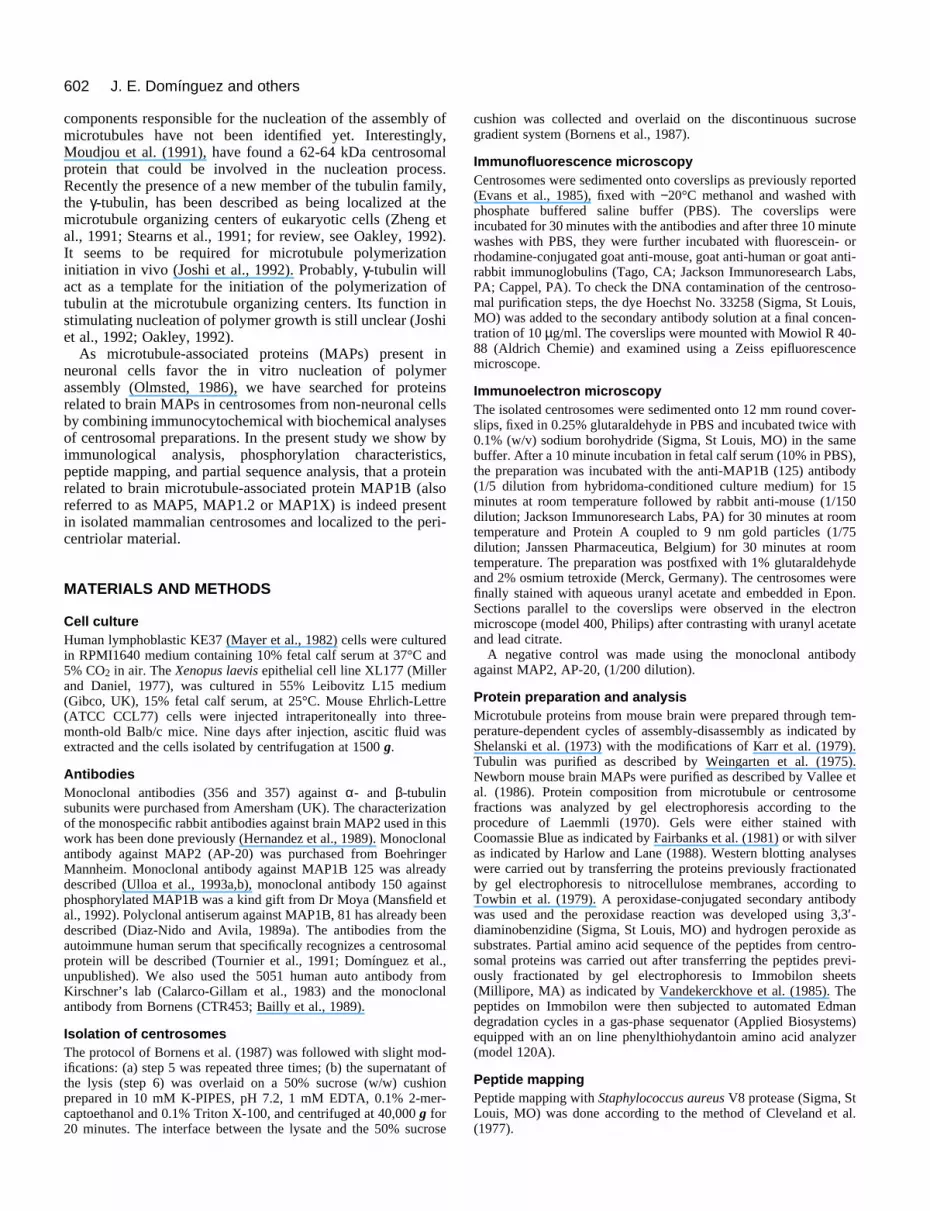
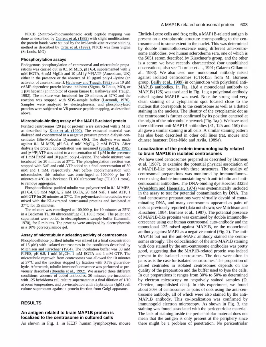
Localization of the protein immunologically relatedto brain MAP1B in isolated centrosomesWe have used centrosomes prepared as described by Bornenset al. (1987), to examine the potential physical association ofa MAP1B-like protein with these structures. The quality ofcentrosomal preparations was monitored by immunofluores-cence using double immunostaining with anti-tubulin and anti-centrosomal antibodies. The DNA-binding dye Hoechst 33258(Weisblum and Haenssler, 1974) was systematically includedin the assay to test for potential contamination by DNA. Thefinal centrosome preparations were virtually devoid of conta-minating DNA, and many centrosomes appeared as pairs ofdots as previously reported (data not shown; see Mitchison andKirschner, 1984; Bornens et al., 1987). The potential presenceof MAP1B-like proteins was examined by double immunoflu-orescence using our human centrosomal auto-antibody and themonoclonal 125 raised against MAP1B, or the monoclonalantibody against MAP2 as a negative control (Fig. 2). The anti-MAP1B but not the anti-MAP2 antibody stained the centro-somes strongly. The colocalisation of the anti-MAP1B stainingwith dots stained by the anti-centrosome antibodies was prettygood, suggesting that the MAP1B-related protein was indeedpresent in the isolated centrosomes. The dots were often inpairs as is the case for isolated centrosomes. The proportion ofpaired centrioles in isolated centrosomes depends on thequality of the preparation and the buffer used to lyse the cells.In our preparations it ranges from 30% to 50% as determinedby electron microscopy on negatively stained samples (D.Chrétien, unpublished data). In this experiment, we foundabout 30% of centrosomes as pairs of dots using the anti-cen-trosome antibody, all of which were also stained by the anti-MAP1B antibody. This co-localization was confirmed byimmunogold electron microscopy. As shown in Fig. 3, thestaining was found associated with the pericentriolar material.The lack of staining inside the pericentriolar material does notmean that the antigen is only present at the periphery sincethere might be a problem of penetration. No pericentriolar

604 J. E. Domínguez and others
Fig. 1. Double immunofluorescence staining of Xenopuslaevis tissue culture cells (XL177) with a rabbit anti-tubulin antibody (a,c) and with the monoclonal antibody125 against MAP1B (b,d). Both (a) and (b), and (c) and(d) represent the same field. (e and f) The doubleimmunofluorescence staining of the mouse cell lineEhrlich-Lettre with the polyclonal antibody to MAP1B81 (e), and with our human serum specific tocentrosomes (f) (same field). (g and h) The doubleimmunofluorescence staining of human lymphoblasticcells (KE37) with the polyclonal antibody to MAP1B 81(g), and the human serum specific to centrosomes (h)(same field). Arrows indicate the centrosomes. Bars, 5µm.
605A MAP1B-related centrosomal protein
Fig. 2. Double immunofluorescence staining ofpurified centrosomes labeled with antibodies to:(a,c) centrosomes (human auto-immune serum);(b) MAP1B (monoclonal 125); and (d) a MAP2monoclonal. Arrows indicate some centrosomaldoublets. Bar, 10 µm.
Fig. 3. Immunoelectronmicroscopy ofcentrosomes incubatedwith the monoclonalantibody againstMAP1B (125) andrevealed by a rabbitanti-mouseimmunoglobulins andProtein A coupled to 9nm gold particles. Bar,0.2 µm.
staining was found with anti-MAP2 antibody that was used asnegative control in the experiment (not shown).
In order to characterize the polypeptides present in the cen-trosomes, 2×10⁹ centrosomes containing about 50 µg ofprotein were subjected to gel electrophoresis. Fig. 4 shows thepolypeptide pattern of a mouse centrosomal preparationanalyzed on a 5% acrylamide gel in urea. In addition to onemajor protein doublet migrating with an electrophoreticmobility of about 230 kDa, the centrosome preparationcontained one electrophoretic band co-migrating with brainMAP1B (Fig. 4A,B). This latter protein band reacted withdifferent anti-MAP1B antibodies by western blotting analysis.Fig. 4C shows the result obtained with the polyclonal antibody
(81) against MAP1B (see arrow). The same result was obtainedwith a human centrosome preparation. Looking closely at thewestern blot (Fig. 4C) the MAP1B immunoreactivity can beseen in the form of a doublet. This could reflect the presencein this centrosomal protein of two different isoforms that ariseby its differential phosphorylation features (Riederer et al.,1990; Ulloa et al., 1993a). Fig. 4, section 2, shows the elec-trophoretic analysis of purified centrosomes (50 µg) on a 10-20% linear gradient polyacrylamide gel after staining withCoomassie Blue. The absence of any detectable protein of elec-trophoretic mobility below 45 kDa, and a high abundance ofproteins in the range of 45-68 kDa is clear.
Several cytoskeletal proteins like spectrin and myosin have

606 J. E. Domínguez and others
1 2kDa kDa
Fig. 4. SDS-polyacrylamide gelelectrophoresis and immunoblottinganalysis of centrosomal proteins frommouse Ehrlich-Lettre cells. (1) Theproteins were ran on a 5% acrylamide gelcontaining 4 M urea. Lane A, mousebrain microtubule proteins; lane B,centrosomal proteins (Coomassiestaining); lane C, centrosomal proteinstransferred to nitrocellulose and stainedwith rabbit polyclonal antibody toMAP1B 81. A centrosomal protein withsimilar mobility to mouse brain MAP1Bis stained by this antibody (see arrow).The other antibodies against MAP1Bgave similar results. (2) Centrosomalproteins (Coomassie staining) analyzedon a 10-20% linear gradient ofpolyacrylamide. Sizes of marker proteins(in kDa) are indicated at the right margin.
Fig. 5. The MAP1B-related protein is enriched in purifiedcentrosomes. In lysed cells (A, lane 1) or in centrosomes recoveredafter the concentration step (A, lane 2), there is no visible proteinmigrating as MAP1B whereas in the purified centrosomes (A, lane3), a 325 kDa band appears (arrowhead). Sizes (in kDa) of markerproteins are indicated to the right margin. Dot blot analysis on 20 µgof protein from each fraction (B) shows a dramatic increase inimmunoreactivity for the polyclonal antibody to MAP1B (81) in thepurified centrosome fraction (1, 2 and 3 correspond, respectively, tolysed cells, concentration step and purified centrosomes).
A
B
kDa
been found in centrosomal preparations and are suspected tobe mere contaminants that could either sediment like centro-somes on sucrose gradients or become adsorbed non-specifi-cally to them during lysis of the cells in low ionic strengthbuffer (Komesli et al., 1989). This does not seem to be the casefor MAP1B, since it can be detected by immunofluorescenceat the centrosome of cultured cells. To strengthen the case, wefirst compared the electrophoretic pattern of the final centro-some preparation with the protein composition of the lysedcells (Fig. 5). The band co-migrating with brain MAP1B andstained by the anti-MAP1B antibodies (Figs 4 and 5) wasenriched in the final centrosome preparation (Fig. 5A), whereasit was not visible in the cell lysate. The enrichment of this bandwas concomitant with an increment in the anti-MAP1Bimmunoreactivity (Fig. 5B). At the moment we do not knowif this 325 kDa band actually corresponds to a single protein.We have also analyzed by dot blot the distribution of theMAP1B-related antigen in the sucrose gradient used to purifycentrosomes. MAP1B-related protein co-migrated exactly withmouse Ehrlich-Lettre cell centrosomes (not shown). The sameresult was obtained with centrosomes isolated from humanlymphoblastic cells.Taken together these results indicate that MAP1B copurifieswith, and becomes enriched in, centrosomes, arguing againstit being a mere contaminant of the centrosome preparation.
Characterization of the centrosomal MAP1B-relatedproteinIn order to check for structural similarities between the cen-trosomal MAP1B-related protein and brain MAP1B, it was ofinterest to have some information about the amino acidsequence of the centrosomal protein. The centrosomalMAP1B-related protein turned out to have a blocked amino-terminal residue, just like brain MAP1B. Interestingly, in somecentrosome preparations, low molecular mass polypeptideswere recognized by anti-MAP1B antibodies, suggesting thatthey were degradation products of the complete molecule. Wetook advantage of this feature and carried out an experiment inwhich proteolytic degradation was enhanced purposely by
607A MAP1B-related centrosomal protein
kDa
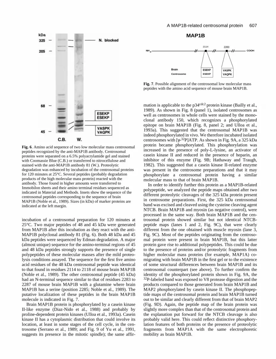
Fig. 6. Amino acid sequence of two low molecular mass centrosomalpeptides recognized by the anti-MAP1B antibody. Centrosomalproteins were separated on a 6.5% polyacrylamide gel and stainedwith Coomassie Blue (C.B.) or transferred to nitrocellulose andstained with the anti-MAP1B antibody 81 (W.). Proteolyticdegradation was enhanced by incubation of the centrosomal proteinsfor 120 minutes at 25°C. Several peptides (probably degradationproducts of the high molecular mass protein) reacted with theantibody. Those found in higher amounts were transferred toImmobilon sheets and their amino terminal residues sequenced asindicated in Material and Methods. Insets show the sequence of thecentrosomal peptides corresponding to the sequence of brainMAP1B (Noble et al., 1989). Sizes (in kDa) of marker proteins areindicated at the left margin.
Fig. 7. Possible alignment of the centrosomal low molecular masspeptides with the amino acid sequence of mouse brain MAP1B.
MAP1B
ESEKPESEKP
NH2
COOH
2114 2283
Brain
Centrosome
VASPKVAQPK
incubation of a centrosomal preparation for 120 minutes at25°C. Two major peptides of 48 and 45 kDa were generatedfrom MAP1B after this incubation as they react with the anti-MAP1B polyclonal antibody 81 (Fig. 6). Both 48 kDa and 45kDa peptides were sequenced by Edman degradation. A major(almost unique) sequence for the amino-terminal regions of 45and 48 kDa peptides strongly supports the presence of singlepolypeptides of these molecular masses after the mild proteo-lysis conditions assayed. The sequence for the first five aminoacid residues of the 48 kDa centrosomal peptide was identicalto that found in residues 2114 to 2118 of mouse brain MAP1B(Noble et al., 1989). The other centrosomal peptide (45 kDa)had an N-terminal sequence similar to that of residues 2283 to2287 of mouse brain MAP1B with a glutamine where brainMAP1B has a serine (position 2285; Noble et al., 1989). Theputative localization of these peptides in the brain MAP1Bmolecule is indicated in Fig. 7.
Brain MAP1B protein is phosphorylated by a casein kinaseII-like enzyme (Diaz-Nido et al., 1988) and probably byproline-dependent protein kinases (Ulloa et al., 1993a). Caseinkinase II has a cytoplasmic distribution that could involve itslocation, at least in some stages of the cell cycle, in the cen-trosome (Serrano et al., 1989; and Fig. 9 of Yu et al., 1991,suggests its presence in the mitotic spindle); the same affir-
mation is applicable to the p34cdc2 protein kinase (Bailly et al.,1989). As shown in Fig. 8 (panel 1), isolated centrosomes aswell as centrosomes in whole cells were stained by the mono-clonal antibody 150, which recognizes a phosphorylatedepitope on brain MAP1B (Fig. 8, panel 2; and Ulloa et al.,1993a). This suggested that the centrosomal MAP1B wasindeed phosphorylated in vivo. We therefore incubated isolatedcentrosomes with [γ-32P]ATP. As shown in Fig. 9A, a 325 kDaprotein became phosphorylated. This phosphorylation wasincreased in the presence of poly-L-lysine, an activator ofcasein kinase II and reduced in the presence of heparin, aninhibitor of this enzyme (Fig. 9B; Hathaway and Traugh,1982). This suggested that a casein kinase II-related enzymewas present in the centrosome preparations and that it mayphosphorylate a centrosomal protein having a similarmolecular mass to that of brain MAP1B.
In order to identify further this protein as a MAP1B-relatedpolypeptide, we analyzed the peptide maps obtained after twodifferent proteolytic cleavages of the 325 kDa protein presentin centrosome preparations. First, the 325 kDa centrosomalband was excised and cleaved using the cysteine cleaving agentNTCB. Brain MAP1B and myosin (as negative control), wereprocessed in the same way. Both brain MAP1B and the cen-trosomal protein showed similar but not identical NTCB-peptide maps (lanes 1 and 2, Fig. 9C), that were clearlydifferent from the one obtained with muscle myosin (lane 3,Fig. 9C). Most of the peptides originating from the centroso-mal protein were present in brain MAP1B, but this latterprotein gave rise to additional polypeptides. This could be dueto the presence of proteins and/or proteolytic fragments fromhigher molecular mass proteins (for example, MAP1A) co-migrating with brain MAP1B in the first gel or to the existenceof some structural differences between brain MAP1B and itscentrosomal counterpart (see above). To further confirm theidentity of the phosphorylated protein shown in Fig. 9A, the32P-labeled band was exposed to V8 protease digestion and theproducts compared to those generated from brain MAP1B andMAP2 phosphorylated by casein kinase II. The phosphopep-tide maps of the centrosomal protein and brain MAP1B turnedout to be similar and clearly different from that of brain MAP2(Fig. 9D). Again, the peptide map of the brain protein wasslightly more complex than that of the centrosomal protein andthe explanation put forward for the NTCB cleavage is alsoprobably valid here. This could reflect distinctive phosphory-lation features of both proteins or the presence of proteolyticfragments from MAP1A with the same electrophoreticmobility as brain MAP1B.

608 J. E. Domínguez and others
Fig. 8. (1) Immunofluorescencestaining of isolatedcentrosomes incubated (a) withthe polyclonal antibody toMAP1B 81, and (b) with themonoclonal antibody (150)against a phosphorylatedepitope on this protein. (c andd) A doubleimmunofluorescence stainingof mouse Ehrlich-Lettre cellswith monoclonal 150 and thehuman anti-centrosome serum,respectively. Bar, 5 µm. (2) Western blotting of brainMAPs incubated or not withalkaline phosphatase andresolved on a 5% acrylamide, 4M urea electrophoresis gel. Theimmunoreactivity with themonoclonal antibody 150decreases upon removal ofphosphate groups from theprotein. +AP, treated withalkaline phosphatase; −AP,untreated.
1 2
kDa
In summary, the 325 kDa protein present in isolated centro-somes shares with the brain MAP1B its molecular mass,antigenic cross reactivity, partial microsequence identity,peptide maps and some phosphorylation characteristics.Although definitive identification will require cloning of thisprotein, this is a pretty strong case.
Microtubule nucleation of centrosomes in thepresence of anti-MAP1B antibodiesTo look for a possible functional role of the centrosomalMAP1B-related protein in microtubule nucleation the centro-some-nucleating activity in the presence or absence of thepurified monoclonal antibody against MAP1B 125 was tested.The number of microtubules per centrosome in the control (noantibody added) was 26±18, whereas in the presence of theantibody against Golgi proteins it was 26±20 and that found inthe presence of antibody 125 against MAP1B was 18±9. Atotal of 50 centrosomes were counted. This result indicates aslight decrease, probably not significant, in the number ofmicrotubules per centrosome when anti-MAP1B antibody waspresent.
DISCUSSION
Immunofluorescence staining of mammalian cell centrosomesby antibodies raised against brain microtubule-associatedprotein MAP1 had been reported by several authors (Sherlineand Mascardo, 1982; Sato et al., 1983; De Mey et al., 1984;Bonifacino et al., 1985; Vallee et al., 1986; Diaz-Nido andAvila, 1989a). However, these immunocytochemical studiesdid not provide evidence for a tight interaction between theMAP1B-related antigen and centrosomes. Moreover, immuno-logical cross reaction is not sufficient to establish that the cen-trosomal antigen is closely related to the known brain MAP1B
protein. In the present work, we have shown that a proteinrelated to brain MAP1B is indeed present in purifiedmammalian centrosomes. The relationship between the cen-trosomal protein and brain MAP1B has been established byseveral criteria: (i) specific antibodies directed against MAP1Bstain cultured cells and purified centrosomes by immunofluo-rescence and recognize a protein with a similar molecular massto brain MAP1B on immunoblots of centrosomal proteins; (ii)a partial amino acid sequence of degradation products of thecentrosomal protein is very similar to that of a region of brainMAP1B (Noble et al., 1989); (iii) both proteins can be phos-phorylated by a casein kinase II-like enzyme giving similarphosphopeptides that are different from those obtained fromMAP2; (iv) the peptidic maps of both proteins are similar.Finally, preliminary results indicate that the centrosomalMAP1B-related protein binds to microtubules. This was doneby extraction of centrosomal proteins using KI as described byKlotz et al. (1990) followed by dialysis and binding to micro-tubules after in vitro phosphorylation of the protein (seeMaterials and Methods). In the extracted fraction, the onlyprotein that is phosphorylated and binds to microtubules has amolecular mass of 325 kDa, which strongly suggests that it isthe MAP1B-related protein we have characterized. However,because of the difficulties encountered when preparing thisfraction, we have not been able to identify it positively in thesame experiments.
We have localized the centrosomal MAP1B-relatedpolypeptide at the periphery of the pericentriolar material byimmunoelectron microscopy. Obviously, this does not excludeits presence inside the pericentriolar material. In any case, sucha localization is compatible with a role for this MAP in thenucleation of microtubules by centrosomes. Indeed, brainMAP1B has been shown to have microtubule promotingactivity in vitro (Riederer et al., 1986; Diaz-Nido and Avila,1989b). However, the addition of a purified antibody against

609A MAP1B-related centrosomal protein
Fig. 9. Autophosphorylation of centrosomal proteins. (A) Purifiedcentrosomes were incubated with 10 µM [γ-32P]ATP under theconditions indicated in Materials and Methods in the presence of 10µg/ml of poly-L-lysine plus 10 µM cAMP-dependent protein kinaseinhibitor. Lane C.B., Coomassie staining of the preparation used inthis experiment; lane A, 32P incorporation into the proteins(autoradiography of gel C.B.). The percentage of acrylamide of theelectrophoresis gel was 6.5% plus 2 M urea. (B) Incorporation of 32Pin the 325 kDa protein is inhibited by heparin (+H), and increased bypoly-L-lysine (+PL) compared to the incorporation observed withouteither of these compounds (−). (C) Comparative peptide mappingobtained by NTCB proteolysis (as described in Material andMethods) of the centrosomal 325 kDa protein (lane 1); mouse brainMAP1B (lane 2); and myosin (lane 3). Proteins on the 5-15%acrylamide electrophoresis gradient were detected after silverstaining. Black dots indicate the coincident peptides between brainand centrosomal MAP1B (D) Phosphopeptides from mousecentrosomal MAP1B (lane 1), mouse brain MAP1B (lane 2) andMAP2 (lane 3). Phosphopeptides were generated by V8 proteolysisof phosphoproteins cut out from acrylamide gels (Cleveland et al.,1977), separated on 7.5-20% gradient acrylamide gels and identifiedby autoradiography. Sizes (in kDa) of marker proteins are indicatedat the right margins.
kDa
kDa
kDa
MAP1B (125), does not clearly inhibit the microtubule nucle-ating activity of purified centrosomes. There are two alterna-tive explanations for this result; the first one is that there could
be a cooperative effect between MAP1B and other microtubulenucleating molecules such as γ-tubulin; the second could bethat the region recognized by antibody 125 on the MAP1Bmolecule is not involved in this function. There are severalindirect data compatible with a function for MAP1B-relatedprotein in nucleation. For example, preliminary experimentshave shown that after centrosomal inactivation by ureatreatment (Klotz et al., 1990), MAP1B protein is released.Moreover, in tubulin solutions, MAPs (such as MAP1B) havebeen described to have the property of stabilizing microtubulesand they also promote microtubule nucleation by lowering thecritical concentration required for polymerization. Therefore,in vivo, a MAP that is soluble in a given cell type could favorspontaneous, random nucleation and stabilize microtubules,whereas in an other cell type it could be targeted by linkermolecules to insoluble structures, like centrioles or pericentri-olar material, where it would induce local microtubule nucle-ation. MAP1B could belong to this class of MAP. A secondgroup of MAPs would include the MAP2/tau/MAP4 family. Ithas previously been shown that both MAP2 and tau canincrease the number of microtubules nucleated by isolated cen-trosomes (Bré et al., 1987) and these MAPs were postulated toincrease the nucleating activity of centrosomes by reducingmicrotubule instability rather than through binding directly tothe centrosomes. This function could be achieved in non-neuronal cells by MAP2-tau-related proteins like MAP4. Addi-tionally MAP1B, or immunologically related proteins, havebeen described in all the tissues so far tested. The expressionof these proteins is developmentally regulated in brain, thoughin the non-nervous tissues its level does not change during thedifferent developmental stages (Diaz-Nido and Avila, 1989a).Moreover, the relative ratio MAP1B/tubulin is constant in allthe tissues tested, which is not the case for other MAPs (Diaz-Nido and Avila, 1989b). In conclusion, MAP1B seems to playa general role in the promotion of microtubule assembly inmost cells from all mammalian tissues. Centrosomal stainingby antibodies directed against MAP1B has been described incells of organisms ranging from Xenopus (this work) to human(Vallee et al., 1984, 1986; Diaz-Nido and Avila, 1989a). Inview of these data, an important role for MAP1B or MAP1B-related proteins in the promotion of microtubule nucleation inmammalian cells can be proposed. As hypothesized by Oakley(1992), γ-tubulin could be the initiator of tubulin assembly atthe centrosome behaving as a ‘nucleating cap’ for the bindingof tubulin dimers. The centrosomal protein related to MAP1B,could then act as a microtubule nucleation promoting factor bylowering the critical concentration for polymer assembly.Finally, soluble MAPs (like MAP4) probably act on thevarious parameters of microtubule dynamics.
In the present study we have also shown that a MAP1Bphosphorylated epitope is located to the centrosomal region intissue culture cells and in purified centrosomes, and that akinase activity that could be related to casein kinase II ispresent in centrosomes. This work suggests that one of the invitro substrates of this activity is the centrosomal proteinrelated to MAP1B. These observations argue in favor of a rolefor phosphorylation in the regulation of the putative micro-tubule nucleating activity of the centrosomal MAP1B-relatedprotein, in vivo. This is currently under investigation. As anexample, it has been previously reported that brain MAP1B isphosphorylated by a casein kinase II-related enzyme and that

610 J. E. Domínguez and others
this phosphorylation is correlated with increased microtubuleassembly during axonal outgrowth (Diaz-Nido et al., 1988).This raises the possibility that phosphorylation of the centro-somal MAP1B-related protein could be one of the mechanismsby which the intrinsic microtubule nucleating activity of cen-trosomes is regulated.
In conclusion, it is tempting to speculate that in vivo, theapparent nucleating activity of centrosomes in a given cell andat a given stage of the cell cycle is the result of a combinationof 3 factors: (i) the presence in the pericentriolar material ofMAPs like MAP1B that could promote microtubule nucleationlocally; (ii) the cytoplasmic concentration in soluble MAPscapable of modulating the dynamic instability of microtubules(Bré et al., 1987); and (iii) the phosphorylation status of cen-trosome-associated and soluble MAPs that could regulatemicrotubule nucleation and dynamics (Vandre et al., 1986;Verde et al., 1990; Buendia et al., 1992).
The authors thank Drs P. Navarro, I. Correas, J. Díaz-Nido, M.Medina,, L. Ledesma, M. García, F. J. Díez-Guerra, E. Montejo deGarcini, and L. A. Ulloa for stimulating discussions related to thework presented in the article. We also thank Dr Nancita R. Lomax(Natural Products Branch, National Cancer Institute, Bethesda,Maryland, USA) for generously providing taxol and Drs M. Kirschnerand M. Bornens for the gift of anti-centrosome antibodies. This workwas supported by the CICYT (Spain) and by an institutional grant ofFundación Ramón Areces. J. E. Domínguez was partially supportedby an European Molecular Biology Organization short term fellow-ship (ASTF 5631) and by a postdoctoral fellowship from UniversidadAutónoma de Madrid. Part of this work was supported by an HFSPgrant to E. Karsenti.
REFERENCES
Bailly, E., Dorée, M., Nurse, P. and Bornens, M. (1989). P34cdc2 is locatedin both nucleus and cytoplasm; part is centrosomally associated at G2/M andenters vesicles at anaphase. EMBO J. 8, 3985-3995.
Balczon, R. and West, K. (1991). The identification of mammaliancentrosomal antigens using human anticentrosome antisera. Cell Motil.Cytoskel. 20, 121-135.
Bonifacino, J. S., Klausner, R. D. and Sandoval, I. V. (1985). A widelydistributed nuclear protein immunologically related to the microtubuleassociated protein MAP1 is associated with the mitotic spindle. Proc. Nat.Acad. Sci. USA 82, 1146-1150.
Bornens, M. and Karsenti, E. (1984). The Centrosome. In MembraneStructure and Function (ed. E. E. Bittar), pp. 99-171. New York: John Wileyand Sons.
Bornens, M., Bailly, E., Gosti, F. and Keryer, G. (1990). The centrosome:recent advances on structure and function. In Progress in Molecular andSubcellular Biology, vol. 11 (ed. W. E. G. Müller), pp. 86-114. Berlin,Heidelberg, New York: Springer-Verlag.
Bornens, M. (1992). Structure and functions of isolated centrosomes. In TheCentrosome (ed. V. I. Kalnins), pp. 2-37. San Diego, New York, Boston,London, Sydney, Tokyo, Toronto: Academic Press, Inc.
Bornens, M., Paintrand, M., Berges, J., Marty, M. C. and Karsenti, E.(1987). Structural and chemical characterization of isolated centrosomes.Cell Motil. Cytoskel. 8, 238-249.
Bré, M. H., Kreis, T. E. and Karsenti, E. (1987). Control of microtubulenucleation and stability in Madin-Darby canine kidney cells: The occurrenceof non centrosomal, stable detyrosinated microtubules. J. Cell Biol. 105,1283-1296.
Buendia, B., Draetta, G. and Karsenti, E. (1992). Regulation of themicrotubule nucleating activity of centrosomes in Xenopus egg extracts:Role of cyclin A associated protein kinase. J. Cell Biol. 116, 1431-1442.
Calarco-Gillam, P. D., Siebert, M. C., Hubble, R., Mitchison, T. andKirschner, M. (1983). Centrosome development in early mouse embryos asdefined by an autoantibody against pericentriolar material. Cell 35, 621-629.
Cande, W. Z. (1990). Centrosomes: composition and reproduction. Curr.Opin. Cell Biol. 2, 301-305.
Cande, W. Z. and Stearns, T. (1991). At the heart of the organizing center.Curr. Opin. Cell Biol. 1, 254-256.
Cleveland, D. W., Fischer, M. W., Kirschner, M. W. and Laemmli, U. K.(1977). Peptide mapping by limited proteolysis in sodium dodecyl sulfateand analysis by gel electrophoresis. J. Biol. Chem. 252, 1102-1106.
Correas, I., Diaz-Nido, J. and Avila, J. (1992). Microtubule-associated tau isphosphorylated by protein kinase C on its tubulin binding domain. J. Biol.Chem. 267, 15721-15728.
Chevrier, V., Komesli, S., Schmit, A.-C., Vantard, M., Lambert, A.-M. andJob, D. (1992). A monoclonal antibody, raised against mammaliancentrosomes and screened by recognition of plant microtubule organizingcenters, identifies a pericentriolar component in different cell types. J. CellSci. 95, 405-411.
De Mey, J., Aerts, F., Moermans, M., Geuens, G., Dancels, G. and DeBrabander, M. (1984). Anti-MAP1 reacts with the centrosomes,kinetochores, midbody and spindle of mitotic PTK-2 cells. J. Cell Biol. 99,447a.
Diaz-Nido, J., Serrano, L., Mendez, E. and Avila, J. (1988). A casein kinaseII related activity is involved in phosphorylation of microtubule associatedprotein MAP1B during neuroblastoma differentiation. J. Cell Biol. 106,2057-2065.
Diaz-Nido, J. and Avila, J. (1989a). Characterization of proteinsimmunologically related to brain microtubule associated protein MAP1B innon-neuronal cells. J. Cell Sci. 92, 607-620.
Diaz-Nido, J. and Avila, J. (1989b). Quantitation of microtubule associatedprotein MAP1B in brain and other tissues. Int. J. Biochem. 21, 723-730.
Evans, L., Mitchison, T. J. and Kirschner, M. W. (1985). Influence of thecentrosome on the structure of nucleated microtubules. J. Cell Biol. 100,1185-1191.
Fairbanks, G., Steck, N. C. and Wallach, D. F. (1981). Electrophoreticanalysis of the major polypeptides of the human erythrocyte membrane.Biochemistry 10, 2606-2617.
Frankel, F. R. (1976). Organization and energy dependent growth ofmicrotubules in cells. Proc. Nat. Acad. Sci. USA 73, 2798-2802.
Gosti-Testu, F., Marty, M. C., Berges, J., Maunoury, M. and Bornens, M.(1986). Identification of centrosomal proteins in a human lymphoblastic cellline. EMBO J. 5, 2545-2550.
Gosti-Testu, F., Marty, M. C., Courvalin, J. C., Maunoury, M. andBornens, M. (1987). Centrosomal proteins and lactate dehydrogenasepossess a common epitope in human cell lines. Proc. Nat. Acad. Sci. USA 84,1000-1004.
Gould, R. R. and Borisy, G. G. (1977). The pericentriolar material in chinesehamster ovary cells nucleates microtubule formation. J. Cell Biol. 73, 601-615.
Hall, J. L., Ramanis, Z. and Luck, J. L. (1989). Basal body/centriolar DNA:molecular genetic studies in Chlamydomonas. Cell 59, 121-132.
Harlow, E. and Lane, D. (1988). Antibodies. A Laboratory Manual. ColdSpring Harbor, New York: Cold Spring Harbor Laboratory Press.
Hathaway, G. M. and Traugh, J. A. (1982). Casein kinases, multipotentialprotein kinases. Curr. Top. Cell Regul. 21, 101-127.
Hernandez, M. A., Avila, J., Moya, F. and Alberto, C. (1989).Rearrangement of microtubule associated proteins parallels themorphological transformation of neurons from dorsal root ganglion.Neuroscience 29, 471-477.
Johnson, D. E. and Dutcher, S. K. (1991). Molecular studies of linkage groupXIX of Chlamydomonas reinhardtii: evidence against a basal body location.J. Cell Biol. 113, 339-346.
Johnson, K. A. and Rosenbaum, J. L. (1991). Basal bodies and DNA. TrendsCell Biol. 1, 145-149.
Joshi, H. C., Palacious, M. J., McNamara, L. and Cleveland, D. W. (1992).γ-tubulin is a centrosomal protein required for cell cycle dependentmicrotubule nucleation. Nature 356, 80-83.
Kalt, A. and Schliwa, M. (1993). Molecular components of the centrosome.Trends Cell Biol. 3, 118-128.
Karr, T. L., White, H. D. and Purich, D. L. (1979). Characterization of brainmicrotubule proteins prepared by selective removal of mitochondrial andsynaptosomal components. J. Biol. Chem. 254, 6107-6111.
Keryer, G., Rios, R. M., Landmark, B. F., Skalhegg, B., Lohmann, S. M.and Bornens, M. (1993). A high-affinity binding protein for the regulatorysubunit of cAMP-dependent protein kinase II in the centrosome of humancells. Exp. Cell Res. 204, 230-240.
Klotz, C., Dabauvalle, M.-C., Paintrand, M., Weber, T., Bornens, M. and

611A MAP1B-related centrosomal protein
Karsenti, E. (1990). Parthenogenesis in Xenopus eggs requires centrosomalintegrity. J. Cell Biol. 110, 405-415.
Komesli, S., Tournier, F., Paintrand, M., Margolis, R. L., Job, D. andBornens, M. (1989). Mass isolation of calf thymus centrosomes:identification of a specific configuration. J. Cell Biol. 109, 2869-2878.
Kuriyama, R. and Borisy, G. G. (1981). Centriole cycle in chinese hamsterovary cells as determined by whole mount electron microscopy. J. Cell Biol.91, 814-821.
Kuriyama, R. (1992). Monoclonal antibodies to microtubule-organizingcenter antigens. In The Centrosome (ed. V. I. Kalnins), pp. 131-165. SanDiego, California: Academic Press, Inc.
Laemmli, U. K. (1970). Cleavage of structural proteins during the assembly ofthe head of bacteriophage T4. Nature 277, 680-685.
Mansfield, S. G., Diaz-Nido, J., Gordon-Weeks, P. R. and Avila, J. (1992).Phosphorylation and distribution of microtubule-associated protein 1B innerve growth cones. J. Neurocytol. 21, 1007-1022.
Maro, B. and Bornens, M. (1980). The centriole-nucleus association: effectsof cytochalasin B and nocodazole. Biol. Cell 39, 287-290.
Mayer, L., Shu Man, F. and Kunkel, H. G. (1982). Human T cell hybridomassecreting factors for IgA-specific help, polyclonal B cell activation, and Bcell proliferation. J. Exp. Med. 156, 1860-1865.
Miller, L. and Daniel, J. C. (1977). Comparison of in vivo and in vitroribosomal RNA synthesis in nucleolar mutants of Xenopus laevis. In Vitro13, 557-567.
Mitchison, T. and Kirschner, M. (1984). Microtubule assembly nucleated byisolated centrosomes. Nature 312, 232-237.
Moudjou, M., Paintrand, M., Vigues, B. and Bornens, M. (1991). A humancentrosomal protein is immunologically related to basal body-associatedproteins from lower eucaryotes and is involved in the nucleation ofmicrotubules. J. Cell Biol. 115, 129-140.
Noble, M., Lewis, S. A. and Cowan, N. J. (1989). The microtubule bindingdomain of microtubule associated protein MAP1B contains a repeatedsequence motif unrelated to that of MAP2 and tau. J. Cell Biol. 109, 3367-3376.
Oakley, B. R. (1992). γ-tubulin: the microtubule organizer. Trends Cell Biol. 2,1-5.
Olmsted, J. B. (1986). Microtubule associated proteins. Annu. Rev. Cell Biol.2, 421-457.
Ortiz, M. L., Calero, M., Fernandez-Patron, L., Castellanos, L. andMendez, E. (1992). Imidazole-SDS-Zn reverse staining of proteins in gelscontaining or not SDS and microsequence of individual unmodifiedelectroblotted proteins. FEBS Lett. 296, 300-304.
Osborn, M. and Weber, K. (1976). Cytoplasmic microtubules in tissue cultureappear to grow from an organizing structure towards the plasma membrane.Proc. Nat. Acad. Sci. USA 73, 867-871.
Rieder, C. L. and Borisy, G. G. (1982). The centrosome cycle in PTK2 cells:asymmetric distribution and structural changes in the pericentriolar material.J. Cell Biol. 44, 117-132.
Riederer, B., Cohen, R. and Matus, A. (1986). MAP5: a novel brainmicrotubule associated protein under strong developmental regulation. J.Neurocytol. 70, 765-768.
Riederer, B. M., Guadaño-Ferraz, A. and Innocenti, G. M. (1990).Differences in distribution of microtubule-associated proteins 5a and 5bduring development of cerebral cortex and corpus callosum in cats:dependence of phosphorylation. Dev. Brain Res. 56, 235-243.
Rothbarth, K., Petzelt, C., Lu, X., Todorov, I. T., Joswig, G., Pepperkok,R., Ansorge, W. and Werner, D. (1993). cDNA-derived molecularcharacteristics and antibodies to a new centrosome-associated and G2/Mphase-prevalent protein. J. Cell Sci. 104, 19-30.
Sato, C., Nishikawa, K., Nakamura, H., Komagoe, Y., Shimada, K., Ueda,R. and Suzuki, S. (1983). Monoclonal antibody against microtubuleassociated protein 1 produces immunofluorescent spots in the nucleus andcentrosome of cultured mammalian cells. Cell Struct. Funct. 8, 245-254.
Sellitto, C., Kimble, M. and Kuriyama, R. (1992). Heterogeneity ofmicrotubule organizing center components as revealed by monoclonalantibodies to mammalian centrosomes and to nucleus-associated bodies fromDyctiostelium. Cell Motil. Cytoskel. 22, 7-24.
Serrano, L., Hernandez, M. A., Diaz, N. J. and Avila, J. (1989). Associationof casein kinase II with microtubules. Exp. Cell Res. 181, 263-72.
Shelanski, M. L., Gaskin, F. and Cantor, C. R. (1973). Microtubule assemblyin the absence of added nucleotides. Proc. Nat. Acad. Sci. USA 70, 765-768.
Sherline, P. and Mascardo, R. (1982). Epidermal growth factor inducedcentrosomal separation: mechanism and relationship to mitogenesis. J. CellBiol. 95, 316-322.
Smith, P. K., Krohn, R. I., Hermanson, G. T., Mallia, A. K., Gartner, F. H.,Provenzano, M. D., Fujimoto, E. K., Goeke, N. M., Olson, B. J. andKlenk, D. C. (1985). Measurement of protein using bicinchoninic acid. Anal.Biochem. 150, 76-85.
Snyder, J. A. and McIntosh, J. R. (1975). Initiation and growth ofmicrotubules from mitotic centers in lysed mammalian cells. J. Cell Biol. 67,744-760.
Stearns, T., Evans, L. and Kirschner, M. (1991). γ-Tubulin is a highlyconserved component of the centrosome. Cell 65, 825-836.
Tournier, F., Cyrklaff, M., Karsenti, E. and Bornens, M. (1991).Centrosomes competent for parthenogenesis in Xenopus eggs supportprocentriole budding in cell-free extracts. Proc. Nat. Acad. Sci. USA 88,9929-9933.
Towbin, H., Staehelin, T. and Gordon, J. (1979). Electrophoretic transfer ofproteins from polyacrylamide gels to nitrocellulose sheets: procedure andsome applications. Proc. Nat. Acad. Sci. USA 76, 4350-4354.
Tucker, J. (1992). The microtubule-organizing centre. BioEssays 14, 861-866. Tucker, J. B., Paton, C. C., Richardson, G. P., Mogensen, M. M. and
Russell, I. J. (1992). A cell surface-associated centrosomal layer ofmicrotubule-organizing material in the inner pillar cell of the mouse cochlea.J. Cell Sci. 102, 215-226.
Ulloa, L., Avila, J. and Díaz-Nido, J. (1993a). Heterogeneity in thephosphorylation of microtubule-associated protein MAP1B during rat braindevelopment. J. Neurochem. 61, 961-972.
Ulloa, L., Díaz-Nido, J. and Avila, J. (1993b). Depletion of casein kinase II byantisense oligonucleotide prevents neuritogenesis in neuroblastoma cells.EMBO J. 12, 1633-1640.
Vallee, R. B., Bloom, G. S. and Theurkauf, W. E. (1984). Microtubuleassociated proteins: subunits of the cytomatrix. J. Cell Biol. 99, 38s.
Vallee, R. B., Bloom, G. S. and Luca, F. C. (1986). Differential structure anddistribution of the high molecular weight microtubule associated proteinsMAP1B and MAP2. Ann. NY Acad. Sci. 466, 134-144.
Vandekerckhove, J. G., Baw, G., Puype, M., Van Damme, J. and VanMontagu, M. (1985). Protein blotting in polybrene-coated glass fiber sheets.A basis for acid hydrolysis and gas-phase sequencing of picomole quantitiesof proteins previously separated on sodium dodecyl sulfate polyacrylamidegel. Eur. J. Biochem. 152, 9-12.
Vandre, D. D. and Borisy, G. G. (1989). The centrosome cycle in animal cells.In Mitosis: Molecules and Mechanisms (ed. J. S. Hyams and B. R. Brinkley),pp. 39-75. London, San Diego, New York, Berkeley, Boston, Sydney,Tokyo, Toronto: Academic Press.
Vandre, D. D., Davis, F. M., Rao, P. N. and Borisy, G. G. (1986).Distribution of cytoskeletal proteins sharing a conserved phosphorylatedepitope. Eur. J. Cell Biol. 41, 72-81.
Verde, F., Labbé, J. C., Dorée, M. and Karsenti, E. (1990). Regulation ofmicrotubule dynamics by cdc2 protein kinase in cell-free extracts of Xenopuseggs. Nature 343, 233-238.
Vorobjev, I. A. and Nadezhdina, E. S. (1987). The centrosome and its role inthe organization of microtubules. Int. Rev. Cytol. 106, 227-293.
Weingarten, M. D., Lockwood, A. H., Hwo, S.-Y. and Kirschner, M. W.(1975). A protein factor essential for microtubule assembly. Proc. Nat. Acad.Sci. USA. 72, 1858-1862.
Weisblum, B. and Haenssler, E. (1974). Fluorometric properties of thedibenzimidazole derivative Hoechst 23458, a fluorescent probe specific forAT concentration in chromosomal DNA. Chromosoma 46, 225-260.
Weisenberg, R. C. and Rosenfeld, A. C. (1975). In vitro polymerization ofmicrotubules into asters and spindles in homogenates of surf-clam eggs. J.Cell Biol. 64, 146-158.
Yu, I. J., Spector, D. L., Bae, Y.-S. and Marshak, D. R. (1991).Immunocytochemical localization of casein kinase II during interphase andmitosis. J. Cell Biol. 114, 1217-1232.
Zheng, Y., Jung, M. K. and Oakley, B. R. (1991). γ-Tubulin is present inDrosophila melanogaster and Homo sapiens and is associated with thecentrosome. Cell 65, 817-823.
(Received 10 June 1993 - Accepted, in revised form,15 November 1993)