Cell Cycle-Dependent Changes in Localization of a 210-kDa Microtubule-Interacting Protein in...
9
Cell Cycle-Dependent Changes in Localization of a 210-kDa Microtubule-Interacting Protein in Leishmania Lenka Kratzerova ´,* , ² Eduarda Dra ´ berova ´ ,* Claudia Juliano,‡ Vladimı ´r Viklicky ´ ,* Pier Luigi Fiori,§ Piero Cappuccinelli,§ and Pavel Dra ´ ber* ,1 *Department of Biology of the Cytoskeleton, Institute of Molecular Genetics, Czech Academy of Sciences, CZ-14220 Prague, Czech Republic; ²Department of Animal Physiology and Developmental Biology, Faculty of Sciences, Charles University, CZ-12820 Prague, Czech Republic; and ‡Department of Pharmacological Sciences and §Department of Biomedical Sciences, Division of Clinical and Experimental Microbiology, University of Sassari, I-07100 Sassari, Italy Using the monoclonal antibody MA-01, a new 210- kDa microtubule-interacting protein was identified in Leishmania promastigotes by immunoblotting and by immunoprecipitation. The protein was thermostable and was located on microtubules prepared by taxol- driven polymerization in vitro. On fixed cells the anti- body gave specific staining of flagellum, flagellar pocket, and mitotic spindle. Subpellicular microtu- bules were basically not decorated but posterior poles of the cells were labeled in a cell-cycle-dependent manner. In anterior and posterior poles of cells the 210-kDa protein codistributed with the 57-kDa pro- tein, immunodetected with anti-vimentin antibody, that was located only on cell poles. Immunolocaliza- tion of the 57-kDa protein was most prominent in di- viding cells. The presented data suggest that the 210- kDa protein is a newly identified microtubule- interacting protein of Leishmania that could be involved in anchoring the microtubules in posterior poles of these cells. The striking codistribution of the microtubule-interacting protein and the 57-kDa pro- tein in protozoa is described for the first time. © 2001 Academic Press Key Words: antibodies; cell cycle; Leishmania; micro- tubule-interacting protein. INTRODUCTION Protozoan parasites of the genus Leishmania cause a variety of diseases in humans. Leishmaniasis com- prises a variety of syndromes from asymptomatic in- fections to those with a very high mortality. It may be confined to skin lesions or it could cause the severe visceral disease “kala-azar,” which is very often fatal. In recent years Leishmania/HIV co-infection has emerged as an extremely serious new disease. The life cycle of Leishmania includes two distinct stages. Pro- mastigotes are slender actively motile flagellated cells that live and multiply extracellularly in the lumen of the sand fly’s gut. The second stage is round-shaped immobile intracellular amastigotes that inhabit para- sitophorous vacuoles within the mammalian host’s macrophages [1]. L. tropica belongs to Leishmania spe- cies of the Old World and causes mainly cutaneous leishmaniasis. The cutaneous forms are most common and represent more than 50% of all new cases of leish- maniasis with about 1.5 million new cases reported annually worldwide [2]. Because millions of people live under the risk of Leishmania-caused diseases, safe and effective chemo- therapeutic agents are needed. Since tubulin is the most abundant protein in Leishmania, attention has also focused on widely used anti-microtubule drugs. However, colchicine-site agents show little or no effect on Leishmania tubulin [3], and vinca alkaloids (vin- cristine and vinblastine) have only a temporary, re- versible effect on kinetoplastid protozoa [4]. Taxol seems to be more promising, but a taxol-resistant mu- tant has already been isolated [5]. A better under- standing of microtubules of Leishmania and identifica- tion of new microtubule proteins could thus lead to the development of new field-applicable methods of diag- nosis and more effective treatment with specific drugs. Microtubules form various arrangements in Leish- mania. Subpellicular parallel arrays of microtubules are present just under the plasma membrane. These microtubules are connected to each other as well as to the plasma membrane [6]. Promastigotes carry a single flagellum that protrudes from the cell through a flagel- lar pocket. A subpellicular microtubular coat is not present in the vicinity of the flagellar pocket. The basal body of the flagellum lies within the cytoplasm next to the kinetoplast, an electron-dense material containing all mitochondrial DNA. Near the basal body, four 1 To whom correspondence and reprint requests should be ad- dressed at Department of Biology of the Cytoskeleton, Institute of Molecular Genetics, Czech Academy of Sciences, Vı ´den ˇ ska ´ 1083, 142 20 Prague 4, Czech Republic. Fax: (1420) 2 475-2758. E-mail: [email protected]. 270 0014-4827/01 $35.00 Copyright © 2001 by Academic Press All rights of reproduction in any form reserved. Experimental Cell Research 266, 270 –278 (2001) doi:10.1006/excr.2001.5225, available online at http://www.idealibrary.com on
Transcript of Cell Cycle-Dependent Changes in Localization of a 210-kDa Microtubule-Interacting Protein in...

ipmt
vpfcv
Experimental Cell Research 266, 270–278 (2001)doi:10.1006/excr.2001.5225, available online at http://www.idealibrary.com on
Cell Cycle-Dependent Changes in Localization of a 210-kDaMicrotubule-Interacting Protein in Leishmania
Lenka Kratzerova,*,† Eduarda Draberova,* Claudia Juliano,‡ Vladimır Viklicky,* Pier Luigi Fiori,§Piero Cappuccinelli,§ and Pavel Draber*,1
*Department of Biology of the Cytoskeleton, Institute of Molecular Genetics, Czech Academy of Sciences, CZ-14220 Prague, Czech Republic;†Department of Animal Physiology and Developmental Biology, Faculty of Sciences, Charles University, CZ-12820 Prague,
Czech Republic; and ‡Department of Pharmacological Sciences and §Department of Biomedical Sciences,
Division of Clinical and Experimental Microbiology, University of Sassari, I-07100 Sassari, Italymttism
tdn
mamtfllpbt
Using the monoclonal antibody MA-01, a new 210-kDa microtubule-interacting protein was identified inLeishmania promastigotes by immunoblotting and byimmunoprecipitation. The protein was thermostableand was located on microtubules prepared by taxol-driven polymerization in vitro. On fixed cells the anti-body gave specific staining of flagellum, flagellarpocket, and mitotic spindle. Subpellicular microtu-bules were basically not decorated but posterior polesof the cells were labeled in a cell-cycle-dependentmanner. In anterior and posterior poles of cells the210-kDa protein codistributed with the 57-kDa pro-tein, immunodetected with anti-vimentin antibody,that was located only on cell poles. Immunolocaliza-tion of the 57-kDa protein was most prominent in di-viding cells. The presented data suggest that the 210-kDa protein is a newly identified microtubule-interacting protein of Leishmania that could benvolved in anchoring the microtubules in posterioroles of these cells. The striking codistribution of theicrotubule-interacting protein and the 57-kDa pro-
ein in protozoa is described for the first time. © 2001
Academic Press
Key Words: antibodies; cell cycle; Leishmania; micro-tubule-interacting protein.
INTRODUCTION
Protozoan parasites of the genus Leishmania cause aariety of diseases in humans. Leishmaniasis com-rises a variety of syndromes from asymptomatic in-ections to those with a very high mortality. It may beonfined to skin lesions or it could cause the severeisceral disease “kala-azar,” which is very often fatal.
1 To whom correspondence and reprint requests should be ad-dressed at Department of Biology of the Cytoskeleton, Institute ofMolecular Genetics, Czech Academy of Sciences, Vıdenska 1083, 14220 Prague 4, Czech Republic. Fax: (1420) 2 475-2758. E-mail:
2700014-4827/01 $35.00Copyright © 2001 by Academic PressAll rights of reproduction in any form reserved.
In recent years Leishmania/HIV co-infection hasemerged as an extremely serious new disease. The lifecycle of Leishmania includes two distinct stages. Pro-
astigotes are slender actively motile flagellated cellshat live and multiply extracellularly in the lumen ofhe sand fly’s gut. The second stage is round-shapedmmobile intracellular amastigotes that inhabit para-itophorous vacuoles within the mammalian host’sacrophages [1]. L. tropica belongs to Leishmania spe-
cies of the Old World and causes mainly cutaneousleishmaniasis. The cutaneous forms are most commonand represent more than 50% of all new cases of leish-maniasis with about 1.5 million new cases reportedannually worldwide [2].
Because millions of people live under the risk ofLeishmania-caused diseases, safe and effective chemo-therapeutic agents are needed. Since tubulin is themost abundant protein in Leishmania, attention hasalso focused on widely used anti-microtubule drugs.However, colchicine-site agents show little or no effecton Leishmania tubulin [3], and vinca alkaloids (vin-cristine and vinblastine) have only a temporary, re-versible effect on kinetoplastid protozoa [4]. Taxolseems to be more promising, but a taxol-resistant mu-tant has already been isolated [5]. A better under-standing of microtubules of Leishmania and identifica-ion of new microtubule proteins could thus lead to theevelopment of new field-applicable methods of diag-osis and more effective treatment with specific drugs.Microtubules form various arrangements in Leish-ania. Subpellicular parallel arrays of microtubules
re present just under the plasma membrane. Theseicrotubules are connected to each other as well as to
he plasma membrane [6]. Promastigotes carry a singleagellum that protrudes from the cell through a flagel-
ar pocket. A subpellicular microtubular coat is notresent in the vicinity of the flagellar pocket. The basalody of the flagellum lies within the cytoplasm next tohe kinetoplast, an electron-dense material containing
ll mitochondrial DNA. Near the basal body, four
WpiAalOdfactm
ppma
op
iDCs[Mwcl
Mb
dbctbtetsaaontI
c(FesM
w
rw5e2tw
wt3asa
271MICROTUBULE-INTERACTING PROTEIN IN Leishmania
unique microtubules of the subpellicular corset origi-nate. They remain attached to the basal body regioneven after high-salt treatment [7]. During division, thebasal body of the flagellum divides first and a newflagellum is generated from the daughter basal body.Meanwhile the kinetoplast and subsequently the nu-cleus divide. The process of cytokinesis starts from theanterior (flagellar) end of the cell [1].
Although tubulin polymorphism is well documented inLeishmania [8, 9], our knowledge about microtubule-as-sociated proteins (MAPs) in Leishmania is limited. Up tonow only the 230-kDa kinesin-related antigen had beenidentified in L. chagasi, but its subcellular distribution isunknown [10]. On the other hand, in the closely relatedkinetoplastid parasites Trypanosomes a number of pro-teins associated with tubulin have been described. Forexample, a heavily phosphorylated thermolabile 210-kDaprotein (WCB210) was identified in T. brucei [11]. The
CB210 protein is specifically associated with the sub-ellicular array of microtubules. This protein could benvolved in the regulation of microtubular cross bridges.
family of high-molecular-weight microtubule-associ-ted repetitive proteins (MARPs) associated with subpel-icular microtubules was also described in T. brucei [12].ne member, MARP-1, binds to microtubules via tubulinomains other than the carboxy-termini used by MAPsrom mammalian brain. With the subpellicular array islso associated the 36-kDa repetitive protein I/6 [13]. Inontrast, the T. brucei Gb4 protein has a restricted loca-ion within the subpellicular corset at the posterior end oficrotubule array [14].The presence of an intermediate filament class of
roteins or structures in kinetoplastid protozoa isoorly documented. In L. mexicana intermediate fila-ent-like proteins have been detected using cross-re-
cting heterologous antibodies [15].Here we describe the immunological characterization
f a new microtubule-interacting protein and the 57-kDarotein in L. tropica and their distribution in interphase
and dividing cells. We show for the first time that the57-kDa protein is uniquely distributed in anterior andposterior cell poles and codistributes with the newly char-acterized L. tropica 210-kDa microtubule-interacting pro-tein in cell-cycle-dependent manner.
MATERIALS AND METHODS
Cell cultures. L. tropica, strain Z-K (MHOM/JO/99/Z-K), an orig-nal isolate from human cutaneous leishmaniasis, was provided byr. E. Nohynkova (Charles University, Prague, Czech Republic). Dr.. L. Eisenberger (Hebrew University, Jerusalem, Israel) did thepecies identification using a permissively primed intergenic PCR16]. Promastigotes were grown at 28°C in Schneider’s Drosophila
edium (Sigma-Aldrich, Prague, Czech Republic) supplementedith 10% (v/v) fetal calf serum, penicillin (500 units/ml), and amyka-
in (200 mg/ml). For immunofluorescence and preparation of cell
ysates, cells from the logarithmic phase of growth were used.Antibodies. The following monoclonal antibodies were used: TheA-01 antibody (IgG1) was raised against porcine brain microtu-
ule-associated protein MAP2ab, Mr ;280 kDa, and recognizes the210-kDa protein in different nonneural cells of various species [17,18]. The antibody MA-02 (IgG1) is directed against a differentepitope of the same protein (E. Draberova, unpublished). The anti-bodies TU-01 (IgG1) and TU-06 (IgM) are directed against conserva-tive epitopes in N-terminal structural domains of a-tubulin or b-tu-bulin, respectively [19]. The antibody TU-16 (IgM) is directed againsta-tubulin and is suitable for immunoprecipitation experiments [20].Mouse monoclonal antibody VI-01 (IgM) is directed against the an-tigenic determinant on vimentin and smooth muscle desmin [21],and VI-10 (IgM) is directed against vimentin [22]. In double-labelimmunofluorescence tests the microtubule structures were detectedby a rabbit affinity-purified polyclonal antibody against ab-tubulin
imer [23]. The MT-02 (IgM) monoclonal antibody against microtu-ule-associated protein MAP-2ab [24] and the HTF-14 (IgG1) mono-lonal antibody against human transferrin [25] were used as nega-ive controls. Antibodies of IgG class were purified from ascitic fluidy DEAE-chromatography following ammonium sulfate precipita-ion. The VI-01 antibody was purified from ascitic fluid by poly-thylene glycol precipitation followed by hydroxyapatite chroma-ography. Purified VI-01 was conjugated with biotin usingulfosuccinimidyl-6-(biotinamido)hexanoate (Pierce, Rockford, IL)ccording to the manufacturer’s instruction. Secondary anti-mousentibodies conjugated with alkaline phosphatase or horseradish per-xidase were purchased from Promega (Madison, WI). Indocarbocya-in 3 (Cy3)-conjugated anti-mouse Ig antibody and fluorescein iso-hiocyanate (FITC)-conjugated anti-rabbit Ig were from Jacksonmmunoresearch Laboratories (West Grove, PA).
Preparation of cell extracts. To prepare the whole-cell extract,ells were centrifuged, washed twice in phosphate-buffered salinePBS), solubilized in SDS sample buffer [26], and boiled for 5 min.or preparation of soluble and insoluble fractions, washed cells werextracted with 1 volume of 0.5% Triton X-100 (v/v) in microtubule-tabilizing buffer (MSB; 20 mM Mes, pH 6.9, 2 mM EGTA, 2 mMgCl2) supplemented with protease and phosphatase inhibitors (1
mg/ml each of leupeptin, aprotinin, pepstatin, antipain, 1 mM NaF,and 1 mM Na3VO4). After 5 min of mild shaking at 28°C, the samples
ere centrifuged at 20,000g for 15 min at 4°C.For preparation of high-speed supernatants, washed cells were
esuspended in MSB supplemented with inhibitors. Suspended cellsere disrupted by sonication at 4°C 3 3 20 s (amplitude 20) with a00-W ultrasonic homogenizer (Cole–Parmer, Vernon Hills, IL)quipped with a microtip probe. The homogenate was centrifuged at0,000g for 15 min at 4°C, the supernatant was collected and cen-rifuged again at 40,000g for 15 min at 4°C. High-speed supernatantas obtained by further centrifugation at 200,000g for 40 min at 4°C.To prepare a heat-stable fraction, NaCl and 2-mercaptoethanolere added to 0.7 ml of the high-speed extract to a final concentra-
ion of 0.8 M and 1% (v/v), respectively, and the extract was boiled formin. After chilling on ice, the heat-treated extract was centrifuged
t 20,000g for 30 min at 4°C to remove denatured proteins. Theupernatant was transferred to MSB and concentrated to 50 ml usingVivaspin 500 ml concentrator (Vivascience Limited, Lincoln, UK).
After concentration to 50 ml, five-times-concentrated SDS samplebuffer was added and the samples were boiled. Centrifuged dena-tured proteins were dissolved in 50 ml of SDS sample buffer.
Preparation of microtubule proteins. The high-speed extract waspolymerized in the presence of 20 mM taxol and 1 mM GTP for 15 minat 37°C and thereafter 15 min at 4°C. Control samples were treatedunder the same conditions in the absence of taxol and GTP. Micro-tubules were spun down at 30,000g for 30 min at 4°C through a 10%(w/v) sucrose cushion in MSB containing 20 mM taxol and 1 mM GTP[27]. Samples of supernatants were mixed with five-times-concen-trated SDS sample buffer. Polymerized microtubules were gently
resuspended in cold MSB with 20 mM taxol and 1 mM GTP and spun
sn
gmns0dasla1whc
tstPdpsiamu
laeacc
a1bPcCbp(
272 KRATZEROVA ET AL.
down at 30,000g for 30 min at 4°C. Two-times-washed microtubuleswere resuspended in MSB containing 1.2 M NaCl, spun down at30,000g for 20 min, and dissolved in SDS sample buffer.
Immunoprecipitation. Sheep anti-mouse antibody was co-valently coupled to CNBr-activated Sepharose 4B according to themanufacturer’s directions and the gel was equilibrated in TBST(10 mM Tris, pH 7.4, 150 mM NaCl, 0.05% Tween 20). The 80-mlaliquot of sedimented gel with immobilized anti-mouse antibodywas incubated under rocking with 1 ml of primary antibody at aconcentration of 0.5 mg/ml in TBST. The beads were pelleted bycentrifugation and washed four times in cold TBST. Then thebeads were further incubated under rocking for 1 h with 1 ml ofhigh-speed extract diluted 1:1 with TBST. The pelleted beads werewashed four times and thereafter boiled for 5 min in 100 ml of SDSample buffer. The HTF-14 monoclonal antibody was used asegative control.Gel electrophoresis and immunoblotting. SDS–polyacrylamide
el electrophoresis (SDS–PAGE) was performed according to theethod of Laemmli [26]. Separated proteins were transferred ontoitrocellulose filters with 0.4-mm pores (Schleicher & Schuell, Das-el, Germany) by electroblotting [28] for 80 min in the presence of.1% SDS in blotting buffer. Details of the immunostaining proce-ure are described elsewhere [29]. Primary antibodies, in the form ofscitic fluid, were diluted in the range from 1:500 to 1:5000, whereasupernatants were used undiluted; secondary anti-mouse antibodyabeled with alkaline phosphatase was diluted 1:7500. Anti-mousentibody conjugated with horseradish peroxidase was diluted:10,000. SuperSignal WestPico chemiluminescent reagents (Pierce)ere used according to the manufacturer’s recommendation to detectorseradish peroxidase. Autoradiography films X-Omat K were pur-hased from Eastman Kodak (Rochester, NY).Immunofluorescence microscopy. Cells were harvested by cen-
rifugation, washed three times in PBS, resuspended in PBS, andettled on poly-L-lysine-coated coverslips. Attached cells werereated with 0.5% Triton X-100 in MSB supplemented with 4%EG 6000 for 5 min, washed in MSB, and fixed with 3% formal-ehyde for 30 min. The immunofluorescence staining was thenerformed as described [30]. For double-label immunofluorescencetaining with polyclonal anti-tubulin and MA-01 or VI-01 antibod-es, the fixed cells were first incubated with a mixture of the rabbitnti-tubulin antibody (dilution 1:5) and the corresponding mouseonoclonal antibody for 45 min. Purified MA-01 antibody was
sed at a concentration of 20 mg/ml or as undiluted supernatant,and VI-01 was used as undiluted supernatant. The slides werethen washed three times in PBS and incubated with a mixture ofsecondary antibodies. The anti-mouse antibody conjugated withFITC was diluted 1:200 and the anti-rabbit antibody conjugatedwith Cy3 was diluted 1:1000. For double-label immunofluores-cence with MA-01 and VI-01 antibody, the slides were incubatedwith MA-01 antibody, washed, and stained with anti-mouse anti-body conjugated with FITC. The remaining binding sites of thesecondary antibody were blocked with normal mouse serum, di-luted 1:5, prior to incubation with biotin-conjugated VI-01 anti-body (dilution 1:50). After three washes, slides were incubatedwith Cy3-conjugated streptavidin (dilution 1:1000) from JacksonImmunoresearch Laboratories. Coverslips were mounted inMowiol 4.88 (Calbiochem AG, Lucerne, Switzerland) containing 2mg/ml 4,6-diamidino-2-phenylindole and 6.25% (w/v) propyl gal-ate (Fluka AG, Buchs, Switzerland). The preparations were ex-mined with an AX-70 Provis (Olympus) fluorescence microscopequipped with 100/1.35 fluorescence oil-immersion objective. Im-ges were recorded using a Life Science Resources KAF 1400ooled CCD camera. Neither the control antibodies of IgG or IgM
lasses nor the conjugates alone gave any detectable staining.RESULTS
Antibody Characterization
Monoclonal antibodies were characterized by immuno-blotting on whole-cell lysates of L. tropica. The antibod-ies TU-01 and TU-06 directed, respectively, against a-and b-tubulin reacted specifically with proteins of rela-tive electrophoretic mobilities around 55 kDa. The TU-01antibody stained a band with slightly lower mobility thanthat recognized by the TU-06 antibody (Fig. 1A, lanes 2and 3). No cross-reactivity with other proteins was ob-served. The antibodies MA-01 and MA-02 reactedstrongly with proteins of relative electrophoretic mobilityapproximating 210 kDa. Both antibodies also showedweaker staining of proteins with relative molecularweight around 160 kDa, which could imply proteolyticdegradation during the process of sample preparation.No reactivity was detected in positions corresponding tobrain high-molecular-mass MAP1 and MAP2 proteins ortubulin (Fig. 1A, lanes 4 and 5). The anti-vimentin anti-bodies VI-01 and VI-10 stained only a single 57-kDa bandwith slightly lower mobility compared with the immuno-reactive proteins recognized by anti-tubulin antibodies(Fig. 1A, lanes 6 and 7). The same results were obtainedwith monoclonal antibodies used in the form of dilutedascitic fluids and spent culture supernatants. No reactiv-ities with blotted proteins were detected when negativecontrol IgG-and IgM-class monoclonal antibodies or sec-ondary antibodies alone were applied. Extraction of cellsin 0.5% Triton X-100 in microtubule-stabilizing buffer at28°C showed that MA-01 antigen was present in insolu-
FIG. 1. Immunoblot of L. tropica cell extracts with monoclonalntibodies against cytoskeletal proteins. (A) Whole-cell lysate. Lane, Coomassie blue staining; lanes 2–7, immunostaining with anti-odies TU-01, TU-06, MA-01, MA-02, VI-01, and VI-10. 5–15% SDS–AGE. (B) Insoluble (lanes 1, 3) and soluble (lanes 2, 4) fractions ofells treated for 5 min at 28°C in 0.5% Triton X-100. Lanes 1 and 2,oomassie blue staining; lanes 3 and 4, immunostaining with anti-ody MA-01. 7.5% SDS–PAGE. Bars on the left margins indicateositions, from top to bottom, of molecular mass markers in kDa205, 116, 97.4, 84, 66, 55, 45, and 36).
ble (cytoskeletal) as well as in soluble fractions (Fig. 1B).

rs les
273MICROTUBULE-INTERACTING PROTEIN IN Leishmania
Localization of MA-01 Antigen and 57-kDa Protein inInterphase Cells
Triple-label fluorescence staining of fixed interphasecells of L. tropica was used to estimate the subcellulardistribution of MA-01 antigen and the 57-kDa protein(Fig. 2). For a better localization of cell structures,DNA in the nucleus and in kinetoplast was stained byDNA-binding dye (Figs. 2C, 2G, and 2K). Polyclonalanti-tubulin antibody gave specific staining of flagel-lum and subpellicular array of microtubules (Figs. 2Aand 2E). Similar staining was obtained with monoclo-nal antibodies TU-01 and TU-06 directed against a-tu-bulin or b-tubulin, respectively (not shown). The
FIG. 2. Fluorescence triple-label staining of L. tropica interphase ced), MA-01 antibody (B, I, green), anti-vimentin antibody VI-01 (F, grehow superpositions of staining in each row. Arrows denote anterior po
MA-01 antibody stained the flagellum in all cells and
the anterior pole of cells in the basal body region,where strong immunofluorescence surrounded theflagellar pocket. Subpellicular microtubules were basi-cally not decorated and only the posterior pole of thecells was labeled. The intensity of labeling, however,varied and in some cells this part of the cell body wasnot stained at all (Figs. 2B and 2I). The MA-02 anti-body gave the same staining pattern (not shown). Theanti-vimentin antibody VI-01 did not stain the flagel-lum and subpellicular region, but stained in all cellsthe anterior poles of the cells (Figs. 2F and 2J). Thelabeling of posterior poles varied in intensity and insome cells they were not decorated, similar to the case
. Cells were stained with polyclonal anti-tubulin antibody TUB (A, E,; J, red), and DNA-binding dye (C, G, K, blue). Color pictures (D, H, L)and arrowheads denote posterior poles in cells. Bar, 5 mm.
ellsen
of MA-01 antibody staining. When the other anti-vi-
afa2ssp1
siakimpbwiMafibts
artp1ffahotfsttpi1wIawirtbmmvb
L
tta
s(iiwta
274 KRATZEROVA ET AL.
mentin antibody (VI-10) was used in immunofluores-cence assay, no specific staining was observed at theantibody concentrations tested. Direct comparison ofthe distributions of the MA-01 antigen and 57-kDaprotein detected by VI-01 antibody clearly demon-strated that the proteins codistributed in anterior andposterior poles of the cell body. However, MA-01 deco-rated a wider region of flagellar pocket in the anteriorpole of the cell, while VI-01 staining was restricted tothe basal body region (Figs. 2I–2L). The same stainingpatterns were observed when monoclonal antibodieswere used in the form of spent culture supernatants.
Characterization of MA-01 Antigen
To identify the protein(s) recognized in L. tropica byntibody MA-01, cell extracts were prepared and usedor immunoprecipitation, thermostability experiments,nd in vitro preparation of microtubule proteins. The10-, 160-, and sometimes 130-kDa proteins could bepecifically precipitated from the high-speed fraction ofonicated cells. The amount of precipitated 210-kDarotein, however, substantially exceeded those of the
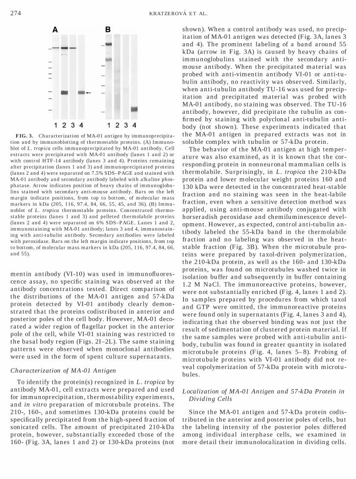
FIG. 3. Characterization of MA-01 antigen by immunoprecipita-tion and by immunoblotting of thermostable proteins. (A) Immuno-blot of L. tropica cells immunoprecipitated by MA-01 antibody. Cellextracts were precipitated with MA-01 antibody (lanes 1 and 2) orwith control HTF-14 antibody (lanes 3 and 4). Proteins remainingafter precipitation (lanes 1 and 3) and immunoprecipitated proteins(lanes 2 and 4) were separated on 7.5% SDS–PAGE and stained withMA-01 antibody and secondary antibody labeled with alkaline phos-phatase. Arrow indicates position of heavy chains of immunoglobu-lins stained with secondary anti-mouse antibody. Bars on the leftmargin indicate positions, from top to bottom, of molecular massmarkers in kDa (205, 116, 97.4, 84, 66, 55, 45, and 36). (B) Immu-noblot of L. tropica thermostable proteins. Concentrated thermo-table proteins (lanes 1 and 3) and pelleted thermolabile proteinslanes 2 and 4) were separated on 6% SDS–PAGE. Lanes 1 and 2,mmunostaining with MA-01 antibody; lanes 3 and 4, immunostain-ng with anti-tubulin antibody. Secondary antibodies were labeledith peroxidase. Bars on the left margin indicate positions, from top
o bottom, of molecular mass markers in kDa (205, 116, 97.4, 84, 66,nd 55).
60- (Fig. 3A, lanes 1 and 2) or 130-kDa proteins (not m
hown). When a control antibody was used, no precip-tation of MA-01 antigen was detected (Fig. 3A, lanes 3nd 4). The prominent labeling of a band around 55Da (arrow in Fig. 3A) is caused by heavy chains ofmmunoglobulins stained with the secondary anti-
ouse antibody. When the precipitated material wasrobed with anti-vimentin antibody VI-01 or anti-tu-ulin antibody, no reactivity was observed. Similarly,hen anti-tubulin antibody TU-16 was used for precip-
tation and precipitated material was probed withA-01 antibody, no staining was observed. The TU-16
ntibody, however, did precipitate the tubulin as con-rmed by staining with polyclonal anti-tubulin anti-ody (not shown). These experiments indicated thathe MA-01 antigen in prepared extracts was not inoluble complex with tubulin or 57-kDa protein.The behavior of the MA-01 antigen at high temper-
ture was also examined, as it is known that the cor-esponding protein in nonneuronal mammalian cells ishermolabile. Surprisingly, in L. tropica the 210-kDarotein and lower molecular weight proteins 160 and30 kDa were detected in the concentrated heat-stableraction and no staining was seen in the heat-labileraction, even when a sensitive detection method waspplied, using anti-mouse antibody conjugated withorseradish peroxidase and chemiluminescence devel-pment. However, as expected, control anti-tubulin an-ibody labeled the 55-kDa band in the thermolabileraction and no labeling was observed in the heat-table fraction (Fig. 3B). When the microtubule pro-eins were prepared by taxol-driven polymerization,he 210-kDa protein, as well as the 160- and 130-kDaroteins, was found on microtubules washed twice insolation buffer and subsequently in buffer containing.2 M NaCl. The immunoreactive proteins, however,ere not substantially enriched (Fig. 4, lanes 1 and 2).
n samples prepared by procedures from which taxolnd GTP were omitted, the immunoreactive proteinsere found only in supernatants (Fig. 4, lanes 3 and 4),
ndicating that the observed binding was not just theesult of sedimentation of clustered protein material. Ifhe same samples were probed with anti-tubulin anti-ody, tubulin was found in greater quantity in isolatedicrotubule proteins (Fig. 4, lanes 5–8). Probing oficrotubule proteins with VI-01 antibody did not re-
eal copolymerization of 57-kDa protein with microtu-ules.
ocalization of MA-01 Antigen and 57-kDa Protein inDividing Cells
Since the MA-01 antigen and 57-kDa protein codis-ributed in the anterior and posterior poles of cells, buthe labeling intensity of the posterior poles differedmong individual interphase cells, we examined in
ore detail their immunolocalization in dividing cells.
cnb
pbLhabMkgMHi23whdplDstpttMWtlflpkahtiabmScnicbsLCpt
ncis
275MICROTUBULE-INTERACTING PROTEIN IN Leishmania
Anti-tubulin antibody decorated not only the flagellumand subpellicular array of microtubules, but also themitotic spindle (arrowhead in Fig. 5A). In cells theMA-01 antibody labeled strongly both poles and flagel-lum as well as mitotic spindle (arrowhead in Fig. 5B).In the anterior pole it decorated the whole area sur-rounding the flagellar pocket. The anti-vimentin anti-body strongly decorated cells only at both poles (Figs.5F and 5J). In contrast with interphase cells, bothantibodies reacted with posterior poles in all dividingcells. No cells were detected showing mitotic spindleand lacking immunolabeling of posterior poles. A directcomparison of the distribution of MA-01 antigen and57-kDa protein clearly demonstrated that the corre-sponding proteins codistributed in anterior and poste-rior poles of the cell body. However, like in the anteriorpole of interphase cells, the MA-01 decorated a widerarea, whereas the VI-01 staining was restricted to thebasal body region (Figs. 2I–2L). The same stainingpatterns were observed when monoclonal antibodieswere used in the form of spent culture supernatants.
DISCUSSION
Previously it has been shown that MA-01 antibodyrecognizes an epitope on MAP2ab proteins [17, 31]. Theepitope was not detected on the other structural micro-tubule-associated proteins MAP2c, MAP2d, MAP4,and tau (E. Draberova, unpublished results) that shareonserved microtubule-binding repeats [32]. On immu-oblots of various types of nonneuronal cells this anti-
FIG. 4. Immunoblot of L. tropica microtubule proteins preparedby taxol-driven polymerization. Cell extracts were polymerized inthe presence of 20 mM taxol and 1 mM GTP (lanes 1 and 2, 5 and 6)or in the absence of taxol and GTP (lanes 3 and 4, 7 and 8). Aftercentrifugation through sucrose cushion, supernatants (lanes 1, 3, 5,7) and pelleted material (lanes 2, 4, 6, 8) were separated on 6%SDS–PAGE. Lanes 1–4, immunostaining with MA-01 antibody;lanes 5–8, immunostaining with anti-tubulin antibody. Secondaryantibodies were labeled with peroxidase. Bars on the left marginindicate positions, from top to bottom, of molecular mass markers inkDa (205, 116, 97.4, 84, 66, and 55).
ody reacted with a 210-kDa protein [17, 18, 33]. A e
rotein of the same relative mobility was detected alsoy immunoblotting on whole lysates of L. tropica and. major (not shown) and by immunoprecipitation ofigh-speed extracts of L. tropica. Neither the MA-01nd MA-02 antibodies nor the other monoclonal anti-odies prepared against different epitopes on porcineAP2ab [24, 34] reacted with proteins in the 280–300
Da region on L. tropica immunoblots. These data sug-est that MAP2 and 210-kDa protein detectable byA-01 and MA-02 antibodies share a common epitope.owever, in contrast to the immunoreactive proteins
n nonneuronal mammalian cell lines tested so far, the10-kDa protein in L. tropica is thermostable. Two20-kDa thermostable proteins, MARP-1 and MARP-2,ere identified in T. brucei. They are characterized byighly conserved 38-amino-acid repeat units and by aistinct proteolytic pattern detected after electro-horetic separation. They were located on subpellicu-ar arrays of microtubules but not in axoneme [12, 35].ue to the different subcellular location and the ob-
erved relative molecular weight, we think it unlikelyhat the MA-01 antigen belongs to the MARP family ofroteins. Moreover, we never observed on SDS–PAGEhe typical proteolytic pattern when protease inhibi-ors proteases were not applied (not shown). TheA-01 antigen also differs from the 210-kDa proteinCB210 identified in T. brucei. The WCB210 is a
hermolabile, heavily phosphorylated protein that isocated on subpellicular microtubules and absent inagellum [10]. To the group of high-molecular-weightroteins belongs also the 2G4 antigen (700 to 2500Da), which is confined to microtubules of the flagellarttachment zone in T. cruzi [36]. The MA-01 antigenas a different subcellular distribution and does nothus resemble the 2G4 protein(s). The MA-01 antigens present both in cytoskeletal and in soluble form andssociates with tubulin upon taxol-induced microtu-ule formation. However, we did not observe its accu-ulation in microtubule proteins prepared in vitro.imilarly, the MA-01 antigen was not enriched in mi-rotubule proteins prepared from 3T3 cells [30]. Immu-ofluorescence labeling of interphase and dividing cells
ndicates that it could as well be associated with mi-rotubules in vivo. It was detected in the flagella, basalody regions, and posterior poles of the cells containingubpellicular microtubules and on mitotic structures of. tropica (Figs. 2 and 4) and L. major (not shown).ombined data indicate that the MA-01 antigen mightossibly represent a new microtubule-interacting pro-ein of Leishmania.
When whole-cell lysates were probed with monoclo-al anti-vimentin antibodies VI-01 and VI-10, a spe-ific staining of the 57-kDa protein was observed. Butn immunofluorescence only the VI-01 antibody gavepecific reaction. As the antibodies recognize different
pitopes on the vimentin molecule, the reactivity of the
276 KRATZEROVA ET AL.
VI-10 antibody could be the result of specific maskingof the corresponding region in fixed cells. Intermediatefilament-like proteins were already identified in L.mexicana with polyclonal antibodies against desminand vimentin. In immunoblots the anti-desmin anti-body reacted with the 66- and 60-kDa proteins andanti-vimentin antibody with the 60-kDa protein. Theanti-desmin antibody stained the basal body region,the flagella, and, diffusely, the cell body. The anti-vimentin antibody decorated the basal body region andnucleus [15]. Similarly, VI-01 antibody stained thebasal body region, but no staining of the nucleus orflagella was seen. On the other hand, the antibody
FIG. 5. Fluorescence triple-label staining of L. tropica mitotic celred), MA-01 antibody (B, I, green), anti-vimentin antibody VI-01 (F,H, L) show superpositions of staining in each row. Arrows denotearrowheads in A–D show position of mitotic structure. Bar, 5 mm.
stained the posterior region of the cell. Although inter-
mediate filaments were not identified in protozoa [37,38], fibrils of approximately 11 nm, linking the kineto-plast to basal bodies, were found in T. cruzi [39]. It wassuggested that those fibrils could be intermediate fila-ment-related structures [15]. The common structuralprinciple of all intermediate filament proteins is a cen-tral a-helical rod domain of coiled-coil-forming ability,which is flanked by a hypervariable head-and-tail do-main. The rod is divided into subdomains which areconnected by nonhelical linkers. Type III intermediatefilament proteins exhibit a highly conserved aminoacid motif at the C-terminal end of their central a-he-lical rod domain [40], as well as in the head domain
Cells were stained with polyclonal anti-tubulin antibody TUB (A, E,en; J, red), and DNA-binding dye (C, G, K, blue). Color pictures (D,erior poles and arrowheads denote posterior poles in cells. Bigger
ls.greant
[41]. However, such domain structure and consensus
r
Lswccfi
277MICROTUBULE-INTERACTING PROTEIN IN Leishmania
motifs were not found in Leishmania proteins so fardeposited in databases. As the epitope recognized bythe VI-01 antibody is present on vimentin as well as ondesmin [21], one cannot exclude that a correspondingepitope is located in the coiled-coil region of type IIIintermediate filament proteins. The antibody couldthen react in Leishmania with a similarly structuredprotein that is not homologous to type III intermediatefilament proteins.
What the function of MA-01 antigen and 57-kDaprotein in the posterior poles of Leishmania could be isunclear. Interestingly, while in the anterior pole theMA-01 antibody decorated a wider region of the flagel-lar pocket and the VI-01 antibody stained a smallerbasal body region, both antibodies stained approxi-mately the same area in the posterior pole of inter-phase as well as dividing cells (Figs. 2L and 5L). Wheninterphase cells were examined, the posterior poleswere stained by both antibodies with varying intensi-ties and some cells were not stained at all in thisregion. In contrast, the posterior poles in dividing cellswith duplicated kinetoplast and nuclei were decoratedin all examined cells. This suggests that the staining ofthis part of the cell is possibly dependent on the cellcycle phase or that the corresponding epitopes aremore exposed in mitotic cells. In T. brucei, subpellicu-lar microtubules converge on the posterior pole of thecell body, creating a ring-shaped opening that is delim-ited by the plus (1) ends of microtubules [42]. A repet-itive 28-kDa protein, Gb4, which caps the microtubulesat the posterior end microtubules, was described in thisprotozoon [14]. It was proposed that the Gb4 proteincould regulate microtubule elongation and/or it mightbe involved in mediating the contact of these microtu-bule ends with the cell membrane. In connection withthe striking codistribution of the MA-01 antigen andthe 57-kDa protein containing the vimentin epitope inthe posterior poles, it is worth mentioning that theMA-01 antigen is directly or indirectly involved in theinteraction of microtubules with vimentin-type inter-mediate filaments of 3T3 cells [30]. It is possible thatsome kind of similar interaction(s) could be involved inanchoring the Leishmania microtubules in the poste-ior pole of the cell.
In conclusion, the presented data indicate that ineishmania the newly described 210-kDa thermo-table microtubule-interacting protein codistributesith the 57-kDa protein in various stages of the cell
ycle. The striking codistribution of these two kinds ofytoskeletal protein is described in protozoa for therst time.
We thank Dr. E. Nohynkova, Department of Tropical Medicine,First Faculty of Medicine, Charles University, Prague, Czech Repub-lic, for providing the cell lines. Taxol was a generous gift from the
Drug Synthesis and Chemistry Branch, National Cancer Institute(Bethesda, MD). This work was supported by Grant 304/00/0553from the Grant Agency of the Czech Republic and Grant A5052004from the Grant Agency of the Czech Academy of Sciences.
REFERENCES
1. Kreier, J. P. (1995). “Parasitic Protozoa,” 2nd ed., AcademicPress, San Diego.
2. Herwaldt, B. L. (1999). Leishmaniasis. Lancet 354, 1191–1199.3. Werbovetz, K. A., Brendle, J. J., and Sackett, D. L. (1999).
Purification, characterization, and drug susceptibility of tubu-lin from Leishmania. Mol. Biochem. Parasitol. 98, 53–65.
4. Grellier, P., Sinou, V., Garreau-de Loubresse, N., Bylen, E.,Boulard, Y., and Schrevel, J. (1999). Selective and reversibleeffects of vinca alkaloids on Trypanosoma cruzi epimastigoteforms: Blockage of cytokinesis without inhibition of the or-ganelle duplication. Cell Motil. Cytoskeleton 42, 36–47.
5. Kapoor, P., Ghosh, A., and Madhubala, R. (1999). Isolation of ataxol-resistant Leishmania donovani promastigote mutant thatexhibits a multidrug-resistant phenotype. FEMS Microbiol.Lett. 176, 437–441.
6. Hou, W. Y., Pimenta, P. F., Shen, R. L., and Da Silva, P. P.(1992). Stereo views and immunogold labeling of the pellicularmicrotubules at the inner surface of the plasma membrane ofLeishmania as revealed by fracture-flip. J. Histochem. Cyto-chem. 40, 1309–1318.
7. Gull, K. (1999). The cytoskeleton of trypanosomatid parasites.Annu. Rev. Microbiol. 53, 629–655.
8. Huang, P. L., Roberts, B. E., Pratt, D. M., David, J. R., andMiller, J. S. (1984). Structure and arrangement of the beta-tubulin genes of Leishmania tropica. Mol. Cell Biol. 4, 1372–1383.
9. Mendoza-Leon, A., Havercroft, J. C., and Barker, D. C. (1995).The RFLP analysis of the beta-tubulin gene region in NewWorld Leishmania. Parasitology 111, 1–9.
10. Burns, J. M., Jr., Shreffler, W. G., Benson, D. R., Ghalib, H. W.,Badaro, R., and Reed, S. G. (1993). Molecular characterizationof a kinesin-related antigen of Leishmania chagasi that detectsspecific antibody in African and American visceral leishmania-sis. Proc. Natl. Acad. Sci. USA 90, 775–779.
11. Woods, A., Baines, A. J., and Gull, K. (1992). A high molecularmass phosphoprotein defined by a novel monoclonal antibody isclosely associated with the intermicrotubule cross bridges inthe Trypanosoma brucei cytoskeleton. J. Cell Sci. 103, 665–675.
12. Hemphill, A., Affolter, M., and Seebeck, T. (1992). A novelmicrotubule-binding motif identified in a high molecular weightmicrotubule-associated protein from Trypanosoma brucei.J. Cell Biol. 117, 95–103.
13. Detmer, E., Hemphill, A., Muller, N., and Seebeck, T. (1997).The Trypanosoma brucei autoantigen I/6 is an internally repet-itive cytoskeletal protein. Eur. J. Cell Biol. 72, 378–384.
14. Rindisbacher, L., Hemphill, A., and Seebeck, T. (1993). A repet-itive protein from Trypanosoma brucei which caps the microtu-bules at the posterior end of the cytoskeleton. Mol. Biochem.Parasitol. 58, 83–96.
15. Hernandez, C., Dunia, I., Benedetti, E. L., and Dagger, F.(1997). Intermediate filament-like proteins in Leishmania. CellBiol. Int. 21, 65–67.
16. Eisenberger, C. L. and Jaffe, C. L. (1999). Leishmania: Identi-fication of Old World species using a permissively primed in-tergenic polymorphic-polymerase chain reaction. Exp. Parasi-
tol. 91, 70–77.
1
1
2
2
2
2
2
2
2
2
3
3
3
3
278 KRATZEROVA ET AL.
17. Draberova, E., Draber, P., and Viklicky, V. (1986). Cellulardistribution of protein related to neuronal microtubule-associ-ated protein MAP-2 in Leydig cells. Cell Biol. Int. Rep. 10,881–890.
8. Hasek, J., Jochova, J., Draber, P., Viklicky, V., and Streiblova,E. (1992). Localization of a 210 kDa microtubule-interactingprotein in yeast Saccharomyces cerevisiae. Can. J. Microbiol.38, 149–152.
9. Draber, P., Draberova, E., Linhartova, I., and Viklicky, V.(1989). Differences in the exposure of C- and N-terminal tubu-lin domains in cytoplasmic microtubules detected with domain-specific monoclonal antibodies. J. Cell Sci. 92, 519–528.
0. Draberova, E., and Draber, P. (1998). Novel monoclonal anti-bodies TU-08 and TU-16 specific for tubulin subunits. FoliaBiol. (Prague) 44, 35–36.
1. Draberova, E., Draber, P., Havlıcek, F., and Viklicky, V. (1986).A common antigenic determinant of vimentin and desmin de-fined by monoclonal antibody. Folia Biol. (Prague) 32, 295–303.
2. Draberova, E., Zıkova, M., and Draber, P. (1999). Monoclonalantibody VI-10 specific for vimentin. Folia Biol. (Prague) 45,35–36.
3. Draber, P., Draberova, E., and Viklicky, V. (1991). Immuno-staining of human spermatozoa with tubulin domain-specificmonoclonal antibodies. Histochemistry 195, 519–524.
4. Riederer, B. M., Draberova, E., Viklicky, V., and Draber, P.(1995). Changes of MAP2 phosphorylation during braindevelopment. J. Histochem. Cytochem. 43, 1269–1284.
5. Bartek, J., Viklicky, V., Horejsı, V., Verlova, H., and Draber, P.(1984). Production and characterization of monoclonal antibod-ies against human transferrin. Folia Biol. (Prague) 30, 137–140.
6. Laemmli, U. K. (1970). Cleavage of structural proteins duringthe assembly of the head of bacteriophage T4. Nature 227,680–685.
7. Vallee, R. B., and Collins, C. A. (1986). Purification of microtu-bules and microtubule-associated proteins from sea urchin eggsand cultured mammalian cells using taxol, and use of exoge-nous taxol-stabilized brain microtubules for purifying microtu-bule-associated proteins. Methods Enzymol. 134, 116–127.
28. Towbin, H., Staehelin, T., and Gordon, J. (1979). Electro-phoretic transfer of proteins from polyacrylamide gels to nitro-cellulose sheets: Procedure and some applications. Proc. Natl.Acad. Sci. USA 76, 4350–4354.
29. Draber, P., Lagunowich, L. A., Draberova, E., Viklicky, V., andDamjanov, I. (1988). Heterogeneity of tubulin epitopes in mouse
fetal tissues. Histochemistry 89, 485–492.0. Draberova, E., and Draber, P. (1993). A microtubule-interactingprotein involved in coalignment of vimentin intermediate fila-ments with microtubules. J. Cell Sci. 106, 1263–1273.
31. Kuznetsov, S. A., Rodionov, V. I., Nadezhdina, E. S., Murphy,D. B., and Gelfand, V. I. (1986). Identification of a 34-kDpolypeptide as a light chain of microtubule-associated protein-1(MAP-1) and its association with a MAP-1 peptide that binds tomicrotubules. J. Cell Biol. 102, 1060–1066.
2. Kaech, S., and Matus, A. (1997). Possible roles for MAP2 inneuronal pathology. In “Brain Microtubule Associated Proteins.Modifications in Disease” (J. Avila, R. Brandt, and K. Kosik,Eds.), Harwood Academic, Amsterdam.
3. Zıkova, M., Sulimenko, V., Draber, P., and Draberova, E.(2000). Accumulation of 210 kDa microtubule-interacting pro-tein in differentiating P19 embryonal carcinoma cells. FEBSLett. 473, 19–23.
4. Zıkova, M., Draberova, E., Sulimenko, V., and Draber, P.(2000). New monoclonal antibodies specific for microtubule-associated protein MAP2. Folia Biol. (Prague) 46, 87–88.
35. Schneider, A., Hemphill, A., Wyler, T., and Seebeck, T. (1988).Large microtubule-associated protein of T. brucei has tandemlyrepeated, near-identical sequences. Science 241, 459–462.
36. Ruiz-Moreno, L., Bijovsky, A. T., Pudles, J., Alves, M. J., andColli, W. (1995). Trypanosoma cruzi: Monoclonal antibody tocytoskeleton recognizes giant proteins of the flagellar attach-ment zone. Exp. Parasitol. 80, 605–615.
37. Klymkowsky, M. W. (1995). Intermediate filaments: New pro-teins, some answers, more questions. Curr. Opin. Cell Biol. 7,46–54.
38. Weber, K. (1999). Evolutionary aspects of IF proteins. In“Guidebook to the Cytoskeletal and Motor Proteins” (T. Kreisand R. Vale, Eds.), Oxford Univ. Press, Oxford.
39. Souto-Padron, T., de Souza, W., and Heuser, J. E. (1984).Quick-freeze, deep-etch rotary replication of Trypanosoma cruziand Herpetomonas megaseliae. J. Cell Sci. 69, 167–178.
40. Herrmann, H., Strelkov, S. V., Feja, B., Rogers, K. R., Brettel,M., Lustig, A., Haner, M., Parry, D. A., Steinert, P. M.,Burkhard, P., and Aebi, U. (2000). The intermediate filamentprotein consensus motif of helix 2B: Its atomic structure andcontribution to assembly. J. Mol. Biol. 298, 817–832.
41. Herrmann, H., Hofmann, I., and Franke, W. W. (1992). Identi-fication of a nonapeptide motif in the vimentin head domaininvolved in intermediate filament assembly. J. Mol. Biol. 223,637–650.
42. Robinson, D. R., Sherwin, T., Ploubidou, A., Byard, E. H., andGull, K. (1995). Microtubule polarity and dynamics in the con-trol of organelle positioning, segregation, and cytokinesis in the
trypanosome cell cycle. J. Cell Biol. 128, 1163–1172.Received October 18, 2000Revised version received March 8, 2001