
3Methyladenine blocks Toxoplasma gondii division prior to centrosome replication
Upload
independentCategory
view
3download
0
1
Dictyostelium LIS1 is a centrosomal protein required for microtubule/cell cortex
interactions, nucleus/centrosome linkage and actin dynamics
Markus Rehberg, Julia Kleylein-Sohn, Jan Faix, Thi-Hieu Ho, Irene Schulz and Ralph Gräf*
A.-Butenandt-Institut/Zellbiologie, Ludwig-Maximilians-Universität München,
Schillerstrasse 42, D-80336 München, Germany
*corresponding author
email: [email protected]; phone +49 89 218075862; fax +49 89 218075882
Running title: Characterization of Dictyostelium LIS1
Keywords: LIS1, Dictyostelium, DdCP224, dynein, centrosome, actin dynamics, Rac1A
http://www.molbiolcell.org/content/suppl/2005/03/23/E05-01-0069.DC1.htmlSupplemental Material can be found at:
2
The widespread LIS1-proteins were originally identified as the target for sporadic mutations
causing lissencephaly in humans. Dictyostelium LIS1 (DdLIS1) is a microtubule-associated
protein exhibiting 53 % identity to human LIS1. It colocalizes with dynein at isolated,
microtubule-free centrosomes, suggesting that both are integral centrosomal components.
Replacement of the DdLIS1 gene by the hypomorphic D327H allele or overexpression of an
MBP-DdLIS1 fusion disrupted various dynein-associated functions. Microtubules lost contact
with the cell cortex and were dragged behind an unusually motile centrosome. Previously, this
phenotype was observed in cells overexpressing fragments of dynein or the XMAP215-
homologue DdCP224. DdLIS1 was coprecipitated with DdCP224, suggesting that both act
together in dynein-mediated cortical attachment of microtubules. Furthermore, DdLIS1-
D327H mutants showed Golgi dispersal and reduced centrosome/nucleus association. Defects
in DdLIS1 function also altered actin dynamics characterized by traveling waves of actin
polymerization correlated with a reduced F-actin content. DdLIS1 could be involved in actin
dynamics through Rho-GTPases, since DdLIS1 interacted directly with Rac1A in vitro. Our
results show that DdLIS1 is required for maintenance of the microtubule cytoskeleton, Golgi
apparatus and nucleus/centrosome association, and they suggest that LIS1-dependent
alterations of actin dynamics could also contribute to defects in neuronal migration in
lissencephaly patients.

3
INTRODUCTION
The LIS1 gene was originally identified as the target for sporadic mutations resulting in
haploinsufficiency and a severe brain developmental disease called type I lissencephaly in
human infants. Lissencephaly (greek lissos = smooth) is characterized by a smooth
appearance of the neocortical surface due to the absence of gyri and sulci (Reiner et al.,
1993). This is believed to be the consequence of impaired migration of neuronal precursors
from the paraventricular area, where they divide, to the cerebral cortex during development.
The LIS1 protein has a calculated molecular mass of ~45 kDa and is characterized by seven
WD40-repeats, which are thought to form a β-propeller fold as in structurally similar β-
subunits of heterotrimeric G-proteins. Indeed, LIS1 could be identified as a subunit of a brain-
specific isoform of the G-protein-like platelet-activating factor acetylhydrolase. Yet, the first
clues for the molecular function of LIS1 in neuronal migration came from a filamentous
fungus. The Aspergillus nidulans LIS1 homologue, NUDF, was identified in a screen for
nuclear distribution mutants (Xiang et al., 1995). Further nud mutants include nudA,
encoding the cytoplasmic dynein heavy chain, and nudE. Mutations in nudA, nudE and nudF
caused similar defects in nuclear migration during hyphal stalk formation (Morris et al.,
1998). Nuclear migration is an important factor in neuronal cell migration as well (reviewed
by Gupta et al., 2002), and it is achieved through the activity of dynein/dynactin localized at
the cell cortex. The microtubule minus end-directed pulling forces exerted by dynein are
transmitted to the nucleus through microtubules emanating from the nucleus-associated
centrosome (reviewed by Dujardin and Vallee, 2002). Since mammalian LIS1 could be
immunoprecipitated with both dynein and dynactin subunits (Faulkner et al., 2000; Smith et
al., 2000), it was hypothesized that defects in LIS1 disrupt dynein function, which in turn
causes the neuronal migration disorder observed in lissencephaly. Interestingly, both NUDF
and NUDE as well as their mammalian homologues LIS1 and NUDEL (= NUDE-like)
directly interact with the dynein heavy chain (Sasaki et al., 2000). Recent data suggest that
LIS1 and NUDEL form a complex with dynein and have a synergistic effect on the promotion
of dynein function. Complex formation appears to be positively regulated by phosphorylation
of NUDEL through CDK5/p53, while the ser/thr-phosphate-binding protein 14-3-3ε protects
NUDEL from dephosphorylation by PP2A (Toyo-Oka et al., 2003). In addition to the LIS1
interactors mentioned above, there are several further binding partners such as CLIP170 or
doublecortin (Caspi et al., 2000; Coquelle et al., 2002; Schaar et al., 2004), which may assist
in the capture of microtubule plus ends by dynein/dynactin at the cell cortex.
4
The essential role of LIS1 for neuronal migration appears to be mediated not only through its
interaction with dynein. Recently Kholmanskikh and co-workers showed that LIS1
haploinsufficiency resulted in a reduced F-actin content at the leading edge of migrating
cerebellar granule cells (Kholmanskikh et al., 2003). Interestingly, this effect was
accompanied by altered activity of small GTPases regulating cortical actin dynamics. While
Rac1 and Cdc42 activities were downregulated, the antagonizing GTPase RhoA was
upregulated under these conditions. However, no binding of LIS1 to one of these GTPases or
their regulators could be shown. Thus, the relationship between cellular LIS1 levels and
GTPase activities remained unclear.
In addition to its role in actin dynamics and dynein function at the cell cortex, LIS1 is also a
regulator of microtubule dynamics. LIS1 binds to microtubules in vivo and in vitro and
promotes microtubule elongation by reducing the catastrophe rate (Sapir et al., 1997).
Recently we have shown in Dictyostelium amoebae that DdCP224, a member of the
ubiquitous XMAP215-family of microtubule-associated proteins (Ohkura et al., 2001), is also
involved both in dynein-dependent microtubule interactions with the cell cortex and in the
promotion of microtubule growth (Gräf et al., 2003; Hestermann and Gräf, 2004). Due to the
similarity of LIS1 protein function in mammalian and fungal cells and the roles of DdCP224
in Dictyostelium cells, we suspected a functional relationship between LIS1 and XMAP215-
family proteins and performed a functional analysis of DdLIS1, the Dictyostelium LIS1
homologue. We show that DdLIS1 is a microtubule-associated protein that also constitutes an
integral component of the centrosome. Disruption of DdLIS1 function affects microtubule tip
interactions with the cell cortex, cell morphology and actin dynamics. Furthermore, we
demonstrate for the first time an association of a LIS1 protein with a member of the
XMAP215-family of microtubule-associated proteins and direct binding to an actin-regulating
small GTPase. The elucidation of these interactions may help to explain the central role of
DdLIS1 in the dynamics of both the microtubule and actin cytoskeleton.

5
RESULTS
DdLIS1 is closely related to human LIS1
A partial cDNA sequence encoding a Dictyostelium homologue of LIS1 (DdLIS1) was
identified on clone SLE307 in the data library of the Dictyostelium cDNA project (University
of Tsukuba, Japan; Morio et al., 1998). Using this clone and a PCR-fragment containing the
missing 5’-cDNA sequence we have constructed a complete DdLIS1 cDNA clone (EMBL
database accession no. AJ512794; see Materials and Methods). Among all non-animal LIS1-
homologues, DdLIS1 is most closely related to human LIS1. Both proteins share 55% of
identical amino acids (72% similarity). Moreover, with a calculated molecular mass of 47
kDa and an N-terminal LIS1-homology domain followed by a short coiled-coil domain and
seven WD40 repeats, DdLIS1 displays the same domain structure and almost the same size as
its human counterpart (Fig. 1).
DdLIS1 is a microtubule-associated protein and an integral centrosomal component together
with dynein
The subcellular localization of DdLIS1 was investigated using polyclonal antibodies raised
against the recombinant protein expressed in E. coli (Fig. 2A). DdLIS1 was localized along
microtubules and at the centrosome throughout the entire cell cycle (Fig. 2B, C, D). In
contrast to human LIS1 (Faulkner et al., 2000), microtubule labeling by anti-DdLIS1
antibodies was not biased towards the microtubule plus ends neither at the peripheral
microtubule tips in interphase nor at kinetochores during mitosis. In order to test whether the
centrosomal localization of DdLIS1 is microtubule-dependent, we analyzed the presence of
DdLIS1 at isolated centrosomes. This assay was used since treatment with microtubule-
depolymerizing drugs such as thiabendazole or nocodazole is rather ineffective in
Dictyostelium and leads to only partial depolymerization of microtubules (Kitanishi et al.,
1984), while isolated Dictyostelium centrosomes are completely devoid of microtubules (Gräf
et al., 1998). The Dictyostelium centrosome is a cytosolic, nucleus-associated body that lacks
centrioles. Instead it consists of a three-layered core structure surrounded by the corona,
which is the functional equivalent of the pericentriolar matrix of centriole-containing
centrosomes (Euteneuer et al., 1998; Gräf et al., 2000a). Confocal images of isolated
centrosomes labeled with anti-DdLIS1 antibodies clearly revealed that DdLIS1 is part of the
centrosomal corona (Fig. 2D). When labeled with antibodies against known components such
as DdCP224 or γ-tubulin (Gräf et al., 2000b), the corona displays a ring-like appearance in
6
confocal images. In deconvolved confocal images, the diameter of the anti-DdCP224 labeled
ring was always larger than that observed upon labeling with anti-DdLIS1 antibodies,
suggesting that DdLIS is localized closer to the unlabeled centrosomal core structure than
DdCP224 (Fig. 2D). Therefore, DdLIS1 is a genuine centrosomal component. This was
unexpected, since centrosomal LIS1 localization was thought to be mediated through its
binding to the dynein heavy chain (Sasaki et al., 2000), which one would expect to require
microtubules to localize it to the centrosome through its microtubule minus end-directed
motor activity. However, our confocal analysis of isolated centrosomes revealed that the
dynein heavy chain, which precisely colocalizes with DdCP224 at the centrosomal corona, is
an integral component of the Dictyostelium centrosome as well (Fig. 2E).
The DdLIS1 D327H point mutation causes defects in microtubule/cortex interactions, Golgi
positioning and centrosome/nucleus association.
To shed light on DdLIS1 function we have created a loss-of-function mutant. Since a
complete knockout of the gene encoding DdLIS1 (lis1) was unsuccessful, we replaced lis1 by
a hypomorphic allele. The D317H point mutation in human LIS1 found in several
lissencephaly patients causes partial misfolding of the LIS1 protein, which results in a mild
form of lissencephaly characterized by pachygyria (Pilz et al., 1999; Sapir et al., 1999; Caspi
et al., 2003). The corresponding D327H mutation in Dictyostelium was generated through
homologous recombination by replacement of lis1 with a point-mutated copy. Although the
subcellular localization of DdLIS1-D327H was indistinguishable from wildtype DdLIS1 (our
own unpublished data), cells expressing only point-mutated DdLIS1 displayed a clear mutant
phenotype. During interphase, the vast majority of cells expressing DdLIS1-D327H showed a
striking disruption of their microtubule cytoskeleton. The usually radial interphase
microtubule arrays were collapsed in a way that the microtubules appeared bundled and
whorled around the nucleus (Fig. 3A, B). Four-dimensional confocal microscopy of DdLIS1-
D327H cells expressing GFP-α-tubulin revealed an extraordinary motility of these
microtubule arrays (Fig. 3C, D). The centrosome circulated rapidly and continuously, often
close to the cell cortex, with most of the microtubules dragged behind like a comet tail (Fig.
3D’). In GFP-α-tubulin expressing control cells, the centrosome always stayed close to the
cell center and moved only over short distances (Fig. 3C’). Compared to GFP-α-tubulin cells
where the centrosome moved with an average speed of 0.054 µm/s (n=5; S.D. = 0.007) and a
maximal speed of 0.15 µm/s, centrosomes in DdLIS1-D327H cells moved three times faster
7
with an average speed of 0.142 µm/s (n=21; S.D.=0.022) and a maximum of even 0.5 µm/s
(Fig. 3E). Within 7 min, the area encircled by the moving centrosomes was 15-fold larger in
DdLIS1-D327H cells than in GFP-α-tubulin cells (110 µm2 versus 7.2 µm2) (Fig. 3E). This
microtubule/centrosome phenotype was strikingly similar to Dictyostelium cells
overexpressing the motor domain of the dynein heavy chain (Koonce and Samso, 1996;
Koonce et al., 1999), fragments of the dynein intermediate chain (Ma et al., 1999) or an N-
terminal fragment of DdCP224 (Hestermann and Gräf, 2004). In all these strains, including
our DdLIS1-D327H cells, the microtubule phenotype was also accompanied by a dispersal of
the Golgi apparatus (Fig. 4). This remarkable phenotype was explained by a loss of
dynein/dynactin function, which is required both for pericentrosomal localization of the Golgi
apparatus and for the maintenance of radial microtubule arrays through cortically bound
dynein pulling at microtubule ends. The existence of such cortical microtubule pulling forces
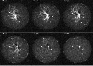
is illustrated by Movie3 (Fig. 5), which shows the mitosis of a cell with GFP-fluorescent
microtubules. As a normal feature of the Dictyostelium centrosome duplication cycle (Ueda et
al., 1999), microtubules are severed from the centrosome at the G2/M transition prior to
centrosome splitting (starting at time point 110s in Movie3). Subsequently the shrinking
microtubules appear to be quickly pulled towards the cell cortex.
A further phenotype frequently observed in DdLIS1-D327H cells was an unusually large
distance between the centrosome and the nucleus. In control cells, the centrosome was always
tightly linked to the nucleus, with a distance to the nucleus rarely exceeding its own diameter
(Fig. 6A). However, in DdLIS1-D327H cells the centrosome was often located more than 2
µm away from the nucleus (Fig. 6B), which, interestingly, can also be weakly labeled with
anti-dynein heavy chain antibodies (Fig. 7).
Despite of its effects on the microtubule array and centrosome/nucleus association, the
D327H mutation caused only a moderate reduction in growth rate (our own unpublished data)
but had no striking influence on mitotis. The representative mitotic cell in Fig. 3D (arrow)
shows normal mitotic progression in terms of total duration, centrosome duplication and
spindle formation. The failure of cytokinesis in this example can be attributed to the imaging
conditions under agar overlay, where proper cytokinesis is a rare event. Since multinucleated
cells are not more frequent in the DdLIS-D327H strain than in control cells, neither in
adherent nor in shaking culture, a cytokinesis defect can be excluded.
Taken together, the cellular defects observed in DdLIS1-D327H cells suggest that DdLIS1 is
required for Golgi integrity, nucleus/centrosome association and dynein/dynactin-mediated
interactions of microtubule tips with the cell cortex.

8
DdLIS1 interacts with dynein and DdCP224
The similarity of the DdLIS1-D327H phenotype and the dynein and DdCP224 mutants
mentioned above raised the question whether DdLIS1 could interact not only with dynein but
also with DdCP224. Thus, we performed co-immunoprecipitations of these proteins from
cytosolic Dictyostelium extracts using specific polyclonal antibodies and protein G coated
beads. Since the similar size of DdLIS1 and antibody heavy chains complicated the
identification of DdLIS1 in Western blots of immunoprecipitates, we have overexpressed
DdLIS1 as a fusion protein with the E. coli maltose-binding protein (MBP) in Dictyostelium
(Gräf, 2001b). MBP-DdLIS1 was overexpressed approximately 10-fold as judged from
Western blots stained with anti-DdLIS1 antibodies. Using cytosolic extracts from these cells,
the ~90-kDa MBP-DdLIS1 fusion protein clearly co-precipitated with DdCP224 using anti-
DdCP224 antibodies (Fig. 8). Conversely, DdCP224 was co-precipitated with DdLIS1 when
anti-DdLIS1 antibodies were used for co-immunoprecipitation (Fig. 8). As expected for a
LIS1-family protein, DdLIS1 also co-precipitated with the dynein heavy chain (Fig. 8).
We wondered whether disruption of DdLIS1 function affects subcellular localization of
Dynein and DdCP224, however, we observed no displacement of the dynein heavy chain or
DdCP224 in DdLIS1-D327H mutants, neither at the cell cortex nor at isolated centrosomes
or, in case of the dynein heavy chain, at the nucleus (Fig. 7). Nevertheless, the co-
immunoprecipitation of these proteins from cytosolic extracts, their colocalization and the
similarity of phenotypes in corresponding mutants indicate that dynein, DdLIS1 and
DdCP224 act together in the cortical attachment of microtubules.
DdLIS1-D327H cells and MBP-DdLIS1 overexpressors exhibit altered cell shape
Overexpression of MBP-DdLIS1 frequently disrupted radial microtubule arrays in a similar
manner as described for DdLIS1-D327H cells, suggesting a dominant-negative effect of
MBP-DdLIS1 overexpression (Fig. 9). However, the microtubule disruption phenotype upon
MBP-DdLIS1 overexpression was less frequent (approximately 25% of cells in freshly
transformed cultures) and tended to disappear in older cultures with reduced MBP-DdLIS1
expression.
Since impaired LIS1 function strongly affects the motile behavior of neuronal precursors, we
suspected that the DdLIS1-D327H point mutation or MBP-DdLIS1 overexpression may
interfere with Dictyostelium cell motility. In neither case we could detect any defect in
random cell motility or chemotaxis (our own unpublished data), but both MBP-DdLIS1
9
overexpressors and DdLIS1-D327H mutants were altered in cell shape and appeared much
flatter than untransformed cells. This became obvious when both cell lines were viewed by
reflection interference contrast microscopy (RICM) in comparison with control cells. This
technique, which can be applied at conventional confocal microscopes, visualizes the contact
surface of a cell to the substratum in a reflection image (Weber, 1999). In both MBP-DdLIS1
overexpressors and DdLIS1-D327H mutants the cell contact surface to the coverslip was
greatly enlarged (Fig. 10). Furthermore, these flat cells appeared to be more active in
filopodia formation (arrowheads in Fig. 10).
DdLIS1 is involved in the regulation of the cellular F-actin content
Alterations in cell shape usually involve the actin cytoskeleton, thus, we looked for changes in
actin dynamics under these conditions. Indeed, phalloidin staining of DdLIS1-D327H cells
revealed unusual distributions of F-actin, especially in very flat cells. Here F-actin was
distributed in broad ring- or crescent-like arrangements close to the lower cell surface (Fig.
11A, B). In order to investigate these actin structures in living cells, we expressed GFP-actin
(Westphal et al., 1997) in both DdLIS1-D327H mutants and MBP-DdLIS1 overexpressors
and compared actin dynamics with GFP-actin control cells. When these control cells were
migrating on a glass surface, actin was concentrated at the cell cortex, predominantly in
protruding filopodia and lamellipodia (Fig. 11D) (Bretschneider et al., 2004). By contrast, in
DdLIS1-D327H cells and upon overexpression of MBP-DdLIS1, we frequently observed
characteristic traveling waves of dense F-actin assemblies propagating along the substrate
attached cell surface. This pattern was usually persistent throughout the entire observation
period of more than 20 min (Fig. 11E, F, G). This dynamic actin organization seems to reflect
changes in size and shape of cell/substrate contact sites. In contrast to DdLIS1-D327H
mutants and MBP-DdLIS1 overexpressors, such actin waves were only rarely encountered in
untreated GFP-actin cells. However, they were frequently observed in cells where the actin
cytoskeleton was partially disrupted by treatment with a low concentration of latrunculin A
(0.2 µM) (Fig. 11F). Latrunculin A causes F-actin depolymerization through sequestering of
G-actin into non-polymerizing complexes (Spector et al., 1989). At high latrunculin A
concentrations of 2 µM, Dictyostelium cells round up completely and lose contact with the
substratum (our own unpublished data). Since latrunculin A treatment reduces the cell’s F-
actin content, the similarity of altered actin dynamics upon treatment with low concentrations
of latrunculin A and our DdLIS1 mutants suggested that compromised DdLIS1 function may
cause partial actin depolymerization. Thus, we compared the F- and G-actin content of these

10
cells at the biochemical level. After cell lysis with Triton X-100 followed by
ultracentrifugation, relative amounts of F-actin (pellet) and G-actin fractions (supernatant)
were evaluated on Western blots stained with an anti-actin monoclonal antibody. In untreated
control cells, the amounts of F- and G-actin were approximately equal, whereas treatment
with low concentrations of latrunculin A, as used to induce the actin waves, caused a
prevalence of G-actin over F-actin (Fig. 12). The same strong prevalence of G-actin was
observed in DdLIS1-D327H cells and upon overexpression of MBP-DdLIS1. Taken together,
these results indicate that impaired DdLIS1 function reduces the cellular F-actin content.
Since LIS1 usually cooperates with dynein in its functions, we wanted to analyze whether the
actin phenotype in DdLIS1-D327H cells could be dynein dependent. Therefore, we analyzed
subcellular F-actin distribution in cells overexpressing an N-terminal fragment of the dynein
intermediate chain (DIC∆C), a treatment that is known to disrupt dynein function (Ma et al.,
1999). As upon disruption of DdLIS1 function or latrunculin treatment, F-actin was spread in
broad ring- or crescent-like arrangements close to the substratum (Fig. 11C). Due to the
instability of DIC∆C overexpressors, we were unable to perform similar live cell studies as in
case of the other cell lines or to obtain sufficient amounts of cells to evaluate the F-/G-actin
ratio. Nevertheless, our data supports a dynein-dependent role of DdLIS1 in actin dynamics.
DdLIS1 may alter actin dynamics through its interaction with the small GTPase Rac1A
We wondered how DdLIS1 could participate in the regulation of actin dynamics. Since
DdLIS1 showed no clear co-localization with actin, we suspected that DdLIS1 might interact
with regulators of actin dynamics such as small GTPases. This assumption was also supported
by the observation that both DdLIS1-D327H mutants and MBP-DdLIS1 overexpressors were
more active in filopodia formation, a process regulated by Rac1 GTPases (Dumontier et al.,
2000). Therefore, we investigated whether DdLIS1 was able to interact with Rac1A (Bush et
al., 1993), the Dictyostelium Rac1 homologue (Dumontier et al., 2000). Since the available
antibodies against Rac1A were not suitable for immunoprecipitations, we performed a GST-
Rac1A pull-down experiment. GST-Rac1A preloaded with either non-hydrolysable GTPγS or
GDP was incubated with a cytosolic Dictyostelium extract (strain AX2). In both cases
endogenous DdLIS1 clearly coprecipitated with GST-Rac1A (Fig. 13, lanes 1-3). The
interaction between DdLIS1 and Rac1A was specific as we detected no co-precipitation when
the cytosolic extracts were incubated with GST alone. GST-Rac1A also specifically brought
down purified, recombinant, 6xHis-tagged DdLIS1 (His-DdLIS1) (Fig. 13, lanes 4-6). This

11
proves that DdLIS1 and Rac1A interact directly with each other. Again, the interaction was
not dependent on the guanosine nucleotide, GTPγS or GDP that was bound to GST-Rac1A.
When the GST-Rac1A pull-down assay was performed with cytosolic extracts from MBP-
DdLIS1 overexpressing cells (Fig. 13, lane 7-9), the MBP-DdLIS1 fusion protein surprisingly
showed no coprecipitation with GST-Rac1A, although it was overexpressed approximately
18-fold (as calculated from Western blot band intensities) (Fig. 13, lane 10). This suggests
that the bulky N-terminal MBP-tag interfered with the ability of DdLIS1 to bind Rac1A
resulting in a dominant-negative effect on DdLIS1 function.
12
DISCUSSION
In vertebrate cells, the centrosomal localization of LIS1 is microtubule-dependent (Tanaka et
al., 2004). It is thought to be mediated by binding of LIS1 to dynein, which accumulates at
the centrosome through its microtubule minus end-directed motor activity. Thus, dynein itself
is also considered a temporal guest at the centrosome. However, in Dictyostelium, the
centrosomal presence of DdLIS1 and dynein was completely independent of microtubules,
because both were integral components of isolated, microtubule-free centrosomes.
DdLIS1 was also strongly localized along microtubules, but there was no bias of its
distribution towards microtubule plus ends as observed in mammalian cells, where LIS1
seems to cooperate with CLIP-170 and dynein/dynactin in the interaction of microtubule tips
with capturing sites at the cell cortex (Coquelle et al., 2002). This suggests that DdLIS1 might
not be necessary for microtubule capture itself, however, our data demonstrate that it is
certainly required for maintenance of radial microtubule arrays and centrosome positioning
near the centroid of the cell (Fig. 3). This process is apparently based on microtubule pulling
forces provided by dynein that is evenly distributed at the Dictyostelium cell cortex (Koonce
and Khodjakov, 2002). The first experimental evidence for the necessity of cortical
dynein/dynactin for centrosome positioning and maintenance of microtubule arrays came
from Koonce and coworkers (Koonce and Samso, 1996; Koonce et al., 1999) who
overexpressed the dynein motor domain in Dictyostelium. As a consequence, microtubules
frequently lost contact with the cell cortex and trailed behind an extraordinarily motile
centrosome that circulated around, often close to the cell cortex. This phenotype could also be
elicited by overexpression of fragments of the dynein intermediate chain (Ma et al., 1999), an
N-terminal fragment of the Dictyostelium XMAP215-homologue DdCP224 (DdCP224∆C)
(Hestermann and Gräf, 2004) or, as described in this work, by interfering with DdLIS1
function through the D327H point mutation or overexpression of dominant-negative MBP-
tagged DdLIS1. The simplest explanation for this phenotype is that all these manipulations
disrupt dynein/dynactin-mediated linkage of membrane structures with microtubules. The
observation of Golgi dispersal in all Dictyostelium cell lines mentioned above strongly
supports this view, since dynein/dynactin-mediated coupling of Golgi vesicles and
microtubules is crucial for Golgi positioning in the centrosomal vicinity (Burkhardt et al.,
1997). Consequently, disrupted dynein/dynactin-mediated linkage of membrane structures
and microtubules causes dissociation of Golgi vesicles from microtubules and, in a similar
manner, loss of cortical tethering of microtubule ends. However, remaining, still functional

13
cortical dynein/dynactin complexes could exert asymmetric pulling forces on single
microtubules resulting in rapid displacement of the centrosome with all the untethered
microtubules dragged behind (reviewed in Gräf et al., 2004). In this work we have provided
live cell data supporting the existence of such cortical pulling forces. Movie3 reveals that
microtubules are not just depolymerized once they lose their connection to the centrosome in
early prophase, but that they move towards and along the cell cortex. It is conceivable that
these microtubule movements are driven by cortical dynein.
The problems in dynein/dynactin-dependent linkage of membrane structures and microtubules
could arise either from an inability of the dynein/dynactin complex to bind to microtubules,
from dissociation of dynactin from membrane structures or, as it seems to occur in dynein
intermediate chain mutants and DdCP224∆C cells (Ma et al., 1999; Hestermann and Gräf,
2004), from dissociation of dynein from the dynactin complex. In case of our DdLIS1 mutants
it appears more likely that the mutant phenotypes are based on defective binding of membrane
associated dynein/dynactin to microtubules, since we found no indications for reduced
presence of the dynein heavy chain at any of its localizations.
Since both DdCP224 (Hestermann and Gräf, 2004) and DdLIS1 interact with dynein and each
other in the cytosol, it is likely that these proteins cooperate in Golgi positioning and the
interaction of microtubule ends with the cell cortex. We cannot judge whether DdLIS1 and
DdCP224 and dynein interact directly or indirectly, because so far it has been impossible to
express functional, recombinant DdCP224 or dynein for in vitro binding assays. The
interaction between a LIS1 and an XMAP215-family protein is shown here for the first time.
However, due to the functional and structural conservation of these proteins, it is likely that it
will also be found in higher cells where it could also be involved in microtubule/cortex
interactions and centrosome positioning. The requirement of cortical dynein/dynactin for
centrosome positioning in mammalian cells has been demonstrated in wound-healing
experiments (Dujardin et al., 2003). In fibroblast monolayers, cells at the wound edge reorient
their centrosomes toward the direction of migration during the healing process (Schliwa et al.,
1999). Centrosome reorientation between the leading edge of the cell and the nucleus is
blocked by inhibition of cytoplasmic dynein and dynactin and regulated through the small
GTPase Cdc42 and PKCζ (Etienne-Manneville and Hall, 2001; Palazzo et al., 2001). This
suggests that cortical dynein/dynactin is required for capturing of microtubules extending into
cortical regions of a freshly formed pseudopod. An involvement of LIS1 in this process has
not been demonstrated yet, however, it could explain at least part of the neuronal migration
defect in lissencephaly.

14
Cortical anchorage of microtubule ends is a prerequisite for nucleokinesis, a major aspect of
neuronal cell motility. The term nucleokinesis describes the “reeling in” of the nucleus into
the freshly extended leading process of the migrating neuron. At the leading process,
cortically anchored dynein is thought to exert a force on microtubules, which extend from the
nucleus-associated centrosome to the leading edge. In this process, LIS1 appears to be not
only required for cortical dynein function but also for the linkage of the centrosome to the
nucleus. Recently, (Tanaka et al., 2004) could show in migrating cerebellar granule neurons
that the distance between the centrosome and the nucleus was significantly larger in case of
LIS1 haploinsufficiency. According to a model established in C. elegans, centrosome/nucleus
attachment is mediated by centrosomally bound microtubules together with dynein
immobilized at the nuclear surface and it is maintained through SUN and Hook-family
proteins (Malone et al., 2003). It is conceivable that the role of LIS1 in this pathway is also
mediated by dynein as in case of almost all other known LIS1 functions. This view is
supported by our observation of a small fraction of the dynein heavy chain at the nucleus in
immunofluorescence specimens in Dictyostelium. Thus, our data clearly support a dynein-
dependent role of LIS1 in centrosome/nucleus association, even in cells with different
centrosomal structure and closed mitosis such as Dictyostelium amoebae.
Until recently, the severe defects in neuronal cell migration observed upon compromised
LIS1 function were mainly attributed to the role of LIS1 in microtubule-dependent dynamic
processes through the regulation of dynein (Gupta et al., 2002). Yet, Kholmanskikh et al.
(2003) have just shown that the actin cytoskeleton also contributes to the migration defect in
LIS1-deficient neurons. Haploinsufficiency of LIS1 caused a reduced F-actin content at the
leading edge of migrating neurons, which was accompanied by dysregulation of Rho-
GTPases. While RhoA was upregulated under these conditions, the activities of the
antagonistic GTPases Rac1 and Cdc42 were decreased. The motility defect of LIS1 deficient
neurons was rescued when Rac1 and Cdc42 activity could be restored by inhibition of the
RhoA effector p160ROCK. These data suggested that LIS1 promotes actin polymerization
through the regulation of Rho-GTPase activities. However, no direct interaction of LIS1 with
Rho-GTPases or any of their effectors or modulators could be shown. Since microtubule
growth has been shown to stimulate Rac1 activity and thereby to promote actin
polymerization at the leading edge (Waterman-Storer et al., 1999), LIS1 could also increase
Rac1 activity indirectly through its role as a suppressor of microtubule catastrophes and
promoter of increased MT length (Sapir et al., 1997).

15
In this paper we have demonstrated for the first time a direct interaction of a LIS1-family
protein with a Rac1-homologue (Dictyostelium Rac1A) by GST-pull down assays. This
suggests that LIS1 could regulate actin dynamics directly through Rho-family GTPases. Since
MBP-tagged DdLIS1 was essentially unable to bind Rac1A although it displayed the same
subcellular localization as endogenous DdLIS1, we concluded that its overexpression exerts a
dominant-negative effect, possibly through replacement of endogenous DdLIS1. This
explains why MBP-DdLIS1 overexpression in Dictyostelium caused phenotypes, which were
rather similar to those observed in DdLIS1-D327H cells and in LIS1+/- cerebellar granule cells
with reduced LIS1 activity (Kholmanskikh et al., 2003). In all these cases, the F-actin content
was reduced.
Furthermore, the microtubule-related phenotypes suggested a dominant-negative effect as
well, because MBP-DdLIS1 overexpression caused Golgi dispersal and indicated reduced
cortical pulling forces. By contrast, in mammalian cells LIS1 overexpression resulted in Golgi
compaction and disordered microtubule arrays, which are indicative for increased cortical
microtubule pulling forces (Faulkner et al., 2000; Smith et al., 2000). There are two likely
explanations for the dominant-negative effect of MBP-DdLIS1 overexpression on dynein
function. First, overexpressed MBP-DdLIS1 could sequester other dynein/dynactin-regulating
binding partners, which would then be missing at the cell cortex or Golgi apparatus where
they are required for proper dynein function. Second, the failure of MBP-DdLIS1 to associate
with other binding partners such as Rac1A could be responsible for the disruption of dynein
function.
How could DdLIS1 be involved in Rac1A-dependent actin dynamics? Rac1A regulates the
cortical actin cytoskeleton through the actin-bundling proteins cortexillin I and II. Both are
required for cortical stiffness and are directed to cortical sites with activated Rac1A through
their Rac1A effector DGAP1 (Faix, 2002). It is unlikely that DdLIS1 acts as a Rac1A effector
in this pathway, because it binds both Rac1A-GDP and Rac1A-GTPγS. Yet, it is more likely
that DdLIS1 is required for Rac1A activation or for delivery of Rac1A to its site of action.
This view is supported by the fact that MBP-tagged DdLIS1 localizes similar to endogenous
DdLIS1 but does not bind to Rac1A. Replacement of endogenous DdLIS1 by overexpressed,
non-functional MBP-DdLIS1 would lead to reduced amounts of active Rac1A at the cortex.
Traveling waves of actin polymerization appear to recruit Arp2/3-rich actin foci that are
distributed all over the substrate attached surface of the cell (Bretschneider et al., 2004). Such
foci are also visible in Movie8 (for example see time points 308-350 s). Although traveling
actin waves have been described in untransformed Dictyostelium cells (Vicker, 2002;

16
Bretschneider et al., 2004), we have only rarely encountered them in our AX2 wild-type
control cells. Wave appearance seemed to depend on the cell shape, since traveling actin
waves could only be detected in flattened cells. Yet, such cells were rare in AX2 cultures but
were rather frequently observed after latrunculin treatment and upon disruption of DdLIS1
function due to the D327H mutation or MBP-DdLIS1 overexpression. This suggests that both
the appearance of flattened cells and traveling actin waves were based on a reduced cortical F-
actin content. Dynein has not yet been recognized as a player in actin dynamics and, thus, it
was an interesting question whether disturbed dynein function could result in similar
alterations of actin dynamics as observed in our DdLIS1 mutants. Although our dynein
intermediate chain overexpression cell line was very unstable, we could reproducibly observe
similarly altered F-actin distribution is these cells as detected upon disruption of DdLIS1
function. This strongly suggests that the role of DdLIS1 in actin dynamics also involves
dynein just like all other LIS1 functions known so far.
Bearing in mind the effects of partial loss of DdLIS1 function on both the tubulin and the
actin cytoskeleton it is surprising that our mutants showed no defects in cell migration as they
were observed in migrating neurons. However, our own unpublished results with nocodazole
clearly showed that Dictyostelium cells do not require microtubules for directed cell
migration. Even the partial disassembly of the actin cytoskeleton by treatment with low
dosages of latrunculin A, did not prevent chemotactic cell migration of Dictyostelium
amoebae. Thus, Dictyostelium cells are more reminiscent of non-neuronal cells, which are
also not strongly affected by partial loss of LIS1 function. However, like LIS1 in mammals,
DdLIS1 is certainly an essential protein since it was not possible to knock out the DdLIS1
gene completely or to replace it by a stronger hypomorphic allele such as H149R.
Taken together, in this work we have shown that DdLIS1 is not only a microtubule-associated
protein but also a genuine centrosomal component. The D327H point mutation of DdLIS1 as
well as overexpression of MBP-tagged DdLIS1 disrupted DdLIS1 function and resulted in a
palette of mutant phenotypes including Golgi dispersal, decreased centrosome/nucleus
association, disturbed microtubule tip/cell cortex interactions and altered actin dynamics. All
these phenotypes appeared to be dynein-dependent. Furthermore, we have demonstrated for
the first time an association between LIS1 and XMAP215-family proteins in a cytosolic
complex and direct binding of a LIS1-family protein to an actin regulating small GTPase,
suggesting a contribution of these proteins in LIS1 function and, thus, the pathogenesis of
lissencephaly.

17
18
MATERIALS AND METHODS
Construction of a complete DdLIS1 cDNA
Clone SLE307 of the Dictyostelium cDNA project (Morio et al., 1998) was kindly provided
by T. Morio (University of Tsukuba, Japan;). It is based on pSPORT1 (Invitrogen, Karlsruhe,
Germany) and encoded a partial DdLIS1 sequence. The missing 5’-cDNA sequence including
216 bp of additional coding sequence was amplified by PCR from a Dictyostelium cDNA
library in λZAP (Gräf et al., 2000b) using the T3-primer and two nested DdLIS1-specific
primers. The resulting 662 bp fragment included an EcoRI site derived from the λ-vector and
an NsiI site located within the DdLIS1 coding sequence. These restriction sites were used to
construct a complete DdLIS1 cDNA clone in SLE307 (EMBL database accession no.
AJ512794).
Vector construction, protein expression and antibodies
In order to express DdLIS1 as a maltose-binding fusion protein in E. coli, a PCR-fragment
including the complete DdLIS coding sequence flanked by EcoRI and XbaI restriction sites
was cloned into pMALc2. The fusion protein was expressed and purified by amylose affinity
chromatography as described by the manufacturer (New England Biolabs, Frankfurt,
Germany) and used for the custom immunization of a rabbit (Dr. J. Pineda Antikörperservice,
Berlin, Germany).
Likewise, a His-DdLIS1 vector based on pQE30 (Qiagen, Hilden, Germany) was constructed
using the PCR product obtained after amplification of the DdLIS1 coding sequence with an
upstream BamHI linker primer and a downstream SalI linker primer. His-DdLIS1 was
expressed and purified by NiNTA affinity chromatography as described by the manufacturer
(Qiagen, Hilden, Germany).
For expression of MBP-DdLIS1 in Dictyostelium, the MBP-DdLIS1/pMALc2 vector was
used as a template to amplify the sequence encoding the fusion protein with a suitable
upstream MBP-KpnI linker primer and a downstream DdLIS1-BamHI linker primer. The
obtained MBP-DdLIS1 PCR fragment was subsequently cloned into p1ABsr8 (Gräf et al.,
2000b) and transformed into Dictyostelium cells.
The gene replacement vector for generation of the DdLIS1-D327H point mutant was based on
pLPBLP (Faix et al., 2004). The final plasmid contained the 5’ untranslated sequence and the
complete coding sequence including the point mutation upstream from the blasticidin S
resistance cassette and 887 bp of 3’ untranslated sequence of the gene downstream from the
19
resistance cassette. A genomic DdLIS-fragment containing the point mutation was generated
by PCR in a two-step approach using two mutagenesis primers that were used to introduce a
new SalI site resulting in the desired single amino acid exchange. Thus, two PCR fragments
with 1466 bp and 1261 bp, respectively, were generated using genomic DNA as a template.
For the first fragment, an upstream BamHI linker primer binding to the 3’ untranslated region
of the gene and a downstream mutagenesis primer
GTCGACTACCAGTAGCTAAATAACCACATT were used, and for the second fragment
an upstream mutagenesis primer GTCGACATAAAACTATTAAAATTTGGG and a
downstream PstI linker primer binding to the 5’ end of the coding sequence. Both PCR
fragments were cleaved with BamHI/SalI and SalI/PstI, respectively, and were both cloned
into pLPBLP in a single step using the BamHI and PstI sites of the vector. The resulting
plasmid was cleaved with HindIII and KpnI and used to clone 887 bp of 3’ untranslated
region of the gene, which was generated by PCR using genomic DNA as a template and an
upstream HindIII linker primer directed to the beginning of the 3’ untranslated region and a
downstream primer (named LIS1-14) binding immediately downstream from the endogenous
KpnI site. Prior to transformation of Dictyostelium cells, the final gene replacement vector
was linearized by cleavage with BamHI and KpnI, and the desired DdLIS1/blasticidin
cassette was isolated from an agarose gel. After blasticidin selection, genomic DNA was
isolated using the HighPure PCR template preparation kit (Roche, Mannheim, Germany) and
positive clones were identified by a genotyping PCR using the LIS1-14 downstream primer
and a GTGGTTATTTAGCTACTGGTAGTC upstream primer. The latter primer is specific
for the point mutation because its last four bases fit only to the mutated but not to the original
genomic sequence and the binding site for LIS1-14 is not located within the linearized
transformation construct. Therefore amplification of a specific 2.6 kb PCR-fragment from
genomic DNA is only possible in clones, where gene replacement of endogenous DdLIS1 by
the artificial construct through homologous recombination has occurred and which carry the
D327H point mutation.
The vector for DIC∆C overexpression corresponds to the DIC∆C construct published
previously (Ma et al., 1999). In brief the coding sequence encoding the N-terminal fragment
of the Dictyostelium dynein intermediate chain (DIC; 279 amino acids) was amplified by PCR
using linker primers and cDNA as a template. The PCR product was cloned into a derivative
of pA15GFPSSEB2 using NheI and BamHI. This cloning vector is identical to
pDiscGFPSSEB2 (Daunderer and Gräf, 2002) except that the discoidin promoter is replaced
by the actin15 promoter.
20
Light microscopy
Light microscopy was essentially performed as described previously (Gräf et al., 1998). All
images and movies were acquired on a Zeiss Axiovert 200M/510META laser scanning
system equipped with a 63x/1.4 lens. Live cells were viewed in glass bottom dishes (MatTek
corporation, Ashland, MA, USA) either in phosphate buffer or under agar overlay (Fukui,
1987) at low laser intensity. GFP-actin cell lines were cultivated overnight in co-culture with
Klebsiella aerogenes in 17 mM Na-K-phosphate buffer (pH = 6.0) in order to increase their
resistance to phototoxic effects. For reflection interference contrast microscopy (RICM) a 633
nm HeNe-laser was used and the reflection image was recorded without any filter. In case of
4D live cell imaging, three to five z-slices per time point were acquired at a frame rate of 2
per second and a resolution of up to 512x512 pixel. Z-stacks were projected onto one plane
using a brightest point algorithm and ImageJ software (National Institutes of Health,
Bethesda, MD). All images from fixed cells were processed through the Huygens Essential
Deconvolution software (SVI, Hilversum, Netherlands) using the maximum likelihood
estimation method.
Estimation of the actin content
Cells cultivated overnight in shaking culture (density approx. 3 x 10⁶/ml) were spread onto 5
cm petri dishes to approximately 50% confluency and allowed to settle for 10 min.
Subsequently, medium was exchanged for phosphate buffer with or without 0.2 µM
latrunculin A (Sigma, Deisenhofen, Germany) and cells were incubated for 1 h at room
temperature. Cells lysis occurred on ice by rinsing the attached cells with 2 ml of ice cold
lysis buffer (80 mM Na-PIPES, pH = 6.8, 5 mM EGTA, 5 mM MgCl2, 1 mM DTT, 1%
Triton-X100, 25% glycerol, protease inhibitors (Gräf et al., 1998)). 250 µl of the lysate were
ultracentrifuged with 80.000 rpm for 40 min at 4°C using a Beckman TLA100 rotor. The
pellet containing the F-actin fraction was resuspended in 250 µl of lysis buffer. Both F-actin
and G-actin (supernatant) fractions were supplemented with SDS-gel loading buffer and
separated by SDS gel electrophoresis and Western blotting. The blot was stained using anti-
Dictyostelium-actin antibodies. Actin bands were visualized using an enhanced
chemiluminescence kit (Applichem, Darmstadt, Germany).
Other methods

21
Dictyostelium cells (strain AX2) were cultivated and transformed as reported earlier (Gräf et
al., 1998; Gräf et al., 2000b). SDS gel electrophoresis and Western blotting were carried out
according to Gräf et al. (1998). Centrosomes were isolated as described previously (Gräf,
2001a). Preparation of cell extracts of Dictyostelium cells was reported by (Daunderer and
Gräf, 2002). Immunoprecipitation was performed according to (Hestermann and Gräf, 2004)
and GST pull-down assays were carried out as described by Faix et al. (1998).
Antibodies
Primary antibodies: rabbit polyclonal anti-DdLIS1 (this work), mouse monoclonal anti-
DdCP224 (Gräf et al., 1999), rabbit polyclonal anti-DdCP224 (Hestermann and Gräf, 2004),
rabbit polyclonal anti-dynein heavy chain Y7 and 695 (Koonce and Samso, 1996), rat
monoclonal anti-α-tubulin YL1/2 (Chemicon, Hofheim, Germany), mouse monoclonal anti-
comitin (Weiner et al., 1993), rabbit polyclonal anti-DdSpc97 (Daunderer and Gräf, 2002),
rabbit polyclonal anti-MBP (Gräf, 2001b), monoclonal anti-Dictyostelium actin 224-236-1
(Westphal et al., 1997)
Secondary antibodies were from Dianova, Hamburg, Germany (all cy3 and peroxidase
conjugates), Sigma, Deisenhofen, Germany (alkaline phosphatase conjugates) and Molecular
Probes, Hilversum, Netherlands (all AlexaFluor 488 conjugates).

22
ACKNOWLEDGEMENTS
We are very grateful to Mike Koonce, Rex Chisholm and Michael Schleicher for providing
antibodies against the dynein heavy chain, dynein intermediate chain and comitin,
respectively. We also would like to thank Manfred Schliwa for his continuous support and
Alexandra Lepier for critical comments. Supported by the DFG (SFB413 and GR1642/2-1).
23
FIGURE LEGENDS
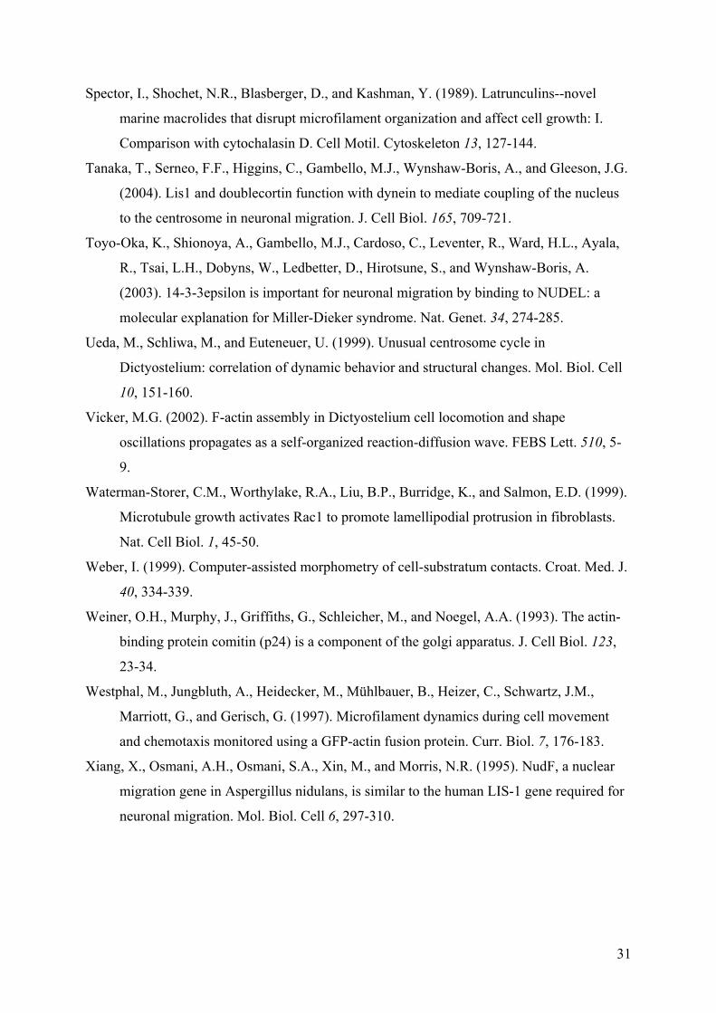
Figure 1. Domain structure of DdLIS1 and alignment of the DdLIS1 and human LIS1 amino
acid sequences. Like other LIS1 homologues, DdLIS1 is segmented into a LIS1-homology
domain (LisH), a short coiled coil region (CC) and seven WD40 repeats. The alignment was
performed with GAP-alignment (GCG package). The position of the D327H point mutation is
highlighted by a black box.
Figure 2. DdLIS1 localizes to microtubules, the mitotic spindle and to isolated centrosomes.
DdLIS1 was stained using affinity purified anti-DdLIS1 antibodies. The specificity of affinity
purified anti-DdLIS1 antibodies used for immunofluorescence microscopy is shown on a
Western blot (A) of a Dictyostelium cell extract where only a single band at approximately 50
kDa is stained. Subcellular localization of DdLIS1 during interphase (B) and mitosis
(metaphase) (C) is shown. Counterstainings were performed with anti-α-tubulin and the anti-
DdCP224 monoclonal antibody 2/165 (C’), which mainly stains spindle poles and
kinetochores during this mitotic stage (Gräf et al., 1999, 2000b). The merged images (B’’,
C’’) show DdLIS1 in green and α-tubulin in red. DdLIS1 and the dynein heavy chain are also
present at isolated centrosomes, which are devoid of microtubules (D, E). The control
stainings against DdCP224, which is an established component of the centrosomal corona,
demonstrates colocalization of these proteins at the corona (D’, D’’, E’, E’’). The merged
images (D’’, E’’) show DdCP224 in red and DdLIS1 and dynein, respectively in green. (D’’’)
and (E’’’) show respective tracings of fluorescence intensity along a line through the center of
the centrosome (white line in D and E). The positions of the maxima of fluorescence intensity
reveal exact colocalization of the dynein heavy chain and DdCP224, whereas DdLIS1-
labeling is concentrated closer to the centrosomal core structure than DdCP224. All
specimens were fixed with methanol. Bar = 2 µm.
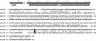
Figure 3. The DdLIS1-D327H point mutation causes a collapse of interphase microtubule
arrays and unusual centrosome motility. Immunofluorescence labeling of control cells (A) and
DdLIS1-D327H cells (B) with anti-α-tubulin (yellow) and the DNA staining dye TOPRO3
(blue) (Molecular Probes, Hilversum, Netherlands). Cells were fixed with glutaraldehyde.
Live analysis of microtubule and centrosome behavior was performed with GFP-α-tubulin
control cells (C, see movie1) and DdLIS1-D327H cells expressing GFP-α-tubulin (D, see

24
movie2). Each image represents a brightest point z-projection of 5 confocal slices with a
distance of 1 µm each. (C’) and (D’) displays the movements of each centrosome within a
time of 450 seconds by colored lines. The arrow points on a mitotic cell (prophase at time
point 0 sec). Average speed (E), maximum speed (E’) and the area enclosed by the
centrosome movements (E’’) within 450 s of control cells (grey columns) and DdLIS1-
D327H cells (black columns) were evaluated using the manual tracking plugin for ImageJ.
Bar = 5 µm.
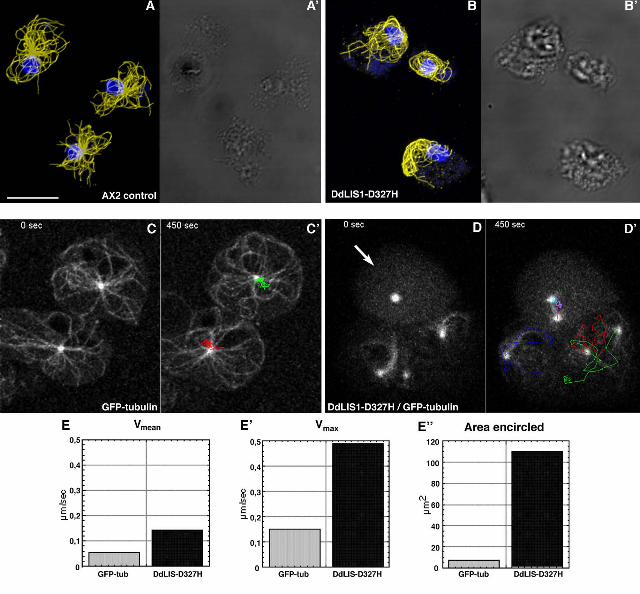
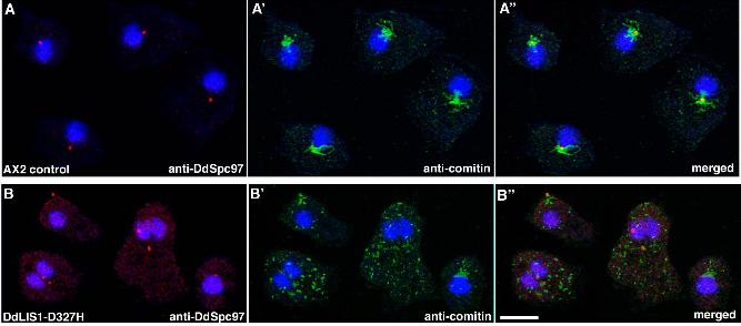
Figure 4. DdLIS1-D327H cells show Golgi dispersal. Immunofluorescence labeling of
control cells (A) and DdLIS1-D327H cells (B) with anti-DdSpc97 as a centrosome marker
(red; A, B) and anti-comitin as a Golgi marker (green; A’, B’). Nuclei were stained with
TOPRO3 (blue). These stainings were merged in (A’’) and (B’’). Cells were fixed with
methanol. Bar = 5 µm.
Figure 5/movie3. Live observation of GFP-labeled microtubules during mitosis suggests the
existence of cortical microtubule pulling forces. The movie shows abscission of the radial
microtubules from the centrosome in prophase (time point 120 s) and movement of the free
microtubules towards the cell cortex. Each image represents a brightest point z-projection of 5
confocal slices with a distance of 1.6 µm each.
Figure 6. The DdLIS1-D327H point mutation weakens the nucleus/centrosome association.
Immunofluorescence labeling of control cells (A) and DdLIS1-D327H cells (B) with anti-α-
tubulin antibodies (green; A, B) and polyclonal anti-DdCP224 as a centrosome marker (red;
A’, B’). Nuclei were stained with TOPRO3 (blue). These stainings were merged in (A’’) and
(B’’). Cells were fixed with glutaraldehyde. In DdLIS-D327H cells the distance between
centrosomes and the DNA is greatly enhanced. Bar = 2 µm
Figure 7. Subcellular localizations of dynein and DdCP224 are not significantly altered in
DdLIS1-D327H cells. Immunofluorescence labeling of whole cells (A-A’’, C-C’’) or isolated
centrosomes (B-B’’, D-D’’) with the anti-dynein heavy chain antibody 695 (green; A, B, C,
D) and the monoclonal anti-DdCP224 antibody 2/165 (red; A’, B’, C’, D’). Nuclei were
stained with TOPRO3 (blue; A’’, C’’). Stainings were merged in (A’’-D’’). AX2 control cells
were used in (A-B’’) and DdLIS1-D327H cells in (C-D’’). Cells were fixed with
formaldehyde/acetone and centrosomes with methanol.

25
Figure 8. Coprecipitation of DdLIS1 with DdCP224 with dynein. Experiments were
performed using cytosolic extracts from MBP-DdLIS1 cells or wildtype cells (strain AX2).
The respective antibodies used staining of the immunoblots and for immunoprecipitation are
indicated above and below the blots, respectively. Abbreviations and antibodies: LIS, anti-
DdLIS1; DdCP, anti-DdCP224 mAb 2/165 for immunoblot staining and polyclonal anti-
DdCP224 for immunoprecipitation; DHC, anti-dynein heavy chain Y7; control, anti-rabbit
preimmune serum.
Figure 9. MBP-DdLIS1 overexpression disrupts radial microtubules arrays.
Immunofluorescence labeling of MBP-DdLIS1 overexpressing cells with anti-α-tubulin
antibodies (green; A) and anti-MBP antibodies (red; A’, B’). Nuclei were stained with
TOPRO3 (blue). In many cells microtubules show no point-symmetric radial arrangement.
Like endogenous DdLIS1 (Fig. 2), MBP-DdLIS1 colocalizes with microtubules as revealed
by the merged image (C). Cells were fixed with glutaraldehyde. Bar = 5 µm
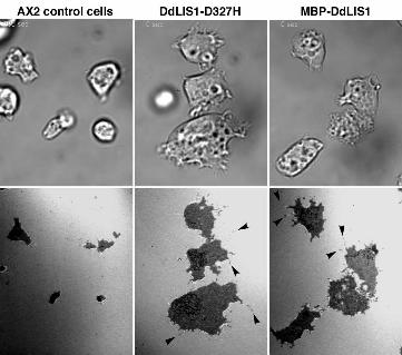
Figure 10/movies4, 5 and 6. DdLIS1-D327H cells and MBP-DdLIS1 overexpressors are
flatter than usual and display an increase contact surface to the substrate. Living AX2 control
cells (movie4), DdLIS-D327H cells (movie5) and MBP-DdLIS1 overexpressors (movie6)
were viewed by RICM. RICM images are shown in the lower panel and corresponding bright
field images in the upper panel. The dark area in the RICM channel corresponds to the cell’s
contact surface to the glass substrate. Only a single time point of each movie is shown here
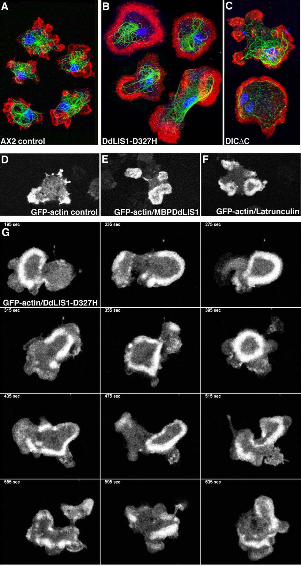
Figure 11. DdLIS1-D327H cells, MBP-DdLIS1 overexpressors and DIC∆C overexpressors
often show altered F-actin distribution and alterations in actin dynamics similar to control
cells treated with low concentrations of latrunculin A. Immunofluorescence labeling of
control cells (A), DdLIS1-D327H cells (B) and DIC∆C overexpressors (C) with anti-α-
tubulin (green) and phalloidin-AlexaFluor568 (Molecular Probes, Hilversum, Netherlands) as
an F-actin labeling probe (red). Nuclei were stained with TOPRO3 (blue). Note the broad F-
actin distribution at the bottom of the cells along with disrupted microtubule cytoskeletons in
(B, C). Cells were fixed with glutaraldehyde. Bar = 5 µm. Note that phalloidin labeling
intensities in these images do not illustrate the F-actin content, since all images were acquired
at microscope settings allowing coverage of the full dynamic range of the 8-bit scale. Live

26
analysis of actin dynamics was performed with GFP-actin control cells (D, movie7), GFP-
actin/MBP-DdLIS1 overexpressors (E, movie8), GFP-actin cells treated with 0.2 µM
latrunculin A (F, movie9) and GFP-actin cells carrying DdLIS1-D327H mutation (G,
movie10). Each image represents a brightest point z-projection of 3 confocal slices with a
distance of 0.8 µm each. One a single, representative time frame of each movie is shown in
(D, E, F), whereas (G) represents a time sequence.
Figure 12. The F-actin content in DdLIS1-D327H cells and MBP-DdLIS1 overexpressors is
reduced to a similar extent as upon treatment with low concentrations of latrunculin A. The
Western Blot containing equivalent amounts of F-actin and G-actin fractions (lanes “F” and
“G”) from AX2 control cells, AX2 cells treated with 0.2 µM latrunculin A, DdLIS1-D327H
cells and MBP-DdLIS1 overexpressors was stained with anti-actin antibodies and enhanced
chemiluminescence.
Figure 13. DdLIS1 but not MBP-DdLIS1 interacts with Rac1A. Western blots containing
equivalent amounts of protein coprecipitated with GST-Rac1A-GTPγS, GST-Rac1A-GDP or
GST alone (control) from a wildtype cell extract (AX2, lanes 1-3), a purified His-tagged
DdLIS1 protein solution (lanes 4-6), and an MBP-DdLIS1 cell extract (lanes 7-9) are shown.
Blots were stained with anti-DdLIS1 antibodies and enhanced chemiluminescence. Lane 10
shows a sample of the same MPB-DdLIS1 cell extract as in lanes 7-9 and shows the extent of
overexpression of MBP-DdLIS1 compared to the endogenous protein. Note that MBP-
DdLIS1 bands are missing in lanes 7-9.
27
REFERENCES
Bretschneider, T., Diez, S., Anderson, K., Heuser, J., Clarke, M., Müller-Taubenberger, A.,
Köhler, J., and Gerisch, G. (2004). Dynamic actin patterns and Arp2/3 assembly at the
substrate-attached surface of motile cells. Curr. Biol. 14, 1-10.
Burkhardt, J.K., Echeverri, C.J., Nilsson, T., and Vallee, R.B. (1997). Overexpression of the
dynamitin (p50) subunit of the dynactin complex disrupts dynein-dependent
maintenance of membrane organelle distribution. J. Cell Biol. 139, 469-484.
Bush, J., Franek, K., and Cardelli, J. (1993). Cloning and characterization of seven novel
Dictyostelium discoideum rac-related genes belonging to the rho family of GTPases.
Gene 136, 61-68.
Caspi, M., Atlas, R., Kantor, A., Sapir, T., and Reiner, O. (2000). Interaction between LIS1
and doublecortin, two lissencephaly gene products. Hum. Mol. Genet. 9, 2205-2213.
Caspi, M., Coquelle, F.M., Koifman, C., Levy, T., Arai, H., Aoki, J., De Mey, J.R., and
Reiner, O. (2003). LIS1 missense mutations: variable phenotypes result from
unpredictable alterations in biochemical and cellular properties. J. Biol. Chem. 278,
38740-38748.
Coquelle, F.M., Caspi, M., Cordelieres, F.P., Dompierre, J.P., Dujardin, D.L., Koifman, C.,
Martin, P., Hoogenraad, C.C., Akhmanova, A., Galjart, N., De Mey, J.R., and Reiner,
O. (2002). LIS1, CLIP-170's key to the dynein/dynactin pathway. Mol. Cell Biol. 22,
3089-3102.
Daunderer, C., and Gräf, R. (2002). Molecular analysis of the cytosolic Dictyostelium
gamma-tubulin complex. Eur. J. Cell Biol. 81, 175-184.
Dujardin, D.L., Barnhart, L.E., Stehman, S.A., Gomes, E.R., Gundersen, G.G., and Vallee,
R.B. (2003). A role for cytoplasmic dynein and LIS1 in directed cell movement. J. Cell
Biol. 163, 1205-1211.
Dujardin, D.L., and Vallee, R.B. (2002). Dynein at the cortex. Curr. Opin. Cell Biol. 14, 44-
49.
Dumontier, M., Hocht, P., Mintert, U., and Faix, J. (2000). Rac1 GTPases control filopodia
formation, cell motility, endocytosis, cytokinesis and development in Dictyostelium. J.
Cell Sci. 113 ( Pt 12), 2253-2265.
Etienne-Manneville, S., and Hall, A. (2001). Integrin-mediated activation of Cdc42 controls
cell polarity in migrating astrocytes through PKCzeta. Cell 106, 489-498.

28
Euteneuer, U., Gräf, R., Kube-Granderath, E., and Schliwa, M. (1998). Dictyostelium
gamma-tubulin: molecular characterization and ultrastructural localization. J. Cell Sci.
111, 405-412.
Faix, J. (2002). The actin-bundling protein cortexillin is the downstream target of a Rac1-
signaling pathway required for cytokinesis. J. Muscle Res. Cell Motil. 23, 765-772.
Faix, J., Clougherty, C., Konzok, A., Mintert, U., Murphy, J., Albrecht, R., Mühlbauer, B.,
and Kuhlmann, J. (1998). The IQGAP-related protein DGAP1 interacts with Rac and is
involved in the modulation of the F-actin cytoskeleton and control of cell motility. J.
Cell Sci. 111, 3059-3071.
Faix, J., Kreppel, L., Shaulsky, G., Schleicher, M., and Kimmel, A.R. (2004). A rapid and
efficient method to generate multiple gene disruptions in Dictyostelium discoideum
using a single selectable marker and the Cre-loxP system. Nucleic Acids Res. 32, e143.
Faulkner, N.E., Dujardin, D.L., Tai, C.Y., Vaughan, K.T., O'Connell, C.B., Wang, Y., and
Vallee, R.B. (2000). A role for the lissencephaly gene LIS1 in mitosis and cytoplasmic
dynein function. Nat. Cell Biol. 2, 784-791.
Fukui, Y., Yumura, S. and Yumura, T. K. (1987). Agar-overlay immunofluorescence: high
resolution studies of cytoskeletal components and their changes during chemotaxis.
Methods Cell Biol. 28, 347 - 356.
Gräf, R. (2001a). Isolation of Centrosomes from Dictyostelium. Methods Cell Biol. 67, 337-
357.
Gräf, R. (2001b). Maltose-binding protein as a fusion tag for the localization and purification
of cloned proteins in Dictyostelium. Anal. Biochem. 289, 297-300.
Gräf, R., Brusis, N., Daunderer, C., Euteneuer, U., Hestermann, A., Schliwa, M., and Ueda,
M. (2000a). Comparative structural, molecular and functional aspects of the
Dictyostelium discoideum centrosome. Curr. Top. Dev. Biol. 49, 161 - 185.
Gräf, R., Daunderer, C., and Schliwa, M. (1999). Cell cycle-dependent localization of
monoclonal antibodies raised against isolated Dictyostelium centrosomes. Biol. Cell 91,
471-477.
Gräf, R., Daunderer, C., and Schliwa, M. (2000b). Dictyostelium DdCP224 is a microtubule-
associated protein and a permanent centrosomal resident involved in centrosome
duplication. J. Cell Sci. 113, 1747-1758.
Gräf, R., Daunderer, C., and Schulz, I. (2004). Molecular and Functional Analysis of the
Dictyostelium Centrosome. Int. Rev. Cytol. 241, 155-202.

29
Gräf, R., Euteneuer, U., Ho, T.H., and Rehberg, M. (2003). Regulated Expression of the
Centrosomal Protein DdCP224 Affects Microtubule Dynamics and Reveals
Mechanisms for the Control of Supernumerary Centrosome Number. Mol. Biol. Cell 14,
4067-4074.
Gräf, R., Euteneuer, U., Ueda, M., and Schliwa, M. (1998). Isolation of nucleation-competent
centrosomes from Dictyostelium discoideum. Eur. J. Cell Biol. 76, 167-175.
Gupta, A., Tsai, L.H., and Wynshaw-Boris, A. (2002). Life is a journey: a genetic look at
neocortical development. Nat. Rev. Genet. 3, 342-355.
Hestermann, A., and Gräf, R. (2004). The XMAP215-family protein DdCP224 is required for
cortical interactions of microtubules. BMC Cell Biol. 5, 24.
Kholmanskikh, S.S., Dobrin, J.S., Wynshaw-Boris, A., Letourneau, P.C., and Ross, M.E.
(2003). Disregulated RhoGTPases and actin cytoskeleton contribute to the migration
defect in Lis1-deficient neurons. J. Neurosci. 23, 8673-8681.
Kitanishi, T., Shibaoka, H., and Fukui, Y. (1984). Disruption of microtubules and retardation
of development of Dictyostelium with Ethyl N-phenylcarbamate and Thiabendazole.
Protoplasma 120, 185-196.
Koonce, M.P., and Khodjakov, A. (2002). Dynamic microtubules in Dictyostelium. J. Muscle
Res. Cell Motil. 23, 613-619.
Koonce, M.P., Kohler, J., Neujahr, R., Schwartz, J.M., Tikhonenko, I., and Gerisch, G.
(1999). Dynein motor regulation stabilizes interphase microtubule arrays and
determines centrosome position. EMBO J. 18, 6786-6792.
Koonce, M.P., and Samso, M. (1996). Overexpression of cytoplasmic dynein's globular head
causes a collapse of the interphase microtubule network in Dictyostelium. Mol. Biol.
Cell 7, 935-948.
Ma, S., Triviños Lagos, L., Gräf, R., and Chisholm, R.L. (1999). Dynein intermediate chain
mediated dynein-dynactin interaction is required for interphase microtubule
organization and centrosome replication and separation in Dictyostelium. J. Cell Biol.
147, 1261-1274.
Malone, C.J., Misner, L., Le Bot, N., Tsai, M.C., Campbell, J.M., Ahringer, J., and White,
J.G. (2003). The C. elegans hook protein, ZYG-12, mediates the essential attachment
between the centrosome and nucleus. Cell 115, 825-836.
Morio, T., Urushihara, H., Saito, T., Ugawa, Y., Mizuno, H., Yoshida, M., Yoshino, R.,
Mitra, B.N., Pi, M., Sato, T., Takemoto, K., Yasukawa, H., Williams, J., Maeda, M.,
Takeuchi, I., Ochiai, H., and Tanaka, Y. (1998). The Dictyostelium developmental
30
cDNA project: generation and analysis of expressed sequence tags from the first-finger
stage of development. DNA Res. 5, 335-340.
Morris, N.R., Efimov, V.P., and Xiang, X. (1998). Nuclear migration, nucleokinesis and
lissencephaly. Trends Cell Biol. 8, 467-470.
Ohkura, H., Garcia, M.A., and Toda, T. (2001). Dis1/TOG universal microtubule adaptors -
one MAP for all? J. Cell Sci. 114, 3805-3812.
Palazzo, A.F., Joseph, H.L., Chen, Y.J., Dujardin, D.L., Alberts, A.S., Pfister, K.K., Vallee,
R.B., and Gundersen, G.G. (2001). Cdc42, dynein, and dynactin regulate MTOC
reorientation independent of Rho-regulated microtubule stabilization. Curr. Biol. 11,
1536-1541.
Pilz, D.T., Kuc, J., Matsumoto, N., Bodurtha, J., Bernadi, B., Tassinari, C.A., Dobyns, W.B.,
and Ledbetter, D.H. (1999). Subcortical band heterotopia in rare affected males can be
caused by missense mutations in DCX (XLIS) or LIS1. Hum. Mol. Genet. 8, 1757-
1760.
Reiner, O., Carrozzo, R., Shen, Y., Wehnert, M., Faustinella, F., Dobyns, W.B., Caskey, C.T.,
and Ledbetter, D.H. (1993). Isolation of a Miller-Dieker lissencephaly gene containing
G protein beta-subunit-like repeats. Nature 364, 717-721.
Sapir, T., Eisenstein, M., Burgess, H.A., Horesh, D., Cahana, A., Aoki, J., Hattori, M., Arai,
H., Inoue, K., and Reiner, O. (1999). Analysis of lissencephaly-causing LIS1 mutations.
Eur. J. Biochem. 266, 1011-1020.
Sapir, T., Elbaum, M., and Reiner, O. (1997). Reduction of microtubule catastrophe events by
LIS1, platelet- activating factor acetylhydrolase subunit. EMBO J. 16, 6977-6984.
Sasaki, S., Shionoya, A., Ishida, M., Gambello, M.J., Yingling, J., Wynshaw-Boris, A., and
Hirotsune, S. (2000). A LIS1/NUDEL/cytoplasmic dynein heavy chain complex in the
developing and adult nervous system. Neuron 28, 681-696.
Schaar, B.T., Kinoshita, K., and McConnell, S.K. (2004). Doublecortin microtubule affinity is
regulated by a balance of kinase and phosphatase activity at the leading edge of
migrating neurons. Neuron 41, 203-213.
Schliwa, M., Euteneuer, U., Gräf, R., and Ueda, M. (1999). Centrosomes, microtubules and
cell migration. Biochem. Soc. Symp. 65, 223-231.
Smith, D.S., Niethammer, M., Ayala, R., Zhou, Y., Gambello, M.J., Wynshaw-Boris, A., and
Tsai, L.H. (2000). Regulation of cytoplasmic dynein behaviour and microtubule
organization by mammalian Lis1. Nat. Cell Biol. 2, 767-775.
31
Spector, I., Shochet, N.R., Blasberger, D., and Kashman, Y. (1989). Latrunculins--novel
marine macrolides that disrupt microfilament organization and affect cell growth: I.
Comparison with cytochalasin D. Cell Motil. Cytoskeleton 13, 127-144.
Tanaka, T., Serneo, F.F., Higgins, C., Gambello, M.J., Wynshaw-Boris, A., and Gleeson, J.G.
(2004). Lis1 and doublecortin function with dynein to mediate coupling of the nucleus
to the centrosome in neuronal migration. J. Cell Biol. 165, 709-721.
Toyo-Oka, K., Shionoya, A., Gambello, M.J., Cardoso, C., Leventer, R., Ward, H.L., Ayala,
R., Tsai, L.H., Dobyns, W., Ledbetter, D., Hirotsune, S., and Wynshaw-Boris, A.
(2003). 14-3-3epsilon is important for neuronal migration by binding to NUDEL: a
molecular explanation for Miller-Dieker syndrome. Nat. Genet. 34, 274-285.
Ueda, M., Schliwa, M., and Euteneuer, U. (1999). Unusual centrosome cycle in
Dictyostelium: correlation of dynamic behavior and structural changes. Mol. Biol. Cell
10, 151-160.
Vicker, M.G. (2002). F-actin assembly in Dictyostelium cell locomotion and shape
oscillations propagates as a self-organized reaction-diffusion wave. FEBS Lett. 510, 5-
9.
Waterman-Storer, C.M., Worthylake, R.A., Liu, B.P., Burridge, K., and Salmon, E.D. (1999).
Microtubule growth activates Rac1 to promote lamellipodial protrusion in fibroblasts.
Nat. Cell Biol. 1, 45-50.
Weber, I. (1999). Computer-assisted morphometry of cell-substratum contacts. Croat. Med. J.
40, 334-339.
Weiner, O.H., Murphy, J., Griffiths, G., Schleicher, M., and Noegel, A.A. (1993). The actin-
binding protein comitin (p24) is a component of the golgi apparatus. J. Cell Biol. 123,
23-34.
Westphal, M., Jungbluth, A., Heidecker, M., Mühlbauer, B., Heizer, C., Schwartz, J.M.,
Marriott, G., and Gerisch, G. (1997). Microfilament dynamics during cell movement
and chemotaxis monitored using a GFP-actin fusion protein. Curr. Biol. 7, 176-183.
Xiang, X., Osmani, A.H., Osmani, S.A., Xin, M., and Morris, N.R. (1995). NudF, a nuclear
migration gene in Aspergillus nidulans, is similar to the human LIS-1 gene required for
neuronal migration. Mol. Biol. Cell 6, 297-310.


Copyright © 2022 FDOKUMEN