Centrosome amplification induced by hydroxyurea leads to aneuploidy in pRB deficient human and mouse...
8
Centrosome amplification induced by hydroxyurea leads to aneuploidy in pRB deficient human and mouse fibroblasts Laura Lentini 1 , Flora Iovino 1 , Angela Amato, Aldo Di Leonardo * Dipartimento di Biologia Cellulare e dello Sviluppo “A.Monroy”, Universita ` di Palermo, and Centro di OncoBiologia Sperimentale, viale delle Scienze 90128, Palermo, Italy Received 26 April 2005; received in revised form 1 July 2005; accepted 4 July 2005 Abstract Alterations in the number and/or morphology of centrosomes are frequently observed in human tumours. However, it is still debated if a direct link between supernumerary centrosomes and tumorigenesis exists and if centrosome amplification could directly cause aneuploidy. Here, we report that hydroxyurea treatment induced centrosome amplification in both human fibroblasts expressing the HPV16 -E6-E7 oncoproteins, which act principally by targeting p53 and pRB, respectively, and in conditional pRB deficient mouse fibroblasts. Following hydroxyurea removal both normal and p53 deficient human fibroblasts arrested. On the contrary pRB deficient fibroblasts entered the cell cycle generating aneuploid cells. Also the majority of conditional Rb deficient MEFs showed supernumerary centrosomes and aneuploid cells which increased over time. Finally, our results suggest that pRB dysfunction both in human and murine fibroblasts transiently arrested in G1/S by hydroxyurea allows centrosomes amplification, in the absence of DNA synthesis, that in turn could drive aneuploidy. q 2005 Elsevier Ireland Ltd. All rights reserved. Keywords: pRB; Centrosome amplification; CIN; Aneuploidy 1. Introduction Centrosomes are the microtubule-organizing centre of animal cells, that helps assembling of the mitotic spindle necessary for the correct segregation of chromosomes. Generally, the number of centrosomes determines the number of mitotic spindle poles so that the two centrosomes normally present in a cell organize a bipolar spindle. Conversely, supernumerary centrosomes could result in the formation of a multipolar spindle leading to chromosome missegre- gation. Centrosome amplification is observed in the majority of solid tumors analyzed so far and it is believed an early feature of tumor initiation/progres- sion. A growing number of reports indicate that chromosome instability (CIN) parallels the degree of centrosome alterations in tumors of different origins as well as in derived cell lines [1]. Although CIN might be caused by mutations in genes involved in the mitotic checkpoint [2,3,4], our previous results [5] suggested that dysfunction of the Retinoblastoma protein (pRB) could play an important role in this process. In fact, by allowing DNA re-replication and centrosome Cancer Letters 238 (2006) 153–160 www.elsevier.com/locate/canlet 0304-3835/$ - see front matter q 2005 Elsevier Ireland Ltd. All rights reserved. doi:10.1016/j.canlet.2005.07.005 * Corresponding author. Tel.: C39 091 6577340; fax: C39 091 6577347. E-mail address: [email protected] (A. Di Leonardo). 1 These authors contributed equally to this work.
-
Upload
independent -
Category
Documents
-
view
2 -
download
0
Transcript of Centrosome amplification induced by hydroxyurea leads to aneuploidy in pRB deficient human and mouse...
Centrosome amplification induced by hydroxyurea leads
to aneuploidy in pRB deficient human and mouse fibroblasts
Laura Lentini1, Flora Iovino1, Angela Amato, Aldo Di Leonardo*
Dipartimento di Biologia Cellulare e dello Sviluppo “A.Monroy”, Universita di Palermo, and Centro di OncoBiologia Sperimentale,
viale delle Scienze 90128, Palermo, Italy
Received 26 April 2005; received in revised form 1 July 2005; accepted 4 July 2005
Abstract
Alterations in the number and/or morphology of centrosomes are frequently observed in human tumours. However, it is still
debated if a direct link between supernumerary centrosomes and tumorigenesis exists and if centrosome amplification could
directly cause aneuploidy. Here, we report that hydroxyurea treatment induced centrosome amplification in both human
fibroblasts expressing the HPV16 -E6-E7 oncoproteins, which act principally by targeting p53 and pRB, respectively, and in
conditional pRB deficient mouse fibroblasts. Following hydroxyurea removal both normal and p53 deficient human fibroblasts
arrested. On the contrary pRB deficient fibroblasts entered the cell cycle generating aneuploid cells. Also the majority of
conditional Rb deficient MEFs showed supernumerary centrosomes and aneuploid cells which increased over time. Finally, our
results suggest that pRB dysfunction both in human and murine fibroblasts transiently arrested in G1/S by hydroxyurea allows
centrosomes amplification, in the absence of DNA synthesis, that in turn could drive aneuploidy.
q 2005 Elsevier Ireland Ltd. All rights reserved.
Keywords: pRB; Centrosome amplification; CIN; Aneuploidy
1. Introduction
Centrosomes are the microtubule-organizing centre
of animal cells, that helps assembling of the mitotic
spindle necessary for the correct segregation of
chromosomes. Generally, the number of centrosomes
determines the number of mitotic spindle poles so that
the two centrosomes normally present in a cell
organize a bipolar spindle. Conversely, supernumerary
0304-3835/$ - see front matter q 2005 Elsevier Ireland Ltd. All rights re
doi:10.1016/j.canlet.2005.07.005
* Corresponding author. Tel.: C39 091 6577340; fax: C39 091
6577347.
E-mail address: [email protected] (A. Di Leonardo).1 These authors contributed equally to this work.
centrosomes could result in the formation of a
multipolar spindle leading to chromosome missegre-
gation. Centrosome amplification is observed in the
majority of solid tumors analyzed so far and it is
believed an early feature of tumor initiation/progres-
sion. A growing number of reports indicate that
chromosome instability (CIN) parallels the degree of
centrosome alterations in tumors of different origins as
well as in derived cell lines [1]. AlthoughCINmight be
caused by mutations in genes involved in the mitotic
checkpoint [2,3,4], our previous results [5] suggested
that dysfunction of the Retinoblastoma protein (pRB)
could play an important role in this process. In fact,
by allowing DNA re-replication and centrosome
Cancer Letters 238 (2006) 153–160
www.elsevier.com/locate/canlet
served.
L. Lentini et al. / Cancer Letters 238 (2006) 153–160154
re-duplication, pRB dysfunction enabled aneuploid
cells to proliferate despite the presence of super-
numerary centrosomes. CIN could be a phenomenon
consisting in a two stages process, where centrosome
amplification would lead to aberrant spindles and
catastrophic variations in chromosome number. Most
of the progeny of cells with dysfunctional spindles
would die, leaving an occasional progenitor with just
the right combination of chromosomes to transform
into a tumour cell [6]. Even though many reports
suggested the existence of a relationship between
reduplicated centrosomes and aneuploidy, is still
debated if centrosome defects could lead directly to
chromosome instability. To this aim we analyzed the
effects of the presence of multiple centrosomes in
human fibroblasts both wild type and expressing the
HPV16-E6-E7 oncoproteins as well as in conditional
pRB deficient mouse fibroblasts. Here, we show that
deregulation of the centrosome duplication cycle by
hydroxyurea (HU) in pRB deficient primary human
fibroblasts as well as in pRb null MEFs resulted in
centrosome amplification that in turn drove
aneuploidy.
2. Materials and methods
2.1. Cells and cell culture
Normal human fibroblasts IMR90 (diploid human
embryonic lung fibroblasts, A.T.C.C. CCL-186),
stably expressing a neomycin resistance gene
(IMR90), or neomycin resistance and the gene
encoding the HPV16-E7 protein (IMR90E7) or the
HPV16-E6 protein (IMR90E6) were generated by
retroviral gene transduction. Cells were at passage 8
when infected and they were used within 4–6 passages
from the infection. Mouse Embryonic Fibroblasts with
conditional Rb alleles (RbLoxP/LoxP MEFs) were
infected (MOI: 300, that ensures 90–95% of successful
infected fibroblasts) with the empty vector J-pCA13 or
with adenoviruses (Ad-Cre) expressing the Cre
recombinase under control of the human cytomegalo-
virus (hCMV) immediate-early promoter to generate
MEFs devoid of pRB (RbK/KMEFs). All adenoviruses
used in this study were replication-deficient (E1 region
deleted). Cells were cultured in DMEM supplemented
with 10% FBS, 100 units/ml penicillin and 0.1 mg/ml
streptomycin (Euroclone Ltd, UK).
2.2. Cell cycle analysis
Asynchronously growing cells, 106cells/100 mm
dish, were treated with 2 mM hydroxyurea (HU) for
48 h and then released into complete medium without
HU. To monitor cells actively engaged in DNA
synthesis they were labelled with BromodeoxyUr-
idine (BrdU, 10 mM for 2 h), then harvested and
stained with an anti-BrdU antibody. DNA content was
determined by Propidium Iodide (PI) staining.
Analysis of BrdU labelled cells was conducted as
described previously [5] and samples were analyzed
on a Beckman Coulter Epics-XL. Experiments were
repeated at least twice with similar results. For each
sample 10,000 events were analyzed by EXPO32
software and representative experiments are shown.
2.3. Immunoblot analysis
For immunoblot analyses cells were lysed in
SDS/PAGE sample buffer, protein extracts were
resuspended in loading buffer (0.125 M Tris–HCl,
4% SDS, 20% v/v Glycerol, 0.2 M dithiothreitol,
0.02% Bromophenol Blue, pH 6.8) and 50 mg of
protein (as determined by the Bradford assay) was
loaded per lane on a SDS PAGE gel. After gel
electrophoresis proteins were electrotransferred onto
Immobilon-PVDF membrane (Millipore) blocked in
5% (w/v) no-fat milk in TBST buffer (10 mM Tris
pH8.0,150 mM NaCl, 0.1% Tween 20) at room
temperature and incubated overnight at 4 8C with
the primary antibody. After three washes with TBST
buffer the blot was incubated in horseradish peroxi-
dase-conjugated secondary antibody (Santa Cruz
Biotechnology, diluted 1:2000) for 1 h RT. To detect
amount of E7 oncoprotein, p53 and pRb proteins in
human fibroblasts blots were probed with mouse
monoclonal antibodies HPV16-E7 (sc-6981, Santa
Cruz), p53 (sc-126, Santa Cruz) and pRB (554136,
Becton Dickinson), respectively. MEFs’ western blot
was probed with a mouse monoclonal anti pRB
antibody (554136, Becton Dickinson). Equal loading
of proteins was evaluated by probing the blot with a
mouse monoclonal antibody b-actin (A53-16 Sigma).

L. Lentini et al. / Cancer Letters 238 (2006) 153–160 155
Blots were developed with chemiluminescent
reagent (SuperSignalWest Pico, Pierce Rockford, IL)
and exposed to CL-Xposure film (Pierce Rockford, IL)
for 1–5 min.
2.4. Determination of ploidy
Asynchronous cells were treated with 0.2 mg/ml
colcemid (Demecolcine, Sigma, St Louis, MO) for
4 h. Cells were harvested by trypsinization, swollen in
75 mM KCl at 37 8C, fixed with 3:1 methanol/acetic
acid (v/v), and dropped onto clean, ice-cold glass
microscope slides. The slides were air dried and
stained with 3% Giemsa in phosphate-buffered saline
for 10 min. Chromosome numbers were evaluated
using a Zeiss Axioskop microscope under a 100!objective. At least 50 metaphases were analyzed at
each time point.
2.5. Immunofluorescence microscopy
To detect centrosomes cells (4!104) left untreated
or treated with HU for 48 h were grown on glass
coverslips, fixed in methanol/acetone at K20 8C,
permeabilized with 0.1% Triton X (Sigma, St Louis,
MO) and blocked with 0.1% BSA both at room
temperature. Then, coverslips were incubated with a
mouse monoclonal antibody against g-tubulin(Sigma, diluted 1:250 in PBS) overnight at 4 8C,
washed in PBS and incubated with a FITC-conjugated
goat anti-mouse IgG secondary antibody (Sigma,
diluted 1:100 in PBS) for 1 h at 37 8C. Nuclei were
visualized with 4 0,6-Diamidino-2-phenylindole
(DAPI) and examined on a Zeiss Axioskopmicroscope
equipped for fluorescence, images were captured with
a CCD digital camera (Axiocam, Zeiss) and then
transferred to Adobe PhotoShop for printing.
3. Results
3.1. Centrosome reduplication occurs and drives
aneuploidy in pRB deficient human fibroblasts
In the attempt to discriminate between centrosome
amplification and ploidy alteration (transient tetra-
ploidy) as the trigger for CIN, we investigated
whether after treatment with hydroxyurea (HU), an
inhibitor of ribonucleotide reductase activity that
accumulate cells at the G1/S border, centrosomes
amplification occurs both in human and murine
fibroblasts as observed in rodent and human tumor
cell lines [7,8]. To this aim we used both normal and
p53- and pRB-compromised human fibroblasts,
because of the presence of the Human Papilloma
Virus type 16 (HPV16)-E6 and -E7 oncoproteins, that
are potent viral oncoproteins extensively used to study
defects in the pRb and p53 pathways. Centrosome
numbers were detected by g-tubulin staining in
normal and p53-, pRB- deficient human fibroblasts,
both untreated and HU treated, growing onto cover-
slips. Immunocytochemistry and fluorescence
microscopy revealed that untreated cells showed a
normal number of centrosomes (Fig. 1a,c). Our
finding that in proliferating human fibroblasts lack
of pRB, caused by HPV16-E7 oncoprotein
expression, did not affect centrosome number
differently of what previously observed in human
keratinocytes [9] at a first glance appearing unusual,
could be related to cell type specific differences. In
fact, primary fibroblasts could respond differently to
the presence of the HPV oncoproteins in comparison
to keratinocytes, which are the natural target of HPV
lesions. Even though fibroblasts stably expressing the
HPV16-E7 oncoprotein could increase S phase entry
this could not cause centrosome amplification as long
as the two processes, DNA and centrosome dupli-
cation, remain coupled. Then, fibroblasts stably
expressing the HPV oncoproteins could be more
refractory to re-duplicate centrosomes, though they
could be able under some circumstances to accumu-
late supernumerary centrosomes. Following HU
treatment (Fig. 1a,c) control cells (IMR90) accumu-
lated with duplicated centrosomes and did not show
cells with more than two centrosomes. On the
contrary, supernumerary centrosomes were present
in both E7 (IMR90E7) and E6 (IMR90E6) expressing
cells (Fig. 1a,c). Both IMR90E6 and IMR90E7
fibroblasts showed (Fig. 1c) a marked reduction of
cells with one centrosome (23 and 28%, respectively)
and an increase in cells harbouring two (60 and 43%,
respectively) or more than two centrosomes (17 and
29%, respectively) suggesting that these cells are
lacking of a putative surveillance system monitoring
the occurrence of centrosome amplification. Con-
versely, the fact that IMR90 cells showed only two

Fig. 1. Centrosome amplification and aneuploidy in human fibroblasts after HU treatment. (a) Centrosomes were revealed by g-tubulin detection
(green) and nuclei were stained with DAPI (blue). After HU treatment the majority of IMR90 fibroblasts showed two centrosomes (arrows), on
the contrary both IMR90E6 and IMR90E7 fibroblasts showed the presence of supernumerary centrosomes (arrowheads). (b) Western-blot
analysis showing decreased levels of p53 and pRB in IMR90E6 and IMR90E7 cells (expressing HPV16-E6 and -E7, respectively) and detection
of HPV-16 E7 protein in IMR90 cells stably expressing this protein. (c) Histograms summarizing the percentage of cells with one, two or more
than two centrosomes. An average of 100 cells were analyzed. Each bar indicates meanCSE of at least three independent experiments.
(d) Histograms showing percentage of cells with diploid (46 chromosomes), hypodiploid (!46 chromosomes) and hyperdiploid (O46
chromosomes) metaphases. An average of 50 metaphases were analyzed for each time point, each bar represents meanCSE of at least two
experiments.
L. Lentini et al. / Cancer Letters 238 (2006) 153–160156
centrosomes after HU exposure suggests that this
putative control is fully working in normal human
fibroblasts. Evaluation of centrosome amplification in
IMR90E7 cells at 3 weeks from the HU treatment
showed a similar number of cells (30%) harbouring
supernumerary centrosomes. This finding suggests
that lack of pRB made fibroblasts more permissive to
the presence of centrosome abnormalities. As
expected western-blot analysis (Fig. 1b) showed in
IMR90E7 cells a large reduction of the amount of
pRB correlated with the presence of the HPV-E7
oncoprotein that did not affect p53 expression, and the
absence of p53 in IMR90E6 cells. Even though HU
treated IMR90E7 and IMR90E6 human fibroblasts
(Fig. 1c) harboured multiple centrosomes indicating
that both oncoproteins are able to disrupt centrosome
homeostasis [9], only pRB deficient human fibroblasts
were able to proliferate despite of the presence of an
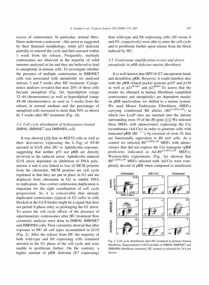
Fig. 2. Cell cycle distribution after HU treatment in primary human
fibroblasts. Representative FACS profiles of IMR90, IMR90E7 and
IMR90E6 fibroblasts untreated, HU treated or released for 24 h are
shown.
L. Lentini et al. / Cancer Letters 238 (2006) 153–160 157
excess of centrosomes. In particular, normal fibro-
blasts underwent a senescent—like arrest as suggested
by their flattened morphology, while p53 deficient
partially re-entered the cycle and then arrested within
1 week from the release. Frequently, multiple
centrosomes are observed in the majority of solid
tumours analyzed so far and they are believed to lead
to aneuploidy in tumour cells. To investigate whether
the presence of multiple centrosomes in IMR90E7
cells was associated with aneuploidy we analyzed
mitoses 3 and 5 weeks after HU treatment. Cytoge-
netics analyses revealed that near 20% of these cells
became aneuploid (Fig. 1d), hypodiploid (range
32–44 chromosomes) as well as hyperdiploid (range
48–88 chromosomes), as soon as 3 weeks from the
release in normal medium and the percentage of
aneuploid cells increased to more than 50% as shown
by 5 weeks after HU treatment (Fig. 1d).
3.2. Cell cycle distribution of hydroxyurea treated
IMR90, IMR90E7 and IMR90E6 cells
It was showed [10] that wt-REF52 cells as well as
their derivatives expressing the L-Tag of SV40
arrested in G1/S after HU or Aphidicolin exposure,
suggesting that neither p53 nor pRB are directly
involved in the induced arrest. Aphidicolin induced
G1/S arrest depended on inhibition of DNA poly-
merase a and it was linked to loss of MCM proteins
from the chromatin. MCM proteins are cell cycle
regulated in that they are put in place in G1 and are
displaced from chromatin in G2 to inhibit DNA
re-replication. Also correct centrosome duplication is
important for the right coordination of cell cycle
progression. So it is conceivable that already
duplicated centrosomes (typical of G2 cells) in cells
blocked at the G1/S border might be a signal that does
not permit S-phase entry so prolonging the G1 arrest.
To assess the cell cycle effects of the presence of
supernumerary centrosomes after HU treatment flow
cytometry analyses were done in IMR90, IMR90E7
and IMR90E6 cells. Flow cytometry showed that after
exposure to HU all cell types accumulated in G1/S
(Fig. 2). After the release from HU the majority of
both wild-type and E6 expressing cells remained
arrested in the G1 phase of the cell cycle and were
unable to proliferate further. On the contrary, a
higher amount of pRB deficient (E7 expressing)
than wild-type and E6 expressing cells (20 versus 6
and 9%, respectively) were able to enter the cell cycle
and to proliferate further upon release from the block
induced by HU.
3.3. Centrosome amplification occurs and drives
aneuploidy in pRB deficient murine fibroblasts
It is well-known that HPV16-E7 oncoprotein binds
and destabilize pRB. However, it could interfere also
with the pRB related pocket proteins p107 and p130
as well as p21WAF1 and p27KIP1.To assess that the
results we obtained in human fibroblasts (amplified
centrosomes and aneuploidy) are dependent mainly
on pRB inactivation, we shifted to a mouse system.
We used Mouse Embryonic Fibroblasts (MEFs)
carrying conditional Rb alleles (RbLoxP/LoxP) in
which two LoxP sites are inserted into the introns
surrounding exon 19 of the Rb gene [11] We infected
these MEFs with adenoviruses expressing the Cre
recombinase (Ad-Cre) in order to generate cells with
truncated pRB (RbK/K), by excision of exon 19, that
are functionally equivalent to Rb null cells. As a
control we infected RbLoxP/LoxP MEFs with adeno-
viruses that did not express the Cre transgene (pRB
proficient, indicated as Ad-RbLoxP/LoxP MEFs).
Western-blot experiments (Fig. 3a) showed that
RbLoxP/LoxP MEFs infected with Ad-Cre were com-
pletely devoid of pRB when compared to uninfected

Fig. 3. Centrosome amplification and aneuploidy in pRB deficient MEFs following HU treatment. (a) Western blot analysis showing the level of
pRB expression after 96 h from the adenoviral infection: pRbloxP/loxP MEF uninfected (lane 1), infected with the empty adenoviral vector
(lane 2) and with the Ad-Cre (lane 3). (b) Centrosomes were revealed by g-tubulin detection (green) and nuclei were stained with DAPI (blue).
The majority of pRbloxP/loxP MEF uninfected or infected with the empty vector showed normal number of centrosomes (arrows). Arrowheads
indicate abnormal centrosomes in RbK/K MEFs, insets illustrate centrosomes at higher magnification. (c) Histograms indicating the percentage
of centrosomes numbers in untreated, HU treated (48 h) and released (72 h) MEFs. (d) Histograms showing percentage of cells with normal
(40 chromosomes), hypodiploid (!40 chromosomes) and hyperdiploid (O40 chromosomes) metaphases after 14 and 30 days following HU
treatment. Each bar indicates meanCSE of at least three independent experiments. (e) Karyotypic analysis of metaphase spreads showing the
presence of aneuploid metaphases by using a Zeiss Axioskop microscope under a 63! objective.
L. Lentini et al. / Cancer Letters 238 (2006) 153–160158
cells and cells infected with adenoviruses that did not
express the Cre recombinase. Then parental and
infected cells were treated with HU, 2 weeks after
infection with Ad-Cre, to detect any centrosome
amplification by g-tubulin staining (Fig. 3b). All of
the untreated MEFs showed a similar percentage of
cells with 1 or 2 centrosomes (Fig. 3c). After HU
treatment both RbLoxP/LoxP and Ad-RbLoxP/LoxP MEFs
showed (Fig. 3c) similar number of cells with one or
two centrosomes (25–30 and 65–60%, respectively).
On the contrary, we scored a high increase in cells
harbouring more than two centrosomes (55–60%) in
L. Lentini et al. / Cancer Letters 238 (2006) 153–160 159
the RbK/K MEFs, when compared to the untreated
cells (8%). RbK/K MEFs showing amplified centro-
somes were still present at 72 h of release in drug free
medium (Fig. 3c). These results suggest that pRB
ablation allowed additional rounds of centrosome
duplication in absence of DNA synthesis. Cytogenetic
analyses performed 2 and 4 weeks after the release
from the block imposed by HU showed that the
majority of RbK/K MEFs became aneuploid
(Fig. 3d,e), hypodiploid as well as hyperdiploid,
suggesting also that pRB absence allows aneuploid
cells to proliferate.
4. Discussion
Centrosome amplification and aneuploidy occur
in virtually all of solid tumors analyzed so far and
could be considered early features in tumor initiation
and progression [1]. Centrosome alterations were
reported in high grade prostate cancer, pancreatic
carcinoma and cervix tumors, and were correlated
with the presence of multipolar mitoses and
chromosomal instability [12,13,14]. Our results
show that human fibroblasts expressing HPV16-E6
and -E7 proteins, that target p53 and pRB,
respectively, as well as conditional pRB deficient
MEFs underwent centrosome amplification after
hydroxyurea treatment, whereas wild-type cells did
not. Because centrosome duplication normally
occurs only once per cell cycle these cells have
lost the synchrony between the DNA and centrosome
duplication likely by undergoing a prolonged G1/S
arrest. Recently, it was reported that centrosome
duplication was unaffected by HU treatment in p53
competent MCF7 cells but not in MCF7 cells with
altered p53 [8]. In addition, our results are consistent
with previous reports showing that dysfunction of
p53 [15] and pRB [5] results in deregulation of
centrosome duplication [16].
Interestingly, after hydroxyurea removal normal
human fibroblasts accumulated with two centrosomes
and underwent a senescent—like arrest, while p53
deficient fibroblasts partially re-entered the cycle and
then arrested within one week from the release. This
finding was similar to that previously noted for p53
deficient (E6 expressing) HeLa cells [10] which
underwent a partial replication on release from HU,
though this was followed by death likely an
alternative outcome to arrest in G1/S in these tumor
cells.
On the contrary, pRB deficient (E7 expressing)
cells re-entered the cell cycle and proliferated despite
the presence of amplified centrosomes. More inter-
estingly, pRB deficient human fibroblasts, analyzed 3
and 5 weeks after hydroxyurea treatment, showed an
increase in the number of aneuploid cells. These
findings suggest that pRB not only could have a role in
centrosome homeostasis, but it might be part of
pathway that act downstream by arresting cells that
have extra centrosomes. This putative checkpoint
could be activated in pRB competent cells by the
presence of an incorrect number of centrosomes (for
the G1 cell cycle phase) or their related effects (i.e.
chromosomal instability) halting further cell cycle
progression. We cannot rule out that a certain degree
of DNA damage after HU could elicit the G1 arrest we
observed in IMR90 cells. Activation of the G1/S
checkpoint triggered by DNA damage will block
centrosome re-duplication as an additional safeguard
mechanism for the integrity of the cell. However, it
was reported that antimetabolites such as HU arrested
cells at the G1/S border in the absence of any
detectable DNA damage [18].
Recently, it was reported that expression of
HPV-16 E7 oncoprotein could induce abnormal
centrosome duplication independently of its ability
to inactivate pRB, p107, and p130 pocket proteins
[17]. However, centrosome amplification was less
pronounced in pRB negative than in pRB positive
cells, indicating that degradation of this protein by
E7 contributes to centrosome duplication errors.
Our findings that HU treated pRB deficient MEFs
behaved similar to human fibroblasts expressing the
HPV16-E7 oncoprotein, strongly indicate that lack
of pRB could allow generation of multiple
centrosomes in a single cell cycle as well as their
maintenance. Finally, our results in pRB deficient
fibroblasts support the model proposed by Nigg
[19] to explain the occurrence of centrosome
amplification in one cell cycle and his effects on
ploidy, suggesting that multiple centrosome could
be a prerequisite in order to generate aneuploidy in
cells that lack important functions such as those
under pRB control.

L. Lentini et al. / Cancer Letters 238 (2006) 153–160160
Acknowledgements
MEFs with conditional Rb alleles were kindly
provided by Dr Marco Crescenzi (ISS, Rome). Floxed
MEFs were derived from mice generated in A. Berns’
laboratory. This work was supported by grants from
Associazione Italiana per la Ricerca sul Cancro
(AIRC) and Universita di Palermo (ex 60%).
References
[1] A.B. D’Assoro, W.L. Lingle, J.L. Salisbury, Centrosome
amplification and the development of cancer, Oncogene 21
(2002) 6146–6153.
[2] P.V. Jallepalli, I.C. Waizenegger, F. Bunz, S. Langer,
M.R. Speicher, J.M. Peters, et al., Securin is required for
chromosomal stability in human cells, Cell 105 (2001)
445–457.
[3] L.S. Michel, V. Liberal, A. Chatterjee, R. Kirchwegger,
B. Pasche, W. Gerald, et al., MAD2 haplo-insufficiency causes
premature anaphase and chromosome instability in mamma-
lian cells, Nature 409 (2001) 355–359.
[4] D.P. Cahill, C. Lengauer, J. Yu, G.J. Riggins, J.K. Willson,
S.D. Markowitz, et al., Mutations of mitotic checkpoint genes
in human cancers, Nature 392 (1998) 300–303.
[5] L. Lentini, L. Pipitone, A.Di Leonardo, Functional inactivation
of pRB results in aneuploid mammalian cells after release from
a mitotic block, Neoplasia 4 (2002) 380–387.
[6] B.R. Brinkley, Managing the centrosome numbers game: from
chaos to stability in cancer cell division, Trends Cell Biol. 11
(2001) 18–21.
[7] P. Meraldi, J. Lukas, A.M. Fry, J. Bartek, E.A. Nigg,
Centrosome duplication in mammalian somatic cells requires
E2F and Cdk2- cyclin A, Nat. Cell Biol. 1 (1999) 88–93.
[8] A.B. D’Assoro, R. Busby, K. Suino, E. Delva,
G.J. Almodovar-Mercado, H. Johnson, et al., Genotoxic stress
leads to centrosome amplification in breast cancer cell lines
that have an inactive G1/S cell cycle checkpoint, Oncogene 23
(2004) 4068–4075.
[9] S. Duensing, A. Duensing, C.P. Crum, K. Munger, Human
papillomavirus type 16 E7 oncoprotein-induced abnormal
centrosome synthesis is an early event in the evolving
malignant phenotype, Cancer Res. 61 (2001) 2356–2360.
[10] F. Borel, F.B. Lacroix, R. Margolis, Prolonged arrest of
mammalian cells at the G1S boundary results in permanent S
phasis stasis, J. Cell Sci. 115 (2002) 2829–2838.
[11] S. Marino, M. Vooijs, H. van Der Gulden, J. Jonkers, A. Berns,
Induction of medulloblastomas in p53-null mutant mice by
somatic inactivation of Rb in the external granular layer cells
of the cerebellum, Genes Dev. 14 (2000) 994–1004.
[12] G.A. Pihan, A. Purohit, J. Wallace, R. Malhotra, L. Liotta,
S.J. Doxsey, Centrosome defects can account for cellular and
genetic changes that characterize prostate cancer progression,
Cancer Res. 61 (2001) 2212–2219.
[13] N. Sato, K. Mizumoto, M. Nakamura, K. Nakamura,
M. Kusumoto, H. Niiyama, et al., Centrosome abnormalities
in pancreatic ductal carcinoma, Clin. Cancer Res. 5 (1999)
963–970.
[14] S. Duensing, K. Munger, Centrosome abnormalities and
genomic instability induced by human papillomavirus onco-
proteins, Prog. Cell Cycle Res. 5 (2003) 383–391.
[15] P. Tarapore, K. Fukasawa, Loss of p53 and centrosome
hyperamplification, Oncogene 21 (2002) 6234–6240.
[16] S. Duensing, L.Y. Lee, A. Duensing, J. Basile,
S. Piboonniyom, S. Gonzalez, et al., The human papilloma-
virus type 16 E6 and E7 oncoproteins cooperate to induce
mitotic defects and genomic instability by uncoupling
centrosome duplication from the cell division cycle, Proc.
Natl Acad. Sci. USA 97 (2000) 10002–10007 [18].
[17] S. Duensing, K. Munger, Human papillomavirus type 16 E7
oncoprotein can induce abnormal centrosome duplication
through a mechanism independent of inactivation of
retinoblastoma protein family members, J. Virol. 77
(2003) 12331–12335.
[18] S.P. Linke, K.C. Clarkin, A. Di Leonardo, A. Tsou,
G.M. Wahl, A reversible, p53-dependent G0/G1 cell cycle
arrest induced by ribonucleotide depletion in the absence of
detectable DNA damage, Genes Dev. 10 (1996) 934–947.
[19] E.A. Nigg, Centrosome aberrations: cause or consequence of
cancer progression? Nat. Rev. Cancer (2002) 815–825.