Non-invasive prenatal diagnostics of aneuploidy using next-generation DNA sequencing technologies,...
14
DOI 10.1515/cclm-2012-0281 Clin Chem Lab Med 2013; 51(6): 1141–1154 Review Yana N. Nepomnyashchaya, Artem V. Artemov, Sergey A. Roumiantsev, Alexander G. Roumyantsev and Alex Zhavoronkov* Non-invasive prenatal diagnostics of aneuploidy using next-generation DNA sequencing technologies, and clinical considerations Abstract Rapidly developing next-generation sequencing (NGS) technologies produce a large amount of data across the whole human genome and allow a large number of DNA samples to be analyzed simultaneously. Screening cell-free fetal DNA (cffDNA) obtained from maternal blood using NGS technologies has provided new opportunities for non-invasive prenatal diagnosis (NIPD) of fetal aneuploi- dies. One of the major challenges to the analysis of fetal abnormalities is the development of accurate and reliable algorithms capable of analyzing large numbers of short sequence reads. Several such algorithms have recently been developed. Here, we provide a review of recent NGS- based NIPD methods as well as the available algorithms for short-read sequence analysis. We furthermore introduce the practical application of these algorithms for the detec- tion of different types of fetal aneuploidies, and compare the performance, cost and complexity of each approach for clinical deployment. Our review identifies several main technologies and trends in NGS-based NIPD. The main con- siderations for clinical development for NIPD and screen- ing tests using DNA sequencing are: accuracy, intellectual property, cost and the ability to screen for a wide range of chromosomal abnormalities and genetic defects. The cost of the diagnostic test depends on the sequencing method, diagnostic algorithm and volume of the tests. If the cost of sequencing equipment and reagents remains at or around current levels, targeted approaches for sequencing-based aneuploidy testing and SNP-based methods are preferred. Keywords: aneuploidy; cell-free fetal DNA; Down syn- drome; fetal aneuploidies; next-generation sequencing; non-invasive prenatal diagnosis; prenatal screening. *Corresponding author: Alex Zhavoronkov, PhD, Bioinformatics and Medical Information Technology Laboratory, Federal Clinical Research Center for Pediatric Hematology, Oncology and Immunology – Experimental and Molecular Medicine, Samory Mashela 1, Moscow 117198, Russian Federation, E-mail: [email protected] Yana N. Nepomnyashchaya, Artem V. Artemov, Sergey A. Roumiantsev and Alexander G. Roumyantsev: Bioinformatics and Medical Information Technology Laboratory, Federal Clinical Research Center of Pediatric Hematology, Oncology and Immunology, Moscow, Russian Federation Alex Zhavoronkov: The Biogerontology Research Foundation, London, UK Introduction To date, invasive procedures, such as amniocentesis and chorionic villus sampling (CVS) have been used successfully for the detection of fetal aneuploidy in high-risk pregnancies. However, invasive methods are associated with significant risks of the fetal loss – a major adverse consequence of prenatal diagnosis in obstetric practices [1]. In 1997, a research paper on the discovery of circulating cffDNA from the Y chromosome of male fetuses in the maternal blood stream during pregnancy opened up new horizons for NIPD [2]. Since then there have been numerous studies describing the use of cffDNA for NIPD of chromosomal aneuploidy; in particular, trisomies 21 (Down syndrome), 18 (Edward syndrome) and 13 (Patau syndrome), all of which used digital PCR analysis [3–5]. The appearance of cffDNA in the maternal circula- tion occurs when normal placental cell death causes the chromosomes to break into short fragments, most of which are under 300 bp in length [6–8]. The proportion of cffDNA in maternal blood increases as pregnancy pro- gresses. cffDNA comprises around 5%–10% of the total cell-free DNA during the first and second trimesters and can be detected reliably as early as the seventh week Unauthenticated | 95.165.92.74 Download Date | 11/23/13 10:46 PM
-
Upload
insilicomedicine -
Category
Documents
-
view
0 -
download
0
Transcript of Non-invasive prenatal diagnostics of aneuploidy using next-generation DNA sequencing technologies,...

DOI 10.1515/cclm-2012-0281 Clin Chem Lab Med 2013; 51(6): 1141–1154
Review
Yana N. Nepomnyashchaya , Artem V. Artemov , Sergey A. Roumiantsev ,
Alexander G. Roumyantsev and Alex Zhavoronkov *
Non-invasive prenatal diagnostics of aneuploidy using next-generation DNA sequencing technologies, and clinical considerations
Abstract
Rapidly developing next-generation sequencing (NGS)
technologies produce a large amount of data across the
whole human genome and allow a large number of DNA
samples to be analyzed simultaneously. Screening cell-free
fetal DNA (cffDNA) obtained from maternal blood using
NGS technologies has provided new opportunities for
non-invasive prenatal diagnosis (NIPD) of fetal aneuploi-
dies. One of the major challenges to the analysis of fetal
abnormalities is the development of accurate and reliable
algorithms capable of analyzing large numbers of short
sequence reads. Several such algorithms have recently
been developed. Here, we provide a review of recent NGS-
based NIPD methods as well as the available algorithms for
short-read sequence analysis. We furthermore introduce
the practical application of these algorithms for the detec-
tion of different types of fetal aneuploidies, and compare
the performance, cost and complexity of each approach
for clinical deployment. Our review identifies several main
technologies and trends in NGS-based NIPD. The main con-
siderations for clinical development for NIPD and screen-
ing tests using DNA sequencing are: accuracy, intellectual
property, cost and the ability to screen for a wide range of
chromosomal abnormalities and genetic defects. The cost
of the diagnostic test depends on the sequencing method,
diagnostic algorithm and volume of the tests. If the cost of
sequencing equipment and reagents remains at or around
current levels, targeted approaches for sequencing-based
aneuploidy testing and SNP-based methods are preferred.
Keywords: aneuploidy; cell-free fetal DNA; Down syn-
drome; fetal aneuploidies; next-generation sequencing;
non-invasive prenatal diagnosis; prenatal screening.
*Corresponding author: Alex Zhavoronkov, PhD, Bioinformatics and
Medical Information Technology Laboratory, Federal Clinical
Research Center for Pediatric Hematology, Oncology and
Immunology – Experimental and Molecular Medicine, Samory
Mashela 1, Moscow 117198, Russian Federation,
E-mail: [email protected]
Yana N. Nepomnyashchaya , Artem V. Artemov , Sergey A. Roumiantsev and Alexander G. Roumyantsev: Bioinformatics and
Medical Information Technology Laboratory , Federal Clinical
Research Center of Pediatric Hematology, Oncology and
Immunology, Moscow , Russian Federation Alex Zhavoronkov: The Biogerontology Research Foundation ,
London , UK
Introduction To date, invasive procedures, such as amniocentesis
and chorionic villus sampling (CVS) have been used
successfully for the detection of fetal aneuploidy in
high-risk pregnancies. However, invasive methods are
associated with significant risks of the fetal loss – a
major adverse consequence of prenatal diagnosis in
obstetric practices [1] . In 1997, a research paper on the
discovery of circulating cffDNA from the Y chromosome
of male fetuses in the maternal blood stream during
pregnancy opened up new horizons for NIPD [2] . Since
then there have been numerous studies describing the
use of cffDNA for NIPD of chromosomal aneuploidy; in
particular, trisomies 21 (Down syndrome), 18 (Edward
syndrome) and 13 (Patau syndrome), all of which used
digital PCR analysis [3 – 5] .
The appearance of cffDNA in the maternal circula-
tion occurs when normal placental cell death causes
the chromosomes to break into short fragments, most of
which are under 300 bp in length [6 – 8] . The proportion
of cffDNA in maternal blood increases as pregnancy pro-
gresses. cffDNA comprises around 5 % – 10 % of the total
cell-free DNA during the first and second trimesters and
can be detected reliably as early as the seventh week
Unauthenticated | 95.165.92.74Download Date | 11/23/13 10:46 PM

1142 Nepomnyashchaya et al.: Non-invasive prenatal diagnostics of aneuploidy using NGS
of gestation [2, 9] . Circulating cffDNA is rapidly cleared
from maternal blood after delivery, except for cases
where small amounts remain, including cells from previ-
ous pregnancies [10] . It has recently been found that the
entire fetal genome, in the form of cffDNA, is present in
maternal blood [11] .
New NGS technologies permit the simultaneous,
or ‘ massively parallel ’ , sequencing of extremely large
quantities of DNA molecules. NGS produces billions
of short sequence reads per instrument run [12] . Mas-
sively parallel sequencing of fetal DNA from maternal
blood has enormous potential, not only for increasing
our understanding of the causes of prenatal genetic dis-
orders in the fetus but also for designing non-invasive
clinical diagnostic tests [11] . At the moment, non-inva-
sive methods do not detect other genetic abnormalities,
such as SNP and indel variations causing Mendelian dis-
orders, and invasive procedures are thus still required.
Non-invasive methods of fetal genotyping are currently
in development, and accurate screening of both domi-
nant and recessive single gene disorders may be possi-
ble in clinical practice in the near future [13] . The pos-
sibility of using massively parallel shotgun sequencing
to detect non-invasive fetal trisomies from maternal
blood by analyzing the relevant chromosomes in locus-
independent assays has been demonstrated [14, 15] , and
recent studies have confirmed this finding [16, 17] . An
alternative approach to sequencing whole genomes for
non-invasive detection of fetal abnormalities would be to
enrich for the region of interest using array capture prior
to sequencing [18 – 20] .
NGS technologies have already begun to show their
remarkable potential for detecting the most common
aneuploidies in live births, including Down, Edward,
and Patau syndromes. These research discoveries have
been translated into clinical tests, resulting in major
benefits for NIPD. In this review, we describe different
approaches to non-invasive detection of prenatal aneu-
ploidy which, for their clinical application, use the NGS
technologies of four innovative companies. The compa-
nies Sequenom, Inc (San Diego, CA, USA) and Verinata
Health, Inc (Redwood City, CA, USA) offer methods based
on collecting and analyzing information across the
entire genome, while the approach of Ariosa Diagnos-
tics, Inc (San Jose, CA, USA) and Natera, Inc (San Carlos,
CA, USA) is based on the selection of only the chromo-
somes of interest. Both approaches are capable of detect-
ing the most common fetal aneuploidies in the popula-
tion. To date of submission of this manuscript, three of
the aforementioned companies – Sequenom, Verinata
Health, and Ariosa Diagnostics – have published clinical
validation studies. These companies have furthermore
launched cffDNA-based non-invasive prenatal laboratory
developed tests (LDT) using next-generation sequencing
through the Clinical Laboratory Improvement Amend-
ments (CLIA) laboratories. Several clinical diagnostics
laboratories in other countries have also launched NIPD
screening tests using NGS, including the Beijing Geno-
mics Institute in China. Although every company has
sponsored clinical validation studies and published their
results in peer-reviewed, high-impact industry journals,
none of the tests are approved by the US Food and Drug
Administration (FDA). Although tests and clinical vali-
dation studies are still ongoing, it is expected that the
diagnostic accuracy, sensitivity, and specificity of these
techniques will be very high.
Limitations of current methods of prenatal diagnostics Many prenatal screening and diagnostic tests have been
developed and introduced into clinical practice. While
standard methods of prenatal care vary, combination
screening using biochemical markers in serum, includ-
ing pregnancy-associated plasma protein A (PAPP-A),
free β -subunit of human chorionic gonadotropin (free
β-hCG), α -fetoprotein (AFP) and unconjugated estriol
(uE3), in conjunction with a sonographic measurement
of nuchal translucency and detection of the presence
or absence of the nasal bone, are commonly used. The
detection rate of non-invasive combination screening
has improved greatly over the past decade to over 90 % in
the first trimester of pregnancy [21] . However, achieving
such a high detection rate requires exceptionally skilled
physicians and strict adherence to protocols. Invasive
diagnostic methods are still recommended for high-risk
pregnancies.
Currently, diagnostic testing requires that the fetal
cells that are to be tested be removed directly from the
uterus using either CVS between 11 and 14 weeks of ges-
tation, or amniocentesis between 15 and 20 weeks. While
using direct fetal material provides over 99 % accuracy,
both of these procedures are invasive and are associated
with an increased risk of transplacental hemorrhage
and spontaneous abortion [22] . The risk of miscarriage
following these procedures can be conservatively esti-
mated at around 1 % [23] , but this varies greatly depend-
ing on a number of human and environmental factors
including the country, the hospital or clinic, and the
physician.
Unauthenticated | 95.165.92.74Download Date | 11/23/13 10:46 PM

Nepomnyashchaya et al.: Non-invasive prenatal diagnostics of aneuploidy using NGS 1143
Whole-genome sequencing approaches for NIPD of fetal aneuploidy In cases where a woman is carrying a fetus with aneu-
ploidy – trisomy 21, e.g., the amount of copies of chromo-
some 21 is expected to be slightly higher in comparison
with other autosomes. Rapidly developing NGS tech-
nologies, which provide a vast amount of data across
the entire genome, appear to be suitable for counting
genome representation and determining the over-rep-
resented chromosomes of interest in the affected fetus.
Two scientific groups have independently shown that,
from cffDNA obtained from maternal blood, NGS can
clearly identify plasma samples from women carrying
aneuploid fetuses by comparing them with samples
taken from women with euploid fetuses [14, 15] . A short
region at one end of each DNA molecule of maternal
plasma was sequenced using synthesis technology on
an Illumina platform, and mapped against the reference
human genome to determine the chromosomal origin of
each sequence. The density of sequenced tags from the
MaternalDNA
FetalDNA
Trisomydetected
Threshold
chr 1
chr i
chr j
…
…
Counting of reads per each chromosome
Mapping of the reads
Figure 1 Schematic illustration of the procedural framework for using massively parallel genomic sequencing for the non-invasive prenatal
detection of fetal chromosomal aneuploidy.
(A) Fetal DNA (green fragments) circulates in maternal plasma as a minor population among a high background of maternal DNA (blue frag-
ments). A sample containing a representative profile of DNA molecules in maternal plasma is obtained. Short fragments of cell-free DNA
are then sequenced by some NGS technique. (B) The chromosomal origin of each sequence is identified by mapping the obtained reads to
the human reference genome. (C) The number of unique sequences mapped to each chromosome are counted and genome representation
of the chromosomes of interest is determined in subsequent analysis. Aneuploidy can be detected by various statistical techniques based
on the number of reads representing the chromosome of interest compared to the other chromosomes. Notice that blue and green circles
schematically representing the reads mapped to each chromosome, originating from maternal and fetal DNA fractions, respectively, are
indistinguishable and only a difference in the total amount of reads can be detected (as shown for ‘ chr j ’ in the example).
chromosome of interest from an aneuploid fetus was
compared with cases of trisomy and euploid pregnancies
(Figure 1 ).
Fan et al. [14] used from 1.3 to 3.2 mL of plasma taken
from pregnant women at risk for fetal aneuploidy. Their
study examined a total of 18 normal and aneuploid preg-
nancies, which included nine cases of trisomy 21, two of
trisomy 18 and one of trisomy 13. Amniocentesis or CVS
was conducted to analyze and confirm the fetal karyotype.
Cell-free plasma DNA was sequenced on the 1G Genome
Analyzer platform (Solexa/Illumina, Inc, San Diego, CA,
USA). Each of the 25 bp reads were mapped against the
non-repeat-masked reference human genome build 36
(hg18) using ELAND from the Solexa data analysis pipe-
line. Sequencing resulted in approximately five million
unique sequence reads per patient sample with, at most,
one mismatch against the human genome. Each set of
patient sequence reads covered approximately 4 % of the
entire human genome. The average sequence read density
for 50-kb windows across the chromosome of interest
was normalized using the median value obtained from
all autosomes in the euploid cases. A 99 % confidence
interval in the distribution sequence read density of the
Unauthenticated | 95.165.92.74Download Date | 11/23/13 10:46 PM

1144 Nepomnyashchaya et al.: Non-invasive prenatal diagnostics of aneuploidy using NGS
chromosome of interest for disomy pregnancies was cal-
culated. The values for normalized sequence tag density
of trisomy 21, 18 and 13 cases were reported to be outside
the upper boundary of the confidence interval, and those
for all disomy 21, 18 and 13 cases were below the boundary,
indicating 100 % sensitivity with a false-positive rate 0 % .
The coverage of chromosome 21 for trisomy 21 pregnan-
cies was on average ∼ 11 % higher than that of the disomy
21 cases. The method described by Fan et al. reveals that
the minimum fraction of fetal DNA that could be detected
by NGS is approximately 2 % .
The distribution of sequence reads across each chro-
mosome for all samples was not uniform. In addition,
the GC content of the sequenced reads of all the samples
was ∼ 10 % higher than the value of the sequenced human
genome [24] . The mean read density of chromosomes,
which was reported to correlate with the GC content of
the chromosomes, was observed to have a positive bias
for chromosomes with high GC content. Interestingly –
and differing from this report – Chiu et al. [25] observed a
negative correlation between chromosome representation
and GC content in their analysis employing massively par-
allel sequencing of fetal DNA from maternal plasma using
ligation technology on the SOLiD ™ 3 platform (Applied
Biosystems/Life Technologies, Carlsbad, CA, USA). In both
sets of data, a strong GC bias and the non-uniform repre-
sentation of read density between chromosomes are likely
to be explained by analytical rather than biological factors
stemming from the sequencing process, which would be
eliminated by the use of appropriate algorithms [26 – 28] .
Importantly, blood samples were collected from 15 to
30 min after amniocentesis or CVS, and the average gesta-
tional period of the aneuploidy pregnancies (20.6 weeks)
was longer than that of the euploid group (13.8 weeks).
Since the proportion of fetal DNA in maternal blood was
reported to increase with the length of gestation age [29,
30] , the above-mentioned factors should be taken into
consideration in future analyses. Nonetheless, this pio-
neering work has shown that NGS could be used as a
non-invasive prenatal diagnostic tool for the quantitative
measurement of a number of chromosomes.
Chiu et al. analyzed cell-free DNA from 5 to 10 mL
of plasma taken from 14 trisomy 21 and 14 euploid preg-
nancies in women at risk for fetal aneuploidy [15] . In the
majority of cases, maternal blood samples were collected
before invasive obstetrics procedures had been carried
out, with comparable median lengths of gestation for both
the euploid and the trisomy case groups. One end of the
clonally expanded copies of each plasma DNA fragment
was sequenced (36 bp reads) and processed by stand-
ard post-sequencing bioinformatic alignment analysis
with the Illumina Genome Analyzer, which uses ELAND
software. The obtained mean of approximately 2 million
unique reads per sample resulted in no mismatches with
the repeat-masked reference human genome (NCBI Build
36, version 48). The number of reads originating from chro-
mosomes of interest was normalized by the total number
of reads generated by the sequencing run. Z-scores repre-
senting the number of standard deviations from the mean
proportion of chromosomes-of-interest reads in a refer-
ence set of euploid cases were calculated for each case. A
statistically significant difference between the parameter
estimated in the test case and that in the reference group
was 99 % under z-score > 3. Z-scores for chromosome 21
were above + 5 (range 5.03 – 25.11) for all 14 trisomy 21 cases,
i.e., at three standard deviations above the reference
established from the male euploid fetuses. In the study,
the method accurately detected all trisomy 21 cases and
produced no false-positive results.
One caveat of using this approach is that the distribu-
tion of reads on each chromosome can vary from sequenc-
ing run to sequencing run; hence, these intra- and
inter-run sequencing variations can increase the overall
variation in the aneuploidy detection metric.
Both studies demonstrated that massively parallel
sequencing can both identify and count DNA fragments
in maternal plasma to detect small quantitative altera-
tions in the genomic distribution of plasma DNA. Methods
that do not require the differentiation between fetal and
maternal DNA are currently being developed, and can be
applied to arbitrarily small concentrations of fetal DNA in
maternal plasma. Since fetal DNA is natively fragmented,
no further fragmentation step for the library preparation
is needed, substantially simplifying subsequent analysis.
Non-invasive NGS detection of the variation in the
number of copied fetal chromosomes in maternal plasma
is already in use on pregnant women in proof-of-concept
studies, and has been brought into clinical application.
A number of subsequent large-scale clinical studies were
carried out by academic research institutions and biotech-
nology companies to validate various approaches to NIPD
using massively parallel genomic sequencing for use in
clinical practice [16, 17, 31 – 33] . Table 1 presents a summary
of these studies for detection of Down syndrome.
Targeted approaches to sequencing-based NIPD of fetal aneuploidy The two methods described above rely on the massively
parallel sequencing of all the DNA in a maternal plasma
Unauthenticated | 95.165.92.74Download Date | 11/23/13 10:46 PM

Nepomnyashchaya et al.: Non-invasive prenatal diagnostics of aneuploidy using NGS 1145
sample without targeting specific chromosomes. However,
several targeted approaches were recently developed
based on the a priori selection of DNA regions for analy-
sis. Compared to sequencing and counting all reads from
all chromosomes, limiting the number of DNA regions to
quantify greatly reduces the effort required to assess the
dosage of a chromosome. Moreover, careful selection of
the regions to quantify can potentially reduce the con-
founding variation in the number of reads per locus by
taking into account only the loci with similar properties,
e.g., GC content or the number of repeats of a particular
sequence in the genome [19, 20] .
Different strategies can be used to select and enrich
for the genomic regions of interest. The enrichment step
should meet two requirements: the method should mini-
mize introductory bias (i.e., the output quantity of a frag-
ment should depend only on its input quantity and not on
other factors, such as the features of its sequence) to allow
further quantitative analysis, and the method should be
capable of dealing with small amounts of sampled DNA
[34] . Using the SureSelect System (Agilent Technologies,
Santa Clara, CA, USA), Liao et al. successfully performed
a 213-fold enrichment of the selected loci of cfDNA from
the blood samples of pregnant women [35] as an example
of an in-solution, separation-based enrichment technique
[34] . The enrichment did not introduce any bias in the
ratio of maternal and fetal DNA.
Sparks et al. [19] describe a method for detecting
chromosome aneuploidy using NGS-sequencing of DNA
enriched by a so-called Digital ANalysis of Selected
Regions amplification-based enrichment assay. The
assay comprises three oligos per analyzed locus. Liga-
tion oligos, “ Left ” , “ Middle ” and “ Right ” can comple-
mentarily hybridize to the respective DNA locus, thus
forming a contiguous chain with two nicks, which
can subsequently be ligated. Besides the fragment,
complementary to the matrix, the “ Left ” and “ Right ”
oligos contain constitutive fragments on their ends,
which are then used for PCR amplification with univer-
sal primers. The authors obtained and sequenced DNA
product. Of the 298 samples analyzed (including 39
trisomy 21 and 7 trisomy 18 samples), all the aneuploid
samples were correctly distinguished from the controls,
the authors concluded to 100 % sensitivity and speci-
ficity. The level of sequencing, covering only 420,000
reads per sample, was nevertheless sufficient to detect
trisomy 21 and trisomy 18 reliably (z statistics exceeded
3.6 in all samples). This level corresponds to < 5 % of
the level required by non-targeted approaches, and
enables multiplexing [the study claims that 96 samples
were processed simultaneously in one HiSeq2000
Full
sam
ple,
n a
Tris
omy
21
sam
ple,
nFa
lse-
posi
tive
rate
(95 %
CI),
%
Sens
itivi
ty (9
5 %
CI),
%
Spec
ifici
ty (9
5 %
CI),
%
Gest
atio
nal a
ge,
rang
e (m
ean)
wee
ks b
Met
hod
Refe
renc
es
18
90
(0
– 3
7)
10
0 (
63
– 1
00
)1
00
(6
3 –
10
0)
10
– 3
5 (
18
.3)
Ma
ss
ive
ly p
ara
lle
l s
eq
ue
nci
ng
Fan
et
al.
, 2
00
8 c [
14
]
28
14
0 (
0 –
27
)1
00
(7
3 –
10
0)
10
0 (
73
– 1
00
)1
1.2
– 2
0.3
(1
4.9
)M
as
siv
ely
pa
rall
el
se
qu
en
cin
gC
hiu
et
al.
, 2
00
8 d [
15
]
47
13
0 (
0 –
13
)1
00
(7
2 –
10
0)
10
0 (
87
– 1
00
)1
0.4
– 2
8.3
(1
5.3
)M
as
siv
ely
pa
rall
el
se
qu
en
cin
gS
eh
ne
rt e
t a
l.,
20
11
c [2
8]
44
93
90
.3 (
0.1
– 1
.5)
10
0 (
89
– 1
00
)9
9.7
(9
8.5
– 9
9.9
)8
.0 –
36
.0 (
16
.0)
4-P
lex
ed
ma
ss
ive
ly p
ara
lle
l s
eq
ue
nci
ng
Eh
rich
et
al.
, 2
01
1 d [
31
]
14
68
62
.1 (
0.6
– 6
.4)
10
0 (
95
– 1
)9
7.9
(9
3.6
– 9
9.4
)1
2.3
– 1
3.5
(1
3.0
)2
-Ple
xe
d m
as
siv
ely
pa
rall
el
se
qu
en
cin
gC
hiu
et
al.
, 2
01
1 d [
32
]
57
18
61
.1 (
0.5
– 2
.4)
79
.1 (
69
– 8
7)
98
.9 (
97
.6 –
99
.5)
8-P
lex
ed
ma
ss
ive
ly p
ara
lle
l s
eq
ue
nci
ng
16
96
21
20
.2 (
0.1
– 0
.7)
98
.6 (
95
.9 –
99
.7)
99
.8 (
99
.3 –
99
.9)
9.2
– 2
1.3
(1
5.3
)4
-Ple
xe
d m
as
siv
ely
pa
rall
el
se
qu
en
cin
gP
alo
ma
ki
et
al.
, 2
01
1 d [
33
]
29
83
90
(0
– 1
.8)
10
0 (
88
.8 –
1)
10
0 (
98
.2 –
10
0)
13
.4 –
35
.4 (
20
.5)
Targ
ete
d m
as
siv
ely
pa
rall
el
se
qu
en
cin
g e
Sp
ark
s e
t a
l.,
20
12
a f [
19
]
16
73
60
(0
– 3
.6)
10
0 (
88
.0 –
10
0)
10
0 (
96
.4 –
10
0)
11
.0 –
36
.1 (
18
.6)
Targ
ete
d m
as
siv
ely
pa
rall
el
se
qu
en
cin
gS
pa
rks
et
al.
, 2
01
2b
f [2
0]
53
28
90
(0
– 1
.1)
10
0 (
94
.8 –
10
0)
10
0 (
98
.9 –
10
0)
10
.0 –
23
.0 (
15
.1)
Ma
ss
ive
ly p
ara
lle
l s
eq
ue
nci
ng
Bia
nch
i e
t a
l.,
20
12
c [1
6]
Tabl
e 1
Dia
gn
os
tic
pe
rfo
rma
nce
of
ma
tern
al
pla
sm
a D
NA
se
qu
en
cin
g f
or
de
tect
ing
fe
tal
tris
om
y 2
1.
a O
nly
th
e v
ali
da
tio
n s
et
is c
on
sid
ere
d.
b G
es
tati
on
al
ag
e o
f th
e t
ris
om
y 2
1 c
as
es
. F
rom
c Ve
rin
ata
He
alt
h,
Inc
(Re
dw
oo
d C
ity,
CA
, U
SA
). d
Se
qu
en
om
In
c (S
an
Die
go
, C
A,
US
A).
e Ta
rge
t e
nri
chm
en
t o
f
38
4 l
oci
pe
r ch
rom
os
om
e 2
1.
f Ari
os
a D
iag
no
sti
cs,
Inc
(Sa
n J
os
e,
CA
, U
SA
).
Unauthenticated | 95.165.92.74Download Date | 11/23/13 10:46 PM

1146 Nepomnyashchaya et al.: Non-invasive prenatal diagnostics of aneuploidy using NGS
run (Illumina, Inc)]; thus greatly reducing the cost of
analysis.
A method for detecting aneuploidy based on the
assessment of heterozygosity for various polymorphisms
is described by Natera in the US patent application
US20110288780A1 [18] . The key feature of this method is
that it takes the mixture of maternal and fetal DNA obtained
from blood plasma into account separately from the DNA
from one or both parents. As fetal DNA is represented
exclusively in the cell-free fraction, purely maternal DNA
can be extracted from the blood cells – white blood cells in
particular. Paternal DNA can be additionally included in
the analysis to improve the accuracy of the test, although
the test can still be performed based solely on the samples
taken from the pregnant mother. This method implies
that the sequencing of DNA regions is targeted. One of the
patent ’ s claims suggests the enrichment of the explored
polymorphic loci with an assay of pre-circularized probes,
which have been previously been described [36, 37] . Col-
lected sequencing data contains information on the counts
of sequences having each allele at each of the selected
SNP loci. A statistical model estimates a likelihood ratio
for the total number of reads containing one or another
allele for every possible combination of parental geno-
types and for each number of maternal n m and paternal n f
chromosomes inherited by the fetus. This model takes the
fraction of fetal DNA σ as a parameter. First, a maximum
likelihood estimation is performed for the fetal DNA frac-
tion σ , then n m and n f are determined for the chromosomes
of interest using the value of σ estimated in the previous
step. As the approach described builds statistical models
of the euploid state as well as all the other abnormal ploidy
states, it not only finds the most likely ploidy state of the
fetus for each chromosome, but also permits an estima-
tion of the significance of this decision. This allows a sta-
tistically grounded identification of the samples where no
reliable decision can be made without choosing arbitrary
thresholds or estimating them from a training set. Unfor-
tunately, the patent does not describe the results of the
method ’ s performance tests, thus it is difficult to estimate
its clinical efficiency. The sensitivity and specificity of the
method depend on the fetal DNA fraction and the avail-
ability of the paternal genotype. The availability of pater-
nal DNA allows for samples of the maternal peripheral
blood with lower cffDNA content to be accepted for diag-
nosis. Methods using paternal content are likely to enable
NIPD at earlier stages of the pregnancy when the concen-
tration of fetal material is below the minimal threshold
for methods utilizing only maternal samples. Separate
sequencing of purely maternal cellular DNA may further
improve performance. Along with trisomy 21, trisomy 18
and trisomy 13, sex chromosome aneuploidies (e.g., X0,
XXY, XXX, XYY) can also be detected (Natera, personal
communication), which is an important advantage of this
method in light of the high occurrence of these abnormali-
ties. However, the detection performance of the method is
still to be published. The clinical trial of the Prenatal Non-
invasive Aneuploidy Testing Using SNPs supported by the
National Institutes of Health is underway [38] . As it is SNP-
based, the method may need to be tested on patients from
different populations.
The recent SNP-based targeted NIPD methods of
Sparks et al. [20] and Rabinowitz et al. [18] seem to be
highly efficient. As they can be performed on a sequenc-
ing machine with a lower price per run and lower
throughput [e.g., PGM (Ion Torrent/Life Technologies,
Carlsbad, USA) or MiSeq (Illumina, Inc)], these methods
are preferred, especially for average-sized clinics. In
addition to modeling disomy and trisomy, both methods
model other ploidy states, thus enhancing statistical per-
formance. The Natera method can potentially perform
better because it extracts both cell-free DNA and cellu-
lar DNA from the same blood sample of purely maternal
origin. It is also claimed that the Natera method can detect
aneuploidy in sex chromosomes. Nevertheless, perfor-
mance tests for the Natera method have not yet been
published.
Counting statistics for improvements in the sensitivity of the NIPD of fetal aneuploidy Early reports suggest that inaccuracy in measuring
genomic representation is variable [14, 15] . Although the
algorithms used in recently published studies success-
fully classify fetal trisomy 21, they appear to be unable to
effectively detect other aneuploidies, such as trisomy 18
and 13, which would inevitably occur in the population.
However, it has been reported that high throughput mas-
sively parallel sequencing assays using specific bioinfor-
matic algorithms, may enable the non-invasive detection
of any type of fetal aneuploidy [17, 26 – 28] .
The existence of a substantial GC bias in Illumina/
Solexa and ABI/SOLiD sequencing has recently been
shown. This issue limits the sensitivity of measuring
genomic representation in chromosomes [14, 39 – 41] .
Fan and Quake [26] have analyzed sequencing data col-
lected in a previous study [14] , as described earlier. In this
study, a bioinformatic algorithm was developed to remove
Unauthenticated | 95.165.92.74Download Date | 11/23/13 10:46 PM

Nepomnyashchaya et al.: Non-invasive prenatal diagnostics of aneuploidy using NGS 1147
GC-content-dependent artifacts in shotgun sequencing
data by applying weight to each sequenced read based
on the local genomic GC content of large regions of the
human genome with relatively homogeneous GC content
[42] . After calculating the standard z-statistic, z-scores for
chromosomes 18 and 13 were increased in two of the two
trisomy 18 and one of the one trisomy 13 cases, respec-
tively. Consequently, all trisomy 18 and 13 cases were cor-
rectly classified. The algorithms for removal of the effect
of GC bias used in these studies appear to be able to effec-
tively detect cases of trisomy 18 and 13 as well as trisomy
21. Although the classification accuracy for trisomy 18 and
13 improved, there were not enough positive samples to
measure a representative distribution.
Chen et al. [27] have demonstrated the successful use
of two-plex massively parallel plasma DNA sequencing
for NIPD of trisomy 18 and 13 on the Genome Analyzer IIx
platform (Illumina) for a large sample set. Cell-free DNA
from 5 to 10 mL of plasma from 25 trisomy 13, 37 trisomy
18, 86 trisomy 21, one sex chromosome mosaic case and
140 euploid pregnancies was used in the analysis. A
total of 392 cases were analyzed, including 103 cases of
women pregnant with euploid fetuses, which were used
as normal controls for z-score calculation. As previously
described, standard z-scores representing the number of
standard deviations away from the mean proportion of
chromosome 18 and 13 reads in a reference set of euploid
cases were determined for each case [15, 32] . Based on the
previous findings that the statistical power of the mole-
cular counting approach increases with the number of
molecules counted, a mean of approximately 4.6 million
unique reads per sample, without mismatches to the non-
repeat-masked reference human genome, was obtained.
As a result, the classification accuracies for trisomy 18
and 13 were improved. For trisomy 18 detection, 31 of 37
trisomy 18 cases and 247 of 252 non-trisomy 18 cases were
identified correctly, corresponding to sensitivity and spec-
ificity of 83.8 % (95 % CI 67.3 % – 93.2 % ) and 98.0 % (95 % CI
95.2 % – 99.3 % ), respectively. For trisomy 13 detection, 11 of
25 trisomy 13 cases and 247 of 264 non-trisomy 13 cases
were identified correctly, corresponding to improved sen-
sitivity and specificity of 44.0 % (95 % CI 25.0 % – 64.7 % ) and
93.6 % (95 % CI 89.7 % – 96.1 % ), respectively. As the impreci-
sion of measuring the genomic representation of chromo-
somes was shown to be variable and dependent on the GC
content of each chromosome, a statistical approach based
on z-score calculation, but with an additional GC correc-
tion algorithm, has been developed in order to improve
diagnosis of trisomy 18 and 13 [14, 15] . Specifically, all
chromosomes from each sample were first divided into 50
kb bins using bioinformatics. Chen et al. determined the
number of sequenced reads and GC content in each bin
and applied the locally weighted scatter plot smoothing
(LOESS) regression to calculate the predicted (P) value
for each bin. Using the raw read counts (RC raw ) the GC-
corrected read counts (RC GC ) of each bin were calculated
with the correlation factor (F), which was derived from the
median counts of all the bins (M) and the LOESS fit pre-
dicted value by the following equations:
F = M/P (1)
and
RC GC = RC raw × F (2)
The standard z-score was calculated using the
genomic representations of chromosomes 18 and 13, and
all of the trisomy 13 cases (25 out of 25) were clearly identi-
fied. Two hundred and sixty-one out of 264 non-trisomy
13 cases were correctly determined under z-score > 3, indi-
cating 100 % (95 % CI 83.4 % – 100 % ) sensitivity and 98.9 %
(95 % CI 96.4 % – 99.7 % ) specificity of the GC correction
approach. Thirty-four out of 37 trisomy 18 samples and 247
out of 252 non-trisomy 18 cases were classified providing
91.9 % (95 % CI 77.0 % – 97.9 % ) sensitivity and 98.0 % (95 %
CI 95.2 % – 99.3 % ) specificity. This study has shown that
the use of both the mapping of sequence reads against
the non-repeat-masked genome and the GC correction
approaches have improved the accuracy of trisomy 18 and
13 diagnosis. Essentially, correct aneuploidy detection
was achieved by increasing the number of aligned reads
in general and hence performing deeper sequencing.
This bioinformatic method will be able to lower the cost
of NGS in the future. Current algorithms for removing GC
bias improve the precision of measuring the genomic rep-
resentation of chromosomes as well as allow the effective
classification of aneuploidies, most notably trisomy 13.
Larger sample collections are required to further examine
the algorithm for trisomy 18 detection.
An alternative for improving the accuracy of the
detection of chromosomal abnormalities is to develop
an optimized algorithm using a normalized chromo-
some value (NCV) from the sequencing data of the refer-
ence group (training set) of 71 samples with 26 abnormal
karyotypes. This would minimize the intra- and inter-run
sequencing variation as previously described [28] . Short,
single-end reads of each plasma sample were sequenced
on the Genome Analyzer IIx platform and unambigu-
ously mapped to the non-repeat-masked reference
human genome, allowing for up to two base mismatches
during alignment. In the test set, the number of unique
sequence tags varied from approximately 13 × 10 6 – 26 × 10 6 .
To determine cases of fetal aneuploidy, the NCVs for the
Unauthenticated | 95.165.92.74Download Date | 11/23/13 10:46 PM

1148 Nepomnyashchaya et al.: Non-invasive prenatal diagnostics of aneuploidy using NGS
chromosome of interest from the test set were compared
with the respective NCVs for those from the training set.
Initially, in the sequencing data from the training set,
the sequence read density (number of mapped sites) for
the chromosome of interest was normalized to counts
observed on another predetermined chromosome (or set
of chromosomes). Each autosome was considered as a
potential denominator in a ratio of counts with our chro-
mosomes of interest from an unaffected subset of the
training data in order to determine the optimal chromo-
some ratio for each chromosome of interest. Denominator
chromosomes that minimized the variation of the chro-
mosome ratios within and between sequencing runs were
selected. An NCV representing the number of standard
deviations away from the mean chromosome ratios for the
unaffected samples in the training set was determined for
each sample and chromosome of interest, and was calcu-
lated with the following equation:
NCV ij = x ij − μ j / σ j (3)
where μ j and σ
j are the estimated training set mean and
SD, respectively, for the j-th chromosome ratio and x ij is the
observed j-th chromosome ratio for sample i. NCVs > 4.0
classify a chromosome as affected, whereas NCVs < 2.5
specify a chromosome as unaffected, which indicates a
99 % chance of a statistically significant difference in the
assessed parameter for the test set compared with the ref-
erence training set. All eight samples with clinical karyo-
types indicating fetal trisomy 18 were correctly identified,
with NCVs between 8.5 and 22, indicating 100 % (95 % CI
59.7 % – 100 % ) sensitivity. The single trisomy 13 individual
with an NCV of approximately three was classified as a ‘ no
call ’ . Hence, the current algorithm demonstrated a 100 %
accurate classification of samples with cases of trisomy 18.
However, larger sample collections are required in order
to test the algorithm further for the detection of trisomy
13. There were no discernible differences in results with
respect to ethnicity. It was also shown that this approach
is particularly informative in the case of twins. Canick
et al. analyzed 27 samples of multiple gestations, collected
during a large-scale study, and used the algorithm correct-
ing for the GC shift [17, 33, 43] . All seven trisomy 21 cases
and one trisomy 13 case were accurately detected (95 % CI
59 % – 100 % ) with no false-positives. Two triplet pregnan-
cies were analyzed and correctly confirmed as euploid as
well. The study confirmed that the algorithm may be used
to correctly detect trisomies in cases involving multiple,
simultaneous gestations.
To return to the recent study by Sparks et al. [19] , in
the current report the authors introduced Fetal-fraction
Optimized Risk of Trisomy Evaluation – an improved
statistical algorithm for trisomy detection [20] . It esti-
mates the risk of aneuploidy by computing an odds ratio
that compares the probability of observing the outcome
according to a model representing a disomic fetal chro-
mosome and a model representing a trisomic fetal
chromosome. Modeling the case of trisomy represents
a major improvement in the study. Both disomic and
trisomic models are normal distributions of the number
of reads from the chromosome of interest scaled by the
number of reads from a different chromosome. The
mean value for the disomy model was taken as a mean
proportion over the reference disomy samples (pos-
sibly with bootstrapping of samples). For the trisomy
model, adjustment of the mean proportion was based
on the estimated fetal DNA fraction. In order to assess
the fetal DNA fraction for a sample, a set polymorphic
loci was quantified together with the constitutive loci
in the assay. A maximum likelihood estimation of the
fetal DNA fraction was performed for every sample,
based on loci where fetal and maternal genotypes differ.
The standard deviation for both the proportion of reads
mapped on the chromosome of interest and of the frac-
tion of fetal DNA was estimated by bootstrapping the
reference samples and taking into account polymorphic
loci. As a result, of the total of 192 polymorphic and 576
non-polymorphic loci quantified in the samples from
the training set, the polymorphic regions and 384 non-
polymorphic loci showing the highest residual difference
between normal and trisomic samples were selected.
Thirty-six samples of trisomy 21 and 8 trisomy 18 from
167 samples in the test set were identified correctly,
showing 100 % for trisomy 21 (95 % CI 88.0 % – 100 % ) and
for trisomy 18 (95 % CI 59.8 % – 100 % ) detection rate with
a false-positive rate of 0 % for trisomy 21 (95 % CI 0 % –
3.6 % ) and for trisomy 18 (95 % CI 0 % – 3.0 % ). The method
is promising in terms of the small amount of sequenced
reads required and the potential for screening for sub-
chromosomal abnormalities. Nevertheless, a study on
a larger cohort is required. Moreover, as the approach
is SNP-based and the frequency of SNP genotypes can
vary in different populations, further studies should be
conducted to verify whether extending the cohort would
require the number of screened polymorphic loci to be
extended.
Clinical considerations To date three diagnostic companies – Sequenom, Veri-
nata Health, and Ariosa Diagnostics – have published
Unauthenticated | 95.165.92.74Download Date | 11/23/13 10:46 PM

Nepomnyashchaya et al.: Non-invasive prenatal diagnostics of aneuploidy using NGS 1149
the results of sponsored clinical studies validating their
methods and started offering NGS-based NIPD services
commercially as LDTs [44 – 46] .
Sequenom was the first to launch the NIPD screening
test in November 2011. Presently, this non-invasive, LDT
detects the increased number of reads from chromosomes
21, 18 and 13 resulting from whole-genome sequencing. In
particular, the reports by Palomaki et al. [17, 33] provide
a large scale international investigation into the determi-
nation of fetal trisomy 21, 18 and 13 involving around two
thousand validation samples. The sensitivity for detect-
ing cases of trisomy 21 was 98.6 % (95 % CI 95.9 % – 99.7 % )
with a false-positive rate 0.2 % (95 % CI < 0.1 – 0.6). Failure
to obtain results occurred in 0.8 % of cases. Trisomy 18 and
13 detection rates were 100 % (95 % CI 93.9 % – 100 % ) and
91.7 % (95 % CI 61 % – 99 % ), respectively with false-positive
rates of 0.3 % (95 % CI 0.1 % – 0.7 % ) for chromosome 18 and
0.9 % (95 % CI 0.5 % – 1.5 % ) for chromosome 13. The average
gestation period at the time the maternal blood was
sampled was 15 weeks and 3 days. Although these studies
are large scale, 63.6 % samples were excluded from analy-
sis due to poor sample quality, volume or long processing
time. The developed test has a high accuracy for determi-
nation of the most common trisomies in the population
and can be offered in combination with other non-inva-
sive methods for diagnosis aneuploidy as a pre-invasive
procedure for high-risk pregnancies. However, since only
four to eight samples can be analyzed in one sequencing
run, and the processing and analysis of whole-genome
sequencing is required, the test cost is high and thus is a
limitation factor for widespread use of this test for diagno-
sis in a clinical setting.
Verinata Health launched the verifi ™ prenatal test
for diagnosis of trisomy 21, 18 and 13 as early as 10 weeks
gestational age, based on a clinical study which was con-
ducted by Bianchi et al. [16] . Bianchi et al. carried out a
study examining 532 maternal blood samples where every
sample was analyzed for six independent categories in
order to define test performance and determine the pres-
ence of trisomy 21, 18, 13 aneuploidy male, female or
monosomy X. It is the first published study which is able
to detect sex chromosome aneuploidy, including mono-
somy X. The sensitivity for detection of trisomy 21, 18 and
13 was 100 % (95 % CI 95.9 – 100), 97.2 % (95 % CI 85.5 – 99.9)
and 78.6 % (95 % CI 49.2 – 99.9), respectively, with a speci-
ficity of 100 % (95 % CI more than 98.5 – 100) for all trisomy
cases. Also, all chromosomes of the human genome were
analyzed using this approach. Current test determines of
the presence of trisomy 21 with a 100 % detection rate and
may be utilized for trisomy 21 diagnosis along with other
NIPD methods. Bianchi et al. [16] additionally provide the
first report in which the non-invasive diagnosis of trisomy
13 occurs during the first trimester of gestation. However,
because the detection rate of trisomy 13 is low, this test
cannot be used as the sole screening method for Patau
syndrome, and negative results should be confirmed with
further testing.
Ariosa Diagnostics launched the Harmony Prenatal
Test for NIPD of trisomy 21, 18 and 13 using direct sequenc-
ing of selective chromosome regions of interest, which
was developed by Sparks et al. [20] . From a blinded vali-
dation set of 167 individuals, all trisomy 21 and 18 cases
were correctly determined with 100 % sensitivity (95 % CI
88.0 % – 100 % for chromosome 21 and 95 % CI 59.8 % – 100 %
for chromosome 18) and 100 % specificity (95 % CI 96.4 % –
100 % for chromosome 21 and 95 % CI 97.0 % – 100 % for
chromosome 18). Data for the study of trisomy 13 is not
shown. If one relies exclusively on published data, this
test appears to be the most effective in the detection of
fetal aneuploidy. However, only a small sample of posi-
tive trisomy cases were examined in comparison with the
general number of samples examined in the study. One
relevant advantage of this approach is the possibility of
analyzing a huge amount of samples in one sequencing
run, which thus reduces the cost of the test.
CffDNA-based NIPD of aneuploidy using NGS tech-
nologies is the first NIPD method that both appears in
clinical practice and detects trisomy with high accuracy.
Through the continuous development and improve-
ment of algorithms for data sequencing and analysis, it
has been possible to raise the accuracy, sensitivity and
specificity of aneuploidy detection using this approach to
100 % . Today, successfully used non-invasive integrated
screening during the first and second trimesters for fetal
aneuploidy is both safe for the fetus and accurate for diag-
nosis, which is reflected in a high sensitivity of between
72 % and 95 % (5 % false-positive rate) reported during
the last 10 years. However, in some cases, the patient
does not pass all screening tests, necessitating succes-
sive tests, which require a lot of time [47 – 49] . The inde-
pendent conduction of serum-based integrated screening
during the first and second trimesters and the interpre-
tation of results lead to unnecessary invasive procedures
for normal pregnancies in 11 % – 17 % of cases because the
current false-positive rate is high. Although the combina-
tion of ultrasonographic detection, serum markers and
maternal age at first trimester detect trisomy 21 at a rate of
90 % , the false-positive rate is high – approximately 20 %
[50, 51] . One relevant advantage of NGS-based tests for the
diagnosis of aneuploidy is the ability to obtain informa-
tion about the ploidy of the fetus in early pregnancy. The
combination of data obtained from NGS-based testing
Unauthenticated | 95.165.92.74Download Date | 11/23/13 10:46 PM

1150 Nepomnyashchaya et al.: Non-invasive prenatal diagnostics of aneuploidy using NGS
with ultrasonographic detection, serum markers and
maternal age at first trimester may increase the detec-
tion of aneuploidy with a low false-positive rate. After
such screening, women at high risk of fetal aneuploidy
can choose CVS during the first trimester or amniocen-
tesis during the second trimester. This approach would
decrease the likelihood that a woman with a normal fetus
would undergo an invasive procedure.
Since the current NIPD tests are designed to identify
a limited number of aneuploidy, only invasive procedures
are defining multiple fetal chromosome abnormalities
with high accuracy. Although the methods NGS for ana-
lyzing cffDNA have huge diagnostic potential for detecting
all possible aneuploidies in a clinical practice setting, the
screening for other multifactorial birth defects remains a
big challenge. For instance, neural tube defects are one
of the most common defects in the general population,
with both genetic and environmental factors contributing
to their development. Neural tube defects are successfully
detected by measuring the level of an AFP in the amniotic
fluid between 13 and 22 weeks’ gestation [52, 53] .
NGS methods usually have a fixed price per run, thus
a serious reduction of in cost can be achieved by analyz-
ing multiple samples in one sequencing run, while bar-
coding DNA samples in order to determine the origin of
every read obtained. In theory, maximal multiplexing can
dramatically decrease the price. Unfortunately, the time
in which the test can be performed is naturally limited by
the growth of the fetus. That is why it is not always possi-
ble for a clinic to collect hundreds of samples for each run
if runs start approximately every 10 – 20 days (rough esti-
mation, 11 days is the run time for the Illumina HiSeq2000
sequencer. We also believe that the test will be of no use if
it takes more than a month to perform).
We compared some of the prenatal diagnostic methods
based on the equipment they use (Table 2 ). According to
rough estimates of minimal sequencing depth, equip-
ment required, and maximal multiplexing, the Ariosa
Diagnostics method appears to be the most cost-effective.
Targeted approaches can be the only choice if performed
on sequencing machines with lower run price and lower
throughput, like PGM or MiSeq, which seems to be a more
practical choice for a clinic with tens rather than hun-
dreds of pregnancies to analyze every month. For targeted
approaches, the sample-enrichment step of particular
DNA regions should also be taken into account, both in
terms of the cost of the equipment and reagents and in
terms of personnel trained to perform it.
Despite providing high accuracy and sensitivity early
in the pregnancy without risk to the fetus or the mother,
NGS-based NIPD of aneuploidy has several disadvantages
slowing down the propagation into the mainstream clini-
cal use. The long test turnaround time, high reagent and
equipment costs and high percentage of cases, where
the diagnosis cannot be made due to insufficient cffDNA
content or other factors impede mass adoption. When
deciding on the NGS-based NIPD strategy these additional
factors should be considered in addition to the diagnos-
tic sensitivity, specificity and scalability. The concept of
clinical utility may include elements of whether the clini-
cal outcomes are effective and whether its implementa-
tion offers an economically efficient solution compared to
alternative methods [54, 55] .
Conclusions Since the discovery of the cell-free fetal nucleic acid
sequences in maternal peripheral blood, several methods
for highly accurate and highly sensitive aneuploidy testing
using NGS technology either for full genome sequenc-
ing or sequencing of targeted areas of the genome were
developed. Prenatal tests utilizing these methods are
already offered as screening tests for trisomy 21, 18 and
13, reducing the need for risky invasive procedures.
Additional clinical trials are underway to validate these
methods for use as diagnostic tests for both high-risk
pregnancies and screening of the general population.
The final decision on the implementation of a NGS-based
test for NIPD of aneuploidy in clinical practice should
be based on the criteria of high diagnostic accuracy,
Company a Study Sequencing depth, reads
Equipment used Multiplexing
Ariosa diagnostics Sparks et al., 2012 a [19] 204 K – 410 K Illumina HiSeq 2000 96
Sequenom Palomaki et al., 2011 [33] NA Illumina HiSeq 2000 4*8 = 32 (1)
Verinata health Fan et al., 2010 [26] ∼ 10 M Solexa/Illumina 3 × 10 9 /10 × 10 6 = 300
Table 2 Sequencing throughput requirements for selected NIPD methods.
a Clinical study sponsor.
Unauthenticated | 95.165.92.74Download Date | 11/23/13 10:46 PM

Nepomnyashchaya et al.: Non-invasive prenatal diagnostics of aneuploidy using NGS 1151
clinical and cost-effectiveness and the ability to make a
diagnosis even in cases where the content of cffDNA is
low. Furthermore, large-scale validation studies should
be carried out independent from the tests ’ manufactur-
ing companies. Tests implemented in a clinical setting
should not be time consuming, which is very important
in prenatal diagnosis. It is also important to take into
account the nationality of the patients in order to imple-
ment the test in clinics around the world. Tests should
also require a minimal cost of equipment and infrastruc-
ture in order to be available to small laboratories around
the world. Today NGS-based tests for diagnosis of tri-
somies 21, 18 and 13 may be combined with ultrasono-
graphic detection and serum markers for more accurate
diagnosis of fetal aneuploidy, in order to avoid invasive
procedures. Methods utilizing full genome sequencing
allow for accurate detection of other autosomal and sex
aneuploidies, but are limited by the high cost of sequenc-
ing. Sequencing of targeted areas of the genome allows
one to significantly lower the cost of sequencing while
providing high accuracy and sensitivity in diagnosing
common aneuploidies. Methods utilizing parental geno-
types, where DNA from one or both parents is available,
in addition to common trisomy detection, provide for
highly accurate counts of autosomes and sex chromo-
somes and can be performed using significantly cheaper
and easier to operate sequencing equipment. Our review
demonstrated that NGS-based NIPD is a rapidly evolving
field with many research teams developing and commer-
cializing tests using new technologies and performing
large scale clinical trials. As the new NGS technologies
become available, new methods for NIPD will be devel-
oped that allow the analysis of a broader spectrum of
chromosomal abnormalities and genetic diseases, and
cost will be reduced. Several commercial NIPD provid-
ers developed proprietary fetal quantifiers and proto-
cols for increasing diagnostic accuracy of the tests and
these may not be publicly available. All of the reviewed
methods bear equipment, technology, cost, intellectual
property and performance risk; thus, careful consid-
eration should be given to each of these aspects when
deploying or developing NGS-based NIPD in a clinical
setting.
Data sources and method of study selection We searched the PubMed, PubMed Central, Bookshelf,
FreePatentsOnline and ClinicalTtrials.gov databases for
reports published after 1997 using the key words – ‘ pre-
natal diagnostics aneuploidy ’ and ‘ fetal next-generation
DNA sequencing ’ . Both thorough and theoretical reviews
of relevant full-text articles were performed. An extensive
analysis of references and literature sources was con-
ducted for the most relevant publications.
Conflict of interest statement Authors ’ conflict of interest disclosure: The authors stated that there
are no conflicts of interest regarding the publication of this article.
Research funding: None declared.
Employment or leadership: None declared.
Honorarium: None declared.
Received May 4, 2012; accepted August 29, 2012; previously
published online September 29, 2012
References 1. ACOG. ACOG Practice Bulletin No. 88, December 2007. Invasive
prenatal testing for aneuploidy. Obstet Gynecol 2007;110:
1459 – 67.
2. Lo YM, Corbetta N, Chamberlain PF, Rai V, Sargent IL, Redman
CW, et al. Presence of fetal DNA in maternal plasma and serum.
Lancet 1997;350:485 – 7.
3. Fan HC, Quake SR. Detection of aneuploidy with digital
polymerase chain reaction. Anal Chem 2007;79:7576 – 9.
4. Lo YM, Lun FM, Chan KC, Tsui NB, Chong KC, Lau TK, et al.
Digital PCR for the molecular detection of fetal chromosomal
aneuploidy. Proc Natl Acad Sci USA 2007;104:13116 – 21.
5. Fan HC, Blumenfeld YJ, El-Sayed YY, Chueh J, Quake SR.
Microfluidic digital PCR enables rapid prenatal diagnosis of fetal
aneuploidy. Am J Obstet Gynecol 2009;200:e541 – 9.
6. Chan KC, Zhang J, Hui AB, Wong N, Lau TK, Leung TN, et al. Size
distributions of maternal and fetal DNA in maternal plasma. Clin
Chem 2004;50:88 – 92.
7. Li Y, Zimmermann B, Rusterholz C, Kang A, Holzgreve W, Hahn
S. Size separation of circulatory DNA in maternal plasma
permits ready detection of fetal DNA polymorphisms. Clin Chem
2004;50:1002 – 11.
8. Alberry M, Maddocks D, Jones M, Abdel Hadi M, Abdel-Fattah S,
Avent N, et al. Free fetal DNA in maternal plasma in anembryonic
pregnancies: confirmation that the origin is the trophoblast.
Prenat Diagn 2007;27:415 – 8.
9. Lun FM, Chiu RW, Chan KC, Leung TY, Lau TK, Lo YM. Microfluidics
digital PCR reveals a higher than expected fraction of fetal DNA in
maternal plasma. Clin Chem 2008;54:1664 – 72.
Unauthenticated | 95.165.92.74Download Date | 11/23/13 10:46 PM

1152 Nepomnyashchaya et al.: Non-invasive prenatal diagnostics of aneuploidy using NGS
10. Lo YM, Zhang J, Leung TN, Lau TK, Chang AM, Hjelm NM. Rapid
clearance of fetal DNA from maternal plasma. Am J Hum Genet
1999;64:218 – 24.
11. Lo YM, Chan KC, Sun H, Chen EZ, Jiang P, Lun FM, et al. Maternal
plasma DNA sequencing reveals the genome-wide genetic and
mutational profile of the fetus. Sci Transl Med 2010;2:61ra91.
12. Metzker ML. Sequencing technologies – the next generation.
Nat Rev Genet 2010;11:31 – 46.
13. Kitzman JO, Snyder MW, Ventura M, Lewis AP, Qiu R, Simmons
LE, et al. Noninvasive whole-genome sequencing of a human
fetus. Sci Transl Med 2012;4:137ra76.
14. Fan HC, Blumenfeld YJ, Chitkara U, Hudgins L, Quake SR.
Noninvasive diagnosis of fetal aneuploidy by shotgun
sequencing DNA from maternal blood. Proc Natl Acad Sci USA
2008;105:16266 – 71.
15. Chiu RW, Chan KC, Gao Y, Lau VY, Zheng W, Leung TY,
et al. Noninvasive prenatal diagnosis of fetal chromosomal
aneuploidy by massively parallel genomic sequencing of DNA in
maternal plasma. Proc Natl Acad Sci USA 2008;105:20458 – 63.
16. Bianchi DW, Platt LD, Goldberg JD, Abuhamad AZ, Sehnert AJ,
Rava RP. Genome-wide fetal aneuploidy detection by maternal
plasma DNA sequencing. Obstet Gynecol 2012;119:890 – 901.
17. Palomaki GE, Deciu C, Kloza EM, Lambert-Messerlian GM,
Haddow JE, Neveux LM, et al. DNA sequencing of maternal
plasma reliably identifies trisomy 18 and trisomy 13 as well as
Down syndrome: an international collaborative study. Genet
Med 2012;3:296 – 305.
18. Rabinowitz M, Gemelos G, Banjevic M, Ryan A, Demko Z, Hill M,
et al. Methods for non-invasive prenatal ploidy calling. Patent
2011;App:US 2011/0288780.
19. Sparks AB, Wang ET, Struble CA, Barrett W, Stokowski R,
McBride C, et al. Selective analysis of cell-free DNA in maternal
blood for evaluation of fetal trisomy. Prenat Diagn 2012;32:3 – 9.
20. Sparks AB, Struble CA, Wang ET, Song K, Oliphant A. Non-invasive
prenatal detection and selective analysis of cell-free DNA
obtained from maternal blood: evaluation for trisomy 21 and
trisomy 18. Am J Obstet Gynecol 2012;206:319.e1 – 9.
21. Wright D, Spencer K, Kagan KK, T ø rring N, Petersen OB,
Christou A, et al. First-trimester combined screening for trisomy
21 at 7 – 14 weeks ’ gestation. Ultrasound Obstet Gynecol
2010;36:404 – 11.
22. Wilson RD. Amniocentesis and chorionic villus sampling. Curr
Opin Obstet Gynecol 2000;12:810 – 6.
23. Mujezinovic F, Alfirevic Z. Procedure-related complications of
amniocentesis and chorionic villous sampling: a systematic
review. Obstet Gynecol 2007;110:687 – 94.
24. Lander ES, Linton LM, Birren B, Nusbaum C, Zody MC, Baldwin
J, et al. Initial sequencing and analysis of the human genome.
Nature 2001;409:860 – 921.
25. Chiu RW, Sun H, Akolekar R, Clouser C, Lee C, McKernan K,
et al. Maternal plasma DNA analysis with massively parallel
sequencing by ligation for noninvasive prenatal diagnosis of
trisomy 21. Clin Chem 2010;56:459 – 63.
26. Fan HC, Quake SR. Sensitivity of noninvasive prenatal detection of
fetal aneuploidy from maternal plasma using shotgun sequencing
is limited only by counting statistics. PLoS One 2010;5:e10439.
27. Chen EZ, Chiu RW, Sun H, Akolekar R, Chan KC, Leung TY,
et al. Noninvasive prenatal diagnosis of fetal trisomy 18 and
trisomy 13 by maternal plasma DNA sequencing. PLoS One
2011;6:e21791.
28. Sehnert AJ, Rhees B, Comstock D, de Feo E, Heilek G, Burke J,
et al. Optimal detection of fetal chromosomal abnormalities by
massively parallel DNA sequencing of cell-free fetal DNA from
maternal blood. Clin Chem 2011;57:1042 – 9.
29. Lo YM, Tein MS, Lau TK, Haines CJ, Leung TN, Poon PM, et al.
Quantitative analysis of fetal DNA in maternal plasma and
serum: implications for noninvasive prenatal diagnosis. Am J
Hum Genet 1998;62:768 – 75.
30. Samura O, Miharu N, Hyodo M, Honda H, Ohashi Y, Honda N,
et al. Cell-free fetal DNA in maternal circulation after
amniocentesis. Clin Chem 2003;49:1193 – 5.
31. Ehrich M, Deciu C, Zwiefelhofer T, Tynan JA, Cagasan L, Tim R,
et al. Noninvasive detection of fetal trisomy 21 by sequencing of
DNA in maternal blood: a study in a clinical setting. Am J Obstet
Gynecol 2011;204:205.e1 – 11.
32. Chiu RW, Akolekar R, Zheng YW, Leung TY, Sun H, Chan KC, et al.
Non-invasive prenatal assessment of trisomy 21 by multiplexed
maternal plasma DNA sequencing: large scale validity study.
Br Med J 2011;342:c7401.
33. Palomaki GE, Kloza EM, Lambert-Messerlian GM, Haddow JE,
Neveux LM, Ehrich M, et al. DNA sequencing of maternal plasma
to detect Down syndrome: an international clinical validation
study. Genet Med 2011;13:913 – 20.
34. Liao GJ, Lun FM, Zheng YW, Chan KC, Leung TY, Lau TK, et al.
Targeted massively parallel sequencing of maternal plasma DNA
permits efficient and unbiased detection of fetal alleles. Clin
Chem 2011;57:92 – 101.
35. Gnirke A, Melnikov A, Maguire J, Rogov P, LeProust EM,
Brockman W, et al. Solution hybrid selection with ultra-long
oligonucleotides for massively parallel targeted sequencing.
Nat Biotech 2009;27:82 – 189.
36. Porreca GJ, Zhang K, Li JB, Xie B, Austin D, Vassallo SL, et al.
Multiplex amplification of large sets of human exons. Nat
Methods 2007;4:931 – 6.
37. Turner EH, Lee C, Ng SB, Nickerson DA, Shendure J. Massively
parallel exon capture and library-free resequencing across 16
genomes. Nat Methods 2009;6:315 – 6.
38. Natera, Inc. Available from: http://natera.com. Accessed on 25
June 2012.
39. Dohm JC, Lottaz C, Borodina T, Himmelbauer H. Substantial
biases in ultra-short read data sets from high-throughput DNA
sequencing. Nucleic Acids Res 2008;36:e105.
40. Chiang DY, Getz G, Jaffe DB, O ’ Kelly MJ, Zhao X, Carter SL,
et al. High resolution mapping of copy-number alterations
with massively parallel sequencing. Nat Methods 2009;6:
99 – 103.
41. Chu T, Bunce K, Hogge WA, Peters DG. Statistical model for
whole genome sequencing and its application to minimally
invasive diagnosis of fetal genetic disease. Bioinformatics
2009;25:1244 – 50.
42. Oliver JL, Carpena P, Roman-Roldan R, Mata-Balaguer T, Mejias-
Romero A, Hackenberg M, et al. Isochore chromosome maps of
the human genome. Gene 2002;300:117 – 27.
43. Canick JA, Kloza EM, Lambert-Messerlian GM, Haddow JE,
Ehrich M, van den Boom D, et al. DNA sequencing of
maternal plasma to identify Down syndrome and other
trisomies in multiple gestations. Prenat Diagn 2012;32:
740–4.
44. Sequenom, Inc. Available from: http://www.sequenom.com.
Accessed on 25 June 2012.
Unauthenticated | 95.165.92.74Download Date | 11/23/13 10:46 PM

Nepomnyashchaya et al.: Non-invasive prenatal diagnostics of aneuploidy using NGS 1153
45. Verinata Health, Inc. Available from: http://www.verinata.com
Accessed on 25 June 2012.
46. Ariosa Diagnostics, Inc. Available from: http://www.ariadx.com.
Accessed on 25 June 2012.
47. Canick J. Prenatal screening for trisomy 21: recent advances and
guidelines. Clin Chem Lab Med 2011;50:1003 – 8.
48. Miao ZY, Liu X, Shi TK, Xu Y, Song QH, Tang SH. First trimester,
second trimester, and integrated screening for Down ’ s
syndrome in China. J Med Screen 2012;19:68 – 71.
49. Boyd P, Rounding C, Chamberlain P, Wellesley D, Kurinczuk
J. The evolution of prenatal screening and diagnosis and its
impact on an unselected population over an 18-year period.
BJOG 2012;119:1131 – 40.
50. Platt LD, Greene N, Johnson A, Zachary J, Thom E, Krantz D,
et al. First trimester maternal serum biochemistry and fetal
nuchal translucency screening (BUN) study group. Sequential
pathways of testing after first-trimester screening for trisomy
21. Obstet Gynecol 2004;104:661 – 6.
51. Malone FD, Canick JA, Ball RH, Nyberg DA, Comstock CH,
Bukowski R, et al. First- and second-trimester evaluation of
risk (FASTER) research consortium. First-trimester or second-
trimester screening, or both, for Down ’ s syndrome. N Engl J Med
2005;353:2001 – 11.
52. Crandall BF, Chua C. Detecting neural tube defects by
amniocentesis between 11 and 15 weeks ’ gestation. Prenat
Diagn 1995;15:339 – 43.
53. Barber R, Shalat S, Hendricks K, Joggerst B, Larsen R, Suarez
L, et al. Investigation of folate pathway gene polymorphisms
and the incidence of neural tube defects in a Texas hispanic
population. Mol Genet Metab 2000;70:45 – 52.
54. Ashcroft R. What is clinical effectiveness ? Stud Hist Philos Biol
Biomed Sci 2002;33:219 – 33.
55. Gray A. Critical appraisal of methods: economic evaluation. In:
Dawes M, Davies P, Gray A, Mant J, Seers K, Snowball R, editors.
Evidence-based practice: a primer for healthcare professionals,
2nd ed. London: Elsevier, 2005:121–33.
Dr. Yana N. Nepomnyashchaya, PhD, is a research fellow at the
Laboratory of Bioinformatics and Medical Information Technology at
the Federal Research and Clinical Center for Pediatric Hematology,
Oncology and Immunology. Her primary research interests include
non-invasive prenatal diagnosis of aneuploidy and single gene dis-
orders. She is working on developing a novel optimized algorithm to
estimate the risk of the fetal abnormalities, such as Down syndrome
on the basis of nucleotide sequences data analysis.
Artem Artemov is a research fellow at the Laboratory of Bioinformat-
ics and Medical Information Technology at the Federal Research and
Clinical Center for Pediatric Hematology, Oncology and Immunology
and a PhD student at the Laboratory of Bioinformatics at Moscow
State University (Department of Bioengineering and Bioinformatics),
where he previously received a Master ’ s degree in bioinformatics.
He is mainly interested in epigenetics and non-invasive cell-free
DNA-based diagnostics of fetal abnormalities as well as cancer. He
is currently working on developing a novel optimized algorithm to
estimate the risk of the fetal abnormalities.
Prof. Sergey A. Roumiantsev, MD, PhD, DSc, is Head of the Depart-
ment of Molecular and Experimental Medicine at the Federal Clinical
Research Center of Pediatric Hematology, Oncology and Immunol-
ogy, and a Professor at the Russian National Research Medical Uni-
versity (Department of Oncology and Hematology, Pediatric Faculty).
He has more than 160 scientific publications in medical journals.
His primary research interests include molecular diagnostics,
regenerative medicine, immunobiology and immunopharmacology,
biology of neoplastic growth, properties of leukemic and normal
blood and bone marrow cells, normal and leukemic hematopoiesis,
stem cell banking, angiogenesis, oncogenetic, target therapy, and
blood doping.
Unauthenticated | 95.165.92.74Download Date | 11/23/13 10:46 PM
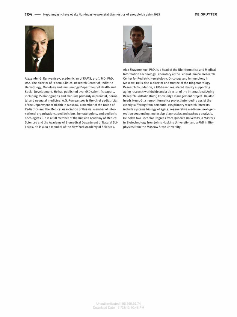
1154 Nepomnyashchaya et al.: Non-invasive prenatal diagnostics of aneuploidy using NGS
Alexander G. Rumyantsev, academician of RAMS, prof., MD, PhD,
DSc. The director of Federal Clinical Research Center of Pediatric
Hematology, Oncology and Immunology Department of Health and
Social Development. He has published over 650 scientific papers,
including 35 monographs and manuals primarily in prenatal, perina-
tal and neonatal medicine. A.G. Rumyantsev is the chief pediatrician
of the Department of Health in Moscow, a member of the Union of
Pediatrics and the Medical Association of Russia, member of inter-
national organizations, pediatricians, hematologists, and pediatric
oncologists. He is a full member of the Russian Academy of Medical
Sciences and the Academy of Biomedical Department of Natural Sci-
ences. He is also a member of the New York Academy of Sciences.
Alex Zhavoronkov, PhD, is a head of the Bioinformatics and Medical
Information Technology Laboratory at the Federal Clinical Research
Center for Pediatric Hematology, Oncology and Immunology in
Moscow. He is also a director and trustee of the Biogerontology
Research Foundation, a UK-based registered charity supporting
aging research worldwide and a director of the International Aging
Research Portfolio (IARP) knowledge management project. He also
heads NeuroG, a neuroinformatics project intended to assist the
elderly suffering from dementia. His primary research interests
include systems biology of aging, regenerative medicine, next-gen-
eration sequencing, molecular diagnostics and pathway analysis.
He holds two Bachelor Degrees from Queen ’ s University, a Masters
in Biotechnology from Johns Hopkins University, and a PhD in Bio-
physics from the Moscow State University.
Unauthenticated | 95.165.92.74Download Date | 11/23/13 10:46 PM