Inactivation of the PRDM1/BLIMP1 gene in diffuse large B cell lymphoma
Upload
tecnojuliacaCategory
view
0download
0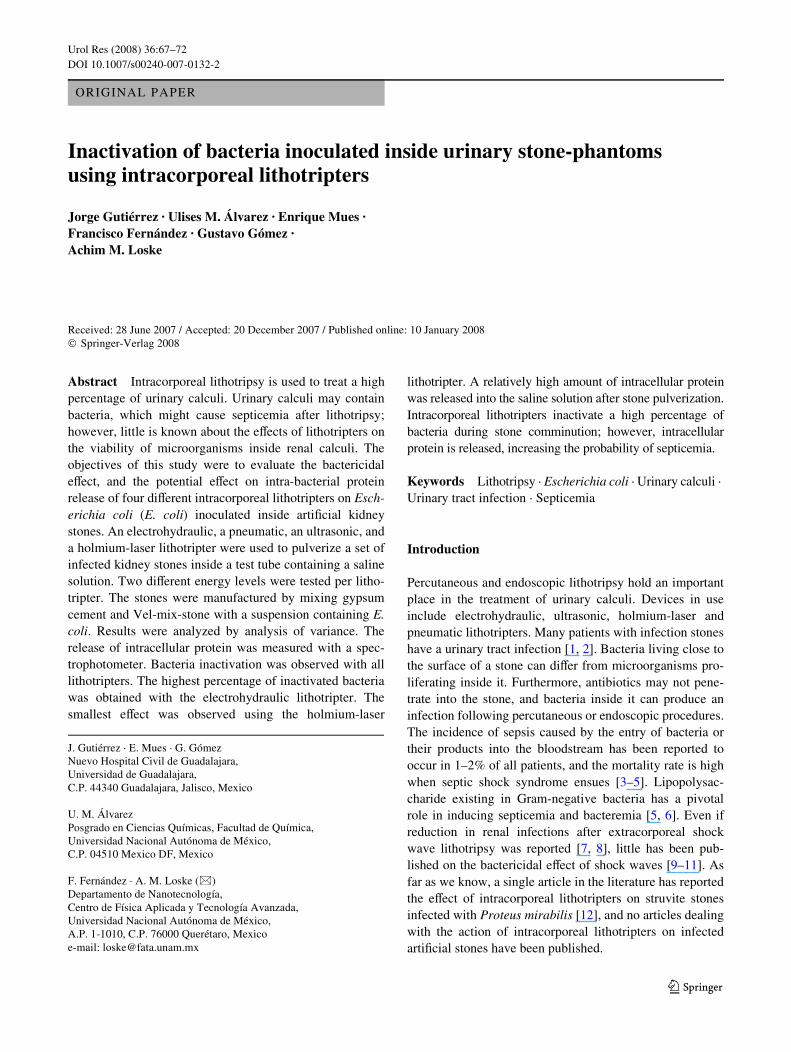
Urol Res (2008) 36:67–72
DOI 10.1007/s00240-007-0132-2ORIGINAL PAPER
Inactivation of bacteria inoculated inside urinary stone-phantoms using intracorporeal lithotripters
Jorge Gutiérrez · Ulises M. Álvarez · Enrique Mues · Francisco Fernández · Gustavo Gómez · Achim M. Loske
Received: 28 June 2007 / Accepted: 20 December 2007 / Published online: 10 January 2008© Springer-Verlag 2008
Abstract Intracorporeal lithotripsy is used to treat a highpercentage of urinary calculi. Urinary calculi may containbacteria, which might cause septicemia after lithotripsy;however, little is known about the eVects of lithotripters onthe viability of microorganisms inside renal calculi. Theobjectives of this study were to evaluate the bactericidaleVect, and the potential eVect on intra-bacterial proteinrelease of four diVerent intracorporeal lithotripters on Esch-erichia coli (E. coli) inoculated inside artiWcial kidneystones. An electrohydraulic, a pneumatic, an ultrasonic, anda holmium-laser lithotripter were used to pulverize a set ofinfected kidney stones inside a test tube containing a salinesolution. Two diVerent energy levels were tested per litho-tripter. The stones were manufactured by mixing gypsumcement and Vel-mix-stone with a suspension containing E.coli. Results were analyzed by analysis of variance. Therelease of intracellular protein was measured with a spec-trophotometer. Bacteria inactivation was observed with alllithotripters. The highest percentage of inactivated bacteriawas obtained with the electrohydraulic lithotripter. Thesmallest eVect was observed using the holmium-laser
lithotripter. A relatively high amount of intracellular proteinwas released into the saline solution after stone pulverization.Intracorporeal lithotripters inactivate a high percentage ofbacteria during stone comminution; however, intracellularprotein is released, increasing the probability of septicemia.
Keywords Lithotripsy · Escherichia coli · Urinary calculi · Urinary tract infection · Septicemia
Introduction
Percutaneous and endoscopic lithotripsy hold an importantplace in the treatment of urinary calculi. Devices in useinclude electrohydraulic, ultrasonic, holmium-laser andpneumatic lithotripters. Many patients with infection stoneshave a urinary tract infection [1, 2]. Bacteria living close tothe surface of a stone can diVer from microorganisms pro-liferating inside it. Furthermore, antibiotics may not pene-trate into the stone, and bacteria inside it can produce aninfection following percutaneous or endoscopic procedures.The incidence of sepsis caused by the entry of bacteria ortheir products into the bloodstream has been reported tooccur in 1–2% of all patients, and the mortality rate is highwhen septic shock syndrome ensues [3–5]. Lipopolysac-charide existing in Gram-negative bacteria has a pivotalrole in inducing septicemia and bacteremia [5, 6]. Even ifreduction in renal infections after extracorporeal shockwave lithotripsy was reported [7, 8], little has been pub-lished on the bactericidal eVect of shock waves [9–11]. Asfar as we know, a single article in the literature has reportedthe eVect of intracorporeal lithotripters on struvite stonesinfected with Proteus mirabilis [12], and no articles dealingwith the action of intracorporeal lithotripters on infectedartiWcial stones have been published.
J. Gutiérrez · E. Mues · G. GómezNuevo Hospital Civil de Guadalajara, Universidad de Guadalajara, C.P. 44340 Guadalajara, Jalisco, Mexico
U. M. ÁlvarezPosgrado en Ciencias Químicas, Facultad de Química, Universidad Nacional Autónoma de México, C.P. 04510 Mexico DF, Mexico
F. Fernández · A. M. Loske (&)Departamento de Nanotecnología, Centro de Física Aplicada y Tecnología Avanzada, Universidad Nacional Autónoma de México, A.P. 1-1010, C.P. 76000 Querétaro, Mexicoe-mail: [email protected]
123
68 Urol Res (2008) 36:67–72
The aim of this research was to compare the bactericidaleVect of four intracorporeal lithotripters on bacteria con-tained inside artiWcial stones. Escherichia coli (E. coli) waschosen for this study because it is a well-known non-spore-forming, Gram-negative bacterium, which is the Wrst causeof urinary tract infection. A nonpathogenic strain wasselected for security reasons, and well-standardized artiW-cial kidney stones were manufactured. These stones pro-vide reliable results and have been used for many years toexamine the eVectiveness of lithotripters.
Methods
Independent viability counts, using the plate count method,were performed on non-treated infected stones (controlstones), shock-wave-treated infected stones and E. colisuspensions.
The lithotripters
Four intracorporeal lithotripters were used in this study: (a)a holmium 20 W Versa Pulse Power Suite laser lithotripter(Lumenis Ltd, Santa Clara, CA, USA) with a 520 �m Wber,(b) a USL-2000 ultrasonic lithotripter (Circon Acmi, Stamford,CT, USA), (c) a Calcutript, model 27080C electrohydrauliclithotripter (Karl Storz Endoscope, Tuttlingen, Germany)with a 1.6 Fr Wber, and (d) an EMS pneumatic lithotripter(Lithoclast, Le Sentier, Switzerland). Two energy settings(H = high; L = low) were chosen for each lithotripter.Direct comparisons between energy settings, frequency or“dose” should not be done, because each lithotripter oper-ates with a diVerent physical principle. The energy settingsand exposure times used in this study were similar to thoseused in clinical practice.
Non-infected artiWcial kidney stones
Stones were manufactured at our laboratory by mixing 45%gypsum cement and 5% Vel-mix-stone (Kerr Division ofSyborn Corp., Romulus, MI, USA) with 50% distilledwater, using a hand mixer. The mixture was cast in cylin-drical molds and dried for 4 h at 30°C. The desired com-pressive strength was obtained in previous experiments (notreported here) by varying the amount of gypsum, Velmixand water. The length and diameter of the stones was10 § 0.2 mm, and 10 § 0.1 mm, respectively. Mean weightof non-infected stones was 0.785 § 0.012 g.
Infected artiWcial kidney stones
A strain of E. coli ATCC 10536 was used to inoculate a setof stones. All infected stones were manufactured as
explained in the previous section; however, in this case asaline solution containing E. coli was used instead of dis-tilled water. Unless otherwise indicated, all microbiologicalmedia used were purchased from Bioxon (Becton Dickinson,Mexico City, Mexico). Microorganisms were stored onnutrient agar (Merck, Darmstadt, Germany) at about 5°Cand transferred each month. The strain was activated byincubation on trypticase soy broth supplemented with 0.6%(w/v) yeast extract (TSBY) at 35°C for 24 h. Cells wereobtained in the stationary phase of growth. Bacteria wereconcentrated by centrifugation (3,000 rpm) for 10 min atroom temperature and resuspended (0.85% w/v) in isotonicsaline solution. The initial viable count was about2.34 £ 107 colony-forming units per milliliter (CFU/ml). A0.01% (v/w) inoculum of the bacterial suspension wasused. Stones were prepared, dried for 4 h close to two burn-ers at about 30°C, and used immediately. A set of eightnon-treated infected stones was crushed with a hand pressto determine the variations in the amount of bacteria inocu-lated inside the stone phantoms.
Stone comminution
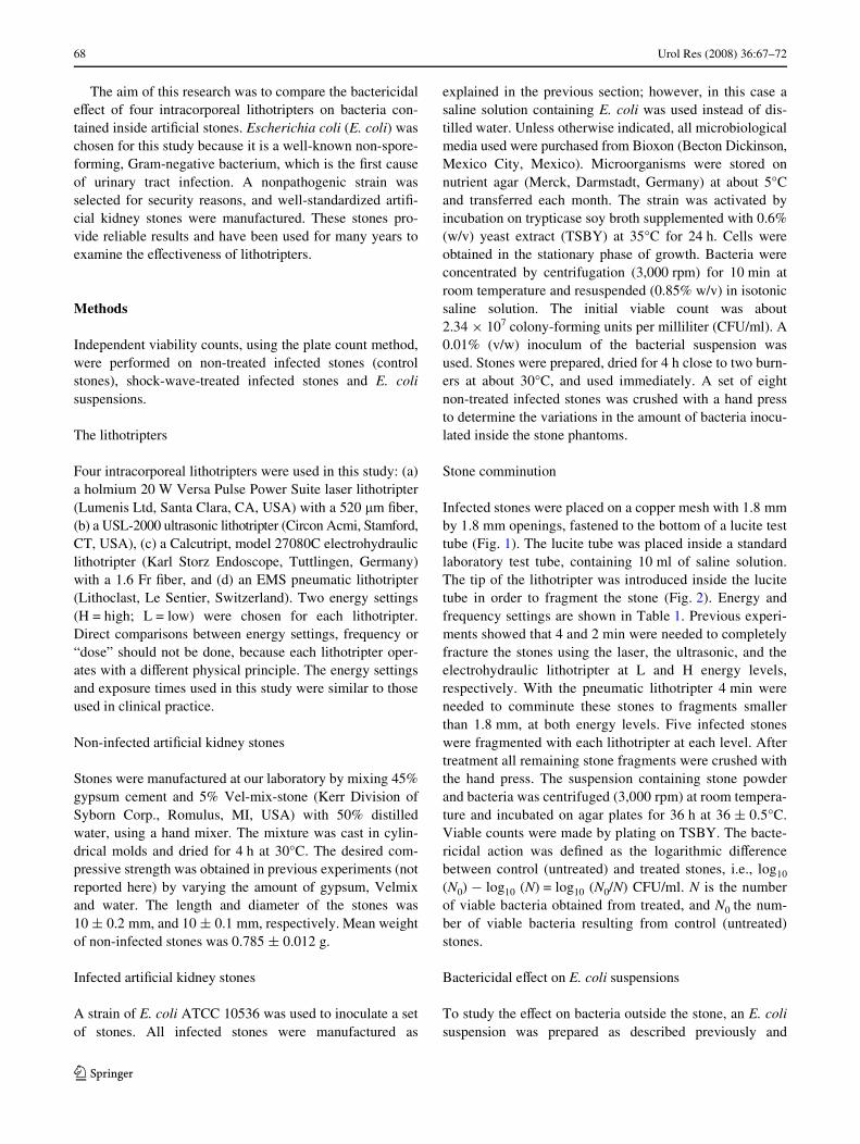
Infected stones were placed on a copper mesh with 1.8 mmby 1.8 mm openings, fastened to the bottom of a lucite testtube (Fig. 1). The lucite tube was placed inside a standardlaboratory test tube, containing 10 ml of saline solution.The tip of the lithotripter was introduced inside the lucitetube in order to fragment the stone (Fig. 2). Energy andfrequency settings are shown in Table 1. Previous experi-ments showed that 4 and 2 min were needed to completelyfracture the stones using the laser, the ultrasonic, and theelectrohydraulic lithotripter at L and H energy levels,respectively. With the pneumatic lithotripter 4 min wereneeded to comminute these stones to fragments smallerthan 1.8 mm, at both energy levels. Five infected stoneswere fragmented with each lithotripter at each level. Aftertreatment all remaining stone fragments were crushed withthe hand press. The suspension containing stone powderand bacteria was centrifuged (3,000 rpm) at room tempera-ture and incubated on agar plates for 36 h at 36 § 0.5°C.Viable counts were made by plating on TSBY. The bacte-ricidal action was deWned as the logarithmic diVerencebetween control (untreated) and treated stones, i.e., log10
(N0) ¡ log10 (N) = log10 (N0/N) CFU/ml. N is the numberof viable bacteria obtained from treated, and N0 the num-ber of viable bacteria resulting from control (untreated)stones.
Bactericidal eVect on E. coli suspensions
To study the eVect on bacteria outside the stone, an E. colisuspension was prepared as described previously and
123
Urol Res (2008) 36:67–72 69
subjected to the action of each lithotripter, using the sameenergy settings and exposure times as for E.coli insideinfected stones (Table 1). The tip of each lithotripter wasimmersed in 20 ml of bacterial suspension inside a steriletest tube. The process was repeated Wve times for eachlithotripter at each energy setting.
Endotoxin release
Stone comminution of infected artiWcial kidney stones wasrepeated as described before, in order to measure therelease of protein as a result of lithotripsy. After treatment,5 ml of the suspension were centrifuged (3,000 rpm) atroom temperature, and placed inside a spectrophotometer tomeasure absorbance at 280 nm [13].
Data analysis
Data were analyzed with the standard aov function of the Rlanguage (R Foundation for Statistical Computing, Vienna,Austria) [14]. Replicate inXuence was included as a block-ing eVect.
Results
Inactivation of bacteria inoculated inside artiWcial kidney stones
Mean initial viable count for infected stones, pulverizedonly with the hand press, was 7.37 log10 (N0/N) § 0.34log10 (N0/N) CFU/ml. The variation in the amount of bacteria
Fig. 1 Photograph of a special test tube, with copper-mesh bottom(disassembled), manufactured to fracture a urinary stone in vitro, usingan intracorporeal lithotripter
Fig. 2 Sketch of the experimental set-up to fragment urinary stones invitro
Table 1 Settings used with the laser, the ultrasonic, the pneumatic,and the electrohydraulic lithotripter
L low, H high
Level Energy(J)
Rate (Hz)
Power(W)
Treatment time (min)
Laser lithotripter
L 0.5 5 2.5 4
H 2.5 5 12.5 2
Level Pulse rate setting (%)
Amplitude setting (%)
Treatment time (min)
Ultrasonic lithotripter
L 55 55 4
H 100 100 2
Level Air pressure (power) (bar)
Treatment time (min)
Pneumatic lithotripter
L 1.2 4
H 3.0 4
Level Energy setting(no units)
Frequency setting (no units)
Treatment time (min)
Electrohydraulic lithotripter
L 1 C 4
H 3 C 2
123
70 Urol Res (2008) 36:67–72
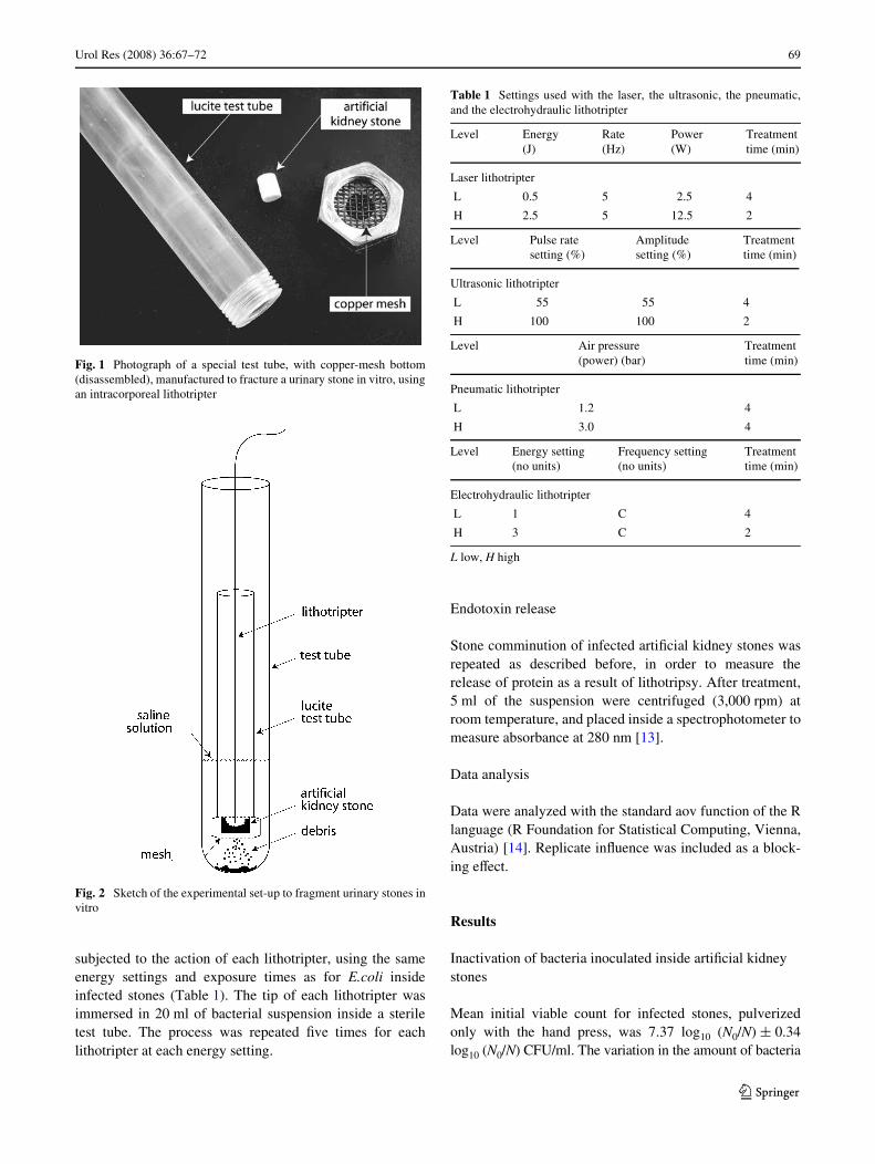
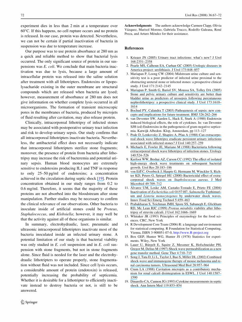
inside these stones was not signiWcant. Bacteria inactivationafter lithotripsy to infected stones is given in Table 2.Figure 3 shows these results on two graphs. Completeinactivation (7.37 log10 (N0/N) CFU/ml) resulted with theelectrohydraulic lithotripter at both energy levels. No sig-niWcant diVerence was obtained between inactivationobtained with the other lithotripters at their low energy set-tings. Increasing the energy resulted in higher bacteria inac-tivation for these three lithotripters (Fig. 3). The lowestinactivation was achieved with the laser lithotripter. The
analysis of variance (ANOVA) revealed a statistical signiW-cant interaction between the energy level and the bacterici-dal eVect of the laser, pneumatic and ultrasonic lithotripter,i.e., bacteria inactivation increased as the energy settingwas increased.
Inactivation of bacteria in suspension
No bacteria inactivation was observed using the pneumaticlithotripter in E. coli suspension without a stone to break.The other three lithotripters completely inactivated E. coliin suspension. This means that no viable bacteria could bedetected after exposure to the laser, the electrohydraulicand the ultrasonic lithotripter. The temperature of the sus-pension containing E. coli increased up to about 60°C whenusing these lithotripters, except for the pneumatic lithotrip-ter. This temperature increase was not observed during lith-otripsy of infected stones.
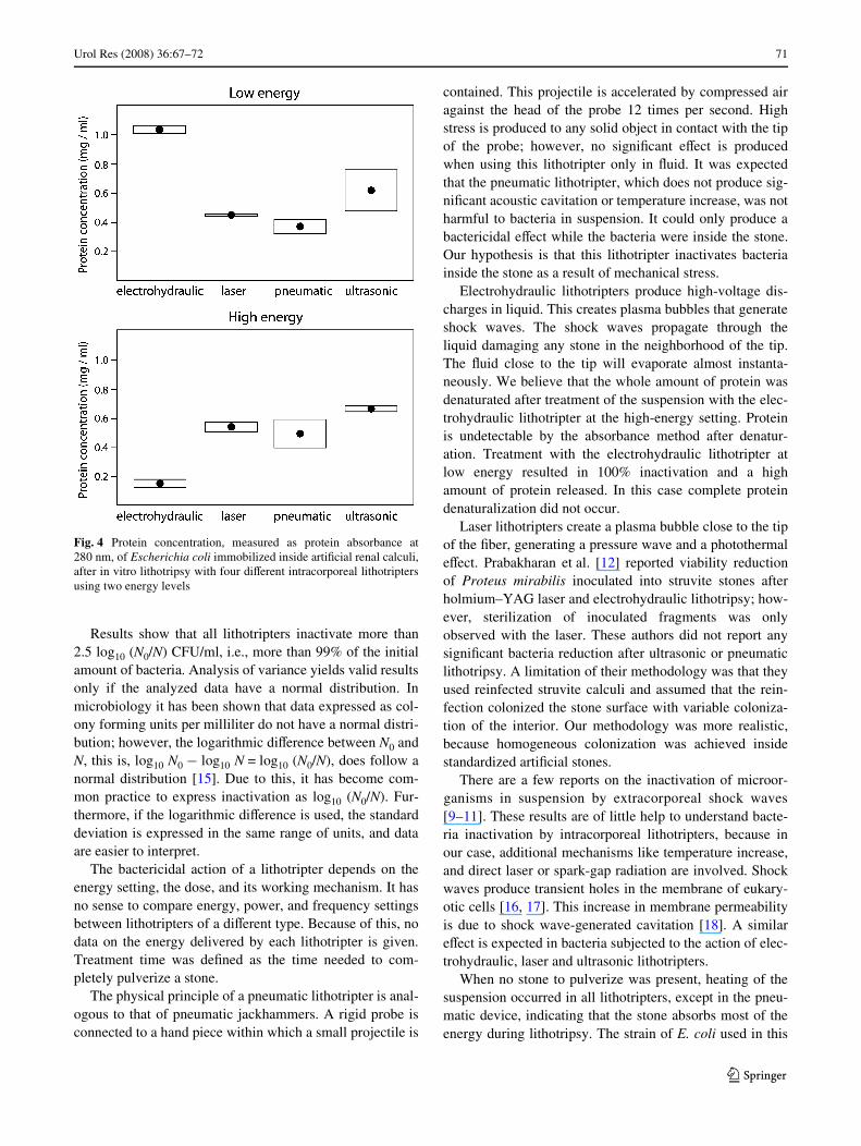
Bacteria endotoxin release
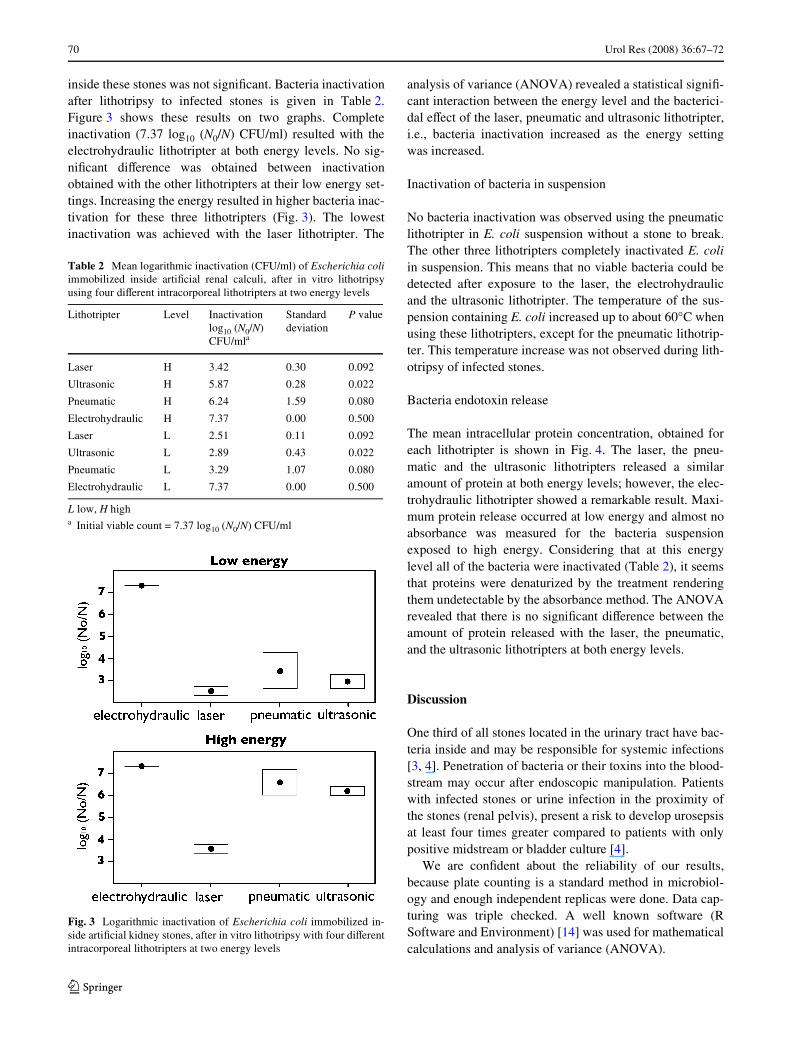
The mean intracellular protein concentration, obtained foreach lithotripter is shown in Fig. 4. The laser, the pneu-matic and the ultrasonic lithotripters released a similaramount of protein at both energy levels; however, the elec-trohydraulic lithotripter showed a remarkable result. Maxi-mum protein release occurred at low energy and almost noabsorbance was measured for the bacteria suspensionexposed to high energy. Considering that at this energylevel all of the bacteria were inactivated (Table 2), it seemsthat proteins were denaturized by the treatment renderingthem undetectable by the absorbance method. The ANOVArevealed that there is no signiWcant diVerence between theamount of protein released with the laser, the pneumatic,and the ultrasonic lithotripters at both energy levels.
Discussion
One third of all stones located in the urinary tract have bac-teria inside and may be responsible for systemic infections[3, 4]. Penetration of bacteria or their toxins into the blood-stream may occur after endoscopic manipulation. Patientswith infected stones or urine infection in the proximity ofthe stones (renal pelvis), present a risk to develop urosepsisat least four times greater compared to patients with onlypositive midstream or bladder culture [4].
We are conWdent about the reliability of our results,because plate counting is a standard method in microbiol-ogy and enough independent replicas were done. Data cap-turing was triple checked. A well known software (RSoftware and Environment) [14] was used for mathematicalcalculations and analysis of variance (ANOVA).
Table 2 Mean logarithmic inactivation (CFU/ml) of Escherichia coliimmobilized inside artiWcial renal calculi, after in vitro lithotripsyusing four diVerent intracorporeal lithotripters at two energy levels
L low, H higha Initial viable count = 7.37 log10 (N0/N) CFU/ml
Lithotripter Level Inactivation log10 (N0/N) CFU/mla
Standard deviation
P value
Laser H 3.42 0.30 0.092
Ultrasonic H 5.87 0.28 0.022
Pneumatic H 6.24 1.59 0.080
Electrohydraulic H 7.37 0.00 0.500
Laser L 2.51 0.11 0.092
Ultrasonic L 2.89 0.43 0.022
Pneumatic L 3.29 1.07 0.080
Electrohydraulic L 7.37 0.00 0.500
Fig. 3 Logarithmic inactivation of Escherichia coli immobilized in-side artiWcial kidney stones, after in vitro lithotripsy with four diVerentintracorporeal lithotripters at two energy levels
123
Urol Res (2008) 36:67–72 71
Results show that all lithotripters inactivate more than2.5 log10 (N0/N) CFU/ml, i.e., more than 99% of the initialamount of bacteria. Analysis of variance yields valid resultsonly if the analyzed data have a normal distribution. Inmicrobiology it has been shown that data expressed as col-ony forming units per milliliter do not have a normal distri-bution; however, the logarithmic diVerence between N0 andN, this is, log10 N0 ¡ log10 N = log10 (N0/N), does follow anormal distribution [15]. Due to this, it has become com-mon practice to express inactivation as log10 (N0/N). Fur-thermore, if the logarithmic diVerence is used, the standarddeviation is expressed in the same range of units, and dataare easier to interpret.
The bactericidal action of a lithotripter depends on theenergy setting, the dose, and its working mechanism. It hasno sense to compare energy, power, and frequency settingsbetween lithotripters of a diVerent type. Because of this, nodata on the energy delivered by each lithotripter is given.Treatment time was deWned as the time needed to com-pletely pulverize a stone.
The physical principle of a pneumatic lithotripter is anal-ogous to that of pneumatic jackhammers. A rigid probe isconnected to a hand piece within which a small projectile is
contained. This projectile is accelerated by compressed airagainst the head of the probe 12 times per second. Highstress is produced to any solid object in contact with the tipof the probe; however, no signiWcant eVect is producedwhen using this lithotripter only in Xuid. It was expectedthat the pneumatic lithotripter, which does not produce sig-niWcant acoustic cavitation or temperature increase, was notharmful to bacteria in suspension. It could only produce abactericidal eVect while the bacteria were inside the stone.Our hypothesis is that this lithotripter inactivates bacteriainside the stone as a result of mechanical stress.
Electrohydraulic lithotripters produce high-voltage dis-charges in liquid. This creates plasma bubbles that generateshock waves. The shock waves propagate through theliquid damaging any stone in the neighborhood of the tip.The Xuid close to the tip will evaporate almost instanta-neously. We believe that the whole amount of protein wasdenaturated after treatment of the suspension with the elec-trohydraulic lithotripter at the high-energy setting. Proteinis undetectable by the absorbance method after denatur-ation. Treatment with the electrohydraulic lithotripter atlow energy resulted in 100% inactivation and a highamount of protein released. In this case complete proteindenaturalization did not occur.
Laser lithotripters create a plasma bubble close to the tipof the Wber, generating a pressure wave and a photothermaleVect. Prabakharan et al. [12] reported viability reductionof Proteus mirabilis inoculated into struvite stones afterholmium–YAG laser and electrohydraulic lithotripsy; how-ever, sterilization of inoculated fragments was onlyobserved with the laser. These authors did not report anysigniWcant bacteria reduction after ultrasonic or pneumaticlithotripsy. A limitation of their methodology was that theyused reinfected struvite calculi and assumed that the rein-fection colonized the stone surface with variable coloniza-tion of the interior. Our methodology was more realistic,because homogeneous colonization was achieved insidestandardized artiWcial stones.
There are a few reports on the inactivation of microor-ganisms in suspension by extracorporeal shock waves[9–11]. These results are of little help to understand bacte-ria inactivation by intracorporeal lithotripters, because inour case, additional mechanisms like temperature increase,and direct laser or spark-gap radiation are involved. Shockwaves produce transient holes in the membrane of eukary-otic cells [16, 17]. This increase in membrane permeabilityis due to shock wave-generated cavitation [18]. A similareVect is expected in bacteria subjected to the action of elec-trohydraulic, laser and ultrasonic lithotripters.
When no stone to pulverize was present, heating of thesuspension occurred in all lithotripters, except in the pneu-matic device, indicating that the stone absorbs most of theenergy during lithotripsy. The strain of E. coli used in this
Fig. 4 Protein concentration, measured as protein absorbance at280 nm, of Escherichia coli immobilized inside artiWcial renal calculi,after in vitro lithotripsy with four diVerent intracorporeal lithotriptersusing two energy levels
123
72 Urol Res (2008) 36:67–72
experiment dies in less than 2 min at a temperature over60°C. If this happens, no cell rupture occurs and no proteinis released. In our case, protein was detected. Nevertheless,we can not be certain if partial inactivation of bacteria insuspension was due to temperature increase.
Our purpose was to use protein absorbance at 280 nm asa quick and reliable method to show that bacterial lysisoccurred. The only signiWcant source of protein in our sus-pensions was E. coli. We conclude that main bacteria inac-tivation was due to lysis, because a large amount ofintracellular protein was released into the saline solutionafter treatment with all lithotripters. Endotoxins or lipopo-lysacharide existing in the outer membrane are structuralcompounds which are released when bacteria are lysed;however, measurement of absorbance at 280 nm does notgive information on whether complete lysis occurred in allmicroorganisms. The formation of transient microscopicpores in the membranes of bacteria, produced by microjetsof Xuid resulting after cavitation, may also release protein.
Clinically, intracorporeal lithotripsy of infected stonesmay be associated with postoperative urinary tract infectionand risk to develop urinary sepsis. Our study conWrms thatall intracorporeal lithotripters inactivate bacteria. Neverthe-less, the antibacterial eVect does not necessarily indicatethat intracorporeal lithotripters sterilize stone fragments;moreover, the presence of protein from bacteria after litho-tripsy may increase the risk of bacteremia and potential uri-nary sepsis. Human blood monocytes are extremelysensitive to endotoxins producing IL1 and TNF in responseto only 25–50 pg/ml of endotoxins; a concentrationachieved in the circulation during septic shock [19]. Proteinconcentration obtained in our study ranges from 0.2 to0.6 mg/ml. Therefore, it seems that the majority of theseproteins are not absorbed into the bloodstream after stonemanipulation. Further studies may be necessary to conWrmthe clinical relevance of our observations. Other bacteria toinoculate inside of artiWcial stones could be Proteus,Staphalococcus, and Klebsiella; however, it may well bethat the activity against all of these organisms is similar.
In summary, electrohydraulic, laser, pneumatic andultrasonic intracorporeal lithotripters inactivate most of thebacteria inoculated inside an infected urinary stone. Apotential limitation of our study is that bacterial viabilitywas only studied in E. coli suspension and in E. coli sus-pension with stone fragments, but not in stone fragmentsalone. Since Xuid is needed for the laser and the electrohy-draulic lithotripters to operate properly, stone fragmenta-tion without Xuid was not included. Since cell lysis occurs,a considerable amount of protein (endotoxin) is released,potentially increasing the probability of septicemia.Whether it is desirable for a lithotripter to eYciently inacti-vate instead to destroy bacteria or not, is still to beanswered.
Acknowledgments The authors acknowledge Carmen Clapp, OliviaVázquez, Marisol Moreno, Gabriela Trucco, Rodolfo Galeana, RenéPreza, and Arturo Méndez for their assistance.
References
1. Krieger JN (2005) Urinary tract infections: what’s new? J Urol168:2351–2358
2. Pearle MS, Calhoun EA, Curhan GC (2005) Urologic diseases inAmerica project: urolithiasis. J Urol 173:848–857
3. Mariapan P, Loong CW (2004) Midstream urine culture and sen-sitivity test is a poor predictor of infected urine proximal to theobstructing ureteral stone or infected stones: a prospective clinicalstudy. J Urol 171:2142–2145
4. Mariapan P, Smith G, Bariol SV, Moussa SA, Tolley DA (2005)Stone and pelvic urinary culture and sensitivity are better thanbladder urine as predictors of urosepsis following percutaneousnephrolithotripsy: a prospective clinical study. J Urol 173:1610–1614
5. Bochud PY, Calandra T (2003) Pathogenesis of sepsis: new con-cepts and implications for future treatment. BMJ 326:262–266
6. van Deventer SW, Aarden L, Hack E, Sturk A (1988) Endotoxininduced biological eVects, the role of cytokines. In: van DeventerSJH (ed) Endotoxins in the pathogenesis of gram-negative septice-mia. Katwijk Albedon- Klop, Amsterdam, pp 113–127
7. Pode D, Lenkovsky Z, Shapiro A, Pfau A (1988) Can extracorpo-real shock wave lithotripsy eradicate persistent urinary infectionsassociated with infected stones? J Urol 140:257–259
8. Michaels E, Fowler JE, Mariano M (1988) Bacteriuria followingextracorporeal shock wave lithotripsy of infection stones. Urology140:524–526
9. Kerfoot WW, Beshai AZ, Carson CC (1992) The eVect of isolatedhigh-energy shock wave treatments on subsequent bacterialgrowth. Urol Res 20:183–186
10. von EiV C, Overbeck J, Haupts G, Hermann M, Winckler S, Rich-ter KD, Peters G, Spiegel HU (2000) Bactericidal eVect of extra-corporeal shock waves on Staphylococcus aureus. J MedMicrobiol 49:709–712
11. Álvarez UM, Loske AM, Castaño-Tostado E, Prieto FE (2004)Inactivation of Escherichia coli O157:H7, Salmonella Typhimuri-um and Listeria monocytogenes by underwater shock waves.Innov Food Sci Emerg Technol 5:459–463
12. Prabakharan S, Teichman JMH, Spore SS, Sabanegh E, GlickhamRD, Mc Lean RJC (1999) Proteus mirabilis viability after litho-tripsy of struvite calculi. J Urol 162:1666–1669
13. Whitaker JR (1993) Principles of enzymology for the food sci-ences. CRC, New York
14. R Development Core Team (2006) R: a language and environmentfor statistical computing. R Foundation for Statistical Computing,Vienna, ISBN 3-900051-07-0, http://www.R-project.org
15. Box GEP, Hunter WG, Hunter JS (1978) Statistics for experi-ments. Wiley, New York
16. Lauer U, Bürgelt E, Squire Z, Messmer K, Hofschneider PH,Gregor M, Delius M (1997) Shock wave permeabilization as a newgene transfer method. Gene Ther 4:710–715
17. Song J, Tata D, Li L, Taylor J, Bao S, Miller DL (2002) Combinedshock-wave and immunogene therapy of mouse melanoma and re-nal carcinoma tumors. Ultrasound Med Biol 28:957–964
18. Crum LA (1988) Cavitation microjets as a contributory mecha-nism for renal calculi disintegration in ESWL. J Urol 140:1587–1590
19. Dinarello CA, Cannon JG (1993) Cytokine measurements in septicshock. Ann Intern Med 119:853–854
123
Copyright © 2022 FDOKUMEN