Thermal stability and inactivation of hepatitis C virus grown in cell culture
12
RESEARCH Open Access Thermal stability and inactivation of hepatitis C virus grown in cell culture Hongshuo Song 1 , Jin Li 1 , Shuang Shi 1 , Ling Yan 1 , Hui Zhuang 1* , Kui Li 2* Abstract Background: Hepatitis C virus (HCV) is a blood-borne flavivirus that infects many millions of people worldwide. Relatively little is known, however, concerning the stability of HCV and reliable procedures for inactivating this virus. Methods: In the current study, the thermostability of cell culture-derived HCV (HCVcc, JFH-1 strain) under different environmental temperatures (37°C, room temperature, and 4°C) and the ability of heat, UVC light irradiation, and aldehyde and detergent treatments to inactivate HCVcc were evaluated. The infectious titers of treated viral samples were determined by focus-forming unit (FFU) assay using an indirect immunofluorescence assay for HCV NS3 in hepatoma Huh7-25-CD81 cells highly permissive for HCVcc infection. MTT cytotoxicity assay was performed to determine the concentrations of aldehydes or detergents at which they were no longer cytotoxic. Results: HCVcc in culture medium was found to survive 37°C and room temperature (RT, 25 ± 2°C) for 2 and 16 days, respectively, while the virus was relatively stable at 4°C without drastic loss of infectivity for at least 6 weeks. HCVcc in culture medium was sensitive to heat and could be inactivated in 8 and 4 min when incubated at 60°C and 65°C, respectively. However, at 56°C, 40 min were required to eliminate HCVcc infectivity. Addition of normal human serum to HCVcc did not significantly alter viral stability at RT or its susceptibility to heat. UVC light irradiation (wavelength = 253.7 nm) with an intensity of 450 μW/cm 2 efficiently inactivated HCVcc within 2 min. Exposures to formaldehyde, glutaraldehyde, ionic or nonionic detergents all destroyed HCVcc infectivity effectively, regardless of whether the treatments were conducted in the presence of cell culture medium or human serum. Conclusions: The results provide quantitative evidence for the potential use of a variety of approaches for inactivating HCV. The ability of HCVcc to survive ambient temperatures warrants precautions in handling and disposing of objects and materials that may have been contaminated with HCV. Background Hepatitis C virus (HCV) is a small enveloped, positive- stranded RNA virus classified within the family Flavivir- idae, genus Hepacivirus. HCV affects an estimated 170 million people worldwide and is a global health pro- blem. Unlike most RNA viruses which usually cause acute diseases, HCV establishes life-long, persistent, intrahepatic infections in a majority of infected indivi- duals, leading frequently to the development of cirrhosis and hepatocellular carcinoma [1,2]. Because the current, interferon-based treatment regimens eradicate HCV in only about 50% of patients, prevention of HCV infection is pivotal for controlling this viral pathogen. HCV is transmitted primarily via percutaneous expo- sure to infectious blood. Prior to the introduction of anti-HCV screening tests in the early 1990s, receiving blood and blood products or organ transplants was a major risk factor for acquiring HCV infection. Currently, injection of illicit drugs represents a major risk, while other routes of infection, including occupational expo- sure (such as needle stick), sex, and mother-to-infant transmission (with the exception of HIV-coinfected mother), seem infrequent [3]. Interestingly, it was shown recently in the chimpanzee model that HCV in infec- tious plasma could survive drying and environmental exposure to room temperature for at least 16 h. This finding has raised the possibility of person-to-person * Correspondence: [email protected]; [email protected] 1 Department of Microbiology, Peking University Health Science Center, Beijing 100191, China 2 Department of Molecular Sciences, University of Tennessee Health Science Center, Memphis, Tennessee 38163, USA Song et al. Virology Journal 2010, 7:40 http://www.virologyj.com/content/7/1/40 © 2010 Song et al; licensee BioMed Central Ltd. This is an Open Access article distributed under the terms of the Creative Commons Attribution License (http://creativecommons.org/licenses/by/2.0), which permits unrestricted use, distribution, and reproduction in any medium, provided the original work is properly cited.
-
Upload
independent -
Category
Documents
-
view
3 -
download
0
Transcript of Thermal stability and inactivation of hepatitis C virus grown in cell culture

RESEARCH Open Access
Thermal stability and inactivation of hepatitis Cvirus grown in cell cultureHongshuo Song1, Jin Li1, Shuang Shi1, Ling Yan1, Hui Zhuang1*, Kui Li2*
Abstract
Background: Hepatitis C virus (HCV) is a blood-borne flavivirus that infects many millions of people worldwide.Relatively little is known, however, concerning the stability of HCV and reliable procedures for inactivating thisvirus.
Methods: In the current study, the thermostability of cell culture-derived HCV (HCVcc, JFH-1 strain) under differentenvironmental temperatures (37°C, room temperature, and 4°C) and the ability of heat, UVC light irradiation, andaldehyde and detergent treatments to inactivate HCVcc were evaluated. The infectious titers of treated viralsamples were determined by focus-forming unit (FFU) assay using an indirect immunofluorescence assay for HCVNS3 in hepatoma Huh7-25-CD81 cells highly permissive for HCVcc infection. MTT cytotoxicity assay was performedto determine the concentrations of aldehydes or detergents at which they were no longer cytotoxic.
Results: HCVcc in culture medium was found to survive 37°C and room temperature (RT, 25 ± 2°C) for 2 and 16days, respectively, while the virus was relatively stable at 4°C without drastic loss of infectivity for at least 6 weeks.HCVcc in culture medium was sensitive to heat and could be inactivated in 8 and 4 min when incubated at 60°Cand 65°C, respectively. However, at 56°C, 40 min were required to eliminate HCVcc infectivity. Addition of normalhuman serum to HCVcc did not significantly alter viral stability at RT or its susceptibility to heat. UVC lightirradiation (wavelength = 253.7 nm) with an intensity of 450 μW/cm2 efficiently inactivated HCVcc within 2 min.Exposures to formaldehyde, glutaraldehyde, ionic or nonionic detergents all destroyed HCVcc infectivity effectively,regardless of whether the treatments were conducted in the presence of cell culture medium or human serum.
Conclusions: The results provide quantitative evidence for the potential use of a variety of approaches forinactivating HCV. The ability of HCVcc to survive ambient temperatures warrants precautions in handling anddisposing of objects and materials that may have been contaminated with HCV.
BackgroundHepatitis C virus (HCV) is a small enveloped, positive-stranded RNA virus classified within the family Flavivir-idae, genus Hepacivirus. HCV affects an estimated 170million people worldwide and is a global health pro-blem. Unlike most RNA viruses which usually causeacute diseases, HCV establishes life-long, persistent,intrahepatic infections in a majority of infected indivi-duals, leading frequently to the development of cirrhosisand hepatocellular carcinoma [1,2]. Because the current,interferon-based treatment regimens eradicate HCV in
only about 50% of patients, prevention of HCV infectionis pivotal for controlling this viral pathogen.HCV is transmitted primarily via percutaneous expo-
sure to infectious blood. Prior to the introduction ofanti-HCV screening tests in the early 1990s, receivingblood and blood products or organ transplants was amajor risk factor for acquiring HCV infection. Currently,injection of illicit drugs represents a major risk, whileother routes of infection, including occupational expo-sure (such as needle stick), sex, and mother-to-infanttransmission (with the exception of HIV-coinfectedmother), seem infrequent [3]. Interestingly, it was shownrecently in the chimpanzee model that HCV in infec-tious plasma could survive drying and environmentalexposure to room temperature for at least 16 h. Thisfinding has raised the possibility of person-to-person
* Correspondence: [email protected]; [email protected] of Microbiology, Peking University Health Science Center,Beijing 100191, China2Department of Molecular Sciences, University of Tennessee Health ScienceCenter, Memphis, Tennessee 38163, USA
Song et al. Virology Journal 2010, 7:40http://www.virologyj.com/content/7/1/40
© 2010 Song et al; licensee BioMed Central Ltd. This is an Open Access article distributed under the terms of the Creative CommonsAttribution License (http://creativecommons.org/licenses/by/2.0), which permits unrestricted use, distribution, and reproduction inany medium, provided the original work is properly cited.
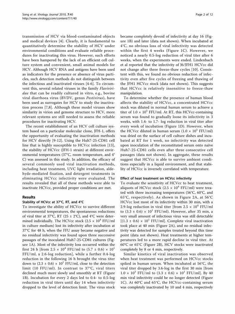
transmission of HCV via blood-contaminated objectsand medical devices [4]. Clearly, it is fundamental toquantitatively determine the stability of HCV underenvironmental conditions and evaluate reliable proce-dures for inactivating this virus. However, such effortshave been hampered by the lack of an efficient cell cul-ture system and convenient, small animal models forHCV. Although HCV RNA and antigens have been usedas indicators for the presence or absence of virus parti-cles, such detection methods do not distinguish betweenthe infectious and inactivated viruses [4-6]. To circum-vent this, several related viruses in the family Flaviviri-dae that can be readily cultured in vitro, e.g., bovineviral diarrhoea virus (BVDV, genus Pestivirus), havebeen used as surrogates for HCV to study the inactiva-tion process [7,8]. Although these model viruses showsimilarity in virion and genome structure to HCV, morerelevant systems are still needed to assess the reliableprocedures for inactivating HCV.The recent establishment of an HCV cell culture sys-
tem based on a particular molecular clone, JFH-1, offersthe opportunity of evaluating the inactivation methodsfor HCV directly [9-12]. Using the Huh7-25-CD81 cellline that is highly susceptible to HCVcc infection [13],the stability of HCVcc (JFH-1 strain) at different envir-onmental temperatures (37°C, room temperature, and 4°C) was assessed in this study. In addition, the efficacy ofseveral commonly used viral inactivation methods,including heat treatment, UVC light irradiation, alde-hyde-mediated fixation, and detergent treatments ineliminating HCVcc infectivity were evaluated. Theresults revealed that all of these methods were able toinactivate HCVcc, provided proper conditions are met.
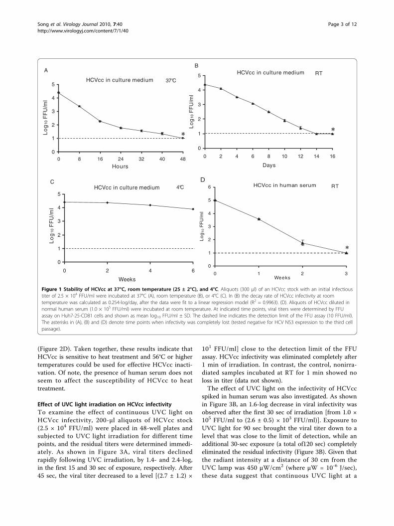
ResultsStability of HCVcc at 37°C, RT, and 4°CTo investigate the ability of HCVcc to survive differentenvironmental temperatures, the spontaneous reductionsof viral titer at 37°C, RT (25 ± 2°C), and 4°C were deter-mined individually. The HCVcc stock (2.5 × 10⁴ FFU/mlin culture medium) lost its infectivity after incubation at37°C for 48 h, when the FFU assay became negative andno residual infectivity was found upon three successivepassages of the inoculated Huh7-25-CD81 cultures (Fig-ure 1A). Most of the infectivity loss occurred within thefirst 24 h [from 2.5 × 10⁴ FFU/ml to (5.7 ± 0.6) × 101
FFU/ml, a 2.6-log reduction], while a further 0.4-logreduction in the following 16 h brought the virus titerdown to (2.3 ± 0.6) × 101 FFU/ml, close to the detectionlimit (10 FFU/ml). In contrast to 37°C, viral titersdeclined much more slowly and smoothly at RT (Figure1B). Incubation for every 2 days led to 0.4- to 0.5-logreduction in viral titers until day 14 when infectivitydropped to the level of detection limit. The virus stock
became completely devoid of infectivity at day 16 (Fig-ure 1B) and later (data not shown). When incubated at4°C, no obvious loss of viral infectivity was detectedwithin the first 4 weeks (Figure 1C). However, wenoticed a nearly 0.5-log reduction of viral titer after 6weeks, when the experiments were ended. Lindenbachet al reported that the infectivity of J6/JFH1 HCVcc didnot change after three freeze-thaw cycles [10]. Consis-tent with this, we found no obvious reduction of infec-tivity even after five cycles of freezing and thawing ofthe JFH1 HCVcc stock (data not shown). This suggeststhat HCVcc is relatively insensitive to freeze-thawmanipulation.To determine whether the presence of human blood
affects the stability of HCVcc, a concentrated HCVccstock was diluted in normal human serum to achieve atiter of 1.0 × 10⁵ FFU/ml. At RT, this HCVcc-containingserum was found to gradually loose its infectivity in 3weeks, with 1.4- to 1.7- log reduction in viral titer afterevery week of incubation (Figure 1D). However, whenthe HCVcc diluted in human serum (1.0 × 10⁵ FFU/ml)was dried on the surface of cell culture dishes and incu-bated at RT for 1 week, no infectivity was detectedupon inoculation of the reconstituted serum onto naïveHuh7-25-CD81 cells even after three consecutive cellpassages (data not shown). In aggregate, these resultssuggest that HCVcc is able to survive ambient condi-tions especially in a liquid environment, and that stabi-lity of HCVcc is inversely correlated with temperature.
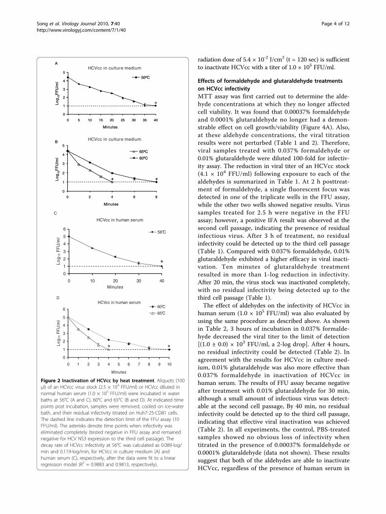
Effect of heat treatment on HCVcc infectivityTo evaluate the sensitivity of HCVcc to heat treatment,aliquots of HCVcc stock (2.5 × 10⁴ FFU/ml) were trea-ted with three increasing temperatures (56°C, 60°C, and65°C, respectively). As shown in Figure 2A, at 56°CHCVcc lost most of its infectivity within 30 min, with a2.9-log reduction in viral titer [from 2.5 × 10⁴ FFU/mlto (3.3 ± 0.6) × 101 FFU/ml). However, after 35 min, avery small amount of infectious virus was still detectable[(1.3 ± 0.6) × 101 FFU/ml]. Complete viral inactivationtook place at 40 min (Figure 2A), and no residual infec-tivity was detected for samples treated beyond this timepoint (data not shown). Heat treatments at higher tem-peratures led to a more rapid decline in viral titer. At60°C or 65°C (Figure 2B), HCV stocks were inactivatedcompletely by 8 or 4 min, respectively.Similar kinetics of viral inactivation was observed
when heat treatment was performed on HCVcc stocksspiked in human serum. When incubated at 56°C, theviral titer dropped by 3.6-log in the first 30 min [from1.0 × 105 FFU/ml to (3.3 ± 0.6) × 101 FFU/ml]. By 40min viral infectivity could be no longer detected (Figure1C). At 60°C and 65°C, the HCVcc-containing serumwas completely inactivated by 10 and 4 min, respectively
Song et al. Virology Journal 2010, 7:40http://www.virologyj.com/content/7/1/40
Page 2 of 12
(Figure 2D). Taken together, these results indicate thatHCVcc is sensitive to heat treatment and 56°C or highertemperatures could be used for effective HCVcc inacti-vation. Of note, the presence of human serum does notseem to affect the susceptibility of HCVcc to heattreatment.
Effect of UVC light irradiation on HCVcc infectivityTo examine the effect of continuous UVC light onHCVcc infectivity, 200-μl aliquots of HCVcc stock(2.5 × 10⁴ FFU/ml) were placed in 48-well plates andsubjected to UVC light irradiation for different timepoints, and the residual titers were determined immedi-ately. As shown in Figure 3A, viral titers declinedrapidly following UVC irradiation, by 1.4- and 2.4-log,in the first 15 and 30 sec of exposure, respectively. After45 sec, the viral titer decreased to a level [(2.7 ± 1.2) ×
101 FFU/ml] close to the detection limit of the FFUassay. HCVcc infectivity was eliminated completely after1 min of irradiation. In contrast, the control, nonirra-diated samples incubated at RT for 1 min showed noloss in titer (data not shown).The effect of UVC light on the infectivity of HCVcc
spiked in human serum was also investigated. As shownin Figure 3B, an 1.6-log decrease in viral infectivity wasobserved after the first 30 sec of irradiation [from 1.0 ×10⁵ FFU/ml to (2.6 ± 0.5) × 103 FFU/ml)]. Exposure toUVC light for 90 sec brought the viral titer down to alevel that was close to the limit of detection, while anadditional 30-sec exposure (a total of120 sec) completelyeliminated the residual infectivity (Figure 3B). Given thatthe radiant intensity at a distance of 30 cm from theUVC lamp was 450 μW/cm2 (where μW = 10-6 J/sec),these data suggest that continuous UVC light at a
37°C
0
1
2
3
4
5
0 8 16 24 32 40 48
Hours
Lo
g10
FF
U/m
l
HCVcc in culture medium
A RT
0
1
2
3
4
5
0 2 4 6 8 10 12 14 1
Days
Lo
g10
FF
U/m
l
6
HCVcc in culture mediumB
4°C
0
1
2
3
4
5
0 2 4 6
Weeks
Lo
g10
FF
U/m
l
HCVcc in culture mediumC
RT
0
1
2
3
4
5
6
0 1 2 3Weeks
Lo
g1
0 F
FU
/ml
HCVcc in human serumD
Figure 1 Stability of HCVcc at 37°C, room temperature (25 ± 2°C), and 4°C. Aliquots (300 μl) of an HCVcc stock with an initial infectioustiter of 2.5 × 10⁴ FFU/ml were incubated at 37°C (A), room temperature (B), or 4°C (C). In (B) the decay rate of HCVcc infectivity at roomtemperature was calculated as 0.254-log/day, after the data were fit to a linear regression model (R2 = 0.9963). (D). Aliquots of HCVcc diluted innormal human serum (1.0 × 10⁵ FFU/ml) were incubated at room temperature. At indicated time points, viral titers were determined by FFUassay on Huh7-25-CD81 cells and shown as mean log10 FFU/ml ± SD. The dashed line indicates the detection limit of the FFU assay (10 FFU/ml).The asterisks in (A), (B) and (D) denote time points when infectivity was completely lost (tested negative for HCV NS3 expression to the third cellpassage).
Song et al. Virology Journal 2010, 7:40http://www.virologyj.com/content/7/1/40
Page 3 of 12
radiation dose of 5.4 × 10⁻² J/cm2 (t = 120 sec) is sufficientto inactivate HCVcc with a titer of 1.0 × 10⁵ FFU/ml.
Effects of formaldehyde and glutaraldehyde treatmentson HCVcc infectivityMTT assay was first carried out to determine the alde-hyde concentrations at which they no longer affectedcell viability. It was found that 0.00037% formaldehydeand 0.0001% glutaraldehyde no longer had a demon-strable effect on cell growth/viability (Figure 4A). Also,at these aldehyde concentrations, the viral titrationresults were not perturbed (Table 1 and 2). Therefore,viral samples treated with 0.037% formaldehyde or0.01% glutaraldehyde were diluted 100-fold for infectiv-ity assay. The reduction in viral titer of an HCVcc stock(4.1 × 10⁴ FFU/ml) following exposure to each of thealdehydes is summarized in Table 1. At 2 h posttreat-ment of formaldehyde, a single fluorescent focus wasdetected in one of the triplicate wells in the FFU assay,while the other two wells showed negative results. Virussamples treated for 2.5 h were negative in the FFUassay; however, a positive IFA result was observed at thesecond cell passage, indicating the presence of residualinfectious virus. After 3 h of treatment, no residualinfectivity could be detected up to the third cell passage(Table 1). Compared with 0.037% formaldehyde, 0.01%glutaraldehyde exhibited a higher efficacy in viral inacti-vation. Ten minutes of glutaraldehyde treatmentresulted in more than 1-log reduction in infectivity.After 20 min, the virus stock was inactivated completely,with no residual infectivity being detected up to thethird cell passage (Table 1).The effect of aldehydes on the infectivity of HCVcc in
human serum (1.0 × 10⁵ FFU/ml) was also evaluated byusing the same procedure as described above. As shownin Table 2, 3 hours of incubation in 0.037% formalde-hyde decreased the viral titer to the limit of detection[(1.0 ± 0.0) × 103 FFU/ml, a 2-log drop]. After 4 hours,no residual infectivity could be detected (Table 2). Inagreement with the results for HCVcc in culture med-ium, 0.01% glutaraldehyde was also more effective than0.037% formaldehyde in inactivation of HCVcc inhuman serum. The results of FFU assay became negativeafter treatment with 0.01% glutaraldehyde for 30 min,although a small amount of infectious virus was detect-able at the second cell passage, By 40 min, no residualinfectivity could be detected up to the third cell passage,indicating that effective viral inactivation was achieved(Table 2). In all experiments, the control, PBS-treatedsamples showed no obvious loss of infectivity whentitrated in the presence of 0.00037% formaldehyde or0.0001% glutaraldehyde (data not shown). These resultssuggest that both of the aldehydes are able to inactivateHCVcc, regardless of the presence of human serum in
0
1
2
3
4
5
0
Minutes
A
5 10 15 20 25 30 35 40
*
0
1
2
3
4
5
0 2 4 6
Minutes
Lo
g10
FF
U/m
l
8
65ºC
60ºC
B
56ºC
Lo
g10
FF
U/m
l
0
1
2
3
4
5
0
Minutes
A
5 10 15 20 25 30 35 40
*
0
1
2
3
4
5
0 2 4 6
Minutes
Lo
g10
FF
U/m
l
8
65ºC
60ºC
B
56ºC
Lo
g10
FF
U/m
l
HCVcc in culture medium
HCVcc in culture medium
HCVcc in human serum
0
1
2
3
4
5
6
0 10 20 30Minutes
Lo
g1
0 F
FU
/ml
40
56°C
C
HCVcc in human serum
0
1
2
3
4
5
6
0 1 2 3 4 5 6 7 8 9 1
Minutes
Lo
g1
0 F
FU
/ml
0
60°C
65°C
D
Figure 2 Inactivation of HCVcc by heat treatment. Aliquots (100μl) of an HCVcc virus stock (2.5 × 10⁴ FFU/ml) or HCVcc diluted innormal human serum (1.0 × 10⁵ FFU/ml) were incubated in waterbaths at 56°C (A and C), 60°C and 65°C (B and D). At indicated timepoints post incubation, samples were removed, cooled on ice-waterbath, and their residual infectivity titrated on Huh7-25-CD81 cells.The dashed line indicates the detection limit of the FFU assay (10FFU/ml). The asterisks denote time points when infectivity waseliminated completely (tested negative in FFU assay and remainednegative for HCV NS3 expression to the third cell passage). Thedecay rate of HCVcc infectivity at 56°C was calculated as 0.089-log/min and 0.119-log/min, for HCVcc in culture medium (A) andhuman serum (C), respectively, after the data were fit to a linearregression model (R2 = 0.9883 and 0.9813, respectively).
Song et al. Virology Journal 2010, 7:40http://www.virologyj.com/content/7/1/40
Page 4 of 12

HCVcc stocks, and that glutaraldehyde is more effectivethan is formaldehyde.
Effects of detergent treatments on HCVcc infectivityTo determine the detergent concentrations at whichthey were no longer cytotoxic to cells, Huh7-25-CD81cells were treated with individual, serially diluted deter-gents and subjected to MTT assay to assess cell viability.Based on the MTT assay results (Figure 4B and 4C),HCVcc stocks (4.1 × 10⁴ FFU/ml) or those spiked inhuman serum (1.0 × 10⁵ FFU/ml) were treated with0.1% SDS, 0.2% Triton X-100, or 0.2% NP-40, and thentested for infectivity at either 100-fold (SDS-treatedsamples) or 400-fold (Triton X-100- or NP-40-treatedsamples) dilution. As summarized in Table 3, all of the
detergent-treated samples were negative in the FFUassay and demonstrated no residual infectivity uponthree consecutive passages of the inoculated cells(Table 3). In contrast, control samples not treated withdetergents showed no obvious loss in virus titers.The effect of detergents on disrupting intracellular
HCVcc virions was also evaluated. JFH-1-infectedHuh7-25-CD81 cultures (with 100% cells positive forNS3 as determined by immunofluorescence assay) werelysed in each of the detergent solutions (0.1% SDS, 0.2%Triton X-100, or 0.2% NP-40 in PBS, respectively), andthe clarified supernatants were tested for infectivity onnaïve Huh7-25-CD81 cells at 100-fold (SDS-lysed sam-ples) or 400-fold (Triton X-100- or NP-40-lysed sam-ples) dilution, respectively. No infectivity was detected
HCVcc in culture medium
0
1
2
3
4
5
0 15 30 45 6
Seconds
Lo
g10
FF
U/m
l
0
A
HCVcc in human serum
0
1
2
3
4
5
6
0 30 60 90 120
Seconds
Lo
g10
FF
U/m
l
B
Figure 3 Inactivation of the HCVcc by UVC light irradiation. Aliquots (200 μl) of an HCVcc stock (A) or normal human serum containingHCVcc (B) were placed 30 cm beneath the longitudinal midpoint of a UVC lamp. Samples were removed at indicated time points, and viraltiters were determined on Huh7-25-CD81 cells immediately. The dashed line indicates the detection limit of the FFU assay (10 FFU/ml). Theasterisk denotes the time point when infectivity was completely lost (tested negative in FFU assay and remained negative for HCV NS3expression to the third cell passage). The decay rate of HCVcc infectivity after UVC light irradiation was calculated as 0.067-log/sec and 0.041-log/sec, for HCVcc in culture medium (A) and human serum (B), respectively, after the data were fit to a linear regression model (R2 = 0.9738 and0.9891, respectively).
Song et al. Virology Journal 2010, 7:40http://www.virologyj.com/content/7/1/40
Page 5 of 12

0
20
40
60
80
100
120
4000 20000 100000 500000
Dilution factor
Cel
l via
bili
ty (%
)
Formaldehyde (37%)
Glutaraldehyde (50%)
A
0
20
40
60
80
100
120
5 10 20 40 80
Dilution factor
Cel
l via
bili
ty (%
)
SDS (0.1%)
B
0
20
40
60
80
100
120
50 100 200 400 800
Dilution factor
Cel
l via
bili
ty (%
)
Triton X-100 (0.2%)
NP-40 (0.2%)
C
Figure 4 MTT assay of cytotoxicity associated with various concentrations of aldehydes or detergents. Solutions of formaldehyde (37%or glutaraldehyde (50%) (A), SDS (0.1%) (B), and Triton X-100 (0.2%) or NP-40 (0.2%) (C) were diluted serially in cell culture medium by dilutionfactors indicated in the X-axes, then added to Huh7-25-CD81 cells seeded in 96-well plates. The treated cells were refed with 100 μl of freshmedium after 6 h of incubation. MTT assay was carried out after an additional 72 h as described in Methods. Data are presented as thepercentage of cell viability relative to the untreated controls (mean ± SD, n = 3).
Song et al. Virology Journal 2010, 7:40http://www.virologyj.com/content/7/1/40
Page 6 of 12

from any of these cell lysates in three consecutive pas-sages of inoculated cells (Table 3), while the control celllysate prepared by freezing and thawing in the absenceof any detergents retained high infectivity (Table 3).Taken collectively, these results suggest that each of thedetergents, at the tested concentration, is highly effectivein eliminating the infectivity of both extracellular andintracellular HCVcc particles.
DiscussionIn this study, a detailed analysis was conducted on thestability of HCVcc at various environmental tempera-tures. Also evaluated was the efficacy of several conven-tional viral inactivation procedures in eliminatingHCVcc infectivity.It has been shown previously that genotype 1a HCV in
infectious plasma could survive drying and environmen-tal exposure to RT for at least 16 h [4]. The results ofthe current study have demonstrated that JFH-1 virus(genotype 2a) grown in cell culture can survive 37°Cand RT for 2 and 16 days, respectively (Figure 1A and1B). Of note, the stability of JFH1 HCVcc spiked inhuman serum did not differ much from those in cellculture medium when incubated at RT (Fig. 1D). Whenstored at 4°C, JFH-1 virus was found to be relativelystable, without drastic loss of titer during the 6-weekobservation period (Figure 1C). The latter result is inagreement with a previous report dealing with the J6/JFH1 chimeric virus [10]. The ability of HCVcc to sur-vive various environmental temperatures warrants pre-cautions in handling and disposing objects and materials
that may have been contaminated with HCV, to mini-mize the risk of HCV transmission.Heat treatment is a widely used viral inactivation
method that is effective against both enveloped andnonenveloped viruses [14]. The mechanisms of heat-mediated inactivation include denaturation of viral pro-teins, as well as disassembly of virus particles into non-infectious viral subunits and single proteins [15]. Virusesother than HCV in the family Flaviviridae have beenshown to be sensitive to heat treatment. Yellow fevervirus is routinely inactivated at 56°C for 30 min. At 60°C, BVDV and yellow fever virus have been reported tobe inactivated effectively in 30 and 5 min, respectively[8,16]. In the current study, similar kinetics of viral inac-tivation following heat treatment was observed for boththe HCVcc in culture medium and those in humanserum. While 10 min at 60°C or 4 min at 65°C was suf-ficient to eliminate the infectivity of HCVcc, incubationfor 40 min was required to achieve complete viral inacti-vation at 56°C (Figure 2). Therefore, pretreatment ofHCV positive sera for 30 min at 56°C may not be abso-lutely reliable in eliminating their infectivity. However,because the efficiency of heat treatment could beaffected by a variety of factors, such as the initial viraltiter, protein concentration in virus suspension, as wellas the existence of viral aggregates [8,17] the exact tem-perature and time required for reliable HCV inactivationshould be evaluated under each specific condition.UV light irradiation is another commonly used physi-
cal method for viral inactivation. UVC with a wave-length range of 200-280 nm prevents viral replication byinducing formation of pyrimidine dimers in the viral
Table 1 Effects of formaldehyde and glutaraldehyde oninfectivity of HCVcc in culture medium
Treatment Infectious titer(FFU/ml)
IFA result
2nd cell passage 3rd cell passage
Formaldehyde (0.037%)
0 h (3.9 ± 0.3) × 104a NTc NT
0.5 h (1.1 ± 0.1) × 104 NT NT
1.0 h (4.0 ± 1.0) × 103 NT NT
1.5 h (1.7 ± 0.6) × 103 NT NT
2.0 h ≤ 1.0 × 10³b positive NT
2.5 h < 1.0 × 10³ positive NT
3.0 h < 1.0 × 10³ negative negative
Glutaraldehyde (0.01%)
0 min (4.0 ± 0.3) × 104a NT NT
10 min (3.3 ± 0.6) × 103 NT NT
20 min < 1.0 × 10³ negative negativea Untreated viral samples were retitrated in culture medium containing0.00037% formaldehyde or 0.0001% glutaraldehyde as controls.bOne focus was detected in one of the triplicate wells in FFU assay.c NT, not tested. Samples with positive results in FFU assay or at the secondcell passage were not tested for NS3 staining for the third cell passage.
Table 2 Effects of formaldehyde and glutaraldehyde oninfectivity of HCVcc in human serum
Treatment Infectious titer(FFU/ml)
IFA result
2nd cell passage 3rd cell passage
Formaldehyde (0.037%)
0 h (8.5 ± 0.7) × 104a NTb NT
1.0 h (1.9 ± 0.4) × 104 NT NT
2.0 h (3.5 ± 0.7) × 103 NT NT
3.0 h (1.0 ± 0.0) × 103 NT NT
4.0 h < 1.0 × 10³ negative negative
Glutaraldehyde (0.01%)
0 min (9.5 ± 0.7) × 104a NT NT
10 min (1.8 ± 0.2) × 104 NT NT
20 min (2.5 ± 0.7) × 103 NT NT
30 min < 1.0 × 10³ Positive NT
40 min < 1.0 × 10³ negative negativea Untreated viral samples were retitrated in culture medium containing0.00037% formaldehyde or 0.0001% glutaraldehyde as controls.b NT, not tested. Samples with positive results in FFU assay or at the secondcell passage were not tested for NS3 staining for the third cell passage.
Song et al. Virology Journal 2010, 7:40http://www.virologyj.com/content/7/1/40
Page 7 of 12

genome [18]. A recent study reported that BVDV, whensuspended in PBS, could be inactivated completely by1.6 J/cm2 UVC light, while viral suspension containing5% FBS required a higher radiation dose [7]. The cur-rent study demonstrated that HCVcc in culture medium(2.5 × 10⁴ FFU/ml, volume depth of 0.2 cm) could beinactivated completely by UVC irradiation at a dose of2.7 × 10-2 J/cm2 within 1 min (Figure 3A), while thosespiked in human serum (1.0 × 10⁵ FFU/ml) required anirradiation dose of 5.4 × 10⁻² J/cm2 for full inactivation(Figure 3B). Therefore, UVC light irradiation representsa highly effective means for inactivating HCVcc, the effi-ciency of which is not affected by human serum compo-nents that may interact with HCV virons in vivo.However, the irradiation dose required for each specificoccasion may depend on the sample volume and itsinitial viral titer.As a chemical cross-linking reagent, formaldehyde
inactivates viruses primarily by denaturing viral proteins,as well as the nucleic acids [19,20]. Because the immu-nogenicity of the viral particles can be retained duringinactivation, formalin (37% formaldehyde) treatment isthe most used technique for preparing inactivated virusvaccines. For tissue fixation for histology or immunohis-tochemistry, 10% formalin (or 4% paraformaldehyde) isroutinely used. Glutaraldehyde is another effective pro-tein cross-linking reagent, mostly used for fixation oftissues for electron microscopy. Although the detailedmechanisms are not entirely clear yet, successful inacti-vation of many viruses with glutaraldehyde, includinghepatitis B virus, human immunodeficiency virus (HIV),and SARS coronavirus, has been reported [18,21,22].We demonstrated here that at RT, 3 h of exposure toformaldehyde (0.037%) or 20 min of exposure to glutar-aldehyde (0.01%), respectively, could reduce HCVccinfectivity from 4.1 × 10⁴ FFU/ml to undetectable levels
(Table 1). At these concentrations both aldehydes werealso effective in inactivating HCVcc in the presence ofhuman serum (Table 2). The slightly longer timesrequired (4 h for formaldehyde treated samples and 40min for glutaraldehyde treated samples, respectively)were most likely attributed to the 2.5-fold higher initialtiter of the HCVcc stock tested (1.0 × 10⁵ FFU/ml).However, a limitation of the current study is that,because of the cytotoxic effect of the aldehydes, theinfectivity of viral samples could be analyzed at only the100-fold dilution, which may somehow have reducedthe sensitivity of the assay. It should be noted, however,the routinely used concentrations of aldehydes for fixa-tion purposes (4% for formaldehyde and 2.5% for glutar-aldehyde) are far in excess of the ones examined in thecurrent study and, therefore, should be highly efficientin achieving HCV inactivation.Detergents are highly efficient at disrupting the lipid-
enveloped viruses, and solvent/detergent (S/D) treat-ment is a standard method for inactivating viruses pre-sent in human blood products [22]. The effects of bothionic (SDS) and nonionic (Triton X-100 and NP-40)detergents on HCVcc infectivity have been investigatedhere. All three detergents at the tested concentrationsreduced HCVcc infectivity rapidly to undetectable levels(Table 3). Importantly, both intracellular HCVcc andthose released into culture fluid could be inactivated byeach of these detergents, regardless of the presence ofhuman serum, indicating that components of culturemedium, human serum or intracellular proteins did notinterfere with the disruptive processes exerted by thesedetergents. Under current experimental conditions,effective HCVcc inactivation took place immediatelyafter vortex-mixing, rendering it impossible to delineatethe kinetics of viral infectivity reduction during thedetergent treatment process. This finding is reminiscent
Table 3 Effects of detergent treatments on HCVcc infectivity
Treatment Infectious titers before treatment (FFU/ml)a FFU assay after treatment IFA result at the 3rd cell passage
0.1% SDS
HCVcc in culture medium (4.1 ± 0.2) × 104 negative negative
HCVcc in human serum (9.5 ± 0.7) × 104 negative negative
cell lysates (5.5 ± 0.1) × 105 negative negative
0.2% Triton X-100
HCVcc in culture medium (2.0 ± 0.2) × 104 negative negative
HCVcc in human serum (6.0 ± 0.0) × 104 negative negative
cell lysate (3.1 ± 0.2) × 105 negative negative
0.2% NP-40
HCVcc in culture medium (1.6 ± 0.2) × 104 negative negative
HCVcc in human serum (5.5 ± 0.2) × 104 negative negative
cell lysate (2.9 ± 0.2) × 105 negative negativea Untreated viral samples or intracellular viral infectivity in untreated, HCVcc-infected cell lysates prepared by freeze-and-thaw were retitrated in the presence of0.001% SDS, 0.0005% Triton X-100 or 0.0005% NP-40, respectively, to keep in line with the final concentration of the detergents in the treated samples duringtitration.
Song et al. Virology Journal 2010, 7:40http://www.virologyj.com/content/7/1/40
Page 8 of 12

of that reported for HIV in a previous study, whichdemonstrated that HIV-1 spiked in solution containing1% Triton X-100 was inactivated completely within 1min [23]. As in the case of the aldehyde-inactivationexperiments, the cytotoxic effect of detergents limitedthe sensitivity of the current assays. Interestingly,although 0.0005% Triton X-100 and 0.0005% NP-40 hadno detectable effect on cell viability (Figure 4C), theystill lowered HCVcc infectivity by 1.7- to 2.5-fold whenthe latter was compared with those determined in thepresence of 0.001% SDS or without any detergent (Table3). Most likely, the residual Triton X-100 or NP-40 stillhad some disruptive effect on virion integrity, which isimportant for viral infectivity. Alternatively, these deter-gents may have caused some cell surface alterations atthis extremely low concentration that affect the processof HCVcc entry. However, to maintain the sensitivity ofthe assay, the detergent-treated samples were notdiluted further for the infectivity test. Collectively, therobustness and immediate action of detergents indestroying HCVcc infectivity support the use of S/Dtreatment procedures in eliminating potential HCV con-taminations in blood products.
ConclusionsIn summary, results presented in the current studyrevealed the stability of HCVcc (genotype 2a, JFH-1strain) under different temperatures and provided quanti-tative evidence that heat, UVC light irradiation, aldehyde(formaldehyde and glutaraldehyde), and detergent treat-ments all can be used as effective means for inactivatingHCVcc. However, because the stability and resistance ofHCV to different inactivation methods may vary fromgenotype to genotype, and even strain to strain, the opti-mal method and procedure used for HCV inactivationshould be verified under each particular circumstance.We also note that the results of the current study weredeveloped using an in vitro cell culture system based onhepatoma Huh7 cells, which may differ from normalhuman hepatocytes in supporting HCV infection. Thus,to what extent the procedures described herein can beapplied to an in vivo setting awaits further evaluation.
MethodsCell culture and virus stocksThe Huh7-25-CD81 cell line (a generous gift from Dr.Takaji Wakita), a Huh7 cell clone that stably expresseshuman CD81 [13], was used throughout the experi-ments. This cell line was chosen because we found itwas approximately 1.5- to 2-fold more sensitive for titra-tion of HCVcc infectivity than was the Huh7.5.1 cell line(data not shown). Cells were maintained in DMEM sup-plemented with 10% fetal bovine serum (Invitrogen), 10mM HEPES (Invitrogen), and 400 μg/ml G418 (Merck,
Germany) at 37°C in 5% CO2. To generate JFH-1 virusstocks, cell culture supernatant collected from full-length JFH-1 RNA-transfected Huh7 cells (kindly pro-vided by Dr. Takaji Wakita) was used to infect Huh7-25-CD81 cells grown in T25 flasks at a multiplicity ofinfection (MOI) of 0.01. The infected cells were pas-saged at 3-day intervals with 1:3 to 1:4 split ratios intoprogressively larger culture vessels. At 12 days postinfec-tion, the culture supernatants were harvested, clarifiedby centrifugation (5 min at 4000 rpm), and stored in ali-quots at -70°C as the HCVcc stock. The infectious titerof the virus stock was determined by focus-forming unit(FFU) assay as described immediately below (the infec-tious titers of the un-concentrated virus stocks usedwere either 2.5 × 10⁴ FFU/ml or 4.1 × 10⁴ FFU/ml inthe current study, as specified in each experiment).To determine the stability of HCVcc and its suscept-
ibility to individual inactivation methods in the presenceof human serum, a condition which better mimics circu-lating HCV virions in vivo, HCVcc stock was first con-centrated using the Amicon Ultra-15 device (100,000NMWL membrane; Millipore) as described previously[11]. The concentrated virus stock (1.1 × 10⁶ FFU/ml)was then diluted 11-fold in normal human serum thathad been heat-inactivated to achieve an infectious titerof 1.0 × 10⁵ FFU/ml. This human serum containingHCVcc was stored at -70°C in aliquots until use.
HCV infectivity assayThe infectious titers of virus stocks and treated viralsamples were determined by FFU assay, as describedpreviously [24], using an indirect immunofluorescenceassay (IFA) for HCV NS3. In brief, 100 μl of 10-foldserially diluted samples (the dilution factors generallyranged from 1:1 to1:1000) were inoculated onto naïveHuh7-25-CD81 cells seeded in 96-well plates 1 daybefore infection (7000 cells/well). After 6 h of incuba-tion at 37°C, cells were refed with 100 μl fresh medium.Following an additional 72 h, cells were fixed in 4% par-aformaldehyde for 30 min at room temperature (RT),blocked for 60 min in a blocking buffer (3% BSA, 0.3%Triton X-100, 10% FBS in PBS), followed by incubationwith a polyclonal antibody against HCV NS3 (kindlyprovided by Dr. Takaji Wakita) at 1:500 dilution. After 2h incubation at RT, cells were washed extensively withPBS and then incubated with an FITC-conjugated goatanti-rabbit IgG (Beijing Zhongshanjinqiao, China) at1:100 dilution for 1 h. Following PBS washes, the num-bers of fluorescent foci (a focus is defined as a cluster ofinfected cells immunostained positive for NS3 antigen)per well at appropriate dilutions (generally containing 5to 100 foci per well) were counted. The infectious titers,expressed as FFU/ml, were calculated from the averagefoci number of triplicate or duplicate (for samples
Song et al. Virology Journal 2010, 7:40http://www.virologyj.com/content/7/1/40
Page 9 of 12
derived from human serum spiked with HCVcc) wells.The detection limit of the FFU assay was 10 FFU/ml.For samples with infectious titers below the detectionlimit of the assay, the potential residual infectivity wasexamined as follows. Naïve Huh7-25-CD81 cells seededin 96-well plates were inoculated with samples to betested (100 μl/well). Inoculated cells were passaged at 3-day intervals from one well into three wells at each pas-sage (with a 1:3 split ratio) to allow growth of residualinfectious virus. IFA for NS3 were performed on eachcell passage. If the IFA results remained negative forthree successive cell passages (up to 9 days postinocula-tion), the tested sample was considered to be inactivatedcompletely.
Viral stability assaysAn HCVcc stock with a titer of 2.5 × 10⁴ FFU/ml wasdispensed into 300-μl aliquots in tightly capped, 1.5-mlmicrocentrifuge tubes and then incubated at 37°C, RT(25 ± 2°C), and 4°C, respectively, and protected fromlight. Aliquots incubated at 37°C were removed every 8h, while those incubated at RT or 4°C were removedevery 2 days or every 2 weeks, respectively, and storedat -70°C until virus titration on Huh7-25-CD81 cells.For viral stability assays for HCVcc spiked in normalhuman serum (1.0 × 10⁵ FFU/ml), aliquots were incu-bated at RT and removed every 7 days for titration.All the time points selected in the experimentswere designed based on the results of several pilotexperiments.
Heat treatmentAn HCVcc stock in culture medium (2.5 × 10⁴ FFU/ml)or concentrated HCVcc stock diluted in human serum(1.0 × 10⁵ FFU/ml) was dispensed into 100-μl aliquotsin tightly capped, 1.5-ml microcentrifuge tubes and thenincubated in water baths with temperatures of 56°C, 60°C, and 65°C, respectively. At designated time points, ali-quots were removed, transferred immediately into ice-water bath to stop the effect of heat, and then subjectedto FFU assay for virus titration.
UVC light irradiationTwo hundred-microliter aliquots of an HCVcc stock(2.5 × 10⁴ FFU/ml) or HCVcc diluted in normal humanserum (1.0 × 10⁵ FFU/ml) were placed in 48-well platesto give a volume depth of about 0.2 cm and thenexposed to continuous UVC light 30 cm beneath thelongitudinal midpoint of a UVC lamp (model: ZSZ20D,wavelength = 253.7 nm, Beijing Haidian Konghou HighTemperature Composite Material Factory, China). Atthe distance of 30 cm, the radiant intensity of the UVClamp was 450 μW/cm2 (where μW = 10-6 J/sec), as spe-cified by the manufacturer. After varying lengths of
exposure, samples (200 μl) were removed, and their resi-dual infectivity was titrated on Huh7-25-CD81 cellsimmediately. Control samples were set up in paralleland incubated for the same time period but protectedfrom UVC light.
MTT cytotoxicity assayMTT [3-(4, 5-dimethylthiazol-2-yl)-2, 5-diphenyl tetra-zolium bromide] cytotoxicity assay was carried out todetermine the concentrations of aldehydes or detergentsat which they were no longer cytotoxic to Huh7-25-CD81 cell. Solutions of aldehydes (37% formaldehydeand 50% glutaraldehyde) or detergents [sodium dodecylsulfate (SDS, 0.1%), Triton X-100 (0.2%) or nonidet P-40(NP-40, 0.2%)] were diluted serially in cell culture med-ium, respectively (the range of concentrations for eachreagent was designed based on the results of pilotexperiments). The diluted reagents were then added toHuh7-25-CD81 cells seeded in 96-well plates (7000cells/well) 1 day before. After 6 h of incubation at 37°C,the treated cells were refed with 100 μl of fresh culturemedium to keep the exposure time to the individualreagents the same as that in HCV infectivity assay. Fol-lowing an additional 72 h, 20 μl of MTT solution (5mg/ml, Sigma-Aldrich) was added to each well. After a4-h incubation at 37°C, the MTT solution was removedand replaced with 200 μl of dimethyl sulfoxide (DMSO,Sigma-Aldrich) per well. After the formazan crystalswere dissolved by agitation (10 min at RT), the absor-bance of solution in each well was measured at 490 nmusing an enzyme-linked immunosorbent assay platereader (Bio-Rad). The percentage of cell viability wascalculated as the ratio of absorbance in treated cellscompared with that in untreated controls. All experi-ments were performed in triplicate and repeated twice.
Formaldehyde and glutaraldehyde treatmentsFormaldehyde (37%) or glutaraldehyde (50%) solutions(Beijing Chemical Reagents Company, China) werediluted in PBS at 1:10 (formaldehyde) or 1:50 (glutaral-dehyde), respectively, then added to 500-μl viral samples[HCVcc stock in cell culture medium (4.1 × 10⁴ FFU/ml) or HCVcc-containing human serum (1.0 × 10⁵ FFU/ml)] to achieve a final concentration of 0.037% (formal-dehyde) or 0.01% (glutaraldehyde), respectively. Afterdifferent time periods at RT, treated samples werediluted 100-fold in culture medium immediately to stopthe inactivation reaction, as well as to eliminate thecytotoxic effect of aldehydes in subsequent FFU assays(according to the results of the MTT assay, the presenceof 0.00037% formaldehyde or 0.0001% glutaraldehyde hadno appreciable effect on the viability of Huh7-25-CD81cells). Immediately after the dilution, HCV infectivity insamples was titrated by FFU assay in Huh7-25-CD81
Song et al. Virology Journal 2010, 7:40http://www.virologyj.com/content/7/1/40
Page 10 of 12
cells. Samples showing negative results in FFU assay weresubjected to the residual infectivity test, as described in“HCV infectivity assay” As control, PBS was substitutedfor the aldehydes to treat the virus stocks, which werethen diluted 100-fold to infect cells in the presence ofeither 0.00037% formaldehyde or 0.0001% glutaraldehyde,respectively.
Detergent treatmentsSolutions of 0.5% SDS (w/v), 1% Triton X-100 (v/v), or1% NP-40 (v/v) (all prepared in PBS) were added to500-μl aliquots of viral samples [HCVcc stock in cellculture medium (4.1 × 10⁴ FFU/ml) or normal humanserum containing HCVcc (1.0 × 10⁵ FFU/ml)] to achievea final concentration of either 0.1% (SDS) or 0.2% (Tri-ton X-100 and NP-40). After a gentle mixing (within 1min), treated samples were diluted 100-fold (SDS-trea-ted samples) or 400-fold (Triton X-100- or NP-40-trea-ted samples) immediately in culture medium to negatethe cytotoxic effect of detergents (based on the MTTassay results, the presence of 0.001% SDS, 0.0005% Tri-ton X-100, or 0.0005% NP-40 had no demonstrableeffect on the viability of Huh7-25-CD81 cells), then sub-jected to FFU assay. Samples with negative FFU assayresults were examined for residual infectivity. As con-trol, PBS was used in place of the detergents to treat thevirus stocks, which were subsequently diluted either100- or 400-fold to infect the Huh7-25-CD81 cells inthe presence of 0.001% SDS, 0.0005% Triton X-100, or0.0005% NP-40, respectively.To assess the ability of detergents to disrupt intracel-
lular HCV, JFH-1 infected Huh7-25-CD81 cell mono-layers grown in 24-well plates (approximately 100% ofcells stained positive for NS3 at the time of cell lysis asexamined by IFA) were detached by trypsin/EDTA andwashed extensively with PBS, and cell pellets were resus-pended in 50 μl PBS containing 0.1% SDS, or 0.2% Tri-ton X-100, or 0.2% NP-40, respectively (each detergentwas disruptive to cells at these concentrations as visua-lized by microscopy). After centrifugation, the superna-tants of cell lysates were diluted 100-fold (SDS-lysedsamples) or 400-fold (Triton X-100- or NP-40-lysedsamples) in culture medium for infectivity assays. IFA ofHCV NS3 was performed on inoculated cells for threeconsecutive cell passages. As control, infected cellswashed extensively with PBS were pelleted and resus-pended in 50 μl PBS and lysed by three cycles of freez-ing and thawing (-70°C to 37°C), and the infectivity ofsupernatants was titrated on Huh7-25-CD81 cells.
AcknowledgementsWe are grateful to Dr. Takaji Wakita (National Institute of Infectious Diseases,Tokyo, Japan) for providing JFH-1 virus, Huh7-25-CD81 cell line, and HCVNS3 polyclonal antibody. We also thank Dr. Teng Ma (Peking University
Health Science Center, Beijing, China) for technical assistance and Dr. DavidArmbruster (University of Tennessee Health Science Center) for carefullyreading the manuscript. This work was supported by the Major Science andTechnology Special Project of China Eleventh Five-year Plan (2008ZX10002-013), and the financial grants from the National Key Science and from theNational Institute of Allergy and Infectious Diseases (R01-AI069285).
Author details1Department of Microbiology, Peking University Health Science Center,Beijing 100191, China. 2Department of Molecular Sciences, University ofTennessee Health Science Center, Memphis, Tennessee 38163, USA.
Authors’ contributionsHZ and KL conceived the study and designed the experiments. HSS, JL, SSand LY carried out the experimental work. HSS, HZ and KL wrote the paper.All Authors have read and approved the final manuscript.
Competing interestsThe authors declare that they have no competing interests.
Received: 3 November 2009Accepted: 18 February 2010 Published: 18 February 2010
References1. Lemon SM, Walker C, Alter MJ, Yi M, Knipe D, Howley P, Griffin DE,
Martin MA, Lamb RA, Roizman B, Straus SE: Hepatitis C viruses. FieldsVirology Philadelphia: Lippincott Williams & WilkinsKnipe DM, Howley PM ,Fifth 2007, 1253-1304.
2. Webster DP, Klenerman P, Collier J, Jeffery KJ: Development of noveltreatments for hepatitis C. Lancet Infect Dis 2009, 9:108-117.
3. Alter MJ: The detection, transmission, and outcome of hepatitis C virusinfection. Infect Agents Dis 1993, 2:155-166.
4. Kamili S, Krawczynski K, McCaustland K, Li X, Alter MJ: Infectivity ofhepatitis C virus in plasma after drying and storing at roomtemperature. Infect Control Hosp Epidemiol 2007, 28:519-524.
5. Hilfenhaus J, Groner A, Nowak T, Weimer T: Analysis of human plasmaproducts: polymerase chain reaction does not discriminate between liveand inactivated viruses. Transfusion 1997, 37:935-940.
6. Sattar SA, Tetro J, Springthorpe VS, Giulivi A: Preventing the spread ofhepatitis B and C viruses: where are germicides relevant?. Am J InfectControl 2001, 29:187-197.
7. Azar Daryany MK, Hosseini SM, Raie M, Fakharie J, Zareh A: Study oncontinuous (254 nm) and pulsed UV (266 and 355 nm) lights on BVDvirus inactivation and its effects on biological properties of fetal bovineserum. J Photochem Photobiol B 2009, 94:120-124.
8. Remington KM, Trejo SR, Buczynski G, Li H, Osheroff WP, Brown JP,Renfrow H, Reynolds R, Pifat DY: Inactivation of West Nile virus, vacciniavirus and viral surrogates for relevant and emergent viral pathogens inplasma-derived products. Vox Sang 2004, 87:10-18.
9. Cai Z, Zhang C, Chang KS, Jiang J, Ahn BC, Wakita T, Liang TJ, Luo G:Robust production of infectious hepatitis C virus (HCV) from stably HCVcDNA-transfected human hepatoma cells. J Virol 2005, 79:13963-13973.
10. Lindenbach BD, Evans MJ, Syder AJ, Wolk B, Tellinghuisen TL, Liu CC,Maruyama T, Hynes RO, Burton DR, McKeating JA, Rice CM: Completereplication of hepatitis C virus in cell culture. Science 2005, 309:623-626.
11. Wakita T, Pietschmann T, Kato T, Date T, Miyamoto M, Zhao Z, Murthy K,Habermann A, Krausslich HG, Mizokami M, Bartenschlager R, Liang TJ:Production of infectious hepatitis C virus in tissue culture from a clonedviral genome. Nat Med 2005, 11:791-796.
12. Zhong J, Gastaminza P, Cheng G, Kapadia S, Kato T, Burton DR, Wieland SF,Uprichard SL, Wakita T, Chisari FV: Robust hepatitis C virus infection invitro. Proc Natl Acad Sci USA 2005, 102:9294-9299.
13. Akazawa D, Date T, Morikawa K, Murayama A, Miyamoto M, Kaga M,Barth H, Baumert TF, Dubuisson J, Wakita T: CD81 expression is importantfor the permissiveness of Huh7 cell clones for heterogeneous hepatitisC virus infection. J Virol 2007, 81:5036-5045.
14. Burnouf T, Radosevich M: Reducing the risk of infection from plasmaproducts: specific preventative strategies. Blood Rev 2000, 14:94-110.
15. Schlegel A, Immelmann A, Kempf C: Virus inactivation of plasma-derivedproteins by pasteurization in the presence of guanidine hydrochloride.Transfusion 2001, 41:382-389.
Song et al. Virology Journal 2010, 7:40http://www.virologyj.com/content/7/1/40
Page 11 of 12

16. Charm SE, Landau S, Williams B, Horowitz B, Prince AM, Pascual D: High-temperature short-time heat inactivation of HIV and other viruses inhuman blood plasma. Vox Sang 1992, 62:12-20.
17. Darnell ME, Subbarao K, Feinstone SM, Taylor DR: Inactivation of thecoronavirus that induces severe acute respiratory syndrome, SARS-CoV. JVirol Methods 2004, 121:85-91.
18. Perdiz D, Grof P, Mezzina M, Nikaido O, Moustacchi E, Sage E: Distributionand repair of bipyrimidine photoproducts in solar UV-irradiatedmammalian cells. Possible role of Dewar photoproducts in solarmutagenesis. J Biol Chem 2000, 275:26732-26742.
19. Fraenkel-Conrat H: Reaction of nucleic acid with formaldehyde. BiochimBiophys Acta 1954, 15:307-309.
20. Jiang W, Schwendeman SP: Formaldehyde-mediated aggregation ofprotein antigens: comparison of untreated and formalinized modelantigens. Biotechnol Bioeng 2000, 70:507-517.
21. Payan C, Cottin J, Lemarie C, Ramont C: Inactivation of hepatitis B virus inplasma by hospital in-use chemical disinfectants assessed by a modifiedHepG2 cell culture. J Hosp Infect 2001, 47:282-287.
22. Roberts PL, Dunkerley C: Effect of manufacturing process parameters onvirus inactivation by solvent-detergent treatment in a high-purity factorIX concentrate. Vox Sang 2003, 84:170-175.
23. Kim IS, Choi YW, Woo HS, Chang CE, Lee S: Solvent/detergent inactivationand chromatographic removal of human immunodeficiency virus duringthe manufacturing of a high purity antihemophilic factor VIIIconcentrate. J Microbiol 2000, 38:187-191.
24. Yi M, Ma Y, Yates J, Lemon SM: Compensatory mutations in E1, p7, NS2,and NS3 enhance yields of cell culture-infectious intergenotypicchimeric hepatitis C virus. J Virol 2007, 81:629-638.
doi:10.1186/1743-422X-7-40Cite this article as: Song et al.: Thermal stability and inactivation ofhepatitis C virus grown in cell culture. Virology Journal 2010 7:40.
Submit your next manuscript to BioMed Centraland take full advantage of:
• Convenient online submission
• Thorough peer review
• No space constraints or color figure charges
• Immediate publication on acceptance
• Inclusion in PubMed, CAS, Scopus and Google Scholar
• Research which is freely available for redistribution
Submit your manuscript at www.biomedcentral.com/submit
Song et al. Virology Journal 2010, 7:40http://www.virologyj.com/content/7/1/40
Page 12 of 12