Sress tolerant virulent strains of Cronobacter sakazakii from food
Upload
independentCategory
view
3download
0
FULL PAPER Pathology
Late Marek’s Disease in Adult Chickens Inoculated with Virulent Marek’s Disease Virus
Mitsutaka IKEZAWA1,2), Masanobu GORYO1,2)*, Jun SASAKI2), Mohie HARIDY1) and Kosuke OKADA1,2)
1)Department of Pathogenic Veterinary Science, The United Graduated School of Veterinary Sciences, Gifu University, 1–1 Yanagido, Gifu 501–1193 and 2)Department of Veterinary Pathology, Faculty of Agriculture, Iwate University, 3–18–8 Ueda, Iwate 020–8550, Japan
(Received 13 May 2010/Accepted 7 July 2010/Published online in J-STAGE 21 July 2010)
ABSTRACT. Recently, excessive losses from Marek’s disease (MD) have been noted in adult laying flocks over the age of 40 weeks. Wedefined these late outbreaks in adult chickens as “late MD”, and experimentally reproduced the disease in adult SPF P2 line (50-week-old) or commercial line (74-week-old) chickens inoculated with a virulent strain of Marek’s disease virus (MDV). Commercial linechickens were given MDV vaccines (HVT and CVI 988) at hatch. The occurrence of MD was evaluated periodically by the evidenceof neurologic signs such as paralysis, torticollis, ataxia, and/or nervous tics, as well as histopathological examination. In P2 line chickens,neurologic signs and MD lymphoma were observed from day 21 onward, and they tended to increase in a time-dependent manner. Mean-while, in commercial line chickens, only one chicken exhibited MD lymphoma on day 70 post inoculation, but its pathogenesis was ques-tionable. No regression of MD lymphoma was noted in either case. The lesions in the visceral organs, thymus, peripheral nerves, andfeather pulps of P2 line chickens were characterized by proliferation of variably sized lymphoid cells. In the feather follicle epithelium,numerous inclusion bodies were noted on day 21 post-inoculation, which tended to decrease afterwards. The morphological findingsobtained resembled late MD in field cases. In conclusion, our results demonstrate that adult SPF P2 line chickens are susceptible tovirulent MDV, and would be useful for investigation of late MD.KEY WORDS: adult chickens, experimental reproduction, histopathology, late Marek’s disease.
J. Vet. Med. Sci. 72(12): 1539–1545, 2010
Marek’s disease virus (MDV), currently reclassified asGallid herpesvirus 2, is an α-herpesvirus belonging to thegenus Mardivirus and the family Herpesviridae. MDVinfection in chickens causes Marek’s disease (MD), a lym-phoproliferative and neurologic disease, resulting in the for-mation of malignant T cell lymphomas [3, 17].
Generally, MD has been thought to be a disease for youngchickens (ca. 4 to 20 weeks old), but, recently excessivelosses from MD have been noted in adult laying flocks overthe age of 40 weeks [21]. It was reported that MD fre-quently occurred after molting in preparation for a secondlay cycle [11, 12, 21]. Moreover, MD outbreaks in adultchickens were reported to become very common recently[16]. In this study, we defined these late MD outbreaks inadult chickens as “late MD” in contrast with conventional“early MD” in young chickens.
In commercial poultry industries, MD is usually pre-vented by vaccination, which inhibits the onset of tumor for-mation, but not that of virus infection on its own. Themechanism of tumor prevention by vaccination still remainsunclear [2, 5, 15]. If the protective immunity against MDwas not established after vaccination, chickens woulddevelop MD at younger ages, and consequently die. Forinstance, co-infection with immunodepressive pathogenssuch as chicken anemia virus enhanced MD outbreaks inboth young and adult flocks [7, 16, 19]. Thus, age or infec-tious stresses were considered to be predisposing factors in
late MD outbreaks [18, 21].So-called “age resistance” which is regarded as an
expression of genetic resistance is best demonstrated in SPFchickens [4, 21]. In most experimental studies, the combi-nation of SPF chickens aged 12 to 22 weeks and virulentMDV strains has been well utilized [1, 4, 20, 23], althoughthey were considerably young compared to the ages elicitinglate MD outbreaks in field flocks. There have been fewreports addressing the susceptibility of adult chickens (>30-week-old) to MD. It is essential to establish an appropriateexperimental model of late MD for elucidating its pathogen-esis. In the present work, adult SPF P2 line (50-week-old)or commercial line (74-week-old) chickens were inoculatedwith virulent MDV for an attempt to reproduce late MDexperimentally and assessed them neurologically and histo-pathologically.
MATERIALS AND METHODS
Chickens: Twenty-five adult SPF P2 line (geneticallyMD-susceptible) chickens aged 50 weeks were in-bred atthe Laboratory of Veterinary Pathology, Iwate University(Morioka, Japan). They were derived from White Leghornline P2 flocks. The chickens were free from antibody toadenovirus, avian infectious bronchitis virus, chicken ane-mia virus, infectious bursal disease virus, MDV, Newcastledisease virus, reovirus, avian leukosis virus, and other avianpathogens. They were reared in a small isolated room fromhatch and supplied food (Chubushiryo Co., Ltd., Nagoya,Japan) and tap water ad libitum. Twenty adult commercialline chickens aged 74 weeks were provided kindly from a
* CoRRESPONDENCE TO: GORYO, M., Department of VeterinaryPathology, Faculty of Agriculture, Iwate University, 3–18–8Ueda, Iwate 020–8550, Japan.
e-mail: [email protected]

M. IKEZAWA ET AL.1540
commercial farm. They are a genetically MD-resistant line.The commercial line chickens were inoculated with MDvaccines (HVT and CVI 988, Ghen Co., Ltd., Gifu, Japan)at hatch, and reared in the field environment.
All chicken were reared and manipulated in accordancewith the Guidelines for Animal Experimentation issued bythe Japanese Association for Laboratory Animal Science[10] and also approved by the Animal Experimental EthicsCommittee of Iwate University (Morioka, Japan).
Virus: Virulent KS strain [9, 18] of oncogenic serotype 1was used as the MDV in this study. This strain is equivalentto the JM strain. The inoculum contained virulent MDV attiter of 3,000 PFU/0.1 ml.
Polymerase chain reaction (PCR): Before the experi-ment, expression of the MDV genome in each chicken wasinvestigated by PCR. Briefly, total DNA was extractedfrom the tip of growing wing feathers as described by Davi-son and Borenshtain [6] with some modifications. Thefeather tips were chopped and incubated overnight in lysisbuffer at 55C, purified by phenol/chloroform, precipitatedwith ethanol, and dissolved in T10E1 buffer. The primers(M1 and M2) were designed to detect the 132 base pair tan-dem repeat (BamH1-H,D genomic fragments).
M1: (5’-TACTTCCTATATAGATTGAGACCGT-3’) M2: (5’-GAGATCCTCGTAAGGTGTAATATA-3’) The primers were synthesized by SIGMA-Aldrich Japan
Co., Ltd. (Tokyo, Japan). The PCR was carried out using aThermal Cycler PERSONAL (TaKaRa Bio Co., Ltd., Otsu,Japan), and performed by the procedure [6] as describedpreviously.
Experimental designs: Twenty P2 line chickens (chickenNos. S1 to S20) were inoculated intramuscularly at the fem-oral region with 0.1 ml of inoculum. The inoculation daywas regarded as day 0. Each groups of five chickens wereeuthanized by exsanguination under ether anesthesia ondays 21 (Nos. S1 to S5) and 42 (Nos. S7 to S11). During theexperiment periods, two chickens were dead and necropsiedupon discovery on days 24 (No. S6) and 54 (No. S12), andfour moribund chickens were found and euthanized underether anesthesia on days 56 (Nos. S13 and S14) and 66(No.S15 and S16). At termination on day 70, 4 survivingchickens (Nos. S17 to S20) were euthanized by the afore-mentioned procedure. Five control chickens (Nos. S21 toS25) received a 0.9% physiological saline solution on day 0,and were euthanized on day 70.
Fifteen commercial line chickens (Nos. C1 to C15) wereinoculated intramuscularly at the femoral region with 0.1 mlof inoculum. Five chickens (Nos. C1 to C5) were eutha-nized on day 21, and ten remaining chickens (Nos. C6 toC15) were euthanized on day 70. Five control chickens(Nos. C16 to C20) received a 0.9% physiological saline onday 0, and were euthanized on day 70.
Neurologic signs: Neurologic signs including paralysis,torticollis, ataxia, and/or nervous tics were scored accordingto the observation battery of Gimeno et al. [8] with minormodifications.
Histopathological examination: After gross examination
of dead, moribund, and schedule-killed chickens, the vis-ceral organs (liver, spleen, kidney, heart, and lung), periph-eral nerves, skin, lymphoid organs (thymus and cecaltonsil), and other affected organs were removed, fixed in10% formalin and embedded with a routine process in par-affin-wax. All sections were cut approximately 4 m thickand stained with hematoxylin and eosin (HE). The sectionswere histopathologically examined using an optical micro-scope. The severity of MD lymphomas in the affectedorgans and the histological lesions in the peripheral nerveand skin was scored as (–) to (3+) and (–) to (4+), respec-tively, under the criteria as shown in the foot notes of corre-sponding Tables 1 and 2.
RESULTS
Identification of MDV infection by PCR: Expression ofthe MDV genome in P2 line and commercial line chickenswas confirmed to be negative and positive, respectively,prior to the experiments.
Neurologic signs and MD lymphoma: In P2 line chickens(Table 1), one (No. S4) had renal lymphoma without neuro-logic signs on day 21. On day 42, three (Nos. S8, S9, andS11) displayed neurologic signs, while one (No. S8) of themexhibited renal lymphoma. On days 54 to 66, five (Nos. S12to S16) died or were euthanized due to poor health, precededby apparent neurologic signs. Three (Nos. S12, S13, andS15) of them had lymphoma in the visceral organs exceptthe spleen. At termination on day 70, four (Nos. S17 to S20)survived and two of them (Nos. S18 and S19) showed neu-rologic signs with or without lymphoma in the multipleorgans. The incidence of neurologic signs and visceral MDlymphoma was 50% (10/20) and 30% (6/20), respectively.During days 42 and 70, the incidence of neurologic signsand visceral MD lymphoma was 71.4% (10/14) and 35.7%(5/14), respectively.
In commercial line chickens, neither neurologic signs norgross lymphomas were observed throughout the experimen-tal period (data not shown). However, one (No. C4) had apulmonary MD lymphoma, diagnosed by the histopatholog-ical examination as mentioned below.
No clinicopathological abnormalities were seen in thecorresponding control chickens.
Histopathological findings: Detailed histopathologicalfindings for individual P2 line chickens are summarized inTables 1 and 2. In MD lymphomas of the visceral organsand thymus, moderate to massive proliferation of variablysized lymphoid cells was observed sporadically throughoutthe experimental period.
In the peripheral nerves, small foci of lymphoid cells(Fig. 1A) with or without inflammatory lesions were notedon day 21. On day 42, minimal to diffuse proliferation ofvariably sized lymphoid cells and scant to severe neuritiswith infiltration of lymphocytes, plasma cells, heterophilsand macrophages were observed (Fig. 1B). Additionally,various degrees of edema, necrosis, and axon degenerationwere commonly noted. On days 54 to 66, diffuse lymphoid

1541EXPERIMENTAL LATE MD IN ADULT CHICKENS
cell proliferation was seen (Fig. 1C), but inflammatorylesions were not identified because of marked neoplasticlesions. On day 70, slight lymphocyte infiltration wasobserved with or without focal neoplastic lesions and edem-atous nerve fibers (Fig. 1D).
In the skin, numerous inclusion bodies were seen in thefeather follicle epithelium (Fig. 2) with slight neoplastic andinflammatory lesions on day 21. On day 42, moderate todiffuse lymphoid cell proliferation in the feather pulps wasobserved in association with a moderate number of inclu-sion bodies in the feather follicle epithelium and slight tomoderate inflammatory lesions around the feather follicles.On days 54 and 56, severe lymphoid cell proliferation withvarious degrees of inflammatory cell infiltration wasobserved with moderate inclusion bodies. On days 66 and70, slight lymphocyte infiltration or severe lymphoid cellproliferation was noted with only a few inclusion bodies.
In commercial line chickens, a small number of inclusionbodies was observed in the feather follicle epithelium onday 21, but not thereafter. On days 21 to 70, only a few lym-phocytes were observed around blood vessels in the periph-eral nerves (Fig. 3). On day 70, only one chicken (No. C4)showed lymphoma containing variably sized lymphoid cellsin the lung and skin (Fig. 4). No neoplastic or inflammatory
lesions were seen in other chickens throughout the experi-mental period.
No histopathological lesions suggesting regression ofMD lymphoma were noted in either case.
DISCUSSION
Susceptibility to MDV is considered to be much lower inolder chickens than in younger chicks. Although the “age-resistance” remained unclear, the immune system matura-tion likely participated in this event. Witter et al. [22]reported that vaccinated-susceptible SPF line chickens (18-week-old) did not cause MD regardless of infection with ahighly virulent strain (vv and vv+) of MDV. In the fieldflocks, although chicks receiving vaccination were exposedto environmentally resident MDV during their lives, onlyindividuals bearing a proper immune response exerted aprotective action against MD. Conversely, adult chickenswith poor immune responses including SPF were consid-ered to possess at least in part a high susceptibility to lateMD. In our work using SPF P2 line chickens (50-week-old), MD developed from day 21 onward, and a total of sixchickens were found dead or moribund during the experi-mental period.
Table 1. Neurologic signs (NS) and MD lymphoma of the visceral organs and thymus in adult SPFP2 line chickens inoculated with virulent MDV
DPIa) Chicken NSOrgans
No. Liver Spleen Kidney Heart Lung Thymus
S1 –b) –c) – – – – –S2 – – – – – – –
21d) S3 – – – – – – –S4 – – – 2+ – – –S5 – – – – – – –
24e) S6 – – – – – – –
S7 – – – – – – –S8 + – – 3+ – – –
42d) S9 + – – – – – –S10 – – – – – – –S11 + – – – – – –
54e) S12 + – – 3+ 3+ – 3+
56e) S13 + – – – – 2+ 3+S14 + – – – – – –
66e) S15 + 3+ – – – – –S16 + – – – – – –
S17 – – – – – – –S18 + 3+ – 3+ 3+ 2+ –
70d)S19 + – – – – – –S20 – – – – – – –
a) DPI=days post inoculation.b) –; no abnormal signs, +; incidence of paralysis, torticollis, ataxia and/or nervous tics.c) –; no abnormal lesion, +; small accumulation of lymphoid cells, 2+; moderate proliferation or massformation of lymphoid cells, 3+; massive proliferation or mass formation of lymphoid cells disrupting thenormal architectures.d) schedulled euthanasia.e) death or euthanized becouse of the moribund conditions.
M. IKEZAWA ET AL.1542
In previous studies of virulent strain MDV infection [1, 4,20], chickens aged 12 to 22 weeks served as an adultchicken model, because avian immune system becameestablished by 4 to 6 weeks of age. Under these experimen-tal conditions, MD was induced between 16 and 32 weeks.In contrast, late MD in the field flocks was recognized tooccur mainly at 60–80 weeks [21]. Thus, there have beenconsiderable gaps of age in chickens between the results ofthese experimental and field studies. Since our aim is toexamine the reproduction of late MD in field flocks, weselected adult SPF chickens aged 50 weeks old and com-mercial line field (74 weeks old) for our current investiga-tion.
The incidence of neurologic signs and visceral MD lym-phoma in the P2 line between days 21 and 70 was 55 and30%, respectively, which were relatively higher than that inprevious reports [1, 4, 20]. Differences in the incidence mayhave resulted from host-virus interactions. In a study usingSPF susceptible line chickens (18-week-old) inoculatedwith virulent (JM/102W, GA/22, 617A, or RB1B) MDV byWitter et al. [22], the occurrence of MD by 63 days post-inoculation was 79–100% for the strain JM/102W, and 7–
21% for strains GA/22, 617A, and RB1B strains. Takentogether, it is probable that adult chickens, bearing the samegenetic background, might elicit a different response, wheninoculated with different strains of virulent MDV. The inci-dence of MD may be attributable to interactions between thegenetic background in adult chickens and the genetic diver-sity of the used MDV. In P2 line chickens, the incidence ofneurologic signs and MD lymphoma between days 42 and70 was 71.4 and 35.7%, respectively, suggesting that theywere likely to be more susceptible to virulent MDV KSstrain. It is surmised that the SPF P2 line is still susceptibleto virulent MDV infection even at older ages.
Histopathologically, the lesions in the visceral organs,thymus and peripheral nerves were characterized by prolif-eration of variably sized lymphoid cells, resembling MD inadult chickens [20, 22], as well as late MD in our field cases[unpublished data]. In the feather follicle epithelium,numerous inclusion bodies were noted on day 21, but thesealterations tended to decrease thereafter. Our findings weresupported by the data from younger P2 line chicks [9, 18].To the best of our knowledge, no reports have dealt withbetween initial inclusion body formation and later MD
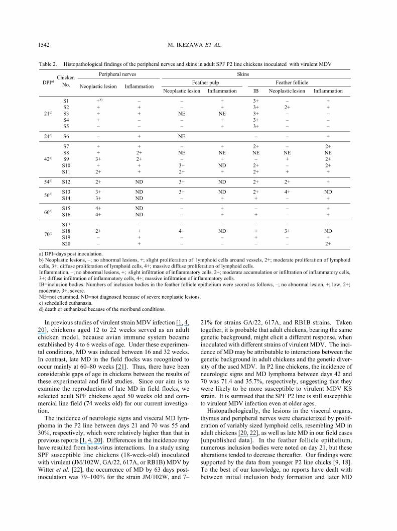
Table 2. Histopathological findings of the peripheral nerves and skins in adult SPF P2 line chickens inoculated with virulent MDV
ChickenPeripheral nerves Skins
DPIa)No. Neoplastic lesion Inflammation
Feather pulp Feather follicle
Neoplastic lesion Inflammation IB Neoplastic lesion Inflammation
S1 +b) – – + 3+ – +S2 + + – + 3+ 2+ +
21c) S3 + + NE NE 3+ – –S4 + – – + 3+ – –S5 – – – + 3+ – –
24d) S6 – + NE – – +
S7 + + – + 2+ – 2+S8 + 2+ NE NE NE NE NE
42c) S9 3+ 2+ – + – + 2+S10 + + 3+ ND 2+ – 2+S11 2+ + 2+ + 2+ + +
54d) S12 2+ ND 3+ ND 2+ 2+ +
56d) S13 3+ ND 3+ ND 2+ 4+ NDS14 3+ ND – + + – +
66d)S15 4+ ND – + – – +S16 4+ ND – + + – +
S17 – – – – – – –
70c) S18 2+ + 4+ ND + 3+ NDS19 – + – – + – +S20 – + – – – – 2+
a) DPI=days post inoculation.b) Neoplastic lesions, –; no abnormal lesions, +; slight proliferation of lymphoid cells around vessels, 2+; moderate proliferation of lymphoidcells, 3+; diffuse proliferation of lymphoid cells, 4+; massive diffuse proliferation of lymphoid cells.Inflammation, –; no abnormal lesions, +; slight infiltration of inflammatory cells, 2+; moderate accumulation or infiltration of inflammatory cells,3+; diffuse infiltration of inflammatory cells, 4+; massive infiltration of inflammatory cells.IB=inclusion bodies. Numbers of inclusion bodies in the feather follicle epithelium were scored as follows, –; no abnormal lesion, +; low, 2+;moderate, 3+; severe.NE=not examined. ND=not diagnosed because of severe neoplastic lesions.c) schedulled euthanasia.d) death or euthanized because of the moribund conditions.

1543EXPERIMENTAL LATE MD IN ADULT CHICKENS
Fig. 1. Inflammatory and neoplastic lesions in the peripheral nerve of adult SPF P2 line chickens (50-week-old)inoculated with virulent MDV. (A) Small foci of variably sized lymphoid cells in a chicken (No. S4) on day 21.(B) Diffuse proliferation of variably sized lymphoid cells and severe neuritis with infiltration of lymphocytes,plasma cells, heterophils and macrophages in a chicken (No. S9) showed neurologic signs on day 42. (C) Diffuseproliferation of variably sized lymphoid cells among the axonal fibers in a chicken (No. S13) showed neurologicsigns and MD lymphoma on day 56. (D) Moderate proliferation of lymphoid cells in a chicken (No. S18) showedneurologic signs and MD lymphoma on day 70. Note proliferative lesions without inflammatory findings. HEstain. Bar=50 m.
Fig. 2. MDV intranuclear (arrowhead) and intracyto-plasmic inclusion bodies (arrow) on day 21 in thefeather follicle epithelium of adult SPF P2 line chick-ens (50-week-old) inoculated with virulent MDV.Note infiltrations of lymphocytes, heterophils andmacrophages around the feather follicle. HE stain.Bar=20 m.
Fig. 3. Infiltrations of a small number of lymphocytesaround the blood vessel in the peripheral nerve on day70 in a commercial line chicken (74-week-old) inocu-lated with virulent MDV. HE stain. Bar=20 m.

M. IKEZAWA ET AL.1544
development in adult chickens. In the feather pulp, moder-ate to diffuse proliferation of lymphoid cells was observedin five chickens (Nos. S10 to S13, and S18). Three (Nos.S12, S13 and S18) of five chickens showed visceral lym-phomas, while the remaining two (Nos. S10 and S11)showed no lesions. The lesions in the feather pulp seemedto be associated with visceral MD lymphoma, although thenumber of affected chickens was small. The incidence ofMD is reported to be closely related to the dynamics ofinclusion body formation and feather pulp lesions in chicks[13, 14]. The histopathological lesions of this study werealso resembled which in younger chicks inoculated with KSstrain [9, 18]. However, in younger chicks, the incidence ofMD was higher, and the onset of tumor formation was earlynoted.
In commercial line chickens, although one chickenshowed lymphoma on day 70, most chickens were refrac-tory to challenge with MDV. These results would be attrib-uted to their genetically resistance back ground andimmunity induced by MD vaccines.
There are two alternative hypothesis concerning patho-genesis of late MD, “new infection theory” and “old infec-tion theory” [21]. New infection theory is that adultchickens have susceptibility to de novo challenge withMDV, and newly infection induces late MD. Meanwhile,old infection theory is that latent infection MDV which trig-gered or exacerbated by several factors like immunodepres-sion induces late MD. The incidence of MD in P2 linechickens would be comparable to new infection theory. Thepathogenesis of one commercial line chicken developingMD is questionable. This chicken was in good clinical con-ditions throughout the experiment, and showed no histo-pathological immunosuppressive changes. Because MDVpersisted in the chickens for a long period of time, it was notdetermined whether the onset of MD was caused by theinoculation or previous infection with MDV in field flocks.Further investigations for late MD in adult chickens areneeded to reveal its pathogenesis. However, our model
would become a tool for elucidating the etiology and patho-genesis of late MD in the field flocks.
In conclusion, this study demonstrated that adult SPF P2line chickens are susceptible to virulent MDV, and would beuseful as a late MD model.
REFERENCES
1. Anderson, D. P., Eidson, C. S. and Richey, D. J. 1971. Agesusceptibility of chickens to Marek’s disease. Am. J. Vet. Res.32: 935–938.
2. Baaten, B. J., Butter, C. and Davison, T. F. 2004. Study ofhost-pathogen interactions to identify sustainable vaccine strat-egies to Marek’s disease. Vet. Immuno. Immunopathol. 100:165–177.
3. Calnek, B. W. 2001. Pathogenesis of Marek’s disease virusinfection. pp. 25–56. In: Current Topics in Microbiology andImmunology (Hirai, K. ed.), Springer-Verlag, Berlin.
4. Calnek, B. W. 1973. Influence of age at exposure on the patho-genesis of Marek’s disease. J. Natl. Cancer Inst. 51: 929–939.
5. Davison, F. and Kaiser, P. 2004. Immunity to Marek’s disease.pp. 126–141. In: Marek’s Disease: An Evolving Problem(Davison, F. and Nair, V. eds.), Elsevier Academic Press,London.
6. Davidson, I. and Borenshtain, R. 2002. The feather tips ofcommercial chickens are a favorable source of DNA for theamplification of Marek’s disease virus and avian leukosisvirus, subgroup J. Avian pathol. 31: 237–240.
7. Fehler, F. and Winter, C. 2001. CIAV infection in older chick-ens: an apathogenic infection? pp. 391–394. In: Proceedings of2nd International Symposium on Infectious Bursal Disease andChicken Anemia, Institut für Geflugelkrankheiten, Justus Lie-big University, Rauischholzhausen, Giessen.
8. Gimeno, I. M., Witter, R. L. and Reed, W. M. 1999. Four dis-tinct neurologic syndromes in Marek’s disease: effect of viralstrain and pathotype. Avian Dis. 43: 721–737.
9. Goryo, M., Tsukui, M., Saijo, K., Takeuchi, J., Obata, F. andFujikawa, Y. 1980. Studies on a virulent Marek’s disease virus(CVI988 strain). pp 17–30. In: English Summaries of the Stud-ies on Poultry Disease in Japan, The Japanese Veterinary Poul-try Association, Tsukuba.
Fig. 4. A large mass of MD lymphoma constituted neoplastic lymphoid cells in the lung on day 70 in a commercialline chicken (74-week-old) inoculated with virulent MDV. (A) A large mass of neoplastic lymphoid cells suggestingMD lymphoma. Bar=50 m. (B) Variably sized lymphoid cells suggesting MD lymphoma. HE stain. Bar=20 m.

1545EXPERIMENTAL LATE MD IN ADULT CHICKENS
10. Japanese Association for Laboratory Animal Science. 1987.Guidelines for animal experimentation. Exp. Anim. 3: 285–288.
11. Kreager, K. S. 1997. Marek’s disease: clinical aspects and cur-rent field problems in layer chickens. pp. 23–26. In: Diagnosisand Control of Neoplastic Disease of Poultry (Fadly, A. M.,Schat, K. A. and Spencer, J. L. eds.), American Association ofAvian Pathologists Inc., Kennett Square.
12. Lucio-Martinez, B. 1999. Impact of vv Marek’s disease onmortality and production in a multiple-age farm. pp. 55–56. In:Proceedings of the 48th Western Poultry Disease Conference,Vancouver.
13. Moriguchi, R. and Izawa, H. 1979. Marek’s disease in chick-ens: correlation of Marek’s disease with nuclear-inclusion for-mation in feather-follicle epithelium. Avian Dis. 23: 547–554.
14. Moriguchi, R., Fujimoto, Y. and Izawa, H. 1982. Chronologi-cal observations of feather pulp lesions in chickens inoculatedwith Marek’s disease virus. Avian Dis. 26: 375–388.
15. Morimura, T., Ohashi, K., Sugimoto, C. and Onuma, M. 1998.Pathogenesis of Marek’s disease (MD) and possible mecha-nisms of immunity induced by MD vaccine. J. Vet. Med. Sci.60: 1–8.
16. Morrow, C. and Fehler, F. 2004. Marek’s disease: a worldwideproblem. pp. 49–61. In: Marek’s Disease: An Evolving Prob-lem (Davison, F. and Nair, V. eds.), Elsevier Academic Press,London.
17. Payne, L. N. Pathological responses to infection. 2004. pp. 78–97. In: Marek’s Disease: An Evolving Problem (Davison, F.and Nair, V. eds.), Elsevier Academic Press, London.
18. Saijo, K., Takeuchi, J., Goryo, M., Tamura, S., Obata, F. andFujikawa, Y. 1980. Biological characteristics of a virulentMarek’s disease virus strain isolated from the affected chickenof the field flock. pp. 23–24. In: English Summaries of theStudies on Poultry Disease in Japan, The Japanese VeterinaryPoultry Association, Tsukuba.
19. Schat, K. A. 2004. Marek’s disease immunosuppression. pp.142–155. In: Marek’s Disease: An Evolving Problem (Davi-son, F. and Nair, V. eds.), Elsevier Academic Press, London.
20. Sharma, J. M., Witter, R. L. and Burmerster, B. R. 1973.Pathogenesis of Marek’s disease in old chickens: lesion regres-sion as the basis for age-related resistance. Infect. Immun. 8:715–724.
21. Witter, R. L. 2001. Protective efficacy of Marek’s disease vac-cines. pp. 58–90. In: Current Topics in Microbiology andImmunology (Hirai, K. ed.), Springer-Verlag, Berlin.
22. Witter, R. L. and Gimeno, I. M. 2006. Susceptibility of adultchickens, with and without prior vaccination, to challenge withMarek’s disease virus. Avian Dis. 50: 354–365.
23. Witter, R. L., Sharma, J. M., Solomon, J. J. and Champion, L.R. 1973. An age-related resistance of chickens to Marek’s dis-ease: some preliminary observations. Avian Pathol. 2: 43–54.
Copyright © 2022 FDOKUMEN