The McArdle Disease Handbook
183
1 The McArdle Disease Handbook A guide to the scientific and medical research into McArdle disease explained in plain English. Written by Kathryn Elizabeth Wright, Ph.D. Copyright ©Kathryn Wright 2010 Disclaimer Unless otherwise stated, this Handbook represents the views and opinions of the author, Kathryn Wright, and does not represent the views and opinions of AGSD (UK) or Vodafone World of Difference. The purpose of this Handbook is to explain scientific research and knowledge about McArdle disease in layman’s language so that it can be understood by people with McArdle disease or those interested in McArdle disease. It is not intended to replace medical advice from your family doctor or specialist. The information provided in this Handbook is correct to the best of the author’s knowledge. If you have any doubts about the accuracy of the information in this Handbook, it is recommended that you read the original source (full details in the reference list). Where no definitive information is available, the author has sought to suggest scientific rationale behind anecdotal observations reported by people with McArdle’s. It is stated where a theory or opinion of the author is given. Due to the nature of scientific research, current theories and understanding of the science behind McArdle’s may change over time and subsequently be proven or disproven. It is recommended that you check the AGSD (UK) website frequently to ensure you are reading the most up-to-date version of this Handbook. Funding for this project Kathryn Wright submitted a proposal and successfully obtained funding from the Vodafone World of Difference charitable foundation under the “World of Difference UK” scheme. This grant enabled AGSD (UK) to employ Kathryn Wright from January to March 2010 to write this Handbook. Further writing was carried out by Kathryn Wright on an unpaid voluntary basis. Kathryn Wright has no competing financial or academic interests. Acknowledgements I would like to acknowledge Vodafone World of Difference (UK) charitable foundation for providing two months funding for this project. I would also like to thank the AGSD (UK) for supporting my application for funding, and in particular I have to thank Andrew Wakelin for his encouragement and support. (This was originally going to be a pamphlet, but has become a book.) My Ph.D. project, funded by AGSD (UK) and The Institute of Orthopaedics, RJAH Orthopaedic Hospital, Oswestry, provided me with an opportunity to develop an interest in McArdle disease. I would like to thank Mum, Dad and Madelyn for their continued interest and encouragement and for proof-reading the Handbook. I am very grateful to Mr James Birch, for his endless patience and support. Definitions of terms used in this Handbook In this Handbook, “McArdle person” is used to mean a person who has received a definitive diagnosis of McArdle disease (who has no functional muscle glycogen phosphorylase enzyme in their
-
Upload
khangminh22 -
Category
Documents
-
view
0 -
download
0
Transcript of The McArdle Disease Handbook
1
The McArdle Disease Handbook A guide to the scientific and medical research into McArdle disease explained in plain English.
Written by Kathryn Elizabeth Wright, Ph.D.
Copyright ©Kathryn Wright 2010
Disclaimer
Unless otherwise stated, this Handbook represents the views and opinions of the author, Kathryn
Wright, and does not represent the views and opinions of AGSD (UK) or Vodafone World of
Difference. The purpose of this Handbook is to explain scientific research and knowledge about
McArdle disease in layman’s language so that it can be understood by people with McArdle disease
or those interested in McArdle disease. It is not intended to replace medical advice from your family
doctor or specialist. The information provided in this Handbook is correct to the best of the author’s
knowledge. If you have any doubts about the accuracy of the information in this Handbook, it is
recommended that you read the original source (full details in the reference list). Where no
definitive information is available, the author has sought to suggest scientific rationale behind
anecdotal observations reported by people with McArdle’s. It is stated where a theory or opinion of
the author is given. Due to the nature of scientific research, current theories and understanding of
the science behind McArdle’s may change over time and subsequently be proven or disproven. It is
recommended that you check the AGSD (UK) website frequently to ensure you are reading the most
up-to-date version of this Handbook.
Funding for this project
Kathryn Wright submitted a proposal and successfully obtained funding from the Vodafone World of
Difference charitable foundation under the “World of Difference UK” scheme. This grant enabled
AGSD (UK) to employ Kathryn Wright from January to March 2010 to write this Handbook. Further
writing was carried out by Kathryn Wright on an unpaid voluntary basis. Kathryn Wright has no
competing financial or academic interests.
Acknowledgements
I would like to acknowledge Vodafone World of Difference (UK) charitable foundation for providing
two months funding for this project. I would also like to thank the AGSD (UK) for supporting my
application for funding, and in particular I have to thank Andrew Wakelin for his encouragement and
support. (This was originally going to be a pamphlet, but has become a book.) My Ph.D. project,
funded by AGSD (UK) and The Institute of Orthopaedics, RJAH Orthopaedic Hospital, Oswestry,
provided me with an opportunity to develop an interest in McArdle disease. I would like to thank
Mum, Dad and Madelyn for their continued interest and encouragement and for proof-reading the
Handbook. I am very grateful to Mr James Birch, for his endless patience and support.
Definitions of terms used in this Handbook
In this Handbook, “McArdle person” is used to mean a person who has received a definitive
diagnosis of McArdle disease (who has no functional muscle glycogen phosphorylase enzyme in their
2
skeletal muscle cells). “Unaffected by McArdle’s” is used to describe a “normal” person who has no
mutations in either copy of the PYGM gene. People unaffected by McArdle’s have two wildtype
copies of the PYGM gene. (Wildtype means that it is a version of the gene with no mutations.)
People unaffected by McArdle’s have a normal amount of active muscle glycogen phosphorylase
enzyme in their muscle cells. “Carrier” is used to describe a person who has one copy of the PYGM
gene without any mutations, and one copy of the PYGM gene which does carry a mutation. A carrier
is likely to have approximately half the normal level of muscle glycogen phosphorylase enzyme.
Carriers do not usually have symptoms of McArdle disease.
Further definitions are given where appropriate throughout the Handbook. There is also a glossary
at the end of the Handbook for scientific or medical words used frequently in the Handbook which
would not be included in a standard English glossary.

3
Contents
Disclaimer............................................................................................................................................ 1
Funding for this project ....................................................................................................................... 1
Acknowledgements ............................................................................................................................. 1
Definitions of terms used in this Handbook ....................................................................................... 1
1 Introduction to McArdle disease .................................................................................................. 12
1.1 A brief introduction to McArdle disease ............................................................................... 12
1.2 What is the cause of McArdle disease? ................................................................................ 13
1.3 McArdle disease is one of the family of glycogen storage diseases ..................................... 13
GAA ............................................................................................................................................... 14
1.4 Why is it called “McArdle disease”? ..................................................................................... 15
1.4.1 Other names for McArdle disease ................................................................................ 15
1.4.2 Other names and abbreviations for muscle glycogen phosphorylase enzyme ............ 15
1.5 How can I explain my McArdle disease to my friends and family who have never heard of it
before? .............................................................................................................................................. 15
1.6 The future is promising for people with McArdle disease .................................................... 16
2 Symptoms and diagnosis of McArdle disease ............................................................................... 17
2.1 The personal history of symptoms described by a typical McArdle person ......................... 17
2.1.1 Activities which McArdle people have reported can cause pain/fatigue in muscles: .. 17
2.2 Symptoms of McArdle disease .............................................................................................. 18
2.3 Diagnosis of McArdle disease ............................................................................................... 19
2.3.1 Exercise test .................................................................................................................. 21
2.3.2 Muscle biopsy ............................................................................................................... 25
2.3.3 DNA testing ................................................................................................................... 27
2.3.4 Electromyogram (EMG)................................................................................................. 28
2.3.5 31P magnetic resonance spectroscopy (31P MRS) .......................................................... 29
2.4 Causes of similar symptoms to McArdle disease (in people who don’t have McArdle’s) .... 30
2.5 Other diseases which have similarities to McArdle disease ................................................. 30
2.5.1 Some of the diseases which McArdle people have been misdiagnosed with before
getting a proper diagnosis of McArdle’s ....................................................................................... 30
2.5.2 Other muscle diseases with similar symptoms to McArdle disease ............................. 31
2.6 McArdle disease was considered a possible diagnosis for a patient with chronic pain in the
television series “House” .................................................................................................................. 33
3 The genetics of McArdle disease .................................................................................................. 35

4
3.1 Genes, mRNA and protein .................................................................................................... 35
3.1.1 Genes are used to make mRNA, which is used to make protein .................................. 35
3.2 Mutations in the PYGM gene prevent production of active muscle glycogen phosphorylase
enzyme and cause McArdle’s ........................................................................................................... 36
3.2.1 Stop codons ................................................................................................................... 37
3.2.2 Missense mutations ...................................................................................................... 38
3.2.3 Frameshift mutations .................................................................................................... 39
3.2.4 Splice site mutations ..................................................................................................... 39
3.2.5 Theoretical possible outcomes of various mutations ................................................... 39
3.2.6 How many of these mutations have been identified in the PYGM gene so far? .......... 40
3.2.7 What caused the first mutation in the PYGM gene? .................................................... 42
3.3 How is McArdle disease inherited?....................................................................................... 42
3.3.1 One copy of each gene is inherited from each parent ................................................. 42
3.3.2 What are the combinations of wildtype and genes with mutations which can occur? 43
3.3.3 Methods of inheritance ................................................................................................ 43
3.3.4 Examples of how McArdle’s is inherited ....................................................................... 44
3.3.5 Consanguineous parents; why are they more likely to produce children with inherited
disease? 48
3.3.6 How many people have McArdle disease or are carriers of a mutation in PYGM? ...... 48
4 Exercise, muscle contractions, and contractures (muscle cramps) .............................................. 49
4.1 Types of exercise ................................................................................................................... 49
4.1.1 Anaerobic exercise ........................................................................................................ 49
4.1.2 Aerobic exercise ............................................................................................................ 49
4.2 Exercise recommendations for McArdle people .................................................................. 49
4.2.1 Anaerobic exercise or static muscle contractions should be avoided .......................... 49
4.2.2 Moderate aerobic exercise and conditioning improves symptoms of McArdle’s ........ 50
4.2.3 The phrase “No pain, no gain” does NOT apply to McArdle people ............................ 51
4.2.4 Being overweight can make McArdle people find it harder to move around .............. 51
4.3 Muscles require energy (ATP) to contract and to relax during exercise .............................. 52
4.4 Energy is stored in bonds between adenosine and phosphate, and energy can be released
when the bonds are broken .............................................................................................................. 52
4.4.1 A brief description of muscle contraction and relaxation ............................................ 53
4.4.2 A more detailed description of the role of the calcium ATPase pump ......................... 53
4.4.3 A more detailed description of the role of the sodium-potassium ATPase pump ....... 53
4.5 A muscle contracture (cramp) can occur if McArdle people exercise anaerobically............ 53

5
4.5.1 Treatment for contractures .......................................................................................... 55
5 Muscle damage (rhabdomyolysis) can lead to raised creatine kinase levels in the blood,
myoglobinuria, and kidney failure ........................................................................................................ 57
5.1 Rhabdomyolysis and acute kidney failure ............................................................................ 57
5.2 Causes of rhabdomyolysis ..................................................................................................... 57
5.2.1 Causes of rhabdomyolysis in people unaffected by McArdle disease .......................... 57
5.2.2 Causes of rhabdomyolysis in McArdle people .............................................................. 59
5.3 Symptoms of rhabdomyolysis ............................................................................................... 59
5.3.1 General symptoms ........................................................................................................ 59
5.3.2 Raised creatine kinase (CK) levels in the bloodstream ................................................. 59
5.3.3 Myoglobinuria (blood in the urine) ............................................................................... 60
5.3.4 Treatment for rhabdomyolysis: .................................................................................... 60
5.3.5 McArdle people are at an increased risk of rhabdomyolysis, raised creatine kinase
levels and kidney failure ............................................................................................................... 61
6 Sources of energy in muscle cells ................................................................................................. 62
6.1 Methods of producing energy in muscle cells of people unaffected by McArdle’s ............. 62
6.1.1 Storage, release and breakdown of carbohydrates to produce energy ....................... 62
6.1.2 Storage, release and breakdown of fats to produce energy ........................................ 62
6.1.3 Breakdown of proteins to produce energy ................................................................... 63
6.1.4 Acetyl CoA from carbohydrates, fats and proteins can be broken down in the citric
acid cycle 63
6.1.5 NADH and FADH2 from the breakdown of carbohydrates, fats and proteins is used to
produce ATP by oxidative phosphorylation .................................................................................. 63
6.2 Glucose from the blood is an important source of energy for muscle cells ......................... 63
6.2.1 Glucose is transported from the blood into muscle cells ............................................. 63
6.2.2 Excess glucose is stored as glycogen ............................................................................. 63
6.2.3 Insulin and glucagon are two hormones which maintain the right amount of glucose in
the bloodstream ........................................................................................................................... 64
6.3 In people unaffected by McArdle’s, enzyme activity of muscle glycogen phosphorylase is
controlled to maintain the right amount of glucose within the cell ................................................. 64
6.3.1 Muscle glycogen phosphorylase is made of several identical subunits which require
the presence of phosphate ........................................................................................................... 65
6.3.2 Muscle glycogen phosphorylase activity is controlled by ligands and cofactors .......... 65
6.3.3 The levels of glycogen may affect the activity of muscle glycogen phosphorylase ...... 66
6.4 How is energy produced in the muscles of McArdle people? .............................................. 66
6.4.1 Glycogen cannot be broken down into glucose to provide energy for exercise .......... 66
6
6.4.2 Other mechanisms provide the energy needed for the second wind .......................... 67
6.4.3 The production of energy by other mechanisms is not as efficient in McArdle people
67
6.4.4 The way in which the body of a McArdle person responds to exercise ....................... 68
6.4.5 How would having a sugary drink before exercise increase the amount of energy
available for the muscles of McArdle people? ............................................................................. 69
6.4.6 The amount of some other proteins may be increased in the muscle cells of people
with McArdle’s to compensate for the lack of muscle glycogen phosphorylase ......................... 70
6.5 Non-muscle isoforms of glycogen phosphorylase breakdown glycogen into glucose-1-
phosphate in other areas of the body .............................................................................................. 71
6.5.1 Muscle glycogen phosphorylase ................................................................................... 71
6.5.2 Brain glycogen phosphorylase ...................................................................................... 71
6.5.3 Liver glycogen phosphorylase ....................................................................................... 72
6.5.4 After muscle damage, regenerating immature muscle produces brain and/or liver
isoform 72
6.5.5 A brief discussion about the three isoforms of glycogen phosphorylase ..................... 72
6.5.6 Non-muscle isoforms of glycogen phosphorylase spare the smooth tissue and organs
of the body from symptoms of McArdle disease .......................................................................... 73
6.6 The balance of protein, carbohydrate and fat in the diet .................................................... 74
6.6.1 The role of protein, carbohydrate and fat in McArdle’s ............................................... 74
6.6.2 Research into whether the balance of protein, carbohydrate and fat in the diet affects
McArdle people ............................................................................................................................. 75
6.6.3 Comments on these trials ............................................................................................. 76
7 A review of dietary supplements which have been considered for McArdle’s ............................ 78
7.1 Supplements which have been tested as possible treatments for McArdle’s ..................... 78
7.1.1 Amino acids ................................................................................................................... 78
7.1.2 Vitamin B6 ..................................................................................................................... 79
7.1.3 Creatine ......................................................................................................................... 79
7.1.4 Cornstarch (such a the commercially available “Glycosade”) ...................................... 80
7.1.5 Dantrolene sodium ....................................................................................................... 81
7.1.6 Sugar ............................................................................................................................. 81
7.1.7 Verapamil ...................................................................................................................... 83
7.2 Supplements which have not been tested to see if they are effective in McArdle’s ........... 83
7.2.1 Vitamins ........................................................................................................................ 84
7.2.2 Amino acids ................................................................................................................... 84
7.2.3 Coenzyme Q-10 (CoQ10; also known as ubiquinone or ubidecarenone) ...................... 84

7
7.2.4 Medical advice should be obtained before commencing use of a supplement ........... 85
8 The effect of age on the symptoms of McArdle disease .............................................................. 86
8.1 Are there several forms of McArdle’s? ................................................................................. 86
8.1.1 A rare fatal infant form: ................................................................................................ 86
8.1.2 A milder form with delayed motor milestones, limb muscle weakness and high CK ... 87
8.1.3 A late-onset form which begins in adult life and leads to progressive muscle weakness
87
8.2 The classic form..................................................................................................................... 88
8.2.1 The classic form of McArdle’s from baby to toddler .................................................... 88
8.2.2 The classic form of McArdle’s in childhood .................................................................. 89
8.2.3 McArdle’s in adulthood/middle age ............................................................................. 95
8.2.4 McArdle’s in older age .................................................................................................. 95
8.2.5 Will I end up in a wheelchair? ....................................................................................... 97
8.2.6 Does McArdle disease affect lifespan? ......................................................................... 97
9 Factors which may explain differences in the severity of symptoms of McArdle disease from
person to person ................................................................................................................................... 98
9.1 The amount of active muscle glycogen phosphorylase is an important factor which
determines the severity of symptoms .............................................................................................. 98
9.1.1 The amount of functional enzyme determines whether carriers of the PYGM mutation
have symptoms of McArdle disease ............................................................................................. 98
9.1.2 Different mutations do not correlate to differences in severity of symptoms............. 98
9.1.3 Rarely, some McArdle people have low levels of muscle glycogen phosphorylase
activity 99
9.1.4 Even a very low level of muscle glycogen phosphorylase activity improves the ability
of McArdle people to exercise ...................................................................................................... 99
9.2 Raised levels of cytokines may cause low-level inflammation in McArdle people............... 99
9.3 Phenotype modulators (genes other than PYGM) may affect the severity of McArdle’s
symptoms ........................................................................................................................................ 101
9.3.1 Angiotensin-converting enzyme ................................................................................. 101
9.3.2 Muscle adenosine monophosphate deaminase ......................................................... 102
9.3.3 Myostatin .................................................................................................................... 102
9.3.4 α-actinin-3 ................................................................................................................... 103
9.3.5 Peroxisome proliferator-activated receptor coactivator 1 α ................................... 103
9.4 There is a difference in the amount and type of pain felt by different McArdle people ... 103
9.5 Gender has an effect on phenotype modulators and the severity of McArdle’s symptoms
104

8
9.6 McArdle symptoms may be more severe if combined with another disease .................... 105
9.6.1 McArdle symptoms can be more severe if combined with another muscle disease,
causing “double trouble” ............................................................................................................ 105
9.6.2 A second disease (which is not a muscle disease) may exacerbate the symptoms of
McArdle disease .......................................................................................................................... 105
9.7 A second disease may make carriers of McArdle disease more likely to have symptoms of
McArdle disease .............................................................................................................................. 106
10 Mental and emotional aspects of McArdle disease ................................................................ 107
10.1 Many McArdle people have symptoms which are like chronic fatigue syndrome ............. 107
10.2 Lack of muscle glycogen phosphorylase may affect brain function of McArdle people .... 108
10.3 Psychological issues ............................................................................................................ 109
10.3.1 Before diagnosis .......................................................................................................... 109
10.3.2 Trying to get a diagnosis ............................................................................................. 109
10.3.3 After diagnosis ............................................................................................................ 110
10.3.4 Positive steps to dealing with these issues ................................................................. 112
10.3.5 Websites and internet resources for people interested in McArdle disease ............. 113
11 The effect of McArdle’s on sexual activity, pregnancy and birth ........................................... 115
11.1 Sexual activity ..................................................................................................................... 115
11.2 The menstrual cycle and it’s effect upon exercise and perception of pain for McArdle’s
women ............................................................................................................................................ 115
11.2.1 Does the menstrual cycle affect the ability of women unaffected by McArdle’s to
exercise 116
11.2.2 Studies on the perception of pain in women unaffected by McArdle’s ..................... 116
11.2.3 The emotional changes associated with the menstrual cycle may make McArdle’s
women consider their symptoms to be worse at certain points ................................................ 117
11.3 Fertility and contraception ................................................................................................. 117
11.3.1 Prenatal testing for McArdle’s and infant screening .................................................. 117
11.4 Pregnancy and birth ............................................................................................................ 118
11.4.1 Some McArdle’s women report having fewer McArdle’s symptoms during pregnancy
118
11.4.2 Method of giving birth; vaginal birth or caesarean delivery ....................................... 119
12 Medicines, activities and other things which may be a greater risk for McArdle people ...... 121
12.1 Medicines ............................................................................................................................ 121
12.1.1 Lipid-lowering drugs such as statins may exacerbate rhabdomyolysis in McArdle
people 121
9
12.1.2 When combined with a statin, some drugs produce a much higher risk of
rhabdomyolysis ........................................................................................................................... 122
12.1.3 Other drugs to reduce cholesterol may exacerbate rhabdomyolysis in McArdle people
123
12.2 Pain relieving mediation ..................................................................................................... 123
12.3 McArdle people are at increased risks of particular complications during surgery ........... 123
12.3.1 Lack of muscle glycogen phosphorylase may put McArdle people at increased risk of
malignant hyperthermia-like symptoms caused by anaesthetics .............................................. 123
12.3.2 McArdle people may be at risk of a rare condition called “Compartment syndrome”
caused by use of a tourniquet or cuff ......................................................................................... 124
12.4 Some situations may make McArdle’s symptoms worse ................................................... 125
12.4.1 Cold temperature ........................................................................................................ 125
12.4.2 Getting angry .............................................................................................................. 125
12.4.3 Swimming .................................................................................................................... 125
12.4.4 Treatment by anyone who moves the body e.g. physiotherapy, osteopathy,
chiropractor, massage................................................................................................................. 126
13 McArdle disease may increase the chances of having some diseases and conditions ........... 127
13.1 McArdle’s may increase the risk of gout............................................................................. 127
13.2 Brain functioning ................................................................................................................. 127
13.3 Respiratory problems .......................................................................................................... 128
13.4 McArdle’s may cause symptoms of insulin resistance (similar to type 2 diabetes) ........... 128
13.4.1 A discussion of whether insulin resistance could be caused by McArdle disease ...... 131
13.4.2 Anecdotal reports of a feeling of low blood sugar could be explained by insulin
resistance preventing muscle cells taking up glucose, despite test results showing high blood
glucose levels .............................................................................................................................. 132
13.4.3 Exercise can be used to prevent and treat insulin resistance in people unaffected by
McArdle’s .................................................................................................................................... 132
13.5 There is no evidence that McArdle’s may increase the risk of heart problems ................. 133
13.6 McArdle’s is not reported to cause liver disease, but can have an effect on the results of
blood tests for liver disease ............................................................................................................ 133
14 McArdle’s specialists and general family doctors ................................................................... 135
14.1 McArdle’s specialists ........................................................................................................... 135
14.1.1 Benefits of visiting a McArdle’s specialist ................................................................... 135
14.1.2 Paying to see a McArdle’s specialist ........................................................................... 136
14.2 Your family doctor- how to help them treat you, what information to give them, what to
remind them ................................................................................................................................... 137
10
14.2.1 Your family doctor may not know very much about McArdle’s ................................. 137
14.2.2 Important things to remind your family doctor .......................................................... 137
14.3 Become your own personal expert on McArdle’s .............................................................. 137
15 Models of McArdle’s can be used to test treatments ............................................................ 139
15.1 Models can be used to test potential treatments .............................................................. 139
15.1.1 Sheep with McArdle disease ....................................................................................... 139
15.1.2 Cows with McArdle disease ........................................................................................ 139
15.1.3 Mouse and rat models of McArdle disease ................................................................ 140
15.1.4 Muscle cell culture from unaffected people and McArdle people ............................. 140
15.1.5 Creation of cell models of McArdle’s .......................................................................... 141
15.2 Animal and cell models can be used to test potential therapies for McArdle disease ...... 141
16 Potential therapies for McArdle disease ................................................................................ 142
16.1 Therapies to correct or replace the PYGM genomic sequence .......................................... 142
16.1.1 Gene therapy to provide a copy of the wildtype PYGM ............................................. 142
16.1.2 Targeted correction of mutation within the PYGM gene ........................................... 143
16.2 Therapies to read-through mutations in the mRNA transcript to produce full-length PYGM
protein 144
16.2.1 Read-through of premature stop codons by aminoglycoside drugs .......................... 144
16.2.2 Therapeutic exon skipping .......................................................................................... 146
16.3 Therapies to replace the muscle glycogen phosphorylase protein within skeletal muscle
cells 147
16.3.1 Upregulation of an alternative isoform (such as brain/foetal glycogen phosphorylase)
147
16.3.2 Enzyme replacement therapy ..................................................................................... 148
16.4 A discussion of research into therapies for McArdle’s ....................................................... 150
17 Details about this Handbook and the information in it .......................................................... 155
17.1 The purpose of this Handbook ............................................................................................ 155
17.2 About the author ................................................................................................................ 155
17.2.1 Feedback about this Handbook; comments, suggestions, criticisms ......................... 155
17.3 How to understand references given in this Handbook ..................................................... 155
17.4 A note about the names of the gene, mRNA and protein .................................................. 156
17.5 How to read medical/scientific papers critically ................................................................. 156
17.6 An introduction to clinical trials .......................................................................................... 158
17.6.1 The placebo effect ...................................................................................................... 159
17.6.2 Randomised trials ....................................................................................................... 160

11
17.6.3 Cross-over trial ............................................................................................................ 160
17.6.4 Single case study ......................................................................................................... 161
17.6.5 A home trial is an alternative to a clinical trial ........................................................... 161
17.6.6 Statistical significance ................................................................................................. 161
18 Glossary ................................................................................................................................... 163
19 References .............................................................................................................................. 166

12
1 Introduction to McArdle disease
1.1 A brief introduction to McArdle disease
Muscle contractions are required to generate movement. Muscle cells require a source of energy in
order to perform muscle contractions. Anaerobic exercise is a short burst of high intensity effort,
such as a sprint for a bus. During anaerobic exercise, glucose within muscle cells is broken down to
produce ATP. ATP is the source of energy for muscle cells. The breakdown of glucose to produce
ATP is called “glycolysis”. However, only a small amount of glucose is present in the muscle cells and
this is used up within a few minutes of anaerobic exercise.
Muscle cells also contain much larger stores of glycogen. Glycogen can be converted into glucose by
a process called “glycogenolysis”. In people unaffected by McArdle disease, the process of
converting glycogen into glucose requires several enzymes, one of which is called “muscle glycogen
phosphorylase”.
McArdle disease is caused by the lack of the muscle glycogen phosphorylase enzyme in muscle cells.
In McArdle people, muscle glycogen phosphorylase is either absent or not functional. The muscle
cells are not able to convert the stored glycogen into glucose. The muscle cells therefore run out of
glucose and run out of energy. The short term lack of glucose causes tiredness and stiffness in
muscles of McArdle people when they carry out anaerobic exercise (Rommel et al., 2006), but this
improves once exercise ceases.
McArdle people must rest until energy (ATP) is produced in the muscle cells by another method
such as fatty acid oxidation or until glucose is obtained through the blood from the liver. A period of
rest is necessary because these other methods are slower to produce energy than glycogenolysis
(the method which normally involves muscle glycogen phosphorylase). Once these other methods
begin to replenish the amount of ATP in the muscle cells, McArdle people can continue to exercise.
This is known as a “second wind” (Amato, 2003).
However, if McArdle people continue to exercise without rest, the muscle cells use up all the
available ATP and have no energy source available. This can lead to breakdown of muscle cells
(rhabdomyolysis) and muscle cramps (contractures), both of which cause McArdle people to
experience muscle pain. Following rhabdomyolysis, the components of the broken muscle cells are
released into the bloodstream. An enzyme normally found in muscle cells called creatine kinase (CK)
(also known as creatine phosphokinase (CPK)) is released into the bloodstream following muscle
damage. A blood test performed by a family doctor at a hospital can be used to measure the
amount of CK in the blood, which can be used as an indicator of the extent to which muscle damage
which has occurred. The components of the broken muscle cells are transported through the
bloodstream to the kidneys. Myoglobin is another protein released from these broken muscle cells.
Myoglobin is transported in the bloodstream to the kidneys, where it is removed from the body in
the urine, resulting in dark red/cola coloured urine (known as myoglobinuria or proteinuria). A rare,
but serious effect of extreme muscle damage is that broken muscle cells may block the filtration
system of the kidneys, preventing them working, and resulting in kidney failure (Martin et al., 2001;
DiMauro et al., 2002; Quinlivan et al., 2008).

13
1.2 What is the cause of McArdle disease?
McArdle disease is caused by the absence of the muscle glycogen phosphorylase enzyme
(Mommaerts, 1956; Schmid et al., 1959). An enzyme is a protein which has a special function of
changing or breaking down one compound to another. The muscle glycogen phosphorylase enzyme
breaks down glycogen into glucose-1-phosphate. A mutation in the PYGM gene which encodes
muscle glycogen phosphorylase prevents the production of functional muscle glycogen
phosphorylase enzyme.
1.3 McArdle disease is one of the family of glycogen storage diseases
There are many enzymes involved in the breakdown of glycogen into glucose. If a mutation occurs
in the enzyme which prevents it from functioning, it will result in an inability to break down glycogen
and its components to form glucose. Diseases caused by a mutation in an enzyme required to break
down glycogen are called “glycogen storage diseases (GSDs)”. To date, 14 glycogen storage diseases
have been identified (Table 1.1). There is further information about the glycogen storage diseases
on the AGSD website (http://www.agsd.org.uk). The major symptom of every glycogen storage
disease is an intolerance to exercise. Glycogen storage is characteristic of all the diseases except
GSD 0. Tarnopolsky et al. (2006) described McArdle disease as the most common glycogen storage
disease.
GSD VIII is caused by a mutation in phosphorylase b kinase. Phosphorylase b kinase is essential for
activation of the muscle glycogen phosphorylase enzyme. However, disease symptoms for GSD VIII
are not very similar to McArdle disease, possibly because there are difference mechanisms for
activation of muscle glycogen phosphorylase in the absence of phosphorylase b kinase (Orngreen et
al., 2009).

14
Glycogen Storage Disease Alternative name Deficient enzyme Gene name
GSD 0 Glycogen synthase GYS2
GSD Ia Von Gierke disease Glucose-6-phosphatase G6PC
GSD Ib Glucose-6-phosphate translocase G6PT1
GSD Ic Endoplasmic reticulum inorganic phosphate transporter
NPT-I/NPT-II/NPT-III (not fully determined)
GSD II Pompe disease α-1, 4-glucosidase and α-1, 6-glucosidase GAA
GSD III Cori disease Debranching enzyme AGL
GSD IV Andersen disease Branching enzyme GBE1
GSD V McArdle disease Muscle glycogen phosphorylase PYGM
GSD VI (and X) Her’s disease Liver glycogen phosphorylase PYGL
GSD VII Tarui disease Phosphofructokinase PFK
GSD VIII Phosphorylase b kinase deficiency
Phosphorylase b kinase (lack of one of the four subunits)
PHKA2
GSD IX Phosphorylase b kinase deficiency
Phosphorylase b kinase (lack of one of the four subunits)
PHKA2
GSD XI Fanconi-Bickel syndrome Glucose transporter GLUT2
Table 1.1 Summary of the glycogen storage diseases identified to date

15
1.4 Why is it called “McArdle disease”?
McArdle disease is named after Dr Brian McArdle, the British family doctor who first published a
paper describing a patient with the disease. In 1951, Dr McArdle described a 30 year old male
patient for whom light exercise caused pain in the muscles, and continued exercise led to weakness
and stiffness. Pain during exercise would occur in any muscle in the body – the most noticeable
being in the arms or legs. The pain would force the patient to stop and rest, but it was noted that
after a period of rest, the patient was then able to exercise further. Dr McArdle realised that after
exercise the lactate level of the patient did not increase as expected, and that glycogenolysis was
incomplete. He also noticed that the patient’s muscles were very weak even though they had quite
a large bulk. His astonishingly perceptive theory was that “it is the… enzyme system that is at fault…
it seems… that the patient has a disorder of carbohydrate metabolism… during exercise a change
took place in the muscle chemistry which effectively led to a breakdown in glycogenolysis” (McArdle,
1955).
Mommaerts et al. (1956) realised that McArdle disease was caused solely by the loss of muscle
glycogen phosphorylase, not the loss of any other related enzymes. Schmid et al. (1959) took
samples of different skeletal muscles from a McArdle person (muscles located between the back and
shoulder, middle of the back, and the calf) and found there was a lack of glycogen phosphorylase
activity in all of these muscles compared to a person who did not have the disease. He tested the
different enzymes involved in the breakdown of glycogen and identified the cause of the disease as
the loss of the ability to produce glucose-1-phosphate, because muscle glycogen phosphorylase
wasn’t functional.
1.4.1 Other names for McArdle disease
McArdle disease is also known as McArdle disease, McArdle’s, McArdle syndrome, McArdle's
syndrome, Muscle Phosphorylase Deficiency, Myophosphorylase Deficiency, Phosphorylase
Deficiency, McArdle Myopathy, McArdle's Myopathy, Muscle Glycogen Phosphorylase Deficiency
and Glycogen Storage Disease (GSD) type V. It may be called MacArdle’s, but this is incorrect
because it is named after Dr Brian McArdle. (“Myopathy” is a general name for muscle disease.)
1.4.2 Other names and abbreviations for muscle glycogen phosphorylase enzyme
Glycogen phosphorylase, Phosphorylase, muscle phosphorylase a and b, myophosphorylase, PYGM,
GP-M, MGP, alpha-1, 4-glucan orthophosphate glycosyltransferase or EC 2.4.1.1.
1.5 How can I explain my McArdle disease to my friends and family who
have never heard of it before?
This is my suggestion of how I would describe it to friends and family who haven’t heard of McArdle
disease before:
“Your muscles use glucose to provide energy to move. There isn’t much glucose in your muscles, so
after a couple of minutes of vigorous (anaerobic) exercise, the glucose is all used up. There are also
stores of glycogen in the muscle, and there is an enzyme called “muscle glycogen phosphorylase”
which normally changes the glycogen into glucose, which gives the muscles more energy to continue
to exercise. In people with McArdle disease, this enzyme doesn’t work, so the muscles run out of

16
energy and can’t get any more. If the McArdle people continue to exercise, the muscles basically
“starve” and can be damaged. However, if the McArdle person rests for a short period, the muscles
can get energy from glucose in the blood or from other sources, such as fat which is stored in the
body. The McArdle person can then continue to exercise”
1.6 The future is promising for people with McArdle disease
The future is positive for McArdle people. In the 60 years since Brian McArdle published the first
paper describing McArdle disease, a lot of research into understanding McArdle’s has been done (as
outlined in this Handbook). As discussed in section 14.1, there are many research groups around the
world carrying out research into McArdle’s. These range from research into brain functioning by Drs
Quinlivan and Edelstyn in the UK, to the investigations into exercise and diet by Drs Vissing and
Haller in Denmark and the US, to Prof Howell and colleagues carrying out research to increase the
amount of a different form of glycogen phosphorylase (the brain isoform) using a drug called
valproate in the McArdle sheep in Australia.
In the shorter term, there are some excellent specialists who are highly knowledgeable about
McArdle’s and can offer up-to-date advice on diet and exercise for people with McArdle’s. The
internet has also enabled people with McArdle disease to compare symptoms and advice and to
provide support with others around the world through online patient support groups.
Research into improving everyday life with McArdle’s is ongoing, including investigating whether
other genes (phenotype modulators) may have an effect upon the severity of symptoms. In the
longer term, many different avenues for treatment are being considered, including correcting the
expression of muscle glycogen phosphorylase which contains a mutation, or replacing it with the
brain glycogen phosphorylase enzyme.
Online resources:
There is information about McArdle disease and the other glycogen storage diseases on the AGSD
(UK) website: http://www.agsd.org.uk

17
2 Symptoms and diagnosis of McArdle disease
2.1 The personal history of symptoms described by a typical McArdle
person
A typical McArdle person will have pain which occurs within a few minutes of anaerobic exercise.
They will remember these symptoms from childhood. Often they will have struggled in sports
lessons at school. Children with McArdle’s have great difficulty in carrying out activities such as
cross country running if their teacher does not allow them to rest and get into a second wind.
Outside of school, many McArdle children and adults will have developed coping mechanisms to
allow themselves to rest without other people noticing. These techniques could include frequently
pretending to tie up their shoelace, stopping to look in shop windows, or pretending to use a mobile
phone. Some McArdle people will have discovered the “second wind” phenomenon; they will have
learnt that if they rest when they feel muscle pain, they are then able to continue to exercise for a
much longer period of time. Some McArdle people will not have experienced the “second wind”
phenomenon, but all are able to experience it if taught (Quinlivan and Vissing, 2007). Most McArdle
people will have experienced contractures (stiff, contracted, enlarged muscles), often following
more intense exercise. Examples of exercise which is likely to lead to contractures includes intense
activity such as running for the bus, repetitive activity such as chewing or peeling potatoes, or an
activity where the muscles hold the body in one place for a long time such as squatting or some yoga
positions. Some McArdle people will have experienced dark red/cola coloured urine, which is
particularly likely after a muscle contracture. Some McArdle people will have attended a hospital
emergency department because of the cola coloured urine and contractures. In rare cases, they
may have had kidney failure and required dialysis. McArdle people typically find that a very
sedentary lifestyle makes it more of a struggle to perform any exercise. They may find that if they
keep fit, they are able to do more. However, at the other extreme, intense exercise can make the
muscles very painful, forcing the McArdle person to rest for many days while the muscles repair and
recover. After this period of time, they may then find that exercise is harder and the muscles feel
weaker than before. For most McArdle people, the symptoms remain similar throughout their life,
although some muscle weakness may occur as they get older.
The above description is a combination of information published by Quinlivan and Vissing (2007),
Lucia et al. (2008a)and information from McArdle people provided to me via online discussion
groups.
2.1.1 Activities which McArdle people have reported can cause pain/fatigue in muscles:
Here are some examples of activities which McArdle people have said can cause pain or fatigue in
muscles (activities are taken from published papers and from internet chat groups and e-mail
conversations). This is a brief list, designed to give some examples of the types of activities.

18
Repetitive movements:
Holding a pose:
Rapid movements:
Chewing (McArdle, 1955)
Squatting or crouching
Running (Lucia et al., 2008a),
such as running for the bus
Using can opener
Standing on tiptoes
Climbing stairs (Lucia et al.,
2008a)
Brushing teeth (Lucia, 2008)
Lifting a heavy weight, such as
carrying a box/bag (Lucia, 2008)
Very brisk walking without
pausing to rest (Lucia et al.,
2008a)
Grating cheese or peeling
vegetables
Some yoga poses
Cycling fast on a bike
Table 2.1 Activities which cause muscle pain for McArdle people. Taken from published papers
(references in brackets) and personal communication with McArdle people (unreferenced).
2.2 Symptoms of McArdle disease
The symptoms of McArdle disease are well characterised, and are summarised below. Exceptions
include some reported cases of late-onset symptoms, which are discussed further in section 8.1.3.
Differences in severity of symptoms have been reported, and possible explanations are discussed in
section 9.
Very common symptoms of McArdle disease (seen in almost all McArdle people):
Exercise intolerance; muscles becoming tired very quickly and running out of energy (Lucia
et al., 2008a).
Continued exercise causing painful cramps (contractures) (Lucia et al., 2008a).
Myoglobinuria; dark red/cola coloured urine after intense exercise. Lucia et al. (2008a) say
that the colour of urine due to myoglobinuria has been described by McArdle people as
looking like “cola, marsala, or red wine”.
Muscle pain during intense exercise will usually have existed since childhood (Quinlivan and
Vissing, 2007).
Some people with McArdle’s are able to experience a “second wind”: They will exercise
gently to warm up, and rest when they feel pain. They will then find that they can exercise
for a much longer period. It should be noted that a “second wind” is unique to McArdle
disease (Lucia et al., 2008a). However, many McArdle people do not know how to get into a

19
second wind or do not realise that this is occurring unless guided through it by a family
doctor or specialist (Quinlivan and Vissing, 2007).
High levels of creatine kinase (CK) in the blood at rest, even when a McArdle person has not
exercised intensely for hours or even days. Lucia et al. (2008a) say that 100% of McArdle
people have CK levels above 200U/l, and approximately 50% of McArdle people have CK
above 1000U/l.
Occasional instances of very high levels creatine kinase (CK) in the blood. (Lucia et al., 2008a
define “very high” as being in the region of several thousand U/l.) This is likely to be
detected hours or days after the McArdle person has performed an intense exercise.
Less common symptoms of McArdle disease (seen only in some McArdle people):
Some McArdle people have “fixed proximal weakness”. “Fixed proximal weakness” is
found in approximately 33% of people with McArdle disease (Lucia et al., 2008). (It has
not been possible to find a definition of “fixed proximal weakness”, but I believe that
“fixed” means non –reversible/ permanent, and “proximal” is used to describe the
muscles closest to the trunk of the body, such as the shoulder and around the pelvis.)
Some McArdle people find that they are able to exercise more easily if they have had a
high sugar/glucose drink or eaten carbohydrates (such as pasta or rice) prior to exercise
(Lucia et al., 2008a).
Some of the more severe symptoms which can lead to diagnosis of McArdle disease:
Kidney (renal) failure due to rhabdomyolysis and myoglobinuria can lead to hospital
investigations which result in a diagnosis of McArdle’s (Biller, 2007).
Muscle pain (myalgia), inflammation (myositis) and damage caused by statins (drugs
taken to lower cholesterol) can sometimes lead to hospital investigations which result in
a diagnosis of McArdle disease (Biller, 2007).
2.3 Diagnosis of McArdle disease
McArdle disease can be suspected based on a person having the symptoms described above, or if a
sibling has already been diagnosed with McArdle disease.
There are several methods used to diagnose McArdle disease. A brief description of each, along
with the pros and cons, and limitations is given in Table 2.2. They are not listed in any particular
order. An indication of how commonly I believe each method is used to diagnose McArdle’s is also
given.

20
Type of test How often is this test used to
diagnose McArdle’s?
Will a positive result
definitively diagnose
McArdle’s?
Notes
Ischaemic/non-ischaemic
forearm exercise test
Very commonly used. No Use of this test was first described by Dr Brian McArdle and
has been in use for about 50 years.
Cycle ergometer exercise test Sometimes used. No Often used by scientists testing the effects of exercise or
diet.
Treadmill exercise test Rarely used. No Used predominantly in the McArdle’s clinic at Oswestry.
Muscle biopsy; staining of slides Very commonly used. Yes High success rate at producing a definitive diagnosis. Most
invasive method of diagnosis.
Muscle biopsy; enzyme activity
test
Rarely used. Probably not High risk of inaccurate result. Requires invasive muscle
biopsy. Often used by scientists in the past.
DNA/genetic testing Recently become one of the most
common methods.
Yes High success rate at producing a definitive diagnosis. Not
very invasive. Can be prohibitively expensive but likely to
become cheaper in the future.
Electromycrogram (EMG) Rarely used. No Not diagnostic.
Magnetic resonance
spectroscopy (31P MRS)
Rarely used. No Principally used by scientists investigating changes which
occur in the muscle cells during exercise. Requires complex
and expensive equipment.
Table 2.2: Methods to diagnose McArdle disease. An overview of my opinion of how commonly each method is used, whether it produces a definitive diagnosis,
and relevant notes.
21
2.3.1 Exercise test
There are three types of exercise test; a forearm test, a cycling test or a treadmill. All three are intended
to test whether the body is able to break down glycogen to produce glucose in order to provide the
muscles with energy during exercise.
What is tested: When a muscle of an unaffected person is exercised vigorously (anaerobic exercise), the
free glucose is rapidly used up. Stored glycogen is then broken down by the process of glycogenolysis to
produce energy. During glycogenolysis, lactate and pyruvate are produced. In people unaffected by
McArdle’s, the amount of lactate and pyruvate should increase 5-6 fold (Dubowitz et al., 2007).
Glycogenolysis is required to produce the rise in lactate and pyruvate levels. In McArdle people, the
absence of functional muscle glycogen phosphorylase enzyme blocks glycogenolysis. McArdle people
therefore do not have the expected increase in lactate and pyruvate levels.
In the ischaemic forearm test, a cuff is used to reduce blood flow to the arm. (Ischaemic means to
reduce blood flow.) It is performed as an ischaemic test to prevent the blood bringing glucose or fatty
acids to the muscle. This ensures that anaerobic, not aerobic, exercise occurs. However, recent studies
have shown that similar results with less risk of muscle damage can be achieved with a non-ischaemic
forearm test (Niepel, 2004).
Cons of all exercise tests: The level of effort must be below the maximum so that severe complications
like rhabdomyolysis and myoglobinuria do not occur (Fernandes, 2006).
Following exercise, increased ammonia levels, increased uric acid levels (see section 13.1 for the
relationship between uric acid levels and gout) and increased creatine kinase are often seen (Milunksky,
2010).
Limitations: The exercise tests do not definitively diagnose McArdle disease. An absence of increase in
lactate and pyruvate levels indicates a metabolic disease caused by a block in glycogenolysis. Many
other glycogen storage diseases prevent lactate production after anaerobic exercise (Lane, 1996). The
exercise test does not distinguish whether the person has McArdle disease or another other metabolic
disease, for example, another glycogen storage disease such as Tauri disease (phosphofructokinase
deficiency) (Abramsky, 2001). Cori disease and Tauri disease can produce flat (not increasing) lactate
levels after the forearm test (Biller, 2007).
If a small increase in lactate (1.5-3 fold of resting levels), and a very high increase in ammonia occurs, it
may suggest that the person has a disease where a small amount of enzyme is still functional, examples
of these diseases would be phosphoglycerate mutase, phosphoglycerate kinase and lactate
dehydrogenase deficiencies (Abramsky, 2001).
A different disease called myoadenylate deaminase deficiency (MADD) is another metabolic diseases
characterised by decreased ammonia production (Lane, 1996). (MADD is discussed further in section
9.3.2.) If a person with MADD exercises, the amount of lactate will increase, and the amount of
ammonia will be lower than expected (Lane, 1996; Abramsky, 2001). For this reason, the level of
22
ammonia in the blood (plasma ammonia) is usually measured before and after an ischaemic forearm
test (Lane, 1996).
2.3.1.1 Ischaemic or non-ischaemic forearm exercise test
How the ischaemic forearm test is carried out: A pre-test blood sample is taken before the test. A cuff
(tight band) is put around the forearm (or occasionally the thigh). The forearm is contracted by
squeezing a ball or balloon, or the thigh is contracted at maximum force/strength for one minute or until
extreme pain. The cuff is then loosened. Blood samples are taken (for example at 1, 3, 5, and 10
minutes) after exercise. The blood is analysed to determine whether the expected increase in lactate
and pyruvate occurs.
After exercise, the amount of lactate in the blood will not increase (Cush, 2005) in McArdle people, but
McArdle people will have an increase in ammonia levels in the blood, which can go up to 360-560µg/dl
(Lane, 1996). It is important that ammonia levels in the blood rise, as this shows that the person has
exercised enough, as an incorrect result could be obtained if the person who is being tested does not
exercise with enough effort (Lane, 1996).
Cons of the ischaemic forearm test: The test can lead to muscle damage. Cramping, muscle pain and
contracture of the muscle may occur following the test (Cush, 2005) There is a small risk of the severe
problem of compartment syndrome (discussed further in section 12.3.2). The risk of compartment
syndrome is much lower if the non-ischaemic forearm test is performed. There is also a risk of the test
causing severe muscle damage which could lead to kidney failure (see section 5 for further information
on rhabdomyolysis and kidney failure). Meinck et al. (1982) published a report where a 57 year old
McArdle person was asked to perform an ischaemic forearm test. The muscle of the tested forearm was
damaged, which resulted in myoglobinuria and raised creatine kinase levels in the blood. The person
was placed under medical observation and instructed to drink plenty of fluids. Although kidney failure
did not occur, the authors (Meinck et al., 1982) warned that it could be a potential hazardous side effect
of the ischaemic forearm test.
How the non-ischaemic forearm test is carried out: A non-ischaemic forearm test (similar to that
described above but without use of a cuff) is now recommended. A study by Kazemi et al., 2002, found
that the non-ischaemic forearm test was able to distinguish McArdle people from unaffected people.
The non-ischaemic forearm test is much less likely to cause damage (Niepel, 2004). The ischaemic
forearm test can cause a lot of pain and discomfort for McArdle people, whereas the non-ischaemic test
produces “almost no discomfort” (Abramsky, 2001).
Cons of the non-ischaemic forearm test: I think that it seems possible that muscle damage could also be
a side effect of the non-ischaemic forearm test if the person exercises too vigorously (as described by
Meinck et al., 1982 for the ischaemic forearm test).
Pros of both the ischaemic and non-ischaemic forearm exercise tests: It is not very invasive (the only
invasive part is taking blood samples). It can be performed with relatively simple equipment.

23
Cons of both the ischaemic and non-ischaemic forearm exercise tests: If people who don’t have
McArdle’s are very weak or are unmotivated during the exercise test, no increase in lactate and
pyruvate may be seen, resulting in an incorrect diagnosis of McArdle disease (Lucia et al., 2008a).
It may produce a positive result in people with other similar diseases which affect glycogenolysis or
glycolysis (like some of the other glycogen storage disease).
Only works for children old enough to squeeze the ball/balloon.
It was suggested by Lane (1996) that false negative results could be seen in the rare cases of McArdle
people with low levels of phosphorylase activity, but no experimental data was provided to support this
theory.
If both lactate and ammonia increase only a small amount, it suggests that either the person being
tested did not put enough effort into the exercise or that the wrong vein was used to sample the blood
The correct vein to use is called the “median cubital vein”, and one example of an incorrect vein to use
is the basilica vein (Abramsky, 2001). The blood samples must be assayed quickly, so it is essential that
the test is performed at a location near to a biochemistry laboratory (Barnes, 2003).
2.3.1.2 Cycle ergometer exercise test
A cycle ergometer is a static bike commonly found in a gym. Pedalling turns a wheel which runs over a
band. The band can be tightened to provide more resistance, making it harder work to pedal and
increasing the amount of energy the person needs to move the pedals (energy is measured as Watts
(W)).
What is tested: This test measures whether exercise leads to an increase in lactate and pyruvate in the
blood. In addition, breathing apparatus is often used to quantify the amount of oxygen used for
exercise (VO2max).
How the cycle ergometer test is carried out: McArdle people have very low work capacities, so the cycle
ergometer should be precisely adjusted to provide low amount of resistance (0-50W). The person
begins to pedal gently, with the amount of resistance being increased by 5-10W every other minute.
The person being tested wears a type of oxygen mask over their head. This allows measurement of the
amount of oxygen they breathe in (oxygen consumption, called VO2) and the amount of carbon dioxide
breathed out (called VCO2). These two numbers can be combined to produce a VCO2/VO2 ratio. A heart
rate monitor can be used to measure heart rate. A blood sample is taken prior to exercise, and after
exercise to find out the lactate levels in the blood (Abramsky, 2001).
If the blood lactate levels do not rise, it may suggest a diagnosis of McArdle‘s or Tauri disease. To
determine which of these diseases a person has, the person is asked to briefly exercise at maximum
capacity; which is called VO2max. The person is then asked to exercise at approximately 40% of VO2max
(Abramsky, 2001). In McArdle people, this level of exercise causes a high heart rate and a high level of
perceived exhaustion (it feels like really hard work to pedal) until 8-10minutes into the exercise, when
the second wind occurs. At this point (8-10mins into exercise), McArdle people have a dramatic drop in

24
heart rate and it feels much easier to exercise/pedal even though they are pedalling at the same rate as
before . It can be further tested by increasing the resistance (making the band tighter so that the person
has to pedal even harder). This causes the persons heart rate to increase. In some experiments, the
person is then given intravenous glucose (glucose via a needle and drip in the arm; 50ml of a 50%
solution). In McArdle people, the glucose leads to a second “second wind” – the heart rate will drop
again, and it will feel easier to pedal again.
These changes in heart rate and second wind are diagnostic of McArdle disease. In Tauri disease, a
second wind does not occur, and intravenous glucose makes it harder for the person to exercise
(Abramsky, 2001).
To ensure that the person exercised is not working at their maximum level, their pulse rate should be
kept below 150 beats/min for adults and 150-180 beats/min for children (Fernandes, 2006).
Pros of cycle ergometer for exercise tests (pros are not specific to testing for McArdle disease): Keeps
person being tested in the same place, so it is easy for them to wear a facemask which is used to
monitor the amount of oxygen being breathed in, and amount of carbon dioxide being breathed out. It
is also easier to take blood from the person as they are staying in the same place. It is easy to use the
cycle machine to accurately quantify the amount of exercise the person is doing (adapted from Cooper
and Storer, 2001). For these reasons, a cycle ergometer is often used by scientists testing the effect of
diet or exercise on the ability of McArdle people to exercise, for example, Drs Haller and Vissing
frequently publish papers using cycle ergometers (e.g. Vissing et al., 2009).
Cons of cycle ergometer for exercise tests (cons are not specific to testing for McArdle disease): If
people do not cycle regularly, it may feel strange, and may result in premature leg tiredness if it is an
unfamiliar form of exercise (adapted from Cooper and Storer, 2001). Children have to be old enough to
be able to cycle.
2.3.1.3 Treadmill test
How the test works: This is similar to the ischaemic/non-ischaemic forearm tests, and also similar to the
cycle ergometer test.
What is tested: The treadmill test is used to measure presence of second wind, effect of exercise on
heart rate, and to test whether exercise leads to muscle pain. Breathing apparatus can be used to
measure the amount of oxygen breathed in and carbon dioxide breathed out (used to calculate VO2
max) (Perez et al., 2009).
How the treadmill test is carried out: The person being tested walks on a treadmill. The speed of the
belt and the slope of the belt (level of inclination) can be adapted so that the person is walking at a
speed of 3-5km/h with a pulse rate of 150-180beats/min. The length of time that it takes for the person
to become exhausted can indicate which disease they may have. Glycogen storage diseases will make
people exhausted more rapidly, whereas diseases caused by defects in fatty-acid oxidation will make
people feel exhausted later (Fernandes, 2006).

25
Pros of the treadmill test: It can be used to test very young children (as soon as they can
walk)(Fernandes, 2006; Perez et al., 2009). Everyone is used to walking around, so it is a very natural
and familiar way to test (Cooper and Storer, 2001).
Cons of the treadmill test: It can be harder to measure oxygen and carbon dioxide. It is harder to obtain
blood samples.
Note: At the McArdle’s clinic at the RJAH Orthopaedic Hospital in Oswestry, UK, the treadmill test can
also be used by experienced physiotherapists to teach McArdle people how to achieve a second wind, if
they have not experienced second wind before (personal observation).
2.3.2 Muscle biopsy
What is tested:
A muscle biopsy can be used to test for two things:
1) An accumulation of glycogen. McArdle people and people with other glycogen storage diseases (apart from GSD 0) have an accumulation of glycogen within the muscle cells. Unaffected people have a much lower amount of glycogen in their muscle cells.
2) An absence of functional muscle glycogen phosphorylase enzyme in the muscle cells. Unaffected people have a high level of muscle glycogen phosphorylase enzyme in their muscle cells.
How is the muscle biopsy test carried out: The McArdle person is placed under either local or general
anaesthetic. A surgeon removes a piece of muscle from one of the large muscles such as the upper arm,
thigh, or calf. The piece of muscle is sent to a histology department who will preserve it if necessary,
and carry out the necessary tests. It should be compared to a sample from someone who is known not
to have any muscle disease (a negative control). The family doctor or specialist should then be sent a
report from the histology department outlining the results.
It should be noted that muscle biopsies can either be taken as a needle biopsy (a hollow needle is used
to cut and remove a sample of the muscle), or as an open biopsy (a surgeon cuts and removes a small
sample of muscle). A needle biopsy is normally smaller than an open biopsy, is likely to cause less
damage to the muscle, and have a quicker healing time. A needle biopsy is recommended by Dubowitz
and Sewry (Heckmatt et al., 1984; Dubowitz et al., 2007).
Some textbooks recommended that a muscle biopsy be performed in the most symptomatic area (Cush,
2005). However, in theory, I think that it shouldn’t matter which muscle the biopsy is taken from,
because if a person has McArdle’s, muscle glycogen phosphorylase is missing/not functional in all the
skeletal muscles of the body. Surgeons usually chose to biopsy the thigh, calf, or bicep because they are
large muscles, so it is easier to take a small biopsy without damaging any surrounding tissue.

26
Cons of the muscle biopsy test: Malignant hyperthermia is an inherited condition whereby some
anaesthetic drugs produce an adverse reaction which includes an extreme rise in body temperature (see
section 12.3.1. McArdle people are at an increased risk of having malignant hyperthermia-like
symptoms which can cause a dangerous reaction to general anaesthetic). It is important to
remind/inform your surgeon of this risk prior to surgery. Dubowitz and Sewry (2007) recommend
muscle biopsy be performed under local anaesthetic, which reduces risk of side effects like malignant
hyperthermia.
Limitations: An inaccurate result may be obtained if muscle biopsy is performed shortly after a period of
rhabdomyolysis and muscle damage. If muscle damage has occurred prior to the biopsy being taken,
small (immature/regenerating) muscle fibres may be seen which are positive for the phosphorylase stain
due to expression of other isoforms of glycogen phosphorylase enzyme (Lane, 1996). (An explanation of
the other isoforms of glycogen phosphorylase is given in section 6.5.) Serial sections (pieces of tissue
cut from the same muscle biopsy sample) can be stained with antibodies for other immature proteins
such as myosin, which will confirm that these fibres are immature (Dubowitz et al., 2007). It is not
possible to distinguish between the different isoforms of glycogen phosphorylase enzyme in the
phosphorylase staining test. Testing a muscle biopsy shortly after rhabdomyolysis has occurred is likely
to result in a false negative result; a person who really does have McArdle disease will be told that there
is nothing wrong with them.
Notes: It should be noted that McArdle disease cannot be diagnosed by skin biopsy. This is because
McArdle disease only affects skeletal muscle cells. Skin cells have a different form of glycogen
phosphorylase enzyme.
It would be advisable to ask/request that the muscle biopsy is stored by the laboratory carrying out the
tests (in liquid nitrogen or -80 freezer as appropriate) until the diagnosis is confirmed. If there are any
questions or uncertainty about the diagnosis, the stored muscle biopsy can be used to perform further
tests. This eliminates the need for a further biopsy.
2.3.2.1 Staining of slides with slices of muscle
What is tested: Following a muscle biopsy, thin slices of muscle are made and mounted onto thin glass
slides. The muscle fibres can be stained to determine the amount of glycogen present. A special stain
called the periodic acid-Schiff (PAS) stain (DiMauro et al., 2002; Dubowitz et al., 2007) is used to stain
glycogen.
A chemical reaction is carried out to determine whether there is functional muscle glycogen
phosphorylase in the muscle fibres (Amato, 2003). The muscle glycogen phosphorylase enzyme is used
to produce a compound which can be stained to produce the purple/brown colour. If the muscle
glycogen phosphorylase enzyme is not functional, it will not produce this compound and no colour will
be seen.
After staining, the slides with slices of muscle will be examined under a microscope. It is important that
the laboratory carrying out these tests performs the same tests on a biopsy from an unaffected person

27
(a “negative control”) at the same time. This removes the possibility of a false negative test if some part
of the test does not work correctly.
Pros of the staining of slides with slices of muscle test: If the phosphorylase staining is carried out
correctly, it will provide an accurate and specific diagnosis of McArdle disease. An absence of muscle
glycogen phosphorylase is diagnostic of McArdle disease. It will work whether the mutation is known or
a brand new mutation which has not been identified before. One muscle biopsy can also be used to test
for (and exclude) many different muscle diseases.
Cons of the staining of slides with slices of muscle test: It should be noted that an accumulation of
glycogen will be seen from almost all the glycogen storage diseases, and therefore is not diagnostic of
McArdle disease. The site of the biopsy may be painful until healed and may cause a scar. It is more
invasive than any of the other tests.
2.3.2.2 Enzyme activity test
How is an enzyme activity test carried out: It is also possible that the amount of active muscle glycogen
phosphorylase in muscle biopsy sample can be tested in a different way. A biopsy sample is taken as
described below. Instead of making thin slices, the sample is homogenised (mashed up), and a chemical
reaction is carried out to measure how much active muscle glycogen phosphorylase is present (if any).
This technique is not used very commonly.
Pros of an enzyme activity test: It may be possible to use it for diagnosis. It may be easier to quantify a
very low level of phosphorylase activity, but with the limitation described below.
Cons of an enzyme activity test: One limitation with this technique is that it will also detect other
isoforms of glycogen phosphorylase (which is found in the blood vessels in muscle). These other
isoforms of glycogen phosphorylase may produce a false positive result.
I think that there is no advantage of the enzyme activity test over the usual staining of slides with slices
of muscle biopsy test.
2.3.3 DNA testing
How is DNA tested for McArdle disease: DNA may be tested in various ways, either looking for specific
mutations with a PCR based test, or using genetic sequencing to search the whole genome for
mutations.
PCR is a laboratory technique whereby a small amount of DNA can be amplified into a large amount. For
the PCR based technique, a DNA sample is first amplified using PCR, and then special proteins called
“restriction enzymes” are used to cut up the DNA. Each enzyme will only cut one particular DNA
sequence, for example it may cut DNA with the mutation but not DNA with the wildtype sequence. The
sample can then be analysed; if the enzyme has cut the DNA it indicates that it contains the mutation.

28
Genetic/DNA sequencing is a different modification of a PCR based test which can be used to determine
the exact genetic sequence of a gene. This sequence can then be compared to a reference wildtype
sequence and analysed to identify mutations.
How is the DNA test carried out: A blood sample is taken. DNA is prepared from the cells in the blood
sample (Tsujino et al., 1993). The DNA is analysed in a laboratory either using the PCR based test or by
genetic sequencing.
Pros of both types of DNA test: This is a highly accurate test. It causes very little pain during or after the
test. It is minimally invasive.
Pros of the PCR based DNA test: It is relatively cheap once a laboratory has become familiar with the
test. It is relatively easy with standard laboratory equipment.
Cons of the PCR based DNA test: A PCR based test will only identify common mutations. It is only
useful for populations where a few mutations commonly occur; for example the R50X mutation is very
common in the UK and North America. It will not detect a less common mutation or a mutation which
has never been reported/detected before. It may be unavailable in some countries.
Pros of genetic sequencing: It can be used to identify any/all mutations, even if they are rare or have
never been reported before. Recently, sequencing has become much cheaper, and is in the process of
becoming an affordable method of diagnosis. A gene is made up of both coding sequence (exons) and
non-coding sequence (introns), with splicing sequence in between. The splicing sequence should also be
tested for mutations as an increasing number of mutations have been found in splicing sequence
(explained in further detail in section 3.2.4).
Cons of genetic sequencing: This is currently an expensive test. It may be unavailable in some
countries.
2.3.4 Electromyogram (EMG)
What is tested: A muscle contraction occurs when an electrical impulse/signal travels from a nerve to a
muscle fibre or a set of muscle fibres. A standard pattern of electrical activity is seen when a person
unaffected by McArdle’s contracts and relaxes their muscles. A standard pattern of electrical activity is
also seen in the muscles when an unaffected person is at rest. Some muscle diseases affect the
electrical impulses; either by producing electrical activity when the muscles are at rest, or by reducing
the amount of electrical impulses seen (Lane, 1996).
How is EMG test carried out: An electromyogram (EMG) measures this electrical activity within muscles.
Surface electrode probes may be placed on the skin next to the muscle being tested or a very fine
needle electrode is inserted in the muscle. This probe/electrode is connected to apparatus which
measures the electrical impulses in the muscles.

29
Braakhekke et al. (1986) performed the test on McArdle people as follows: Surface EMG was performed
on the quadricep (thigh) muscle. Two electrodes were placed 5cm apart. The electric signal was
recorded during exercise. A computer was then used to identify bursts of electrical pulses.
The test detects whether the electrical waves are showing the normal expected pattern. In the McArdle
people, an increase in EMG activity was seen during the first 6 to 7 min of exercise, then stabilised. EMG
activity did not increase in unaffected people (Braakhekke et al., 1986). An increase in electrical activity
was also seen in the bicep muscles of McArdle people during the ischaemic forearm test (Linssen et al.,
1990). It has been suggested that as some muscle fibres use up their energy and become unable to
contract, the electrical signals increase to stimulate more muscle fibres to help the muscle
move/contract in McArdle people (Braakhekke et al., 1986). Unusual EMG readings were seen in
approximately half (49%) of McArdle people tested (Darras and Friedman, 2000).
Pros of the EMG test: If surface probes are used then it is non-invasive.
Cons of the EMG test: It does not provide a definitive diagnosis and would just suggest that a person
may have McArdle disease. Based upon the figures provided by Darras and Friedman (2000), 51% of
McArdle people have normal EMG. If EMG were used to diagnose McArdle’s, half the people tested
would receive a false negative diagnosis.
Different results may be seen depending which part of the muscle the electrodes are placed on, or
which muscle is tested. Different muscles have different combinations of type I and type II fibres, which
can affect the EMG measurement.
2.3.5 31P magnetic resonance spectroscopy (31P MRS)
This technique is not used routinely, but a few papers report using 31P MRS to diagnose McArdle
disease. Greutter et al. (1990) described the use of 31P MRS to diagnose children in a situation where
both parents were known to have McArdle disease. Ross et al. (1982) also used 31P MRS to diagnose
McArdle disease. 31P MRS has been used by some scientists (such as Martinuzzi, 2007 and also Vorgerd
et al., 2000) to investigate the effects of treatments in McArdle people.
What is tested: 31P MRS was used by Greutter et al. (1990) to study the pH (acidity) within the muscle
cells during exercise. In unaffected people, lactic acid is generated in muscles during exercise causing
the pH to decrease as the contents of the muscle cells became more acidic. In McArdle people, the pH
does not decrease (and may increase slightly; becoming more alkaline) during exercise.
In McArdle people, there is no decrease in intracellular pH after exercise, low resting ATP, and no
accumulation of phosphomonoesters (phosphorylated sugars) (Lane, 1996).
How is the test carried out: The person being tested either lies on their back with the thickest part of
their calf muscle within the MRS machinery or places their forearm inside the MRS machinery (de
Kerviler et al., 1991). The MRS machinery is used to take a reading before exercise. The person is then
instructed to move their foot up and down with increasing force or contract their hand/arm using hand
grips. The MRS machinery is then used to take another reading.
30
Pros: This technique is non-invasive (Boesch, 2007). 31P MRS can be used to diagnose asymptomatic
people with McArdle’s (Gruetter et al., 1990).
Cons: 31P MRS cannot distinguish between other glycogen storage diseases, for example it cannot be
used to distinguish between McArdle disease and phosphofructokinase deficiency (de Kerviler et al.,
1991). In most cases an ischaemic test is performed, meaning that a cuff is used to reduce blood flow to
the limb – this could cause damage in the same way as described for the ischaemic forearm test (section
2.3.1.1). 31P MRS is time-consuming and expensive (Lane, 1996). Use of MRS requires access to the
specialist MRS equipment and staff trained to use it. The MRS machinery is typically found in hospitals,
and some McArdle’s clinics may not have the appropriate accessories and setting to perform 31P MRS
(Boesch, 2007).
2.4 Causes of similar symptoms to McArdle disease (in people who don’t
have McArdle’s)
Muscle pain and fatigue may be caused by many other diseases, some of which are listed below (section
2.5.2).
Muscle damage and rhabdomyolysis (muscle breakdown) can occur in people unaffected by McArdle’s
and can be caused by many factors (listed in more detail in section 5) including excessive exercise or as a
side effect of the use of statins (drugs taken to reduce cholesterol levels).
Blood in the urine could be caused by urinary tract infections (e.g. cystitis). Menstruation (a woman
having a period) may also result in blood in the urine.
2.5 Other diseases which have similarities to McArdle disease
Many McArdle people are misdiagnosed with a different disease before they are correctly diagnosed
with McArdle disease. Pavic et al. (2003) carried out one study on McArdle people diagnosed between
1962 and 2002, and found that one third of these people had at least three muscle biopsies before the
correct diagnosis was made. This was particularly the case in McArdle people where symptoms
appeared later in life or were milder than usual. The authors described some of the reasons why family
doctors struggle to diagnose McArdle disease. They say that muscle cell damage caused by McArdle
disease may lead to inflammation. A muscle biopsy may therefore look the same as other type of
inflammatory muscle disease (for example, polymyositis). Also, they said that if muscle damage had
caused the muscle cells to die (cell death due to damage is called necrosis), it may not be possible to see
if glycogen storage has occurred.
2.5.1 Some of the diseases which McArdle people have been misdiagnosed with before
getting a proper diagnosis of McArdle’s
There are a few published cases of McArdle people who were initially misdiagnosed with another
disease. It is likely that most cases are not published because cases of misdiagnosis are not considered
to be of interest to the medical community!

31
2.5.1.1 Polymyositis
O’Brien et al. (1998) reported two cases of men with McArdle’s who had been misdiagnosed with an
inflammatory myopathy, thought to be idiopathic polymyositis (idiopathic polymyositis is muscle
inflammation with no known cause). These men were aged 50 and 62, and did not report the classic
symptoms of McArdle disease; they said that they had had relatively late onset of symptoms. They had
no muscle glycogen phosphorylase enzyme activity in their muscle biopsies. They had both been
treated with immunosuppressants, which is a treatment for idiopathic polymyositis. Paradas et al.
(2005) reported a woman with McArdle’s who had been misdiagnosed with polymyositis, and treated
with steroids, prior to diagnosis of McArdle’s.
2.5.1.2 Refractory dermatomyositis
Gómez-Cerezo et al. (2008) reported a case of a 25 year old woman who was misdiagnosed as having
refractory dermatomyositis (refractory dermatomyositis is inflammation of muscles and skin which does
not respond to treatment). As well as McArdle’s symptoms of muscle weakness and high CK level
(93728 U/L), she had skin lesions on both arms, which led to diagnoses by three independent family
doctors that she had dermatitis. She was treated with predisolone (a steroid), 60 mg/day of prednisone,
150 mg/day of azathioprine and 7.5 mg/week of methotrexate which appeared to reduce the CK level.
A biopsy showed absence of muscle glycogen phosphorylase enzyme, confirmed by genetic
identification of the mutations in the PYGM gene.
2.5.2 Other muscle diseases with similar symptoms to McArdle disease
Sometimes people are suspected of having McArdle disease but actually have another, similar disease.
A very brief list of some of the diseases which have similar symptoms to McArdle disease is given in
Table 2.3.

32
General name for the group of diseases
Brief description Examples of some of the specific diseases in each group
Glycogen storage diseases
Characterized by glycogen storage (apart from GSD 0) Tauri disease, Pompe disease
Lipid storage diseases
A group of inherited metabolic disorders in which harmful amounts of fatty materials called lipids accumulate in some of the body’s cells and tissues.
Gaucher disease, Fabryl disease
Muscular dystrophies
A group of muscle diseases (usually inherited) which usually lead to muscle weakness in various different muscles of the body.
Duchenne muscular dystrophy, Limb-girdle muscular dystrophy
Myositosis Myositosis is a general term for inflammation of the muscles. Many of these are considered to be caused by autoimmune conditions and are treated with immunosuppressant drugs. In many cases these muscle diseases are not inherited.
Polymyositis
Mitochondrial myopathies/ Disorders of fatty acid metabolism
Muscle diseases caused by defects in the mitochondria. Mitochondria are a part of the cells which play an important role in producing energy. These are inherited diseases caused by the lack of function of an enzyme which is needed to break down fatty acid to provide energy in muscle cells.
Carnitine palmitoyltransferase I deficiency (CPT I), Carnitine palmitoyltransferase II deficiency (CPT II), Medium-chain acyl coenzyme A dehydrogenase deficiency (MCAD), Short chain acyl coenxyme A dehydrogenase deficiency (SCAD)
Chronic fatigue syndrome
The primary symptom is extreme tiredness and fatigue. It may also include muscle pain and feeling ill after exercise. The cause is not known, although it can sometimes begin after a virus or infection. It is not believed to be an inherited disease.
Chronic fatigue syndrome/ ME/Post viral fatigue syndrome
Table 2.3 Other muscle diseases with similar symptoms to McArdle disease.
33
2.6 McArdle disease was considered a possible diagnosis for a patient with
chronic pain in the television series “House”
The challenges of correctly diagnosing a patient with McArdle disease is mentioned in the television
series “House”. (“House” is produced and distributed by Universal Playback.) “House” is a hospital
drama which stars Huge Laurie as Dr Gregory House, a consultant who specialises in diagnosing patients
whom no other consultants have been able to successfully diagnose. In series 5, the episode called
“Painless” focuses upon the diagnosis of a patient in chronic pain. One suggestion by the medical team
assisting House is that the patient may have McArdle disease.
Information about McArdle’s is mostly described in an accurate way in this programme. In the
programme, the family doctors hypothesise (guess) that the patient’s pain could be caused by McArdle
disease, which they correctly described as a glycogen storage disease. The patient is experiencing
chronic pain throughout his body. (Chronic/permanent pain is reported by some patients with
McArdle’s, see 9.4). In the programme, they say that there are muscle cells in the wall of the intestine,
which could lead to pain (I am not sure that this statement is correct. The intestine is made of smooth
muscle, which is unaffected by McArdle disease and therefore would not be painful. However, it is
possible that there is skeletal muscle overlaying the intestine, which would be affected by McArdle’s and
could be painful.) They correctly propose to test whether the patient has McArdle disease using the
ischaemic forearm test (this is a commonly used method to diagnose McArdle’s, and is discussed further
in section 2.3.1.1). They take blood from the patient while he is undergoing the ischaemic forearm test
and monitor the level of lactate in the blood. The medical team tell the patient’s wife that McArdle’s
can be treated with gene replacement therapy and lifestyle changes. (At present gene replacement
therapy is not available as a treatment for McArdle’s, although it is being investigated by scientists as a
potential therapy, sees section 16. Lifestyle changes such as regular gentle aerobic exercise are
recommended as a good treatment for McArdle’s, see section 4.2.2.) So overall, most of the
information about McArdle disease given in this episode is correct, or almost correct.
The popularity of this programme should mean that many more people will now have heard of McArdle
disease and will have learned a little about the disease.
Online resources:
There is information about McArdle disease and the other glycogen storage diseases on the AGSD (UK)
website: http://www.agsd.org.uk
There is information about McArdle disease and other muscle diseases, including muscular dystrophies
on the Muscular Dystrophy Campaign (MDC) website:
http://www.muscular-dystrophy.org/about_muscular_dystrophy/conditions
Recommended reading:

34
Inborn metabolic diseases: diagnosis and treatment by John Fernandes, Jean-Marie Saudubray, Georges
Van Den Berghe, John H. Walter for a brief description of each disease (Google free books)
35
3 The genetics of McArdle disease
3.1 Genes, mRNA and protein
3.1.1 Genes are used to make mRNA, which is used to make protein
Although this may seem complicated, a basic understanding of genes, mRNA, and protein is important
to understand how mutations in the PYGM gene lead to McArdle disease.
Proteins are essential within the body. They help to make up the structure of the body and control most
of the processes of the body. The body is able to use genetic information to make almost all the protein
it requires. Each gene contains the genetic information (the genetic code) which the body can use to
produce a particular protein. The PYGM gene encodes the genetic information for the cell to make
muscle glycogen phosphorylase enzyme. Muscle glycogen phosphorylase is an enzyme. An enzyme is a
special kind of protein which is able to change one thing to another. For example, muscle glycogen
phosphorylase is able to change glycogen into glucose-1-phosphate. Glucose-1-phosphate is then
broken down by several other enzymes, eventually producing glucose.
Genes are made up of DNA sequences. DNA is made up of chemical compounds called “nucleotides”.
There are four nucleotides in DNA, which are cytosine (C), thymine (T), and guanine (G) and adenine (A).
Different genes each have a unique order or sequence of nucleotides. It is possible to use special
techniques and machinery (a process known as “genetic sequencing”) to determine the nucleotide/DNA
sequence. The PYGM gene is made of 2,523 nucleotides (Burke et al., 1987; Kubisch et al., 1998;
DiMauro et al., 2002). The complete DNA sequence of the PYGM gene was first published by Burke et
al. in 1987.
Genetic material is contained in chromosomes. Chromosomes are basically a string of genes. Humans
have 23 pairs of chromosomes. There are 22 chromosomes which are unrelated to gender; these are
called “autosomes”. The X and Y chromosomes are the “sex chromosomes” which determine gender.
Men have one X and one Y chromosome and women have two X chromosomes. The PYGM gene which
encodes muscle glycogen phosphorylase is located on chromosome number 11. Chromosome 11 is an
autosome (this is why McArdle disease is not related to gender and both men and women can have
McArdle disease). The chromosomes are located within the nucleus of cells (a special compartment in
the centre of the cell).
Proteins are made is another area of the cell called the endoplasmic reticulum (ER), which is basically
the protein-making-factory. However, the protein-making-factory (ER) is located in a different area of
the cell to the nucleus. As outlined above, the gene has the information which the cell needs to make
proteins. However, each gene is contained within a chromosome (which is relatively large), so the gene
cannot be taken outside of the nucleus to the ER. In order to solve this problem, a temporary copy of
the gene is made. This copy is called mRNA (which stands for messenger RNA). The mRNA is a copy of
the DNA sequence of the gene, which is made in the nucleus. The mRNA is very small, and is able to
move from the nucleus to the ER. The mRNA is then used to produce the protein. A very simple
36
analogy would be to baking a cake. The chromosome is like a cook book with different recipes on each
page. Each recipe is like a gene. You need the recipe at home so that you can bake the cake in your
kitchen. But the recipe can’t be taken away from the library (a gene can’t leave the nucleus). If a
photocopy is made of a page with one recipe, that is like mRNA. You can take the recipe home and put
it in your kitchen (mRNA can be transported from the nucleus to the ER). The recipe is used to make a
cake in your kitchen at home (mRNA is used to make protein in the ER). But the photocopy is easily
destroyed (mRNA does not last long in the cells).
In the ER, the mRNA is “decoded”. A component of the protein-making-factory (ER), called ribosomes,
makes the protein by joining together amino acids in a long chain. Amino acids are the building blocks
of proteins. The ribosomes use the code in the mRNA to determine in what order to put the amino
acids. The mRNA is decoded in triplets. Three nucleotides are decoded together to identify one amino
acid. These triplets are known as “codons”. For example, in muscle glycogen phosphorylase, the first
three nucleotides (ATG) encode an amino acid called methionine (the single letter code for methionine
is “M”). (Real life is slightly more complicated than this because the first amino acid is removed from
the muscle glycogen phosphorylase protein during the process of making it functional. This is discussed
further in section 17.5.1.7.)
About 20 different amino acids exist, which can each be written as a single letter code. Each amino acid
has a different chemical structure. Within the protein, amino acids can interact together to make
complex structures which are necessary for the protein to function as an enzyme. In some proteins
(including muscle glycogen phosphorylase), the order of the amino acids is very important for the
protein to be able to function correctly. The order of the amino acids is known as a “protein sequence”
or “amino acid sequence”. The protein sequence for muscle glycogen phosphorylase begins
“MSRPLSDQEKRKQISVRGLAGVENVTE...”. Muscle glycogen phosphorylase is 842 amino acids long
(Kubisch et al., 1998).
3.2 Mutations in the PYGM gene prevent production of active muscle
glycogen phosphorylase enzyme and cause McArdle’s
McArdle’s is caused by the lack of functional muscle glycogen phosphorylase enzyme in muscle cells.
Almost all mutations result in a complete lack of functional muscle glycogen phosphorylase protein. It is
possible that some mutations may still result in the production of muscle glycogen phosphorylase, but
that the muscle glycogen phosphorylase protein is not able to fold into the correct shape or to function
as an enzyme and therefore it cannot break glycogen down into glucose-1-phosphate.
There are several types of mutations in the PYGM gene which prevent production of functional muscle
glycogen phosphorylase enzyme. These are illustrated in Table 3.2 and explained more fully below.

37
Type of mutation The sentence
(This represents
PYGM mRNA.)
Effect on protein
production
Correct PYGM
mRNA with no
mutation
The sentence can be read correctly. The cat saw the
rat.
Protein is made
correctly.
Mutations Premature stop codon leads to nonsense-
mediated decay of the mRNA (no writing is
seen).
No protein is
made.
Premature stop codon (a full stop is
introduced between “s” and a”)
The cat s. Shortened
protein is made.
Missense mutation (the “s” is changed to a
“p”)
The cat paw the
rat.
Protein has
incorrect
sequence.
Frameshift (the “s” is removed/deleted): The cat awt het
at.
Protein has
incorrect
sequence.
Splice site mutation (the space before the “s”
is removed)
The catsaw the
rat.
Protein has
incorrect
sequence.
Table 3.1 An example of how different types of mutations disrupt the reading frame using the sentence “The
cat saw the rat”, which represents PYGM mRNA with the triplet codons. The person reading the sentence
(you!) represents the ribosome which has to decode the mRNA. The effect of the mutation upon protein
production is also given.
3.2.1 Stop codons
Stop codons are also known as “premature stop codons”, “premature termination codons”, “nonsense
mutations,” or “nonsense codons”. In a DNA sequence, premature stop codons are indicated by an “X”.
An example of how this would be written in a protein sequence would be “R50X”. “50” indicates that it
is amino acid number 50 in the protein sequence, arginine (single letter code R) is the original amino
acid, which has been replaced by a premature stop codon, as are shown by “X”. Stop codons occur
naturally at the end of the gene, and tell the body that it has got to the end of the instructions to make a
particular protein. However, mutations which introduce stop codons in the wrong place, for example
halfway through a gene, interrupt the instructions. Mutations which introduce premature stop codons
can have several effects:

38
3.2.1.1 Nonsense-mediated decay destroys mRNA containing some premature stop codons
Some premature stop codons make the mRNA highly susceptible to being destroyed before it can be
used to make a protein. mRNA which has premature stop codons could lead to production of a protein
which is too short. The body has a surveillance system in place to identify mRNA which has mutations.
mRNA with some premature stop codons are specifically identified and are then destroyed by a process
called “nonsense-mediated decay”. It has been suggested that the benefit of nonsense-mediated decay
is to prevent the production of proteins which could potentially be bad for the cell due to the mutations
they encode (Frischmeyer and Dietz, 1999). (For example proteins with an incorrect sequence or which
are too short may not fold correctly, and may instead clump together and stop the cell working
properly.)
Several premature stop mutations are known to cause nonsense-mediated decay in mRNA encoding
muscle glycogen phosphorylase in McArdle people. Experimentally, if nonsense-mediated decay is
taking place, than it is possible to identify a premature stop codon mutation in the gene/genomic DNA,
but no mRNA will be detected. To date, nonsense-mediated decay has been shown to occur to PYGM
mRNA with the premature stop codon mutations R50X, L5VfsX22, Q73HfsX7, E125X, N134KfsX161,
W388_V390delinsSfsX33, R491AfsX7 and D534VfsX4 (Bartram et al., 1993; Nogales-Gadea et al., 2008;
Sohn et al., 2008). I think that it is likely that other premature stop codon mutations which lead to
nonsense-mediated decay will be identified in the future.
3.2.1.2 A non-functional protein may be made
Not all mutations are destroyed by nonsense-mediated decay. The body is not always able to recognise
mutations. If the body does not identify a mutation, then mRNA which contains a mutation is produced
in the nucleus, and transported to the ER where it is used to make protein. However, the protein will
not have the correct sequence as it will contain the mutation. If the mutation was a stop codon, the
protein may not be full-length and will be too short. Both ends of the muscle glycogen phosphorylase
amino acid sequence are very important for muscle glycogen phosphorylase to form the correct shape
and be functional. It is not functional if the protein is too short because of a premature stop codon. For
example, at present, the premature stop codon nearest the end of the PYGM gene is C784X (Rubio et al.,
2007). Muscle glycogen phosphorylase is 842 amino acids long, so the C784X mutation results in a
protein which is missing 58 amino acids. It is therefore missing approximately 1/14th of the protein.
Removing this number of amino acids stops muscle glycogen phosphorylase being functional.
3.2.2 Missense mutations
Missense mutations change the code of the gene. Missense mutations change the code for one amino
acid (building block of protein) into the code for a different amino acid. The ribosome will not know that
a missense mutation is present in the mRNA, and will include the wrong amino acid in the protein.
Some proteins are able to function even if the wrong amino acid is present, but others are not able to
function. Muscle glycogen phosphorylase is not able to function if the wrong amino acids are present.
Missense mutations (which result in the wrong amino acid being included in the protein) can make the
protein form the wrong shape (or misfold). In some cases, the cell will recognise that the protein is the

39
wrong shape, and will destroy it. To date, 61 missense mutations have been identified which prevent
muscle glycogen phosphorylase from being able to function (Table 3.2).
An example of how a missense mutation would be written in a protein sequence would be G205S,
where “205” indicates that it is amino acid number 205 in the sequence (the numbers start at 1), that
“G” (single letter code for glycine) was the original amino acid, and “S” (single letter code for serine)
indicates that the missense mutation has changed the DNA sequence so that it now encodes serine.
3.2.3 Frameshift mutations
“Frameshift mutations” occur if just one nucleotide is removed (deleted) from the gene. This causes the
ribosome to misread the amino acid sequence. Misreading of the amino acid sequence leads to the
production of a protein which doesn’t have the right sequence of amino acids, and is not able to
function in the normal way. Frameshift mutations change the way in which the ribosome reads the
amino acids sequence, and often this results in a premature stop codon. An example of how this would
be written would be: L5VfsX22. This indicates that amino acid number 5 was originally L (single letter
code for Leucine), and fs indicates that a frameshift mutation has now occurred. “22” means that after
misreading 22 amino acids, a premature stop codon (shown by ”X”) has been introduced.
3.2.4 Splice site mutations
Genes are slightly more complicated than already described. Basically, genes are composed of “coding”
regions (called exons) which are used to make mRNA and encode the sequenced for making proteins,
and “non-coding” regions (called introns), which are not used to make mRNA. During the process of
making mRNA, mRNA is first made which includes both the exons and introns regions. However, splicing
then takes place. During splicing, introns in the mRNA are removed. The exons are then joined together
at “splice sites”. It is possible to have a mutation at the splice site. This can disrupt the removal of
introns or prevent the joining of exons so that the mRNA sequence is no longer correct. It will therefore
not be possible to use this mRNA to produce functional protein.
Splice site mutations can cause McArdle disease. Fernandez-Cadenas et al. (2003) described a McArdle
person with a splice site mutation (K608K) in the muscle glycogen phosphorylase gene. When the mRNA
was studied closely, it was shown that this mutation caused a major alteration in mRNA splicing,
including exon skipping, activation of cryptic splice-sites, and exon-intron reorganizations. Garcia-
Consuegra et al. (2009) identified a nucleotide change c.529-8 g> a in the splice sequence of intron 4
which caused retention of 6 nucleotides in the coding region of intron 4, which would lead to the
insertion of two amino acids in the coding region. However, there are a few very rare cases where
mutations in splice sites still result in production of a very low level of active enzyme (see section 9.1.3).
3.2.5 Theoretical possible outcomes of various mutations
There remains a lack of published data on whether having a specific mutation has an effect upon if the
mRNA can be detected and if there is any muscle glycogen phosphorylase protein present. Different
amounts of mRNA and protein have been reported by researchers, but in many cases the specific

40
mutation was not known. The flowchart shown in Figure 3.1 is my own theoretical explanation of what
may cause the different amounts of mRNA and protein which have been reported in published papers.
PYGM mRNA containing a premature stop codon may be subject to nonsense-mediated decay, and
degraded, preventing translation of the amino acid sequence. If mRNA with a premature stop codon is
not subject to nonsense-mediated decay, the protein may be stable and detectable, or it may be
unstable and quickly destroyed by the cell. Other mutations, such as missense, splice site, insertion,
inversion or deletion mutations are likely to lead to detectable, stable mRNA unless they introduce a
premature stop codon which causes nonsense-mediated decay. These mRNAs may result in either
stable, detectable protein being produced, or unstable protein which is quickly broken down by the cell.
Mechanisms to identify and destroy incorrect proteins are known as protein degradation pathways. The
variable outcomes illustrated in Figure 3.1 have been demonstrated by studies of muscle glycogen
phosphorylase protein from McArdle people. Normal levels, reduced levels, or a complete absence of
muscle glycogen phosphorylase protein have been reported in McArdle people (although in all these
cases the protein is non-functional).
Figure 3.1 Flowchart illustrating theoretical explanations for the possible effects of different mutations in the
PYGM gene upon stability of mRNA and production of protein.
3.2.6 How many of these mutations have been identified in the PYGM gene so far?
Currently 135 mutations in the PYGM gene have been found which cause McArdle disease (reviewed in
Wright, 2009). Mutations found in the PYGM gene have included missense, premature stop codons,
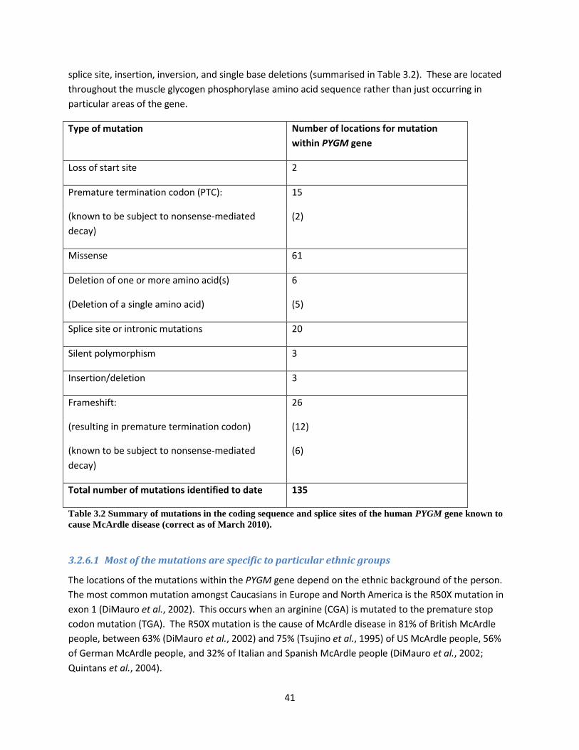
41
splice site, insertion, inversion, and single base deletions (summarised in Table 3.2). These are located
throughout the muscle glycogen phosphorylase amino acid sequence rather than just occurring in
particular areas of the gene.
Type of mutation Number of locations for mutation
within PYGM gene
Loss of start site 2
Premature termination codon (PTC): 15
(known to be subject to nonsense-mediated
decay)
(2)
Missense 61
Deletion of one or more amino acid(s)
(Deletion of a single amino acid)
6
(5)
Splice site or intronic mutations 20
Silent polymorphism 3
Insertion/deletion 3
Frameshift: 26
(resulting in premature termination codon)
(known to be subject to nonsense-mediated
decay)
(12)
(6)
Total number of mutations identified to date 135
Table 3.2 Summary of mutations in the coding sequence and splice sites of the human PYGM gene known to
cause McArdle disease (correct as of March 2010).
3.2.6.1 Most of the mutations are specific to particular ethnic groups
The locations of the mutations within the PYGM gene depend on the ethnic background of the person.
The most common mutation amongst Caucasians in Europe and North America is the R50X mutation in
exon 1 (DiMauro et al., 2002). This occurs when an arginine (CGA) is mutated to the premature stop
codon mutation (TGA). The R50X mutation is the cause of McArdle disease in 81% of British McArdle
people, between 63% (DiMauro et al., 2002) and 75% (Tsujino et al., 1995) of US McArdle people, 56%
of German McArdle people, and 32% of Italian and Spanish McArdle people (DiMauro et al., 2002;
Quintans et al., 2004).

42
Other mutations appear to be specific to ethnic groups. For example, the single codon deletion 708/709
has only been found in Japan. Some “unique mutations … [have been] described in Japanese, Finnish
and Greek families” (DiMauro et al., 2002). Other ethnic mutations include R270X and R602Q, which
were found in two unrelated Yemenite-Jewish families (Hadjigeorgiou et al., 2002).
Until recently, no McArdle people had been identified in some ethnic groups. It seems likely that this is
due to incorrect or absent diagnoses in these groups, particularly in countries such as India and Africa
where the availability of access to qualified medical professionals may be lower. Quinlivan et al. (2010)
recently described an Pakistani McArdle person where both copies of the PYGM gene had a newly
discovered single base pair deletion in exon 1 (c.14delT) which resulted in a premature stop codon
(pL5RfsX20). A Korean person who had a novel single codon deletion (p779delE) plus the R50X mutation
was described in 2008 by Sohn et al. (2008). Several cases of people where McArdle disease was caused
by both copies of the PYGM gene having the V456M mutation in exon 11 have been described; this
mutation was found in a black Morrocan baby (Mancuso et al., 2003), in a Latin American person born in
Ecuador (Fernandez-Cadenas et al., 2007), and in people from Tunisia, and Algeria (Aquaron et al.,
2007).
3.2.7 What caused the first mutation in the PYGM gene?
It is not known what caused the first mutation in the PYGM gene. The body has to copy all the DNA
(including the PYGM gene) to produce egg and sperm cells. If this copying process introduces an error
into the PYGM gene in an egg or sperm cell which is then used to conceive a baby, the mutation in the
PYGM gene will also be inherited. Mutations occur relatively frequently in the human genome, but
often have no effect or mild effect. In general, natural selection removes mutations which have a lethal
effect. It is highly likely that mutations in the PYGM gene and McArdle disease have been around from a
much earlier time than Dr Brian McArdle.
3.3 How is McArdle disease inherited?
3.3.1 One copy of each gene is inherited from each parent
During conception, each person will inherit one set of chromosomes from their mother and one set of
chromosomes from their father. They will inherit a copy of the PYGM gene from each parent – giving a
total of two copies of the PYGM gene in each person.
To continue the analogy to baking a cake with a cook book which was used earlier (section 3.1.1); each
person will have one set of books from their mother and one set from their father. Both the
chromosome (book) from the mother and chromosome (book) from the father will have a gene (recipe
e.g. the PYGM gene) for each protein (e.g. muscle glycogen phosphorylase). So each person will have
two copies of the gene (PYGM) which contains the genetic information for muscle glycogen
phosphorylase enzyme.
43
3.3.2 What are the combinations of wildtype and genes with mutations which can occur?
People unaffected by McArdle’s have two wildtype copies of the PYGM gene. (Wildtype means that it is
a version of the gene with no mutations.) These genes are used to produce active muscle glycogen
phosphorylase enzyme which works normally in muscle.
Carriers have one wildtype copy of the PYGM gene, and one mutant copy of the PYGM gene. They do
not usually have symptoms of McArdle disease (see section 9.1.1). The mutant copy of the PYGM gene
does not result in production of any active muscle glycogen phosphorylase enzyme, but the wildtype
PYGM gene does result in production of active muscle glycogen phosphorylase enzyme. Carriers have
approximately half the amount of active muscle glycogen phosphorylase enzyme than normal controls
(see section 9.1.1), and this is enough enzyme to allow the muscle to work normally.
McArdle people have mutations in both their copies of the PYGM gene. These mutations may be
different in each copy of the PYGM gene. Neither of the mutant copies of the PYGM gene can be used
to produce active muscle glycogen phosphorylase enzyme (for rare exceptions see section 9.1.3).
McArdle people do not have any active muscle glycogen phosphorylase enzyme in their muscles – and
therefore have McArdle disease.
3.3.3 Methods of inheritance
3.3.3.1 Recessive disease
McArdle disease is a recessive disease. In the case of a recessive disease, carriers will not have any
symptoms of the disease as they have one wildtype copy of the gene and one copy of the gene with a
mutation in. With a recessive disease, if both copies of the gene have the mutation, the person will
have McArdle’s.
3.3.3.2 Dominant disease
In a dominant disease, carriers will have symptoms of the disease. Just one copy of the gene with the
mutation in it is sufficient to have all the symptoms of the disease even thought the carried has one
wildtype copy of the gene. An example of a dominant disease is malignant hyperthermia (which is
discussed further in section 12.3.1).
Although there has been at least one paper suggesting that McArdle’s had been inherited in a dominant
or pseudo-dominant way, this was incorrect, and was due to the combination of an asymptomatic
carrier parent with a McArdle parent who had the R50X and a novel mutation which was not discovered
until later (Tsujino et al., 1993; Isackson et al., 2005).
3.3.3.3 Autosomal disease versus sex-linked disease
McArdle disease is an autosomal disease. This means that inheritance is unrelated to gender, and men
and women are equally likely to have McArdle’s.
Sex-linked inherited diseases usually occur due to a mutation in a gene which is located on the X
chromosome. If the disease is sex-linked and recessive, women are much less likely to have the disease

44
as they have two X chromosomes. Even if the gene has a mutation on one of the X chromosomes, they
are likely to have a wildtype copy on the other X chromosome. Men only have one X chromosome, so if
they have a mutation in a gene on that chromosome, they have no “back-up” gene on their second X
chromosome. Men are much more likely to have sex-linked inherited diseases than women.
Haemophilia is an example of a sex-linked recessive disease where men are affected much more
frequently than women.
3.3.4 Examples of how McArdle’s is inherited
Unless otherwise specified, please assume that parents are not related in the following examples. A
different pattern of inheritance might be seen if the parents were closely related, which is explained
further in section 3.3.5.)
As McArdle’s is inherited in an autosomal manner, the possible outcomes are unaffected by whether the
parent with McArdle’s is the mother or father.
NOTE: The following information is based upon theoretical methods of inheritance. It is not intended to
replace genetic counselling. Medical advice/genetic counselling should be sought if you would like to
discuss or further understand the way in which McArdle’s is inherited or the chances of having a child
with McArdle’s.
3.3.4.1 Genetics nomenclature
In the following examples, upper case “M” is used to represent the wildtype copy of the PYGM gene for
muscle glycogen phosphorylase. Lower case “m” is used to represent a copy of PYGM with a mutation
in it because there is no standard one letter code used to represent PYGM, so I have chosen to use the
letter “m”. However, the use of upper and lower case as described above is standard practice in
genetics.
Information about a person’s genotype (information about which copies of the PYGM gene they have)
can therefore be written as follows:
A McArdle person would be written as “mm”
A carrier would be written “Mm”
A person unaffected by McArdle’s would be written “MM”.
Humans have two copies of the PYGM gene. A baby will inherit one copy of the PYGM gene from the
mother and one copy of the PYGM gene from the father. This is achieved because during the
production of egg and sperm (known as gametes) in the parents, only one of the two copies of the
PYGM gene go into each. (The actual mechanism for egg and sperm production is a little more
complicated than this, and involves a few extra steps before this part, but this is the important part and
is sufficient to understand inheritance.) If the parent is unaffected by McArdle’s (MM), the gametes will
be either “M” or “M”, so the child can only inherit “M”. If the parent is a carrier (Mm), the child could
inherit either M” or “m”. If the parent has McArdle’s (mm), the child could only inherit “m”. When the

45
parents have a baby, the possible outcomes can be illustrated by a diagram, known as a Punnet square
(invented by Mr Punnet!). The chances for each subsequent child are exactly the same and are not
altered by whether the previous child had McArdle’s, a carrier or was unaffected.
3.3.4.2 Neither of my parents had symptoms of McArdle’s, but I have McArdle’s
It is likely that both parents were carriers (each parent had one wildtype copy of the PYGM gene and
one copy of PYGM with a mutation). In order for the child to have McArdle’s, the child must have
inherited a copy of PYGM with a mutation from each parent – resulting in two copies of PYGM with
mutations and no wildtype copies.
Carrier parent (Mm) plus carrier parent (Mm):
If both parents are carriers, there is a 1 out of 4 (25%) chance that the child will be unaffected, a 2 out of
4 (50%) chance that the child will be a carrier, and a 1 out of 4 (25%) chance that the child will have
McArdle’s.
Father (Mm, carrier)
Sperm cells have:
M m
Mother (Mm, carrier)
Egg cells have:
M MM (unaffected) Mm (carrier)
m Mm (carrier) mm (McArdle’s)
Figure 3.2 A punnet square illustrating the possible outcomes when a father who is a carrier (Mm) has a
baby with a mother who is also a carrier (Mm). There is a 1 out of 4 (25%) chance that the child will be
unaffected (MM), a 2 out of 4 (50%) chance that the child will be a carrier (Mm), and a 1 out of 4 (25%)
chance that the child will have McArdle’s (mm).
3.3.4.3 I have McArdle disease and my partner does not; will my children have McArdle
disease?
The parent with McArdle disease will be homozygote for the mutation; will have two copies of PYGM
each with a mutation. There are two possible situations for the partner. The partner could be
unaffected (have two copies of the wildtype gene), or could be a carrier.
McArdle’s parent (mm) plus unaffected parent (MM):
The only possible outcome is that the child is a carrier (Mm). There is a 4 out of 4 (100%) possibility that
the child will be a carrier. This is the most commonly occurring situation, and results in a child which is a
carrier and does not have any symptoms of McArdle’s.

46
Father (mm, McArdle’s)
Sperm cells have:
m m
Mother (MM, unaffected-
homozygote for wildtype
copy of PYGM)
Egg cells have:
M Mm (carrier) Mm (carrier)
M Mm (carrier) Mm (carrier)
Figure 3.3 A punnet square illustrating the possible outcomes when a father who has McArdle’s (mm) has a
baby with a mother who is unaffected (MM). The only possible outcome is that the child is a carrier (Mm).
There is a 4 of 4 (100%) chance that the child will be a carrier.
McArdle’s parent (mm) plus carrier parent (Mm):
There is a 2 out of 4 (50%) chance that the child will be a carrier, and a 2 out of 4 (50%) chance that the
child will have McArdle’s. None would avoid receiving at least one copy of PYGM with a mutation.
There is a 50% chance that both the parent (the father in this example) and the child will have
McArdle’s. This can be the explanation when McArdle’s appears to run in families and to be passed
down the generations. This doesn’t happen very often because McArdle’s is relatively rare.
Father (mm, McArdle’s)
Sperm cells have:
m m
Mother (Mm, carrier)
Egg cells have:
M Mm (carrier) Mm (carrier)
m mm (McArdle’s) mm (McArdle’s)
Figure 3.4 A punnet square illustrating the possible outcomes when a father who has McArdle’s (mm) has a
baby with a mother who is a carrier (Mm). There is a 2 out of 4 (50%) chance that the child will be a carrier
(Mm), and a 2 out of 4 (50%) chance that the child will have McArdle’s (mm).
3.3.4.4 Both myself and my partner have McArdle’s
This is a very unusual situation, but is included for completeness. If both parents have McArdle’s, they
must both be homozygote for the mutation; must have two copies of PYGM each with a mutation.
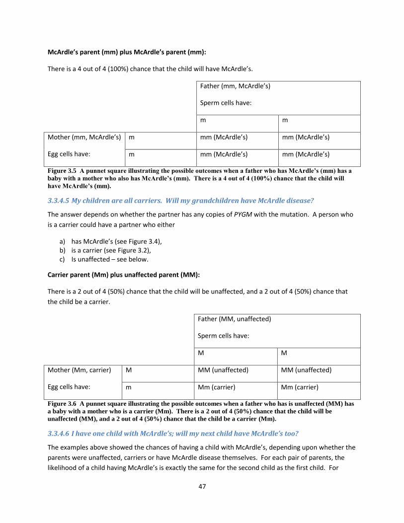
47
McArdle’s parent (mm) plus McArdle’s parent (mm):
There is a 4 out of 4 (100%) chance that the child will have McArdle’s.
Father (mm, McArdle’s)
Sperm cells have:
m m
Mother (mm, McArdle’s)
Egg cells have:
m mm (McArdle’s) mm (McArdle’s)
m mm (McArdle’s) mm (McArdle’s)
Figure 3.5 A punnet square illustrating the possible outcomes when a father who has McArdle’s (mm) has a
baby with a mother who also has McArdle’s (mm). There is a 4 out of 4 (100%) chance that the child will
have McArdle’s (mm).
3.3.4.5 My children are all carriers. Will my grandchildren have McArdle disease?
The answer depends on whether the partner has any copies of PYGM with the mutation. A person who
is a carrier could have a partner who either
a) has McArdle’s (see Figure 3.4), b) is a carrier (see Figure 3.2), c) Is unaffected – see below.
Carrier parent (Mm) plus unaffected parent (MM):
There is a 2 out of 4 (50%) chance that the child will be unaffected, and a 2 out of 4 (50%) chance that
the child be a carrier.
Father (MM, unaffected)
Sperm cells have:
M M
Mother (Mm, carrier)
Egg cells have:
M MM (unaffected) MM (unaffected)
m Mm (carrier) Mm (carrier)
Figure 3.6 A punnet square illustrating the possible outcomes when a father who has is unaffected (MM) has
a baby with a mother who is a carrier (Mm). There is a 2 out of 4 (50%) chance that the child will be
unaffected (MM), and a 2 out of 4 (50%) chance that the child be a carrier (Mm).
3.3.4.6 I have one child with McArdle’s; will my next child have McArdle’s too?
The examples above showed the chances of having a child with McArdle’s, depending upon whether the
parents were unaffected, carriers or have McArdle disease themselves. For each pair of parents, the
likelihood of a child having McArdle’s is exactly the same for the second child as the first child. For

48
example, in the case of a carrier parent plus an carrier parent, as illustrated in Table 3.1, the first child
has a 25% chance of having McArdle’s. If the same parents have a second child, this child will also have
a 25% chance of having McArdle’s. All subsequent children will each have a 25% chance of having
McArdle’s.
3.3.4.7 My grandmother/grandfather/aunt/uncle has McArdle disease
Having a grandparent or aunt/uncle with McArdle’s may suggest that you could be a carrier. It will not
be possible to determine this without a genetic test (or possibly a muscle biopsy to accurately quantify
the amount of muscle glycogen phosphorylase, as carriers usually have approximately half the amount
of muscle glycogen phosphorylase compared to unaffected people). Unless family members have had
children together, the chances of a carrier having a partner who is also a carrier is relatively unlikely. It
is therefore unlikely that you would have a child which has McArdle’s. For this reason, I suspect that
many family doctors would not be interested in performing tests to determine if you are a carrier.
3.3.5 Consanguineous parents; why are they more likely to produce children with
inherited disease?
Consanguineous means that the parents are closely related. Examples of this could be two siblings or
first cousins having children together. The disadvantage of the close relationship between the parents is
that if there is a recessive disease in the family, siblings or cousins are more likely to both be carriers. If
two carriers then have children together, the chances of having a child with the disease are much
higher. In addition, siblings or cousins are more likely to be carriers for more than one genetic disease,
and this may result in the children having multiple inherited diseases. From a genetic point of view,
consanguineous parents are not recommended. Mancuso et al. (2003) reported a consanguineous
marriage, which resulted in the birth of a female baby (see section 8.1.1). This baby had both McArdle
disease and also mitochondrial DNA depletion syndrome, both of which are autosomal recessive
diseases. It is likely that both parents were carriers for both of these diseases, and the child inherited
several diseases.
3.3.6 How many people have McArdle disease or are carriers of a mutation in PYGM?
In order to determine whether you are likely to have children with a partner who is a carrier of the
mutation in PYGM, it is useful to know how commonly carriers are found in the population. It has been
estimated that the number of people with McArdle disease is approximately 1 in 100,000 (Applegarth et
al., 2000; Haller, 2000; Tarnopolsky, 2006; Beynon et al., 2002). It has been suggested that the number
of people who are carriers for McArdle disease is between 1 in 66 (Tarnopolsky, 2006) and 1 in 158
(Isackson et al., 2005).
Further reading:
Basic Genetics: Textbook and Activities By Ahmed Abouelmagd, Hussein M. Ageely (Google free books).
(For general information about inheritance. There are many other complicated patterns of inheritance,
so focus on “autosomal recessive inheritance”.)

49
4 Exercise, muscle contractions, and contractures (muscle cramps)
4.1 Types of exercise
Exercise can basically be divided into two classes; aerobic and anaerobic.
4.1.1 Anaerobic exercise
Anaerobic exercise occurs when energy is produced in the muscle cells without using oxygen. This
occurs when the blood is not able to supply enough oxygen to the muscle cells for aerobic exercise to
occur (Sleamaker and Browning, 1996). Anaerobic exercise is usually when a short burst of high
intensity exercise occurs. An example of this would be fast sprinting, like running for a bus. The energy
for muscle cells to move during exercise is usually provided by a combination of aerobic and anaerobic
exercise. However, the first 10 to 30 seconds of exercise are solely anaerobic r (Sleamaker and
Browning, 1996).
Initially, free glucose in the muscle cells is used to provide energy for movement. However, this is used
up within the first few minutes of exercise. In people unaffected by McArdle’s, energy is then produced
by glycogenolysis. This is the chemical reaction where stored glycogen is broken down to produce
glucose, which is used to generate ATP. The muscle glycogen phosphorylase enzyme is essential for this
chemical reaction in muscle cells.
4.1.2 Aerobic exercise
Aerobic exercise takes place when oxygen is supplied in the bloodstream to the muscles. Taking a
breath of air pulls the air into in the lungs. The lungs are made up of lots of alveoli which are tiny sacks.
The surfaces of the alveoli are covered in lots of blood vessels. Oxygen from the air binds to
haemoglobin in the red blood cells. Haemoglobin, with oxygen bound to it, is then pumped around the
body to many places, including the muscles. In the muscles, oxygen is taken from the haemoglobin in
the blood vessels into the muscle cells. The oxygen is then combined with sources of energy such as
free fatty acids in chemical reactions which generate ATP (energy). Aerobic exercise is defined as
exercise for a minimum of ten minutes (Sleamaker, 1996). An example of aerobic exercise would be
walking on a road or treadmill for 20 minutes.
4.2 Exercise recommendations for McArdle people
4.2.1 Anaerobic exercise or static muscle contractions should be avoided
Strenuous exercise must be avoided as it can lead to muscle damage, muscle cell breakdown and
possible kidney failure. Lucia et al. (2008a) recommend McArdle people to avoid exercise that induces
severe pain, and in particular to avoid static muscle contractions. Static muscle contractions (also called
isometric contractions) are those which are required to hold something in one place for a long time,
such as holding the body in one place for a long time by squatting, or holding a heavy bag or weight for a
long time.

50
Andrew Wakelin, McArdle’s representative for AGSD UK, suggests a “six second rule” (see Box 4.1). This
suggestion is based upon the experiences of McArdle people, but has not been scientifically proven.
The six second rule:
“To avoid damage when doing something of maximum intensity it is a good idea to time 6 seconds by
saying to "One thousand, two thousand..." up to six. If the task is not completed by 6, stop or put it
down. Take a break, let the muscles recover and try again later.”
Box 4.1 The six second rule as suggested by Andrew Wakelin. (Taken from www.agsd.org.uk.)
4.2.2 Moderate aerobic exercise and conditioning improves symptoms of McArdle’s
The breakdown of fatty acids and amino acids, and the release of glucose from the liver, provide the
energy for the second wind. In the second wind, energy is produce using oxygen for oxidative
phosphorylation, and is therefore aerobic. McArdle people are able to continue to exercise aerobically
for a long period of time using fatty acid oxidation to provide energy (Quinlivan and Vissing, 2007).
Exercising regularly (but gently) improves aerobic conditioning. Aerobic conditioning is the ability of the
lungs to take in more oxygen, and the ability of the heart to pump blood round the body. Aerobic
conditioning improves the supply of glucose, fatty acids, and oxygen to the muscles via the bloodstream,
and also improves the ability of the mitochondria to utilise sources of energy (Vissing and Haller, 2003;
Quinlivan and Vissing, 2007; Quinlivan et al., 2008). Warming up prior to exercise will improve blood
supply to the muscles, and can aid the transition to “second wind” (Haller, 2000).
Low level warm-up (Amato, 2003) and regular light aerobic exercise can speed the beneficial transition
to second wind from anaerobic to fatty acid oxidation in McArdle people (Quinlivan et al., 2008). Perez
et al. (2008) described a 9 year old boy with McArdle disease who presented with severe myalgia
(muscle pain), muscle weakness, myoglobinuria, and elevated creatine kinase after exercise. He was
recommended to consume 20g of carbohydrate before exercise, and to carry out age-appropriate
exercise. One year later, his fitness had improved, he was able to exercise and perform ordinary
activities, and had almost normal serum creatine kinase levels. He had not had any acute clinical
episodes (such as myoglobinuria).
Haller et al.(2006) investigated whether aerobic conditioning (regular aerobic exercise) could improve
the ability of McArdle people to exercise. The training programme for eight McArdle people was as
follows: they were trained on a cycle ergometer for 30-40 minutes, four days a week, for 14 weeks.
They exercised at an intermediate rate, not the hardest they could do. The authors found that after this
period of training, the participants were able to carry out more exercise, take in more oxygen (which is
needed to produce energy during the second wind), and their heart was able to pump blood more
efficiently. They also found that levels of some of the enzymes involved in producing energy (called
“citrate synthase” and “β-hydroxyacyl coenzyme A dehydrogenase”) had increased. The participants did
not have pain, cramps, and the level of creatine kinase in the blood did not rise during the exercise –
which suggested that the exercise was not causing muscle damage. The participants were still able to
achieve a second wind in the same way as before the training programme. The authors concluded that
51
moderate aerobic exercise was a way to increase the ability of McArdle people to exercise. The training
programme improved the ability of the heart to pump blood around the body, which increased the
amount of energy sources which could be taken by the blood to the muscles.
Other researchers have also described positive results of aerobic training for McArdle people. Quinlivan
and Vissing (2007) describe unpublished data by Portero et al.: Portero et al. carried out a trial where
four McArdle people performed aerobic exercise for 30-45 minutes three times a week for eight weeks.
The authors said that the results showed an improvement in the amount of oxygen taken into the body,
an increase in the amount of glucose carried in the blood to the muscle cells, and increase in the ability
to exercise and the length of time which exercise could be carried out, and also a reduction in the CK
levels in the blood. Quinlivan and Vissing (2007) also describe an unpublished study by Zange et al., who
studied two McArdle people who carried out aerobic training daily for 12 weeks. Following the training
programme, these McArdle people also had “an improvement in improvement in strength without any
significant adverse effects. There were no episodes of pain or cramping.”McArdle people
4.2.3 The phrase “No pain, no gain” does NOT apply to McArdle people
For people with McArdle’s, pain is felt in the muscles when the muscle cells run out of energy. This pain
is a way of the body telling you to stop exercising and rest. After a rest, energy should then be provided
by the second wind and it should then be possible to exercise further. For McArdle people, the pain is a
warning to tell you to stop exercising and rest. Continuing to exercise once pain is felt could lead to
muscle damage. Lucia et al. (2008a) describe the severe pain which is felt during exercise as a “self-
protective mechanism”, and say that if a McArdle person continues to exercise once pain occurs then it
could increase the risk of muscle damage, myoglobinuria and kidney failure. For McArdle people, the
saying “no pain, no gain” does NOT apply and could have dangerous consequences!
4.2.4 Being overweight can make McArdle people find it harder to move around
A large number of McArdle people are overweight. Quinlivan et al. (2010) reported that 71% (32 of 45)
of the McArdle people seen at the Oswestry clinic were clinically overweight, with a body mass index
(BMI) greater than 26. This is not surprising when you remember that in the past, McArdle people were
advised to avoid exercise. And also that in the past, McArdle people were advised to have a sugary drink
before exercise. If a sedentary lifestyle is combined with consuming a lot of sugar, it is likely to lead to
weight gain. The advice to McArdle people is now difference; moderate exercise is recommended (see
section 4.2.2), and although a sugary drink can be of use in certain circumstances, it obviously has to be
used in moderation (see section 7.1.6.2). McArdle’s experts are now keen to ensure McArdle people are
warned about this disadvantage of a sugary drink. “When informing people with McArdle disease about
such treatment [sugary drinks before exercise], family doctors should stress the importance of
restricting its use to avoid unintentional weight gain” (Vissing and Haller, 2003).
For people unaffected by McArdle disease, the ways in which they can maintain a healthy weight include
a balanced diet, and regular exercise. For McArdle people, there is some debate about what the
balance of carbohydrate and protein in the diet should be (see section 0), but it remains the case that if
you consume more calories (more food or energy) than you use, then this will result in weight gain.

52
Energy from food is used up in everyday life (walking around or doing housework) in addition to during
exercise.
For anybody (whether they have McArdle’s or not), being overweight leads to an increased risk of other
serious health problems such as heart disease and cancers of the breast, colon, and prostate (source:
http://www.nhs.uk/Livewell/loseweight/Pages/Whyyourweightmatters.aspx). In addition to these risks,
a disadvantage of being overweight for a McArdle person is that it may make it harder to exercise
(Amato, 2003). A heavier body weight increases the amount of work which the muscles have to do in
holding the body upright and in moving around. This is more likely to lead to muscle damage
(rhabdomyolysis and muscle pain) if the muscles are unfit or unconditioned.
4.3 Muscles require energy (ATP) to contract and to relax during exercise
Muscle contractions require energy. Food is the ultimate source of energy. Food is digested in the
digestive system then transported in the blood to the muscle cells where it is further broken down by
several different mechanisms to release energy (as ATP). The muscle cells require ATP to contract.
Muscle contractions are needed to carry out all forms of exercise. However, in McArdle people, the lack
of muscle glycogen phosphorylase means that muscles are not able to use ATP produced from the
breakdown of glycogen into glucose.
4.4 Energy is stored in bonds between adenosine and phosphate, and
energy can be released when the bonds are broken
Within muscle cells (and other cells in the body), energy is stored in bonds between adenosine and
phosphates. Up to three phosphates can be attached to adenosine, and the names of the compound
will change accordingly (see Table 4.1). When the bond between a phosphate and the adenosine is
broken, energy is released. In the example given in Box 4.2, adenosine triphosphate (ATP) is converted
into phosphate (denoted as “Pi”) and adenosine diphosphate (ADP), releasing energy. This release of
energy can be used for many purposes, including driving a calcium pump, as described above.
Number of phosphates attached to adenosine Name of the structure
1 Adenosine monophosphate (AMP)
2 Adenosine diphosphate (ADP)
3 Adenosine triphosphate (ATP)
Table 4.1 Names of the structure depends on how many phosphates are bound
ATP ADP + Pi + energy
Box 4.2 Energy can be released from ATP

53
4.4.1 A brief description of muscle contraction and relaxation
A very simplified description of muscle cells is that they are like a bag of liquid. The liquid is called the
cytoplasm or sarcoplasm. Inside this bag, are small compartments, one of which is called the
sarcoplasmic reticulum. A neuromuscular junction joins a nerve to a muscle. In order for a muscle to
contract, a compound called “acetylcholine” is released from the nerve. This passes through the
neuromuscular junction and binds to the end of the muscle. When acetylcholine binds to the muscle, it
opens special channels which let sodium flow into the sarcoplasm and potassium flow out. This causes
more channels to open along the length of the muscle, causing a ripple or wave effect. There is also a
calcium-sodium pump. Once the concentration of sodium has built up within the cells, a calcium-
sodium pump uses the high concentration of sodium to move calcium from the sarcoplasm to the
sarcoplasmic reticulum. It also causes the release of calcium from the sarcoplasmic reticulum, which
binds to troponin (a component of muscle) causing muscle contraction. After a muscle contraction has
occurred, the calcium ATPase pumps use ATP to provide the energy to pump calcium from the
sarcoplasm to the sarcoplasmic reticulum. This removes calcium from the troponin, which results in
relaxation of the muscles (Martonosi, 2000).
4.4.2 A more detailed description of the role of the calcium ATPase pump
In muscle cells, a special calcium pump moves calcium from the sarcoplasm into the sarcoplasmic
reticulum. Rather like hydroelectric power, the body uses a calcium pump (called an “ATPase”) powered
by ATP, to build up a concentration of calcium inside the sarcoplasmic reticulum. When a nerve
stimulates a muscle, calcium is released and flows from the sarcoplasmic reticulum into the sarcoplasm.
This flow of calcium stimulates the muscle to contract. In people unaffected by McArdle’s, the calcium
is all pumped back into the sarcoplasmic reticulum within 30 milliseconds, causing the muscles to relax
(Alberts et al., 1994).
4.4.3 A more detailed description of the role of the sodium-potassium ATPase pump
There is also a sodium-potassium pump in muscle cells which works in a very similar way to the calcium
pump (Alberts et al., 1994). The sodium-potassium pump is also an ATPase, and is powered by ATP. The
sodium-potassium pump builds up a high concentration a concentration of sodium outside the cell, and
a high concentration of potassium inside the cell, in the sarcoplasm. The sarcoplasm has a very high
concentration of potassium, and the sarcoplasmic reticulum has a high concentration of sodium. The
sodium has several purposes. Firstly, sodium is used to transmit a nerve impulse (as described above).
In addition, accumulating the sodium in the sarcoplasmic reticulum keeps the amount of water at the
right concentration. If the sodium stopped being pumped out, water would diffuse into the cell by a
process called “osmosis”, and this could cause the cells to swell.
4.5 A muscle contracture (cramp) can occur if McArdle people exercise
anaerobically
Muscle contractures (cramps) are a well known phenomenon in McArdle people following intense
exercise (Rommel et al., 2006; Lucia et al., 2008a and many other publications). Contractures cause the
muscles to go hard, to swell up, and it becomes very hard to move or relax the muscle. Contractures
54
can occur if the muscles are exercised intensely, and if the McArdle person continues to exercise once
pain is felt.
The biochemical reason for contractures is still not properly understood (Lucia et al., 2008a). The
general theory appears to be that contractures are due to ATPase “pumps” not working. One possibility
is that this is due to a lack of ATP, which provides the pumps with energy, but this does not appear to be
correct. Further studies suggest that a build of up Pi and ADP (both of which are produced from ATP
when energy is released) might inhibit the ATPase pumps and stop them working. But the research does
not yet seem to be conclusive.
The first theory was that the contracture could be caused by the lack of ATP in the muscle cells. ATP is
the source of energy in the muscle cells, which is usually produced from glucose. Since it is not possible
for McArdle people to break down glycogen and produce glucose, the muscles may run out of ATP. As
early as 1968, a research group including Dr Brian McArdle initially suggested that it could be “depletion
of ATP giving rise to a contracture similar to rigor mortis” (Gruener et al., 1968). Studies by Gruener et
al. found that “there was little or no reduction in the ATP level of muscle in contracture”, which was
later confirmed by Lewis et al. (1985), Lewis and Haller (1986) and Lofberg et al. (2001). These studies
appear to have shown that the levels of ATP in the muscles of McArdle people do NOT decrease with
exercise, suggesting that contractures are not caused in the same way as rigor mortis.
An alternative theory of why the lack of ATP could cause a contracture was that a lack of ATP may
prevent a sodium-potassium pump (powered by ATP) from working. Rather like hydroelectric power,
the body uses a sodium-potassium pump, powered by ATP, to build up a concentration of sodium on
one side of the cell membrane, and a concentration of potassium on the other side. When a nerve
stimulates a muscle contraction, the sodium and potassium flow back across the membrane, allowing a
“wave” to spread rapidly along the muscle. This acts as a very fast signal for the muscle to contract. If
the pump runs out of power, this concentration cannot be built up, and the muscle won’t be able to
contract. Haller et al. (1998) studied the effect of exercise on the sodium-potassium pumps in McArdle
people. They found that the sodium-potassium pump levels were low, and this suggested that not
enough potassium was being pumped, and would be unable to keep up during exercise.
When ATP is used to produce energy for muscle contraction, it is broken down into Pi and ADP. Lewis and Haller (1986) said that exercise would lead to an increased amount of Pi and ADP in muscle cells. They state that “accumulations of Pi and ADP are known to inhibit the myofibrillar, Ca2+ [calcium], and Na+-[sodium] [and] K+ [potassium] - ATPase reactions”. These are the ATPase pumps which are described above.
Swift and Brown (1978) studied two McArdle people. They used radioactivity to see what happened to
the bone and muscle during contractures. They found unusual results, because the muscles became
labelled in a way which suggested that calcium was involved in contractures. The authors suggested
that either the exercise caused muscle damage which led to accumulation of calcium in the muscles
(during contracture), or that certain small capsules in the muscle cells (called sarcoplasmic reticulum)
were unable to pump the calcium back into them after activity, so that the calcium remained in the main

55
part of the cell (the cytoplasm/sarcoplasm). This may be the research that led Quinlivan and Vissing
(2007) to say that contracture is caused by “inhibition of calcium ATPase” (unfortunately they did not
give a reference for the research to justify this statement).
If the sodium stopped being pumped out, water would diffuse into the cell by a process called osmosis,
and this could cause the cells to swell. I wonder if this leads to the swelling of muscle cells observed by
McArdle people during contracture, but haven’t found any published evidence to support this theory.
It may be relevant to note that dantrolene sodium (see section 7.1.5) was tested as a treatment for
McArdle’s. Dantrolene sodium acts on the muscle cells, by providing a very high amount of sodium
which stops the movement of sodium. Dantrolene sodium works as a muscle relaxant, and can be used
to treat malignant hyperthermia (see section 12.3.1).
4.5.1 Treatment for contractures
Contractures occur if a muscle is continued to be exercise/used after pain is felt. If possible, it is best to
avoid producing a contracture.
There is limited information on why contractures cause pain. Mense et al. (2001) say that the pain
seems to be due to the muscle needing energy, and being unable to get energy. During exercise the
liquid inside the cells may become more acidic, and this may trigger pain sensations. In addition, the
accumulation of potassium outside the muscle cell can cause pain.
There is very little published information on how to treat contractures once they have occurred. Mense
et al. (2001) say that contractures are “relieved by rest”. McArdle people have anecdotally reported the
following home treatments; use of cool/ice packs, heat packs, strong painkillers, massage. There is no
published information on any of these home treatments, and some of these treatments (like massage)
could lead to further damage to the muscles. Pain can serve as a warning to protect the muscle from
further damage, and the use of strong painkillers could lead to the muscle being used, which could
cause further damage.
IMPORTANT: Contractures may be accompanied by rhabdomyolysis (muscle breakdown), which can lead
to increased creatine kinase levels and myoglobinuria. Severe muscle damage may lead to kidney
failure, which can be fatal. (See section 5 for further information.) In the event of severe muscle
damage, medical attention should be sought.
Further reading:
Serious training for endurance athletes by Rob Sleamaker, Ray Browning, 1996 (Information about
different types of exercise.)
Biology: Concepts and Applications Without Physiology By Cecie Starr, Christine A. Evers, Lisa Starr (An easier explanation of the role of the calcium, sodium and potassium pumps, see page 85.)

56
Essential medical physiology By Leonard R. Johnson, John H. Byrne (A more complicated explanation of the role of the calcium, sodium and potassium pumps.)
Muscle pain: understanding its nature, diagnosis, and treatment By Siegfried Mense, David G. Simons, I.
Jon Russell, 2001

57
5 Muscle damage (rhabdomyolysis) can lead to raised creatine kinase
levels in the blood, myoglobinuria, and kidney failure
5.1 Rhabdomyolysis and acute kidney failure
Rhabdomyolysis occurs when muscle damage leads to the breakdown of skeletal muscle cells. Muscle
cells are like a balloon which is filled with water and lots of little components, including creatine kinase
and myoglobin. When muscle damage occurs, the components are released. The broken parts of the
muscle cells and the components go into the bloodstream, and then pass through the kidneys. The
kidneys act like sieves, and filter these broken parts from the blood. The smaller bits, like myoglobin,
pass out in the urine – producing dark red/brown coloured urine known as myoglobinuria. (An
alternative name for myoglobinuria is proteinuria.) If the muscle damage is severe, the parts of the cells
may clog up the kidneys. Kidney failure occurs if the kidneys become clogged and are unable to
function.
The kidneys also play an important role in keeping the correct concentration of several compounds
including potassium, sodium and calcium (called “electrolytes”) in the bloodstream. The kidneys ensure
that the concentration of potassium and calcium is maintained. If kidney failure occurs, the kidneys are
no longer able to maintain the correct concentrations of electrolytes.
If a small amount of rhabdomyolysis occurs, the person may feel fine, but may have raised CK levels. It
is most likely to lead to muscle weakness, muscle pain and dark urine. If a very large amount of
rhabdomyolysis occurs, it could lead to extremely high CK levels in the blood, electrolyte imbalances,
acute renal failure, and the formation of blood clots in blood vessels (Huerta-Alardín, 2005).
5.2 Causes of rhabdomyolysis
5.2.1 Causes of rhabdomyolysis in people unaffected by McArdle disease
There are many causes of rhabdomyolysis in people unaffected by McArdle’s. These are outlined in
Table 5.1. It is likely that these may also increase the chance of rhabdomyolysis in McArdle people, and
would be best avoided.crush syndrome: being crushed or trapped. E.g. being crushed or trapped
following an earthquake or car accident.

58
Immobilisation: if a person lies motionless for a long period of time this can compress (and damage) the
muscle. E.g. if an elderly person has a fall or a stroke and then remains in one position for a long time, if
a person is drunk and lies motionless for a long time.
Surgery: if a surgical procedure is performed in an improper position or a tourniquet is used for a long
period of time.
Extreme physical exertion, especially in conditions of high temperature and humidity.
Hypokalemia (when potassium levels in the blood are lower than they should be) increases the risk of
rhabdomyolysis during strenuous exercise. Diuretics are drugs which increase the rate of urine
production and can lead to depletion of electrolytes such as potassium. Abuse of diuretics may lower
potassium levels, making athletes more likely to develop rhabdomyolysis during strenuous exercise.
Any condition which produces major electrolyte losses and dehydration, such as severe diarrhoea,
vomiting, or bulimia (see cross ref) could lead to rhabdomyolysis.
Electric shock and lightning strikes.
Hyperthermia including malignant hyperthermia (see section 12.3.1 for more information on malignant
hyperthermia): A symptom of malignant hyperthermia is an extreme rise in body temperature, leading
to excessive sweating, which can reduce potassium levels. Neuroleptic malignant syndrome is a reaction
to antipsychotic drugs like butyrophenones, phenothiazines and thioxanthenes. This can lead to
hyperthermia, muscle rigidity, and rhabdomyolysis.
Heat stroke: where people have a core body temperature above 40.5 °C.
Hypothermia (being very cold or frozen) can reduce the blood flow to the muscles, reducing the amount
of glucose and energy available for movement. Freezing of muscle cells can breakdown and damage the
cells.
Recreational drugs such as cocaine and LSD (D-lysergic acid diethylamide). Cocaine can cause
rhabdomyolysis by having a toxic effect on muscle cells; prolonged use can limit the amount of blood
flow to the muscles, and by inducing coma and immobility for a long time.
Prescribed drugs. Statins are some of the most widely prescribed drugs in the United States. Statins are
well known to have the side effect of rhabdomyolysis. Rhabdomyolysis may be triggered by statins
within a short period of taking the drug, or many years later. Statins can also cause inflammation of the
muscles (myositis), causing pain, and weakness of the muscles.
Excessive amounts of alcohol, either binge drinking or alcohol abuse, can lead to pain and swelling of the
muscles.

59
Excessive eating of large quantities of liquorice (often found in sweets), which has a component that
reduces levels of potassium in the kidney.
Infections including influenza, Epstein–Barr virus, herpes simplex virus, HIV, sepsis, legionella infection,
bacteria pyomyositis (bacterial infection of the skeletal muscles)
Metabolic diseases including McArdle disease, Tauri disease, phosphoglycerate mutase deficiency, and
carnitine palmitoyltransferase deficiency (see cross ref).
Table 5.1 Causes of rhabdomyolysis in people unaffected by McArdle’s (summarised from Huerta-Alardín,
2005).
5.2.2 Causes of rhabdomyolysis in McArdle people
In McArdle people, rhabdomyolysis is most likely to be caused by excessive exertion during exercise.
This can occur if a McArdle person continues to exercise once pain if felt.
Some drugs have the side effect of rhabdomyolysis in people who are unaffected by McArdle’s, and
recently, some of these have been found to also increase the risk of rhabdomyolysis in McArdle people.
For example, the cholesterol lowering drugs known as statins have been reported to increase the
likelihood of rhabdomyolysis in McArdle people. See section 12.1 for a list of drugs which may cause
rhabdomyolysis in McArdle people.
Voduc et al. (2004) recommend that McArdle people should be advised to avoid dehydration
(dehydration is the lack of water and lack of essential salts like potassium), and to seek medical
attention if they have dark urine.
5.3 Symptoms of rhabdomyolysis
5.3.1 General symptoms
General symptoms are feeling ill, fever, increased heart rate (tachycardia), nausea and vomiting. The
early complications include an increased level of potassium in the blood (hyperkalemia), very low levels
of calcium in the blood (hypocalcaemia), elevated liver enzymes, cardiac dysrrhythmias and cardiac
arrest, while the late complications include acute renal failure and clotting of blood in the blood vessels
(called “disseminated intravascular coagulation”).
5.3.2 Raised creatine kinase (CK) levels in the bloodstream
Creatine kinase (CK) is also known as phosphocreatine kinase (PCK). CK levels in the blood can be used
as an indicator of muscle damage (there is an approximate relationship that the higher the amount of CK
in the blood, the greater the muscle damage).
Normal CK levels in men unaffected by McArdle’s are 50–500 U/l, although those of women are often
25% lower (Cush, 2005). In people unaffected by McArdle’s, raised CK level above 5000U/L indicates
severe muscle injury, and suggests an increased risk of kidney failure (Huerta-Alardín, 2005). An
increase in CK due to rhabdomyolysis will occur within 12 hours of the onset of muscle injury. The CK

60
level will reach a peak in 1–3 days, and begin to decrease 3–5 days afterwards. The peak CK level may
indicate the likelihood of kidney failure (Huerta-Alardín, 2005).
5.3.2.1 McArdle people always have raised CK levels, even at rest
McArdle people always have raised CK levels, even at rest. All (100%) of McArdle people have CK levels
above 200U/L at rest (Lucia et al., 2008a). Approximately half (approximately 50%) of McArdle people
have CK levels above 1000 U/L at rest (Lucia et al., 2008a). Anecdotally, the CK levels at rest which are
“normal” for a McArdle person seem to vary from person to person. If you have a CK test, it is
important to inform the family doctor that you have McArdle’s. It is also useful to know your “normal”
CK range.
In McArdle people, the CK levels can rise to “several thousand U/l, indicating marked rhabdomyolysis
after intense exercise” (Lucia et al., 2008a). (This is known as “hyper-CK-emia”.)
5.3.3 Myoglobinuria (blood in the urine)
It can be useful to quantify the amount of myoglobin the blood and urine as a measure of the amount of
muscle damage which has occurred. Although the amount of myoglobin in the blood can be measured,
CK levels remain higher in the blood for longer than myoglobin levels. (The half-life of CK is 1.5 day).
Myoglobinuria (blood in the urine) can cause the urine to be a colour ranging from “pink-tinged, to cola-
coloured, to dark black” (Huerta-Alardín, 2005). The red-brown colour is usually seen when the
myoglobin concentrations in urine are above 300mg/l. A urine dipstick can be used to test for
myoglobinuria. The dipsticks are designed to detect haemoglobin (at concentrations of 0.3mg/l), but
should also detect similar concentrations of myoglobin (Huerta-Alardín, 2005).
IMPORTANT: Urgent medical attention should be sought if you are unable to urinate, as it could indicate
kidney failure (Greenberg, 2005 ).
5.3.4 Treatment for rhabdomyolysis:
In order to protect the kidneys, the person will need vigorous hydration (Huerta-Alardín, 2005). The
main treatment is aggressive fluid replacement with saline (water containing salts), usually delivered via
an intravenous drip. The amount of urine which is passed will probably be measured to determine how
much fluid is passing through the kidneys. A catheter may be inserted into the urethra to achieve this,
or you may be asked to urinate into a cup/bedpan or other measuring device. The quicker rehydration
is achieved, the lower the risk of kidney failure.
Mannitol and bicarbonate are often given after saline has been given. Mannitol may reduce the amount
of cell injury, may reduce the amount of heme which is left in the tubes of the kidneys (heme deposits
are bad for the kidneys), and also may help to keep the tubes of the kidney open (is a renal vasodilator).
Sometimes other diuretics such as furosemide are given to try to encourage the flow of liquids through
the kidneys. After rhabdomyolysis, a large amount of acidic urine may pass through the kidneys. Acidic
urine may be damaging to the kidneys. Bicarbonate would help to neutralise the acidic urine, and could
help to reduce the level of damage. However, there is not very much clinical evidence for use of

61
bicarbonate or mannitol, and just the use of saline alone seems to have an equal result (Huerta-Alardín,
2005).
Even if immediate treatment is give, some people may have kidney failure (acute renal failure). The
treatment for acute renal failure is an intravenous drip to provide the correct fluid amount of fluid, to
make sure the acidity is correct, and to add any electrolytes (such as potassium or calcium) which are
require. Dialysis may be needed to remove urea and potassium, which are released during muscle
damage. It is very important to correct the concentration of potassium in the bloodstream as
hyperkalemia can lead to cardiac arrest (the heart stopping).
At the most extreme, advanced life support (airway, breathing and circulation) may be required.
IMPORTANT: Kidney failure can be fatal, but early treatment usually allows kidney function to be
restored. The Muscular Dystrophy Campaign recommends that you get prompt medical help if you have
any of the symptoms which could indicate kidney failure, such as myoglobinuria, swollen and tender
muscles, or flu-like symptoms (source: http://www.muscular-
dystrophy.org/about_muscular_dystrophy/conditions/124_mcardles_disease).
5.3.5 McArdle people are at an increased risk of rhabdomyolysis, raised creatine kinase
levels and kidney failure
McArdle people are at an increased risk of muscle damage (rhabdomyolysis) when they exercise.
McArdle people always have raised creatine kinase levels, even at rest (see section 5.3.2.1), which may
suggest that even a small amount of movement is producing a small amount of muscle damage.
McArdle people should stop exercising when they feel muscle pain. Continuing to exercise once severe
pain is felt could result in muscle damage and potential kidney failure. Lucia et al. (2008a) reported that
when McArdle people continued to exercise despite feeling severe pain, myoglobinuria occurred in
about half (50%) of McArdle people, with a quarter (25%) suffering from renal failure, which required
hospital treatment.
Recommended reading:
Bench-to-bedside review: Rhabdomyolysis – an overview for clinicians by Ana L Huerta-Alardín, Joseph
Varon and Paul E Marik Crit Care. 2005; 9(2): 158–169. (This paper is the source for most of the
information given here about rhabdomyolysis in people unaffected by McArdle’s.)
McArdle disease: what do neurologists need to know? Alejandro Lucia, Gisela Nogales-Gadea, Margarita
Pérez, Miguel A Martín, Antoni L Andreu and Joaquín Arenas. (For information about CK levels which are
normal for McArdle people and rhabdomyolysis in McArdle people.)
62
6 Sources of energy in muscle cells
6.1 Methods of producing energy in muscle cells of people unaffected by
McArdle’s
Muscle cells need ATP (a form of energy) to contract and relax, as described in section 4.3. Energy
originally comes from food. Protein, carbohydrates, and fats are eaten as part of the diet. During
digestion, these are each broken down into smaller units; fats are broken down into fatty acids and
glycerol, proteins are broken down into amino acids, and carbohydrates are broken down into sugars.
6.1.1 Storage, release and breakdown of carbohydrates to produce energy
Carbohydrates are often stored as glycogen in the body and about 75% of the total body glycogen is
stored in muscle. At rest, glucose entering the muscle from the blood is converted to glycogen, and very
little is used to produce ATP. The primary source of ATP at rest is fatty acid oxidation (described below).
Fatty acid oxidation is an aerobic process which supplies 85% of energy needs (Berg et al., 2008).
When exercise begins, muscle cells need energy to contract and relax. During the first few moments of
intense exercise, free glucose in the muscle is used to provide energy. In people unaffected by
McArdle’s, glycogenolysis then takes place. Glycogenolysis is the breakdown of stored glycogen into
glucose. Muscle glycogen phosphorylase is one if the key enzymes involved in this process.
The glucose produced by glycogenolysis is then used in glycolysis. Glycolysis occurs in the
cytoplasm/sarcoplasm. During glycolysis, glucose is broken down into pyruvate. Glycolysis is able to
produce ATP without requiring oxygen (it is therefore anaerobic). During glycolysis, ATP, NADH, and
pyruvate are produced.
Pyruvate is then moved from the cytoplasm into the mitochondria. Pyruvate is then converted into
acetyl CoA. Acetyl CoA is then broken down further by the citric acid cycle.
6.1.2 Storage, release and breakdown of fats to produce energy
Fats in food are digested in the intestine, and transported around the body, to the muscles and other
locations. They are converted to free fatty acids in the muscles, or stored in special cells in the body
called adipose tissue. When energy is needed, fatty acids can be released from the adipose tissue. High
levels of glucagon (see section 6.2.3) is one of several triggers for the cells to release fatty acids, but
insulin can inhibit the release of fatty acids. Free fatty acids released from the adipose tissue can be
taken by the blood to the muscle cells.
Fatty acids are broken down in the mitochondria by a process called “fatty acid oxidation or beta (β)-
oxidation”. Fatty acid oxidation produces NADH, FADH2, and acetyl-CoA. Acetyl-CoA is broken down
further in the citric acid cycle to produce more NADH, FADH2 and ATP. NADH and FADH2 are then
broken down further to produce more energy.

63
If a very high amount of fatty acid oxidation occurs (particularly in the liver), very large amounts of
acetyl-CoA are produced. This is called ketogenesis, and can result in the production of ketone bodies.
If fat is the main/only source of energy, then muscle will use these ketone bodies to produce energy,
and will leave any glucose in the blood so that it can be transported to and used by the brain. These fats
can be broken down further in the citric acid cycle.
6.1.3 Breakdown of proteins to produce energy
Proteins are more important for use as building blocks to build, strengthen, and repair the body, rather
than a source of energy. However, some amino acids released from proteins during digestion can be
taken in the blood to the muscle cells. These amino acids can be broken down in the citric acid cycle.
6.1.4 Acetyl CoA from carbohydrates, fats and proteins can be broken down in the citric
acid cycle
The citric acid cycle is also known as the tricarboxylic acid (TCA) cycle or the Krebs cycle. Acetyl CoA is
produced from the breakdown of carbohydrates, fats, and proteins as described above. In the citric acid
cycle, acetyl CoA is broken down to produce NADH, FADH2 and carbon dioxide. NADH and FADH2 are
then used to produce ATP by oxidative phosphorylation.
6.1.5 NADH and FADH2 from the breakdown of carbohydrates, fats and proteins is used to
produce ATP by oxidative phosphorylation
NADH is produced by the breakdown of carbohydrates, fats, and amino acids. FADH2 is also produced
from the breakdown of fats and amino acids. The breakdown of NADH and FADH2 is a process called
oxidative phosphorylation. (It is called “oxidative” because it requires oxygen and it therefore aerobic).
Both NADH and FADH2 are “supercharged” with hydrogen, which can be used to power a process called
the electron transport chain, which produces lots of ATP.
6.2 Glucose from the blood is an important source of energy for muscle cells
6.2.1 Glucose is transported from the blood into muscle cells
Muscle cells require energy (ATP) to contract and relax. Glucose is used as a source of energy, and is
broken down by muscle cells to release ATP.
GLUT-4 is a special protein (called a “transporter”) which takes glucose from the blood into muscle cells.
At rest, insulin can bind to GLUT-4, and stimulate (make) the GLUT-4 transporter take up glucose from
the blood into the muscle cells. During exercise, GLUT-4 is stimulated in a different way, possibly by
calcium and Pi (the role of calcium and Pi in exercising muscle is discussed further in section 4.3).
6.2.2 Excess glucose is stored as glycogen
If there is an excess of glucose in the blood (more than is needed), it can be taken up by the muscle cells
and converted to glycogen by a process called “glycogen synthesis”. Glycogen can then be stored in the
muscle cells until energy is needed (for example, during exercise). In people unaffected by McArdle’s,
when this energy is needed, a group of enzymes including muscle glycogen phosphorylase are used to
64
convert glycogen into glucose, and use the glucose to produce energy. McArdle people are able to store
glucose as glycogen as normal, but are not able to convert the glycogen back into glucose. This results in
increased stores of glycogen in the muscle cells of McArdle people. This is why McArdle disease is
known as a “Glycogen Storage Disease” (GSD).
6.2.3 Insulin and glucagon are two hormones which maintain the right amount of glucose
in the bloodstream
The human body needs a constant amount of glucose in the bloodstream. This is achieved using the
hormones insulin and glucagon, both of which are produced by the pancreas. Insulin and glucagon work
in opposite directions to lower and raise the levels of glucose in the blood.
If blood glucose levels are high, this stimulates islet cells within the pancreas to release insulin into the
blood. Insulin then has an affect on several cells including muscle cells, red blood cells, and fat cells.
These cells respond to insulin, and absorb glucose out of the blood. This lowers the level of glucose in
the blood to the normal amount. As the amount of glucose in the blood gets lower, less insulin is
produced.
Insulin can stimulate GLUT-4 to take glucose from the bloodstream into the muscle cells and stimulate
glycogen synthesis. Insulin can also stimulate the body to convert excess glucose into fats for storage.
In the opposite manner, if blood glucose gets low (for example between meals or during exercise), more
glucagon is produced by the pancreas; this affects many cells, especially the liver. Excess glucose is
stored as glycogen in the liver, and glucagon stimulates liver glycogen phosphorylase to convert
glycogen into glucose, which is then released into the bloodstream (Biesalski, 2005).
Diabetes is the inability to control the level of glucose in the blood. Diabetes and McArdle disease are
discussed further in section 13.4.
6.3 In people unaffected by McArdle’s, enzyme activity of muscle glycogen
phosphorylase is controlled to maintain the right amount of glucose
within the cell
In people unaffected by McArdle’s, the enzyme activity of muscle glycogen phosphorylase is strictly
controlled in order to maintain the correct amount of glucose within the cell. The function of muscle
glycogen phosphorylase is to help to breakdown glycogen into glucose to provide energy for muscle
contractions. But it is also important that muscle glycogen phosphorylase can be inactivated so that it
does not continue this process when the muscles are at rest, which would lead to an excess of glucose in
the cell. Muscle glycogen phosphorylase can be activated and inactivated an infinite number of times.
(A very very basic analogy would be to say that the activity can be turned on and off like a light switch.)
There are several layers of control of the activity of muscle glycogen phosphorylase, which includes the
physical structure of the enzyme, and also the presence of ligands and cofactors. These are very
complicated, so only a brief explanation is given below.
65
6.3.1 Muscle glycogen phosphorylase is made of several identical subunits which require
the presence of phosphate
As described in section 3.1, ribosomes use PYGM mRNA as a template to assemble amino acids into a
chain (also known as polypeptide chains). Initially, two of these chains bind together; this is called a
“dimer”. The dimer is often known as “phosphorylase b”. A dimer is not the right shape for enzyme
activity. Several steps are needed to make the dimer the right shape. Several compounds (such as ATP)
bind to the chains of the dimer, and another enzyme called phosphorylase b kinase adds a phosphate to
each chain in the dimer. The presence of these phosphates encourages the polypeptide chains of the
dimer to change shape and to bind together with another dimer. The four polypeptide chains are
together known as a “tetramer”. The tetramer is often known as “phosphorylase a” (Barford and
Johnson, 1992; Johnson, 1992). The tetramer is now ready to be activated.
Protein phosphatase 1 can work in the opposite way to phosphorylase b kinase. Protein phosphatase 1
can remove the phosphate. This makes the tetramer fall apart, back into dimers. Insulin can stimulate
protein phosphatase 1 to remove the phosphate (Johnson, 1992).
6.3.2 Muscle glycogen phosphorylase activity is controlled by ligands and cofactors
Muscle glycogen phosphorylase does not always need to be active. Activity is most needed during
exercise when energy is required. The activity of muscle glycogen phosphorylase is under strict control
(Mutalik and Venkatesh, 2005). Special compounds called ligands bind to binding sites spread over the
enzyme. Ligands are used to control the speed at which muscle glycogen phosphorylase breaks down
glycogen (Johnson, 1992). These sites have names like “catalytic side”, “active site”, “nucleoside/AMP
inhibitor site”. When glycogen binds to the active site, it can be broken down into glucose-1-phosphate
(which is then broken down by other enzymes into glucose). Muscle glycogen phosphorylase also has a
cofactor, which is needed for it to be active. The cofactor is called pyridoxal-1-phosphate (PLP) and is
produced from vitamin B6. PLP attaches onto muscle glycogen phosphorylase near to the catalytic site
(Johnson, 1992; Klinov and Kurganov, 2001).

66
Figure 6.1 3D model of human muscle glycogen phosphorylase a, shown as a dimer of a green and a blue
polypeptide chain. Locations of important sites are shown by colour with the atoms of the side chains shown
as spheres. AMP is shown in aquamarine, the AMP binding site is shown in sky blue, serine-15 is shown in
magenta, nucleoside inhibitor site is shown in purple, glucose binding site is shown in orange and PLP
binding site is shown in yellow. All known mutations are shown in red in the green polypeptide chain, with
R50X and G205S (not visible) labelled. Picture created using the PyMol program using file 1z8d.mmol from
EBI website (based on PDB ID 1z8d; Lukacs et al., 2006)
6.3.3 The levels of glycogen may affect the activity of muscle glycogen phosphorylase
During activation of muscle glycogen phosphorylase, having some glycogen nearby helps to increase
how active muscle glycogen phosphorylase can be. However, abnormally high levels of glycogen may
reduce muscle glycogen phosphorylase activity. Schliselfeld et al. (2002) compared the amount of
glycogen and phosphorylase activity between unaffected mice and mice genetically modified to have
increased amounts of glycogen in their muscles. They found an inverse relationship between mouse
muscle glycogen phosphorylase activity and the muscle glycogen content. The unaffected mice had 52%
greater muscle glycogen phosphorylase activity. If the same applied in humans, this could mean that
even if a McArdle person had a very low level of muscle glycogen phosphorylase enzyme activity (see
section 9.1.4), it may be inhibited by high levels of glycogen in the muscle cells.
6.4 How is energy produced in the muscles of McArdle people?
6.4.1 Glycogen cannot be broken down into glucose to provide energy for exercise
In McArdle people, at rest, carbohydrates are stored as glycogen within the muscle cells. At rest, energy
is provided to the muscle cells primarily by fatty acid oxidation (section 6.1.2).

67
During exercise, free glucose within the muscle cells is used up within a few minutes. Normally, stored
glycogen would then be broken down into glucose to provide the muscle cells with energy. However, in
McArdle people, the lack of functional muscle glycogen phosphorylase means that this cannot take
place. Muscle cells of McArdle people are not able to break down glycogen into glucose.
If McArdle people continue to exercise, they will feel muscle pain and tiredness as the muscle cells run
out of energy. This indicates that the McArdle person should stop exercising. If they continue to
exercise, rhabdomyolysis (muscle damage) and contractures (see section 4.5) may occur.
However, after a brief rest, the pain and tiredness will reduce and McArdle people can continue to
exercise for a long time. This is the second wind phenomenon. The second wind occurs because the
muscles begin to get energy from different sources; glucose from the liver and free fatty acids from
adipose tissue (Vissing and Haller, 2003).
6.4.2 Other mechanisms provide the energy needed for the second wind
If McArdle people rest for a few minutes, the muscle cells will begin to produce energy by the break
down of fatty acids and amino acids. Fatty acids will be released body stores of adipose tissue into the
bloodstream and taken to the muscle. The fats will be broken down by fatty acid oxidation, citric acid
cycle, and oxidative phosphorylation. These processes will generate ATP (energy). Amino acids can also
be broken down by the citric acid cycle and oxidative phosphorylation to produce ATP. Since these
processes require oxygen, McArdle people begin to breathe more heavily, which increases the amount
of oxygen in the bloodstream and this oxygen is then taken to the muscle cells (Hilton-Jones, 2001).
In addition, glucose is also released from the liver and transported in the bloodstream to the muscle
cells. This glucose can then be broken down by glycolysis to generate pyruvate and NADH. Pyruvate is
broken down further in the citric acid cycle to produce NADH and FADH2. NADH and FADH2are both
used to generate energy by oxidative phosphorylation.
Although some McArdle people may not have experienced a second wind, McArdle’s specialists agree
that all McArdle people are capable of it (Quinlivan and Vissing, 2007).
During an ischaemic forearm test (see section 2.3.1.1), a cuff is placed around the arm or leg to restrict
blood flow. Restricted blood flow is called “ischaemia”. This reduces the amount of glucose and free
fatty acids able to get into the exercising muscle, and prevents the muscle from getting a second wind.
6.4.3 The production of energy by other mechanisms is not as efficient in McArdle people
Normally, the breakdown of glucose (which has been produced from glycogen), generates pyruvate.
McArdle people don’t produce as much pyruvate, and this has a knock-on effect which reduces the
ability of McArdle’s to produce energy by oxidative phosphorylation. The result of this decreased rate of
oxidative phosphorylation is that the amount of oxygen consumed (the amount of oxygen used to
produce energy) is reduced in McArdle people (Haller et al., 2006).
68
6.4.4 The way in which the body of a McArdle person responds to exercise
During exercise, the heart increases the number of contractions per minute. This increases the pulse
rate and the heart rate. Porte et al. (1966) were the first to test the effect of exercise on
cardiopulmonary system and to find an accelerated heart rate response is usually seen. When
exercising, the heart rate of McArdle people increases much more steeply than people unaffected by
McArdle’s. This may be so that the blood can pump more oxygen from the lungs to the muscle, and also
so that the blood can carry more glucose from the liver to the muscles.
During exercise, muscles breakdown fuel sources (such as carbohydrates and fats) and generate waste
products (such as potassium, phosphate, lactate and carbon dioxide) (described further in section 5). As
the amount of these waste products increases, it stimulates nerves in the exercising muscles. These
nerves are linked up with nerves which run throughout the body (called the sympathetic nervous
system). Stimulation of nerves in exercising muscles in turn leads to stimulation of the sympathetic
nervous system. Sympathetic nerves cause the heart to beat faster, producing an increase in heart rate.
(In addition, muscle pain can also stimulate the sympathetic nervous system.) (Information from Voduc
et al., 2004; and Khan, 2005).
McArdle people also breathe more heavily in response to exercise. If a person breathes in and out more
than is expected, this is known as “hyperventilation”. However, McArdle people do not use as much
oxygen as expected. This can be measured as VO2 max.
6.4.4.1 VO2 max / VO2 peak
McArdle disease appears to have an effect upon the rate of breathing by causing hyperventilation
(breathing faster or deeper than usual) and also upon the amount of oxygen which is used by the body.
Air that is breathed into the lungs contains several kinds of gas, including oxygen and carbon dioxide.
Air that is breathed in has more oxygen and less carbon dioxide. In the lungs a lot of the oxygen travels
into tiny blood vessels and binds to red blood cells. This oxygen is carried around the body, to many
locations, including the muscles. The oxygen is then used in chemical reactions with fuel sources such as
fat or carbohydrate. These chemical reactions produce energy, and produce carbon dioxide as a waste
product. The carbon dioxide is carried by the red blood cells back to the lungs. The air that is breathed
out has more carbon dioxide and less oxygen. It is possible to accurately measure how much carbon
dioxide and oxygen is breathed in and out. This can be used to calculate how much has been used for
the chemical reactions in the muscles.
In order to determine VO2 max , the person being tested wears a type of oxygen mask over their head.
This allows measurement of the amount of oxygen they breathe in (oxygen consumption, called VO2)
and the amount of carbon dioxide breathed out (called VCO2). These two numbers can be combined to
produce a VCO2/VO2 ratio. When the person exercises at maximum capacity, and has the maximum
oxygen uptake, this is called VO2max . When a person is doing aerobic exercise with the maximum
possible effort, the VO2 max is determine by the capacity of the heart, lungs and blood to transport
oxygen to the muscles, and the use of oxygen by the muscles during exercise (Heyward, 2006).

69
In exercise tests, a McArdle person may be asked to exercise at approximately 40% of VO2max . In
McArdle people, this level of exercise causes a high heart rate and a high level of perceived exhaustion
(it feels like really hard work to pedal) until 8-10 minutes into the exercise, when the second wind occurs
(Abramsky, 2001).
Some types of exercise cause a peak rather than a maximum rate of oxygen use. VO2 peak is the highest
rate of oxygen consumption.
Absolute VO2 is the actual amount of oxygen used for leg or arm cycle ergometer. Relative VO2max is
used to determine how good the cardio respiratory (heart, lungs and blood) fitness is (Heyward, 2006).
Many studies report that the amount of oxygen used is reduced in McArdle people compared to
unaffected people. Hagberg et al. (1982) showed that McArdle people had hyperventilation in response
to exercise, but did not not use as much oxygen as expected. (Oxygen is used to produce energy in
muscle cells, but if some oxygen is breathed out, not as much is being used as expected.) Some research
suggested that the result of this decreased rate of oxidative phosphorylation is that the amount of
oxygen consumed (VO2max; the amount of oxygen used to produce energy) is less than half the amount
of people unaffected by McArdle’s. VO2max was on average 14 ml x kg-1 x min-1 in McArdle people
compared to 37.7 ml x kg-1 x min-1 in unaffected people (Haller et al., 1985).
Although it has been reported the amount of oxygen (VO2) used by McArdle people may be less than
unaffected people, this may have been difficult to measure because there are big differences between
the amount of exercise McArdle people can do compared to unaffected people (Dochartaigh, 2004).
Dochartaigh et al. suggested that if the amount of exercise was kept similar, it would become clear that
the VO2would be higher in McArdle people than unaffected people. They found that the amount of
oxygen used (VO2) and amount of carbon dioxide breathed out (VCO2) were both higher in McArdle
people. The authors suggest that McArdle people may use more oxygen because more oxygen is
needed to produce energy from the breakdown of fats than the amount of oxygen needed to
breakdown carbohydrates. Riley et al. (1993) had also seen that the amount free fatty acid in the blood
was increased during exercise and suggested that the use of fat to produce energy could explain the
unusual breathing patterns (like hyperventilation) seen in McArdle people.
6.4.5 How would having a sugary drink before exercise increase the amount of energy
available for the muscles of McArdle people?
A sugary drink (sucrose dissolved in water) would be quickly digested (broken down into glucose and
fructose) and absorbed into the bloodstream. The glucose will be carried to the muscle cells. The
glucose could then be used by the muscle cells to produce pyruvate by the process of glycolysis, and this
pyruvate could then be used to produce energy. Having a sugary drink a couple of minutes before
exercise seems to put the glucose into the bloodstream faster than the liver is able to. In the liver,
glycogen which is stored in the liver must be converted to glucose (by liver glycogen phosphorylase),
before the glucose can be released into the bloodstream.

70
6.4.5.1 A sugary drink may inhibit the second wind
There is evidence that having a very high level of glucose in the blood may inhibit the transition to
second wind. Having a sugary drink (or a glucose infusion given intravenously) will lead to a very high
level of glucose in the blood. Vissing et al. (1992) tested the effect of a glucose infusion on McArdle
people. They found that a glucose infusion cause raised glucose levels (hyperglycaemia) and raised
insulin levels (hyperinsulinemia) in the bloodstream. They also found that the glucose infusion reduced
the normal increase in glucose production (release of glucose from the liver), and reduced the amount
of free fatty acids released in the bloodstream. The heart rate also did not increase as much as is usually
seen in McArdle people during exercise. This may suggest that a sugary drink is most useful for very
short term exercise, but not very useful for prolonged exercise.
6.4.6 The amount of some other proteins may be increased in the muscle cells of people
with McArdle’s to compensate for the lack of muscle glycogen phosphorylase
It has been found that the amount of some proteins is increased in people with McArdle’s, possibly to
compensate and try to make up for the lack of muscle glycogen phosphorylase. Robertshaw et al.
(2008) found that people with McArdle’s had more phosphofructokinase (PFK) protein than people
unaffected by McArdle’s. There are many enzymes involved in the many steps glycolysis (the
breakdown of glucose to release energy as ATP). Glycolysis is the process where glucose is broken down
to release energy in the form of ATP. There are many steps in this process, and PFK is one of several
enzymes involved in the breakdown of glycogen.
Robertshaw et al. (2008) also found that people with McArdle’s had more glucose transporter 4 (GLUT-
4) protein level than people unaffected by McArdle’s. GLUT-4 is involved taking glucose from the blood
into the muscle cells (GLUT-4 is discussed further in section 6.2.1). The authors suggested these changes
may occur in the cells of people with McArdle’s to increase the amount of glucose taken from the
bloodstream into the muscle cells, and that this may explain why oral sucrose can alleviate symptoms
during exercise.
Muscle α 2 AMP-activated protein kinase (α2AMPK) is an enzyme which is involved in the production of energy in many different ways. When muscle cells exercise and begin to suffer from a lack of energy, AMPK becomes more active and helps to increase the amount of energy available for the muscle cell by increasing muscle glucose uptake, and stimulating fatty acid oxidation. Nielsen et al. (2002b) found that during exercise, an increase in muscle α2AMPK activity was seen in McArdle patients but not in unaffected people. This result may suggest that the body of a McArdle person will try to overcome the lack of muscle glycogen phosphorylase by increasing the activity of a different protein.
When there are excess levels of glucose in the body, for example when digestion of meal results in the
release of lots of glucose, it is converted into glycogen so that it can be stored until needed. Glycogen
synthase is an enzyme which helps to convert glucose into glycogen. (By producing glycogen, glycogen
synthase works in the opposite manner to muscle glycogen phosphorylase.) During exercise, glycogen
synthase activity was decreased in McArdle people, and increased in unaffected people. Glycogen

71
synthase activity appears to be increased by glycogen breakdown, and decreased by AMPK activation
(Nielsen et al., 2002b).
6.5 Non-muscle isoforms of glycogen phosphorylase breakdown glycogen
into glucose-1-phosphate in other areas of the body
Mammals have three very similar forms of glycogen phosphorylase within their body. The DNA
sequence and the amino acids which make up these proteins are very similar, and for this reason these
proteins are known as “isoforms”. Each isoform is expressed predominantly in the respective tissue;
brain, muscle or liver (Newgard et al., 1988). The muscle and brain isoforms have greater similarity to
each other than to the liver isoform (Hudson et al., 1993). The human brain isoform is slightly longer, so
that it is 862 amino acids long, compared to 846 for the human liver isoform and 841 for the human
muscle isoform. Each gene is located on a different chromosome. The PYGB gene for the brain isoform
is located on chromosome 20, the PYGL gene for the liver isoform is located on chromosome 14, and the
PYGM gene for the muscle isoform is located on chromosome 11 (Newgard et al., 1988; Glaser et al.,
1989). There are control regions located next to each gene to control the location within the body
where each isoform is produced. All three isoforms; brain, muscle and liver glycogen phosphorylase
break down glycogen into glucose-1-phosphate.
6.5.1 Muscle glycogen phosphorylase
Muscle glycogen phosphorylase is the only form of glycogen phosphorylase produced by skeletal
muscles. In people unaffected by McArdle’s, muscle glycogen phosphorylase is able to breakdown
glycogen to glucose-1-phosphate to provide a source of energy for muscle contractions. In the skeletal
muscle of McArdle people, there is no (or very little) muscle glycogen phosphorylase, leading to McArdle
disease. Brain glycogen phosphorylase and liver glycogen phosphorylase have not been found in adult
skeletal muscle, as shown by the lack of a positive phosphorylase stain in a muscle biopsy from a
McArdle person (section 2.3.2). Adult skeletal muscle contains the genes for brain and liver glycogen
phosphorylase, but they are “turned off” so that they are not used to produce protein. It has been
suggested that a potential therapy for McArdle disease could be to use drugs to “turn on” these genes,
leading to the production of brain or liver glycogen phosphorylase in skeletal muscle (see section
16.3.1).
6.5.2 Brain glycogen phosphorylase
Brain glycogen phosphorylase is a form of glycogen phosphorylase which is found in smooth muscle
(such as the stomach wall, wall of the intestine, wall of bladder, probably the wall of the womb). Brain
glycogen phosphorylase is also found in the brain and in the heart.
6.5.2.1 The brain isoform is also known as the foetal isoform
Historically, it was believed that the brain isoform was the only isoform of glycogen phosphorylase
expressed in a foetus (a foetus is the name for a baby in the womb prior to birth). However, some
researchers disagree. Newgard et al. (1991) found that the Pygm mRNA was the predominant mRNA of
rabbit foetal muscle and that Pygb mRNA was barely detectable. “Thus, our studies of phosphorylase
72
mRNA in the rabbit provide no evidence for general predominance of the brain isoform in foetal tissues,
or for isoform 'switching' from the brain to the liver or muscle forms during development, as has been
suggested by others”(Newgard et al., 1991). Walker (2006) found there to be a gradual decrease of the
brain isoform and a concurrent gradual increase of muscle isoform over time in the sheep foetal muscle
and sheep neonatal (newly born) skeletal muscle. By 15 days after birth, there was only a trace of the
brain isoform in the sheep muscle.
6.5.2.2 Mutations in brain glycogen phosphorylase have not been reported
No humans have been reported who have mutations in PYGB gene which encodes brain glycogen
phosphorylase. If the brain isoform is also the predominant foetal isoform, mutations in the PYGB gene
may be lethal and prevent development and survival of the foetus.
6.5.3 Liver glycogen phosphorylase
Liver glycogen phosphorylase is a form of glycogen phosphorylase which is found in smooth muscle and
in many locations where brain glycogen phosphorylase is also found. Liver glycogen phosphorylase has
been found in the liver, intestine, and bladder.
6.5.3.1 Mutations in liver glycogen phosphorylase cause Her’s Disease
Hers Disease/Glycogen Storage Disease VI is caused by mutations in the PYGL gene (Burwinkel et al.,
1998).
6.5.4 After muscle damage, regenerating immature muscle produces brain and/or liver
isoform
Mature skeletal muscle cells produce only muscle glycogen phosphorylase. However, after muscle
damage has occurred, muscle cells divide to produce new cells to replace the damaged cells. These
newly dividing cells are called “regenerating” or “immature”. Immature muscle cells produce other
forms of glycogen phosphorylase which are not the muscle isoform. At present, it is not clear if this is
just the brain isoform, just the liver isoform, or both.
6.5.5 A brief discussion about the three isoforms of glycogen phosphorylase
It hasn’t been possible to definitively state which isoforms of glycogen phosphorylase are found in which
tissues. In some cases the available literature is contradictory. This may be because different detection
methods have different levels of sensitivity, so some may detect a form of protein which others do not
(for example, methods which detect mRNA may be able to detect much smaller amounts of mRNA than
methods which detect protein). It should also be noted that much of this information is not based upon
humans and is instead based on studies of other mammals such as rat, rabbit, and sheep.
To summarise published information, it seems that two general statements could be made about the
isoforms of glycogen phosphorylase:
Brain and liver glycogen phosphorylase are often found in the same locations; smooth muscle such as
bladder, and intestine.
73
Brain and liver glycogen phosphorylase are not found in the same location as muscle glycogen
phosphorylase. Muscle glycogen phosphorylase is not detected in bladder or intestine. Brain and liver
glycogen phosphorylase are not found in skeletal muscle.
6.5.6 Non-muscle isoforms of glycogen phosphorylase spare the smooth tissue and organs
of the body from symptoms of McArdle disease
Only limited studies have been performed upon McArdle people to find out which isoforms of glycogen
phosphorylase are expressed in which tissues. My opinions of the expression of different isoforms in
the body of a McArdle person are given in Table 6.1.
Part of the body of a
McArdle person
Comments in relation to McArdle disease
Brain Glycogen phosphorylase in the brain of a person unaffected by McArdle
disease is approximately 50% brain glycogen phosphorylase and 50%
muscle glycogen phosphorylase. In a McArdle person only brain glycogen
phosphorylase is present. This does not appear to have a major effect
upon the functioning of the brain. It has been suggested that it may have
a small effect upon the functioning of the brain but further studies are
needed to prove this (see section 10.2).
Skin I believe that skin cells express either brain or liver glycogen
phosphorylase (or both). They are unaffected by the absence of muscle
glycogen phosphorylase in McArdle disease.
Heart Glycogen phosphorylase in the heart of a person unaffected by McArdle
disease is approximately 50% brain glycogen phosphorylase and 50%
muscle glycogen phosphorylase. In a McArdle person only brain glycogen
phosphorylase is present. This does not appear to have any effect upon
the functioning of the heart (see section 13.5).
Lungs These are composed of smooth muscle and express both brain and liver
glycogen phosphorylase. They are unaffected by the absence of muscle
glycogen phosphorylase in McArdle disease.
Digestive system:
Intestine, digestive tract,
bladder, liver, kidney
These are composed of smooth muscle and express either brain or liver
glycogen phosphorylase (or both). Muscle glycogen phosphorylase has
been found in the kidneys of rats unaffected by McArdle disease. These
organs do not seem to be affected by the absence of muscle glycogen
phosphorylase in McArdle disease.
Reproductive system:
uterus, testis, probably
These are composed of smooth muscle and express either brain or liver
glycogen phosphorylase (or both). Brain glycogen phosphorylase has
been found in the testis. They are unaffected by the absence of muscle

74
ovaries glycogen phosphorylase in McArdle disease.
Skeletal muscles of the
entire body: biceps,
triceps, quadriceps,
calves and many others.
These are composed of skeletal muscle. In a person unaffected by
McArdle disease they would express muscle glycogen phosphorylase. In
a McArdle person no glycogen phosphorylase is present, leading to
symptoms of McArdle disease.
Nervous system: spinal
chord and nerves
These express either brain or muscle glycogen phosphorylase (or both).
There is no information about whether the lack of muscle glycogen
phosphorylase has any effect upon the nervous system of McArdle
people.
Table 6.1 The expression of different isoforms of glycogen phosphorylase in the body of a McArdle person
and the effect upon symptoms of McArdle disease. Based upon limited published information (summarised
from information reviewed by Wright, 2009) and my opinion.
6.6 The balance of protein, carbohydrate and fat in the diet
In the search for an effective treatment for McArdle’s, many researchers have looked at diet. Changing
the balance between the amount of protein, carbohydrate, and fat in the diet is a cheap and easy
approach to try to improve the amount of energy available to the muscles. There is not a consensus
between McArdle’s specialists over what the balance of protein, carbohydrate, and fat in the diet should
be, and several different theories have been suggested. Reasons why each diet has been recommended
for McArdle people are given below. Some clinical trials to try to prove these theories have been carried
out, and there are likely to be further trials in the future.
There is already much information available about balanced diet and healthy eating for the general
population, such as the information on the NHS website. The purpose of this chapter is to consider
whether diet may play a role in improving the supply of energy to the muscles and reducing McArdle’s
symptoms.
6.6.1 The role of protein, carbohydrate and fat in McArdle’s
Protein is used as the main component to build and maintain cells in the body. It has been suggested
that McArdle people may need an increased amount of protein to repair muscle cells due to repeated
muscle injury (Quinlivan et al., 2008). The body can also breakdown protein into amino acids, which can
be used as a source of energy (see section 6.1.3), but this is a slow release form.
Carbohydrate is principally used to provide energy. Simple carbohydrates such as sugar, glucose,
fructose are quickly digested and provide a rapidly available source of energy. Complex carbohydrates
such as bread or pasta can take longer to digest, and provide a slow release of energy.
Fat is a source of energy. Free fatty acids are a source of energy during the second wind. Theoretically,
a high fat diet may increase the amount of free fatty acids available.

75
The production of energy from carbohydrates, fats and proteins is discussed further in section 6.
6.6.2 Research into whether the balance of protein, carbohydrate and fat in the diet affects
McArdle people
Slonim and Groans (1985) had a theory that a diet rich in protein and adequate in carbohydrate would
ensure protein requirements caused by the ongoing muscle injury and increased muscle regeneration
that are typical of this condition. Slonim and Groans (1985) studied one McArdle man who was fed
either glucose, protein (broiled beef) or had an intravenous injection of fructose. The man was
exercised into the second wind, and then tested to see how long he could exercise before becoming
exhausted. He was able to exercise for longer after the protein meal than after having glucose or
fructose. Kushner (1990) and Maclean (1998) each tried giving McArdle people protein (branched-chain
amino acids) supplements, but it was not shown to have any benefit (see section 7.1.1.2).
Jensen et al. (1999) compared one McArdle man who was fed either a diet of 15% protein, 42% fat and
43% carbohydrate, or a high protein diet of 28% protein, 29% fat and 43% carbohydrate. He was able to
exercise for longer following the high protein diet. They also tested the man following an intravenous
infusion of amino acids (proteins). They found that giving protein intravenously did not improve the
ability to exercise.
There are several criticisms of the studies by Slonim et al. (1985) and Jensen (1990). The first criticism is
that they were single case studies – they only looked at one person. The second criticism is that they
were not blinded – the people could see what food they were eating, and did not have a placebo. Much
larger studies with many more McArdle people would be needed to produce scientifically valid results
(see section 17.6 for more details about how the best clinical trials are carried out). As mentioned by
Quinlivan et al. (2008), a further criticism of the high protein diet is that there are no published
randomised controlled trials .
Andersen and Vissing (2008) carried out a crossover open study of seven McArdle people. They had
either a carbohydrate or protein rich diet for 3 days. Their ability to exercise and the amount of exercise
they were able to do was compared before and after this diet. Each person was tested after each diet.
The results were that on the carbohydrate diet, the participant’s heart rate was lower and the
participants felt it was easier to exercise than on the protein rich diet. Participants had a “25%
improvement in maximal oxidative work capacity on the carbohydrate versus the protein diet”. The
authors concluded that “the carbohydrate diet not only improves tolerance to everyday activities, but
will probably also help to prevent exercise-induced episodes of muscle injury in McArdle disease.”
Vorgerd and Zange (2007) tested a ketogenic diet. This was diet with a high level of fat, and a restricted
level of carbohydrate in this case, 80% fat, 14% protein, (and I calculate there would be approximately
6% carbohydrate). Vorgerd and Zange (2007) tested a single McArdle person (a 55 year old man) with
this ketogenic diet for one year. The participant had improved muscle symptoms and his ability to
exercise was increased between three and ten times what it had been before the trial. However, no
visible changes in muscle energy metabolism were seen within the muscle cells using 31P MRS (see
section 2.3.5 for explanation of 31P MRS).

76
Orngreen et al. (2009) investigated whether the bodies of McArdle people compensate for not being
able to break down glycogen by instead using more fat to provide energy. They studied whether fat was
used to provide the muscles with energy in 11 McArdle people. Orngreen et al. (2009) found that total
fat and free fat acids were used more to provide energy during exercise, and carbohydrates were used
less to provide energy, in McArdle people. They found that during the start of the second wind, fat was
used more to provide energy, but if more free fatty acids were given, it did not increase the ability to
exercise even further. They conclude that their results suggest that the bodies of McArdle people do
use fat to provide energy during prolonged, low intensity exercise, and this may compensate for not
being able to produce glucose from glycogen in the muscle cells. They also suggest that energy
produced using fat could be important in producing the energy for the second wind. However, they
think that there may ultimately be a limit, so that increasing the amount of fat does not keep leading to
increases in energy.
Andersen et al. (2009) also investigated the role of fat in providing energy during exercise for McArdle
people. They studied ten McArdle people. They compared the amount of exercise the participants
were able to do when given either nicotinic acid (which prevents the breakdown of fat to produce
energy) or gave a 20% “Intralipid” infusion (free fatty acids). They compared these to a placebo (isotonic
sodium chloride solution – basically a salt solution at the same concentration as found in the body) and
also glucose. Each of these treatments was given as an intravenous injection. They compared the effect
on the heart rate during exercise. As predicted, they confirmed that giving an Intralipid injection
increased the amount of free fatty acid in the blood, and that nicotinic acid reduced the levels of free
fatty acids by about half. “Heart rate was significantly higher during exercise in the Intralipid infusion
and nicotinic acid trials compared with the placebo and glucose infusion trials, an effect that was
observed before and after the people had experienced the second wind phenomenon.” They concluded
that although free fatty acids are an important source of energy for muscles of McArdle people during
exercise, artificially increasing the levels of free fatty acids above those normally present does not lead
to an increased ability to exercise. They suggest that the lack of breakdown of glycogen into glucose
(caused by the lack of muscle glycogen phosphorylase in McArdle people) may have knock-on effects
which reduce how much energy can be made from the free fatty acids. Fat is made into energy by a
series of reactions called the “tricarboxylic acid cycle”, and Andersen et al. suggest that methods to
improve these reactions will be necessary if researchers want to develop a therapy whereby McArdle
people could use more fat to produce energy.
6.6.3 Comments on these trials
The research published to date (described above), suggests that protein, carbohydrate and fat are all
required by McArdle people. It is likely that some protein is required to rebuild muscles which get
damaged, and also as a source of energy, but studies by Andersen and Vissing (2008) suggest that a high
carbohydrate diet enables McArdle people to exercise more easily than a high protein diet. Although
free fatty acids are required to produce energy by oxidative phosphorylation in order to generate a
second wind, research by Orngreen et al. (2009) and Andersen et al. (2009) suggests that having a very
high level of free fatty acids in the blood does not enable oxidative phosphorylation to produce any

77
more energy than usual. This data could suggest that a high fat diet would not be of benefit to McArdle
people, and that a balanced diet with a normal level of fat would be sufficient.
One criticism of all the trials of different diets is that the participants were not blinded; they knew which
diets they were being given and would have been able to work out whether they had received the high
fat, high carbohydrate or high protein diets. The participants may have had preconceptions that one
diet would help them exercise better and this may have had an effect subconsciously. However, this
effect becomes reduced as the number of participants increases, as they are unlikely to all have the
same preconceptions.
IMPORTANT: It is important to obtain medical advice before commencing an unusual diet such as those
described above. This is because without appropriate advice, you may not get all the essential nutrients
that are required, which could lead to a deficiency that could have negative effects on the body. Vogerd
and Zange (2007) say for a diet such as the ketogenic diet “very careful calculation of nutrient
composition has to be combined with a very restrictive and demanding medical and nutritional
supervision”.
6.6.3.1 The importance of not over-eating
Protein, fat, and carbohydrates each contain calories. Calories are a measure of energy, which is needed
for many body processes, including providing energy for exercise. Calories are burnt by exercise, but
also in everyday activities. An excessive amount is consumed if more calories are eaten than calories
used by the body, and this can lead to weight gain. It is important for McArdle people to try to avoid
becoming overweight (see section 4.2.4).
Further reading:
Most of the information about the production of energy in muscle cells in people unaffected by McArdle’s
is based upon Molecular Biology of the Cell by Alberts et al., 2007. This is a university undergraduate
degree level textbook, and therefore quite complicated. The textbook can be read online for free:
http://www.ncbi.nlm.nih.gov/bookshelf/br.fcgi?book=mboc4A discussion of the diets which have been
considered for McArdle’s
A good source of reliable information on healthy eating and a balanced diet (not specific to McArdle
people) is the NHS website:
http://www.nhs.uk/Livewell/Goodfood/Pages/Goodfoodhome.aspx

78
7 A review of dietary supplements which have been considered for
McArdle’s
Descriptions of supplements which it has been suggested may help McArdle’s are given below. Please
note that the inclusion of a supplement in this list DOES NOT indicate that it is a recommended
treatment for McArdle’s! In order to provide the reader with a comprehensive overview, the following
chapter includes information on supplements which has been shown to be effective, to have no effect
or a negative effect, and for which there is limited information. A brief description of the
medical/scientific reason for these supplements is given. And the results of any trials are also given.
7.1 Supplements which have been tested as possible treatments for
McArdle’s
7.1.1 Amino acids
7.1.1.1 Amino acids (given intravenously)
What it is: Amino acids are the simple building blocks of proteins.
Form of the supplement: Intravenous (injected in solution into a vein).
Reason why it might help McArdle’s: Amino acids are used to build proteins for body growth and
maintenance. Amino acids can also be used as a fuel to provide energy for movement. Exercise can
lead to rhabdomyolysis (muscle damage) in McArdle people. McArdle people may have a high
requirement for amino acids which are used to build and repair muscle damage.
Results of clinical trials: Jensen et al. (1990) tested a McArdle man following an intravenous infusion of
amino acids (proteins). They found that giving protein intravenously did not improve the ability to
exercise. Criticisms of these studies are discussed in section 6.6.2.
7.1.1.2 Branched-chain amino acids (BCAAs)
What it is: BCAAs are made up of branched chains of amino acids; a form of proteins which are medium
complex.
Reason why it might help McArdle’s: proteins are broken down by the citric acid cycle and used to
produce energy by oxidative phosphorylation (see section 6.1). BCAAs may provide “an alternative
energy source for exercising muscle” (Kushner and Berman, 1990), and therefore help McArdle people
exercise more easily.
How is it taken: orally as a supplement to the usual diet
What were the results of clinical trials: Kushner (1990) studied three McArdle people before treatment.
They gave them a short term treatment: they tested whether 300mg of BCAAs per kg of body weight,
consumed 45-60 minutes before exercise had any effect. They also gave them longer term treatment:

79
one and two months of having 300mg of BCAAs per kg of body weight daily. They measured muscle
strength and how quickly muscles became tired. Neither the short term nor long term treatment had
any positive effect.
Maclean (1998) also tested BCAAs, and found that the ability to exercise was worse when taking the
supplement.
7.1.2 Vitamin B6
What it is: B6 is a vitamin.
Form of the supplement: A powdered sachet made up in water as a drink (used by Beynon et al. as
described in the report by Quinlivan et al., 2008)). B6 is also available as a tablet.
Reason why it might help McArdle’s: B6 is used to produce PLP. PLP is usually bound to muscle
glycogen phosphorylase. However, not all B6 is bound to muscle glycogen phosphorylase. Benyon et al.
(1996) suggested that in people unaffected by McArdle’s, the PLP bound to muscle glycogen
phosphorylase could serve as a way of buffering the body if the amount of vitamin B6 obtained from the
diet varies from day to day. For McArdle people, the lack of muscle glycogen phosphorylase may reduce
the amount of PLP available. Their theory was therefore that unless enough B6 is obtained from the
diet, McArdle people may suffer from a B6 deficiency.
Results of clinical trials: Pheonix (1998) studied a McArdle person who had been taking 50mg of
vitamin B6 daily for two years. The ability of the participant was compared when he was given either
vitamin B6 or a placebo. He did not know which he was receiving. He felt less well when given placebo,
but it did not seem to affect the strength of the muscles.
Quinlivan et al. (2008) described an unpublished trial of vitamin B6 by Beynon et al. Muscle strength
was measured, and the amount of B6 in the body was also measured. No significant difference was
seen with the B6 treatment compared to placebo treatment. It should be noted that the data from
Beynon et al. is unpublished and therefore has not been subjected to peer review or approved for
publication by other professionals in the field.
7.1.3 Creatine
What it is: Creatine is a compound which is found naturally in muscle cells. It can be made by the body
from the amino acids L-arginine, glycine, and L-methionine.
Form of the supplement: capsule/tablet
Reason why it might help McArdle’s: Muscle cells contain both creatine and phosphocreatine, and the
balance of each is used to control the levels of ADP and ATP within the muscle cells. This helps to
maintain a supply of ATP for energy, and also to control the level of ADP, which can have an effect upon
the speed of some energy producing reactions in the body. Creatine supplements had previously been
found to improve the ability to do intense exercise in people unaffected with McArdle’s and also people
with mitochondrial myopathies (see section 2.5.2).

80
What were the results of clinical trials: Vogerd (2000) carried out a double blind crossover trial where
McArdle people were given either a low dose creatine (150mg of creatine per kg of body weight for one
week, followed by 60mg of creatine per kg of body weight for four weeks) or a placebo. They showed
that McArdle people were able to exercise more easily after taking the creatine. Vogerd et al. (2002)
then went on to compare high dose (150mg of creatine per kg of body weight) to low dose (60mg
creatine per kg of body weight). When taking high dose creatine, the McArdle people reported that
they felt muscle pain more frequently during exercise and the level of pain was higher.
Low dose creatine appeared to help McArdle people to exercise more easily. Vogerd (2002) felt that
“An effective *creatine+ dosage without adverse effects may be between 60 and 150 mg/kg daily”, but
that further trials were necessary.
It was not possible to determine how creatine improved the ability to exercise as treatment did not
increase creatine levels in the muscles. It was therefore possible that creatine was having a different,
albeit positive effect upon the body (Vorgerd and Zange, 2007).
7.1.4 Cornstarch (such a the commercially available “Glycosade”)
What is cornstarch: Cornstarch is a source of carbohydrate.
Form of the supplement used to treat other GSD’s: powder made into a drink for oral use.
Reason why it might help McArdle’s: Cornstarch is not likely to help McArdle’s. It is used to treat
several other Glycogen Storage Diseases. Cornstarch is useful for other GSDs where the main problem is
either storing glycogen in the liver, or releasing this glycogen from the liver into the blood - this
is important as it helps the body keep the right amount of sugar in the blood between meals. In
McArdle people, the liver form of the glycogen phosphorylase enzyme works perfectly well, so the body
is able to store excess sugar as glycogen in the liver, and then convert it back into glucose which can be
released into the blood when needed.
Although there have been one or two suggestions published by family doctors that cornstarch might
help McArdle's (in a similar way to a sugary drink), the view of the experts seems to be that cornstarch is
NOT likely to help. A sugary drink can help, as the sugar is very rapidly absorbed through the
stomach/intestine into the blood, and can quickly get to the muscle cells where it is needed (quickly in
this case would be sometime between 5mins and 2 hours). In contrast, cornstarch takes much longer to
absorb and for the body to break it down into sugar (Sweetman, 2009).
McArdle people don't need cornstarch to provide energy as they would get this from the food
they usually eat. If you tried drinking/eating cornstarch just before exercise, the sugar from the
cornstarch would not be released until a long time after you had finished exercise, and would not
provide any sugar for the muscles during exercise. Having cornstarch might be more like eating extra
food in addition to your normal meals, and might be more likely to result in becoming overweight -
which is not recommended for McArdle's.

81
What were the results of clinical trials: There has been one published study of cornstarch plus vitamin
B6 by Sugie et al. (2003) however, I wasn't able to read it as it was in Japanese! The abstract said that it
was an open trial - which means that the participants knew if they were having the treatment or not -
which is much less scientifically/medically useful. In addition, the abstract suggests that the McArdle
people were given both cornstarch plus B6 - which isn't very useful because it wouldn't have been
possible to tell which (if either) was producing a positive result.
It may be that no other trials of cornstarch have been performed because most researchers have
decided based on the science that it would be unlikely to help, and therefore not bothered testing it.
7.1.5 Dantrolene sodium
What it is: Dantrolene sodium is a chemical compound.
Form of the supplement: It can be given orally (as in the trial by Poels et al., 1990) or into the vein by
intravenous injection. A total of 150mg (given as three doses of 50mg) was given to participants in the
trial by Poels et al. (1990).
Note: An intravenous injection of dantrolene sodium can be used as a treatment for malignant
hyperthermia (Sweetman, 2009)(see section 12.3.1).
Reason why it might help McArdle’s: Dantrolene sodium is a muscle relaxant, which works directly on
the skeletal muscles (Sweetman, 2009). It was thought that dantrolene sodium might induce or improve
the second-wind (Poels et al., 1990). There was also a case report that dantrolene sodium prevented
rhabdomyolysis caused by exercise, in a person who had rhabdomyolysis and whose muscles quickly
became tired during exercise (Haverkort-Poels et al., 1987). However, this person had NOT been
diagnosed with McArdle’s. The researchers may have hoped that dantrolene sodium would have a
similar positive effect on McArdle people.
Results of clinical trials: Poels et al. (1990) carried out a double-blind placebo-controlled study. High
doses led to the side effects of tiredness, dizziness, and muscle weakness. Surface EMG results showed
that after treatment, the muscles of McArdle people responded differently as they became tired during
exercise. However, the overall conclusion was that “None of the patients experienced beneficial effect
of dantrolene sodium medication” (Poels et al., 1990).
7.1.6 Sugar
7.1.6.1 Ribose
What it is: Ribose is a simple sugar. Ribose comes in two forms, one of which is D-ribose.
Form of the supplement: D-ribose dissolved in water as a drink
Reason why it might help McArdle’s: Ribose has several functions in the body. It is part of riboflavin,
which is a building block of two components involved in aerobic energy metabolism. Ribose is also used

82
to make ATP, the energy source for muscle contraction. Ribose is usually obtained from the diet or by
making it from glucose (Kreider, 2009)
It has been suggested that giving ribose supplements to people unaffected by McArdle’s (particularly
athletes), may help to increase the amount of ATP available in muscle cells. Initial studies suggested
that ribose supplements did appear to help people with either myoadenylate deaminase deficiency (see
section 2.3.1) or coronary artery disease (Kreider, 2009). Steele et al. (1996) thought that D-ribose could
be used by the body to produce ATP in McArdle people.
Results of clinical trials: Several studies have been carried out in people unaffected by McArdle’s to see
if ribose supplements help them exercise for longer, but in general they did not have much effect.
Steele et al. (1996) gave McArdle people D-ribose. There was no significant difference in ability to
exercise before or after treatment. Participants didn’t like taking the ribose. Steele et al. (1996) said
that it did not appear to benefit McArdle people.
7.1.6.2 Sucrose (glucose)
What is sucrose: Sucrose is a form of sugar.
Reason why it might help McArdle’s: Sucrose is rapidly split into glucose and fructose within the
muscle cells. This process does not require muscle glycogen phosphorylase. Glucose and fructose can
be used in glycolysis to provide energy (Fernandes, 2006). This removes the need to breakdown
glycogen into glucose – which is requires muscle glycogen phosphorylase and does not occur in McArdle
people (Fernandes, 2009). Having a sugary drink just before exercise can increase the level of glucose in
the blood. Glucose in the blood can be transported and taken into the muscle cells, and used to
produce energy for exercise. This source of glucose should prevent muscle cells running out of glucose,
and reduce symptoms of McArdle’s which are normally induced by exercise; such as muscle pain and
cramps.
Form of the supplement used in the trials: sucrose powder dissolved in water to produce a drink.
What were the results of clinical trials: Vissing and Haller (2003) gave McArdle people 660ml of a drink
containing either 75g of sucrose or artificial sweeteners (as a placebo). Drinking sucrose increased the
level of glucose in the blood, and made exercise easier for the McArdle people. The McArdle people
who were given glucose had a lower heart rate and felt that exercise was easier compared to when they
had the placebo. Exercise was done 30-40 minutes after drinking the sucrose. The exercise was a
stationary bicycle (ergometer) which was cycled for 15 minutes.
Andersen, Haller and Vissing then followed this up in 2008 with the following experiment. McArdle
people were given either; 75 g of sucrose or a placebo 40 minutes before exercise, or 37 g of sucrose or
a placebo 5 minutes before exercise. People were tested with each on different days but did not know
which treatment they had each day. The results were that having either 75g or 37g of sucrose made
exercise easier for McArdle people. However, taking of sucrose five minutes before exercise had a
longer lasting positive effect than taking sucrose 40 minutes before exercise. The author’s conclusions
were “This study shows that 37 g of sucrose ingested shortly before exercise has a marked and
83
prolonged effect on exercise tolerance in patients with McArdle disease. This treatment is more
convenient for the patients and saves more calories than the currently recommended sucrose
treatment.”
7.1.6.3 The pros and cons of drinking a sugary drink before exercise
Pros: 37g of sucrose in a sugary drink, taken 5 minutes prior to exercise has been shown to make
exercise easier for McArdle people (Andersen et al., 2008). It is a quick, easy, and cheap treatment.
Cons: Having a sugary drink before exercise is a short term treatment, and can lead to weight gain
(Amato, 2003; Quinlivan et al., 2008). (Weight gain and McArdle’s is discussed further in section 4.2.4
and 6.6.3.1) It is not helpful if the exercise is unplanned (Quinlivan et al., 2008) (for example, you
suddenly have to run for a bus). Sugar isn’t a suitable treatment for McArdle people who have diabetes
(Quinlivan et al., 2008).
It has been suggested that sucrose may prevent or slow down the second wind. High levels of sucrose
may prevent utilisation of fatty acids as a fuel for prolonged exercise (Amato, 2003)). (See 6.4 for a
further description of how energy is produce in second wind.)
7.1.7 Verapamil
What it is: Verapamil is a chemical compound.
Form of the supplement: It can be given orally as a tablet, or by intravenous injection.
Reason why it might help McArdle’s: Verapamil is a calcium-chanel blocker. It slows heart rate and is
used to treat angina. I think that treatment with verapamil was proposed as a way to reduce the
increased heart rate which is usually seen in McArdle people when they exercise. (I am not sure if the
authors knew why it might work since they say “mechanism of action of the drug in such cases is
unclear”.)
Results of clinical trials: Participants were asked to exercise at home and to keep a diary of the amount
of pain experienced, to test the effect of the treatment on exercise. But none of people tested recorded
enough information in their diaries. “None of the patients with McArdle disease responded to
verapamil” (Lane et al., 1984).
7.2 Supplements which have not been tested to see if they are effective in
McArdle’s
There are several supplements which McArdle people have mentioned in online chat groups. These
supplements have not been tested in clinical trials, and there is no clinical evidence that they could treat
McArdle’s.
Many of these supplements are involved in the breakdown of fatty acids. Fatty acids can be broken
down to produce energy, which provides energy for the second wind. This is an alternative method to
produce energy than glycolysis (the breakdown of glycogen to glucose). I imagine that people would

84
take these supplements in the hope of improving the second wind phenomenon. However, there is no
clinical evidence for taking any of these supplements.
These supplements include:
7.2.1 Vitamins
(Note: for vitamin B6, see section 7.1.2).
B12 Vitamin B12 is a water soluble vitamin essential for the brain and nervous system, and also in
formation of blood. It is also involved in production of fatty acid and energy.
Biotin is a water soluble B complex vitamin. Biotin contributes to breakdown of fatty acids and the
amino acid leucine. Biotin is also involved in gluconeogenesis.
Vitamin D is a fat soluble vitamin. Its principal role is bone growth and maintaining bone strength.
7.2.2 Amino acids
(Note: for branched-chain amino acids, see section 7.1.1.2).
L Alanine – an amino acid
L Carnatine – made from a compound of several amino acids. Carnatine is involved in breaking down of
fatty acids to produce energy.
7.2.3 Coenzyme Q-10 (CoQ10; also known as ubiquinone or ubidecarenone)
CoQ10 is is a naturally occurring compound that is involved in generating energy (ATP) during aerobic
respiration (Sweetman, 2009). Coenzyme Q-10 (CoQ10) is also known as ubiquinone or ubidecarenone.
There is no published data or any published hypotheses that CoQ10 supplements are of any benefit to
McArdle people. The only relevant mention of CoQ10 was in conjunction with statin treatment (see
12.1.1 for further discussion of statins). Statins can reduce the levels of CoQ10 in the blood (Sweetman,
2009). It was hypothesised that taking CoQ10 at the same time as statins may protect muscles against
damage caused by the statins. However, research to date has been inconclusive. Molyneaux et al.
(2008) describe a trial they carried out: People unaffected by McArdle disease took a statin (called
simvastatin) with or without CoQ10, but there was no significant difference in the amount of muscle
damage seen in the two groups.
CoQ10 supplement is mentioned as an aside in report by Kono et al. (1984). They were testing the effect
of glucagon on a McArdle woman, aged 26 years old. She had been taking CoQ10 supplements for the
past year and had felt these had led to improvement of her symptoms. The authors did not test
whether CoQ10 had any effect on McArdle’s symptoms.
There have been no clinical trials of CoQ10 supplements as a treatment for McArdle disease.

85
7.2.4 Medical advice should be obtained before commencing use of a supplement
IMPORTANT: All supplements should be discussed with your family doctor before you begin taking
them.
Medical advice should be obtained before commencing use of a supplement. This is for a variety of
reasons. An excessive level of some supplements may restrict absorption of other nutrients or vitamins,
leading to a deficiency. Some supplements can interact with prescription or herbal medicines/drugs. It
is possible to overdose on some supplements, especially those which are fat soluble, and this could have
health risks.
Recommended reading:
Pharmacological and nutritional treatment for McArdle disease (Glycogen Storage Disease type V)
(Review) Quinlivan R, Beynon RJ, Martinuzzi A, 2009 (It is also useful to search online and see if there is
an updated version.)
Exercise & Sport Nutrition: Principles, Promises, Science, & Recommendations, 2009, by Richard B.
Kreider, Brian C. Leutholtz, Frank I. Katch, Victor L. Katch (Useful general information on several of the
supplements described above.)(Google free books)

86
8 The effect of age on the symptoms of McArdle disease
8.1 Are there several forms of McArdle’s?
Historically is has been suggested that there may be several forms of McArdle disease, which I have
summarised these below as four forms. 1 to 3 are outlined by Roubertie et al. (1998) and 3 and 4 are
outlined by Papadimitriou et al. (1990):
1. a rare fatal infant form
2. a milder form with delayed motor milestones, limb muscle weakness and high CK
3. a classic form, which begins in childhood, with symptoms of difficulty in exercising, and normal CK
levels alternate with acute episodes of exercise intolerance
4. a late-onset form which begins in adult life and leads to progressive muscle weakness
Each of these forms is discussed in more detail below. Personally I believe that only the classic form is
genuinely McArdle disease. It should be noted that no cases of the rare fatal infant form or milder form
have been reported in the last ten years. A criticism of the papers which reported these two forms is
that they were performed before genetic testing for McArdle disease was possible. The papers also do
not make it clear if a second disease was tested for or excluded. A great difficulty with these cases is
that it is likely that no samples remain, so it is not possible to carry out further tests on samples from
these people to determine if they had been misdiagnosed.
If only the classic form is genuinely McArdle disease, there is a risk that the other forms are perpetuated
by published papers repeating old theories until they appear to be fact. I suspect that modern clinicians
and experts on McArdle disease now also believe that only the classic form is McArdle disease, as there
is no mention of any of the other forms in the recent papers published about McArdle’s (such as papers
by Quinlivan and Vissing, 2007; and Lucia et al., 2008a). In my opinion, if a survey of all McArdle people
was conducted, 98% of them would have the classic form. A possible explanation for the remaining 2%
would be that they either have double trouble (see section 9.6.1) or have been misdiagnosed and
actually have another similar muscle disease (see section 2.5).
8.1.1 A rare fatal infant form:
There have only been three cases reported (Roubertie et al., 1998). Two of these died at 13 weeks and
16 days respectively. All three of the cases suffered from respiratory failure (i.e. were not able to
breathe). But there were other unusual factors. One of the children had general weakness, one was
quadriplegic (not able to move any limbs), and one was a child from consanguineous parents. One
possible explanation for these unusual cases of infant fatality is that the child inherited McArdle disease
and also inherited a second recessive disease (this is known as double trouble, see section 9.6), and it
was the effect of the second disease which was fatal.
87
It is well known that consanguineous parents often lead to an increased likelihood of the child having a
recessive disease. (Consanguineous means that the parents were highly related e.g. brother and sister,
or first cousins. See section 3.3.5.) There is a report of an infant girl who died at 5 months of age. She
had mutations in both copies of the PYGM gene and also in both the copies of the deoxyguanosine
kinase (dGK) genes (Mancuso et al., 2003). Her Moroccan parents were related (they were first cousins).
Deoxyguanosine kinase is an enzyme involved in producing energy in the mitochondria, with the genetic
information also provided by mitochondria. Mitochondrial DNA depletion syndrome (caused by lack of
deoxyguanosine kinase), is usually fatal. This child had liver failure, which is not seen with McArdle
disease, but is commonly seen with mitochondrial DNA depletion syndrome. The mutation in the
deoxyguanosine kinase (dGK) gene was a 4bp deletion of GATT at 763. It is relevant to note that Salviati
et al. (2002) had reported a child with this mutation, who did not have McArdle’s, who began to have
symptoms at the age of 2 months, and died at the age of 5 months. It is frustrating that Mancuso et al.
(2003) did not mention this in their paper, and instead suggested that the 5 month old child may have
died from the fatal infant form of McArdle’s. In my opinion it is much likely that it was the mutation in
the deoxyguanosine kinase (dGK) which was fatal.
It is also possible that child with McArdle disease could die of a completely different cause, which may
not be inherited. There is a case of a 3 month old child with McArdle disease who died of sudden infant
death syndrome (SIDS) (el-Schahawi et al., 1997). Unlike the other children mentioned above who died
shortly after birth, her parents did not notice any muscle weakness. SIDS occurs to adults and children
unaffected by McArdle disease, and the cause of SIDS is still not known (Shaffer, 2009). El-Schahawi et
al. (1997)suggest that McArdle disease could be a predisposing factor for SIDS. But I disagree as a single
case of a McArdle’s child who died from SIDS is not sufficient evidence to suggest that the SIDs and
McArdle disease could be related.
8.1.2 A milder form with delayed motor milestones, limb muscle weakness and high CK
There have only been two cases of this reported (Roubertie et al., 1998). One child had delayed motor
milestones (took longer to crawl/sit up/walk than most children), limb muscle weakness and high CK
levels. The fact that only it is only mentioned in two published reports suggests that delayed motor
milestones are not typical symptom of McArdle disease. Therefore a possible explanation is that this
child did not have McArdle disease, and it was as case of misdiagnosis. These symptoms are all
characteristics of other muscle diseases such as congenital myopathy with type 1 fiber predominance
(CMT1P) (Na et al., 2006) or Duchenne muscular dystrophy (Dubowitz and Sewry, 2007).
8.1.3 A late-onset form which begins in adult life and leads to progressive muscle weakness
There are several explanations for this. One explanation is that this is seen in carriers of McArdle’s who
have a second muscle disease. If the second muscle disease makes the muscles weaker, this may trigger
symptoms of McArdle disease. An example of this could be the case described by Papadimitriou et al.
(1990) who described a man who developed McArdle’s symptoms at the age of 30 years, and who was a
carrier (he had 25% of normal muscle glycogen phosphorylase activity). (Further details about this case
are given in section 9.7.)

88
A second explanation is if a McArdle person had managed to have a lifestyle which was ideal for
McArdle’s, and had avoided symptoms without even knowing they had McArdle’s. For example, an
office worker would not have needed to do intense activity on a daily basis. They may not notice
McArdle’s symptoms until their muscles begin to grow weaker as part of getting older. Tunzun et al.
(2002) said that “Many life-long [McArdle’s+ sufferers are finally diagnosed in their 50’s and 60’s. It is
now believed that many cases may go unnoticed.” Makary et al. (2008) reported a man who was
diagnosed with McArdle’s at the age of 65 years old. His symptoms seemed to have got slightly worse
with age, but he did not seem to have had any contractures or severe symptoms. He was not diagnosed
until he had treatment with Lipidor (a statin), which seems to have made his McArdle’s symptoms
worse. Harris et al. (1985) reported a woman who was diagnosed with McArdle’s at the age of 71 years.
She had myoglobinuria, and had muscle breakdown when having very little exertion. She had high CK
levels (Harris and Dowben, 1985). She had led a sedentary lifestyle. She had no muscle glycogen
phosphorylase activity in her muscles. Her parents were first cousins (consanguineous parents, see
section 3.3.5). Her mother had severe muscle cramps when she exercised, and her sister had very high
CK and muscle cramps after strenuous exercise (Harris and Dowben, 1985). It is unusual to have several
family members (mother, sister, and niece) all with symptoms of high CK and muscle cramps. Wolfe et
al. (2000) described a man who was diagnosed with McArdle’s at the age of 73. He had no history of
exercise causing muscle cramps, muscle pain, or myoglobinuria. He had creatine kinase levels which
were elevated, serum lactate did not rise when he did an ischaemic forearm test and he had vacuolar
myopathy with no muscle glycogen phosphorylase activity.
A third explanation could be that the symptoms were present, but that the McArdle person did not
consult a family doctor, or that a family doctor did not recognise the symptoms. As McArdle disease is
relatively uncommon, some family doctors may not realise that their patient has McArdle’s. The
difficulties that patients may find in obtaining a diagnosis are discussed further in section 10.3.2.
Apart from the suggestions given above, I find it hard to explain how late-onset could occur, since
McArdle people will have had a lack of muscle glycogen phosphorylase since a baby, and therefore I
would expect symptoms to have been present since childhood.
8.2 The classic form
In this classic form of McArdle’s, the principle symptoms are difficulty with exercise, and normal CK
levels between acute episodes of exercise intolerance.
8.2.1 The classic form of McArdle’s from baby to toddler
Ito et al. (2003) claimed to have found the youngest clinically diagnosed patient with McArdle disease
confirmed by muscle biopsy, in a 14 month old girl. She was admitted to hospital because of repeated
high CK levels of 5340 U/L, 17,700 U/L and 1017U/L. as well as a fever (pyrexia). She was diagnosed with
McArdle’s after a muscle biopsy showed an absence of muscle glycogen phosphorylase, although it was
noted that there was no obvious glycogen storage in the muscle cells. Other muscle diseases were ruled
out. This McArdle’s girl offered a rare opportunity to get a recent history of a McArdle’s baby, and the
authors provide many useful details about the baby and her growth. The baby was born “at term after
89
an uneventful pregnancy”. “She was capable of visual tracking and social smiling within 2 months of
birth, head control at 4 months, sitting unsupported at 6 months, rolling at 6–7 months, walking with
support by 12 months, and walking unaided at 13 months.” She was beginning to speak at the age of 14
months. Both her development and her physical growth were standard for her age. This McArdle’s girl
did not demonstrate any of the delayed motor milestones described in the milder infant form (section
8.1.2).
8.2.2 The classic form of McArdle’s in childhood
A summary of published reports of McArdle’s children is provided in Table 8.1. Several criticisms could
be made of the information presented in this section. There is a lack of data as at present, very few
studies of children with McArdle’s have been performed. Further studies are needed to determine
whether the children studied are representative of McArdle’s in childhood. As some of these
publications are up to 30 years old, the mutations were not identified in some cases, and did not have
the benefit of knowledge of McArdle’s which has been gained since then.
If we assume that the information in Table 8.1 is reliable, it may be possible to draw some conclusions
about children with McArdle’s. From the very limited information available, it could be suggested that
up to the age of 5 or 6, children do not have as many symptoms of McArdle’s. However Perez et al.
(2009) found that a 4 year old child had a raised resting CK (2656U/L) despite being asymptomatic.
Since children may not be able to speak clearly or have a wide vocabulary until the age of 5, one
possibility is that children under 6 years old are able to experience McArdle symptoms, but are not able
to put them into words. It is possible that younger children have a slightly different metabolism (for
example newborn babies have brown fat which provides a source of energy), and this may explain why
some children do not appear to have McArdle symptoms. Perez et al. suggested that children may get
energy from other methods rather than glycogenonolysis.
Typical symptoms of classic McArdle’s in children from birth to the age of 5 or 6 onwards (summarised
from Table 8.1):
May be completely asymptomatic, with no physical signs of McArdle disease
Do not have delayed motor milestones
May have raised resting CK levels
Typical symptoms of classic McArdle’s in children from the age of 5 or 6 onwards to adulthood
(summarised from Table 8.1):
Difficulty in exercising
Raised resting CK levels
Muscle cramps

90
Muscle pain
Myoglobinuria
Extreme exertion (chopping wood or swimming) can cause myalgia (muscle pain),
myoglobinuria, and potentially kidney failure.
These symptoms, summarised in Table 8.1, and the anecdotal reports from McArdle adults recollecting
their childhood, I think that this strongly suggests that the classic form is the most common and
“normal” childhood form of McArdle’s.
However, children with McArdle’s also sometimes do not have the same symptoms as adults, as the
second wind may be less noticeable or absent (Perez et al., 2008; Perez et al., 2009) and some McArdle’s
children are able to sprint. Perez et al. (2009) reported that child C had a second wind phenomenon,
but that the other three McArdle’s children (A, B and D) had a much less noticeable second wind than is
usually seen in McArdle’s adults. Perez et al. (2008) reported that the child H (aged 8-9) did not
experience “second wind” – he didn’t feel the early tiredness, rapid heart rate, or breathlessness which
adult McArdle people get before the second wind begins. Perez et al. (2009) noted that the second
wind phenomenon may not have been seen if the children were not asked to exercise hard enough
when tested. They also noted that the heart rate of the youngest child (child A) during exercise was not
as high as would have been expected, suggesting that the child was not exercising enough to trigger the
second wind response. Williams et al. (1985) made the surprising observation that two of the McArdle’s
children (J and K) “were able to sprint well for short distances without discomfort”.

91
Child Age (years) Gender PYGM mutations Symptoms Notes Publication
A 4 Male R50X homozygous Did not complain of any
symptoms of McArdle’s,
but did have quite a high
resting CK (2656U/L).
Child A was able to run
around and keep up with
his friends in physical
education classes, and the
amount of oxygen he
needed during exercise
(his VO2 peak) was normal
for his age and gender.
A and B were
siblings.
(Perez et al., 2009)
B 12 Female R50X homozygous Struggled to exercise since
the age of about 6
onwards. Resting CK 829
U/l.
(Perez et al., 2009)
C 14 Male Not known. No
muscle glycogen
phosphorylase activity
in muscle biopsy.
Struggled to exercise since
the age of about 6
onwards. Resting CK was
2855U/l.
(Perez et al., 2009)
D 17 Female R50X homozygous Struggled to exercise since
the age of about 6
onwards. Resting CK was
1500 U/l/
(Perez et al., 2009)
Unaffected 6 Female R50X carrier Resting CK level was 99 Unaffected (Perez et al., 2009)

92
U/L. sibling of A
and B
E 5 Not known Diagnosed by 31P MRS
- the pH in the muscle
cells did not fall below
6.94.
Did not appear to have
symptoms
Both parents
had
McArdle’s. E
and F were
siblings.
(Gruetter et al.,
1990)
F 9 Not known No muscle glycogen
phosphorylase
Reported having muscle
cramps since the age of 5.
G 8 Male low levels of muscle
glycogen
phosphorylase
enzyme activity
He had been performing
quite vigorous exercise
(chopping wood). He was
admitted to hospital with
myoglobinuria and muscle
pain. Myoglobinuria had
led to kidney failure. He
also had a CK level of
33,766 U/L. He required
dialysis to treat the kidney
failure.
(Delibas et al.,
2008)
H 8 Male R50X homozygous.
No muscle glycogen
phosphorylase activity
in muscle biopsy
From the age of 5, the boy
had had general muscle
pain, muscle weakness
and struggled to exercise.
At the age of 8 he was
admitted to hospital with
severe muscle pain
(myalgia), muscle
(Perez et al., 2008)
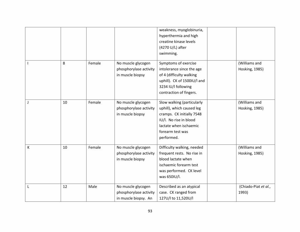
93
weakness, myoglobinuria,
hyperthermia and high
creatine kinase levels
(4270 U/L) after
swimming.
I 8 Female No muscle glycogen
phosphorylase activity
in muscle biopsy
Symptoms of exercise
intolerance since the age
of 4 (difficulty walking
uphill). CK of 1500IU/l and
3234 IU/l following
contraction of fingers.
(Williams and
Hosking, 1985)
J 10 Female No muscle glycogen
phosphorylase activity
in muscle biopsy
Slow walking (particularly
uphill), which caused leg
cramps. CK initially 7548
IU/l. No rise in blood
lactate when ischaemic
forearm test was
performed.
(Williams and
Hosking, 1985)
K 10 Female No muscle glycogen
phosphorylase activity
in muscle biopsy
Difficulty walking, needed
frequent rests. No rise in
blood lactate when
ischaemic forearm test
was performed. CK level
was 650IU/l.
(Williams and
Hosking, 1985)
L 12 Male No muscle glycogen
phosphorylase activity
in muscle biopsy. An
Described as an atypical
case. CK ranged from
127U/l to 11,520U/l
(Chiado-Piat et al.,
1993)

94
accumulation of
glycogen.
(normal was up to 70U/l).
Symptoms did not change
between the ages of 12
and 15. “Lazy since early
childhood. Persistent mild
muscle weakness. Never
had cramp or muscle pain.
M 12 Male No phosphorylase
reaction was seen
with muscle biopsy,
and reduce
phosphorylase activity
was seen. Activity
was 032U/g of muscle
(normal was 25.1-
37.0U/g).
Rapid walking caused
muscle fatigue from the
age of 6 onwards.
However, able to get into
second wind. Had
myoglobinuria twice.
Cycling caused muscle
pains. Ischaemic forearm
test caused a contracture.
However, a small rise in
lactate was seen from 0.86
to 1.07mmol/l.
(Sengers et al.,
1980)
N 8 Male R50X homozygous Exercise caused
myoglobinuria.
(Kristjansson et al.,
1994)
Table 8.1 Clinical features of McArdle's children from published papers. (R50X homozygous means that in one person, both copies of the PYGM gene
have the R50X mutation.)

95
8.2.2.1 Diagnosis of children with McArdle’s
Based upon the information available, this suggests that raised CK levels in children under the age of 6
(even in the absence of other symptoms of McArdle’s) could indicate that they may have McArdle’s.
For children above the age of 6, most of the symptoms (outlined above) are similar to those experienced
by McArdle’s adults.
Since children with McArdle’s do not seem to have all the classic symptoms seen in adults, it is
recommended that a high CK level when resting should be the first clue that a child has McArdle’s, and
this should be followed by genetic testing (rather than muscle biopsy) if at all possible (Perez et al.,
2009).
8.2.3 McArdle’s in adulthood/middle age
Most McArdle people experience McArdle’s symptoms in childhood. Quinlivan et al. (2007; 2010)said
that 84% of 45 McArdle people recalled symptoms before 10 years of age, although many were not
diagnosed until adulthood Only about half (51%) were diagnosed by the age of 30, with the remaining
49% being diagnosed when older than 30. Most of the classic symptoms of McArdle’s are discussed in
detail in other sections of this Handbook, including the typical symptoms listed below.
Typical symptoms of classic McArdle’s in adulthood/middle age are:
Muscle pain upon exercising
Contractures
Raised creatine kinase levels
Second wind
8.2.4 McArdle’s in older age
As discussed by Perez et al. (2008), historically, McArdle people were recommended to have a sedentary
lifestyle to avoid the risk of exercise causing rhabdomyolysis. Elderly McArdle people are likely to have
received this advice. They may have had a sedentary lifestyle since childhood or whenever they were
diagnosed. In terms of lifestyle, Perez et al. say that it is well known that if children get into the habit of
exercising when young, they are more likely to continue to exercise when older. Conversely, if McArdle
people were advised not to exercise many years ago, they are likely to have continued this trend. It
should be noted that the recommendations for McArdle people have now changed and a moderate level
of exercise is now recommended (see section 4.2.2).
Perez et al. (2006) reported the case of a 78 year old male with McArdle disease who was able to do
very little exercise when first tested, which he suggested was the result of a very sedentary lifestyle. At
the time the paper was written, there had not been any studies performed to assess the amount of
exercise which elderly people with McArdle’s were able to do. The authors suggested that one reason
96
why this study had not been conducted was because of the “risk of discomfort and rhabdomyolysis”.
They gave the 78 year old man 660ml of solution which contained 75g of sucrose. They gave him a 10
minute warm up period before testing his exercise capacity using a cycle ergometer test (see section
2.3.1.2). They also measured the amount of oxygen he breathed in (called the VO2 peak). Based upon
this single McArdle person, who had led a very sedentary lifestyle, the authors of study suggest that it is
likely that elderly McArdle people have a decrease in their VO2 peak. However, they also state that
research on elderly people in general (mostly those who do not have McArdle’s) suggests that elderly
people are able to safely learn how to exercise and increase their ability to exercise. Although this
report is interesting, it must be emphasised that a single case study is not considered a large enough
sample size to base medical advice. This study would have been greatly improved if they had set a basic
exercise programme for the patient, and then tested him again a year later to see if he was fitter and
better able to exercise.
8.2.4.1 Muscle wasting and muscle weakness is a symptom of McArdle’s which may develop or
increase in older age
Case studies of several elderly McArdle people have been reported. Lucia et al. (2008) described an
elderly McArdle person who was now 81 – this was the same person described when aged 78 by Perez
et al. (2006). This person had lived a sedentary lifestyle since childhood. In older age, this person had
quite severe exercise intolerance and had “proximal muscle atrophy” and “fixed weakness”.
Pourmound et al. (1983) described a 76 year old McArdle man whose symptoms began at age 74 years
with sudden onset of proximal muscle weakness and fatigability. Electromyography disclosed
substantial spontaneous activity and myopathic features as seen in inflammatory muscle disease. The
diagnosis of McArdle disease was made by histochemical studies of muscle, an abnormal ischemic
lactate test, and absence of myophosphorylase activity.
The amount of muscle wasting (hypertrophy) and weakness seen in McArdle people appears to increase
with age (Amato, 2003; Nadaj-Pakleza et al., 2009). Quinlivan et al. (2010) reported that 24% of 45
McArdle people had muscle hypertrophy, and 16% (all aged over 40 years old) had mild upper limb and
trunk weakness. Nadaj-Paleza et al. (2009) found that 10% of 80 McArdle people, all aged over 40, had
proximal fixed muscle disease and that 37.5% of the McArdle people aged over 40 years old had muscle
weakness.
There are many possible explanations for muscle weakness. It is not clear whether activity in younger
life has an effect upon the development of muscle wasting and weakness. One possibility is that a
sedentary/inactive lifestyle means that the muscles are not used, and become weak and waste away
An opposite possibility is that excessive exercise when younger could cause repeated damage, and that
eventually the muscles become unable to repair themselves, leading to weakness and wastage. Voduc
(2004) suggested that fixed muscle weakness could be caused by repeated muscle damage and loss of
skeletal muscle fibres due to rhabdomyolysis.

97
Schotland et al. (1965) investigated muscle biopsies from two McArdle people, and suggested that
glycogen accumulation may disrupt the structure of the muscle, and this could be one of the causes of
permanent weakness that has been observed in the older people with McArdle disease.
Another possibility is that muscle weakness may be at least partially caused by damaged muscle being
replaced as fat (De Kerviler et al., 1996). Nadaj-Pakleza et al. (2009) carried out a scan which showed
“fatty infiltration in the shoulder and pelvic girdle muscles”. If damaged muscle is replaced as fat, I
wonder if this could contribute to many McArdle people becoming overweight.
An alternative possibility is that a different gene (a phenotype modulator) may have an effect upon the
strength of the muscle, and would explain why some (but not all) McArdle people develop weakness in
older age. This gene may not have been identified. Phenotype modulators and other factors which
affect the severity of McArdle’s symptoms are discussed in section 9.
It should be remembered that muscle wastage and weakness with older age is not a specific symptom of
McArdle’s. Weakness and wastage in the muscles with age is common in the population unaffected by
McArdle’s. Saidoff and Apfel (2005) say that “by age 65, muscle strength is diminished by as much as
80%, and about half of the body’s entire muscle mass is lost by age 80” in people unaffected by
McArdle’s.
8.2.5 Will I end up in a wheelchair?
The quick answer is “no, most McArdle people do not need to use a wheelchair”. “In McArdle disease,
only few patients were reported who were severely handicapped and complained of generalized pain
with minimal exercise” (Rommel et al., 2006).
McArdle people who use a wheelchair may not have McArdle’s (they may have been misdiagnosed and
may actually have a different disease), or may have McArdle’s plus a second muscle disease (see section
9.6 on double trouble). They may also be people whose muscles have got very weak due to lack of
exercise, and where exercise now results in severe muscle damage and weakness, so they are in a
negative spiral of muscle weakness (anecdotal).
Many people who are unaffected by McArdle’s require the use of a wheelchair in old age, obviously for
reasons unrelated to McArdle’s. The average age for wheelchair users in the UK population (unaffected
by McArdle’s) is 67-68 years old (Sapey, 2004).
8.2.6 Does McArdle disease affect lifespan?
There is no published information on whether McArdle’s has any effect upon lifespan (how long you
live). There are several reports published reports of elderly McArdle people, including a 76 year old
McArdle man (Pourmand et al., 1983), a 78 year old McArdle man (Perez et al., 2006), and an 81 year
old McArdle person (Lucia et al., 2008a). The fact that there are many reports of elderly McArdle people
suggests that McArdle’s does not have any effect upon lifespan.

98
9 Factors which may explain differences in the severity of symptoms
of McArdle disease from person to person
McArdle disease is caused by mutations in the PYGM gene which encodes muscle glycogen
phosphorylase (see section 3.2 for more details). Almost all McArdle people do not have any active
muscle glycogen phosphorylase enzyme. (For the moment we will ignore the very small number of
McArdle people who have a small amount of active enzyme, discussed further in section 9.1.3.) It would
therefore be expected that all McArdle people would have similar symptoms. But this does not appear
to be the case; some McArdle people report much more severe McArdle’s symptoms than others.
Possible explanations for these differences are discussed below. Psychological aspects of perception of
pain and ability to cope with pain are discussed in section 10.
9.1 The amount of active muscle glycogen phosphorylase is an important
factor which determines the severity of symptoms
9.1.1 The amount of functional enzyme determines whether carriers of the PYGM mutation
have symptoms of McArdle disease
McArdle carriers do not normally have symptoms of McArdle disease as they have some functional
glycogen phosphorylase enzyme. Andersen et al. (2006) compared McArdle patients, carriers and
unaffected people during exercise. Carriers had normal oxidative capacity and lactate responses which
were identical to controls. They were therefore able to exercise normally and did not have symptoms of
McArdle disease, an observation already reported by family doctors treating McArdle people (Quinlivan
and Vissing, 2007). (An exception where a carrier had McArdle’s symptoms is discussed in section 9.7.)
Carriers of the PYGM mutation have intermediate levels (approximately half the amount) of muscle
glycogen phosphorylase enzyme activity compared to unaffected people. The amount of glycogen
phosphorylase activity in carriers has been reported as 25-45% (Bogusky et al., 1986) and 30-45%
(Manfredi et al., 1993) that of unaffected people. It is likely that this amount of glycogen phosphorylase
is sufficient to enable carriers to exercise in a similar way to unaffected people.
9.1.2 Different mutations do not correlate to differences in severity of symptoms
The most obvious explanation for differences between McArdle people would be that different
mutations in the PYGM gene could lead to differences in severity of symptoms. However, from a
biochemical point of view, this idea does not work as most mutations result in a complete absence of
active muscle glycogen phosphorylase protein. (The exception is the small number of McArdle people
who have a very low “residual” level of muscle glycogen phosphorylase activity. These are discussed
further in section 9.1.4.) If differences in the mutation (the “genotype”) had an effect upon the physical
effect of those genes on the body (including severity of symptoms) (the “phenotype”), it could be
described as a “genotype-phenotype” relationship. No clear genotype-phenotype relationship has been
seen despite studies of large numbers of people with McArdle’s (Martin et al., 2001; DiMauro et al.,
2002; Deschauer et al., 2007; Delmont et al., 2008). Almost all the mutations in the PYGM gene appear

99
to prevent production of functional enzyme in the majority of McArdle people, irrespective of whether a
non-functional protein is produced.
9.1.3 Rarely, some McArdle people have low levels of muscle glycogen phosphorylase
activity
There have also been rare reports of McArdle people with enzymatically active muscle glycogen
phosphorylase protein. One person had enzyme with 13% of normal activity, and three cases were 3%
active compared to normal levels (Martinuzzi et al., 1996). One person with 2% of muscle glycogen
phosphorylase activity was described by Andersen et al. (2006). Delibas et al. (2008) and Delmont et al.
(2008) each reported a McArdle person with detectable muscle glycogen phosphorylase activity.
Information about the mutations of these McArdle people with low levels of muscle glycogen
phosphorylase activity would be very informative.
9.1.4 Even a very low level of muscle glycogen phosphorylase activity improves the ability
of McArdle people to exercise
Quinlivan and Vissing (2007) reported research by Haller that McArdle people with even a very low level
of muscle glycogen phosphorylase activity (1-2.5%) had much higher “oxidative capacity” (were able to
get more energy from food and able to exercise more easily). Vissing et al. (2009) reported two McArdle
people with unusually high exercise capacity. The two people had either R50X or G205S on one copy of
the PYGM gene, and novel splice mutations in introns 3 [IVS3-26A>G (c.425-26A>G)] and 5 [IVS5-601G>A
(c.856-601G>A)]. (For more information about different types of mutations see section 3.2.4.) It seems
that these splice mutations allowed some active muscle glycogen phosphorylase to be made in the
muscles of these McArdle people, which allowed them to exercise more easily than typical McArdle
people. These unusual McArdle people were able to reach a peak workload 2-fold higher than typical
McArdle patients, and oxygen uptake was more normal. The authors claimed that this was the first
published evidence of a relationship between the mutation and the ability of a McArdle person to
exercise (called a genotype-phenotype relationship). This evidence suggests that even low levels of
muscle glycogen phosphorylase can lead to an improved ability of the McArdle person to exercise.
9.2 Raised levels of cytokines may cause low-level inflammation in McArdle
people
Cytokines are small proteins which are produced by almost all cells in the body. Cells use cytokines as a
way to communicate either with neighbouring cells, or throughout the body (if the cytokines are carried
in the blood). Chemokines are a sub-group of cytokines, and are also small proteins produced by cells.
Some chemokines are used by cells to create an immune or inflammatory response. Cells can release
chemokines during infection by bacteria or viruses, which attracts cells of the immune system to the
location to fight the cause of the infection. (Information on cytokines and chemokines is summarised
from Janeway, 2001).
Lucia et al. (2008b) studied the levels of many cytokines and chemokines in both McArdle people and in
unaffected people before and after exercise. In McArdle people, Lucia et al. found raised levels of
several cytokines; “tumor necrosis factor (TNF-α), interleukin (IL)-1ra, IL-10, IL-12 and IL-17”. They also

100
found that McArdle people had a higher level of neutrophils. Neutrophils are cells which are part of the
immune system. Neutrophils are some of the first cells which are attracted by the release of cytokines
during infection. They also found that the amount of cytokine IL-6 (which the authors say has an anti-
inflammatory effect), was increased in both McArdle people and unaffected people after exercise. The
authors conclude that “Our results suggest that McArdle disease is associated with low-level systemic
inflammation whereas appropriate exercise induces … a significant increase in the anti-inflammatory
myokine IL-6.” They suggest that this anti-inflammatory effect may explain why frequent moderate
exercise has been shown to improve symptoms in McArdle people.
Several criticisms of the study conducted by Lucia et al. (2008b) may be made. These include the fact
that they gave sucrose to the McArdle people but not to the unaffected control participants before
exercise. This means that it is not possible to be sure that the differences in cytokine levels were due to
McArdle’s rather than sucrose. For example, it may be that sucrose causes raised cytokine levels in
people irrelevant of whether they have McArdle’s or not. Despite these criticisms, Lucia et al. found
that the levels of many of the cytokines were higher in people with McArdle’s. This was a new
discovery, and it would be ideal for it to be repeated and confirmed by other researchers.
The authors of this study suggested since exercise may cause muscle damage in McArdle people, the
body might increase the amount of IL-6 might in order to have an anti-inflamatory effect. I have an
alternative explanation (an unproven theory) why the amount of IL-6 may increase in McArdle people
when exercising. There may be an association between the amount of glycogen in exercising muscle
cells and IL-6 production. Exercise increases the amount of IL-6 in healthy athletes unaffected by
McArdle’s (Cannon et al., 1999; Robson-Ansley et al., 2007). IL-6 appears to be released into the blood
by exercising muscles to stimulate the liver to break down stored glycogen and release energy via the
bloodstream (Pedersen, 2001; Steensburg, 2001). Raised IL-6 levels in McArdle people reported by Lucia
et al. may be due to the shortage of free glucose in the muscle cells. Pedersen (2001) described an
increase in production of IL-6 by muscle cells due to lack of glycogen in unaffected people. However the
same effect would occur in McArdle people as the glycogen can’t be utilized and converted to glucose to
provide energy. In McArdle people, IL-6 may be released by the muscle cells in an attempt to increase
production of glucose by the liver.
Raised cytokine levels in McArdle people have several possible implications:
1) Many McArdle people are misdiagnosed with an inflammatory muscle disease such as
polymyositis (which is often treated with steroids to reduce the inflammation) (section 2.5.1). If
McArdle’s is also an inflammatory muscle disease, it is easy to understand how this misdiagnosis
could occur.
2) There appears to be a link between McArdle’s and type II diabetes (characterised by insulin
resistance). There is some evidence that the cytokine TNF-α (and possibly the cytokine IL-6)
could lead to insulin resistance (see section 13.4.1.2).
101
3) Studies suggest that approximately a third of McArdle people experience anxiety or depression
at some point in their lives (Rommel et al., 2006; Quinlivan et al., 2010). de Ridder et al. (2008)
say that raised levels of some cytokines can produce feelings of fatigue, irritability,
demoralisation, and may cause feelings of depression. It is possible that the feelings of
depression experienced by McArdle people could be related to increased levels of some
cytokines.
At present, none of these possible implications have been fully investigated or proven, so the
implications are speculative.
9.3 Phenotype modulators (genes other than PYGM) may affect the severity
of McArdle’s symptoms
The combination of genes which a person has is known as a “genotype”, the physical effect of those
genes on the body (including severity of symptoms) is known as a “phenotype”. A “phenotype
modulator” is a second gene which affects the phenotype of the first gene. In a McArdle person, the
first gene is the PYGM gene; being homozygous for mutations in the PYGM gene causes a person to have
McArdle disease. It is possible that there is second gene which has an effect upon how severe the
McArdle’s symptoms are. This second gene is not related to the PYGM gene. Depending what form of
the second gene a McArdle person has, the severity of the symptoms could vary between McArdle
people.
Phenotype modulators are a possible explanation for why different McArdle people can have different
symptoms. Recent research has identified several genes which appear to be phenotype modulators. It
is logical that proteins encoded by other genes, for example proteins which help the muscle cells take up
glucose or produce energy more efficiently, could have an effect on the severity of McArdle’s. Several
researchers have looked at genes which may be phenotype modulators. Rubio et al. (2007), Rubio et al.
(2008) and Martinuzzi et al. (2003) studied large numbers of McArdle people to try and find out which (if
any) of these phenotype modulator genes could be having an effect upon the severity of McArdle’s.
9.3.1 Angiotensin-converting enzyme
In skeletal muscle the angiotensin-converting enzyme (ACE) is an enzyme involved in the metabolic
response to exercise. The ACE enzyme is encoded by an ACE gene. There are two versions (called
“alleles”) of the ACE gene, the “I” and the “D” form”. Each allele encodes a different form of the ACE
protein, which are known as “isoforms”. Each person inherits two copies of the gene, one from each
parent. A person may inherit two copies of the D gene (DD), one copy of D and one copy of I (DI) or two
copies of I (II). In humans, the different ACE genotypes are well known to affect the level of activity of
the ACE enzyme. The combination with the most ACE enzyme activity is DD, DI is intermediate and II is
lowest (Sabbagh et al., 2007). Lower ACE activity may increase glucose uptake to muscles. People with
the I isoform respond better to muscle training and aerobic conditioning.
A peptide (small protein) called “bradykinin” causes blood vessels to enlarge (dilate) and blood pressure
to become lower. Angiotensin-converting enzyme is one of the enzymes which break down bradykinin.

102
If an ACE inhibitor drug (such as Ramipril) is taken, less bradykinin will be destroyed, leading to an
overall increase in bradykinin. This will have the effect of increasing the size of the blood vessels, which
may allow more blood to be pumped to the muscles, bringing more glucose and fatty acids and oxygen
to the muscle cells.
Both Martinuzzi et al. (2003) and Rubio et al. (2007) found that there was a strong correlation between
the number of D alleles in the ACE gene and the severity of McArdle’s symptoms in McArdle people;
McArdle people who had the DD genotype had more severe symptoms and McArdle people with the II
genotype had less severe symptoms. These results led to a further study by Martinuzzi et al. in 2008 to
test whether treatment with the ACE inhibitor Ramipril would improve McArdle’s symptoms. When
treated with Ramipril, a very small improvement in the ability to exercise was seen in McArdle people
who had the DD genotype, but not people who had the DI genotype.
9.3.2 Muscle adenosine monophosphate deaminase
In skeletal muscle, AMP deaminase is an enzyme which converts a molecule called adenosine
monophosphate (AMP) to a molecule called inosine monophosphate (IMP) as part of a process called
“purine catabolism” that produces energy within muscle cells. The AMP deaminase enzyme is encoded
by an AMPD1 gene. Adensoine monophosphate deaminase deficiency (AMDD) is caused by mutation in
the AMPD1 gene. Most cases of AMDD are caused by the premature termination codon mutation
written as Q12X. In this mutation, a single mutation in the genetic code changes the code from “c” to
“t”; so that glycine amino acid is replaced by a premature termination codon. This results in the
production of an abnormally short enzyme which cannot function. Mutations in AMPD1 gene are very
common, with MADD identified in approximately 2% of all muscle biopsy specimens tested by research
labs (Gross, 1997). In many of these cases, people with the Q12X mutation are asymptomatic. Where
people do have symptoms, the symptoms of MADD are muscle weakness, muscle pain and muscle
cramps following exercise. MADD is usually inherited in an autosomal recessive way, where people only
have symptoms of MADD if they have two copies of the AMPD1 gene which both have mutations.
People do not usually have symptoms of MADD if they are a carrier for the Q12X mutation. However,
Rubio et al. (2008) wondered whether McArdle people might have more problems with exercise if they
were carriers for the Q12X mutation rather than being wildtype homozygous (have no mutations in
either copy of the AMPD1 gene). They looked at the effect of AMPD1 (C34T) genotypes on 44 Caucasian
McArdle people (23 males, 21 females) using an ergometer test comparing those with two copies of the
gene (C34C) to carriers for the for the Q12X mutation. No differences were seen in male McArdle
people, but in female McArdle people with McArdle disease, being a carrier for the Q12X mutation of
the AMPD1 gene was associated with reduced aerobic capacity.
9.3.3 Myostatin
Myostatin is a protein which helps to regulate and control skeletal muscle growth and may also affect
muscle repair. Myostatin is also known as growth differentiation factor 8 and is encoded by the GDF-8
gene. It may therefore have an effect upon the strength and ability of muscle to repair itself following
damage. Normally, myostatin has a negative effect upon skeletal muscle growth. A mutation (like the
K153R missense mutation) can stop myostatin being able to function. At present, the effect of the

103
K153R mutation is not known, but one possibility is that having the mutation could enable an increased
amount of muscle growth which could increase muscle strength. In women unaffected by McArdle’s,
women with K153R mutation have lower muscle strength than those without the mutation. Gonzalez-
Freire et al. (2009) found McArdle’s women with the K153R mutation in the myostatin gene had worse
McArdle’s symptoms than McArdle’s women who did not have the mutation.
9.3.4 α-actinin-3
α-actinin-3 is a protein which is the main component of skeletal muscle. It has a role in generating
muscle contractions. α-actinin-3 is encoded by the ACTN3 gene. The R577X mutation introduces a
premature stop codon which results in an absence of α-actinin-3. In people unaffected by McArdle’s,
having wildtype ACN3 may enable them to exercise for longer, and elite sportspeople are often wildtype
for ACN3, but studies are unclear, and having the R577X mutation does not appear to be a disadvantage
for most of the population. Lucia et al. (2007a) found that McArdle’s women with the R577X mutation
were able to exercise better (when tested using a cycle ergometer) than those with wildtype ACTN3, but
the presence or absence of the mutation had no significant effect in McArdle’s men. Rubio et al. (2007)
also looked at whether mutations in ACN3 had any effect on severity of McArdle’s symptoms. They did
not find any effect, but did not separate the data for men and women.
9.3.5 Peroxisome proliferator-activated receptor coactivator 1 α
Peroxisome proliferator-activated receptor coactivator 1 α is a protein which is encoded by the
PPARGC1A gene. Peroxisome proliferator-activated receptor coactivator 1 α is involved in regulating
the expression/production of proteins involved in generating energy within the cell. As a missense
mutation (G482S) had been shown to improve human aerobic capacity in people unaffected by
McArdle’s, Rubio et al. (2007) looked at whether this gene might affect the severity of McArdle’s
symptoms. The results did not show that this gene had any effect on severity of McArdle’s symptoms,
but they did not separate the data for men and women.
9.4 There is a difference in the amount and type of pain felt by different
McArdle people
Some McArdle people have constant and permanent chronic pain. Other McArdle people have pain
caused by exercise, or occasionally muscle pain after exercise if some muscle damage has occurred.
Rommel et al. (2006) studied 24 McArdle people. They asked many questions to determine whether the
McArdle people only had pain caused by exercise, or whether they had permanent pain. They found the
majority of McArdle people who had permanent pain were women. 8 of the 24 McArdle people had
permanent pain, and of these, 7 were women. There was only one man with permanent pain, so they
used the women to compare those with permanent pain with those with exercise-induced pain. For the
women with permanent pain, the pain had a greater impact on the daily life, work, and social activity. In
contrast, where women principally had exercise-induced pain, their McArdle’s symptoms had much less
effect upon their daily life, work, and social activity. They found that those with permanent pain felt
more fatigue, and tried harder to avoid pain. However, “differences regarding depression and pain-
related help-hopelessness were not significant”.
104
Differences in the type or level of pain felt were not related to which mutation was in the PYGM gene or
which allele of the ACE gene the McArdle people had (Rommel, 2006).
Rommel et al. (2006) found that women who had exercise-induced pain seemed to ignore the pain by
endurance coping; making sure they finished projects they started, even if in pain, and not resting as
often. They tried to ignore and play down any pain they experienced. On the other hand, women who
had permanent pain seemed to feel that the pain was greater, and worry about it. They tried to avoid
any activities which would cause pain. It is not obvious whether there was an original difference
between the women who had exercise-induced pain or permanent pain, or whether the difference was
due to differences in attitude and different methods of coping with pain. I inferred from the report that
women who had exercise-induced pain found that it had less effect upon their lifestyle than those who
had permanent pain. Women with permanent pain found that it had a greater effect upon their general
activity, and caused sleep disturbance and fatigue. The authors suggest that regular moderate exercise
may be a better way to cope with the symptoms of McArdle’s than avoiding exercise. The authors do
point out that this study was limited by the small number of participants (24 McArdle people), and a
larger scale study would generate more useful information. The authors suggest that “Further studies
should also address the question if these subgroups [people with permanent pain versus people with
exercise-induced pain] respond differently to therapeutic strategies like glucose substitution, pain
medication or regular moderate aerobic exercise”.
9.5 Gender has an effect on phenotype modulators and the severity of
McArdle’s symptoms
Researchers looking for phenotype modulators have found that in McArdle’s women a mutation in the
myostatin gene led to an increase in the severity of symptoms (Gonzalez-Freire et al., 2009). Lucia et al.
(2007b) found that the R577X allele of the ACTN3 gene was associated with improved exercise capacity
(using a cycle ergometer) in women with McArdle disease, compared to the R577R allele, but had no
significant effect in men. Rubio et al. (2007) looked at the angiotensin-converting enzyme (ACE) gene,
muscle adenosine monophosphate deaminase (AMPD1) gene, the peroxisome proliferator-activated
receptor coactivator 1 α gene (PPARGC1A) gene, and α-actinin-3 (ACTN3) gene. The results of this
study did not show any of these genes had any effect on severity of symptoms, but unfortunately, the
authors did not separate the data for men and women to see if gender had any affect on the effects of
the different genes. Overall, the disease severity was greater in McArdle’s women than McArdle’s men.
“Among the middle-aged patients whom we studied, proximal muscle wasting and weakness was more
frequent in women than in men; in some cases, the severity was such that the patients had to use an
electric toothbrush as they easily became fatigued when brushing their teeth manually. In addition, the
few adult patients in whom respiratory muscles have been shown to be affected have all been women”
(Lucia et al., 2008a).

105
9.6 McArdle symptoms may be more severe if combined with another
disease
9.6.1 McArdle symptoms can be more severe if combined with another muscle disease,
causing “double trouble”
Rarely, two muscle diseases may both occur in the same person causing “double trouble”. Vladuitu et
al. (2000) looked at nine McArdle people, and tested them for other muscle disease (myopathies). Two
of the McArdle people had a second muscle disease, one had a Q12X stop codon causing myoadenylate
deaminase (AMPD) deficiency, and one had S113L missense mutation causing carnitine
palmitoyltransferase II (CPT II) deficiency. They suggested that the effects of these second mutations,
and therefore the high frequency of diagnosing double trouble may be because the first muscle disease
lowers the threshold for manifestation of the symptoms.
Aguilera et al. (2001) reported a person with biochemical evidence of McArdle disease (heterozygous
for R50X) and also with a mitochondrial DNA point mutation in the cytochrome c oxidase subunit I (COI)
gene. Lucia et al. (2007b) reported a 29 year old woman with McArdle disease and myasthenia gravis,
for whom aerobic training significantly increased her exercise capacity. The severity of symptoms will
depend which two diseases the person has. There is a report of an infant girl born to consanguineous
Moroccan parents, who died at 5 months of age. She had homozygous mutations in both the PYGM and
the deoxyguanosine kinase (dGK) genes (Mancuso et al., 2003) (see section 8.1.1). Calpainopathy is an
autosomal recessive limb girdle muscular dystrophy (LGMD2A) caused by mutations in the gene
encoding calpain-3. Pulur et al. (2009) reported a person who had both calpainopathy and McArdle’s.
The boy had started to have difficult with exercise like walking upstairs from the age of 14. His muscle
weakness was much more severe than that usually seen in McArdle people. His parents were
consanguineous. The authors said this “points to the need to search for other diseases in the presence
of any unusual clinical manifestation.” (For example, further studies could be performed to see if a
second disease is the explanation for a McArdle person with unusually severe symptoms.)
9.6.2 A second disease (which is not a muscle disease) may exacerbate the symptoms of
McArdle disease
9.6.2.1 Bulimia and sickle cell anaemia can make McArdle symptoms worse
It has been reported that the eating disorder bulimia may exacerbate the symptoms of McArdle disease.
Pillarisetti and Ahmed (2007) described a McArdle person who had both bulimia and sickle cell trait (by
sickle cell trait the authors meant that the person was heterozygous for sickle cell anaemia). The
authors said that bulimia could make rhabdomyolysis more likely because bulimia could cause
electrolyte changes in the body like hypokalemia and hypophosphatemia which could also precipitate
rhabdomyolysis. The authors also say that both sickle cell trait and bulimia are known to make people
(unaffected by McArdle’s) more likely to have rhabdomyolysis.

106
9.6.2.2 Epileptic seizures can make McArdle symptoms worse
An example of a second disease making McArdle symptoms worse was presented by Ford et al. (1973)
who describe a male McArdle person who also had epilepsy. Epileptic seizures caused the muscles to
cramp up, spasm, and produced rhabdomyolysis. Salmon and Turner (1965) also reported a McArdle’s
boy aged 16 who was diagnosed with McArdle’s after a grand mal epilepsy convulsion led to
rhabdomyolysis.
9.7 A second disease may make carriers of McArdle disease more likely to
have symptoms of McArdle disease
If a person is a carrier for two similar diseases which affect energy metabolism, they may
(uncharacteristically) have symptoms of one or both diseases. Vockley et al. (2000) described this as
“synergistic heterozygosity”. They reported one person who had 0.04% muscle glycogen phosphorylase
activity and was heterozygous for the R50X mutation, providing a diagnosis of McArdle disease, and also
were heterozygous for the S113L mutation in the CPT2 gene. A different person seemed to be a carrier
for McArdle disease (29% of normal amount of muscle glycogen phosphorylase), phosphorylase b kinase
deficiency (GSD VIII) (37% of normal) and CPT II deficiency (50% of normal; heterozygous for Q12X
mutation in AMPD1). This research suggests that a second disease may explain why symptoms are
occasionally seen in carriers of McArdle disease.
Papadimitriou et al. (1990) described a man who developed McArdle’s symptoms at the age of 30 years.
His muscle weakness then increased as he got older. At the age of 40 he had myoglobinuria twice after
heavy muscle exercise, but did not have cramps/contractures. When he performed a forearm ischaemic
test, a rise in lactate was seen (which suggested a diagnosis of “It is not McArdle’s). However, he had
increased creatine kinase levels (his ck level was 1040 IU, when the normal in this case would have been
20-93 IU). It was found that he was a carrier of McArdle disease, and had 25% of normal muscle
glycogen phosphorylase activity. Carriers do not usually have symptoms of McArdle disease (Quinlivan
and Vissing, 2007 and many other publications). Possible explanations for why this man did develop
symptoms of McArdle’s could be because he had had a very physical lifestyle as a younger man
(including time in the army). Alternatively, it may be that he had a second (undiagnosed) muscle disease
which led to muscle degeneration as he got older and triggered McArdle symptoms. The authors did
not say if they had tried to rule out other similar diseases.
Recommend reading:
“Muscle pain in myophosphorylase deficiency (McArdle disease): The role of gender, genotype, and
pain-related coping” by Oliver Romme, Rudolf A. Kley, Gabriele Dekomien, Jörg T. Epplen, Matthias
Vorgerd and Monika Hasenbring

107
10 Mental and emotional aspects of McArdle disease
The way in which McArdle people perceive their McArdle’s symptoms can have an effect upon how
difficult they find it to cope with symptoms on a day to day basis. As discussed below, many McArdle
people have struggled for many years to get a diagnosis, sometimes being told by family doctors that
they are imagining the symptoms or are malingerers (see section 10.3.2.2).
However, these symptoms are not necessarily just emotional. There is now a small amount of evidence
that some McArdle people may suffer from symptoms similar to chronic fatigue. There is also a small
amount of evidence that McArdle’s may perform less well than people unaffected by McArdle’s at some
tests of how well the brain can perform. There is only a very limited amount of research into these
factors, and much more is needed.
10.1 Many McArdle people have symptoms which are like chronic fatigue
syndrome
Almost half (40%; 18 of 45) of the McArdle people attending the McArdle Clinic at Oswestry reported
chronic fatigue-like symptoms (Quinlivan et al., 2010). Quinlivan et al. (2007) suggest that the fact that
it often takes McArdle people many years to get a diagnosis could be a reason why many people have
symptoms of chronic fatigue, depression, and anxiety. It is an understandable reaction to many years of
wondering “What is wrong with me?”. Chronic fatigue syndrome (CFS) causes long-term tiredness
(fatigue) that affects everyday life. It does not go away with sleep or rest. CFS is also known as ME,
which stands for myalgic encephalomyelitis. Myalgia means muscle pain, and encephalomyelitis means
inflammation of the brain and spinal cord.
CFS/ME symptoms (taken from the NHS website http://www.nhs.uk/me/introduction.aspx):
Mild: you are able to care for yourself, but may need days off work to rest.
Moderate: you may have reduced mobility, and your symptoms can vary. You may also have disturbed
sleep patterns, and sleep in the afternoon.
Severe: you are able to carry out minimal daily tasks, such as brushing your teeth, but occasionally you
may need to use a wheelchair. You may also have difficulty concentrating.
Very severe: you are unable to carry out any daily tasks for yourself and need bed rest for most of the
day. Often, in severe cases, you may experience intolerance to noise and become very sensitive to
bright lights.
The causes of chronic fatigue-like symptoms in McArdle people may not be the same as in people
unaffected by McArdle’s, and that some treatments may therefore not be suitable. One treatment,
called “pacing” is a way to even out how much activity a person does each day. This avoids having
alternating good and bad days; a “good” day where the person feels good and does a lot of activity, but
is then so tired that the next day is a “bad” day where they have to spend the whole day resting. Pacing

108
is a technique to reduce the amount of activity done on a “good” day so that they are less tired the next
day and therefore avoid having a “bad” day. It may be the case that for McArdle people who do have
chronic fatigue-like symptoms, treatments such as pacing may help them cope with those symptoms
better, and improve their overall quality of life. It is important to remember that the causes of chronic
fatigue-like symptoms in McArdle people may not be the same as in people unaffected by McArdle’s,
and that some treatments may therefore not be suitable. There is no published research that any
treatments for chronic fatigue have been tried on McArdle people.
10.2 Lack of muscle glycogen phosphorylase may affect brain function of
McArdle people
Edelstyn and Quinlivan (2007) described a pilot study of neuropsychological performance (brain
function) in people with McArdle disease. Ten McArdle people were compared to ten unaffected
people, and were found to perform significantly worse on tests of verbal fluency and verbal memory and
less well than unaffected people in some other cognitive tests. This suggests that the lack of muscle
glycogen phosphorylase may also be detrimental to brain function in McArdle people. There are several
possible explanations for this.
Unaffected human brain has been shown to contain muscle glycogen phosphorylase, brain glycogen
phosphorylase and a dimer (pair) made of the brain and the muscle form. In unaffected human brain,
muscle glycogen phosphorylase was found to provide 25% of the phosphorylase activity (Bresolin et al.,
1983). In McArdle people, muscle glycogen phosphorylase is absent in the brain, so this may reduce the
total amount of glycogen phosphorylase in the brain. The brain consumes about 60% of the glucose
used by the whole body when resting (Berg et al., 2008). The lack of functional muscle glycogen
phosphorylase may affect brain function in McArdle people directly by reducing the amount of glucose
available to the brain.
80% of the vitamin B6 in the body is associated with muscle glycogen phosphorylase (Beynon et al.,
1995). Pyridoxal-5’-phosphate (PLP) is the metabolically active form of vitamin B6. Many reactions in
amino acid metabolism require the presence of PLP, such as decarboxylases which are required in the
synthesis of neurotransmitters. An adequate PLP supply in brain tissue is therefore essential for normal
brain function (Williams, 2003). If muscle glycogen phosphorylase is absent, this may decrease the
amount of PLP available both in the brain and throughout the body (Beynon et al., 1995).
It has been shown that muscle and brain glycogen phosphorylase can bind together (reviewed in Wright,
2009). If a small number of McArdle people have muscle glycogen phosphorylase which is present but
not functional, this non-functional muscle glycogen phosphorylase could bind to brain glycogen
phosphorylase, stopping it from functioning.
An excessive accumulation of glycogen in the brain may be detrimental. Edelstyn and Quinlivan (2007)
mention a report of a McArdle person with abnormal deposition of glycogen detected by brain MRS.
They also suggest that the abnormal rise in ammonia that occurs throughout the body in McArdle
people during exercise may cause abnormal psychological function.

109
It should be noted that there has only been one study of psychological function to date (by Edelstyn and
Quinlivan), and further studies are needed to confirm the results.
10.3 Psychological issues
There is no published information on the psychological effect of McArdle’s. Anecdotally, McArdle
people report childhood trauma, everyday embarrassment, depression, fear and in some cases possibly
even hysteria or a hyper-awareness (hypochondria). These experiences are likely to be similar to those
experienced by other people with rare diseases. Unless a reference is given, the information in this
section is anecdotal.
10.3.1 Before diagnosis
10.3.1.1 Psychology of being an undiagnosed McArdle’s child
Most McArdle people were not diagnosed until adulthood, and therefore spent their childhood
struggling to run and exercise, but unsure that there was a definite problem (such as McArdle’s). Some
McArdle’s children found that their parents did not believe them when they said that exercise caused
them pain. They may have suffered from teasing or bullying because they were unable to run around in
play or during exercise classes at school.
10.3.1.2 A rapid diagnosis in childhood is ideal
A person, who gets a rapid diagnosis, ideally in childhood, is probably likely to have fewer psychological
problems. An early diagnosis should allow the child to grow up following current recommendations
(such as frequent moderate exercise) but avoiding intense exercise which is likely to lead to muscle
damage. An early diagnosis would allow the parents to inform physical education/gym teachers to
ensure that the McArdle’s child is not pushed to exercise beyond their limits. An early diagnosis would
allow the child to inform friends and family of their limitations, and hopefully feel comfortable with
stopping and resting when necessary.
10.3.1.3 Psychology of being an undiagnosed McArdle’s adult
A McArdle’s adult who has not yet been correctly diagnosed may find that their family doctor doesn’t believe that the symptoms are real, and dismisses them as a hypochondriac or malingerer. A McArdle’s adult may also find that family and friends also don’t believe that the symptoms are real. They may find this very depressing and frustrating, as they know that there is something wrong, but it has not been correctly diagnosed. Family and friends may not be very sympathetic, since they have no proof that anything is wrong. An undiagnosed McArdle’s adult may feel angry that family/friends/family doctor doesn’t believe that you have a problem. Prior to being diagnosed, McArdle people may feel isolated because no-one else has similar symptoms.
10.3.2 Trying to get a diagnosis
10.3.2.1 It can take a long time for McArdle people to get a correct diagnosis
Many McArdle people spend many years seeking diagnosis before obtaining a correct diagnosis. They
often have to see many different family doctors and specialists. Based upon a study of 45 McArdle

110
patients visinting the Oswestry clinic, Quinlivan et al. (2010) reported that 84% had McArdle symptoms
from the age of 10 years old or younger, but about half (49%) were not diagnosed until they were more
than 30 years old. This suggests that it can take around 10-20 years for McArdle people to receive a
correct diagnosis. Many McArdle people have been misdiagnosed, and some will have received
treatments for this incorrect diagnosis. Some examples of these diseases are given in section 2.5.1.
10.3.2.2 Many family doctors may incorrectly imagine that a McArdle person is a
hypochondriac, malingerer or that symptoms are due to being overweight
McArdle’s is a real condition, with a physical cause (the lack of muscle glycogen phosphorylase). Much is
known about the cause of McArdle’s, the way in which it affects the body, and current treatments
(these are subjects covered in this Handbook). However, family doctors who have not heard of McArdle
disease may dismiss a person with McArdle’s as having hypochondria or being a malingerer.
Hypochondria is a mental disorder where people believe that they have a medical illness even though
family doctors are unable to find anything wrong. In some cases the physical symptoms can be caused
by the person thinking that they have the illness. People with hyperchondria may visit the family doctor
frequently, and may have a high level of worry about their symptoms. People with hypochondria
genuinely believe that they have an illness, even though family doctors are unable to find anything
wrong. It is possible to understand how a family doctor can (incorrectly) imagine that a McArdle person
who comes to see them frequently saying that something is wrong, but who is able to walk around and
appears to be physically well could have hypochondria. A person who is a malingerer knows that they
do not have an illness, but pretends to have symptoms in order to avoid work or for financial reasons
(e.g. to get disability benefit) or for another purpose (such as getting sympathy from family members).
Many family doctors have not heard of McArdle’s, and don’t recognise the symptoms. Many McArdle
people are overweight (discussed in more details in section 4.2.4), and it is understandable that when a
family doctor is present with a patient who is overweight, struggles to exercise, and has muscle pains,
then the family doctor may (incorrectly) imagine these symptoms are all caused by the person being
overweight, and recommend more exercise.
10.3.3 After diagnosis
10.3.3.1 Receiving a diagnosis of McArdle’s
Many people will feel relief at finally getting a diagnosis of McArdle’s, especially if it has taken a long
time to obtain that diagnosis. McArdle people may feel vindicated because a diagnosis will prove to
family/friends/your family doctor that the problem is real not imagined. A diagnosis allows you begin
reading about McArdle’s online and elsewhere and to find out more about treatments. It also allows
you to make contact with other McArdle people via chat groups or e-mail groups who can provide
support.
10.3.3.2 Coping with a diagnosis of a chronic illness
Although many McArdle people will feel relief at having finally got a diagnosis, diagnosis of a chronic
illness may also pose challenges. The information which follows is not specific to McArdle disease, but is

111
general information about how to cope with a diagnosis of a chronic illness. de Ridder et al. (2008)
define “chronic illnesses” as “disorders that persist for an extended period and affect a person’s ability
to function normally”. Interestingly, de Ridder et al. say that it is estimated that half the population
(50% of people) have a chronic physical condition which requires medical treatment. Examples of
chronic illnesses include diabetes, which can be controlled with medication, or rheumatoid arthritis,
which can cause physical disability and pain. Chronic illnesses can change the persons life is many ways;
for example by limiting the amount of activity they can do.
Quinlivan et al. (2010) found that 31% (14 of 45) of McArdle people “had been treated at some time for
anxiety and/or depression”, and Rommel et al. (2006) reported that 29% (7 of 24) of McArdle people
had experienced mild depression. de Ridder et al. (2008) note that people with “chronic illness typically
have anxiety, depression, and other negative emotions”. Emotional reactions to chronic illness can
basically be divided into two groups; one is avoidance and suppression of emotions, and the other is
expression and acknowledgement of emotions. Despite the common belief that it is better to express
emotions, the authors note that this is only the case in more expressive cultures such as North American
and Western European cultures. The authors note that non-expression of emotions is better for people
who live in less emotionally expressive cultures such as some Asian cultures. de Ridder et al. reviewed
many studies and found that emotional expression led to improvements in the amount of physical
acivtiy people could do, mobility, and in the number of symptoms reported, and even produced positive
changes in the body which could be measured by laboratory tests (suggesting it was a physical
improvement, not just a change in the person’s perception of the situation). It may be that discussing
the emotions and feelings associated with being diagnosed with a chronic illness may help people to feel
more comfortable with the diagnosis. People who do not discuss their feelings and “bottle them up”
may feel more stressed. Stress is known to have negative effects upon health. Another benefit of
discussing feelings and emotions may be that it leads to feelings of closeness with either the
friend/relative or health professional who you talk to.
The challenge is how to learn to adapt life to work around McArdle’s. de Ridder et al. note that many
people with chronic diseases also have an increase of some cytokines (the cytokine IL-6 is discussed
further in section 9.2). It has been shown that some cytokines can produce feelings of fatigue,
irritability, demoralisation, and may cause feelings of depression. In this case, the cytokines are
producing real changes in the feelings and emotions. Drugs which block the effect of these cytokines
have been tested in people with rheumatoid arthritis and have helped to improve the way those people
felt. At present, it is not known if cytokines in McArdle people may have this effect, or whether drug
treatments would help.
Humans have a natural response to illness which is to rest, as this “conserves energy and promotes
healing” (de Ridder et al., 2008). However, this response is not always of use to the person, and is in
contrast to the moderate exercise now recommended for McArdle people (see section 4.2). Some
people diagnosed with chronic illness find it hard to follow a new diet, exercise or other advice. This is
most likely if people feel that a large amount of time and effort is needed to follow this advice and/or it
may not produce much benefit. Some people may suffer from major depression, whereas other people
may have guilt or anxiety about not following “the ideal diet” or “the ideal exercise programme”, or
112
worry about the negative effects of the disease; such as worries about muscle wasting in older age. The
authors also noted that having a positive mood can help people manage their condition better and
improve wellbeing .
de Ridder et al. (2008) describe a person who has successfully adjusted to a chronic illness as having
successfully adapted to the new situation; learning to work around the disability, staying emotionally
balanced, and maintaining healthy relationships with friends and family. Not suffering from
psychological disorders, not letting the disability have a negative effect on your life, and being able to do
tasks, such as a job, which you want to do. A person who has successfully adjusted to a chronic illness
will be able to feel satisfied and happy with their life.
10.3.3.3 Psychology of how other people perceive a McArdle’s adult
There are several issues which have been mentioned to me by McArdle’s adults. One problem is that
you look completely “normal”. Since McArdle’s does not mean that you have to use a wheelchair or a
walking stick, the general population may not realise that you struggle to walk long distances. Some
McArdle people may feel embarrassed if they need to stop and rest during exercise, for example, during
an exercise class, or while out for a walk with friends. Unless the class teacher or your friends knows
about McArdle’s, it can be embarrassing to be the one person who needs to stop and rest. In particular,
I would imagine that it is hardest being a young person or a young man, as these would be perceived by
others as being fit and able to exercise easily. Some McArdle’s adults who use disabled parking spaces,
or lifts rather than stairs, have been accused of using them inappropriately by other people. McArdle
people may also feel annoyed/depressed/frustrated that friends, family and work colleagues may have
to help you out.
Many McArdle’s adults use coping mechanisms to allow themselves to rest without other people
noticing. These techniques could include frequently pretending to tie up their shoelace, stopping to
look in shop windows, or pretending to use a mobile phone.
10.3.4 Positive steps to dealing with these issues
Many McArdle people find that visiting a specialist (such as Dr Quinlivan at the Oswestry clinic) can have
a huge benefit. Unlike a family doctor, who may never have met a person with McArdle’s before, a
specialist will have met many people with McArdle’s. The specialist will understand McArdle’s much
better, and will know what is normal or unusual for people with McArdle’s. They are also likely to be
involved in research and clinical trials and will have up to date on the latest theories and treatments.
Many McArdle people gain huge benefit from being in contact with other people with McArdle’s. E-mail
chat groups (such as GSDnet) and internet chat groups (such a McArdle disease Facebook page) allows
people with McArdle’s to compare notes and share hints and tips. There can be differences in the
advice given by McArdle’s specialists, and this can be a way for McArdle people to tell each other about
this advice. This is also a good way to find out if and when new treatment ideas are developed.
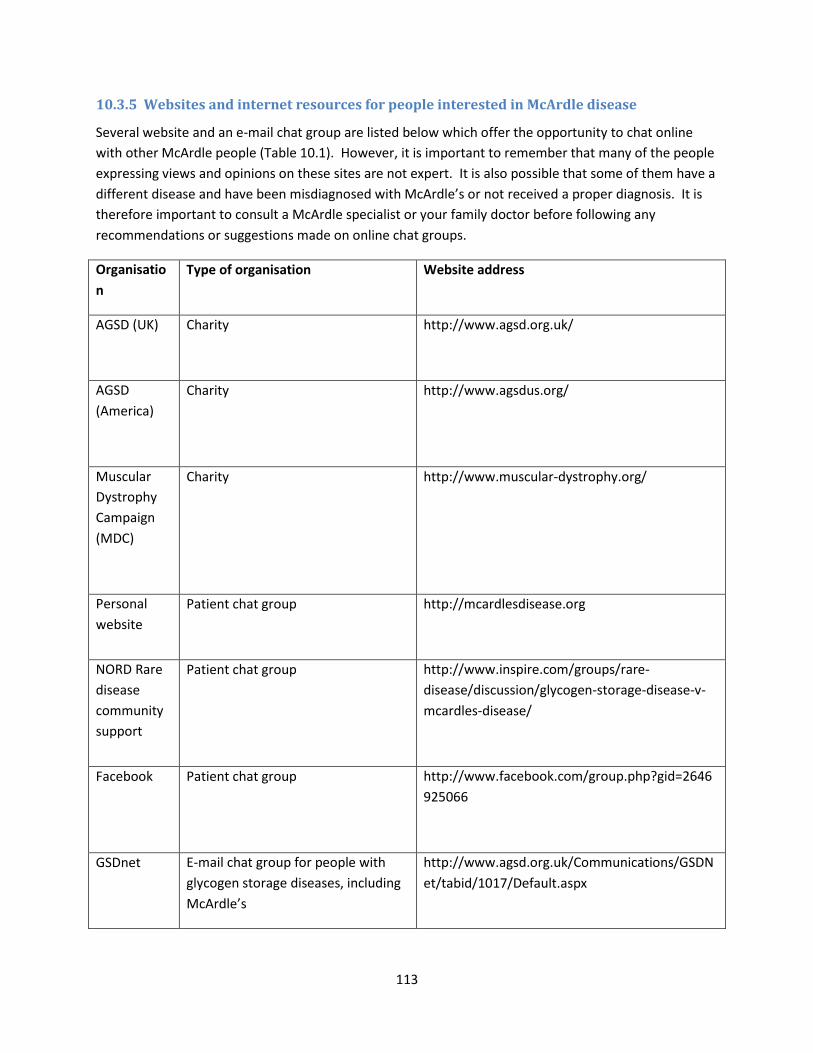
113
10.3.5 Websites and internet resources for people interested in McArdle disease
Several website and an e-mail chat group are listed below which offer the opportunity to chat online
with other McArdle people (Table 10.1). However, it is important to remember that many of the people
expressing views and opinions on these sites are not expert. It is also possible that some of them have a
different disease and have been misdiagnosed with McArdle’s or not received a proper diagnosis. It is
therefore important to consult a McArdle specialist or your family doctor before following any
recommendations or suggestions made on online chat groups.
Organisatio
n
Type of organisation Website address
AGSD (UK)
Charity http://www.agsd.org.uk/
AGSD
(America)
Charity http://www.agsdus.org/
Muscular
Dystrophy
Campaign
(MDC)
Charity http://www.muscular-dystrophy.org/
Personal
website
Patient chat group http://mcardlesdisease.org
NORD Rare
disease
community
support
Patient chat group http://www.inspire.com/groups/rare-
disease/discussion/glycogen-storage-disease-v-
mcardles-disease/
Facebook Patient chat group http://www.facebook.com/group.php?gid=2646
925066
GSDnet E-mail chat group for people with
glycogen storage diseases, including
McArdle’s
http://www.agsd.org.uk/Communications/GSDN
et/tabid/1017/Default.aspx
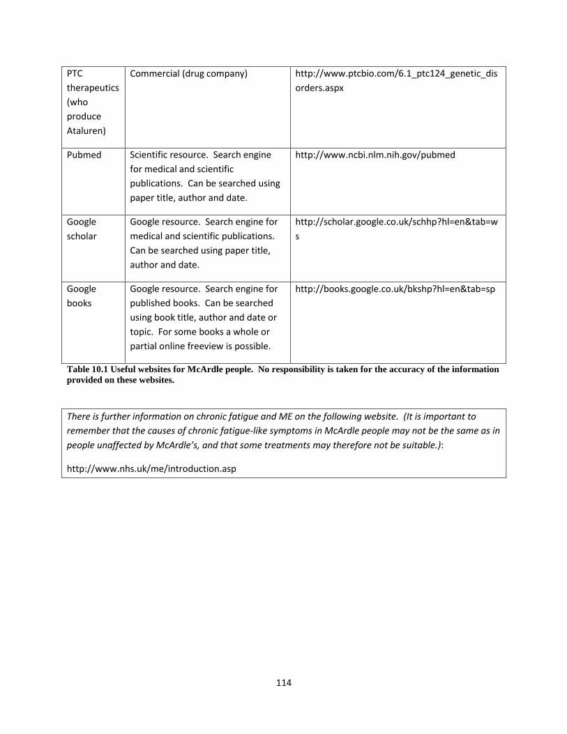
114
PTC
therapeutics
(who
produce
Ataluren)
Commercial (drug company) http://www.ptcbio.com/6.1_ptc124_genetic_dis
orders.aspx
Pubmed Scientific resource. Search engine
for medical and scientific
publications. Can be searched using
paper title, author and date.
http://www.ncbi.nlm.nih.gov/pubmed
scholar
Google resource. Search engine for
medical and scientific publications.
Can be searched using paper title,
author and date.
http://scholar.google.co.uk/schhp?hl=en&tab=w
s
books
Google resource. Search engine for
published books. Can be searched
using book title, author and date or
topic. For some books a whole or
partial online freeview is possible.
http://books.google.co.uk/bkshp?hl=en&tab=sp
Table 10.1 Useful websites for McArdle people. No responsibility is taken for the accuracy of the information
provided on these websites.
There is further information on chronic fatigue and ME on the following website. (It is important to
remember that the causes of chronic fatigue-like symptoms in McArdle people may not be the same as in
people unaffected by McArdle’s, and that some treatments may therefore not be suitable.):
http://www.nhs.uk/me/introduction.asp

115
11 The effect of McArdle’s on sexual activity, pregnancy and birth
11.1 Sexual activity
Sexual activity usually involves exercise; this may be static (using your muscles to hold yourself in one position for a long time, like yoga), or repetitive movements for a long time, or fast movements. It is obvious that McArdle’s is likely to have an effect upon the ability of McArdle people to do these activities. Unfortunately there is hardly any published information on the effect of McArdle disease on sexual activity and intimate relationships. Hockaday et al. (1964) reported the case of a 51 year old McArdle man who found that McArdle’s symptoms of muscle pain in the legs was worse if he had had “sexual intercourse the previous night”. Quinlivan et al. (2010) said that McArdle men and McArdle women reported muscle cramps, with or without myoglobinuria, occurring as a consequence of sexual intercourse. The lack of published information is likely to be due to several reasons; because it can be hard to quantify, because McArdle people may be embarrassed to mention it to their family doctors, and because the family doctors who diagnose and treat McArdle people tend to be neurologists and muscle specialists, and therefore do not have any expertise (and possibly no interest) in sexual issues. The most relevant expert for these issues would be a sexual therapist with an interest in helping people who have limited mobility. In the absence of any published advice, here are some approaches I would suggest:
Find and apply advice for people who have limited mobility (for example, advice for people who
have arthritis), bearing in mind what you know about McArdle disease, and avoiding movements
or positions which cause pain or are likely to lead to muscle damage or contracture.
Use the latest advice regarding exercise. Warm up and get into a second wind if possible. The
only published piece of advice relating to McArdle disease and sexual intercourse which I have
been able to find is a suggestion to have a sugary drink just prior to sexual intercourse; “This
[having a sucrose drink] might be a useful therapy prior to planned vigorous exercise, such as
sexual intercourse” (Quinlivan and Vissing, 2007).
Communication. Be willing to communicate what you want, what works, what doesn’t work,
what you enjoy or don’t enjoy, being prepared to stop or change positions if your muscles begin
to hurt.
Consider using pillows or other accessories to reduce the need to use your muscles to hold you
in a certain position.
11.2 The menstrual cycle and it’s effect upon exercise and perception of pain
for McArdle’s women
There is no published information on the effect of female hormones or the female menstrual cycle on
McArdle’s symptoms. It has anecdotally been reported by women with McArdle’s that they experience
a worsening of McArdle’s symptoms a week or two before menstruation (having a period). Possible
explanations for this this anecdotal observation could include:
116
The menstrual cycle could physically affect the ability of McArdle’s women to exercise.
The menstrual cycle could physically or mentally affect the perception of pain by McArdle’s
women.
The menstrual cycle could mentally affect how distressing women find McArdle’s symptoms.
There is no information available on these topics regarding women with McArdle’s, but some research
on similar topics on women unaffected by McArdle’s is discussed below.
The menstrual cycle can be broken down into two parts; the time between the start of menstrual
bleeding (the period) and ovulation (which is called the follicular phase), and the time between
ovulation and the start of menstrual bleeding (called the luteal phase).
11.2.1 Does the menstrual cycle affect the ability of women unaffected by McArdle’s to
exercise
There have been many investigations into whether the menstrual cycle affects the ability of women to
exercise. In particular, studies have focussed on the effect of menstrual cycle on the performance of
female athletes. According to Janse de Jonge (2003), “current literature indicates that VO2 max is not
affected by the menstrual cycle” (see 6.4.4.1 for further discussion of VO2 max), which suggests that
performance of female athletes is not affected by the menstrual cycle. However, the authors mention
that some studies suggest that women unaffected by McArdle’s may have a “higher cardiovascular
strain during moderate exercise in the mid-luteal phase”. They noted that women athletes are able to
exercise for a shorter time before becoming exhausted “during the mid-luteal phase, when body
temperature is elevated. “ This may mean that McArdle’s women may find exercise slightly harder
during the mid-luteal phase, especially in hot conditions.
11.2.2 Studies on the perception of pain in women unaffected by McArdle’s
Riley et al. (1999) reviewed 16 studies where women unaffected by McArdle’s, who were healthy and
had no conditions which caused pain, were tested to see whether their experience of pain varied at
different times in their menstrual cycle. They found that the women experienced least pain from
pressure stimulation, cold, heat, and ischemic muscle pain, during the follicular phase than at other
points in the menstrual cycle. The authors of the review note that the research was not perfect as
different stimuli were used to produce pain in different studies (stimuli included pressure, heat or cold).
They may have struggled to define exactly which part of the menstrual cycle the women were in. A
further problem is that only small numbers of women were studied.
Pfleeger et al. (1997) carried out a study with 11 women unaffected by McArdle’s, who performed an
ischaemic forearm test. In this case, the women had to say when the pain from their forearm became
too great for them to continue exercising. Their blood pressure and their pain levels from the forearm
test were monitored at different times during the menstrual cycle, and it was found that the level of
pain experienced and the blood pressure were both highest during the luteal phase. However, it should
be noted thatHoeger Bement (2009)) found the opposite results; that when women were asked to carry

117
out a forearm test, their perception of pain was not affected by which stage of the menstrual cycle they
were in. Clearly more research is required!
11.2.3 The emotional changes associated with the menstrual cycle may make McArdle’s
women consider their symptoms to be worse at certain points
It is possible that hormone changes during the menstrual cycle will affect how women perceive their
symptoms. Women who are unaffected by McArdle’s often report emotional changes during their
menstrual cycle. The most dramatic of these occur just prior to the menstrual bleed/period, and are
known as “premenstrual syndrome or tension (PMS/PMT)”. It has been suggested that all women
experience some PMS symptoms, which can vary from woman to woman. Since some symptoms of
PMS include a depressed mood, crying, and tearfulness, and anxiety, my theory is a woman with PMS
may perceive her McArdle’s symptoms as being worse than usual - although the difference may be in
her frame of mind rather than the symptoms.
It has been reported that symptoms of chronic illnesses can be worse when women are also
experiencing PMS symptoms. Examples of physical illnesses which are known to become worse for a
woman when she is premenstrual include migrane (Dzoljic et al., 2002), epilepsy (Hussain et al., 2007),
asthma (Pereira Vega et al., 2010), irritable bowel syndrome (Houghton et al., 2002), rheumatoid
arthritis and diabetes (Case and Reid, 1998). There does not seem to be any information published
about whether symptoms of muscle diseases become worse during the menstrual cycle, which is
probably because no research has been done into this topic.
11.3 Fertility and contraception
There is no published information on whether McArdle disease has any effect upon fertility, but it seems
likely that McArdle disease does not affect the fertility of men or women with McArdle’s, so appropriate
contraception should be used if pregnancy is not desired. Unfortunately the promising sounding paper
“Contraception and pregnancy in women affected by glycogen storage diseases” (Mairovitz et al., 2002)
is focussed on GSDI and the information is not relevant to McArdle’s.
As discussed in section 14.2.2, it would be worthwhile discussing any medication or contraceptive
method with your family doctor whilst also considering McArdle’s. For example, you would want to
discuss whether there could be any side effects, such as an increased risk of rhabdomyolysis, which
might make some methods less suitable for a McArdle person.
11.3.1 Prenatal testing for McArdle’s and infant screening
An absence of published reports suggests that prenatal testing is not currently available during
pregnancy to determine whether an unborn baby will have McArdle disease. Theoretically it would be
possible to obtain a DNA sample from cells obtained by chorionic villus sampling or amniocentesis.
Milunky et al. (2010) say that although testing for the mutations which cause McArdle’s is theoretically
possible, “there are no reports in the literature that they have been done”.
Prenatal testing may not be currently available party due to the rarity of McArdle’s. Also, the
development of prenatal tests tends to be prioritized for diseases which are fatal or highly debilitating.
118
As McArdle’s symptoms are not as severe as many other muscle diseases (for example, Spinal muscular
atrophy or Duchenne muscular dystrophy), it may be a lower priority. As McArdle’s is recessive, unless
one or both parents have McArdle’s, there is often not a family history of McArdle’s, and therefore no
clue that prenatal testing should/could be considered.
Infant screening for McArdle’s (e.g. by a heel prick or blood test from a newborn baby), is also not
currently available. It is likely to be for similar reasons as those given above; because McArdle’s is
relatively rare, not as severe as some other diseases, and also because the advantage of the having a
diagnosis must be weighed against the cost of screening many newborns who would not have
McArdle’s.
Information about the chances of passing on McArdle’s to your children should be discussed with a
genetic counselor, but a scientific explanation of the inheritance of McArdle’s is given in section 3.3.
11.4 Pregnancy and birth
McArdle disease does not appear to increase the risk of complications for McArdle women during
pregnancy or for giving birth (Quinlivan et al., 2010).
11.4.1 Some McArdle’s women report having fewer McArdle’s symptoms during pregnancy
Anecdotally, some McArdle’s women say that they have fewer McArdle’s symptoms when they are
pregnant. There is no published data to confirm these anecdotal observations or to explain them.
Possible explanations for these anecdotal observations include:
Could a foetus (unborn baby) supply a McArdle’s mother with the enzyme she is lacking?
Do the pregnancy hormones increase the level of glucose in the bloodstream of the McArdle’s
mother, reducing symptoms?
Both these possibilities are discussed below.
11.4.1.1 Could a foetus (unborn baby) supply a McArdle’s mother with the enzyme she is
lacking?
The explanation for this theory is that in the foetus, the whole body is provided with energy by the
foetal glycogen phosphorylase enzyme (this has the alternative name of brain glycogen phosphorylase
enzyme). The theory is that some of the foetal glycogen phosphorylase may be taken from the foetus
in the bloodstream, and transferred across the placenta, into the mother’s blood. It would then be
carried to the mother’s muscles, and be taken into the muscle cells. It could then function in the muscle
cells to replace the missing muscle glycogen phosphorylase, so that the mother will no longer have
symptoms of McArdle’s.
I do not agree with this theory. It seems unlikely that the foetal glycogen phosphorylase could get out of
the foetus into the blood and then into the skeletal muscles. If this was the case, it would be a form of
enzyme replacement therapy. However, as described in section 16.3.2, enzyme replacement therapy is
not a suitable treatment for McArdle’s as the enzyme is taken up into the lysosome (the wrong location)

119
not the cytoplasm (the location where glycogen phosphorylase is needed). In an adult who is not
pregnant, the brain isoform does not leak out of the heart or smooth muscle (such as the smooth
muscle of the digestive tract) and into skeletal muscle and “cure” McArdle’s. This suggests that the
foetal enzyme probably can’t leak out of the foetus and into the skeletal muscle.
Walker (2006) carried out research into sheep and lambs with McArdle’s or unaffected by McArdle’s.
The gestation of a lamb is approximately 150 days . Research by Walker suggests that foetal glycogen
phosphorylase can be detected in a sheep foetus from the age of 40 days after conception (the earliest
time period tested by Walker), and that foetal glycogen phosphorylase is the main isoform until 50 days
after conception, when muscle glycogen phosphorylase begins to take over and become the main
isoform in sheep muscles . It is likely that a similar situation occurs in humans.
11.4.1.2 Do the pregnancy hormones increase the level of glucose in the bloodstream of the
McArdle’s mother, reducing symptoms?
I suggest an alternative theory; that that being pregnant raises the level of glucose in the blood (causing
hyperglycemia), which would produce a similar effect to drinking a sugary drink, and would help
pregnant McArdle’s women exercise more easily with fewer McArdle’s symptoms.
The foetus requires a high level of glucose for growth and development. During pregnancy, from about
6 weeks gestation, the placenta produces a hormone called human placental lactogen (HPL), also known
as Human Chorionic somatomammotropin. HPL is released from the placenta into the mother’s
bloodstream. HPL has an anti-insulin effect, which results in an increased amount of both glucose and
fatty acids in the bloodstream in order to supply enough nutrients and energy to the foetus. The
amount of HPL secreted by the placenta increases as the foetus grows bigger. HPL can also increase the
amount of fatty acids in the bloodstream. My unproven theory is that the increased levels of glucose
and fatty acids in the blood would provide an improved energy source for the muscles, reducing
McArdle’s symptoms for a pregnant McArdle woman. HPL also has a small effect on increasing
formation of protein tissues including muscle, in a similar but weaker way to growth hormone.
The increase in glucose during pregnancy in women unaffected by McArdle’s is well known. If the levels
of glucose in the blood are too high (possibly due to the effect of HPL), gestational diabetes may occur in
pregnant women unaffected by McArdle’s. This is also known as hyperglycemia. Gestational diabetes
can occur very early in pregnancy, and can last throughout pregnancy. Gestational diabetes usually
stops following birth. If a phenomenon similar to gestational diabetes is occurring in pregnant
McArdle’s women, this could explain why they return to a normal level of symptoms following birth
(which has been reported anecdotally by McArdle’s women).
11.4.2 Method of giving birth; vaginal birth or caesarean delivery
McArdle’s does not appear to have any effect upon the ability of McArdle’s women to have a natural
birth, and does not increase the need for a caesarean delivery. There are a limited number of reports
about McArdle’s women giving birth, but those which are avilable report both vaginal and caesarean
births. Quinlivan et al. (2007; 2010) carried out a review of the McArdle women who had attended the
clinic in Oswestry, UK. They described 14 McArdle’s women, who had had 21 pregnancies between
120
them;“all pregnancies were uncomplicated”, although “one lady had mild myoglobinuria 24 hours after
delivery” The authors found that 15% of these cases (about 3 of 25 pregnancies) had required
intervention or caesarean, which was not dissimilar from the 15% national average in the UK. This is
very limited data, but it suggests that having McArdle’s does not make women more likely to require a
caesarean.
The uterus/womb is made of smooth muscle. Smooth muscle has a different isoform of glycogen
phosphorylase and is therefore not affected in McArdle’s. For this reason, McArdle’s women should not
have a problem having a vaginal birth. During vaginal birth, your family doctors may choose to give you
sucrose by an intravenous drip to provide an energy source to the muscles. Cochrane and Alderman
(1973) reported a case of a 21 year old McArdle woman. She had a normal pregnancy. She had a
relatively normal vaginal delivery. The second stage (pushing stage) was quite slow as she had been
given an epidural anaesthetic (which is well known to slow down delivery in women unaffected by
McArdle’s). Labour began spontaneously (naturally, not induced), but was quite slow (not unusual in a
first birth). She received dextrose (a form of simple sugar) and oxytocin (chemical which stimulates
contractions of the uterus) intravenously. The baby was healthy. Both mother and child made “normal
progress” after delivery. Contractions of the uterus were normal, further suggesting that it is smooth
muscle with another isoform of glycogen phosphorylase.
During pregnancy, in case a planned or emergency caesarean is required, you should remind your family
doctor that McArdle people are at increased risk of malignant hyperthermia (see section 12.3).
Anecdotally some McArdle’s women have reported that their arms were tired after giving birth, so that
they found it harder to hold the baby immediately afterwards.
There is further information on premenstrual syndrome on the NHS website:
http://www.nhs.uk/Conditions/Premenstrual-syndrome/Pages/Introduction.aspx
121
12 Medicines, activities and other things which may be a greater risk
for McArdle people
12.1 Medicines
There are some drugs which have an increased risk of causing rhabdomyolysis as a side effect in people
unaffected by McArdle’s, and are summarised in table Table 12.1. These drugs are more likely to cause
rhabdomyolysis in McArdle people. Some of these drugs have been reported to cause rhabdomyolysis
in McArdle people, and this is discussed in more detail below. Rhabdomyolysis is discussed further in
section 5.
If your family doctor suggests that you take one of the drugs listed in Table 12.1, I would recommend
that you discuss with them whether McArdle’s puts you at increased risk of rhabdomyolysis during the
treatment. Your family doctor may still decide to prescribe the drug, but may regularly monitor you.
See section 14.2 for more information on helping your family doctor to treat you.
12.1.1 Lipid-lowering drugs such as statins may exacerbate rhabdomyolysis in McArdle
people
Some people unaffected by McArdle’s had side effects including rhabdomyolysis when treated with
lipid-lowering drugs such as statins (Vladutiu et al., 2006). When these people were tested and
compared to people receiving the therapy with no symptoms of muscle damage and also to people who
had not been treated with statins, it was found that 10% of the 110 people with side effects were
heterozygous or homozygous for mutations causing carnitine palmitoyltransferase II (CPT II) deficiency
(which affects fatty acid metabolism), McArdle disease or myoadenylate deaminase deficiency (MADD)
(involved in energy metabolism in muscle cells). The effect of the statins upon energy metabolism
combined with a genetic disease affecting mitochondrial or fatty acid metabolism seemed to exacerbate
muscle damage and rhabdomyolysis.
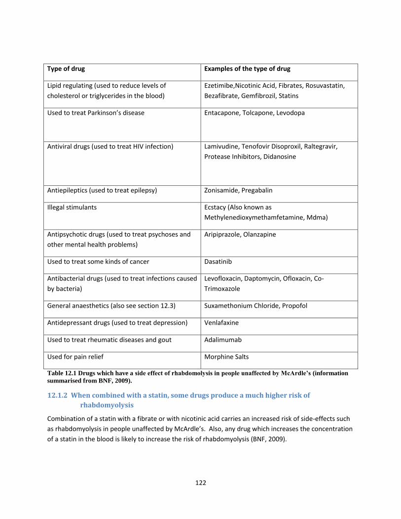
122
Type of drug Examples of the type of drug
Lipid regulating (used to reduce levels of
cholesterol or triglycerides in the blood)
Ezetimibe,Nicotinic Acid, Fibrates, Rosuvastatin,
Bezafibrate, Gemfibrozil, Statins
Used to treat Parkinson’s disease Entacapone, Tolcapone, Levodopa
Antiviral drugs (used to treat HIV infection) Lamivudine, Tenofovir Disoproxil, Raltegravir,
Protease Inhibitors, Didanosine
Antiepileptics (used to treat epilepsy) Zonisamide, Pregabalin
Illegal stimulants Ecstacy (Also known as
Methylenedioxymethamfetamine, Mdma)
Antipsychotic drugs (used to treat psychoses and
other mental health problems)
Aripiprazole, Olanzapine
Used to treat some kinds of cancer Dasatinib
Antibacterial drugs (used to treat infections caused
by bacteria)
Levofloxacin, Daptomycin, Ofloxacin, Co-
Trimoxazole
General anaesthetics (also see section 12.3) Suxamethonium Chloride, Propofol
Antidepressant drugs (used to treat depression) Venlafaxine
Used to treat rheumatic diseases and gout Adalimumab
Used for pain relief Morphine Salts
Table 12.1 Drugs which have a side effect of rhabdomolysis in people unaffected by McArdle’s (information
summarised from BNF, 2009).
12.1.2 When combined with a statin, some drugs produce a much higher risk of
rhabdomyolysis
Combination of a statin with a fibrate or with nicotinic acid carries an increased risk of side-effects such
as rhabdomyolysis in people unaffected by McArdle’s. Also, any drug which increases the concentration
of a statin in the blood is likely to increase the risk of rhabdomyolysis (BNF, 2009).
123
12.1.2.1 Fibrates
Fibrates include bezafibrate, ciprofibrate, fenofibrate, and gemfibrozil. They act by decreasing the
amount of triglycerides in the blood. However, a combination of a fibrate with a statin increases the risk
of rhabdomyolysis in people unaffected by McArdle’s. The British National Formulary (edition 58) states
that gemfibrozil and statins should not be used at the same time.
12.1.2.2 Nicotinic acid
Nicotinic acid can be used to lower cholesterol and triglyceride concentrations by reducing the amounts
of these compounds made by the body. In people unaffected by McArdle’s, muscle pain and
rhabdomyolysis have been reported as rare side effects.
12.1.3 Other drugs to reduce cholesterol may exacerbate rhabdomyolysis in McArdle people
Perez-Calvo (2005) reported a case of a McArdle man who was treated with ezetimibe. Ezetimibe is a
drug which inhibits absorption of cholesterol in the intestine. The person was treated for many months.
The person had no ill effects for 20 weeks, and then began to suffer from fatigue, myoglobinuria, and
weakness. His CK levels also rose. The family doctors treating him decided to discontinue the
treatment, and the CK values went down to normal after 4 weeks. It must be noted that this is a single
case study, and that this person also had several other medical problems including type 2 diabetes
mellitus, which could have affected the outcome.
Ezetimbe is used to treat primary hypercholesterolaemia (high cholesterol levels). Ezetimibe may be
marketed alone as a drug called Ezetrol or Zetia. Ezetimibe combined with a statin is known as a
simvastatin, and marketed as “Zocor” or “Inergy” (BNF, 2009).
12.2 Pain relieving mediation
Anecdotal advice is that drugs for muscle pain should not be used routinely. The principle reason for
this is because if muscle damage is causing the pain, then the pain will warn the person not to exercise,
which could cause further muscle damage. In this way, the pain has a positive effect to protect the
muscles from being damaged further. Pain relieving medication would remove this pain, and could lead
to the muscle being used and damaged further.
Some forms of pain relief may also have an increased risk of rhabdomyolysis (see Table 12.1).
It is important to note that the adage “No pain, no gain” does not apply to McArdle people!
12.3 McArdle people are at increased risks of particular complications
during surgery
12.3.1 Lack of muscle glycogen phosphorylase may put McArdle people at increased risk of
malignant hyperthermia-like symptoms caused by anaesthetics
Malignant hyperthermia is a life-threatening condition which is triggered by certain anaesthetics, one
example of which is muscle relaxant succamethonium chloride. These drugs can cause a major increase

124
in skeletal muscle oxidative metabolism whereby the muscle cells run out of oxygen and develop an
excess of carbon dioxide. The increase in metabolism can cause an abnormal rise in body temperature,
which is why the condition is described as hyperthermia. Medical treatment to stop the temperature
rise is essential as in the worst case scenario it would be possible for the body temperature to continue
to rise until it leads to death (Price Evans, 1993).
Malignant hyperthermia is an autosomal dominantly inherited disorder associated with central core
disease. Central core disease is caused by a mutation in the RYR1 gene which encodes ryanodine
receptor 1, a channel for calcium ions in skeletal muscle, required for muscle contractions (the role of
calcium in muscle contractions is discussed further in section 4.3). Central core disease causes a lack of
muscle glycogen phosphorylase activity within the core of the muscle cell (Isaacs et al., 1975) (Dubowitz
et al., 2007). This may suggest that McArdle people (who also don’t have muscle glycogen
phosphorylase activity) could be at risk of malignant hyperthermia caused by anaesthetics. Cases of
McArdle people being positive for malignant hyperthermia have been reported; Isaacs et al. (1989)
reported one person, and and Aquaron et al. (2007) described two families, one with three affected
siblings and one with two affected siblings. One current test for susceptibility to malignant
hyperthermia is an in vitro contracture test (a muscle biopsy in bathed in a solution containing caffeine
or halothane to see whether contracture occurs), which is positive for many McArdle disease people
(Bollig et al., 2005). If a person is susceptible, malignant hyperthermia usually occurs within one hour of
being given a general anaesthetic (Rosenberg et al., 2007). Dantrolene sodium can be used to
prevent/treat malignant hyperthermia. (Dantrolene sodium is discussed further in section 7.1.5.)
IMPORTANT: Before having a general anaesthetic, you should inform your surgeon/anaesthetist that
McArdle people are at an increased risk of malignant hyperthermia. (Ideally you would inform them in
advance of the surgery, and then remind them on the day of surgery.) This will allow the medical staff
to decide carefully which anaesthetic to use, and to ensure they monitor you carefully. They may also
decide to have dantrolene sodium available in case malignant hyperthermia does occur. If a caesarean
delivery is required for a pregnant McArdle woman, the surgeon should be aware of the risk of
malignant hyperthermia. Bollig et al. (2005) also recommend to surgeons that “measures for preventing
muscle ischaemia and rhabdomyolysis should be kept in mind, as well as the potential for these patients
[with McArdle disease] to develop postoperative fatigue, myoglobinuria and renal failure”.
12.3.2 McArdle people may be at risk of a rare condition called “Compartment syndrome”
caused by use of a tourniquet or cuff
Compartment syndrome is also known as “compartment pressure syndrome”. This is a rare
complication cause by use of a tourniquet or cuff. The compartment is a small area between the layers
of the muscle. Usually, a very small amount of fluid is present in this space, and is used for contraction
and relaxation of the muscle. There is no room for any additional fluid. Compartment syndrome is a
build up of fluid in the compartment. Symptoms of compartment syndrome include severe muscle pain,
muscle weakness, and very tense skin over the muscle. It can reduce the ability of blood to pass through
the muscle (so that no pulse can be felt), and the worst case scenario is paralysis.
125
There is a report of compartment syndrome in a person with Her’s disease. Niepel et al. (2004) reported
a person with Her’s disease who was given an ischaemic forearm test. “Later that afternoon”, he had
“continuing pain in his forearm and inability to extend his fingers”, followed by reduced sensation in his
hand. Compartment syndrome was diagnosed, and an emergency operation was performed.
Unfortunately “his recovery was incomplete “and he was left with some loss of sensation. Although this
case occurred in a person with Her’s disease, there is also a single reported case of compartment
syndrome in a McArdle person who performed the ischaemic forearm test (Lindner et al., 2001).
General recommendations (for people unaffected by McArdle’s) are that a patient’s own and family
history (information about previous cases of compartment syndrome) should be obtained. Tourniquet
time should be limited to less than 90 minutes, and that the tourniquet should be released before
encasing a muscle in a solid cast (such as that used on a broken arm).
IMPORTANT: Before having a surgery, you should inform your surgeon/anaesthetist that McArdle
people are at an increased risk of compartment syndrome. Ideally the surgeon would avoid use of a
tourniquet or cuff, although this would have to be balanced against the risk of not using it (for example
if a person might bleed to death). If a tourniquet is still needed, the surgeon should be informed about
the possible risk of muscle damage with subsequent myoglobinuria (Bollig et al., 2005). Medical advice
should be sought urgently if, after carrying out an ischaemic forearm exercise test, a loss of sensation is
felt in the fingers, continued pain is present, and an inability to extend (move) the fingers.
12.4 Some situations may make McArdle’s symptoms worse
12.4.1 Cold temperature
Rommel et al. (2006) found that some McArdle people had increased pain caused by environmental
factors such as a cold temperature.
12.4.2 Getting angry
Rommel et al. (2006) found that some McArdle people had increased pain caused by factors such as a
psychological distress. Becoming very angry can cause a McArdle person to tense up their muscles. I
have heard an anecdotal case where a McArdle person became very angry so that their muscles all
tensed up, resulting in contractures and rhabdomyolysis. It is possible that being very scared could
produce a similar effect.
12.4.3 Swimming
Swimming is an activity which is potentially dangerous for McArdle people. This is because if the
muscles run out of energy whilst a McArdle person is swimming, they will not be able to tread water or
swim to safety. This could result in the McArdle person drowning. There is a published report of a 6
year old McArdle’s child who almost drowned whilst swimming (prior to being diagnosed with
McArdle’s) (Roubertie et al., 1998).

126
12.4.4 Treatment by anyone who moves the body e.g. physiotherapy, osteopathy,
chiropractor, massage
If you require treatment by a person who moves the body; treatment for muscle pain, massage, or bone
related issues, it is very important to ensure that the person understands McArdle’s. Anecdotally, some
McArdle people report that treatment by a physiotherapist can cause muscle pain and potentially
muscle damage.

127
13 McArdle disease may increase the chances of having some diseases
and conditions
13.1 McArdle’s may increase the risk of gout
Gout is a painful swelling of the joints which is caused by build up of uric acid crystals. It can be caused
by high concentrations of uric acid crystals in the blood. Uric acid can be produced during the
breakdown of purines in food during digestion. It is also possible that level of uric acid may increase in
the blood following exercise, although whether this is the case, and what the mechanism could be is still
unclear (McCrudden, 2008).
There is some evidence that having McArdle’s may increase the risk of having gout. 11% (5 out of 45) of
the McArdle people attending the Oswestry clinic had had treatment for gout (Quinlivan et al., 2010).
Puig et al. (1992) reported the case of a person with McArdle disease who also had gout. They carried
out investigations to measure the amount of purine in the blood and urine after the McArdle person had
vigorously exercised, but did not see an increase in the level of uric acid in the blood or urine. There
therefore concluded that “in this patient, the association of McArdle disease with gout is coincidental”.
Jinnai et al. (1993) reported the case of a 28 year old McArdle man who had continuous hyperuricemia
(raised levels of uric acid in the blood). They found that when the McArdle man carried out aerobic
exercise using a bicycle ergometer, it led to an increase in uric acid. Exercise seemed to make the
muscles speed up the rate of purine degradation, which increased the levels of uric acid. In this case
report, the authors claimed that there was a relationship between exercise and increased uric acid in
the bloodstream. They described the condition as “myogenic hyperuircemia” (“myogenic” means it is
caused by muscle contractions, and hyperuricemia” means an increased level of uric acid in the blood).
Mineo et al. (1995) said that myogenic hyperuricemia is seen in glycolyctic defects (McArdle’s is a
glycolytic defect). They say that myogenic hyperuricemia is caused by excessive degradation of muscle
purine nucleotides, presumably as an energy source, as the cells are not able to make energy in the
usual way (the usual way would be glycogenolysis). Mineo et al. (1987) carried out a study where they
examined the amounts of purine in the bloodstream after exercise. They found that exercises in a
person with McArdle’s (and also two people with GSD III and one person with GSD VII) lead to
accelerated breakdown of purines in the muscle cells. This resulted in increased levels of breakdown
products (called ammonia, inosine and hypoxanthine) in the blood. The authors noted that these
breakdown products can be used by the body to produce uric acid, leading to hyperuircemia.
13.2 Brain functioning
There is some evidence that McArdle people may struggle more with some aspects of psychological
functioning than people unaffected by McArdle’s. This is discussed in more detail in section 10.2.

128
13.3 Respiratory problems
Respiratory problems appear to be unusual in McArdle people, but there have been some cases
reported. Lucia et al. (2008a) reported some McArdle people who had respiratory problems, but they
were all women. (Differences in the severity of symptoms between men and women are discussed
further in section 9.5.) Paradas et al. (2005) report a McArdle woman with shortness of breath (also
known as dyspnea). This same woman had been previously reported in the paper “McArdle disease
presenting as unexplained dyspnea in a young woman.” by Voduc et al. (2004). Harris et al. (1985) also
reported an elderly McArdle woman with shortness of breath.
There are a few reports of a rare infant form of McArdle's which was fatal due to respiratory failure, but
there were only 3 cases published, and I strongly suspect that they all had a second disease/problem
which caused the respiratory failure. (This rare infant form is discussed further in section 8.2.1.)
13.4 McArdle’s may cause symptoms of insulin resistance (similar to type 2
diabetes)
Insulin plays a key role in controlling the amount of glucose in the blood and in enabling muscle cells to
absorb glucose from the bloodstream, which can then be used to provide energy (see section 6.2). Diabetes is
the inability to control the level of glucose (sugar) in the blood. There are two forms of diabetes; type 1 and
type 2. Information about these forms of diabetes (in people unaffected by McArdle’s), is provided in
Table 13.1.

129
Type of
diabetes
Also known as Age when
this type of
diabetes
first begins
Can be
associated
with
Is insulin
required as a
treatment
Insulin
receptors
on cells
Symptoms Out of all the
people with
diabetes, how
common is this
form
Type 1 Type I
Insulin dependent diabetes
mellitus (IDDM)
Ketosis-prone
Brittle diabetes
Diabetes mellitus type I
Less than
20 years of
age,
although
occasionally
older than
40 years of
age
Viral infection Yes Normal Quite
severe
Rare (5-10% of
people with
diabetes)
Type 2 Type II
Non Insulin dependent
diabetes mellitus (NIDDM)
Adult-onset diabetes
Ketosis-resistant
Stable
Diabetes mellitus type 2
Older than
40 years of
age
Obesity (being
very
overweight)
No Low or
normal
Quite
moderate
Very common (90-
95% of people with
diabetes)
Table 13.1 Information about diabetes in people unaffected by McArdle’s (adapted from McArdle, 2000).
130
There is no suggestion that McArdle’s may be associated with type 1 diabetes (IDDM). In the absence of
any evidence to suggest a link, it is likely that a report of a person having both McArdle’s and type 1
diabetes is likely to be a coincidence. Davies et al. (1977) reported a McArdle person who had type 1
diabetes. At present this appears to be the only published report of a McArdle person with type 1
diabetes.
McArdle people have very high levels of glycogen stored in their muscle cells. There are several
published reports that high levels of stored glycogen reduce the ability of insulin to stimulate the cells to
take glucose from the bloodstream into the muscle cells. This is known as “insulin resistance”, and is
similar to type 2 diabetes (also known as non-insulin dependent diabetes mellitus). There has not been
much research into insulin resistance in McArdle people, but it is an important topic. Personally, I
wonder if future research will show that almost all McArdle people have some insulin resistance caused
by the high amount of glycogen stored in their muscle cells, so I have included the following
information. Many more studies will be needed to show if this is the case.
Menada et al. (2002) reported a case of a 29 year old McArdle woman who also had insulin resistance.
“Pre-administration of glucagon had no effect on serum lactate and pyruvate levels after ischemic
forearm exercise test while serum glucose elevated. The glucose clamp test confirmed insulin
resistance.” Maeda et al.noted that that there was no reducing in the number of insulin receptors. Her
weight was 78.4kg and BMI was 32.0, so the authors suggested that “Her obesity might be a causative
factor of insulin resistance.” I personally disagree, and suggest that it may have been excess glycogen
storage (due to McArdle’s) which had caused insulin resistance.
Yamauchi et al. (1996) reported a 64 year old McArdle woman who had type 2 diabetes (NIDDM).
“Thus, we suggested that high plasma glucose and insulin due to NIDDM may induce blood-borne
glucose uptake with exercise.”
Dorin et al. (1996) reported a McArdle person with type 2 diabetes (NIDDM). They hypothesised that
insulin resistance would reduce the ability of the muscles to take up glucose during exercise and
suggested that adding insulin (intravenously) would improve this. They found that when they artificially
added insulin, it increased the amount of glucose able to get to the muscles, and increased the amount
of work that the McArdle person was able to do.
Nielsen et al. (2002a) compared 6 McArdle people with 6 unaffected people. They gave each person a
glucose drink, and investigated whether the body could use insulin to stimulate the muscle cells to take
up the glucose. They found that the McArdle people had much less insulin-stimulated use of glucose
than the unaffected people.
Mineo et al. (1984) gave McArdle people glucagon prior to exercise. This relieved “patients with
McArdle disease from muscular symptoms during exercise and enhanced exercise performance”. It is
likely that glucagon acted upon the liver, causing a release of glucose into the bloodstream. This would
have acted in a similar way to having a sugary/glucose drink immediately prior to exercise, which is
known to help McArdle people exercise more easily (section 7.1.6.3). The authors note that a similar
improvement in the ability to exercise was seen after giving glucose, or glucose plus insulin (the insulin

131
would probably have helped the muscle cells to take up the glucose). Interestingly, the authors did not
see an improvement in the ability to exercise when McArdle people were given insulin alone, without
glucose.
13.4.1 A discussion of whether insulin resistance could be caused by McArdle disease
It is not clear to me whether the insulin resistance observed in some McArdle people is exactly the same
as type 2 diabetes (NIDDM) observed in people unaffected by McArdle’s. It is possible that there are
important differences between the insulin resistance seen in people with McArdle’s and those
unaffected by McArdle’s. In McArdle people, the cause of insulin resistance may be different to the
cause in people unaffected by McArdle’s. Nielsen et al. (2002a) suggested that the high level of
glycogen storage in the muscle cells of McArdle people might cause insulin resistance. This is not the
cause of insulin resistance in people unaffected by McArdle’s. My personal unproven theory is that
most McArdle people have some level of insulin resistance caused by glycogen storage in their muscle
cells.
13.4.1.1 A high level of glycogen in the muscle cells of McArdle people may lead to insulin
resistance
Usually, insulin can stimulate muscle cells to take glucose from the bloodstream into the muscle cells.
This functions as a way of keeping the amount of glucose in the bloodstream constant. It also helps to
increase the amount of glucose in the muscle cells. Derave et al. (2000) found that a very high amount
of glycogen in muscle cells led to the GLUT-4 proteins not responding to insulin (this is called “insulin
resistance”). Nielsen et al. (2002a) compared McArdle people with unaffected people. They gave each
person a glucose drink, and investigated whether the body could use insulin to stimulate the muscle
cells to take up the glucose. They found that the McArdle people had much less insulin-stimulated use
of glucose than the unaffected people. The authors concluded that the high level of glycogen in the
skeletal muscle cells caused McArdle people to be insulin resistant in terms of glucose uptake, and
stated that “insulin action is decreased by high muscle glycogen content in skeletal muscle.” In addition,
the ability of insulin to decrease and increase the use of fat and carbohydrate to produce energy was
also reduced in people with McArdle’s.McArdle disease
13.4.1.2 High levels of some cytokines, such as TNF-α and IL-6, in the bloodstream of McArdle
people, could lead to insulin resistance
There is some evidence that McArdle people have raised levels of two cytokines (small proteins) called
TNF-α and IL-6 (Lucia et al., 2008b). (These are both discussed further in section 9.2.) It has been found
that people with type 2 diabetes (unaffected by McArdle’s) have high levels of TNF-α. TNF-α is known to
decrease the ability of insulin to stimulate muscle cells to take up glucose in rats by inhibiting the action
of GLUT-4 and inhibit insulin receptor activity. A possible explanation for the insulin resistance seen in
McArdle people could be that the high levels of TNF-α (and possibly IL-6) may reduce the ability of
insulin to stimulate uptake of glucose from the bloodstream by GLUT-4. TNF-α (and possibly IL-6) could
therefore lead to insulin resistance. (This is an unproven theory.)

132
13.4.2 Anecdotal reports of a feeling of low blood sugar could be explained by insulin
resistance preventing muscle cells taking up glucose, despite test results showing
high blood glucose levels
Several McArdle people have reported on online chat groups that they experience a feeling of low blood
sugar levels. If these people have insulin resistance, a feeling of low blood sugar could occur because
the insulin isn’t able to act, and the cells aren’t able to take up glucose, even though there are high
glucose levels in the blood. If this was the case, blood test results would show high blood sugar levels,
but the McArdle person would feel that they had low blood sugar. Although studies have shown that
some McArdle people have insulin resistance, these studies did not ask McArdle people whether they
felt that their blood sugar levels were low. If this were the case, I wonder whether insulin resistance
could lead to weight gain in McArdle people. If McArdle people have a feeling of low blood sugar, this
would make the person want to eat a high sugar or high carbohydrate food. If the muscle cells are
unable to take in glucose, the muscle will not be able to use the glucose to provide energy for
movement. Instead, this excess glucose would be stored as glycogen or as fat. This is an unproven
theory.
Weight gain should be avoided by McArdle people (see section 4.2.4).
13.4.3 Exercise can be used to prevent and treat insulin resistance in people unaffected by
McArdle’s
People unaffected by McArdle’s are less likely to develop type 2 diabetes if they have a physically active
lifestyle. In people unaffected by McArdle’s, “an increase in abdominal fat accumulation and loss of
muscle mass are highly associated with the development of insulin resistance” (Ivy, 1997). Exercise
burns this fat, can prevent muscle wastage, and stimulate muscle development.
Hawley (2008) found that in studies of people unaffected by McArdle’s, who have type 2 diabetes, even
a single bout of exercise can increase the amount of glucose taken up into the muscle cells In this case,
the glucose is taken up in a different way, which is stimulated by exercise. However, the authors say
that this affect will only last for approximately 48 hours. “In contrast, repeated physical activity (i.e.
exercise training) results in a persistent increase in insulin action in skeletal muscle from obese and
insulin-resistant individuals”. In addition, they found that regular training seemed to increase the ability
of the muscles to use fat for energy.
Exercise has been reported as beneficial for McArdle people (see section 4.2.2), but at present no
research has been carried out to see whether exercise could be used to treat McArdle people with
insulin resistance.
13.4.3.1 Research suggests that a sugary drink before exercise can be effective even in McArdle
people with insulin resistance but should only be used under medical supervision
Although sugary drinks are not recommended for people with diabetes (see important note below), they
have been experimentally tested on two McArdle people who also had insulin resistance (NIDDM).
These tests were performed under close medical supervision. Vissing and Haller (2003) tested a
133
McArdle person with NIDDM. They found that a sucrose drink was equally effective in this person as in
McArdle people who did not have NIDDM. Similar experimental results had been previously found by
Yamuchi et al. (1996) who found that a McArdle personwomen with NIDDM was able to take up and use
glucose from the blood during exercise. Yamuchi et al. struggled to explain this observation, but Vissing
and Haller, suggested that glucose uptake in these McArdle people with NIDDM may have been induced
by muscle contractions, not by insulin sensitivity.
IMPORTANT: People with diabetes are not recommended to have sugar/sugary drinks. McArdle people
with diabetes should obtain medical advice before using sugar/sugary drinks.
13.5 There is no evidence that McArdle’s may increase the risk of heart
problems
The heart muscle in unaffected people has a 50:50 mixture of brain glycogen phosphorylase and muscle
glycogen phosphorylase (Miranda et al., 1979; Bresolin et al., 1983). Although McArdle people do not
have muscle glycogen phosphorylase, brain glycogen phosphorylase is present. The presence of brain
glycogen phosphorylase in the heart appears to be sufficient for it to function normally, and heart
problems are not generally reported in McArdle people.
In McArdle people, an increased (very rapid) heart rate is seen during intense exercise (before the
second wind). This response to exercise is discussed further in section 6.4.4. The increased heart rate
during exercise is only seen for a short period and when the muscle pain causes the McArdle person to
stop and rest the heart rate becomes lower.
I have heard a suggestion that as McArdle people are not able to maintain a high heart rate during
exercise (which is normally recommended for people unaffected by McArdle’s); this may lead to an
increased risk of heart disease or increased cholesterol. Perez et al. (2006) reported a case of a 78 year
old McArdle man who had had coronary heart disease. However, this single case report is not sufficient
evidence to support this theory. Nicholls et al. (1996) reported a case of a 66 year old McArdle man
with angina. Angina is pain in the heart which is caused by narrowing of the arteries of the heart. This
narrowing can reduce blood flow. This narrowing can be caused by several factors, one of which is
cholesterol. Anti-cholesterol drugs called statins may therefore be prescribed, but these should be
avoided if possible by McArdle people (see section 12.1.1).
13.6 McArdle’s is not reported to cause liver disease, but can have an effect
on the results of blood tests for liver disease
There is no published data that McArdle’s leads to liver disease. Liver disease can be shown by an
increase in the levels of proteins called Alanine Transaminase (ALT; formerly called SPGT)) and Aspartate
Transaminase (AST; formerly called SGOT), which can be detected by a blood test. However, skeletal
muscle cells also contain AST and ALT. If muscle damage has occurred, raised AST and ALT levels may be
seen. Tuzun et al. (2002) reported a case of a McArdle person who had raised levels of transaminases in
the blood. Measuring creatine kinase levels in the blood could indicate if muscle damage has
occurred. (Note: Raised AST and ALT levels can be caused by taking drugs, viral hepatitis or excessive

134
alcoholic consumption, so if you are treated by a family doctor unfamiliar with McArdle’s, you may have
to explain that McArdle’s could also cause this result.)

135
14 McArdle’s specialists and general family doctors
14.1 McArdle’s specialists
There are several medical professionals who have a strong interest and specialise in McArdle disease.
Some of them are listed below. The McArdle’s clinic at Oswestry, Shropshire, is the only McArdle’s clinic
in the UK. McArdle people in the UK must be referred to the Oswestry clinic by their family doctor. It is
sometimes possible for McArdle people in Ireland to also visit this clinic. In the USA, the Muscular
Dystrophy Association (MDA) is able to put people in contact with specialists who are located nearby.
Many of the McArdle’s specialists carry out research into McArdle’s and run clinical trials as well as
seeing patients in a clinic. There are some differences in opinion between McArdle’s specialists, such as
what is the ideal diet for McArdle people (see section 0), and so the advice you receive may not be
exactly the same from each specialist.
Name of McArdle’s specialist Address of clinic
Dr Ros Quinlivan RJAH Orthopaedic Hospital, Oswestry, Shropshire, UK
Dr McConvile Belfast City Hospital
Dr Ronald Haller Presbyterian Hospital of Dallas (Texas)
Dr Alfred Slonim Children’s Hospital at North Shore (Long Island, New
York)
Dr Tarnopolsky Canada
Dr John Vissing Denmark
Table 14.1 McArdle’s experts and their addresses. Please contact Kathryn Wright if you are able to
contribute more information about McArdle specialists and their details.
14.1.1 Benefits of visiting a McArdle’s specialist
Visiting a McArdle’s specialist can be of great benefit to McArdle people. Unlike your family doctor,
McArdle’s specialists have a much more thorough understanding of McArdle’s. McArdle’s specialists
may be able to:
Confirm the diagnosis of McArdle’s is correct.
Teach you how to reach second wind.
Provide you with information about the usual symptoms and progression of McArdle’s.
Tell you about current treatments and recent research.
Recruit you for clinical trials into treatments for McArdle’s.

136
Provide you with a letter/note/instruction sheet with information about McArdle’s to take into
hospital and show the family doctors and nurses if you require emergency treatment (for
example for severe muscle damage).
Devise a personal treatment plan for if you have myoglobinuria. Myoglobinuria is the most
obvious sign of muscle damage which can be assessed by eye without any medical tests. You
should discuss whether a low level of myoglobinuria could be treated at home (for example by
drinking plenty of fluids), or whether you should go to a hospital each time. (Anecdotally, this
seems to vary from person to person, but any decision to treat myoglobinuria at home should
be made in consultation with a medical specialist.
Discuss how to treat contractures; whether they will prescribe a strong painkiller (anecdotally
McArdle people say that normal painkillers often are not strong enough to treat the pain of a
contracture). If strong painkillers are prescribed, they should provide clear instructions about
when and how (e.g. how many pills to take, how often) to take the drug, and whether it is safe
to take with any other drugs you take. Also whether it would affect your ability to drive or
operate machinery.
14.1.1.1 The McArdle’s clinic at Oswestry, UK
The McArdle’s clinic at Oswestry is one of the few McArdle’s clinics in the world. I attended the
Oswestry clinic several times between the years of 2005 and 2008 as an observer where I was able to sit
in on consultations between patients and Dr Quinlivan and other members of the team. The Oswestry
clinic offers McArdle people an opportunity to meet specialists from several disciplines. Dr Ros
Quinlivan is one of the leading McArdle’s specialists. She primarily sees patients, but is also involved in
clinical trials and in collaborations with other people carrying out research into McArdle’s around the
world. Other members of the multi-disciplinary clinic include a physiotherapist who specialises in
neuromuscular conditions, a dietician, and an exercise physiologist (who can measure the ability to
exercise and help people learn to reach second wind). Dr Quinlivan also runs clinics for other muscle
diseases at Oswestry, so her team are experienced at dealing with people with muscle diseases and any
associated problems.
14.1.2 Paying to see a McArdle’s specialist
In the UK, referral to the the Oswestry clinic is made under the National Health Service (NHS). The
patient does not have to pay for the consultation, although it may be necessary to pay for any
prescriptions required.
In many other countries, patients must either pay for their consultation or have a health insurance
policy which will cover the cost. Anecdotally, patients report that some McArdle’s specialists do not
accept health insurance payments for consultations. It would be sensible to enquire about how to pay
for the consultation and whether health insurance payments are accepted prior to arranging an
appointment.

137
14.2 Your family doctor- how to help them treat you, what information to
give them, what to remind them
14.2.1 Your family doctor may not know very much about McArdle’s
Many McArdle people anecdotally report that their family doctor does not know very much about
McArdle’s. I would suggest that you ask your family doctor to obtain and read the medical paper
“McArdle disease: what do neurologists need to know?” by Alejandro Lucia, Gisela Nogales-Gadea,
Margarita Pérez, Miguel A Martín, Antoni L Andreu and Joaquín Arenas. This is a very useful and
informative (and relatively short) paper, which summarizes most of the key information about
McArdle’s.
14.2.2 Important things to remind your family doctor
You should check with your family doctor that any prescribed medicine does not have the side
effect of rhabdomyolysis (see section 5). If it does have this side effect, you must decide
together with your family doctor how to monitor for or reduce the risk of rhabdomyolysis. One
approach would be frequent measuring of creatine kinase levels in the blood to check for
muscle damage.
That a raised CK level, even at rest, is usual for McArdle people (see section 5.3.2.1).
That muscle damage could lead to an increase in the AST and ALT levels, and so high levels of
these proteins in the blood is less likely to indicate liver damage (see section 13.6).
McArdle disease may make you slightly more likely to have certain other diseases/conditions, such as
insulin resistance (see section 13.4) or gout (see section 13.1).
14.2.2.1 It’s not all about McArdle disease
It is also important to remember that not all problems or symptoms will be due to McArdle disease. If a
person has McArdle’s, their family doctor may assume that any other symptom or disease is due to
McArdle’s. However, unless there is a well known relationship between McArdle’s and the second
problem/disease, then it is important that the second problem is investigated thoroughly and treated
appropriately. (You may have to remind your family doctor that other symptoms or problems could be
completely unrelated to McArdle’s.)
14.3 Become your own personal expert on McArdle’s
If you have an ongoing medical condition (such as McArdle disease), it can be very helpful to keep your
own record of medical investigations and treatments you have. This can be particularly useful if you see
different specialists over time. Or if you move house or country, you may need this information to tell
your new family doctor.
You may also find it helpful to write relevant ideas in your notebook for future reference, for example if
you hear something relating to McArdle’s in the media or on a chatgroup.

138
Information to make a note of could include:
Tests that were performed and the results, for example if you regularly have your CK levels
measured in blood tests, over time you will find out what level is “normal” for you. (If the CK
level is higher than usual, it might be helpful to note what activity you were doing before the
test was done.)
Medicines you are prescribed. Once you have finished a course of medicine, you could stick the
medicine box or information leaflet into your notebook. Useful information to note could
include: manufacturer, the active ingredient(s) (or all ingredients), the tradename of the
product, the date you started taking and finished taking the medicine. If you have positive
effects (it cured the problem), or negative effects (like side effects).
Similar notes could be made if you decide to try supplements or herbal medicines or to alter
your diet or exercise regime (although you should consult your FAMILY DOCTOR prior to any of
these).
Everyday differences. For example, if you go for a walk on the beach, and have severe
contractures later that day, this could be noted in the notebook. Or if you find that your
symptoms are better in summer or winter.
Keeping notes like these can help you to see how the disease is progressing, for example
whether it gets worse as you get older or remains constant. It can be useful to have a record of
different treatments you have tried, and to see which had beneficial or negative effects.
There is further information about the McArdle’s clinic on the AGSD website:
http://www.agsd.org.uk/tabid/1143/default.aspx

139
15 Models of McArdle’s can be used to test treatments
15.1 Models can be used to test potential treatments
Many researchers are interested in developing treatments for McArdle’s. It is often not sensible to test
treatments directly on humans, in case they may produce bad side effects or not even work. During the
development of a treatment, initial trials are usually performed on cells which have been made to mimic
the disease. If positive results are seen, the treatment is then tested upon animals with the disease,
usually mice, but occasionally birds or other animals. If positive results are seen (and no or few bad side
effects), the treatment may then be tested upon humans. Cells which have been made to mimic the
disease, and animals with the disease are usually known as “models”. Animal models of McArdle
disease could provide valuable information about the disease and offer a valuable opportunity to test
out possibly forms of treatment. There are two existing whole animal models of McArdle disease, which
are Charolais cattle (Angelos et al., 1995) and Merino sheep (Tan et al., 1997). Both of these animal
models have occurred naturally. These animals have the potential to be used to test possible
treatments which, if successful, could then be used to treat humans with McArdle’s.
15.1.1 Sheep with McArdle disease
In Australia, a sheep model of McArdle disease has been identified (Tan et al., 1997). The mutation is a
bit complicated as it is G to A mutation in the introns 19 3’ splice site, which leads to a new splice site 8
base pairs into exon 20. This causes a frame shift, scrambling of 18 amino acids and then a premature
truncation of the protein removing 31 amino acids from the C terminal of the protein. (Basically the
protein gets mixed up at the end and is shorter than usual.) In the sheep, the amount of Pygm mRNA is
reduced and is only about 20% the level of mRNA of normal unaffected sheep (carrier sheep produced
almost as much mRNA as normal sheep) (Walker, 2006). McArdle sheep do not have any muscle
glycogen phosphorylase enzyme activity (Tan et al., 1997; Walker, 2006). No muscle glycogen
phosphorylase protein can be detected from McArdle’s sheep (Walker, 2006). It has also been found
that the McArdle’s sheep have exercise intolerance (are unable to run). Carrier sheep do not appear to
demonstrate McArdle symptoms of difficulty with exercise or myoglobinuria (Walker, 2006). A muscle
biopsy showed the absence of the muscle glycogen phosphorylase enzyme, and increased glycogen
storage (Tan et al., 1997). Walker (2006) found that carrier sheep have approximately 45% of normal
levels of glycogen phosphorylase and Tan et al. (1997) found they had 24-62% of normal levels of
glycogen phosphorylase.
15.1.2 Cows with McArdle disease
In 1995, six Charolais cows were described with rhabdomyolysis. Muscle biopsies were used to show an
increased muscle glycogen concentrations and an absence of histochemical staining for phosphorylase.
McArdle cows have a reduced amount of muscle glycogen phosphorylase compared to unaffected cows,
and an increased amount of glycogen in the skeletal muscle cells (Angelos et al., 1995; Tsujino et al.,
1996). Each cow had a common ancestor from both parents, suggesting autosomal recessive

140
inheritance (Angelos et al., 1995). Tsujino et al. (1996) identified the mutation as R490W. This mutation
results in detectable Pygm mRNA, but a reduced amount of PYGM protein.
15.1.3 Mouse and rat models of McArdle disease
Although various research groups are reported to be working on a genetically modified mouse model of
McArdle disease (Lucia et al., 2008a), none have yet been published. Various physical mechanisms have
been used to create a model of McArdle disease, which have contributed to understanding about
glycogen phosphorylase but were not used to test therapies for McArdle disease. These have included
injection of sodium iodoacetate in adult male rats (Brumback, 1980). This led to muscle cramps during
exercise, rhabdomyolysis, elevated creatine kinase levels and damaged muscle fibres after exercise.
Gorin et al. (1996) stopped some of the leg muscles of rats being able to contract by injecting them with
botulinum toxin A (which is also used for cosmetic Botox). This prevented the nerve transmitting signal
to the muscles to stimulate contraction for 10-12 days, which then slowly recovered. Baker et al. (2006)
used an inhibitor to inactivate muscle glycogen phosphorylase which created a model of McArdle
disease, although this was not their purpose.
15.1.4 Muscle cell culture from unaffected people and McArdle people
Cultured muscle cells from McArdle people would be a useful model for McArdle disease. These would
be obtained by a muscle biopsy from McArdle people, and then grown in a research laboratory. When
cells are grown in a laboratory this is known as “cell culture”. These muscle cells could be used to test
potential treatments. Muscles cells have successfully been used to create models of many other human
diseases, but unfortunately it seems that it is not possible to use muscle cells as a model for McArdle’s.
The problem is that during normal muscle development, muscle glycogen phosphorylase is not usually
produced by the muscle cells until a late stage in muscle development, when the muscles become
mature. Before that, immature muscles produce a different isoform; brain glycogen phosphorylase,
which is also known as foetal glycogen phosphorylase. (This is also produced by immature muscle cells
as they regrow after muscle damage, see section 6.5.4.)
The research into whether muscle cells will produce muscle glycogen phosphorylase in culture is
confusing and contradictory. Sato et al. (1977)observed that cultured muscle cells from both unaffected
people and McArdle people did not produce muscle glycogen phosphorylase. In contrast there have
been two reports of muscle glycogen phosphorylase “re-appearing” in cultured McArdle muscle cells
(Meienhofer et al., 1977; and Martinuzzi et al., 1993) despite a lack of muscle glycogen phosphorylase
protein in a muscle biopsy from the same McArdle people. It is difficult to interpret these results in the
absence of knowing which mutations were present, as some mutations may allow protein to be made
and even have some activity.
Some early studies were limited by lack of knowledge of the different phosphorylase isoforms, lack of
specific antibodies (which can be used to detect and differentiate between different isoforms), and
absence of knowledge of the specific mutations in each McArdle person. It seems likely that some of
these cases may be due to mis-interpretation of the brain or liver form as the muscle form of glycogen
phosphorylase. DiMauro et al. (1978) reported the brain isoform to be the principle isoform in cultured

141
muscle cells from McArdle people. Martinuzzi et al. (1986; 1993) found that about 40% of
phosphorylase activity in innervated cultures and about 60% in aneural cultures was due to the brain
and possibly liver isoform, and Gorin et al. (1989) found that primary rat skeletal explants expressed
“non-muscle phosphorylase isoforms” in vitro.
Culturing a nerve in the same container as the muscle cells can help the muscle cells become mature.
Culture of muscle cells with nerve cells is called “innervation”. Several studies by Andreu Martinuzzi
showed that only very low levels of muscle glycogen phosphorylase mRNA and protein are produced by
normal control differentiated muscle cells in culture unless the cells are innervated with either chick or
rat nerve explant (Martinuzzi et al., 1986; Martinuzzi et al., 1988; Martinuzzi et al., 1993).
The problems with using cultured muscle cells as a model of McArdle’s are therefore:
Only a low level of muscle glycogen phosphorylase is produced in cultures of muscle cells from
unaffected people.
The brain and liver isoforms are also produced by muscle cells in culture. The isoforms are so similar
that this makes it much harder to see whether there is a complete absence of muscle glycogen
phosphorylase in cells from McArdle people, and also harder to see if a treatment has caused an
increase in the amount of the muscle glycogen phosphorylase.
Unless a special (and complex) procedure called “immortalisation” is performed, muscle cells can only
be grown for a certain period of time before the cells will stop growing. This limits the amount of time
in which experiments can be performed on the cells, and would mean repeated muscle biopsies to
obtain fresh muscle cells to culture.
15.1.5 Creation of cell models of McArdle’s
My PhD project was to create other types of cell models of McArdle’s (Wright, 2009). These cell models
were produced to overcome/avoid the problems of using muscle cells. At present this research is
unpublished, but a fuller description of the research will be included in this section at a later date.
15.2 Animal and cell models can be used to test potential therapies for
McArdle disease
As discussed in the next section (section 16), the sheep model of McArdle disease has been used to test
several different potential treatments for McArdle disease. The sheep model has the advantage of
being a whole animal, which offers the opportunity to explore and try to overcome difficulties of getting
the treatment to move around the body to the muscles. However, the sheep are more expensive to
maintain than cells in culture. The cow model of McArdle disease has not been used to test any
treatment, probably because cows are large and it would be expensive to maintain them and to use
them to test potential therapies. Cell culture models have also been used to test potential therapies,
and these are discussed below.
142
16 Potential therapies for McArdle disease
Currently the best treatment available for McArdle disease is regular moderate exercise (see section 4.2)
and a sensible diet (although there is some controversy about what the idea diet is for McArdle people,
see section 0). However, there are many possible therapies which have been suggested for McArdle’s.
Some of these have been tested in humans, or in animal or cell models of McArdle’s, but none of these
treatments are currently in use for McArdle people. A discussion of these potential therapies, and why
some of them would not be suitable for McArdle’s is given below, and summarised in Table 16.1.
16.1 Therapies to correct or replace the PYGM genomic sequence
McArdle disease is caused by mutations in the PYGM gene. There are several therapies which could be
used to correct or replace these mutations.
16.1.1 Gene therapy to provide a copy of the wildtype PYGM
Gene therapy could be used to provide McArdle people with an additional copy of the PYGM gene which
does not have any mutations. If this gene worked well, it could change the symptoms of the McArdle
person to be similar to a carrier. Carriers do not usually have symptoms of McArdle’s. Methods to put
this additional copy of PYGM gene into the cells could include a kind of electric shock (Yoshida et al.,
2004), using a modified virus called an “adenovirus” to put the additional copy of the gene into the
body, or to remove muscle cells from a McArdle person, add the gene in a laboratory using an
adenovirus, and then put these muscle cells back into the McArdle person’s body (Neuwelte, 1995).
Adenoviruses are an attractive option because they are very efficient at putting genes into cells, can
easily be produced at a high concentration, and have already undergone much research (Parker et al.,
2008).
However, at present the risks of gene therapy make it an unacceptable treatment. Possible risks of gene
therapy include:
An immune reaction to the new protein (e.g. muscle glycogen phosphorylase).
An immune reaction to the adenovirus.
The new gene may be put into the genome of the cell at a location which interferes with cell growth or cell survival. (This could even have the effect of causing cancer.)
There is also a worry that even though the adenovirus has been modified to make it safe, it could use some of the virus DNA sequence which is naturally found in the human genome and together form a more dangerous virus.
The immune system is the body’s natural defence system against organisms like parasites and worms,
bacteria, and viruses such as adenovirus, which may try to invade the body. Although the adenovirus
used for gene therapy has been inactivated so that it cannot harm the body, the body may still recognise
it as a virus and cause an immune reaction to try to destroy the virus. An immune reaction is also more
likely to occur if the body is exposed to a new and unfamiliar protein. As most McArdle people do not
have any muscle glycogen phosphorylase protein, if gene therapy treatment leads to the protein being

143
produced, the body may not recognise the protein and may assume that it is a foreign organism. This
could lead to an immune reaction against the protein. This is discussed further in section 16.3.2.
At present gene therapy is only being tried on people with a life-threatening disease. X-linked SCID
(SCID-X1) is an inherited disease which causes children to not have any immune system to protect them
from everyday colds, bacteria, and viruses. Children with SCID-X1 are sometimes called “bubble
boy/bubble girl”. Schwarzwaelder et al. (2007) carried out a trial where ten children with SCID-X1 were
treated with kind of modified virus called gammaretrovirus-mediated to provide them with a wildtype
copy of a gene to treat the disease.The treatment improved their immune system without any serious
adverse events for at least 5 years after treatment . However, more than 9 months after treatment,
some of the cells from the treated children were examined. It was found that in some cases the
retrovirus had inserted itself into the genome of the cells in areas which contained important genes.
The insertion of the retrovirus may have disrupted genes which were necessary for cell survival and
growth.
Gene therapy for McArdle disease by providing an additional (wildtype) copy of the PYGM gene involves
the new copy being inserted into a random location of the genome in the cell. However, it is most likely
to be inserted into a region of the genome which the cell is using at the time (Berns and Linden, 1995).
Some genes help to prevent cancer (these are called tumour-suppressor genes) and if the new copy of
PYGM was inserted into this gene and stopped it working, it could increase the risk of cancer (Temin,
1998). It is also possible that the PYGM gene could integrate into a different important gene and
inactivate it (Nienhuis et al., 2006). It is impossible to predict the outcome if an essential gene were
inactivated. The risks make gene therapy a currently unacceptable treatment for McArdle disease.
Gene transfer has been tried on some cell based models of McArdle disease. Baque et al.(1994) used an
adenovirus vector to introduce mouse muscle glycogen phosphorylase into mouse muscle cells which
had been grown in culture for a very long time (this type of cells was called C2C12 cell line). The amount
of muscle glycogen phosphorylase mRNA and the amount of muscle glycogen phosphorylase enzyme
increased in the cells for up to 20 days. An adenovirus containing full length human muscle
phosphorylase has been put into muscle cells cultured from McArdle human and sheep muscle. The
authors claimed that this led to production of muscle glycogen phosphorylase, suggesting this procedure
could be tried in muscles of McArdle people (Pari et al., 1999). However, I think that the results are
questionable as the antibody was “reactive against all isoforms” and would also have detected the liver
and brain isoforms of glycogen phosphorylase which would normally be expressed by the muscle cells in
culture (see section 15.1.4).
16.1.2 Targeted correction of mutation within the PYGM gene
Rather than adding a whole new copy of the PYGM gene (as described in section 16.1.1), it may also be
possible to specifically correct the mutation in the gene. This is called “in vivo gene correction”. “In vivo
gene correction would be an alternative approach to replacement gene therapy” (Thorpe et al., 2002),
and because it would correct the PYGM gene, it would provide a long term “cure”. The major benefit of
correcting a defective copy of a gene in-situ in the genome is that once it has been repaired, the cell

144
could control how much of the protein to make in the normal way. It would be a permanent cure
because the corrected gene will be passed on during cell growth and division.
Methods to specifically correct the mutation are complicated, but basically involve putting a short
stretch of DNA into the cell which contains the correct sequence. The cell then uses this as a template
to correct the gene within the genome by a process known as “homologous recombination”.
Sometimes a bit of protein (called a carrier peptide) can be added to the end of this DNA to stimulate
the cell to use it to repair the gene (Svasti et al., 2009). An alternative technique uses specific enzymes
which cut up the DNA. This stimulates the cell to repair and correct the gene. Some proteins called
“zinc finger nucleases” can be used to direct correction and homologous replacement of a region of a
gene (Paques and Duchateau, 2007).
Problems with current gene correction strategies include the difficulty in carrying this out in all the cells
of a whole animal as the process doesn’t work very well (it was found to correct DNA in less than 0.1%
of the cells in a mouse model of Duchenne muscular dystrophy (Kapsa et al., 2001)). It may be
necessary to put the short stretches of DNA in to the cells many times (Kapsa et al., 2001). There is also
a risk that the short stretches of DNA could interfere with other genes, possible stopping them working,
and the long term risks are currently unknown.
Gene correction has been used successfully in several animal models of disease; correction of a
zebrafish model of Menkes disease using antisense oligonucleotides (Madsen et al., 2008), correction of
a mouse model of Pompe Disease (GSD II) by modified single-stranded oligonucleotides (Lu et al., 2003)
and correction of the mdx mouse model of Duchenne muscular dystrophy using single-stranded short-
fragment homologous replacement (Kapsa et al., 2001).
16.1.2.1 An in vivo trial of gene therapy in the sheep model of McArdle disease
Gene replacement using an adenovirus has been tested in the sheep model of McArdle disease (McC
Howell, 2006). Two different types of adenovirus which had a copy of human PYGM gene were injected
into skeletal muscle of McArdle’s sheep. The injection caused some muscle damage, and which also led
to expression of the brain and liver isoforms by regenerating “immature” muscle cells. Muscle cells
which had been injected with the adenovirus did produce muscle glycogen phosphorylase enzyme.
Although this appeared to be very successful, the production of muscle glycogen phosphorylase was
limited to the region of the injection, and did not spread to other regions of the muscle. As suggested
by the authors a method of getting the adenovirus into all regions of the body would be required.
16.2 Therapies to read-through mutations in the mRNA transcript to produce
full-length PYGM protein
16.2.1 Read-through of premature stop codons by aminoglycoside drugs
More than 1800 inherited human genetic disorders (up to 70% of individual cases) can be caused by
premature stop codons (Kellermayer, 2006; Welch et al., 2007), usually resulting in lack of protein.
Premature stop codons can also produce shortened (truncated) proteins which can have cause
disruption in the cell and sometimes cause a disease (Frischmeyer and Dietz, 1999).
145
Aminoglycosides are a group of antibiotics already used in humans. Aminoglycoside antibiotics can bind
to different parts of the ribosome and interfere with the production of protein. At a low level this can
be used therapeutically to allow the ribosome to ignore and read-through premature stop codons,
resulting in production of full-length protein. However, at a higher concentration, this may interfere
with protein production leading to cell death. Wolstencroft et al. (2005) tested six aminoglycosides:
geneticin, gentamicin, lividomycin, streptomycin, tobramycin and amikacin in a cell model of Spinal
Muscular Atrophy. Spinal Muscular Atrophy is a disease caused by the lack of several SMN proteins.
They found that two of the aminoglycosides caused read-through of premature stop codons which
increased levels of the SMN protein. Kimura et al. (2005) treated muscle cells which were a cell model
of Duchenne muscular dystrophy with gentamicin, which caused read-through of a premature stop
codon mutation in the dystrophin gene.
A premature stop codon (the codon is TGA) causes the R50X amino acid mutation in about 80% of UK
McArdle patients (DiMauro et al., 2002). It is possible that aminoglycosides could be used to read-
through the premature stop codon and produce full-length functional muscle glycogen phosphorylase
protein. Schroers et al. (2006) tried to use gentamicin to enable read-through of R50X and R270X
premature stop codons in the PYGM gene in McArdle patients. They treated four patients with
gentamicin sulphate at 8mg/kg/day intravenously for 10 minutes each day for 10 days. They also took
muscle cells from one person and tested them with different concentrations of gentamicin (zero, 400 or
1000ug/ml) for 5 days. No increase in amounts of active phosphorylase was seen with gentamicin in
either the experiments in people or in cells. However, this experiment was flawed because they didn’t
get muscle cells from a person who was unaffected by McArdle’s and show that muscle glycogen
phosphorylase was produced in culture. It is therefore not possible to determine whether the lack of
results was because a) muscle glycogen phosphorylase is not produced by cells in culture even when the
cells are from unaffected people, or b) gentamicin treatment did not produce an increase in muscle
glycogen phosphorylase. (The problems with trying to use muscle cells as a model of McArdle’s are
discussed in section 15.1.4).
One worry would be whether an aminoglycoside would read-through the stop codons which are
naturally found at the end of a gene. These natural stop codons play an important role in telling the
ribosome that it has reached the end of making a protein. The good news is that aminoglycosides seem
to work better on premature stop codons and have less effect on natural stop codons. Aminoglycosides
are unlikely to read-through the natural stop codon of the gene because there are often multiple stop
codons. A new drug called Ataluren (formerly called PTC124) has been shown not to read-through
multiple stop codons (Welch et al., 2007).
16.2.1.1 The future of read-through drugs: Ataluren (also called PTC124)
Before Ataluren became available, gentamicin had been the most promising aminoglycoside drug, and
read-through experiments had been done in the mdx mouse model of Duchenne muscular dystrophy
and in the mouse model of cystic fibrosis. However, gentamicin was not highly effective, and had
damaging side effects because it was toxic and damaging to the kidney and ear. An additional problem
was that gentamicin had to be injected into the areas of the body needing treatment. In contrast,
146
Ataluren could be taken orally (by mouth) (Du et al., 2008). Ataluren is not an aminoglycoside, but
works in a similar manner. “PTC124 [Ataluren] is a novel, non-antibiotic drug which promotes read-
through of mutant stop codons in mRNA” (Bönnemann et al., 2007). Ataluren has only mild to
moderate adverse side effects, which do not increase with increased dosage.
There are three different stop codons (TAA, TAG and TGA ) which can occur in a gene. Any of them can
be premature stop codons. Some aminoglycosides are better at reading-through one stop codon than
another. Ataluren was shown to be able to read-through of all three premature stop codons in a cell
model. TGA was the stop codon which was most efficiently read-through by Ataluren (Welch et al.,
2007). Ataluren has been tested on muscle cells from humans with Duchenne muscular dystrophy and
from muscle cells from the mdx mouse model of Duchenne muscular dystrophy. In these muscle cells,
Ataluren was able to read-through a premature stop codon so that dystrophin was produced. Ataluren
also helped to improve muscle function after 2-8 weeks of treatment when given to the mdx mice
(Welch et al., 2007). Ataluren was found to read-through the G542X premature stop codon mutation in
a mouse model of cystic fibrosis. This resulted in production of the CFTR protein. Mutations in the CFTR
protein can reduce the chloride currents in humans and mice with cystic fibrosis. Functional assays
showed that treatment with Ataluren increased the chloride currents of treated mice to 24-29% that of
wildtype mice (Du et al., 2008).
Ataluren could be a promising potential treatment for McArdle disease; however, premature stop
codons (such as R50X) often induce nonsense mediated decay, which causes rapid destruction of PYGM
mRNA. A read-through drug would need to protect mRNA from nonsense mediated decay to allow it to
remain present for long enough for the ribosome to read-through the stop codon and produce full
length protein. There have been no published data to suggest that Ataluren may stabilize mRNA or
protect it from nonsense mediated decay and Welch et al. (2007) did not see an increase in the levels of
dystrophin mRNA.
16.2.2 Therapeutic exon skipping
As discussed in section 17.4, splicing occurs to mRNA whereby the coding regions (exons) are spliced
(joined) together, and the non-coding regions (introns) are discarded. Exon skipping can be done by
adding short stretch of DNA (called an oligonucleotide). This can be used to help the cell to ignore an
exon which has a mutation in, and remove it during splicing. One form of therapy for diseases caused by
a single premature stop codon mutation or a single missense mutation is to skip (avoid) the exon which
contains the mutation. This form of therapy is not suitable for some other forms of genetic disease such
as increased triplet repeats – like Huntingdon’s Disease.
Structural proteins act as building blocks to provide structure to the cell. Exon skipping appears to be
successful in genetic diseases caused by mutations in structural proteins. Duchenne muscular dystrophy
(DMD) is caused by mutations in the dystrophin protein which lead to a loss of protein in the muscle
cells. Dystrophin is a long protein with many exons. The aim of exon skipping is to skip past the exon
with the mutation so that an almost full length, functional protein can be produced. The protein that is
produced as the results of this exon skipping is sufficient to function and to reduce the severity of the
disease .

147
Enzymes are proteins which change something from one thing to another. Exon skipping does not seem
to be a successful way to treat genetic diseases caused by a mutation in an enzyme. Many enzymes
(including muscle glycogen phosphorylase) must form specific shapes to be active. Mutations at almost
any place in the amino acid chain of muscle glycogen phosphorylase prevent correct formation or
functioning of the muscle glycogen phosphorylase enzyme. For example, skipping the first exon of
PYGM, which contains the R50X mutation, would seem a likely target to treat McArdle disease, but it
contains Serine 15, an essential binding site for the enzyme. It therefore seems that exon skipping
would not be a suitable therapy for McArdle’s.
Fernandez-Cadenza et al. (2003) reported a McArdle person who had the Y573X premature stop codon
and the K608K silent mutation. Some of the mRNA with the K608K mutation had skipped (did not
contain) exon 15 of PYGM. This mRNA could a natural example of exon-skipping. However, no muscle
glycogen phosphorylase activity was detected in this McArdle person. This natural experiment suggests
that exon skipping wouldn’t work for McArdle people.
16.3 Therapies to replace the muscle glycogen phosphorylase protein within
skeletal muscle cells
16.3.1 Upregulation of an alternative isoform (such as brain/foetal glycogen phosphorylase)
It may be possible to alleviate symptoms of McArdle disease by upregulating the production of the brain
or liver isoform of glycogen phosphorylase in the skeletal muscle cells. Within mature skeletal muscle
cells, the expression of many genes, including foetal genes, is prevented. The DNA containing these
genes is tightly wound around proteins called histones (the DNA is like cotton wrapped around cotton
reels). While the DNA is tightly wound around the histones, the cell cannot use the genes to make
proteins. Valproate/valproic acid is an enzyme which can help the DNA to become unwound and
detached from the histones. Once the DNA is unwound, the cell can use the genes to make proteins. In
adult human skeletal muscle, foetal/brain glycogen phosphorylase is not produced. Theoretically,
treatment with valproate could cause the DNA to be unwound so that the cell can use the brain
glycogen phosphorylase DNA to make brain glycogen phosphorylase protein. This protein could then
replace muscle glycogen phosphorylase in the muscles of McArdle people. Valproate is commonly used
as an anti-epilepsy drug which stabilises electrical activity in the brain, and is already approved for use in
humans. Valproate is now being trialled therapeutically for several muscle diseases such as spinal
muscular atrophy (SMA) (Tsai et al., 2008), DMD (Gurpur et al., 2009), and amyotrophic lateral sclerosis
(ALS) (Rouaux et al., 2007).
16.3.1.1 Trials of valproate and notexin in the sheep model of McArdle disease
A trial was carried out whereby valproate or saline was injected into the muscles of McArdle sheep by
Howell et al. (Saline is salt solution in water. It is used as a placebo as it is not known to have any
effect). Nine days after the injection there were 2861 muscle fibres which were expressing
phosphorylase in the muscle which had been injected with valproate. There were 283 muscle fibres
expressing phosphorylase in the muscle injected with saline. Since this could have been due to physical
injury from the needle, an oral trial was carried out. McArdle’s lambs were given a drink of valproate

148
similar to the way in which humans take valproate. Preliminary results indicated that phosphorylase
was present in some of the fibres (Howell, 2008), . This treatment has a lot of promise as a therapy for
McArdle’s, but further experiments will be needed to determine whether valproate also activates other
proteins, which could have a negative effect.
Notexin, a toxin from the tiger snake, damages muscle fibres, leading to regeneration. Regenerating
fibres express the foetal/brain isoform of glycogen phosphorylase, but once they are mature, they stop
expression of the foetal isoform, and switch to expression of the muscle isoform. Trials were carried out
by injecting notexin into muscles of the McArdle’s sheep. Brain and liver isoforms of glycogen
phosphorylase were expressed (McC Howell, 2008) due to damage from either the notexin or physical
damage from the injection. The expression of the brain and liver isoforms of glycogen phosphorylase
improved the strength and fatigueability of the muscle fibres in the treated McArdle sheep, although
not totally. This is not a very practical form of treatment for McArdle people, as it would involve the use
of a toxin which damages muscles and would involve frequent injections throughout skeletal muscles.
16.3.2 Enzyme replacement therapy
More than 3,500 people across the world have received enzyme replacement therapy, and have
subsequently had a better quality of life (Weinreb et al., 2005). At present enzyme replacement therapy
(ERT) is available for three lysosomal diseases: Type 1 Gaucher Disease (GD1), Fabry Disease, Hunter
syndrome (MPS II), and also Maroteaus-Lamy syndrome (MPS VI). It is also available for Glycogen
Storage Disease II (Pompe disease). An account of the development of ERT is given in the film
“Extraordinary Measures” produced by CBS Film Company and first shown in 2010. This film is about
the development of enzyme replacement therapy (ERT) as a treatment for Pompe disease.
Diseases caused by the lack of an enzyme in the lysosome can be corrected by ERT. The replacement
enzyme is manufactured by a drug company. One method is to put the gene for that enzyme into
special cells in a laboratory which then use the gene to produce lots of enzyme. The enzyme is then
purified, and injected into the patient. The enzyme is taken around the body in the bloodstream, and
then taken into muscle cells. When muscle cells take up proteins from the bloodstream, they usually
put them into the lysosome. The lysosome is like the recycling centre of the cell.
Diseases caused by the lack of an enzyme in the lysosome can be treated because the cells take up the
enzyme from the bloodstream and transport it into the lysosome. The lysosome is the desired location
for the enzyme, so it treats the disease. Myozyme (manufactured by Genzyme) is a form of ERT which is
given as an intravenous injection. Myozyme provides human acid α-glucosidase, which is lacking in GSD
II /Pompe disease. This was the first example of ERT to skeletal muscle. This therapy has aided cardiac
muscle and extended the life of infant patients, but has been less effective in skeletal muscle than
hoped. It was successful in decreasing the amount of glycogen stored in the skeletal muscle (Schoser et
al., 2008). Gaucher disease is an autosomal recessive lysosomal storage disease caused by a deficiency
in β-glucocerebrosidase enzyme. Recombinant human replacement enzyme Cerezyme (made by
Genzyme) is given intravenously and reduces the symptoms (Weinreb, 2008). Laronidase enzyme
replacement therapy has used for MPS I since 2003, and recombinant arylsulfatase B (Naglazyme, made
by Biomarin (Bailey, 2008)) used for MPS VI since 2005. Irdurslfase (Elaprase made by ShireHGT (Bailey,

149
2008)) is now available for treatment for MPS II (Heese, 2008). These are all rare inherited metabolic
disorders caused by lack of an enzyme. Without treatment, cell damage occurs, leading to physical and
mental impairment. The number of diseases which can be treated with intravenous ERT is continually
expanding; however, it is still generally restricted to defects based in the lysosome. An exception is oral
ERT which is used for to replace enzymes which are needed in the pancreas in cystic fibrosis patients
(Walkowiak et al., 2005).
Negative reactions such as anaphylactic shock, tachycardia, hypertension, chest and throat tightness,
nausea, vomiting, rashes and headaches can occur during intravenous administration, which is initially
conducted in a medical setting. In some Pompe patients, their bodies viewed ERT as foreign and
produced antibodies against the enzyme. This was particularly likely if the Pompe patients did not have
any protein (not even inactive protein) before they received ERT. These antibodies made further ERT
unsuccessful (Schoser et al., 2008). A limitation of ERT is that at present it can’t cross the blood-brain
barrier (Brady and Schiffmann, 2004).
Since the symptoms of McArdle disease are caused by the lack of the enzyme muscle glycogen
phosphorylase, one obvious form of treatment would be ERT. At the moment ERT is only used to treat
lysosomal storage diseases. There is no published data of any enzyme therapy being tried in people
with McArdle’s or non-lysosomal defects (apart from oral treatment). This may be because McArdle
disease is less suitable for enzyme therapy. Muscle glycogen phosphorylase is needed in the
sarcoplasm/cytoplasm of the muscle cells, which is the wrong location. In ERT, the enzyme is injected
into the bloodstream. It is then taken up by the cells. Anything which the cells take up is usually put
into the lysosome, where they are broken down into smaller parts for reuse. ERT works for lysosomal
storage diseases because the enzyme is taken into the lysosome - exactly the place it is needed. This
would not work for McArdle disease as the enzyme is needed in a different place; the
cytoplasm/sarcoplasm. (Please note: the enzyme needed in Pompe disease is different to the one
needed in McArdle disease.) At the moment, no-one has found a way to make the the cell take up the
enzyme and put it into the cytoplasm. If muscle glycogen phosphorylase was given as ERT, it would
probably be taken into the lysosome and destroyed. ERT is unlikely to be a suitable therapy for
McArdle’s unless a method of packaging the enzyme is developed which tells the cell to put it into the
cytoplasm and not the lysosome. I am not aware of any scientists working on this as a treatment for
McArdle disease at the moment, probably for the reasons I've just outlined.
16.3.2.1 Enzyme chaperone therapy
It is likely that many of the missense mutations in muscle glycogen phosphorylase may prevent the
enzyme from forming the correct structure. The cell may recognise that the structure is wrong and
destroy the protein. Some chemicals (called “pharmacological chaperones” or “active site inhibitors”)
may be able to bind to muscle glycogen phosphorylase which contains a mutation. These chaperones
may help muscle glycogen phosphorylase form the correct shape. This may have several benefits. It
may prevent the quality control system of the cell from recognising or destroying the protein. It may
also help muscle glycogen phosphorylase begin to work properly.

150
Deoxynojirimycin (DNJ) is one possible chaperone. DNJ (or some chemicals made from it) have been
found to increase the amount and activity of acid alpha-glucosidase (GAA) in Pompe disease, but this
result varied depending upon the exact missense mutation (Schoser et al., 2008). DNJ is similar in shape
to glucose, so the GAA protein bound to it. DNJ allowed the GAA protein to be made properly and to be
active. Increasing amounts of DNJ allowed an increased amount of protein to be made when the
protein contained a missense mutation, but not a nonsense mutation (Okumiya et al., 2007).
N-butyl- deoxynojirimycin (NB-DNJ; Miglustat made by Actelion) is a chemical that is very similar in
shape to glucose (a glucose analogue). Miglustat is the only commercially available pharmacological
chaperone therapy for patients with GD1/Gaucher disease, and is available as an oral treatment
(Alfonso et al., 2005; Weinreb et al., 2005; Pastores et al., 2009).
16.4 A discussion of research into therapies for McArdle’s
There are many ways in which therapies could help to reduce symptoms of McArdle’s. All of these are
covered in this Handbook, and are summarised in Table 16.1. I have included therapies which at present
are only theoretical, and have not yet been tested as potential treatments for McArdle’s. Some of these
therapies have only been tested for a few different diseases at present, and are relatively new. For
example, enzyme chaperone therapy was first suggested in 1999. Ten years is a relatively short period
of time for a therapy to be explored by scientists and to become a treatment used for patients.
Future research may reveal other phenotype modulators, which have not yet been discovered. It may
be possible to use drugs that work with these genes (in a similar way to the use of ACE inhibitors), to
improve symptoms of McArdle’s.

151
Type of therapy
(Section of the
Handbook)
Purpose of the therapy Current
recommendations
from recent
research
Comments How quickly is this
treatment likely to be
available?
Frequent
moderate
aerobic exercise
(Section 4.2)
Helps to improve the ability of the
heart to pump blood around the
body, to get more oxygen per
breath, and for the muscles to use
energy more efficiently.
This is highly
recommended.
This is one of the best forms of treatment
for McArdle’s at present.
Available now.
Sugary drink
before exercise
(Section 6.4.5)
Quickly provide muscles with
energy before exercise.
Can be used 5
minutes before
exercise, can make
it easier to
exercise.
Having a sugary drink can lead to weight
gain. It may reduce the ability to get into
second wind.
Available now.
Diet (Section 0) How to get the maximum amount
of energy from food you eat, and
ensure enough protein is eaten for
muscle growth.
A diet high in
carbohydrate and
moderate in
protein.
There is still some debate about what the
ideal diet is for McArdle people.
Available now.
Supplements
(Section 7)
Various Low dose creatine
supplements may
help. No other
supplements
shown to help.
High dose creatine can increase muscle
pain.
Available now.

152
ACE inhibitor
drugs e.g.
ramipril
(Sections 7.1.7
and 9.3.1)
May increase the amount of
glucose taken from the
bloodstream into the muscles to
give muscles energy.
No
recommendations
at present.
It would only be a treatment for McArdle
people who have the DD isoform of the
ACE gene. McArdle people would have to
be genetically tested to see if this
treatment was suitable for them.
Not available, as not
enough evidence to
justify it’s use. If more
research was carried
out, it could be available
relatively quickly as it
already approved for
use in humans.
Giving insulin as
treatment for
insulin
resistance
(Section 13.4)
May increase the amount of
glucose taken from the
bloodstream into the muscles to
give muscles energy.
No
recommendations
at present.
Not enough research has been done. It is
not known whether the insulin resistance
seen in McArdle people is similar or
different to type 2 diabetes.
Not available, as not
enough evidence to
justify it’s use. If more
research was carried
out, it could be available
relatively quickly as it
already approved for
use in humans.
Gene
replacement
therapy or gene
correction
(section 16.1)
Add a correct copy of the PYGM
gene or correct the mutation
No
recommendations
at present.
There are too many risks for this to be
considered as a treatment. At present,
this is just a theoretical treatment. If
these risks could be overcome, it could
provide a permanent cure for McArdle’s.
Not available. Not likely
to be available for a
long time.

153
Read-through
of premature
stop codons
with
aminoglycoside
drugs,
PTC124/Atalure
n, or drugs
which work in
the same way
(Section 16.2.1)
Read-through stop codon to
produce full length protein.
No
recommendations
at present.
Hasn’t been tried for McArdle’s. It isn’t
known if the full-length protein would be
functional. Clinical trials are underway to
treat similar muscle diseases. If
successful and approved for human use,
it may be available for clinical trials for
McArdle’s. Would not work if nonsense-
mediated decay destroys PYGM mRNA
which has the R50X mutation. Would not
work on mutations which are a frameshift
followed by a stop codon.
Not available, as not
enough evidence to
justify it’s use. If more
research was carried
out, it could be available
relatively quickly as it
already approved for
use in humans.
Exon skipping
(Section 16.2.2)
Skip (avoid) an exon which contains
a mutation, producing a protein
which isn’t quite full-length.
No
recommendations
at present.
At present, this is just a theoretical
treatment. Highly unlikely to work as a
treatment for McArdle’s. No research
has been done into it.
Not likely to become
available.
Valproate/valpr
oic acid, or
other drugs
which work in
the same way
(Section 16.3.1)
Upregulation of an alternative
isoform of glycogen phosphorylase,
which would be functional
No
recommendations
at present.
Valproate is already licensed for use in
humans (to treat epilepsy). Some success
has been seen in treating McArdle’s
sheep. It is not known if it would
upregulate other genes, which you don’t
want turned on.
Not available, as not
enough evidence to
justify it’s use. If more
research was carried
out, it could be available
relatively quickly as it
already approved for
use in humans.
Notexin
(section
16.3.1.1)
Causes muscle damage.
Regenerating muscle temporarily
produces the brain and/or liver
isoform of glycogen phosphorylase.
No
recommendations
at present.
Not a practical treatment as it would
require inducing muscle damage.
Not likely to become
available.
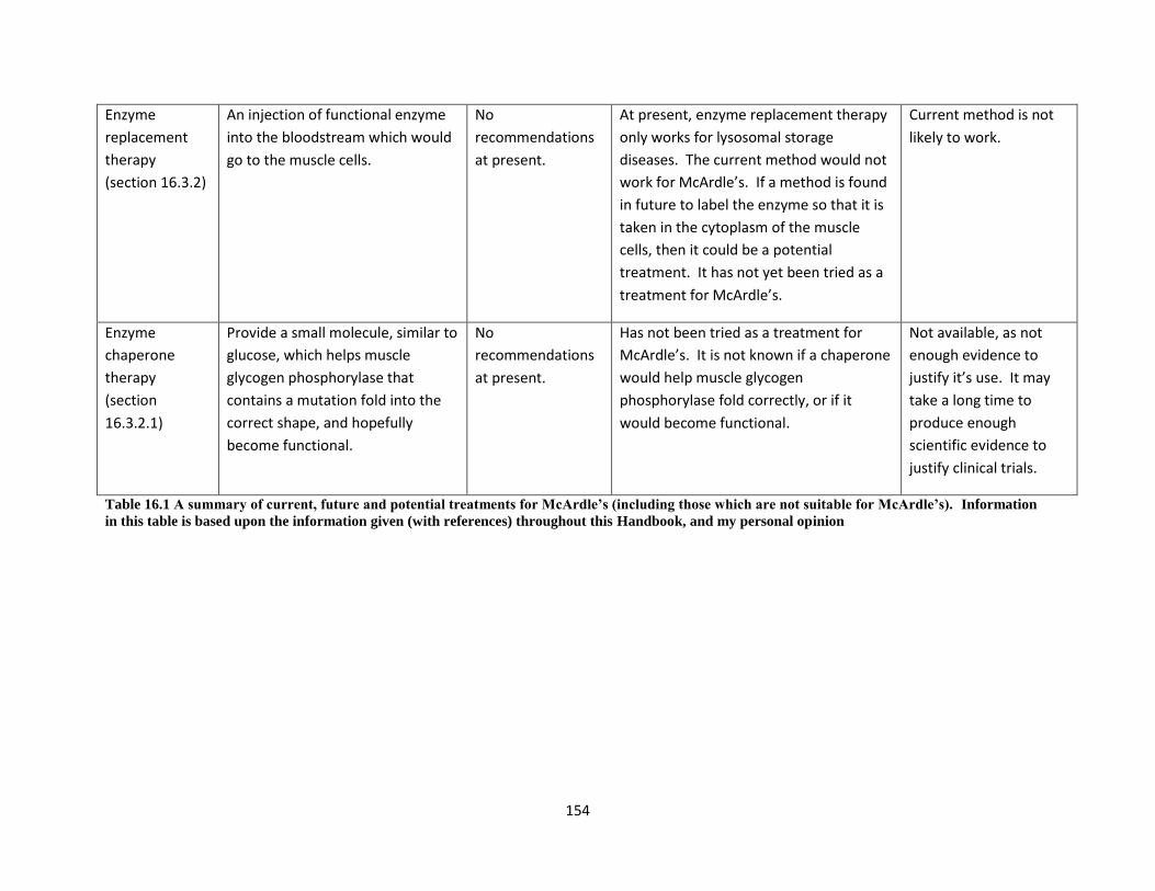
154
Enzyme
replacement
therapy
(section 16.3.2)
An injection of functional enzyme
into the bloodstream which would
go to the muscle cells.
No
recommendations
at present.
At present, enzyme replacement therapy
only works for lysosomal storage
diseases. The current method would not
work for McArdle’s. If a method is found
in future to label the enzyme so that it is
taken in the cytoplasm of the muscle
cells, then it could be a potential
treatment. It has not yet been tried as a
treatment for McArdle’s.
Current method is not
likely to work.
Enzyme
chaperone
therapy
(section
16.3.2.1)
Provide a small molecule, similar to
glucose, which helps muscle
glycogen phosphorylase that
contains a mutation fold into the
correct shape, and hopefully
become functional.
No
recommendations
at present.
Has not been tried as a treatment for
McArdle’s. It is not known if a chaperone
would help muscle glycogen
phosphorylase fold correctly, or if it
would become functional.
Not available, as not
enough evidence to
justify it’s use. It may
take a long time to
produce enough
scientific evidence to
justify clinical trials.
Table 16.1 A summary of current, future and potential treatments for McArdle’s (including those which are not suitable for McArdle’s). Information
in this table is based upon the information given (with references) throughout this Handbook, and my personal opinion

155
17 Details about this Handbook and the information in it
17.1 The purpose of this Handbook
The purpose of this Handbook is to clearly explain medical and scientific research so that it can be
understood by McArdle people. Information in this Handbook is not intended as medical advice, it is not
intended to replace medical advice, and is not intended to be a guide to treatments for McArdle’s. The
author (Kathryn Wright) has no medical training, and is not qualified to offer medical advice.
Where possible, for each statement, the name and date of the published paper or book is given. The
title of the paper or book can be found in the References list (section 19) at the end of the Handbook.
The reader is therefore able to read the original publication for further information. The information
provided is as up-to-date and accurate as is possible, but reflects current theories and opinions. Future
research may improve the understanding of McArdle’s and change these theories.
17.2 About the author
Kathryn Wright is a scientist and has not had any medical training. She studied Biology, English
Literature, Mathematics and Music GCE “A” Levels at secondary school, before reading for an
undergraduate degree in Natural Sciences at Cambridge University, UK, specialising in Genetics in her
final year. She worked for three years as a Research Assistant in a laboratory in Berkshire, learning
experimental techniques. She then moved to a research lab at the RJAH Orthopaedic Hospital in
Oswestry, UK, where she carried out a Ph.D project creating cell models of McArdle disease, through
Keele University, UK. Kathryn Wright had the idea of writing this Handbook during her Ph.D, when she
became aware that a lot of the medical and scientific research into McArdle disease was inaccessible to
people with McArdle’s because it was either written in complex scientific language or too expensive to
obtain.
17.2.1 Feedback about this Handbook; comments, suggestions, criticisms
Please provide feedback about this Handbook to Kathryn Wright. If you believe that information is
incorrect, or would like to suggest new information to include, please contact the author:
kathrynewrightmcardledisease (at) googlemail.com. Please include the reference to a published paper if
possible.
17.3 How to understand references given in this Handbook
A reference is the original paper or book which I have used to find out information. Where possible,
each sentence or paragraph is referenced. This enables you to go away and read the same paper or
book which I read before I wrote that sentence which allows you to either get more information than I
provided, or to check if I reported the information accurately. (If you think I have made a mistake,
please contact me as described in section 17.2.1.)

156
Throughout this Handbook, I have given references either before or after a sentence. If no reference is
give, and it includes phrases like “I think...”, or “my theory”, this indicates that it is my theory, which has
not been scientifically proven. References are given as either:
Wright et al. ( 2009) said grass is green. Or Grass is green (Wright et al., 2009). In these examples,
Wright is the name of the first author who wrote the paper or book, and the date is the date when the
paper or book was published. You can find the rest of the information about the reference in the
“References” list at the end of the Handbook. References are listed alphabetically, in order of the
surname of the first author. So, for example, Wright et al. (2009) would be near the end of the list of
references. You will be able to look up the whole name of the paper or book, which could look like;
Wright KE and Wright EK, 2009, Studies of Grass. Journal of plants, 133-134, 2. You can then use these
details to obtain the paper from Journal of Plants, pages 133-134. This paper is located in volume
number 2. (This reference has been invented, to serve as an example.)
17.4 A note about the names of the gene, mRNA and protein
There are standard ways to write the names of gene, mRNA, and protein, which are useful to know if
you are reading scientific papers. These are summarised in Table 17.1. Although only humans are
discussed in this section, animal models of McArdle’s are discussed further in section 15.
For clarity, muscle glycogen phosphorylase has been described as “muscle glycogen phosphorylase” in
this Handbook, rather than “PYGM”.
Humans Animals
Name of the gene PYGM (capitals, italic) Pygm (mixed case, italic)
Names of the mRNA PYGM (capitals, italic) Pygm (mixed case, italic)
Name of the protein PYGM (capitals, non italic) PYGM (capitals, non italic)
Table 17.1 Standard ways to write the gene, mRNA and protein name for muscle glycogen phosphorylase
(correct in 2010).
17.5 How to read medical/scientific papers critically
Rather than simply accepting everything that is stated, scientific papers (and other documents such as
this Handbook) should be read critically as the data in them and the interpretation of the results could
be affected by many things. Some of the things to consider when reading a paper include:
17.5.1.1 What is the date (year) when the paper was published?
New data may make old papers hard to understand e.g. old papers which don’t specify which isoform of
glycogen phosphorylase they are discussing are not useful now. New data and understanding can make
old papers out of date and the advice in them inappropriate. For example, in the past, McArdle people

157
were recommended not to exercise, but current advice is that frequent moderate exercise is best for
McArdle people.
17.5.1.2 Who are the authors?
Authors may be biased. Not all McArdle’s specialists share the same point of view. They may emphasise
their point of view strongly in a paper, even if there is very little scientific evidence to support this view.
If you find more than two papers, which have no authors in common, but both give the same
conclusion, then it is likely that it is a genuine result.
17.5.1.3 How good is the clinical trial?
Not all clinical trials are equal. The best clinical trials are double blind clinical trials. Single case studies
are of limited use. It is important to have good controls. The placebo effect should be avoided. (See
section 17.6.1 for further information).
17.5.1.4 Was the result “statistically significant”?
A result that is “statistically significant” has been tested using a set of statistical tests, and passed the
criteria (see section 17.6.6). These tests are used to determine if the result could have occurred by
chance or as a fluke. If the result is statistically significant, then it is unlikely to have occurred by chance.
However, different statistical tests can give different results, and some scientists (incorrectly) will try lots
of different tests until they find one which gives the result they want, which is poor scientific technique.
17.5.1.5 Errors can occur
Despite several levels of proof reading before a paper is published, errors can still occur. For example, I
believe that the mutation which Hadjigeorgiou et al. (2002) describe as a R207X mutation at c.808 in
exon 6 is actually the R270X mutation, but the authors refer to it throughout the paper incorrectly as
R207X.
17.5.1.6 The same person may be mentioned more than once
As McArdle’s is very rare, the same person may be mentioned in more than one paper, especially if they
have an unusual complication. This can give the impression that the complication is much more
common than it actually is. A criticism of some of the papers is that they mention the same person in
more than one paper. It can require quite a bit of detective work to figure out if this is the case. But it is
important because if one individual has particularly unusual symptoms, they may be reported on more
than one paper. If you didn’t realise (or your family doctor didn’t realise), you may think that the
symptom is much more common than it really is. For example, there is a case of a woman with
McArdle’s who has respiratory problems becasue the muscles involved in breathing didn’t work as well
as they should. But she is mentioned in two papers; “McArdle disease presenting as unexplained
dyspnea in a young woman” By Voduc et al. (2004), and “Variable presentation of the clinical
phenotype of McArdle disease in a kindred harbouring a novel compound genotype in the muscle
glycogen phosphorylase gene” by Paradas(2005). The clue is usually (although not in this case) if the
two papers have one or more author in common. If these two papers are taken together, it suggests
158
that McArdle disease might caused respiratory problems, but in fact both are describing the same
person.
17.5.1.7 The numbers of the mutations have changed
When reading papers about McArdle’s, it is important to remember that the mutation formerly known
as “R49X” is now known as “R50X”. The reason is as follows. In the PYGM gene, the DNA code has the
first amino acid of muscle glycogen phosphorylase as methionine (single letter code “M”). (This is
shown in section 3.1.1.) This is also in the mRNA. When a protein is made, the methionine is used as
the first amino acid of the amino acid sequence. However, in the process of make the protein mature,
the first methionine is chopped off and removed. When the first scientific studies were performed on
muscle glycogen phosphorylase, scientists studied the protein, and found that it had 841 amino acids.
At this time the mutation was known as “R49X”. However, at a much later date, scientists studied the
PYGM gene, and discovered that there was genetic code for the first methionine, even though it was
chopped off in the process of making the protein. They therefore decided to rename the mutation as
“R50X”.
When reading papers, it can be confusing trying to understand whether the mutation being described is
an old one or is newly discovered. I would suggest looking out for whether the old numbering scheme
of “R49X” and “G204S” or new numbering scheme of “R50X” and “G205S” are being used.
17.6 An introduction to clinical trials
Clinical trials are an essential method of testing whether a potential treatment for McArdle’s (or any
other disease) really works. Clinical trials are conducted by researchers; either scientists or family
doctors, or the two working together. Clinical trials are based upon a hypothesis that a particular
treatment may help to alleviate symptoms of a disease. For example; a theory that taking drug X may
enable McArdle people to exercise for longer. Clinical trials are designed to test whether this theory is
correct. The best clinical trials have the components described below: they include a placebo, are
randomised, are double blind, and are statistically significant.
Although clinical trials often involve testing new or existing drugs, they do not need to involve drugs.
Non-drug clinical trials carried out on McArdle people have included prescribing regular exercise and
testing a sugary drink.
Clinical trials must have ways of measuring whether the treatment has produced any benefit. The best
ways are to measure something which the participant has little or no control over. Examples would
include using 31P MRS to measure changes within the cells, measuring heart rate, or VO2 max (6.4.4.1).
It is less accurate to ask participants how they feel – for example whether they feel that they have much
less muscle pain. It can be very hard for participants to give useful and accurate answers, and hard to
use these answers to determine if a treatment is working. For example, if they have just had an
argument with their partner, they may feel more negative about muscle pain than on a different day –
but this would be unrelated to a treatment.

159
The researchers carrying out a clinical trial will want to publish the results once the trial is completed.
The results should be published whether or not the treatment has a positive effect, negative effect or no
effect. Positive results may lead family doctors to start prescribing the treatment/drug for their
McArdle’s patients. Negative results may warn family doctors to avoid prescribing the treatment/drug
for their McArdle’s patients. Often, clinical trials are first done on small group of participants, and if a
positive effect is seen, they will be repeated on a larger number of participants. It is ideal to wait until a
positive result has been seen in a large scale clinical trial before considering it as a conventional
treatment for that disease. In a rare disease like McArdle’s, there may not be enough people with
McArdle’s for large scale trials to be possible.
Most countries now have very strict guidelines for researchers wishing to carry out clinical trials. The
trials have to be approved by a committee, often at the hospital where the trial would be carried out.
The committee will consider whether the potential side effects and risks of the treatment are
worthwhile compared to the potential benefit of the treatment. The researchers also have to prepare
information sheets written in plain (non-technical) language, to ensure that the participants fully
understand all the risks and tests involved. For example, that the participants are willing to give a blood
sample or undertake a muscle biopsy if that is required. When participants fully understand the risks
and tests involved, and then agree to take part, this is known as “informed consent”. In the UK, and
some other countries, researchers are not allowed to pay participants to take part in clinical trials,
although they are allowed to pay a certain amount for travel and other costs incurred by the participant
in getting to the location where the trial is taking place.
Most trials involve comparison; for example, a group of McArdle people may be compared to a group of
people unaffected by McArdle’s. (A person who is “unaffected by McArdle’s” is often described as a
“control” in scientific papers.) Or, a group of McArdle people may be given a treatment, and compared
to a group of McArdle people who were not given the treatment.
17.6.1 The placebo effect
A clinical trial must be carefully planned in order to determine whether a treatment is having a real
effect or a “placebo effect”. The placebo effect is a well known phenomenon that some people feel that
their symptoms improve even if they are given a pill with no active medicine – for example a pill made of
sugar or flour. The placebo effect was demonstrated in a clinical trial of dantrolene sodium carried out
by Poels et al. (1990); some improvement was felt by one McArdle person on treatment and three
McArdle people taking the placebo. There are various explanations for the placebo effect. Taking a
placebo pill may change the way in which the person perceives their symptoms. Sometimes being part
of a clinical trial may make a person feel more important. Having a family doctor/specialist discussing
McArdle’s, acknowledging symptoms and providing support and encouragement may lead to a more
positive view of the symptoms. (This is why cognitive behaviour therapy (CBT) is sometimes used. CBT
can help people come to terms with symptoms and limitations of their condition even if no treatment or
cure is available.) Some participants, either deliberately or unknowingly, will give the researcher the
answer they think they want to hear. During a clinical trial, the “control” group are the group of people
who do not receive the treatment, and in many cases these people receive the placebo.

160
To determine whether a drug treatment is having a real or placebo effect, it is important that the people
being tested are given either the real drug or a placebo which looks/smells/tastes like the real drug. The
participants should not be able to tell the difference between the placebo and the real drug. If the
participant doesn’t know if they are receiving the placebo or real drug, this is known as a “blind” trial. In
the best studies, the researcher also does not know which participants are receiving the real drug or the
placebo, and does not find out until the very end of the experiment. If the participant doesn’t know if
they are receiving the placebo or real drug AND the researcher doesn’t know which participants are
receiving each treatment, it is known as a “double blind” trial (because both the participant and
researcher are blinded to the treatment). If the participant and researcher don’t know whether the
participant is receiving the treatment or placebo, this is also known as “allocation concealment”.
An “open label” trial is when the participant know which drug they are receiving, or know if they are
receiving the placebo. This is not such a good trial as participants may be biased by whether or not they
are receiving the treatment. Sometimes it is impossible to blind the participant, and the trial has to be
open label. One example would be a trial of diet; where it would be very difficult to prevent the
participant seeing whether they are receiving a high fat or high carbohydrate meal.
17.6.2 Randomised trials
A really good trial will be designed so that the researcher cannot unwittingly bias the outcome. One way
in which this could occur would be if the researcher divides the participants into two groups; for
example so that one set can receive the placebo and the other set receive the real drug. If the
researcher picks which people should be in each group, the researcher may not divide the people
evenly; one example would be to put all the women in one group and the men in the other – in this case
it wouldn’t be possible to say if the results were an effect of the real drug or an due to differences in
gender. Another example would be to put all the fit people in one group and all the unfit people in the
other group. These are extreme examples to illustrate the problems of allowing the researcher to
choose who goes into which group. A much better method is to allocate each person a number, and to
use a computer (or even draw numbers out of a hat) to randomly allocate each person into a group.
This removes the possibility of the researcher choosing who goes into which group.
17.6.3 Cross-over trial
A cross-over trial occurs when the participants are divided into two groups; let us call them group A and
group B. For the first period of time (several days to several years, depending on the study), group A will
receive the placebo and group B will receive the real drug. The researchers will then perform tests to
see if the treatments have had any effect, for example on the ability of the participants to exercise.
There may then be a “wash-out” period, when no drug/placebo is given. This is to allow the amount of
real drug in the body to reduce and be removed by the body. In a cross-over trial, group A will now
receive the real drug, and group B will receive the placebo. Further tests will then be performed. A
cross-over trial is particularly important when there are only a small number of participants (often the
case in trials with McArdle people). If one person was much more fit than the others, this might
disproportionately affect the results if no cross-over was performed. For example, if a very fit person
was put into the group taking the real drug, and the real drug actually had no effect, the ability of the

161
very fit person to exercise could lead the researchers to think that the drug had helped that person
exercise. A cross-over trial allows the researchers to compare results obtained with the real drug and
placebo each in the same person.
17.6.4 Single case study
A single case study is when just one participant is studied. Published papers are used by family doctors
and scientists as a way of communicating. Single case studies are sometimes reported if a new
phenomenon occurs; for example, if it is found that a person with McArdle’s had a particularly bad
reaction to a drug or treatment. Specialists who read the study will be aware that McArdle’s may lead
to sensitivity to that drug or treatment, and will be aware to look out for it in the McArdle person they
deal with. The disadvantage of single case studies is that it is very hard to determine if a treatment will
work in other McArdle people. A single case study cannot determine what other factors may affect
whether a treatment may work.
Here is an example of a result from a large scale study which could not have been obtained from a single
case study: The ACE gene is unrelated to the PYGM gene which encodes muscle glycogen phosphorylase
(see section 9.3.1 for more information on the ACE gene). McArdle people (and other people in the
population) may have either the DD, DI or II genotype. It has been found that the drug treatment
ramipril only helps people who have the DD genotype for the ACE gene (Martinuzzi et al., 2007). It was
only possible to find out this result because many McArdle people took part in the drug trial. If only one
person had taken part in a single case study, it could have had very different results. For example, if the
one participant had the II genotype, the drug treatment would not have had any effect, and would
probably not have been tried again.
A further problem with a single case study (or a study with very few participants) is the lack of statistical
significance.
17.6.5 A home trial is an alternative to a clinical trial
The alternative to a clinical trial would be a trial performed at home. Many McArdle people on internet
chat groups say that they have carried out home trials. Home trials are not planned or carried out by a
researcher. They will not have been assessed by a committee to decide if they have any health risks or
negative effects. People carrying out a home trial on themselves are likely to know whether they are
taking the treatment/drug or not, which does not exclude the placebo effect. If a home trial suggests
that a treatment does work, it is not sufficient evidence to prescribe the treatment to others. It would
be sensible to discuss any treatment/drug with your family doctor before carrying out a home trial.
17.6.6 Statistical significance
Mathematicians have devised several statistical calculations which can be used to see whether a
treatment/drug has a genuine effect. These calculations take into account the number of participants,
and how strong the effect of a treatment/drug is. The result of the calculation can be looked up in a
special table to determine if the result is “statistically significant”. (These calculations are nowadays
performed using a computer.)
162
A statistically significant result will be accepted by the scientific/medical community as a genuine result
which shows that a drug/treatment really does cause an effect (but please note this effect could be
positive or negative). A result may be classed as “not statistically significant” there were not enough
participants, or if very little difference was seen between the treatment/drug and non-
treatment/placebo. It is possible that if the trial were repeated with more participants, it would
generate results that were statistically significant. Single case studies and trials with a small number of
participants are less likely to generate statistically significant results.
An example of a statement in a paper is from given below. Lucia et al. (2008b) performed some tests to
look at the amount of cytokines in the blood of patients (McArdle people) compared to controls (people
unaffected by McArdle’s). The authors say “...circulating levels of several cytokines were significantly
higher (P≤0.05) in patients than in controls”. The important parts of this sentence are in bold. This can
be translated as:
They measured the amount of cytokines in patients and controls, and compared them using a statistical
test. “Significantly” means that the statistical test result showed that the levels were statistically
different – they are “real” differences and are very unlikely to occur by chance. “higher” obviously tells
you that there were more cytokines than in controls. “P≤0.05” is another part of the result of the
statistical test. A result that is P≤0.01, P≤0.05 or P≤0.001, tells you that the statistical test showed that
the differences are “real” and are unlikely to occur by chance.

163
18 Glossary
Most words are defined when they are used in the Handbook at the point when they are used. Only
scientific or medical words which are used repeatedly in this Handbook are defined below.
Adipose tissue Special cells in the body which stores energy as fatty acids.
Allele Different versions of the same gene.
Amino acid sequence
The order of the amino acids in a protein.
Amino acids The building blocks of proteins.
Autosome A chromosome which does not determine gender.
Carrier In this Handbook, “carrier” is used to describe a person who has one copy of the PYGM gene without any mutations, and one copy of the PYGM gene which does carry a mutation. A carrier is likely to have approximately half the normal level of muscle glycogen phosphorylase enzyme. Carriers do not usually have symptoms of McArdle disease. A more scientific name for a “carrier” is “heterozygote” (see below).
Cell culture Cells obtained by a muscle biopsy from McArdle people and then grown in a research laboratory.
Chemokines A sub-group of cytokines, which are small proteins produced by cells.
Chromosomes A structure within the cell which is basically a string of genes.
Codons The mRNA is decoded in triplets with each triplet being used to identify one amino acid. These triplets are known as codons.
Contractures Severe muscle cramps.
Cytokines Small proteins which are produced by almost all cells in the body. Cytokines are used by the cells as a method to communicate.
Double blind A trial where the participant doesn’t know if they are receiving the placebo or real drug AND the researcher doesn’t know which participants are receiving each treatment. Therefore both the participant and researcher are blinded to the treatment.
Double trouble When two unrelated muscle diseases occur in the same person.
Electrolytes Several compounds including potassium, sodium and calcium. It is important for the correct functioning of the body that the correct amounts compounds are maintained in the body.
Endoplasmic reticulum (ER)
An area of the cell involved in producing proteins (basically the protein-making factory).
Exons The coding regions of genes which are used to make mRNA. The exons encode the sequence for making proteins.
Fatty acid oxidation
A mechanism where fatty acids are broken down to release compounds which can be used to produce energy.
Frameshift mutations
These are mutations which occur if just one nucleotide is removed (deleted) from the gene. This causes the ribosome to misread the amino acid sequence. Misreading of the amino acid sequence leads to the production of a protein which doesn’t have the right sequence of amino acids, and is not able to function in the normal way.
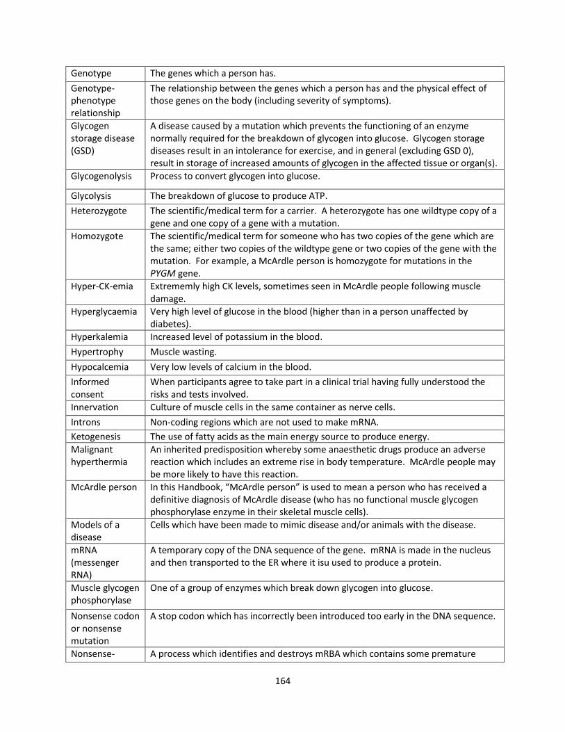
164
Genotype The genes which a person has.
Genotype-phenotype relationship
The relationship between the genes which a person has and the physical effect of those genes on the body (including severity of symptoms).
Glycogen storage disease (GSD)
A disease caused by a mutation which prevents the functioning of an enzyme normally required for the breakdown of glycogen into glucose. Glycogen storage diseases result in an intolerance for exercise, and in general (excluding GSD 0), result in storage of increased amounts of glycogen in the affected tissue or organ(s).
Glycogenolysis Process to convert glycogen into glucose.
Glycolysis The breakdown of glucose to produce ATP.
Heterozygote The scientific/medical term for a carrier. A heterozygote has one wildtype copy of a gene and one copy of a gene with a mutation.
Homozygote The scientific/medical term for someone who has two copies of the gene which are the same; either two copies of the wildtype gene or two copies of the gene with the mutation. For example, a McArdle person is homozygote for mutations in the PYGM gene.
Hyper-CK-emia Extrememly high CK levels, sometimes seen in McArdle people following muscle damage.
Hyperglycaemia Very high level of glucose in the blood (higher than in a person unaffected by diabetes).
Hyperkalemia Increased level of potassium in the blood.
Hypertrophy Muscle wasting.
Hypocalcemia Very low levels of calcium in the blood.
Informed consent
When participants agree to take part in a clinical trial having fully understood the risks and tests involved.
Innervation Culture of muscle cells in the same container as nerve cells.
Introns Non-coding regions which are not used to make mRNA.
Ketogenesis The use of fatty acids as the main energy source to produce energy.
Malignant hyperthermia
An inherited predisposition whereby some anaesthetic drugs produce an adverse reaction which includes an extreme rise in body temperature. McArdle people may be more likely to have this reaction.
McArdle person In this Handbook, “McArdle person” is used to mean a person who has received a definitive diagnosis of McArdle disease (who has no functional muscle glycogen phosphorylase enzyme in their skeletal muscle cells).
Models of a disease
Cells which have been made to mimic disease and/or animals with the disease.
mRNA (messenger RNA)
A temporary copy of the DNA sequence of the gene. mRNA is made in the nucleus and then transported to the ER where it isu used to produce a protein.
Muscle glycogen phosphorylase
One of a group of enzymes which break down glycogen into glucose.
Nonsense codon or nonsense mutation
A stop codon which has incorrectly been introduced too early in the DNA sequence.
Nonsense- A process which identifies and destroys mRBA which contains some premature
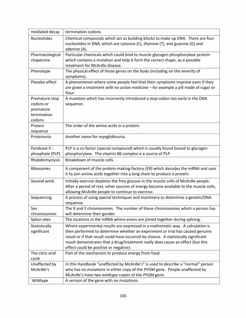
165
mediated decay termination codons.
Nucleotides Chemical compounds which act as building blocks to make up DNA. There are four nucleotides in DNA, which are cytosine (C), thymine (T), and guanine (G) and adenine (A).
Pharmacological chaperone
Particular chemicals which could bind to muscle glycogen phosphorylase protein which contains a mutation and help it form the correct shape, as a possible treatment for McArdle disease.
Phenotype The physical effect of those genes on the body (including on the severity of symptoms).
Placebo effect A phenomenon where some people feel that their symptoms improve even if they are given a treatment with no active medicine – for example a pill made of sugar or flour.
Premature stop codons or premature termination codons
A mutation which has incorrectly introduced a stop codon too early in the DNA sequence.
Protein sequence
The order of the amino acids in a protein.
Proteinuria Another name for myoglobinuria.
Pyridoxal 5’-phosphate (PLP)
PLP is a co-factor (special compound) which is usually found bound to glycogen phosphorylase. The vitamin B6 complex is a source of PLP.
Rhabdomyolysis Breakdown of muscle cells.
Ribosomes A component of the protein-making-factory (ER) which decodes the mRNA and uses it to join amino acids together into a long chain to produce a protein.
Second wind Initially exercise depletes the free glucose in the muscle cells of McArdle people. After a period of rest, other sources of energy become available to the muscle cells, allowing McArdle people to continue to exercise.
Sequencing A process of using special techniques and machinery to determine a genetic/DNA sequence.
Sex chromosomes
The X and Y chromosomes. The number of these chromosomes which a person has will determine their gender.
Splice sites The locations in the mRNA where exons are joined together during splicing.
Statistically significant
Where experimental results are expressed in a mathematic way. A calculation is then performed to determine whether an experiment or trial has caused genuine result or if that result could have occurred by chance. A statistically significant result demonstrates that a drug/treatment really does cause an effect (but this effect could be positive or negative).
The citric acid cycle
Part of the mechanism to produce energy from food.
Unaffected by McArdle’s
In this Handbook “unaffected by McArdle’s” is used to describe a “normal” person who has no mutations in either copy of the PYGM gene. People unaffected by McArdle’s have two wildtype copies of the PYGM gene.
Wildtype A version of the gene with no mutations.
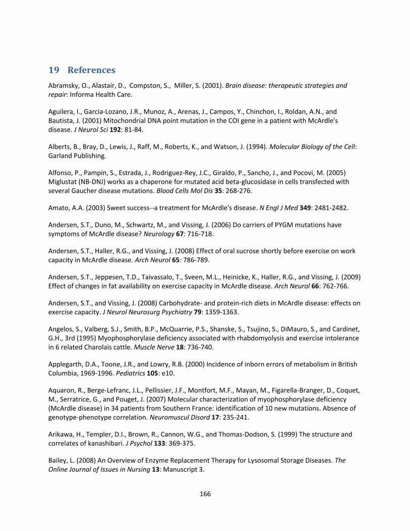
166
19 References
Abramsky, O., Alastair, D., Compston, S., Miller, S. (2001). Brain disease: therapeutic strategies and repair: Informa Health Care.
Aguilera, I., Garcia-Lozano, J.R., Munoz, A., Arenas, J., Campos, Y., Chinchon, I., Roldan, A.N., and Bautista, J. (2001) Mitochondrial DNA point mutation in the COI gene in a patient with McArdle's disease. J Neurol Sci 192: 81-84.
Alberts, B., Bray, D., Lewis, J., Raff, M., Roberts, K., and Watson, J. (1994). Molecular Biology of the Cell: Garland Publishing.
Alfonso, P., Pampin, S., Estrada, J., Rodriguez-Rey, J.C., Giraldo, P., Sancho, J., and Pocovi, M. (2005) Miglustat (NB-DNJ) works as a chaperone for mutated acid beta-glucosidase in cells transfected with several Gaucher disease mutations. Blood Cells Mol Dis 35: 268-276.
Amato, A.A. (2003) Sweet success--a treatment for McArdle's disease. N Engl J Med 349: 2481-2482.
Andersen, S.T., Duno, M., Schwartz, M., and Vissing, J. (2006) Do carriers of PYGM mutations have symptoms of McArdle disease? Neurology 67: 716-718.
Andersen, S.T., Haller, R.G., and Vissing, J. (2008) Effect of oral sucrose shortly before exercise on work capacity in McArdle disease. Arch Neurol 65: 786-789.
Andersen, S.T., Jeppesen, T.D., Taivassalo, T., Sveen, M.L., Heinicke, K., Haller, R.G., and Vissing, J. (2009) Effect of changes in fat availability on exercise capacity in McArdle disease. Arch Neurol 66: 762-766.
Andersen, S.T., and Vissing, J. (2008) Carbohydrate- and protein-rich diets in McArdle disease: effects on exercise capacity. J Neurol Neurosurg Psychiatry 79: 1359-1363.
Angelos, S., Valberg, S.J., Smith, B.P., McQuarrie, P.S., Shanske, S., Tsujino, S., DiMauro, S., and Cardinet, G.H., 3rd (1995) Myophosphorylase deficiency associated with rhabdomyolysis and exercise intolerance in 6 related Charolais cattle. Muscle Nerve 18: 736-740.
Applegarth, D.A., Toone, J.R., and Lowry, R.B. (2000) Incidence of inborn errors of metabolism in British Columbia, 1969-1996. Pediatrics 105: e10.
Aquaron, R., Berge-Lefranc, J.L., Pellissier, J.F., Montfort, M.F., Mayan, M., Figarella-Branger, D., Coquet, M., Serratrice, G., and Pouget, J. (2007) Molecular characterization of myophosphorylase deficiency (McArdle disease) in 34 patients from Southern France: identification of 10 new mutations. Absence of genotype-phenotype correlation. Neuromuscul Disord 17: 235-241.
Arikawa, H., Templer, D.I., Brown, R., Cannon, W.G., and Thomas-Dodson, S. (1999) The structure and correlates of kanashibari. J Psychol 133: 369-375.
Bailey, L. (2008) An Overview of Enzyme Replacement Therapy for Lysosomal Storage Diseases. The Online Journal of Issues in Nursing 13: Manuscript 3.

167
Baker, D.J., Greenhaff, P.L., MacInnes, A., and Timmons, J.A. (2006) The experimental type 2 diabetes therapy glycogen phosphorylase inhibition can impair aerobic muscle function during prolonged contraction. Diabetes 55: 1855-1861.
Baque, S., Newgard, C.B., Gerard, R.D., Guinovart, J.J., and Gomez-Foix, A.M. (1994) Adenovirus-mediated delivery into myocytes of muscle glycogen phosphorylase, the enzyme deficient in patients with glycogen-storage disease type V. Biochem J 304 ( Pt 3): 1009-1014.
Barford, D., and Johnson, L.N. (1992) The molecular mechanism for the tetrameric association of glycogen phosphorylase promoted by protein phosphorylation. Protein Sci 1: 472-493.
Barnes, P.R.J., Hilton-Jones, D. (2003). Myopathies in Clinical Practice: Informa Health Care.
Bartram, C., Edwards, R.H., Clague, J., and Beynon, R.J. (1993) McArdle's disease: a nonsense mutation in exon 1 of the muscle glycogen phosphorylase gene explains some but not all cases. Hum Mol Genet 2: 1291-1293.
Berg, J., Tymoczko, J., and Stryer, L. (2008). Biochemistry: W. H. Freeman.
Berns, K.I., and Linden, R.M. (1995) The cryptic life style of adeno-associated virus. Bioessays 17: 237-245.
Beynon, R.J., Bartram, C., Flannery, A., Evershed, R.P., Leyland, D., Hopkins, P., Toescu, V., Phoenix, J., and Edwards, R.H. (1996) Interrelationships between metabolism of glycogen phosphorylase and pyridoxal phosphate--implications in McArdle's disease. Adv Food Nutr Res 40: 135-147.
Beynon, R.J., Bartram, C., Hopkins, P., Toescu, V., Gibson, H., Phoenix, J., and Edwards, R.H. (1995) McArdle's disease: molecular genetics and metabolic consequences of the phenotype. Muscle Nerve 3: S18-22.
Beynon, R.J., Quinlivan, R., and Sewry, C. ( 2002). Selected disorders of carbohydrate metabolism, in K. G, (ed.), Structural and molecular basis of skeletal muscle disease.: Wiley, pp. 182–188.
Biesalski, G. (2005) Pocket atlas of nutrition.
Biller, J. (2007). The Interface of Neurology & Internal Medicine: Lippincott Williams & Wilkins.
BNF, 58. (2009). British National Formulary: Joint Formulary Committee London: British Medical Association and Royal Pharmaceutical Society.
Boesch, C. (2007) Musculoskeletal spectroscopy. J Magn Reson Imaging 25: 321-338.
Bogusky, R.T., Taylor, R.G., Anderson, L.J., Angelos, K.L., Lieberman, J.S., and Walsh, D.A. (1986) McArdle's disease heterozygotes. Metabolic adaptation assessed using 31P-nuclear magnetic resonance. J Clin Invest 77: 1881-1887.
Bollig, G., Mohr, S., and Raeder, J. (2005) McArdle's disease and anaesthesia: case reports. Review of potential problems and association with malignant hyperthermia. Acta Anaesthesiol Scand 49: 1077-1083.
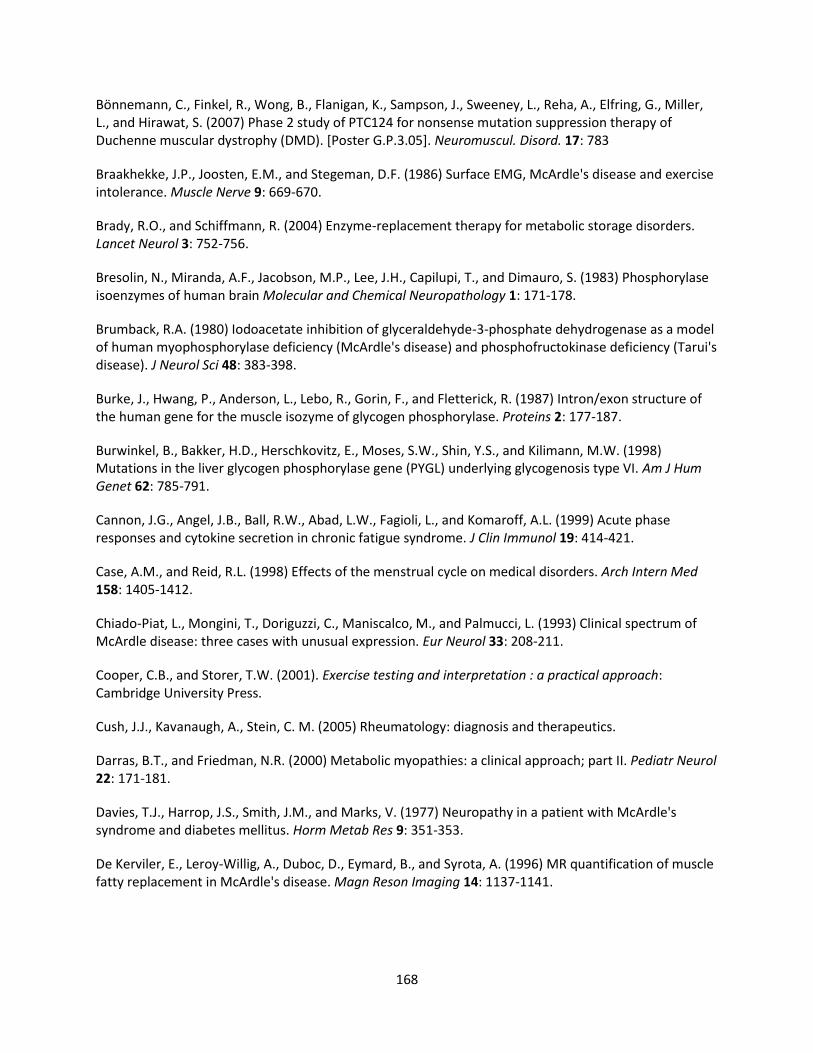
168
Bönnemann, C., Finkel, R., Wong, B., Flanigan, K., Sampson, J., Sweeney, L., Reha, A., Elfring, G., Miller, L., and Hirawat, S. (2007) Phase 2 study of PTC124 for nonsense mutation suppression therapy of Duchenne muscular dystrophy (DMD). [Poster G.P.3.05]. Neuromuscul. Disord. 17: 783
Braakhekke, J.P., Joosten, E.M., and Stegeman, D.F. (1986) Surface EMG, McArdle's disease and exercise intolerance. Muscle Nerve 9: 669-670.
Brady, R.O., and Schiffmann, R. (2004) Enzyme-replacement therapy for metabolic storage disorders. Lancet Neurol 3: 752-756.
Bresolin, N., Miranda, A.F., Jacobson, M.P., Lee, J.H., Capilupi, T., and Dimauro, S. (1983) Phosphorylase isoenzymes of human brain Molecular and Chemical Neuropathology 1: 171-178.
Brumback, R.A. (1980) Iodoacetate inhibition of glyceraldehyde-3-phosphate dehydrogenase as a model of human myophosphorylase deficiency (McArdle's disease) and phosphofructokinase deficiency (Tarui's disease). J Neurol Sci 48: 383-398.
Burke, J., Hwang, P., Anderson, L., Lebo, R., Gorin, F., and Fletterick, R. (1987) Intron/exon structure of the human gene for the muscle isozyme of glycogen phosphorylase. Proteins 2: 177-187.
Burwinkel, B., Bakker, H.D., Herschkovitz, E., Moses, S.W., Shin, Y.S., and Kilimann, M.W. (1998) Mutations in the liver glycogen phosphorylase gene (PYGL) underlying glycogenosis type VI. Am J Hum Genet 62: 785-791.
Cannon, J.G., Angel, J.B., Ball, R.W., Abad, L.W., Fagioli, L., and Komaroff, A.L. (1999) Acute phase responses and cytokine secretion in chronic fatigue syndrome. J Clin Immunol 19: 414-421.
Case, A.M., and Reid, R.L. (1998) Effects of the menstrual cycle on medical disorders. Arch Intern Med 158: 1405-1412.
Chiado-Piat, L., Mongini, T., Doriguzzi, C., Maniscalco, M., and Palmucci, L. (1993) Clinical spectrum of McArdle disease: three cases with unusual expression. Eur Neurol 33: 208-211.
Cooper, C.B., and Storer, T.W. (2001). Exercise testing and interpretation : a practical approach: Cambridge University Press.
Cush, J.J., Kavanaugh, A., Stein, C. M. (2005) Rheumatology: diagnosis and therapeutics.
Darras, B.T., and Friedman, N.R. (2000) Metabolic myopathies: a clinical approach; part II. Pediatr Neurol 22: 171-181.
Davies, T.J., Harrop, J.S., Smith, J.M., and Marks, V. (1977) Neuropathy in a patient with McArdle's syndrome and diabetes mellitus. Horm Metab Res 9: 351-353.
De Kerviler, E., Leroy-Willig, A., Duboc, D., Eymard, B., and Syrota, A. (1996) MR quantification of muscle fatty replacement in McArdle's disease. Magn Reson Imaging 14: 1137-1141.

169
de Kerviler, E., Leroy-Willig, A., Jehenson, P., Duboc, D., Eymard, B., and Syrota, A. (1991) Exercise-induced muscle modifications: study of healthy subjects and patients with metabolic myopathies with MR imaging and P-31 spectroscopy. Radiology 181: 259-264.
de Ridder, D., Geenen, R., Kuijer, R., and van Middendorp, H. (2008) Psychological adjustment to chronic disease. Lancet 372: 246-255.
Delibas, A., Bek, K., Ezgu, F.S., Demircin, G., Oksal, A., and Oner, A. (2008) Acute renal failure due to rhabdomyolysis in a child with McArdle disease. Eur J Pediatr 167: 939-940.
Delmont, E., Sacconi, S., Berge-Lefranc, J.L., Aquaron, R., Butori, C., and Desnuelle, C. (2008) [McArdle disease (gycogenosis type V): analysis of clinical, biological and genetic features of five French patients]. Rev Neurol (Paris) 164: 912-916.
Derave, W., Hansen, B.F., Lund, S., Kristiansen, S., and Richter, E.A. (2000) Muscle glycogen content affects insulin-stimulated glucose transport and protein kinase B activity. Am J Physiol Endocrinol Metab 279: E947-955.
Deschauer, M., Morgenroth, A., Joshi, P.R., Glaser, D., Chinnery, P.F., Aasly, J., Schreiber, H., Knape, M., Zierz, S., and Vorgerd, M. (2007) Analysis of spectrum and frequencies of mutations in McArdle disease. Identification of 13 novel mutations. J Neurol 254: 797-802.
DiMauro, S., Andreu, A., Bruno, C., and Hadjigeorgiou, G. (2002) Myophosphorylase deficiency (glycogenosis type V; McArdle disease). Curr Mol Med 2: 189-196.
DiMauro, S., Arnold, S., Miranda, A., and Rowland, L.P. (1978) McArdle disease: the mystery of reappearing phosphorylase activity in muscle culture--a fetal isoenzyme. Ann Neurol 3: 60-66.
Dochartaigh (2004) Oxygen consumption is increased relative to work rate in patients with McArdle’s disease.
Dorin, R.I., Field, J.C., Boyle, P.J., Eaton, R.P., and Icenogle, M.V. (1996) Insulin resistance limits glucose utilization and exercise tolerance in myophosphorylase deficiency and NIDDM. J Appl Physiol 81: 1273-1278.
Du, M., Liu, X., Welch, E.M., Hirawat, S., Peltz, S.W., and Bedwell, D.M. (2008) PTC124 is an orally bioavailable compound that promotes suppression of the human CFTR-G542X nonsense allele in a CF mouse model. Proc Natl Acad Sci U S A 105: 2064-2069.
Dubowitz, V., Sewry, C., and Lane, R. (2007). Muscle biopsy: a practical approach Elsevier Health Sciences.
Dzoljic, E., Sipetic, S., Vlajinac, H., Marinkovic, J., Brzakovic, B., Pokrajac, M., and Kostic, V. (2002) Prevalence of menstrually related migraine and nonmigraine primary headache in female students of Belgrade University. Headache 42: 185-193.
Edelstyn, N., Quinlivan, R., (2007) A pilot study of neuropsychological performance in McArdle disease [Poster: M.P.4.04]. Neuromuscular Disorders 17: 860-860.

170
el-Schahawi, M., Bruno, C., Tsujino, S., Sarrazin, A.M., Shanske, S., LeRoux, M.G., and DiMauro, S. (1997) Sudden infant death syndrome (SIDS) in a family with myophosphorylase deficiency. Neuromuscul Disord 7: 81-83.
Fernandes, J., Saudubray, J., Van Den Berghe, G., Walter, J.H. (2006). Inborn metabolic diseases: diagnosis and treatment.
Fernandez-Cadenas, I., Andreu, A.L., Gamez, J., Gonzalo, R., Martin, M.A., Rubio, J.C., and Arenas, J. (2003) Splicing mosaic of the myophosphorylase gene due to a silent mutation in McArdle disease. Neurology 61: 1432-1434.
Fernandez-Cadenas, I., Nogales-Gadea, G., Llige, D., Rubio, J.C., Montaner, J., Arenas, J., Raspall-Chaure, M., Roig-Quilis, M., and Andreu, A.L. (2007) [Private mutations in the myophosphorylase gene: the first case in a patient of Latin American descent]. Rev Neurol 45: 280-283.
Ford, P.M., Manugian, A.A., and Weir, W.R. (1973) Acute renal failure complicating McArdle's syndrome. Postgrad Med J 49: 926-928.
Frischmeyer, P.A., and Dietz, H.C. (1999) Nonsense-mediated mRNA decay in health and disease. Hum Mol Genet 8: 1893-1900.
Garcia-Consuegra, I., Rubio, J.C., Nogales-Gadea, G., Bautista, J., Jimenez, S., Cabello, A., Lucia, A., Andreu, A.L., Arenas, J., and Martin, M.A. (2009) Novel mutations in patients with McArdle disease by analysis of skeletal muscle mRNA. J Med Genet 46: 198-202.
Glaser, T., Matthews, K., Hudson, J., Seth, P., Housman, D., and Crerar, M. (1989) Localization of the muscle, liver, and brain glycogen phosphorylase genes on linkage maps of mouse chromosomes 19, 12, and 2, respectively. Genomics 3: 510-521.
Gomez-Cerezo, J.F., Pagan Munoz, B., Gutierrez, M., Alfageme, M., Morales, C., and Barbado, F.J. (2008) McArdle's disease presented as refractory dermatomyositis. Eur J Intern Med 19: e20-22.
Gonzalez-Freire, M., Santiago, C., Gomez-Gallego, F., Perez, M., Foster, C., Arenas, J., and Lucia, A. (2009) Does the K153R variant of the myostatin gene influence the clinical presentation of women with McArdle disease? Neuromuscul Disord 19: 220-222.
Gorin, F., Herrick, K., Froman, B., Palmer, W., Tait, R., and Carlsen, R. (1996) Botulinum-induced muscle paralysis alters metabolic gene expression and fatigue recovery. Am J Physiol 270: R238-245.
Gorin, F., Ignacio, P., Gelinas, R., and Carlsen, R. (1989) Abnormal expression of glycogen phosphorylase genes in regenerated muscle. Am J Physiol 257: C495-503.
Greenberg, M.I. (2005 ). Greenberg's text-atlas of emergency medicine: Lippincott Williams & Wilkins.
Gross, M. (1997) Clinical heterogeneity and molecular mechanisms in inborn muscle AMP deaminase deficiency. J Inherit Metab Dis 20: 186-192.
Gruener, R., McArdle, B., Ryman, B.E., and Weller, R.O. (1968) Contracture of phosphorylase deficient muscle. J Neurol Neurosurg Psychiatry 31: 268-283.

171
Gruetter, R., Kaelin, P., Boesch, C., Martin, E., and Werner, B. (1990) Non-invasive 31P magnetic resonance spectroscopy revealed McArdle disease in an asymptomatic child. Eur J Pediatr 149: 483-486.
Gurpur, P.B., Liu, J., Burkin, D.J., and Kaufman, S.J. (2009) Valproic acid activates the PI3K/Akt/mTOR pathway in muscle and ameliorates pathology in a mouse model of Duchenne muscular dystrophy. Am J Pathol 174: 999-1008.
Hadjigeorgiou, G.M., Sadeh, M., Musumeci, O., Dabby, R., De Girolami, L., Naini, A., Papadimitriou, A., Shanske, S., and DiMauro, S. (2002) Molecular genetic study of myophosphorylase deficiency (McArdle's disease) in two Yemenite-Jewish families. Neuromuscul Disord 12: 824-827.
Hagberg, J.M., Coyle, E.F., Carroll, J.E., Miller, J.M., Martin, W.H., and Brooke, M.H. (1982) Exercise hyperventilation in patients with McArdle's disease. J Appl Physiol 52: 991-994.
Haller, R.G. (2000) Treatment of McArdle disease. Arch Neurol 57: 923-924.
Haller, R.G., Clausen, T., and Vissing, J. (1998) Reduced levels of skeletal muscle Na+K+ -ATPase in McArdle disease. Neurology 50: 37-40.
Haller, R.G., Lewis, S.F., Cook, J.D., and Blomqvist, C.G. (1985) Myophosphorylase deficiency impairs muscle oxidative metabolism. Ann Neurol 17: 196-199.
Haller, R.G., Wyrick, P., Taivassalo, T., and Vissing, J. (2006) Aerobic conditioning: an effective therapy in McArdle's disease. Ann Neurol 59: 922-928.
Harris, R.A., and Dowben, R.M. (1985) McArdle's disease in an elderly woman. South Med J 78: 191-193.
Haverkort-Poels, P.J., Joosten, E.M., and Ruitenbeek, W. (1987) Prevention of recurrent exertional rhabdomyolysis by dantrolene sodium. Muscle Nerve 10: 45-46.
Hawley, Lessard (2008) Exercise training-induced improvements in insulin action.
Heckmatt, J.Z., Moosa, A., Hutson, C., Maunder-Sewry, C.A., and Dubowitz, V. (1984) Diagnostic needle muscle biopsy. A practical and reliable alternative to open biopsy. Arch Dis Child 59: 528-532.
Heese, B.A. (2008) Current strategies in the management of lysosomal storage diseases. Semin Pediatr Neurol 15: 119-126.
Heyward, V.H. (2006) Advanced fitness assessment and exercise prescription
Hilton-Jones, D. (2001) Neurological Rarities: McArdle’s Disease. Practical Neurology 1: 122-125.
Hockaday, T.D., Downey, J.A., and Mottram, R.F. (1964) A Case of Mcardle's Syndrome with a Positive Family History. J Neurol Neurosurg Psychiatry 27: 186-197.
Hoeger Bement, M.K., Rasiarmos, R.L., DiCapo, J.M., Lewis, A., Keller, M.L., Harkins, A.L., and Hunter, S.K. (2009) The role of the menstrual cycle phase in pain perception before and after an isometric fatiguing contraction. Eur J Appl Physiol 106: 105-112.

172
Houghton, L.A., Lea, R., Jackson, N., and Whorwell, P.J. (2002) The menstrual cycle affects rectal sensitivity in patients with irritable bowel syndrome but not healthy volunteers. Gut 50: 471-474.
Howell, J.M., Quinlivan, R., Sewry, C. (2008) Investigation of possible treatment regimes for McArdle’s disease using the sheep model of the disease [Poster: G.P.16.06]. Neuromuscular Disorders 18: 724–833.
Hudson, J., Golding, G., and Crerar, M. (1993) Evolution of allosteric control in glycogen phosphorylase. J Mol Biol. 234: 700-721.
Huerta-Alardín, A.L., Varon, J., Marik, P. E., (2005) Bench-to-bedside review: Rhabdomyolysis – an overview for clinicians Crit Care 9: 158-169.
Hussain, Z., Hasan, K.Z., Aziz, H., and Qureshi, M.A. (2007) Clinical and neurological study of women with precatamenial epilepsy. J Coll Physicians Surg Pak 17: 211-214.
Isaacs, H., Badenhorst, M.E., and Du Sautoy, C. (1989) Myophosphorylase B deficiency and malignant hyperthermia. Muscle Nerve 12: 203-205.
Isaacs, H., Heffron, J.J., and Badenhorst, M. (1975) Central core disease. A correlated genetic, histochemical, ultramicroscopic, and biochemical study. J Neurol Neurosurg Psychiatry 38: 1177-1186.
Isackson, P.J., Tarnopolsky, M., and Vladutiu, G.D. (2005) A novel mutation in the PYGM gene in a family with pseudo-dominant transmission of McArdle disease. Mol Genet Metab 85: 239-242.
Ito, Y., Saito, K., Shishikura, K., Suzuki, H., Yazaki, E., Hayashi, K., Fukuda, T., Ito, M., Sugie, H., and Osawa, M. (2003) A 1-year-old infant with McArdle disease associated with hyper-creatine kinase-emia during febrile episodes. Brain Dev 25: 438-441.
Ivy, J.L. (1997) Role of exercise training in the prevention and treatment of insulin resistance and non-insulin-dependent diabetes mellitus. Sports Med 24: 321-336.
Janeway, C.A. (2001). Immunobiology 5 : the immune system in health and disease: Garland ; Edinburgh : Churchill Livingstone.
Janse de Jonge, X.A. (2003) Effects of the menstrual cycle on exercise performance. Sports Med 33: 833-851.
Jensen, K.E., Jakobsen, J., Thomsen, C., and Henriksen, O. (1990) Improved energy kinetics following high protein diet in McArdle's syndrome. A 31P magnetic resonance spectroscopy study. Acta Neurol Scand 81: 499-503.
Jinnai, K., Kono, N., Yamamoto, Y., Kanda, F., Ohno, S., Tsutsumi, M., Yamada, Y., Kawachi, M., Tarui, S., and Fujita, T. (1993) Glycogenosis type V (McArdle's disease) with hyperuricemia. A case report and clinical investigation. Eur Neurol 33: 204-207.
Johnson, L.N. (1992) Glycogen phosphorylase: control by phosphorylation and allosteric effectors. FASEB J 6: 2274-2282.
173
Kellermayer, R. (2006) Translational readthrough induction of pathogenic nonsense mutations. Eur J Med Genet 49: 445-450.
Khan, M.I.G. (2005) Encyclopedia of heart diseases.
Kimura, S., Ito, K., Miyagi, T., Hiranuma, T., Yoshioka, K., Ozasa, S., Matsukura, M., Ikezawa, M., Matsuo, M., Takeshima, Y., and Miike, T. (2005) A novel approach to identify Duchenne muscular dystrophy patients for aminoglycoside antibiotics therapy. Brain Dev 27: 400-405.
Klinov, S.V., and Kurganov, B.I. (2001) Combined kinetic mechanism describing activation and inhibition of muscle glycogen phosphorylase b by adenosine 5'-monophosphate. Biophys Chem 92: 89-102.
Kono, N., Mineo, I., Sumi, S., Shimizu, T., Kang, J., Nonaka, K., and Tarui, S. (1984) Metabolic basis of improved exercise tolerance: muscle phosphorylase deficiency after glucagon administration. Neurology 34: 1471-1476.
Kreider, R.B., Leutholtz, B.C., Katch, F.I., Katch, V. L.,. (2009). Title Exercise & Sport Nutrition: Principles, Promises, Science, & Recommendations Exercise & Sport Nutrition.
Kristjansson, K., Tsujino, S., and DiMauro, S. (1994) Myophosphorylase deficiency: an unusually severe form with myoglobinuria. J Pediatr 125: 409-410.
Kubisch, C., Wicklein, E.M., and Jentsch, T.J. (1998) Molecular diagnosis of McArdle disease: revised genomic structure of the myophosphorylase gene and identification of a novel mutation. Hum Mutat 12: 27-32.
Kushner, R.F., and Berman, S.A. (1990) Are high-protein diets effective in McArdle's disease? Arch Neurol 47: 383-384.
Lane, R.J., Turnbull, D.M., Hudgson, P., and Walton, J. (1984) Trials of verapamil and dantrolene sodium in McArdle disease. Muscle Nerve 7: 592-594.
Lane, R.J.M., . (1996). Handbook of Muscle Disease.
Lewis, S.F., and Haller, R.G. (1986) The pathophysiology of McArdle's disease: clues to regulation in exercise and fatigue. J Appl Physiol 61: 391-401.
Lewis, S.F., Haller, R.G., Cook, J.D., and Nunnally, R.L. (1985) Muscle fatigue in McArdle's disease studied by 31P-NMR: effect of glucose infusion. J Appl Physiol 59: 1991-1994.
Lindner, A., Reichert, N., Eichhorn, M., and Zierz, S. (2001) Acute compartment syndrome after forearm ischemic work test in a patient with McArdle's disease. Neurology 56: 1779-1780.
Linssen, W.H., Jacobs, M., Stegeman, D.F., Joosten, E.M., and Moleman, J. (1990) Muscle fatigue in McArdle's disease. Muscle fibre conduction velocity and surface EMG frequency spectrum during ischaemic exercise. Brain 113 ( Pt 6): 1779-1793.

174
Lofberg, M., Lindholm, H., Naveri, H., Majander, A., Suomalainen, A., Paetau, A., Sovijarvi, A., Harkonen, M., and Somer, H. (2001) ATP, phosphocreatine and lactate in exercising muscle in mitochondrial disease and McArdle's disease. Neuromuscul Disord 11: 370-375.
Lucia, A., Gomez-Gallego, F., Santiago, C., Perez, M., Mate-Munoz, J.L., Chamorro-Vina, C., Nogales-Gadea, G., Foster, C., Rubio, J.C., Andreu, A.L., Martin, M.A., and Arenas, J. (2007a) The 577X allele of the ACTN3 gene is associated with improved exercise capacity in women with McArdle's disease. Neuromuscul Disord 17: 603-610.
Lucia, A., Mate-Munoz, J.L., Perez, M., Foster, C., Gutierrez-Rivas, E., and Arenas, J. (2007b) Double trouble (McArdle's disease and myasthenia gravis): how can exercise help? Muscle Nerve 35: 125-128.
Lucia, A., Nogales-Gadea, G., Perez, M., Martin, M.A., Andreu, A.L., and Arenas, J. (2008a) McArdle disease: what do neurologists need to know? Nat Clin Pract Neurol 4: 568-577.
Lucia, A., Smith, L., Naidoo, M., Gonzalez-Freire, M., Perez, M., Rubio, J.C., Martin, M.A., Andreu, A.L., and Arenas, J. (2008b) McArdle disease: another systemic low-inflammation disorder? Neurosci Lett 431: 106-111.
Lukacs, C.M., Oikonomakos, N.G., Crowther, R.L., Hong, L.N., Kammlott, R.U., Levin, W., Li, S., Liu, C.M., Lucas-McGady, D., Pietranico, S., and Reik, L. (2006) The crystal structure of human muscle glycogen phosphorylase a with bound glucose and AMP: an intermediate conformation with T-state and R-state features. Proteins 63: 1123-1126.
MacLean, D., Vissing, J., Vissing, S.F., and Haller, R.G. (1998) Oral branched-chain amino acids do not improve exercise capacity in McArdle disease. Neurology 51: 1456-1459.
Maeda, N., Kanda, F., Nakazawa, K., and Chihara, K. (2002) [McArdle's disease with insulin resistance caused by obesity]. Rinsho Shinkeigaku 42: 632-634.
Mairovitz, V., Labrune, P., Fernandez, H., Audibert, F., and Frydman, R. (2002) Contraception and pregnancy in women affected by glycogen storage diseases. Eur J Pediatr 161 Suppl 1: S97-101.
Makary, R., Berger, A., Talpur, N., Shuja, S. (2008) Diagnosis of McArdle's disease in an elderly patient: Case report and review of literature. The Internet Journal of Neurology 9.
Mancuso, M., Filosto, M., Tsujino, S., Lamperti, C., Shanske, S., Coquet, M., Desnuelle, C., and DiMauro, S. (2003) Muscle glycogenosis and mitochondrial hepatopathy in an infant with mutations in both the myophosphorylase and deoxyguanosine kinase genes. Arch Neurol 60: 1445-1447.
Manfredi, G., Silvestri, G., Servidei, S., Ricci, E., Mirabella, M., Bertini, E., Papacci, M., Rana, M., and Tonali, P. (1993) Manifesting heterozygotes in McArdle's disease: clinical, morphological and biochemical studies in a family. J Neurol Sci 115: 91-94.
Martin, M.A., Rubio, J.C., Buchbinder, J., Fernandez-Hojas, R., del Hoyo, P., Teijeira, S., Gamez, J., Navarro, C., Fernandez, J.M., Cabello, A., Campos, Y., Cervera, C., Culebras, J.M., Andreu, A.L., Fletterick, R., and Arenas, J. (2001) Molecular heterogeneity of myophosphorylase deficiency (McArdle's disease): a genotype-phenotype correlation study. Ann Neurol 50: 574-581.

175
Martinuzzi, A., Askanas, V., Kobayashi, T., and Engel, W.K. (1988) Asynchronous regulation of muscle specific isozymes of creatine kinase, glycogen phosphorylase, lactic dehydrogenase and phosphoglycerate mutase in innervated and non-innervated cultured human muscle. Neurosci Lett 89: 216-222.
Martinuzzi, A., Askanas, V., Kobayashi, T., Engel, W.K., and Di Mauro, S. (1986) Expression of muscle-gene-specific isozymes of phosphorylase and creatine kinase in innervated cultured human muscle. J Cell Biol 103: 1423-1429.
Martinuzzi, A., Liava, A., Trevisi, E., Antoniazzi, L., and Frare, M. (2007) Chronic therapy for McArdle disease: the randomized trial with ACE inhibitor. Acta Myol 26: 64-66.
Martinuzzi, A., Sartori, E., Fanin, M., Nascimbeni, A., Valente, L., Angelini, C., Siciliano, G., Mongini, T., Tonin, P., Tomelleri, G., Toscano, A., Merlini, L., Bindoff, L.A., and Bertelli, S. (2003) Phenotype modulators in myophosphorylase deficiency. Ann Neurol 53: 497-502.
Martinuzzi, A., Tsujino, S., Vergani, L., Schievano, G., Cadaldini, M., Bartoloni, L., Fanin, M., Siciliano, G., Shanske, S., DiMauro, S., and Angelini, C. (1996) Molecular characterization of myophosphorylase deficiency in a group of patients from northern Italy. J Neurol Sci 137: 14-19.
Martinuzzi, A., Vergani, L., Carrozzo, R., Fanin, M., Bartoloni, L., Angelini, C., Askanas, V., and Engel, W.K. (1993) Expression of muscle-type phosphorylase in innervated and aneural cultured muscle of patients with myophosphorylase deficiency. J Clin Invest 92: 1774-1780.
Martonosi, A.N. (2000) Animal electricity, Ca2+ and muscle contraction. A brief history of muscle research. Acta Biochim Pol 47: 493-516.
McArdle, B. (1955) Myopathy due to a defect in muscle glycogen breakdown. Clin Sci. 10: 13-33.
McC Howell, J.W., KR. Nalbantoglou, J,.Laing, N. Karpati, G. (2006) Gene therapy trials in the ovine model of McArdle’s disease. *Poster G.O.8 +. Neuromuscular Disorders 16: 725.
McCrudden, F.H. (2008). Uric Acid: BiblioBazaar, LLC.
Meienhofer, M.C., Askanas, V., Proux-Daegelen, D., Dreyfus, J.C., and Engel, W.K. (1977) Muscle-type phosphorylase activity present in muscle cells cultured from three patients with myophosphorylase deficiency. Arch Neurol 34: 779-781.
Meinck, H.M., Goebel, H.H., Rumpf, K.W., Kaiser, H., and Neumann, P. (1982) The forearm ischaemic work test--hazardous to McArdle patients? J Neurol Neurosurg Psychiatry 45: 1144-1146.
Mense, S., Simons, D.G.,Russel, I.J. (2001). Muscle pain: understanding its nature, diagnosis, and treatment
Milunksky, M. (2010). Genetic Disorders and the Fetus: Diagnosis, Prevention and Treatment.
Mineo, I., Kono, N., Hara, N., Shimizu, T., Yamada, Y., Kawachi, M., Kiyokawa, H., Wang, Y.L., and Tarui, S. (1987) Myogenic hyperuricemia. A common pathophysiologic feature of glycogenosis types III, V, and VII. N Engl J Med 317: 75-80.

176
Mineo, I., Kono, N., Shimizu, T., Sumi, S., Nonaka, K., and Tarui, S. (1984) A comparative study on glucagon effect between McArdle disease and Tarui disease. Muscle Nerve 7: 552-559.
Mineo, I., and Tarui, S. (1995) Myogenic hyperuricemia: what can we learn from metabolic myopathies? Muscle Nerve 3: S75-81.
Miranda, A.F., Nette, E.G., Hartlage, P.L., and DiMauro, S. (1979) Phosphorylase isoenzymes in normal and myophosphorylase-deficient human heart. Neurology 29: 1538-1541.
Molyneux, S.L., Young, J.M., Florkowski, C.M., Lever, M., and George, P.M. (2008) Coenzyme Q10: is there a clinical role and a case for measurement? Clin Biochem Rev 29: 71-82.
Mommaerts, W.F., Illingworth, B., Pearson, C.M., Guillory, R.J., Seraydarian, K. (1956) A functional disorder of muscle associated with the absence of phosphorylase. Proc Natl Acad Sci U S A 45: 791-797.
Mutalik, V.K., and Venkatesh, K.V. (2005) Quantification of the glycogen cascade system: the ultrasensitive responses of liver glycogen synthase and muscle phosphorylase are due to distinctive regulatory designs. Theor Biol Med Model 2: 19.
Na, S.J., Kim, W.K., Kim, T.S., Kang, S.W., Lee, E.Y., and Choi, Y.C. (2006) Comparison of clinical characteristics between congenital fiber type disproportion myopathy and congenital myopathy with type 1 fiber predominance. Yonsei Med J 47: 513-518.
Nadaj-Pakleza, A.A., Vincitorio, C.M., Laforet, P., Eymard, B., Dion, E., Teijeira, S., Vietez, I., Jeanpierre, M., Navarro, C., and Stojkovic, T. (2009) Permanent muscle weakness in McArdle disease. Muscle Nerve 40: 350-357.
Neuwelte, A.P., M. A.; Geller, A. ; Muldoon, L. L (1995) Gene replacement therapy in the central nervous system : viral vector-mediated therapy of global neurodegenerative disease : Controversies in neuroscience II : neural transplantation : introduction. Behavioral and brain sciences 18: 1-107
Newgard, C.B., Littman, D.R., van Genderen, C., Smith, M., and Fletterick, R.J. (1988) Human brain glycogen phosphorylase. Cloning, sequence analysis, chromosomal mapping, tissue expression, and comparison with the human liver and muscle isozymes. J Biol Chem 263: 3850-3857.
Newgard, C.B., Norkiewicz, B., Hughes, S.D., Frenkel, R.A., Coats, W.S., Martiniuk, F., and Johnston, J.M. (1991) Developmental expression of glycogenolytic enzymes in rabbit tissues: possible relationship to fetal lung maturation. Biochim Biophys Acta 1090: 333-342.
Nicholls, D.P., Campbell, N.P., Stevenson, H.P., and Patterson, V.H. (1996) Angina in McArdle's disease. Heart 76: 372-373.
Nielsen, J.N., Vissing, J., Wojtaszewski, J.F., Haller, R.G., Begum, N., and Richter, E.A. (2002a) Decreased insulin action in skeletal muscle from patients with McArdle's disease. Am J Physiol Endocrinol Metab 282: E1267-1275.
Nielsen, J.N., Wojtaszewski, J.F., Haller, R.G., Hardie, D.G., Kemp, B.E., Richter, E.A., and Vissing, J. (2002b) Role of 5'AMP-activated protein kinase in glycogen synthase activity and glucose utilization: insights from patients with McArdle's disease. J Physiol 541: 979-989.

177
Nienhuis, A.W., Dunbar, C.E., and Sorrentino, B.P. (2006) Genotoxicity of retroviral integration in hematopoietic cells. Mol Ther 13: 1031-1049.
Niepel, G.G., Lowe, J., Wills, A. J., (2004) Compartment syndrome during an ischaemic forearm exercise test. Practical Neurology 4: 242–245.
Nogales-Gadea, G., Rubio, J.C., Fernandez-Cadenas, I., Garcia-Consuegra, I., Lucia, A., Cabello, A., Garcia-Arumi, E., Arenas, J., Andreu, A.L., and Martin, M.A. (2008) Expression of the muscle glycogen phosphorylase gene in patients with McArdle disease: the role of nonsense-mediated mRNA decay. Hum Mutat 29: 277-283.
O'Brien, T., Collins, S., Dennett, X., Byrne, E., and McKelvie, P. (1998) McArdle's disease resembling an inflammatory myopathy. J Clin Neurosci 5: 210-212.
Okumiya, T., Kroos, M.A., Vliet, L.V., Takeuchi, H., Van der Ploeg, A.T., and Reuser, A.J. (2007) Chemical chaperones improve transport and enhance stability of mutant alpha-glucosidases in glycogen storage disease type II. Mol Genet Metab 90: 49-57.
Orngreen, M.C., Jeppesen, T.D., Andersen, S.T., Taivassalo, T., Hauerslev, S., Preisler, N., Haller, R.G., van Hall, G., and Vissing, J. (2009) Fat metabolism during exercise in patients with McArdle disease. Neurology 72: 718-724.
Papadimitriou, A., Manta, P., Divari, R., Karabetsos, A., Papadimitriou, E., and Bresolin, N. (1990) McArdle's disease: two clinical expressions in the same pedigree. J Neurol 237: 267-270.
Paques, F., and Duchateau, P. (2007) Meganucleases and DNA double-strand break-induced recombination: perspectives for gene therapy. Curr Gene Ther 7: 49-66.
Paradas, C., Fernandez-Cadenas, I., Gallardo, E., Llige, D., Arenas, J., Illa, I., and Andreu, A.L. (2005) Variable presentation of the clinical phenotype of McArdle's disease in a kindred harbouring a novel compound genotype in the muscle glycogen phosphorylase gene. Neurosci Lett 391: 28-31.
Pari, G., Crerar, M.M., Nalbantoglu, J., Shoubridge, E., Jani, A., Tsujino, S., Shanske, S., DiMauro, S., Howell, J.M., and Karpati, G. (1999) Myophosphorylase gene transfer in McArdle's disease myoblasts in vitro. Neurology 53: 1352-1354.
Parker, A.L., Nicklin, S.A., and Baker, A.H. (2008) Interactions of adenovirus vectors with blood: implications for intravascular gene therapy applications. Curr Opin Mol Ther 10: 439-448.
Pastores, G.M., Giraldo, P., Cherin, P., and Mehta, A. (2009) Goal-oriented therapy with miglustat in Gaucher disease. Curr Med Res Opin 25: 23-37.
Pavic, M., Petiot, P., Streichenberger, N., Dupond, J.L., Drouet, A., Flocard, F., Bouhour, F., Colin, J.Y., Bielefeld, P., Gouttard, M., Maire, I., Pellat, J., Vital Durand, D., and Rousset, H. (2003) [Analysis of 12 cases of McArdle's disease diagnosed after 30 years]. Rev Med Interne 24: 716-720.
Pereira Vega, A., Sanchez Ramos, J.L., Maldonado Perez, J.A., Alvarez Gutierrez, F.J., Ignacio Garcia, J.M., Vazquez Oliva, R., Romero Palacios, P., Bravo Nieto, J.M., Sanchez Rodriguez, I., and Gil Munoz, F. (2010) Variability in the prevalence of premenstrual asthma. Eur Respir J 35: 980-986.

178
Perez-Calvo, J., Civeira-Murillo, F., and Cabello, A. (2005) Worsening myopathy associated with ezetimibe in a patient with McArdle disease. QJM 98: 461-462.
Perez, M., Foster, C., Gonzalez-Freire, M., Arenas, J., and Lucia, A. (2008) One-year follow-up in a child with McArdle disease: exercise is medicine. Pediatr Neurol 38: 133-136.
Perez, M., Martin, M.A., Rubio, J.C., Mate-Munoz, J.L., Gomez-Gallego, F., Foster, C., Andreu, A.L., Arenas, J., Lucia, A., and Fleck, S.J. (2006) Exercise capacity in a 78 year old patient with McArdle's disease: it is never too late to start exercising. Br J Sports Med 40: 725-726; discussion 726.
Perez, M., Ruiz, J.R., Fernandez Del Valle, M., Nogales-Gadea, G., Andreu, A.L., Arenas, J., and Lucia, A. (2009) The second wind phenomenon in very young McArdle's patients. Neuromuscul Disord 19: 403-405.
Pfleeger, M., Straneva, P.A., Fillingim, R.B., Maixner, W., and Girdler, S.S. (1997) Menstrual cycle, blood pressure and ischemic pain sensitivity in women: a preliminary investigation. Int J Psychophysiol 27: 161-166.
Phoenix, J., Hopkins, P., Bartram, C., Beynon, R.J., Quinlivan, R.C., and Edwards, R.H. (1998) Effect of vitamin B6 supplementation in McArdle's disease: a strategic case study. Neuromuscul Disord 8: 210-212.
Pillarisetti, J., Ahmed, A., (2007) McArdle disease presenting as acute renal failure. The Free Library.
Poels, P.J., Braakhekke, J.P., Joosten, E.M., and Stegeman, D.F. (1990) Dantrolene sodium does influence the second-wind phenomenon in McArdle's disease. Electrophysiological evidence during exercise in a double-blind placebo-controlled, cross-over study in 5 patients. J Neurol Sci 100: 108-112.
Porte, D., Jr., Crawford, D.W., Jennings, D.B., Aber, C., and McIlroy, M.B. (1966) Cardiovascular and metabolic responses to exercise in a patient with McArdle's syndrome. N Engl J Med 275: 406-412.
Pourmand, R., Sanders, D.B., and Corwin, H.M. (1983) Late-onset Mcardle's disease with unusual electromyographic findings. Arch Neurol 40: 374-377.
Price Evans, D.A. (1993). Genetic factors in drug therapy: clinical and molecular pharmacogenetics: Cambridge University Press.
Puig, J.G., de Miguel, E., Mateos, F.A., Miranda, E., Romera, N.M., Espinosa, A., and Gijon, J. (1992) McArdle's disease and gout. Muscle Nerve 15: 822-828.
Pulur, N., Parman, Y., Deymeer, F. (2009) Myophosphorylase deficiency and calpainopathy in the same patient [Poster abstract]. Neuromuscular Disorders 19.
Quinlivan, R., Beynon, R.J., and Martinuzzi, A. (2008) Pharmacological and nutritional treatment for McArdle disease (Glycogen Storage Disease type V). Cochrane Database Syst Rev: CD003458.
Quinlivan, R., Buckley, J., James, M., Twist, A., Ball, S., Duno, M., Vissing, J., Bruno, C., Cassandrini, D., Roberts, M., Winer, J., Rose, M., and Sewry, C. (2010) McArdle disease: a clinical review. J Neurol Neurosurg Psychiatry.

179
Quinlivan, R., and Vissing, J. (2007) 144th ENMC International Workshop: Outcome Measures in McArdle Disease, 29 September-1 November 2006, Naarden, The Netherlands. Neuromuscul Disord 17: 494-498.
Quinlivan, R.J., M. Buckley, J. Short, D. Bruno, C. Cassandrini, D. Winer, J. Roberts, M. Rose, M. and Sewry, C . (2007) Clinical aspects of McArdle disease in the UK [Poster: M.P.4.01]. Neuromuscular Disorders 17: 859-859.
Quintans, B., Sanchez-Andrade, A., Teijeira, S., Fernandez-Hojas, R., Rivas, E., Lopez, M.J., and Navarro, C. (2004) A new rare mutation (691delCC/insAAA) in exon 17 of the PYGM gene causing McArdle disease. Arch Neurol 61: 1108-1110.
Riley, J.L., 3rd, Robinson, M.E., Wise, E.A., and Price, D.D. (1999) A meta-analytic review of pain perception across the menstrual cycle. Pain 81: 225-235.
Riley, M., Nicholls, D.P., Nugent, A.M., Steele, I.C., Bell, N., Davies, P.M., Stanford, C.F., and Patterson, V.H. (1993) Respiratory gas exchange and metabolic responses during exercise in McArdle's disease. J Appl Physiol 75: 745-754.
Robertshaw, H.A., Raha, S., Kaczor, J.J., and Tarnopolsky, M.A. (2008) Increased PFK activity and GLUT4 protein content in McArdle's disease. Muscle Nerve 37: 431-437.
Robson-Ansley, P.J., Blannin, A., and Gleeson, M. (2007) Elevated plasma interleukin-6 levels in trained male triathletes following an acute period of intense interval training. Eur J Appl Physiol 99: 353-360.
Rommel, O., Kley, R.A., Dekomien, G., Epplen, J.T., Vorgerd, M., and Hasenbring, M. (2006) Muscle pain in myophosphorylase deficiency (McArdle's disease): the role of gender, genotype, and pain-related coping. Pain 124: 295-304.
Rosenberg, H., Davis, M., James, D., Pollock, N., and Stowell, K. (2007) Malignant hyperthermia. Orphanet J Rare Dis 2: 21.
Rouaux, C., Panteleeva, I., Rene, F., Gonzalez de Aguilar, J.L., Echaniz-Laguna, A., Dupuis, L., Menger, Y., Boutillier, A.L., and Loeffler, J.P. (2007) Sodium valproate exerts neuroprotective effects in vivo through CREB-binding protein-dependent mechanisms but does not improve survival in an amyotrophic lateral sclerosis mouse model. J Neurosci 27: 5535-5545.
Roubertie, A., Patte, K., Rivier, F., Pages, A.M., Maire, I., and Echenne, B. (1998) McArdle's disease in childhood: report of a new case. Eur J Paediatr Neurol 2: 269-273.
Rubio, J.C., Gomez-Gallego, F., Santiago, C., Garcia-Consuegra, I., Perez, M., Barriopedro, M.I., Andreu, A.L., Martin, M.A., Arenas, J., and Lucia, A. (2007) Genotype modulators of clinical severity in McArdle disease. Neurosci Lett 422: 217-222.
Rubio, J.C., Perez, M., Mate-Munoz, J.L., Garcia-Consuegra, I., Chamorro-Vina, C., Fernandez del Valle, M., Andreu, A.L., Martin, M.A., Arenas, J., and Lucia, A. (2008) AMPD1 genotypes and exercise capacity in McArdle patients. Int J Sports Med 29: 331-335.

180
Sabbagh, A.S., Otrock, Z.K., Mahfoud, Z.R., Zaatari, G.S., and Mahfouz, R.A. (2007) Angiotensin-converting enzyme gene polymorphism and allele frequencies in the Lebanese population: prevalence and review of the literature. Mol Biol Rep 34: 47-52.
Saidoff, D.C., Apfel, S. C., . (2005). The Healthy Body Handbook: A Total Guide to the Prevention and Treatment of Sports Injuries: Demos Medical Publishing.
Salmon, S.E., and Turner, C.E. (1965) Mcardle's Disease Presenting as Convulsion and Rhabdomyolysis. Am J Med 39: 142-146.
Salviati, L., Sacconi, S., Mancuso, M., Otaegui, D., Camano, P., Marina, A., Rabinowitz, S., Shiffman, R., Thompson, K., Wilson, C.M., Feigenbaum, A., Naini, A.B., Hirano, M., Bonilla, E., DiMauro, S., and Vu, T.H. (2002) Mitochondrial DNA depletion and dGK gene mutations. Ann Neurol 52: 311-317.
Sapey, B., Stewart, J., Donaldson, G. (2004). The Social Implications of Increases in Wheelchair Use. Department of Applied Social Science, Lancaster University.
Sato, K., Imai, F., Hatayama, I., and Roelofs, R.I. (1977) Characterization of glycogen phosphorylase isoenzymes present in cultured skeletal muscle from patients with McArdle's disease. Biochem Biophys Res Commun 78: 663-668.
Schliselfeld, L.H., and Danon, M.J. (2002) Inverse relationship of skeletal muscle glycogen from wild-type and genetically modified mice to their phosphorylase a activity. Biochem Biophys Res Commun 290: 874-877.
Schmid, R., Robbins, P.W., and Traut, R.R. (1959) Glycogen Synthesis in Muscle Lacking Phosphorylase. Proc Natl Acad Sci U S A 45: 1236-1240.
Schoser, B., Hill, V., and Raben, N. (2008) Therapeutic approaches in glycogen storage disease type II/Pompe Disease. Neurotherapeutics 5: 569-578.
Schotland, D.L., Spiro, D., Rowland, L.P., and Carmel, P. (1965) Ultrastructural studies of muscle in McArdle's disease. J Neuropathol Exp Neurol 24: 629-644.
Schroers, A., Kley, R.A., Stachon, A., Horvath, R., Lochmuller, H., Zange, J., and Vorgerd, M. (2006) Gentamicin treatment in McArdle disease: failure to correct myophosphorylase deficiency. Neurology 66: 285-286.
Schwarzwaelder, K., Howe, S.J., Schmidt, M., Brugman, M.H., Deichmann, A., Glimm, H., Schmidt, S., Prinz, C., Wissler, M., King, D.J., Zhang, F., Parsley, K.L., Gilmour, K.C., Sinclair, J., Bayford, J., Peraj, R., Pike-Overzet, K., Staal, F.J., de Ridder, D., Kinnon, C., Abel, U., Wagemaker, G., Gaspar, H.B., Thrasher, A.J., and von Kalle, C. (2007) Gammaretrovirus-mediated correction of SCID-X1 is associated with skewed vector integration site distribution in vivo. J Clin Invest 117: 2241-2249.
Sengers, R.C., Stadhouders, A.M., Jaspar, H.H., Lamers, K.J., Trijbels, J.M., and Notermans, S.L. (1980) Muscle phosphorylase deficiency in childhood. Eur J Pediatr 134: 161-165.
Shaffer, D.R., Kipp, K.,. (2009). Developmental Psychology: Childhood and Adolescence: Cengage Learning.
181
Sleamaker, R., and Browning, R. (1996). Serious training for endurance athletes: Human Kinetics.
Sleamaker, R., Browning, R. (1996). Serious training for endurance athletes.
Slonim, A.E., and Goans, P.J. (1985) Myopathy in McArdle's syndrome. Improvement with a high-protein diet. N Engl J Med 312: 355-359.
Sohn, E.H., Kim, H.S., Lee, A.Y., Fukuda, T., Sugie, H., and Kim, D.S. (2008) A novel PYGM mutation in a Korean patient with McArdle disease: the role of nonsense-mediated mRNA decay. Neuromuscul Disord 18: 886-889.
Steele, I.C., Patterson, V.H., and Nicholls, D.P. (1996) A double blind, placebo controlled, crossover trial of D-ribose in McArdle's disease. J Neurol Sci 136: 174-177.
Sugie, H., Fukuda, F., Ito, M., Sugie, Y., Ozeki, T., Nishino, I., Saito, K., Shimizu, T., (2003) Treatment and diagnosis based on the disease state of the muscle type glycogenosis. (1). Clinical trial protocol of vitamin B6 and corn starch therapy for the McArdle disease. (2). Examination on the diagnostic method of the muscle type glycogenosis. Annual Report of the Research on Nervous and Mental Disorders 2002 583.
Svasti, S., Suwanmanee, T., Fucharoen, S., Moulton, H.M., Nelson, M.H., Maeda, N., Smithies, O., and Kole, R. (2009) RNA repair restores hemoglobin expression in IVS2-654 thalassemic mice. Proc Natl Acad Sci U S A 106: 1205-1210.
Sweetman, S.C. (2009). Martindale: the complete drug reference.
Swift, T.R., and Brown, M. (1978) Tc-99m pyrophosphate muscle labeling in McArdle syndrome. J Nucl Med 19: 295-297.
Tan, P., Allen, J.G., Wilton, S.D., Akkari, P.A., Huxtable, C.R., and Laing, N.G. (1997) A splice-site mutation causing ovine McArdle's disease. Neuromuscul Disord 7: 336-342.
Tarnopolsky, M.A. (2006) What can metabolic myopathies teach us about exercise physiology? Appl Physiol Nutr Metab 31: 21-30.
Temin, H.M. (1998) Overview of biological effects of addition of DNA molecules to cells. Dev Biol Stand 93: 37-44.
Thorpe, P.H., Stevenson, B.J., and Porteous, D.J. (2002) Functional correction of episomal mutations with short DNA fragments and RNA-DNA oligonucleotides. J Gene Med 4: 195-204.
Tsai, L.K., Tsai, M.S., Ting, C.H., and Li, H. (2008) Multiple therapeutic effects of valproic acid in spinal muscular atrophy model mice. J Mol Med 86: 1243-1254.
Tsujino, S., Shanske, S., and DiMauro, S. (1993) Molecular genetic heterogeneity of myophosphorylase deficiency (McArdle's disease). N Engl J Med 329: 241-245.
Tsujino, S., Shanske, S., Nonaka, I., and DiMauro, S. (1995) The molecular genetic basis of myophosphorylase deficiency (McArdle's disease). Muscle Nerve 3: S23-27.

182
Tsujino, S., Shanske, S., Valberg, S.J., Cardinet, G.H., 3rd, Smith, B.P., and DiMauro, S. (1996) Cloning of bovine muscle glycogen phosphorylase cDNA and identification of a mutation in cattle with myophosphorylase deficiency, an animal model for McArdle's disease. Neuromuscul Disord 6: 19-26.
Tuzun, A., Erdil, A., Bagci, S., Bolu, E., Onguru, O., Kurt, I., Yesilova, Z., and Dagalp, K. (2002) McArdle's disease: case report and review of the literature. Turk J Gastroenterol 13: 56-59.
Vissing, J., Duno, M., Schwartz, M., and Haller, R.G. (2009) Splice mutations preserve myophosphorylase activity that ameliorates the phenotype in McArdle disease. Brain 132: 1545-1552.
Vissing, J., and Haller, R.G. (2003) The effect of oral sucrose on exercise tolerance in patients with McArdle's disease. N Engl J Med 349: 2503-2509.
Vissing, J., Lewis, S.F., Galbo, H., and Haller, R.G. (1992) Effect of deficient muscular glycogenolysis on extramuscular fuel production in exercise. J Appl Physiol 72: 1773-1779.
Vladutiu, G.D., Simmons, Z., Isackson, P.J., Tarnopolsky, M., Peltier, W.L., Barboi, A.C., Sripathi, N., Wortmann, R.L., and Phillips, P.S. (2006) Genetic risk factors associated with lipid-lowering drug-induced myopathies. Muscle Nerve 34: 153-162.
Vladutiu, G.D., Smail, D., Bennett, M.J. (2000) Complex phenotypes and genotypes among the triggerable myopathies. Journal of Inherited Metabolic Disease 23.
Vockley, J., Rinaldo, P., Bennett, M.J., Matern, D., and Vladutiu, G.D. (2000) Synergistic heterozygosity: disease resulting from multiple partial defects in one or more metabolic pathways. Mol Genet Metab 71: 10-18.
Voduc, N., Webb, K.A., D'Arsigny, C., McBride, I., and O'Donnell, D.E. (2004) McArdle's disease presenting as unexplained dyspnea in a young woman. Can Respir J 11: 163-167.
Vorgerd, M., Grehl, T., Jager, M., Muller, K., Freitag, G., Patzold, T., Bruns, N., Fabian, K., Tegenthoff, M., Mortier, W., Luttmann, A., Zange, J., and Malin, J.P. (2000) Creatine therapy in myophosphorylase deficiency (McArdle disease): a placebo-controlled crossover trial. Arch Neurol 57: 956-963.
Vorgerd, M., and Zange, J. (2007) Treatment of glycogenosys type V (McArdle disease) with creatine and ketogenic diet with clinical scores and with 31P-MRS on working leg muscle. Acta Myol 26: 61-63.
Vorgerd, M., Zange, J., Kley, R., Grehl, T., Husing, A., Jager, M., Muller, K., Schroder, R., Mortier, W., Fabian, K., Malin, J.P., and Luttmann, A. (2002) Effect of high-dose creatine therapy on symptoms of exercise intolerance in McArdle disease: double-blind, placebo-controlled crossover study. Arch Neurol 59: 97-101.
Walker, K.N. (2006). Characterisation of the ovine model of McArdle's disease: development of therapeutic strategies, Murdoch University.
Walkowiak, J., Sands, D., Nowakowska, A., Piotrowski, R., Zybert, K., Herzig, K.H., and Milanowski, A. (2005) Early decline of pancreatic function in cystic fibrosis patients with class 1 or 2 CFTR mutations. J Pediatr Gastroenterol Nutr 40: 199-201.

183
Weinreb, N.J. (2008) Imiglucerase and its use for the treatment of Gaucher's disease. Expert Opin Pharmacother 9: 1987-2000.
Weinreb, N.J., Barranger, J.A., Charrow, J., Grabowski, G.A., Mankin, H.J., and Mistry, P. (2005) Guidance on the use of miglustat for treating patients with type 1 Gaucher disease. Am J Hematol 80: 223-229.
Welch, E.M., Barton, E.R., Zhuo, J., Tomizawa, Y., Friesen, W.J., Trifillis, P., Paushkin, S., Patel, M., Trotta, C.R., Hwang, S., Wilde, R.G., Karp, G., Takasugi, J., Chen, G., Jones, S., Ren, H., Moon, Y.C., Corson, D., Turpoff, A.A., Campbell, J.A., Conn, M.M., Khan, A., Almstead, N.G., Hedrick, J., Mollin, A., Risher, N., Weetall, M., Yeh, S., Branstrom, A.A., Colacino, J.M., Babiak, J., Ju, W.D., Hirawat, S., Northcutt, V.J., Miller, L.L., Spatrick, P., He, F., Kawana, M., Feng, H., Jacobson, A., Peltz, S.W., and Sweeney, H.L. (2007) PTC124 targets genetic disorders caused by nonsense mutations. Nature 447: 87-91.
Williams, J., and Hosking, G. (1985) Type V glycogen storage disease. Arch Dis Child 60: 1184-1186.
Williams, S.R., Schlenker, E. D. (2003). Essentials of nutrition and diet therapy: Elsevier Health Sciences.
Wolfe, G.I., Baker, N.S., Haller, R.G., Burns, D.K., and Barohn, R.J. (2000) McArdle's disease presenting with asymmetric, late-onset arm weakness. Muscle Nerve 23: 641-645.
Wolstencroft, E.C., Mattis, V., Bajer, A.A., Young, P.J., and Lorson, C.L. (2005) A non-sequence-specific requirement for SMN protein activity: the role of aminoglycosides in inducing elevated SMN protein levels. Hum Mol Genet 14: 1199-1210.
Wright, K.E. (2009). Development and use of cell models to investigate McArdle disease, Keele University.
Yamauchi, A., Amano, K., Ichikawa, Y., Nakamoto, S., Takei, I., Maruyama, H., Kono, N., and Saruta, T. (1996) McArdle's disease with non-insulin-dependent diabetes mellitus: the beneficial effects of hyperglycemia and hyperinsulinemia for exercise intolerance. Intern Med 35: 403-406.
Yoshida, M., Iwasaki, Y., Asai, M., Nigawara, T., and Oiso, Y. (2004) Gene therapy for central diabetes insipidus: effective antidiuresis by muscle-targeted gene transfer. Endocrinology 145: 261-268.