IFN-γ Plays a Unique Role in Protection against Low Virulent Trypanosoma cruzi Strain
9
IFN-c Plays a Unique Role in Protection against Low Virulent Trypanosoma cruzi Strain Adele A. Rodrigues 1 , Jasson S. S. Saosa 2,3 , Grace K. da Silva 2 , Fla ´ via A. Martins 1 , Aline A. da Silva 1 , Cecı´lio P. da Silva Souza Neto 1 , Catarina V. Horta 4 , Dario S. Zamboni 4 , Joa ˜o S. da Silva 2 , Eloisa A. V. Ferro 1 , Claudio V. da Silva 1 * 1 Instituto de Cie ˆncias Biome ´dicas, Universidade Federal de Uberla ˆndia, Uberla ˆndia, Minas Gerais, Brazil, 2 Departamento de Bioquı ´mica e Imunologia, Universidade de Sa ˜ o Paulo, Ribeira ˜ o Preto, Sa ˜o Paulo, Brazil, 3 Universidad Auto ´ noma de Bucaramanga, Bucaramanga, Colo ˆ mbia, 4 Departamento de Biologia Celular, Universidade de Sa ˜o Paulo, Ribeira ˜o Preto, Sa ˜o Paulo, Brazil Abstract Background: T. cruzi strains have been divided into six discrete typing units (DTUs) according to their genetic background. These groups are designated T. cruzi I to VI. In this context, amastigotes from G strain (T. cruzi I) are highly infective in vitro and show no parasitemia in vivo. Here we aimed to understand why amastigotes from G strain are highly infective in vitro and do not contribute for a patent in vivo infection. Methodology/Principal Findings: Our in vitro studies demonstrated the first evidence that IFN-c would be associated to the low virulence of G strain in vivo. After intraperitoneal amastigotes inoculation in wild-type and knockout mice for TNF-a, Nod2, Myd88, iNOS, IL-12p40, IL-18, CD4, CD8 and IFN-c we found that the latter is crucial for controlling infection by G strain amastigotes. Conclusions/Significance: Our results showed that amastigotes from G strain are highly infective in vitro but did not contribute for a patent infection in vivo due to its susceptibility to IFN-c production by host immune cells. These data are useful to understand the mechanisms underlying the contrasting behavior of different T. cruzi groups for in vitro and in vivo infection. Citation: Rodrigues AA, Saosa JSS, da Silva GK, Martins FA, da Silva AA, et al. (2012) IFN-c Plays a Unique Role in Protection against Low Virulent Trypanosoma cruzi Strain. PLoS Negl Trop Dis 6(4): e1598. doi:10.1371/journal.pntd.0001598 Editor: Genevieve Milon, Institut Pasteur, France Received January 29, 2011; Accepted February 23, 2012; Published April 3, 2012 Copyright: ß 2012 Rodrigues et al. This is an open-access article distributed under the terms of the Creative Commons Attribution License, which permits unrestricted use, distribution, and reproduction in any medium, provided the original author and source are credited. Funding: This study was supported by grants and fellowships from CAPES (process number: 3038.005295/2011-40), FAPEMIG (process number: APQ-00621-11), CNPq, and FAPESP grant number 10-50959-4 (to DSZ). The funders had no role in study design, data collection and analysis, decision to publish, or preparation of the manuscript. Competing Interests: The authors have declared that no competing interests exist. * E-mail: [email protected] Introduction Chagas disease is a chronic, systemic, parasitic infection caused by the protozoan Trypanosoma cruzi. The disease affects about 8 million people in Latin America, of whom 30–40% either have or will develop cardiomyopathy, digestive megasyndromes, or both [1]. Knowledge of the pathology and immune response to T. cruzi infection has been largely obtained from murine models. These models have shown that the innate and adaptive immune responses play an important role in parasite control, depending on the combined action of various cellular types including NK, CD4+ and CD8+ as well as on the production of antibodies by B cells [2–5]. Resistance to T. cruzi infection has been associated with the production of the pro-inflammatory cytokines IL-12 and IFN-c and with the local production of RANTES, MIP-1a, MIP-1b and MCP-1. These cytokines activate the production of nitric oxide by macrophages, which is responsible for elimination of the parasite [6–9]. TNF-a has also been associated with macrophage activation as a secondary signal for nitric oxide production [10]. In contrast, cytokines such as IL-4 and TGF-b are associated with parasite susceptibility [11,12]. T. cruzi strains have been divided into six discrete typing units (DTUs) according to their genetic background. These groups are designed T. cruzi I to VI [13]. The geographical distribution of these groups indicate that T. cruzi II to VI are the main causal agent of Chagas’ disease in the southern parts of South America, with T. cruzi I only present in the sylvatic cycle [13–15]. In contrast, in Colombia, Venezuela, and Central America T. cruzi I have been reported as the primary parasite present in human cases [16–18]. T. cruzi G strain (obtained from Nobuko Yoshida and originally from Mena Barreto), isolated from an opossum in the Amazon region, belongs to genotype I and shows a particular behavior; Metacyclic trypomastigotes from G strain show low infectivity in vitro and no in vivo parasitemia [19]. This phenotype was attributed to the expression of a glycoprotein GP90, a stage specific negative modulator of cell invasion [20]. Conversely amastigotes from G strain are highly infective in vitro [21,22] but do not sustain a patent infection in vivo (data not published), regardless that amastigotes and blood stream trypomastigotes are the main forms encountered during the disease progression. Indeed, the presence of blood stream typomastigotes reflects the full completion of the www.plosntds.org 1 April 2012 | Volume 6 | Issue 4 | e1598
-
Upload
independent -
Category
Documents
-
view
5 -
download
0
Transcript of IFN-γ Plays a Unique Role in Protection against Low Virulent Trypanosoma cruzi Strain

IFN-c Plays a Unique Role in Protection against LowVirulent Trypanosoma cruzi StrainAdele A. Rodrigues1, Jasson S. S. Saosa2,3, Grace K. da Silva2, Flavia A. Martins1, Aline A. da Silva1,
Cecılio P. da Silva Souza Neto1, Catarina V. Horta4, Dario S. Zamboni4, Joao S. da Silva2,
Eloisa A. V. Ferro1, Claudio V. da Silva1*
1 Instituto de Ciencias Biomedicas, Universidade Federal de Uberlandia, Uberlandia, Minas Gerais, Brazil, 2Departamento de Bioquımica e Imunologia, Universidade de
Sao Paulo, Ribeirao Preto, Sao Paulo, Brazil, 3Universidad Autonoma de Bucaramanga, Bucaramanga, Colombia, 4Departamento de Biologia Celular, Universidade de Sao
Paulo, Ribeirao Preto, Sao Paulo, Brazil
Abstract
Background: T. cruzi strains have been divided into six discrete typing units (DTUs) according to their genetic background.These groups are designated T. cruzi I to VI. In this context, amastigotes from G strain (T. cruzi I) are highly infective in vitroand show no parasitemia in vivo. Here we aimed to understand why amastigotes from G strain are highly infective in vitroand do not contribute for a patent in vivo infection.
Methodology/Principal Findings: Our in vitro studies demonstrated the first evidence that IFN-c would be associated to thelow virulence of G strain in vivo. After intraperitoneal amastigotes inoculation in wild-type and knockout mice for TNF-a,Nod2, Myd88, iNOS, IL-12p40, IL-18, CD4, CD8 and IFN-c we found that the latter is crucial for controlling infection by Gstrain amastigotes.
Conclusions/Significance: Our results showed that amastigotes from G strain are highly infective in vitro but did notcontribute for a patent infection in vivo due to its susceptibility to IFN-c production by host immune cells. These data areuseful to understand the mechanisms underlying the contrasting behavior of different T. cruzi groups for in vitro and in vivoinfection.
Citation: Rodrigues AA, Saosa JSS, da Silva GK, Martins FA, da Silva AA, et al. (2012) IFN-c Plays a Unique Role in Protection against Low Virulent Trypanosomacruzi Strain. PLoS Negl Trop Dis 6(4): e1598. doi:10.1371/journal.pntd.0001598
Editor: Genevieve Milon, Institut Pasteur, France
Received January 29, 2011; Accepted February 23, 2012; Published April 3, 2012
Copyright: ! 2012 Rodrigues et al. This is an open-access article distributed under the terms of the Creative Commons Attribution License, which permitsunrestricted use, distribution, and reproduction in any medium, provided the original author and source are credited.
Funding: This study was supported by grants and fellowships from CAPES (process number: 3038.005295/2011-40), FAPEMIG (process number: APQ-00621-11),CNPq, and FAPESP grant number 10-50959-4 (to DSZ). The funders had no role in study design, data collection and analysis, decision to publish, or preparation ofthe manuscript.
Competing Interests: The authors have declared that no competing interests exist.
* E-mail: [email protected]
Introduction
Chagas disease is a chronic, systemic, parasitic infection causedby the protozoan Trypanosoma cruzi. The disease affects about 8million people in Latin America, of whom 30–40% either have orwill develop cardiomyopathy, digestive megasyndromes, or both[1]. Knowledge of the pathology and immune response to T. cruziinfection has been largely obtained from murine models. Thesemodels have shown that the innate and adaptive immuneresponses play an important role in parasite control, dependingon the combined action of various cellular types including NK,CD4+ and CD8+ as well as on the production of antibodies by Bcells [2–5]. Resistance to T. cruzi infection has been associated withthe production of the pro-inflammatory cytokines IL-12 and IFN-cand with the local production of RANTES, MIP-1a, MIP-1b andMCP-1. These cytokines activate the production of nitric oxide bymacrophages, which is responsible for elimination of the parasite[6–9]. TNF-a has also been associated with macrophage activationas a secondary signal for nitric oxide production [10]. In contrast,cytokines such as IL-4 and TGF-b are associated with parasitesusceptibility [11,12].
T. cruzi strains have been divided into six discrete typing units(DTUs) according to their genetic background. These groups aredesigned T. cruzi I to VI [13]. The geographical distribution ofthese groups indicate that T. cruzi II to VI are the main causalagent of Chagas’ disease in the southern parts of South America,with T. cruzi I only present in the sylvatic cycle [13–15]. Incontrast, in Colombia, Venezuela, and Central America T. cruzi Ihave been reported as the primary parasite present in human cases[16–18].T. cruzi G strain (obtained from Nobuko Yoshida and originally
from Mena Barreto), isolated from an opossum in the Amazonregion, belongs to genotype I and shows a particular behavior;Metacyclic trypomastigotes from G strain show low infectivity invitro and no in vivo parasitemia [19]. This phenotype was attributedto the expression of a glycoprotein GP90, a stage specific negativemodulator of cell invasion [20]. Conversely amastigotes from Gstrain are highly infective in vitro [21,22] but do not sustain a patentinfection in vivo (data not published), regardless that amastigotesand blood stream trypomastigotes are the main forms encounteredduring the disease progression. Indeed, the presence of bloodstream typomastigotes reflects the full completion of the
www.plosntds.org 1 April 2012 | Volume 6 | Issue 4 | e1598

developmental parasite life cycle program otherwise known toproceed in the most frequent conditions. These results raisedquestions that remained to be addressed along the past years. Howcome amastigotes from G strain are highly infective in vitro and donot contribute for a patent infection in vivo? How would the hostrespond to the infection that complete abolishes parasitemia?Thus, to gain insight concerning this issue we performed this studythat provided us a unique result; IFN-c production per se issufficient to control infection by G strain amastigotes. It isextensively known the role of IFN-c in controlling intracellularparasite infection [23–26]. However it is completely new for usthat this cytokine is unique in controlling infection by T. cruzi Gstrain. It is generally observed that T. cruzi strains are sensible tomost of immune response mechanisms and also that are able toovercome these responses establishing an acute phase character-ized by parasitemia and animal death [6–9]. Indeed, this is the firstreport which showed that the traditionally well known immuneresponse mechanism based on IFN-c production is sufficient tocontrol infection by low virulent T. cruzi G strain.
Methods
AnimalsFemale wild type BALB/c and C57BL/6 mice and also, iNOS,
Nod2, Myd88, IL-12p40, TNF-a, IFN-c, CD4, CD8, IL-18, gp91phox subunit of NADPH oxidase knockout (KO) were providedand maintained at the animal facilities of the Department ofBiochemistry and Immunology, School of Medicine of RibeiraoPreto, USP (Ribeirao Preto, Brazil). Male or female mice were sixto eight weeks old and were kept under standard conditions on a12-h light, 12-h dark cycle in a temperature-controlled room(2562uC) with food and water ad libitum Maintenance and care ofthese animals complied with the guidelines of the LaboratoryAnimal Ethics Committee from the Institution. Animal euthanasiawas performed in accordance with international welfare grounds,according to the American Veterinary Medical AssociationGuidelines on Euthanasia.
Parasites and cellsT. cruzi from G was maintained in Vero cells culture. To obtain
the amastigotes forms, trypomastigotes were incubated in LIT
medium (liver infusion tryptose), pH 5.8 overnight. Vero, HeLaand MEF (murine embryonic cells) cells were maintained inDulbecco’s modified Eagle’s medium (DMEM) (Gibco BRL,Gaithersburg, MD) with L-glutamine and Dglucose (4500 mg/L),sodium bicarbonate (2000 mg/L), HEPES (2380 mg/L), sodiumpyruvate (1100 mg/L), supplemented with Fetal bovine serum(10%) and Penicillin (60 mg/L), gentamicin (40 mg/L) andstreptomycin (10 mg/L). Cells were grown at 37uC with 5%CO2 in cell plates.
In vitro multiplication assayHeLa and MEF cells were plated into 24 wells plate (105 cells/
well). Each well contained a 13 mm round coverslips and were leftovernight. After, amastigotes from G (20 parasites/cell) strain wereput in contact with cells for 3 hours.After, wells were washed threetimes with PBS to remove non-internalized parasites. 3 and48 hours post-infection cells were fixed with Bouin solution andGiemsa stained. Then, coverslips were glued onto glass slides.Number of internalized parasites and multiplication were countedin a total of 100 infected cells. The experiment was performed intriplicate and three times.
Ex-vivo multiplication assay in inflammatory peritonealmacrophagesInflammatory peritoneal macrophages, from C57BL/6, were
recruited with the injection of thioglicollate 3% (3 g/L). Two daysafter, animals were intraperitoneally injected with 105 amastigotesfrom G strain and macrophages extracted only after 3 hours. Thecells were plated into 24 well plates (56105 cells/well). Finally,cells were Bouin fixed and Giemsa stained, 48 and 72 hours post-inoculation and number of internalized parasites was counted in atotal of 100 infected macrophages. The experiment was performedin triplicate and three times.
Release assayUndifferentiated cells were extracted from C57BL/6 bone
marrow. Primarily, the femur of mice was withdrawn and cellswere extracted with a PBS squirt into the marrow. Afterwards,these cells were placed on a Petri dish with a medium containing20% of fetal bovine serum and 30% of the supernatant of L929cell line which secretes M-CSF (macrophage colony-stimulatingfactor) a macrophage differentiated factor. Once differentiated,cells were plated in a 96 well plate, some of them stimulated with10 or 100 ng/mL of IFN-c and others not. Subsequently, cellswere infected with amastigotes from G strain (20 parasites/cell)and the release of trypomastigotes was observed over ten days.
Parasitemia and mortality analysisBALB/c, C57BL/6 and iNOS, TNF-a, IL-12p40, IL-18, CD4,
CD8, IFN-c and gp91 phox subunit of NADPH oxidase KOanimals were intraperitoneally inoculated with 105 amastigotesfrom G strain. Each group was composed of five animals. Bloodwas collected from animal orbital plexus and 5 mL was placed on aslide to parasitemia analyses. Parasitemia and animals mortalitywas observed over thirty days post-inoculation.
Parasitemia and mortality analysis of inoculated andimmunosuppressed miceC57BL/6 mice were intraperitoneally inoculated with 105
amastigotes from G strain. Each group was composed of fiveanimals. Ten days after inoculation, the animals were immuno-suppressed with Decadron (dexamethasone) 10 mg/mL. Themedication was added to the water bottle of the immunossup-
Author Summary
Trypanosoma cruzi, an obligate intracellular protozoan, isthe etiological agent of Chagas disease that represents animportant public health burden in Latin America. Theinfection with this parasite can lead to severe complica-tions in cardiac and gastrointestinal tissue depending onthe strain of parasite and host genetics. Currently, sixgenetic groups (T. cruzi I to VI) have been identified in thishighly genetic and diverse parasite.The majority ofpublished data concerning host immune response hasbeen obtained from studying T. cruzi II to VI-infected mice,and the genetic differences between T. cruzi II to VI and T.cruzi I strains are large. Here we aimed to understand howamastigotes from T. cruzi I G strain are highly infective invitro and do not contribute for a patent parasitemia in vivo.Our results showed that amastigotes from G strain arehighly susceptible to IFN-c treatment in vitro and secretionby immune cells in vivo. This information may representimportant findings to design novel immune strategies tocontrol pathology that may be caused by different strainsin the same host.
IFN-c Controls Low Virulent T. cruzi Strain
www.plosntds.org 2 April 2012 | Volume 6 | Issue 4 | e1598

pressed groups. Control groups were given just water. Blood wascollected from animal tail and 5 mL was placed on a slide toparasitemia analyses. Parasitemia and animals mortality wasobserved over forty days post-inoculation.
Flow cytometryC57BL/6 mice were intraperitoneally inoculated with 105
amastigotes from G strain. Each group was composed of fourfemale animals. Control group was not infected. Blood wascollected through orbital plexus at 8 and 25 days post-inoculation.Lymphocytes were separated from other blood cells using Ficoll-PaqueTM gradient (Amersham Biosciences).Cells were washedwith FACS buffer, counted, and 56105cells were labeled withCD16/32-APC and CD69-PE or NK1.1-PE (BD). NK1.1 is asurface molecule expressed in NK cells in selected strains of mice,including C57BL/6 (an specific marker); CD16 and/or CD32 areexpressed on NK, monocytes, macrophages, dendritic cells,kupffer cells, granulocytes, mast cells, B lymphocytes, immaturethymocytes and some activated mature T lymphocytes (here anunspecific marker); CD69 is expressed upon activation oflymphocytes (T, B, NK, and NK-T cells), neutrophils andmacrophages, also on IL-2 activated NK cells (an activationmarker).The samples were acquired by FACSCantoII (BD), andthe results were analyzed by FlowJo software (version 7.6.3).
Statistical analysesThe significance of experiments was determined by one way
ANOVA performed according to VassarStats program (RichardLowry 1998–2006), available http://faculty.vassar.edu/lowry/VassarStats.html or by GraphPad Prism program, version 5.01for Student-t analysis. The results were considered significantwhen p,0.05.The mortality analysis was performed by a survivalcurve according to GraphPad Prism program.
Results
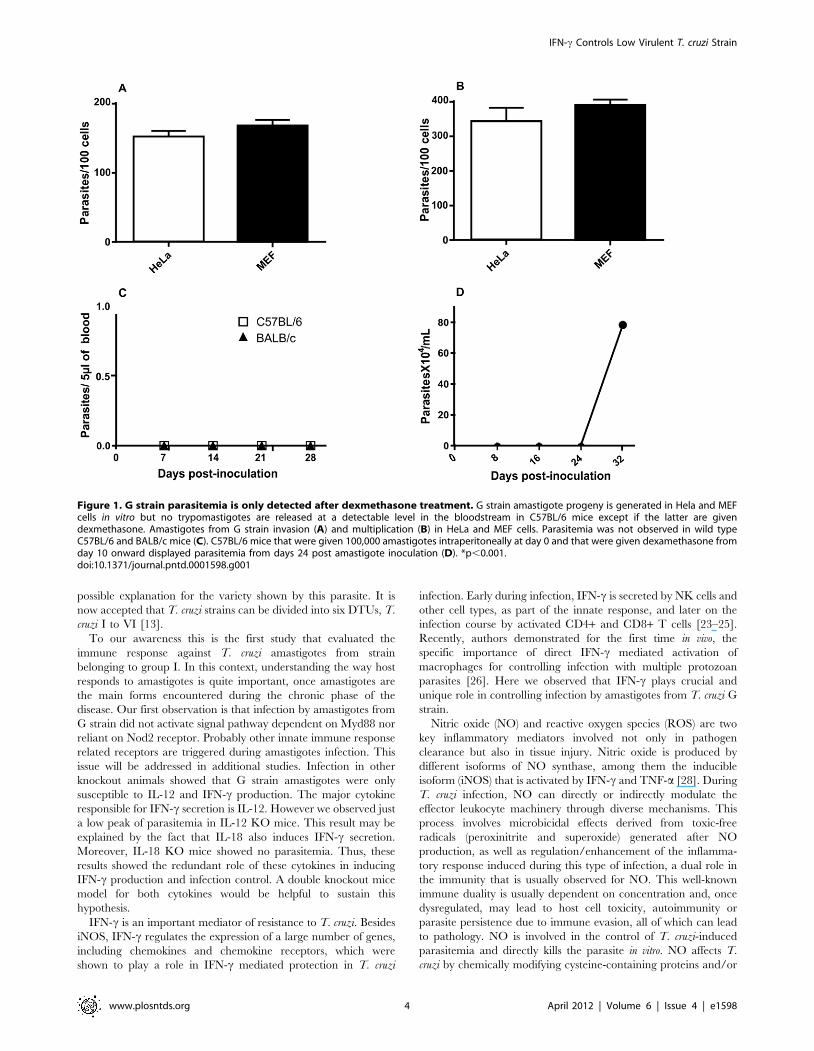
Amastigotes from G strain provided a patent in vitroinfection. On the other hand, was rapidly controlled invivo but was not completely eliminatedInvasion assays using amastigotes from G strain were performed
during 3 hours and the multiplication verified 48 h post HeLa andMEF cells invasion. The results showed that G strain amastigotesshowed high invasion and multiplication indexes in bothmammalian cell lines (Figure 1 A and B). Also, 80 to 90% ofcells were infected by the parasite. However, when BALB/c andC57BL/6 mice were intraperitoneally inoculated with amastigotesfrom G strain, no parasitemia was observed in both animal models(Figure 1 C). One could argue that this T. cruzi strain would benon-virulent. Nonetheless, in immunossupressed C57BL/6 ani-mals, parasitemia reached high peak after 24 days post-inoculation(Figure 1 D).
IFN-c controls G strain amastigotes infectionIn order to verify the impact of different host immune
components in protection against infection by amastigotes fromT. cruzi G strain we performed a screening using differentknockout mice model. First, we inoculated Myd88, Nod2, CD4and CD8 KO animals. The results showed no change in thecourse of infection and mortality comparing to WT mice (Figure 2a, b, c, d, e, f, g, and h). After, we verified if cytokines would playany role in animal protection against amastigotes from T. cruzi Gstrain infection. In this sense, we used TNF-a, IL-18, IL-12 andIFN-c KO mice. We observed that deficiency on TNF-a and IL-
18 secretion did not have impact on parasitemia and animalsurvival (Figure 3 b, c, f and g). On the other hand, IL12p40 KOmice showed parasitemia on the 12 day post-inoculation (p,0.01)and 40% of mortality by the 30 post-inoculation (p,0.01) (Figure 3a and e). Moreover, IFN-c KO mice presented a high parasitemiapeak by the 16 day post-inoculation (p,0.001) and all animalsdied by the 24 day (p,0.01) (Figure 3 d and h). These resultsturned our attention to the role of macrophages during T. cruzi Gstrain clearance. To gain insight about this issue, we performed anex vivo assay. For that purpose, C57BL/6 mice were intraperito-neally inoculated with amastigotes from G strain for 3 h.Inflammatory peritoneal macrophages were collected and seededinto coverslips. After 48 and 72 h of culture the number ofintracellular amastigotes was counted. It was observed that Gstrain amastigotes did not multiply intracellularly in the macro-phage cultures (Figure 4 A). Moreover, naive macrophagesobtained from C57BL/6 mice bone marrow undifferentiated cellswere in vitro infected with amastigotes from G strain and thenumber of trypomastigotes in the supernatant was counted afterthree, five and seven days of infection and treatment or not withdifferent concentrations of IFN-c. The number of trypomastigotesfrom G strain in the supernatant was higher in non-treated cells(p,0.001), showing that naive macrophages could not impairparasite multiplication and differentiation. However, the numberof released parasites was dramatically reduced in treated cells in anIFN-c dose dependent manner (Figure 4 B). The next step was toidentify the mechanism activated by macrophages during parasiteclearance. For that purpose we inoculated amastigotes from Gstrain in iNOS and gp91 KO mice. However, the results showedno difference in the course of infection, neither on the mortalityrates (Figure 5 a, b, c and d).
NK cells are recruited early during infection by G strainamastigotesOne important question raised by our results is the source of
IFN-c, since CD4 and CD8 cells seemed not to play an importantrole. Also, it is worth mentioning that the control occurred duringthe first days after inoculation. Thus, we performed flow cytometryin order to verify if NK cells were recruited during infection. Theresults showed that in non inoculated animals the lymphocytespopulation in peripheral blood were stained neither for CD16/32nor for NK1.1 and CD69, indicating a phenotype of inactivated Tcells (Figure 6 b and c). However, when we observed the animalsby 8 days post-inoculation, we were capable of identifying anotherdistinct cell population, which was denominated of ‘‘large granularlymphocytes’’ (LGL). It is know that the NK cells are largest thanother lymphocytes and they have granular contents. Thereby, thisLGL population had the same NK cells phenotype. We observedthat this population was mostly double positive to CD16/32 andNK1.1 (Figure 6 d9) or CD69 (Figure 6 e9), confirming NKphenotype, and that they were activated. Afterward, our resultsdemonstrated a dramatic increase in activated NK cell by the 8day post-inoculation (p,0.001) (Figure 6 a9, d9, and e9), howeverthis behavior was not maintained in 25 day post-inoculation,returning to basal levels (Figure 6 a0 d0 and e0).
Discussion
T. cruzi is a very heterogeneous flagellate parasite; and itspopulations are characterized by a diverse morphology, aheterogeneous biological behavior, a high genetic variability, anddistinctly different clinical courses. The clonal-histotropic model ofChagas’ disease [27] describes a correlation between the clonal-population structure of T. cruzi and its tissue tropism; and it gives a
IFN-c Controls Low Virulent T. cruzi Strain
www.plosntds.org 3 April 2012 | Volume 6 | Issue 4 | e1598
possible explanation for the variety shown by this parasite. It isnow accepted that T. cruzi strains can be divided into six DTUs, T.cruzi I to VI [13].To our awareness this is the first study that evaluated the
immune response against T. cruzi amastigotes from strainbelonging to group I. In this context, understanding the way hostresponds to amastigotes is quite important, once amastigotes arethe main forms encountered during the chronic phase of thedisease. Our first observation is that infection by amastigotes fromG strain did not activate signal pathway dependent on Myd88 norreliant on Nod2 receptor. Probably other innate immune responserelated receptors are triggered during amastigotes infection. Thisissue will be addressed in additional studies. Infection in otherknockout animals showed that G strain amastigotes were onlysusceptible to IL-12 and IFN-c production. The major cytokineresponsible for IFN-c secretion is IL-12. However we observed justa low peak of parasitemia in IL-12 KO mice. This result may beexplained by the fact that IL-18 also induces IFN-c secretion.Moreover, IL-18 KO mice showed no parasitemia. Thus, theseresults showed the redundant role of these cytokines in inducingIFN-c production and infection control. A double knockout micemodel for both cytokines would be helpful to sustain thishypothesis.IFN-c is an important mediator of resistance to T. cruzi. Besides
iNOS, IFN-c regulates the expression of a large number of genes,including chemokines and chemokine receptors, which wereshown to play a role in IFN-c mediated protection in T. cruzi
infection. Early during infection, IFN-c is secreted by NK cells andother cell types, as part of the innate response, and later on theinfection course by activated CD4+ and CD8+ T cells [23–25].Recently, authors demonstrated for the first time in vivo, thespecific importance of direct IFN-c mediated activation ofmacrophages for controlling infection with multiple protozoanparasites [26]. Here we observed that IFN-c plays crucial andunique role in controlling infection by amastigotes from T. cruzi Gstrain.Nitric oxide (NO) and reactive oxygen species (ROS) are two
key inflammatory mediators involved not only in pathogenclearance but also in tissue injury. Nitric oxide is produced bydifferent isoforms of NO synthase, among them the inducibleisoform (iNOS) that is activated by IFN-c and TNF-a [28]. DuringT. cruzi infection, NO can directly or indirectly modulate theeffector leukocyte machinery through diverse mechanisms. Thisprocess involves microbicidal effects derived from toxic-freeradicals (peroxinitrite and superoxide) generated after NOproduction, as well as regulation/enhancement of the inflamma-tory response induced during this type of infection, a dual role inthe immunity that is usually observed for NO. This well-knownimmune duality is usually dependent on concentration and, oncedysregulated, may lead to host cell toxicity, autoimmunity orparasite persistence due to immune evasion, all of which can leadto pathology. NO is involved in the control of T. cruzi-inducedparasitemia and directly kills the parasite in vitro. NO affects T.cruzi by chemically modifying cysteine-containing proteins and/or
Figure 1. G strain parasitemia is only detected after dexmethasone treatment. G strain amastigote progeny is generated in Hela and MEFcells in vitro but no trypomastigotes are released at a detectable level in the bloodstream in C57BL/6 mice except if the latter are givendexmethasone. Amastigotes from G strain invasion (A) and multiplication (B) in HeLa and MEF cells. Parasitemia was not observed in wild typeC57BL/6 and BALB/c mice (C). C57BL/6 mice that were given 100,000 amastigotes intraperitoneally at day 0 and that were given dexamethasone fromday 10 onward displayed parasitemia from days 24 post amastigote inoculation (D). *p,0.001.doi:10.1371/journal.pntd.0001598.g001
IFN-c Controls Low Virulent T. cruzi Strain
www.plosntds.org 4 April 2012 | Volume 6 | Issue 4 | e1598

by binding to metallo-proteins that mediate crucial metabolicprocesses. The strength of NO toxicity is dependent on thesensitivity of the parasite, which differs among parasite strains and
according to the physiological microenvironment [29]. Moreover,Oxidative burst of activated phagocytes results in the release ofROS, e.g., superoxide (O2
N2), hydrogen peroxide (H2O2), and
Figure 2. Whatever gene (CD4, CD8, Nod2 and Myd88) deletion, trypomastigotes were never detected in mice bloodstream. Wildtype and CD4 (a, e), CD8 (b, f) Nod2 (c, g) and Myd88 (d, h) knockout mice mice were given intraperitoneally 100,000 G strain amastigotes. Parasitemiavalues were monitored in mouse blood at 7, 12, 19 and 26 days post-inoculation; survival was checked every day until 30 post-inoculation . (n = 5mice per group).doi:10.1371/journal.pntd.0001598.g002
IFN-c Controls Low Virulent T. cruzi Strain
www.plosntds.org 5 April 2012 | Volume 6 | Issue 4 | e1598

hydroxyl radical, via activation of NADPH oxidase (NOX) and/ormyeloperoxidase (MPO) enzymes. The inflammatory cytokinesand ROS are important for the control of T. cruzi and may becytotoxic to the host cellular components. Many of the ROS arehighly reactive and diffusible and may be released into the
extracellular milieu. Whereas intracellular ROS serve mainly forhost defense against infectious agents, the extracellular release ofROS, when present in abundance, directly damages thesurrounding tissues or promotes inflammatory processes (revisedin [30]). Our results obtained from iNOS and gp91 KO mice
Figure 3. Deletion in IL12p40 and IFN-c induced bloodstream parasitemia. Though with distinct profiles, in mice genetically deleted fromeither IL12p40, IFN-c, the G strain trypomastigote progeny was detected in the bloodstream, while in mice deleted from either IL-18 or TNF-a, notrypomastigote progeny was detected. Wild type and IL-12p40 (a, e), IL-18 (b, f), TNF-a (c, g) and IFN-c (d, h) knockout mice were givenintraperitoneally 100,000 G strain amastigotes. Parasitemia values were monitored in mouse blood at 7, 12, 19 and 26 days post-inoculation; survivalwas checked every day until 30 post-inoculation . (n = 5 mice per group). It was observed parasitemia peak and mortality only for IL-12p40 and IFN-cKO mice. *p,0.01; ***p,0.001.doi:10.1371/journal.pntd.0001598.g003
IFN-c Controls Low Virulent T. cruzi Strain
www.plosntds.org 6 April 2012 | Volume 6 | Issue 4 | e1598

showed no parasitemia during the 30 days post-inoculation.Thus, is conceivable to believe that NO and ROS may playredundant role during parasite clearance. Another hypothesisthat does not completely exclude the first is that othermechanisms of parasite clearance would be activated by G strainparasites, such as a group of IFN-c induced genes that plays amajor role in host control of intracellular pathogens. These genesbelong to a family encoding a series of 47- to 48-kDa GTPases forinstance LRG-47 that can influence T. cruzi Y strain control by
simultaneously regulating macrophage-microbicidal activity andhemopoietic function [31].Our results showed the impact of innate immune response in
controlling infection by amastigotes from G strain. In addition,CD4 and CD8 KO mice showed no difference in the infectioncourse. This information may represent important finding todesign novel immune strategies focused on enhancing the innateimmune response to control pathology that may be caused bydifferent strains of the parasite in the same host.
Figure 4. Inflamatory and IFN-c treated naive macrophages impaired cell-cycling trypomastigotes differentiate from amastigotes.Amastigotes did not multiply in inflammatory peritoneal macrophages in an ex-vivo assay (A). Treatment with recombinant IFN-c controlled in a dosedependent manner trypomastigotes release from bone marrow derived naive macrophages (B) (p,0.001).doi:10.1371/journal.pntd.0001598.g004
Figure 5. In iNOS and gp91 KO mice no trypomastigote progeny was detected. Wild type, iNOS (a, c) and gp91 KO (b, d) mice were givenintraperitoneally 100 000 G strain amastigotes. Parasitemia values were monitored in mouse blood at 7, 12, 19 and 26 days post-inoculation ; survivalwas checked every day until 30 post-inoculation . (n = 5 mice per group).doi:10.1371/journal.pntd.0001598.g005
IFN-c Controls Low Virulent T. cruzi Strain
www.plosntds.org 7 April 2012 | Volume 6 | Issue 4 | e1598

Figure 6. Monitoring presence of activated NK cells in bloodstream post amastigote intraperitoneal inoculation. Flow cytometry wasperformed with mononuclear cells prepared from mice left without any inoculation (a, b, c, d and e), and mice that were given intraperitoneal G strainamastiogotes at day-8 post-inoculation (a9, b9, c9, d9 and e9) at day-25 (b0, c0, d0 and e0). Note the higher percentage of activated NK cells at day-8post-inoculation (p,0.01). Gates: L – lymphocytes and LGL – large granular lymphocytes (NK cells).doi:10.1371/journal.pntd.0001598.g006
IFN-c Controls Low Virulent T. cruzi Strain
www.plosntds.org 8 April 2012 | Volume 6 | Issue 4 | e1598

To gain insight into the source of IFN-c production, weperformed flow cytometry and observed that the lymphocytepopulation in the peripheral blood samples showed an inactivatedphenotype in infected and non infected animals. While in infectedanimals, we observed a significant increase in NK population withan activated phenotype. This result suggested that the main sourceof IFN-c produced to protect animal against amastigotes from T.cruzi G strain is NK cells. However, depletion of NK cell in WTand IFN-c KO mice would be interesting to confirm thehypothesis.In conclusion, our research showed that although amastigotes
from G strain were highly infective in vitro they did not induce apatent infection in vivo due to the high susceptibility to IFN-cproduction early in infection. This study highlighted the need to
consider strain biases when investigating host immune responseagainst T. cruzi.
Acknowledgments
We would like to thank Deivid Willian da Fonseca Batistao, Msc, for theassistance in flow cytometry analysis, and Paula Brıgido for assistance inimages processing.
Author Contributions
Conceived and designed the experiments: CVS AAR DSZ JSS EAVF.Performed the experiments: AAR JSSS GKS FAM AAS CPSSN CVH.Analyzed the data: CVS AAR. Contributed reagents/materials/analysistools: CVS DSZ JSS. Wrote the paper: CVS.
References
1. Rassi A, Rassi A, Marin-Neto JA (2010) Chagas disease. The Lancet 375:1388–1402.
2. Brener Z, Gazzinelli RT (1997) Immunological control of Trypanosoma cruziinfection and pathogenesis of Chagas’ disease. Int Arch Allergy Immunol 114:103–110.
3. Dos Reis GA (1997) Cell-mediated immunity in experimental Trypanosoma cruziinfection. Parasitol Today 13: 335–342.
4. Kayama H, Takeda K (2010) The innate immune response to Trypanosoma cruziinfection. Microbes Infect 12: 511–517.
5. Dos Reis GA (2011) Evasion of immune responses by Trypanosoma cruzi, theetiological agent of Chagas disease. Braz J Med Biol Res 44: 84–90.
6. Aliberti JCS, Cardoso MAG, Martins GA, Gazzinelli RT, Vieira LQ, et al.(1996) Interleukin- 12 mediates resistance to Trypanosoma cruzi in mice andisproduced by murine macrophages in response to live trypomastigotes. Infectionand Immunity 64: 1961–1967.
7. Aliberti JCS, Machado FS, Souto JT, Campanelli AP, Teixeira MM, et al.(1999) b- Chemokines enhance parasite uptake and promote nitricoxide-dependent microbiostatic activity in murine inflammatory macrophages infectedwith Trypanosoma cruzi. Infection and Immunity 67: 4819–4826.
8. Antunez MI, Cardoni RL (2000) IL-12 and IFN-c production, and NK cellactivity, in acute and chronic experimental Trypanosoma cruzi infections.Immunology Letters 71: 103–109.
9. Talvani A, Ribeiro CS, Aliberti JC, Michailowsky V, Santos PV, et al. (2000)Kinetics of cytokine gene expression in experimental chagasic cardiomyopathy:tissue parasitism and endogenous IFN-c as important determinants ofchemokine mRNA expression during infection with Trypanosoma cruzi. Microbesand Infection 2: 851–866.
10. Abrahamsohn IA, Coffman RL (1996) Trypanosoma cruzi: IL-10, TNF, IFN-c andIL-12 regulate innate and acquired immunity to infection. ExperimentalParasitology 84: 231–244.
11. Rodrigues MM, Ribeirao M, Boscardin SB (2000) CD4 Th1 but not Th2 clonesefficiently activate macrophages to eliminate Trypanosoma cruzi through a nitricoxide dependent mechanism. Immunology Letters 73: 43–50.
12. Hiyama K, Hamano S, Nakamura T, Nomoto K, Tada I (2001) IL-4 reducesresistance of mice to Trypanosoma cruzi infection. Parasitology Research 87:269–274.
13. Zingales B, Andrade SG, Briones MR, Campbell DA, Chiari E, et al. (2009) Anew consensus for Trypanosoma cruzi intraspecific nomenclature: second revisionmeeting recommends T. cruzi I to TcVI. Mem Inst Oswaldo Cruz 104:1051–1054.
14. Di Noia JM, Buscaglia CA, De Marchi CR, Almeida IC, Frasch ACC (2002) ATrypanosoma cruzi small surface molecule provides the first immunologicalevidence that Chagas’ disease is due to a single parasite lineage. Journal ofExperimental Medicine 195: 401–413.
15. Fernandes O, Mangia RH, Lisboa CV, Pinho AP, Morel CM, et al. (1999) Thecomplexity of the sylvatic cycle of Trypanosoma cruzi in Rio de Janeiro state(Brazil) revealed by the non-transcribed spacer of themini-exon gene.Parasitology 118: 161–166.
16. Anez N, Crisante G, da Silva FM, Rojas A, Carrasco H, et al. (2004)Predominance of lineage I among Trypanosoma cruzi isolates from Venezuelan
patients with different clinical profiles of acute Chagas’ disease. TropicalMedicine and International Health 9: 1319–1326.
17. Black CL, Ocana S, Riner D, Costales JA, Lascano MS, et al. (2007) Householdrisk factors for Trypanosoma cruzi seropositivity in two geographic regions ofEcuador. Journal of Parasitology 93: 12–16.
18. Mejıa-Jaramillo AM, Pena VH, Triana-Chavez O (2009) Trypanosoma cruzi:biological characterization of lineages I and II supports the predominance oflineage I in Colombia. Experimental Parasitology 121: 83–91.
19. Yoshida N (1983) Surface antigens of metacyclic trypomastigotes of Trypanosomacruzi. Infection and Immunity 40: 836–839.
20. Yoshida N, Blanco SA, Araguth MF, Russo M, Gonzalez J (1990) The stage-specific 90-Kilodalton surface antigen of metacyclic trypomastigotes ofTrypanosoma cruzi. Mol Biochem Parasitol 39: 39–46.
21. Silva CV, Luquetti AO, Rassi A, Mortara RA (2006) Involvement of Ssp-4-related carbohydrate epitopes in mammalian cell invasion by Trypanosoma cruziamastigotes. Microbes and Infection 8: 2120–2129.
22. Silva CV, Kawashita SY, Probst CM, Dallagiovanna B, Cruz MC, et al. (2009)Characterization of a 21 kDa protein from Trypanosoma cruzi associated withmammalian cell invasion. Microbes and Infection 11: 563–570.
23. Gazzinelli RT, Oswald IP, Hieny S, James SL, Sher A (1992) The microbicidalactivity of interferon-gamma-treated macrophages against Trypanosoma cruziinvolves an L-arginine-dependent, nitrogen oxide-mediated mechanism inhib-itable by interleukin-10 and transforming growth factor-beta. Eur J Immunol 22:2501–2506.
24. Holscher C, Kohler G, Muller U, Mossmann H, Schaub GA, et al. (1998)Defective nitric oxide effector functions lead to extreme susceptibility ofTrypanosoma cruzi-infected mice deficient in gamma interferon receptor orinducible nitric oxide synthase. Infect Immun 66: 1208–1215.
25. Aliberti JC, Souto JT, Marino AP, Lannes-Vieira J, Teixeira MM, et al. (2001)Modulation of chemokine production and inflammatory responses in inter-ferongamma- and tumor necrosis factor-R1-deficient mice during Trypanosomacruzi infection. Am J Pathol 158: 1433–1440.
26. Lykens JE, Terrell CE, Zoller EE, Divanovic S, Trompette A, et al. (2010) Micewith a selective impairment of IFN-gamma signaling in macrophage lineage cellsdemonstrate the critical role of IFN-gamma-activated macrophages for thecontrol of protozoan parasitic infections in vivo. J Immunol 184: 877–885.
27. Padilla AM, Bustamante JM, Tarleton RL (2009) CD8+ T cells in Trypanosomacruzi infection. Curr Opin Immunol 21: 385–390.
28. Bogdan C (2001) Nitric oxide and the immune response. Nat Immunol 2:907–916.
29. Gutierrez FRS, Mineo TWP, Pavanelli WR, Guedes PMM, Silva JS (2009) Theeffects of nitric oxide on the immune system during Trypanosoma cruzi infection.Mem Inst Oswaldo Cruz 104: 236–245.
30. Gupta S, Dhiman M, Wen JJ, Garg NJ (2011) ROS signalling of inflammatorycytokines during Trypanosoma cruzi infection. Adv Parasitol 76: 153–70.
31. Santiago HC, Feng CG, Bafica A, Roffe E, Arantes RM, et al. (2005) MiceDeficient in LRG-47 Display Enhanced Susceptibility to Trypanosoma cruziInfection Associated with Defective Hemopoiesis and Intracellular Control ofParasite Growth. J Immunol 175: 8165–8172.
IFN-c Controls Low Virulent T. cruzi Strain
www.plosntds.org 9 April 2012 | Volume 6 | Issue 4 | e1598