The Bacterial Protein Azurin Impairs Invasion and FAK/ Src Signaling in P-Cadherin-Overexpressing...
8
The Bacterial Protein Azurin Impairs Invasion and FAK/ Src Signaling in P-Cadherin-Overexpressing Breast Cancer Cell Models Nuno Bernardes 1,2 , Ana Sofia Ribeiro 2 , Sofia Abreu 1 , Bruna Mota 1 , Rute G. Matos 3 , Cecilia M. Arraiano 3 , Raquel Seruca 2 , Joana Paredes 2 , Arsenio M. Fialho 1 * 1 Institute for Biotechnology and Bioengineering, Center for Biological and Chemical Engineering, Instituto Superior Te ´cnico, Lisbon, Portugal, 2 Institute of Molecular Pathology and Immunology of the University of Porto (IPATIMUP), Porto, Portugal, 3 Instituto de Tecnologia Quimica e Biolo ´ gica (ITQB), Universidade Nova de Lisboa, Av a da Repu ´ blica, Oeiras, Portugal Abstract P-cadherin overexpression occurs in about 30% of all breast carcinomas, being a poor prognostic factor for breast cancer patients. In a cellular background of wild-type E-cadherin, we have previously shown that its expression promotes invasion, motility and migration of breast cancer cells due to the induced secretion of metalloproteases (MMPs) to the extracellular medium and to the concomitant shedding of a pro-invasive soluble form of this protein (sP-cad). Azurin is secreted by Pseudomonas aeruginosa and induces in vitro and in vivo cytotoxicity after its preferential penetration in human cancer cells relative to normal cells. Three different breast cancer cell lines, MCF-7/AZ.Mock, MCF-7/AZ.Pcad and SUM149 were treated with sub-killing doses of azurin. Invasion of these cells was measured using Matrigel Invasion Assays and MTT assays were performed to determine cell viability upon treatment and the effects on cadherins expression was determined by Western blot and Immunofluorescence. Gelatin Zymography was used to determine activity of MMP2 in the conditioned media of azurin treated and untreated cells and the phosphorylation levels of intracellular signaling proteins were determined by Western blot. The invasive phenotype of these breast cancer cells was significantly reduced by azurin. Azurin (50–100 mM) also caused a specific decrease on P-cadherin protein levels from 30–50% in MCF-7/AZ.Pcad and SUM149 breast cancer cell lines, but the levels of E-cadherin remain unaltered. More, the levels of sP-cad and the activity of MMP2 were reduced in the extracellular media of azurin treated cells and we also observed a decrease in the phosphorylation levels of both FAK and Src proteins. Our data show that azurin specifically targets P-cadherin, not E-cadherin, abrogating P-cadherin-mediated invasive effects and signaling. Therefore, azurin could possibly be considered a therapeutic tool to treat poor-prognosis breast carcinomas overexpressing P-cadherin in a wild type E-cadherin context. Citation: Bernardes N, Ribeiro AS, Abreu S, Mota B, Matos RG, et al. (2013) The Bacterial Protein Azurin Impairs Invasion and FAK/Src Signaling in P-Cadherin- Overexpressing Breast Cancer Cell Models. PLoS ONE 8(7): e69023. doi:10.1371/journal.pone.0069023 Editor: Claudia Daniela Andl, Vanderbilt University, United States of America Received February 27, 2013; Accepted June 3, 2013; Published July 19, 2013 Copyright: ß 2013 Bernardes et al. This is an open-access article distributed under the terms of the Creative Commons Attribution License, which permits unrestricted use, distribution, and reproduction in any medium, provided the original author and source are credited. Funding: The work presented was supported by a scientific project (PTDC/EBBBIO/100326/2008) financed by the Portuguese Science and Technology Foundation (FCT). FCT also provides research grants as follows: PhD research grant for Nuno Bernardes (SFRH/BD/48763/2008) and post-doc research grant to Ana Sofia Ribeiro (SFRH/BPD/75705/2011) and Programa Cie ˆncia 2007 for Joana Paredes [POPH - QREN - Tipology 4.2]. IPATIMUP is an Associate Laboratory of the Portuguese Ministry of Science, Technology and Higher Education and is partially supported by FCT. Work at ITQB was supported by grant PEst-OE/EQB/LA0004/ 2011, also from FCT. The funders had no role in study design, data collection and analysis, decision to publish, or preparation of the manuscript. Competing Interests: The authors have declared that no competing interests exist. * E-mail: [email protected] Introduction Cell invasion is a critical step in cancer progression [1]. Invasive cancer cells have significant altered properties, namely in polarity and morphology, as well as in their ability to adhere to other cells and to the extracellular matrix (ECM) components. Indeed, cell- cell adhesion and cell-ECM adhesion need to be very tightly regulated for the maintenance of a normal epithelial architecture [2]. Classical type I cadherins, namely E-cadherin (or epithelial- cadherin - Ecad), are crucial players that regulate cell-cell adhesion. During epithelial-to-mesenchymal transition (EMT), E- cadherin is usually down-regulated or functionally inactivated and de novo expression of other cadherins is frequently observed, a process named cadherin switch. These de-regulations cause alterations that are reflected in terms of intracellular signaling, as well as in cell behavior, as loss of cell polarity and acquisition of invasive capacity [3]. P-cadherin (Pcad) overexpression occurs in 30% of invasive breast carcinomas, being associated with poor patient prognosis. Interestingly, in some metastatic breast cancer models, as well as in high-grade primary carcinomas and in the aggressive local inflammatory breast cancer, E-cadherin expression is maintained alongside with aberrant expression of P-cadherin [4,5]. We have previously found that the increased expression of P-cadherin promotes invasive effects in breast cancer cells, which can be, at least in part, attributed to the release of a soluble form of P- cadherin (sP-cad) to the extracellular media, that is capable by itself to cause invasion of E-cadherin positive, non-invasive, cell lines [6]. Also, increased expression and activity of matrix metalloproteases (MMPs), namely MMP-1 and MMP-2, are involved in cell invasion mediated by P-cadherin overexpression PLOS ONE | www.plosone.org 1 July 2013 | Volume 8 | Issue 7 | e69023
Transcript of The Bacterial Protein Azurin Impairs Invasion and FAK/ Src Signaling in P-Cadherin-Overexpressing...
The Bacterial Protein Azurin Impairs Invasion and FAK/Src Signaling in P-Cadherin-Overexpressing BreastCancer Cell ModelsNuno Bernardes1,2, Ana Sofia Ribeiro2, Sofia Abreu1, Bruna Mota1, Rute G. Matos3, Cecilia M. Arraiano3,
Raquel Seruca2, Joana Paredes2, Arsenio M. Fialho1*
1 Institute for Biotechnology and Bioengineering, Center for Biological and Chemical Engineering, Instituto Superior Tecnico, Lisbon, Portugal, 2 Institute of Molecular
Pathology and Immunology of the University of Porto (IPATIMUP), Porto, Portugal, 3 Instituto de Tecnologia Quimica e Biologica (ITQB), Universidade Nova de Lisboa, Ava
da Republica, Oeiras, Portugal
Abstract
P-cadherin overexpression occurs in about 30% of all breast carcinomas, being a poor prognostic factor for breast cancerpatients. In a cellular background of wild-type E-cadherin, we have previously shown that its expression promotes invasion,motility and migration of breast cancer cells due to the induced secretion of metalloproteases (MMPs) to the extracellularmedium and to the concomitant shedding of a pro-invasive soluble form of this protein (sP-cad). Azurin is secreted byPseudomonas aeruginosa and induces in vitro and in vivo cytotoxicity after its preferential penetration in human cancer cellsrelative to normal cells. Three different breast cancer cell lines, MCF-7/AZ.Mock, MCF-7/AZ.Pcad and SUM149 were treatedwith sub-killing doses of azurin. Invasion of these cells was measured using Matrigel Invasion Assays and MTT assays wereperformed to determine cell viability upon treatment and the effects on cadherins expression was determined by Westernblot and Immunofluorescence. Gelatin Zymography was used to determine activity of MMP2 in the conditioned media ofazurin treated and untreated cells and the phosphorylation levels of intracellular signaling proteins were determined byWestern blot. The invasive phenotype of these breast cancer cells was significantly reduced by azurin. Azurin (50–100 mM)also caused a specific decrease on P-cadherin protein levels from 30–50% in MCF-7/AZ.Pcad and SUM149 breast cancer celllines, but the levels of E-cadherin remain unaltered. More, the levels of sP-cad and the activity of MMP2 were reduced in theextracellular media of azurin treated cells and we also observed a decrease in the phosphorylation levels of both FAK andSrc proteins. Our data show that azurin specifically targets P-cadherin, not E-cadherin, abrogating P-cadherin-mediatedinvasive effects and signaling. Therefore, azurin could possibly be considered a therapeutic tool to treat poor-prognosisbreast carcinomas overexpressing P-cadherin in a wild type E-cadherin context.
Citation: Bernardes N, Ribeiro AS, Abreu S, Mota B, Matos RG, et al. (2013) The Bacterial Protein Azurin Impairs Invasion and FAK/Src Signaling in P-Cadherin-Overexpressing Breast Cancer Cell Models. PLoS ONE 8(7): e69023. doi:10.1371/journal.pone.0069023
Editor: Claudia Daniela Andl, Vanderbilt University, United States of America
Received February 27, 2013; Accepted June 3, 2013; Published July 19, 2013
Copyright: � 2013 Bernardes et al. This is an open-access article distributed under the terms of the Creative Commons Attribution License, which permitsunrestricted use, distribution, and reproduction in any medium, provided the original author and source are credited.
Funding: The work presented was supported by a scientific project (PTDC/EBBBIO/100326/2008) financed by the Portuguese Science and TechnologyFoundation (FCT). FCT also provides research grants as follows: PhD research grant for Nuno Bernardes (SFRH/BD/48763/2008) and post-doc research grant to AnaSofia Ribeiro (SFRH/BPD/75705/2011) and Programa Ciencia 2007 for Joana Paredes [POPH - QREN - Tipology 4.2]. IPATIMUP is an Associate Laboratory of thePortuguese Ministry of Science, Technology and Higher Education and is partially supported by FCT. Work at ITQB was supported by grant PEst-OE/EQB/LA0004/2011, also from FCT. The funders had no role in study design, data collection and analysis, decision to publish, or preparation of the manuscript.
Competing Interests: The authors have declared that no competing interests exist.
* E-mail: [email protected]
Introduction
Cell invasion is a critical step in cancer progression [1]. Invasive
cancer cells have significant altered properties, namely in polarity
and morphology, as well as in their ability to adhere to other cells
and to the extracellular matrix (ECM) components. Indeed, cell-
cell adhesion and cell-ECM adhesion need to be very tightly
regulated for the maintenance of a normal epithelial architecture
[2].
Classical type I cadherins, namely E-cadherin (or epithelial-
cadherin - Ecad), are crucial players that regulate cell-cell
adhesion. During epithelial-to-mesenchymal transition (EMT), E-
cadherin is usually down-regulated or functionally inactivated and
de novo expression of other cadherins is frequently observed, a
process named cadherin switch. These de-regulations cause
alterations that are reflected in terms of intracellular signaling,
as well as in cell behavior, as loss of cell polarity and acquisition of
invasive capacity [3].
P-cadherin (Pcad) overexpression occurs in 30% of invasive
breast carcinomas, being associated with poor patient prognosis.
Interestingly, in some metastatic breast cancer models, as well as in
high-grade primary carcinomas and in the aggressive local
inflammatory breast cancer, E-cadherin expression is maintained
alongside with aberrant expression of P-cadherin [4,5]. We have
previously found that the increased expression of P-cadherin
promotes invasive effects in breast cancer cells, which can be, at
least in part, attributed to the release of a soluble form of P-
cadherin (sP-cad) to the extracellular media, that is capable by
itself to cause invasion of E-cadherin positive, non-invasive, cell
lines [6]. Also, increased expression and activity of matrix
metalloproteases (MMPs), namely MMP-1 and MMP-2, are
involved in cell invasion mediated by P-cadherin overexpression
PLOS ONE | www.plosone.org 1 July 2013 | Volume 8 | Issue 7 | e69023
[6], as well as the activation of the intracellular non-receptor
tyrosine kinases FAK and Src, that regulate a wide number of
signaling pathways involved in cell spreading, adhesion, migration,
invasion, survival, proliferation, differentiation and angiogenesis
[7].
Azurin is a small copper protein (14 kDa), water-soluble,
produced by the bacterium Pseudomonas aeruginosa. Besides its
described function as a redox partner in electron transfer
reactions, azurin can act as an anticancer agent exerting
cytotoxicity in vitro against several cancer derived cell lines and
promotes tumor regression in xenografted mice [8–11]. In
cultured cells, azurin enters cancer cells preferentially when
compared to normal cells derived from the same tissue, mediated
by a portion of the protein spanning residues 50–77 (termed p28),
which adopts an amphipathic alpha-helical conformation [12].
p28, as a lead compound supported by CDG Therapeutics, has
finished Phase I clinical trial, which defined it as an anticancer
agent under an investigational new drug application (IND 77,754)
approved by the Food and Drug Administration.
In mechanistic terms, it is known that azurin targets different
cell proliferation pathways. Once internalized, azurin can interact
directly with p53 and stabilize it, increasing its protein levels in
both nuclear and cytoplasmic fractions [10,12–15]. Besides p53-
mediated cell cytotoxicity, this protein also binds the extracellular
EphB2 tyrosine kinase receptor, being able to prevent the tumor
progression caused by the binding of the natural ligand ephrinB2
[16]. Recently, p28 was found to penetrate endothelial cells
(HUVEC) and mediate the decrease in their motility and
migration, which was associated with an inhibition of VEGFR-2
(vascular endothelial growth factor receptor 2) kinase activity [17].
Moreover, the levels of phosphorylated FAK (Focal adhesion
kinase) and Akt proteins were also reduced, altering the
intracellular architecture of endothelial cytoskeleton and cell
contact proteins that limit endothelial cell motility and migration.
Based on the previous described anticancer and anti-migratory
effects of azurin, we hypothesized that it could also be used as a
therapeutic tool in highly aggressive breast cancers overexpressing
P-cadherin, being able to inhibit its pro-invasive effects. In this
work, we describe, for the first time, that azurin decreases P-
cadherin expression at the cellular membrane and inhibits P-
cadherin-induced breast cancer cell invasion at sub-killing doses.
Furthermore, we identified that this phenomenon is associated to a
MMP-2 decreased activity in the extracellular media and decrease
in FAK/Src complex signaling, possibly mediating the anti-
invasive effects of azurin in P-cadherin-overexpressing breast
cancer cells.
Materials and Methods
AntibodiesPrimary antibodies: P-cadherin (Western blot: clone 56, BD
Biosciences, Lexington, KY, USA; immunofluorescence: Cell
Signaling Technology, Boston, MA, USA), E-cadherin (clone
HECD1, Takara Bio Inc., Shiga, Japan), b-actin (I-19, Santa Cruz
Biotechnologies, CA, USA), total FAK (BD Biosciences), pFAK
Y397(Cell Signaling), total Src (Cell Signaling), pSrc Y416 (Cell
Signaling), total Akt (Santa Cruz Biotechnologies), pAkt S473 (Cell
Signaling).
Bacteria and Growth MediaAzurin-encoding gene from P.aeruginosa PAO1 was amplified by
PCR, using genomic DNA as a template. Forward and reverse
primers used were: 59-CGGGATCCGCCGAGTGCTCGGTG-
GACAT-39 and 59-CCCAAGCTTGCATCACTTCAGGGT-
CAGGGT-39. Azurin gene was placed downstream the T7
promoter in the pWH844 vector. E.coli SURE strain was used
as host for expression of the protein in the following conditions:
cells were incubated overnight in LB medium at 37uC with
150 mg/ml of ampicillin and cultured, at an initial optical density
at 640 nm (OD640) of 0.1, in SB medium (3.2% tryptone, 2% yeast
extract and 0.5% NaCl). At OD640 of 0.65, IPTG was added to
the culture at a final concentration of 0.2 mM and grown for 4 h
at the same conditions. Cells were harvested by centrifugation at
8000 rpm for 10 minutes at 4uC, washed once and resuspended in
buffer I (10 mM Imidazol, 0.2 M mM sodium phosphate, 0.5 M
NaCl, pH 7.4). Cells were stored at 280uC for further use.
Protein PurificationCells were disrupted by sonication and the purification steps
were performed by histidine affinity chromatography, using
HisTrapTM HP columns (GE Healthcare). Briefly, disrupted cells
were centrifuged for 5 min, at 176006g and 4uC; the supernatantwas centrifuged again at the same conditions for 1 h. The clarified
extract was then loaded into a 5 ml HisTrap HP column
equilibrated with START buffer (10 mM Imidazol, phosphate
buffer: 0.2 M sodium phosphate, 1 M NaCl, pH 7.4). Protein
elution was achieved with a continuous imidazole gradient (from
20 to 500 mM) in the same buffer. After purification, protein was
immediately de-salted and buffer exchanged to phosphate-
buffered saline (PBS) pH 7.4, in a HiPrep 26/10 Desalting
column (GE Healthcare) in an AKTA purifier system, following
the manufacturer’s instructions. Finally, protein was concentrated
by centrifugation at 4uC with Amicon Ultra Centrifugal Devices
(Milipore), with a molecular mass cutoff of 10 kDa. Purified
protein was passed through 1 ml Detoxi-GelTM Endotoxin
Removing column (Thermo Scientific) to remove endotoxins from
E. coli host strain. At all steps, protein concentration was assessed
with BCATM Protein Assay kit (Thermo Scientific), following the
manufacturer’s instructions. The purity of protein was analyzed by
sodium dodecyl-polyacrylamide gel electrophoresis (SDS-PAGE).
Cell CultureTwo distinct human breast cancer cell lines were used in this
study: MCF7/AZ [kindly provided by Prof. Marc Mareel (Ghent
University, Belgium); MCF-7/AZ.Mock and MCF-7/AZ.Pcad
[18] were stably transduced with empty vector or CDH3/
Pcadherin cDNA, respectively] and SUM149 [19] [kindly
provided by Prof. Stephen Ethier (University of Michigan, MI,
USA]. Both cell lines were routinely maintained at 37uC, 5%CO2, in the following media (Invitrogen Ltd, Paisley, UK): 50%
DMEM and 50% HamF12, supplemented with 10% heat-
inactivated fetal bovine serum (Lonza, Basel, Switzerland),
100 IU/ml penicillin and 100 mg/ml streptomycin (Invitrogen).
SUM149 medium was additionally supplemented with 5 mg/ml of
insulin and 1 mg/ml of hydrocortisone (Sigma-Aldrich, St. Louis,
MO, USA).
MCF-7/AZ cell line was retrovirally stable transduced to
encode human P-cadherin cDNA (MCF-7/AZ.Pcad cell line), as
previously described (Paredes et al. 2004). MCF-7/AZ.Mock cell
line, encoding only EGFP, was used as a control. SUM149 cell line
constitutively expresses high levels of P-cadherin. All the cell lines
used express normal levels of E-cadherin.
MTT Cell Viability AssayMTT [3-(4,5 dimethylthiazol-2-yl-2,5 tetrazolium bromide)]
assays were used to determine viability of breast cancer cells upon
azurin exposure. Breast cancer cells were seeded in 96-well plates
(3 replicates) (Orange Scientific) at a density of 26104 cells (MCF-
Azurin Impairs Invasion of Breast Cancer Cells
PLOS ONE | www.plosone.org 2 July 2013 | Volume 8 | Issue 7 | e69023
7/AZ cell lines) or 56104 cells (SUM149). After 24 h, medium was
exchanged and fresh azurin or a similar volume of media without
protein (100 mL) were added. After another 24 h (SUM149) or
48 h (MCF-7/AZ cell lines), 10 mL of MTT (5 mg/ml) were
added to each well and incubated at 37uC for 3.5 h. Reaction was
stopped with the addition of 40 mM HCL in isopropanol. MTT
formazan formed was spectrophotometrically read at 590 nm in a
96-well plate reader. Untreated cells were used as control, in order
to to determine the relative cell viability of treated cells.
Protein Extraction and Western Blot AnalysisCultured cells, treated with azurin (50 or 100 mM) or untreated,
were lysed using catenin lysis buffer (1% Triton X-100, and 1%
NP-40 in deionized PBS), supplemented with 1:7 protease
inhibitor cocktail (Roche Diagnostics GmbH, Germany) and with
1:100 of phosphatase inhibitor cocktail 3 (Sigma). Cells were
washed twice with PBS and allowed to lyse in 100 mL of catenin
lysis buffer for 10 minutes, at 4uC. Lysed cells were collected and
vortexed three times, for 10 seconds, prior to centrifugation at
176006g for 10 minutes at 4uC. Total protein quantification was
performed with BCATM Protein Assay kit (Pierce). 20 (E- and P-
cadherin, total FAK, Src and Akt) or 30 mg (phosphorylated FAK,
Src and Akt) of the total protein lysate was dissolved in sample
buffer [Laemmli with 5% (v/v) 2-b-mercaptoethanol and 5% (v/v)
bromophenol blue], boiled for 5 minutes at 95uC, and separated
by SDS-PAGE. Proteins were transferred onto nitrocellulose
membranes (Pall) at 120 V for 1 hour and 30 minutes. Membranes
were blocked with 5% (w/v) non-fat dry milk in PBS containing
0.5% (v/v) Tween-20 (PBS-T) or 5% BSA (for phosphorylated
protein detection) for 1 hour, incubated with primary antibodies,
overnight at 4uC (E-cadherin, P-cadherin and total FAK,Src and
Akt) or for 1.5 h at room temperature in 5% BSA (w/v) (pFAK,
pSrc and pAkt), and washed three times for 5 minutes with PBS-T.
Membranes were then incubated for 1 hour with secondary
antibodies, conjugated with horseradish peroxidase. Proteins were
detected through the addition of ECL reagent (Pierce) as a
substrate and exposed to an autoradiographic film (Amersham).
Three experiments were independently performed and represen-
tative results are shown. Signal quantifications were performed
using ImageJ and results are presented as the ratio between the
signal intensities in azurin treated samples to untreated cells, both
normalized to the respective actin band intensities.
ImmunofluorescenceCells were cultured on glass coverslips and treated with azurin
at different concentrations. After the desired exposition time,
medium was collected and cells were washed twice with PBS.
Fixation was performed with NH4Cl for 20 minutes at room
temperature. Permeabilization was achieved with 1% Triton X-
100 in PBS for 5 minutes at room temperature and coverslips were
blocked with 5% BSA solution in PBS for 30 minutes. Primary
antibodies were added for 1 hour at room temperature as follows:
1:100 dilution for E-cadherin antibody and 1:50 dilution for P-
cadherin antibody. After the incubation time, cells were washed
three times for 5 minutes with PBS and incubated with secondary
antibodies for 1 hour, at room temperature, at 1:500 dilution as
described: mouse polyclonal conjugated with ALexa-488 for E-
cadherin and rabbit polyclonal antibody conjugated with Alexa-
594 for P-cadherin. Each sample was washed with PBS after the
incubation period and mounted with Vectashield (Vector Labo-
ratories Inc., Burlingame, CA, USA) containing 4,6-diamidine-2-
phenylindolendihydrochloride (DAPI). Cell staining was observed
in a Zeiss microscope (Imager Z1) and images acquired using the
Axiovision software.
Quantitative Real-Time PCRTotal RNA from MCF-7/AZ and SUM149 cell lines was
extracted using the RNeasy Extraction kit (Qiagen), according to
the manufacturer’s instructions. Samples were subjected to
treatment with DNase (Qiagen) during the extraction procedure.
qRT-PCR was performed using gene-specific TaqMan probes
(Applied Biosystems, Foster City, CA). Analysis was performed
using the ABI PRISM 7500 Sequence Detection System
Instrument and software (Applied Biosystems). CDH1 and CDH3
relative quantifications between treated and untreated samples
were determined by the DDCt method using the internal standard
human 18S to normalize cDNA quantity.
Matrigel Invasion AssaysMatrigel Invasion assay was performed according to the
manufacturers instructions (BD Coat Matrigel Invasion Cham-
bers, BD Biosciences). Briefly, Matrigel inserts containing an 8
micron pore size PET membrane with a thin layer of Matrigel
Basement Membrane Matrix were pre-incubated with serum-free
media for 2 hours at 37uC. 56104 cells for MCF7/AZ.Mock and
MCF7/AZ. Pcad, and 2.56104 cells for SUM149, were seeded in
the upper compartment with or without azurin (control). After
48 h or 24 h, invasive cells were colored with DAPI and counted
under the microscope. In each condition, 10 independent fields
were counted and the average of these fields considered as the
mean number of invasive cells per condition.Results are presented
as the fold change in invasion of cells in comparison with the
MCF-7/AZ.Mock untreated cells.
Gelatin ZymographyCell conditioned media was collected to analyse the activity of
MMP2, using gelatin zymography. Cells were cultured in 6-well
plates coated with collagen type I (1 mg/ml). Gelatin gels were
loaded with a 15 mL sample for each condition, mixed with sample
buffer [0.03% bromophenol blue, 0.25 M Tris-HCl pH 6.8, 10%
SDS (w/v) and 4% sucrose (w/v)]. Electrophoresis was performed
on 10% polyacrylamide gels containing 0.1% (w/v) of gelatin
(Sigma) at 80V, under non-reducing conditions. Gels were washed
twice in 2% (v/v) Triton X-100 (Sigma), 30 minutes each time, at
room temperature, to remove SDS. After, gels were incubated in a
Substrate Reaction Buffer (50 mM Tris-HCl, 5 mM CaCl2,
pH 7.5, 1% Triton X-100) for 17 hours. After incubation, gels
were stained in Coomassie Blue Staining Solution [0.1% (w/v)
Coomassie Blue R250 in 10% acetic acid solution and 40% (v/v)
methanol], for 30 minutes, and de-stained in 20% (v/v) methanol
and 10% (v/v) acetic acid solution, until white bands against the
blue background of the gels appeared. MMP2 was identified
according to their molecular weight.
Statistical AnalysisData are expressed as mean values of a minimum of three
independent experiments 6 s.d. Student’s t-tests were performed
to determine statistically significant differences. A P value ,0.05
was considered statistically significant.
Results
Azurin Impairs Invasion of MCF-7/AZ.Pcad and SUM149Invasive Breast Cancer CellsPrevious work from our group has demonstrated that invasion
of breast cancer cells can be induced by P-cadherin overex-
pression in a wild-type E-cadherin context [6]. In fact, we
showed that expression of P-cadherin in MCF-7/AZ cells
Azurin Impairs Invasion of Breast Cancer Cells
PLOS ONE | www.plosone.org 3 July 2013 | Volume 8 | Issue 7 | e69023
induces an increase in cell invasion [6], whereas the knocking-
down of P-cadherin by siRNA causes a decrease in the invasive
behavior of SUM149 breast cancer cells [21]. Thus, in order to
study if azurin could impair the invasion mediated by P-
cadherin, these same human breast cancer cell models were
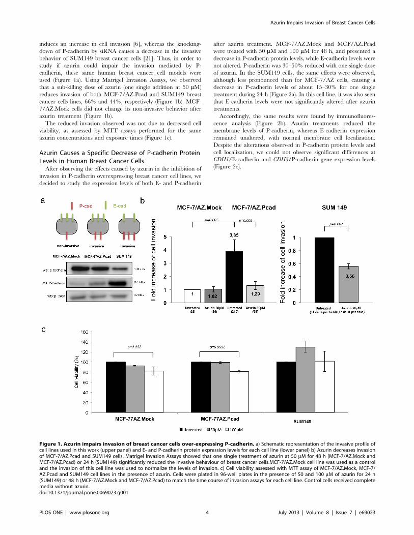
used (Figure 1a). Using Matrigel Invasion Assays, we observed
that a sub-killing dose of azurin (one single addition at 50 mM)
reduces invasion of both MCF-7/AZ.Pcad and SUM149 breast
cancer cells lines, 66% and 44%, respectively (Figure 1b). MCF-
7/AZ.Mock cells did not change its non-invasive behavior after
azurin treatment (Figure 1b).
The reduced invasion observed was not due to decreased cell
viability, as assessed by MTT assays performed for the same
azurin concentrations and exposure times (Figure 1c).
Azurin Causes a Specific Decrease of P-cadherin ProteinLevels in Human Breast Cancer CellsAfter observing the effects caused by azurin in the inhibition of
invasion in P-cadherin overexpressing breast cancer cell lines, we
decided to study the expression levels of both E- and P-cadherin
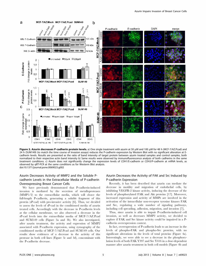
after azurin treatment. MCF-7/AZ.Mock and MCF/AZ.Pcad
were treated with 50 mM and 100 mM for 48 h, and presented a
decrease in P-cadherin protein levels, while E-cadherin levels were
not altered. P-cadherin was 30–50% reduced with one single dose
of azurin. In the SUM149 cells, the same effects were observed,
although less pronounced than for MCF-7/AZ cells, causing a
decrease in P-cadherin levels of about 15–30% for one single
treatment during 24 h (Figure 2a). In this cell line, it was also seen
that E-cadherin levels were not significantly altered after azurin
treatments.
Accordingly, the same results were found by immunofluores-
cence analysis (Figure 2b). Azurin treatments reduced the
membrane levels of P-cadherin, whereas E-cadherin expression
remained unaltered, with normal membrane cell localization.
Despite the alterations observed in P-cadherin protein levels and
cell localization, we could not observe significant differences at
CDH1/E-cadherin and CDH3/P-cadherin gene expression levels
(Figure 2c).
Figure 1. Azurin impairs invasion of breast cancer cells over-expressing P-cadherin. a) Schematic representation of the invasive profile ofcell lines used in this work (upper panel) and E- and P-cadherin protein expression levels for each cell line (lower panel) b) Azurin decreases invasionof MCF-7/AZ.Pcad and SUM149 cells. Matrigel Invasion Assays showed that one single treatment of azurin at 50 mM for 48 h (MCF-7/AZ.Mock andMCF-7/AZ.Pcad) or 24 h (SUM149) significantly reduced the invasive behaviour of breast cancer cells.MCF-7/AZ.Mock cell line was used as a controland the invasion of this cell line was used to normalize the levels of invasion. c) Cell viability assessed with MTT assay of MCF-7/AZ.Mock, MCF-7/AZ.Pcad and SUM149 cell lines in the presence of azurin. Cells were plated in 96-well plates in the presence of 50 and 100 mM of azurin for 24 h(SUM149) or 48 h (MCF-7/AZ.Mock and MCF-7/AZ.Pcad) to match the time course of invasion assays for each cell line. Control cells received completemedia without azurin.doi:10.1371/journal.pone.0069023.g001
Azurin Impairs Invasion of Breast Cancer Cells
PLOS ONE | www.plosone.org 4 July 2013 | Volume 8 | Issue 7 | e69023
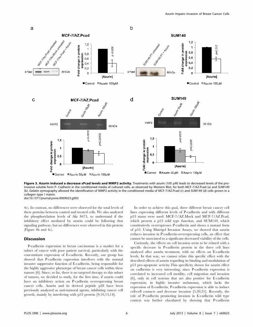
Azurin Decreases Activity of MMP2 and the Soluble P-cadherin Levels in the Extracellular Media of P-cadherinOverexpressing Breast Cancer CellsWe have previously demonstrated that P-cadherin-induced
invasion is mediated by the secretion of metalloproteases
(MMP1/2) to the extracellular media, which will cleave the
full-length P-cadherin, generating a soluble fragment of this
protein (sP-cad) with pro-invasive activity [6]. Thus, we decided
to assess the levels of sP-cad in the conditioned media of azurin
treated cells. Accordingly with the decrease in P-cadherin levels
at the cellular membrane, we also observed a decrease in the
sP-cad levels into the extracellular media of MCF-7/AZ.Pcad
and SUM149 cells (Figure 3a and 3b). We also investigated,
after azurin treatment, the activity and expression of MMP2
associated with P-cadherin expression, using zymography of the
conditioned media of MCF-7/AZ.Pcad and SUM149 cells. Our
results show evidences of a decrease in the activity of this
protease in both cell lines (Figure 3c and 3d), concomitant with
the P-cadherin decrease.
Azurin Decreases the Activity of FAK and Src Induced byP-cadherin ExpressionRecently, it has been described that azurin can mediate the
decrease in motility and migration of endothelial cells, by
inhibiting VEGFR-2 kinase activity, inducing the decrease of the
levels of phosphorylated FAK and Akt proteins [17]. Moreover,
increased expression and activity of MMPs are involved in the
activation of the intracellular non-receptor tyrosine kinases FAK
and Src, regulating a wide number of signaling pathways,
including cell spreading, adhesion, migration, and invasion [7],
Thus, since azurin is able to impair P-cadherin-induced cell
invasion, as well as decreases MMP2 activity, we decided to
explore if FAK and Src kinase activity could be impaired in a P-
cadherin overexpression context.
In fact, overexpression of P-cadherin leads to an increase in the
levels of phospho-FAK and phospho-Src proteins, with no
significant alterations in the levels of total proteins (Figure 4a).
Interestingly, we were able to see a decrease in the phosphory-
lation levels of both FAK Y397 and Src Y416 in a dose-dependent
manner after azurin treatment in both cell models (Figure 4b and
Figure 2. Azurin decreases P-cadherin protein levels. a) One single treatment with azurin at 50 mM and 100 mM for 48 h (MCF-7/AZ.Pcad) and24 h (SUM149) (to match the time course of invasion assays) reduces the P-cadherin expression by Western Blot with no significant alteration at E-cadherin levels. Results are presented as the ratio of band intensity of target protein between azurin treated samples and control samples, bothnormalized to their respective actin band intensity b) Same results were observed by immunofluorescence analysis of both cadherins in the sametreatment conditions. c) Azurin does not significantly change the expression levels of CDH1/E-cadherin or CDH3/P-cadherin at mRNA levels, asobserved by qRT-PCR at the same conditions as for Westerm Blot analysis.doi:10.1371/journal.pone.0069023.g002
Azurin Impairs Invasion of Breast Cancer Cells
PLOS ONE | www.plosone.org 5 July 2013 | Volume 8 | Issue 7 | e69023
4c). In contrast, no differences were observed for the total levels of
these proteins between control and treated cells. We also analyzed
the phosphorylation levels of Akt S473, to understand if the
inhibitory effect mediated by azurin could be following that
signaling pathway, but no differences were observed in this protein
(Figure 4b and 4c).
Discussion
P-cadherin expression in breast carcinomas is a marker for a
subset of cancer with poor patient survival, particularly with the
concomitant expression of E-cadherin. Recently, our group has
showed that P-cadherin expression interferes with the normal
invasive suppressive function of E-cadherin, being responsible for
the highly aggressive phenotype of breast cancer cells within these
tumors [6]. Since, so far, there is no targeted therapy to this subset
of tumors, we decided to study, for the first time, if azurin could
have an inhibitory action on P-cadherin overexpressing breast
cancer cells. Azurin and its derived peptide p28 have been
previously analyzed as anti-tumoral agents, inhibiting cancer cell
growth, mainly by interfering with p53 protein [9,10,13,14].
In order to achieve this goal, three different breast cancer cell
lines expressing different levels of P-cadherin and with different
p53 status were used: MCF-7/AZ.Mock and MCF-7/AZ.Pcad,
which present a p53 wild type function, and SUM149, which
constitutively overexpresses P-cadherin and shows a mutant form
of p53. Using Matrigel Invasion Assays, we showed that azurin
reduces invasion in P-cadherin-overexpressing cells, an effect that
cannot be associated to a significant decreased viability of the cells.
Curiously, the effects on cell invasion seem to be related with a
specific decrease in P-cadherin protein in the three cell lines
analysed after azurin treatment, with no effects on E-cadherin
levels. In that way, we cannot relate this specific effect with the
described effects of azurin regarding its binding and modulation of
p53 pro-apoptotic activity.This specificity shown for azurin effect
on cadherins is very interesting, since P-cadherin expression is
correlated to increased cell motility, cell migration and invasion
[6], only in cell systems that are also positive for E-cadherin
expression; in highly invasive melanoma, which lacks the
expression of E-cadherin, P-cadherin expression is able to induce
cell-cell contacts and decrease invasion [5,20,21]. Recently, the
role of P-cadherin promoting invasion in E-cadherin wild type
context was further elucidated by showing that P-cadherin
Figure 3. Azurin induced a decrease sPcad levels and MMP2 activity. Treatments with azurin (100 mM) leads to decreased levels of the pro-invasive soluble form P- Cadherin in the conditioned media of cultured cells, as observed by Western Blot, for both MCF-7/AZ.Pcad (a) and SUM149(b). Gelatin zymography allowed the identification of MMP2 activity in the conditioned media of MCF-7/AZ.Pcad (c) and SUM149 (d) cells grown in acollagen type I matrix.doi:10.1371/journal.pone.0069023.g003
Azurin Impairs Invasion of Breast Cancer Cells
PLOS ONE | www.plosone.org 6 July 2013 | Volume 8 | Issue 7 | e69023

promotes a disruption in the interaction of E-cadherin and
cytoplasmic catenins [22]. Moreover, the expression of both these
cadherins correlated significantly with high grade breast carcino-
mas and poor patient survival. In vivo, a breast cancer cell model
expressing both cadherins was found more aggressive, with higher
tumour growth when compared with the same model expressing
only one of the cadherins by suppressing each cadherin by siRNA
technology [22]. These results reinforce the anti-invasive role of
azurin in this context, since its action was preferential to P-
cadherin and not for E-cadherin. We could not observe a clear
reduction in CDH3/P-cadherin gene expression, suggesting that
other mechanisms at protein degradation level are mediating P-
cadherin decrease. These mechanisms are now under investiga-
tion.
A known mechanism that determines at least part of the
aggressiveness of P-cadherin expression is the release of soluble
forms of this protein, sP-cad, to the extracellular media of the cells.
This form is per se capable of causing cell invasion of non-invasive
cells [6]. In the presence of azurin, and probably due to the caused
P-cadherin decrease, lower levels of sP-cad are detected in azurin
treated cells. This result is interesting, since soluble forms of
classical cadherins have been associated with malignant effects
[23]: soluble E-cadherin was associated with increased invasion
and with the inhibition of normal E-cadherin-dependent cell-cell
aggregation [24], and higher levels of soluble P-cadherin were
found in nipple aspirate fluids of breast cancer patients than in
healthy women [25].
The cleavage and shedding of P-cadherin, and therefore the
higher invasive capacity of these cell lines, is mediated by MMPs.
In fact, several tumorigenic processes are mediated by these
proteases, namely the breakdown of extracellular components,
which accounts greatly to the ability of tumor cells to invade the
surrounding tissues through an extensive matrix remodeling [7].
MMPs also promote the release of bioactive molecules able to
induce invasion, like the cleavage of laminin-5 c2 chains by
MMP2, producing a fragment containing a epidermal growth
factor (EGF)-like domain, which induces integrin signaling and cell
migration. These can also play a role in angiogenesis, increasing
the bio-availability of vascular endothelial growth factor (VEGF),
fibroblast growth factor-2 (FGF-2) and transforming growth
factor-b (TGF-b), among others [26]. Ribeiro et al. (2010) [6]
identified an increase in the activity of MMP2 and MMP1 in P-
cadherin-overexpressing cells. We assessed the activity of one of
these proteases, MMP2, by gelatin zymography in the conditioned
Figure 4. Effect of azurin in FAK-Src signaling. Azurin at 50 mM and 100 mM decreased phosphorylation levels of FAK Y397 and Src Y416 in bothMCF-7/AZ.Pcad and SUM149 breast cancer cells, but not Akt S473, in a dose-dependent manner. Levels for total FAK, Src and Akt were also analyzed.Results are presented as the ratio of band intensity of target protein between azurin treated samples and control samples, both normalized to theirrespective actin band intensity.doi:10.1371/journal.pone.0069023.g004
Azurin Impairs Invasion of Breast Cancer Cells
PLOS ONE | www.plosone.org 7 July 2013 | Volume 8 | Issue 7 | e69023
media of these breast cancer cells treated with azurin and could
observe a decrease in its activity.
Our data also show that the decrease in P-cadherin caused by
azurin is parallel to a decrease in the phosphorylation level of FAK
and Src without any alteration in total FAK and Src protein levels.
It is known that FAK is necessary to the regulation of invadopodia
in ovarian carcinoma cells and to promote breast cancer cell
invasion [27]. Additionally, in v-Src transformed fibroblast cells,
FAK promotes the formation of a v-Src-Cas-Crk-Dock180
complex, which leads to the activation of Rac1 and JNK proteins
and elevated expression of MMP2 and MMP9 [28]. Also Src,
when activated, can facilitate motility and invasion through
reorganization of the actin cytoskeleton and disruption of normal
cell-cell and cell-matrix adhesion [29]. Interestingly, it has been
reported that azurin can also inhibit cancer-induced angiogenesis.
Recently, p28 was reported to inhibit angiogenesis and tumor
growth by inhibiting phosphorylation of VEGFR-2, FAK and Akt.
These effects led to decreased motility and migration in HUVEC
cells that the authors attributed to the decrease in pFAK and to a
corresponding Akt-associated reduction in cell matrix attachment
and survival [17]. Based on these data, we also evaluated the
phosphorylation level of Akt, but did not detect any alteration in
this particular protein, suggesting that a divergence occurs in the
signaling pathway that regulates the invasion of breast cancer cells.
Recently, a monoclonal antibody against P-cadherin was
developed by Pfizer (PF-03732010). This antibody was very
effective, in anti-tumor and anti-metastatic terms, against a diverse
panel of P-cadherin-overexpressing cancer models. It failed in
binding to other cadherins and in vivo experiments showed that it is
able to reduce lymph node metastases and to decrease the number
of tumor circulating cells [30]. It is interesting to note that azurin
displays, at least in part, some of the same effects as those mediated
by a particular monoclonal antibody offering a possible new
therapeutic strategy to this particular type of breast carcinomas.
Author Contributions
Conceived and designed the experiments: AF JP RS CA. Performed the
experiments: NB ASR SA BM RM. Analyzed the data: NB JP RS AF.
Contributed reagents/materials/analysis tools: AF JP RS CA. Wrote the
paper: NB AF RS JP.
References
1. Kessenbrock K, Plaks V, Werb Z (2010) Matrix Metalloproteinases: Regulators
of the Tumor Microenvironment. Cell 141: 52–67. doi:10.1016/
j.cell.2010.03.015.
2. Zhao J, Guan J-L (2009) Signal transduction by focal adhesion kinase in cancer.
Cancer metastasis reviews 28: 35–49. doi:10.1007/s10555–008–9165–4.
3. Paredes J, Figueiredo J, Albergaria A, Oliveira P, Carvalho J, et al. (2012)
Epithelial E- and P-cadherins: role and clinical significance in cancer.
B iochimica et biophys ica ac ta 1826: 297–311. doi :10.1016/
j.bbcan.2012.05.002.
4. Paredes J, Albergaria A, Oliveira JT, Jeronimo C, Milanezi F, et al. (2005) P-
cadherin overexpression is an indicator of clinical outcome in invasive breast
carcinomas and is associated with CDH3 promoter hypomethylation. Clinical
cancer research 11: 5869–5877. doi:10.1158/1078–0432.CCR-05–0059.
5. Albergaria A, Ribeiro AS, Vieira AF, Sousa B, Nobre AR, et al. (2011) P-
cadherin role in normal breast development and cancer. The International
journal of developmental biology 55: 811–822. doi:10.1387/ijdb.113382aa.
6. Ribeiro A, Albergaria A, Sousa B, Correia A, Bracke M, et al. (2010)
Extracellular cleavage and shedding of P-cadherin: a mechanism underlying the
invasive behaviour of breast cancer cells. Oncogene 29: 392–402. doi:10.1038/
onc.2009.338.
7. Luo M, Guan JL (2010) Focal adhesion kinase: a prominent determinant in
breast cancer initiation, progression and metastasis. Cancer letters 289: 127–139.
doi:10.1016/j.canlet.2009.07.005.
8. Zaborina O, Dhiman N, Chen ML, Kostal J, Holder IA, et al. (2000) Secreted
products of a nonmucoid Pseudomonas aeruginosa strain induce two modes of
macrophage killing: external-ATP- dependent, P2Z-receptor-mediated necrosis
apoptosis. Microbiology 146: 2521–2530.
9. Yamada T, Goto M, Punj V, Zaborina O, Chen ML, et al. (2002) Bacterial
redox protein azurin, tumor suppressor protein p53, and regression of cancer.
PNAS 99: 14098–14103. doi:10.1073/pnas.222539699.
10. Yamada T, Hiraoka Y, Ikehata M, Kimbara K, Avner BS, et al. (2004)
Apoptosis or growth arrest: modulation of tumor suppressor p53 specificity by
bacterial redox protein azurin, Proc. PNAS 101: 4770–4775.
11. Punj V, Bhattacharyya S, Saint-dic D, Vasu C, Cunningham EA, et al. (2004)
Bacterial cupredoxin azurin as an inducer of apoptosis and regression in human
breast cancer. Oncogene 23: 2367–2378.
12. Yamada T, Fialho AM, Punj V, Bratescu L, Gupta TK Das, et al. (2005)
Internalization of bacterial redox protein azurin in mammalian cells: entry
domain and specificity. Cellular microbiology 7: 1418–1431. doi:10.1111/
j.1462–5822.2005.00567.x.
13. Taylor BN, Mehta RR, Yamada T, Lekmine F, Christov K, et al. (2009)
Noncationic peptides obtained from azurin preferentially enter cancer cells.
Cancer research 69: 537–546. doi:10.1158/0008–5472.CAN-08–2932.
14. Yamada T, Mehta RR, Lekmine F, Christov K, King ML, et al. (2009) A
peptide fragment of azurin induces a p53-mediated cell cycle arrest in human
breast cancer cells. Molecular cancer therapeutics 8: 2947–2958. doi:10.1158/
1535–7163.MCT-09–0444.
15. Apiyo D, Wittung-Stafshede P (2005) Unique complex between bacterial azurin
and tumor-suppressor protein p53. Biochemical and Biophysical Research
Communications 332: 965–968. doi:10.1016/j.bbrc.2005.05.038.
16. Chaudhari A, Mahfouz M, Fialho AM, Yamada T, Granja AT, et al. (2007)
Cupredoxin-cancer interrelationship: azurin binding with EphB2, interference
in EphB2 tyrosine phosphorylation, and inhibition of cancer growth.Biochemistry 46: 1799–1810. doi:10.1021/bi061661x.
17. Mehta RR, Yamada T, Taylor BN, Christov K, King ML, et al. (2011) A cell
penetrating peptide derived from azurin inhibits angiogenesis and tumor growthby inhibiting phosphorylation of VEGFR-2, FAK and Akt. Angiogenesis 14:
355–369. doi:10.1007/s10456–011–9220–6.18. Bracke ME, Van Larebeke N, Vyncke BM, Mareel MM (1991) Retinoic acid
modulates both invasion and plasma membrane ruffling of MCF-7 human
mammary carcinoma cells in vitro. British journal of cancer 63: 867–872.19. Willmarth NE, Ethier SP (2006) Autocrine and juxtacrine effects of
amphiregulin on the proliferative, invasive, and migratory properties of normaland neoplastic human mammary epithelial cells. The Journal of biological
chemistry 281: 37728–37737. doi:10.1074/jbc.M606532200.20. Paredes J, Stove C, Stove V, Milanezi F, Van Marck V, et al. (2004) P-cadherin
is up-regulated by the antiestrogen ICI 182,780 and promotes invasion of
human breast cancer cells. Cancer research 64: 8309–8317. doi:10.1158/0008–5472.CAN-04–0795.
21. Van Marck V, Stove C, Van Den Bossche K, Stove V, Paredes J, et al. (2005) P-cadherin promotes cell-cell adhesion and counteracts invasion in human
melanoma. Cancer research 65: 8774–8783. doi:10.1158/0008–5472.CAN-
04–4414.22. Ribeiro AS, Sousa B, Carreto L, Mendes N, Nobre AR, et al. (2012) P-cadherin
functional role is dependent on E-cadherin cellular context: a proof of conceptusing the breast cancer model. The Journal of pathology. doi:10.1002/
path.4143.
23. De Wever O, Derycke L, Hendrix A, De Meerleer G, Godeau F, et al. (2007)Soluble cadherins as cancer biomarkers. Clinical & experimental metastasis 24:
685–697. doi:10.1007/s10585–007–9104–8.24. Noe V, Fingleton B, Jacobs K, Crawford HC, Vermeulen S, et al. (2001) Release
of an invasion promoter E-cadherin fragment by matrilysin and stromelysin-1.Journal of cell science 114: 111–118.
25. Mannello F, Tonti GA, Medda V, Pederzoli A, Sauter ER (2008) Increased
shedding of soluble fragments of P-cadherin in nipple aspirate fluids fromwomen with breast cancer. Cancer science 99: 2160–2169. doi:10.1111/j.1349–
7006.2008.00921.x.26. Hua H, Li M, Luo T, Yin Y, Jiang Y (2011) Matrix metalloproteinases in
tumorigenesis: an evolving paradigm. Cellular and molecular life sciences:
CMLS 68: 3853–3868. doi:10.1007/s00018–011–0763-x.27. Shibata K, Kikkawa F, Nawa A, Thant AA, Naruse K, et al. (1998) Both Focal
Adhesion Kinase and c-Ras Are Required for the Enhanced MatrixMetalloproteinase 9 Secretion by Fibronectin in Ovarian Cancer Cells Advances
in Brief Both Focal Adhesion Kinase and c-Ras Are Required for the EnhancedMatrix Metalloproteinase 9. Cancer research 58: 900–903.
28. Hsia DA, Mitra SK, Hauck CR, Streblow DN, Nelson JA, et al. (2003)
Differential regulation of cell motility and invasion by FAK. The Journal of CellBiology 160: 753–767. doi:10.1083/jcb.200212114.
29. Guarino M (2010) Src signaling in cancer invasion. Journal of cellular physiology223: 14–26. doi:10.1002/jcp.22011.
30. Zhang CC, Yan Z, Zhang Q, Kuszpit K, Zasadny K, et al. (2010) PF-03732010:
a fully human monoclonal antibody against P-cadherin with antitumor andantimetastatic activity. Clinical cancer research: an official journal of the
American Association for Cancer Research 16: 5177–5188. doi:10.1158/1078–0432.CCR-10–1343.
Azurin Impairs Invasion of Breast Cancer Cells
PLOS ONE | www.plosone.org 8 July 2013 | Volume 8 | Issue 7 | e69023