Downregulation of the cAMP/PKA Pathway in PC12 Cells Overexpressing NCS1

Volume 17 Number 12 1989 Nucleic Acids Research
Highly inducible expression from vectors containing multiple GRE's in CHO cells overexpressing theglucocorticoid receptor
David I.Israel* and Randal J.Kaufman
Genetics Institute, 87 Cambridge Park Drive, Cambridge, MA 02140, USA
Received March 9, 1989; Revised and Accepted April 21, 1989
ABSTRACTA conditional glucocorticoid-responsive expression vector system is described for highly inducibleexpression of heterologous genes in mammalian cells. This host-vector system requires high levelexpression of the glucocorticoid receptor (GR) protein in the host cell and multiple copies of thereceptor binding site within the expression vector. Transfection and selection of Chinese hamsterovary cells with expression vectors encoding the rat GR yielded cell lines which express functionalreceptor at high levels. Insertion of multiple copies of the MMTV enhancer (glucocorticoid responsiveelement, GRE) into an Adenovirus major late promoter (AdMLP) based expression vector yieldedgreater than 1000-fold inducible expression by dexamethasone (dex) in transient DNA transfectionassays. The induced expression level was 7-fold greater than that obtained with an AdMLP basedvector containing an SV40 enhancer, but lacking GRE's. Vectors containing the SV40 enhancerin combination with multiple GRE's exhibited elevated basal expression in the absence of dex, butretained inducibility in both transient assays and after integration and amplification in the CHO genome.This expression system should be of general utility for studying gene regulation and for expressingheterologous genes in a regulatable fashion.
INTRODUCTIONGlucocorticoids are members of a family of steroid hormones that act through specificintracellular receptor proteins. The structures of the various steroid hormones, which areall derived from cholesterol, are similar. Likewise, the amino acid sequences of membersof the steroid hormone receptor family are also similar. Despite this similarity, a givensteroid hormone will bind to its receptor with high affinity to elicit a specific biologicaleffect that typically results from altered expression of a small number of genes. The cellularresponse to a given steroid hormone will therefore be determined by the presence offunctional hormone receptor, as well as genes competent to respond to the hormone/receptorcomplex.
Glucocorticoid hormones may positively (1-6) or negatively (7-13) regulate expressionof specific genes during development, and in response to environmental stress to maintainhomeostasis. The expression of several viral genomes, typified by the mouse mammarytumor virus (MMTV), is under glucocorticoid regulation (14). In most of these cases,glucocorticoids and other steroid hormones exert their effect at the transcriptional level,although these hormones can also alter the stability of specific mRNA's (13) and alterprotein processing (15).
Molecular clones encoding the rat (16), human (17), and mouse (18) glucocorticoidreceptors have been isolated and characterized by functional studies using wild-type andmutant receptors. Like other steroid hormone receptors, the GR has distinct domains for
© IRL Press 4589
Nucleic Acids ResearchVolume 17 Number 12 1989

Nucleic Acids Research
hormone binding, DNA binding, transcriptional activation and nuclear localization(19-22). Cumulative results indicate that the hormone binding domain masks thetranscriptional modulatory activities of the receptor in the absence of hormone. Uponhormone binding, a temperature-dependent change in the receptor occurs that elicits itstransformation into a DNA binding protein that can modulate gene expression. Receptoractivation may involve displacement of an hsp9O protein dimer from the non-activated9S cytosolic complex (23).
Characterization of the transcription elements of MMTV, and of several cellular geneswhose expression is regulated by glucocorticoids, have identified specific DNA sequences(24-26) that bind the hormone/receptor complex and function as hormone-responsiveenhancer elements. The isolation of clones for both steroid receptor proteins and theresponsive DNA elements has allowed detailed molecular studies of hormone-controlledgene expression, making this an excellent model system for studying eukaryotic generegulation. The steroid hormone system may also be exploited to regulate the expressionof heterologous genes.
In this paper, we describe a glucocorticoid inducible expression vector system. The ratglucocorticoid receptor gene was stably expressed at high levels in Chinese hamster ovary(CHO) cell lines by coamplification with dominant selectable markers. As a result, thesecell lines have become highly responsive to glucocorticoid hormones. Furthermore, wehave developed a series of expression vectors that contain multiple copies of the MMTVGRE. These vectors are hyperresponsive to glucocorticoids and can be used to obtain stable,inducible expression of heterologous genes. These cell lines and vectors should be usefulfor studying the molecular mechanisms of steroid hormone action, and for regulating theexpression of amplified heterologous genes in stable cell lines.
MATERIALS AND METHODSConstruction of Expression VectorsThe rat glucocorticoid receptor cDNA was isolated from the plasmid pRBal 117 (16) byXbaI digestion (all restriction endonucleases were obtained from New England Biolabs)and gel purification. The expression vectors pMT2T and pMT3SVA were linearized withEcoRI. The expression vector pMT2 (27) contains an SV40 enhancer adjacent to theadenovirus type-2 major late promoter (AdMLP), the tripartite leader from adenovirus,a hybrid intron containing a 5' splice site from the first adenovirus late leader and 3' splicesite from a mouse IgG gene, a mouse DHFR cDNA, and the SV40 polyadenylation region.Plasmid pMT2T was derived from pMT2 (27) by inserting the sequenceTGAGATCTAACTAACAATT immediately after the EcoRI site. This oligonucleotideintroduces translation termination codons in each reading frame upstream from the DHFRcoding region and downstream from the cDNA insertion site. This results in improvedtranslation of DHFR from a bicistronic transcript (28). Plasmid pMT3SVA was derivedfrom pMT2-Ada-vWF (27) by deleting the vWF and DHFR sequences contained betweenthe EcoRI site and HpaI site within the SV40 polyadenylation region, and replacing itwith an oligonucleotide containing the sequence AATTCCGTCGACTCTAGAG. Theglucocorticoid receptor fragment and linearized expression vectors were treated with Klenowfragment of DNA polymerasel (BRL) in the presence of deoxynucleoside triphosphates,and were ligated together. Bacterial colonies were screened with a glucocorticoid receptorcDNA labelled by nick-translation, and plasmid DNA from positive colonies was analyzed
4590

Nucleic Acids Research
by restriction endonuclease digestion and agarose gel electrophoresis. The expression vectorspMT2T-GR and pMT3GR-SVA (Fig. 1) contain the glucocorticoid receptor cDNA undertranscriptional control of the SV40 enhancer/Ad MLP.A semisynthetic fragment of the MMTV LTR containing the GRE's was cloned into
pSP64. The HaeHII/SacI fragment of the LTR (29), corresponding to nucleotides -225to -108 relative to the start of transcription, was excised from paLTR2 (obtained fromKeith Yamamoto). Synthetic oligonucleotides containing the sequences from - 108 to -50of the LTR followed by a BgllI and EcoRI site were prepared, and ligated with the HaelI-SacI fragment from the LTR into pSP64 which was previously linearized with SmaI andEcoRI. The resulting plasmid, pLTR-225: -50, contains 175 bases from the LTR flankedby restriction sites from pSP64, and the additional Bglll site from the syntheticoligonucleotide.The inducible mammalian cell expression vectors were obtained by inserting one or
multiple copies of the LTR fragment from pLTR -225: -50 into pMT2. Inducible pMT2derivatives containing a single copy of the GRE were made by ligating the BamHI-BglILTR fragment from pLTR-225: -50 into one of the BamHI sites or the BglI site withinpMT2. Head to tail multimers of the GRE were prepared by ligating the BamHI-BgIIIfragment from pLTR-225: -50 to itself, digesting with BamHI and BgIlI, and isolating thepolymerized products after agarose gel electrophoresis. GRE multimers were inserted intothe BamHI or BglIl sites of pMT2 as described above. Appropriate restriction fragmentsfrom the pMT2 derivatives containing one or more GRE's were then combined to yieldvectors containing GRE's at multiple sites around the vector. The plasmid pMT2MASwas made by replacing the HinDIH to BamHI SV40 enhancer fragment from pMT2 witha HinDLI-BglIl GRE fragment from pLTR-225:-50. Plasmid pMGl8PC was derived frompMT2A9B5C4 by deleting approximately 100 base pairs from the 5' untranslated end ofDHFR. Plasmid pMG18AS was derived from pMG18PC by replacing the HinDI toBamHI SV40 enhancer fragment with a HinDHI-BaniHI oligonucleotide linker containingan internal XhoI site.
Inducible expression vectors for tPA and CSF-l were made by introducing the cDNAclone encoding either tPA (30) or CSF-1 (31) into the EcoRI site of pMG18PC or
pMT2A9B5C4, respectively. These vectors direct the synthesis of either tPA or CSF-1and DHFR on a single bicistronic transcript.Establishment of Stable Cell Lines Expressing GRCHO DUKX cells and derivatives were grown in a-medium as described previously (30,32). In all experiments, the fetal bovine serum (MA Bioproducts) was extensively dialyzedagainst PBS in order to remove endogenous steroids. CHO DUKX cells were transfectedwith pMT2T-GR or pMT3GR-SVA by electroporation (33) at 200 volts and 1250,Farads.Two days later, cells were selected for DHFR expression from pMT2T-GR by growthin nucleoside-free medium, or for Ada expression from pMT3GR-SVA by growth in 0.1/tM 2-deoxycoformycin (dCF) (Sigma) and 1.1 mM adenosine, 50 ziM alanosine, 1 mMuridine (1 1-AAU) (32). Cells transfected with pMT3GR-SVA were subsequently selectedfor resistance to 1.0 yM dCF in order to amplify the GR expression, and are termed GRAEstablishment of Stable GR Cell Lines Expressing Inducible GenesThe cell lines GRAl or GRA2 were transfected with the inducible tPA or CSF-1 expressionvectors by electroporation as described above, and selected two days later for DHFRexpression in nucleoside-free medium. Cells were subsequently selected in increasingmethotrexate (MTX) (Sigma) (30) in the presence of 1.0 FM dCF and l-AAU.
4591

Nucleic Acids Research
A.
P-lactmase
ColElOrigin
SV40Enhancer AdMLP\TPL
Rat GlucocorticoidReceptor cDNA
SV40polyA
B.
-lactamase
ColEl (Origin
SV40
Rat GlucocorticoidReceptor cDNA
4592
SV40-wEarly Ada

Nucleic Acids Research
Transient Expression of VectorsCells were transfected as described (34), except a-medium was used, and cells wereincubated with DNA for 4 hrs. Cells were maintained in the presence or absence of 11LM dexamethasone (dex) (Sigma) following transfection, and were analyzed 36-40 hrslater. Expression of chloramphenicol acetyltransferase (CAT) activity was analyzed asdescribed (35). For analysis of DHFR mRNA expression, RNA was prepared by theguanidine thiocyanate method (36) and analyzed using Northern blot hybridizationprocedures (37).Analysis of Glucocorticoid ReceptorThe synthesis of the glucocorticoid receptor protein was analyzed by labelling cells with[35S]methionine (NEN, 7800 Ci/mMol) and lysing cells in RIPA buffer (150 mM NaCl,20 mM Tris, pH 7.4, 0.1% SDS, 1% Na deoxycholate, 1% Triton X-100, 0.05% Naazide, 1 mM phenylmethanesulfonyl fluoride, and 1 mg/ml soybean trypsin inhibitor).Cell lysates were incubated with the BUGRI monoclonal antibody (38) and protein A-Sepharose CL-4B (Pharmacia). Bound material was electrophoresed through SDS-8%polyacrylamide gels under reducing conditions. The binding of [3H]dex (NEN, 39.4Ci/mMol) to the receptor was assayed using the charcoal absorption method (39), exceptcytosol was prepared in 10mM Tris, pH 7.5, 1mM EDTA, 1mM DTT and 10% glycerol,and unbound hormone was removed with charcoal:dextran (10:1). Data was analyzedaccording to the method of Scatchard (40). The functional properties of the GR weredetermined using transient CAT and DHFR assays described above.tPA and CSF-I AssaysFor both tPA and CSF-1 assays, cells were grown to approximately 50% confluence in410-cm diameter tissue culture plates, and the medium was replaced with 10 ml fresh camedium containing 1IM dex (1 mM in DMSO), or 0.1 % DMSO (minus hormone).Conditioned medium was harvested 24 hr. later. t-PA activity was assayed by a chromogenicassay with purified recombinant t-PA as the reference standard (30). For determinationof CSF-1 antigen levels, an ELISA assay utilizing rabbit-anti-human CSF-1 polyclonalantibody was employed, with purified recombinant human CSF-1 (31) as the referencestandard.
RESULTSExpression of Glucocorticoid Receptor ProteinA cDNA clone for the rat glucocorticoid receptor (16) was inserted into the expressionvector pMT2T (Figure IA). The resulting plasmid pMT2T-GR contains a single bicistronictranscription unit containing both glucocorticoid receptor and DHFR coding regionsexpressed from the Adenovirus major late promoter. In addition, the plasmid contains anSV40 enhancer element and origin for replication in COS-1 monkey kidney cells. The
Fig. 1. Expression vectors for the glucocorticoid receptor cDNA clone-An XbaI fragment from pRBal 117(Miesfeld et al, 1986), containing the entire coding region plus 24 nucleotides of 5' noncoding sequences and360 nucleotides of 3' nonding sequences from the rat glucocorticoid receptor cDNA clone, was subcloned intothe expression vectors pMT2T and pMT3SVA. The products, pMT2T-GR (Fig IA) and pMT3GR-SVA (Fig1B), contain the glucocorticoid receptor clone under transcriptional control of the SV40 enhancer and Adenovirusmajor late promoter. In pMT2T-GR, a second open reading frame encoding murine DHFR immediately followsthe glucocorticoid receptor insert, and can be used to select for DHFR expression in CHO cells. The DHFRsequences have been removed in pMT3GR-SVA, allowing for expression of glucocorticoid receptor in the absenceof DHFR. In this vector, a second transcription unit in the opposite orientation as the glucocorticoid receptorcDNA encodes human adenosine deaminase, and serves as a dominant, amplifiable marker (Kaufman et al., 1986).
4593

Nucleic Acids Research
Fig. 2. Expression of the glucocorticoid receptor protein in COS-l and CHO cells -Cells were pulse labelled(1 hr) with [35S]methionine and lysed in RIPA buffer. Approximately 107 cpm of TCA-precipitable counts wereimmunoprecipitated overnight with the BUGRI monoclonal antibody, and immunoabsorbed on protein A-sepharose.Following reduction, samples were electrophoresed on 8% polyacrylamide gels. The gels were fixed, treatedwith Enhance, dried, and autoradiographed at -80°C. Fig 2A) Expression of glucocorticoid receptor proteinin COS cells. Cells were transfected with pMT2T-GR, or mock transfected. Approximately 66 hr post-transfection,cells were analyzed for expression of glucocorticoid receptor protein. Fig 2B) Stable Expression of glucocorticoidreceptor protein in CHO DUKX and the PGR1 cell line.
expression of glucocorticoid receptor from pMT2T-GR was analyzed by transfection ofCOS-1 cells and immunoprecipitation of [35S]methionine labelled cells using a GR specificmonoclonal antibody, BUGRI (38). The results (Figure 2A) show a predominant 90 kDaprotein expressed in COS-1 cells transfected with pMT2T-GR. This protein can be detectedin total cell extracts (lane 2, asterisk), and is enriched after immunoprecipitation withBUGRI (lane 4, asterisk). Several minor immunoreactive GR species are also detectedin the transfected COS- 1 cells. Similar observations of multiple forms of endogenous (41)and in vitro produced (16) glucocorticoid receptor have been noted by others. Glucocorticoidreceptor expressed from pMT2T-GR can stimulate transcription from a cotransfectedMMTV-based vector in COS-l cells upon exposure to dex (data not shown). Thus, relativelyhigh levels of functional glucocorticoid receptor protein are obtained in COS cells transfectedwith this expression vector.PMT2T-GR was stably introduced into DHFR-deficient CHO DUKX cells by
electroporation and selection for DHFR expression by propagation in nucleoside-freemedium. Pools of transformants were analyzed for expression of glucocorticoid receptormRNA. PGR1, a pool from three transformants, contained the highest level of rat
4594

Nucleic Acids Research
1.0
0.8- PGR1K =5.7x 10Od
B = 3.0 pMoles0.6 nmaB/F~~~~~~~mg ProteinB/F-\0.4-
CHO -0.2- Kd =2.5x1O M
_nB =150fMoles* \B \> ~~~~mgProtein\
100 200 300 400 500 600
fMoles BoundFig. 3. Dexamethasone binding activity in cytosol from CHO DUKX and PGRl cell lines-Cytosol preparations(100 Al, 1.3-2.3 mg/ml protein) were incubated with increasing concentrations (4 x 101OM-4x 10⁻⁸M) of[3H]dex. Duplicate samples also contained 10-5 M cold dexamethasone. Following extraction with dextran-coatedcharcoal, specific binding was defined as the amount of [3H]dex binding that was inhibited by the large excessof non-radioactive hormone. Data was plotted by the method of Scatchard (Scatchard, 1949).
glucocorticoid receptor mRNA (data not shown) and was chosen for further analyses. PGR1cells were labelled with [35S]methionine, and cell extracts were immunoprecipitated withBUGR1 for analysis by SDS-PAGE. A 90 kDa molecular weight protein is present incell extracts from PGR1 cells, but not CHO DUKX (Figure 2B, asterisk). This proteinis similar in size to that produced in transfected COS cells (Figure 2A), and to endogenousreceptor protein (16, 41). The bands of approximately 46 kDa and 180 kDa are backgroundCHO proteins that occasionally bind to the BUGRI/protein A-sepharose.Hormone Binding Properties of Recombinant Rat GRThe binding properties of GR expressed in PGR1 cells were studied by measuring thespecific interaction of [3H]dex to cytosolic preparations. The parental CHO DUKX cellscontain very low cytosolic binding activity (Figure 3). In contrast, cytosol from PGR1cells binds approximately 3 pMoles[3H]dex/mg protein with a Kd of 6 x 10⁻⁹M. This issimilar in affinity and 5 to 10-fold more abundant than the glucocorticoid receptor proteinpresent in rat liver (42) and hepatoma cells (39). Thus, GR expressed in the PGR1 CHOcell line has similar binding properties to native receptor present in rat liver cells.Transient Expression of CAT ActivityThe functional properties of the rat GR expressed in PGR1 cells were examined by atransient expression assay for CAT activity. CHO and PGR1 cells were transfected withCAT expression vectors, incubated in the presence or absence of dex for 48 hr, and assayed
4595

Nucleic Acids Research
Fig. 4. Transient expression of CAT activity in CHO and PGRlI cells-Cells were transfected with the indicated
plasmids for 4 hr, followed by a 2 hr incubation with chloroquin. Following transfection, the cells were treated
for 48 hr in the absence or presence of IltM dex, scraped into PBS, and sonicated. The CAT assay was performed
using cell lysates from 1/4 of a 10 cm tissue culture plate, as described previously (Gorman et al., 1982).
for CAT activity. CAT expression in pGMCS is under control of the MMTV-LTR and
a second GRE from the Moloney sarcoma virus present within the 3' end of the CAT
transcription unit (43). For comparison, cells were transfected with pSV2CAT, a vector
which expresses CAT constitutively (35). No CAT activity is detected by transfection of
pGMCS into CHO cells in the presence or absence of dex (Figure 4, lanes 2 and 3), or
into PGRl cells in the absence of dex (Figure 4, lane 7). Addition of dex to PGRI cells
transfected with pGMCS elicits a dramatic increase in the amount of CAT activity (Figure4, lane 8). Northern blot analysis of cells transfected with the same vectors shows that
CAT mRNA levels parallel the activity seen in Figure 4 (data not shown). Since basal
CAT activity and CAT RNA in PGRlI cells transfected with pGMCS is undetectable even
after a prolonged exposure of the autoradiogram, a minimum estimate of the induction
is 1000-fold. These results demonstrate that at least three components are necessary for
glucocorticoid-regulated gene expression; 1) a cell line that expresses functional hormone
receptor; 2) an expression vector that contains a glucocorticoid-responsive element(s) (GRE),
and; 3) the inducing steroid hormone. Interestingly, dex treatment of PGRI cells, but not
CHO cells, results in a decrease in CAT activity (Figure 4, lane 10) and CAT RNA (datanot shown) after transfection with pSV2CAT. Subsequent experiments have indicated that
glucocorticoids repress expression of vectors containing the SV40 enhancer element
(manuscript in preparation).
Design of Inducible Vectors For Expression at High Levels
The large induction ratio observed above (Figure 4) and by others (14, 19) is a desirable
feature of a conditional expression system. However, the maximum induced expression
4596

Nucleic Acids Research
levels from several different vectors containing the MMTV LTR were always significantlylower than that obtained with constitutive expression vectors utilizing the SV40enhancer/early promoter or SV40 enhancer/AdMLP (Figure 4, unpublished observations).We therefore constructed a series of plasmids designed to combine the inducible propertiesof MMTV-based vectors with the high expression levels obtained with SV40/AdMLP basedvectors.The experimental strategy was to insert one or multiple copies of the inducible enhancer
element from the MMTV LTR at various sites within the vector pMT2 (Figure 5). A 180bp BamHI/BglII fragment containing the MMTV GRE's was prepared and ligated as a
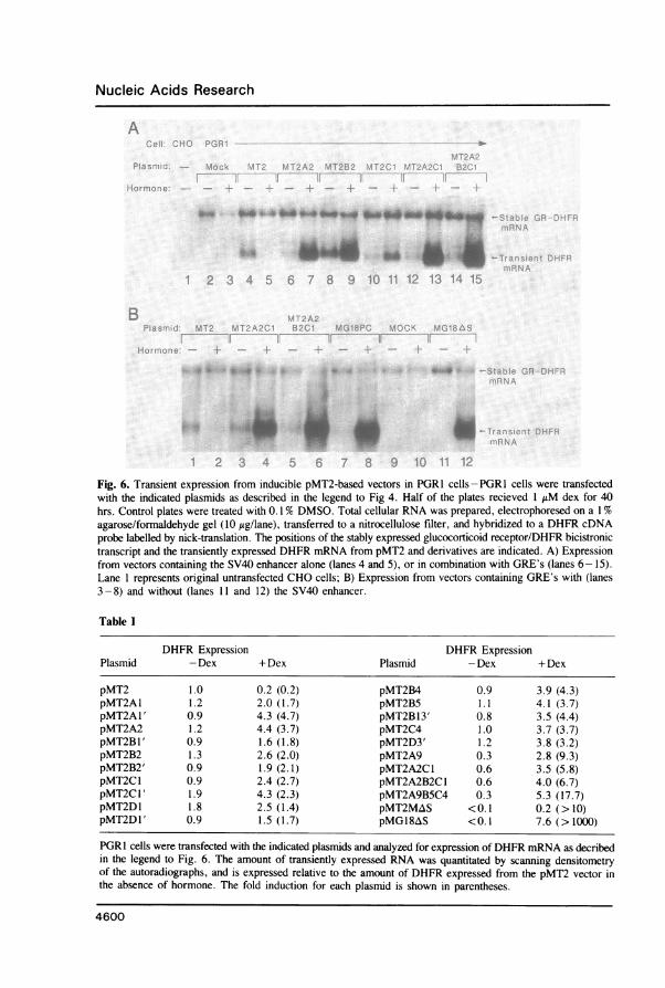
monomer or multimer to various BamHI and BglII sites within the pMT2 expression vectoras indicated in Figure 5. The different GRE-containing pMT2 derivatives were assayedby measuring DHFR mRNA production after transient transfection of PGR1 cells (Figure6A). The DHFR transcript from the transfected pMT2 test plasmid is smaller than theGR/DHFR transcript expressed from pMT2T-GR in PGR1 cells. Expression from theoriginal pMT2 vector which lacks GRE's is inhibited by dex (Figure 6A, lanes 2 and 3),consistent with the inhibition of the pSV2CAT vector observed previously (Figure 4, lanes9 and 10). Every derivative of pMT2 that contains one or more GRE's plus the SV40enhancer is induced by addition of dex. Furthermore, the maximum expression level afterhormone treatment is approximately five-fold greater than that obtained by constitutiveexpression ofpMT2 in several of the inducible pMT2 derivatives. The highest expressionlevels and induction ratios are obtained with vectors that contained multiple GRE's at severaldifferent sites within the vector.-All vectors that contain the SV40 enhancer are constitutivelyexpressed in the absence of dex, although this basal expression decreases 3-fold when 9GRE's are located between the SV40 enhancer and AdMLP. The results, summarizedin Table I, demonstrate that both hormonal responsiveness and high expression levels are
retained when the SV40 enhancer and MMTV GRE's are combined within the same vector.To reduce the basal level of hormone independent expression, the SV40 enhancer was
deleted from the vector pMG18PC, which contains 18 copies of the GRE fragment, togenerate the vector pMG18AS. Expression of DHFR mRNA from pMG18AS is notdetectable in transfected cells in the absence of dex (Figure 6B, lane 11). However, inthe presence of dex, DHFR mRNA is induced to levels greater than that obtained withthe parental pMT2 vector containing the SV40 enhancer, but lacking GRE's (Figure 6B,compare lanes 1 and 12). Thus, mRNA expression from a vector containing 18 copiesof the MMTV GRE but lacking a constitutive enhancer is not detectable in the absenceof dex and in the presence of dex is induced to levels greater than that observed with theSV40 enhancer alone. By analyzing serial 10-fold dilutions of RNA from induced cells,we estimate the minimum induction to be 1000-fold (data not shown).Stable, Inducible Expression of Heterologous ProteinsThe expression of CAT activity (Figure 4) and DHFR mRNA (Figure 6) demonstratesthat hormonal responsiveness is retained in transiently transfected plasmids that containGRE's. We next determined if high expression and induction levels are retained upon stableintegration and amplification of these vectors into the host chromosome. In order to generatea GR expressing CHO cell line which can be used as a host for DHFR co-transfectionand co-amplification of glucocorticoid responsive heterologous genes, a second GRexpression vector, pMT3GR-SVA (Figure 1B), was constructed to lack DHFR, and containa second transcription unit directing Ada expression. CHO cells were transfected withpMT3GR-SVA (Figure 1B), selected for Ada expression, and subsequently selected for
4597

Nucleic Acids Research
HIII SII +1
paLTR2 _
- - ~~~~~~~~~~~~~BIIEl,-'- syndetic oligo
~~~~ +~~~~~~TCTAGACIrAATGTCr
Ligate with pSP6cut with SmaI &EcoRI
4/I BII EIsynthetic oligo , I- I
:.. G A TCI.,.''7:AlT A
IUwl I1TGncr
pSP64-225:-50
HIII + BII
SV40
d-lactamase
ColElOrigin
HIll Sal BII PI iXI
BI + BII
,BIi®
4598
'01.1

Nucleic Acids Research
amplification in increasing concentrations of the Ada inhibitor dCF. Several cell lines,termed GRA, were found to express GR RNA and GR protein at levels comparable toPGR1 cells (Table H). Since GRA cells are DHFR deficient, they can be used as a hostfor selection and amplification of heterologous genes with DHFR. GRA cells weretransfected with a vector containing a dex-inducible transcription unit encoding either tPAor CSF-1 with a DHFR coding region within the 3' portion of the bicistronic mRNA,and were selected for stable expression of DHFR. While translation ofDHFR in this positionis inefficient relative to the upstream open reading frame (28), it is sufficient for the selectionof stable cell lines for growth in nucleoside-free medium. Cells were selected for DHFRexpression in the absence of dex by virtue of the basal expression that occurs with vectorscontaining both the SV40 enhancer and GRE's (Figure 6, Table I). A majority of celllines assayed for either tPA or M-CSF activity showed a moderate (2-5 fold) increasein secretion of these proteins after a 24 hr induction by dex (data not shown). Severalcell lines possessed higher induction responses to dex, or expressed the heterologous proteinat a high level, as shown in Table III. Furthermore, amplification of the inducible genesby selection for increased methotrexate resistance resulted in cells that retained inducibilityat higher absolute expression levels (Table IH). Thus, stable, inducible expression ofamplified genes at relatively high levels can be achieved with this system.
DISCUSSIONWe have developed an inducible host-vector system for mammalian cells suitable for eithertransient or stable expression of heterologous genes at high levels. Stable CHO cell linesthat are competent for glucocorticoid-regulated gene transcription were established by highlevel expression of a rat GR clone. Our results with transient DNA transfection into thesecell lines demonstrated that addition of multiple GRE's to the AdMLP yields 1000-foldinducible expression to mRNA levels 7-fold greater than that observed by addition of anSV40 enhancer alone to the AdMLP. Addition of both the SV40 enhancer and GRE'sincreased basal expression with little effect on the maximal induced level of expression.Upon stable integration of vectors containing both GRE's and the SV40 enhancer intothe chromosome, cell lines can be obtained which exhibit dex-responsiveness. We arepresently studying the inducibility of stably integrated vectors lacking the SV40 enhancer.
Several laboratories have demonstrated that expression of a steroid receptor cDNA cloneconfers hormonal responsiveness to a previously nonresponsive cell (16, 18, 19, 44). We
Fig. 5. Construction of inducible derivatives of the pMT2 expression vector-A 175 bp fragment correspondingto nucleotides -225 through -50 relative to the start site ofRNA transcription in the MMTV LTR was excisedfrom pSP64-225: -50 by digestion with BgIlI and either HinDEIl or BamHI, and was purified by extraction afteragarose gel electrophoresis. This fragment contains a portion of the MMTV LTk that has three binding sitesfor the activated glucocorticoid receptor (Payvar et al., 1983). The GRE-containing fragment was ligated intopMT2 at the indicated positions. In order to obtain multimers of the LTR fragment, the BamHI to BgIII fragmentwas ligated to itself, digested with BamHI and BglIH, gel purified, and ligated with pMT2. Various restrictionfragments from the inducible pMT2 derivatives containing one or more LTR inserts at a single site were thencombined to yield vectors with LTR sequences at several sites. Derivatives of pMT2 were named by indicatingthe location (A-D) and number of the GRE element within pMT2. In some cases, the orientation of the GREfragment(s) within the expression vector is reversed relative to its orientation within the MMTV LTR, and isindicated with an asterisk. In pMT2MAS, the SV40 enhancer was removed and replaced with the HinDHI toBgIl fragment from pSP64-225: -50. The location of recognition sites for the endonucleases BamHI (BI), Bgil(BII), EcoRI (El), HinDuI (HUI), PstI (PI), SacI (SI), Sall (Sal), and Xbal (XI) are indicated.
4599
Nucleic Acids Research
A
B
U
Fig. 6. Transient expression from inducible pMT2-based vectors in PGR 1 cells - PGR1 cells were transfectedwith the indicated plasmids as described in the legend to Fig 4. Half of the plates recieved 1 jiM dex for 40hrs. Control plates were treated with 0. 1% DMSO. Total cellular RNA was prepared, electrophoresed on a 1%agarose/formaldehyde gel (10 jig/lane), transferred to a nitrocellulose filter, and hybridized to a DHFR cDNAprobe labelled by nick-translation. The positions of the stably expressed glucocorticoid receptor/DHFR bicistronictranscript and the transiently expressed DHFR mRNA from pMT2 and derivatives are indicated. A) Expressionfrom vectors containing the SV40 enhancer alone (lanes 4 and 5), or in combination with GRE's (lanes 6-15).Lane 1 represents original untransfected CHO cells; B) Expression from vectors containing GRE's with (lanes3-8) and without (lanes 11 and 12) the SV40 enhancer.
Table I
DHFR Expression DHFR ExpressionPlasmid - Dex + Dex Plasmid - Dex + Dex
pMT2 1.0 0.2 (0.2) pMT2B4 0.9 3.9 (4.3)pMT2A1 1.2 2.0 (1.7) pMT2BS 1.1 4.1 (3.7)pMT2Al' 0.9 4.3 (4.7) pMT2B13' 0.8 3.5 (4.4)pMT2A2 1.2 4.4 (3.7) pMT2C4 1.0 3.7 (3.7)pMT2B1' 0.9 1.6 (1.8) pMT2D3' 1.2 3.8 (3.2)pMT2B2 1.3 2.6 (2.0) pMT2A9 0.3 2.8 (9.3)pMT2B2' 0.9 1.9 (2. 1) pMT2A2C1 0.6 3.5 (5.8)pMT2Cl 0.9 2.4 (2.7) pMT2A2B2C1 0.6 4.0 (6.7)pMT2Cl' 1.9 4.3 (2.3) pMT2A9BSC4 0.3 5.3 (17.7)pMT2D1 1.8 2.5(1.4) pMT2MAS <0.1 0.2(>10)pMT2Dl' 0.9 1.5 (1.7) pMG18AS <0.1 7.6 (>1000)
PGR1 cells were transfected with the indicated plasmids and analyzed for expression of DHFR mRNA as decribedin the legend to Fig. 6. The amount of transiently expressed RNA was quantitated by scanning densitometryof the autoradiographs, and is expressed relative to the amount of DHFR expressed from the pMT2 vector inthe absence of hormone. The fold induction for each plasmid is shown in parentheses.
4600
Ab-,A ak,&&.&.
W. -s::
- .;-..' ..'
II .1
-.i
.7

Nucleic Acids Research
Table H
Cell Line Kd Binding Capacity
CHO DUKX 2.1 x 10⁻⁹M 160 fMoles/mg proteinPGR1 6.6x 10⁻⁹M 2700 fMoles/mg proteinGRA1 (0.1 dCF) 6.x 10-9M 1200 fMoles/mg proteinGRAI (1.0 dCF) 5.4x 10⁻⁹M 3500 fMoles/mg proteinGRA2 (1.0 dCF) 5.8 x 10⁻⁹M 3000 fMoles/mg proteinGRA8 (1.0 dCF) 4.5 x 10⁻⁹M 2400 fMoles/mg proteinMl. 19 (HTC cells) 6.4x 10⁻⁹M 600 fMoles/mg protein
Cytosol was incubated with various concentrations of [3H]dexamethasone, and extracted with dextran-coatedcharcoal. Specific binding was determined as described in the legend to Fig. 3, and used to determine bindingaffinity and capacity by the method of Scatchard.
have extended these initial observations by expressing the glucocorticoid receptor proteinat high levels using efficient, amplifiable vectors. In addition, a series of expression vectorswere developed that show both high induction ratios and maximum expression levels incells containing amplified receptors.
In CHO cells, endogenous glucocorticoid receptor is present at low levels, and failsto detectably induce transcription from the MMTV LTR in a transient assay system. Otherestablished cell lines retain some glucocorticoid responsiveness, apparently due to thepresence of endogenous functional glucocorticoid receptor (1, 3, 45-48). It has previouslybeen reported that CHOki cells contain functional glucocorticoid receptors (46). We havealso observed a low level of inducible expression in a stable G-418-resistant CHO cellline cotransfected with a MMTV-based CAT vector and an SV40-based Neo vector (un-published data). We do not know why the MMTV LTR fails to respond to hormone ina transient assay, yet possesses some inducibility when integrated. Due to its weak pro-moter strength in CHO cells, the MMTV LTR may need to be integrated adjacent to eitheran endogenous enhancer or an enhancer element on the cotransfected plasmid in orderto detect induced expression.
The recombinant glucocorticoid receptor protein stably expressed in CHO cells is similarby gel analysis to the purified receptor from rat liver (41). The binding affinity of dexis indistinguishable from endogenous receptor (39, 42). In addition, the ED^5 for inductionof tPA or M-CSF activity by dex in the cell lines shown in Table Im is approximately10-9M (unpublished data), which is very similar to the biological responsiveness observedwith endogenous receptor from several cell lines (5, 12, 39, 43). Thus, by all criteriatested, the overproduced glucocorticoid receptor protein is identical to endogenous receptor
Table HI
0 MTX .005 MTX .02 MTX 0.1 MTXCell Line - + - + - + - +
GlF39cl 24 258 63 673 192 1289 -(tPA -units/mi)GlM18c3 0.11 0.33 - 1.53 2.88 6.5 15.0(CSF-1I- ig/ml)
Cell lines were selected to the indicated concentration (juM) of methotrexate (MTX) resistance. Expression oftPA activity or CSF 1 protein in the presence (+) or absence (-) of dex was determined as described in Materialsand Methods.
4601

Nucleic Acids Research
present in liver or tissue culture cells. The amount of receptor expressed in the PGRland GRA cell lines ( 3pMoles/mg cytosoloic protein) is approximately 5-10 fold higherthan that found in rat liver (42) or hepatoma cell lines (39). These cell lines should providea rich source of glucocorticoid receptor protein for biochemical and functional studies ofglucocorticoid action.Many of the vectors generated by the strategy diagrammed in Figure 5 contain both
the constitutively active SV40 enhancer and one or more copies of the inducible MMTVenhancer. Expression from these vectors in both transient and stable assays demonstratesthat properties of both enhancer elements are retained, since high constitutive expressionis further increased by treatment with glucocorticoids. A decrease in basal expression occurs
only when a large number of GRE's are between the SV40 enhancer and AdMLP. Thisdecrease may result from the physical seperation of the SV40 enhancer from the promoterand not the GRE's per se, since basal expression is not changed when 13 GRE's are atanother site in the plasmid. Thus, the MMTV enhancer does not silence theSV40 enhancerin the absence of hormone, nor does the constitutive activity of theSV40 enhancer obliteratehormone-dependant transcriptional activation by the MMTV enhancer. Therefore, theactions of these two types of enhancer elements appear additive. Many endogenous geneshave similar combinations of constitutive and/or conditional transcription elements (49 -52).The combination of enhancer elements with different functional properties may thereforebe a general strategy for regulating gene expression.The additivity of multiple GRE's in stimulating transcription has been reported previously
(51, 53). We have extended these observations by inserting up to 18 copies of the GREfragment into a single expression vector. While there is a general tendency towards increasedinducible expression with progressively greater numbers of GRE's, there does not appearto be a strict linear relationship between GRE number and transcriptional strength. Thereiteration of binding sites for transcription factors occurs with many cellular and viralgenes (54) and may be one evolutionary mechanism for assuring efficient gene expression.In fact, the 175 bp GRE fragment used here contains three glucocorticoid receptor bindingsites (55), and is contained in both the 5' and 3' LTR's of the MMTV provirus (29).
Several other inducible systems have been developed for expression of heterologous genes.The signals for induction in these other systems include heat shock, double-stranded nucleicacid, heavy metals, and steroid hormones (reviewed in 56). The system presented hereoffers several advantages over these other systems. First, the genetic events involved inglucocorticoid induction of gene transcription are well understood. We have used molecularclones of the cis-acting DNA elements to construct expression vectors that are highlyinducible by hormone. Inducible vectors are available that allow some basal expression,or that are tightly regulated (pMG 1 8AS) by glucocorticoids. We have also developed celllines that express at high levels the transcription factor that permits efficient hormone-dependent expression from these vectors. Secondly, switching to the induced state merelyinvolves the addition of an inexpensive and readily available hormone that is relativelynontoxic to cells. Following addition of the hormone, induction is rapid and persistant.Finally, the inducible vectors can be amplified using methotrexate selection, allowing forstable inducible expression at high levels.We have encountered several limitations with this system. The fold-induction in stable
cell lines is variable, as has been observed previously with integrated MMTV proviruses
(14). Therefore, many cell lines may have to be screened in order to obtain one whichis highly inducible. In addition, the growth rate of CHO cells containing highly amplified
4602

Nucleic Acids Research
receptors is approximately 32% slower in the presence of inducing steroid (unpublishedobservations). We are currently refining several aspects of this system to try to overcomethese limitations.The system described here should have several useful scientific and biotechnological
applications. Stable cell lines containing amplified copies of both hormone receptor genesand inducible genes should be useful for analyzing the molecular events that occur uponglucocorticoid stimulation. This system also offers the opportunity to stably express andstudy proteins that are toxic to the cell. In some systems, it may be desireable to varythe levels of a particular protein in order to study its role in a cell structure or function.By using different concentrations of hormone, we can easily vary the level of expressionobtained with the inducible vectors (unpublished data). It will also be possible to studygene activation and RNA processing in finer detail, since addition of hormone shouldsynchronously and rapidly turn on gene transcription. The early events ofRNA synthesis,processing, and transport can then be studied as a function of time in cells sychronizedwith regards to transcription of the induced gene. Finally, this system may be of use forthe efficient production of proteins from heterologous genes.
ACKNOWLEDGEMENTSWe are deeply indebted to Roger Miesfeld and Keith Yamamoto for providing us withpRBall17, paLTR2, and the BUGRI monoclonal antibody. We thank Andrew Dornerand Robert Kamen for their carefull review of this manuscript, and the Drug Synthesisand Chemistry Branch, Division of Cancer Treatment, National Cancer Institute for theirgenerous gift of alanosine.*To whom correspondence should be addressed
REFERENCES1. Mayo, K. and Palmiter, R. D. (1981) J. Biol. Chem. 256, 2621-2624.2. Evans, R.M., Birnberg, N.C. and Rosenfeld, M.G. (1982) Proc. Natl. Acad. Sci. USA 79, 7659-7663.3. Robins, D. M., Paek, I., Seeburg, P. H. and Axel, R. (1982) Cell 29, 623-631.4. Danesch, U., Hashimoto, S., Renkawitz, R. and Schutz, G. (1983) J. Biol. Chem. 258, 4750-4753.5. Cwikel, B. J., Barouski-Miller, P. A., Coleman, P. L. and Gelehrter, T. D. (1984) J. Biol. Chem. 259,
6847-6851.6. Hashimoto, S., Schmid, W. and Schutz, G. (1984) Proc. Natl. Acad. Sci. USA 81, 6637-6641.7. Nakanishi, S., Kita, T., Taii, S., Imura, H. and Numa, S. (1977) Proc. Natl. Acad. Sci. USA 74, 3283-3286.8. Guertin, M., Baril, P., Bartkowiak, J., Anderson, A. and Belanger, L. (1983) Biochemistry 22, 4296-4302.9. Camper, S. A., Yao, Y. A. S. and Rottman, F. M. (1985) J. Biol. Chem. 260, 12246-12251.
10. Okret, S., Poellinger, L., Dong, Y. and Gustafsson, J. A. (1986) Proc. Natl. Acad. Sci. USA 83, 5899-5903.11. Akerblom, I. E., Slater, E. P., Beato, M., Baxter, J. D. and Mellon, P. L. (1988) Science, 241, 350-353.12. Adler, G.K., Smas, C.M. and Majzoub, J.A. (1988) J. Biol. Chem. 263, 5846-5852.13. Lee, S. W., Tsou, A., Chan, H., Thomas, J., Petrie, K., Eugui, E. M. and Allison, A. C. (1988) Proc.
Natl. Acad. Sci. USA 85, 1204-1208.14. Ringold, G. M., Shank, P. R., Varmus, H. E., Ring, J. and Yamamoto, K. R. (1979) Proc. Natl. Acad.
Sci. USA 76, 665-669.15. Firestone, G.L., Payvar, F. and Yamamoto, K.R. (1982) Nature 300, 221-225.16. Miesfeld R., Rusconi, S., Godowski P. J., Maler, B. A., Okret, S., Wikstrom, A., Gustafsson, J. and
Yamamoto, K. R. (1986) Cell 46, 389-399.17. Hollenberg, S. M., Weinberger, C., Ong, E. S., Cerelli, G., Oro, A., Lebo, R., Thompson, E. B., Rosenfeld,
M. G. and Evans, R. M. (1985) Nature 318, 635-641.18. Danielsen, M., Northrop, J. P. and Ringold, G. M. (1986) EMBO J. 10, 2513-2522.19. Giguere, V., Hollenberg, S. M., Rosenfeld, M. G. and Evans, R. M. (1986) Cell 46, 645-652.20. Godowski, P. J., Rusconi, S., Miesfeld, R. and Yamamoto, K. R. (1987) Nature 325, 365-368.21. Rusconi, S. and Yamamoto, K. R. (1987) EMBO J. 6, 1309-1315.22. Picard, D. and Yamamoto, K. R. (1987) EMBO J. 6, 3333-3340.
4603

Nucleic Acids Research
23. Denis, M., Wilkstrom, A. and Gustafsson, J. (1987) J. Biol. Chem. 262, 11803-11806.24. Payvar, F., Wrange, O., Carlstedt-Duke, J., Okret, S., Gustafsson, J. and Yamamoto, K. R. (1981) Proc.
Natl. Acad. Sci. USA 78, 6628-6632.25. Karin, M., Haslinger, A., Holtgreve, H., Richards, R. I., Krauter, P., Westphal, H. M. and Beato, M.
(1984) Nature 308, 513-519.26. Jantzen, H. M., Strahle, U., Gloss, B., Stewart, F., Schmid, W., Boshart, M., Miksicek, R. and Schutz,
G. (1987) Cell 49, 29-38.27. Bonthron, D. T., Handin, R. I., Kaufman, R. J., Wasley, L. C., Orr, E. C., Mitsock, L. M., Ewenstein,
B., Loscalzo, J., Ginsburg, D. and Orkin, S. H. (1986) Nature 324, 270-273.28. Kaufman, R. J., Murtha, P. and Davies, V. (1987) EMBO J. 6, 187-193.29. Majors, J. E. and Varmus, H. E. (1981) Nature 289, 253-258.30. Kaufman, R. J., Wasley, L. C., Spiliotes, A. J., Gossels, S. D., Latt, S. A., Larsen, G. R. and Kay, R.
M. (1985) Mol. Cell. Biol. 5, 1750-1759.31. Wong, G. G., Temple, P. A., Leary, A. C., Witek-Giannotti, J. S., Yang, Y., Ciarletta, A. B., Chung,
M., Murtha, P., Kriz. R., Kaufman, R. J., Ferenz, C. R., Sibley, B.S., Turner, K. J., Hewick, R. M.,Clark, S. C., Yanai, N., Yokota, H., Yamada, M., Saito, M., Motoyoshi, K. and Takaku, F. (1987) Science235, 1504-1508.
32. Kaufman, R. J., Murtha, P., Ingolia, D. E., Yeung, C. and Kellems, R. E. (1986) Proc. Natl. Acad. Sci.USA 83, 3136-3140.
33. Neumann, E., Schaefer-Ridder, M., Wang, Y. and Hofschneider, P. H. (1982) EMBO J. 1, 841-845.34. Oprian, D. D., Molday, R. S., Kaufman, R. J. and Khorana, G. (1987) Proc. Natl. Acad. Sci. USA 84,
8874-8878.35. Gorman, C. M., Moffat, L. F. and Howard, B. H. (1982) Mol. Cell. Biol. 2, 1044-1051.36. Chirgwin,J.M., Przbyla,A.E., MacDonald, R.J. and Rutter, W.J. (1979) Biochemistry 18, 5294-5299.37. Thomas, P. S. (1980) Proc. Natl. Acad. Sci. USA 77, 5201-5205.38. Eisen, L. P., Reichman, M. E., Thompson, E. B., Gametchu, B., Harrison, R. W. and Eisen, H. J. (1985)
J. Biol. Chem. 260, 11805-11810.39. Baxter, J. D. and Tomkins, G. M. (1971) Proc. Natl. Acad. Sci. USA 68, 932-937.40. Scatchard, G. (1949) Ann. N. Y. Acad. Sci. 51, 660-672.41. Wrange, O., Okret, S., Radojcic, M., Carlstedt-Duke, J. and Gustafsson, J. (1984) J. Biol. Chem. 259,
4534-4541.42. Beato, M. and Feigelson, P. (1972) J. Biol. Chem. 247, 7890-7896.43. DeFranco, D., Wrange, O., Merryweather, J. and Yamamoto, K. R. (1985) In I. Herskowitz and M. Simon,
(eds.). Genome Rearrangement, UCLA Symposium on Molecular and Cellular Biology, New Series, AlanR. Liss, Inc., New York, pp. 305-321.
44. Gronemeyer, H., Turcotte, B., Quirin-Stricker, C., Bocqvel, M.T., Meyer, M.E., Krozowski, Z., Jeltsch,J.M., Lerouge, T., Gamier, J.M. and Chambon, P (1987) EMBO J 6, 3985-3994.
45. Sibley, C.H. and Tomkins, G.M. (1974) Cell 2, 213-220.46. Lee, F., Mulligan, R., Berg, P. and Ringold, G. (1981) Nature 294, 228-232.47. Chapman, A.B., Costello, M.A., Lee, F. and Ringold, G.M. (1983) Mol. Cell. Biol. 3, 1421-1429.48. Ostrowski, M.C., Richard-Foy, H., Wolford, R.G., Berard, D.S. and Hager, G.L. (1983) Mol. Cell. Biol
3, 2045-2057.49. Jones, P. B. C., Galeazzi, D. R., Fisher, J. M. and Whitlock, J. P. Jr. (1985) Science 227, 1499-1502.50. Wu, B. J., Kingston, R. E. and Morimoto, R. I. (1986) Proc. Nati. Acad. Sci. USA 83, 629-633.51. Strahle, U., Schmid, W. and Schutz, G. (1988) EMBO J. 11, 3389-3395.52. Guertin, M., LaRue, H., Bernier, D., Wrange, O., Chevrette, M., Gingras, M. and Belanger, L. (1988)
Mol.Cell. Biol. 8, 1398-1407.53. Toohey, M. G., Morley, K. L. and Peterson, D. 0. (1986) Mol. Cell. Biol. 6, 4526-4538.54. Gluzman, Y. and Shenk, T. (1983) Enhancers and Eukaryotic Gene Expression. Cold Spring Harbor
Laboratory, USA.55. Payvar, F., DeFranco, D., Firestone, G. L., Edgar, B., Wrange, O., Okret, S., Gustafsson, J. and Yamamoto,
K. R. (1983) Cell 35, 381-392.56. Kaufman, R. J. (1987) In Setlow, J. K. (ed.). Genetic Engineering-Principles and Methods. Plenum Press,
New York, Vol. 9. pp. 155-198.
This article, submitted on disc, has been automaticallyconverted into this typeset format by the publisher.
4604
Copyright © 2022 FDOKUMEN