Late Onset Death of Motor Neurons in Mice Overexpressing Wild-Type Peripherin
14
The Rockefeller University Press, 0021-9525/99/11/531/14 $5.00 The Journal of Cell Biology, Volume 147, Number 3, November 1, 1999 531–544 http://www.jcb.org 531 Late Onset Death of Motor Neurons in Mice Overexpressing Wild-Type Peripherin Jean-Martin Beaulieu, Minh Dang Nguyen, and Jean-Pierre Julien Centre for Research in Neurosciences, McGill University, The Montréal General Hospital Research Institute, Montréal, Québec, H3G 1A4, Canada Abstract. Peripherin, a type III intermediate filament (IF) protein, upregulated by injury and inflammatory cytokines, is a component of IF inclusion bodies associ- ated with degenerating motor neurons in sporadic amyotrophic lateral sclerosis (ALS). We report here that sustained overexpression of wild-type peripherin in mice provokes massive and selective degeneration of motor axons during aging. Remarkably, the onset of peripherin-mediated disease was precipitated by a defi- ciency of neurofilament light (NF-L) protein, a phe- nomenon associated with sporadic ALS. In NF-L null mice, the overexpression of peripherin led to early- onset formation of IF inclusions and to the selective death of spinal motor neurons at 6 mo of age. We also report the formation of similar peripherin inclusions in presymptomatic transgenic mice expressing a mutant form of superoxide dismutase linked to ALS. Taken to- gether, these results suggest that IF inclusions contain- ing peripherin may play a contributory role in motor neuron disease. Key words: peripherin • neurofilament • ALS • mo- tor neuron • transgenic A MYOTROPHIC lateral sclerosis (ALS) 1 is an adult-onset neurological disorder that affects primarily motor neurons in the brain and spinal cord. The degener- ation of motor neurons leads to atrophy of skeletal mus- cles and, ultimately, to paralysis and death. Approximately 10% of the ALS cases are familial with the disease inher- ited in an autosomal dominant manner. Mutations in the copper/zinc superoxide dismutase (SOD1) gene have been found in z20% of familial cases (2% of total ALS cases) (Rosen et al., 1993). Whereas transgenic mouse studies provided compelling evidence that SOD1 mutations cause ALS through a gain of deleterious activities (Gurney et al., 1994; Wong et al., 1995; Ripps et al., 1995; Tu et al., 1996; Bruijn et al., 1997, 1998), the etiology of ALS remains un- known for the vast majority of sporadic and familial cases. One hallmark of ALS is the presence of cytoplasmic inclu- sions of disorganized proteins in the cytoplasm of motor neurons (reviewed in Chou, 1995). Some inclusions have been shown to contain mutant SOD1 protein (Kato et al., 1996; Shibata et al., 1996; Bruijn et al., 1998), whereas oth- ers contain neuronal intermediate filament (IF) proteins (Carpenter, 1968; Tu et al., 1996). Peripherin is a type III neuronal IF protein of 57 kD which is found with neurofilament (NF) proteins in the majority of IF inclusions (89%) in motor neurons of ALS patients (Corbo and Hays, 1992; Migheli et al., 1993; Tu et al., 1996). In adults, peripherin is mostly expressed in auto- nomic nerves and in peripheral sensory neurons with only low levels detectable in spinal motor neurons (Parysek and Goldman, 1988; Brody et al., 1989; Escurat et al., 1990; Troy et al., 1990a,b). However, peripherin gene expression is increased by up to 300% in spinal motor neurons after injury of the sciatic nerve (Troy et al., 1990a), whereas ex- pression of all three NF subunits mRNAs is reduced (Muma et al., 1990). Inflammation is one mechanism that can explain the upregulation of peripherin in injured neu- rons. In fact, peripherin gene expression is upregulated by the inflammatory cytokines interleukin-6 (IL-6) and leu- kemia inhibitory factor (LIF; Djabali et al., 1993; Sterneck et al., 1996) through the action of a JAK/STAT signaling pathway activating the STAT 1 and STAT 3 transcription factors (Lecomte et al., 1998). The action of cytokines on peripherin expression suggests that an upregulation of pe- ripherin may be part of a general response of motor neu- rons to noxious stress that may explain the presence of nu- Address correspondence to Dr. Jean-Pierre Julien, The Montréal General Hospital Research Institute, 1650 Cedar Avenue, Montréal, Québec, H3G 1A4, Canada. Tel.: (514) 934-8058. Fax: (514) 934-8265. E-mail: mdju@ musica.mcgill.ca 1. Abbreviations used in this paper: ALS, amyotrophic lateral sclerosis; DRG, dorsal root ganglion; IF, intermediate filament; IL-6, interleukin 6; LIF, leukemia inhibitory factor; NF, neurofilament; NF-H, neurofilament heavy subunit; NF-L, neurofilament light subunit; NF-M, neurofilament medium subunit; SOD1, copper/zinc superoxide dismutase; WT, wild- type. on October 17, 2013 jcb.rupress.org Downloaded from Published November 1, 1999 on October 17, 2013 jcb.rupress.org Downloaded from Published November 1, 1999 on October 17, 2013 jcb.rupress.org Downloaded from Published November 1, 1999 on October 17, 2013 jcb.rupress.org Downloaded from Published November 1, 1999 on October 17, 2013 jcb.rupress.org Downloaded from Published November 1, 1999 on October 17, 2013 jcb.rupress.org Downloaded from Published November 1, 1999 on October 17, 2013 jcb.rupress.org Downloaded from Published November 1, 1999 on October 17, 2013 jcb.rupress.org Downloaded from Published November 1, 1999 on October 17, 2013 jcb.rupress.org Downloaded from Published November 1, 1999 on October 17, 2013 jcb.rupress.org Downloaded from Published November 1, 1999 on October 17, 2013 jcb.rupress.org Downloaded from Published November 1, 1999 on October 17, 2013 jcb.rupress.org Downloaded from Published November 1, 1999 on October 17, 2013 jcb.rupress.org Downloaded from Published November 1, 1999 on October 17, 2013 jcb.rupress.org Downloaded from Published November 1, 1999
-
Upload
independent -
Category
Documents
-
view
0 -
download
0
Transcript of Late Onset Death of Motor Neurons in Mice Overexpressing Wild-Type Peripherin

The Rockefeller University Press, 0021-9525/99/11/531/14 $5.00The Journal of Cell Biology, Volume 147, Number 3, November 1, 1999 531–544http://www.jcb.org 531
Late Onset Death of Motor Neurons in Mice OverexpressingWild-Type Peripherin
Jean-Martin Beaulieu, Minh Dang Nguyen, and Jean-Pierre Julien
Centre for Research in Neurosciences, McGill University, The Montréal General Hospital Research Institute, Montréal, Québec, H3G 1A4, Canada
Abstract.
Peripherin, a type III intermediate filament (IF) protein, upregulated by injury and inflammatory cytokines, is a component of IF inclusion bodies associ-ated with degenerating motor neurons in sporadicamyotrophic lateral sclerosis (ALS). We report here that sustained overexpression of wild-type peripherin in mice provokes massive and selective degeneration of motor axons during aging. Remarkably, the onset ofperipherin-mediated disease was precipitated by a defi-ciency of neurofilament light (NF-L) protein, a phe-nomenon associated with sporadic ALS. In NF-L null mice, the overexpression of peripherin led to early-
onset formation of IF inclusions and to the selective death of spinal motor neurons at 6 mo of age. We also report the formation of similar peripherin inclusions in presymptomatic transgenic mice expressing a mutant form of superoxide dismutase linked to ALS. Taken to-gether, these results suggest that IF inclusions contain-ing peripherin may play a contributory role in motor neuron disease.
Key words: peripherin • neurofilament • ALS • mo-tor neuron • transgenic
A
MYOTROPHIC
lateral sclerosis (ALS)
1
is an adult-onsetneurological disorder that affects primarily motorneurons in the brain and spinal cord. The degener-
ation of motor neurons leads to atrophy of skeletal mus-cles and, ultimately, to paralysis and death. Approximately10% of the ALS cases are familial with the disease inher-ited in an autosomal dominant manner. Mutations in thecopper/zinc superoxide dismutase (SOD1) gene have beenfound in
z
20% of familial cases (2% of total ALS cases)(Rosen et al., 1993). Whereas transgenic mouse studiesprovided compelling evidence that SOD1 mutations causeALS through a gain of deleterious activities (Gurney et al.,1994; Wong et al., 1995; Ripps et al., 1995; Tu et al., 1996;Bruijn et al., 1997, 1998), the etiology of ALS remains un-known for the vast majority of sporadic and familial cases.One hallmark of ALS is the presence of cytoplasmic inclu-sions of disorganized proteins in the cytoplasm of motor
neurons (reviewed in Chou, 1995). Some inclusions havebeen shown to contain mutant SOD1 protein (Kato et al.,1996; Shibata et al., 1996; Bruijn et al., 1998), whereas oth-ers contain neuronal intermediate filament (IF) proteins(Carpenter, 1968; Tu et al., 1996).
Peripherin is a type III neuronal IF protein of 57 kDwhich is found with neurofilament (NF) proteins in themajority of IF inclusions (89%) in motor neurons of ALSpatients (Corbo and Hays, 1992; Migheli et al., 1993; Tu etal., 1996). In adults, peripherin is mostly expressed in auto-nomic nerves and in peripheral sensory neurons with onlylow levels detectable in spinal motor neurons (Parysekand Goldman, 1988; Brody et al., 1989; Escurat et al., 1990;Troy et al., 1990a,b). However, peripherin gene expressionis increased by up to 300% in spinal motor neurons afterinjury of the sciatic nerve (Troy et al., 1990a), whereas ex-pression of all three NF subunits mRNAs is reduced(Muma et al., 1990). Inflammation is one mechanism thatcan explain the upregulation of peripherin in injured neu-rons. In fact, peripherin gene expression is upregulated bythe inflammatory cytokines interleukin-6 (IL-6) and leu-kemia inhibitory factor (LIF; Djabali et al., 1993; Sternecket al., 1996) through the action of a JAK/STAT signalingpathway activating the STAT 1 and STAT 3 transcriptionfactors (Lecomte et al., 1998). The action of cytokines onperipherin expression suggests that an upregulation of pe-ripherin may be part of a general response of motor neu-rons to noxious stress that may explain the presence of nu-
Address correspondence to Dr. Jean-Pierre Julien, The Montréal GeneralHospital Research Institute, 1650 Cedar Avenue, Montréal, Québec, H3G1A4, Canada. Tel.: (514) 934-8058. Fax: (514) 934-8265. E-mail: [email protected]
1.
Abbreviations used in this paper:
ALS, amyotrophic lateral sclerosis;DRG, dorsal root ganglion; IF, intermediate filament; IL-6, interleukin 6;LIF, leukemia inhibitory factor; NF, neurofilament; NF-H, neurofilamentheavy subunit; NF-L, neurofilament light subunit; NF-M, neurofilamentmedium subunit; SOD1, copper/zinc superoxide dismutase; WT, wild-type.
on October 17, 2013
jcb.rupress.orgD
ownloaded from
Published November 1, 1999
on October 17, 2013
jcb.rupress.orgD
ownloaded from
Published November 1, 1999
on October 17, 2013
jcb.rupress.orgD
ownloaded from
Published November 1, 1999
on October 17, 2013
jcb.rupress.orgD
ownloaded from
Published November 1, 1999
on October 17, 2013
jcb.rupress.orgD
ownloaded from
Published November 1, 1999
on October 17, 2013
jcb.rupress.orgD
ownloaded from
Published November 1, 1999
on October 17, 2013
jcb.rupress.orgD
ownloaded from
Published November 1, 1999
on October 17, 2013
jcb.rupress.orgD
ownloaded from
Published November 1, 1999
on October 17, 2013
jcb.rupress.orgD
ownloaded from
Published November 1, 1999
on October 17, 2013
jcb.rupress.orgD
ownloaded from
Published November 1, 1999
on October 17, 2013
jcb.rupress.orgD
ownloaded from
Published November 1, 1999
on October 17, 2013
jcb.rupress.orgD
ownloaded from
Published November 1, 1999
on October 17, 2013
jcb.rupress.orgD
ownloaded from
Published November 1, 1999
on October 17, 2013
jcb.rupress.orgD
ownloaded from
Published November 1, 1999

The Journal of Cell Biology, Volume 147, 1999 532
merous peripherin inclusion bodies in motor neurons ofALS patients.
In contrast to NFs, which are obligate heteropolymers ofthree subunits (Ching and Liem, 1993; Lee et al., 1993),peripherin can self-assemble to establish an IF network incultured cells (Cui et al., 1995; Ho et al., 1995; Beaulieu etal., 1999). Peripherin can also interact in vivo with NF pro-teins (Parysek et al., 1991) and can form heterodimerswith each one of the three NF subunits in vitro (Athlanand Mushynski, 1997). We have recently studied the inter-actions of peripherin with NF proteins in cultured SW13cells devoid of endogenous cytoplasmic IF proteins (Beau-lieu et al., 1999). Our results showed that peripherin canassemble with NFs as well as with the light NF subunit(NF-L) protein alone to form an IF network. Unexpect-edly, the expression of peripherin with the heavy NF sub-units (NF-H or NF-M) in the absence of NF-L preventedthe formation of an IF network in transfected cells (Beau-lieu et al., 1999).
We have used a transgenic mouse approach to investi-gate the potential detrimental effects of a sustained pe-ripherin overexpression in motor neurons. Our resultsshow that an upregulation of peripherin in mice causes alate-onset and selective motor neuron disease character-ized by the formation of IF inclusions similar to thosefound in human ALS and in mice expressing mutant formof SOD1 linked to ALS. Moreover, the formation of IF in-clusion bodies and the onset of disease were precipitatedby a deficiency in NF-L protein probably through a delete-rious action of the large NF subunits on peripherin organi-zation.
Materials and Methods
Transgenic and Knockout Mice
The DNA constructs used to produce mice expressing the mouse periph-erin gene under the control of the human Thy-1 gene promoter were de-rived as followed. The Thy-1 regulatory sequences were isolated by longrange PCR amplification (XL PCR™ system; Perkin Elmer) from a vector(pBSV; a gift of Dr. Vincent Giguère, McGill University, Montreal) con-taining the complete human Thy-1 gene (GenBank/EMBL/DDBJ acces-sion number M11749) (van Rijs et al., 1985). The 5
9
primer (TGCCCGC-CTGATGAATGCTCATCCGGAATTC) corresponded to the vectorsequence upstream from the Thy-1 locus in pBSV. The 3
9
primer(GGATCCAGGACTGAGATCCCAGAACC) was complementary tothe last 26 bp from the noncoding fraction of the second exon of the hu-man Thy-1 gene. Both primers were purchased from GIBCO-BRL. Theresulting 3.5-kb PCR product was digested with EcoRI to remove thepBSV sequence of the 5
9
primer before its insertion in pBluescript SK
1
(Stratagene) in the EcoRI to EcoRV orientation. The resulting vector in-cluded the promoter, the first exon, the first intron, and the complete 5
9
noncoding region of the second exon of the human Thy-1 gene subclonedupstream of an HindIII site in the multicloning cassette of pBluescriptSK
1
. A 5.9-kb peripherin fragment was then obtained by the Eco47III,EcoRV digestion of a bacteriophage clone containing the complete mouseperipherin locus (a gift from Dr. André Royal, Université de Montréal,Montréal). This fragment was subcloned into the HindIII site of the Thy-1vector. The correct orientation of the peripherin fragment was confirmedby XhoI and ApaI digestion. The resulting construct (TPer) included thehuman Thy-1 gene regulatory sequences followed by the complete codingsequence of the mouse peripherin gene starting at 23 bp upstream of theputative translation start site (Cui et al., 1995; Ho et al., 1995) and ending2.5 kb downstream of the polyadenylation signal (see Fig. 1 A).
The 9.5-kb DNA fragment (Per), used to derive mice expressing themouse peripherin gene under the control of its own promoter element,was obtained by digestion of the original bacteriophage DNA withEcoRV. The Per fragment contained the complete peripherin gene along
with 3.1 kb from the 5
9
and 2.5 kb from the 3
9
flanking genomic sequences(see Fig. 1 A).
The constructs were isolated from their vector sequences by digestionwith endonuclease restriction enzymes followed by purification on aga-rose gels using a Gene Clean II Kit™ (Bio101). The resulting linear DNAfragments were microinjected into one-cell mouse embryos of C57Bl/C3Hgenetic background according to standard procedures (Brinster et al.,1981). The integration of the transgenes into mouse genomic DNA wasconfirmed by Southern blot analysis as described below.
Mice carrying the transgenes in NF-L null background were obtainedby breeding transgenic mice with NF-L knockout mice (L
2/2
mice) (Zhuet al., 1997). The breeding was performed following a two-step procedure.In the first step, F1 transgenic mice were bred with NF-L knockout miceto generate mice carrying the transgene in an L
1
/
2
background. Thesemice were then backcrossed with L
2/2
mice leading to the production ofmice carrying the transgene in L
2/2
background. All peripherin trans-genic mice used in this study were heterozygous for the peripherin trans-genes.
The SOD1
G37R
mice (Wong et al., 1995) were a kind gift of Drs. DonaldL. Price (Johns Hopkins Medical School, Baltimore, MD) and Don Cleve-land (Ludwig Institute, San Diego, CA).
Southern Blot Analysis
Southern blot analysis of genomic DNA isolated from mouse tails was car-ried out following procedures described previously (Côté et al., 1993). A1.8-kb peripherin probe was generated by digestion of the Per DNA con-struct with EcoRI (see Fig. 1 A). This probe was designed for detection ofa 1.8-kb band corresponding to the endogenous peripherin gene while theTPer transgene was detected as a 4.9-kb band. The probe and enzymaticdigestion protocols used to detect the NF-L knockout genotype have beendescribed previously (Zhu et al., 1997). The number of transgene copiesinserted in the genome was estimated by Southern blotting with serial di-lution of DNA extracted from F1 transgenic and nontransgenic mice fromeach line. The relative intensity of the bands was compared with the signaldetected for the endogenous peripherin gene using Gel-pro 2.0.1 (MediaCybernetic) with a Power Macintosh computer.
Northern Blot Analysis
6-mo-old animals were killed by injection of an overdose of chloral hy-drate. Tissues were collected, frozen in liquid nitrogen, and kept at
2
80
8
Cbefore extraction of total RNA using the Trizol reagent™ (GIBCO-BRL). Total RNA (10
m
g except 5
m
g for dorsal root ganglia) were frac-tionated on 1% (wt/vol) agarose-formaldehyde gels, transferred to Genescreen plus™ membranes (NEN Life Science), hybridized, and washed ac-cording to standard procedures (Sambrook et al., 1989). The mRNA spe-cies were detected by autoradiography using Biomax MR™ film (Kodak)in the presence of an intensifying screen.
The actin probe corresponded to the complete cDNA from the rat
b
-actin. The peripherin probe corresponded to the first 538 bp of the periph-erin transcript (GenBank/EMBL/DDBJ accession number X59840). Thisprobe was derived by PCR from mouse genomic DNA using the (AT-GAGCCATCATCACTCGTCGGGCC) 5
9
primer and the (TCTGCT-TGAGCGCCGCTAGGTCCT) 3
9
primer. The NF-L probe was derivedby PCR of the mouse NF-L first exon (GenBank/EMBL/DDBJ accessionnumber M20480) using the (ATGAGTTCGTTCGGCTCGGATCC-GAT) 5
9
primer and the (CCTCATAGCGAGCCTGCAGGTTGCG) 3
9
primer. The primers used to derive these two probes were purchased fromGIBCO-BRL and the PCR reactions were carried out using Taq poly-merase (Pharmacia). All probes were purified on agarose gels and labeledusing the random primer method before hybridization (Feinberg and Vo-gelstein, 1983).
Antibodies
The anti-peripherin monoclonal antibody (MAB1527), the anti-periph-erin polyclonal antibody (AB1530), and the anti
a
-internexin polyclonalantibody (AB1515) were from Chemicon. The anti-peripherin mono-clonal antibody (NCL-Periph) was from Novocastra Laboratories Ltd.The anti–NF-H polyclonal antibody (N-4142) is from Sigma. The anti–NF-Mmonoclonal antibody (NN-18), the anti–NF-L monoclonal antibody (NR-4),the anti-hyperphosphorylated NF-H monoclonal antibody (RT-97), andthe anti-actin clone c4 monoclonal antibody were from Boehringer Mann-heim. The anti-hypophosphorylated NF-H monoclonal antibody (Smi-32)was from Steinberger Monoclonal Inc.
Beaulieu et al.
Motor Neuron Death Caused by Peripherin Inclusions
533
SDS-PAGE and Western Blot Analysis
6–10-mo-old mice (except if otherwise mentioned in figure legend) werekilled and the tissues collected as for Northern blot analysis. For total pro-tein extraction, tissues were homogenized in 0.5% (wt/vol) SDS, and 8 Murea (SDS/urea buffer). Preparation of the cytoskeletal insoluble proteinswas carried out using a modified version of the method described byChing and Liem (1993). Tissues were first homogenized at 48
8
C in a buffercontaining Tris (10 mM), NaCl (150 mM), EDTA (1 mM), Triton X-100(1% wt/vol), PMSF (2 mM), leupeptin (2 mg/ml), pepstatin (1 mg/ml),and aprotinin (10 mg/ml). The homogenates were then centrifuged at14,000
g
for 15 min at 4
8
C using a Sorvall MC-12V centrifugator (Dupont).The supernatants (soluble fraction) were collected and kept for furtheruse. The pellets (insoluble fraction) were resuspended in a volume ofSDS/urea buffer equivalent to the volume of the soluble fraction. The pro-tein concentration in the different extracts was measured using the DC-Protein assay™, (BioRad). The protein extracts were diluted in 2
3
sam-ple buffer (30% [wt/vol] glycerol, 4% [wt/vol] SDS, 160 mM Tris HCl [pH6.8], 10% [vol/vol]
b
-mercaptoethanol and 0.02% [wt/vol] bromophenolblue) and boiled for 3 min. Proteins were separated on 8% or 10% SDS-PAGE and either transferred to a nitrocellulose membrane or stainedwith 0.2% (wt/vol) Coomassie blue-R250 in a 10% (vol/vol) acetic acidand 45% (vol/vol) methanol solution. Nitrocellulose membranes wereblocked in PBS containing 5% (wt/vol) skimmed milk powder (PBS/milk)before incubation with the primary antibodies diluted as follows:MAB1527, NR-4, and NN-18, 1:1,000 dilution; AB1515 and N-4142, 1:2,000;anti-actin, 1:5,000. After incubation, the blots were rinsed in PBS contain-ing 0.1% (wt/vol) of Tween-20 (PBS/Tween) and incubated for 1 h withthe appropriate secondary antibody (anti–mouse IgG peroxidase or anti–rabbit IgG peroxidase from the Jackson ImmunoResearch Laboratories)diluted 1:1,000 in PBS/milk. The immune complexes were revealed usingthe RenaissanceÙ chemiluminescence reagents (NEN Life Science).
Immunohistological Analysis
Immunohistochemical analysis was carried out as described by Jacomyand Bosler (1995). Mice were anesthetized by injection of chloral hydrateand perfused with a 16-g/liter sodium cacodylate buffer (pH 7.5) followedby fixative (3% [vol/vol] glutaraldehyde in sodium cacodylate buffer). 50-
m
m tissue sections were prepared using a vibratome. Floating sectionswere rinsed in PBS and treated for 30 min with a 1% (wt/vol) sodiumborohydride solution to reduce epitope masking by glutaraldehyde. Sec-tions were then blocked for 1 h in a PBS solution containing 3% (wt/vol)BSA, 0.5% (vol/vol) Triton X-100, and 0.03% (wt/vol) hydrogen perox-ide. Incubation with the various antibodies was performed overnight atroom temperature with agitation in a PBS solution containing 3% (wt/vol)BSA and 0.05% (vol/vol) Triton X-100. The antibody labeling was devel-oped using a Vector ABC kit (Vector Laboratories Ltd.) and Sigmafast™tablets (Sigma).
For indirect double immunofluorescence analysis, mice were perfusedwith PBS (pH 7.5) followed by fixative (4% vol/vol paraformaldehyde in aphosphate buffered solution). Tissues were postfixed for 2 h in paraform-aldehyde, rinsed in PBS, and incubated overnight in a 20% (wt/vol) phos-phate-buffered sucrose solution. 20-
m
m tissue sections were prepared us-ing a cryostat. Sections were then blocked for 30 min in a PBS solutioncontaining 3% (wt/vol) BSA and 0.5% (vol/vol) Triton X-100. Incubationwith mixtures of primary antibodies was performed overnight at roomtemperature in a PBS solution containing 3% (wt/vol) BSA and 0.05%(wt/vol) Triton X-100. Incubation with a mixture of the anti–mouse-FITCand anti–rabbit rhodamine-conjugated antibody (Jackson ImmunoRe-search Laboratory) was carried out for one hour under the same condi-tions. The samples were finally mounted in Prolong™ (Molecular Probe,Inc.) and examined with a fluorescence microscope.
Morphology and Electron Microscopy
Tissue sections were prepared for embedding in Epon as described inZhu et al. (1997). Thin sections were stained with Toluidine blue and ex-amined under a light microscope. The counting of axons in the L5 ventralroot was performed manually from photographs. For electron microscopy,ultrathin sections were stained with a lead citrate solution and examinedwith a Philips CM10 transmission electron microscope.
To evaluate the survival of spinal motor neurons, a portion of the spinalcord spanning 1-mm rostral to 1-mm caudal of the L5 ventral root entrypoint was dissected out from two 14-mo-old mice of each genotype. Thespinal cord was then cut in 50-
m
m sections using a vibratome and stained
with cresyl violet according to standard procedures (Bancroft and Stevens,1990). The large motor neurons in the ventral horn were then countedwith the help of a technician who ignored the genotypes of the mice. Sincethis method did not produce an equal number of sections from each sam-ple, the mean number of motor neurons per ventral horn section was cal-culated and compared for each genotype.
Motor Dysfunction
To evaluate motor dysfunction in the mice, we simply measured their ca-pacity to grasp a vertical grid. The mice were placed at the center of a hor-izontal grid. The grid was then moved slowly to a vertical position. Thetime spent by the mice on the vertical grid was measured for a maximumof 2 min. This test was performed using five mice of each genotype andwas repeated three times on separate days for a total of 15 tests per geno-type.
Results
Generation of Transgenic Mice Overexpressing the Wild-Type Mouse Peripherin Gene
Two different DNA constructs were microinjected to gen-erate transgenic mice overexpressing the wild-type mouseperipherin gene in motor neurons. The first DNA con-struct, called TPer, included the promoter of the Thy-1gene with noncoding sequences of exon 1 and 2 (van Rijset al., 1985) fused to the mouse peripherin gene starting at23 nucleotides upstream from the initiation codon (Fig. 1A). The second DNA construct, called Per, consisted ofthe complete mouse peripherin gene with 3.4 kb of 5
9
and2.5 kb of 3
9
untranslated sequences (Fig. 1 A).Six founders were obtained for each DNA construct.
After Northern blot analysis of nervous tissues, we fo-cused our efforts on the characterization of two lines oftransgenic mice, the TPer line carrying 4 integrated copiesof the TPer construct, and the Per line bearing 20 copies ofthe Per construct (Fig. 1 B).
Northern blot analysis was carried out to study the ex-pression pattern of peripherin mRNA in normal andtransgenic mice. Peripherin mRNA was detected in dorsalroot ganglia (DRG) of normal (WT) mice, TPer trans-genic mice, and Per transgenic mice (Fig. 1 C). PeripherinmRNA was also detected in the brain but not in the cere-bellum of the TPer mice (Fig. 1 C). No peripherin mRNAwas detected in nonnervous tissues of transgenic mice(Fig. 1 C). A combination of immunohistochemistry andWestern blot analysis using anti-peripherin monoclonalantibodies was carried out to further analyze the periph-erin expression patterns in the transgenic lines. Our analy-sis of normal mice allowed the detection of peripherin inDRG, spinal cord, and some sensory fibers of the brainstem. The TPer line showed a larger expression patternwith the detection of peripherin in the brain, in the opticnerve, in some reticular neurons of the brainstem, in themesencephalic trigeminal nucleus, in the motor nucleus ofthe trigeminal and facial nerves, in the nucleus ambiguous,in the ventral and dorsal columns of the spinal cord, in spi-nal motor neurons, and in DRG. The Per mice overex-pressed peripherin in sensory and motor neurons from thespinal cord and brain stem but they did not express detect-able levels of peripherin in the brain.
Densitometry of Western blots, made up by the serialdilution of total protein extracts from dorsal and ventralroots (Fig. 1 D), showed that as compared with WT mice

The Journal of Cell Biology, Volume 147, 1999 534
the peripherin levels were increased by approximately four-fold in the ventral roots of TPer mice, approximately sev-enfold in the ventral roots of Per mice, approximately two-fold in dorsal roots of TPer mice, and approximatelythreefold in dorsal roots of Per mice. An analysis of cy-toskeletal Triton-insoluble extracts from the brain andventral roots revealed a recovery of peripherin in the in-soluble fraction (Fig. 1 E). Densitometry of Coomassieblue–stained SDS-PAGE of Triton-insoluble extracts wasfurther carried out to compare the peripherin levels tothose of NF-L in ventral and dorsal root axons (Fig. 1 Fand Table I). The identification of the bands correspond-ing to NF-M, NF-L, and peripherin was confirmed byWestern blot analysis (Fig. 1 F). The results indicated thatthe levels of peripherin in ventral roots of transgenic miceoverexpressing peripherin remained inferior to NF-L pro-tein levels (Table I).
Extra Peripherin Causes Loss of Ventral Root Axons Late in Life
The peripherin transgenic mice developed normally anddid not exhibit overt phenotypes and pathology until verylate in life. A small number of mice from the TPer and Perlines were maintained for more than two years. After twoyears, the mice from both TPer and Per lines started to de-velop motor dysfunctions. Examination at microscopy andcounting of L5 ventral roots revealed the loss of
z
35% ofmotor axons in aged mice overexpressing peripherin whencompared with their WT littermates (Fig. 2 and Table II).Note that axonal loss occurred predominantly in the ven-tral roots and that DRG axons were essentially spared.Unlike spinal motor neurons from WT mice (Fig. 2 C),most spinal motor neurons from transgenic mice werecharacterized by strong and diffuse peripherin immunore-activity in their perikarya and neurites without evidence of
Figure 1.
Transgenic mice overexpressing WT peripherin.
(A)Partial restriction map of the Per and TPer DNA constructs. AnEco47III
3
EcoRV fragment of the mouse peripherin gene wassubcloned downstream of the human Thy-1 gene promoter.Black boxes represent exons. The white box represents the 5
9
transcriptional regulatory elements of the Thy-1 gene. The poly-Adesignates the polyadenylation signal of the peripherin gene. Theperipherin probe used for the Southern blot analysis is indicated.(B) Southern blot analysis of genomic DNA extracted from thetails of control mice (WT) and of mice bearing the Per
or TPertransgenes. (C) Northern blot showing expression of peripherinmRNA in different tissues of WT, TPer, and Per mice. (D) West-ern blot analysis of total protein extracts showing the increasedperipherin levels in ventral and dorsal roots of TPer and Pertransgenic mice. (E) Western blot analysis of soluble and insolu-ble protein extracts from the brain of WT and transgenic TPermice. Two micrograms of insoluble protein extracts and an equiv-alent volume of soluble protein extracts were fractionated onSDS-PAGE and the blot was incubated with a mixture of mono-clonal antibodies directed against NF-M (M), NF-L (L), periph-erin (P), and actin (A). (F) Coomassie blue–stained SDS-PAGE
of Triton X-100 insoluble fractions (20
m
g) from the ventralroots showing the ratio of the peripherin (57-kD band) to NF-L(66-kD band) in WT and transgenic mouse extracts. R corre-sponds to a track loaded with the Rainbow molecular massmarker (GIBCO). The identity of the bands was confirmed by in-cubating a Western blot (W) of another part of the same gel witha mixture of antibodies against NF-M (M), NF-L (L), and periph-erin (P).
Table I. Relative Protein Levels of Peripherin Compared with NF-L in Cytoskeletal Fractions of Dorsal and Ventral Roots
Tissues GenotypeRatio of
peripherin to NF-L
Ventral root WT (
n
5
5) 0.090
6
0.037TPer (
n
5
3) 0.650
6
0.092Per (
n
5
4) 0.870
6
0.130Dorsal root WT (
n
5
5) 0.316
6
0.164TPer (
n
5
3) 0.590
6
0.095Per (
n
5
4) 1.180
6
0.057
20
m
m of Triton X-100–insoluble fractions were fractionated on SDS-PAGE andstained with Coomassie blue. The OD corresponding to the peripherin and NF-Lbands were then evaluated by densitometry. Ratios were calculated by dividing the pe-ripherin OD by the NF-L OD for each mice. Results are mean
6
SD.

Beaulieu et al.
Motor Neuron Death Caused by Peripherin Inclusions
535
perikaryal swellings (Fig. 2 D). However, some peripherininclusion bodies were observed in neurites especially inaged mice (Fig. 2 D).
Peripherin Overexpression in NF-L Null Background
Because substantial declines in NF-L gene expression oc-curs in ALS (Bergeron et al., 1994), we tested the effect ofreducing NF-L levels on the progression of the peripherin-mediated disease. Mice of the TPer and Per lines werebred with our L
2/2
mice (Zhu et al., 1997) to obtain miceexpressing the peripherin transgenes in a context of NF-Ldeficiency. The breeding resulted in the mendelian trans-mission of the peripherin transgenes and of the L
2/2
geno-type.
Previous results obtained from cell culture studies andfrom L
2/2
mice (Beaulieu et al., 1999; Williamson et al.,1998) suggested that the organization of peripherin mightbe affected by the absence of NF-L. Western blot analysisof spinal cord extracts from the L
2/2
, Per;L
2/2
, andTPer;L
2/2
mice revealed reduced levels of peripherin,NF-H, and NF-M proteins as a consequence of NF-L defi-ciency (Fig. 3 A). Declines in levels of peripherin werealso detected in the brain and optic nerve extracts fromTPer;L
2/2
mice indicating that peripherin produced un-
der the control of the Thy-1 promoter was also affected bythe absence of NF-L (Fig. 3 A). Northern blot analysis ofRNA from the brain and DRG of mice with various geno-types revealed that the NF-L disruption had no effects onperipherin mRNA levels (Fig. 3 B).
Figure 2. Motor axon de-generation and peripherininclusions in aged mice over-expressing wild-type periph-erin. (A and B) Toluidineblue staining of thin sectionsof L5 ventral root axons from28-mo-old mouse littermatesof WT (A) or Per (B) geno-types. (C and D) Immunohis-tochemical detection of pe-ripherin in the spinal cordwith a monoclonal antibody(NCL-Periph 1:200 dilution).No peripherin signal was de-tected in motor neurons ofWT mice (C), whereas mo-tor neurons of transgenicmice exhibited a strong dif-fuse labeling for peripherin intheir perikarya (black arrow-head in D). Peripherin posi-tive inclusions were also ob-served in neurites of spinalmotor neurons (white arrow-head in D). Bars: (B) 100 mm;(D) 25 mm.
Table II. Number of Motor Axons in L5 Ventral Roots of Mice with Different Genotypes
Genotype Age Number of axonsLoss of
motor axons
mo %
WT (
n
5
3) 6 1055
6
64 0WT (
n
5
2) 28 1069
6
76 0L
2/2
(
n
5
3) 6 860
6
52 19L
2/2
(
n
5
3) 20 918
6
40 14Per (
n
5
2) 6 1056
6
45 0Per (
n
5
2) 28 688
6
87 35TPer (
n
5
3) 6 1074
6
54 0TPer (
n
5
1) 28 684 35Per;L
2/2
(
n
5
3) 6 567
6
190 46TPer;L
2/2
(
n
5
2) 2 741
6
14 30TPer;L
2/2
(
n
5
3) 6 563
6
140 47TPer;L
2/2
(
n
5
3) 14 438
6 27 59
Loss of motor axons represents the percentage of missing axons as compared to WT.n represents the number of mice for each category. Results are mean 6 SD.

The Journal of Cell Biology, Volume 147, 1999 536
In addition to NFs, CNS neurons express another typeIV IF protein called a-internexin (Pachter and Liem,1985). We have therefore examined the levels of a-inter-nexin to test whether the downregulation of peripherin inNF-L null mice was due to a general destabilization ofneuronal IF proteins. In contrast to peripherin, the levelsof a-internexin were not affected by the absence of NF-Lin L2/2 and TPer;L2/2 mice as revealed by Western blotanalysis of optic nerve extracts (Fig. 3 C). Then we exam-ined whether the peripherin levels in motor and sensoryneurons were equally affected by the absence of NF-L(Fig. 3 D). Whereas peripherin was readily detected byWestern blot in ventral root extracts of normal mice, thelevels of peripherin and NF proteins were at exceedinglylow levels in mice of the NF-L null background. It is note-worthy that the levels of peripherin were less affected bythe absence of NF-L in dorsal roots than in ventral roots(Fig. 3 D). Electron microscopy of nondegenerating axonsfrom ventral roots of 6-mo-old mice from the Per;L2/2and TPer;L2/2 genotypes revealed that the cytoskeletalorganization of these axons did not greatly differ from theone of motor axons of L2/2 mice. In both cases there wasa scarcity of IF structures with an elevated density of mi-crotubules when compared with ventral root axons frommice of the WT and TPer genotypes (Fig. 4, A–D). In con-trast to ventral root axons, small unmyelinated axons ofdorsal roots in L2/2 and TPer;L2/2 mice showed little orno changes in the density of IF structures (Fig. 4, E–H).This observation is compatible with our Western blot re-sult (Fig. 3 D) and with a report indicating that some un-myelinated sensory axons express peripherin in the ab-sence of NFs (Goldstein et al., 1996).
NF-L Deficiency AcceleratesPeripherin-mediated Disease
Mice with the TPer;L2/2 and Per;L2/2 genotypes devel-oped aberrant hind limb positions and a progressive loss ofhind limb mobility. This motor dysfunction was generallynot apparent before 8 mo of age and often started on oneside before spreading to the other side. In mice of bothgenotypes that were allowed to live until 14 mo of age, theprogression of the phenotypes resulted in paralysis of thedigits and of the lower limb articulations (Fig. 5 A). Thisphenotype prompted us to examine signs of dysfunction inyounger mice. Thus, we measured the ability of 6-mo-oldmice of different genotypes to grasp a vertical grid. Thissimple test revealed that the average grasping time for theTPer;L2/2 mice was reduced by 80% when comparedwith mice with L2/2, TPer, or TPer;L1/2 genotypes(Fig. 5 B).
Light microscopy of L5 dorsal roots from 6-mo-old ani-mals showed no degenerating axons in L2/2, TPer;L2/2,and Per;L2/2 mice (Fig. 6, A–C). In contrast, examina-tion of the ventral roots revealed a massive degenerationof axons in the TPer;L2/2 and Per;L2/2 mice (Fig. 6, Gand I) but not in the other types of mice that were exam-ined at this age (Fig. 6, D, E, F, and H). Further examina-tion of degenerating ventral roots by electron microscopyrevealed the presence of degenerating myelin, axonalsproutings, and occasional axonal filamentous spheroids inthe ventral roots of 6-mo-old Per;L2/2 and TPer;L2/2mice (Fig. 6, J and K).
To evaluate the degree of degeneration, we counted thenumber of axons in the ventral roots of 6-mo-old mice of
Figure 3. Reduced peripherin levels inabsence of NF-L. (A) Western blotanalysis of total protein extracts fromthe spinal cord, brain, and optic nervesof normal and transgenic 6-mo-oldmice. The gels were loaded with 5 mgof protein from the brain and opticnerve or with 20 mg of protein from thespinal cord. Blots were incubated witha polyclonal antibody directed towardNF-H (top) or with a cocktail of mono-clonal antibodies against NF-M, NF-L,peripherin, and actin (bottom). (B)Northern blot analysis of peripherinmRNA from the brain and DRG in thepresence and absence of NF-L. Theblots were hybridized with the periph-erin probe, stripped, rehybridized withthe NF-L probe, stripped again, and re-hybridized with the actin probe. (C)Western blot analysis showing thata-internexin levels are not reduced inNF-L knockout mice. Duplicate gelswere transferred to nitrocellulosemembranes and incubated with eitherthe monoclonal antibody cocktail asdescribed above or with a polyclonalantibody against a-internexin. (D)
Western blot of total protein extracts (5 mg) from ventral and dorsal roots of 4-mo-old mice showing that peripherin levels were less af-fected in dorsal roots than in ventral roots by the absence of NF-L.

Beaulieu et al. Motor Neuron Death Caused by Peripherin Inclusions 537
different genotypes. The results are shown in Table II. Theupregulation of peripherin alone did not affect the numberof motor axons at this age, whereas the L2/2 genotypewas associated with a loss of z16% of motor axons at both6 and 20 mo of age. 6-mo-old mice overexpressing periph-erin in NF-L null background exhibited declines of 45–50% in the number of their motor axons. The progressivenature of the overt phenotypes observed in TPer;L2/2and Per;L2/2 mice prompted us to monitor the loss ofmotor axons in mice of different age. Analysis of ventralroot axons in the TPer;L2/2 mice at 2, 6, and 14 mo ofage revealed a parallel increase of motor dysfunction andloss of motor axons culminating in the loss of 59% of mo-tor axons in paralyzed mice at 14 mo of age (Table II).
To examine whether the massive loss of motor axonswas accompanied by a corresponding death of neurons, wecounted the number of motor neurons in a spinal cord re-gion spanning a 3-mm rostral to caudal region at the levelof L5 ventral roots. These counts were performed onNissl-stained sections (Fig. 7). Two mice from each geno-type were used for the analysis. Since similar results wereobtained for the two mice of each genotype, the data werepooled and the mean number of large motor neuron per
Figure 4. Peripherin does not substitute for NF-L. (A–D) Electron microscopy of large myelinated axons from the ventral root of WT(A), L2/2 (B), TPer (C), and TPerL2/2 (D) mice. Note the increased number of microtubules (white arrows) and the scarcity of IFstructures (black arrows) in mice of L2/2 and TPer;L2/2 genotypes when compared with WT and TPer mice. (E–H) Electron micros-copy of small unmyelinated axons from the dorsal root of either WT (E), L2/2 (F), TPer (G), or TPer;L2/2 (H) mice. Note that someof the null NF-L axons did not develop a scarcity of IF (black arrows). Bars, 0.2 mm.
Figure 5. Motor dysfunction in NF-L null mice overexpressingperipherin. (A) Abnormal hind limb posture in a 14-mo-old
TPer;L2/2 mouse. (B) 6-mo-old TPer;L2/2 mice have reducedgrasping ability to a vertical grid. The results presented in thegraphic are a mean from three separate tests performed on fivemice of each genotype. Error bars correspond to standard devia-tions. **Indicates a significant difference from WT (P , 0.01) ac-cording to a Student’s t test (unpaired, double tail).

The Journal of Cell Biology, Volume 147, 1999 538
ventral horn was calculated (Table III). This analysis re-vealed a dramatic reduction in the mean number of largespinal motor neurons (64% neuronal loss) present in theventral horns of 14-mo-old TPer;L2/2 mice as comparedwith normal, TPer, and L2/2 mice (Fig. 7 and Table III).Moreover, the loss of motor neurons during aging of theTPer;L2/2 mice was progressive since 5-mo-old miceshowed a lower degree of neuronal loss (Table III).
We then verified whether an increased abundance of pe-ripherin inclusions was associated with the acceleration ofthe peripherin-mediated disease in the absence of NF-L. Avery weak peripherin immunostaining was observed in spi-nal motor axons of WT and L2/2 mouse using an anti-peripherin monoclonal antibody (Fig. 8 A). In TPer and Pertransgenic mice having a wild-type NF-L background, agenerally diffused peripherin staining with the occasional
presence of peripherin inclusion bodies was observed inspinal motor neurons (Figs. 8 B and 2 D). Peripherin inclu-sions were more frequent in the proximal axons of motorneurons of TPer;L1/2 and Per;L1/2 mice (Fig. 8 C). Fi-nally, the spinal cord of the TPer;L2/2 and Per;L2/2mice was characterized by the presence of abundant pe-ripherin inclusions in the perikarya and axons of motorneurons and by a general disappearance of diffuse periph-erin staining (Fig. 8, D and E). Similar peripherin inclu-sions were also observed in the motor nucleus of the facialand trigeminal nerves, in the nucleus ambiguous, in largeDRG neurons, and in the mesencephalic trigeminal nu-cleus of the TPer;L2/2 and Per;L2/2 mice (data notshown). In the TPer;L2/2 mice, the peripherin inclusionswere detectable in spinal motor neurons of mice at 2, 6,and 14 mo of age. In thin sections stained with Toluidine
Figure 6. Accelerated degenera-tion of ventral root axons in miceoverexpressing peripherin in ab-sence of NF-L. (A–C) Toluidineblue staining of thin sections of L5dorsal root axons from 6-mo-oldL2/2 (A), TPer;L2/2 (B), andPer;L2/2 (C) did not reveal degen-eration of sensory axons in thesemice. (D–H) No degeneration ofaxons was detected in L5 ventralroots of 6-mo-old WT (D), TPer(F), and Per (H) mice. The rootsfrom 6-mo-old L2/2 mice (E) werecharacterized by an atrophy of ax-ons but did not contained degener-ating axons. In contrast, massive de-generation of axons was evident inroots of 6-mo-old TPer;L2/2 (G)and Per;L2/2 (I) mice. (J and K)Electron microscopy of these sameroots revealed the presence of de-generating axons (black arrow in J),axonal sproutings (white arrows in J),and axonal filamentous spheroids(sph in K) in ventral roots of theTPer;L2/2 (J), and Per;L2/2 (K)mice. The black arrow in K pointsto a mitochondria entrapped inan axonal spheroid. Bars: (A–C)25 mm; (D–I) 100 mm; (J) 5 mm; (K)2 mm.

Beaulieu et al. Motor Neuron Death Caused by Peripherin Inclusions 539
blue, the peripherin inclusions appeared as hyaline inclu-sion bodies with sizes ranging from 2 to 30 mm in length(Fig. 8 F). Multiple distinct inclusion bodies were often de-tected within the same neuron. These inclusions neverfilled completely the perikarya of motor neurons. At elec-tron microscopy, the peripherin inclusions appeared asdisorganized accumulations of 10-nm filaments mingledwith various membranous residues (Fig. 8, G and H). In-clusion bodies containing mitochondria were also fre-quently observed (Fig. 8 H).
Detection of NF-M and NF-H in Peripherin Inclusions
Since cell culture studies indicated that the large NF sub-units can have a destabilizing effect on the organization ofperipherin (Beaulieu et al., 1999), we examined whetherthe NF-M or NF-H proteins were present in the peripherininclusion bodies. Double indirect immunofluorescencewas carried out on spinal cord sections from the TPer;L2/2mice using a peripherin polyclonal antibody and differ-ent monoclonal antibodies against NF-M or NF-H pro-teins. All peripherin inclusions observed in these analysiswere immunoreactive for NF-M, as revealed by the NN-18antibody (Fig. 8 I), and for dephosphorylated NF-H as re-vealed by the SMI-32 antibody (Fig. 8 J). A majority of in-clusions also contained hyperphosphorylated NF-H as re-vealed by their immunoreactivity to the RT-97 antibody(Fig. 8 K). In rare cases, motor neurons showed a diffusepattern of peripherin staining in their perikarya. One suchneuron was encountered in the course of a double labelingexperiment involving the anti–NF-M monoclonal anti-body. It is noteworthy that this neuron, which lacked in-clusion bodies, was also showing a really weak NF-M im-munoreactivity (Fig. 8 L) suggesting that the formation ofperipherin aggregates in motor neurons requires the pres-ence of the large NF subunits.
Early Detection of Peripherin Inclusions in Mice Bearing SOD1G37R
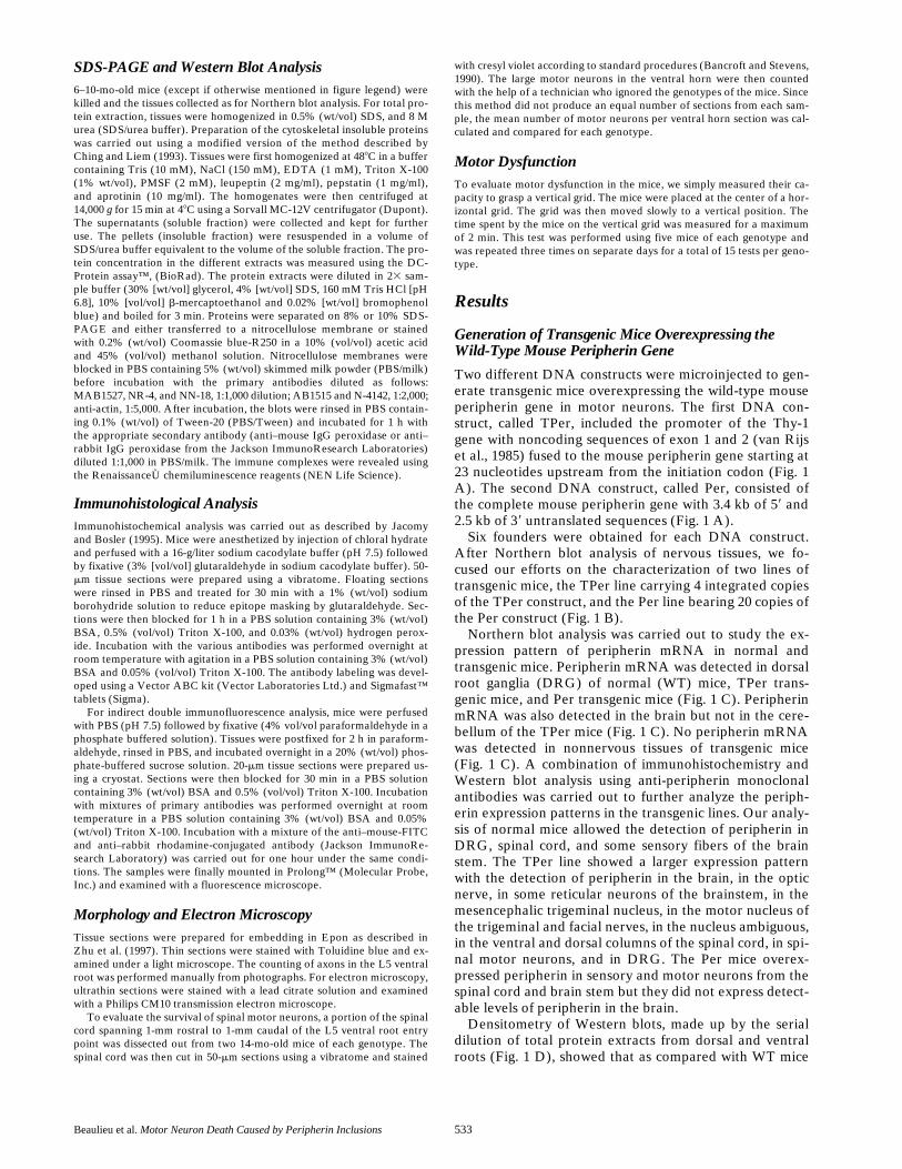
Taken together, our results and the previous reports thatperipherin is present in the majority of IF inclusions insporadic ALS cases (Corbo and Hays, 1992; Migheli et al.,1993; Tu et al., 1996) suggest that peripherin may play acontributory role in ALS pathogenesis. Further supportfor this view came from our immunodetection of periph-erin inclusions in spinal cord sections of paralyzed and pre-symptomatic mice expressing a Glycine 37 to Argininemutant form of SOD1 (SOD1G37R) associated with familialhuman ALS (Wong et al., 1995). This analysis was carriedout in two different lines of transgenic mice, lines 42 and29, which are characterized by disease onset at z5 andz11 mo, respectively. Staining of spinal cord sections fromnormal mice did not show any peripherin inclusions but al-lowed the detection of peripherin in axons from the dorsalcolumn and in some motor axons of the ventral horns (Fig.9, A and E). In contrast, peripherin inclusion bodies wereobserved in paralyzed mice from both SOD1G37R lines(Fig. 9, B and D). Staining of sections from presymptom-atic mice of line 29 revealed the presence of peripherin in-clusions in 5-mo-old mice (Fig. 9 C). Similar inclusionswere also observed in one out of three mice studied at 3mo of age, indicating that the formation of peripherin in-clusions in motor axons occurs at an early stage of the dis-ease. It is noteworthy that peripherin inclusions were alsodetected in the dorsal column in paralyzed but not in pre-symptomatic mice from both SOD1G37R lines (Fig. 9, F–H).
DiscussionThe transgenic mouse models described here are the firstexamples of extensive motor neuron death in mice causedby an upregulation of a wild-type IF protein. There areprevious reports of neuronal death due to expression of IF
Figure 7. Cresyl violet staining of L5 spinal cords. Light micros-copy of spinal cords from 14-mo-old WT, L2/2, TPer, and TPer;L2/2 mice showed a marked loss of large motor neurons (ar-rows) in TPer;L2/2 mice.
Table III. Number of Large Motor Neurons per Ventral Horn in L5 Spinal Cord Sections
Genotype Age
Mean number ofmotor neurons
per ventral hornLoss of
motor neurons
mo %
WT (n 5 103) 14 19.9 6 4.8 0TPer (n 5 115) 14 21.4 6 5.1 0L2/2 (n 5 110) 14 15.9 6 4.2 20TPer;L2/2 (n 5 74) 5 11 6 3.8 45TPer;L2/2 (n 5 148) 14 7.3 6 4.4 64
Loss of motor axons represents the percentage of missing axons as compared to WT.n represents the number of ventral horn sections for each category. Results aremean 6 SD.

The Journal of Cell Biology, Volume 147, 1999 540
transgenes (Lee et al., 1994; Tu et al., 1997; Ching et al.,1999; Ma et al., 1999), but this is the first example of mas-sive and selective motor neuron death due to high levels ofwild-type IF protein. Our results demonstrate that an up-regulation of wild-type peripherin is sufficient to induceselective motor neuron death during aging and that a defi-ciency of NF-L is a factor that precipitates dramaticallythe onset and progression of disease, probably through thedeleterious effects of NF-M and NF-H on peripherin orga-nization. Moreover, the acceleration of disease correlatedwith the frequency of IF inclusions containing peripherinin spinal motor neurons.
Three independent studies have shown that peripherin
is present in a majority of IF inclusions in ALS patients(Corbo and Hays, 1992; Migheli et al., 1993; Tu et al.,1996). We have also detected similar inclusions in pre-symptomatic mice expressing a mutant SOD1G37R linked tofamilial ALS (Fig. 9). Such IF inclusions in ALS may occuras a consequence of axonal transport defects (Zhang et al.,1997; Williamson and Cleveland, 1999). Alternatively, theresults presented here and the intense immunoreactivityof IF inclusions for peripherin in ALS make it plausiblethat such inclusion bodies could originate from an upregu-lation of peripherin occurring at the level of gene tran-scription. Peripherin is normally expressed at low levels inmotor neurons (Troy et al., 1990a), but enhanced levels,
Figure 8. IF inclusions contain peripherin and NF proteins. (A–E) Immunohistochemical detection of peripherin in spinal cord sampleswith a monoclonal antibody (NCL-Periph 1:200 dilution). (A) A very weak peripherin signal was detected in motor neurons (black ar-rows in A) of NF-L2/2 mice. (B) In the TPer mice, diffuse peripherin labeling (black arrow) and some small inclusions (white arrow)were detected in motor neurons. (C) The lack of 50% of NF-L in the TPer;L1/2 mice caused the formation of more frequent periph-erin inclusions in motor neuron axons (black arrow). (D and E) The total absence of NF-L resulted in the disappearance of a diffuse pe-ripherin staining and in the formation of numerous peripherin inclusions in the cell bodies and axons of spinal motor neurons (black ar-rows) of TPer and Per mice. (F) Hyaline inclusion bodies (black arrow) in the cytoplasm of motor neurons detected by Toluidine bluestaining of thin sections of the spinal cord from TPer;L2/2 mice. N indicates the cell nucleus. (G and H) Electron microscopy of inclu-sion bodies found in motor neurons of TPer;L2/2 (G) and PerL2/2 (H) mice. The inclusions are formed of 10-nm filaments that aresequestering membranous material (black arrows in G) and mitochondria (H). (I–L) Double indirect immunofluorescence of motorneurons from the spinal cord of TPer;L2/2 mice. A polyclonal anti-peripherin antibody (AB1530 1:1,000 dilution) in red and the vari-ous monoclonal antibodies directed against NF-M (NN-18 1:100 dilution), dephosphorylated NF-H (SMI-32 1:500 dilution) or hyper-phosphorylated NF-H (RT 97 1:500 dilution) in green reveal the colocalization of peripherin, NF-M (I) and NF-H (J and K) in the inclu-sion bodies. A double immunofluorescence staining of one neuron showing a diffused peripherin staining (L) in absence ofimmunoreactivity for NF-M. All results presented in this figure were obtained from 6–8-mo-old mice. Bars: (A–E) 100 mm; (F) 110 mm;(G) 1 mm; (H) 0.4 mm; (I–L) 15 mm.
Beaulieu et al. Motor Neuron Death Caused by Peripherin Inclusions 541
and perhaps IF inclusion formation, could be part of a re-generative response observed in ALS. It is well establishedthat after nerve injury the levels of peripherin mRNA areinduced by three- to fourfold in motor neurons (Troy etal., 1990a). Other genes associated with the regenerativeresponse after neuronal injury, such as GAP43 and a1-tubulin, have been reported to be upregulated in ALS(Parhad et al., 1992). Moreover, peripherin is upregulatedby cytokines such as IL-6 and LIF (Djabali et al., 1993;Sterneck et al., 1996). There is growing evidence for an in-volvement of inflammatory reactions in ALS that couldaccount for enhanced levels of peripherin. A recent studyhas indicated an increase of IL-6 in the cerebrospinal fluidof ALS patient (Sekizawa et al., 1998). Moreover, an up-regulation of LIF has been reported in an in vivo model ofglutamate excitotoxicity (Kurek et al., 1998), which is an-other hypothesized mechanism of toxicity associated withALS (Lin et al., 1998). Finally, inflammatory cytokines areassociated with astrogliosis, which is a common phenome-non in ALS and in other neurodegenerative diseases(Klein et al., 1997).
Our previous cell culture studies with peripherin and NFcDNA clones in SW13 cells demonstrated that peripherincan self-assemble to form an IF network and that it assem-bles with the NF network in the presence of NF-L (Beau-lieu et al., 1999). However, in absence of NF-L, the NF-Mand NF-H proteins interact with peripherin to form disor-ganized IF structures (Beaulieu et al., 1999). Perhaps, thiscould explain why the peripherin-mediated disease wasexacerbated in the NF-L null background. The increasednumber of IF inclusion bodies in mice deficient for NF-L isprobably due to abnormal interactions of peripherin andthe high molecular mass NF subunits. In this regard, it isnoteworthy that rare motor neurons in TPer;L2/2 micethat were devoid of NF-M protein did not develop IF in-clusions bodies even though peripherin was detected at el-evated levels (Fig. 8 L). Similarly, small sensory neuronsthat express peripherin but are devoid of NFs (Ferri et al.,1990; Goldstein et al., 1996) maintained a normal IF net-work in the NF-L null background (Fig. 4).
The NF-L null background mimics in part the depletionin NF-L mRNA levels occurring in motor neurons of ALS
Figure 9. Peripherin inclu-sions in SOD1G37R mice. Im-munohistochemical labelingwith a peripherin polyclonalantibody (AB1530 1:1,000 di-lution) revealed the presenceof peripherin-containing in-clusions in motor axons ofmice expressing the mutantSOD1G37R, at early stage (ar-rowheads in C), and endstage of disease (arrowheadsin B and D). Staining fromspinal cord sections of nor-mal mice yielded a weakstaining for peripherin inventral horn axons (arrow-head in A) and dorsal columnaxons (E) but no peripherininclusions were detected.Note the detection of periph-erin inclusions in the dorsalcolumn at end stage of dis-ease in mice from line 42 andline 29 (arrowhead in F andH). Bars: (A–D) 75 mm;(E–H) 50 mm.
The Journal of Cell Biology, Volume 147, 1999 542
patients. In situ hybridization studies revealed a striking60% reduction of NF-L mRNA levels in the a-motor neu-rons of ALS patients as compared with age-matched con-trols (Bergeron et al., 1994). Moreover, greater declines inNF-L mRNA levels (87%) were detected in motor neu-rons having spheroids or perikaryal filamentous accumula-tions (Bergeron et al., 1994). Remarkably, our data dem-onstrate that a partial depletion in NF-L levels is sufficientto increase the abundance of peripherin inclusions. A 50%reduction of NF-L mRNA in the TPer;L1/2 and Per;L1/2mice resulted in the formation of peripherin inclusions inproximal axons of motor neurons (Fig. 8 C)
The formation of IF inclusions in motor neurons hasbeen associated with motor dysfunction in three othertypes of transgenic mice (Côté et al., 1993; Xu et al., 1993;Lee et al., 1994). However, the properties of IF inclusionsdescribed here differ in several aspects from those of IF in-clusions observed in mice overexpressing NF transgenes.Whereas large perikaryal NF accumulations excludingother cytoplasmic material were observed in the NF-Hand NF-L transgenic mice (Côté et al., 1993; Xu et al.,1993), the inclusion bodies found in peripherin transgenicmice are relatively small, they contain membranous mate-rial including mitochondria and they occur in both cellbodies and axons. Because of their size and axonal local-ization, the inclusions observed in peripherin transgenicmice are more similar to the IF inclusions found in humanALS (reviewed in Chou, 1995) and in mutant SOD1 trans-genic mice than are the NF swellings observed in the NFtransgenic mice. More importantly, unlike mice overex-pressing wild-type NF proteins (Côté et al., 1993; Xu et al.,1993; our unpublished observation), the mice with periph-erin inclusions exhibited axonal degeneration and massivedeath of motor neurons.
Unexpectedly, the results shown here demonstrate thatthe presence of NF-L is not required for the formation ofIF inclusions in motor neurons. This is an important find-ing because an involvement for NF proteins in ALS hasbeen recently challenged by studies with L2/2 mice andNF-H/lacZ mice showing that the absence of intact NFs inaxons did not prevent SOD1-mediated disease (William-son et al., 1998; Eyer et al., 1998). This led to the beliefthat IF proteins play a minor role in ALS pathogenesis, anotion that must be reexamined at the light of our resultsshowing that disorganized IF structures containing highmolecular weight NF proteins together with peripherincan provoke motor neuron death in absence of NF-L.
The exact mechanism underlying cell death by such IFinclusion bodies remains to be elucidated. A block of in-tracellular transport by multiple IF inclusions in the axonis a possible mechanism that may contribute to neurode-generation and perhaps cell death in the peripherin trans-genic mice described here. Alternatively, the peripherin-containing inclusions could provoke neurodegenerationby the sequestration of organelles such as mitochondria(Fig. 8) or other components essential for cell survival.Such a mechanism has been postulated to explain the tox-icity of SOD1 aggregates detected in SOD1 mutant mice(Bruijn et al., 1998). In fact, there is growing evidence forprotein aggregation as a common etiology in neurode-generative disorders including ALS, Alzheimer’s disease,prion disease, Parkinson’s disease, and a class of disorders
caused by expanded CAG repeats encoding polyglu-tamines (for review see Kakizuka, 1997). The generalworking hypothesis is that mutations in proteins may re-sult in misfolding and decreased protein degradation, lead-ing to disturbance in homeostatic mechanisms. For in-stance, an upregulation of proteins with chaperoningactivity was found to protect against aggregation of mu-tant ataxin-1 (Cummings et al., 1998) and also from thetoxicity of SOD1 mutants (Bruening et al., 1998). Re-cently, polyglutamine domain proteins derived from ex-panded CAG repeats were found to bind specifically toNF-L and to alter NF network (Nagai et al., 1999). Thedata presented here introduces a novel concept in that nomutant proteins are required for the development of inclu-sion bodies. In future studies it would be of interest to in-vestigate whether peripherin inclusions can also be de-rived by posttranslational modifications of IF proteins. Itis well documented that changes in phosphorylation canalter the assembly properties of NF proteins (for reviewsee Julien and Mushynski, 1998). In addition, the nitrationof tyrosine residues in the NF-L protein, a phenomenonthat may occur in ALS (Crow et al., 1997), could impair itsassembly and perhaps would favor interactions of periph-erin with the high-molecular mass NF subunits to formnoxious IF inclusions.
In conclusion, our study demonstrates for the first timethat an upregulation of wild-type peripherin can cause theselective death of motor neurons during aging and that aNF-L deficiency is a factor that precipitates the disease.These results combined with the specific detection of pe-ripherin inclusions in motor neurons of sporadic ALScases (Corbo and Hays, 1992; Migheli et al., 1993; Tu et al.,1996) and in motor neuron neurons of presymptomaticSOD1G37R mice (Fig. 9) provide compelling evidence for apotential role of peripherin in ALS pathogenesis.
The technical assistance of P. Hince, D. Houle, and G. Gagnon is grate-fully acknowledged. We also thank J. Robertson, W.E. Mushynski, andJ.S. Couillard-Després.
This research was supported by the Medical Research Council of Can-ada (MRC), the ALS association (USA), and the ALS society of Canada.J.-M. Beaulieu is a recipient of studentships from the Natural Science andEngineering Research Council of Canada (NSERC), from Le Fond deRecherche en Santé du Québec (FRSQ) and from the McDonald StewartFoundation. M.D. Nguyen was a recipient of a MRC studentship. J.-P.Julien has a MRC Senior Scholarship.
Submitted: 4 June 1999Revised: 23 September 1999Accepted: 24 September 1999
References
Athlan, E.S., and W.E. Mushynski. 1997. Heterodimeric associations betweenneuronal intermediate filament proteins. J. Biol. Chem. 272:31073–31078.
Bancroft, J.D., and A. Stevens. 1990. Theory and practice of histological tech-niques. 3rd edition. Churchill Livingstone, Inc. New York, NY. 726 pp.
Beaulieu, J.M., J. Robertson, and J.P. Julien. 1999. Interactions between pe-ripherin and neurofilaments in cultured cells: disruption of peripherin as-sembly by the NF-M and NF-H subunits. Biochem. Cell Biol. 77:41–45.
Bergeron, C., K. Beric-Maskarel, S. Muntasser, L. Weyer, M.J. Somerville, andM.E. Percy. 1994. Neurofilament light and polyadenylated mRNA levels aredecreased in amyotrophic lateral sclerosis motor neurons. J. Neuropathol.Exp. Neurol. 53:221–230.
Brinster, R.L., H.Y. Chen, M. Trumbauer, A.W. Senear, R. Warren, and R.D.Palmiter. 1981. Somatic expression of herpes thymidine kinase in mice fol-lowing injection of a fusion gene into eggs. Cell. 27:223–231.
Brody, B.A., C.A. Ley, and L.M. Parysek. 1989. Selective distribution of the 57

Beaulieu et al. Motor Neuron Death Caused by Peripherin Inclusions 543
kDa neural intermediate filament protein in the rat CNS. J. Neurosci.9:2391–2401.
Bruening, W., B. Giasson, W. Mushynski, and H.D. Durham. 1998. Activationof stress-activated MAP protein kinases up-regulates expression of trans-genes driven by the cytomegalovirus immediate/early promoter. Nucleic Ac-ids Res. 26:486–489.
Bruijn, L.I., M.K. Houseweart, S. Kato, K.L. Anderson, S.D. Anderson, E.Ohama, A.G. Reaume, R.W. Scott, and D.W. Cleveland. 1998. Aggregationand motor neuron toxicity of an ALS-linked SOD1 mutant independentfrom wild-type SOD1. Science. 281:1851–1854.
Bruijn, L.I., M.W. Becher, M.K. Lee, K.L. Anderson, N.A. Jenkins, N.G. Cope-land, S.S. Sisodia, J.D. Rothstein, D.R. Borchelt, D.L. Price, and D.W.Cleveland. 1997. ALS-linked SOD1 mutant G85R mediates damage to as-trocytes and promotes rapidly progressive disease with SOD1-containing in-clusions. Neuron. 18:327–338.
Carpenter, S. 1968. Proximal axonal enlargement in motor neuron disease.Neurology. 18:841–851.
Ching, G.Y., and R.K.H. Liem. 1993. Assembly of type IV neuronal intermedi-ate filaments in nonneuronal cells in the absence of preexisting cytoplasmicintermediate filaments. J. Cell Biol. 122:1323–1335.
Ching, G.Y., C.L. Chien, R. Flores, and R.K. Liem. 1999. Overexpression of al-pha-internexin causes abnormal neurofilamentous accumulations and motorcoordination deficits in transgenic mice. J. Neurosci. 19:2974–2986.
Chou, S.M. 1995. Pathology of motor system disorder. In Motor Neuron Dis-ease: Biology and Management. P.N. Leigh and M. Swash, editors. Springer-Verlag, London. 53–92.
Corbo, M., and A.P. Hays. 1992. Peripherin and neurofilament protein coexistin spinal spheroids of motor neuron disease. J. Neuropathol. Exp. Neurol. 51:531–537.
Côté, F., J.F. Collard, and J.-P. Julien. 1993. Progressive neuropathy in trans-genic mce expressing the human neurofilamen heavy gene: a mouse modelof amyotrophic lateral sclerosis. Cell. 73:35–46.
Crow, J.P., Y.Z. Ye, M. Strong, M. Kirk, S. Barnes, and J.S. Beckman. 1997. Su-peroxide dismutase catalyzes nitration of tyrosines by peroxynitrite in therod and head domains of neurofilament-L. J. Neurochem. 69:1945–1953.
Cui, C., P.J. Stambrook, and L.M. Parysek. 1995. Peripherin assembles into ho-mopolymers in SW13 cells. J. Cell Sci. 108:3279–3284.
Cummings, C.J., M.A. Mancini, B. Antalffy, D.B. DeFranco, H.T. Orr, H.Y.Zoghbi. 1998. Chaperone suppression of aggregation and altered subcellularproteasome localization imply protein misfolding in SCA1. Nature Genet.19:148–154.
Djabali, K., A. Zissopoulou, M.J.D. Hoop, S.D. Georgatos, and C.G. Dotti.1993. Peripherin expression in hippocampal neurons induced by muscle sol-uble factors. J. Cell Biol. 123:1197–1206.
Escurat, M., K. Djabali, M. Gumpel, F. Gros, and M.M. Portier. 1990. Differen-tial expression of two neuronal intermediate-filament proteins, peripherinand the low-molecular-mass neurofilament protein NF-L during the devel-opment of the rat. J. Neurosci. 10:764–784.
Eyer, J., D.W. Cleveland, P.C. Wong, and A.C. Peterson. 1998. Pathogenesis oftwo axonopathies does not require axonal neurofilaments. Nature. 391:584–587.
Feinberg, A.P., and B. Vogelstein. 1983. A technique for radiolabeling DNArestriction endonuclease fragments to high specific activity. Anal. Biochem.132:6–13.
Ferri, G.L., A. Sabani, L. Abelli, J.M. Polak, D. Dahl, and M.M. Portier. 1990.Neuronal intermediate filaments in rat dorsal root ganglia: differential distri-bution of peripherin and neurofilament protein immunoreactivity and effectof capsaicin. Brain Res. 515:331–335.
Goldstein, M.E., P. Grant, S.B. House, D.B. Henken, and H. Gainer. 1996. De-velopmental regulation of two distinct neuronal phenotypes in rat dorsalroot ganglia. Neuroscience. 71:243–258.
Gurney, M.E., H. Pu, A.Y. Chiu, M.C. Dal Canto, C.Y. Polchow, D.D. Alex-ander, J. Caliendo, A. Hentati, Y.W. Kwon, H.X. Deng, et al. 1994. Motorneuron degeneration in mice that express a human Cu,Zn superoxide dismu-tase mutation. Science. 264:1772–1775.
Ho, C.L., S.S.M. Chin, K. Carnevale, and R.K.H. Liem. 1995. Translation initia-tion and assembly of peripherin in cultured cells. Eur. J. Cell Biol. 68:103–112.
Jacomy, H., and O. Bosler. 1995. Catecholaminergic innervation of the supra-chiasmatic nucleus in the adult rat: ultrastructural relationships with neuronscontaining vasoactive intestinal peptide or vasopressin. Cell Tissue Res. 280:87–96.
Julien, J.P., and W.E. Mushynski. 1998. Neurofilaments in health and disease.Prog. Nucleic Acid Res. Mol. Biol. 61:1–23.
Kakizuka, A. 1997. Degenerative ataxias: genetics, pathogenesis and animalmodels. Curr. Opin. Neurol. 10:285–290.
Kato, S., M. Shimoda, Y. Watanabe, K. Nakashima, K. Takahashi, and E.Ohama. 1996. Familial amyotrophic lateral sclerosis with a two base pair de-letion in superoxide dismutase 1: gene multisystem degeneration with intra-cytoplasmic hyaline inclusions in astrocytes. J. Neuropathol. Exp. Neurol. 55:1089–1101.
Klein, M.A., J.C. Moller, L.L. Jones, H. Bluethmann, G.W. Kreutzberg, and G.Raivich. 1997. Impaired neuroglial activation in interleukin-6 deficient mice.Glia. 19:227–233.
Kurek, J.B., T.M. Bennett, J.J. Bower, C.M. Muldoon, and L. Austin. 1998.
Leukaemia inhibitory factor (LIF) production in a mouse model of spinaltrauma. Neurosci. Lett. 249:1–4.
Lecomte, M.J., M. Basseville, F. Landon, V. Karpov, and M. Fauquet. 1998.Transcriptional activation of the mouse peripherin gene by leukemia inhibi-tory factor: involvement of STAT proteins. J. Neurochem. 70:971–982.
Lee, M.K., Z. Xu, P.C. Wong, and D.W. Cleveland. 1993. Neurofilaments areobligate heteropolymers in vivo. J. Cell Biol. 122:1337–1350.
Lee, M.K., J.R. Marszalek, and D.W. Cleveland. 1994. A mutant neurofilamentsubunit causes massive, selective motor neuron death: implications for thepathogenesis of human motor neuron disease. Neuron. 13:975–988.
Lin, C.L., L.A. Bristol, L. Jin, M. Dykes-Hoberg, T. Crawford, L. Clawson, andJ.D. Rothstein. 1998. Aberrant RNA processing in a neurodegenerative dis-ease: the cause for absent EAAT2, a glutamate transporter, in amyotrophiclateral sclerosis. Neuron. 20:589–602.
Ma, D., L. Descarries, K.D. Micheva, Y. Lepage, J.-P. Julien, and G. Doucet.1999. Severe neuronal losses with age in the parietal cortex and ventrobasalthalamus of mice transgenic for the human NF-L neurofilament protein. J.Comp. Neurol. 406:433–448.
Migheli, A., T. Pezzulo, A. Attanasio, and D. Schiffer. 1993. Peripherin immu-noreactive structures in amyotrophic lateral sclerosis. Lab. Investig. 68:185–191.
Muma, N.A., P.N. Hoffman, H.H. Slunt, M.D. Applegate, I. Lieberburg, andD.L. Price. 1990. Alterations in levels of mRNAs coding for neurofilamentprotein subunits during regeneration. Exp. Neurol. 107:230–235.
Nagai, Y., O. Onodera, J. Chun, W.J. Strittmatter, and J.R. Burke. 1999. Ex-panded polyglutamine domain proteins bind neurofilament and alter theneurofilament network. Exp. Neurol. 155:195–203.
Pachter, J.S., and R.K. Liem. 1985. alpha-Internexin, a 66-kD intermediate fila-ment-binding protein from mammalian central nervous tissues. J. Cell Biol.101:1316–1322.
Parhad, I.M., R. Oishi, and A.W. Clark. 1992. GAP-43 gene expression is in-creased in anterior horn cells of amyotrophic lateral sclerosis. Ann. Neurol.31:593–597.
Parysek, L.M., and R.D. Goldman. 1988. Distribution of a novel 57 kDA inter-mediate filament IF protein in the nervous system. J. Neurosci. 8:555–563.
Parysek, L.M., M.A. McReynolds, R.D. Goldman, and C.A. Ley. 1991. Someneural intermediate filaments contain both peripherin and the neurofila-ment proteins. J. Neurosci. Res. 30:80–91.
Ripps, M.E., G.W. Huntley, P.R. Hof, J.H. Morrison, and J.W. Gordon. 1995.Transgenic mice expressing an altered murine superoxide dismutase geneprovide an animal model of amyotrophic lateral sclerosis. Proc. Natl. Acad.Sci. USA. 92:689–693.
Rosen, D.R., T. Siddique, D. Patterson, D.A. Figlewicz, P. Sapp, A. Hentati, D.Donaldson, J. Goto, J.P. O’Regan, H.X. Deng, et al. 1993. Mutations in Cu/Zn superoxide dismutase gene are associated with familial amyotrophic lat-eral sclerosis. Nature. 362:59–62.
Sambrook, J., E.F. Fritsh, and T. Maniatis. 1989. Molecular Cloning: A Labora-tory Manual. 2nd edition. Cold Spring Harbor Laboratory Press, ColdSpring Harbor, New York.
Sekizawa, T., H. Openshaw, K. Ohbo, K. Sugamura, Y. Itoyama, and J.C. Ni-land. 1998. Cerebrospinal fluid interleukin 6 in amyotrophic lateral sclerosis:immunological parameter and comparison with inflammatory and non-inflammatory central nervous system diseases. J. Neurol. Sci. 154:194–199.
Shibata, N., A. Hirano, M. Kobayashi, T. Siddique, H.X. Deng, W.Y. Hung, T.Kato, and K. Asayama. 1996. Intense superoxide dismutase-1 immunoreac-tivity in intracytoplasmic hyaline inclusions of familial amyotrophic lateralsclerosis with posterior column involvement. Neuropathol. Exp. Neurol. 55:481–490.
Sterneck, E., D.R. Kaplan, and P.F. Johnson. 1996. Interleukin-6 induces ex-pression of peripherin and cooperates with Trk receptor signaling to pro-mote neuronal differentiation in PC12 cells. J. Neurochem. 67:1365–1374.
Troy, C.M., N.A. Muma, L.A. Greene, D.L. Price, and M.L. Shelanski. 1990a.Regulation of peripherin and neurofilament expression in regenerating ratmotor neurons. Brain Res. 529:232–238.
Troy, C.M., K. Brown, L.A. Greene, and M.L. Shelanski. 1990b. Ontogeny ofthe neuronal intermediate filament protein, peripherin, in the mouse em-bryo. Neuroscience. 30:217–237.
Tu, P.H., P. Raju, K.A. Robinson, M.E. Gurney, J.Q. Trojanowski, and V.M.Lee. 1996. Transgenic mice carrying a human mutant superoxide dismutasetransgene develop neuronal cytoskeletal pathology resembling human amyo-trophic lateral sclerosis lesions. Proc. Natl. Acad. Sci. USA. 93:3155–3160.
Tu, P.H., K.A. Robinson, F. de Snoo, J. Eyer, A. Peterson, V.M. Lee, and J.Q.Trojanowski. 1997. Selective degeneration of Purkinje cells with Lewy body-like inclusions in aged NFHLACZ transgenic mice. J. Neurosci. 17:1064–1074.
van Rijs, J., V. Giguere, J. Hurst, T. van Agthoven, A. Geurts van Kessel, S.Goyert, and F. Grosveld. 1985. Chromosomal localization of the humanThy-1 gene. Proc. Natl. Acad. Sci. USA. 82:5832–5835.
Williamson, T.L., L.I. Bruijn, Q. Zhu, K.L. Anderson, S.D. Anderson, J.P.Julien, and D.W. Cleveland. 1998. Absence of neurofilaments reduces theselective vulnerability of motor neurons and slows disease caused by a famil-ial amyotrophic lateral sclerosis-linked superoxide dismutase 1 mutant.Proc. Natl. Acad. Sci. USA. 95:9631–9636.
Williamson, T.L., and D.W. Cleveland. 1999. Slowing of axonal transport is avery early event in the toxicity of ALS-linked SOD1 mutants to motor neu-
The Journal of Cell Biology, Volume 147, 1999 544
rons. Nat. Neurosci. 2:50–56.Wong, P.C., C.A. Pardo, D.R. Borchelt, M.K. Lee, N.G. Copeland, N.A. Jen-
kins, S.S. Sisodia, D.W. Cleveland, and D.L. Price. 1995. An adverse prop-erty of a familial ALS-linked SOD1 mutation causes motor neuron diseasecharacterized by vacuolar degeneration of mitochondria. Neuron. 14:1105–1116.
Xu, Z., L.C. Cork, J.W. Griffin, and D.W. Cleveland. 1993. Increased expres-sion of neurofilament subunit NF-L produces morphological alterations that
resemble the pathology of human motor neuron disease. Cell. 73:23–33.Zhang, B., P.H. Tu, F. Abtahian, J.Q. Trojanowski, and V.M.Y. Lee. 1997.
Neurofilaments and orthograde transport are reduced in ventral root axonsof transgenic mice that express human SOD1 with a G93A mutation. J. CellBiol. 139:1307–1315.
Zhu, Q., S. Couillard-Després, and J.P. Julien. 1997. Delayed maturation of re-generating myelinated axons in mice lacking neurofilaments. Exp. Neurol.148:299–316.