Patient-specific modelling of cortical spreading depression ...
Upload
independentCategory
view
2download
0
A novel Cas family member, HEPL, regulates FAK and cell spreading. Mahendra K. Singh, Disha Dadke, Emmanuelle Nicolas, Ilya G. Serebriiskii, Sinoula Apostolou, Adrian Canutescu, Brian L. Egleston, and Erica A. Golemis^ Fox Chase Cancer Center, 333 Cottman Ave., Philadelphia, PA 19111 Running Head: HEPL, a fourth Cas family member. Key words: Cas, HEPL, HEF1, p130Cas, Efs, Character count (exclusive of Title Page, Materials and Methods, and References): 24,792 References: 43 ^corresponding author: Erica Golemis Fox Chase Cancer Center, W406 333 Cottman Ave. Philadelphia, PA 19111 USA (215) 728-2860 ph, -3616 fax [email protected]
1 http://www.molbiolcell.org/content/suppl/2008/02/05/E07-09-0953.DC1.html
Supplemental Material can be found at:

Abstract. For over a decade, p130Cas/BCAR1, HEF1/NEDD9/Cas-L, and Efs/Sin have defined
the Cas scaffolding protein family. Cas proteins mediate integrin-dependent signals at focal
adhesions, regulating cell invasion and survival; at least one family member, HEF1, regulates
mitosis. We here report a previously undescribed novel branch of the Cas protein family,
designated HEPL (for HEF1-Efs-p130Cas-like). The HEPL branch is evolutionarily conserved
through jawed vertebrates, and HEPL is found in some species lacking other members of the
Cas family. The human HEPL mRNA and protein is selectively expressed in specific primary
tissues and cancer cell lines, and HEPL maintains Cas family function in localization to focal
adhesions, as well as regulation of FAK activity, focal adhesion integrity, and cell spreading. It
has recently been demonstrated that upregulation of HEF1 expression marks and induces
metastasis, while high endogenous levels of p130Cas are associated with poor prognosis in
breast cancer, emphasizing the clinical relevance of Cas proteins. Better understanding of the
complete protein family should better inform prediction of cancer incidence and prognosis.
2

Introduction. The Cas (Crk-associated substrate) scaffolding protein family contains three defined
members: p130Cas/BCAR1 (Sakai et al., 1994; Brinkman et al., 2000); HEF1/Cas-L/NEDD9
(Kumar et al., 1992; Law et al., 1996; Minegishi et al., 1996), and Efs/Sin (Ishino et al., 1995;
Alexandropoulos and Baltimore, 1996). Elevated expression of p130Cas/BCAR1 has been
linked to poor prognosis in breast cancer (van der Flier et al., 2000), while overexpression of
HEF1/NEDD9/Cas-L recently been found to potently induce metastatic melanoma (Kim et al.,
2006). Mechanistically, the best-studied functions of the Cas family proteins include regulation
of attachment-dependent survival signaling or anoikis, and regulation of cell motility and
invasion, although there is evidence for additional roles for some of these proteins in control of
cell cycle, growth factor signaling, cell differentiation, and bacterial and viral infection (reviewed
in (Defilippi et al., 2006; Singh et al., 2007)). These many functions reflect the ability of the Cas
proteins to interact with multiple partner proteins, as the predominant structural feature of Cas
proteins is their possession of numerous protein interaction domains (discussed in (O'Neill et al.,
2000)), allowing them to act as scaffolding proteins for different functional complexes.
An important current issue in understanding cancer pathogenesis is that of why different
oncogenes and tumor suppressors are selectively targeted in tumors arising from different
tissue sources. For example, while elevation of HEF1/Cas-L/NEDD9 induces metastasis in
melanomas, reduced levels of the same gene have been associated with metastasis in breast
cancers that metastasize aggressively to the lung ((Minn et al., 2005); see also (O'Neill et al.,
2007) for discussion). Undoubtedly, the differing physiology and complement of expressed
genes in differing precursor cell types imposes distinct requirements for the type of genetic or
epigenetic change required to make a cell cancerous. For protein families, another relevant
issue is likely to be that for a given cell type, the expression of one family member may
condition the impact of modulating the expression of a paralogous family member with
overlapping biological activities. The complexity of cellular signaling networks currently
emerging through systems-level analysis (Mak et al., 2007) emphasizes the importance of
exactly defining the composition, expression, and functional properties of protein family groups.
In this study, we have identified a previously unreported but evolutionarily conserved
member of the Cas group, which we have termed HEPL (HEF1-Efs-p130Cas-like). We show
that the HEPL mRNA and protein are expressed in cultured cell lines and tumors, and that
HEPL has biological activities similar to those of other family members in influencing cell
attachment and movement. The identification of HEPL provides an important context to further
studies of this increasingly important protein group.
3

Materials and Methods.
Genomic and structural analysis of the Cas protein family. Details of sequence collection
and processing are provided in the legend to Supp. Figure 1. Dendrograms showing family
relationships were displayed using the Treeview program. The HEPL SH3 domain was
modeled using as template the high-resolution crystal structure (1.1Å) of the SH3 domain of
p130Cas (PDB code 1WYX (Wisniewska et al., 2005)). The rat p130Cas structure (PDB code
1Z23 (Briknarova et al., 2005)) was used to model the 4-helix bundle region. Homology
modeling was initiated using a multiple-round PSI-BLAST (Altschul et al., 1997) sequence
search using HEPL and p130Cas sequences to build profiles. The profiles were used to identify
suitable templates in the Protein Data Bank (PDB (Berman et al., 2000)). The profile/template
match was refined using secondary structure predictions from PSIPRED (Jones, 1999).
Conserved backbone and side chain residues were copied from the template structure, while
divergent residues were rebuilt using SCWRL3 (Canutescu et al., 2003). LOOPY (Xiang et al.,
2002) was used to build loops at points of insertion and deletion. Molecular graphics and 3D
structural manipulation was performed using Chimera (Pettersen et al., 2004). The Cas multiple
sequence alignment was overlaid with secondary structure prediction rendered by MolIDE
(Canutescu and Dunbrack, 2005).
Quantitative Real-time PCR assays. Total RNA was isolated using an RNeasy kit (Qiagen
Inc., USA). Contaminating DNA was removed using TURBO DNA-free™ (Ambion). RNA was
quantified using the Agilent 2100 BioAnalyzer in combination with a RNA 6000 Nano LabChip.
See Supplemental Tables 1 and 2 for technical details of PCR assays. Ambion's First Choice ®
human total RNA survey panel was used as a source of RNA from 20 different normal tissues.
HEPL plasmids and siRNA, and cell culture. HEPL was cloned using conventional molecular
biology techniques by combining sequences from Human MGC Verified Full Length cDNA
(Clone Id#: 5205865, Open Biosystems, Huntsville, AL) and human genomic DNA. HA-epitope
tagged HEF1, HEPL, FAK and negative controls (empty vector or ΔBioB, an extensively
truncated E. coli BioB) were expressed from pcDNA3.1-6HA for transfections. Cell lines were
cultured under standard conditions, in Dulbecco's modification of Eagle's medium (DMEM) or in
RPMI-1640 plus 10% fetal bovine serum (FBS) supplemented with antibiotics, as specified by
the ATCC. Scrambled (control) siRNA and siRNA duplexes against HEPL (NM_020356) and
HEF1 were made by Dharmacon Research, Lafayette, CO. HEPL-directed siRNAs were used
4

both as a Smartpool and four individual deconvoluted sequences, as described in text. Plasmid
transfections were done using LipofectAMINE-Plus reagent (Invitrogen, Carlsbad, CA) and
siRNA transfections using the Cell Line Nucleofector® Kit V from Amaxa Biosystems
(Gaithersburg, MD, USA).
Antibodies and immunoprecipitation. Rabbit polyclonal antibody to HEPL was generated
using a peptide corresponding to HEPL amino acids 773-786 (by ZYMED Laboratories, San
Francisco, CA). Antibody was purified from sera using the NAbTM Protein A Spin Purification Kit
(Pierce Biotechnology, Inc., Rockford, IL, USA). Other antibodies included anti-HA mAb (Santa
Cruz Biotechnology, Santa Cruz, CA), anti-paxillin and anti-p130Cas (BD Transduction
Laboratories, Carlsbad, CA), anti-HEF1 (2G9 (Pugacheva and Golemis, 2005)), anti-FAK[pY397]
(Biosource, Nevelle, Belgium), anti-gelsolin (BD Biosciences, San Jose, CA), Alexa Fluor™488
and 568-conjugated anti-mouse (Molecular Probes, Eugene, OR), and anti-mouse and anti-
rabbit IgG antibodies conjugated to HRP (Amersham Biotech, Buckinghamshire, England). For
immunoprecipitations, transfected cells were lysed in M-PER®Mammalian Protein Extraction
Reagent (PIERCE, Rockford, IL) and immunoprecipitated with either anti-HA, or anti-HEPL Abs,
using Immobilized Protein A/G Agarose (Pierce Biotechnology, Inc., Rockford, IL). To establish
HEPL and FAK interaction, HA-epitope tagged HEF1, HEPL, FAK and negative control (ΔBioB,
an extensively truncated E. coli BioB) were expressed from pcDNA3.1-6HA for transfections in
293T cells, and immunoprecipitated with anti-FAK monoclonal antibody, clone 4.47 (Millipore
Corporation, Bedford, MA).
To study cell adhesion-dependent tyrosine phosphorylation, trypsinized HOP-62 cells
were either maintained in suspension in serum-free medium for 45 minutes at 370C, or
subsequently replated on fibronectin (4 ug/cm2; Chemicon International, Temecula, CA)-coated
dishes for 30 minutes. Experiments were performed in parallel in the presence or absence of 10
μM PP2 (Calbiochem, San Diego, CA). Cell lysates were prepared using M-PER®Mammalian
Protein Extraction Reagent supplemented with protease inhibitors and HaltTM Phosphatase
Inhibitor Cocktail (PIERCE, Rockford, IL), immunoprecipitated with antibodies to HEPL or HEF1,
and immunoblotted with anti-phosphotyrosine monoclonal antibody (BD Transduction
Laboratories, Carlsbad, CA, USA).
Yeast two-hybrid analysis. The modified Interaction Trap form of two-hybrid system was used
to study HEPL protein interactions, using reagents and approaches as described in (Serebriiskii
et al., 2002). The LexA-fused HEPL SH3 domain (aa 1-148) was used to assess interactions
5

with B42 activation domain-fused FAK C-terminus (aa 688-997). LexA fused to the SHC PTB
domain and to a truncated derivative of bacterial BioB, and B42-fused Raf and truncated BioB,
were used as non-specific negative controls. Expression of all protein fusions was analyzed by
Western blot.
Immunofluorescence, cell spreading, cell migration, cell size, cell cycle, apoptosis. Cells
were fixed in 4% paraformaldehyde for 10 minutes, permeabilized in 0.2% Triton X-100 for
5min. and blocked with 3% BSA in PBS. After incubation with primary antibodies, cells were
stained with either Alexa Fluor™ 488- or 568-conjugated secondary antibodies. Epifluorescence
microscopy was performed using an inverted Nikon TE300 microscope. Confocal microscopy
was performed using a Radiance 2000 laser scanning confocal microscope (Carl Zeiss,
Thornwood, NY). All images were acquired as 12-bit images with a Spot RT monochrome
camera (Diagnostic Instruments). For cell spreading analysis, cells were transfected with
indicated plasmids or siRNAs for 18-48 hours prior to fixation, as indicated. Anti-paxillin mAb
was used to mark focal adhesions and outline cells. Cell area measurements were made using
MetaMorph or MetaVue software (Molecular Devices Corporation, Downingtown, PA) software
to score pixels within cell perimeters.
To measure motility, movement of siRNA-treated HOP62 cells plated in 6-well tissue
culture dishes was monitored with a Nikon TE300 microscope using 10x NA 0.25 A-Plan
objective and images were collected with CCD video camera (Roper Scientific, Trenton, NJ,
USA) at 20 minute intervals over a 12 hour period, then digitized and stored as image stacks
using MetaMorph software. Velocity and persistence of migratory directionality (D/T) were
determined by tracking the positions of cell nuclei using the Track Point function of MetaMorph.
Apoptosis was measured using an APOPercentage apoptosis assay kit (Biocolor Ltd.,
Belfast, Northern Ireland, UK) and Western blot to measure appearance of cleaved gelsolin.
Cell cycle compartmentalization was measured using a Guava Personal Cell Analysis (PCA)
System (Guava Technologies, Hayward, CA). Treatment for 48 hrs with 200 µM etoposide
(Sigma-Aldrich Corp., St. Louis, MO) or 10 nM dasatinib (a gift of Dr. Andrew Godwin) was used
as positive control for apoptosis assays. All calculations of statistical significance were made
using the GraphPad InStat software package (San Diego, CA) and STATA software (Stata
Corps, TX). Approaches included unpaired T-tests, ANOVA analysis, and generalized linear
models estimated using generalized estimating equations (GEE).
6

Results Prediction of a new Cas family member, HEPL. Using the p130Cas, HEF1, and Efs protein
and mRNA sequences in reiterative BLAST analysis against genomic sequences and EST
resources, we search for Cas-related sequences in an evolutionarily diverse group of organisms
(Figures 1A, B), and Supplemental Figure 1). No protein strongly related to the Cas family was
identified in S. cerevisiae or C. elegans, while a single ancestral family member was detected in
arthropods, echinoderms, and primitive chordates. The family branches in gnathostomes to
produce the three previously characterized mammalian family members. Unexpectedly, we
detected a completely novel member of the Cas family that was conserved as an ortholog group
from gnathostomes through mammals. We have designated this ortholog group as HEPL (for
HEF1/Efs/P130Cas-Like). Dendrogram analysis indicates that the HEPL does not diverge at a
significantly more rapid rate than other Cas branches, suggesting that it maintains a biological
function, rather than representing a pseudogene (Figure 1B).
Human HEPL localizes to chromosome 20q13.31, and is annotated in Unigene as
C20orf32. Comparison of the human HEPL protein sequence with the other three human Cas
family members (Supp. Figure 1) shows overall identity with other family members up to 26%,
and similarity up to 42%. Human HEPL is 786 amino acids (aa), versus 870 aa for p130Cas,
834 aa for HEF1, and 561 aa for Efs. For the three well-studied Cas proteins, a highly
conserved amino-terminal SH3 domain is followed by a moderately conserved region
encompassing multiple SH2-binding sites, a serine-rich region shown to encompass a four-helix
bundle in p130Cas (Briknarova et al., 2005), and a well-conserved carboxy-terminal domain that
contributes to focal adhesion targeting (Nakamoto et al., 1997; Harte et al., 2000; O'Neill and
Golemis, 2001) (Figure 1C). Although the HEPL proteins maintain a recognizable SH3 domain,
this domain has the lowest overall similarity to other SH3 domains in the Cas group. Human
HEPL has a limited number of candidate SH2 binding sites, in contrast to p130Cas and HEF1.
The HEPL carboxy-terminus has a short region of detectable Cas family homology (res ~670 to
the carboxy-terminus), but otherwise lacks obvious similarity at the level of primary sequence.
Flanking the carboxy-terminal domain, HEPL also lacks a YDYVHL sequence conserved among
the other three Cas family proteins (bold, Supp. Figure 1) that is an important binding site for the
Src SH2 domain (Tachibana et al., 1997), suggesting reduced functionality.
To better assess whether the predicted HEPL proteins maintain important features of the
Cas family, we used molecular modeling to compare the Cas proteins based on predicted
7

secondary and tertiary structure, using structures of p130Cas as templates (Briknarova et al.,
2005; Wisniewska et al., 2005). Figure 2A demonstrates that HEPL and p130Cas are predicted
to fold almost identically within the SH3 domain. Further, despite only 28% primary sequence
identity, the predicted secondary structure for residues 432-591 of HEPL is extremely similar to
that for residues 449-610 for p130Cas, implying a well-conserved fold (Figure 2B). At present,
no adequate template exists in PDB to create a tertiary model for the Cas carboxy-terminus.
However, comparison of the predicted secondary structure for the 4 Cas proteins reveals a
strikingly similar periodicity of α-helices and β-sheets (Figure 2C) that is again compatible with
the idea of a conserved tertiary structure.
Expression of endogenous HEPL. The evolutionary conservation of HEPL suggested it
encoded a functional protein product rather than an unexpressed pseudogene. To test this
directly, we first used quantitative RT-PCR to analyze HEPL expression in mRNAs prepared
from 20 human tissues (Figure 3A). HEPL was most abundant in lung and spleen, and detected
at lower levels in additional tissues. These results were in accord with online resources in
NCBI/Unigene (Wheeler et al., 2004) that estimate the relative abundance of mRNAs based on
their frequency of isolation in sequencing of tissue-specific libraries (results not shown). We next
used quantitative RT-PCR to analyze mRNAs prepared from a panel of commonly used
laboratory cell lines derived from diverse cell lineages (but enriched in carcinomas and
leukemias, in accord with the expression prediction), to establish the general abundance of
HEPL mRNA in cultured cells.
HEPL mRNA was detected in the majority of the cell lines (15 of 26), with highest levels
of HEPL in the leukemia and ovarian cell lines (Figure 3B). As reference, HEF1 and p130Cas
were readily detected in most of the cell lines examined, although p130Cas was not detected in
most of the lymphoma/ leukemia cell lines analyzed. In contrast, although very abundant in one
breast carcinoma cell line (T47D), the Efs mRNA was only detected in 6 of the 26 cell lines
assessed. Because increased expression of some Cas proteins has been linked to cancer
progression, we investigated the relative expression of HEPL mRNA in a series of normal
primary human ovarian surface epithelial (HOSE) cells, human SV40-immortalized ovarian
(HIO) cell lines, and primary ovarian tumor tissue (Figure 3C). From this preliminary analysis,
HEPL mRNA expression levels did not correlate significantly with ovarian transformation status.
To analyze HEPL at the protein level, we cloned the gene, and prepared antibody
against HEPL-derived peptide sequences that specifically recognized overexpressed epitope-
tagged HEPL (Figure 3D). Using this antibody, we have found that HOP-62, K562 and SR, cell
8

lines predicted by mRNA analysis to contain relatively abundant levels of HEPL, contained a
protein species of ~ 105 kDa, while lower levels of a similarly migrating species are detected in
a number of other cell lines (Figure 3E). This species was removed by treatment of cells with an
siRNA targeted to HEPL (Figure 3F). Together, these data indicate that HEPL is a bona fide
new member of the Cas family. Based on our analysis to date, antibodies to the more widely
studied Cas family members p130Cas and HEF1 do not cross-react with HEPL (Figure 3G),
suggesting the presence of HEPL may mask phenotypes associated with depletion of other
family members.
HEPL conserves Cas family functions in FAK regulation and cell spreading. The best-
defined action of Cas family proteins is as an intermediate in integrin-dependent attachment
signaling, regulating cell attachment, spreading, and migration. Although antibody to
endogenous HEPL worked poorly in immunofluorescence analysis, HA-HEPL transfected into
MCF7 cells (which express a low level of HEPL mRNA) co-localized with paxillin at focal
adhesions (Figure 4A), comparable to other Cas proteins. HA-HEPL transfected cells spread to
a greater degree than control or vector-transfected cells, but to a lesser degree than cells
transfected with HA-HEF1 (Figure 4B). Also suggesting a less potent action than HA-HEF1,
levels of Y397-phosphorylated (activated) FAK were strongly increased at focal adhesions in HA-
HEF1 cells. Interestingly, in HA-HEPL transfected cells, a subpopulation of the cells (~15-20%)
also showed increased levels of Y397-phosphorylated FAK, while the remainder of the population
remained at the levels of the negative control cells (Figure 4C).
Our modeling experiments had suggested that the HEPL SH3 domain would bind FAK
(Figure 2A). Using a two-hybrid approach, we confirmed a direct interaction between the HEPL
SH3 domain and the FAK C-terminus (Figure 4D). This result paralleled previous
demonstrations of interactions between the HEF1 and other Cas family SH3 domains with
proline-rich SH3 domain binding motifs in this region (Polte and Hanks, 1995; Law et al., 1996),
and suggests that overexpressed HEPL activates FAK based on direct interaction. Finally, like
HEF1, full length HEPL was immunoprecipitated with antibody to FAK from cells transfected
with plasmids expressing each protein (Figure 4E). A common characteristic of Cas family
proteins is their phosphorylation by Src family kinases during the cell attachment process, which
provides a docking site for FAK and contributes to assembly of signaling complexes at focal
adhesions (O'Neill et al., 2000). HEPL lacks the YDYVHL site that coordinates docking with the
Src SH2 domain for other family members (Tachibana et al., 1997). Nevertheless, attachment-
9

induced Src phosphorylation of HEPL was observed (Figure 4F), suggesting that additional
interactions between HEPL, FAK, and Src are sufficient to drive this modification.
We next analyzed focal adhesions and cell spreading in HOP-62 cells from which HEPL
had been depleted by siRNA (Figure 5A). In the total population of inspected cells, HEPL
depletion reduced cell spreading (P < 0.01), although to a significantly smaller degree than
HEF1 depletion (P < 0.001). Upon closer inspection, the HEPL-depleted population differed
from both scrambled siRNA control- and HEF1-depleted cells in its heterogeneous nature. A
sub-population of ~20% of HEPL-depleted cells showed reduced and/or differentially localized
staining for Y397-phosphorylated FAK (Figure 5B), with residual staining diffusely distributed at
the cell periphery rather than in discrete focal adhesions. Subsequent staining with antibody to
paxillin (Figure 5C) revealed a similar population of ~ 20% of cells that had greatly reduced
paxillin staining. Re-analyzing spreading data for strongly versus weakly Y397-FAK staining
HEPL-depleted cells (Figure 5A) revealed a clear segregation of weak staining with reduced
spreading. By contrast, almost all cells with depleted HEF1 were less spread than Scr control
cells, and had reduced Y397-phosphorylated FAK, to the same degree as the 20% of responsive
HEPL-depleted cells.
An important function of Cas proteins is regulation of cell migration (Klemke et al., 1998;
van Seventer et al., 2001; Fashena et al., 2002). Analysis of live cell images of HEPL- or HEF1-
depleted cells indicated that depletion distorted migration profiles relative to Scr-depleted
controls. HEF1 depletion uniformly reduced cellular velocity (P <0.001, Figure 5D).
Interestingly, the phenotype observed with HEPL depletion was more complex. HEPL-depleting
siRNAs caused appearance of a population of slow-moving cells, although for one of the
siRNAs the effect was not statistically significant (velocity < 9 μm/hour, P <0.05 and P =0.15).
However, the HEPL-depleting siRNAs each also unexpectedly caused appearance of a faster-
moving (velocity < 18 μm/hour) group of cells, corresponding to ~15% of the population (P <
0.05 and P < 0.02). Greater velocity has been reported to be associated with greater cell
spreading in some cell types with manipulated Cas proteins (e.g. (Fashena et al., 2002) for
HEF1 in MCF7 cells), although there are examples of cell movement where a connection
between spreading and velocity is not observed (Friedl and Wolf, 2003). No “highly spread”
population was detected with HEPL-depleted cells (results not shown). As further difference
from the Cas group, while depletion of HEF1 reduced directionality of movement of cells, no
such effect was seen with HEPL depletion (Figure 5E).
Some members of the Cas family, such as HEF1, also play important roles in regulation
of apoptosis and proliferation (e.g.,(Law et al., 2000; O'Neill and Golemis, 2001; Pugacheva and
10

Golemis, 2005; Dadke et al., 2006)). siRNA depletion of HEPL from K562 cells led to a slightly
slower accumulation of cells over 3 days (Figure 5F), although the general
compartmentalization of cells in the G1, S, and G2/M phases of cell cycle was not significantly
affected (Figure 5G). HEPL depletion did not influence the level of apoptotic cells in the
population (Figures 5H, 5I). As siRNA depletion rarely exceeds 90-95%, a definitive
determination that HEPL does not affect cell cycle or apoptosis requires a gene knockout; at
present, the most demonstrable activity of HEPL is at focal adhesions, as with p130Cas.
11

Discussion. Although the Cas protein family has been studied for over a decade, this study
represents the first extensive examination of the Cas proteins utilizing genomic resources. The
newly identified HEPL proteins define a legitimate novel branch of the Cas family. HEPL
proteins conserve important functional domains required for interaction with FAK, for targeting to
focal adhesions, and for regulating cell spreading and FAK activation. Although human HEPL is
expressed in a relatively limited subset of cell types relative to p130Cas and HEF1, based on
mRNA analysis it appears to be as prevalent as Efs/Sin in cultured cell lines.
HEPL does not appear to be as biologically active as HEF1, based on a number of
criteria presented above. Particularly in control of FAK activation, only a minority of cells
respond either to overexpressed HEPL or to depleted HEPL, under conditions where almost all
cells respond to similarly manipulated HEF1. siRNA depletion typically introduces siRNA into >
90% of cells, and our analysis of HEPL siRNA-transfected cells confirmed >75% depletion in
practice, excluding the trivial explanation of incomplete depletion. Rather, we expect the
difference may relate to cell-specific variability in the intrinsic expression level of the additional
Cas family members within HOP-62 cells: single cell analyses are beginning to demonstrate that
this is an important property governing average gene expression in cell populations (e.g.
(Levsky and Singer, 2003; Mar et al., 2006)). We propose that typically within cells expressing
multiple Cas family members, HEPL may make a minor contribution to regulation of cell growth
properties. Part of the reduced biological activity of HEPL may arise from lack of a key motif for
Src recognition (YDYVHL) (Tachibana et al., 1997). We have shown that HEPL is still
phosphorylated by Src family kinases during cell attachment, suggesting this motif is not
essential for an interaction with Src, presumably because of the presence of multiple interaction
interfaces joining HEPL, FAK, and Src: however, Cas proteins also reciprocally contribute to Src
activation in the attachment process (e.g. (Alexandropoulos and Baltimore, 1996)), and this
function may be limited.
Intriguingly, HEPL activity qualitatively differs from other Cas proteins in at least one
important way, in the regulation of migration. While loss or depletion of p130Cas and HEF1
reduces cell migration (e.g. (Natarajan et al., 2006)), HEPL depletion induced faster migration in
at least a subset of cells. The reason for this is so far unknown: however, an intriguing
possibility is that through possession of some but not all Cas family functions, HEPL may
weakly oppose the action of other Cas family proteins via action as a “dominant negative”.
Particularly in a cell background low in other Cas proteins, HEPL may be important. Hence, it is
important for experiments involving knockdown or knockout of Cas family proteins to
12
subsequently consider HEPL status in interpreting phenotypes. Separately, for each of the Cas
proteins, some interacting partners have been described unique to that family member; the
interaction profile of HEPL has not yet been explored, but may include novel interactors and
intracellular roles.
Intriguingly, the region of chromosome 20 encompassing HEPL is included as an
amplicon in many solid tumors (Dessen et al., 2002). It is hence possible that as with p130Cas
and HEF1 (Singh et al., 2007), altered expression of HEPL contributes to the pathogenesis of
cancers or other diseases. Our data demonstrate that HEPL overexpression is sufficient to
increase cell spreading and FAK activation, phenotypes associated with increased tumor
invasiveness. Based on its expression profile, in non-transformed cells, HEPL may be most
relevant to the normal function of the hematopoietic system, and the lung. As all the Cas
proteins have the potential to interact with multiple partner proteins, sometimes in large
complexes, the presence of additional family members might also be expected to induce cell-
and tissue-type differences in complex assembly and stoichiometry. Clearly, future studies of
the Cas group should consider the possible role of redundant HEPL function in evaluating
knockdown, knockout, or over-expression phenotypes. In sum, this study suggests ample new
ground for further investigation.
Acknowledgments. This work and the authors were supported by NIH RO1s CA63366 and CA113342, by
funding from the Susan B. Komen Foundation, and by Tobacco Settlement funding from the
State of Pennsylvania (to EAG); and by NIH core grant CA-06927 and support from the Pew
Charitable Fund, to Fox Chase Cancer Center. DD was a recipient of the Plain and Fancy
Fellowship of the Fox Chase Cancer Center Board of Associates. We thank E.D. Cohen and
Andrew Godwin for ovarian normal cells and cell lines, and Daniel Bassi, Robert Page, and
Andres Klein-Szanto for ovarian tumors. We thank Edna Cukierman for technical guidance for
migration and attachment assays. We are grateful to Ian Ochs and Nicolas Day for help with
cloning and two-hybrid analysis.
13

References. Alexandropoulos, K., and Baltimore, D. (1996). Coordinate activation of c-Src by SH3- and SH2-binding sites on a novel, p130Cas-related protein, Sin. Genes Dev. 10, 1341-1355. Altschul, S.F., Madden, T.L., Schaffer, A.A., Zhang, J., Zhang, Z., Miller, W., and Lipman, D.J. (1997). Gapped BLAST and PSI-BLAST: a new generation of protein database search programs. Nucleic Acids Res 25, 3389-3402. Berman, H.M., Westbrook, J., Feng, Z., Gilliland, G., Bhat, T.N., Weissig, H., Shindyalov, I.N., and Bourne, P.E. (2000). The Protein Data Bank. Nucleic Acids Res 28, 235-242. Briknarova, K., Nasertorabi, F., Havert, M.L., Eggleston, E., Hoyt, D.W., Li, C., Olson, A.J., Vuori, K., and Ely, K.R. (2005). The serine-rich domain from Crk-associated substrate (p130Cas) is a four-helix bundle. J Biol Chem. Brinkman, A., van Der Flier, S., Kok, E.M., and Dorssers, L.C. (2000). BCAR1, a human homologue of the adapter protein p130Cas, and antiestrogen resistance in breast cancer cells. J Natl Cancer Inst 92, 112-120. Canutescu, A.A., and Dunbrack, R.L., Jr. (2005). MolIDE (Molecular Integrated Development Environment): a homology modeling framework you can click with. Bioinformatics. Canutescu, A.A., Shelenkov, A.A., and Dunbrack, R.L., Jr. (2003). A graph-theory algorithm for rapid protein side-chain prediction. Protein Sci 12, 2001-2014. Dadke, D., Jarnik, M., Pugacheva, E.N., Singh, M.K., and Golemis, E.A. (2006). Deregulation of HEF1 impairs M-phase progression by disrupting the RhoA activation cycle. Mol Biol Cell 17, 1204-1217. Defilippi, P., Di Stefano, P., and Cabodi, S. (2006). p130Cas: a versatile scaffold in signaling networks. Trends Cell Biol 16, 257-263. Dessen, P., Knuutila, S., and Huret, J.L. (2002). Chromosome 20. Atlas Genet Ctogenet Oncol Haematol. Fashena, S.J., Einarson, M.B., O'Neill, G.M., Patriotis, C.P., and Golemis, E.A. (2002). Dissection of HEF1-dependent functions in motility and transcriptional regulation. J. Cell. Sci. 115, 99-111. Friedl, P., and Wolf, K. (2003). Tumour-cell invasion and migration: diversity and escape mechanisms. Nat Rev Cancer 3, 362-374. Harte, M.T., Macklem, M., Weidow, C.L., Parsons, J.T., and Bouton, A.H. (2000). Identification of two focal adhesion targeting sequences in the adapter molecule p130(Cas). Biochim Biophys Acta 1499, 34-48. Ishino, M., Ohba, T., Sasaki, H., and Sasaki, T. (1995). Molecular cloning of a cDNA encoding a phosphoprotein, Efs, which contains a Src homology 3 domain and associates with Fyn. Oncogene 11, 2331-2338. Jones, D.T. (1999). Protein secondary structure prediction based on position-specific scoring matrices. J Mol Biol 292, 195-202. Kim, M., Gans, J.D., Nogueira, C., Wang, A., Paik, J.H., Feng, B., Brennan, C., Hahn, W.C., Cordon-Cardo, C., Wagner, S.N., Flotte, T.J., Duncan, L.M., Granter, S.R., and Chin, L. (2006). Comparative oncogenomics identifies NEDD9 as a melanoma metastasis gene. Cell 125, 1269-1281. Klemke, R.L., Leng, J., Molander, R., Brooks, P.C., Vuori, K., and Cheresh, D.A. (1998). CAS/Crk coupling serves as a "molecular switch" for induction of cell migration. J Cell Biol 140, 961-972. Kumar, S., Tomooka, Y., and Noda, M. (1992). Identification of a set of genes with developmentally down-regulated expression in the mouse brain. Biochem. Biophys. Res. Commun. 185, 1155-1161.
14

Law, S.F., Estojak, J., Wang, B., Mysliwiec, T., Kruh, G.D., and Golemis, E.A. (1996). Human Enhancer of Filamentation 1 (HEF1), a novel p130Cas-like docking protein, associates with FAK, and induces pseudohyphal growth in yeast. Mol. Cell. Biol. 16, 3327-3337. Law, S.F., O'Neill, G.M., Fashena, S.J., Einarson, M.B., and Golemis, E.A. (2000). The docking protein HEF1 is an apoptotic mediator at focal adhesion sites. Mol. Cell. Biol. 20, 5184-5195. Levsky, J.M., and Singer, R.H. (2003). Gene expression and the myth of the average cell. Trends Cell Biol 13, 4-6. Mak, H.C., Daly, M., Gruebel, B., and Ideker, T. (2007). CellCircuits: a database of protein network models. Nucleic Acids Res 35, D538-545. Mar, J.C., Rubio, R., and Quackenbush, J. (2006). Inferring steady state single-cell gene expression distributions from analysis of mesoscopic samples. Genome Biol 7, R119. Minegishi, M., Tachibana, K., Sato, T., Iwata, S., Nojima, Y., and Morimoto, C. (1996). Structure and function of Cas-L, a 105-kD Crk-associated substrate-related protein that is involved in beta-1 integrin-mediated signaling in lymphocytes. J. Exp. Med. 184, 1365-1375. Minn, A.J., Gupta, G.P., Siegel, P.M., Bos, P.D., Shu, W., Giri, D.D., Viale, A., Olshen, A.B., Gerald, W.L., and Massague, J. (2005). Genes that mediate breast cancer metastasis to lung. Nature 436, 518-524. Nakamoto, T., Sakai, R., Honda, H., Ogawa, S., Ueno, H., Suzuki, T., Aizawa, S.-I., Yazaki, Y., and Hirai, H. (1997). Requirements for localization of p130Cas to focal adhesions. Mol. Cell. Biol. 17, 3884-3897. Natarajan, M., Stewart, J.E., Golemis, E.A., Pugacheva, E.N., Alexandropoulos, K., Cox, B.D., Wang, W., Grammer, J.R., and Gladson, C.L. (2006). HEF1 is a necessary and specific downstream effector of FAK that promotes the migration of glioblastoma cells. Oncogene 25, 1721-1732. O'Neill, G.M., Fashena, S.J., and Golemis, E.A. (2000). Integrin signaling: a new Cas(t) of characters enters the stage. Trends Cell Biol. 10, 111-119. O'Neill, G.M., and Golemis, E.A. (2001). Proteolysis of the docking protein HEF1 and implications for focal adhesion dynamics. Mol. Cell. Biol. 21, 5094-5108. O'Neill, G.M., Seo, S., Serebriiskii, I.G., Lessin, S.R., and Golemis, E.A. (2007). A new central scaffold for metastasis: parsing HEF1/Cas-L/NEDD9. Cancer Res 67, 8975-8979. Pettersen, E.F., Goddard, T.D., Huang, C.C., Couch, G.S., Greenblatt, D.M., Meng, E.C., and Ferrin, T.E. (2004). UCSF Chimera--a visualization system for exploratory research and analysis. J Comput Chem 25, 1605-1612. Polte, T.R., and Hanks, S.K. (1995). Interaction between focal adhesion kinase and Crk-associated tyrosine kinase substrate p130Cas. Proc. Nat. Acad. Sci. USA 92, 10678-10682. Pugacheva, E.N., and Golemis, E.A. (2005). The focal adhesion scaffolding protein HEF1 regulates activation of the Aurora-A and Nek2 kinases at the centrosome. Nat Cell Biol 7, 937-946. Sakai, R., Iwamatsu, A., Hirano, N., Ogawa, S., Tanaka, T., Mano, H., Yazaki, Y., and Hirai, H. (1994). A novel signaling molecule, p130, forms stable complexes in vivo with v-Crk and v-Src in a tyrosine phosphorylation-dependent manner. EMBO J 13, 3748-3756. Serebriiskii, I.G., Mitina, O., Pugacheva, E., Benevolenskaya, E., Kotova, E., Toby, G.G., Khazak, V., Kaelin, W.G., Chernoff, J., and Golemis, E.A. (2002). Detection of peptides, proteins, and drugs that selectively interact with protein targets. Genome Res. 12, 1785-1791. Singh, M., Cowell, L., Seo, S., O'Neill, G., and Golemis, E. (2007). Molecular basis for HEF1/NEDD9/Cas-L action as a multifunctional co-ordinator of invasion, apoptosis and cell cycle. Cell Biochem Biophys 48, 54-72. Tachibana, K., Urano, T., Fujita, H., Ohashi, Y., Kamiguchi, K., Iwata, S., Hirai, H., and Morimoto, C. (1997). Tyrosine phosphorylation of crk-associated substrates by focal adhesion kinase. A putative mechanism for the integrin-mediated tyrosine phosphorylation of crk-associated substrates. J. Biol. Chem. 272, 29083-29090.
15

van der Flier, S., Brinkman, A., Look, M.P., Kok, E.M., Meijer-Van Gelder, M.E., Klijn, J.G., Dorssers, L.C., and Foekens, J.A. (2000). Bcar1/p130Cas protein and primary breast cancer: prognosis and response to tamoxifen treatment. J Natl Cancer Inst 92, 120-127. van Seventer, G.A., Salman, H.J., Law, S.F., O'Neill, G.M., Mullen, M.M., Franz, A.A., Kanner, S.B., Golemis, E.A., and van Seventer, J.M. (2001). Focal adhesion kinase regulates beta1 integrin dependent migration through an HEF1 effector pathway. Eur. J. Imm. 31, 1417-1427. Wheeler, D.L., Church, D.M., Edgar, R., Federhen, S., Helmberg, W., Madden, T.L., Pontius, J.U., Schuler, G.D., Schriml, L.M., Sequeira, E., Suzek, T.O., Tatusova, T.A., and Wagner, L. (2004). Database resources of the National Center for Biotechnology Information: update. Nucleic Acids Res 32, D35-40. Wisniewska, M., Bossenmaier, B., Georges, G., Hesse, F., Dangl, M., Kunkele, K.P., Ioannidis, I., Huber, R., and Engh, R.A. (2005). The 1.1 A resolution crystal structure of the p130cas SH3 domain and ramifications for ligand selectivity. J Mol Biol 347, 1005-1014. Xiang, Z., Soto, C.S., and Honig, B. (2002). Evaluating conformational free energies: the colony energy and its application to the problem of loop prediction. Proc Natl Acad Sci U S A 99, 7432-7437.
16

Figure and Table Legends.
Figure 1. Definition of Cas family members. A. Phylogenetic tree of bilateralia (only
branches relevant to analysis detailed; dotted lines indicated intermediate branching not
shown). Arrow 1 indicates emergence of Cas ancestor, arrow 2, emergence of 4 family
members (~500 million years ago). B. Cas protein dendrogram indicating rate of divergence. All
sequences analyzed were full length. Dm, D. melanogaster; Dp, D. pulex (daphnia); Am, A.
mellifera (bee); Sp, S. purpuratus (sea urchin) Dr, D. rerio (zebrafish); Tn, T. nigroviridis
(pufferfish) Xl, X. laevis; Gg, G. gallus (chicken); Bt, B. taurus (cow); Md, M. domestica
(opossum); Cf, C. familiaris (dog); Mm, M. musculus (mouse); Rn, R. norvegicus (rat); MAm, M.
mulatta (macaque); Pt, P. troglodytes (chimpanzee); Hs, H. sapiens. C. Domain structure of Cas
family proteins, including SH3 domain (SH3) preceded by a short region with no defined
functional elements (black box), SH2-binding site rich domain (SH2bd), serine-rich region
encompassing α-helices (in p130Cas) (SRR), and conserved C-terminal (C-term) domain.
Figure 2. Modeled secondary and tertiary structure of Cas domains. A. Superposition of a
model of the SH3 region of human HEPL with the crystal structure of the same domain from
p130Cas. The HEPL sequence alignment with the template had an e value of 4.00e-32, 52%
sequence identity, 70% positives and no gaps, comprising residues 13-79 (3-69 in the template
sequence). The backbone is shown in green for both proteins. For the side chains, oxygen
atoms are red and nitrogen atoms are blue. Carbons in p130Cas are purple and carbons in
HEPL are green. B. Superposition of models of the serine-rich region of human p130Cas
(purple ribbon) and human HEPL (green ribbon). HEPL alignment with the template showed an
e value of 7.00e-53, 28% sequence identity, 48% positives, 8% gaps and contained the 432-591
residue range (4-161 in the template). As the p130Cas and HEPL helix regions are highly
similar, only the HEPL ribbons are shown. Two divergent loop regions are shown with side
chains. Oxygen atoms are red and nitrogen atoms are blue. Carbons in p130Cas are purple and
carbons in HEPL are white. C. Predicted Cas family carboxy-terminal secondary structure. Red
bar indicates predicted helical regions (with lighter shades corresponding to lower certainty of
prediction); green indicates β-sheets; gray indicates ambiguous.
Figure 3. HEPL is most abundant in blood cells and lung tissue. A. Relative abundance of
HEPL transcript assessed by RT-PCR in cDNA prepared from primary human tissues,
17

expressed in arbitrary units (A.U.). Four human cell lines are included for reference of
expression levels. B. Expression of HEPL and other Cas mRNAs in human cell lines. cDNA
made with 100 ng (shown) or 20 ng (similar results; not shown) of total RNA of each sample
was used for relative quantification of the transcripts using Taqman assays. The figure displays
intensity-coded cycle threshold (Ct) values, reflecting the number of PCR cycles required for a
significant fluorescence level above the baseline: Ct values of 35 or greater indicate little or no
gene expression. C. Detection of HEPL and other Cas transcripts in non-transformed,
immortalized, and transformed ovarian cell lines and tumors; data prepared as in B. D. HA-
HEPL or HA-fused to a negative control protein (Methods) were transfected into 293T cells, and
whole cell lysates (WCL) were used directly for Western analysis with antibodies to HEPL or
actin (as loading control), or for immunoprecipitation (IP) with antibody to HA before Western
analysis with anti-HEPL. HEPL doublet seen preferentially in WCL over IP likely reflects a
population of mixed HA-HEPL and HEPL with HA cleaved because of proteolysis. E. Antibody
to HEPL was used to probe immunoprecipitates from cell lysates as indicated; WCL of 293T
cells transfected with HA-HEPL is a sizing control. F. IPs of K562 or HOP-62 cells transfected
with siRNA to HEPL (siHEPL) or control scrambled (Scr) siRNA were probed with antibody to
HEPL; WCL from 293T expressing HA-HEPL provides a sizing control in each case. G. Western
blots of 293 cells expressing HA-HEPL (HP), -HEF1 (H1), -p130Cas (p130), or -negative control
(-), sequentially probed with antibodies indicated.
Figure 4. HEPL localizes to focal adhesions and promotes cell spreading. A. HA-HEPL
(green) colocalizes with paxillin (red) at focal adhesions. Scale bar, 10 μm. B. Relative
spreading (expressed in pixels) in cells transfected with HA-HEPL, HA-HEF1, HA-ΔBioB (a
negative control) or HA vector. Bars indicate standard error. ***, P < 0.001 for increased
spreading on fibronectin or plastic versus negative controls. C. Cells transfected with HA-HEPL,
HA-HEF1, or HA-negative vector control were stained with antibody to HA (green) or to Y397-
FAK (red). Scale bars, 10 μm. D. Two-hybrid measurement of HEPL-SH3 (H-SH3) interaction
with FAK. Graph, relative activation of a lacZ reporter, expressed in arbitrary units. Inset,
relative activation of an auxotrophic reporter (growth on medium lacking leucine) for the
constructs indicated. DBD, DNA-binding domain; AD, activation domain. BioB, PTB, and Raf are
used as negative controls. Bars represent standard error. E. Cells transfected with HA-HEPL,
HA-FAK, HA-FAK, or vector were used for immunoprecipitation with antibody to total FAK,
followed by Western blot with antibodies to the proteins indicated. F. HEPL and HEF1 were
immunoprecipitated from lysates of cells held in suspension (Su) or plated on fibronectin (Pl),
18

either untreated or treated with the Src family inhibitor PP2. Western analysis for phospho-
tyrosine (Ph-Y) content of immunoprecipitated HEPL and HEF1 is shown.
Figure 5. Depletion of HEPL influences FAK activation, cell spreading, and motility. A. Relative spreading (expressed in pixels) in cells transfected with siRNA to HEPL, HEF1, or
scrambled control (Scr). Bars indicate standard error. **, P < 0.01, ***, P < 0.001, ns, non-
significant (P > 0.05). HEPL-depleted cells staining weakly (20%) or more intensely (80%) for
Y397-FAK are presented grouped, and separately. For this and subsequent panels in this Figure,
a Dharmacon Smartpool containing 4 HEPL targeting siRNAs was used. We have separately
confirmed results using individual deconvoluted siRNAs to HEPL (Supplemental Figure 2), and
shown siHEPL1 and siHEPL4, but not siHEPL2 and siHEPL3, deplete HEPL and generate
equivalent results. B., C. Confocal images of HOP62 cells transfected with siRNA to HEPL,
HEF1, or scrambled control (Scr) were stained with antibody to Y397-FAK (B) or paxillin (C). Two
different representative HEPL-depleted cells, corresponding to the 20% and 80% phenotypes,
are shown. All scale bars represent 20 μm. D. The migration of individual cells was tracked in
movies taken from 36-48 hours following treatment with siRNAs as indicated. Cells were binned
into different groups based on average velocity over 12 hours. For each group ~80 cells were
analyzed in two independent experiments. E. The ability of siRNA-treated cells to maintain
directional movement was calculated from the movies analyzed in D. D/T indicates the average
radial distance (D) individual siRNA-treated cells move from position at the beginning of the
assay in comparison to the total (T) distance the cells move during the period of observation. ***, P <0.001; other values not significant versus Scr. F. Proliferation of K562 cells treated with
siRNA to HEPL (siHEPL) or control scrambled (Scr) siRNA, as assessed by Alomar blue
staining 1, 2, or 3 days after depletion, with data represented using Tukey box-plots. Statistical
significance was assessed by use of a GEE-estimated model assuming a Gamma distribution
with log link and an exchangeable correlation over time. G. Guava analysis of cell cycle
compartmentalization of Scr or siHEPL-transfected cells analyzed in F. Note, the high
percentage of cells in S is characteristic of K562 cells. H. Analysis of apoptosis in K562 cells
treated with siRNA to HEPL or control scrambled (Scr) siRNA, or with dasatinib as a positive
control. Apoptosis detected by Apoptag is reported in arbitrary units (AU); difference between
dasatinib and other samples is statistically significant (P <0.001). I. Western blot of full length
and cleaved (**) gelsolin in cells transfected 3 days previously with Scr or HEPL siRNA, or
treated with dasatinib or etoposide as positive controls to induce apoptosis. β-actin provides a
loading control. Similar results were obtained for PARP cleavage (not shown).
19

A.
SH3 SH2bd SRR/helices C-termC.B.
Figure 1, Singh et al.
Dr HEF1Xl HEF1Gg HEF1Bt HEF1
Md HEF1MAm HEF1Hs HEF1
Pt HEF1Cf HEF1Mm HEF1
Rn HEF1Tn CASDr CASXl CAS
Gg CASMm CASRn CAS
Hs CASPt CASMAm CAS
Cf CASBt CASDr HEPL
Xl HEPLMd HEPLCf HEPLMAm HEPL
Hs HEPLPt HEPLGg HEPL
Bt HEPLRn HEPLMm HEPL
Sp CASDp CASDm CASAm CAS
Xl EFSTn EFSMd EFS
MAm EFSHs EFSPt EFS
Cf EFSBt EFSMm EFS
Rn EFS
p130


Figure 3, Singh et al.
B.
D. E.HEPL
Actin
115
82
115
82
49
37
HA-Neg
HA-HEPL
HEPL
IP: α-HA
WCL
F.
HEPL HEF1 p130cas EfsK562
SW620OVCAR2
HL60SR
HOP62HCT15
HIO3Jurkat
MOLT4OVCAR5
CCRF-CEMNK92
NCIH226T47D
NCIH23TK10293T
786-OMCF7
RajiRamos
MDA-MB-231MCF10A
CAKI-1RPMI-8826
Ct212223242526272829303132333435
A.
0
20
40
60
80
100
adipose
bladder
brain
cervi
xco
lon
esophag
us
heart
kidney
liver
lungova
ry
placen
ta
prosta
te
skel
muscle
small
int
splee
ntes
tes
thymus
thyroid
trach
ea
mR
NA
(A.U
.) HEPL HEF1
C.
G.115
115
49
HEPL
HA
Actin
- HP H1 p130
115
115
49
HEF1
HA
Actin
- HP H1 p130
Actin
HA
49
115
115
p130
- HP H1 p130
0
20
40
60
80
100
1 2 3 4 5 6 7 8 9 10 11 12 13 14 15 16 17 18 19 20 21 22 23 24 25 26 27 28 29 30 31 32 33 34 35 36 37 38 39 1 2 3 4 5 1 2
mR
NA
(A.U
.)
Ovarian Tumor HOSE HIONCI-H
23
115
82
HOP-62
HOP-92
NCI-H22
6
A549
NCI-H33
2M
NCI-H46
0
HA-HE
PL
HEPL115
82
K562
293T SR HA-H
EPL
HEPL
HEPL116
82
48
37
64
171Scr siHEPL
K562HA-HEPL293T
Si-HEPLScr
HOP-62HA-HEPL
293T
82
182
110HEPL

Figure 4, Singh et al.
A. B.
C.
0
500
1000
1500
2000
2500
Are
a (p
ixel
s)
FibronectinPlastic
ΔBioB vector HEPL HEF1
*** ***
HA-HEPL
DNA α-HA α -phY397-FAK Merge
HA-Neg
HA-HEF1
DNA HA-HEPL paxillin Merge
D.
2
6
10
14
PTB BioBRaf
H-SH3 H-SH3 H-SH3FAK FAKFAKBioB
H-SH3FAK Raf
H-SH3DBD:AD:
DBD:AD:
Rel
. β-g
al a
ctiv
ity
E.HEPL
115
HEPLFAK
Vector
FAK 115
115HEF1
HEF1 ++
+ ++_
___
_
_
+
IP: α-FAK
HEF1 115
115
115
82
115
82
Ph-Y
HEPL
PP2 treated
Ph-Y
IP:HEPL
IP:HEF1
Su SuPl PlF.
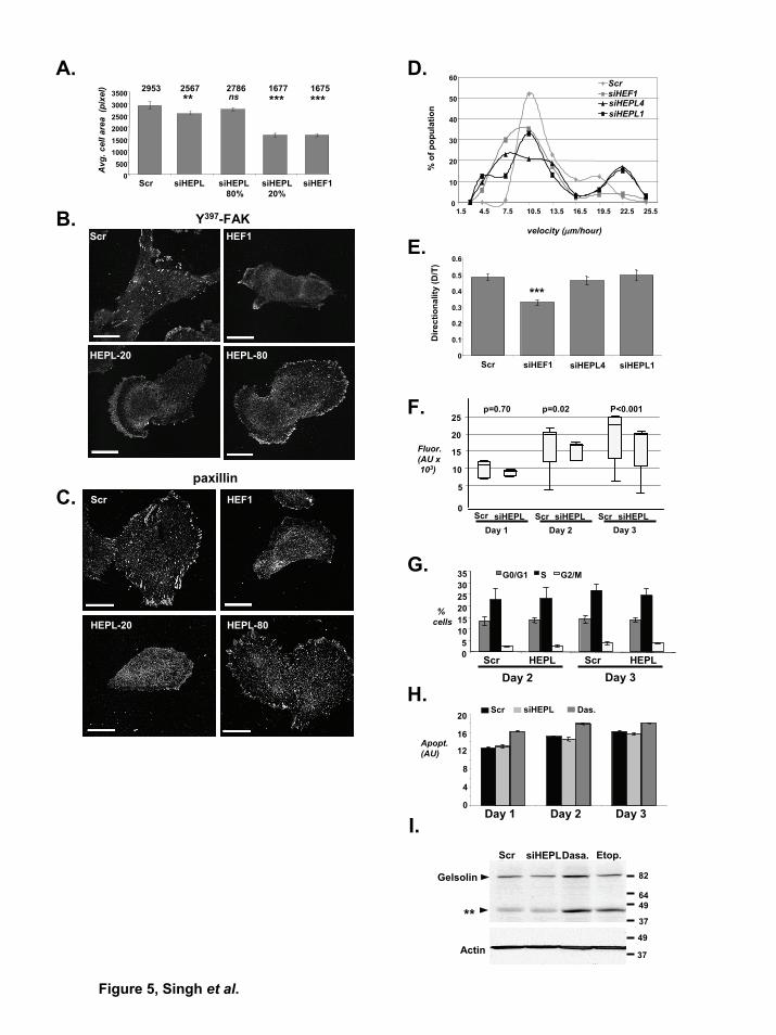
Figure 5, Singh et al.
F.
G.
E.
5
10
20
15
25
Fluor.(AU x103)
p=0.70 p=0.02 P<0.001
Scr siHEPL Scr siHEPL Scr siHEPLDay 1 Day 2 Day 3
0
05
101520253035 G0/G1 S G2/M
% cells
Scr Scr HEPLHEPL
Day 2 Day 3
Actin49
37
Gelsolin 82
4964
37**
Scr siHEPLDasa. Etop.
0
4
8
12
16
20
Day 1 Day 2 Day 3
Apopt.(AU)
Scr siHEPL Das.
A.
B.
C.
D.2953 2786 1677 1675
0500
100015002000250030003500
Scr siHEPL 80%
siHEPL20%
siHEF1
Avg
. cel
l are
a (p
ixel
) 2567
siHEPL
** *** ***ns
Y397-FAKScr HEF1
HEPL-20 HEPL-80
paxillinScr HEF1
HEPL-20 HEPL-80
0
10
20
30
40
50
60
1.5 4.5 7.5 10.5 13.5 16.5 19.5 22.5 25.5
velocity (μm/hour)
% o
f pop
ulat
ion
ScrsiHEF1siHEPL4siHEPL1
I.
H.
0
0.1
0.2
0.3
0.4
0.5
0.6
Scr siHEF1 siHEPL4 siHEPL1
Dire
ctio
nalit
y (D
/T)
***
Copyright © 2022 FDOKUMEN