The chromosome passenger complex is required for fidelity of chromosome transmission and cytokinesis...
9
4292 Research Article Introduction The chromosome passenger complex (CPC) comprises the Aurora B kinase (also known as serine/threonine-protein kinase 12) in a complex with INCENP, Survivin (also known as BIRC5) and Borealin (hDasra-B) and is required to regulate the attachments of kinetochores to microtubules, to ensure proper mitotic checkpoint function and for cytokinesis (reviewed by Ruchaud et al., 2007). Unlike other metazoans that have only the B-form of Aurora kinase in the CPC, vertebrates have another form, known as Aurora C (serine/threonine-protein kinase 13), that is expressed at high levels in the germ line and in a number of cancer cells (Bernard et al., 1998; Kimura et al., 1999; Tseng et al., 1998; Hu et al., 2000). The forced overexpression of an Aurora-C-kinase-deficient mutant in cultured cells disrupts the Aurora-B–INCENP complex and induces polyploidy (Chen et al., 2005). Moreover, Aurora C can complement Aurora B function in mitotic cells (Sasai et al., 2004), and its localisation to centromeric regions of chromosomes and then to the spindle midzone in late anaphase indicates that it also acts as a passenger protein (Yan et al., 2005). The function of Aurora C in the germ lines of disease-free animals is not clear. It has been thought to be redundant with Aurora B in the male germ line because the two enzymes have overlapping distributions in spermatocytes (Tang et al., 2006). It seems possible, however, that Aurora kinases B and C have independent roles in spermatogenesis because overexpression of dominant-negative Aurora B results in abnormal spermatocytes, increased apoptosis and spermatogenic arrest, and Aurora-C-null mice have compromised fertility owing to a variety of sperm defects (Kimmins et al., 2007). Evidence for a yet-uncharacterised role for Aurora C in meiosis I in mammals comes from the formation of tetraploid spermatozoa in sterile human males homozygous for a mutation in the human gene AURKC (Dieterich et al., 2007; Dieterich et al., 2009). In the mouse, AurC –/– females showed sterility, but unfortunately the oocyte phenotype was never examined before the line was lost (P. Sassone-Corsi, personal communication). Several other studies on mouse oocytes have used small-molecule inhibitors of Aurora kinases and shown that these affect meiotic progression in a variety of ways (Uzbekova et al., 2007; Swain et al., 2008; Vogt et al., 2009). However, because these compounds can inhibit all three Aurora kinases, attributing roles to specific enzymes from these studies is difficult. Thus, whether Aurora C has a role in female meiosis and whether its role can be substituted by Aurora B have remained uncertain. There are several key differences between meiotic and mitotic divisions that offer the possibility for differential behavior of M- phase regulatory molecules. The meiotic maturation of mouse oocytes commences with germinal vesicle breakdown (GVBD), which is followed by chromosome condensation and spindle Accepted 2 September 2010 Journal of Cell Science 123, 4292-4300 © 2010. Published by The Company of Biologists Ltd doi:10.1242/jcs.067447 Summary The existence of two forms of the chromosome passenger complex (CPC) in the mammalian oocyte has meant that its role in female meiosis has remained unclear. Here we use loss- and gain-of function approaches to assess the meiotic functions of one of the shared components of these complexes, INCENP, and of the variable kinase subunits, Aurora B or Aurora C. We show that either the depletion of INCENP or the combined inhibition of Aurora kinases B and C activates the anaphase-promoting complex or cyclosome (APC/C) before chromosomes have properly congressed in meiosis I and also prevents cytokinesis and hence extrusion of the first polar body. Overexpression of Aurora C also advances APC/C activation and results in cytokinesis failure in a high proportion of oocytes, indicative of a dominant effect on CPC function. Together, this points to roles for the meiotic CPC in functions similar to the mitotic roles of the complex: correcting chromosome attachment to microtubules, facilitating the spindle-assembly checkpoint (SAC) function and enabling cytokinesis. Surprisingly, overexpression of Aurora B leads to a failure of APC/C activation, stabilization of securin and consequently a failure of chiasmate chromosomes to resolve – a dominant phenotype that is completely suppressed by depletion of INCENP. Taken together with the differential distribution of Aurora proteins B and C on chiasmate chromosomes, this points to differential functions of the two forms of CPC in regulating the separation of homologous chromosomes in meiosis I. Key words: Aurora B, Aurora C, Chromosome passenger complex, INCENP, Meiosis, Mouse oocyte The chromosome passenger complex is required for fidelity of chromosome transmission and cytokinesis in meiosis of mouse oocytes Bedra Sharif 1,2, *, Jie Na 1,3, *, Karin Lykke-Hartmann 1 , Stephen H. McLaughlin 4 , Ernest Laue 4 , David M. Glover 2,‡,§ and Magdalena Zernicka-Goetz 1,‡,§ 1 University of Cambridge, Wellcome Trust and Cancer Research UK Gurdon Institute, Tennis Court Road, Cambridge, UK CB2 1NR 2 University of Cambridge, Department of Genetics, Downing Street, Cambridge, UK CB2 3EH 3 School of Medicine, Tsinghua University, Beijing 100084, China 4 University of Cambridge, Department of Biochemistry, Tennis Court Road, Cambridge, UK CB2 1QW *These authors contributed equally to this work ‡ These authors contributed equally to this work § Authors for correspondence ([email protected]; [email protected]) Journal of Cell Science
-
Upload
independent -
Category
Documents
-
view
2 -
download
0
Transcript of The chromosome passenger complex is required for fidelity of chromosome transmission and cytokinesis...

4292 Research Article
Introduction
The chromosome passenger complex (CPC) comprises the Aurora
B kinase (also known as serine/threonine-protein kinase 12) in a
complex with INCENP, Survivin (also known as BIRC5) and
Borealin (hDasra-B) and is required to regulate the attachments of
kinetochores to microtubules, to ensure proper mitotic checkpoint
function and for cytokinesis (reviewed by Ruchaud et al., 2007).
Unlike other metazoans that have only the B-form of Aurora kinase
in the CPC, vertebrates have another form, known as Aurora C
(serine/threonine-protein kinase 13), that is expressed at high levels
in the germ line and in a number of cancer cells (Bernard et al.,
1998; Kimura et al., 1999; Tseng et al., 1998; Hu et al., 2000). The
forced overexpression of an Aurora-C-kinase-deficient mutant in
cultured cells disrupts the Aurora-B–INCENP complex and induces
polyploidy (Chen et al., 2005). Moreover, Aurora C can
complement Aurora B function in mitotic cells (Sasai et al., 2004),
and its localisation to centromeric regions of chromosomes and
then to the spindle midzone in late anaphase indicates that it also
acts as a passenger protein (Yan et al., 2005).
The function of Aurora C in the germ lines of disease-free
animals is not clear. It has been thought to be redundant with
Aurora B in the male germ line because the two enzymes have
overlapping distributions in spermatocytes (Tang et al., 2006). It
seems possible, however, that Aurora kinases B and C have
independent roles in spermatogenesis because overexpression of
dominant-negative Aurora B results in abnormal spermatocytes,
increased apoptosis and spermatogenic arrest, and Aurora-C-null
mice have compromised fertility owing to a variety of sperm
defects (Kimmins et al., 2007). Evidence for a yet-uncharacterised
role for Aurora C in meiosis I in mammals comes from the
formation of tetraploid spermatozoa in sterile human males
homozygous for a mutation in the human gene AURKC (Dieterich
et al., 2007; Dieterich et al., 2009). In the mouse, AurC–/– females
showed sterility, but unfortunately the oocyte phenotype was never
examined before the line was lost (P. Sassone-Corsi, personalcommunication). Several other studies on mouse oocytes haveused small-molecule inhibitors of Aurora kinases and shown that
these affect meiotic progression in a variety of ways (Uzbekova et
al., 2007; Swain et al., 2008; Vogt et al., 2009). However, because
these compounds can inhibit all three Aurora kinases, attributing
roles to specific enzymes from these studies is difficult. Thus,
whether Aurora C has a role in female meiosis and whether its role
can be substituted by Aurora B have remained uncertain.
There are several key differences between meiotic and mitotic
divisions that offer the possibility for differential behavior of M-
phase regulatory molecules. The meiotic maturation of mouse
oocytes commences with germinal vesicle breakdown (GVBD),
which is followed by chromosome condensation and spindle
Accepted 2 September 2010Journal of Cell Science 123, 4292-4300 © 2010. Published by The Company of Biologists Ltddoi:10.1242/jcs.067447
Summary
The existence of two forms of the chromosome passenger complex (CPC) in the mammalian oocyte has meant that its role in female
meiosis has remained unclear. Here we use loss- and gain-of function approaches to assess the meiotic functions of one of the shared
components of these complexes, INCENP, and of the variable kinase subunits, Aurora B or Aurora C. We show that either the depletion
of INCENP or the combined inhibition of Aurora kinases B and C activates the anaphase-promoting complex or cyclosome (APC/C)
before chromosomes have properly congressed in meiosis I and also prevents cytokinesis and hence extrusion of the first polar body.
Overexpression of Aurora C also advances APC/C activation and results in cytokinesis failure in a high proportion of oocytes,
indicative of a dominant effect on CPC function. Together, this points to roles for the meiotic CPC in functions similar to the mitotic
roles of the complex: correcting chromosome attachment to microtubules, facilitating the spindle-assembly checkpoint (SAC) function
and enabling cytokinesis. Surprisingly, overexpression of Aurora B leads to a failure of APC/C activation, stabilization of securin and
consequently a failure of chiasmate chromosomes to resolve – a dominant phenotype that is completely suppressed by depletion of
INCENP. Taken together with the differential distribution of Aurora proteins B and C on chiasmate chromosomes, this points to
differential functions of the two forms of CPC in regulating the separation of homologous chromosomes in meiosis I.
Key words: Aurora B, Aurora C, Chromosome passenger complex, INCENP, Meiosis, Mouse oocyte
The chromosome passenger complex is required for
fidelity of chromosome transmission and cytokinesis
in meiosis of mouse oocytes
Bedra Sharif1,2,*, Jie Na1,3,*, Karin Lykke-Hartmann1, Stephen H. McLaughlin4, Ernest Laue4,David M. Glover2,‡,§ and Magdalena Zernicka-Goetz1,‡,§
1University of Cambridge, Wellcome Trust and Cancer Research UK Gurdon Institute, Tennis Court Road, Cambridge, UK CB2 1NR2University of Cambridge, Department of Genetics, Downing Street, Cambridge, UK CB2 3EH3School of Medicine, Tsinghua University, Beijing 100084, China4University of Cambridge, Department of Biochemistry, Tennis Court Road, Cambridge, UK CB2 1QW*These authors contributed equally to this work‡These authors contributed equally to this work§Authors for correspondence ([email protected]; [email protected])
Journ
al of C
ell S
cie
nce

formation. The individualization of chromosomes upon their
condensation appears to play an important part in permitting
assembly of the first meiotic spindle (Schuh and Ellenberg, 2007).
During the prometaphase of the first meiotic division, the spindle
migrates to the oocyte cortex to undertake meiosis I in an actin-
and Cdc42-dependent process (Halet and Carroll, 2007; Na and
Zernicka-Goetz, 2006). Tension is generated at metaphase because
homologous chromosomes are held together by chiasmata at the
sites of recombination, as opposed to sister chromatids being held
together at centromeres, as in mitosis. The chiasmata are resolved
when cohesion between chromatid arms, but not centromeres, is
lost through the activity of Separase, which is activated through
the destruction of its Securin partner mediated by the APC/C
(Kudo et al., 2006; McGuinness et al., 2009). Just as in mitosis,
the meiotic activation of the APC/C normally does not occur until
chromosome bi-orientation has been achieved and, until this time,
is kept inactive by the spindle-assembly checkpoint (SAC).
Consequently, depletion of the checkpoint protein Mad2 (Homer
et al., 2005) or the expression of its dominant-negative form
(Wassmann et al., 2003) overcomes the meiotic block induced by
nocodazole. Similarly, a dominant-negative Bub1 advances the
polar body (PB) extrusion (Tsurumi et al., 2004), as does removal
of Bub1 protein in a conditional knockout mouse line (McGuinness
et al., 2009). Once the checkpoint is satisfied, dyads, each
comprising one parental and one recombinant chromatid, segregate
to the opposite poles. The highly asymmetrical positioning of the
spindle ensures the extrusion of a small daughter cell from the egg,
the first PB.
The scant knowledge about the function of the CPC in female
meiosis in mammals has led us to use a series of approaches to
modulate expression of its key components during mouse oocyte
maturation. Depletion of INCENP or combined inhibition of Aurora
kinases B and C demonstrates that CPC function is required to
correct chromosome alignment, assure function of the SAC and to
enable cytokinesis to extrude the PB. However, two aspects of our
data also indicate functional differences between forms of CPC
containing either the Aurora B or Aurora C kinase. First, the two
Aurora kinases show differential localization on chiasmate
chromosomes in meiosis I. Second, we find that expression of
exogenous Aurora B stabilises Securin, leading to failure of
resolution of chiasmate chromosomes, whereas exogenous
expression of Aurora C phenocopies knockdown of INCENP.
Results
INCENP is required for chromosome congression in
meiosis I and II and for cytokinesis
To determine the roles of the CPC in female meiosis, we first
sought to deplete INCENP. To this end, we used RNAi, previously
shown to be effective for the specific downregulation of genes in
mouse oocytes and pre-implantation embryos (Wianny and
Zernicka-Goetz, 2000; Svoboda et al., 2000). We microinjected
oocytes at the germinal vesicle (GV) stage with various
combinations of five different INCENP siRNAs, cultured such
injected oocytes with the phosphodiesterase 3 inhibitor milrinone
to delay GVBD and allow RNAi to take effect and, after 14 hours,
released them to fresh culture medium to determine whether they
could undergo normal meiotic maturation. Knockdowns of 95%
of transcript levels were confirmed by quantitative rtPCR, with no
reduction of levels being observed with siRNAs against scrambled
nucleotide, GFP and GAPDH. To follow chromosome behaviour
upon INCENP depletion, we used time-lapse microscopy and
4293CPC in female meiosis
performed experiments on oocytes that were also injected with
histone H2B–EGFP RNA (Hadjantonakis and Papaioannou, 2004).
To examine the destruction dynamics of Securin–GFP (Hagting et
al., 2002), oocytes were injected with the relevant mRNA at levels
that had no observable effects upon the timing of either GVBD or
on extrusion of the PB (Fig. 1A,B). We found that, under these
conditions, control RNAi oocytes matured normally. We observed
the prometaphase arrays of chromosomes migrating to the cortex
at 2 hours, progressing into anaphase at around 7.5 hours, reaching
cytokinesis around 9 hours and arresting in metaphase II by 10
hours (Fig. 1A). By contrast, meiotic maturation was perturbed in
INCENP RNAi oocytes (Fig. 1B). Although anaphase of meiosis
I took place in all INCENP-depleted oocytes, half of these oocytes
(49%, n106) failed to extrude a PB, in comparison with 8%
(n94) of control oocytes. Those INCENP-depleted oocytes that
failed to extrude the PB underwent membrane ruffling at 8 hours
post GVBD, indicating a failed attempt to organise a contractile
furrow.
We observed the expected destruction of Securin that is required
to release Separase and mediate removal of the Rec8 cohesin subunit
from sister chromatid arms and thus resolve chiasmata (Kudo et al.,
2006; Herbert et al., 2003). This occurred concomitant with the
onset of anaphase both in control oocytes and in all INCENP-
depleted oocytes (respective profiles from three representative
oocytes are shown in Fig. 1A�B�). To calculate the rate of change of
Securin protein and to determine the value and timing of maximal
APC/C activity for each oocyte, we applied a kinetic analysis
(McGuinness et al., 2009) (see also Materials and Methods) and
expressed APC/C activity as its rate constant (kd[APC]). Maximal
APC/C activity was 3.2 hours–1±0.2 (n29) or kd[APC]3.7
hours–1±0.1 (n57) and occurred between 8.0 hours and 8.5 hours
post GVBD, a range of values similar to those described by
McGuinness and colleagues (McGuinness et al., 2009). Maximal
APC/C activity was slightly lower in INCENP-depleted than in
control oocytes {kd[APC]2.5 hours–1±0.1 (n46); Fig. 1C}, and the
time of maximal activity was advanced by 1–2 hours (Fig. 1D).
Thus, INCENP-depleted oocytes activate the APC/C prematurely
and, following Securin destruction, proceed to metaphase II.
The destruction of Securin and progression of anaphase in
INCENP-RNAi oocytes suggested that homologues were being
separated in meiosis I. However, the temporal advancement of these
processes raised the possibility that anaphase might be taking place
before chromosomes were properly aligned. This led us to compare
the positioning of chromosomes on spindles in fixed preparations of
control and INCENP-depleted oocytes at times immediately
preceding the predicted metaphase–anaphase transition of meiosis I
and at maturation. In meiosis I, the chromosomes in control oocytes
adopted a chiasmate arrangement, with their kinetochores being
pulled towards the poles. The chromosomes of INCENP-depleted
oocytes were much less well aligned at metaphase (Fig. 1E). We also
observed that oocytes depleted of INCENP exited metaphase
transiently, and sometimes telophase nuclei briefly formed before a
spindle reassembled for meiosis II. The metaphase II spindle that
formed in such oocytes was populated by numerous chromosome
masses that failed to align correctly on a metaphase plate (Fig. 1E).
This was in contrast to the tightly aligned chromosomes in control
oocytes in meiosis II. When we compared the chromosomes of
control and INCENP-depleted oocytes in spread preparations, we
found similar sets of chiasmate chromosomes in meiosis I of
both types. However, mature oocytes contained 20 univalent
chromosomes, whereas the INCENP-depleted oocytes that failed to
Journ
al of C
ell S
cie
nce

extrude a PB had predominantly 40 univalents, with some oocytes
showing a few persisting bivalents (Fig. 1E). Thus, the total number
of univalents present on spindles of such INCENP-depleted oocytes
was consistent with the observed failure of cytokinesis. In summary,
the depletion of INCENP allowed oocytes to progress to meiosis II
but affected extrusion of the PB. The majority of chromosomes were
well condensed and resolved into univalents, indicating that Separase
had been activated by destruction of Securin. Thus, chromosomes
became scattered along the length of the spindle in both divisions,
indicating that depletion of the INCENP passenger complex permitted
anaphase before complete congression of bivalents in meiosis I and
of univalents in meiosis II.
4294 Journal of Cell Science 123 (24)
Inhibition of Aurora B and Aurora C phenocopies INCENP
depletion
The ability of approximately one half of the INCENP-RNAi-treated
oocytes to undertake cytokinesis suggested to us that we were not
fully depleting the target protein, although we were unsuccessful
in finding an antibody against INCENP that would allow us to
quantitate the level of knockdown. This led us to ask what the
consequences might be of pharmacologically inhibiting the function
of the CPC in oocytes. Of the many small molecules have been
developed as inhibitors of Aurora kinase, most cross-react with
both Aurora A (serine/threonine-protein kinase 6) and Aurora B
(reviewed in Pollard and Mortimore, 2009). There are, however,
Fig. 1. Depletion of INCENP leads to
misaligned chromosomes, advancement of
APC/C activation and prevents cytokinesis.
(A,B)Green fluorescence of Securin–GFP and a
digital interference contrast (DIC) image of
oocytes is presented for a time-series of a control
oocyte injected with an irrelevant siRNA (A) or
siRNA against INCENP. (B). The scale bar for
all time-lapse images represents 50m.
(A�,B�) Quantitation of total fluorescence of
Securin–GFP in three representative oocytes
showing degradation profiles for RNAi control
(blue) and INCENP-RNAi-treated (red) oocytes.
‘RF’ in this and subsequent figures refers to the
relative fluorescence of Securin–GFP (see the
Materials and Methods section and the
supplementary material for a description of the
kinetic parameters). (C,D)Maximal APC/C
activity and time taken to reach maximal activity,
respectively, for every oocyte in the indicated
groups. The minus sign (–) denotes oocytes
otherwise untreated except for the injection of
mRNA encoding Securin–GFP (green). Oocytes
labeled ‘control’ are treated with an irrelevant
siRNA. (E)Oocytes stained to reveal DNA (red)
and microtubules (green) for controls in meiosis
I (MI) and meiosis II (MII; upper row) and
INCENP RNAi treated at MI and at 10 hours
having failed to extrude the PB (lower row).
Preparations of spread chromosomes are shown
alongside each stained panel. The scale bar for
the immunofluorescence images in A and B
represents 10m and, for the chromosome
spreads, 20m. (F)Knockdown of INCENP
transcripts assessed by qrtPCR for the indicated
concentrations of siRNA.
Journ
al of C
ell S
cie
nce
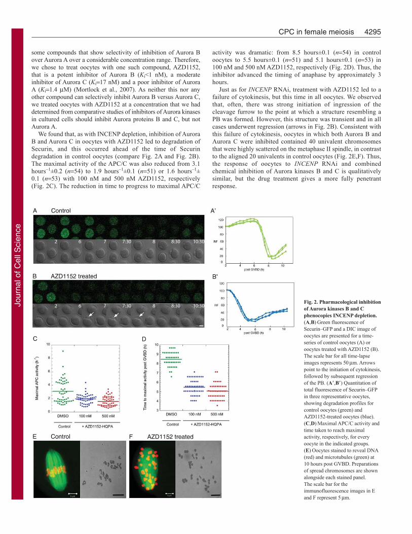
some compounds that show selectivity of inhibition of Aurora B
over Aurora A over a considerable concentration range. Therefore,
we chose to treat oocytes with one such compound, AZD1152,
that is a potent inhibitor of Aurora B (Ki<1 nM), a moderate
inhibitor of Aurora C (Ki17 nM) and a poor inhibitor of Aurora
A (Ki1.4 M) (Mortlock et al., 2007). As neither this nor any
other compound can selectively inhibit Aurora B versus Aurora C,
we treated oocytes with AZD1152 at a concentration that we had
determined from comparative studies of inhibitors of Aurora kinases
in cultured cells should inhibit Aurora proteins B and C, but not
Aurora A.
We found that, as with INCENP depletion, inhibition of Aurora
B and Aurora C in oocytes with AZD1152 led to degradation of
Securin, and this occurred ahead of the time of Securin
degradation in control oocytes (compare Fig. 2A and Fig. 2B).
The maximal activity of the APC/C was also reduced from 3.1
hours–1±0.2 (n54) to 1.9 hours–1±0.1 (n51) or 1.6 hours–1±
0.1 (n53) with 100 nM and 500 nM AZD1152, respectively
(Fig. 2C). The reduction in time to progress to maximal APC/C
4295CPC in female meiosis
activity was dramatic: from 8.5 hours±0.1 (n54) in control
oocytes to 5.5 hours±0.1 (n51) and 5.1 hours±0.1 (n53) in
100 nM and 500 nM AZD1152, respectively (Fig. 2D). Thus, the
inhibitor advanced the timing of anaphase by approximately 3
hours.
Just as for INCENP RNAi, treatment with AZD1152 led to a
failure of cytokinesis, but this time in all oocytes. We observed
that, often, there was strong initiation of ingression of the
cleavage furrow to the point at which a structure resembling a
PB was formed. However, this structure was transient and in all
cases underwent regression (arrows in Fig. 2B). Consistent with
this failure of cytokinesis, oocytes in which both Aurora B and
Aurora C were inhibited contained 40 univalent chromosomes
that were highly scattered on the metaphase II spindle, in contrast
to the aligned 20 univalents in control oocytes (Fig. 2E,F). Thus,
the response of oocytes to INCENP RNAi and combined
chemical inhibition of Aurora kinases B and C is qualitatively
similar, but the drug treatment gives a more fully penetrant
response.
Fig. 2. Pharmacological inhibition
of Aurora kinases B and C
phenocopies INCENP depletion.
(A,B)Green fluorescence of
Securin–GFP and a DIC image of
oocytes are presented for a time-
series of control oocytes (A) or
oocytes treated with AZD1152 (B).
The scale bar for all time-lapse
images represents 50m. Arrows
point to the initiation of cytokinesis,
followed by subsequent regression
of the PB. (A�,B�) Quantitation of
total fluorescence of Securin–GFP
in three representative oocytes,
showing degradation profiles for
control oocytes (green) and
AZD1152-treated oocytes (blue).
(C,D)Maximal APC/C activity and
time taken to reach maximal
activity, respectively, for every
oocyte in the indicated groups.
(E)Oocytes stained to reveal DNA
(red) and microtubules (green) at
10 hours post GVBD. Preparations
of spread chromosomes are shown
alongside each stained panel.
The scale bar for the
immunofluorescence images in E
and F represent 5m.
Journ
al of C
ell S
cie
nce

Aurora B and Aurora C differ in their dominant effects on
meiotic progression upon elevated expression
Aurora B and Aurora C share a high degree of amino acid sequence
similarity, they can each phosphorylate histone H3, and, in somatic
cells, Aurora C can form complexes with INCENP, the known
Aurora B partner, and complement the function of Aurora B (Chen
et al., 2005; Sasai et al., 2004; Li et al., 2004). Nevertheless, they
have been reported to have differing distributions on chiasmate
chromosomes during meiosis I, suggesting that their functions
might not be identical. In spermatozoa, Aurora C localises uniquely
to the interchromatid axes and chiasmata, whereas Aurora B is at
centromeres (Tang et al., 2006). In agreement with previous studies
(Shuda et al., 2009), we found a similar distribution of Aurora
proteins B and C following expression of the GFP- or HA-tagged
kinases in oocytes and by immunostaining of endogenous Aurora
kinases B and C (supplementary material Fig. S1A–D). We were,
however, only able to detect Aurora B associated with chromosomes
in meiosis I and not meiosis II. This localisation of both enzymes
was lost following downregulation of INCENP (supplementary
material Fig. S2 and also below).
To address whether the differing chromosomal distributions of
the two kinases might reflect different functions, we first attempted
to downregulate each Aurora by RNAi. We injected various
combinations of six Aurora B siRNAs at concentrations sufficient
to downregulate specifically greater than 95% of Aurora B RNA
but not Aurora A or Aurora C. However, this proved insufficient to
eliminate Aurora B protein completely, and it had no observable
effects upon meiotic progression (supplementary material Fig.
S1F,G). Attempts to downregulate Aurora C by RNAi led to a
similar outcome (supplementary material Figs S3, S4). We therefore
asked whether, by increasing the concentrations of injected siRNAs,
we might be able to achieve a more substantial knockdown. We
found that, when we used much higher concentrations of siRNAs
than those needed to give specific knockdown of transcripts levels,
this led to a SAC-mediated arrest of maturation in meiosis I.
However, this turned out to be a nonspecific response of the oocyte
to the very high concentrations of siRNA because we observed the
same phenotype with all siRNAs used at such concentrations,
including those directed against scrambled sequences
(supplementary material Fig. S4). Thus, downregulation of these
two kinases independently of each other proved impossible in our
hands in mouse oocytes.
We therefore next examined whether overexpression of Aurora
B or Aurora C, by injecting their synthetic mRNAs, could itself
affect meiotic progression. This revealed that overexpression of
Aurora C decreased the proportion of oocytes successfully
completing maturation (from 84%, n97, to 58%, n104). Through
time-lapse imaging, we found that the dominant-negative phenotype
exhibited by oocytes expressing both endogenous and exogenous
AurC mRNA was a failure of cytokinesis. Instead, at the time of
cytokinesis in the control oocytes, oocytes overexpressing Aurora
C underwent a period of membrane ruffling (Fig. 3A,B).
Nevertheless, the Securin–GFP assay indicated that Securin was
degraded, both in oocytes that failed and those that succeeded in
extruding a PB. This was reflected by the activation of the APC/C
to levels similar to those in oocytes expressing endogenous and
exogenous Aurora C in which cytokinesis failed {kd[APC]2.8
hours–1±0.3 (n39)} and in those in which it succeeded
{kd[APC]2.3 hours–1±0.1 (n35)}. These values were comparable
to values in the control group of oocytes {kd[APC]1.9
hours–1±0.1 (n84)} (Fig. 3D). Interestingly, this elevation of
4296 Journal of Cell Science 123 (24)
Aurora C expression led to advancement of maximal APC/C
activity in all oocytes (t6.7±0.1 hours and 6.8±0.1 hours in the
two groups) compared with 9.3±0.2 hours in control oocytes (Fig.
3E). Immunostaining of oocytes that failed to extrude PBI, as a
result of this elevation of Aurora C expression, revealed spindles
having low levels of chromosome misalignment (one or two
univalents misaligned out of 40) yet fully separated univalents
(Fig. 3F,G; supplementary material Fig. S5A). Thus, Securin
degradation led to the release of Separase and the separation of
homologues in meiosis I, but, as a result of cytokinesis failure,
such oocytes now contained twice the normal complement of
univalents. The similarity of this phenotype to that seen following
depletion of INCENP suggested that the elevated levels of Aurora
C disrupt the CPC complex and its functions.
By contrast, oocytes injected with a similar concentration of
AurB mRNA tended to arrest during maturation (56%, n134). In
those oocytes, degradation of Securin occurred only extremely
slowly {kd[APC]0.6 hours–1±0.1 (n71)}, with the time to
maximal APC/C activity being extended to a mean of 13.3±0.4
hours (Fig. 3C,D). The oocytes that were extensively delayed in
meiosis I had bivalent chromosomes that appeared to be under
tension, with well-separated kinetochores (Fig. 3F; supplementary
material Fig. S5B). To observe the morphology of chromosomes
in more detail, we performed preparations of chromosome spreads,
which revealed 20 chiasmate bivalents, indicating that the
homologues had not separated (Fig. 3G). This is consistent with a
requirement for degradation of Securin to release Separase and
mediate release of the cojoined chromatid arms in the bivalents
(Kudo et al., 2006). The failure to activate the APC/C properly in
order to degrade Securin when levels of Aurora B, but not Aurora
C, are elevated would therefore be expected to lead to persistence
of chiasmate chromosomes. This finding of a different dominant
phenotype resulting from the overexpression of Aurora B compared
with Aurora C suggests that the Aurora B and Aurora C forms of
the CPC have different functions during disjunction of homologues
in meiosis I.
Dominant phenotype resulting from Aurora B
overexpression is suppressed by INCENP depletion
As Aurora B requires INCENP for its activation and function, we
considered whether INCENP was required both for the localization
of Aurora B and for the dominant phenotype resulting from its
overexpression. To address this, we first examined the localization
of ectopic GFP-tagged Aurora B throughout oocyte maturation and
the consequences of its expression. We found that GFP–Aurora-B
began to localize to chromosomes as they condensed around the
time of GVBD and throughout prometaphase. The kinase then
retained localization on the chromatin but became more pronounced
on the kinetochores of chiasmate chromosomes as they entered a
prolonged period of metaphase arrest (5–15 hours post GVBD;
Fig. 4A). Of the oocytes injected with mRNA encoding Aurora-B–
GFP, 88% (n16) showed such arrest. When oocytes were co-
injected with the same concentration of Aurora-B–GFP mRNA
together with siRNA against INCENP, the resulting Aurora-B–
GFP no longer localized to any structure in the cell. Moreover,
86% (n14) no longer arrested at meiosis I and typically entered
anaphase 6 hours post GVBD. They transiently entered a telophase-
like state at around 6.5 hours but did not undertake cytokinesis
before arresting in a metaphase-II-like state but with highly
disorganized chromosomes (Fig. 4B). Thus, downregulation of
INCENP prevented the association of Aurora B with chromosomes
Journ
al of C
ell S
cie
nce

and suppressed its dominant phenotype thus allowing the resolution
of chiasmata.
Discussion
In this study, we have addressed the functions of the two forms of
the CPC, containing either Aurora B or Aurora C, in meiosis in the
mouse oocyte. This has revealed that, although the CPC is not
essential for the execution of the first meiotic division per se, it is
required for the fidelity of this process and the subsequent execution
of cytokinesis. Loss of the CPC in mouse oocytes as a result of
depletion of INCENP led to a failure to correct chromosome
misalignment, advancement of APC/C activation and progression
through anaphase of meiosis I. Cytokinesis failed and, consequently,
oocytes had 40 univalents that also failed to align at metaphase in
meiosis II. Thus, the functions of the meiotic CPC appear to mirror
4297CPC in female meiosis
those of its mitotic counterpart in correcting mis-attached
kinetochores and ensuring SAC activity to control mitotic exit
(Ditchfield et al., 2003; Hauf et al., 2003; Ciferri et al., 2008). The
use of the pharmacological inhibitor of Aurora kinase AZD1152
confirmed the involvement of the CPC in the above cellular
processes. This compound is, respectively, 1000 and 100 times
more efficacious against Aurora B or Aurora C than against Aurora
A (Mortlock et al., 2007) and is thus more selective than compounds
previously used to investigate Aurora kinase function in oocyte
maturation. Neither VX680, used to treat bovine oocytes (Uzbekova
et al., 2007), nor ZM447439, used to treat mouse oocytes (Swain
et al., 2008; Vogt et al., 2009; Shuda et al., 2009), shows sufficient
selectivity to attribute specific functions to the three different
Aurora kinases. Vogt and colleagues (Vogt et al., 2009) suggested
that defects in chromosome condensation, congression, chiasmata
Fig. 3. Overexpression of AurC mRNA prevents
cytokinesis; overexpression of AurB mRNA prevents
APC/C activation and leads to arrest with chiasmate
dyads. Green fluorescence of Securin–GFP and a DIC image
of oocytes are presented for each series in which images are
presented at the indicated time-points. (A)Time-lapse series
of Securin in three representative control oocytes.
(A�)Quantitation of total fluorescence of Securin–GFP in the
three oocytes shown in (A). (B)Time-lapse series of Securin
in three representative oocytes into which AurC mRNA had
been injected at 0.5g/l. (B�)Quantitation of total
fluorescence of Securin–GFP in the three oocytes shown in
(B). Note that Securin destruction occurs in all oocytes,
although cytokinesis [PB extrusion (PBE)] fails. 58% of
oocytes degraded Securin and extruded PBs (black data-
points in panels D and E). (C)Time-lapse series of Securin in
three representative oocytes into which AurB mRNA had
been injected at 0.5g/l. The scale bar referring to all time-
lapse images represents 100m. (C�)Quantitation of total
fluorescence of Securin–GFP in the three oocytes shown in
C. Note the slowed destruction of Securin in these oocytes
that arrested in MI. 44% of oocytes progressed to MII and
showed destruction of Securin (red data-points in panels D
and E). (D)Values of maximal APC/C activity and (E) time
to maximal activity post GVBD for every oocyte in all
groups analysed. Oocytes expressing synthetic AurC mRNA
have been divided into two categories, depending upon
whether they failed to exclude a polar body (no PBE; purple
data-points) or succeeded (PBE; black data-points). Oocytes
expressing synthetic AurB mRNA have also been divided
into two categories, depending upon whether they arrested in
MI (MI; blue data-points) or progressed to MII (MII; red
data-points). (F)Oocytes injected with synthetic mRNA
encoding HA-tagged Aurora kinases B or C, fixed 10 hours
post GVBD and stained to reveal HA–Aurora-B or HA–
Aurora-C (green), tubulin (red) and DNA (blue). Oocytes
overexpressing HA–Aurora-C have a meiosis II spindle with
some misaligned chromosomes. Oocytes overexpressing
HA–Aurora-B have a meiosis I spindle with a chiasmate
arrangement of dyads. (G)Preparations of chromosome
spreads from the indicated number of oocytes that had failed
to extrude a PB as a result of expressing exogenous Aurora C
(note the 40 univalents), and preparations of chromosome
spreads from the indicated number of oocytes that had failed
to extrude a PB as a result of expressing exogenous Aurora B
(note the 20 chiasmate bivalents). The scale bar for the
immunofluorescence images represents 10m and, for the
chromosome spreads, 20m.
Journ
al of C
ell S
cie
nce

resolution and PB extrusion were due to inhibition of Aurora B,
but this conclusion does not take into account the pan-Aurora-
kinase-inhibitory properties of this small molecule. Shuda and
colleagues (Shuda et al., 2009) concluded that defects in
chromosome alignment could result from inhibition of Aurora B
because they could be rescued by increasing the expression of this
enzyme. However, the dominant phenotype we observe following
elevated expression of Aurora B suggests that other explanations
are possible.
The existence of two forms of the CPC is a peculiarity of the
germ line. In normal tissues, Aurora C appears to be expressed
predominantly in the germ line, although it is also present in some
cancer cells, where it can compete with Aurora B for binding to
INCENP (Chen et al., 2005) and can also complement the mitotic
function of Aurora B (Sasai et al., 2004; Li et al., 2004). Two lines
of evidence from our current study suggest that the form of the
CPC containing Aurora C has a subset of functions that differ from
the Aurora B form in the first meiotic division of mouse oocytes.
First, the two kinases localise differentially in meiosis I: Aurora C
is found predominantly along the chromosome arms that hold the
homologues together, whereas Aurora B predominates at the
centromeric regions – a similar pattern is also seen in spermatocytes
(Tang et al., 2006). Only following the resolution of chiasmata do
the kinases behave as typical CPC proteins at the mid-zone of the
central spindle before moving onto the centromeres of metaphase
II chromosomes, as also described recently by Shuda and colleagues
(Shuda et al., 2009). The dependence of this localization of the
kinase upon its CPC partners is demonstrated by our observation
that, following depletion of INCENP, Aurora C fails to localize
either to chromatin from GVBD onwards or to the central spindle
in anaphase. Second, the arrest in meiosis I with stable levels of
4298 Journal of Cell Science 123 (24)
Securin and, consequently, unresolved chiasmata following the
overexpression of Aurora B, but not of Aurora C, points to the
possibility of their differing functions in resolving these
recombinant intermediates at the metaphase–anaphase transition of
meiosis I. Aurora C would seem to be the main contributor to
cytokinesis as increasing its levels to concentrations that would
effectively compete with endogenous Aurora C for the binding of
INCENP advances APC/C activation and results in some
cytokinesis defects. This phenotype strongly resembles that seen
following loss of INCENP or combined inactivation of the two
kinases. Although we were unable to deplete either Aurora B or C
kinase by RNAi in the oocyte, owing to the presence of the
maternally inherited protein, this was possible in the zygote, where
we found cytokinesis was dependent upon Aurora C rather than
Aurora B (data not shown) (Lykke-Andersen et al., 2008) (M.
Malumbres, personal communication).
Definitive resolution of the relative functions of Aurora kinases
B and C in female meiosis will most likely require reassessment
of the phenotypes of genetic knockouts. Previous studies of
spermatogenesis in AurC–/– mice (Kimmins et al., 2007) reported
defects in sperm morphology but did not fully examine meiosis.
Both male and female AurC–/– mice showed sterility, but
unfortunately, owing to these fertility problems, this knockout line
has been lost and so further examination of the basis of this sterility
cannot be undertaken (P. Sassone-Corsi, personal communication).
AURKC mutations have been described for humans, where they
prevent meiosis I in males, leading to the formation of polyploid,
multi-tailed spermatozoa (Dieterich et al., 2007; Dieterich et al.,
2009). However, two women have been found who are homozygous
for this same AURKC mutation and yet are fertile (Dieterich et al.,
2009). Thus, it is possible that Aurora B might be able to substitute
for Aurora C function in female meiosis in the human.
Taken together, our data suggest that Aurora B and Aurora C
might have overlapping, yet partially independent, roles in meiosis
in the mouse. The different response of oocytes to the
overexpression of the two kinases, together with their differential
localization, suggests that, in addition to a common set of CPC
partners, they might have additional differing partners or alternative
substrates. It is noteworthy that two recent papers have shown that
the spatial localization of Aurora A and its choice of partners is
affected by a single amino acid in its primary sequence – mutating
glycine 198 in Aurora A to its asparagine counterpart found at this
site in both Aurora B and Aurora C leads the mutant Aurora A to
behave as a passenger protein (Fu et al., 2009; Hans et al., 2009).
The differential behavior of Aurora B and Aurora C in meiosis
might depend upon similarly subtle differences in their primary
sequence. Exactly how their differential localization and functions
are achieved necessitates future investigation of the precise partners
and/or substrates of Aurora C at its different chromosome locations
and of how its choice of partners and spatial behaviour might be
influenced by its primary sequence.
Materials and MethodsOocyte and embryo collection
Oocytes were collected from wild-type F1 (C57BL/6xCBA) females (Charles River)or from females transgenic for histone-H2B–EGFP (Hajantonakis and Papaioannou,2004) as described previously (Wianny and Zernicka-Goetz, 2000). GV oocytes werecollected from ovary tissue in M2 medium supplemented with 4 mg/ml BSA, asdescribed previously (Na and Zernicka-Goetz, 2006). Milrinone (Sigma) was added toa final concentration of 100 M where indicated. For embryos, superovulated F1females were mated with F1 males. Zygotes were collected 15 hours post treatmentwith human chorionic gonadotrophin (hCG), in M2 containing 200 IU/ml ofhyaluronidase and washed in fresh M2, then cultured in KSOM medium with 4 mg/mlBSA under paraffin oil in an atmosphere of 5% CO2 at 37.5°C (Gray et al., 2004).
Fig. 4. Depletion of INCENP prevents localization of exogenous Aurora B
and suppresses the dominant phenotype of its expression. (A)Time-lapse
series showing chromatin (histone-H2B–RFP; red) from oocytes injected with
synthetic mRNA encoding Aurora-B–GFP (green). Note the localization of
Aurora B to paired homologues held in a chiasmate configuration for at least
10 hours. (B)Time-lapse series showing chromatin (histone-H2B–RFP; red)
from INCENP-RNAi-treated oocytes injected with synthetic mRNA encoding
Aurora-B–GFP (green). Note the failure of Aurora B to localize to
chromosomes or the central spindle and the completion of meiosis I, as
described in the text, in the absence of cytokinesis. Scale bar: 10m.
Journ
al of C
ell S
cie
nce

4299CPC in female meiosis
In vitro fertilization
Spermatozoa released from caudae epididymides of F1 males were suspended in 0.5
ml of fertilization medium (Fraser, 1983) containing 4 mg/ml BSA and incubated for
1.5 hours to allow capacitation and a spontaneous acrosome reaction. The sperm
concentration was approximately 2�107 spermatozoa/ml. Before fertilization, zonae
pellucidae were removed by exposure of oocytes to acidic Tyrode’s solution (pH 2.5)
(Nicolson et al., 1975). Oocytes were placed in 100 l droplets of fertilization
medium, and 1 l of the preincubated sperm suspension was added (final
concentration of sperm suspension was approximately 2�105 spermatozoa/ml). After
30 minutes, the oocytes were pipetted several times to remove loosely attached
spermatozoa and then cultured in KSOM.
cDNA cloning, mRNA synthesis, siRNA preparation and microinjection
Mouse AurC and AurB were cloned with Superscript One-step RT-PCR for long
templates (Invitrogen) using gene-specific primers (supplementary material Table
S1). HA-tagged (N-terminus) and GFP-tagged (C-terminus) Aurora C and Aurora B
were engineered and subcloned into a RN3P vector for in vitro transcription of
mRNA, as described previously (Zernicka-Goetz et al., 1997). siRNA against 5� and
3� UTRs of AurC and control siRNAs were designed and purchased from Invitrogen
(supplementary material Table S2). Microinjection of mRNA into oocytes was
performed as described previously (Na and Zernicka-Goetz, 2006). Annealed siRNAs
were dissolved in DNase, RNase-free water.
Live-imaging experiments
Time-lapse movies were performed either on a Zeiss Axiovert 200M epifluorescence
microscope or a DeltaVision Olympus microscope equipped with 37°C incubator
and 5% CO2 supply, as described previously (Na and Zernicka-Goetz, 2006). The
acquisition of digital time-lapse images was controlled by AQM6 (Andor/Kinetic-
imaging) and DeltaVision software packages.
Securin–GFP fluorescence was quantitated on each image and normalized at
completion of the experiment to assign a 100% value at 5 hours post GVBD. The
data sets were used to quantitate the rate of change of Securin and APC/C activity,
as described below.
Calculation of APC/C activity from the dynamics of Securin–GFP levels
APC/C activity was calculated from changes in the level of Securin–GFP fluorescence
using the analysis devised by McGuinness and colleagues (McGuinness et al., 2009).
In this scheme, the rate of change of Securin–GFP level (dS/dt) is dependent upon
two opposing kinetic processes: production through translation from mRNA and
degradation through APC/C-dependent and -independent processes:
Therefore:
where ks is the kinetic constant for protein synthesis, Mo is the initial concentration
of Securin–GFP mRNA, kd and k�d are the rate constants for APC/C-dependent and -
independent degradation, respectively, S is the concentration of Securin–GFP at time
t and [APC] is the concentration of APC/C. McGuinness and colleagues (McGuinness
et al., 2009) showed that mRNA degradation is negligible over the time course of
the experiment. In control experiments, where [APC] falls to a low level post-PBE
and Securin–GFP fluorescence recovers to ~80% of the initial value, Eqn 1 reduces
to:
Hence:
at t�, SSmax, therefore:
Fitting of recovery-phase data in this control experiment with Eqn 5 gave values
for k�d (in the range of 0.16 to 0.32 hours–1 – of the same order as reported by
McGuinness and colleagues of 0.1 hours–1 for APC/C-minus cells) and ksMo as
Smaxks Mo/k�d. These kinetic parameters were used in Eqn 2 to calculate APC/C
activity (kd[APC]) during oocyte maturation from the levels of S and its rate of
change (dS/dt) calculated at each time point. However, in the majority of experiments,
where there was substantial APC/C activity post PBE and hence recovery of Securin–
GFP to less than 80% of the initial value, typical values of k�d were used and ks Mo
was calculated from Smax* k�d. The validity of this approach can be seen from Eqn 2,
where the S and dS/dt terms dominate, and the absolute values of k�d and ks Mo do not
significantly affect the calculated values of APC/C activity.
dS
dt= ksMo − ( ′kd + kd [APC ])S . (1)
kd [APC ] =ksMo −
dS
dt
S
⎛
⎝
⎜⎜⎜
⎞
⎠
− ′kd , (2)
dS
dt= ksMo − ′kd S . (3)
′′kd
S =ksMo
1 − e− ′kd t( ) , (4)
S = Sm ax 1 − e− ′kd t( ) . (5)
Antibodies, immunostaining and confocal microscopy
The following primary antibodies were used: monoclonal rat anti-tubulin (YL1/2),
polyclonal rabbit anti-phospho-H3S10 and polyclonal rabbit anti-GFP (290) were
from Abcam. Monoclonal rat anti-HA (3F10) and monoclonal mouse anti-GFP were
from Roche. The antibody against Aurora B was Santa Cruz Biotechnology anti-
ARK2 (H-75) and was raised against the N-terminal 75 amino acids specific to
human AurB, a region absent from Aurora C. Our in-house antibody against mouse
Aurora C was raised in rabbits against peptide amino acids 1–17 by Harlan Sera.
Oocytes were first treated with acidic Tyrode’s solution to remove the zona pellucida.
Fixation and immunostaining were performed as described previously.
Immunofluorescence images were acquired on a Bio-Rad MRC-1024 and an Olympus
FV1000 confocal microscope with a �63 oil-immersion objective.
Chromosome spread assay
Oocytes were first incubated in a hypotonic solution (1% Na Citrate) for 10 minutes,
then put on a glass slide, fixed with a drop of freshly made fixative [methanol:acetic
acid (3:1)], dried at room temperature and stained with Giemsa (BDH, product
350862M).
We thank Anna Ajduk for help in assaying Aurora B function in thesecond meiosis, Bernhard Strauss for help in establishing conditionsfor monitoring Securin degradation and Jonathan Pines for the gift ofthe Securin–GFP construct; we also thank Helen Bolton for examiningthe role of Aurora C in embryos and John Crang for help in compilingfigures. This work was supported by a Wellcome Trust grant to M.Z.-G.and an MRC Programme Grant to D.M.G. B.S. held a Gates FoundationStudentship of the University of Cambridge. J.N. is supported by aMRC Career Development Fellowship. K.L.-H. was supported by theAlfred Benzon Stipend, Denmark (current address: Institute of MedicalBiochemistry, Aarhus University, Denmark). Part of the imageacquisition was carried out in the Wellcome-Trust-supported lightmicroscopy facility in the University of Sheffield, UK (grant:GR077544AIA). Confocal microscope facilities in the D.M.G.laboratory were supported by grants from CR-UK and BBSRC.Deposited in PMC for release after 6 months.
Supplementary material available online at
http://jcs.biologists.org/cgi/content/full/123/24/4292/DC1
ReferencesBernard, M., Sanseau, P., Henry, C., Couturier, A. and Prigent, C. (1998). Cloning of
STK13, a third human protein kinase related to Drosophila aurora and budding yeast
Ipl1 that maps on chromosome 19q13.3-ter. Genomics 53, 406-409.
Chen, H. L., Tang, C. J., Chen, C. Y. and Tang, T. K. (2005). Overexpression of an
Aurora-C kinase-deficient mutant disrupts the Aurora-B/INCENP complex and induces
polyploidy. J. Biomed. Sci. 12, 297-310.
Ciferri, C., Pasqualato, S., Screpanti, E., Varetti, G., Santaguida, S., Dos Reis, G.,
Maiolica, A., Polka, J., De Luca, J. G., De Wulf, P. et al. (2008). Implications for
kinetochore-microtubule attachment from the structure of an engineered Ndc80 complex.
Cell 133, 427-439.
Dieterich, K., Soto Rifo, R., Faure, A. K., Hennebicq, S., Ben Amar, B., Zahi, M.,
Perrin, J., Martinez, D., Sèle, B., Jouk, P. S. et al. (2007). Homozygous mutation of
AURKC yields large-headed polyploid spermatozoa and causes male infertility. Nat.
Genet. 39, 661-665.
Dieterich, K., Zouari, R., Harbuz, R., Vialard, F., Martinez, D., Bellayou, H., Prisant,
N., Zoghmar, A., Guichaoua, M. R., Koscinski, I. et al. (2009). The Aurora Kinase
C c.144delC mutation causes meiosis I arrest in men and is frequent in the North
African population. Hum. Mol. Genet. 18, 1301-1309.
Ditchfield, C., Johnson, V. L., Tighe, A., Ellston, R., Haworth, C., Johnson, T.,
Mortlock, A., Keen, N. and Taylor, S. S. (2003). Aurora B couples chromosome
alignment with anaphase by targeting BubR1, Mad2, and Cenp-E to kinetochores. J.
Cell Biol. 161, 267-280.
Fraser, L. R. (1983). Mouse sperm capacitation assessed by kinetics and morphology of
fertilization in vitro. J. Reprod. Fertil. 69, 419-428.
Fu, J., Bian, M., Liu, J., Jiang, Q. and Zhang, C. (2009). A single amino acid change
converts Aurora-A into Aurora-B-like kinase in terms of partner specificity and cellular
function. Proc. Natl. Acad. Sci. USA 106, 6939-6944.
Gray, D., Plusa, B., Piotrowska, K., Na, J., Tom, B., Glover, D. M. and Zernicka-
Goetz, M. (2004). First cleavage of the mouse embryo responds to change in egg shape
at fertilization. Curr. Biol. 14, 397-405.
Hadjantonakis, A. K. and Papaioannou, V. E. (2004). Dynamic in vivo imaging and cell
tracking using a histone fluorescent protein fusion in mice. BMC Biotechnol. 4, 33.
Hagting, A., Den Elzen, N., Vodermaier, H. C., Waizenegger, I. C., Peters, J. M. and
Pines, J. (2002). Human Securin proteolysis is controlled by the spindle checkpoint and
reveals when the APC/C switches from activation by Cdc20 to Cdh1. J. Cell Biol. 157,
1125-1137.
Halet, G. and Carroll, J. (2007). Rac activity is polarized and regulates meiotic spindle
stability and anchoring in mammalian oocytes. Dev. Cell 12, 309-317.
Journ
al of C
ell S
cie
nce

4300 Journal of Cell Science 123 (24)
Hans, F., Skoufias, D. A., Dimitrov, S. and Margolis, R. L. (2009). Molecular distinctions
between Aurora A and B: a single residue change transforms Aurora A into correctly
localized and functional Aurora B. Mol. Biol. Cell 20, 3491-3502.
Hauf, S., Cole, R. W., LaTerra, S., Zimmer, C., Schnapp, G., Walter, R., Heckel, A.,
van Meel, J., Rieder, C. L. and Peters, J. M. (2003). The small molecule Hesperadin
reveals a role for Aurora B in correcting kinetochore-microtubule attachment and in
maintaining the spindle assembly checkpoint. J. Cell Biol. 161, 281-294.
Herbert, M., Levasseur, M., Homer, H., Yallop, K., Murdoch, A. and McDougall, A.
(2003). Homologue disjunction in mouse oocytes requires proteolysis of Securin and
cyclin B1. Nat. Cell Biol. 5, 1023-1025.
Homer, H. A., McDougall, A., Levasseur, M., Yallop, K., Murdoch, A. P. and Herbert,
M. (2005). Mad2 prevents aneuploidy and premature proteolysis of cyclin B and
Securin during meiosis I in mouse oocytes. Genes Dev. 19, 202-207.
Hu, H. M., Chuang, C. K., Lee, M. J., Tseng, T. C. and Tang, T. K. (2000). Genomic
organization, expression, and chromosome localization of a third aurora-related kinase
gene, Aie1. DNA Cell Biol. 19, 679-688.
Kimmins, S., Crosio, C., Kotaja, N., Hirayama, J., Monaco, L., Höög, C., van Duin,
M., Gossen, J. A. and Sassone-Corsi, P. (2007). Differential functions of the Aurora-
B and Aurora-C kinases in mammalian spermatogenesis. Mol. Endocrinol. 21, 726-
739.
Kimura, M., Matsuda, Y., Yoshioka, T. and Okano, Y. (1999). Cell cycle-dependent
expression and centrosome localization of a third human aurora/Ipl1-related protein
kinase, AIK3. J. Biol. Chem. 274, 7334-7340.
Kudo, N. R., Wassmann, K., Anger, M., Schuh, M., Wirth, K. G., Xu, H., Helmhart,
W., Kudo, H., McKay, M., Maro, B. et al. (2006). Resolution of chiasmata in oocytes
requires Separase-mediated proteolysis. Cell 126, 135-146.
Li, X., Sakashita, G., Matsuzaki, H., Sugimoto, K., Kimura, K., Hanaoka, F.,
Taniguchi, H., Furukawa, K. and Urano, T. (2004). Direct association with inner
centromere protein (INCENP) activates the novel chromosomal passenger protein,
Aurora-C. J. Biol. Chem. 279, 47201-47211.
Lykke-Andersen, K., Gilchrist, M. J., Grabarek, J. B., Das, P., Miska, E. and Zernicka-
Goetz, M. (2008). Maternal Argonaute 2 is essential for early mouse development at
the maternal-zygotic transition. Mol. Biol. Cell 19, 4383-4392.
McGuinness, B. E., Anger, M., Kouznetsova, A., Gil-Bernabe, A. M., Helmhart, W.,
Kudo, N. R., Wuensche, A., Taylor, S., Hoog, C., Novak, B. et al. (2009). Regulation
of APC/C activity in oocytes by a Bub1-dependent spindle assembly checkpoint. Curr.
Biol. 19, 369-380.
Mortlock, A. A., Foote, K. M., Heron, N. M., Jung, F. H., Pasquet, G., Lohmann, J.
J., Warin, N., Renaud, F., De Savi, C., Roberts, N. J. et al (2007). Discovery,
synthesis, and in vivo activity of a new class of pyrazoloquinazolines as selective
inhibitors of Aurora B kinase. J. Med. Chem. 50, 2213-2224.
Na, J. and Zernicka-Goetz, M. (2006). Asymmetric positioning and organization of the
meiotic spindle of mouse oocytes requires CDC42 function. Curr. Biol. 16, 1249-
1254.
Nicolson, G. L., Yanagimachi, R. and Yanagimachi, H. (1975). Ultrastructural localization
of lectin-binding sites on the zonae pellucidae and plasma membranes of mammalian
eggs. J. Cell Biol. 66, 263-274.
Pollard, J. R. and Mortimore, M. (2009). Discovery and development of aurora kinase
inhibitors as anticancer agents. J. Med. Chem. 52, 2629-2651.
Ruchaud, S., Carmena, M. and Earnshaw, W. C. (2007). Chromosomal passengers:
conducting cell division. Nat. Rev. Mol. Cell Biol. 8, 798-812.
Sasai, K., Katayama, H., Stenoien, D. L., Fujii, S., Honda, R., Kimura, M., Okano,
Y., Tatsuka, M., Suzuki, F., Nigg, E. A. et al. (2004). Aurora-C kinase is a novel
chromosomal passenger protein that can complement Aurora-B kinase function in
mitotic cells. Cell Motil. Cytoskeleton 59, 249-263.
Schuh, M. and Ellenberg, J. (2007). Self-organization of MTOCs replaces centrosome
function during acentrosomal spindle assembly in live mouse oocytes. Cell 130, 484-
498.
Shuda, K., Schindler, K., Ma, J., Schultz, R. M. and Donovan, P. J. (2009). Aurora
kinase B modulates chromosome alignment in mouse oocytes. Mol. Reprod. Dev. 76,
1094-1105.
Svoboda, P., Stein, P., Hayashi, H. and Schultz, R. M. (2000). Selective reduction of
dormant maternal mRNAs in mouse oocytes by RNA interference. Development 127,
4147-4156.
Swain, J. E., Ding, J., Wu, J. and Smith, G. D. (2008). Regulation of spindle and
chromatin dynamics during early and late stages of oocyte maturation by Aurora
kinases. Mol. Hum. Rep. 14, 291-299.
Tang, C. J., Lin, C. Y. and Tang, T. K. (2006). Dynamic localization and functional
implications of Aurora-C kinase during male mouse meiosis. Dev. Biol. 290, 398-410.
Tseng, T. C., Chen, S. H., Hsu, Y. P. and Tang, T. K. (1998). Protein kinase profile of
sperm and eggs: cloning and characterization of two novel testis-specific protein kinases
(AIE1, AIE2) related to yeast and fly chromosome segregation regulators. DNA Cell
Biol. 17, 823-833.
Tsurumi, C., Hoffmann, S., Geley, S., Graeser, R. and Polanski, Z. (2004). The spindle
assembly checkpoint is not essential for CSF arrest of mouse oocytes. J. Cell Biol. 167,
1037-1050.
Uzbekova, S., Arlot-Bonnemains, Y., Dupont, J., Dalbies-ran, R., Paillier, P., Pennetier,
S., Thelie, A., Perreau, C., Mermillod, P., Prigent, C. et al. (2007). Spatio-temporal
expression patterns of aurora kinases A, B, and C and cytoplasmic polyadenylation-
element-binding protein in bovine oocytes during meiotic maturation. Biol. Reprod. 78,
218-233.
Vogt, E., Kipp, A. and Eichenlaub-Ritter, U. (2009). Aurora kinase B, epigenetic state
of centromeric heterochromatin and chiasma resolution in oocytes. Reprod. Biomed.
Online 19, 352-368.
Wassmann, K., Niault, T. and Maro, B. (2003). Metaphase I arrest upon activation of
the Mad2-dependent spindle checkpoint in mouse oocytes. Curr. Biol. 13, 1596-1608.
Wianny, F. and Zernicka-Goetz, M. (2000). Specific interference with gene function by
double-stranded RNA in early mouse development. Nat. Cell Biol. 2, 70-75.
Yan, X., Cao, L., Li, Q., Wu,Y., Zhang, H., Saiyin, H., Liu, X., Zhang, X., Shi, Q. and
Yu, L. (2005). Aurora C is directly associated with Survivin and required for cytokinesis.
Genes Cells 10, 617-626.
Zernicka-Goetz, M., Pines, J., McLean Hunter, S., Dixon, J. P., Siemering, K. R.,
Haseloff, J. and Evans, M. J. (1997). Following cell fate in the living mouse embryo.
Development 124, 1133-1137.
Journ
al of C
ell S
cie
nce