Global analysis of X-chromosome dosage compensation
22
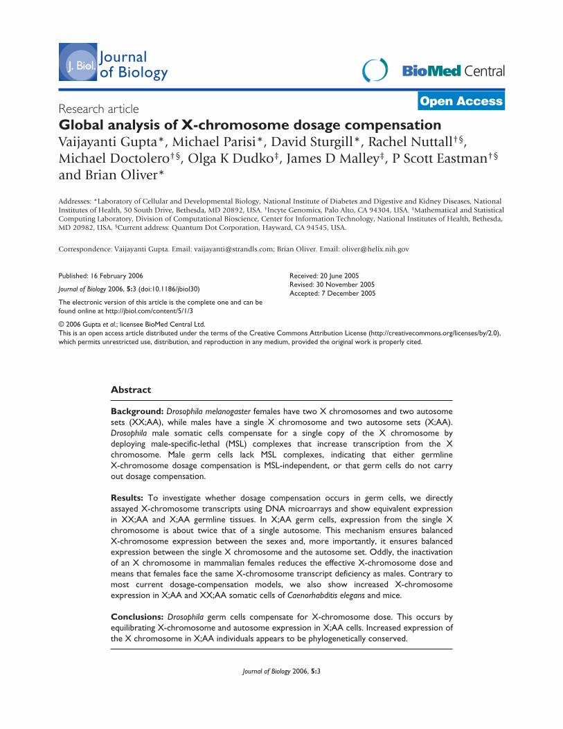
Research article Global analysis of X-chromosome dosage compensation Vaijayanti Gupta*, Michael Parisi*, David Sturgill*, Rachel Nuttall †§ , Michael Doctolero †§ , Olga K Dudko ‡ , James D Malley ‡ , P Scott Eastman †§ and Brian Oliver* Addresses: *Laboratory of Cellular and Developmental Biology, National Institute of Diabetes and Digestive and Kidney Diseases, National Institutes of Health, 50 South Drive, Bethesda, MD 20892, USA. † Incyte Genomics, Palo Alto, CA 94304, USA. ‡ Mathematical and Statistical Computing Laboratory, Division of Computational Bioscience, Center for Information Technology, National Institutes of Health, Bethesda, MD 20982, USA. § Current address: Quantum Dot Corporation, Hayward, CA 94545, USA. Correspondence: Vaijayanti Gupta. Email: [email protected]; Brian Oliver. Email: [email protected] Abstract Background: Drosophila melanogaster females have two X chromosomes and two autosome sets (XX;AA), while males have a single X chromosome and two autosome sets (X;AA). Drosophila male somatic cells compensate for a single copy of the X chromosome by deploying male-specific-lethal (MSL) complexes that increase transcription from the X chromosome. Male germ cells lack MSL complexes, indicating that either germline X-chromosome dosage compensation is MSL-independent, or that germ cells do not carry out dosage compensation. Results: To investigate whether dosage compensation occurs in germ cells, we directly assayed X-chromosome transcripts using DNA microarrays and show equivalent expression in XX;AA and X;AA germline tissues. In X;AA germ cells, expression from the single X chromosome is about twice that of a single autosome. This mechanism ensures balanced X-chromosome expression between the sexes and, more importantly, it ensures balanced expression between the single X chromosome and the autosome set. Oddly, the inactivation of an X chromosome in mammalian females reduces the effective X-chromosome dose and means that females face the same X-chromosome transcript deficiency as males. Contrary to most current dosage-compensation models, we also show increased X-chromosome expression in X;AA and XX;AA somatic cells of Caenorhabditis elegans and mice. Conclusions: Drosophila germ cells compensate for X-chromosome dose. This occurs by equilibrating X-chromosome and autosome expression in X;AA cells. Increased expression of the X chromosome in X;AA individuals appears to be phylogenetically conserved. BioMed Central Journal of Biology Journal of Biology 2006, 5:3 Open Access Published: 16 February 2006 Journal of Biology 2006, 5:3 (doi:10.1186/jbiol30) The electronic version of this article is the complete one and can be found online at http://jbiol.com/content/5/1/3 Received: 20 June 2005 Revised: 30 November 2005 Accepted: 7 December 2005 © 2006 Gupta et al.; licensee BioMed Central Ltd. This is an open access article distributed under the terms of the Creative Commons Attribution License (http://creativecommons.org/licenses/by/2.0), which permits unrestricted use, distribution, and reproduction in any medium, provided the original work is properly cited.
-
Upload
independent -
Category
Documents
-
view
2 -
download
0
Transcript of Global analysis of X-chromosome dosage compensation
Research articleGlobal analysis of X-chromosome dosage compensationVaijayanti Gupta*, Michael Parisi*, David Sturgill*, Rachel Nuttall†§,Michael Doctolero†§, Olga K Dudko‡, James D Malley‡, P Scott Eastman†§
and Brian Oliver*
Addresses: *Laboratory of Cellular and Developmental Biology, National Institute of Diabetes and Digestive and Kidney Diseases, NationalInstitutes of Health, 50 South Drive, Bethesda, MD 20892, USA. †Incyte Genomics, Palo Alto, CA 94304, USA. ‡Mathematical and StatisticalComputing Laboratory, Division of Computational Bioscience, Center for Information Technology, National Institutes of Health, Bethesda,MD 20982, USA. §Current address: Quantum Dot Corporation, Hayward, CA 94545, USA.
Correspondence: Vaijayanti Gupta. Email: [email protected]; Brian Oliver. Email: [email protected]
Abstract
Background: Drosophila melanogaster females have two X chromosomes and two autosomesets (XX;AA), while males have a single X chromosome and two autosome sets (X;AA).Drosophila male somatic cells compensate for a single copy of the X chromosome bydeploying male-specific-lethal (MSL) complexes that increase transcription from the Xchromosome. Male germ cells lack MSL complexes, indicating that either germlineX-chromosome dosage compensation is MSL-independent, or that germ cells do not carryout dosage compensation.
Results: To investigate whether dosage compensation occurs in germ cells, we directlyassayed X-chromosome transcripts using DNA microarrays and show equivalent expressionin XX;AA and X;AA germline tissues. In X;AA germ cells, expression from the single Xchromosome is about twice that of a single autosome. This mechanism ensures balancedX-chromosome expression between the sexes and, more importantly, it ensures balancedexpression between the single X chromosome and the autosome set. Oddly, the inactivationof an X chromosome in mammalian females reduces the effective X-chromosome dose andmeans that females face the same X-chromosome transcript deficiency as males. Contrary tomost current dosage-compensation models, we also show increased X-chromosomeexpression in X;AA and XX;AA somatic cells of Caenorhabditis elegans and mice.
Conclusions: Drosophila germ cells compensate for X-chromosome dose. This occurs byequilibrating X-chromosome and autosome expression in X;AA cells. Increased expression ofthe X chromosome in X;AA individuals appears to be phylogenetically conserved.
BioMed CentralJournalof Biology
Journal of Biology 2006, 5:3
Open Access
Published: 16 February 2006
Journal of Biology 2006, 5:3 (doi:10.1186/jbiol30)
The electronic version of this article is the complete one and can befound online at http://jbiol.com/content/5/1/3
Received: 20 June 2005Revised: 30 November 2005Accepted: 7 December 2005
© 2006 Gupta et al.; licensee BioMed Central Ltd. This is an open access article distributed under the terms of the Creative Commons Attribution License (http://creativecommons.org/licenses/by/2.0),which permits unrestricted use, distribution, and reproduction in any medium, provided the original work is properly cited.
BackgroundIn most organisms, copy number at any given locus haslittle effect on proper organismal function. Very few genesare deleterious if present in only one copy (haploinsuffi-ciency) or are overtly deleterious in three copies. Havingmore or fewer copies (aneuploidy) of a large fraction of thegenome is, however, invariably incompatible with viability.For example, over 10% of human oocytes are aneuploid,but with a few exceptions none of these aneuploid oocytesgives rise to viable offspring [1]. The most common aneu-ploid genotypes in a wide range of species involve the dele-tion or duplication of a chromosome or chromosomesegment. Deletions are the most deleterious.
In Drosophila melanogaster, a systematic study of aneuploidswith deletions of different segments of chromosomes indi-cates that having only a single copy of 1% of the genomereduces viability (and often fertility) and having only asingle copy of 3% or more of the genome is lethal [2]. Fromcurrent estimates of gene content in Drosophila, 3% repre-sents about 500 genes [3]. Therefore, having only a singlecopy of 500 genes or more usually results in the collapse ofa major part of the genetic network. That genetic networksdo indeed collapse because of minor differences in theexpression levels of a few connected nodes is evident fromgenetic interaction studies. In the female germline, forexample, the dose of the gene ovarian tumor (otu) stronglymodifies the sterility phenotype of flies heterozygous forovoD [4]. (The gene ovo encodes a transcription factor thatacts on otu [5]). Similarly, in the male germline, hetero-zygosity for haywire or �-tubulin mutations are tolerated, butheterozygosity for both results in failed spermatogenesis [6].
The sex chromosomes represent an extraordinary exceptionto the genetic imbalance rule. Drosophila males have onecopy of the X chromosome per diploid set of autosomes(X;AA) and females have two (XX;AA) [7]. As the DrosophilaX chromosome bears about 20% of the genome [3],Drosophila males vastly exceed the usual 3% single-copythreshold for viability. This is not due to an underrepresen-tation of dosage-sensitive genes on the X chromosome, asfemales are sensitive to X-chromosome deletions [2]. There-fore, males have a special mechanism(s) to compensate forX-chromosome dose (for reviews see [8,9]). An extensive setof autoradiographic experiments on the giant polytenechromosomes of the salivary gland showed that the X chro-mosome in X;AA flies is expressed at roughly twice the levelas an X chromosome in XX;AA flies. Hypertranscription ofthe X chromosome in karyotypic males is dependent on acomplex of at least five proteins and two non-coding RNAs.The genes encoding the proteins in the complex are referredto as the male specific lethal (msl) loci. Males lacking any ofthe msl activities show reduced X-chromosome transcription
and die as larvae. At the molecular level, these genes encodea histone-modifying MSL complex, which acetylates histone4 on lysine 16 (H4 K16). The modification is thought torelieve the general repressive action of histones and result inincreased transcription.
Interestingly, the MSLs do not function in the germline. The Xchromosomes of male germ cells are not decorated with MSLcomplexes and are not hyperacetylated at H4 K16 [10]. Fur-thermore, neither the genes encoding the MSL complex northe obligate somatic regulators of the MSLs are required forgermline viability [11,12]. There is similar lack of evidence ofdosage compensation in the germline of other organisms[13,14], leading to the hypothesis that germ cells are dosage-tolerant. Alternatively, dosage compensation in germ cellsmay be MSL-independent. Whether the germline X chromo-some of Drosophila, or indeed of any organism, is dosagecompensated is one of the major unresolved issues in thestudy of sex chromosomes. Our array results indicate thatDrosophila germ cells do, in fact, dosage compensate.
Equally enigmatic are the dosage-compensation systems inCaenorhabditis elegans and mammals, which are based onreducing X-chromosome expression in XX;AA cells [15,16].This is seen most clearly in mammals, where one of the Xchromosomes in XX;AA females is inactivated. In C. elegans,both the X chromosomes in XX;AA hermaphrodites showreduced expression. In both cases, the dosage-compensationmodel equilibrates X-chromosome expression between thesexes but it also makes both sexes functionally aneuploidwith respect to the autosomes. Both males and females (orhermaphrodites) become functionally X;AA. It has been sug-gested that this is counterintuitive, as within each diploidX;AA organism, gene expression from a single X chromosomeshould be equilibrated and balanced to the autosomes[17-20]. Therefore, it is more useful to think of X-chromo-some dosage as a mechanism for equilibrating X chromo-some and autosome expression, rather than as only amechanism for equilibrating expression between the sexes.This predicts, for example, that in mammals both the single Xchromosome of males and the single active X chromosome infemales are hypertranscribed [17,18]. While there is an over-whelming literature supporting X-chromosome inactivation,there has been very little experimental evidence to supporthypertranscription of the active X chromosome [21]. Ourexamination of array results in C. elegans and the mouse sug-gests that such X-chromosome hypertranscription does occur.
ResultsComparing XX;AA and X;AA expression ratiosGene expression in XX;AA and X;AA tissues can be simplyvisualized by plotting the hybridization intensities of the
3.2 Journal of Biology 2006, Volume 5, Article 3 Gupta et al. http://jbiol.com/content/5/1/3
Journal of Biology 2006, 5:3
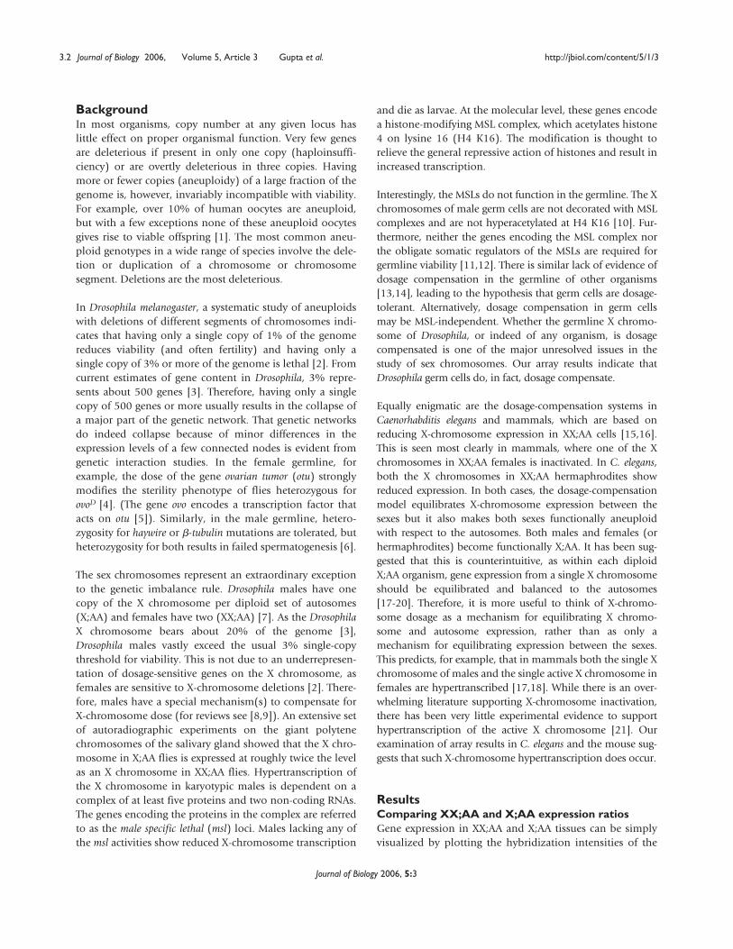
two samples in a scatterplot (Figure 1). There is modestsex-biased expression in the soma, but extensive sex-biasedexpression in the gonads. In the absence of dosage com-pensation, we would expect to see XX tissue expression attwice the level of X tissues. There is no evidence of such adistribution of X-chromosome expression ratios in eithertissue. The bulk of the XX versus X expression data pointsoverlies the bulk of the AA versus AA expression datapoints in both tissues. This indicates that the single X chro-mosomes of X;AA soma and gonads are expressed at thesame level as the two X chromosomes in XX;AA soma andgonads. This strongly suggests that somatic dosage com-pensation is widespread in adult tissues. More importantly,this provides the first strong evidence that X-chromosomedosage compensation occurs in the germline. It is,however, much more difficult to look for subtle effects ofgene dose in the germline because of the dramatic andextensive sex-biased expression observed between ovariesand testes: this sex bias affects about 31% of the genomeand results in a greater than tenfold difference in expres-sion level for some genes.
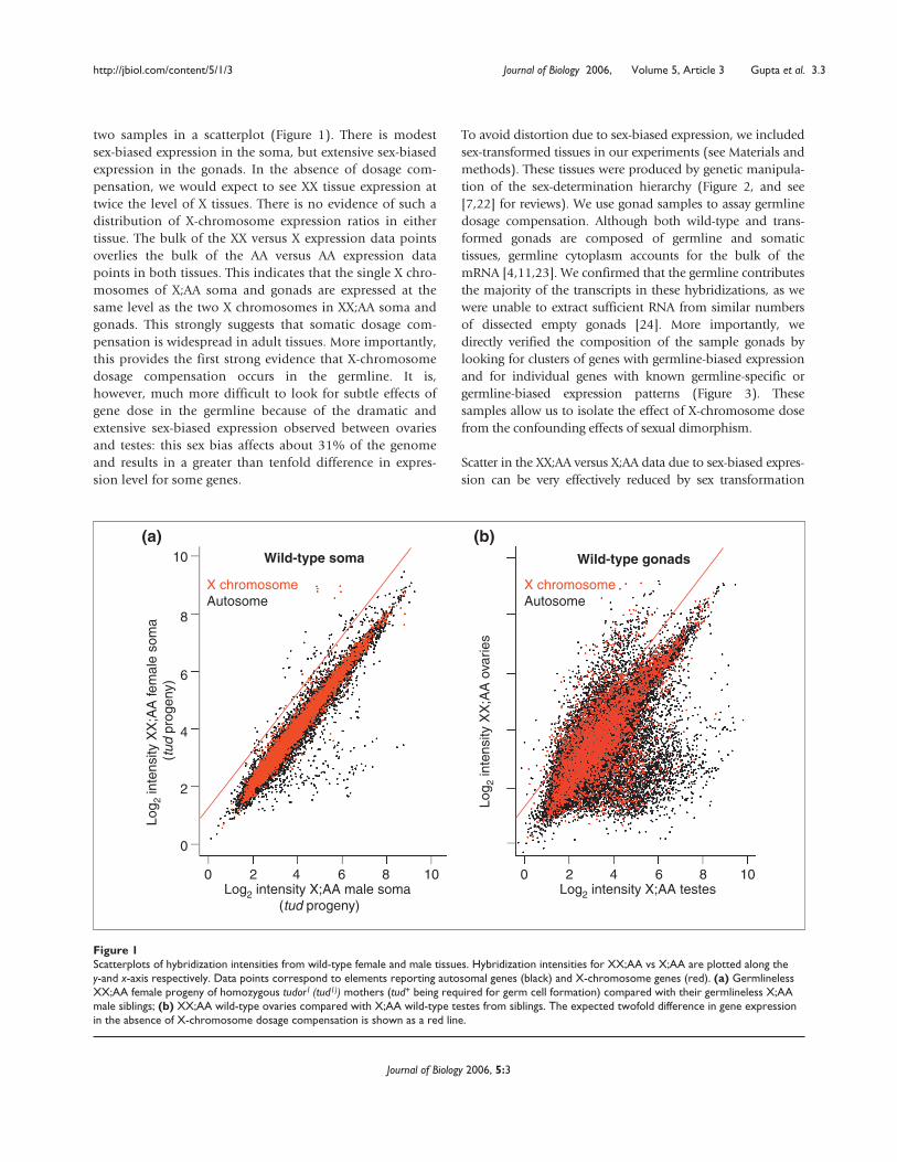
To avoid distortion due to sex-biased expression, we includedsex-transformed tissues in our experiments (see Materials andmethods). These tissues were produced by genetic manipula-tion of the sex-determination hierarchy (Figure 2, and see[7,22] for reviews). We use gonad samples to assay germlinedosage compensation. Although both wild-type and trans-formed gonads are composed of germline and somatictissues, germline cytoplasm accounts for the bulk of themRNA [4,11,23]. We confirmed that the germline contributesthe majority of the transcripts in these hybridizations, as wewere unable to extract sufficient RNA from similar numbersof dissected empty gonads [24]. More importantly, wedirectly verified the composition of the sample gonads bylooking for clusters of genes with germline-biased expressionand for individual genes with known germline-specific orgermline-biased expression patterns (Figure 3). Thesesamples allow us to isolate the effect of X-chromosome dosefrom the confounding effects of sexual dimorphism.
Scatter in the XX;AA versus X;AA data due to sex-biased expres-sion can be very effectively reduced by sex transformation
http://jbiol.com/content/5/1/3 Journal of Biology 2006, Volume 5, Article 3 Gupta et al. 3.3
Journal of Biology 2006, 5:3
Figure 1Scatterplots of hybridization intensities from wild-type female and male tissues. Hybridization intensities for XX;AA vs X;AA are plotted along they-and x-axis respectively. Data points correspond to elements reporting autosomal genes (black) and X-chromosome genes (red). (a) GermlinelessXX;AA female progeny of homozygous tudor1 (tud1)) mothers (tud+ being required for germ cell formation) compared with their germlineless X;AAmale siblings; (b) XX;AA wild-type ovaries compared with X;AA wild-type testes from siblings. The expected twofold difference in gene expressionin the absence of X-chromosome dosage compensation is shown as a red line.
Log2 intensity X;AA male soma (tud progeny)
Log 2
inte
nsity
XX
;AA
fem
ale
som
a (t
ud p
roge
ny)
X chromosomeAutosome
0 4 82 6 10
Wild-type soma
Log2 intensity X;AA testes
Log 2
inte
nsity
XX
;AA
ova
ries
0 4 82 6 10
Wild-type gonads
X chromosomeAutosome
0
2
4
6
8
10
(a) (b)
3.4 Journal of Biology 2006, Volume 5, Article 3 Gupta et al. http://jbiol.com/content/5/1/3
Journal of Biology 2006, 5:3
Figure 2 (see legend on the following page)
XX;AAkaryotype
Sxl
MSLs
tra dsxFFemale somaticdifferentiation
tra2
XX;AAkaryotype
ovo otu SxlOogenic
differentiation
X;AAkaryotype
MSLs
dsxMMale somaticdifferentiation
tra2
Dosagecompensation
X;AAkaryotype
? ? ?Spermatogenicdifferentiation
X;AAkaryotype
MSLs
dsxMtra2
Dosagecompensation
tra
dsxFFemale somaticdifferentiation
X;AAkaryotype
? ? ?No gametogenic
differentiation
No gametogenicdifferentiation
XX;AAkaryotype
Sxl
MSLs
tra dsxFtra2
dsxMMale somaticdifferentiation
XX;AAkaryotype
? ? ? Germlineatrophic
XX;AAkaryotype
ovo otu Sxl
XX;AAkaryotype
Sxl
MSLs
tra dsxFFemale somaticdifferentiation
tra2
Wild type
Sex transformed
Sex transformed
Wild type
Germline transformed
(a)
(b)
(c)
(d)
(e)
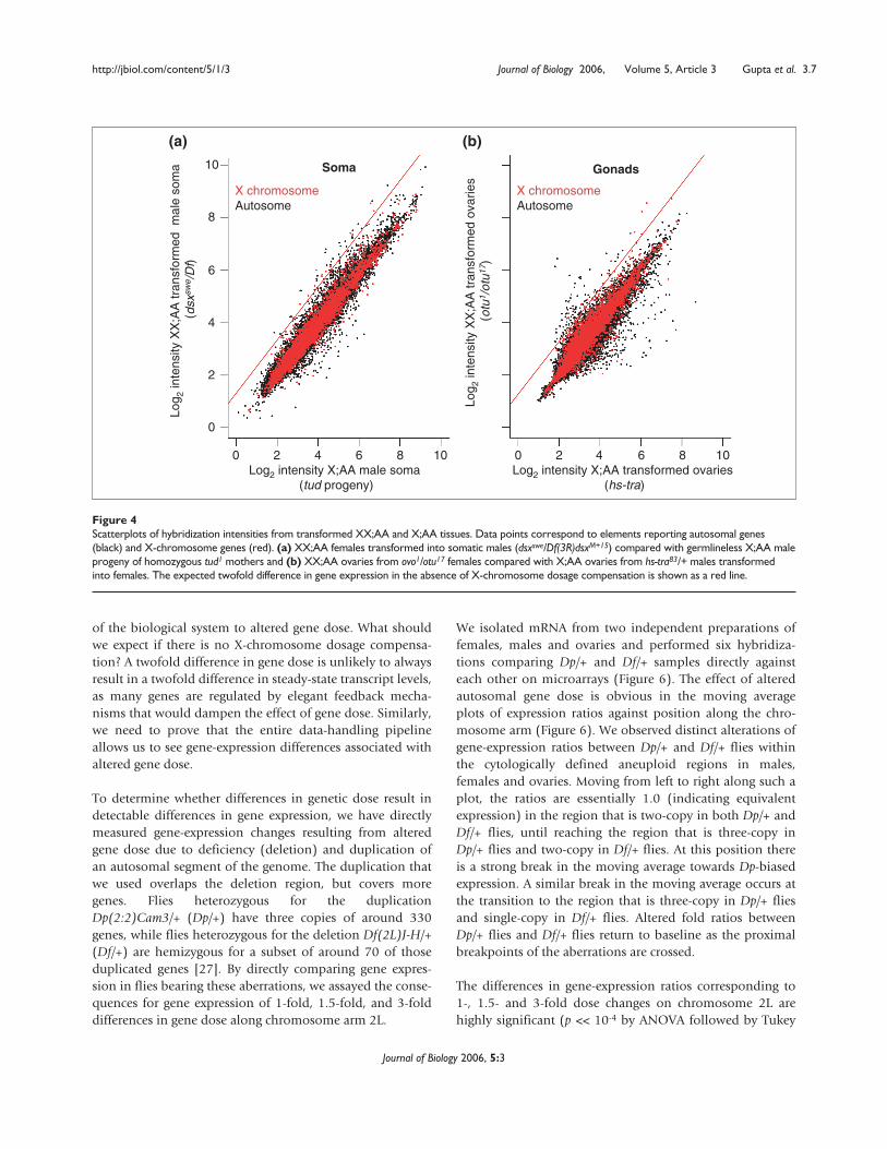
(Figure 4). When we compare expression of XX;AA andX;AA soma that were sexually matched (for example,females versus males transformed into females, and malesversus females transformed into males), we observe verylittle high-magnitude differential expression. Even morestriking is the similarity in the expression profiles of trans-formed ovaries bearing non-differentiating germ cells. Thehigh-magnitude differences in expression observed betweenwild-type XX;AA ovaries and X;AA testes (Figure 1) arenearly abolished when we compared XX;AA and X;AA trans-formed ovaries (Figure 4). The implications from visualexamination of the scatterplots are clear, as hybridization toarray elements corresponding to X-chromosome and auto-somal genes show expression ratios centered tightly onunity. We confirmed the veracity of this exploratory analysisusing multiple statistical tools (see the section Transcrip-tional response to altered autosomal gene dose, below).This strongly supports the idea that X-chromosome dosagecompensation occurs in the germline.
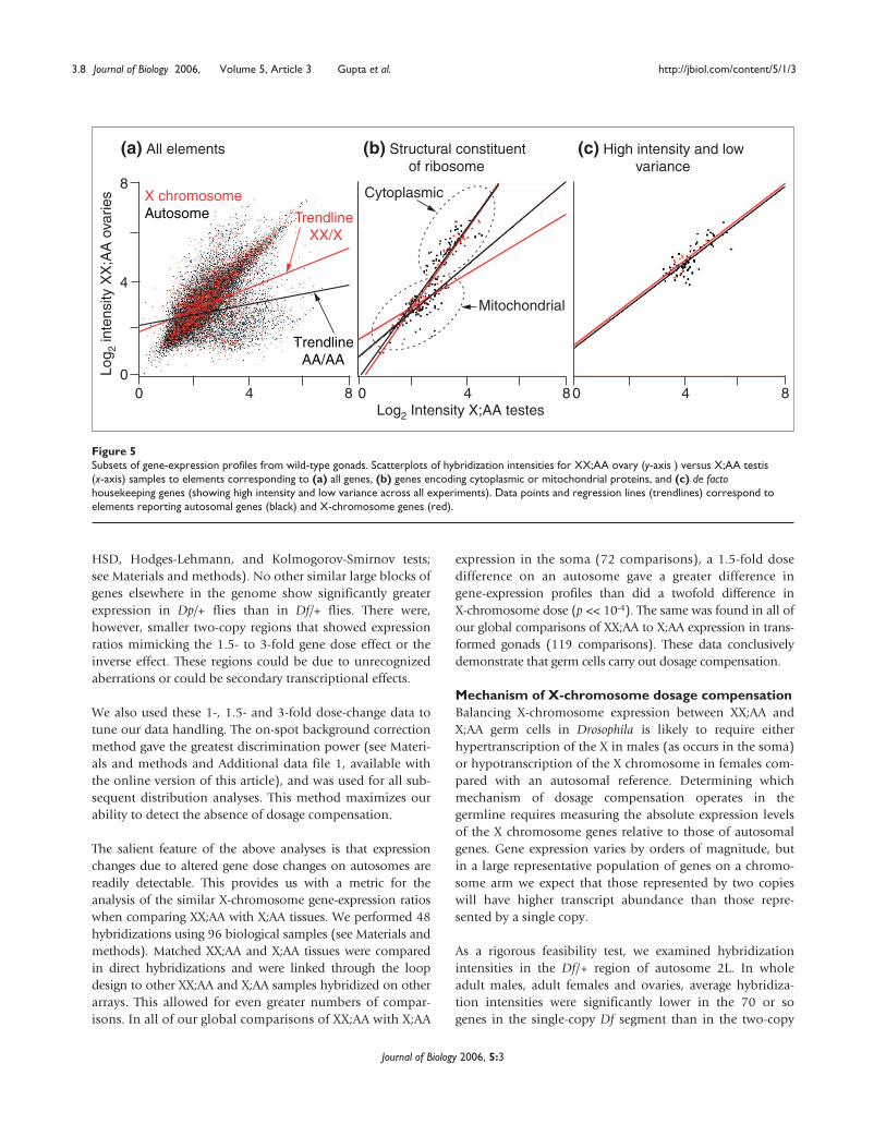
It is formally possible that X chromosomes are dosagecompensated in XX;AA versus X;AA transformed germ cellsbut not in wild-type oogenic and spermatogenic germ cells.Although most of the data points lie along the diagonal inscatterplots of hybridization intensities comparing XX;AAovaries and X;AA testes, regression lines for X chromosomeand autosome expression profiles are differentiallydeflected (Figure 5). This is caused by the large number ofgenes showing male-biased expression on the autosomes
and the lower density of genes with male-biased expressionon the X chromosome [25]. There are, however, manygenes with ‘housekeeping’ functions on both the X chro-mosome and the autosomes that are expected to be equallyexpressed in both oogenic and spermatogenic germ cells.
To determine how genes with housekeeping functionrespond to dose in the germline, we examined in moredetail the expression levels of nuclear genes encoding cyto-plasmic or mitochondrial ribosomal subunits [26]. The Xchromosome and autosomal genes show strikingly similardistributions (p > 0.1 by regression analysis of slopes andintercepts), despite a twofold difference in X-chromosomedose (Figure 5b). We also identified a de facto set of house-keeping genes by investigating array elements showingreduced hybridization variance and high intensityhybridization across all our experiments (Figure 5c). In theabsence of dosage compensation, we would expect a deficitof X chromosome genes in this subset, as they would showincreased variance due to the dose difference. We found nodeficit of X-chromosome encoded genes showing low-variance expression. Both these analyses indicate that atleast some X-chromosome genes are dosage compensated inthe wild-type germline.
Transcriptional response to altered autosomal genedoseTo understand the precision of X-chromosome dosage com-pensation, we first need to understand the general response
http://jbiol.com/content/5/1/3 Journal of Biology 2006, Volume 5, Article 3 Gupta et al. 3.5
Journal of Biology 2006, 5:3
Figure 2 (see figure on the previous page)Sex-determination hierarchy. Sex-biased expression was controlled for by using mutations in sex-determination genes. Relevant aspects of(a) wild-type female, (b) wild-type males, (c) somatic transformation from female to male (sex transformed), (d) somatic transformation frommale to female (sex transformed), and (e) germline transformation are outlined. (a) Sex determination occurs in early embryogenesis, before theactivation of dosage compensation, which leads to higher levels of expression of transcription factors on the X chromosome in XX;AA thanX;AA individuals. These transcription factors activate Sex-lethal (Sxl) in the soma. The Sxl protein regulates the alternative splicing of thetransformer (tra) pre-mRNA such that Tra protein is produced only in females. Sxl also inhibits the formation of the MSL dosage compensationcomplex. Tra protein and non-sex-specifically expressed Transformer2 (Tra2) protein control the alternative splicing of the doublesex (dsx) pre-mRNA. The dsx mRNAs resulting from Tra- and Tra2-mediated splicing encode a female-specific DsxF transcription factor. Sex determination inthe germline is poorly understood and controversial, but a female somatic environment and an independent reading of the XX;AA karyotype ingerm cells increases expression of positively acting Ovo transcription factors and their direct target, ovarian tumor (otu). The otu locus is requiredfor Sxl activity in the germline. Note that Sxl does not regulate the MSLs in the germline. The female sex-determination hierarchy results inoogenic differentiation. (b) In X;AA flies Sxl protein is not present. This permits the formation of the MSL dosage-compensation complex. The trapre-mRNAs are spliced to a non-coding form in the absence of Sxl, and in the absence of Tra protein the dsx pre-mRNA is spliced into a defaultform encoding a male-specific DsxM transcription factor. The germ cells develop into sperm. (c) XX;AA flies are transformed from females intomales using null mutations of tra2 and by using a dsx mutation encoding a pre-mRNA that is constitutively spliced into the male-specific form(dsxswe). Flies bearing dsxswe in trans to a deletion produce DsxM protein and no DsxF protein. Similarly, flies null for tra2 produce only DsxM. Toremove germline expression from the analysis of somatic X-chromosome dosage compensation we took advantage of the fact that XX;AA fliestransformed from females into males usually have no germline. These germline-atrophic (having few to no germ cells) XX;AA femalestransformed into males were compared with X;AA male carcasses (everything but the gonads) or with X;AA males with a genetically ablatedgermline due to the absence of maternal tud+. (d) X;AA flies are transformed from males into females by expressing female-specific tra cDNAtransgenes. The activation of female rather than male sexual differentiation in the X;AA soma results in vast numbers of non-differentiated germcells, presumably due to sexual incompatibility between the soma and germline. The sexual identity of these cells is ambiguous. (e) Mutations inSxl (using allelic combinations effecting only in the germline isoforms of Sxl) or otu also result in vast numbers of non-differentiated germ cells.Positive (arrows) and negative (barred lines) genetic or molecular regulation are indicated. Loss-of-function (red) and gain-of-function (green)mutations and phenotypes are indicated.
3.6 Journal of Biology 2006, Volume 5, Article 3 Gupta et al. http://jbiol.com/content/5/1/3
Journal of Biology 2006, 5:3
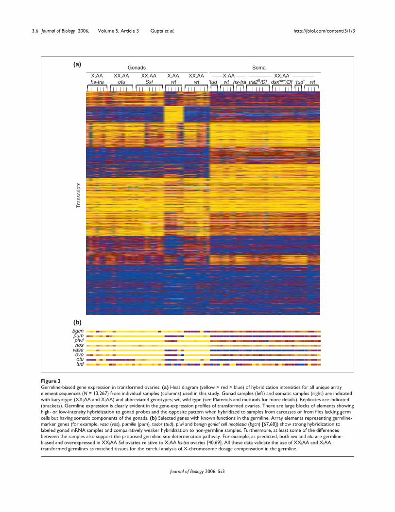
Figure 3 Germline-biased gene expression in transformed ovaries. (a) Heat diagram (yellow > red > blue) of hybridization intensities for all unique arrayelement sequences (N = 13,267) from individual samples (columns) used in this study. Gonad samples (left) and somatic samples (right) are indicatedwith karyotype (XX;AA and X;AA) and abbreviated genotypes; wt, wild type (see Materials and methods for more details). Replicates are indicated(brackets). Germline expression is clearly evident in the gene-expression profiles of transformed ovaries. There are large blocks of elements showinghigh- or low-intensity hybridization to gonad probes and the opposite pattern when hybridized to samples from carcasses or from flies lacking germcells but having somatic components of the gonads. (b) Selected genes with known functions in the germline. Array elements representing germline-marker genes (for example, vasa (vas), pumilio (pum), tudor (tud), piwi and benign gonial cell neoplasia (bgcn) [67,68]) show strong hybridization tolabeled gonad mRNA samples and comparatively weaker hybridization to non-germline samples. Furthermore, at least some of the differencesbetween the samples also support the proposed germline sex-determination pathway. For example, as predicted, both ovo and otu are germline-biased and overexpressed in XX;AA Sxl ovaries relative to X;AA hs-tra ovaries [40,69]. All these data validate the use of XX;AA and X;AAtransformed germlines as matched tissues for the careful analysis of X-chromosome dosage compensation in the germline.
bgcnpumpiwinos
vasaovootutud
X;AAhs-tra
Gonads Soma
XX;AAotu
XX;AASxl
X;AAwt
XX;AAwt
X;AA 'tud' wt hs-tra
XX;AA tra2B/Df dsxswe/Df 'tud' wt
Tran
scrip
ts
(a)
(b)
of the biological system to altered gene dose. What shouldwe expect if there is no X-chromosome dosage compensa-tion? A twofold difference in gene dose is unlikely to alwaysresult in a twofold difference in steady-state transcript levels,as many genes are regulated by elegant feedback mecha-nisms that would dampen the effect of gene dose. Similarly,we need to prove that the entire data-handling pipelineallows us to see gene-expression differences associated withaltered gene dose.
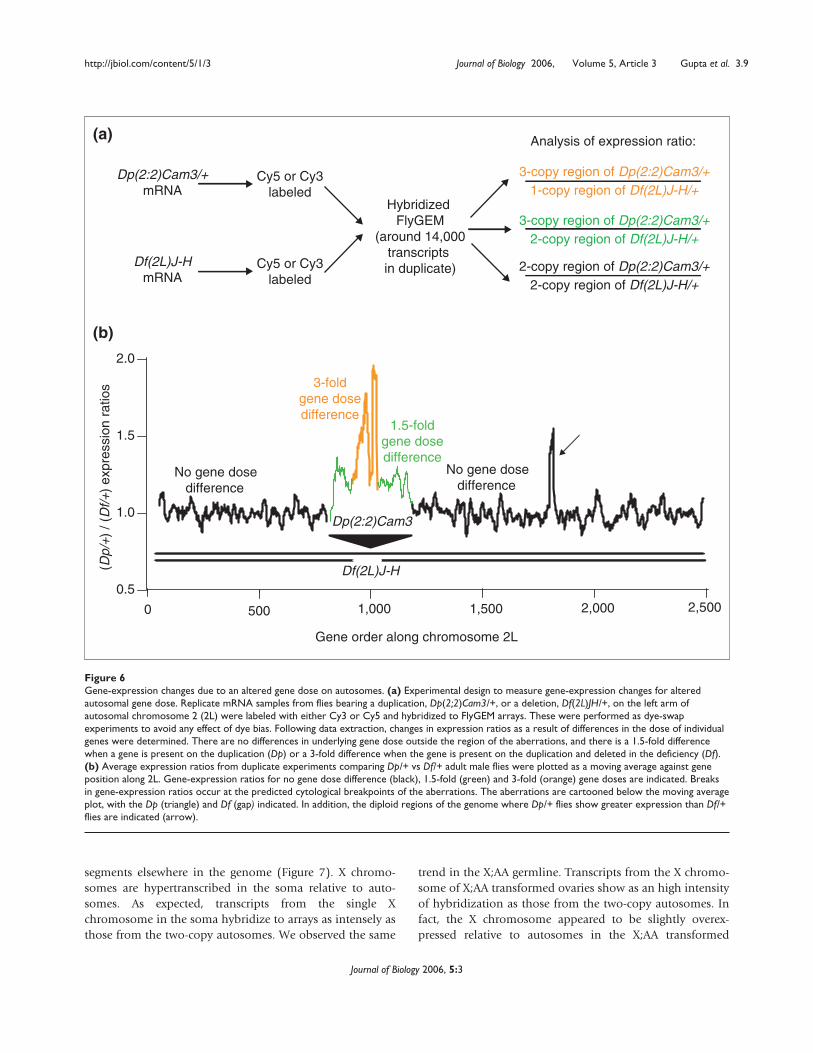
To determine whether differences in genetic dose result indetectable differences in gene expression, we have directlymeasured gene-expression changes resulting from alteredgene dose due to deficiency (deletion) and duplication ofan autosomal segment of the genome. The duplication thatwe used overlaps the deletion region, but covers moregenes. Flies heterozygous for the duplicationDp(2:2)Cam3/+ (Dp/+) have three copies of around 330genes, while flies heterozygous for the deletion Df(2L)J-H/+(Df/+) are hemizygous for a subset of around 70 of thoseduplicated genes [27]. By directly comparing gene expres-sion in flies bearing these aberrations, we assayed the conse-quences for gene expression of 1-fold, 1.5-fold, and 3-folddifferences in gene dose along chromosome arm 2L.
We isolated mRNA from two independent preparations offemales, males and ovaries and performed six hybridiza-tions comparing Dp/+ and Df/+ samples directly againsteach other on microarrays (Figure 6). The effect of alteredautosomal gene dose is obvious in the moving averageplots of expression ratios against position along the chro-mosome arm (Figure 6). We observed distinct alterations ofgene-expression ratios between Dp/+ and Df/+ flies withinthe cytologically defined aneuploid regions in males,females and ovaries. Moving from left to right along such aplot, the ratios are essentially 1.0 (indicating equivalentexpression) in the region that is two-copy in both Dp/+ andDf/+ flies, until reaching the region that is three-copy inDp/+ flies and two-copy in Df/+ flies. At this position thereis a strong break in the moving average towards Dp-biasedexpression. A similar break in the moving average occurs atthe transition to the region that is three-copy in Dp/+ fliesand single-copy in Df/+ flies. Altered fold ratios betweenDp/+ flies and Df/+ flies return to baseline as the proximalbreakpoints of the aberrations are crossed.
The differences in gene-expression ratios corresponding to1-, 1.5- and 3-fold dose changes on chromosome 2L arehighly significant (p << 10-4 by ANOVA followed by Tukey
http://jbiol.com/content/5/1/3 Journal of Biology 2006, Volume 5, Article 3 Gupta et al. 3.7
Journal of Biology 2006, 5:3
Figure 4Scatterplots of hybridization intensities from transformed XX;AA and X;AA tissues. Data points correspond to elements reporting autosomal genes(black) and X-chromosome genes (red). (a) XX;AA females transformed into somatic males (dsxswe/Df(3R)dsxM+15) compared with germlineless X;AA maleprogeny of homozygous tud1 mothers and (b) XX;AA ovaries from ovo1/otu17 females compared with X;AA ovaries from hs-tra83/+ males transformedinto females. The expected twofold difference in gene expression in the absence of X-chromosome dosage compensation is shown as a red line.
Log2 intensity X;AA transformed ovaries (hs-tra)
Log 2
inte
nsity
XX
;AA
tran
sfor
med
ova
ries
(otu
1 /ot
u17 )
0 4 82 6 10
Gonads
Log2 intensity X;AA male soma (tud progeny)
Log 2
inte
nsity
XX
;AA
tran
sfor
med
mal
e so
ma
(ds
xsw
e /D
f)
0 4 82 6 10
Soma
0
2
4
6
8
10
X chromosomeAutosome
X chromosomeAutosome
(a) (b)
HSD, Hodges-Lehmann, and Kolmogorov-Smirnov tests;see Materials and methods). No other similar large blocks ofgenes elsewhere in the genome show significantly greaterexpression in Dp/+ flies than in Df/+ flies. There were,however, smaller two-copy regions that showed expressionratios mimicking the 1.5- to 3-fold gene dose effect or theinverse effect. These regions could be due to unrecognizedaberrations or could be secondary transcriptional effects.
We also used these 1-, 1.5- and 3-fold dose-change data totune our data handling. The on-spot background correctionmethod gave the greatest discrimination power (see Materi-als and methods and Additional data file 1, available withthe online version of this article), and was used for all sub-sequent distribution analyses. This method maximizes ourability to detect the absence of dosage compensation.
The salient feature of the above analyses is that expressionchanges due to altered gene dose changes on autosomes arereadily detectable. This provides us with a metric for theanalysis of the similar X-chromosome gene-expression ratioswhen comparing XX;AA with X;AA tissues. We performed 48hybridizations using 96 biological samples (see Materials andmethods). Matched XX;AA and X;AA tissues were comparedin direct hybridizations and were linked through the loopdesign to other XX;AA and X;AA samples hybridized on otherarrays. This allowed for even greater numbers of compar-isons. In all of our global comparisons of XX;AA with X;AA
expression in the soma (72 comparisons), a 1.5-fold dosedifference on an autosome gave a greater difference ingene-expression profiles than did a twofold difference inX-chromosome dose (p << 10-4). The same was found in all ofour global comparisons of XX;AA to X;AA expression in trans-formed gonads (119 comparisons). These data conclusivelydemonstrate that germ cells carry out dosage compensation.
Mechanism of X-chromosome dosage compensationBalancing X-chromosome expression between XX;AA andX;AA germ cells in Drosophila is likely to require eitherhypertranscription of the X in males (as occurs in the soma)or hypotranscription of the X chromosome in females com-pared with an autosomal reference. Determining whichmechanism of dosage compensation operates in thegermline requires measuring the absolute expression levelsof the X chromosome genes relative to those of autosomalgenes. Gene expression varies by orders of magnitude, butin a large representative population of genes on a chromo-some arm we expect that those represented by two copieswill have higher transcript abundance than those repre-sented by a single copy.
As a rigorous feasibility test, we examined hybridizationintensities in the Df/+ region of autosome 2L. In wholeadult males, adult females and ovaries, average hybridiza-tion intensities were significantly lower in the 70 or sogenes in the single-copy Df segment than in the two-copy
3.8 Journal of Biology 2006, Volume 5, Article 3 Gupta et al. http://jbiol.com/content/5/1/3
Journal of Biology 2006, 5:3
Figure 5Subsets of gene-expression profiles from wild-type gonads. Scatterplots of hybridization intensities for XX;AA ovary (y-axis ) versus X;AA testis(x-axis) samples to elements corresponding to (a) all genes, (b) genes encoding cytoplasmic or mitochondrial proteins, and (c) de factohousekeeping genes (showing high intensity and low variance across all experiments). Data points and regression lines (trendlines) correspond toelements reporting autosomal genes (black) and X-chromosome genes (red).
Log 2
inte
nsity
XX
;AA
ova
ries
8
4
0
(a) All elements (b) Structural constituent of ribosome
(c) High intensity and low variance
Cytoplasmic
Mitochondrial
0 4 0 4 0 4Log2 Intensity X;AA testes
X chromosome Autosome
88 8
Trendline AA/AA
Trendline XX/X
segments elsewhere in the genome (Figure 7). X chromo-somes are hypertranscribed in the soma relative to auto-somes. As expected, transcripts from the single Xchromosome in the soma hybridize to arrays as intensely asthose from the two-copy autosomes. We observed the same
trend in the X;AA germline. Transcripts from the X chromo-some of X;AA transformed ovaries show as an high intensityof hybridization as those from the two-copy autosomes. Infact, the X chromosome appeared to be slightly overex-pressed relative to autosomes in the X;AA transformed
http://jbiol.com/content/5/1/3 Journal of Biology 2006, Volume 5, Article 3 Gupta et al. 3.9
Journal of Biology 2006, 5:3
Figure 6Gene-expression changes due to an altered gene dose on autosomes. (a) Experimental design to measure gene-expression changes for alteredautosomal gene dose. Replicate mRNA samples from flies bearing a duplication, Dp(2;2)Cam3/+, or a deletion, Df(2L)JH/+, on the left arm ofautosomal chromosome 2 (2L) were labeled with either Cy3 or Cy5 and hybridized to FlyGEM arrays. These were performed as dye-swapexperiments to avoid any effect of dye bias. Following data extraction, changes in expression ratios as a result of differences in the dose of individualgenes were determined. There are no differences in underlying gene dose outside the region of the aberrations, and there is a 1.5-fold differencewhen a gene is present on the duplication (Dp) or a 3-fold difference when the gene is present on the duplication and deleted in the deficiency (Df).(b) Average expression ratios from duplicate experiments comparing Dp/+ vs Df/+ adult male flies were plotted as a moving average against geneposition along 2L. Gene-expression ratios for no gene dose difference (black), 1.5-fold (green) and 3-fold (orange) gene doses are indicated. Breaksin gene-expression ratios occur at the predicted cytological breakpoints of the aberrations. The aberrations are cartooned below the moving averageplot, with the Dp (triangle) and Df (gap) indicated. In addition, the diploid regions of the genome where Dp/+ flies show greater expression than Df/+flies are indicated (arrow).
Dp(2:2)Cam3
3-copy region of Dp(2:2)Cam3/+1-copy region of Df(2L)J-H/+
Analysis of expression ratio:
Dp(2:2)Cam3/+mRNA
Cy5 or Cy3labeled
Cy5 or Cy3labeled
Hybridized FlyGEM
(around 14,000transcripts in duplicate)
0 500 1,000 1,500 2,000 2,500
Gene order along chromosome 2L
(Dp/
+)
/ (D
f/+)
expr
essi
on r
atio
s
No gene dosedifference
No gene dosedifference
1.5-foldgene dosedifference
3-foldgene dosedifference
Df(2L)J-H
Df(2L)J-HmRNA
2-copy region of Dp(2:2)Cam3/+2-copy region of Df(2L)J-H/+
3-copy region of Dp(2:2)Cam3/+2-copy region of Df(2L)J-H/+
0.5
1.0
1.5
2.0
(a)
(b)
3.10 Journal of Biology 2006, Volume 5, Article 3 Gupta et al. http://jbiol.com/content/5/1/3
Journal of Biology 2006, 5:3
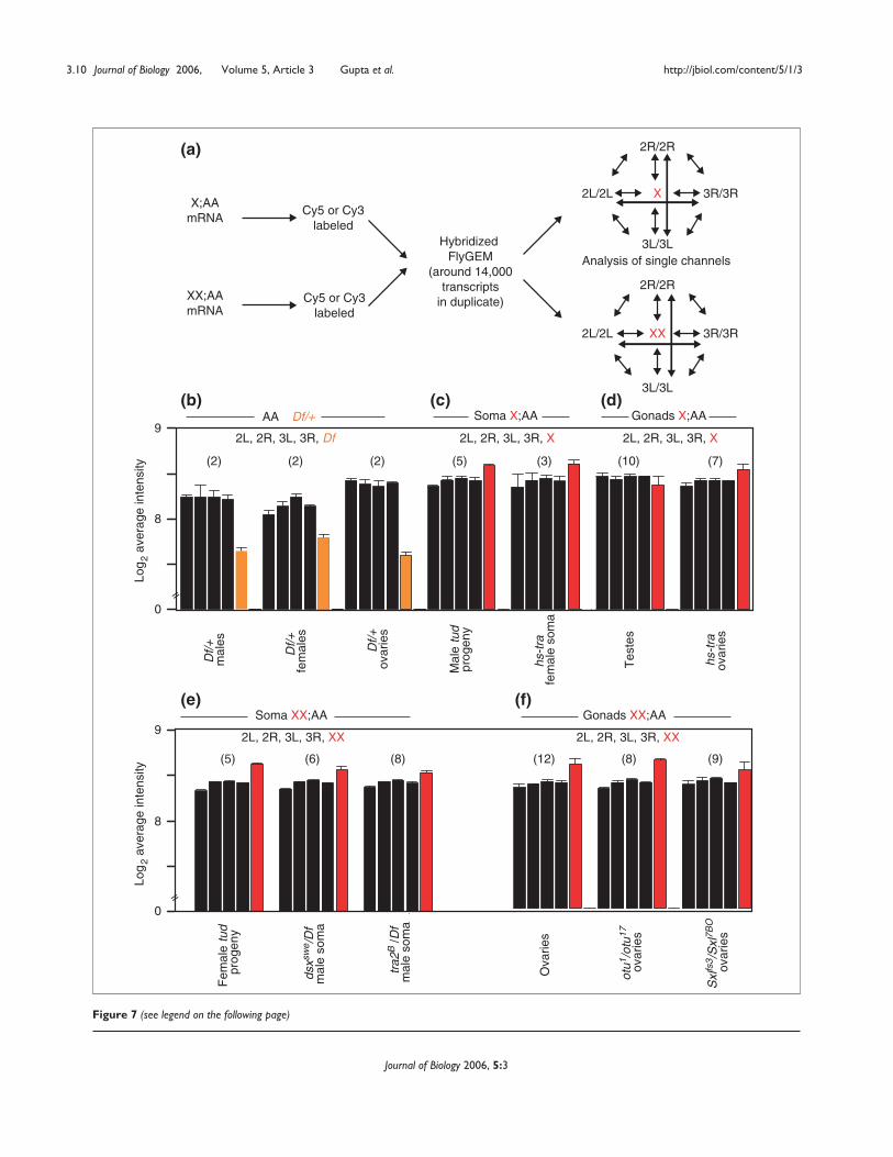
Figure 7 (see legend on the following page)
Analysis of single channels
X;AAmRNA
XX;AAmRNA
Cy5 or Cy3labeled
Cy5 or Cy3labeled
Hybridized FlyGEM
(around 14,000 transcripts in duplicate)
2R/2R
3L/3L
2L/2L 3R/3R
XX
2R/2R
2L/2L 3R/3R
Log
2 av
erag
e in
tens
ity
Log
2 av
erag
e in
tens
ity
AA Df/+
Df/+
m
ales
Df/+
fe
mal
es
Df/+
ova
ries
Fem
ale
tud
prog
eny
Mal
e tu
d pr
ogen
y
hs-t
ra
fem
ale
som
a
hs-t
ra
ovar
ies
Tes
tes
otu1
/otu
17
ovar
ies
Sxl
fs3 /
Sxl
7BO
ov
arie
s
Ova
ries
tra2
B /D
fm
ale
som
a
dsxs
we /
Df
mal
e so
ma
(2) (2) (2)
(12)(5) (6) (8) (8) (9)
(5) (3) (10) (7)
9
8
0
9
8
0
Gonads X;AA Soma X;AA
Soma XX;AA Gonads XX;AA
3L/3L
2L, 2R, 3L, 3R, Df 2L, 2R, 3L, 3R, X 2L, 2R, 3L, 3R, X
2L, 2R, 3L, 3R, XX2L, 2R, 3L, 3R, XX
=
=
X
(a)
(b) (c) (d)
(e) (f)
ovaries, in each of seven replicate experiments. Even thetranscripts from the single X chromosome in X;AA testisshowed higher intensity hybridization than the single-copygenes from any Df+ sample (p << 10-4). These results indi-cate that X-chromosome dosage compensation in theDrosophila soma and germline is the result of higher per-copy X-chromosome transcript accumulation in X;AA cells.Interestingly, the X chromosome shows slight, but highlysignificant, overexpression in all XX;AA samples. This maybe due to inherent hypertranscription of X-chromosomegenes in both males and females.
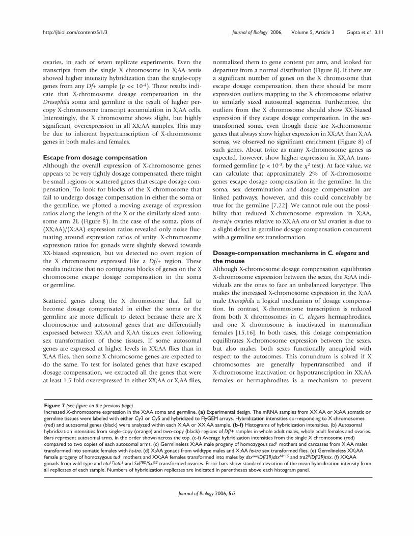
Escape from dosage compensationAlthough the overall expression of X-chromosome genesappears to be very tightly dosage compensated, there mightbe small regions or scattered genes that escape dosage com-pensation. To look for blocks of the X chromosome thatfail to undergo dosage compensation in either the soma orthe germline, we plotted a moving average of expressionratios along the length of the X or the similarly sized auto-some arm 2L (Figure 8). In the case of the soma, plots of(XX;AA)/(X;AA) expression ratios revealed only noise fluc-tuating around expression ratios of unity. X-chromosomeexpression ratios for gonads were slightly skewed towardsXX-biased expression, but we detected no overt region ofthe X chromosome expressed like a Df/+ region. Theseresults indicate that no contiguous blocks of genes on the Xchromosome escape dosage compensation in the somaor germline.
Scattered genes along the X chromosome that fail tobecome dosage compensated in either the soma or thegermline are more difficult to detect because there are Xchromosome and autosomal genes that are differentiallyexpressed between XX;AA and X;AA tissues even followingsex transformation of those tissues. If some autosomalgenes are expressed at higher levels in XX;AA flies than inX;AA flies, then some X-chromosome genes are expected todo the same. To test for isolated genes that have escapeddosage compensation, we extracted all the genes that wereat least 1.5-fold overexpressed in either XX;AA or X;AA flies,
normalized them to gene content per arm, and looked fordeparture from a normal distribution (Figure 8). If there area significant number of genes on the X chromosome thatescape dosage compensation, then there should be moreexpression outliers mapping to the X chromosome relativeto similarly sized autosomal segments. Furthermore, theoutliers from the X chromosome should show XX-biasedexpression if they escape dosage compensation. In the sex-transformed soma, even though there are X-chromosomegenes that always show higher expression in XX;AA than X;AAsomas, we observed no significant enrichment (Figure 8) ofsuch genes. About twice as many X-chromosome genes asexpected, however, show higher expression in XX;AA trans-formed germline (p < 10-3, by the �2 test). At face value, wecan calculate that approximately 2% of X-chromosomegenes escape dosage compensation in the germline. In thesoma, sex determination and dosage compensation arelinked pathways, however, and this could conceivably betrue for the germline [7,22]. We cannot rule out the possi-bility that reduced X-chromosome expression in X;AA,hs-tra/+ ovaries relative to XX;AA otu or Sxl ovaries is due toa slight defect in germline dosage compensation concurrentwith a germline sex transformation.
Dosage-compensation mechanisms in C. elegans andthe mouse Although X-chromosome dosage compensation equilibratesX-chromosome expression between the sexes, the X;AA indi-viduals are the ones to face an unbalanced karyotype. Thismakes the increased X-chromosome expression in the X;AAmale Drosophila a logical mechanism of dosage compensa-tion. In contrast, X-chromosome transcription is reducedfrom both X chromosomes in C. elegans hermaphrodites,and one X chromosome is inactivated in mammalianfemales [15,16]. In both cases, this dosage compensationequilibrates X-chromosome expression between the sexes,but also makes both sexes functionally aneuploid withrespect to the autosomes. This conundrum is solved if Xchromosomes are generally hypertranscribed and ifX-chromosome inactivation or hypotranscription in XX;AAfemales or hermaphrodites is a mechanism to prevent
http://jbiol.com/content/5/1/3 Journal of Biology 2006, Volume 5, Article 3 Gupta et al. 3.11
Journal of Biology 2006, 5:3
Figure 7 (see figure on the previous page)Increased X-chromosome expression in the X;AA soma and germline. (a) Experimental design. The mRNA samples from XX;AA or X;AA somatic orgermline tissues were labeled with either Cy3 or Cy5 and hybridized to FlyGEM arrays. Hybridization intensities corresponding to X chromosomes(red) and autosomal genes (black) were analyzed within each X:AA or XX:AA sample. (b-f) Histograms of hybridization intensities. (b) Autosomalhybridization intensities from single-copy (orange) and two-copy (black) regions of Df/+ samples in whole adult males, whole adult females and ovaries.Bars represent autosomal arms, in the order shown across the top. (c-f) Average hybridization intensities from the single X chromosome (red)compared to two copies of each autosomal arms. (c) Germlineless X;AA male progeny of homozygous tud1 mothers and carcasses from X;AA malestransformed into somatic females with hs-tra. (d) X;AA gonads from wildtype males and X;AA hs-tra sex transformed flies. (e) Germlineless XX;AAfemale progeny of homozygous tud1 mothers and XX;AA females transformed into males by dsxswe/Df(3R)dsxM+15 and tra2B/Df(2R)trix. (f) XX;AAgonads from wild-type and otu17/otu1 and Sxl7BO/Sxlfs3 transformed ovaries. Error bars show standard deviation of the mean hybridization intensity fromall replicates of each sample. Numbers of hybridization replicates are indicated in parentheses above each histogram panel.
3.12 Journal of Biology 2006, Volume 5, Article 3 Gupta et al. http://jbiol.com/content/5/1/3
Journal of Biology 2006, 5:3
Figure 8Escaping dosage compensation. (a,b) Moving average of expression ratios plotted against position along the X chromosome (red) and autosome 2L(black) from (a) sex-transformed soma and (b) germline. (c-f) Histograms of genes showing expression ratios greater than 1.5-fold in either XX;AA(gray) or X;AA (black) transformed soma (c,d) and germline (e,f). Genotypes are indicated. Genes were analyzed by chromosome arm (number ofgenes >1.5-fold on arm/total on arm)/(number of genes >1.5-fold in genome/total in genome). �2 tests (p > 0.3) indicate that there is no enrichmenton the X chromosome for genes expressed more than 1.5-fold in XX;AA soma, but that there is enrichment in gonads (*, p <10-4).
0.5
1.0
1.5
2.0
0.5
1.0
1.5
2.0
XX
;AA
/X;A
A m
ale
som
as(d
sxsw
e /D
f)/(
tud
prog
eny)
XX
;AA
/X;A
A o
varie
s(o
tu1 /
otu1
7 )/(
hs-t
ra)
Position along chromosome
Position along chromosome
Exp
ress
ion
ratio
sE
xpre
ssio
n ra
tios
X 2L 2R 3L 3RXX;AA/X;AA transformed ovaries
(Sxlfs3/Sxl7BO)/(hs-tra)
0.0
0.5
1.0
1.5
2.0
X 2L 2R 3L 3RXX;AA/X;AA transformed ovaries
(otu1/otu17)/(hs-tra)
X 2L 2R 3L 3RXX;AA/X;AA male somas(tra2B/Df)/(tud progeny)
X 2L 2R 3L 3RXX;AA/X;AA female somas
(wild-type carcasses)/(hs-tra carcasses)
higher in XX;AAhigher in X;AA
Mor
e th
an 1
.5-f
old
(fra
ctio
n ex
pect
ed)
Mor
e th
an 1
.5-f
old
(fra
ctio
n ex
pect
ed)
p < 10−3*
**
X chromosome 2L autosome
X chromosome 2L autosome
p < 10−3*
0.0
0.5
1.0
1.5
2.0
Mor
e th
an 1
.5-f
old
(fra
ctio
n ex
pect
ed)
0.0
0.5
1.0
1.5
2.0
(a)
(b)
(c) (d)
(e) (f)
0.0
0.5
1.0
1.5
2.0
Mor
e th
an 1
.5-f
old
(fra
ctio
n ex
pect
ed)
overexpression of the X chromosome. Otherwise, hypertran-scription of the X chromosome in XX;AA individuals wouldresult in functional tetraploidy.
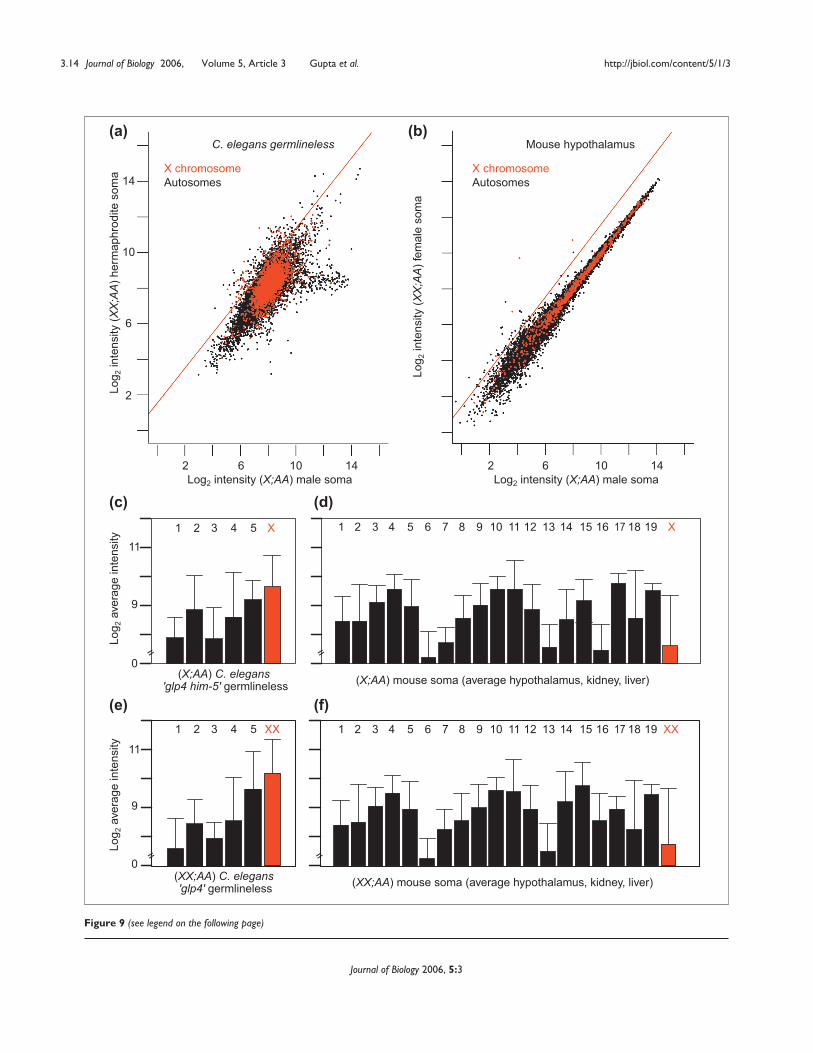
In order to investigate the activity level of the X chromosomein these animals, we reanalyzed previously published arraydata on gene expression in the soma of hermaphrodite andmale C. elegans [28] and female and male mice [29]. Thesestudies reported on gene-expression differences between thesexes rather than dosage compensation. From scatterplots ofthese data there is little difference between the expression ofthe X chromosome in XX;AA versus X;AA germlinelessanimals or somatic tissues (Figure 9). Given that both specieshave somatic dosage-compensation mechanisms that equili-brate expression between the sexes, this is unsurprising.
To determine whether per-copy transcription from the Xchromosome is reduced in XX;AA soma, we measured theaverage hybridization intensity for the X and all autosomesin both X;AA and XX;AA animals (Figure 9). Transcriptsfrom the single X chromosome in X:AA C. elegans malesoma hybridize as well as those of two-copy autosomes.Indeed, the X chromosomes of both X;AA and XX;AAC. elegans appear to be overexpressed relative to autosomes(p << 10-4). Similarly, X-chromosome transcripts from X;AAand XX;AA mouse soma (average of hypothalamus, kidneyand liver) are detected well within the range of those of theautosomes. Many mouse tissues have been examined, andwe find evidence of elevated X-chromosome expression inall cases (data not shown). A study concurrent with this onehas extensively cataloged mammalian X-chromosomedosage compensation, and confirms our observation thatthe single X chromosome in males and the single active Xchromosome in females are compensated [30]. These find-ings suggest that the single X chromosomes of X;AAC. elegans and mice are expressed at levels comparable totwo autosomes, as we observed in Drosophila. Perhaps this isa universal phenomenon.
Discussion Polytene salivary gland chromosomes have greatly facili-tated the analysis of dosage compensation in Drosophila [8],but global analysis of dosage compensation in the othertissues has not been conclusive. A recent reverse transcriptase(RT)-PCR study showed a wide range of states of compensa-tion of X-chromosome genes [31]. Sex-biased expressionpotentially distorts such dosage-compensation analysis,however. There is extensive sex-biased expression inDrosophila [24,32-34] and, furthermore, these genes are notequally distributed between the X chromosome and auto-somes [25,35]. Studies directed at subsets of genes haverevealed complex relationships between X chromosome and
autosome expression. It is, however, difficult to interpret asubtle difference in X-chromosome expression betweenXX;AA and X;AA individuals if effects of a similar magnitudeare seen for autosomal genes [36]. Global analysis of tran-scription in non-polytene tissues could help resolve thenature of X-chromosome dosage compensation in theunderstudied tissues that make up the bulk of the fly.
Microarray analysis is ideally suited to the study of dosagecompensation. Our results suggest that remarkably tight X-chromosome dosage compensation occurs in a wide rangeof adult tissues. Most importantly, we also provide the firstevidence for germline dosage compensation in any organ-ism. The compensation mechanism in both the soma andgermline is the equilibration of X chromosome expressionwith autosomal expression. This is likely to be due toincreased expression from the single X chromosome inX;AA, although decreased expression from all the autosomescould also ensure that X chromosome expression is equili-brated with the autosomes [37].
MSL-independent germline dosage compensation Germline X-chromosome dosage compensation has been ablack box for decades [22]. As the well-known somaticdosage-compensation mechanisms in the major geneticmodel organisms clearly do not function in the germline,the idea has gradually emerged that germ cells are dosage-tolerant. This is due not to evidence of absence, but toabsence of evidence. Although our results show that dosagecompensation in the soma and germline are thematicallysimilar, the mechanisms in the germline must be distinct.
The MSL complexes required for dosage compensation inthe soma are dispensable in the germline [10,12]. Thismight appear to be non-parsimonious, but germline andsomatic gene expression also appear to be distinct. The maleand female germline express different types of basal tran-scription machinery, such as male-germline-specific TATA-box-binding associated factors (TAFs), which might requirea different chromatin structure [38,39], and germline-restricted factors bind the germline-restricted core promoterof the ovo gene [40]. In addition, even though somaticdosage compensation is highly conserved, proteins mediatingdosage compensation are not. For example, in some otherspecies of flies the MSL complex associates with all chromo-somes, not just the X chromosome [41-43], suggesting thatthe complex plays no role in dosage compensation in thoseorganisms. In yeast, which lack well differentiated sex chro-mosomes, one of the main MSL complex components isrequired for viability [44]. These observations indicate thatMSL complexes play markedly different roles in differentspecies. Given the differences in gene expression betweenthe germline and the soma, it is perhaps unsurprising that
http://jbiol.com/content/5/1/3 Journal of Biology 2006, Volume 5, Article 3 Gupta et al. 3.13
Journal of Biology 2006, 5:3
3.14 Journal of Biology 2006, Volume 5, Article 3 Gupta et al. http://jbiol.com/content/5/1/3
Journal of Biology 2006, 5:3
Figure 9 (see legend on the following page)
Log 2
inte
nsity
(XX;
AA)h
erm
aphr
odite
som
a
Log2 intensity (X;AA) male soma
(X;AA) C. elegans 'glp4 him-5' germlineless (X;AA) mouse soma (average hypothalamus, kidney, liver)
Log 2
inte
nsity
(XX;
AA)f
emal
e so
ma
Log2 intensity (X;AA) male soma
Log 2
aver
age
inte
nsity
Lo
g 2av
erag
e in
tens
ity
2 6 10 142 6 10 14
2
6
10
14
(XX;AA) mouse soma (average hypothalamus, kidney, liver)(XX;AA) C. elegans 'glp4' germlineless
1 2 3 4 5 X
1 2 3 4 5 6 7 8 9 10 11 12 13 14 15 16 17 18 19 XX
1 2 3 4 5 6 7 8 9 10 11 12 13 14 15 16 17 18 19 X
1 2 3 4 5
C. elegans germlineless Mouse hypothalamus
X chromosomeAutosomes
X chromosomeAutosomes
0
9
11
0
9
11
=
= =
=
(a) (b)
(c) (d)
(e) (f)XX
different proteins might be involved in Drosophila germlinedosage compensation.
It is also unclear whether the MSL complex accounts for allsomatic dosage compensation. For example, XX;AA flieslacking Sex-lethal (Sxl) protein in the soma die, presumablybecause of a failure to repress MSL complex formation [45].But mutations in the MSL-encoding genes fail to rescue thelethality of XX;AA flies without Sxl, suggesting that Sxlrepresses additional dosage-compensation functions [46].Similarly, in polytene salivary glands, large blocks of the Xchromosome do not associate with MSL complexes [47]. Wefind no evidence to support extensive escape from X-chro-mosome dosage compensation in somatic tissues. Theseresults are consistent with either selective deployment ofMSLs at active genes [47] or MSL-independent dosage com-pensation at a subset of genes.
Mutations in genes regulating germline dosage compensa-tion would be expected to show karyotype-specific pheno-types. Interestingly, there are at least two genes that appear tobe required for the viability of XX;AA but not X;AA germcells; ovo and stand still (stil) [48,49]. Negative and positiveOvo protein isoforms act at the transcription start site of atleast some promoters [5,40,50] and Stil protein decoratesactive chromatin when overexpressed in the soma [49].Perhaps one or both of these proteins serve to block theupregulation of the X chromosome that normally occurs inthe male germline. This could be analogous to the situationin the Drosophila soma, where dosage compensation isactively repressed in XX;AA females [7]. There are also anumber of genes that are required for the viability of malegerm cells [51]. Some of these genes may encode dosage-compensation functions. If such dosage-compensation genesexist, they might encode chromatin-modifying complexes, asin the Drosophila soma. Given that our data show that steady-state transcripts from the X chromosome are compensated,however, there are a host of post-transcriptional mecha-nisms, such as preferential mRNA stability, that could con-ceivably mediate germline dosage compensation.
X-chromosome dosage-compensation mechanisms mightarise from dosage control mechanisms that are active at
many or all genes. Indeed, we have detected transcriptionalbuffering in our control experiments on the effects of auto-some gene dose. We found that the magnitude of the tran-scriptional effect was less than expected from a simplecalculation of gene dose. This inverse effect on transcriptionrelative to gene dose has been known for decades [52-54],and has recently been observed in expression profiling andRT-PCR studies [31,55-57]. It seems likely that many geneshave a self-contained dosage-compensation system, albeit animperfect one. The MSL components have general transcrip-tional activity in yeast and appear to have been co-opted inDrosophila to regulate somatic dosage compensation.Germline dosage compensation might be mediated by otherco-opted cis- or trans-acting components that serve to buffergene expression.
Escape from dosage compensationThere are X-chromosome genes, such as LSP1-�, that escapedosage compensation in Drosophila [58], but it is notknown how common this might be. Similarly, inmammals, many X-chromosome genes escape inactivationin females (escape from dosage compensation in X;AAmales has not been examined). In theory, this is expected,as not all genes need to be dosage compensated in thecourse of X-chromosome evolution [59]. Although we canconfidently analyze populations of X-chromosome genes,it is quite difficult to determine whether a given geneescapes dosage compensation. Indeed, in the absence of adetailed, gene-by-gene mechanistic study, it might be impos-sible. There are many autosomal genes that are more highlyexpressed in XX;AA than X;AA tissues, even after controllingfor sex-biased expression. These gene-expression differencesare clearly not directly related to X-chromosome dosagecompensation. Many of the X-chromosome genes expressedat higher levels in XX;AA than X;AA tissues will be similarlyunrelated to dosage compensation per se. In studies of escapefrom dosage compensation in Drosophila, C. elegans, ormammals where sex-bias is uncontrolled, reports of genesescaping dosage compensation could well be spurious.
On a global level, we find no evidence for escape fromdosage compensation in the Drosophila soma. In thegermline, however, there are clearly more genes with
http://jbiol.com/content/5/1/3 Journal of Biology 2006, Volume 5, Article 3 Gupta et al. 3.15
Journal of Biology 2006, 5:3
Figure 9 (see figure on the previous page)Scatterplots of hybridization intensities from RNA from somatic tissue from XX;AA and X;AA C. elegans and mouse. Hybridization intensities of(a) germlineless (glp4) XX;AA hermaphrodites plotted against a population greatly enriched for germlineless X;AA male C. elegans (glp4 him5), and(b) female mouse hypothalamus tissue plotted against matched tissue from males. X chromosome (red) and autosomal (black) elements as well as thetrend line (red) for the twofold expression difference expected in the absence of dosage compensation are shown. (c-f) Average hybridizationintensities corresponding to genes on the X chromosome (red) and autosomes (black) in (c) X;AA glp4 him5 C. elegans male soma and (d) X;AAmouse male soma (average of hypothalamus, kidney and liver) samples, (e) XX;AA glp4 C. elegans hermaphrodite soma, and (f) XX;AA mouse femalesoma samples. The intensities from individual autosomal arms are averaged across all experiments and standard deviations of the means are indicated.
XX-biased expression than expected. There are several differ-ent interpretations of this finding. It is possible that escapefrom dosage compensation is more common in thegermline (about 2% of genes). Alternatively, there may beslight under-compensation in the germline. The X chromo-somes in both XX;AA and X;AA gonads appear to be overex-pressed relative to autosomes, however. It is thereforepossible that the slight excess of XX-biased expression is dueto failure to fully dampen X-chromosome hypertranscrip-tion in XX;AA females.
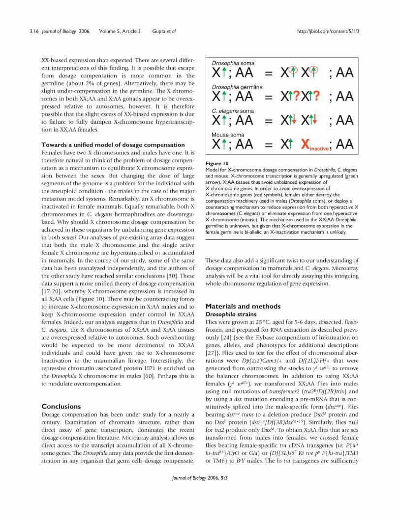
Towards a unified model of dosage compensation Females have two X chromosomes and males have one. It istherefore natural to think of the problem of dosage compen-sation as a mechanism to equilibrate X chromosome expres-sion between the sexes. But changing the dose of largesegments of the genome is a problem for the individual withthe aneuploid condition - the males in the case of the majormetazoan model systems. Remarkably, an X chromosome isinactivated in female mammals. Equally remarkable, both Xchromosomes in C. elegans hermaphrodites are downregu-lated. Why should X chromosome dosage compensation beachieved in these organisms by unbalancing gene expressionin both sexes? Our analyses of pre-existing array data suggestthat both the male X chromosome and the single activefemale X chromosome are hypertranscribed or accumulatedin mammals. In the course of our study, some of the samedata has been reanalyzed independently, and the authors ofthe other study have reached similar conclusions [30]. Thesedata support a more unified theory of dosage compensation[17-20], whereby X-chromosome expression is increased inall X;AA cells (Figure 10). There may be counteracting forcesto increase X-chromosome expression in X;AA males and tokeep X-chromosome expression under control in XX;AAfemales. Indeed, our analysis suggests that in Drosophila andC. elegans, the X chromosomes of XX;AA and X;AA tissuesare overexpressed relative to autosomes. Such overshootingwould be expected to be more detrimental to XX;AAindividuals and could have given rise to X-chromosomeinactivation in the mammalian lineage. Interestingly, therepressive chromatin-associated protein HP1 is enriched onthe Drosophila X chromosome in males [60]. Perhaps this isto modulate overcompensation.
Conclusions Dosage compensation has been under study for a nearly acentury. Examination of chromatin structure, rather thandirect assay of gene transcription, dominates the recentdosage-compensation literature. Microarray analysis allows usdirect access to the transcript accumulation of all X-chromo-some genes. The Drosophila array data provide the first demon-stration in any organism that germ cells dosage compensate.
These data also add a significant twist to our understanding ofdosage compensation in mammals and C. elegans. Microarrayanalysis will be a vital tool for directly assaying this intriguingwhole-chromosome regulation of gene expression.
Materials and methods Drosophila strains Flies were grown at 25°C, aged for 5-6 days, dissected, flash-frozen, and prepared for RNA extraction as described previ-ously [24] (see the Flybase compendium of information ongenes, alleles, and phenotypes for additional descriptions[27]). Flies used to test for the effect of chromosomal aber-rations were Dp(2;2)Cam3/+ and Df(2L)J-H/+ that weregenerated from outcrossing the stocks to y1 w67c to removethe balancer chromosomes. In addition to using XX;AAfemales (y1 w67c), we transformed XX;AA flies into malesusing null mutations of transformer2 (tra2B/Df(2R)trix) andby using a dsx mutation encoding a pre-mRNA that is con-stitutively spliced into the male-specific form (dsxswe). Fliesbearing dsxswe trans to a deletion produce DsxM protein andno DsxF protein (dsxswe/Df(3R)dsxM+15). Similarly, flies nullfor tra2 produce only DsxM. To obtain X;AA flies that are sextransformed from males into females, we crossed femaleflies bearing female-specific tra cDNA transgenes (w; P{w+
hs-tra83}/CyO or Gla) or (Df(3L)stj7 Ki roe pp P{hs-tra}/TM3or TM6) to BsY males. The hs-tra transgenes are sufficiently
3.16 Journal of Biology 2006, Volume 5, Article 3 Gupta et al. http://jbiol.com/content/5/1/3
Journal of Biology 2006, 5:3
Figure 10Model for X-chromosome dosage compensation in Drosophila, C. elegansand mouse. X-chromosome transcription is generally upregulated (greenarrow). X;AA tissues thus avoid unbalanced expression ofX-chromosome genes. In order to avoid overexpression ofX-chromosome genes (red symbols), females either destroy thecompensation machinery used in males (Drosophila soma), or deploy acounteracting mechanism to reduce expression from both hyperactive Xchromosomes (C. elegans) or eliminate expression from one hyperactiveX chromosome (mouse). The mechanism used in the XX;AA Drosophilagermline is unknown, but given that X-chromosome expression in thefemale germline is bi-allelic, an X-inactivation mechanism is unlikely.
Drosophila soma
X ; AA = X X ; AADrosophila germline
X ; AA = X X ; AAC. elegans soma
X ; AA = X X ; AAMouse soma
X ; AA = X Xinactive; AA
? ?
active at 25°C to transform males into phenotypic females.We did not utilize a heat-shock regimen.
In order to study the expression changes resulting from atwofold change in the dose of the X chromosome betweenthe XX;AA and X;AA germlines, we utilized genetic mut-ations that we show remove most sex-biased germlineexpression from the analysis. The activation of female ratherthan male sexual differentiation in the soma with hs-tra inX;AA flies results in gonads with vast numbers of poorly dif-ferentiated germ cells. Mutations in Sxl (y Sxlfs3/y cm Sxl7BO)or otu (ct otu1 v24/y w otu17) result in a similar germline phe-notype. Occasionally, a few ovaries from X;AA hs-tra fliesand germline-transformed Sxl or otu flies bear eggs. Theseovaries were not included in the samples. Similarly, verysmall ovaries that are essentially germlineless were notincluded in any of our samples.
To remove germline expression from the analysis of somaticX-chromosome dosage compensation, we took advantage ofthe fact that XX;AA flies transformed from females to malesusually have no germline, but rarely have a few germ cellsshowing either oogenic or spermatogenic phenotypes. Thesegermline-atrophic XX;AA females transformed into maleswere compared with both gonadectomized X;AA males orsham-dissected X;AA males with a genetically ablatedgermline as a result of the absence of maternal Tud+
(progeny of tud1 bw1 sp1 mothers).
Arrays We have used an extensively tested microarray platformdesigned to detect transcription from 94% ofD. melanogaster release 1 genes [61]. Unlike most arraystudies, where the object is to determine which genes arealtered between tissues, life stages or treatments, we focushere on non-differential expression. In dozens of homo-typic hybridization experiments (where an mRNA sample issplit, labeled with either Cy3 or Cy5, mixed, and hybridizedto the array) performed as part of this (data not shown) andprevious studies, we find that the expression ratio is 1 andthat there are very few outliers [24,25,61]. In the mostextensive set of such homotypic hybridizations, the 99.5%confidence intervals were always between 1.4 and 1.5-fold[61]. Given that only a handful of genes are expected toshow artifactually a greater than 1.5-fold expression differ-ence, we easily had the sensitivity to detect changes inexpression owing to a twofold difference in the 2,245 X-chromosome genes represented on the array.
We have further shown that twofold differences in mRNAconcentrations can be readily detected by adding known con-centrations of mRNA to hybridization mixes in ‘spike-in’experiments [61]. Analysis of spike-in control data (a twofold
change in input results in a twofold difference in expressionratios) and a comparison of sex-biased expression deter-mined by FlyGEM and northern blotting reveal no evidenceof compression of expression ratios in our data [24,61].
Sample preparation and labeling We used 96 biological samples in this study. Careful samplepreparation ensures minimum adjustment in subsequentdata handling. Dissections, flash freezing and RNA extrac-tions were performed as described [24,61]. Briefly, totalRNA was extracted using Trizol (Life Technologies, Carls-bad, USA), followed by mRNA isolation using an Oligotexpoly(A) extraction kit (Qiagen, Valencia, USA). RNA con-centration was determined using RiboGreen dye (MolecularProbes, Oak Ridge, USA) in a fluorescent assay using aLuminescence Spectrometer LS-50B (PerkinElmer, Fremont,USA) fitted with 485 nm (excitation) and 520 nm (emis-sion) filters. RNA quality was determined by capillary elec-trophoresis on a Bioanalyzer 2100 (Agilent, Palo Alto,USA), using the 6000 Nano Assay kit (Agilent) according tothe manufacturer’s instructions.
Samples were labeled with Cy3- or Cy5-labeled randomnonamers (Trilink Biosciences, San Diego, USA). To ensurethat each of the nonamer sets was in fact equally ‘random’,we ordered a single batch of random oligonucleotides thatwas split into half at the penultimate synthetic step with Cy3or Cy5 nucleotides added at the end. To verify performanceof these oligonucleotides, we performed an experiment inwhich RNA was labeled using both Cy3 and Cy5 nonamersin the same tube, followed by hybridization, scanning andanalysis. There was no dye bias evident. Hybridizations ofsamples to the microarrays were performed at 60°C, fol-lowed by washes exactly as described [24,61].
Scanning Arrays were scanned using an Axon GenePix 3000A fluores-cence reader (Molecular Devices Corporation, Union City,USA) in which the photon multiplier tube (PMT) settingswere first adjusted using calibration slides. GenePix v.4.1image acquisition software (Molecular Devices Corporation)was used to extract signal for each target element. Calibra-tion slides were Ultra GAPS (Corning, Acton, USA) spottedwith 10-3 to 10-6 dilutions of a 0.33 nM each stock of Cy3and Cy5 dyes (Amersham Biosciences, Pittsburgh, USA)using GMS 417 Arrayer (Affymetrix, Santa Clara, USA). ThePMT voltages, in the linear range, were balanced using theseslides. With each new batch of labeling and hybridization,we included a homotypic hybridization [61], in which thesame RNA preparation from y1 w67c whole males was splitinto two tubes, labeled with Cy3 or Cy5 nonamers, mixedand hybridized. Slight adjustments of PMT voltages weresometimes made on these control slides. No elements were
http://jbiol.com/content/5/1/3 Journal of Biology 2006, Volume 5, Article 3 Gupta et al. 3.17
Journal of Biology 2006, 5:3
saturated. Once PMT settings were determined, all arrays inthe same hybridization batch were scanned at the samevoltage settings. Hybridization and labeling quality wasdetermined by confirming that the channels were close toglobal balance in the absence of PMT adjustment (no ‘on-the-fly’ adjustments were made). No arrays were discardedbecause of failed labeling reactions in one of the two chan-nels. In addition, all elements were printed in duplicate.Regression analysis of plots of duplicate elements was alsoused as a quality-control step for gradients and severe speck-ling. No arrays were discarded following duplicate elementevaluation. Finally, we determined the fraction of elementsthat returned signal above on-spot background (see Datahandling). Three arrays were discarded from the studybecause less than 50% of elements exhibited above-back-ground signal.
Loop design for microarray pairings Samples from all flies and tissues were prepared (Table 1)and hybridized using a loop design [62]. This design (seeAdditional data file 2) ensured first, that the highest-qualitydirect hybridizations were predominantly between matchedXX;AA and X;AA tissues and second, that any sample couldbe compared with every other sample included in the study,after normalization across all samples. Most of the relevantsamples were compared directly, but all samples are con-nected in the design for indirect comparisons. In most directcomparisons we have included both technical labeling repli-cates (dye-flips) and biological replicates. Although we useda series of pair-wise comparisons of XX;AA and X;AA samplesin this study, the flexible design will allow easy future use toaddress other questions about sex-biased expression.
Data handling Drosophila data were processed using the Bioconductor [63]package limma (Linear Models for Microarray Analysis) v.1.7.6, to adjust for effects that arise from variations inmicroarray technology rather than biological differences.Because of the care taken in using identical RNA concentra-tions in labeling reactions and scanner settings, data handlingresulted in minimal adjustments to the raw data. We did notaverage duplicate array elements as this would reduce statis-tical power in later steps. At the individual array level, theraw intensity data were normalized using print-tip loess(locally weighted scatter plot smooth) [64]. This corrects fortip biases and for minor hybridization gradients. This wasfollowed by one of three different background corrections.For background elimination, intensities less than the averageintensities of the barcode elements (designed against DNAcorresponding to an intron that were included in printingplates for tracking purposes, but are also quite useful forbackground correction [61]) were classified as backgroundand excluded from calculations. For on-spot background
correction, the average intensity of the barcoding elements inthe array were subtracted from the spot intensity for everyarray element. For the no-correction option, we used quan-tile normalization to compare across arrays [64]. Experi-ments with the Df/+ and Dp/+ flies confirm that on-spotbackground correction maximizes expression differenceswithin aneuploid segments, but the effect of the copynumber was evident with both background elimination andno background correction (see Additional data file 1).
Array data from C. elegans and mouse were extracted directlyfrom the Gene Expression Omnibus (GEO) website [65]. Weperformed no additional normalization or background correc-tion. The C. elegans experiments were two-channel hybridiza-tions on spotted arrays [28]. Test samples were hybridizedwith a reference sample. The reference channels cancel in ourcomparison of XX;AA versus X;AA gene expression. The mouseexperiments were performed on Affymetrix arrays [29].
Data sources All array data are available at GEO. An extensive platformdescription of FlyGEM is available at GEO under accessionnumber GPL20 [61]. Data are available under GEO accessionsGSM37438-GSM37451, GSM2464-2467, GSM16582-GSM16583, GSM16570-GSM16581, and GSM77749-GSM77752. Expression data from mouse hypothalamus,kidney and liver were obtained from GEO accessionnumber GSE1148 [29]. C. elegans data were from GEOaccession number GSE715 and GSE724 [28].
Exploratory data visualization Data were clustered and visualized using Cluster 3.0/Tree-View 1.6 [66]. A self-organizing map (SOM) was calculatedto organize normalized hybridization intensities (on-spotbackground subtraction followed by quantile normalizationacross arrays) of array elements at 100,000 iterations,followed by k-means clustering of genes with 10 nodesusing the correlation (uncentered) similarity metric. Genes,but not experiments, were clustered. We averaged duplicateelements for this analysis to avoid crashing the application.
For scatterplots and moving average plots, we used dataprocessed by background elimination followed by quantilenormalization across all experiments. We used on-spotbackground corrected data for histograms of Drosophilaexpression data. Chi-squared tests were performed in Excel(Microsoft, Redmond, USA).
Array elements encoding Drosophila ribosomal subunits wereidentified using the Gene Ontology identifier ‘structural con-stituent of ribosomes’ as assigned in GEO accession GPL20.The de facto housekeeping elements were identified asfollows. Following background elimination and quantile
3.18 Journal of Biology 2006, Volume 5, Article 3 Gupta et al. http://jbiol.com/content/5/1/3
Journal of Biology 2006, 5:3
normalization across all experiments, we selected the ele-ments with hybridization intensities greater than two stan-dard deviations above the median intensity of all arrayelements, from all experiments. From the above, weselected the de facto housekeeping elements as those ele-ments with standard deviations for the expression ratios as± 0.1. Regressions for the X-encoded and the autosome-encoded elements were calculated separately in Bioconduc-tor. Slopes and intercepts were compared at the 95%confidence limit.
Moving averages of expression ratios for every 40 consecutiveelements (two elements per predicted transcript) were
plotted against the corresponding gene positions on chro-mosome arm 2L or X. The predicted cytological break points[27] were used to define the boundaries of aneuploid seg-ments on chromosome arm 2L, in the Dp/+ vs Df/+ experi-ments. Moving averages were calculated in Bioconductor.
Analysis of distributionsThe distribution statistics cited in the text were obtained usingdata processed with print-tip loess and on-spot backgroundcorrection on individual arrays, and quantile normalizationbetween arrays. We can unequivocally show that a modest1.5-fold difference in gene dose has a detectable effect on geneexpression using precisely the same data-handling methods
http://jbiol.com/content/5/1/3 Journal of Biology 2006, Volume 5, Article 3 Gupta et al. 3.19
Journal of Biology 2006, 5:3
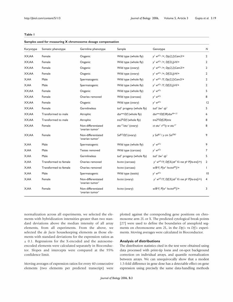
Table 1
Samples used for measuring X chromosome dosage compensation
Karyotype Somatic phenotype Germline phenotype Sample Genotype N
XX;AA Female Oogenic Wild type (whole fly) y1 w67c /+; Dp(2;2)Cam3/+ 2
XX;AA Female Oogenic Wild type (whole fly) y1 w67c /+; Df(2L)J-H/+ 2
XX;AA Female Oogenic Wild type (ovary) y1 w67c /+; Dp(2;2)Cam3/+ 2
XX;AA Female Oogenic Wild type (ovary) y1 w67c /+; Df(2L)J-H/+ 2
X;AA Male Spermatogenic Wild type (whole fly) y1 w67c /Y; Dp(2;2)Cam3/+ 2
X;AA Male Spermatogenic Wild type (whole fly) y1 w67c /Y; Df(2L)J-H/+ 2
XX;AA Female Oogenic Wild type (whole fly) y1 w67c 5
XX;AA Female Ovaries removed Wild type (carcass) y1 w67c 8
XX;AA Female Oogenic Wild type (ovary) y1 w67c 12
XX;AA Female Germlineless tud1 progeny (whole fly) tud1 bw1 sp1 5
XX;AA Transformed to male Atrophic dsxswe/Df (whole fly) dsxswe/Df(3R)dsxM+15 6
XX;AA Transformed to male Atrophic tra2B/Df (whole fly) tra2B/Df(2R)trix 8
XX;AA Female Non-differentiated otu17/otu1 (ovary) ct otu1 v24/y w otu17 8‘ovarian tumor’
XX;AA Female Non-differentiated Sxlfs3/Df (ovary) y Sxlfs3 / y cm Sxl7BO 9‘ovarian tumor’
X;AA Male Spermatogenic Wild type (whole fly) y1 w67c 9
X;AA Male Testes removed Wild type (carcass) y1 w67c 7
X;AA Male Germlineless tud1 progeny (whole fly) tud1 bw1 sp1 5
X;AA Transformed to female Ovaries removed hs-tra (carcass) y1 w67c/Y; Df(3L)stj7 Ki roe pp P{hs-tra}/+) 2
X;AA Transformed to female Ovaries removed hs-tra (carcass) w/BsY; P{w+ hs-tra83}/+ 1
X;AA Male Spermatogenic Wild type (testis) y1 w67c 10
X;AA Female Non-differentiated hs-tra (ovary) y1 w67c/Y; Df(3L)stj7 Ki roe pp P{hs-tra}/+) 4‘ovarian tumor’
X;AA Female Non-differentiated hs-tra (ovary) w/BsY; P{w+ hs-tra83}/+ 3‘ovarian tumor’
that we apply to the study of the X. In addition, the data-han-dling method used is the least favorable method vis-a-vis theconclusions we report. Differences in gene expression in auto-somal aneuploid segments were always significantly greaterthan those observed between the X chromosomes from XX;AAand X;AA samples.
We performed a one-way analyses of variance (ANOVA) fol-lowed by the Tukey HSD method for protection in multiplecomparisons using Matlab (The MathWorks, Natick, USA).All cells were filled ten times. One-way ANOVA was used tocompare three different sets of expression ratios or hybridiza-tion intensities. The single blocking factor for one-wayANOVA was gene dose. We used bootstrap sampling ofpooled array data to fill each of the three ANOVA cells of theone-way design. For ratiometric data, those cells were(Dp/+)/(+/+), AA/AA, and XX/X (see Additional data file 3,Tables 1-3). To fill the (Dp/+)/(+/+) cell, we pooled expres-sion ratios associated with a 1.5-fold difference in gene dosefrom the replicate samples from whole males, whole femalesand ovaries, and sampled 600 ratios. There are fewer genes inthe Dp region than on all autosomes or on the X chromo-some. Therefore, the stability of this inference was verified byfilling this cell with 200 ratios from each of the tissue typesseparately (data not shown). To fill the AA/AA cell we pooledall array data reported in this article where the gene dose was1 and sampled 600 ratios. To fill the XX/X cells we sampled600 X-chromosome ratios for each of three different gonadcomparisons separately. Those are (otu1/otu17)/(hs-tra/+),(Sxlfs3/Sxl7BO)/(hs-tra/+) and ovary/testis (see Additional datafile 3, Tables 1-3). For analysis of Drosophila intensity data,the cells were Df/+, AA, and X (see Additional data file 3,Tables 4,5). As with ratiometric data, intensity data werebootstrap sampled 600 times and the procedure was repeatedten times. Stability for the Df/+ cell was also verified. Theinferences were similar in all ten samplings of 200 intensityvalues from each tissue type (data not shown). For C. elegansand mouse data the cells were AA and X (see Additional datafile 3, Table 6). These cells were filled as for Drosophila. Formouse, we ran ANOVA on pooled intensities from liver,kidney and hypothalamus. We also analyzed each tissue sepa-rately. The Tukey HSD procedure was performed in Matlaband generated a confidence interval for each pair of compar-isons that has a family-wise error rate of, at most, � = 0.05(see Additional data file 3).
For the Drosophila data, we applied a bootstrapped Hodges-Lehmann (HL) estimate of median differences to determine ifX-chromosome expression was more similar to autosomalexpression or to expression from autosomal aberrations (seeAdditional data files 4,5). Additional data file 6 shows acartoon depicting bootstrap HL estimates of median differ-ences followed by the Kolmogorov-Smirnov (KS) test. HL
estimates and KS tests were performed in Bioconductor. Forratiometric data (see Additional data file 4), the HL estimatewas calculated for every drawing of 600 bootstrap samplesfrom the (Dp/+)/(+/+) and AA/AA distributions to generate anew distribution of differences. Next, the same bootstrap sam-pling was used for the XX/X and AA/AA distribution to gener-ate a second distribution of differences. Intensity data weretreated similarly (see Additional data file 5). The two distribu-tions of differences were then compared using the KS test. Thedifferences in the medians and associated D and p-values forthe KS tests then permitted inference concerning the proximityof expression ratios or intensities. In practical terms, a signifi-cant p-value allows us to accept (not reject) the assumptionthat expression ratios AA/AA are closer to XX/X than to Dp/+.This procedure of bootstrap sampling, HL estimation and KSevaluation was repeated ten times to check the stability androbustness of the inferences. In all cases, replicate evaluationsresulted in the same inference.
Additional data filesThe following files are available with the online version ofthis article. Additional data file 1 is a figure showing theeffect of different data handling techniques on differentialexpression resulting from altered gene dose on the auto-somes. Additional data file 2 is a figure showing the experi-mental design in detail. Additional data files 3-5 are tablesshowing the results of statistical analysis of gene expressionratios and absolute intensities for whole chromosome armsand for autosomal aneuploid segments: Additional data file3 shows one-way ANOVA comparisons and Additional datafile 4 shows estimation of Hodges-Lehmann (HL) mediandifferences between expression ratios in various experi-ments and Additional data file 5 shows estimation ofHodges-Lehmann (HL) median differences between signalintensities. Additional data file 6 is a figure showing asimple picture depicting bootstrap HL estimates of mediandifferences followed by the Kolmogorov-Smirnov (KS) test.
Acknowledgements This research was supported in part by the Intramural ResearchProgram of the NIH, NIDDK and CIT. We acknowledge our colleaguesMathias Beller, Jurrien Dean, Sandra Farkas, Leonie Hempel, JamilehJemison, Rasika Kalamegham, Alan Kimmel, James Minor, John E. Smith,Charles Vinson, and Yu Zhang for discussions and technical assistance.We are grateful to Christine Disteche for providing a preprint of workfrom her laboratory and to Jim Birchler for discussions on autosomalaneuploidy and dosage compensation.
References 1. Rosenbusch B: The incidence of aneuploidy in human
oocytes assessed by conventional cytogenetic analysis.Hereditas 2004, 141:97-105.
2. Lindsley DL, Sandler L, Baker BS, Carpenter AT, Denell RE, Hall JC,Jacobs PA, Miklos GL, Davis BK, Gethmann RC, et al.: Segmental
3.20 Journal of Biology 2006, Volume 5, Article 3 Gupta et al. http://jbiol.com/content/5/1/3
Journal of Biology 2006, 5:3
aneuploidy and the genetic gross structure of the Drosophilagenome. Genetics 1972, 71:157-184.
3. Celniker SE, Wheeler DA, Kronmiller B, Carlson JW, Halpern A,Patel S, Adams M, Champe M, Dugan SP, Frise E, et al.: Finishinga whole-genome shotgun: release 3 of the Drosophilamelanogaster euchromatic genome sequence. Genome Biol2002, 3:research0079.1-0079.14.
4. Pauli D, Oliver B, Mahowald AP: The role of the ovarian tumorlocus in Drosophila melanogaster germ line sex determina-tion. Development 1993, 119:123-134.
5. Lu J, Oliver B: Drosophila OVO regulates ovarian tumor tran-scription by binding unusually near the transcription startsite. Development 2001, 128:1671-1686.
6. Regan CL, Fuller MT: Interacting genes that affect micro-tubule function: the nc2 allele of the haywire locus fails tocomplement mutations in the testis-specific ��-tubulingene of Drosophila. Genes Dev 1988, 2:82-92.
7. Cline TW, Meyer BJ: Vive la difference: males vs females inflies vs worms. Annu Rev Genet 1996, 30:637-702.
8. Baker BS, Gorman M, Marin I: Dosage compensation inDrosophila. Annu Rev Genet 1994, 28:491-521.
9. Meller V, Kuroda M: Sex and the single chromosome. AdvGenet 2002, 46:1-24.
10. Rastelli L, Kuroda MI: An analysis of maleless and histone H4acetylation in Drosophila melanogaster spermatogenesis.Mech Dev 1998, 71:107-117.
11. Schüpbach T: Normal female germ cell differentiationrequires the female X chromosome to autosome ratioand expression of Sex-lethal in Drosophila melanogaster.Genetics 1985, 109:529-548.
12. Bachiller D, Sanchez L: Mutations affecting dosage compen-sation in Drosophila melanogaster: effects in the germline.Dev Biol 1986, 118:379-384.
13. Gartler SM, Liskay RM, Campbell BK, Sparkes R, Gant N: Evidence for two functional X chromosomes in humanoocytes. Cell Differ 1972, 1:215-218.
14. Kelly WG, Schaner CE, Dernburg AF, Lee MH, Kim SK, VilleneuveAM, Reinke V: X-chromosome silencing in the germline ofC. elegans. Development 2002, 129:479-492.
15. Heard E, Clerc P, Avner P: X-chromosome inactivation inmammals. Annu Rev Genet 1997, 31:571-610.
16. Meyer BJ: Sex in the worm: counting and compensating Xchromosome dose. Trends Genet 2000, 16:247-253.
17. Ohno S: Sex Chromosomes and Sex-linked Genes. Berlin: Springer-Verlag; 1967.
18. Charlesworth B: The evolution of chromosomal sex determi-nation and dosage compensation. Curr Biol 1996, 6:149-162.
19. Charlesworth B: The evolution of sex chromosomes. Science1991, 251:1030-1033.
20. Charlesworth B: Model for evolution of Y chromosomes anddosage compensation. Proc Natl Acad Sci USA 1978, 75:5618-5622.
21. Adler DA, Rugarli EI, Lingenfelter PA, Tsuchiya K, Poslinski D,Liggitt HD, Chapman VM, Elliott RW, Ballabio A, Disteche CM:Evidence of evolutionary up-regulation of the single activeX chromosome in mammals based on Clc4 expressionlevels in Mus spretus and Mus musculus. Proc Natl Acad Sci USA1997, 94:9244-9248.
22. Oliver B: Genetic control of germline sexual dimorphism inDrosophila. Int Rev Cytol 2002, 219:1-60.
23. Steinmann-Zwicky M, Schmid H, Nothiger R: Cell-autonomousand inductive signals can determine the sex of the germline of Drosophila by regulating the gene Sxl. Cell 1989,57:157-166.
24. Parisi M, Nuttall R, Edwards P, Minor J, Naiman D, Lu J, DoctoleroM, Vainer M, Chan C, Malley J, et al.: A survey of ovary-, testis-,and soma-biased gene expression in Drosophilamelanogaster adults. Genome Biol 2004, 5:R40.
25. Parisi M, Nuttall R, Naiman D, Bouffard G, Malley J, Andrews J,Eastman S, Oliver B: Paucity of genes on the Drosophila Xchromosome showing male-biased expression. Science2003, 299:697-700.
26. Ashburner M, Ball CA, Blake JA, Botstein D, Butler H, Cherry JM,Davis AP, Dolinski K, Dwight SS, Eppig JT, et al.: Gene Ontology:tool for the unification of biology. Nat Genet 2000, 25:25-29.
27. FlyBase: The FlyBase database of the Drosophila genomeprojects and community literature. Nucleic Acids Res 2003,31:172-175.
28. Reinke V, Gil IS, Ward S, Kazmer K: Genome-wide germline-enriched and sex-biased expression profiles in Caenorhab-ditis elegans. Development 2004, 131:311-323.
29. Rinn JL, Rozowsky JS, Laurenzi IJ, Petersen PH, Zou K, Zhong W,Gerstein M, Snyder M: Major molecular differences betweenmammalian sexes are involved in drug metabolism andrenal function. Dev Cell 2004, 6:791-800.
30. Nguyen DK, Disteche CM: Dosage compensation of the activeX chromosome in mammals. Nat Genet 2006, 38:47-53.
31. Chiang PW, Kurnit DM: Study of dosage compensation inDrosophila. Genetics 2003, 165:1167-1181.
32. Jin W, Riley RM, Wolfinger RD, White KP, Passador-Gurgel G,Gibson G: The contributions of sex, genotype and age totranscriptional variance in Drosophila melanogaster.Nat Genet 2001, 29:389-395.
33. Arbeitman MN, Furlong EE, Imam F, Johnson E, Null BH, Baker BS,Krasnow MA, Scott MP, Davis RW, White KP: Gene expressionduring the life cycle of Drosophila melanogaster. Science2002, 297:2270-2275.
34. Andrews J, Bouffard GG, Cheadle C, Lu J, Becker KG, Oliver B:Gene discovery using computational and microarrayanalysis of transcription in the Drosophila melanogastertestis. Genome Res 2000, 10:2030-2043.
35. Ranz JM, Castillo-Davis CI, Meiklejohn CD, Hartl DL: Sex-dependent gene expression and evolution of theDrosophila transcriptome. Science 2003, 300:1742-1745.
36. Birchler JA, Pal-Bhadra M, Bhadra U: Dosage dependent generegulation and the compensation of the X chromosome inDrosophila males. Genetica 2003, 117:179-190.
37. Pal Bhadra M, Bhadra U, Kundu J, Birchler JA: Gene expressionanalysis of the function of the male-specific lethal complexin Drosophila. Genetics 2005, 169:2061-2074.
38. Kai T, Williams D, Spradling AC: The expression profile ofpurified Drosophila germline stem cells. Dev Biol 2005,283:486-502.
39. Hiller M, Chen X, Pringle MJ, Suchorolski M, Sancak Y,Viswanathan S, Bolival B, Lin TY, Marino S, Fuller MT: Testis-spe-cific TAF homologs collaborate to control a tissue-specifictranscription program. Development 2004, 131:5297-5308.
40. Bielinska B, Lu J, Sturgill D, Oliver B: Core promoter sequencescontribute to ovo-B regulation in the Drosophilamelanogaster germline. Genetics 2005, 169:161-172.
41. Ruiz MF, Esteban MR, Donoro C, Goday C, Sanchez L: Evolutionof dosage compensation in Diptera: the gene malelessimplements dosage compensation in Drosophila (Brachyc-era suborder) but its homolog in Sciara (Nematocera sub-order) appears to play no role in dosage compensation.Genetics 2000, 156:1853-1865.
42. da Cunha PR, Granadino B, Perondini AL, Sanchez L: Dosagecompensation in sciarids is achieved by hypertranscriptionof the single X chromosome in males. Genetics 1994,138:787-790.
43. Serna E, Gorab E, Ruiz MF, Goday C, Eirin-Lopez JM, Sanchez L:The gene Sex-lethal of the Sciaridae family (order Diptera,suborder Nematocera) and its phylogeny in dipteraninsects. Genetics 2004, 168:907-921.
44. Hilfiker A, Hilfiker-Kleiner D, Pannuti A, Lucchesi JC: mof, aputative acetyl transferase gene related to the Tip60 andMOZ human genes and to the SAS genes of yeast, isrequired for dosage compensation in Drosophila. EMBO J1997, 16:2054-2060.
45. Kelley RL, Wang J, Bell L, Kuroda MI: Sex lethal controlsdosage compensation in Drosophila by a non-splicingmechanism. Nature 1997, 387:195-199.
46. Cline TW: Autoregulation functioning of a Drosophila geneproduct that establishes and maintains the sexually deter-mined state. Genetics 1984, 107:231-277.
47. Smith ER, Allis CD, Lucchesi JC: Linking global histone acetyla-tion to the transcription enhancement of X-chromosomalgenes in Drosophila males. J Biol Chem 2001, 276:31483-31486.
http://jbiol.com/content/5/1/3 Journal of Biology 2006, Volume 5, Article 3 Gupta et al. 3.21
Journal of Biology 2006, 5:3
48. Oliver B, Singer J, Laget V, Pennetta G, Pauli D: Function ofDrosophila ovo+ in germ-line sex determination depends onX-chromosome number. Development 1994, 120:3185-3195.
49. Sahut-Barnola I, Pauli D: The Drosophila gene stand stillencodes a germline chromatin-associated protein thatcontrols the transcription of the ovarian tumor gene. Devel-opment 1999, 126:1917-1926.
50. Andrews J, Garcia-Estefania D, Delon I, Lu J, Mevel-Ninio M,Spierer A, Payre F, Pauli D, Oliver B: OVO transcriptionfactors function antagonistically in the Drosophila femalegermline. Development 2000, 127:881-892.
51. Wakimoto BT, Lindsley DL, Herrera C: Toward a comprehen-sive genetic analysis of male fertility in Drosophilamelanogaster. Genetics 2004, 167:207-216.
52. Birchler JA: A study of enzyme activities in a dosage seriesof the long arm of chromosome one in maize. Genetics1979, 92:1211-1229.
53. Birchler JA, Riddle NC, Auger DL, Veitia RA: Dosage balance ingene regulation: biological implications. Trends Genet 2005,21:219-226.
54. Devlin RH, Holm DG, Grigliatti TA: Autosomal dosage com-pensation Drosophila melanogaster strains trisomic for theleft arm of chromosome 2. Proc Natl Acad Sci USA 1982,79:1200-1204.
55. Kahlem P, Sultan M, Herwig R, Steinfath M, Balzereit D, Eppens B,Saran NG, Pletcher MT, South ST, Stetten G, et al.: Transcriptlevel alterations reflect gene dosage effects across multi-ple tissues in a mouse model of Down syndrome. GenomeRes 2004, 14:1258-1267.
56. Bond U, Neal C, Donnelly D, James TC: Aneuploidy and copynumber breakpoints in the genome of lager yeastsmapped by microarray hybridisation. Curr Genet 2004,45:360-370.
57. Phillips JL, Hayward SW, Wang Y, Vasselli J, Pavlovich C, Padilla-Nash H, Pezullo JR, Ghadimi BM, Grossfeld GD, Rivera A, et al.:The consequences of chromosomal aneuploidy on geneexpression profiles in a cell line model for prostate car-cinogenesis. Cancer Res 2001, 61:8143-8149.
58. Ghosh S, Lucchesi JC, Manning JE: The non-dosage compen-sated LSP1-�� gene of Drosophila melanogaster lies immedi-ately downstream of the dosage compensated L12 gene.Mol Gen Genet 1992, 233:49-52.
59. Marin I, Siegal ML, Baker BS: The evolution of dosage-com-pensation mechanisms. BioEssays 2000, 22:1106-1114.
60. de Wit E, Greil F, van Steensel B: Genome-wide HP1 bindingin Drosophila: developmental plasticity and genomic tar-geting signals. Genome Res 2005, 15:1265-1273.
61. Johnston R, Wang B, Nuttall R, Doctolero M, Edwards P, Lü J,Vainer M, Yue H, Wang X, Minor J, et al.: FlyGEM, a full tran-scriptome array platform for the Drosophila community.Genome Biol 2004, 5:R19.
62. Churchill GA: Fundamentals of experimental design forcDNA microarrays. Nat Genet 2002, 32 Suppl:490-495.
63. Gentleman RC, Carey VJ, Bates DM, Bolstad B, Dettling M, DudoitS, Ellis B, Gautier L, Ge Y, Gentry J, et al.: Bioconductor: opensoftware development for computational biology andbioinformatics. Genome Biol 2004, 5:R80.
64. Yang YH, Dudoit S, Luu P, Lin DM, Peng V, Ngai J, Speed TP: Nor-malization for cDNA microarray data: a robust compositemethod addressing single and multiple slide systematicvariation. Nucleic Acids Res 2002, 30:e15.
65. Edgar R, Domrachev M, Lash AE: Gene Expression Omnibus:NCBI gene expression and hybridization array data repos-itory. Nucleic Acids Res 2002, 30:207-210.
66. de Hoon MJ, Imoto S, Nolan J, Miyano S: Open source cluster-ing software. Bioinformatics 2004, 20:1453-1454.
67. Deng W, Lin H: Asymmetric germ cell division and oocytedetermination during Drosophila oogenesis. Int Rev Cytol2001, 203:93-138.
68. Mahowald AP: Assembly of the Drosophila germ plasm.Int Rev Cytol 2001, 203:187-213.
69. Nagoshi RN, Patton JS, Bae E, Geyer PK: The somatic sexdetermines the requirement for ovarian tumor geneactivity in the proliferation of the Drosophila germline.Development 1995, 121:579-587.
3.22 Journal of Biology 2006, Volume 5, Article 3 Gupta et al. http://jbiol.com/content/5/1/3
Journal of Biology 2006, 5:3