DOSAGE DETERMINATION IN THE USE OF RADIOACTIVE ...
11
DOSAGE DETERMINATION IN THE USE OF RADIOACTIVE ISOTOPES Leonidas D. Marinelli J Clin Invest. 1949;28(6):1271-1280. https://doi.org/10.1172/JCI102194. Research Article Find the latest version: https://jci.me/102194/pdf
-
Upload
khangminh22 -
Category
Documents
-
view
0 -
download
0
Transcript of DOSAGE DETERMINATION IN THE USE OF RADIOACTIVE ...
DOSAGE DETERMINATION IN THE USE OF RADIOACTIVEISOTOPES
Leonidas D. Marinelli
J Clin Invest. 1949;28(6):1271-1280. https://doi.org/10.1172/JCI102194.
Research Article
Find the latest version:
https://jci.me/102194/pdf

DOSAGEDETERMINATIONIN THE USE OF RADIOACTIVE ISOTOPES
By LEONIDAS D. MARINELLI I
(From the Physics Department, Memorial Hospital, New York City)
INTRODUCTION
As commonly used, most radioactive isotopesare ingested or injected in soluble form and sub-sequently deposited with a greater or lesser de-gree of selectivity in various cells and organs.In the event of a single administration, thenumber of radioactive atoms present at any par-ticular site will increase at first (as they aredrawn from the circulation along with theirstable isotopes), it will reach a maximum and,as the source of supply becomes exhausted, itwill decrease on account of both metabolic turn-over and radioactive decay. The latter is ac-companied by the release of ionizing radiation,the type and emission rate of which is the exclu-sive characteristic of the radioelement itself.
The direct measurement of radiation releasedin this manner in terms of units already estab-lished in Radiology is beset with numerous diffi-culties of experimental nature. Neverthelesswhen the physical factors of half life and radia-tion energy, and the physiological factors ofuptake and excretion, are known, it is possible,in some cases at least, to make satisfactoryestimates of tissue dosage.
The roentgen, as defined by internationalagreement, applies only to x- or gamma radia-tion; it can therefore be used for gamma rayemitting isotopes but not for radiation due toprimary beta particles. (The roentgen is de-fined as "that quantity of x- or gammaradiationsuch that the associated corpuscular emissionper 0.001293 gram of air produces, in air, ionscarrying one e.s.u. of quantity of electricity ofeither sign.")
On the other hand if the energy absorbed pergram of air per roentgen (a--. 83 ergs) is madethe unit of energy absorption for beta rays, it ispossible to establish a comparable basis for betaray dosage. To be sure, in going from air totissues certain corrections will have to be madebecause the energy absorption in tissue perroentgen exposure of x- and gammarays depends
I On leave of absence to the Argonne National Labora-tory, Chicago, Ill.
on both tissue composition and radiation wave-length, but in practice, for soft tissues, thesecorrections are not very large. Hence it ispossible to define an "equivalent roentgen" as"that amount of beta radiation which, underequilibrium conditions, releases in one gram ofair as much energy as one roentgen of gammarays." Since the accepted symbol for the roent-gen is "r," it is convenient to designate theequivalent roentgen by "e.r." (This is essen-tially the same unit as the "rep" or "roentgenequivalent physical" of the Plutonium Project.)In the present paper, formulae expressing therelationship between radiation dose and isotopeconcentrations are presented and their clinicalapplications discussed by the use of tables con-taining a considerable amount of pertinent in-formation. Examples of application of the for-mulae to specific isotopes and definite problemswill also be given. For the mathematical andphysical aspects of the problem the reader isreferred to the literature (1, 2).
CALCULATIONS
Beta Ray Emitters. When a radioisotope emits onlybeta rays, the dose is essentially confined to the regionscontaining the material because the range of the betaparticles in tissue is only a few mm. Many organs insmall animals used in experiments dealing with isotopesemitting high-energy beta rays are not small in comparisonto the range of the beta particles. Proper estimate of thedose in these instances is, in general, very complicated,and must be left to the future.'
The total dose Dp, in equivalent roentgens, due to the com-plete disintegration of a radioelement biologically stableand present in a uniform concentration of C microcuriesper gram of tissue is
Do = KOCe.r.where
I
Ki = 88EpT e.r. per jcd.' per gram; la
T is the half life of the isotope in days and Ep the averageenergy per disintegration of the beta rays in million electronvolts (Mev).
'This problem is somewhat similar to the problem ofgammaemitters videe infra).
' Acd.-microcuries destroyed, i.e., microcuries com-pletely disintegrated in the tissue.
1271

LEONIDAS D. MARINELLI
Doses per hour and per day are respectivelydp(hour) = Dpfh> II
dp(day) = DpfdJwhere fd and fh are the fractions of the entire quantity of
the isotope which disintegrate per hour or per day respec-
tively. Values of T, Ed, Ki and fd are given in Table Iwhich refers to beta ray emitters. Z is the atomic numberand A the atomic weight of the elements given in the first
TABLE I
Physical data pertaining to calculations of radiation dosage resulting from beta rays and/or very soft x-ray radiation
T pfd Wih aiuElement Z A Radiation Half life Ep erlw. fraction So Weight i rangein
in days (Mev) r gram disintegr. yc per kg l0- gram waterdipnegr.dayO' (mm)
Group A: Ed is known to an accuracy of a few per cent
Na 11 22 +. 110io 0.225 22000 6.3 10-4 7.3 197 2.124 j, z 0.61 0.540 29 0.68 5.1 0.113 6.4
P 15 32 , 0 14.5 0.695 885 0.047 2.4 3.6 8.0K 19 42 13-, y 0.515 1.395 63 0.74 2.1 0.167 19Sc 21 46 A-, y 85 1.117 870 0.008 14.3 30 1.0V 23 48 f+, K, y 16 0.175 245 0.042 9.7 5.9 2.8Mn 25 52 0, K, y 6.5 0.085 48 0.101 20.6 2.6 2.2
56 j. 0.108 0.890 8.5 0.998 11.8 0.046 14Fe 26 59 d-, 47 0.120 496 0.015 13.4 21.3 1.5Co 27 56 6+, y 85 0.655 4900 0.008 2.6 36.6 7.0
60 A-.y 1940 0.099 17000 3.6-10-4 16.5 895 0.8Cu 29 61 j3, K, 0 0.142 0.433 5.4 0.992 18.7 0.067 5.5
64 f+, l-, K, 0 0.53 0.120 5.6 0.73 24.4 0.26 2.6As 33 76 -. y 1.12 1.170 115 0.46 1.9 0.655 15.7Br 35 82 , -y 1.5 0.150 20 0.37 13.5 0.95 1.6In 49 114 -(y) 50 0.940 4150 0.014 1.7 44 9.4I 53 130 y3 0.525 0.270 12.4 0.73 11.0 0.53 4.5
131 A-, y 8.0 0.180 127 0.083 9.4 8.1 2.2RaE 83 210 0 4.85 0.330 141 0.133 5.3 7.85 5.2
Group B: Ed is less accurately known than Ep in Group A
C 6 14 83, 0 2.1a101 0.05 9.2.106 3.3 10-7 32 23.5.104 0.24S 16 35 i-, 0 88 0.055 420 0.0079 30 24 0.2Ca 20 45 A- (?) 180 0.10 1580 0.0039 16 62 0.8Sr 38 89 is-,0 55 0.57 2760 0.013 3 38 7
90 W_ 0 9000 0.22 17-104 8-10-6 8 6200 2.2Y 39 90 A_,0 2.6 0.90 200 0.24 2 18 11Sb 51 124 -y 60 0.66 3480 0.012 2.4 57 12.3Au 79 198 |-, 7 2.7 0.32 76 0.23 5.7 4.1 3.8
Group C: E1B includes the total localized x-radiation following decay by electron capture
Mn 25 54 K, y 310 0.0054 147 0.0022 340 128Fe 26 55 K, y 1500 0.0059 780 4.6-10-4 280 633Co 27 58 i+, K, y 65 0.035 20 0.012 415 29 1.5Zn 30 65 d+, K, -y 250 0.01 180 0.003 185 124 1.2
Group D: Ed consists of part of the radiation released in the decay by electron capture
Y 39 86 K, -y 105 0.005 46 0.007 310 69In 49 111 K, 2.7 0.0058 1.4 0.23 310 2.3 0.01
The values of Kp and Sp are based on uniform and biologically stable concentrations of radioelements distributed intissues of linear dimensions large as compared to the range of the beta particles. The sign "0" under the heading"Radiation" indicates the absence of nuclear gammarays.
Ed is the average energy per disintegration.Ki = 88EpT is the radiation dose expressed in equivalent roentgens due to beta rays emitted during the complete
disintegration of 1 ,uc of radioelement per gram of tissue.fd = (1- em69S/T) is the fraction of the entire quantity of isotope which disintegrates in 24 hours.
Sp= KX is the concentration of radioisotope expressed in ,.c per kg which will deliver a dose of 0.1 e.r. to
tissue during the first 24 hours of exposure.
1272

DOSAGEDETERMINATION IN USE OF RADIOACTIVE ISOTOPES 1273
TABLE II
Physical data pertaining to calculations of radiation dosage resulting from gammarays
Ej in MevT ~ __________________ 17 K7f fd It
mEle-n | Z \ A \ Radiation | Half life t Annihila- Es In | atlcm at 1 c fiction perment in hours Annihila- m/u-r rc disintegr. cm
Ele Z A R|radiationHalf life tion Nuclear gammaradiation r/mc-hr perday X 10radiation
Group A: elements not decaying by electron capture, or x-ray emission following electron capture so soft that it can betreated like beta radiation and hence making no significant contribution to I7
Na 11 22 04', y 26500 0.511(2) 1.30(1) 13.2 500 6.3 *10-4 3.224 -. y 14.7 1.38(1) 2.76(1) 19.1 0.40 0.68 2.5
K 19 42 P,. 12.4 1.5 1(0.25) 1.95 0.035 0.74 2.9Sc 21 46 , -, 2040 0.90(1) 1.12(1) 11.4 33.5 0.008 3.2V 23 48 t', K, 384 0.511(1.16) 0.98(1) 1.33(1) 16.3 9.0 0.042 3.2Mn 25 52 . K, - 156 0.511(0.7) 0.736(1) 0.94(1) 1.46(1) 19.5 4.4 0.101 3.2
56 i-, 7 2.59 1.77(0.3) 2.06(0.2) 9.4 0.035 0.998 2.7Fe 26 59 IS-, 7 1128 1.1(0.5) 1.3(0.5) 6.55 10.7 0.015 3.0Co 27 56 P', 7 2040 0.511(2) 0.845(1) 1.26(1) 17.95 37.2 0.008 3.2
60 I-, 46500 1.16(1) 1.32(1) 13.5 900 3.6 -10-4 3.0Cu 29 61 @+' K, 0 3.4 0.511(1.56) 4.8 0.024 0.992 3.4
64 pa, 0, K, 0 12.8 0.511(0.38) 1.2 0.022 0.73 3.4As 33 76 V. - 26.8 0.55(0.37) 1.20(0.12) 1.75(0.01) 2.2 0.083 0.46 3.0Br 35 82 V, 7 36 0.547(1) 0.787(1) 1.35(1) 15.1 0.79 0.37 3.2Sb 51 124 1440 0.6(1) 1.7(0.5) 7.9 16.4 0.012 3.2
130 ~. 7 12.6 0.416(0.55) 0.537(1) 0.667(1) 0.744(1) 13.05 0.237 0.73 3.4131 1, 7 192 0.080(.01) 0.363(.8) 0.638(.15) 0.283(.06) 2.37 0.66 0.083 3.0
Au 79 198 PV. 7 65 0.40(1) 2.4 0.22 0.23 3.4
Group B: elements with x-ray emission following electron capture whose contribution to 17 is not negligible
Mn 25 54 KC. 7450 0.835(l) [0.0054(1)| 4.9+111 52 0.0022 3.2Fe 26 55 K, -r 36000 0.07(2 .10-6) 0.0059(1)1 [101 - 4.6 -10-' 3.2Co 27 58 0, K, 1560 0.511(0.3) 0.805(1) 0.0064(0.85)] 5.7+171 12.8 0.012 3.3Zn 30 65 P', K, S 6000 0.511(0.03) 1.14(0.46) [0.008(0.99)1 3.0+[51 26 0.003 3.0Y 39 86 K, y 2530 0.908(1) 1.89(1) 0.0142(1)1 14.4 +[3.11 52.5 +[10.31 0.007 3.2In 49 111 K, y 65 0.173(1) 0.247(1) [0.0231(1)] 2.3 +[1.4] 0.22 +[0.131 0.23 3.0
The sign "O" under the heading "Radiation" indicates the absence of nuclear gamma rays. The column headed"Annihilation radiation" refers to positron-electron recombination, the column headed "Nuclear gammaradiation" refersto gammarays originating in the disintegrated nucleus. The numbers in parentheses indicate the number of photonsof the particular energy that are released per disintegration.
In Group B, the numbers in square brackets pertain to x-ray emission following electron capture and, because ofthe short range of the radiation, they should not be used in computing e-ray dosage. In practice, this type of dosecomputation is amenable to the simpler formulae used for a-rays (see Group C in Table I).
17 is the exposure in roentgens at 1 cm distance in air from an unfiltered point source of 1 mc, for one hour; ormilli-roentgens per microcurie-hour (see formula IIla).
K7 = 1.44tI7 X 10-8 is the number of roentgens, per microcurie destroyed, at 1 cm distance in air from an unfil-tered point source.
fd = (1 - e 6931T) is the fraction of the entire quantity of isotope which disintegrates in 24 hours. (T is the halflife in days.)
= true absorption coefficient of the 7-radiation in water.
column. The last column gives the ranges in water for themost energetic beta particles of each beta ray spectrumfor the different isotopes. It should be noted that thecolumn for Kp gives immediately the dose in equivalentroentgens for each microcurie completely disintegratingwithin a gram of tissue.
GammaRay Emitters. When a substance is a gammaray emitter, the problem of dosage determination is morecomplicated. The rays released in a given gram of tissueproduce only a small amount of ionization there; most oftheir energy is expended elsewhere along their paths. Theproblem becomes somewhat analogous to that of inter-stitial radium gamma ray dosage, except that instead ofdiscrete sources one is confronted with extended ones.
At a given point the tokal dose D7, due to gamma raysemitted by the complete disintegration of a radioelementbiologically stable and present in a uniform concentrationof C microcuries per gram of tissue, is given by
Dy = KyCg roentgens III
whereK7 = 1.44tI7 X 10-3
The dose delivered in one day isd7(day) = Dyfd,
Ila
IVfd having the meaning expressed above. K7 expresses thenumber of roentgens at 1 cm distance in air due to thecomplete disintegration of an unfiltered point source of1 pc; 1.44 X t is the average life in hours, 17 the dose-ratein roentgens per hour at 1 cm in air from an unfilteredpoint source of 1 mc. The quantity g in equation III isa geometrical factor depending on the location of the givenpoint, the size and shape of the tissue mass containingthe isotope, and on the absorption coefficient p of thegammarays in tissue. The values of 17 and K7 togetherwith other pertinent information for gammaray emittingradioisotopes are given in Table II.
Safe Tracer Concentration. In the mainTables I and II are self explanatory. Atten-

LEONIDAS D. MARINELLI
tion should be directed to column 9 in Table I,headed Sp, which indicates the "safe tracer con-centration," that is, the number of ,uc per kg oftissue weight which will result in a whole tissuedose of 0.1 r the first day, due to the beta raysalone. (The gamma ray contribution will bediscussed below.) In the case of a short-livedelement, the dose on succeeding days will quicklydecrease to the vanishing point, while for long-lived substances it will continue at an appreciablelevel for some time. This criterion of "safedose" is decidedly conservative. One might pre-fer to choose such a level as 1 r in 10 days orsome other value based on a longer period oftime. This might avoid complications due tovarious half lives. However, the present famil-iar idea of safety is based on daily dose; hencethat has been used here. It is not to be assumedthat larger tracer doses should never be adminis-tered. It is sometimes entirely justifiable to useconsiderably higher doses in diagnostic problems.
Distribution of the Radiation in Tissue. Thephysical data presented enable the experimenterto calculate the actual dose delivered to a tissue
w
0a
w
An14iP
w
to0
a
2
4(
wCK
FIG. 1. RADIATION DOSAGE DUE TO THE BETA RAYACTIVITY OF P52 IN DIFFERENT TISSUES IN MICE
AS A FUNCTION OF TIMEThe points marked on the right margin are the actual
tissue doses obtained by extrapolation to infinite time.Theoretical whole animal dose equals K# times the numberof microcuries administered divided by the total weightof the animal.
or individual whenever the isotope concentra-tion C is known as a function of time. Thisdepends on the amount of isotope administered,the species and metabolic state of the testanimal, the mode of administration, the chemicalform under which the radioelement is given, etc.
Physiological information of this nature isof great importance; the literature shows thata good deal of animal work has been done onthe relative uptake of various isotopes by dif-ferent tissues at different times. As an example,in Figure 1 are shown the relative radiationdoses delivered in different tissues in mice in-jected intraperitoneally with P32 in the form ofNa2HPO4. The animals were sacrificed at vari-ous times after the administration of the ma-terial, and the concentration of p32 was deter-mined for the various tissues and organs. Byadding graphically the daily doses as expressedby equation II it was possible to calculate thedose of radiation received by each tissue as afunction of time (3). Similar calculations weremade for the whole body assuming uniformdistribution. The "theoretical" whole animaldose4 after complete disintegration of the isotopewas taken as 100 per cent, and all other tissuedoses referred to this value. Thus at the end ofthree weeks, 64 per cent of the theoretical com-plete dose had been delivered; but all tissuestested, except the bone, had received alreadypractically all their radiation because most p32had disappeared. The liver, for example, hadreceived about 33 per cent of the theoreticaluniform dose. At that time the bone had re-ceived 39 per cent; but in the bone the dosageincreased to 48 per cent at the end of 36 days.
No tissue actually attained the theoreticaldose or would have attained it after longerperiods of time because of biological elimination.From the curves, the actual tissue dosage per,Uc of p32 thus administered in a mouse of 25grams can now be calculated. The total animalconcentration would be 0.04 ,uc per gram and the"theoretical" total animal dose Dp = Kp X 0.04= 885 X 0.04 = 35.4 e.r. (Ke from Table I).The liver, then, at the end of three weeks would
4 By "theoretical" whole animal dose is meant the doseDo, which would have been obtained in the animal aftercomplete disintegration of the isotope on the assumption ofuniform concentration in the animal and of total lack ofbiological elimination.
1274

DOSAGEDETERMINATION IN USE OF RADIOACTIVE ISOTOPES
have received 33 per cent of this value or 12 r;similarly the bone at 36 days would have had0.48 X 35.4 = 17 r.
It is recognized that with PI' in mice thesedeterminations are not as accurate as one wouldwish because the linear dimensions of most or-gans are smaller than the range of a consider-able portion of the beta particles emitted by P32.Similar information in larger animals wouldyield more reliable figures. Conversely this typeof calculation would be fairly accurate even ina small animal, as the mouse, if soft beta rayemitters (HI, C14, S35, etc.) were used.
When working with humans it is not possiblein general to study concentrations throughoutthe body and their variations with time by invivo measurements; the extreme concentrationof iodine in the thyroid is an exception. Formost isotopes information must be obtained frombiopsy and autopsy measurements and hence thedata are meager; moreover they are scatteredthrough various publications which were oftennot prepared with this point in mind. Accord-ingly it has happened too often that the pub-lished results lack some essential fact, such asthe actual weight of the individual. It is to behoped that more of this information will beforthcoming. In the meanwhile, it is wise toadopt a conservative attitude in dealing withhumans whenever physiological information islacking. This can be done by assuming thatno biological elimination takes place.
Differential Absorption Ratio. To take intoaccount the differences in uptake which lead todifferences in dose for the various tissues, it isconvenient to express the concentration in termsof a "differential absorption ratio" (D.A.R.).For any tissue, this is the ratio of concentrationof an isotope in that tissue to the average con-centration in the body (neglecting excretion).Thus under the assumption just made if a par-ticular tissue has a D.A.R. of 1, it receives thesame dose of radiation as the average of thewhole body. On the other hand,, if it has aD.A.R. of 10, it receives ten times as muchradiation as the average.
In calculating the safe doses for tracers, it ishighly desirable to have some idea of the rangeof the D.A.R.'s. The safe dose is assumed tobe 0.1 per day for the entire body; if a certain
tissue has a D.A.R. of 10, it would have receivedI r while the rest of the body received 0.1 r.This might lead to undesirable irradiation ofcertain tissues or organs from a dose believedsafe otherwise.
The procedure outlined above needs clarifica-tion. In general, single tissues and the wholebody have different rates and modes of elimina-tion; hence the tissue D.A.R. will vary withtime. The question then arises as to the timeat which D.A.R.'s have significance in determina-tion of tissue dosage. Evidently the D.A.R.will be too low when taken too soon after theisotope administration because the concentra-tions have not yet stabilized. Likewise adoptionof the D.A.R. after several half lives will havelittle or no significance because most of theisotope has disintegrated. One useful index,whenever the isotope reaches the body tissuesthrough the circulatory system, could be thestabilization of the plasma concentration. Nogeneral rule, however, can be given and goodjudgment is of paramount importance.
Effective Half Life. In instances of extremeconcentrations as in the case of radioiodine I131administered to humans in the form of KI orNaI, greater accuracy can be obtained. It willbe noted that the formula I for the dose, Dp,,contains the half life of the element. If theamount of iodine in the gland decreases both bydecay and by excretion, the effective half life willbe less than the physical. Such effects can bedetermined only by actual measurements. Inthis particular problem, counts are made atintervals with a Geiger counter in a fixed positionover the thyroid gland. If there were no excre-tion of the material from the gland, these countsshould decrease exactly in accordance with thephysical decay curve for the iodine; that is, for113 they should be at half their initial valueafter eight days, one-fourth at 16 days, etc.Actually, they decrease exponentially but morerapidly, half lives as short as two days havingbeen encountered in practice. It is evident thatthe use of this number instead of 8 in equation Iresults in a decrease of 75 per cent in the ad-ministered dose, an amount which is clinicallysignificant in therapeutic work. It would benow possible to calculate from formula I thedose delivered under normal conditions if the
1275
LEONIDAS D. MARINELLI
initial concentration C were known. This canbe determined either by actual biopsy or"guessed" by estimate of the gland weight andmeasurements of the radioactive content of thegland by proper in vivo observations.
It is evident from Table I that the actualamounts of the safe concentrations in tissue donot vary very greatly; it must be realized thatthe intervals at which they can be repeated do.A reasonable interval between full tracer dosesof any particular element would be four or fivehalf lives, unless the material has been shown tobe excreted fairly completely in a shorter period.If no significant excretion occurs, a tracer of Na24could be repeated within about three days, whileP12 should not be repeated in less than abouttwo months, and Cae not before two years haveelapsed.
It should be mentioned in this connection thatthe safe doses listed here are calculated on theassumption that the individual is receiving noradiation from any other source. Extensiveradiographic or fluoroscopic procedures may re-sult in an appreciable dose of radiation, and suchpossibilities must not be overlooked.
TRACERISOTOPES-EXAMPLES
In order to illustrate the use of the materialpresented in this paper sample calculations willbe given for some isotopes commonly used intracer experiments and in therapy. These willbe presented in order of the complexity of theirspectra.
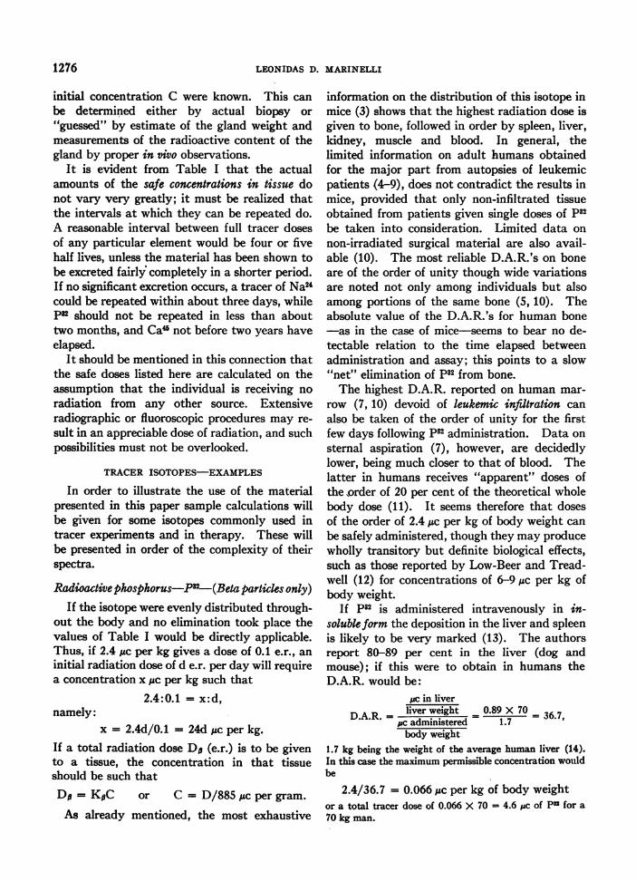
Radioactive phosphorus-P82-(Beta parties only)If the isotope were evenly distributed through-
out the body and no elimination took place thevalues of Table I would be directly applicable.Thus, if 2.4 jc per kg gives a dose of 0.1 e.r., aninitial radiation dose of d e.r. per day will requirea concentration x ,ic per kg such that
2.4:0.1 = x:d,namely:
x = 2.4d/0.1 = 24d /uc per kg.If a total radiation dose Da (e.r.) is to be givento a tissue, the concentration in that tissueshould be such thatD= KpC or C = D/885 ,gc per gram.
As already mentioned, the most exhaustive
information on the distribution of this isotope inmice (3) shows that the highest radiation dose isgiven to bone, followed in order by spleen, liver,kidney, muscle and blood. In general, thelimited information on adult humans obtainedfor the major part from autopsies of leukemicpatients (4-9), does not contradict the results inmice, provided that only non-infiltrated tissueobtained from patients given single doses of p32be taken into consideration. Limited data onnon-irradiated surgical material are also avail-able (10). The most reliable D.A.R.'s on boneare of the order of unity though wide variationsare noted not only among individuals but alsoamong portions of the same bone (5, 10). Theabsolute value of the D.A.R.'s for human bone-as in the case of mice-seems to bear no de-tectable relation to the time elapsed betweenadministration and assay; this points to a slow"net" elimination of p32 from bone.
The highest D.A.R. reported on human mar-row (7, 10) devoid of leukemic infiltration canalso be taken of the order of unity for the firstfew days following p82 administration. Data onsternal aspiration (7), however, are decidedlylower, being much closer to that of blood. Thelatter in humans receives "apparent" doses ofthe order of 20 per cent of the theoretical wholebody dose (11). It seems therefore that dosesof the order of 2.4 ,lc per kg of body weight canbe safely administered, though they may producewholly transitory but definite biological effects,such as those reported by Low-Beer and Tread-well (12) for concentrations of 6-9,gc per kg ofbody weight.
If p32 is administered intravenously in in-soluble form the deposition in the liver and spleenis likely to be very marked (13). The authorsreport 80-89 per cent in the liver (dog andmouse); if this were to obtain in humans theD.A.R. would be:
tc in liver
D.A.R. = liver weight 0.89 X 70 = 36.7,* c administered 1.7body weight
1.7 kg being the weight of the average human liver (14).In this case the maximum permissible concentration wouldbe
2.4/36.7 = 0.066 ,c per kg of body weightor a total tracer dose of 0.066 X 70 = 4.6 uc of Pu for a70 kg man.
1276

DOSAGEDETERMINATION IN USE OF RADIOACTIVE ISOTOPES
Radioactive strontium-Sr89-(beta particles only)
Work on animals (15) indicates that this iso-tope is highly concentrated in the bone. Humanmaterial has been reported by Treadwell et al.
(16) following intravenous injections. Theirdata point to a maximum D.A.R. in the epi-physis of bone of 13.2, and an unweighted aver-
age of 8.3 for bone, mostly in young subjects.A trend of D.A.R. change with time is notapparent. On the basis of this, the toleranceconcentration would be, since Sp= 3 pc per kg,
Sp/8.3 = 3/8.3 = 0.4 ;c per kg of body weight.
Radioactive calcium-Cau-(beta particles only)
The relative concentrations of this isotope inhuman tissue have not been reported to date.Work on mice shows that the D.A.R. for Ca inanimal bone is approximately twice that forSr (15). Hence, if this ratio holds also forhumans one should expect a D.A.R. of about8.3 X 2 = 16.6.
It follows from Table I that the toleranceconcentration would be, since Sp = 16 gc per kg,
16.0/16.6 ; 1.0 Azc per kg of body weight.
It should be noted that if calcium were depositedexclusively in the human skeleton and the dis-tribution were uniform the D.A.R. would be10(weight of skeleton = 10 per cent of bodyweight); nevertheless, the most active parts ofthe bone concentrate it more and hence thehigher value adopted is not unreasonable.
Radioactive sodium-Na24-(beta and gammaradiation)
This isotope of sodium, administered orallyor intravenously, is eliminated in very smallamounts during times comparable to its half life.Since it is distributed rather uniformly through-out the body (17, 18), computation of beta raydosage can be made on the basis of D.A.R. = 1.
The factor So of Table I can be applied directlyin the case of small animals where the beta rays
account for most of the radiation dose, the factorg in the gammaray formula being very small.For larger animals and humans, however, thegamma ray contribution cannot be neglected.In this case dosage calculations become more
involved.
As pointed out above the contribution of gammaraysto the daily tissue dose is
do = CyKfdg roentgens whereC = concentration in ;sc per gram of tissue
K7 = roentgens at 1 cm per pcdfd = fraction of atoms destroyed in one dayg = geometrical factor which is a function of the size
and shape of the tissue volume and the penetra-tion of the radiation.
For purposes of calculation one may assume that thehuman trunk is a cylinder of radius R = 20 cm and height2Z = 60 cm, and that C is constant throughout. Underthese conditions the highest dose is delivered at the mid-point of the axis and g can be expressed with sufficientapproximation (1) as g = 314-4140 JU where ju is the absorp-tion coefficient of the gamma rays in tissue. For thegammarays from sodium (see Table II) the values of pare 0.0296 and 0.0233 per cm, respectively. An average, = 0.025 may be assumed for the present calculation andhence g = 210.5. Substituting for Ky and fd the valuesfrom Table II, one obtains:
d -0.40 X 0.68 X 210.5 C = 58.0 C r per day at thecenter of the trunk if C is given in psc per gram of tissue.
Similarly (Table I):do = 29 X 0.68 X C = 19.7 C e.r. per day if C is given
in puc per gram of tissue.Hence the total dose for the first day is dpy = 77.7 C.
The total dose Dp+7 can be obtained from this figure.Since 68 per cent of the atoms disintegrate during the firstday, 77.7 C is 68 per cent of the total dose. Hence thelatter is:
D = -0 X 77.7 = 114 C e.r.
For the tolerance dose of 0.1 r per day,
SP= 707 77= 0.13 uc per gram
or 1.3 ,uc per kg of body weight.
Radioactive sodium-Na22-(beta and gammaradiation)
The computation of tolerance dose followsclosely that illustrated for Na24 except for thenumerical values involved. Thus, K. = 500;fd = 6.3 X 10⁻⁴; g = (314 - 4140 X 0.032)= 181, since JL can be considered to be 0.032 forthe purpose of this approximation.
Hence,dz = 500 X 6.3 X 10-i X 181 = 57 C at the center of
the trunk anddo = 22,000 X 6.3 X 10-4 X C = 14 C
dp+ = 71C e.r. per day if C is in juc per gram of tissueDp+y = 11.3 X 10⁴ C e.r.
0.1$#= -ft = 0.0014pc per gram = 1.4 pc per kg of tissue
or body weight since C is considered constantthroughout the body.
1277

LEONIDAS D. MARINELLI
This dose (Dp+7) would be delivered over aperiod of years if there were no excretion of thesodium. However, experimentally the materialhas a physiological (or effective) half life of theorder of one month or less, in normals and inindividuals with congestive heart failure (19).In these cases the actual Dp+7 is nearer to3,000 C. For nephrotics excretion is very muchslower, and the dose of radiation would be cor-respondingly larger.
These two sodium isotopes offer a good illus-tration of the point made above regarding therepetition of tracer doses. Although the dosesof the two isotopes which will give 0.1 e.r. thefirst day are essentially the same, the total dosefrom this amount of Na24 may be repeated muchsooner.
Radioactive potassium-K 2-(beta and gammaradiation)
Under the same assumptions of distributionand elimination as in the case of Na24,
Ky = 0.0348, fd = 0.739 and g = 198dy= 0.035 X 0.74 X 198 X C = 5.2 Cat the center of
the trunk, dp = 63 X 0.74 X C = 46.5 Chence
dp+, = 51.7 X C e.r. per day (C in jsc per gram)D+y= 70 C e.r.
and finally
SP= s =1.9 pc per kg of body weight.
Radioactive iodine-I130-(beta and gammaradia-tion)
The distribution of radioiodine in the body ischaracterized by a high and relatively stable dep-osition in normal thyroid tissue and a low con-centration in the rest of the body.
Assays in rabbits and rats (20) show that atvarious times after administration of iodide,most known radiosensitive tissues show averageconcentrations of the isotope close to that ofblood. Unfortunately no information is avail-able for the marrow. The concentration inthyroid tissue, however, is so markedly differentfrom the rest of the body that the dosage problemassumes different aspects according to whethertracer studies or radical therapeutic proceduresare contemplated. Full discussion of the latteris outside the scope of the present paper (21, 22).
In the case of tracer studies it is of interest tocalculate the dose to the thyroid itself, since itis very unlikely that the radiosensitivity of anyother tissue in the body is such as to overcomethe very large thyroid D.A.R. From Table Ithe beta ray daily dose is readily calculated.
d= 12.4 x 0.73 X C = 9.1 C
For the calculation of Dy, from Table II KY= 0.237;fd = 0.73. The geometrical factor g is very different fromthe cases of sodium and potassium in which the wholebody dosage was considered. The usual gland (about25 grams) can be represented by two separate spheres ofradius equal to only 1.4 cm and, for this calculation, wemay neglect the influence of the radiation from one lobe
on the other. In this case g 4ir(1- e-AR) - 4rR
= 17.6 since p&R is very small. It follows that
dz = 0.237 X 0.73 X 17.6 = 3.0 C
at the center of the lobe. Hencedp+y = (3.0 + 9.1) C e.r. per day, C being in pc per
gram of thyroid weight.DO+ = 16.6 C e.r. and Sp+7 = 0.1/12.1 = 0.0083 ,c per
gram of thyroid weight. For a 25 gram thyroid this wouldmean 0.0083 X 25 = 0.2 Ac of Ins.
Radioactive iodine-1181-(beta and gamma radi-ation)Following the same pattern used for 1130,
do = 127 X 0.08 X C = 10.5 Cand d7 = 0.66 X 0.083 X 17.6 - 1.0 C at the center ofthe lobe; hence
d+ = 11.5 C e.r. per day if C is in puc per gram ofthyroid weight.
Dp+y = 139 C e.r. (if there is no elimination).As to the tolerance dose, Sp+y = 0.1/11.5 = 0.0087 uc pergram of thyroid; for the whole gland (25 grams) this wouldmean 0.22 Jc of I131.
These doses, which deliver 0.1 r to the glandand less than one per thousand of this to theentire body, are obviously well below the ac-cepted tolerance dose to the whole body. Theycorrespond to oral or intravenous administra-tion of 0.2 to 2 juc per patient (to account forexcretions of 10 to 90 per cent), and are sufficientfor excretion studies as well as for assay of sur-gical biopsy material. They are, however, in-sufficient for radioautography with thin tissuesections, for extended studies of blood concen-trations and for in vivo investigations of iodineretention by thyroid tissue by means of externaldetectors except, possibly, when scintillationcounters are used.
1278

DOSAGEDETERMINATION IN USE OF RADIOACTIVE ISOTOPES
Studies on blood concentration and in vivomeasurements would require doses of the orderof five to 20 times those calculated (thyroiddoses of 4 to 15 e.r. in about three weeks, if P'-lis used, taking into account elimination by theorgan, and only .4 to 1.5 e.r. in two days for1130). Autoradiographic material can be ob-tained only with minimal concentrations of theorder of several lic per gram of thyroid tissue (23).It seems therefore that in benign conditions,doses permitting excretion and tissue assays,blood concentration and in vivo measurementscan be expected to produce no untoward effects,since the radiation is limited for a relativelyshort time to a very small part of the bodywhich is not particularly radiosensitive. How-ever, in the case of radioautographs, when radia-tion therapy or total thyroidectomy is not con-templated, every effort should be made to obtainthem with the fastest film and low photographicdensity (23).
At this point it is well to consider that whentracer studies are to be carried out once or a fewtimes on an individual, for diagnostic purposes,the physician may legitimately employ dosesconsiderably in excess of those giving only theradiation permitted for continuous exposure.The diagnostic radiologist does not hesitate togive local exposures of several roentgens, andto repeat these at need. The justifiable dosein such cases must be determined by the clinicianresponsible for the patient.
The above examples illustrate the fact thatno general statement can be made regarding therelative importance of the beta and gammaraycontributions to dosage. In the first place, therelative amount of energy emitted in the formof beta and gammarays varies from one isotopeto another. Also the geometrical factor g, whichis a function of both penetration of radiation andsize of organ, is likely to vary tremendously withthe problem, as in the cases of sodium and iodine.
MISCELLANEOUSFORMULAE
The weight of one millicurie of carrier free isotope can bereadily calculated by noting that the weight of an atom ofan element of atomic weight A is (A/N) X 10' mg whereN - 6.02 X 10²', Avogadro's number. Since 1 mc of anisotope contains 1.44 X T X 3.7 X 10' X 8.64 X l04atoms(T in days) it follows that the weight of 1 mc - 1.44 X 3.7X 107 X 8.64 X 104 X 103 X A X T/6.02 X 10" = 7.65
X 10-9 X T X A mg. Values derived from this formulafor the various isotopes are presented in Table I, column 10.
It seems of interest to close this paper with an illustra-tion of the weights of radioelements necessary to producemarked radiation effects. This can be done by the follow-ing considerations. In order to deliver 1 e.r. to a tissueby means of a beta ray emitting isotope a concentration Cis required such that
C = 1/88E#T Mc per gram of tissue.In terms of weightC = 7.65 X 10⁻¹" X 10-3 X A X T/88EpT = 8.7 X 10⁻¹⁷A/En gram per gram of tissue.
As an example, for Pn, A = 32; Ed = 0.7 Mev; hencefor a lethal whole body dose of 1,000 r the concentrationrequired is C = 8.7 X 107 X 32 X 103/0.7 = 3.98 X 10²gram per gram of body weight (a total of 0.28 ug for a70kg man). Since phosphorus constitutes only 1.14 X 10lof the weight of the human body (24) only one atom in2.85 X 109 atoms of phosphorus need be replaced on theaverage to obtain such an effect, on the assumption that noelimination takes place.
SUMMARY
Whenradioactive isotopes are employed eitheras tracers or in therapy, it is important to beable to determine the radiation dosage. Thiscannot, in general, be measured, but when thehalf life, radiation energy, and biological uptakeand excretion are known, it can be calculated.
Since the basic information regarding radia-tion disintegration schemes and energies is scat-tered through many journals, it has been con-sidered desirable to collect pertinent data. Twoextensive tables are presented, for beta andgammarays respectively, giving half life, radia-tion average energy, fraction disintegrating perday, and specific dosage data, including the safetracer concentration, for some 33 isotope ele-ments.
BIBLIOGRAPHY
1. Marinelli, L. D., Quimby, E. H., and Hine, G. J.,Dosage determination with radioactive isotopes.Part II. Am. J. Roentgenol., 1948, 59, 260.
2. Evans, R. D., Tissue dosage in radio-isotope therapy.Am. J. Roentgenol., 1947, 58, 754.
3. Marinelli, L. D., and Kenney, J. M., The absorptionof radiophosphorus in irradiated and non-irradiatedmice. Radiology, 1941, 37, 691.
4. Erf, L. A., Clinical studies. with the aid of radiophos-phorus. II. The retention of radiophosphorus bytissue of patients dead of leukemia. Am. J. M. Sc.,1942, 203, 529.
5. Erf, L. A., Retention of radiophosphorus in whole andaliquot portions of tissue of patient dead of leu-
1279

LEONIDAS D. MARINELLI
kemia. Proc. Soc. Exper. Biol. & Med., 1941, 47,287.
6. Forssberg, A., A study of the distribution of radio-active phosphorus in three cases of cancer. Actaradiol., 1946, 27, 88.
7. Kenney, J. M., Radioactive phosphorus as a thera-peutic agent in malignant neoplastic disease. Can-cer Research, 1942, 2, 130.
8. Reinhard, E. H., Moore, C. V., Bierbaum, 0. S., andMoore, S., Radioactive phosphorus as a therapeuticagent. A review of the literature and analysis ofthe results of treatment of 155 patients with variousblood dyscrasias, lymphomas, and other malignantneoplastic diseases. J. Lab. & Clin. Med., 1946,31, 107.
9. Warren, S., The distribution of doses of radioactivephosphorus in leukemic patients. Cancer Research,1943, 3, 334.
10. Woodard, H. Q., and Kenney, J. M., The relationof phosphatase activity in bone tumors to thedeposition of radioactive phosphorus. Am. J.Roentgenol., 1942, 47, 227.
11. Marinelli, L. D., Dosage determinations with radio-active isotopes. Am. J. Roentgenol., 1942, 47, 210.
12. Low-Beer, B. V. A., and Treadwell, A. de G., Clinicalstudies with the aid of radio-phosphorus. V. Earlyeffect of small amounts of radio-phosphorus onblood cell levels, uptake, and excretion. J. Lab. &Clin. Med., 1942, 27, 1294.
13. Jones, H. B., Wrobell, C. J., and Lyons, W. R.,Method of distributing beta-radiation to reticulo-endothelial system and adjacent tissues. J. Clin.Invest., 1944, 23, 783.
14. Lisco, H., The average man. Private communication.15. Pecher, C., Biologic investigations with radioactive
calcium and strontium; preliminary report on useof radioactive strontium in treatment of metastaticbone cancer. Univ. of Calif. Publ. in Pharmacol.,1942, 2, 117.
16. Treadwell, A. de G., Low-Beer, B. V. A., Friedell,H. L., and Lawrence, J. H., Metabolic studies onneoplasm of bone with aid of radioactive strontium.Am. J. M. Sc., 1942, 204, 521.
17. Greenberg, D. M., Campbell, W. W., and Murayama,M., 1. Studies in mineral metabolism with aid ofartificial radioactive isotopes; absorption, excretion,and distribution of labeled sodium in rats main-tained on normal and low sodium diets. J. Biol.Chem., 1940, 136, 35.
18. Hahn, L., Hevesy, -G., and Rebbe, O., Do potassiumions inside the muscle cells and blood corpusclesexchange with those present in plasma? Biochem.J., 1939, 33, 1549. Table VI.
19. Burch, G., Threefoot, S., and Reaser, P., Aspects ofthe biologic decay periods of sodium in normal anddiseased man. Science, 1948, 107, 91.
20. Perlman, I., Chaikoff, I. L., and Morton, M. E.,Radioactive iodine as indicator of metabolism ofiodine; turnover of iodine in tissues of normalanimal, with particular reference to thyroid. J.Biol. Chem., 1941, 139, 433.
21. Marinelli, L. D., and Hill, R. F., Radioiodine. Studieson dosage in cancer therapy. Brookhaven Conf.Rep., BNL-C- 5, July 1948, 98.
22. Marinelli, L. D., Trunnel, J. B., Hill, R. F., and Foote,F. W., Factors involved in the experimental therapyof metastatic thyroid cancer with 1181. Radiology,1948, 51, 553.
23. Marinelli, L. D., and Hill, R. F., Radioautography.Amer. J. Roentgenol., 1948, 59, 396.
24. Bodansky, M., Introduction to Physiological Chem-istry. John Wiley & Sons, NewYork, 1934, Ed. 3,p. 4.
References for those isotopes which are not included orsupplement those in the paper of Marinelli, Brinckerhoffand Hine, Average energy of beta-rays emitted by radio-active isotopes, from Rev. Mod. Phys., 1947, 19, 25, are:C14 Levy, W. P., Physical Rev., 1947, 72, 248.K42 Siegbahn, K., Archiv. foer Mat. Astr. o. Fysik, 1947,
34B, No. 4.Ca46 Manhattan Project Announcement, Science, 1946,
103, 697.Sc" Peacock, C., and Wilkinson, R. G., Physical Rev.,
1947, 72, 251.Mn'4 Deutsch, M., and Elliott, L. G., Physical Rev.,
1944, 65, 211.Fe" Bradt, H., Gugelot, P. C., Huber, O., Medicus, H.,
Preiswerk, P., Scherrer, P., and Steffen, R.,Helv. phys. Acta, 1946, 19, 222.
Co60 Miller, L. C., and Curtiss, L. F., J. Research Nat.Bur. Standards, 1947, 38, 359.
Zn" Evans, R. D., Nucleonics, 1947, I, No. 2, p. 39.As7' Siegbahn, K., Archiv. foer. Mat. Astr. o. Fysik,
1947, 34A, No. 7.Sr" Rall, W., and Wilkinson, R. G., Physical Rev.,
1947, 71, 321.Sr' Manhattan Project Announcement, Science, 1946,
103, 697.Y8" Gamertsfelder, G. R., Physical Rev., 1944, 66, 288.YO0 Plutonium Project, R1ev. Mod. Phys., 1946, 18, 513.In"'1 Tendam, D. J., and Bradt, H. L., Physical Rev.,
1947, 72, 1118.Sbu24 Meyerhof, W. E., and Scharff-Goldhaber, G.,
Physical Rev., 1947, 72, 273.Au1'" Siegbahn, K., Proc. Roy. Soc., 1947, 189, 527.131 Metzger, F., and Deutsch, M., Physical Rev., 1948,
74, 1640.S Albert, R. D., and Wu, C. S., Physical Rev., 1948,
74, 847.
1280