
Identification and characterization of the T lymphocyte adhesion receptor for an alternative cell...
10
Identification and Characterization of the T Lymphocyte Adhesion Receptor for an Alternative Cell Attachment Domain (CS-1) in Plasma Fibronectin Elizabeth A. Wayner,* Angeles Garcia-Pardo,* Martin J. Humphries,§ John A. McDonald,ll and William G. Carter*1 *Fred Hutchinson Cancer Research Center, Seattle, Washington98104; tThe New YorkBlood Center, New York, 10021; § Department of Biochemistry and Molecular Biology, University of Manchester, M13 9PT Manchester, United Kingdom; II Washington University School of Medicine, St. Louis, Missouri, 63110; and ¶Department of Pathobiology, University of Washington, Seattle, Washington98195 Abstract. Using mAb technology (Wayner, E. A., W. G. Carter, R. Piotrowicz, and T. J. Kunicki. 1988. J. Cell Biol. 107:1881-1891), we have identified a new fibronectin receptor that is identical to the integrin receptor a4B1. mAbs P3E3, P4C2, and P4G9 recog- nized epitopes on the tx4 subunit and completely in- hibited the adhesion of peripheral blood and cultured T lymphocytes to a 38-kD tryptic fragment of plasma fibronectin containing the carboxy-terminal Heparin II domain and part of the type III connecting segment (IIICS). The ligand in IrlCS for ot4B1 was the CS-1 region previously defined as an adhesion site for mela- noma cells. The functionally defined mAbs to or4 par- tially inhibited T lymphocyte adhesion to intact plasma fibronectin and had no effect on their attachment to an 80-kD tryptic fragment containing the RGD (arg-gly- asp) adhesion sequence, mAbs (P1D6 and P1F8) to the previously described fibronectin receptor, ot5B1, com- pletely inhibited T lymphocyte adhesion to the 80-kD fragment but had no effect on their attachment to the 38-kD fragment or to CS-1. Both ot4/~l and ot5/31 localized to focal adhesions when fibroblasts that ex- press these receptors were grown on fibronectin-coated surfaces. These findings demonstrated a specific inter- action of both receptors with fibronectin at focal con- tacts. In conclusion, these findings show clearly that cul- tured T lymphocytes use two independent receptors during attachment to fibronectin and that (a) ~x5~l is the receptor for the RGD containing cell adhesion do- main, and (b) ot4~l is the receptor for a carboxy- terminal cell adhesion region containing the Heparin II and IIICS domains. Furthermore, these data also show that T lymphocytes express a clear preference for a re- gion of molecular heterogeneity in IIICS (CS-1) gener- ated by alternative splicing of fibronectin pre-mRNA and that o~4~1 is the receptor for this adhesion site. W 'E and others (reviewed by Hynes, 1987; Hemler, 1988) have described specific cell surface receptors for the extracellular matrix (ECM) l components collagen, fibronectin, and laminin. The functions of the ex- tracellular matrix receptors (ECMRs I, II, and VI) we de- scribed were defined by affinity chromatography (Wayner and Carter, 1987; Staatz et al., 1989) and by preparing mAbs that specifically inhibited the interaction of cells with pu- rified ligands (Wayner and Carter, 1987) or ECM (Wayner et al., 1988). The ECMRs are members of the integrin Address correspondence to Dr. Elizabeth Wayner, Fred Hutchinson Cancer Research Center, 1124 Columbia Street, Seattle, WA 98104. 1. Abbreviations used in this paper: BLCL, B lymphocyte cell line; CTL, cytotoxic T lymphocytes; ECM, extracellular matrix; ECMR, extracellular matrix receptor; HBSA, heat-denatured BSA; LAK, lymphokine-activated killer cells; LGL, large granular lymphocyte leukemia; RD, human rhab- domyosarcoma cells; IIICS, type III connecting segment region. (Hynes, 1987) family of cell adhesion molecules and possess unique ot subunits complexed to the integrin /~1 subunit (Wayner and Carter, 1987; Wayner et al., 1988). ECMR VI is identical to the prototype fibronectin receptor (Pytela et al., 1985), c~5/31,platelet glycoprotein (gp) Ic/IIa, and VLA 5; ECMR II is identical to c~2~1,platelet gp Ia/IIa and VLA 2 (Hemler et al., 1987b); and ECMR I is identical to ot3/~l and VLA 3 (Kunicki et al., 1988; Takada et al., 1988; Wayner et al., 1988). Monoclonal antibodies to ot2/31, oL3/31 and c~5~/1(P1H5, P1D6, and P1B5) inhibit fibroblast or plate- let adhesion to collagen, fibronectin and laminin-coated sur- faces (Kunicki et al., 1988; Wayner et al., 1988). The ffl integrins are differentially expressed in cultured cells and tissue, and demonstrate clear differences in activa- tion dependent expression. For example, expression of ct5/~l in hematopoietic cells is restricted to subpopulations of thy- mocytes and peripheral blood lymphocytes, monocytes, acute © The Rockefeller University Press, 0021-9525/89/09/1321/10 $2.00 The Journal of Cell Biology, Volume 109, September 1989 1321-1330 1321 on January 9, 2015 jcb.rupress.org Downloaded from Published September 1, 1989
Transcript of Identification and characterization of the T lymphocyte adhesion receptor for an alternative cell...
Identification and Characterization of the T Lymphocyte Adhesion Receptor for an Alternative Cell Attachment Domain (CS-1) in Plasma Fibronectin Elizabeth A. Wayner,* Angeles Garcia-Pardo,* Mar t in J. Humphries ,§ John A. McDonald,ll and Will iam G. Carter*1
*Fred Hutchinson Cancer Research Center, Seattle, Washington 98104; tThe New York Blood Center, New York, 10021; § Department of Biochemistry and Molecular Biology, University of Manchester, M13 9PT Manchester, United Kingdom; II Washington University School of Medicine, St. Louis, Missouri, 63110; and ¶ Department of Pathobiology, University of Washington, Seattle, Washington 98195
Abstract. Using mAb technology (Wayner, E. A., W. G. Carter, R. Piotrowicz, and T. J. Kunicki. 1988. J. Cell Biol. 107:1881-1891), we have identified a new fibronectin receptor that is identical to the integrin receptor a4B1. mAbs P3E3, P4C2, and P4G9 recog- nized epitopes on the tx4 subunit and completely in- hibited the adhesion of peripheral blood and cultured T lymphocytes to a 38-kD tryptic fragment of plasma fibronectin containing the carboxy-terminal Heparin II domain and part of the type III connecting segment (IIICS). The ligand in IrlCS for ot4B1 was the CS-1 region previously defined as an adhesion site for mela- noma cells. The functionally defined mAbs to or4 par- tially inhibited T lymphocyte adhesion to intact plasma fibronectin and had no effect on their attachment to an 80-kD tryptic fragment containing the RGD (arg-gly- asp) adhesion sequence, mAbs (P1D6 and P1F8) to the previously described fibronectin receptor, ot5B1, com- pletely inhibited T lymphocyte adhesion to the 80-kD
fragment but had no effect on their attachment to the 38-kD fragment or to CS-1. Both ot4/~l and ot5/31 localized to focal adhesions when fibroblasts that ex- press these receptors were grown on fibronectin-coated surfaces. These findings demonstrated a specific inter- action of both receptors with fibronectin at focal con- tacts.
In conclusion, these findings show clearly that cul- tured T lymphocytes use two independent receptors during attachment to fibronectin and that (a) ~x5~l is the receptor for the RGD containing cell adhesion do- main, and (b) ot4~l is the receptor for a carboxy- terminal cell adhesion region containing the Heparin II and IIICS domains. Furthermore, these data also show that T lymphocytes express a clear preference for a re- gion of molecular heterogeneity in IIICS (CS-1) gener- ated by alternative splicing of fibronectin pre-mRNA and that o~4~1 is the receptor for this adhesion site.
W 'E and others (reviewed by Hynes, 1987; Hemler, 1988) have described specific cell surface receptors for the extracellular matrix (ECM) l components
collagen, fibronectin, and laminin. The functions of the ex- tracellular matrix receptors (ECMRs I, II, and VI) we de- scribed were defined by affinity chromatography (Wayner and Carter, 1987; Staatz et al., 1989) and by preparing mAbs that specifically inhibited the interaction of cells with pu- rified ligands (Wayner and Carter, 1987) or ECM (Wayner et al., 1988). The ECMRs are members of the integrin Address correspondence to Dr. Elizabeth Wayner, Fred Hutchinson Cancer Research Center, 1124 Columbia Street, Seattle, WA 98104.
1. Abbreviations used in this paper: BLCL, B lymphocyte cell line; CTL, cytotoxic T lymphocytes; ECM, extracellular matrix; ECMR, extracellular matrix receptor; HBSA, heat-denatured BSA; LAK, lymphokine-activated killer cells; LGL, large granular lymphocyte leukemia; RD, human rhab- domyosarcoma cells; IIICS, type III connecting segment region.
(Hynes, 1987) family of cell adhesion molecules and possess unique ot subunits complexed to the integrin /~1 subunit (Wayner and Carter, 1987; Wayner et al., 1988). ECMR VI is identical to the prototype fibronectin receptor (Pytela et al., 1985), c~5/31, platelet glycoprotein (gp) Ic/IIa, and VLA 5; ECMR II is identical to c~2~1, platelet gp Ia/IIa and VLA 2 (Hemler et al., 1987b); and ECMR I is identical to ot3/~l and VLA 3 (Kunicki et al., 1988; Takada et al., 1988; Wayner et al., 1988). Monoclonal antibodies to ot2/31, oL3/31 and c~5~/1 (P1H5, P1D6, and P1B5) inhibit fibroblast or plate- let adhesion to collagen, fibronectin and laminin-coated sur- faces (Kunicki et al., 1988; Wayner et al., 1988).
The ffl integrins are differentially expressed in cultured cells and tissue, and demonstrate clear differences in activa- tion dependent expression. For example, expression of ct5/~l in hematopoietic cells is restricted to subpopulations of thy- mocytes and peripheral blood lymphocytes, monocytes, acute
© The Rockefeller University Press, 0021-9525/89/09/1321/10 $2.00 The Journal of Cell Biology, Volume 109, September 1989 1321-1330 1321
on January 9, 2015jcb.rupress.org
Dow
nloaded from
Published September 1, 1989
lymphocytic or myelogenous leukemias, activated T cells, migrating hemopoietic precursor cells, and some cultured T, B, or erythroleukemia cell lines (Bernardi et al., 1987; Cardarelli et al., 1988; Garcia-Pardo et al., 1989; Giancotti et al., 1986; Liao et al., 1987; Savagner et al., 1986; Wayner et al., 1988).
In experiments designed to examine the function of oL5~l in lymphocytes, we observed that resting periperhal blood and cultured T lymphocytes (Molt 4 or Jurkat) expressed an affinity for fibronectin independent of the prototype fibro- nectin receptor, o~5/~1. Although these cells attached to fibro- nectin-coated surfaces (unpublished), they expressed low or undetectable levels of ct5/31 recognized by our functionally defined mAb, P1D6 (Wayner et al., 1988). Furthermore, T lymphocyte adhesion to fibronectin could only be partially inhibited by P1D6- or RGD-containing peptides, suggesting the involvement of other receptors for fibronectin in the adhesion process. Alternatively, adhesion of other cells to fibronectin such as malignant or transformed fibroblasts and activated T lymphocytes (lymphokine-activated killer [LAK] cells) could be completely inhibited by P1D6. This suggested that resting peripheral blood T lymphocytes and cultured T cell leukemias express multiple independent and functional fibronectin receptors.
Therefore, we identified an alternative fibronectin receptor by preparing mAbs that specifically inhibited the adhesion of T lymphocytes to fibronectin. This receptor was identical to the integrin receptor ot4~l and mediated the attachment of peripheral blood lymphocytes, cultured T cell lines, and RD cells to plasma fibronectin. Furthermore, as we have shown (Garcia-Pardo, A., and O. C. Ferreira, manuscript submitted for publication) T lymphocytes expressed a clear preference for a 38-kD tryptic fragment of plasma fibronectin (Garcia- Pardo et al., 1987) containing the Heparin II domain and 67 amino acid residues of the type III connecting segment (IIICS) spanning the CS-1, CS-2, and CS-3 regions defined by Humphries et al. (1986, 1987). T lymphocytes attached only to CS-I and mAbs to c~4/31 (P3E3, P4C2, P4G9) com- pletely inhibited T lymphocyte adhesion to the 38-kD frag- ment and to CS-1. T lymphocytes also attached (with much lower affinity) to a site present in the heparin II domain and mAbs to a4~l also inhibited this interaction. The function- ally defined mAbs to oA/~l did not inhibit T lymphocyte adhesion to an 80-kD tryptic fragment of plasma fibronectin containing the RGD sequence, whereas antibodies to a5/31 completely inhibited this interaction. These data show that T lymphocytes bear at least two receptors for fibronectin and clearly identify tx4/~l as the receptor for adhesion site(s) lo- cated in the carboxy-terminal region of plasma fibronectin.
Materials and Methods
Materials PMSE N-ethylmaleimide, leupeptin, diisopropyl fluorophosphate, 2-mer- captoethanol, BSA, Triton X-100, Protein A-agarose, soybean trypsin inhib- itor, and V8 protease (from Staphylococcus aureus, strain VS, protease type XVII) were purchased from Sigma Chemical Co. (St. Louis, MO). Lac- toperoxidase and glucose oxidase were from Calbiochem-Behring Corp. (La Jolla, CA). TPCK-trypsin was from Cooper Biomedicais (Malvern, PA).Fluorescein-conjugated goat anti-mouse IgG and IgM (heavy [H] and light [L] chains) or rhodamine-conjugated goat anti-rabbit IgG and IgM (H and L chains) were obtained from Tago, Inc. (Burlingame, CA). R-phy- coerythrin-conjugated strepavidin was from Biomeda (Foster City, CA).
Rabbit anti-mouse IgG (H and L) antiserum was obtained from Cappel Lab- oratories (Malvern, PA). [51Cr] Sodium chromate was from New England Nuclear (Boston, MA). '2sI was from Amersham Corp. (Arlington Heights, IL). Human recombinant IL 2 was a generous gift from Dr. D. Urdal (Im- munex Corp., Seattle, WA). Laminin was purchased from Collaborative Re- search, Inc. (Bedford, MA) and purified plasma fibronectin and collagen types I and III were prepared as previously described (Wayner and Carter, 1987; Wayner et al., 1988).
Cells and Cell Culture
RD (human rhabdomyosarcoma) and HTI080 (human fibrosarcoma) cells were obtained from the American Type Culture Collection (Rockville, MD). PBMC, platelet, and granulocyte populations from normal human donors were prepared as described (Kunicld et al., 1988; Wayner et al., 1988). Peripheral blood cells from patients with acute lymphocytic, large granular lymphocyte (LGL), or myelogenous leukemia were obtained from Dr. I. Bernstein and Dr. T. Longhran (Fred Hutchinson Cancer Research Center). Human LAK cells (500 U/ml IL 2) and the monoclonal HLA B'/- specific human cytotoxic T lymphocyte (CTL) cell line, CIC4, were pre- pared according to standard protocols (Grimm et al., 1982; Glasebrook and Fitch, 1980; Brooks, 1983; Wayner and Brooks, 1984; Wayner and Brooks, 1985). The EBV-transformed B lymphocyte cell line (BLCL) ST-I, was de- rived from the donor spleen used in the production of the CIC4 CTL line. All other cell lines and cell culture conditions were as previously described (Wayner and Carter, 1987; Wayner et al., 1988).
Antibodies
A rabbit polycional antibody, AB33, prepared against the cytoplasmic do- main of the fibronectin receptor, ~5/~1, (Roman et al., 1988) was used to detect a5/31 in focal adhesions, mAbs ALAS, against the common integrin (Hynes, 1987) E1 subunit of the VLA family of receptors (Hemler, 1988) and B5-GI0 to the VLA 4 ~ subunit (Hemler et al., 1987) were obtained from Dr. Martin Hemler (Dana-Farbar Cancer Institute, Boston, MA). mAbs to the integrin receptors ,3/31 (PIB5), c~2/31 (PIH5), and ~5/~1 (PID6) have been described. PIH5 and PID6 inhibit fibroblast and platelet adhesion to collagen and fibronectin-coated substrates, respectively (Wayner and Carter, 1987; Kunicki et ai., 1988; Wayner et al., 1988).
mAbs to lymphocyte adhesion receptors were produced by the methods of Oi and Herzenberg (1980) and Taggart and Samloff (1983) as described (Wayner and Carter, 1987; Wayner et al., 1988). Spleens from RBF/Dn mice immunized with 100 t~l of packed T lymphocytes were removed and fused with NS-I/FOX-NY myeloma cells. Viable heterokaryons were se- lected in RPMI 1640 medium supplemented with adenine/aminopte- rin/thymidine (Taggart and Samloff, 1983). Hybridomas producing anti- body directed to lymphocyte adhesion receptors were screened by specific inhibition of lymphocyte adhesion to fibronectin-coated surfaces and cloned by limiting dilution.
Inhibition of Cell Adhesion to Intact Fibronectin and Fibronectin Fragments
Antibodies that would alter cell adhesion to purified plasma fibronectin, tryptic fragments and CS peptides were identified as previously described (Wayner and Carter, 1987). Briefly, 48-well virgin styrene plates were coated with 5 #g/ml human plasma fibronectin. The plates were blocked with PBS supplemented with 10 mg/ml heat-denatured BSA (HBSA). T lymphocyte or HT1080 cells were labeled with Na251CrO4 (50 ~Ci/mi for 2-4 h) and washed, and 5 × 10⁴ HTI080 or cultured T cells or 5 × 10⁵ PBL/well were incubated with hybridoma culture supernatants (1:2 dilution in PBS supplemented with 1 mg/ml HBSA) or control myeloma cell culture supernatant for 15 rain at room temperature. The cells were allowed to ad- here to the protein-coated surfaces in the presence of the hybridoma super- natants for 15-30 min (HT1080) or 2-4 h (lymphocytes) at 37°C. Nonadher- ent cells were removed by washing with PBS, and the adherent cells were dissolved in SDS/NaOH and bound ~lCr counts per minute were quanti- tated in a gamma counter.
Immune Precipitation, Sequential Immune Precipitation, V8 Protease Peptide Mapping, and PAGE Viable cells were surface labeled with 1251 as described 0Vayner and Carter, 1987) followed by extraction with 1% vol/voi Triton X-100 deter-
The Journal of Cell Biology, Volume 109, 1989 1322
on January 9, 2015jcb.rupress.org
Dow
nloaded from
Published September 1, 1989

gent or 0.3% 3-[(3-cholamidopropyl)dimethylammonio]-l-propanesulfo- hate (CHAPS) detergent in 50 mM PBS, pH 7.2. In some cases, 1 mM CaCI2 was added to the lysis buffer. 1 mM diisopropyl fluorophosphate, I mM PMSF, I mM N-ethylmaleimide, 1 #g/ml leupeptin, and 1 #g/ml soy- bean trypsin inhibitor were used as protease inhibitors. Immune precipita- tion and sequential immune precipitations were parformed exactly as previ- ously described. Peptide analysis followed the basic procedure of Cleveland et al. (1977) with modifications as described (Wayner and Carter, 1987). Polyacrylamide slab gels containing SDS (SDS-PAGE gels) were prepared following the basic stacking gel system of Laemmli (1970).
Preparation of Tryptic Fragments from Human Plasma ~bronectin and Synthesis of CS Peptides Human plasma fibronectin was a generous gift from Dr. Horowitz and Dr. R. Schulman (New York Blood Center, New York). Fibronectin was di- gested with TPCK-trypsin for 90 rain at 37°C, and the digest was fraction- ated by affinity and ion-exchange chromatography as previously described (Garcia-Pardo et al., 1987, 1989). Two overlapping peptides spanning the initial 48 residues of the type III connecting segment (IIICS) region of hu- man fibronectin (CS-I and CS-2) were synthesized and coupled to rabbit IgG as described (Humphries et al., 1986, 1987).
Fluorescence Anaylsis of Receptor Expression Expression of ECMRs on cells in suspension was analyzed by one- or two- color flow cytometry on a dual laser cell sorter (EPICS 750; Coulter Elec- tronics, Hialcah, FL). Positive fluorescence was determined on a three- decade log scale and fluorescence intensity (log FI) was expressed as mean channel number (0-255). Background fluorescence for a nonimmune mouse IgG negative control was determined for each cell population and sub- tracted. Adherent cells were trypsinized and allowed to recover for 15 min at 370C in the presence of serum before use for flow cytometry. For one- or two-color fluorescence measurements, 106 cells in suspension were in- cubated for 30 min first with protein G-Sepharose-purified goat IgG (20 #g/ml) and then with first-stage antibodies at 40C for 60 rain, washed in HBSS containing 10 mg/ml HBSA and 0.02% sodium azide (HBSS/BSA/ SA), and incubated with FITC-conjngated rabbit anti-mouse IgG for 60 min at 4"C in HBSS/BSA/SA. They were washed and fixed in cold 2% parafor- maldehyde (prepared fresh) in PBS. For two-color fluorescence, the purified and biotinylated mAb was added to the FITC-stained and fixed cells to a final concentration of 1 #g/ml in HBSS/BSA/SA and incubated at 40C for 60 rain. Prior fixation with 2 % paraformaldehyde had little effect on expres- sion of lymphocyte intagrin receptors. The fixed cells were washed and in- cubated in 0.5 ml HBSSIBSAISA containing phycoerythrin-conjngated strepavidin (Bionetics Laboratory Products, Charleston, SC) at 1/50 for 30 rain at 4°C. Finally, the stained cells were washed and fixed again in 2% paraformaldehyde in PBS and held at 4*C in the dark for analysis on the flow cytometer.
Localization of Receptors in Focal Adhesions Adherent cells were trypsinized, washed in RPMI 1640 supplemented with I mg/ml BSA plus 100 t~g/ml soybean trypsin inhibitor, and allowed to ad- here to acid-washed and silanized glass cover slips coated with fibronectin, laminin, or collagen (20 #g/ml) in the absence of serum for 1-4 h as de- scribed (Carter, W. G., and E. A. Wayner, manuscript in preparation). At the end of the incubation, nonadherent cells were removed and adherent cells were fixed in 100 mM sodium cacodylate, 100 mM sucrose, 4.5 mM CaCI2, 2% formaldehyde for 20 min. They were permeabilized with 0.5% Triton X-100 for 5 min, washed, and blocked with 25% goat serum in PBS. The permeabilized cells were stained with antibodies to specific receptors (60 min at room temperature), washed, incubated with either FITC-con- jugated goat anti-mouse or rhodamine-conjugated goat anti-rabbit IgG (45 min at room temperature), and washed again. The cover slips were inverted onto glass slides for fluorescence and interference reflexion microscopy as described (Izzard and Lochner, 1976).
Tissue Staining The distribution of the integrin receptors in tissue was determined by fluorescence microscopy of cryostat sections. Cryostat sections (6 ~tm) were prepared from human skin, tonsil, or tumor samples embedded in OCT medium after snap freezing in isopentane/liquid nitrogen. All sections were fixed in 4 % paraformaldehyde in PBS before incubation in primary antibod- ies and secondary fluorescent antibodies as described (Carter and Wayncr,
1988). In control experiments, no fluorescence of rhodamine was detected using the fluorescein filters or vice versa.
Results
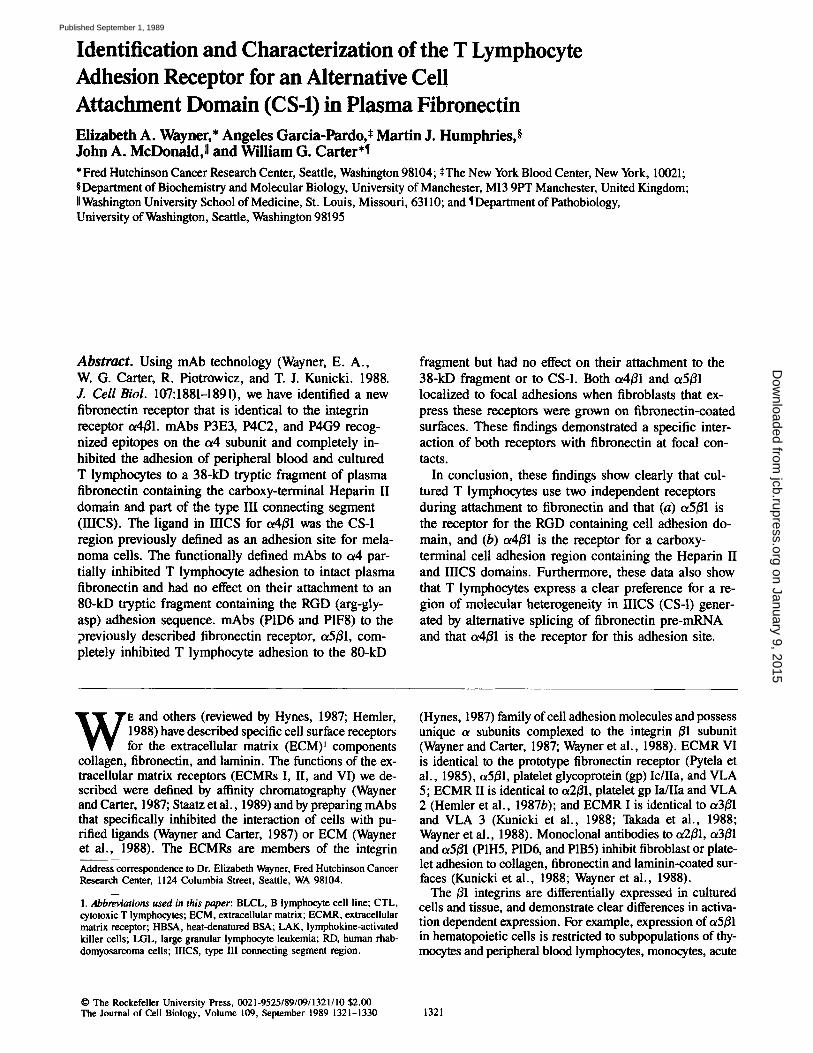
Identification of an Alternative Fibronectin Receptor Cultured T lymphocytes (Molt 4), K562, RD (rhabdomyo- sarcoma), and HTI080 (fibrosarcoma) cells, and freshly de- rived PBL (not shown) adhered to fibronectin-coated sur- faces (Fig. 1, open bars). However, Molt 4 and RD cells expressed low or undetectable levels of the prototype fibro- nectin receptor (integrin ~5~1) recognized by monoclonal antibody P1D6 (Fig. 1, striped bars). Consistent with this, adhesion of Molt 4 and RD cells to fibronectin could not be completely inhibited by P1D6 (Fig. 1, solid bars). Alterna- tively, adhesion of cells to fibronectin that expressed abun- dant a5~l (HT1080 and K562) could be effectively inhibited by P1D6. Furthermore, the synthetic peptide RGDS did not completely inhibit T lymphocyte adhesion to plasma fibro- nectin (50-70% for Molt 4 or Jurkat cells vs. 80-90% for fibroblasts and 100% for K562-1 cells). Together, these data suggested that some cells, such as T lymphocytes, express fibronectin adhesion receptors other than c~5~.
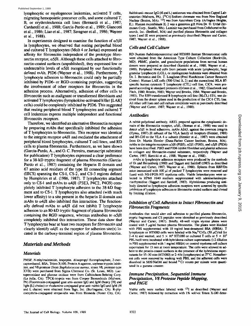
We attempted to identify other putative fibronectin recep- tors by preparing mAbs to cultured T lymphocytes and screening them for their ability to specifically inhibit lym- phocyte but not fibroblast adhesion to fibronectin-coated sur- faces. Using this protocol several mAbs (P4C2, P3E3, P4C~) were identified that inhibited cultured T lymphocyte but not HT1080 cell adhesion to fibronectin (Table I). Immune pre- cipitation from Triton X-100 detergent lysates prepared with t25I-surface-labeled PBL (not shown), Molt 4 or HT1080 (Fig. 2) cells showed that the inhibitory mAbs (data shown for P3E3) reacted with a single protein present in lympho- cyte extracts that migrated at Mr 150,000 (p150) in the pres- ence (not shown) or absence (Fig. 2) of reducing agent. Un- der these immune precipitation conditions p150 lacked an apparent ot-/~ subunit structure and did not co-migrate with either the ol or/3 subunit of the integrin receptors ot2~l or ~3~1 (Fig. 2). The antigen immune precipitated from Triton X-100 detergent extracts prepared with chronically activated CD8+ LAK cells or CTL (not shown) contained, in addition to p150, relatively large quantifies of two smaller proteins that migrated at MT 80,000 and 70,000 in the presence (not shown) or absence of reducing agent. V8 protease peptide mapping revealed that p80 and pT0 were proteolytic frag- ments of p150 (not shown). These lower molecular weight forms could be immune precipitated from chronically acti- vated T cells even when detergent extracts were prepared in the presence of multiple protease inhibitors (legend to Fig. 2). p80 and p70 were virtually absent from extracts prepared with resting PBL, cultured T (Molt 4, Jurkat), or B cell leukemias and RD cells (not shown).
The biochemical characteristics of p150 suggested that it might be related to the VLA 4 antigen described by Hemler (Hemler et al., 1987a). This was confirmed by sequential im- mune precipitation (not shown) with a VLA 4-specific mAb, B5-G10. p150 was established as an c~ subunit of the integrin super family by its association with E1 when immune pre- cipitations were carried out after CHAPS detergent (0.3%) solubilization of t25I-surface-labeled T lymphocytes in the
Wayner et al. Lymphocyte Fibronectin Receptors 1323
on January 9, 2015jcb.rupress.org
Dow
nloaded from
Published September 1, 1989
10g 255
--8O
g ~ de
MOLT4
PI06 EIUgI~Zla0N -~ PI fill INIIII111011 |
C~MTAOL AI~dlESION
RD HTI080 K562-1
Ce l l L i n e
o
C
125
14. ID
Figure 1. Adhesion of T lymphocytes (Molt 4), K562-1, RD, or HT1080 ceils to plasma fibronectin, inhibition with P1D6 mAb, and cell surface expression of ~5~1. 5tCr-labeled cells (105 cells/ml) were incubated with P1D6 mAb (50 #g/ml) or mouse IgG (50 #g/ml) for 60 min at 4°C and allowed to attach to fibronectin-coated (20/~g/ml) plastic surfaces in the presence of P1D6 (solid bars) or mouse IgG (open bars) for 30 min (HTI080 or RD) or 4 h (Molt 4 or K562) at 37°C. Adhesion to plasma fibronectin (pFN) is ex- pressed as 5~Cr bound to the plastic surfaces. Cell surface expres- sion of ~5~ was determined by flow cytometry by staining of cells in suspension with mAb P1D6. Log P1D6 fluorescence (striped bars) is expressed as mean channel number (0-255) above back- ground.
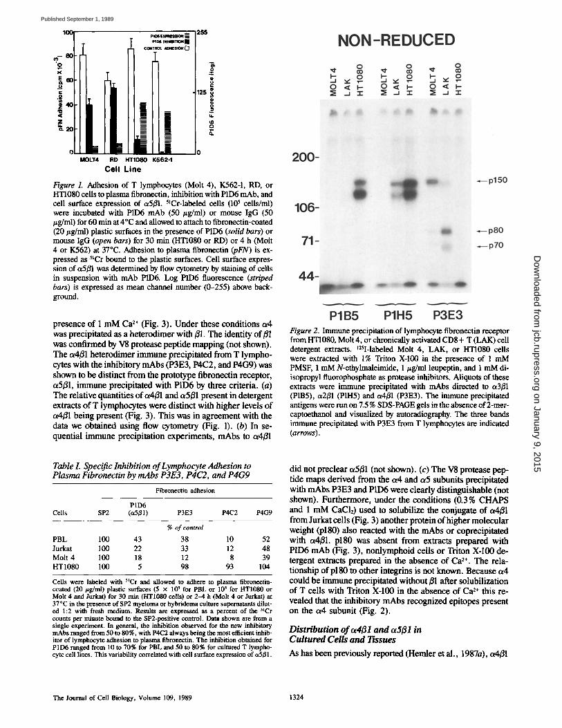
presence of 1 mM Ca 2+ (Fig. 3). Under these conditions oz4 was precipitated as a heterodimer with ~1. The identity of B1 was confirmed by V8 protease peptide mapping (not shown). The ot4B1 heterodimer immune precipitated from T lympho- cytes with the inhibitory rnAbs (P3E3, P4C2, and P4G9) was shown to be distinct from the prototype fibronectin receptor, a5f31, immune precipitated with P1D6 by three criteria. (a) ~__e relative quantities of o~4B1 and oz5B1 present in detergent extracts of T lymphocytes were distinct with higher levels of a4B1 being present (Fig. 3). This was in agreement with the data we obtained using flow cytometry (Fig. 1). (b) In se- quential immune precipitation experiments, mAbs to oz4B1
Figure 2. Immune precipitation of lymphocyte fibronectin receptor from HTI080, Molt 4, or chronically activated CD8 + T (LAK) cell detergent extracts, t2SI-labeled Molt 4, LAK, or HTI080 cells were extracted with 1% Triton X-100 in the presence of 1 mM PMSF, 1 mM N-ethylmaleimide, 1/~g/ml leupeptin, and 1 mM di- isopropyl fluorophosphate as protease inhibitors. Aliquots of these extracts were immune precipitated with mAbs directed to a3/31 (PIB5), ~2B1 (PIH5) and c~4B1 (P3E3). The immune precipitated antigens were run on 7.5 % SDS-PAGE gels in the absence of 2-mer- captoethanol and visualized by autoradiography. The three bands immune precipitated with P3E3 from T lymphocytes are indicated (arrows).
Table L Specific Inhibition of Lymphocyte Adhesion to Plasma Fibronectin by mAbs P3E3, P4C2, and P4G9
Fibronectin adhesion
P1D6 Cells SP2 (~5B1) P3E3 P4C2 P4G9
% of control
PBL 100 43 38 10 52 Jurkat 100 22 33 12 48 Molt 4 100 18 12 8 39 HT1080 100 5 98 93 104
Ceils were labeled with StCr and allowed to adhere to plasma flbronectin- coated (20 t~g/ml) plastic surfaces (5 x 10 ~ for PBL or l0 s for HT1080 or Molt 4 and Jurkat) for 30 rain (HT1080 cells) or 2-4 h (Molt 4 or Jurkat) at 37°C in the presence of SP2 myeloma or hybridoma culture supernatants dilut- ed 1:2 with fresh medium. Results are expressed as a percent of the 5~Cr counts per minute bound to the SP2-positive control. Data shown are from a single experiment. In general, the inhibition observed for the new inhibitory mAbs ranged from 50 to 80%, with P4C2 always being the most efficient inhib- itor of lymphocyte adhesion to plasma flbronectin. The inhibition obtained for PID6 ranged from 10 to 70% for PBL and 50 to 80% for cultured T lympho- cyte cell lines. This variability correlated with cell surface expression of ~5B1.
did not preclear oz5B1 (not shown). (c) The V8 protease pep- tide maps derived from the a4 and or5 subunits precipitated with mAbs P3E3 and P1D6 were clearly distinguishable (not shown). Furthermore, under the conditions (0.3 % CHAPS and 1 mM CaCI2) used to solubilize the conjugate of ot4Bl from Jurkat cells (Fig. 3) another protein of higher molecular weight (p180) also reacted with the mAbs or coprecipitated with o~4/31, plS0 was absent from extracts prepared with P1D6 mAb (Fig. 3), nonlymphoid cells or Triton X-100 de- tergent extracts prepared in the absence of Ca 2+. The rela- tionship of p180 to other integrins is not known. Because or4 could be immune precipitated without B1 after solubilization of T cells with Triton X-100 in the absence of Ca 2÷ this re- vealed that the inhibitory mAbs recognized epitopes present on the c~4 subunit (Fig. 2).
Distribution of ~4{31 and ~5131 in Cultured Cells and Tissues
As has been previously reported (Hemler et al., 1987a), a4/31
The Journal of Cell Biology, Volume 109, 1989 1324
on January 9, 2015jcb.rupress.org
Dow
nloaded from
Published September 1, 1989
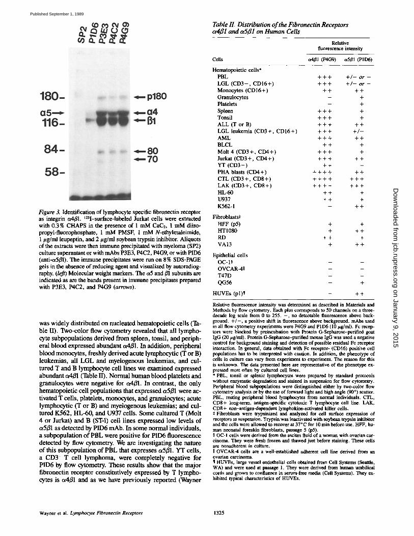
Figure 3. Identification of lymphocyte specific fibronectin receptor as integrin ot4/31. ~25I-surface-labeled Jurkat cells were extracted with 0.3% CHAPS in the presence of 1 mM CaC2, 1 mM diiso- propyl-fluorophosphate, 1 mM PMSF, 1 mM N-ethylmaleimide, 1 #g/ml leupeptin, and 2/zg/ml soybean trypsin inhibitor. Aliquots of the extracts were then immune precipitated with myeloma (SP2) culture supernatant or with mAbs P3E3, P4C2, P4G9, or with P1D6 (anti-tx5/~l). The immune precipitates were run on 8% SDS-PAGE gels in the absence of reducing agent and visualized by autoradiog- raphy. (le~) Molecular weight markers. The ct5 and/~1 subunits are indicated as are the bands present in immune precipitates prepared with P3E3, P4C2, and P4G9 ( a r r o w s ) .
was widely distributed on nucleated hematopoietic cells (Ta- ble II). Two-color flow cytometry revealed that all lympho- cyte subpopulations derived from spleen, tonsil, and periph- eral blood expressed abundant t~4/~l. In addition, peripheral blood monocytes, freshly derived acute lymphocytic (T or B) leukemias, all LGL and myelogenous leukemias, and cul- tured T and B lymphocyte cell lines we examined expressed abundant ot4/~l (Table 1I). Normal human blood platelets and granulocytes were negative for ~4/31. In contrast, the only hematopoietic cell populations that expressed ot5/ffl were ac- tivated T cells, platelets, monocytes, and granulocytes; acute lymphocytic (T or B) and myelogenous leukemias; and cul- tured K562, HL-60, and U93"/cells. Some cultured T (Molt 4 or Jurkat) and B (ST-l) cell lines expressed low levels of ot5/31 as detected by P1D6 mAb. In some normal individuals, a subpopulation of PBL were positive for PID6 fluorescence detected by flow cytometry. We are investigating the nature of this subpopulation of PBL that expresses ot5/31. YT cells, a CD3- T cell lymphoma, were completely negative for P1D6 by flow cytometry. These results show that the major fibronectin receptor constitutively expressed by T lympho- cytes is ot4/~l and as we have previously reported (Wayner
Table IL Distribution of the Fibronectin Receptors tz4~l and ~5~1 on Human Cells
Relative fluorescence intensity
Cells ot4/~l (P4G9) ot5/~l (P1D6)
Hematopoietic cells* PBL + + + + / - o r -
LGL ( C D 3 - , C D I 6 + ) + + + + / - o r -
Monocytes (CD16+) + + + + Granulocytes - + Platelets - + Spleen + + + + Tonsil + + + + A L L ( T o r B ) + + + + + LGL leukemia (CD3+, C D I 6 + ) + + + + / - AML + + + + + BLCL + + + Molt 4 (CD3 + , CD4 +) + + + + Jurkat (CD3 + , C D 4 + ) + + + + + YT ( C D 3 - ) + + - PHA blasts (CD4+) + + + + + + CTL (CD3+ , C D 8 + ) + + + + + + + LAK (CD3+ , C D 8 + ) + + + + + + + HL-60 + + + U937 + + + K562-1 - + +
Fibroblasts~ HFF (p5) + + HT1080 + + + RD + + + VA13 + + +
Epithelial cells OC-I§ - - OVCAR-411 - - T47D - - QG56 - +
HUVEs (pl)¶ - + +
Relative fluorescence intensity was determined as described in Materials and Methods by flow cytometry. Each plus corresponds to 50 channels on a three- decade log scale from 0 to 255. - , no detectable fluorescence above back- ground. + / - , a positive shift in fluorescence above background, mAbs used in all flow cytometry experiments were P4CO and PID6 (10 t~g/ml). Fc recep- tors were blocked by preincubation with Protein G-Sepharose-purified goat IgG (20 #g/ml). Protein G-Sepharose-purified mouse IgG was used a negative control for background staining and detection of possible residual Fc receptor interaction. In general, data obtained with Fc receptor- (CDI6) positive cell populations has to be interpreted with caution. In addition, the phenotype of cells in culture can vary from experiment to experiment. The reason for this is unknown. The data presented here are representative of the phenotype ex- pressed most often by cultured cell lines. * PBL, tonsil or splenic lymphocytes were prepared by standard protocols without enzymatic degradation and stained in suspension for flow cytometry. Peripheral blood subpopulations were distinguished either by two-color flow cytometry analysis or by the use of forward light and high angle (90*) scatter. PBL, resting peripheral blood lymphocytes from normal individuals. CTL, CDS+ long-term, antigen-specific eytotoxic T lymphocyte cell line. LAK, CD8 + non-antigen-dependent lymphokine-activated killer cells. :~ Fibroblasts were trypsinized and analyzed for cell surface expression of receptors in suspension. Trypsin was inactivated with soybean trypsin inhibitor and the cells were allowed to recover at 37"C for 10 rain before use. HFF, hu- man neonatal foreskin fibroblasts, passage 5 (p5). § OC-I cells were derived from the aseites fluid of a woman with ovarian car- cinoma. They were fresh frozen and thawed just before staining. These cells are nonadherent in culture. U OVCAR-4 cells are a well-established adherent cell line derived from an ovarian carcinoma. ¶ HUVEs, large vessel endothelial cells obtained from Cell Systems (Seattle, WA) and were used at passage 1. They were derived from human umbilical cords and grown to confluence in sentm-free media (Cell Systems). They ex- hibited typical characteristics of HUVEs.
Wayner et al. Lymphocy te Fibronectin Receptors 1325
on January 9, 2015jcb.rupress.org
Dow
nloaded from
Published September 1, 1989
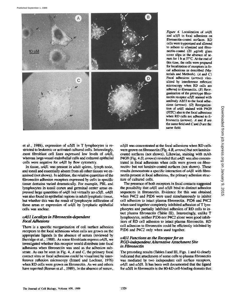
Figure 4. Localization of a4/Jl and c~5/31 in focal adhesions on flbmnectin-coated surfaces. RD cells were trypsinized and allowed to adhere to silanized and fibro- nectin-coated (20 #g/ml) glass cover slips in the absence of se- rum for 1 h at 37"C. At the end of this time, the cells were prepared for localization of receptors in fo- cal adhesions as described (Ma- terials and Methods). (.4 and C) Focal adhesions (arrows) visu- alized by interference reflexion microscopy when RD cells are adhered to fibronectin. (B) Reor- ganization of the prototype fibro- nectin receptor ot5B1 stained with antibody AB33 to the focal adhe- sions (arrows). (D) Reorganiza- tion of oMB1 stained with P4G9 (FITC) also to the focal adhesions when RD cells are adhered to fi- brotw, ctin (arrows). A and B are the same fieM and C and D are the same field.
et al., 1988), expression of c~5/~1 in T lymphocytes is re- stricted to leukemic or activated cultured cells. Interestingly, most fibroblast cell lines expressed low levels of ot4/~l, whereas large-vessel endothelial cells and cultured epithelial cells were negative for ot4/~l by flow cytometry.
In tissue, ot4/~l was present in adult spleen, lymph node, and tonsil and essentially absent from all other tissues we ex- m ined (not shown). In addition, the relative quantities of the fibronectin adhesion receptors expressed by cells in specific tissue domains varied dramatically. For example, PBL and lymphocytes in tonsil cortex and germinal center areas ex- pressed large quantifies of ot4B1 but virtually no ot5fll, o~4/31 was also found in epithelial regions in adult lymphatic tissue, but whether this was the result of lymphocyte infiltration of these areas or expression of ot4fll by lymphatic epithelial cells was unclear.
c~4fl l Localizes in Fibronectin-dependent Focal Adhesions
There is a specific reorganization of cell surface adhesion receptors to the focal adhesions when cells are grown on the appropriate ligands in the absence of serum (reviewed by Burridge et al., 1988). As some fibroblasts express c~4B1, we investigated whether this receptor would distribute into focal adhesions when fibronectin was used as the adhesion sub- strate. As can be seen in Fig. 4, A and C, the primary focal contact sites or focal adhesions could be visualized by inter- ference reflexion microscopy (Izzard and Lochner, 1976) when RD cells were grown on fibronectin. As we and others have reported (Roman et al., 1989), in the absence of serum,
a5/31 was concentrated at the focal adhesions when RD cells were grown on fibronectin (Fig. 4 B, arrows) but not laminin- coated surfaces (not shown). Likewise, staining with mAb P4CO (Fig. 4 D, arrows) revealed that t~4B1 was also concen- trated in focal adhesions when cells were grown on fibro- nectin- but not laminin-coated surfaces (not shown). These results demonstrate a specific interaction of t~4B1 with fibro- nectin present in focal adhesions, the primary adhesion struc- ture of cultured cells.
The presence of both receptors in focal contacts suggested the possibility that ot4/~l and otSBl bind to distinct adhesion sequences in fibronectin. Evidence for this was obtained when P4C2 and P1D6 were used simultaneously to inhibit cell adhesion to intact plasma fibronectin. P1D6 and P4C2 when used together completely inhibited adhesion of T lym- phocytes and partially inhibited adhesion of RD cells to in- tact plasma fibronectin (Table III). Interestingly, unlike T lymphocytes, neither P1D6 nor P4C2 alone were good inhib- itors of RD cell adhesion to intact plasma fibronectin. RD cell adhesion to fibronectin could be efficiently inhibited by P1D6 and P4C2 only when used together.
ot4B1 Functions as the Receptor for an RGD-independent Alternative Attachment Site in l~bronectin
The preceding results (Tables I and HI, Figs. 1 and 4) clearly indicated that attachment of some cells to plasma fibronectin was mediated by two independent cell surface receptors, c~4B1 and ot5B1. It has been well documented that the ligand for ot5B1 in fibronectin is the 80-kD cell-binding domain that
The Journal of Cell Biology, Volume 109, 1989 1326
on January 9, 2015jcb.rupress.org
Dow
nloaded from
Published September 1, 1989
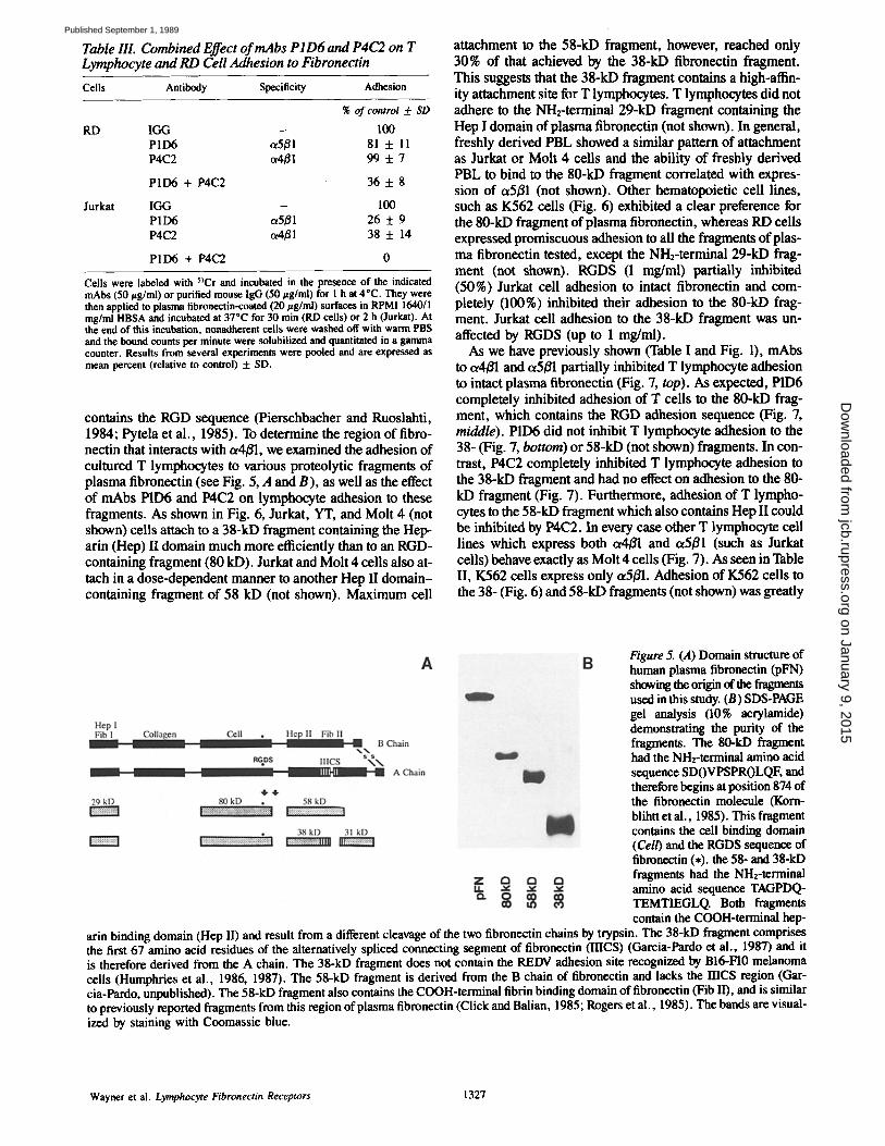
Table IlL Combined Effect o f mAbs P I D6 and P4C2 on T Lymphocyte and RD Cell Adhesion to Fibronectin
Cells Antibody Specificity Adhesion
RD
Jurkat
% of control 5: SD
I GG - 100 P1D6 et5/~l 81 + 11 P4C2 ~4~ 1 99 + 7
PID6 + P4C2 36 -t- 8
IGG - 100 PID6 a5/~l 26 + 9 P4C2 o~4/~1 38 + 14
P1D6 + P4C2 O
Cells were labeled with 5~Cr and incubated in the presence of the indicated mAbs (50 ~tg/ml) or purified mouse IgG (50 ttg/ml) for 1 h at 4"C. They were then applied to plasma fibroneetin-coated (20 ttg/ml) surfaces in RPM11640/I mg/ml HBSA and incubated at 37"C for 30 rain (RD cells) or 2 h (Jurkat). At the end of this incubation, nonadherent cells were washed off with warm PBS and the bound counts per minute were solubilized and quantitated in a gamma counter. Results from several experiments were pooled and are expressed as mean percent (relative to control) + SD.
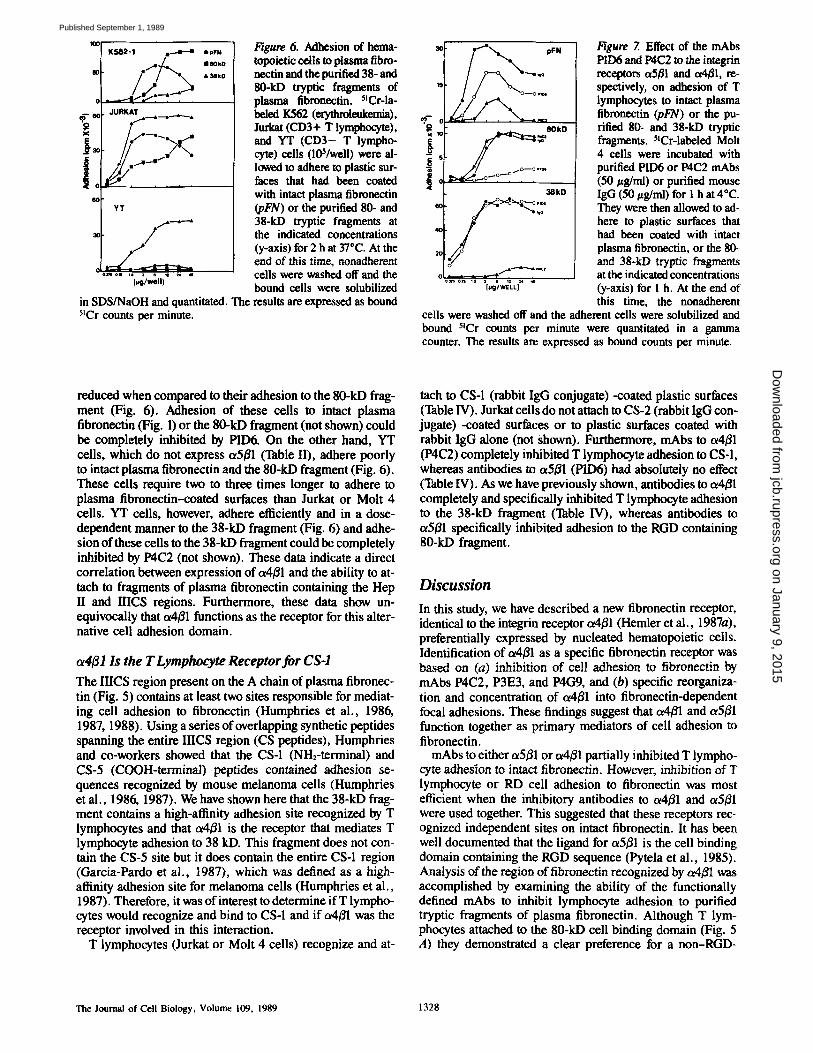
contains the RGD sequence (Pierschbacher and Ruoslahti, 1984; Pytela et al., 1985). To determine the region of fibro- nectin that interacts with o~4/31, we examined the adhesion of cultured T lymphocytes to various proteolytic fragments of plasma fibronectin (see Fig. 5, A and B), as well as the effect of mAbs P1D6 and P4C2 on lymphocyte adhesion to these fragments. As shown in Fig. 6, Jurkat, YT, and Molt 4 (not shown) cells attach to a 38-kD fragment containing the Hep- arin (Hep) II domain much more efficiently than to an RGD- containing fragment (80 kD). Jurkat and Molt 4 cells also at- tach in a dose-dependent manner to another Hep II domain- containing fragment of 58 kD (not shown). Maximum cell
attachment to the 58-kD fragment, however, reached only 30% of that achieved by the 38-kD fibronectin fragment. This suggests that the 38-kD fragment contains a high-affin- ity attachment site for T lymphocytes. T lymphocytes did not adhere to the NH2-terminal 29-kD fragment containing the Hep I domain of plasma fibronectin (not shown). In general, freshly derived PBL showed a similar pattern of attachment as Jurkat or Molt 4 cells and the ability of freshly derived PBL to bind to the 80-kD fragment correlated with expres- sion of ot5/31 (not shown). Other hematopoietic cell lines, such as K562 cells (Fig. 6) exhibited a clear preference for the 80-kD fragment of plasma fibronectin, whereas RD cells expressed promiscuous adhesion to all the fragments of plas- ma fibronectin tested, except the NH2-terminal 29-kD frag- ment (not shown). RGDS (1 mg/ml) partially inhibited (50%) Jurkat cell adhesion to intact fibronectin and com- pletely (100%) inhibited their adhesion to the 80-kD frag- ment. Jurkat cell adhesion to the 38-kD fragment was un- affected by RGDS (up to 1 mg/ml).
As we have previously shown (Table I and Fig. 1), mAbs to a4/~l and ,5/31 partially inhibited T lymphocyte adhesion to intact plasma fibronectin (Fig. 7, top). As expected, P1D6 completely inhibited adhesion of T cells to the 80-kD frag- ment, which contains the RGD adhesion sequence (Fig. 7, middle). P1D6 did not inhibit T lymphocyte adhesion to the 38- (Fig. 7, bottom) or 58-kD (not shown) fragments. In con- trast, P4C2 completely inhibited T lymphocyte adhesion to the 38-kD fragment and had no effect on adhesion to the 80- kD fragment (Fig. 7). Furthermore, adhesion of T lympho- cytes to the 58-kD fragment which also contains Hep II could be inhibited by P4C2. In every case other T lymphocyte cell lines which express both ot4/31 and ot5/31 (such as Jurkat cells) behave exactly as Molt 4 cells (Fig. 7). As seen in Table II, K562 cells express only t~5/31. Adhesion of K562 cells to the 38- (Fig. 6) and 58-kD fragments (not shown) was greatly
Figure 5. (,4) Domain structure of human plasma fibronectin (pFN) showing the origin of the fragments used in this study. (B) SDS-PAGE gel analysis (10% acrylamide) demonstrating the purity of the fragments. The 80-kD fragment had the NH2-terminal amino acid sequence SDOVPSPROLQF, and therefore begins at position 874 of the fibronectin molecule (Koru- blihtt et al., 1985). This fragment contains the cell binding domain (Cell) and the RGDS sequence of fibronectin (*). the 58- and 38-kD fragments had the NH2-terminal amino acid sequence TAGPDQ- TEMTIEGLQ. Both fragments contain the COOH-terminal hep-
arin binding domain (Hep II) and result from a different cleavage of the two fibronectin chains by trypsin. The 38-kD fragment comprises the first 67 amino acid residues of the alternatively spliced connecting segment of fibronectin (IIICS) (Garcia-Pardo et al., 1987) and it is therefore derived from the A chain. The 38-kD fragment does not contain the REDV adhesion site recognized by BI6-FI0 melanoma cells (Humphries et al., 1986, 1987). The 58-kD fragment is derived from the B chain of fibronectin and lacks the mCS region (Gar- cia-Pardo, unpublished). The 58-kD fragment also contains the COOH-terminal fibrin binding domain of fibronectin (Fib II), and is similar to previously reported fragments from this region of plasma fibroncctin (Click and Balian, 1985; Rogers et al., 1985). The bands are visual- ized by staining with Coomassie blue.
Wayner et al. Lymphocyte Fibronectin Receptors 1327
on January 9, 2015jcb.rupress.org
Dow
nloaded from
Published September 1, 1989
1(]0[ K 5 6 2 -1 r . 4 - - a
- - e~ J U R K A T . .__.._. . . . . . . . . ._.
• ?FN
I 8 0 k t )
&38kD
YT
. . . . . . I & * ; , , l . . . .
in SDS/NaOH and quantitated. The 5tCr counts per minute.
l~gure 6. Adhesion of hema- topoietic cells to plasma fibro- neetin and the purified 38- and 80-kD tryptic fragments of plasma fibronectin. 5!Cr-la- beled I(562 (erythroleukemia), Jurkat (CD3+ T lymphocyte), and YT (CD3- T lympho- cyte) cells (10S/well) were al- lowed to adhere to plastic sur- faces that had been coated with intact plasma fibronectin (pFN) or the purified 80- and 38-kD tryptic fragments at the indicated concentrations (y-axis) for 2 h at 37"C. At the end of this time, nonadherent cells were washed off and the bound cells were solubilized results are expressed as bound
. 8OkO
<3 ,,~
* ~ k D ,L ~,~o>~,~.....~,~
. . . . . . . . . I ~ g ' , ~ ' ~ "
Figure 7. Effect of the mAbs PtD6 and 1)4(22 to the integrin receptors (~5#1 and ~4/~1, re- speetively, on adhesion of T lymphocytes to intact plasma fibronectin (pFN) or the pu- rified 80- and 38-kD tryptic fragments. 5'Cr-labeled Molt 4 cells were incubated with purified 1)11:)6 or P4C2 mAbs (50 t~g/ml) or purified mouse IgG (50 t~g/ml) for 1 h at 4"C. They were then allowed to ad- here to plastic surfaces that had been coated with intact plasma fibronectin, or the 80- and 38-kD tryptic fragments at the indicated concentrations (y-axis) for 1 h. At the end of this time, the nonadherent
cells were washed off and the adherent cells were solubilized and bound 5~Cr counts per minute were quantitated in a gamma counter. The results are expressed as bound counts per minute.
reduced when compared to their adhesion to the 80-kD frag- ment (Fig. 6). Adhesion of these cells to intact plasma fibronectin (Fig. 1) or the 80-kD fragment (not shown) could be completely inhibited by P1D6. On the other hand, YT cells, which do not express ot5B1 (Table II), adhere poorly to intact plasma fibronectin and the 80-kD fragment (Fig. 6). These cells require two to three times longer to adhere to plasma fibronectin-coated surfaces than Jurkat or Molt 4 cells. YT cells, however, adhere efficiently and in a dose- dependent manner to the 38-kD fragment (Fig. 6) and adhe- sion of these cells to the 38-kD fragment could be completely inhibited by P4C2 (not shown). These data indicate a direct correlation between expression of (x4/~l and the ability to at- tach to fragments of plasma fibronectin containing the Hep II and IIICS regions. Furthermore, these data show un- equivocally that ot4/~l functions as the receptor for this alter- native cell adhesion domain.
~4/31 Is the T Lymphocyte Receptor for CS-I The IIICS region present on the A chain of plasma fibronec- tin (Fig. 5) contains at least two sites responsible for mediat- ing cell adhesion to fibronectin (Humphries et al., 1986, 1987, 1988). Using a series of overlapping synthetic peptides spanning the entire IIICS region (CS peptides), Humphries and co-workers showed that the CS-1 (NH2-terminal) and CS-5 (COOH-terminal) peptides contained adhesion se- quences recognized by mouse melanoma cells (Humphries et al., 1986, 1987). We have shown here that the 38-kD frag- ment contains a high-affinity adhesion site recognized by T lymphocytes and that o~4#1 is the receptor that mediates T lymphocyte adhesion to 38 kD. This fragment does not con- tain the CS-5 site but it does contain the entire CS-1 region (Garcia-Pardo et at., 1987), which was defined as a high- affinity adhesion site for melanoma cells (Humphries et al., 1987). Therefore, it was of interest to determine ifT lympho- cytes would recognize and bind to CS-1 and if a4/~l was the receptor involved in this interaction.
T lymphocytes (Jurkat or Molt 4 cells) recognize and at-
tach to CS-1 (rabbit IgG conjugate) -coated plastic surfaces (Table IV). Jurkat cells do not attach to CS-2 (rabbit IgG con- jugate) -coated surfaces or to plastic surfaces coated with rabbit IgG alone (not Shown). Furthermore, mAbs to oL4/~l (P4C2) completely inhibited T lymphocyte adhesion to CS-1, whereas antibodies to (x5~l (P1D6) had absolutely no effect (Table IV). As we have previously shown, antibodies to ~4fll completely and specifically inhibited T lymphocyte adhesion to the 38-kD fragment (Table IV), whereas antibodies to ~5/~1 specifically inhibited adhesion to the RGD containing 80-kD fragment.
Discussion
In this study, we have described a new fibronectin receptor, identical to the integrin receptor a4/~l (Hemler et al., 1987a), preferentially expressed by nucleated hematopoietic cells. Identification of ~4~1 as a specific fibronectin receptor was based on (a) inhibition of cell adhesion to fibronectin by mAbs P4C2, P3E3, and P4G9, and (b) specific reorganiza- tion and concentration of a4~l into fibronectin-dependent focal adhesions. These findings suggest that c~4B1 and ~5fll function together as primary mediators of cell adhesion to fibronectin.
rnAbs to either ~5~1 or ot4~l partially inhibited T lympho- cyte adhesion to intact fibronectin. However, inhibition of T lymphocyte or RD cell adhesion to fibronectin was most efficient when the inhibitory antibodies to o~4/~1 and o~5/31 were used together. This suggested that these receptors rec- ognized independent sites on intact fibronectin. It has been well documented that the ligand for c~5fll is the cell binding domain containing the RGD sequence (Pytela et al., 1985). Analysis of the region of fibronectin recognized by a4/31 was accomplished by examining the ability of the functionally defined mAbs to inhibit lymphocyte adhesion to purified tryptic fragments of plasma fibronectin. Although T lym- phocytes attached to the 80-kD cell binding domain (Fig. 5 A) they demonstrated a clear preference for a non-RGD-
The Journal of Cell Biology, Volume 109, 1989 1328
on January 9, 2015jcb.rupress.org
Dow
nloaded from
Published September 1, 1989
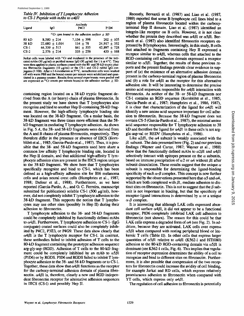
Table IV. Inhibition of T Lymphocyte Adhesion to CS-1 Peptide with mAbs to ce4B1
Antibody Ligand IgG P4C2 P 1 I)6
~lCr cpm bound to the adhesion surface 4- SD
80 kD 8,580 + 214 7,154 + 398 202 5:105 38 kD 22,680 5:1 ,014 114 5 :78 24,917 5:352 CS-1 44,339 + 513 841 + 555 42,897 5 :728 CS-2 2,576 + 214 535 5 :258 435 5 :168
Jurkat cells were labeled with 5~Cr and incubated in the presence of the indi- cated mAbs (50 t~g/ml) or purified mouse IgG (50 t~g/mi) for 1 h at 4"C. They were then applied to plastic surfaces coated with the 80- and 38-kD tryptic plas- ma fibronectin fragments (10/~g/ml) or the CS-1 and CS-2 rabbit IgG con- jugates (1:100). After a 2-h incubation at 370C, nonadherent cells were washed off with warm PBS and the bound counts per minute were solubilized and quan- titated in a gamma counter. Results from several experiments were pooled and are expressed as 5~Cr counts per minute bound to the adhesion surface -1- SD.
containing region located on a 38-kD tryptic fragment de- rived from the A (or heavy) chain of plasma fibronectin. In the present study we have shown that T lymphocytes also recognize and bind to another Hep II-containing 58-kD frag- ment. However, the high-affinity lymphocyte binding site was located on the 38-kD fragment. On a molar basis, the 38-kD fragment was three times more efficient than the 58- kD fragment in mediating T lymphocyte adhesion. As shown in Fig. 5 A, the 38- and 58-kD fragments were derived from the A and B chains of plasma fibronectin, respectively. They therefore differ in the presence or absence of HICS (Korn- blihtt et al., 1985; Garcia-Pardo et al., 1987). Thus, it is pos- sible that the 38- and 58-kD fragments used here share a common low affinity T lymphocyte binding site, located in the Hep II domain, and that additional high-affinity T lym- phocyte adhesion sites are present in the IIICS region unique to the 38-kD fragment. We show here that T lymphocytes specifically recognize and bind to CS-1, which has been defined as a high-affinity adhesion site for B16 melanoma cells and avian neural crest cells (Humphries et al., 1987, 1988; Dufour et al., 1988). Furthermore, as we have reported (Garcia-Pardo, A., and O. C. Ferreira, manuscript submitted for publication) soluble CS-1 (500 t~g/ml), how- ever, did not completely inhibit T lymphocyte adhesion to the 38-kD fragment. This supports the notion that T lympho- cytes may use other sites (possibly in Hep II) during their adhesion to fibronectin.
T lymphocyte adhesion to the 38- and 58-kD fragments could be completely inhibited by functionally defined mAbs to a4E1. Furthermore, T lymphocyte adhesion to CS-1- (IgG conjugate) coated surfaces could also be completely inhib- ited by P4C2, P3E3, or P4G9. These data show clearly that o~4E1 is the T lymphocyte receptor for CS-1. In contrast, these antibodies failed to inhibit adhesion of T cells to the 80-kD fragment containing the prototype adhesion sequence arg-gly-asp (RGD). Adhesion of T cells to the 80-kD frag- ment could be completely inhibited by an mAb to ot5/31 (P1D6) or by RGDS. P1D6 and RGDS failed to inhibit T lym- phocyte adhesion to the 38- and 58-kD fragments or to CS-1. Together, these data show that ~x4E1 functions as the receptor for the carboxy-terminal adhesion domain of plasma fibro- nectin, ot4E1 is, therefore, clearly a new and RGD indepen- dent fibronectin receptor for alternative adhesion sequences in IIICS (CS-1) and possibly Hep II.
Recently, Bernardi et al. (1987) and Liao et al. (198% 1989) reported that some B lymphocyte cell lines bind to a region of plasma fibronectin located within the carboxy- terminal Hep II domain. Liao et al. (1987) identified an integrin-like receptor on B cells. However, it is not clear whether the protein they described was ~x4E1 or ot5E1. Ber- nardi et al. (1987) also identified fibronectin receptors ex- pressed by B lymphocytes. Interestingly, in this study, B cells that attached to fragments containing Hep II expressed a receptor similar to ot4E1, whereas cells that attached to the RGD-containing cell adhesion domain expressed a receptor similar to o~5E1. Together, the results of these previous re- ports and the present findings provide clear evidence in sup- port of (a) the existence of an alternative adhesion domain present in the carboxy-terminal region of plasma fibronectin and (b) a role for ot4131 as the receptor for this alternative adhesion site. It will be interesting to determine the precise amino acid sequences responsible for ot4131 interaction with fibronectin. As neither of the 38- or 58-kD fragments nor CS-1 contains an RGD sequence (Kornblihtt et al., 1985; Garcia-Pardo et al., 1987; Humphries et al., 1986, 1987), it is clear that characterization of the ligand for a4E1 will identify a new amino acid sequence important for cell adhe- sion to fibronectin. Because the 38-kD fragment does not contain CS-5 (Garcia-Pardo et al., 1987), the minimal amino acid sequence responsible for T lymphocyte adhesion to 38 kD and therefore the ligand for u4/31 in these cells is not arg- glu-asp-val or REDV (Humphries et al., 1986).
Like ot2E1, the o~4 subunit is weakly associated with the B1 subunit. The data presented here (Fig. 2) and our previous findings (Wayner and Carter, 1987; Wayner et al., 1988) show that the functionally defined mAbs to ot2/31 and ot4E1 selectively interact with epitopes present on the a subunits, based on immune precipitation of ol2 or c~4 without E1 after subunit dissociation. These results suggest that the unique ot subunit is responsible for determining the ligand-binding specificity of each o~-E complex. This concept is now further supported by the observations presented here that or5 and or4, which are both complexed with El, mediate adhesion to dis- tinct sites on fibronectin. This is not to suggest that the B sub- unit is not important in binding, but that the specificity of receptor-ligand interactions is determined by ot or a unique oL-B complex.
It is interesting that although LAK cells expressed abun- dant cell surface ot4/31, it did not appear to be a functional receptor; P1D6 completely inhibited LAK cell adhesion to fibronectin (not shown). The reason for this could be that LAK cells express a degraded form of a4 (see Fig. 2). In ad- dition, because they are activated, LAK cells over express cx5E1 when compared with resting peripheral blood or leu- kemic T cells (Table II). In other cells that express larger quantities of o~5/31 relative to ot4E1 (K562-1 and HT1080) adhesion to the 80-kD RGD-containing domain via ot5E1 is dominant (see K562-1 cells, Fig. 6). This implies that regula- tion of receptor expression determines the ability of a cell to recognize and bind to different sites on fibronectin. Further- more, it is also possible that coexpression of the two recep- tors for fibronectin could increase the avidity of cell binding, for example Jurkat and RD cells, which express relatively promiscuous adhesion to fibronectin when compared with YT ceils, which express only a4~l.
The regulation of cell adhesion to fibronectin is potentially
Wayner et al. Lymphocyte Fibronectin Receptors 1329
on January 9, 2015jcb.rupress.org
Dow
nloaded from
Published September 1, 1989
complex even under the simplest possible conditions, which assume that a5/~1 and ot4B1 function independently of each other and do not overlap during interaction with the two binding sites on fibronectin. Variation from this simple state provides opportunities for exquisitely sensitive regulation of cell adhesion. At the least complex level, this regulation can be roughly eatagorized as (a) processes that control the syn- thesis and/or exposure of the binding sites on the ligand and (b) regulation of functional expression of the receptors. Ex- amples of regulation at both levels are currently available and include, the observation that lymphokines and specific anti- gen induce o~5/~1 expression on T lymphoeytes followed by increased cell adhesion to fibroneetin (Wayner et al., 1988, and unpublished). In addition, the control of mRNA splicing in the RICS region of fibroneetin (Kornblihtt et al., 1985) during wound healing or inflammation may dictate the speci- fiery of reeeptor-ligand binding in resting or activated T cells. Variations from the simple state are intriguing but re- quire additional experimentation to even begin to identify the multitude of potential mechanisms.
This research was supported by grants BC-419 to W. G. Carter from the American Cancer Society and RO1 CA-388801 from the National Cancer Institute. M. J. Humphries was supported by a grant from the Wellcome Trust.
Received for publication 30 May 1989, and in revised form 25 June 1989.
Note added in proof. We have identified a minimal peptide derived from the carboxy terminal portion of CS- 1, LHGPEILDVPST, which inhibits T lymphocyte adhesion to plasma fibronectin, 38 kD, and CS-I.
References
Bernardi, P., V. P. Patel, and H. F. Lodish. 1987. Lymphoid precursor cells adhere to two different sites on fibronectin. J. Ceil Biol. 105:489-498.
Brooks, C. G. 1983. Reversible induction of natural killer cell activity in cloned murine cytotoxic T lymphocytes. Nature (Land.). 305:155-158.
Burridge, K., K. Fath, T. Kelly, G. Nuckolls, and C. Turner. 1988. Focal adhe- sions: transmembrane junctions between the extracellular matrix and the cytoskeleton. Annu. Rev. Cell Biol. 4:487-525.
Cardarelli, P. M., 1. N. Crispe, and M. D. Pierschhacher. 1988. Preferential expression of fibronectin receptors on immature thymocytes. J. Cell Biol. 106:2183-2190.
Canner, W. G., and E. A. Wayner. 1988. Characterization of a collagen-bind- ing, phosphorylated, transmembrane glycoprotein expressed in nucleated human cells. J. Biol. Chem. 263:4193-4201.
Cleveland, D. W., S. G. Fischer, M. W. Kirschner, and U. K. Laemmli. 1977. Peptide mapping by limited proteolysis in sodium dodecyl sulfate and analy- sis by gel electrophoresis. J. Biol. Chem. 252:1102-1106.
Click, E. M., and G. Balian. 1985. Domain structure of human plasma and cel- lular fibronectin. Use of a monoclonal antibody and beparin affinity to iden- tify three different subunit chains. Biochemistry. 24:6685-6696.
Durfour, S., J. L. Duband, M. Humphries, M. Obara, K. Yamada, and J. P. Thiery. 1988. Attachment, spreading and locomotion of avian neural crest cells are mediated by multiple adhesion sites on fibronectin molecules. EMBO (Eur. Mol. Biol. Organ.) J. 7:2661-2671.
Garcia-Pardo, A., A. Rostagno, and B. Frangione. 1987. Primary structure of human plasma fibronectin. Characterization of a 38 kDa domain containing the C-terminal heparin-binding site (Hap lII site) and a region of molecular heterogeneity. Biochem. J. 241:923-928.
Garcia-Pardo, A., O. C. Ferreira, J. Valinsky, and C. Bianco. 1989. Fihronec- tin receptors of mononuclear phagocytes: binding characteristics and bio- chemical isolation. Exp. Cell Res. 181:420-431.
Giancotti, F. G., P. M. Comoglio, and G. Tarnne. 1986. Fibronectin-plasma membrane interaction in the adhesion of hemopoietic cells. J. Cell Biol. 103:429-437.
Glasehrook, A. L., and F. W. Fitch. 1980. AIIoreactive cloned T cell lines. J. Exp. IVied. 151:876-895.
Grimm, E. A., A. Mazumder, H. Z. Zhang, and S. A. Rosenberg. 1982. Lym- phokine activated killer cell phenomenon. Lysis of natural killer-resistant fresh solid tumor cells by interleukin 2-activated autologous human periph- eral blood lymphocytes. J. Exp. Med. 155:1923-1941.
Hemler, M. E., C. Huang, and L. Schwarz. 1987a. The VLA protein family: characterization of five distinct cell surface heterodimers each with a com- mon 130,000 molecular weight/3 subunit. J. Biol. Chem. 262:3300-3309.
Hemler, M. E., C. Huang, Y. Takade, L. Schwm-z, J. L. Strominger, and M. L. Clabby. 1987b. Characterization of the cell surface heterodimer VLA-4 and related peptides. J. Biol. Chem. 262:11478-11485.
Hemler, M. E. 1988. Adhesive protein recep~rs on hematopoetic cells, lm- raanol. Today. 9:109-113.
Humphries, M. J., S. K. Akiyama, A. Komoriya, K. Olden, and K. M. Yamada. 1986. Identifcation of an alternatively spliced site in human plasma fibmnectin that mediates cell type-specific adhesion. J. Cell Biol. 103:2637- 2647.
Humphries, M. J., A. Komoriya, S. K. Akiyama, K. Olden, and K. M. Yamada. 1987. Identification of two distinct regions of the type Ill connect- ing segment of human plasma fibronectin that promote cell type-specific adhesion. J. Biol. Chem. 262:6886-6892.
Humphries, M. J., S. K. Akiyama, A. Komoriya, K. Olden, and K. M. Yamada. 1988. Neurite extension of chicken peripheral nervous system neu- rons on fibronectin: relative importance of specific adhesion sites in the cen- tral cell-binding domain and the alternatively spliced type Ill connecting seg- ment. J. Cell Biol. 106:1289-1297.
Hynes, R. O. 1987. Integrins: a family of cell surface receptors. Cell. 48:549-554.
lzzard, C. S., and L. R. Lochner. 1976. Cell-to-substrate contacts in living fibroblasts: an interference reflexion study with an evaluation of the tech- nique. J. Cell Sci. 21:129-159.
Kornblihtt, A. R., K. Umezawa, K. Vibe-Pedersen, and F. Baralle. 1985. Pri- mary structure of human plasma fibronectin: differential splicing may gener- ate at least l0 polypeptides from a single gene. EMBO (Eur. Mol. Biol. Or- gan.) J. 4:1755-1759.
Kunicki, T. J., D. J. Nugent, S. J. Staats, R. P. Orchekowski, E. A. Wayner, and W. G. Caner. 1988. The human fibroblast class Il extracellular matrix receptor mediates platelet adhesion to collagen and is identical to the platelet la/Ila complex. J. Biol. Chem. 263:4516-4519.
Laemmli, U. K. 1970. Cleavage of structural proteins during the assembly of the head of bacteriophage T4. Nature (Lond.). 227:680-685.
Liao, N. S., J. St. John, Z. J. Du, and H. T. Cheang. 1987. Adhesion of lym- phoid cell lines to fibronectin-coated substratum: biochemical and physiolog- ical characterization and the identification of a 140-kDa fibronectin receptor. Exp. Cell Res. 171:306-320.
Liao, N. S., J. St. John, J. B. McCarthy, L. T. Furcht, and H. T. Cheung. 1989. Adhesion of lymphoid cells to the carboxy-terminal heparin-binding domains of fibronectin. Exp. Cell Res. 181:348-361.
Oi, V. T., and L. A. Herzenberg. 1980. lmmunoglobulin producing hybrid cell lines. In Selected Methods in Cellular Immunology. B. B. Mishell and S. M. Shiigi, editors. W. H. Freeman & Co., San Francisco, CA. 351-373.
Pierschhacher, M. D., and E. Ruoslahti. 1984. Cell attachment activity of fibronectin can be duplicated by small synthetic fragments of the molecule. Nature (Lond.). 309:30-33.
Pytela, R., M. D. Pierschhacher, and E. Ruoslahti. 1985. Identification and iso- lation of a 140 kd cell surface glycoprotein with properties expected of a fibronectin receptor. Cell. 40:!91-198.
Rogers, S. L., J. McCarthy, S. L. Palm, L. T. Furcht, and P. C. Letourneau. 1985. Neuron-specific interactions with two neurite-promoting fragments of fibronectin. J. Neurosci. 5:369-378.
Roman, J., R. LaChance, T. J. Bronkeimann, C. J. Roberts, E. A. Wayner, W. G. Canner, and J. McDonald. 1988. The fibronectin receptor is organized by extracellular matrix fibronectin: implications for oncogenic transforma- tion and for cell recognition of fibronectin matrices. J. Cell Biol. 108:2529-2543.
Savagner, P., B. A. Imhof, K. M. Yamada, and J. P. Thiery. 1986. Homing of hemopoietic precursor cells to the embryonic thymus: characterization of an invasive mechanism induced by chemotactic peptides. J. Cell Biol. 103:2715-2727.
Staatz, W. D., S. M. Rajpara, E. A. Wayner, W. G. Caner, and S. A. Santoro. 1989. The membrane glycoprotein la-ila (VLA 2) complex mediates the Mg++-dependent adhesion of platelets to collagen. J. Cell Biol. 108:1917- 1924.
Taggart, R. T., and I. M. Samloff. 1983. Stable antibody-producing murine hy- bridomas. Science (Wash. DC). 219:1228-1230.
Takada, Y., E. A. Wayner, W. G. Caner, and M. E. Hemler. 1988. Extracellu- lar matrix receptors, ECMR II and ECMR I, for collagen and fibrnnectin cor- respond to VLA-2 and VLA-3 in the VLA family of heterodimers. J. Cell. Biochem. 37:385-393.
Wayner, E. A., and C. G. Brooks. 1984. Induction of NKCF-like activity in mixed lymphocyte-tumor cell culture: direct involvement of mycoplasma in- fection of tumor cells. J. lmmunol. 132:2135-2142.
Wayner, E. A., and G. Brooks. 1985. An investigation of the role of soluble cytotoxic factors and reactive oxygen intermediates in lysis by NK cells. Adv. Exp. Med. Biol. 184:221-238.
Wayner, E. A., and W. G. Caner. 1987. Identification of multiple cell adhesion receptors for type VI collagen and fibronectin in human fibrosarcoma cells possessing unique a and common/~ subunits. J. Cell Biol. 105:1873-1884.
Wayner, E. A., W. G. Caner, R. Piotrowicz, and T. J. Kunicki. 1988. The function of multiple extracellular matrix receptors (ECMRs) in mediating cell adhesion to ECM: preparation of monoclonal antibodies to the fibronec- tin receptor that specifically inhibit cell adhesion to fibronectin and react with platelet glycoproteins Ic/IIa. J. Cell Biol. 107:1881-1891.
The Journal of Cell Biology, Volume 109, 1989 1330
on January 9, 2015jcb.rupress.org
Dow
nloaded from
Published September 1, 1989