Programmable Cell Adhesion Encoded by DNA Hybridization
6
Angewandte Chemie Communications DOI: 10.1002/anie.200502421 896 # 2006 Wiley-VCH Verlag GmbH & Co. KGaA, Weinheim Angew. Chem. Int. Ed. 2006, 45, 896 – 901 Live cells functionalized with sin- gle-stranded DNA oligonucleotides bind to substrates that bear complementary DNA strands in a sequence-specific manner. This binding is independent of the native adhesion mechanisms. The strategy has been successfully utilized with several common mammalian cell lines and device platforms. For more information see the Communication by M. B. Francis and co-workers on the following pages.
-
Upload
independent -
Category
Documents
-
view
0 -
download
0
Transcript of Programmable Cell Adhesion Encoded by DNA Hybridization
AngewandteChemie
Communications DOI: 10.1002/anie.200502421
896 � 2006 Wiley-VCH Verlag GmbH & Co. KGaA, Weinheim Angew. Chem. Int. Ed. 2006, 45, 896 – 901
Live cells functionalized with sin-gle-stranded DNA oligonucleotides bindto substrates that bear complementary DNA strands in asequence-specific manner. This binding is independent of the nativeadhesion mechanisms. The strategy has been successfully utilized withseveral common mammalian cell lines and device platforms. For moreinformation see the Communication by M. B. Francis and co-workerson the following pages.
DNA Recognition
DOI: 10.1002/anie.200502421
Programmable Cell Adhesion Encoded by DNAHybridization**
Ravi A. Chandra, Erik S. Douglas, Richard A. Mathies,Carolyn R. Bertozzi, and Matthew B. Francis*
Microfabrication and patterning techniques from the semi-conductor industry have yielded promising new tools for thestudy of cell biology,[1] foreshadowing future opportunities forcell-based devices, including biosensors,[2] drug-screeningplatforms,[3] artificial tissues,[4] and designed networks ofneurons.[5] To date, the attachment of cells to surfaces hasbeen achieved by exploiting the cell%s natural repertoire ofadhesion receptors, with a particular emphasis on theintegrins.[6] To achieve cell adhesion in these systems, cognate
surfaces have typically been patterned with integrin-bindingligands, such as laminin or fibronectin, or synthetic RGD-containing peptides (RGD= arginine, glycine, and asparticacid) that are derived from them.[1,6] A particular advantageof this approach is its generality as most adherent cell typesuse integrins as their primary adhesion receptors.[7] However,the commonality of this mechanism makes it difficult to formpatterns in which multiple types of cells are arrayed withprecision on a single surface. As a result, considerableattention has been directed toward the development of newtechnologies for the programmable attachment of cells tomaterial surfaces.[8,9]
Herein we show that cell adhesion events can beprogrammed through the attachment of synthetic single-stranded DNA (ssDNA) strands to the surfaces of living cells.These strands are then used to anchor cells to specifiedlocations on surfaces in a sequence-dependent fashion. Thisprovides an attractive means by which to control the adhesionproperties of living cells without a dependence on the
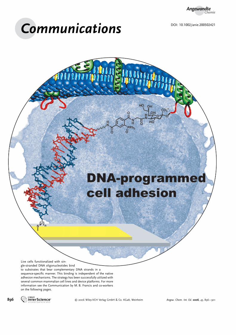
Figure 1. Covalent attachment of ssDNA to cell surfaces. a) Azides can bedelivered to cell-surface glycans thorough metabolism of Ac4ManNAz toSiaNAz. b) The Staudinger ligation of a functionalized phosphine and an azideresults in the formation of an amide bond. c) Phosphine-bearing ssDNA strandsare synthesized from amino ssDNA and phosphine–PFPs. Ac4ManNAz=acetyl-ated-N-a-azidoacetylmannosamine, SiaNAz=N-a-azidoacetyl sialic acid,PFP=pentafluorophenyl.
[*] Prof. M. B. FrancisDepartment of ChemistryUniversity of California, BerkeleyandMaterials Sciences DivisionLawrence Berkeley National LaboratoryBerkeley, CA 94720 (USA)Fax: (+1)510-643-3079E-mail: [email protected]
R. A. ChandraDepartment of ChemistryUniversity of California, BerkeleyBerkeley, CA 94720 (USA)
E. S. DouglasUCB/UCSF Joint Graduate Group in BioengineeringUniversity of California, BerkeleyBerkeley, CA 94720 (USA)
Prof. R. A. MathiesDepartment of Chemistry andUCB/UCSF Joint Graduate Group in BioengineeringUniversity of California, BerkeleyandPhysical Biosciences DivisionLawrence Berkeley National LaboratoryBerkeley, CA 94720 (USA)
Prof. C. R. BertozziDepartments of Chemistry & Molecular and Cell BiologyandHoward Hughes Medical InstituteUniversity of California, BerkeleyandMaterials Sciences DivisionLawrence Berkeley National LaboratoryBerkeley, CA 94720 (USA)
[**] The authors thank H. Lee, J. Prescher, M. Hangauer, and N. Toriellofor helpful discussions and assistance. R.A.C. and E.S.D. weresupported by National Science Foundation Predoctoral Fellowships.This work was supported by generous financial support from theNanoscale Science, Energy, and Technology (NSET) Program of theDepartment of Energy.
Supporting information for this article is available on the WWWunder http://www.angewandte.org or from the author.
AngewandteChemie
897Angew. Chem. Int. Ed. 2006, 45, 896 –901 � 2006 Wiley-VCH Verlag GmbH & Co. KGaA, Weinheim
receptors that they possess. An advantage of DNA-basedsystems is the virtually unlimited number of possible codingspecificities as was demonstrated in previous studies that havedirected synthetic lipid vesicles to supported lipid bilayerswith complementary sequences.[10] It is envisioned that, whencombined with DNA-patterning techniques developed formicroarray platforms,[11] this strategy will allow increasinglycomplex networks of living cells to be created through self-assembly.
Although there are well-established methods for thefunctionalization of devices with ssDNA,[11,12] the attachmentof ssDNA to the surface of living cells has not yet beenreported. To do this,[13] we have developed a synthetic methodbased on the introduction of specific chemical handles ontocell surfaces by using metabolic oligosaccharide engineering.Briefly, this was achieved though the incorporation of azidesinto cell surface glycoconjugates by the administration ofperacetylated N-a-azidoacetylmannosamine (Ac4ManNAz).Previous studies have shownthat this unnatural sugar ismetabolized to the correspond-ing N-a-azidoacetyl sialic acid(SiaNAz) within membrane-associated glycans (Fig-ure 1a).[14]
We have reported previ-ously the use of the Staudingerligation (Figure 1b) as a bio-orthogonal reaction for themodification of cell-surfaceazides with phosphine reagents,yielding stable amide link-ages.[15] For these studies, wehave expanded this technique toinclude oligonucleotide attach-ment.[16] We prepared modifiedssDNA with a phosphine groupthrough the reaction of a 5’-amine-modified ssDNA with aphosphine pentafluorophenyl(PFP) ester, as shown in Fig-ure 1c. Model studies con-ducted with simple azides con-firmed that the phosphine–ssDNA conjugate undergoesthe Staudinger ligation to formthe expected amide-linkedproduct (see Supporting Infor-mation). Little or no oxidationof the phosphine–DNA conju-gates was observed during han-dling or storage.
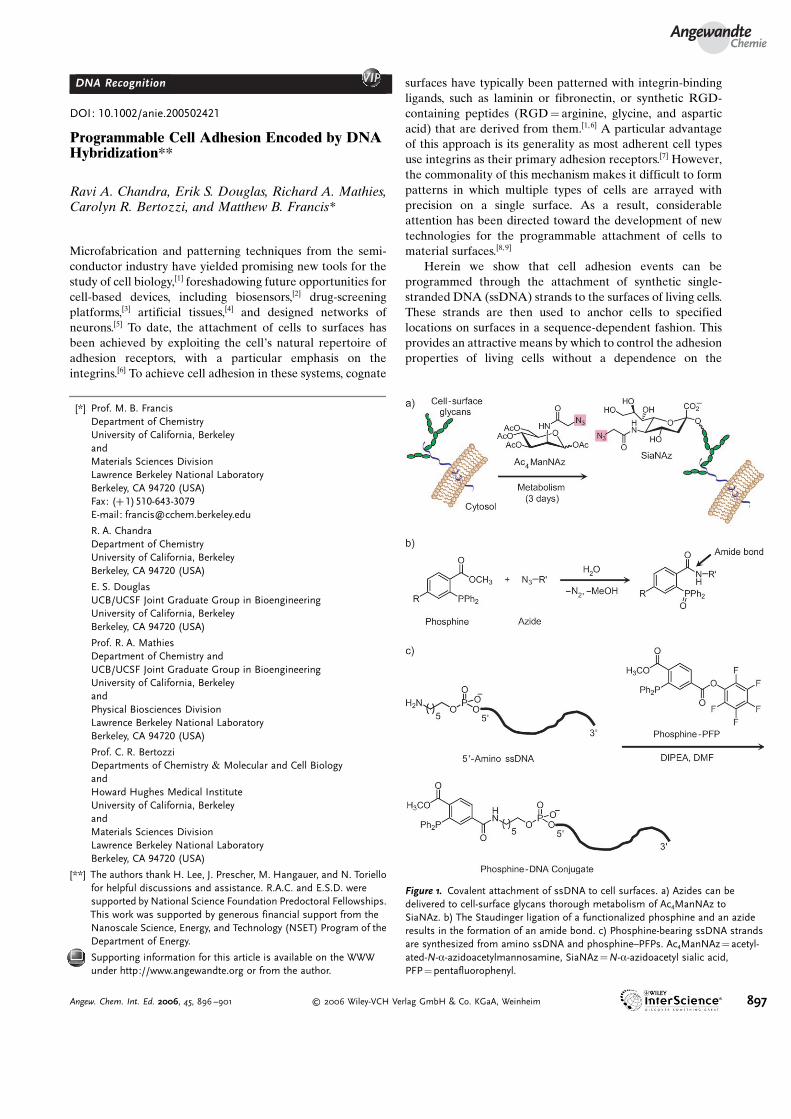
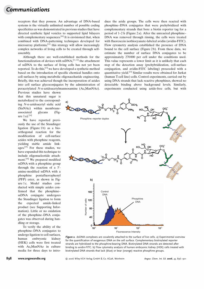
To verify the ability of thephosphine–DNA conjugates toundergo ligation to cell surfaces,human embryonic kidney(HEK) cells were first treatedwith Ac4ManNAz in culturemedia for three days to intro-
duce the azide groups. The cells were then reacted withphosphine–DNA conjugates that were prehybridized withcomplementary strands that bore a biotin reporter tag for aperiod of 1–2 h (Figure 2a). After the unreacted phosphine–DNA was removed through rinsing, the cells were treatedwith fluorescein isothiocyanate-labeled avidin (avidin-FITC).Flow cytometry analysis established the presence of DNAbound to the cell surface (Figure 2b). From these data, weestimate the number of surface DNA conjugates to beapproximately 270000 per cell under the conditions used.This value represents a lower limit as it is unlikely that eachstep of the detection assay (prehybridization, cell-surfaceconjugation, and avidin-FITC labeling) proceeded with aquantitative yield.[15] Similar results were obtained for Jurkat(human T-cell line) cells. Control experiments, carried out byusing DNA strands that lack reactive phosphines, showed nodetectable binding above background levels. Similarly,experiments conducted using azide-free cells, but with
a)
b)
100 101 102 1030
40
80
120
Fluorescence Intensity
Num
ber
of
cells
ControlDNA
PhosphineDNA
160
O
CO2
O
HO
HN
HO
O
OHHO
HN
OP
OOO
5
NH
O
PPh2
O O
Phosphine -reporter duplex
HN
OP
OOO
5
OCH3
O
PPh2
O O
CO2
O
HO
HN
HO
O
N3
OHHO
a)
b)
10 10 10 100
40
80
120
ControlDNA
PhosphineDNA
160
b)
10 10 10 100
40
80
120
ControlDNA
PhosphineDNA
160
O
CO2
O
HO
HN
HO
O
OHHO
HN
OP
OOO
5
NH
O
PPh2
O O
O
CO2
O
HO
HN
HO
O
OHHO
HN
OP
OOO
5
NH
O
PPh2
O O
HN
OP
OOO
5
OCH3
O
PPh2
O O
CO2
O
HO
HN
HO
O
N3
OHHOH
NO
P
OOO
5
OCH3
O
PPh2
O
HN
OP
OOO
5
OCH3
O
PPh2
O O
CO2
O
HO
HN
HO
O
N3
OHHO
O
CO2
O
HO
HN
HO
O
N3
OHHO
Figure 2. dsDNA complexes are covalently attached to the surface of live cells. a) Experimental overviewfor the quantification of exogenous DNA on the cell surface. Complementary biotinylated reporterstrands are hybridized to the phosphine-bearing DNA. Biotinylated DNA strands are detected afterbinding to avidin-FITC. b) Flow cytometry analysis of human embryonic kidney (HEK) cells treated withbiotinylated DNA strands that lack (blue) or bear (orange) reactive phosphine groups.
Communications
898 www.angewandte.org � 2006 Wiley-VCH Verlag GmbH & Co. KGaA, Weinheim Angew. Chem. Int. Ed. 2006, 45, 896 –901

phosphine–DNA, showed detectable but greatly reducedlabeling efficiency.
In parallel, experiments were carried out by usingphosphine–ssDNA conjugates. Although these species arelikely to react with surface azides with rates similar to thoseobserved for double stranded DNA (dsDNA), attempts toquantify these groups through subsequent hybridization tobiotinylated DNAwere unsuccessful. This is likely due to theslow hybridization kinetics associated with the low concen-trations of DNA on the plasma membrane, resulting inincomplete labeling. As noted by others,[17] the kinetics ofDNA hybridization are substantially different at materialsurfaces owing to the dramatically higher local DNA concen-tration. Thus, we chose instead to confirm the presence ofssDNA on the cell membranes through surface attachment.
As an experimental platform, microfluidic devices wereconstructed to create controlled environments for the intro-duction of cells and rinsing solutions (Figure 3a).[18] First, aseries of Au pads was patterned on glass surfaces by using
standard photolithographic techniques (see Supporting Infor-mation). After treatment of the pads with commerciallyobtained 5’-thiolated ssDNA strands, a prefabricated channellayer of (poly)-dimethylsiloxane (PDMS) was affixed on topto form enclosed chambers. The resultant channels were 6 cmlong, 600 mm wide, and 32 mm deep, which corresponds to atotal volume of 1.5 mL. An external syringe pump wasattached to control solution delivery at rates between 1–100 mLmin�1, and a mounted epifluorescence microscopewith a digital camera was used for analysis. Two complemen-tary 20-mer DNA-sequence pairs (A/A’ and B/B’) werechosen such that they possessed an identical overall basecomposition, but no appreciable affinity for each other.Strands A’ and B’ were bound to the Au surfaces within thechannels, and strands A and B were attached to the cells byusing the Staudinger ligation method described above. Thepresence and hybridization ability of the surface-immobilizedDNA strands in the devices were determined throughincubation with fluorescently labeled probe strands (Fig-
ure 3b).To test the system, we introduced azides onto the
surface of Jurkat cells, a naturally non-adherent celltype. The labeled cells were then divided into twosubsets and each of these was modified with either DNAsequence A or B. For tracking purposes, the two subsetsof cells were also labeled with either blue (7-amino-4-chloromethylcoumarin) or green (5-chloromethylfluor-escein diacetate) cytosolic dyes. Equal numbers of eachlabeled subpopulation were then mixed and introducedinto the device through the syringe pump. The cells wereincubated for 35 min under static conditions to allowhybridization, and then unbound cells were flushed fromthe system with excess phosphate-buffered saline (PBS)solution.
Representative fluorescence microscopy data forthese experiments are shown in Figure 4. Only cellsbearing ssDNA strands that were complementary to theimmobilized DNAwere observed to bind to the surface(Figure 4b), whereas otherwise identical cells bearingmismatched sequences were washed away. Quantitativebinding data obtained from three device regions areshown in Figure 4c. We found that 95% and 91% of thebound cells had recognized the appropriate complemen-tary sequence for the A’ and B’ pads, respectively.Control experiments with cells that lack surface DNAshowed no binding.
As further confirmation of the DNA dependence ofour immobilization method, DNA-coated pads wereblocked with complementary DNA sequences prior tothe introduction of labeled cells. Although many cellsbearing complementary DNA strands were exposed tothe pads, virtually no binding was observed following thestandard rinsing procedure (Figure 4d).
A particular advantage of this method is the abilityto immobilize naturally non-adherent cells as well asadherent ones. This is because metabolic engineeringcan be used to introduce azide functional groups onvirtually any cell type that displays sialic acid residues onits surface.[14] We have explored the generality of this
Figure 3. Construction of a microfluidic device for DNA display. a) ThiolatedssDNA strands are bound to patterned Au pads located in microfluidicchambers, solution delivery and washing can be controlled. Coating DNAstrands were A’: 5’-AGT GAC AGC TGG ATC GTT AC-3’ and B’: 5’-CCC TAGAGT GAG TCG TAT GA-3’. The presence of DNA was detected by usingmatched (A, red) and mismatched (B, green) probe strands. b) Fluorescenceimages of 1) Probe A incubated with A’-coated surface; 2) Probe B incubatedwith A’-coated surface; 3) Probe A incubated with B’-coated surface.
AngewandteChemie
899Angew. Chem. Int. Ed. 2006, 45, 896 –901 � 2006 Wiley-VCH Verlag GmbH & Co. KGaA, Weinheim www.angewandte.org

technique through the immobilization of several additionalcell lines, including Chinese Hamster Ovary (CHO) and HEKcells. Both CHO and HEK cells are naturally adherent celltypes and each was successfully immobilized through thisapproach. Furthermore, we have found that this attachmentmethod can be utilized on micropatterned Au pads thatpossess the same spatial dimensions as the cells (Figure 5).
The strength of DNA-mediated binding was determinedby subjecting immobilized cells to increasing lateral shearforce. PBS solution was introduced at increasing flow rates(1–50 mLmin�1 over a period of 10 min) and the bound HEKcells were observed. In almost every instance, the bound cellswere not washed away. Through the use of the Navier-Stokes
method,[19] we calculated that bound cells are stable to shearforces that exceed 31 dyncm�2, which is far greater than thatneeded for most applications.[20] Similar results were noted forJurkat and CHO cells.
For this method to be broadly useful, the longevity of cellsurface–DNA complexes must extend beyond the time scaleof the experiment.[1] Although it might be expected that therewould be a loss of surface DNA owing to cell membranerecycling or secreted nuclease activity,[21] the cells haveremained bound to the surfaces for periods exceeding 25 hdespite the fact that we have taken nomeasures to avoid thesedegradation pathways.
Finally, we evaluated the prolonged loyalty of bound cellsto the surface and their long-term viability though twoadditional studies conducted in parallel. In the first, Jurkatcells labeled with sequenceAwere immobilized as previouslydescribed, and a gentle flow of Jurkat culture medium wascontinuously applied to the device for 25 h. Cells werevisualized by using light microscopy at several time pointsto determine their positions. Virtually all of the bound cellsremained at their initial locations for the entire duration ofthe experiment. Secondly, we investigated cell viability andmembrane integrity by using redoxsensor red CC-1, afluorescent dye that is retained and activated in live cells.To more closely approximate typical culture conditions, thePDMS channel layer was removed from the device prior tointroduction of the cells. After the cells had been allowed tobind for 35 min, Jurkat culture medium was added and thedevice was incubated at 37 8C for 25 h. The cells in the devicewere then treated with redoxsensor red CC-1 for 30 min, andthe device was gently rinsed to identify the unbound cells.Analysis of both the bound and unbound cells indicated that79% of the DNA-bound cells remained viable after the 25 hperiod. This percentage was identical to that observed for theunbound Jurkat cells. We therefore believe that this methodwill be useful for applications that require extended cellimmobilization.[1] More detailed studies of the physiologicaleffects of DNA-based cell immobilization are currentlyunderway.
In summary, we have described a new approach forengineering cell-adhesion events. This DNA-based strategy issequence specific and applicable to a variety of devices and
Figure 4. Immobilization of cells is DNA sequence-specific. a) Blueand green dye-labeled Jurkat cell populations were functionalized withDNA sequence A or B, respectively. An equal number of cells werecombined and the mixture was introduced into the microfluidic device(as in Figure 3). b) Fluorescence images of: 1) the cell mixtureprewash; 2) an A’-coated pad postwash; 3) a B’-coated pad postwash.c) Summary data for the fraction of the bound cells with eithersequence A (blue), or sequence B (green) under each pad condition.d) B’-coated pads were pre-exposed to excess soluble strand B ; thesedata summarize the fraction of green (sequence B bound) cells boundbefore and after washing. Error bars represent the standard deviationof three separate areas of the device.
Figure 5. A single human embryonic kidney (HEK) cell immobilized ona 16-mm by 16-mm square Au pad. Cells bearing surface DNA strandswere bound to Au pads decorated with complementary strands. Brightfield microscopy was used to visualize the cells (magnified portion,40E). Adhesion of cells lacking DNA or bearing noncomplementaryDNA was not observed. Immobilized cells appeared morphologicallysimilar to unbound cells.
Communications
900 www.angewandte.org � 2006 Wiley-VCH Verlag GmbH & Co. KGaA, Weinheim Angew. Chem. Int. Ed. 2006, 45, 896 –901

cell types. Current work is focused on tuning the adhesiveproperties and studying the long-term behavior of DNA-immobilized cells, and ongoing efforts are capitalizing on theprogrammability of this method to form complex, multi-cellular patterns for sensing applications.
Received: July 12, 2005Revised: November 7, 2005Published online: December 21, 2005
.Keywords: cell adhesion · chemical biology · DNA recognition ·glycoproteins · Staudinger ligation
[1] a) R. Singhvi, A. Kumar, G. P. Lopez, G. N. Stephanopoulos,D. I. C. Wang, G. M. Whitesides, D. E. Ingber, Science 1994, 264,696; b) C. S. Chen, M. Mrksich, S. Huang, G. M. Whitesides,D. E. Ingber, Science 1997, 276, 1425; c) C. Roberts, C. S. Chen,M. Mrksich, V. Martichonok, D. E. Ingber, G. M. Whitesides, J.Am. Chem. Soc. 1998, 120, 6548; d) A. M. P. Turner, N. Dowell,S. W. P. Turner, L. Kam, M. Isaacson, J. N. Turner, H. G.Craighead, W. Shain, J. Biomed. Mater. Res. 2000, 51, 430;e) N. M. Toriello, E. S. Douglas, R. A. Mathies, Anal. Chem2005, 77, 6935.
[2] a) J. B. Shear, H. A. Fishman, N. L. Allbritton, D. Garigan, R. N.Zare, R. H. Scheller, Science 1995, 267, 74; b) T. H. Rider, M. S.Petrovick, F. E. Nargi, J. D. Harper, E. D. Schwoebel, R. H.Mathews, D. J. Blanchard, L. T. Bortolin, A. M. Young, J. Chen,M. A. Hollis, Science 2003, 301, 213.
[3] a) J. Ziauddin, D. M. Sabatini, Nature 2001, 411, 107; b) O. E.Beske, S. Goldbard, Drug Discovery Today 2002, 7, S131.
[4] a) A. Revzin, P. Rajagopalan, A. W. Tilles, F. Berthiaume, M. L.Yarmush, M. Toner, Langmuir 2004, 20, 2999; b) E. Cukierman,R. Pankov, D. R. Stevens, K. M. Yamada, Science 2001, 294,1708.
[5] D. Kleinfeld, K. H. Kahler, P. E. Hockberger, J. Neurosci. 1988,8, 4098.
[6] a) C. S. Chen, M. Mrksich, S. Huang, G. M. Whitesides, D. E.Ingber, Biotechnol. Prog. 1998, 14, 356; b) M. Mrksich, Curr.Opin. Chem. Biol. 2002, 6, 794.
[7] a) R. O. Hynes, Cell 1992, 69, 11; b) F. G. Giancotti, E. Ruo-slahti, Science 1999, 285, 1028.
[8] M. Kato, M. Mrksich, J. Am. Chem. Soc. 2004, 126, 6504.[9] C. C. Co, Y.-C. Wang, C.-C. Ho, J. Am. Chem. Soc. 2005, 127,
1598.[10] a) C. Yoshina-Ishii, G. P. Miller, M. L. Kraft, E. T. Kool, S. G.
Boxer, J. Am. Chem. Soc. 2005, 127, 1356; b) C. Yoshina-Ishii,S. G. Boxer, J. Am. Chem. Soc. 2003, 125, 3696.
[11] A. C. Pease, D. Solas, E. J. Sullivan, M. T. Cronin, C. P. Holmes,S. P. A. Fodor, Proc. Natl. Acad. Sci. USA 1994, 91, 5022.
[12] L. M. Demers, D. S. Ginger, S.-J. Park, Z. Li, S.-W. Chung, C. A.Mirkin, Science 2002, 296, 1836.
[13] All materials and methods may be found in the SupportingInformation.
[14] D. H. Dube, C. R. Bertozzi, Curr. Opin. Chem. Biol. 2003, 7, 616.[15] E. Saxon, C. R. Bertozzi, Science 2000, 287, 2007.[16] For a related study that utilizes oligonucleotides and the
Staudinger ligation, see: C. C.-Y. Wang, T. S. Seo, Z. Li, H.Ruparel, J. Ju, Bioconjugate Chem. 2003, 14, 697.
[17] For an example, see: M. F. Hagan, A. K. Chakraborty, J. Chem.Phys. 2004, 120, 4958.
[18] D. C. Duffy, J. C. McDonald, O. J. A. Schueller, G. M. White-sides, Anal. Chem. 1998, 70, 4974.
[19] R. B. Bird, W. E. Stewart, E. N. Lightfoot, Transport Phenom-ena, Wiley, New York, 1966, pp. 80 – 81.
[20] a) M. Takagi, K. Shiwaku, T. Inoue, T. Shirakawa, Y. Sawa, H.Matsuda, T. Yoshida, J. Artif. Organs 2003, 6, 222; b) B. T.Marshall, M. Long, J. W. Piper, T. Yago, R. P. McEver, C. Zhu,Nature 2003, 423, 190.
[21] a) D. A. Nauman, C. R. Bertozzi, Biochim. Biophys. Acta 2001,1568, 147; b) K.-C. Hsia, C.-L. Li, H. S. Yuan, Curr. Opin. Struct.Biol. 2005, 15, 126.
AngewandteChemie
901Angew. Chem. Int. Ed. 2006, 45, 896 –901 � 2006 Wiley-VCH Verlag GmbH & Co. KGaA, Weinheim www.angewandte.org