Structural Adaptation and Heterogeneity of Normal and Tumor Microvascular Networks
Upload
independentCategory
view
0download
0
Diabetologia (2005) 48: 1401–1410DOI 10.1007/s00125-005-1801-8
ARTICLE
H. Farhangkhoee . Z. A. Khan . Y. Barbin .S. Chakrabarti
Glucose-induced up-regulation of CD36 mediates oxidative stressand microvascular endothelial cell dysfunction
Received: 21 October 2004 / Accepted: 26 February 2005 / Published online: 25 May 2005# Springer-Verlag 2005
Abstract Aims/hypothesis: Hyperglycaemia-induced ox-idative stress is implicated in the pathogenesis of chronicdiabetic complications. Glucose-mediated oxidation ofLDL may result in increased oxidative stress and vascularendothelial cell dysfunction via interaction with a cellsurface scavenger receptor, CD36. In this study, we in-vestigated the role of CD36 in cultured microvascularendothelial cells (MVECs) and in the heart by using ananimal model of chronic diabetes. Methods: CulturedMVECs were subjected to varying glucose concentrationsand assayed for alteration in CD36 gene expression andprotein levels. To assess for oxidised LDL (ox-LDL) up-take, MVECs exposed to low and high glucose were treatedwith ox-LDL (80 μg/ml), a ligand for CD36. Haem ox-ygenase-1 (HO-1) and endothelin-1 (ET-1) induction, aswell as oxidative stress were determined. The role of glu-cose-induced CD36 alteration in ox-LDL uptake was alsoassayed following post-transcriptional CD36 gene silenc-ing. For in vivo studies, CD36 mRNA and oxidative DNAand protein damage were measured in heart tissues of1-month-old diabetic Sprague–Dawley rats. Results: Wefound that glucose increased CD36 mRNA and proteinlevels in MVECs. High levels of glucose also augmentedox-LDL uptake, in association with increasing HO-1 andET-1 mRNA levels. CD36 gene silencing prevented glu-cose-induced CD36 alteration, reduced ox-LDL uptake,
and prevented HO-1 and ET-1 up-regulation. Similar to invitro studies, diabetic heart tissues exhibited increasedCD36 mRNA levels and increased oxidative DNA andprotein damage. Conclusions/interpretation: Our resultsprovide evidence that up-regulation of CD36 may have arole in increasing oxidative stress in MVECs and the heartin chronic diabetes.
Keywords Cardiomyopathy . CD36 . Diabeticcomplications . Endothelin-1 . Haem oxygenase .Oxidative stress . Oxidised LDL . Scavenger receptor
Abbreviations 8-OHdG: 8-hydroxy-2′-deoxyguanosine .EC: endothelial cell . ET-1: endothelin-1 . HO-1: haemoxygenase-1 . HG: high glucose . LG: low glucose .MVEC: microvascular endothelial cell . ox-LDL: oxidisedLDL . siRNA: small interfering RNA . SR: scavengerreceptor . STZ: streptozotocin
Introduction
In spite of the improvements in therapeutic modalities, di-abetes and its complications account for significant mor-bidity and mortality [1, 2]. Long-standing diabetes leads tostructural and functional alterations in both the micro- andmacrovasculature [3, 4]. Diabetic cardiomyopathy has beenrecognised as a degenerative microvascular complicationof chronic diabetes [5, 6]. Understanding the pathogenesisof diabetic cardiomyopathy is still obscure; however, oxi-dative stress has been suggested to be involved in thedevelopment and progression of diabetes-induced cardio-myopathy [7].
Hyperglycaemia caused by chronic diabetes is an im-portant mediator in augmenting reactive oxygen species[8]. Oxidative stress has been shown to occur in diabeticpatients before the presentation of overt chronic complica-tions, including cardiomyopathy [7]. The generation ofoxidative stress in diabetes could be due to increased gen-eration of free radicals and nitric oxide [9, 10]. Free radi-cals are produced in all organisms as a result of increased
H. Farhangkhoee . Z. A. Khan . Y. Barbin . S. ChakrabartiDepartment of Pathology, University of Western Ontario,London, ON, N6A 5C1, Canada
Z. A. KhanDepartment of Surgery, Children’s Hospital Boston andHarvard Medical School,Boston, MA, USA
S. Chakrabarti (*)Department of Microbiology & Immunology,University of Western Ontario,London, ON, Canadae-mail: [email protected].: +1-519-6858500Fax: +1-519-6613370
oxidative stress or altered redox balance. Unpaired elec-trons from these free radicals can modify and thus alter thefunction of proteins, DNA, fatty acids and lipids [11]. TheLDLs are very sensitive to these free radical-mediatedperoxidation reactions, yielding oxidised LDL (ox-LDL)molecules [12].
Modified proteins such as ox-LDLs and AGE productsincluding glycated LDLs accumulate over time in diabeticpatients [13–15]. Alterations of these proteins may inter-fere with their recognition by native receptors such as LDLreceptor. Nonetheless, these modified proteins can interactwith greater affinity with a group of multifunctional trans-membrane proteins named scavenger receptors (SRs) [16,17]. Recent studies have demonstrated an important role ofSRs in a number of human diseases [18]. A prominentmember of the SR family is CD36 [19]. CD36 was firstidentified as a surface glycoprotein on platelets [19, 20].Since then, it has been shown to be expressed on macro-phages, adipocytes and other cell types including micro-vascular endothelial cells (MVECs) [21]. High-affinitybinding of ox-LDLs and AGE-modified proteins to CD36suggests that alterations of CD36 could potentially beinvolved in diabetic complications, including cardiomyop-athy [22, 23].
In the present study, we used cultured MVECs, the pri-mary target of sustained hyperglycaemia, to elucidate whetherup-regulation of CD36 underlies increased ox-LDL uptakeand subsequent endothelial dysfunction. Furthermore, weinvestigated whether diabetes leads to an alteration of CD36in the heart and whether such an alteration is associatedwith diabetes-induced oxidative stress.
Materials and methods
In vitro studies Human MVECs (Clonetics, Walkersville,MD, USA) were cultured with endothelial growth medium(Clonetics) as previously described [24]. To determine theoptimal high glucose (HG) concentration, sub-confluentcells were incubated with 10, 15, 25 or 35 mmol/l D-glu-cose. Following a 24-h treatment period, total RNA wasextracted and subjected to CD36mRNA expression by real-time RT-PCR. Subsequent experiments were conductedwith treatment of cells exposed to low glucose (LG) orempirically determined HG levels. These cells were sub-jected to CD36 mRNA and protein expression by real-timeRT-PCR and western blotting, respectively.
In order to determine whether alteration of CD36 coin-cides with functional significance, cells exposed to LG andHG were treated with ox-LDL. Ox-LDL was prepared asdescribed before [25]. Briefly, human LDL (Sigma-AldrichCanada, Oakville, ON, Canada) was resuspended in PBSand incubated in the presence of 5 μmol/l CuSO4 in orderto oxidise the LDL molecules. Following an incubationperiod of 3 h, the ox-LDL samples were extensively di-alysed overnight with repeated buffer changes to removeCuSO4. Uptake of ox-LDL was assessed by immunocyto-chemistry following exposure of endothelial cells (ECs) to80 μg/ml ox-LDL. This concentration of ox-LDL has been
well established in EC culture studies [26]. Cells exposedto ox-LDL were also subjected to mRNA analyses for theoxidative stress marker, haem oxygenase-1 (HO-1) [27, 28]and vasoactive peptide, endothelin-1 (ET-1). ET alterationhas been well established in chronic diabetic complica-tions and vascular endothelial dysfunction [29].
CD36 gene silencing To establish the role of CD36 inmediating ox-LDL uptake, MVECs were transfected withsmall interfering RNA (siRNA) targeted to CD36 mRNA(Ambion, Austin, TX, USA). Cultured MVECs were trans-fected with 100 nmol/l CD36 siRNA using siPORT Lipidtransfection media (Ambion). Following a 24-h incubationperiod, cells were cultured for another 24 h in either 5 or25 mmol/l glucose, as well as treated with the CD36ligand, ox-LDL. All siRNA experiments included trans-fection of ECs with siRNAs which have no sequence ho-mology (negative control transfection) with human genome(Ambion). To measure CD36 siRNA transfection efficien-cy, CD36 mRNA levels were assayed for using real-timeRT-PCR. Ox-LDL uptake was determined by immunohis-tochemical analysis using ox-LDL antibody (1:500).
In vivo studies Male Sprague–Dawley rats (Charles RiverCanada, St Constant, QC, Canada) weighing approximate-ly 250 g were made diabetic by a single i.v. injection ofstreptozotocin (STZ; 65 mg/kg; Sigma-Aldrich) [24]. Age-and sex-matched controls were given an equal volume ofcitrate buffer. Diabetes was confirmed by measuring bloodglucose levels (Surestep; Lifescan, Burnaby, BC, Canada)48 h after the injection of STZ. Animals were also moni-tored for glucosuria and ketonuria (Uriscan Gluketo; YeongDong Co., Seoul, Korea) and given small daily doses (0.1–3.0 U) of ultralente insulin (Novo Nordisk, Princeton, NJ,USA) to prevent ketoacidosis [24]. Following 1 month oftreatment, rats were killed and heart tissues were removed.Prior to killing, blood was collected from the rats to assayfor HbA1c levels (Glycotest; Pierce, Rockford, IL, USA).Heart tissues were sectioned and either snap-frozen inliquid nitrogen for gene expression analyses or embeddedin paraffin for immunohistochemical analysis. The Uni-versity of Western Ontario Council on Animal Care Com-mittee formally approved all experimental protocols.
Real-time RT-PCRTotal RNA from cultured ECs and hearttissues were isolated as previously described [24]. Briefly,RNA was extracted using TRIzol (Invitrogen, Burlington,ON, Canada) with isopropanol precipitation. Following ex-traction, RNA samples were subjected to DNAse treatmentto degrade any contaminating DNA in the samples. Puritywas assessed by measuring OD 260:280 nm.
cDNAwas synthesised using 3 μg total RNAwith oligo-(dT) primers and Superscript-II MMLV-reverse transcrip-tase (Invitrogen). Real-time RT-PCR was performed in theLightCycler (Roche Diagnostics Canada, Laval, QC, Can-ada) as previously described [24]. The reaction mixture(20 μl total volume) consisted of 10 μl SYBR Green TaqReadyMix (Sigma-Aldrich), 1.6 μl 25 mmol/l MgCl2, 1 μlof each forward and reverse 10 μmol/l primers, 4.4 μl
1402
H2O, and 2 μl cDNA template. The primer sequences andPCR temperature profiles for β-actin (human/rat) and HO-1 (rat) were assayed as previously described [24, 30, 31].The primer sequences for CD36 are 5′TAATGGCACAGATGCAGCCT3′ and 5′ACAGCATAGATGGACCTGCAA3′ (human) and 5′GAGAACTGTTATGGGGCTAT3′and 5′TTCAACTGGAGAGGCAAAGG3′ (rat). The PCR
temperature profiles for CD36 are similar to the β-actinPCR profiles [30]. The PCR reaction mixture for ET-1consisted of 2.5 μl 10×PCR Buffer (Invitrogen), 1.25 μl 5mmol/l dNTP, 1.2 μl 50 mmol/l MgCl2, 1 μl primers, 9.8 μlH2O, 2 μl cDNA, and 0.75 μl 15 mmol/l Taqman probePrimer sequences and PCR temperature profiles for ET-1were assayed as previously described [24]. The data were
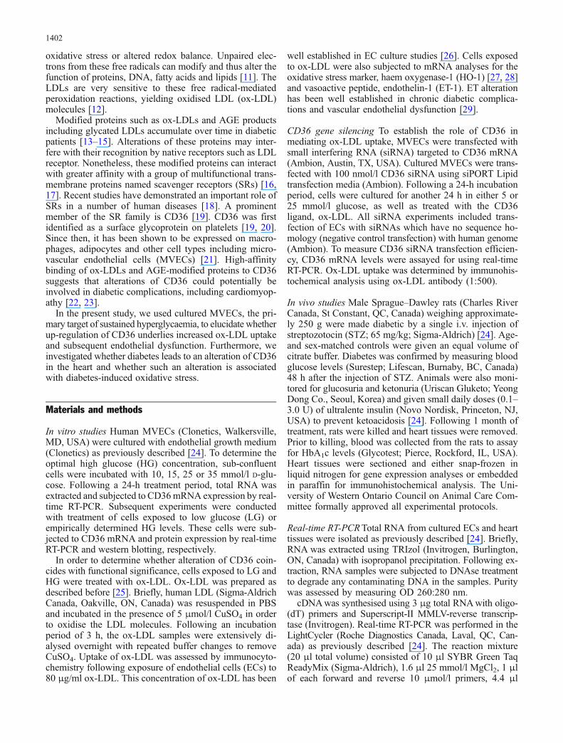
Fig. 1 Real-time RT-PCR am-plification of human CD36showing a PCR amplificationcurves b melting curve analysis(MCA) of post-PCR products,and c mRNA expression fol-lowing 24-h incubation withvarying glucose concentrations.Each trace represents a humanMVEC cDNA sample; all PCRreactions were coupled to MCAto determine specificity of am-plification. CD36 is expressedas ratio of target to β-actin(relative to 10 mmol/l glucose);n=4/treatment; *p<0.05 com-pared with 10 mmol/l glucose
1403
normalised using the housekeeping gene, β-actin, to ac-count for differences in reverse transcription efficienciesand in the amount of template in the reaction mixtures.
Western blotting Total proteins from ECs were isolated andquantified according to well-established methodologies[24]. Briefly, cells were homogenised in complete lysisbuffer (NaCl 0.877 g, deoxycholate 1 g, 1 mmol/l Tris–HCl [pH 7.5] 5 ml, Triton X-100 1 ml, and 10% sodiumdodecyl sulphate 1 ml; volume adjusted to 100 ml usingddH2O) and protease inhibitor. Total proteins were quan-tified by a BCA protein assay kit (Pierce). Western blottingwas performed by polyclonal anti-human CD36 antibody(1:1000; Cayman Chemicals, Ann Arbor, MI, USA) fol-lowed by secondary antibody conjugated with horseradishperoxide. An ECL-Plus Western Blotting Detection kit(Amersham Pharmacia Biotechology, Piscataway, NJ, USA)was used for detection.
Immunochemical analyses Paraffin-embedded heart tissueswere stained for 8-hydroxy-2′-deoxyguanosine (8-OHdG)and nitrotyrosine as described previously [30]. Briefly, 5μm sections were transferred to positively charged slides.Monoclonal anti-mouse 8-OHdG (1:150; Chemicon Lab.,Temecula, CA, USA ) and monoclonal anti-mouse nitro-tyrosine (1:75; Cayman Chemicals) were used for staining.Secondary antibodies conjugated with horseradish perox-idase (Bio-Rad Laboratories, Hercules, CA, USA) wereused to produce signals from the chemiluminescent sub-strate, diaminobenzidine (Amersham Pharmacia Biotech-nology). Negative controls included incubation with PBSwithout primary antibody. Specificity of the antibodies wasconfirmed by blocking tissue sections with 10% horseserum. The experiments were performed in triplicate andslides were read by two investigators unaware of the par-ticular treatment. 8-OHdG immunoreactivity was expressedas the number of positive cardiomyocytes in ten random
fields containing approximately 100 cells. Nitrotyrosinewas evaluated by relative cytoplasmic staining intensity.The data are expressed as percentage of total cell.
For immunocytochemistry, cultured vascular ECs werebriefly trypsinised and seeded in 12-well plates containingcoverslips. Cells were cultured in growth media for 24 h toallow attachment. Following cell attachment, all treatmentswere carried out in serum-free media as described above.Anhydrous ethanol was used to fix the cells. Ox-LDL(1: 500; Biodesign International, Saco, ME, USA) and8-OHdG (1:400; Chemicon) immunochemical analyseswere carried out essentially the same as described for hearttissues.
Statistical analysis The data are expressed as means±SEMand were analysed by ANOVA followed by Student’s t-test.Differences were considered statistically significant at val-ues of p<0.05.
Results
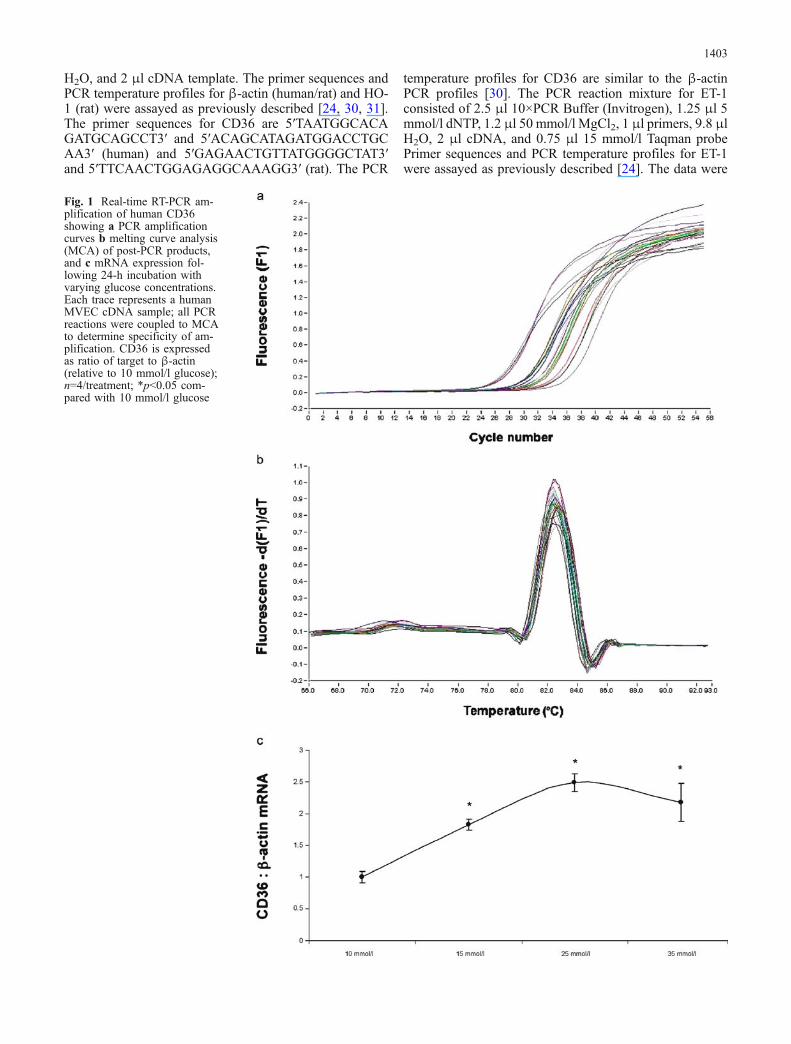
Glucose induces up-regulation of CD36 in MVECs Weused vascular ECs to elucidate whether glucose-inducedalteration of CD36 may mediate increased oxidative stress.Cultured MVECs were exposed to varying glucose con-centrations and assayed for CD36 mRNA alteration. Ourdata show up-regulation of CD36 mRNA by glucose in adose-dependent manner (Fig. 1). The greatest change intranscript levels was observed with 25 mmol/l glucose;thus, subsequent experiments were carried out by treatingcells with either 5 or 25 mmol/l glucose. In order to de-termine whether mRNA levels coincide with protein ex-pression, we used the western blotting technique. Here weshow that, in parallel to mRNA, CD36 protein expressionis augmented by exposure of ECs to 25 mmol/l glucose(Fig. 2a, b; p<0.05).
CD36ββ-actin
LG HG
a
b
*
0
0.2
0.4
0.6
0.8
1
1.2
1.4
1.6
1.8
LG HG
CD
36 :
β-a
ctin
(arb
itra
ry u
nit
s)
Fig. 2 Glucose-induced up-regulation of CD36 protein ex-pression in MVECs showinga representative immunoblot ofCD36 and β-actin, and b semi-quantitative analysis of CD36protein expression (relative toβ-actin). *p<0.05 HG comparedwith LG; n=4/treatment
1404
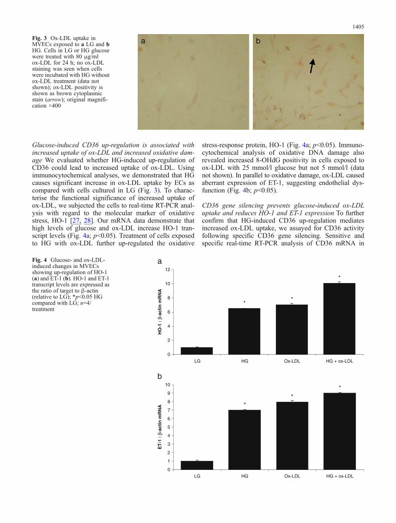
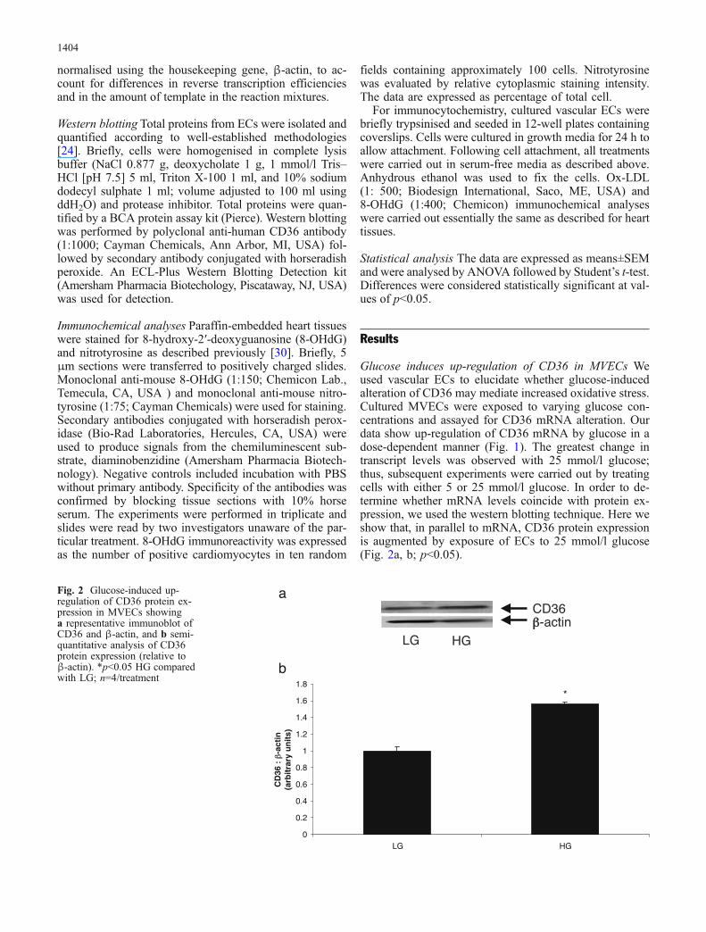
Glucose-induced CD36 up-regulation is associated withincreased uptake of ox-LDL and increased oxidative dam-age We evaluated whether HG-induced up-regulation ofCD36 could lead to increased uptake of ox-LDL. Usingimmunocytochemical analyses, we demonstrated that HGcauses significant increase in ox-LDL uptake by ECs ascompared with cells cultured in LG (Fig. 3). To charac-terise the functional significance of increased uptake ofox-LDL, we subjected the cells to real-time RT-PCR anal-ysis with regard to the molecular marker of oxidativestress, HO-1 [27, 28]. Our mRNA data demonstrate thathigh levels of glucose and ox-LDL increase HO-1 tran-script levels (Fig. 4a; p<0.05). Treatment of cells exposedto HG with ox-LDL further up-regulated the oxidative
stress-response protein, HO-1 (Fig. 4a; p<0.05). Immuno-cytochemical analysis of oxidative DNA damage alsorevealed increased 8-OHdG positivity in cells exposed toox-LDL with 25 mmol/l glucose but not 5 mmol/l (datanot shown). In parallel to oxidative damage, ox-LDL causedaberrant expression of ET-1, suggesting endothelial dys-function (Fig. 4b; p<0.05).
CD36 gene silencing prevents glucose-induced ox-LDLuptake and reduces HO-1 and ET-1 expression To furtherconfirm that HG-induced CD36 up-regulation mediatesincreased ox-LDL uptake, we assayed for CD36 activityfollowing specific CD36 gene silencing. Sensitive andspecific real-time RT-PCR analysis of CD36 mRNA in
Fig. 3 Ox-LDL uptake inMVECs exposed to a LG and bHG. Cells in LG or HG glucosewere treated with 80 μg/mlox-LDL for 24 h; no ox-LDLstaining was seen when cellswere incubated with HG withoutox-LDL treatment (data notshown); ox-LDL positivity isshown as brown cytoplasmicstain (arrow); original magnifi-cation ×400
*
**
0
2
4
6
8
10
12
LG HG Ox-LDL HG + ox-LDL
HO
-1 :
β-a
ctin
mR
NA
ET
-1 :
β-a
ctin
mR
NA
a
b*
**
0
1
2
3
4
5
6
7
8
9
10
LG HG Ox-LDL HG + ox-LDL
Fig. 4 Glucose- and ox-LDL-induced changes in MVECsshowing up-regulation of HO-1(a) and ET-1 (b). HO-1 and ET-1transcript levels are expressed asthe ratio of target to β-actin(relative to LG); *p<0.05 HGcompared with LG; n=4/treatment
1405

ECs transfected with CD36 siRNA showed greater than80% reduction in CD36 transcript levels (Fig. 5a; p<0.05).CD36 siRNA also prevented HG-induced CD36 up-regulation. Following successful transfections, cells weretreated with ox-LDL and assayed for uptake by immuno-histochemistry and molecular markers of oxidative stressby RT-PCR. Our results show complete normalisation ofglucose-induced ox-LDL uptake in CD36 siRNA-trans-fected cells as compared with negative control transfectedcells (Fig. 5b, c). These results indicate that glucose-in-duced CD36 alteration arbitrates increased ox-LDL uptakein ECs.
We next determined whether CD36 inhibition by siRNAprevents glucose/ox-LDL-induced up-regulation of HO-1and ET-1. Our data indicate that CD36 siRNA transfec-tion, which reduced CD36 expression by 80%, reduced
glucose-induced HO-1 and ET-1 up-regulation (Fig. 5d;p<0.05). CD36 siRNA transfection also reduced HO-1 andET-1 mRNA levels in cells exposed to LG (data notshown).
b
a
d
c
* *
0
20
40
60
80
100
120
140
LG + negative control transfection
LG + CD36 siRNA transfection
HG + negative control transfection
HG + CD36 siRNA transfection
% C
D36
mR
NA
exp
ress
ion
**
0
20
40
60
80
100
120
HG + negative control
transfection
HG + CD36 siRNA
transfection
HG + negative control
transfection
HG + CD36 siRNA
transfection
% H
O-1
an
d E
T-1
mR
NA
exp
ress
ion
Fig. 5 Effect of CD36 genesilencing on ox-LDL uptake andoxidative stress in MVECsshowing a real-time RT-PCRanalysis of CD36 mRNA fol-lowing siRNA transfections,b ox-LDL immunoreactivity innegative control transfectedcells, c ox-LDL immunoreac-tivity in CD36 siRNA-transfected cells and d HO-1(solid bars) and ET-1 (shadedbars) gene expression changesfollowing CD36 siRNA trans-fections. CD36 mRNA levelsare shown as % total; ox-LDLimmunoreactivity was assayedin cells exposed to HG and 80μg/ml; ox-LDL positivity isshown as brown cytoplasmicstain (arrow); original magnifi-cation ×400; HO-1 and ET-1mRNA levels are relative to HG+negative control transfections;*p<0.05 compared with negativetransfections; n=4/treatment
Table 1 Clinical monitoring of animals
Control Diabetic
Blood glucose (mg/dl) 71.8±3.6 473.2±5.9*HbA1c (%) 4.9±0.8 17.8±0.7*Body weight (g) 528.5±31.4 439.3±19.8*Systolic BP (mmHg) 107±6 112±7
*p<0.05 compared with control
1406
Diabetes induces up-regulation of CD36 and oxidativestress in the heart In order to determine whether diabetesleads to increased oxidative stress via alteration of CD36receptors, we used a well-established model of chronicdiabetes. This STZ-induced diabetic model shows develop-ment of myocardial abnormalities similar to humans withdiabetes [32]. STZ-induced diabetic animals exhibitedhyperglycaemia as compared with non-diabetic controls
(Table 1). Diabetic animals also demonstrated reducedbody weight gain and elevated HbA1c levels. Systolic BP,recorded by tail plethysmography, did not differ among theanimal groups. These parameters are indicative of diabeticdysmetabolism.
Using the real-time RT-PCR assay (Fig. 6a, b), wemeasured CD36 transcript levels in heart tissues of thesediabetic and non-diabetic control animals. Our results in-
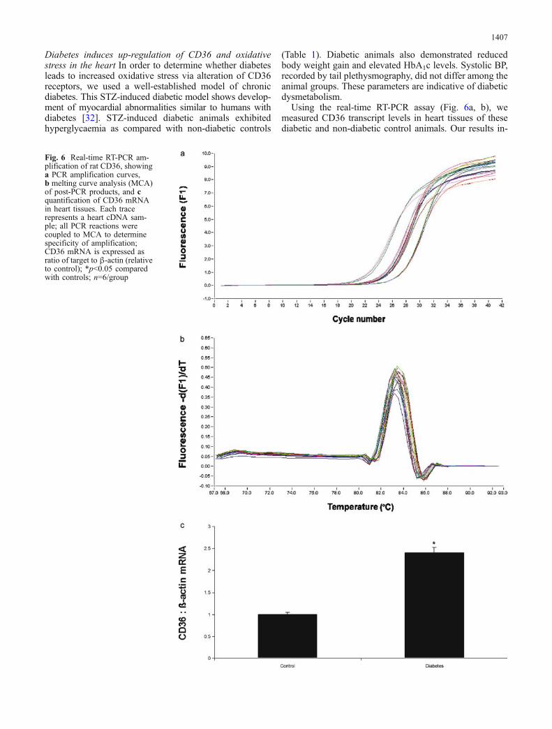
Fig. 6 Real-time RT-PCR am-plification of rat CD36, showinga PCR amplification curves,b melting curve analysis (MCA)of post-PCR products, and cquantification of CD36 mRNAin heart tissues. Each tracerepresents a heart cDNA sam-ple; all PCR reactions werecoupled to MCA to determinespecificity of amplification;CD36 mRNA is expressed asratio of target to β-actin (relativeto control); *p<0.05 comparedwith controls; n=6/group
1407
dicate that diabetes leads to up-regulation of CD36 mRNAin the heart (Fig. 6c; p<0.05). These results provide evi-dence of transcriptional regulation of CD36 in diabetes.
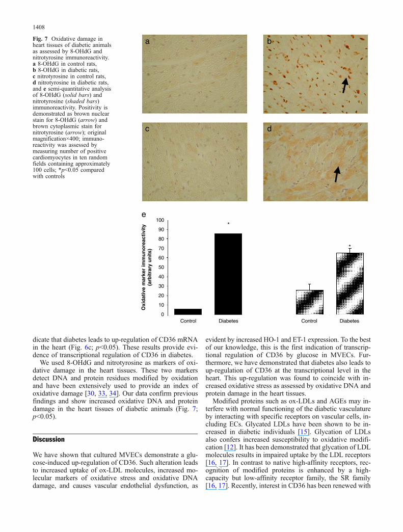
We used 8-OHdG and nitrotyrosine as markers of oxi-dative damage in the heart tissues. These two markersdetect DNA and protein residues modified by oxidationand have been extensively used to provide an index ofoxidative damage [30, 33, 34]. Our data confirm previousfindings and show increased oxidative DNA and proteindamage in the heart tissues of diabetic animals (Fig. 7;p<0.05).
Discussion
We have shown that cultured MVECs demonstrate a glu-cose-induced up-regulation of CD36. Such alteration leadsto increased uptake of ox-LDL molecules, increased mo-lecular markers of oxidative stress and oxidative DNAdamage, and causes vascular endothelial dysfunction, as
evident by increased HO-1 and ET-1 expression. To the bestof our knowledge, this is the first indication of transcrip-tional regulation of CD36 by glucose in MVECs. Fur-thermore, we have demonstrated that diabetes also leads toup-regulation of CD36 at the transcriptional level in theheart. This up-regulation was found to coincide with in-creased oxidative stress as assessed by oxidative DNA andprotein damage in the heart tissues.
Modified proteins such as ox-LDLs and AGEs may in-terfere with normal functioning of the diabetic vasculatureby interacting with specific receptors on vascular cells, in-cluding ECs. Glycated LDLs have been shown to be in-creased in diabetic individuals [15]. Glycation of LDLsalso confers increased susceptibility to oxidative modifi-cation [12]. It has been demonstrated that glycation of LDLmolecules results in impaired uptake by the LDL receptors[16, 17]. In contrast to native high-affinity receptors, rec-ognition of modified proteins is enhanced by a high-capacity but low-affinity receptor family, the SR family[16, 17]. Recently, interest in CD36 has been renewed with
a
c
b
d
e*
*
0
10
20
30
40
50
60
70
80
90
100
Control Diabetes Control Diabetes
Oxi
dat
ive
mar
ker
imm
un
ore
acti
vity
(a
rbitr
ary
un
its)
Fig. 7 Oxidative damage inheart tissues of diabetic animalsas assessed by 8-OHdG andnitrotyrosine immunoreactivity.a 8-OHdG in control rats,b 8-OHdG in diabetic rats,c nitrotyrosine in control rats,d nitrotyrosine in diabetic rats,and e semi-quantitative analysisof 8-OHdG (solid bars) andnitrotyrosine (shaded bars)immunoreactivity. Positivity isdemonstrated as brown nuclearstain for 8-OHdG (arrow) andbrown cytoplasmic stain fornitrotyrosine (arrow); originalmagnification×400; immuno-reactivity was assessed bymeasuring number of positivecardiomyocytes in ten randomfields containing approximately100 cells; *p<0.05 comparedwith controls
1408

the demonstration that this surface protein is involved inthe recognition and internalisation of oxidatively modifiedproteins [18, 19]. Our studies further provide evidence thatCD36 up-regulation may be involved in increased oxida-tive stress in the heart and MVECs. The direct role of CD36in such alteration was characterised following specificCD36 gene silencing. Our data show that inhibition of CD36production prevents ox-LDL uptake; however, whethersimilar mechanisms may mediate increased ox-LDL uptakein vivo remain to be determined. It should be noted thatCD36-mediated alteration of fatty acid uptake and metab-olism may also contribute to the pathogenesis of diabeticcardiomyopathy [35]. Whether such mechanisms also con-tribute to oxidative stress remains to be determined.
CD36 alteration has previously been shown in vascularpreparations and monocytes isolated from diabetic patients[13]. It should be noted that CD36 alteration in monocytesisolated from diabetic patients was only shown to be at thetranslational level [13]. It has also been shown that mono-cytes from diabetic patients show a significantly higherlevel of CD36 expression, which was not changed duringfurther exposure to hyperglycaemic conditions [36]. In thisstudy, we provide the first indication that CD36 is up-regulated at the transcriptional level in MVECs exposed tohigh levels of glucose. Taken together, these findings sug-gest that in addition to increased uptake of modified proteinby monocytes, vascular ECs may suffer oxidative DNAdamage via glucose-induced up-regulation of CD36 [37].We have demonstrated that glucose increases the uptakeof ox-LDL in ECs leading to increased oxidative stressand elaboration of vasoactive peptide, ET-1. In parallel toour studies, ox-LDL has been shown to cause oxidativestress in human fibroblasts [37]. In addition, murine mod-els of type 2 diabetes and mice fed a high-fat diet showup-regulation of CD36 expression in cardiac capillaryECs. It should be noted, however, that CD36 expressionin these models of type 2 diabetes could be attributed tohyperglycaemia and/or dyslipidaemia. Our studies pro-vide evidence that hyperglycaemia may be the mecha-nism underlying CD36 alteration in diabetes. In supportof such a notion are findings of a recent study whichdemonstrates increased CD36 mRNA expression in hearttissues of STZ-diabetic animals [35].
In conclusion, CD36 up-regulation may alter propertiesof vascular ECs. Diabetes-induced up-regulation of CD36may be involved in increased oxidative stress and diabeticcardiomyopathy. These studies provide insight into thepathogenesis of chronic diabetic complications. Further-more, these results may provide avenues which could beexplored for the development of therapeutic modalities.
Acknowledgements The authors acknowledge grant support fromthe Canadian Diabetes Association in honour of the late Glenn W.Liebrock, the Canadian Institutes of Health Research, and theLawson Health Research Institute.
References
1. Harris MI (1998) Diabetes in America: epidemiology and scopeof the problem. Diabetes Care 21:C11–C14
2. International Diabetes Federation (2003) Diabetes atlas, 2ndedn. International Diabetes Federation, Brussels, Belgium
3. Aleksandrovski YA (1998) Molecular mechanisms of diabeticcomplications. Biochemistry (Mosc) 63:1249–1257
4. Sheetz MJ, King GL (2002) Molecular understanding of hyper-glycemia’s adverse effects for diabetic complications. JAMA288:2579–2588
5. Hamby RI, Zoneraich S, Sherman L (1974) Diabetic cardio-myopathy. JAMA 229:1749–1754
6. Rodrigues B, McNeill JH (1992) The diabetic heart: metaboliccauses for the development of a cardiomyopathy. CardiovascRes 26:913–922
7. Giugliano D, Ceriello A, Paolisso G (1995) Diabetes mellitus,hypertension, and cardiovascular disease: which role for oxi-dative stress? Metabolism 44:363–368
8. Nishikawa T, Edelstein D, Du XL et al (2000) Normalizingmitochondrial superoxide production blocks three pathways ofhyperglycaemic damage. Nature 404:787–790
9. Baynes JW, Thorpe SR (1999) Role of oxidative stress indiabetic complications: a new perspective on an old paradigm.Diabetes 48:1–9
10. Nadler J, Winer L (1996) Free radicals, nitric oxide and diabeticcomplications. In: LeRoith D, Taylor SI, Olefsky M (eds) Dia-betes mellitus. Lippincott-Raven, Philadelphia, PA, pp 840–848
11. Cadenas E (1989) Biochemistry of oxygen toxicity. Annu RevBiochem 58:79–110
12. Bucala R, Makita Z, Koschinsky T, Cerami A, Vlassara H(1993) Lipid advanced glycosylation: pathway for lipid oxida-tion in vivo. Proc Natl Acad Sci U S A 90:6434–6438
13. Griffin E, Re A, Hamel N et al (2001) A link between diabetesand atherosclerosis: glucose regulates expression of CD36 atthe level of translation. Nat Med 7:840–846
14. Tsuzura S, Ikeda Y, Suehiro T et al (2004) Correlation of plas-ma oxidized low-density lipoprotein levels to vascular compli-cations and human serum paraoxonase in patients with type 2diabetes. Metabolism 53:297–302
15. Cohen MP, Lautenslager G, Shea E (1993) Glycated LDLconcentrations in non-diabetic and diabetic subjects measuredwith monoclonal antibodies reactive with glycated apolipopro-tein B epitopes. Eur J Clin Chem Clin Biochem 31:707–713
16. Steinbrecher UP, Witztum JL (1984) Glucosylation of low-density lipoproteins to an extent comparable to that seen indiabetes slows their catabolism. Diabetes 33:130–134
17. Klein RL, Laimins M, Lopes-Virella MF (1995) Isolation,characterization, and metabolism of the glycated and non-glycated subfractions of low-density lipoproteins isolated fromtype I diabetic patients and nondiabetic subjects. Diabetes44:1093–1098
18. Boullier A, Bird DA, Chang MK et al (2001) Scavenger re-ceptors, oxidized LDL, and atherosclerosis. Ann N YAcad Sci947:214–222
19. Endemann G, Stanton LW, Madden KS, Bryant CM, White RT,Protter AA (1993) CD36 is a receptor for oxidized low densitylipoprotein. J Biol Chem 268:221–236
20. Tandon NN, Lipsky RH, Burgess WH, Jamieson GA (1989)Isolation and characterization of platelet glycoprotein IV(CD36). J Biol Chem 264:7570–7575
21. Abumrad NA, el Maghrabi MR, Amri EZ, Lopez E, GrimaldiPA (1993) Cloning of a rat adipocyte membrane protein im-plicated in binding or transport of long-chain fatty acids that isinduced during preadipocyte differentiation. Homology withhuman CD36. J Biol Chem 268:17665–17668
1409
22. Greenwalt DE, Scheck SH, Rhinehart-Jones T (1995) HeartCD36 expression is increased in murine models of diabetes andin mice fed a high fat diet. J Clin Invest 96:1382–1388
23. Miyazaki A, Nakayama H, Horiuchi S (2002) Scavenger re-ceptors that recognize advanced glycation end products. TrendsCardiovasc Med 12:258–262
24. Khan ZA, Cukiernik M, Gonder JR, Chakrabarti S (2004)Oncofetal fibronectin in diabetic retinopathy. Invest Ophthal-mol Vis Sci 45:287–295
25. Liu WL, Guo X, Guo ZG (1998) Oxidized low-density lipo-proteins induce apoptosis in vascular smooth muscle cells.Zhongguo Yao Li Xue Bao 19:245–247
26. Chow SE, Chu WK, Shih SH, Chen JK (2002) Exposure tooxidized low-density lipoprotein reduces activable Ras proteinin vascular endothelial cells. In Vitro Cell Dev Biol Anim38:320–325
27. Kamalvand G, Pinard G, Ali-Khan Z (2003) Heme-oxygenase-1 response, a marker of oxidative stress, in a mouse model ofAA amyloidosis. Amyloid 10:151–159
28. Tyrrell RM, Basu-Modak S (1994) Transient enhancement ofheme oxygenase 1 mRNA accumulation: a marker of oxidativestress to eukaryotic cells. Methods Enzymol 234:224–235
29. Khan ZA, Chakrabarti S (2003) Endothelins in chronic diabeticcomplications. Can J Physiol Pharm 81:622–634
30. Farhangkhoee H, Khan ZA, Mukherjee S et al (2003) Hemeoxygenase in diabetes-induced oxidative stress in the heart. JMol Cell Cardiol 35:1439–1448
31. Chen S, Khan ZA, Barbin Y, Chakrabarti S (2004) Pro-oxidantrole of heme oxygenase in mediating glucose-induced endo-thelial cell damage. Free Radic Res 38:1301–1310
32. Rodrigues B, McNeill JH (1999) Physiological and patholog-ical consequences of streptozotocin diabetes on the heart. In:McNeill JH (ed) Experimental model of diabetes. CRC PressLLC, Boca Raton, FL, pp 63–80
33. Piconi L, Quagliaro L, Ceriello A (2003) Oxidative stress indiabetes. Clin Chem Lab Med 41:1144–1149
34. Abu-Qare AW, Abou-Donia MB (2001) Combined exposure tosarin and pyridostigmine bromide increased levels of rat urinary3-nitrotyrosine and 8-hydroxy-2′-deoxyguanosine, biomarkersof oxidative stress. Toxicol Lett 123:51–58
35. Luiken JJ, Arumugam Y, Bell RC et al. (2002) Changes in fattyacid transport and transporters are related to the severity ofinsulin deficiency. Am J Physiol Endocrinol Metabol 283:E612–E621
36. Sampson MJ, Davies IR, Braschi S, Ivory K, Hughes DA(2003) Increased expression of a scavenger receptor (CD36) inmonocytes from subjects with type 2 diabetes. Atherosclerosis167:129–134
37. Maziere C, Meignotte A, Dantin F, Conte MA, Maziere JC(2000) Oxidized LDL induces an oxidative stress and activatesthe tumor suppressor p53 in MRC5 human fibroblasts. BiochemBiophys Res Commun 276:718–723
1410
Copyright © 2022 FDOKUMEN