identification and characterization of potent odorants - CORE

This article was published in an Elsevier journal. The attached copyis furnished to the author for non-commercial research and
education use, including for instruction at the author’s institution,sharing with colleagues and providing to institution administration.
Other uses, including reproduction and distribution, or selling orlicensing copies, or posting to personal, institutional or third party
websites are prohibited.
In most cases authors are permitted to post their version of thearticle (e.g. in Word or Tex form) to their personal website orinstitutional repository. Authors requiring further information
regarding Elsevier’s archiving and manuscript policies areencouraged to visit:
http://www.elsevier.com/copyright
Author's personal copy
Crystal Structure of Plasmodium falciparum SpermidineSynthase in Complex with the Substrate DecarboxylatedS-adenosylmethionine and the Potent Inhibitors 4MCHAand AdoDATO
Veronica Tamu Dufe1†, Wei Qiu2⁎†, Ingrid B. Müller3, Raymond Hui2,Rolf D. Walter3 and Salam Al-Karadaghi1⁎
1Department of MolecularBiophysics, Center forMolecular Protein Science,Lund University, Box 124,S-221 00 Lund, Sweden2Structural GenomicsConsortium, Banting Building,University of Toronto,100 College Street, Toronto,ON, Canada M5G 1L53Department of Biochemistry,Bernhard Nocht Institute forTropical Medicine, Hamburg,Germany
Received 18 April 2007;received in revised form11 July 2007;accepted 21 July 2007Available online2 August 2007
Plasmodium falciparum is the causative agent of the most severe type ofmalaria, a life-threatening disease affecting the lives of over three billionpeople. Factors like widespread resistance against available drugs andabsence of an effective vaccine are seriously compounding control of themalaria parasite. Thus, there is an urgent need for the identification andvalidation of new drug targets. The enzymes of the polyamine biosynthesispathway have been suggested as possible targets for the treatment ofmalaria. One of these enzymes is spermidine synthase (SPDS, putrescineaminopropyltransferase), which catalyzes the transfer of an aminopropylmoiety from decarboxylated S-adenosylmethionine (dcAdoMet) to putres-cine, leading to the formation of spermidine and 5′-methylthioadenosine.Here we present the three-dimensional structure of P. falciparum spermidinesynthase (pfSPDS) in apo form, in complex with dcAdoMet and twoinhibitors, S-adenosyl-1,8-diamino-3-thio-octane (AdoDATO) and trans-4-methylcyclohexylamine (4MCHA). The results show that binding ofdcAdoMet to pfSPDS stabilizes the conformation of the flexible gatekeeperloop of the enzyme and affects the conformation of the active-site aminoacid residues, preparing the protein for binding of the second substrate. Thecomplexes of AdoDATO and 4MCHA with pfSPDS reveal the mode ofinteractions of these compounds with the enzyme. While AdoDATOessentially fills the entire active-site pocket, 4MCHA only occupies part of it,which suggests that simple modifications of this compound may yield morepotent inhibitors of pfSPDS.
© 2007 Elsevier Ltd. All rights reserved.
Edited by G. SchulzKeywords: malaria; inhibitor design; enzyme inhibition; polyamine synth-esis; spermidine synthase
Introduction
Malaria is a life-threatening parasitic diseasetransmitted by female Anopheles mosquitoes. Thereare four types of human malaria parasites: Plasmo-dium vivax, Plasmodium malariae, Plasmodium ovaleand Plasmodium falciparum, the last one being thecausative agent of the most severe malaria. Overthree billion people live under the threat of thedisease and over a million (mostly children) die eachyear‡. Factors such as widespread resistance against
‡www.rollbackmalaria/wmr2005
*Corresponding authors. E-mail addresses:[email protected]; [email protected].† V.T.D. and W.Q. contributed equally to the work.Abbreviations used: AdoDATO,
S-adenosyl-1,8-diamino-3-thio-octane; AdoMetDC,S-adenosylmethionine decarboxylase; dcAdoMet,S-adenosylmethionine; 4MCHA,4-methylcyclohexylamine; MTA, 5′-methylthioadenosine;ODC, ornithine decarboxylase; PAO, polyamine oxidase;SPDS, spermidine synthase; SPMS, spermine synthase;SSAT, N1-acetyltransferase.
doi:10.1016/j.jmb.2007.07.053 J. Mol. Biol. (2007) 373, 167–177
0022-2836/$ - see front matter © 2007 Elsevier Ltd. All rights reserved.
Author's personal copy
available drugs and absence of an effective vaccineare seriously compounding control of the malariaparasite. Thus, there is an urgent need for theidentification and validation of new drug targets.Based on the critical role of polyamines in keyprocesses such as cell growth, differentiation andmacromolecular synthesis, the enzymes of thepolyamine biosynthesis pathway were suggestedas targets in the treatment of parasitic diseases.1–7
Additional support for this suggestion was obtainedwhen it was shown that 2-difluoromethylornithinecould successfully be used in the treatment ofAfrican sleeping sickness caused by Trypanosomabrucei gambiense.7–10 2-Difluoromethylornithine,which is an inhibitor of ornithine decarboxylase(ODC), the first committed enzyme in the polyaminebiosynthesis pathway, has also been demonstratedto have limited activity against other parasiticprotozoa, among which are P. falciparum, Plasmo-dium berghei and Leishmania donovani, the causativeagent of leishmaniasis.11–13
The enzymes involved in the polyamine biosynth-esis pathway include, together with ODC, S-adenosylmethionine decarboxylase (AdoMetDC),spermidine synthase (SPDS, putrescine aminopro-pyltransferase), spermine synthase (SPMS), spermi-dine/spermine N1-acetyltransferase (SSAT) andFAD-dependent polyamine oxidase (PAO). ODCproduces putrescine by decarboxylation of orni-thine. Putrescine in turn accepts an aminopropylgroup to form spermidine in a reaction catalyzedby SPDS. The second product of the SPDS reaction is5′-methylthioadenosine (MTA). The aminopropylgroup is donated to putrescine by decarboxylatedS-adenosylmethionine (dcAdoMet), which is theproduct of AdoMetDC. SSAT and PAO are involvedin the conversion of spermidine and spermine toputrescine.In comparison with mammalian cells, the poly-
amine biosynthesis pathway in P. falciparum issimpler, since no SPMS, SSAT and PAO enzymeactivities, and subsequently no conversion fromspermine and spermidine to putrescine, have beendetected.7 This makes the remaining enzymes of thepathway crucial for the survival of the parasite. Oneof these enzymes, suggested as a target in the treat-ment of P. falciparum infections, is SPDS.14,15 Usingnull mutants, SPDS was also shown to be essentialfor the survival of L. donovani.16 Among the firstpotent and selective inhibitors of SPDS was S-adenosyl-1,8-diamino-3-thio-octane (AdoDATO), amultisubstrate transition-state analogue that wasalready demonstrated in early studies to have aninhibitory effect on mammalian, trypanosomal andbacterial SPDS with an IC50 value of around50 nM.15,17 Another compound, trans-4-methylcy-clohexylamine (4MCHA) was also demonstrated toinhibit rat SPDS with IC50 of about 1.7 μM.18 Themolecule was suggested to occupy part of theputrescine binding cavity and an adjacent hydro-phobic region in the active site.15,18 4MCHA wasalso demonstrated to block development ofP. falciparum (IC50 of 35 μM) and exhibit plasmodi-
cidal activity.19 An advantage of cyclohexylamine-based inhibitors was that they did not have the sameadverse effects on mammalian cells as on malariacultures.20,21 Given that spermidine is required forthe activation of the eukaryotic translation initiationfactor eIF-5A and in trypanosomes for the biosynth-esis of the glutathione mimic, trypanothione, theeffects of SPDS inhibitors have been suggested to beattributed to the accumulation of unmodified eIF-5Ain cells.22–24
SPDS from several sources have been crystallizedand their structures solved by X-ray crystallogra-phy.25–27 In order to get an insight into the structure–activity properties of P. falciparum SPDS (pfSPDS), inthe present work we have determined the structureof the apo-pfSPDS as well as the structures of thecomplexes of pfSPDS with the substrate dcAdoMetand the inhibitors AdoDATO and 4MCHA.
Results
Overall structure of pfSPDS
A comparison of the amino acid sequences ofSPDS from P. falciparum with the sequences of theenzyme from other organisms shows that pfSPDSpossesses an N-terminal extension present only insome plants, among which is Arabidopsis thalianaSPDS19 (Fig. 1). However, the N-terminal part of thesequence (29 amino acid residues) had to be omittedin order to get the expression of pfSPDS inEscherichia coli.19 Crystallization efforts with thisprotein yielded only poor-quality crystals. Onlyfurther truncation of the sequence28 up to residue 39yielded better-quality crystals, which were used inthis study.pfSPDS expressed in E. coliwas earlier reported to
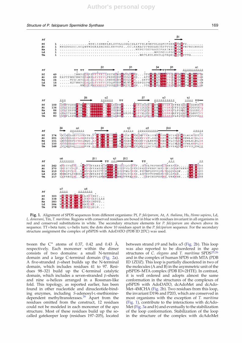
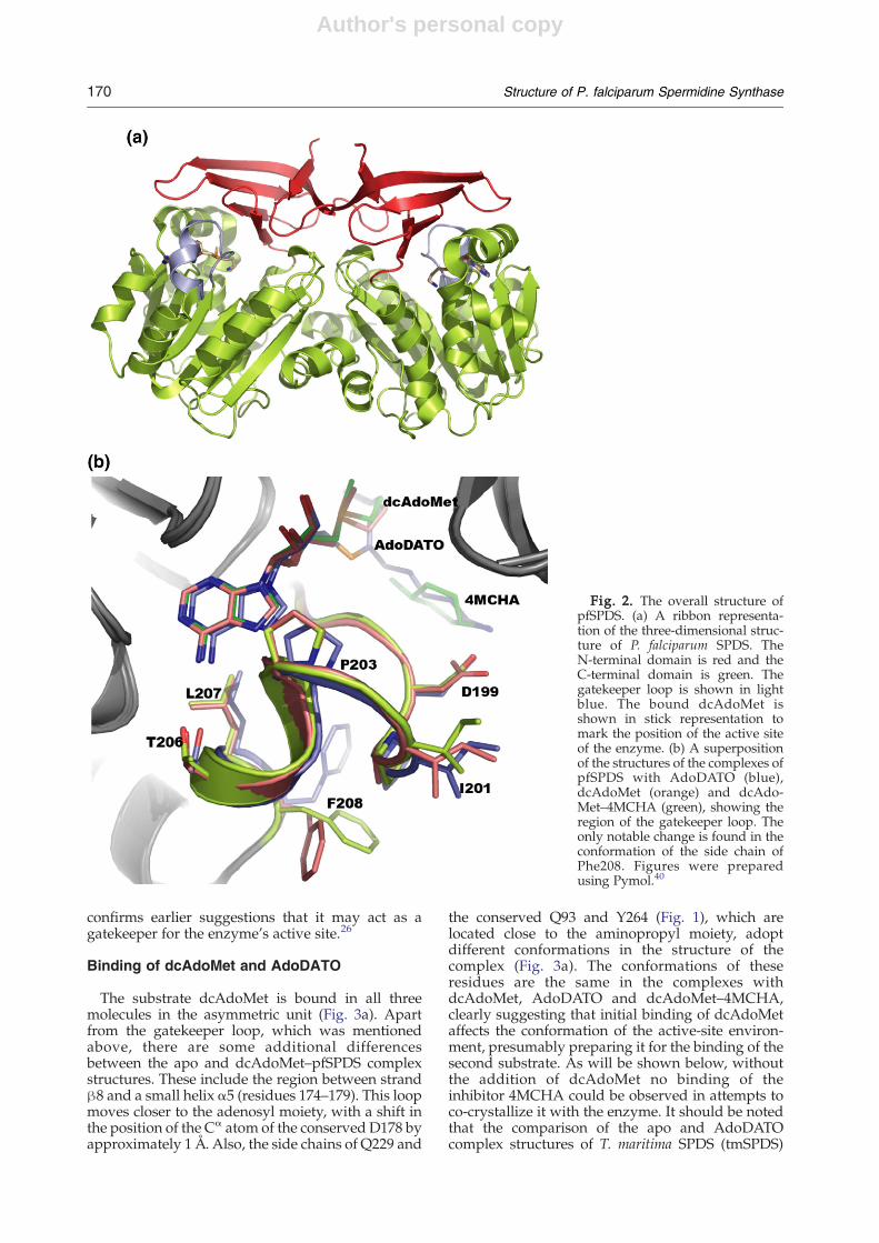
form a dimer in solution.19 Most aminopropyltrans-ferases that have been characterized are also homo-dimers, with the exception of those from thermo-philes, which are tetramers.14,25,26 pfSPDS used inthe current study crystallized with three moleculesper asymmetric unit. The interactions between thesubunits were analyzed using the Protein–ProteinInteractions Server§. The analysis showed thatwithin the asymmetric unit, two of the molecules(B and C) interact with an interface burying around1460 Å2 of the total accessible surface area of eachmonomer, thus forming a dimer (Fig. 2a). On theother hand, the common surface between moleculesA and B is only about 450 Å2, which suggests non-specific crystal packing interactions.The pfSPDS structure did not reveal any major
deviation from the overall fold of Thermotoga mariti-ma and Caenorhabditis elegans SPDS.25,26 A compar-ison of the structures of apo-pfSPDS with thecomplexes of pfSPDS with dcAdoMet, dcAdoMet–4MCHA and AdoDATO gave rmsd values be-
§ http://www.biochem.ucl.ac.uk/bsm/PP/server
168 Structure of P. falciparum Spermidine Synthase
Author's personal copy
tween the Cα atoms of 0.37, 0.42 and 0.43 Å,respectively. Each monomer within the dimerconsists of two domains: a small N-terminaldomain and a large C-terminal domain (Fig. 2a).A five-stranded β-sheet builds up the N-terminaldomain, which includes residues 41 to 97. Resi-dues 98–321 build up the C-terminal catalyticdomain, which includes a seven-stranded β-sheetsand nine α-helices arranged in a Rossman-likefold. This topology, as reported earlier, has beenfound in other nucleotide and dinucleotide-bind-ing enzymes, including S-adenosyl-L-methionine-dependent methyltransferases.26 Apart from theresidues omitted from the construct, 12 residuescould not be modeled in each monomer of the apostructure. Most of these residues build up the so-called gatekeeper loop (residues 197–205), located
between strand β9 and helix α5 (Fig. 2b). This loopwas also reported to be disordered in the apostructures of C. elegans and T. maritima SPDS25,26and in the complex of human SPDS with MTA (PDBID 1ZDZ). This loop is partially disordered in two ofthe molecules (A and B) in the asymmetric unit of thepfSPDS–MTA complex (PDB ID=2HTE). In contrast,it is well ordered and adopts almost the sameconformation in the structures of the complexes ofpfSPDS with AdoDATO, dcAdoMet and dcAdo-Met–4MCHA (Fig. 2b). Two residues from this loop,the invariant D196 and P203, which are conserved inmost organisms with the exception of T. maritima(Fig. 1), contribute to the interactions with dcAdo-Met (Fig. 3a and b) and eventually to the stabilizationof the loop conformation. Stabilization of the loopin the structure of the complex with dcAdoMet
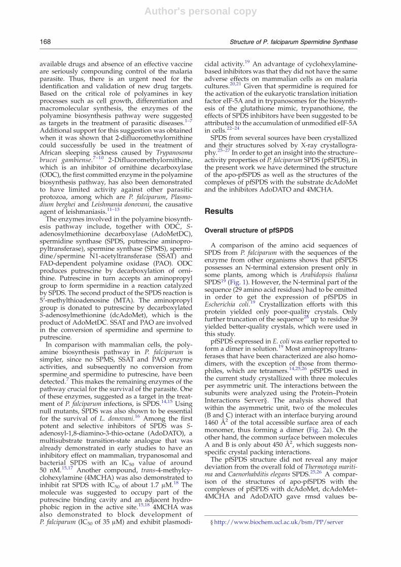
Fig. 1. Alignment of SPDS sequences from different organisms: Pf, P. falciparum, At, A. thaliana, Hs, Homo sapiens, Ld,L. donovani, Tm, T. maritima. Regions with conserved residues are boxed in blue with residues invariant in all organisms inred and conserved substitutions in white. The secondary structure elements for P. falciparum are shown above itssequence. TT=beta turn; η=helix turn; the dots show 10 residues apart in the P. falciparum sequence. For the secondarystructure assignment the complex of pfSPDS with AdoDATO (PDB ID 2I7C) was used.
169Structure of P. falciparum Spermidine Synthase
Author's personal copy
confirms earlier suggestions that it may act as agatekeeper for the enzyme's active site.26
Binding of dcAdoMet and AdoDATO
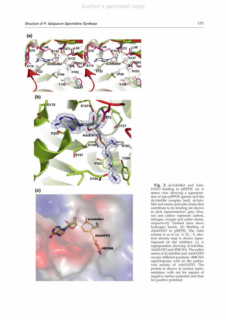
The substrate dcAdoMet is bound in all threemolecules in the asymmetric unit (Fig. 3a). Apartfrom the gatekeeper loop, which was mentionedabove, there are some additional differencesbetween the apo and dcAdoMet–pfSPDS complexstructures. These include the region between strandβ8 and a small helix α5 (residues 174–179). This loopmoves closer to the adenosyl moiety, with a shift inthe position of the Cα atom of the conserved D178 byapproximately 1 Å. Also, the side chains of Q229 and
the conserved Q93 and Y264 (Fig. 1), which arelocated close to the aminopropyl moiety, adoptdifferent conformations in the structure of thecomplex (Fig. 3a). The conformations of theseresidues are the same in the complexes withdcAdoMet, AdoDATO and dcAdoMet–4MCHA,clearly suggesting that initial binding of dcAdoMetaffects the conformation of the active-site environ-ment, presumably preparing it for the binding of thesecond substrate. As will be shown below, withoutthe addition of dcAdoMet no binding of theinhibitor 4MCHA could be observed in attempts toco-crystallize it with the enzyme. It should be notedthat the comparison of the apo and AdoDATOcomplex structures of T. maritima SPDS (tmSPDS)
Fig. 2. The overall structure ofpfSPDS. (a) A ribbon representa-tion of the three-dimensional struc-ture of P. falciparum SPDS. TheN-terminal domain is red and theC-terminal domain is green. Thegatekeeper loop is shown in lightblue. The bound dcAdoMet isshown in stick representation tomark the position of the active siteof the enzyme. (b) A superpositionof the structures of the complexes ofpfSPDS with AdoDATO (blue),dcAdoMet (orange) and dcAdo-Met–4MCHA (green), showing theregion of the gatekeeper loop. Theonly notable change is found in theconformation of the side chain ofPhe208. Figures were preparedusing Pymol.40
170 Structure of P. falciparum Spermidine Synthase
Author's personal copy
Fig. 3. dcAdoMet and Ado-DATO binding to pfSPDS. (a) Astereo view showing a superposi-tion of apo-pfSPDS (green) and thedcAdoMet complex (red). dcAdo-Met and amino acid side chains thatcontribute to its binding are shownin stick representation: grey, blue,red and yellow represent carbon,nitrogen, oxygen and sulfur atoms,respectively. Dashed lines showhydrogen bonds. (b) Binding ofAdoDATO to pfSPDS. The colorscheme is as in (a). A 2Fo−Fc elec-tron density map is shown super-imposed on the inhibitor. (c) Asuperposition showing dcAdoMet,AdoDATO and 4MCHA. The sulfuratoms of dcAdoMet and AdoDATOoccupy different positions. 4MCHAsuperimposes well on the putres-cine moiety of AdoDATO. Theprotein is shown in surface repre-sentation, with red for regions ofnegative surface potential and bluefor positive potential.
171Structure of P. falciparum Spermidine Synthase
Author's personal copy
shows no differences in the conformation of theactive-site residues.26 This may be a result of thetemperature of crystallization, which was far fromthe optimal temperature of growth of T. maritima(around 80 °C).The coordinates of human SPDS in complex with
dcAdoMet have recently been deposited in the PDB(Wu et al., unpublished; PDB ID 2O0L). The bindingof dcAdoMet shows features similar to those of thecomplex of dcAdoMet with pfSPDS.The mode of dcAdoMet binding has been de-
scribed earlier based on the crystal structure of thecomplex of tmSPDS with the transition-state analo-gue AdoDATO.14,15,17 The adenosyl moiety ofdcAdoMet in the present complexes superimposeson that of AdoDATO in complex with pfSPDS, withonly a limited shift of 0.55 Å in the relative positionof the two ligands (Fig. 3b and c). The side chain ofD178 and the carbonyl oxygen of Pro203 formhydrogen bonds with the amine of the adenosylgroups of dcAdoMet and AdoDATO. Other con-served interactions between the two ligands and theprotein include the side chains of E147 and Q72,which interact with the hydroxyls of the ribosylmoiety of dcAdoMet and AdoDATO (about 2.7 and3.2 Å, respectively). As can be seen from the dis-tances between the protein side chains and theligands, no solvent molecules can be fitted into thesepositions to mediate the interactions, as wassuggested by molecular dynamics simulations.29 Itis also seen from Fig. 3c that the sulfonic atom ofAdoDATO has flipped to a position opposite to thatof the sulfur of dcAdoMet. The methyl grouplinked to the sulfur atom of dcAdoMet binds in apocket that is mostly hydrophobic (Fig. 3a). Theaminopropyl chain of dcAdoMet interacts with theside chains of H103 and the invariant D127 andD196, which are located at approximately 2.7 and2.9 Å, respectively, with slight differences in thedifferent subunits. As suggested earlier, the posi-tion of the side chain of D196 is stabilized byinteraction with Y102 (2.6 Å). It has been suggestedthat the role of H103, D127 and D196 is the properorientation of the aminopropyl group and thecarbon atom, which would allow for a nucleophilicattack by putrescine.26
AdoDATO specifically inhibits SPDS from variousorganism with an IC50 value in nano- to lowmicromolar range.17 The IC50 for pfSPDS inhibitionwas determined to be 8.5±0.3 μM and is therebyonly slightly higher than that reported for SPDSfrom other organisms. Overall, AdoDATO bindingmode in pfSPDS is similar to the binding mode inthe complex with tmSPDS.26 A superposition of thestructures of AdoDATO and dcAdoMet–4MCHAcomplexes shows that AdoDATO occupies thedcAdoMet and the putrescine binding pockets(Fig. 3c), with almost the same residues interactingwith AdoDATO as with dcAdoMet and 4MCHA.Interestingly, a comparison of the complexes ofMTA–pfSPDS (PDB ID 2HTE) and AdoDATO–pfSPDS shows that while MTA has essentially thesame conformation and orientation as the adenosyl
moiety of AdoDATO, the sulfide atom of MTA hastwo different positions. One of these (subunit C) issimilar to the position of the sulfur atom ofAdoDATO, while in the other two subunits (A andB) it is different. Since, as mentioned above, subunitC had a relatively ordered gatekeeper loop, while insubunits A and B it was disordered, this suggeststhat the sulfide group of MTA may affect thestability of the loop, facilitating the release of MTAfrom the active site of the enzyme. However, itcannot be excluded that the observed differences inthe position of the sulfide atom of MTA depend onthe crystallization conditions.
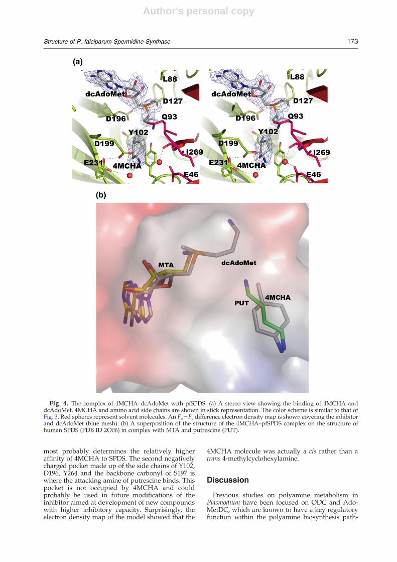
Binding of 4MCHA
As noted above, data collected from crystals ofpfSPDS grown in the presence of 4MCHA alone didnot show the inhibitor at the active site or at anyother location in the structure. On the other hand, co-crystallization with the protein that was preincu-bated with dcAdoMet resulted in a complex with4MCHA bound at the active site of all threemolecules in the asymmetric unit. The substrate-binding pocket, as described earlier in tmSPDS, ishydrophobic in the central part and is occupied byacidic residues at both ends. The negative charge ofthe acidic residues contributes to the stabilization ofthe position of the amino groups of putrescine. Thesuperposition of the structure of human SPDS incomplex with putrescine and MTA (PDB ID=2O06)on the structure of the present complex shows thatthe positions of 4MCHA and putrescine are essen-tially the same (Fig. 4a and b). The cyclohexyl ringand the methyl group of 4MCHA align well with themethylene groups of putrescine, while the amine isaligned with the nonattacking nitrogen of put-rescine. This mode of binding of 4MCHA is differ-ent from that suggested on the basis of dockingstudies.29 The inhibitor appears to be primarilystabilized by a hydrogen bond between the sidechain of the invariant D199 (D176 in human SPDS;Fig. 1) and the amine and by stacking of thecyclohexene ring against the side chain of theinvariant Y264 (Y241 in human SPDS). Most notableis the rotation of the side chain of Y264 upon bindingof 4MCHA: in the apo structure, the aromatic ring ofthe tyrosine is perpendicular to the cyclohexeneplane (Fig. 3a). Two solvent molecules at 2.8 and 3 Åfrom the amine make hydrogen-bonding inter-actions with E231 and E46, respectively. Thesesolvent molecules probably also contribute tothe stabilization of the position of the inhibitor.Thus, the amine of 4MCHA occupies one of thenegatively charged sites responsible for anchoringputrescine.14,26 As seen from Fig. 4b, the methylgroup of 4MCHA occupies the position of themethylene groups of putrescine. In order for thishydrophobic cavity to be accessible to the inhibitor,the side chains of Q93, Q229, Y264 and I269 need toadjust their position, which takes place after dcAdo-Met binding (Fig. 3a). The analysis suggests that themethyl group, which is absent in cyclohexylamine,
172 Structure of P. falciparum Spermidine Synthase
Author's personal copy
most probably determines the relatively higheraffinity of 4MCHA to SPDS. The second negativelycharged pocket made up of the side chains of Y102,D196, Y264 and the backbone carbonyl of S197 iswhere the attacking amine of putrescine binds. Thispocket is not occupied by 4MCHA and couldprobably be used in future modifications of theinhibitor aimed at development of new compoundswith higher inhibitory capacity. Surprisingly, theelectron density map of the model showed that the
4MCHA molecule was actually a cis rather than atrans 4-methylcyclohexylamine.
Discussion
Previous studies on polyamine metabolism inPlasmodium have been focused on ODC and Ado-MetDC, which are known to have a key regulatoryfunction within the polyamine biosynthesis path-
Fig. 4. The complex of 4MCHA–dcAdoMet with pfSPDS. (a) A stereo view showing the binding of 4MCHA anddcAdoMet. 4MCHA and amino acid side chains are shown in stick representation. The color scheme is similar to that ofFig. 3. Red spheres represent solvent molecules. An Fo−Fc difference electron density map is shown covering the inhibitorand dcAdoMet (blue mesh). (b) A superposition of the structure of the 4MCHA–pfSPDS complex on the structure ofhuman SPDS (PDB ID 2O06) in complex with MTA and putrescine (PUT).
173Structure of P. falciparum Spermidine Synthase
Author's personal copy
way. This also made them the primary target forresearch aimed at understanding the mechanisms oftheir function and inhibition. Prior to the biochem-ical characterization, relatively little attention hasbeen given to SPDS from P. falciparum.19
An important feature of SPDS is the highconservation of the active site of the enzyme,although there are some differences in substratespecificity between different species.26 Thus, humanSPDS have been demonstrated to have high speci-ficity towards putrescine, while pfSPDS, E. coli andT. maritima enzymes could produce small amountsof spermine, using spermidine as the aminopropylgroup acceptor.19,26,30 It has also been shown thatpfSPDS had higher Km for dcAdoMet (35 μM) thanmammalian (7–25 μM), E. coli (2 μM) and plantenzymes (0.4–4 μM). This may explain the slightlyelevated IC50 value for AdoDATO (8.5 μM) ascompared to other organisms. It has been suggestedthat substrate specificity of SPDS may be affected bythe gatekeeper loop, which contains the conservedaspartate (D199 in pfSPDS) that anchors the non-attacking amine group of putrescine.14 Thus, intmSPDS this loop appears to have greater flexibilitybecause of an extra residue and a missing proline,which may facilitate the binding of polyaminesother than putrescine.14 The loop between β-strandsβ1 and β2 (F48-W51 in pfSPDS) has also beensuggested to be involved in the closing of the active-site pocket, thus limiting the length of polyaminemolecules that could bind to the enzyme.14,26 Atryptophan (W51 in pfSPDS) serves in closing theactive-site pocket at the side of the nonattackingamino group of the polyamine substrate. Thepromiscuous tmSPDS has a tyrosine at this positionand one amino acid less in the loop, resulting in alarger cavity, which presumably enables the enzymeto bind longer chain diamines. A superposition ofthe human SPDS–MTA–putrescine and dcAdoMet–4MCHA–pfSPDS complex structures shows thatthis loop adopts a similar conformation, thus limit-ing the size of the putrescine-binding pocket. Sincethe gatekeeper loop also adopts a similar conforma-tion in these two structures (Fig. 2b), it may appearthat the putrescine-binding pocket of pfSPDS, likethe human enzyme, does not have room to accom-modate a longer chain polyamine like the triaminespermidine. However, there are some differencesbetween the amino acid sequences of the loop inP. falciparum and the human enzymes, which includethe substitutions Phe47Thr, Ser48Cys, Ile49Ser andMet50Leu (the first amino acid refers to pfSPDS).These substitutions may affect the flexibility of theloop in the case of pfSPDS, and thus allow thebinding of longer chain polyamines, which wouldexplain the limited SPMS activity observed forpfSPDS.19 These differences may be utilized infuture design of inhibitors with higher specificitytowards the Plasmodium SPDS.Our crystallization efforts demonstrated that
dcAdoMet was needed in order for 4MCHA tobind. There were no indications of the protein beingaminopropylated in the dcAdoMet complex struc-
ture, although E. coli and soybean axes SPDS havebeen suggested to carry out aminopropyl transferthrough a ping-pong mechanism.31,32 The require-ment for dcAdomet for 4MCHA binding mostprobably means that for this two-substrate reaction,the two substrates must be added in a compulsoryorder. This observation also supports earlier sugges-tions that the reaction proceeds through an SN2 pathwith the methylene carbon on dcAdoMet under-going nucleophilic attack by the attacking nitrogenof putrescine.14
Materials and Methods
Cloning and expression
In an earlier work, recombinant expression in E. coli ofthe full-length pfSPDS was not successful.19 When the first29 amino acids were omitted, good quantity of expressionof SPDS was achieved. However, attempts to get good-quality diffracting crystals from this protein did notsucceed. Instead, the DNA sequence, corresponding to aprotein lacking 39 residues at the N terminus, wassynthesized and cloned into p15-Tev-LIC vector.28 Theprotein was expressed in E. coli BL21-(DE3)-RosettaOxford in Terrific Broth in the presence of ampicillin/chloramphenicol (100 and 34 μg/ml, respectively). Asingle colony was inoculated into 100 ml of LB with ofampicillin/chloramphenicol (100 and 34 μg/ml, respec-tively) in a 250-ml baffled flask and incubated withshaking at 250 rpm overnight at 37 °C. The culture wastransferred into 1.8 l of Terrific Broth with ampicillin/chloramphenicol (100 and 34 μg/ml, respectively) and0.3 ml of antifoam (Sigma) in a 2-l bottle and culturedusing the LEX system to an OD600 of 4.5.
28 Subsequently, itwas cooled to 15 °C and induced with 0.5 mM IPTGovernight at 15 °C.
Protein purification
The culture was harvested by centrifugation. Pelletsfrom 4 l of culture were resuspended to approximately40 ml/l of cell culture in Binding Buffer [50 mM Hepes(pH 7.5), 500 mMNaCl, 5 mM imidazole and 5% glycerol]with the addition of protease inhibitors (1 mM benzami-dine and 1 mM PMSF). Resuspended pellets stored at−80 °C were thawed overnight at 4 °C on the day beforepurification. Prior to mechanical lysis, each pellet from 1 lof culture was pretreated with 0.5% Chaps (3-[(3-chola-midopropyl)dimethylammonio]propanesulfonic acid)and 500 units of benzonase for 40 min at room temp-erature. Cells were mechanically lysed with a microflui-dizer (Microfluidizer Processor, M-110EH) at approxi-mately 16,000 psi, and the cell lysate was centrifugedusing a Beckman JA-25.50 rotor at 24,000 rpm (∼75,000g)for 20 min at 10 °C.The buffer compositions were as follows: Elution Buffer:
50 mM Hepes (pH 7.5), 500 mM NaCl, 250 mM imidazoleand 5% glycerol; Wash Buffer: 50 mM Hepes (pH 7.5),500 mM NaCl, 30 mM imidazole and 5% glycerol; CrystalBuffer: 10 mM Hepes (pH 7.5), 500 mM NaCl. The clearedlysate was loaded onto a column prepacked with 10 gDE52 (Whatman) anion exchange resin (previouslyactivated with 2.5 M NaCl and equilibrated with BindingBuffer). The DE52 column was further washed with 20 ml
174 Structure of P. falciparum Spermidine Synthase
Author's personal copy
of Binding Buffer. The flow-through was subsequentlyloaded onto a 1.0- to 2.5-ml Ni-NTA (Qiagen) column(preequilibrated with Binding Buffer) at approximately1–1.5 ml/min. The Ni-NTA columnwas then washed with200 ml of Wash Buffer at 2–2.5 ml/min. After washing, theprotein was eluted with 15 ml of Elution Buffer. EDTAwasimmediately added to the elution fraction to 1 mM, andDTT was added to 5 mM after approximately 15 moreminutes. The eluted pfSPDS was applied to a SephadexS200 26/60 gel filtration column preequilibrated withCrystal Buffer. The fractions corresponding to the elutedprotein peak were collected. The His-tag was cleaved withTEV protease overnight at 4 °C in the presence of 1 mMDTT. The cleaved sample was applied to a 1-ml Ni-NTAcolumn preequilibrated with Binding Buffer. The flow-through was collected, and the column was rinsed with anadditional 5 ml of Binding Buffer. These fractions werepooled and concentrated using a 15-ml Amicon Ultracentrifugal filter device (Millipore). Using the same device,Binding Buffer was exchanged with Crystal Buffer byadding 15 ml Crystal Buffer twice to the portion of proteinsolution that was concentrated to around 0.5 ml. Finally,the protein was concentrated to 14.2 mg/ml and stored at−80 °C.
Crystallization, data collection and structuredetermination
Purified pfSPDS (14 mg/ml) was crystallized using thesitting drop vapor diffusion method at 15 °C. Protein
solution (0.5 μl) was mixed with 0.5 μl of the reservoirsolution containing 23% PEG3350, 0.1 M ammoniumsulfate and 0.1 M Bis-Tris (pH 5.5). The pfSPDS–dcAdoMet complex was obtained by first incubatingthe protein with 2.5 mM dcAdoMet for 10 min at roomtemperature before mixing it with the reservoir solution.Protein–ligand solution (0.5 μl) was mixed with 0.5 μl ofthe reservoir solution containing 23% PEG3350, 0.1 Mammonium sulfate and 0.1 M Bis-Tris (pH 5.5). ThepfSPDS–4MCHA–dcAdoMet complex was crystallizedby incubating the protein already incubated withdcAdoMet with 2.5 mM 4MCHA for 10 min at roomtemperature. Protein–ligand solution (0.5 μl) was thenmixed with 0.5 μl of the reservoir solution containing23% PEG3350, 0.1 M ammonium sulfate and 0.1 M Bis-Tris (pH 5.5). The plates were then kept at 15 °C andsingle crystals appeared after 2 days in all setups. TheAdoDATO complex was obtained by transferringpfSPDS–MTA crystals into 15% glycerol cryoprotectantsolution containing 1 mM AdoDATO and incubatingovernight∥.Data were collected at beamline I911-2 (MAX Lab,
Lund, Sweden) and APS Beamline 17-BM (ArgonneNational Laboratory, USA). The apo and complex crystalsbelonged to the same space group, C2, with threemolecules in the asymmetric unit. Prior to data collection,
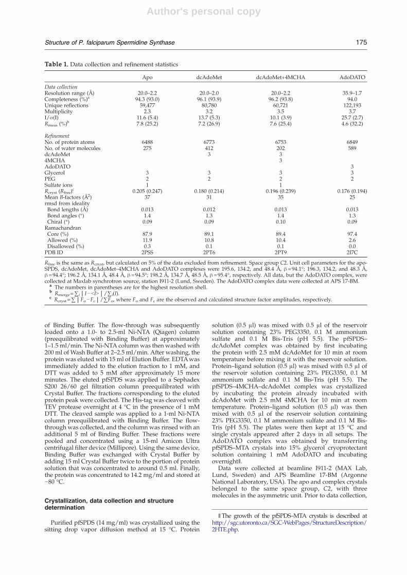
Table 1. Data collection and refinement statistics
Apo dcAdoMet dcAdoMet+4MCHA AdoDATO
Data collectionResolution range (Å) 20.0–2.2 20.0–2.0 20.0–2.2 35.9–1.7Completeness (%)a 94.3 (93.0) 96.1 (93.9) 96.2 (93.8) 94.0Unique reflections 59,477 80,780 60,721 122,193Multiplicity 2.3 3.2 3.5 3.7I/σ(I) 11.6 (5.4) 13.7 (5.3) 10.1 (3.9) 25.7 (2.7)Rmeas (%)b 7.8 (25.2) 7.2 (26.9) 7.6 (25.4) 4.6 (32.2)
RefinementNo. of protein atoms 6488 6773 6753 6849No. of water molecules 275 412 202 589dcAdoMet 3 34MCHA 3AdoDATO 3Glycerol 3 3 3 3PEG 2 2 2 2Sulfate ions 1 1Rcryst (Rfree)
c 0.205 (0.247) 0.180 (0.214) 0.196 (0.239) 0.176 (0.194)Mean B-factors (Å2) 37 31 35 25rmsd from idealityBond lengths (Å) 0.013 0.012 0.013 0.013Bond angles (°) 1.4 1.3 1.4 1.3Chiral (°) 0.09 0.09 0.10 0.09RamachandranCore (%) 87.9 89.1 89.4 97.4Allowed (%) 11.9 10.8 10.4 2.6Disallowed (%) 0.3 0.1 0.1 0.0PDB ID 2PSS 2PT6 2PT9 2I7C
Rfree is the same as Rcryst, but calculated on 5% of the data excluded from refinement. Space group C2. Unit cell parameters for the apo-SPDS, dcAdoMet, dcAdoMet–4MCHA and AdoDATO complexes were 195.6, 134.2, and 48.4 Å, β=94.1°; 196.3, 134.2, and 48.3 Å,β=94.4°; 196.2 Å, 134.1 Å, 48.4 Å, β=94.5°; 198.2 Å, 134.7 Å, 48.5 Å, β=95.4°, respectively. All data, but the AdoDATO complex, werecollected at Maxlab synchrotron source, station I911-2 (Lund, Sweden). The AdoDATO complex data were collected at APS 17-BM.
a The numbers in parentheses are for the highest resolution shell.b Rmerge=∑i|I−bIN|/∑i(I).c Rcryst=∑|Fo−Fc|/∑Fo, where Fo and Fc are the observed and calculated structure factor amplitudes, respectively.
∥The growth of the pfSPDS–MTA crystals is described athttp://sgc.utoronto.ca/SGC-WebPages/StructureDescription/2HTE.php.
175Structure of P. falciparum Spermidine Synthase

Author's personal copy
crystals were transferred to 25% glycerol for a few secondsbefore flash freezing in a boiled-off liquid nitrogen streamat 100 K. Data were processed using the XDS package.33
The data quality was checked with the programTRUNCATE.34 Molecular replacement program PHASERand the coordinates of the pfSPDS–AdoDATO complex(PDB ID 2I7C) with solvent and ligandmolecules removedwere used for obtaining the initial phases.35 Coot andrefmac5 were used for model building and refinement,respectively.36,37 The library files of the ligands weregenerated by the PRODRG2 server.38 Model quality waschecked with PROCHECK39 and the validation menuavailable in Coot. The first two residues at the N terminuscould not be modeled, presumably due to disorder. Asummary of the data collection and refinement statisticsfor four structures are given in Table 1.
pfSPDS inhibition by AdoDATO
To test the inhibitory effect of AdoDATO on theplasmodial enzyme, pfSPDS was cloned, expressed andpurified according to Haider et al.19 The activity wasmeasured as described by Haider et al. and Dufe et al. withthe modification of using lower substrate concentrationsof 50 μMputrescine and 25 μMdcAdoMet.19,25 AdoDATOwas added in the following concentrations: 0, 1, 5, 10, 20,50 and 100 μM.
Protein Data Bank accession numbers
The coordinates for the four structures have beendeposited with the Protein Data Bank with accessionnumbers 2I7C, 2PSS, 2PT6 and 2PT9.
Acknowledgements
We thank the staff of Max II synchrotron forsupport. This work was supported by the DeutscheForschungsgemeinschaft (Wa 395/10/15). S. Al-Karadaghi is supported by a grant from FLÄK(Forskarskolan i läkemedelsvetenskap). The authorsare grateful to Prof. Keijiro Samejima for kindlyproviding dcAdoMet and 4MCHA.
References
1. Assaraf, Y. G., bu-Elheiga, L., Spira, D. T., Desser, H. &Bachrach, U. (1987). Effect of polyamine depletion onmacromolecular synthesis of the malarial parasite,Plasmodium falciparum, cultured in human erythro-cytes. Biochem. J. 242, 221–226.
2. Cohen, S. (1998). A Guide to Polyamines, OxfordUniversity Press, Oxford, UK.
3. Tabor, C. W. & Tabor, H. (1984). Polyamines. Annu.Rev. Biochem. 53, 749–790.
4. Jiang, Y., Roberts, S. C., Jardim, A., Carter, N. S., Shih,S., Ariyanayagam, M. et al. (1999). Ornithine decar-boxylase gene deletion mutants of Leishmania dono-vani. J. Biol. Chem. 274, 3781–3788.
5. Heby, O., Roberts, S. C. & Ullman, B. (2003).Polyamine biosynthetic enzymes as drug targets inparasitic protozoa. Biochem. Soc. Trans. 31, 415–419.
6. Muller, S., Wittich, R. M. & Walter, R. D. (1988). Thepolyaminemetabolism of filarial worms as chemother-apeutic target. Adv. Exp. Med. Biol. 250, 737–743.
7. Muller, S., Coombs, G. H. & Walter, R. D. (2001).Targeting polyamines of parasitic protozoa in che-motherapy. Trends Parasitol. 17, 242–249.
8. Phillips, M. A., Coffino, P. & Wang, C. C. (1987).Cloning and sequencing of the ornithine decarboxy-lase gene from Trypanosoma brucei. Implications forenzyme turnover and selective difluoromethylor-nithine inhibition. J. Biol. Chem. 262, 8721–8727.
9. Phillips, M. A. & Wang, C. C. (1987). A Trypanosomabrucei mutant resistant to alpha-difluoromethylor-nithine. Mol. Biochem. Parasitol. 22, 9–17.
10. Wang, C. C. (1995). Molecular mechanisms andtherapeutic approaches to the treatment of Africantrypanosomiasis. Annu. Rev. Pharmacol. Toxicol. 35,93–127.
11. Bitonti, A. J., McCann, P. P. & Sjoerdsma, A. (1987).Plasmodium falciparum and Plasmodium berghei: effectsof ornithine decarboxylase inhibitors on erythrocyticschizogony. Exp. Parasitol. 64, 237–243.
12. Bitonti, A. J., Dumont, J. A., Bush, T. L., Edwards,M. L.,Stemerick, D. M., McCann, P. P. & Sjoerdsma, A.(1989). Bis(benzyl)polyamine analogs inhibit thegrowth of chloroquine-resistant human malaria para-sites (Plasmodium falciparum) in vitro and in combina-tion with alpha-difluoromethylornithine cure murinemalaria. Proc. Natl Acad. Sci. USA, 86, 651–655.
13. Kaur, K., Emmett, K., McCann, P. P., Sjoerdsma, A.& Ullman, B. (1986). Effects of dl-alpha-difluoro-methylornithine on Leishmania donovani promasti-gotes. J. Protozool. 33, 518–521.
14. Ikeguchi, Y., Bewley, M. C. & Pegg, A. E. (2006).Aminopropyltransferases: function, structure andgenetics. J. Biochem. (Tokyo), 139, 1–9.
15. Pegg, A. E., Poulin, R. & Coward, J. K. (1995). Use ofaminopropyltransferase inhibitors and of non-meta-bolizable analogs to study polyamine regulation andfunction. Int. J. Biochem. Cell Biol. 27, 425–442.
16. Roberts, S. C., Jiang, Y., Jardim, A., Carter, N. S., Heby,O. & Ullman, B. (2001). Genetic analysis of spermidinesynthase from Leishmania donovani. Mol. Biochem.Parasitol. 115, 217–226.
17. Coward, J. K. & Pegg, A. E. (1987). Specific multi-substrate adduct inhibitors of aminopropyltrans-ferases and their effect on polyamine biosynthesis incultured cells. Adv. Enzyme Regul. 26, 107–113.
18. Shirahata, A., Morohohi, T., Fukai, M., Akatsu, S. &Samejima, K. (1991). Putrescine or spermidine bindingsite of aminopropyltransferases and competitive in-hibitors. Biochem. Pharmacol. 41, 205–212.
19. Haider, N., Eschbach, M. L., Dias, S. S., Gilberger,T. W., Walter, R. D. & Luersen, K. (2005). The spermi-dine synthase of the malaria parasite Plasmodiumfalciparum: molecular and biochemical characterisa-tion of the polyamine synthesis enzyme.Mol. Biochem.Parasitol. 142, 224–236.
20. Beppu, T., Shirahata, A., Takahashi, N., Hosoda, H.& Samejima, K. (1995). Specific depletion of spermi-dine and spermine in HTC cells treated with inhi-bitors of aminopropyltransferases. J. Biochem. (Tokyo),117, 339–345.
21. Shirahata, A., Takahashi, N., Beppu, T., Hosoda, H. &Samejima, K. (1993). Effects of inhibitors of spermidinesynthase and spermine synthase on polyamine synth-esis in rat tissues. Biochem. Pharmacol. 45, 1897–1903.
22. Byers, T. L., Ganem, B. & Pegg, A. E. (1992). Cytostasisinduced in L1210 murine leukaemia cells by the S-
176 Structure of P. falciparum Spermidine Synthase
Author's personal copy
adenosyl-l-methionine decarboxylase inhibitor 5′-([(Z)-4-amino-2-butenyl]methylamino)-5′-deoxyade-nosine may be due to hypusine depletion. Biochem. J.287, 717–724.
23. Byers, T. L., Wechter, R. S., Hu, R. H. & Pegg, A. E.(1994). Effects of the S-adenosylmethionine decarbox-ylase inhibitor, 5′-([(Z)-4-amino-2-butenyl]methyla-mino)-5′-deoxyadenosine, on cell growth and poly-amine metabolism and transport in Chinese hamsterovary cell cultures. Biochem. J. 303, 89–96.
24. Muller, S., Liebau, E., Walter, R. D. & Krauth-Siegel,R. L. (2003). Thiol-based redox metabolism of proto-zoan parasites. Trends Parasitol. 19, 320–328.
25. Dufe, V. T., Luersen, K., Eschbach, M. L., Haider, N.,Karlberg, T., Walter, R. D. & Al-Karadaghi, S. (2005).Cloning, expression, characterisation and three-dimensional structure determination of Caenorhabdi-tis elegans spermidine synthase. FEBS Letters, 579,6037–6043.
26. Korolev, S., Ikeguchi, Y., Skarina, T., Beasley, S.,Arrowsmith, C., Edwards, A. et al. (2002). The crystalstructure of spermidine synthase with a multisub-strate adduct inhibitor. Nat. Struct. Biol. 9, 27–31.
27. Lu, P. K., Chien, S. Y., Tsai, J. Y., Fong, C. T., Lee, M. J.,Huang, H. & Sun, Y. J. (2004). Crystallization andpreliminary X-ray diffraction analysis of spermidinesynthase from Helicobacter pylori. Acta Crystallogr.sect. D, 60, 2067–2069.
28. Vedadi, M., Lew, J., Artz, J., Amani, M., Zhao, Y.,Dong, A. et al. (2007). Genome-scale protein expres-sion and structural biology of Plasmodium falciparumand related Apicomplexan organisms. Mol. Biochem.Parasitol. 151, 100–110.
29. Burger, P. B., Birkholtz, L. M., Joubert, F., Haider, N.,Walter, R. D. & Louw, A. I. (2007). Structural andmechanistic insights into the action of Plasmodiumfalciparum spermidine synthase. Bioorg. Med. Chem.15, 1628–1637.
30. Bowman, W. H., Tabor, C. W. & Tabor, H. (1973).Spermidine biosynthesis. Purification and propertiesof propylamine transferase from Escherichia coli.J. Biol. Chem. 248, 2480–2486.
31. Sang, O. Y., Yong, S. L., Sung, H. L. & Young, D. C.(2000). Polyamine synthesis in plants: isolation andcharacterization of spermidine synthase from soybean(Glycine max) axes. Biochim. Biophys. Acta, 1475, 17–26.
32. Zappia, V., Cacciapuoti, G., Pontoni, G. & Oliva, A.(1980). Mechanism of propylamine-transfer reactions.Kinetic and inhibition studies on spermidine synthasefrom Escherichia coli. J. Biol. Chem. 255, 7276–7280.
33. Kabsch, W. J. (1993). Automatic processing of rotationdiffraction data from crystals of initially unknownsymmetry and cell constants. J. Appl. Crystallogr. 26,795–800.
34. French,G. S. &Wilson, K. S. (1978). On the treatment ofnegative intensity observations. Acta Cryst., 517–525.
35. McCoy, A. J., Grosse-Kunstleve, R. W., Storoni, L. C. &Read, R. J. (2005). Likelihood-enhanced fast transla-tion functions. Acta Crystallogr. sect. D, 61, 458–464.
36. Emsley, P. & Cowtan, K. (2004). Coot: model-buildingtools for molecular graphics. Acta Crystallogr. sect. D,60, 2126–2132.
37. Murshudov, G. N., Vagin, A. A. & Dodson, E. J. (1997).Refinement of macromolecular structures by themaximum-likelihood method. Acta Crystallogr. sect.D, 53, 240–255.
38. Schuttelkopf, A. W. & van Aalten, D. M. (2004).PRODRG: a tool for high-throughput crystallographyof protein–ligand complexes. Acta Crystallogr. sect. D,60, 1355–1363.
39. Laskowski, R. A., MacArthur, M. W., Moss, D. S. &Thorton, J. M. (1993). PROCHECK: a program tocheck the stereochemical quality of protein structures.J. Appl. Crystallogr. 26, 283–291.
40. DeLano, W. L. (2002). The PyMOL Molecular GraphicsSystem, DeLano Scientific, San Carlos, CA.
177Structure of P. falciparum Spermidine Synthase
Copyright © 2022 FDOKUMEN