Nigella sativa Extract as a Potent Antioxidant for Petrochemical-Induced Oxidative Stress
6
Various beneficial properties has been attributed to Nigella sativa, including its antioxidant potential. Previously, it was reported that supercritical fluid extraction (SFE) could be used to obtain N. sativa extract rich in antioxidants. In the present study, N. sativa extracts prepared using the previously optimized SFE as well as the traditional Soxhlet extraction approaches were analyzed for various known antioxidants. N. sativa extracts were found to prevent protein carbonyl formation as well as depletion of intracellular glutathione (GSH) in fibroblasts exposed to toluene. Furthermore, partially purified SFE and Soxhlet fractions could prevent loss of hepatic GSH in toluene-induced oxidative stressed Wistar rats as well as in L929 fibroblasts. The results showed that SFE-produced N. sativa extract is richer in antioxidants than the Soxhlet approach. It was also shown using preparative silica gel and reverse phase chromatography that different fractions of SFE-extracted or Soxhlet- extracted N. sativa had different levels of protective effects with regards to GSH depletion in vivo as well as in cell culture. Although fractions rich in thymoquinone were found to be most potent in terms of antioxidant capacity, the data indicates that the protective effects of N. sativa may not only be due to thymoquinone, but perhaps other antioxidants. Introduction The black seed (Nigella sativa) extract, commonly known as Habbat El Baraka in the Arab world, has been in use for genera- tions in various parts of the world, including most of the Arab population. Recent investigations of black seed and many other herbs used for culinary as well as medical purposes have shown that they contain high levels of antioxidants (1,2). In fact, many potent antioxidants have actually been isolated from some nat- ural herbs (3). Thus it is hypothesized that the beneficial effects of black seed and other herbs are most likely due to their protec- tion against cellular damage caused by oxidative stress. The antioxidant properties of black seed oil are recently reviewed (2). It has been shown that some of the compounds isolated from black seed have appreciable free radical scavenging properties (4). This antioxidant property of black seed has also been reported by other investigators (5–7). However, no work has been done to examine the antioxidant effects of black seed oil and its components on petrochemical pollutant induced oxidative stress in vivo and in vitro. Pharmacologically active principles (thymoquinone, dithymoquinone, thymo-hydroquinone, and thymol) of black seed oil have been isolated by solid phase extrac- tion (SPE) and high-performance liquid chromatography (HPLC) separation methods (8). In addition, four novel alkaloids namely nigellicine, nigellidine, nigellimine, and nigellimine N- oxide have also been isolated from black seed (9). The essential components of black seed have been analyzed by gas chromatog- raphy-mass spectrometry (GC–MS) (4). Thymoquinone content of black seed oil has been estimated by GC (10). One of extraction approaches that has recently gained popularity is the supercrit- ical fluid extraction (SFE) method, as this technology uses supercritical carbon dioxide (SC CO2), and thus no solvent residues are left behind in the product (11–14). This technique has the added advantage of recovering the volatile compounds and does not alter the delicate balance of components in natural products. One of the areas that has been a focus of great interest by researchers in the life sciences is the field of reactive oxygen species (ROS). It has been hypothesized for some time that the harmful effects of many xenobiotics and petrochemical pollu- tants and their metabolites are due to their ability to generate ROS (15–17). There is a growing list of reports showing that ben- zene, toluene, and other petrochemical byproducts, when ingested, inhaled or absorbed, are metabolized and transformed into chemicals that lead to the generation of reactive oxygen species (18,19). These ROS, when present in high concentra- tions, can overwhelm cellular antioxidants, and lead to a condi- tion termed “oxidative stress” (20). Cells exposed to oxidative stress undergo extensive cellular damage due to oxy-radical induced DNA-breakage, lipid (membrane) oxidation, and exten- sive protein damage (21). Recently, it has been shown that diverse environmental pollutants including xylene, redox- cycling metals, and UV radiation can cause oxidative stress in skin fibroblasts, leading to GSH depletion and causing S-thiola- 321 Abstract Nigella sativa Extract as a Potent Antioxidant for Petrochemical-Induced Oxidative Stress S. Salman Ashraf 1, * ,† , Madduri V. Rao 1 , Fatima Shad Kaneez 2,§ , Shahnaz Qadri 2,† , Ali H. Al-Marzouqi 3 , Irwin S. Chandranath 4 , and Abdu Adem 4 Departments of 1 Chemistry, 2 Biology, 3 Chemical and Petroleum Engineering, 4 Pharmacology and Therapeutic, U.A.E. University, Al-Ain, P.O. Box: 17551, U.A.E. Reproduction (photocopying) of editorial content of this journal is prohibited without publisher’s permission. Journal of Chromatographic Science, Vol. 49, April 2011 * Author to whom correspondence should be addressed: email salman.ashraf.ac.ae. † Dept. of Chemistry and Biochemistry, University of North Carolina Greensboro, NC. § Present address: Human Physiology, PAP RSB Institute of Health Sciences, Universiti Brunei Darussalam, Brunei.
-
Upload
independent -
Category
Documents
-
view
6 -
download
0
Transcript of Nigella sativa Extract as a Potent Antioxidant for Petrochemical-Induced Oxidative Stress

Various beneficial properties has been attributed to Nigella sativa,including its antioxidant potential. Previously, it was reported thatsupercritical fluid extraction (SFE) could be used to obtain N. sativaextract rich in antioxidants. In the present study, N. sativa extractsprepared using the previously optimized SFE as well as thetraditional Soxhlet extraction approaches were analyzed for variousknown antioxidants. N. sativa extracts were found to preventprotein carbonyl formation as well as depletion of intracellularglutathione (GSH) in fibroblasts exposed to toluene. Furthermore,partially purified SFE and Soxhlet fractions could prevent loss ofhepatic GSH in toluene-induced oxidative stressed Wistar rats aswell as in L929 fibroblasts. The results showed that SFE-producedN. sativa extract is richer in antioxidants than the Soxhlet approach.It was also shown using preparative silica gel and reverse phasechromatography that different fractions of SFE-extracted or Soxhlet-extracted N. sativa had different levels of protective effects withregards to GSH depletion in vivo as well as in cell culture. Althoughfractions rich in thymoquinone were found to be most potent interms of antioxidant capacity, the data indicates that the protectiveeffects of N. sativa may not only be due to thymoquinone, butperhaps other antioxidants.
Introduction
The black seed (Nigella sativa) extract, commonly known asHabbat El Baraka in the Arab world, has been in use for genera-tions in various parts of the world, including most of the Arabpopulation. Recent investigations of black seed and many otherherbs used for culinary as well as medical purposes have shownthat they contain high levels of antioxidants (1,2). In fact, manypotent antioxidants have actually been isolated from some nat-ural herbs (3). Thus it is hypothesized that the beneficial effectsof black seed and other herbs are most likely due to their protec-tion against cellular damage caused by oxidative stress. Theantioxidant properties of black seed oil are recently reviewed (2).
It has been shown that some of the compounds isolated fromblack seed have appreciable free radical scavenging properties(4). This antioxidant property of black seed has also beenreported by other investigators (5–7). However, no work hasbeen done to examine the antioxidant effects of black seed oil andits components on petrochemical pollutant induced oxidativestress in vivo and in vitro. Pharmacologically active principles(thymoquinone, dithymoquinone, thymo-hydroquinone, andthymol) of black seed oil have been isolated by solid phase extrac-tion (SPE) and high-performance liquid chromatography(HPLC) separation methods (8). In addition, four novel alkaloidsnamely nigellicine, nigellidine, nigellimine, and nigellimine N-oxide have also been isolated from black seed (9). The essentialcomponents of black seed have been analyzed by gas chromatog-raphy-mass spectrometry (GC–MS) (4). Thymoquinone contentof black seed oil has been estimated by GC (10). One of extractionapproaches that has recently gained popularity is the supercrit-ical fluid extraction (SFE) method, as this technology usessupercritical carbon dioxide (SC CO2), and thus no solventresidues are left behind in the product (11–14). This techniquehas the added advantage of recovering the volatile compoundsand does not alter the delicate balance of components in naturalproducts.
One of the areas that has been a focus of great interest byresearchers in the life sciences is the field of reactive oxygenspecies (ROS). It has been hypothesized for some time that theharmful effects of many xenobiotics and petrochemical pollu-tants and their metabolites are due to their ability to generateROS (15–17). There is a growing list of reports showing that ben-zene, toluene, and other petrochemical byproducts, wheningested, inhaled or absorbed, are metabolized and transformedinto chemicals that lead to the generation of reactive oxygenspecies (18,19). These ROS, when present in high concentra-tions, can overwhelm cellular antioxidants, and lead to a condi-tion termed “oxidative stress” (20). Cells exposed to oxidativestress undergo extensive cellular damage due to oxy-radicalinduced DNA-breakage, lipid (membrane) oxidation, and exten-sive protein damage (21). Recently, it has been shown thatdiverse environmental pollutants including xylene, redox-cycling metals, and UV radiation can cause oxidative stress inskin fibroblasts, leading to GSH depletion and causing S-thiola-
321
Abstract
Nigella sativa Extract as a Potent Antioxidant forPetrochemical-Induced Oxidative Stress
S. Salman Ashraf1,*,†, Madduri V. Rao1, Fatima Shad Kaneez2,§, Shahnaz Qadri2,†, Ali H. Al-Marzouqi3,Irwin S. Chandranath4, and Abdu Adem4
Departments of 1Chemistry, 2Biology, 3Chemical and Petroleum Engineering, 4Pharmacology and Therapeutic, U.A.E. University, Al-Ain,P.O. Box: 17551, U.A.E.
Reproduction (photocopying) of editorial content of this journal is prohibited without publisher’s permission.
Journal of Chromatographic Science, Vol. 49, April 2011
* Author to whom correspondence should be addressed: email salman.ashraf.ac.ae.† Dept. of Chemistry and Biochemistry, University of North Carolina Greensboro, NC.§ Present address: Human Physiology, PAP RSB Institute of Health Sciences, Universiti BruneiDarussalam, Brunei.
Ashraf.qxd:Article template 3/2/11 11:37 AM Page 1
Journal of Chromatographic Science, Vol. 49, April 2011
322
tion of intracellular proteins (22). Not surprisingly, a largenumber of disease states have been linked to oxidative stress,including cancer, diabetes, Parkinson’s disease, Alzheimer’s dis-ease, and heart disease (21,23). In fact, it was recently shown thatpetrochemical workers are under significant oxidative stress dueto their exposure to petrochemical pollutants and byproducts(24).
In the present study, the antioxidant capacity of N. sativa wastested in a model system involving toluene-induced oxidativestress. Furthermore, it was desired to characterize and analyzesilica-gel fractions of SFE-extracted N. sativa and compare itwith the Soxhlet-extracted fractions. We show that both extrac-tion approaches were successful in producing antioxidant-richN. sativa extracts which showed strong protective effects againstoxyradicals in both cultured hepatocytes as well as in wholeanimal models. Interestingly, it was shown that the antioxidantproperties of N. sativa are probably due to other antioxidants inaddition to its more potent component Thymoquinone.
Materials and Methods
SFEThe SFE was performed as described by Rao et al. (14). Briefly,
the experimental apparatus consisted of a 260 mL capacitysyringe pump and controller system (ISCO 260D), and an ISCOseries 2000 SCF Extraction system (SFX 220) consisting of adual-chamber extraction module with two 10-mL stainless steelvessels. Temperature and pressure within the vessels were mea-sured and could be independently adjusted. The 10 mL stainlesssteel cell was filled with about 5 g of ground black seed. The cellwas pressurized and heated to the desired pressure (100–400 bar)and temperature (30–70°C) and kept for 15 min to reach equi-librium. A known volume of SC CO2 (100–300 mL) was passedthrough the cell at a flow rate of 1 mL/min. The extract was col-lected in a cold trap after depressurization of the gas. The col-lected sample was stored at –18°C until analysis.
Soxhlet extractionApproximately 20 g of finely ground powder of N. sativa was
placed in a thimble and extracted in an all glass Soxhlet extractor
for 8 h using hexane and methanol as solvents. Solvent wasremoved by rotary evaporation at 40°C under vacuum and thelast traces of solvent in the extract were removed under a streamof nitrogen. The extract was stored at –18°C until analysis.
Fractionation of N. sativa extractsSilica gel column fractionation: The extract obtained by
Soxhlet extraction technique was subjected to fractionation onsilica gel. A 250 g of activated silica gel was loaded to a columnand cleaned with about 100 ml of hexane (HPLC grade). About10 g of the extract was loaded on to the column. The compoundswere eluted successively with 500mL each of hexane, 15%diethylether in hexane, diethyl ether, and methanol. Solvent in the frac-tions was removed by rotary evaporation at 40°C under vacuum.A known quantity of each fractionation (100 mg) was made up to10 mL with methanol and subjected to HPLC analysis.
Preparative HPLC fractionationFractionation of SFE extract was carried out on Knauer
preparative HPLC consisting of Knauer K-1800 isocratic pumpand K-2001 UV detector. Black seed oil extracted using aboveSFE conditions was dissolved in acetonitrile–methanol (30:70)and subjected to preparative HPLC fractionation on a C-18column (Eurospher 100, 20 mm i.d.) using acetoni-trile–methanol (30:70) as mobile phase at a flow rate of 5mL/min. The elution of compounds was monitored using a UVdetector at 254 nm. One milliliter of the sample solution wasinjected each time and fractions at time intervals of 0–18 min,18–25 min, 25–30 min, 35–48 min, and 48–60 min. were col-lected successively into 500 mL rotary evaporation flasks. Afterrepeated injections and collection of fractions, solvent in eachfraction was removed by rotary evaporation at 40°C undervacuum. A known quantity of each SFE Fraction (100 mg) wasmade up to 10 mL with methanol and subjected to HPLC anal-ysis under the conditions described in Table I.
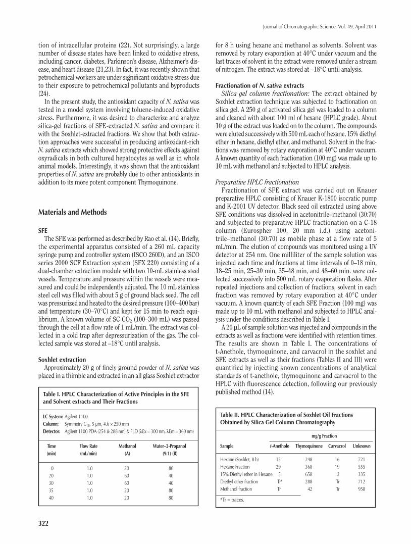
A 20 µL of sample solution was injected and compounds in theextracts as well as fractions were identified with retention times.The results are shown in Table I. The concentrations oft-Anethole, thymoquinone, and carvacrol in the soxhlet andSFE extracts as well as their fractions (Tables II and III) werequantified by injecting known concentrations of analyticalstandards of t-anethole, thymoquinone and carvacrol to theHPLC with fluorescence detection, following our previouslypublished method (14).Table I. HPLC Characterization of Active Principles in the SFE
and Solvent extracts and Their Fractions
LC System: Agilent 1100Column: Symmetry C18, 5 µm, 4.6 × 250 mmDetector: Agilent 1100 PDA (254 & 288 nm) & FLD (λEx = 300 nm, λEm = 360 nm)
Time Flow Rate Methanol Water–2-Propanol(min) (mL/min) (A) (9:1) (B)
0 1.0 20 8020 1.0 60 4030 1.0 60 4035 1.0 20 8040 1.0 20 80
Table II. HPLC Characterization of Soxhlet Oil FractionsObtained by Silica Gel Column Chromatography
mg/g Fraction
Sample t-Anethole Thymoquinone Carvacrol Unknown
Hexane (Soxhlet, 8 h) 15 248 16 721Hexane Fraction 29 368 19 55515% Diethyl ether in Hexane 5 658 2 335Diethyl ether fraction Tr* 288 Tr 712Methanol fraction Tr 42 Tr 958
*Tr = traces.
Ashraf.qxd:Article template 3/2/11 11:37 AM Page 2
Continuous cell line cultureMedia for cell-culture was prepared by adding 86 mL of EMEM
+ 10 mL FBS (Bovine serum) + 2 mL of streptomycin/penicillin+ 1 mL of fungizone + 1 mL of L-glutamine, which was stored inunscrewed bottles in a humidified 5% CO2 37°C incubator overnight before experiment. Continuous cell lines of mouse skinfibroblasts and L929 human hepatocytes HUH7 were cultured byslowly defrosting frozen vials and gently mixing them beforeplacing the cells at a concentration of 2 × 10⁵ cells per plate byplacing 25 µL of the cells in 35 mm plate containing 2 mL of themedia and then incubate them in a CO2 incubator. Plates with70–90% confluency were then pre-incubated with 10 µL of N.sativa overnight and then treated with 10 mM of toluene (indimethyl sulfoxide) for 3 h. Similarly, other sets of similar preincubated plates were treated with hydrogen peroxide for 3 h.After 3 h of treatment, media was decanted and cells were lysedby freeze-thaw and scraping in 0.5 mL of 50 mM Tris + 10 mMEDTA buffer, pH 8.8. The lysate was then centrifuged at 4000rpm to remove cellular debris and the supernatant was used forglutathione (GSH) assay.
In vivo studiesMale Wistar rats weighing between 200–250 g were housed in
polyethylene cages at a room temperature of 22 ± 2°C on a 12–12h light-dark cycle.
A standard pellet diet and tap water were given ad libitum.Groups of animals (5 in each group) were treated once daily for3 days as follows:
Group 1. No treatment, received dimethyl sulfoxide alone(equivalent to the concentration in “toluene-treated” group) asintraperitoneal (i.p.) injection;
Group 2. Toluene-treated, received i.p. injection of toluene 250mg/kg body weight in dimethyl sulfoxide;
Group 3. Toluene + extract (or SFE fraction), received toluenei.p. and + 100 µl, p.o. (oral administration) black seed extract (orSFE fractions) 1 h later; and
Group 4. Toluene + known antioxidants, received toluene, i.p.+ 20 µM thymoquinone (or carvacrol, thymol, t-anethol and p-cymene) i.p., 1 h later.
At the end of the three days and 1 h after the last injection, theanimals were then sacrificed and a piece of liver was taken outand frozen immediately until estimation of GSH was carried out.
Protein estimation and GSH assayProtein concentration of the liver tissues was estimated using
the Bio-Rad Bradford protein assay and expressed as nmol/mgwet weight of tissue. Intracellular GSH was measured by usingthe micro-titer assay as published by Coleman et al. (25). Briefly,GSH standard or cellular lysate was mixed with DTNB in amicro-titer plate. After 5 min of incubation, the plate was read at405 nm. Although this assay essentially measures free thiolgroups (of both, proteins and low-molecular weight thiols), it isgenerally accepted that low molecular weight thiols (of whichGSH comprises ~ 90%) are present in large excess (approxi-mately 10-fold) than protein thiols. Hence, one could use thisassay to get an approximate quantization of intracellular GSHlevels.
Protein carbonyl determinationProtein carbonyls were detected using a kit form Cayman
Chemical (Cat #10005020, Protein Carbonyl Assay Kit) whichmeasures the reaction between protein carbonyls and 2,4-din-tirophenylhydrazine (2,4-DNPH). The results shown are themean and standard deviations from triplicate determinationsnormalized by total protein (Bradford Assay).
Results
Extraction and characterization of N. sativaIt was previously determined that supercritical fluid extraction
(SFE) could be used to produce high quality extracts Nigellasativa that were rich in antioxidants (14). In the present study,the focus was further characterizing the biological (antioxidant)activities of N. sativa extracts. Table IV shows the summary offive different SFE extracts and their ability to prevent loss of glu-tathione in HUH-7 hepatocytes exposed to 10 mM H2O2. As canbe seen from the table, the yields of the extract ranged from0.84% to 30.3% under different conditions, however SFE condi-tion #3 (temperature, pressure and CO2 volume: 50°C, 100 bar,200 mL) which gave lowest yield, had the maximum antioxidantactivity (as measured by prevention of loss of intracellular GSH –defined as “GSH recovery” henceforth). It was interpreted thatmajority of fats and lipids normally present in N. sativa are notextracted under this condition (#3) and that high concentrationsof antioxidants are present in this extract. Therefore, all the sub-sequent SFE of N. sativa reported here was done with this set ofconditions.
Journal of Chromatographic Science, Vol. 49, April 2011
323
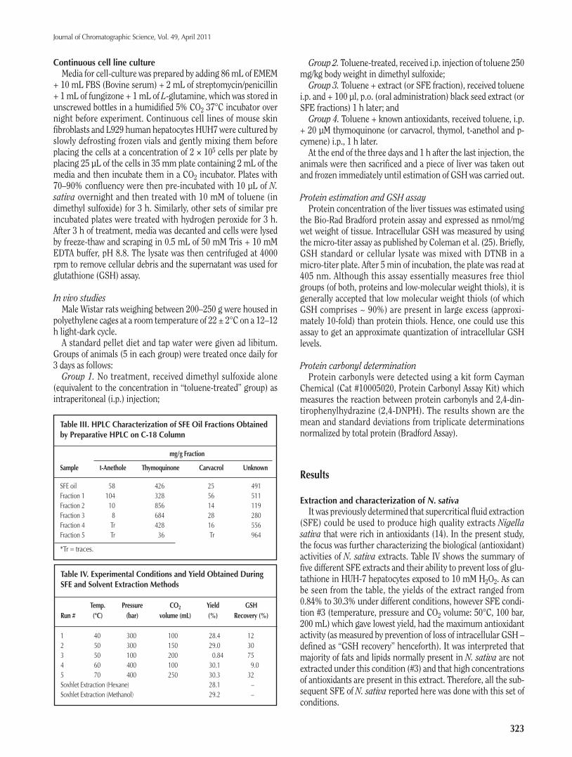
Table III. HPLC Characterization of SFE Oil Fractions Obtainedby Preparative HPLC on C-18 Column
mg/g Fraction
Sample t-Anethole Thymoquinone Carvacrol Unknown
SFE oil 58 426 25 491Fraction 1 104 328 56 511Fraction 2 10 856 14 119Fraction 3 8 684 28 280Fraction 4 Tr 428 16 556Fraction 5 Tr 36 Tr 964
*Tr = traces.
Table IV. Experimental Conditions and Yield Obtained DuringSFE and Solvent Extraction Methods
Temp. Pressure CO2 Yield GSHRun # (°C) (bar) volume (mL) (%) Recovery (%)
1 40 300 100 28.4 122 50 300 150 29.0 303 50 100 200 0.84 754 60 400 100 30.1 9.05 70 400 250 30.3 32Soxhlet Extraction (Hexane) 28.1 –Soxhlet Extraction (Methanol) 29.2 –
Ashraf.qxd:Article template 3/2/11 11:37 AM Page 3
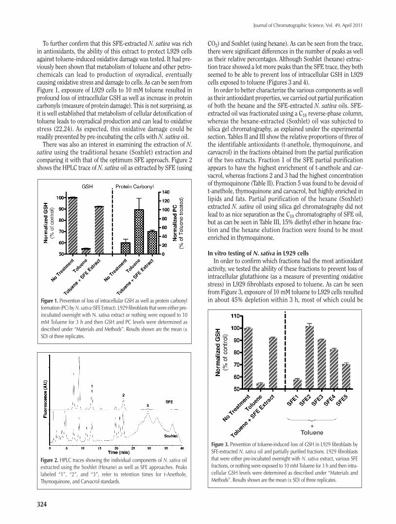
To further confirm that this SFE-extracted N. sativa was richin antioxidants, the ability of this extract to protect L929 cellsagainst toluene-induced oxidative damage was tested. It had pre-viously been shown that metabolism of toluene and other petro-chemicals can lead to production of oxyradical, eventuallycausing oxidative stress and damage to cells. As can be seen fromFigure 1, exposure of L929 cells to 10 mM toluene resulted inprofound loss of intracellular GSH as well as increase in proteincarbonyls (measure of protein damage). This is not surprising, asit is well established that metabolism of cellular detoxification oftoluene leads to oxyradical production and can lead to oxidativestress (22,24). As expected, this oxidative damage could bereadily prevented by pre-incubating the cells with N. sativa oil.
There was also an interest in examining the extraction of N.sativa using the traditional hexane (Soxhlet) extraction andcomparing it with that of the optimum SFE approach. Figure 2shows the HPLC trace of N. sativa oil as extracted by SFE (using
CO2) and Soxhlet (using hexane). As can be seen from the trace,there were significant differences in the number of peaks as wellas their relative percentages. Although Soxhlet (hexane) extrac-tion trace showed a lot more peaks than the SFE trace, they bothseemed to be able to prevent loss of intracellular GSH in L929cells exposed to toluene (Figures 3 and 4).
In order to better characterize the various components as wellas their antioxidant properties, we carried out partial purificationof both the hexane and the SFE-extracted N. sativa oils. SFE-extracted oil was fractionated using a C18 reverse-phase column,whereas the hexane-extracted (Soxhlet) oil was subjected tosilica gel chromatography, as explained under the experimentalsection. Tables II and III show the relative proportions of three ofthe identifiable antioxidants (t-anethole, thymoquinone, andcarvacrol) in the fractions obtained from the partial purificationof the two extracts. Fraction 1 of the SFE partial purificationappears to have the highest enrichment of t-anethole and car-vacrol, whereas fractions 2 and 3 had the highest concentrationof thymoquinone (Table II). Fraction 5 was found to be devoid oft-anethole, thymoquinone and carvacrol, but highly enriched inlipids and fats. Partial purification of the hexane (Soxhlet)extracted N. sativa oil using silica gel chromatography did notlead to as nice separation as the C18 chromatography of SFE oil,but as can be seen in Table III, 15% diethyl ether in hexane frac-tion and the hexane elution fraction were found to be mostenriched in thymoquinone.
In vitro testing of N. sativa in L929 cellsIn order to confirm which fractions had the most antioxidant
activity, we tested the ability of these fractions to prevent loss ofintracellular glutathione (as a measure of preventing oxidativestress) in L929 fibroblasts exposed to toluene. As can be seenfrom Figure 3, exposure of 10 mM toluene to L929 cells resultedin about 45% depletion within 3 h, most of which could be
Journal of Chromatographic Science, Vol. 49, April 2011
324
Figure 2. HPLC traces showing the individual components of N. sativa oilextracted using the Soxhlet (Hexane) as well as SFE approaches. Peakslabeled “1”, “2”, and “3”, refer to retention times for t-Anethole,Thymoquinone, and Carvacrol standards.
Figure 1. Prevention of loss of intracellular GSH as well as protein carbonylformation (PC) byN. sativa (SFE Extract). L929 fibroblasts that were either pre-incubated overnight with N. sativa extract or nothing were exposed to 10mM Toluene for 3 h and then GSH and PC levels were determined asdescribed under “Materials and Methods”. Results shown are the mean (±SD) of three replicates.
Figure 3. Prevention of toluene-induced loss of GSH in L929 fibroblasts bySFE-extracted N. sativa oil and partially purified fractions. L929 fibroblaststhat were either pre-incubated overnight with N. sativa extract, various SFEfractions, or nothing were exposed to 10 mM Toluene for 3 h and then intra-cellular GSH levels were determined as described under “Materials andMethods”. Results shown are the mean (± SD) of three replicates.
Ashraf.qxd:Article template 3/2/11 11:37 AM Page 4

Journal of Chromatographic Science, Vol. 49, April 2011
325
prevented by pre-incubating the cells with SFE-extracted N.sativa oil. When the semi-purified SFE fractions were tested inthe same assay, as expected fractions 2 and 3 (highest in thymo-quinone) were most effective in preventing loss of intracellularGSH. Not surprisingly, fraction 5, which was mostly composed oflipids and fats was not very rich in antioxidants. Interestingly,fraction 1 which was relatively rich in t-anethole (a knownantioxidant) did not show any significant protection against GSHdepletion, suggesting that perhaps t-anethole does not play a sig-nificant role in conferring antioxidant protection to fibroblastsexposed to oxidative stress. It was also suprising to note that thefact that fraction 4, which had half the amount of thymoquinoneas fraction 2, showed significant GSH recovery. This seems toimply that the anti-oxidative effects observed by SFE oil is most-likely not due to thymoquinone alone and there may be addi-tional compounds (perhaps in fraction 4) that may havesignificant antioxidant properties. This hypothesis is further sup-ported by the results obtained from testing of silica gel chro-matography fractions of Soxhlet (hexane)-extracted oil. As can beseen from Figure 4, the diethyl ether fraction, which had lessthan half the amount of thymoquinone (Table II), had almost thesame protective effect (towards GSH recovery) as the 15%diethyl ether/hexane fraction (the “best” fraction). Also, as can beseen from Figure 4, the unpurified Soxhlet (hexane)-extracted N.sativa oil appears not to be as potent as the SFE-extracted oil inpreventing loss of intracellular GSH in L929 cells exposed totoluene.
In vivo testing of N. sativa in Wistar ratsIn order to confirm that theN. sativa antioxidant effect seen in
L929 cells would also hold true for whole animal models, hepaticGSH levels of rats exposed to toluene in the presence or absenceof N. sativa oil was measured. As can be seen in Figure 5, expo-
sure of 250 mg/Kg toluene to rats led to about 25% decrease inhepatic GSH levels. This is very much consistent with what weand others have reported for toluene and petrochemical pollu-tant exposures to humans as well (22,24). As shown in Figure 5,co-injection of SFE-extracted N. sativa oil greatly minimized thetoluene-induced hepatic loss of GSH. Testing of the partiallypurified SFE fractions for their antioxidant effect towardstoluene-induced oxidative stress in rats showed a pattern similarto that observed in L929 cells (Figure 3 vs. 5). SFE fraction 1showed the least protective effect in vivo as it did in L929 cells,again, suggesting that the high proportion of t-anethole found inthis fraction is perhaps not very effective against toluene-induced oxidative stress. This was further supported by datashowing that of the five commercially available phyto-antioxi-dants tested, t-anethol had the least protective effect (GSHrecovery) (Figure 6).
Figure 6. Recovery of toluene-depleted hepatic GSH in rats treated by knownantioxidants.
Figure 4. Prevention of toluene-induced loss of GSH in L929 fibroblasts bySoxhlet-extracted N. sativa oil and partially purified fractions. L929 fibrob-lasts that were either pre-incubated overnight with N. sativa extract, variousfractions, or nothing were exposed to 10 mM Toluene for 3 h and then intra-cellular GSH levels were determined as described under “Materials andMethods”. Results shown are the mean (± SD) of three replicates.
Figure 5. Recovery of toluene-depleted hepatic GSH in rats treated by SFE-extracted N. sativa oil and partially purified fractions.
Ashraf.qxd:Article template 3/2/11 11:37 AM Page 5

Journal of Chromatographic Science, Vol. 49, April 2011
326
Conclusion
In conclusion, the data presented here clearly shows that N.sativa extracts, either extracted using the traditional Soxhlet(hexane) approach or by SFE method, are rich in antioxidants.Furthermore, we demonstrate using in vitro and in vivo assaysthat N. sativa can be used to prevent oxidative damage caused bya known petrochemical pollutant, toluene. Studies with partialpurification and fractionation of N. sativa oil showed thatalthough fractions rich in thymoquinone were most potent interms of their antioxidant capacity, the protective effects of N.sativa may not only be due to thymoquinone, but perhaps due toother antioxidants as well.
Acknowledgment
This work was financially supported by the Research Affairsat the UAE University under a contract #01-04-2-12/04 forInterdisciplinary Research Grant.
References
1. S. Dragland, H. Senoo, K. Wake, K. Holte, and R. Blomhoff. Severalculinary and medicinal herbs are important sources of dietaryantioxidants. J. Nutr. 133: 1286–1290 (2003).
2. B.H. Ali and G. Blunden. Pharmacological and toxicological prop-erties of Nigella sativa. Phytother. Res. 17: 299–305 (2003).
3. M.A. Gyamfi and Y. Aniya. Antioxidant properties of ThonningianinA, isolated from the African medicinal herb, Thonningia sanguinea.Biochem. Pharmacol. 63: 1725–1737 (2002).
4. M. Burits and F. Bucar. Antioxidant activity of Nigella sativa essen-tial oil. Phytotherapy Research 14: 323–328 (2000).
5. M.N. Nagi, K. Alam, O.A. Badary, O.A. Al-Shabanah, H.A. Al-Sawaf,and A.M. Al-Kekairi. Thymoquinone protects against carbon tetra-chloride hepatotoxicity in mice via an antioxidant mechanism.Biochem.Mol. Biol. Int. 47: 143–159 (1999).
6. M.K. Turkdogan, Z. Agaoglu, Z. Yener, R. Sekeroglu, H.A. Akkan,and M.E. Avci. The role of antioxidant vitamins (C and E), seleniumand Nigella sativa in the prevention of liver fibrosis and cirrhosis inrabbits, new hopes. Dtscch. Tierarzt. Wschr. 108: 71–73 (2000).
7. M.R. Mahmoud, H.S. El-Abhar, and S. Saleh. The effect of Nigellasativa oil against the liver damage induced by Schistosoma mansoniinfection in mice. J. Ethnopharmacol. 79: 1–11 (2000).
8. O.A. Ghosheh, A.A. Houdi, and P.A. Crooks. High performanceliquid chromatographic analysis of the pharmacologically activequinones and related compounds in the oil of the black seed(Nigella sativa L.). J. Pharmaceut. Biomed. Anal. 19: 757–762(1999).
9. A. Rahman, S. Malik, S.S. Hasan, M.I. Choudhary, C.Z. Ni, andJ. Clard. Nigellidine, a new indazole alkaloid from the seeds ofNigella sativa. Tetrahedron Lett. 36: 1993–1996 (1995).
10. P.J. Houghton, R. Zarka, B. de las Heras, and J. Hoult. Fixed oil ofNigella sativa and derived thymoquinone inhibit eicosanoid gener-ation in leukocytes and membrane lipid peroxidation. Planta Med.61: 33–36 (1995).
11. M. Fullana, F. Trabelsi, and F. Recasens. Use of neural netcomputing for statistical and kinetic modelling and simulation ofsupercritical fluid extractors. Chem. Eng. Sci. 55: 79–95 (2000).
12. L. Bruhl, B. Matthaus, and J. Fresenius. Extraction of oil seeds bySFE—a comparison with other methods for the determination of theoil content. J. Anal. Chem. 364: 631–634 (1999)
13. A.H. Al-Marzouqi, M.V. Rao, and B. Jobe. A comparative evaluationof SFE and steam distillation methods on the yiels and compositionof essential oil extracted from spearmint (Mentha spicata). J. Liq.Chromatgr. Rel. Tech. 30: 463–475 (2007).
14. M.V. Rao, A.H. Al-Mazrouqi, K. Fatima-Shad, S. Ashraf, andA. Adem. Comparative evaluation of SFE and solvent extractionmetbods on the yield and composition of Black seeds (Nigellasativa). J. Liq. Chromatgr. Rel. Tech. 30: 2545–2555 (2007).
15. N.R. Rao and R. Snyder. Oxidative modifications produced inHL-60 cells on exposure to benzene metabolites. J. Appl. Toxicol.15: 403–409 (1995).
16. S. Edelfors, U. Hass, and K.S. Hougaard. Changes in markers ofoxidative stress and membrane properties in synaptosomes fromrats exposed prenatally to toluene. Pharmacol. Toxicol. 90: 26–31(2002).
17. E.L. Cavalieri and E.G. Rogan. Central role of radical cations inmetabolic activation of polycyclic aromatic hydrocarbons.Xenobiotica 25: 677–688 (1995).
18. C.J. Mattia, J.D. Adams, and S.C. Bondy. Free radical induction inthe brain and liver by products of toluene catabolism. Biochem.Pharmacol. 46: 103–110 (1993).
19. M. Murata, M. Tsujikawa, and S. Kawanishi. Oxidative DNAdamage by minor metabolites of toluene may lead to carcinogen-esis and reproductive dysfunction. Biochem. Biophys. Res.Commun. 261: 478–483 (1999).
20. H. Sies. Oxidative stress: oxidants and antioxidants. Exp.Physiol.82:291–295 (1997).
21. D. Metodiewa and C. Koska. Reactive oxygen species and reactivenitrogen species: relevance to cyto(neuro)toxic events and neuro-logic disorders: An overview. Neurotox. Res. 1: 197–233 (2000).
22. S.S. Ashraf, S. Galadari, and M. Patel. Protein S-thiolation anddepletion of tracellular glutathione in skin fibroblasts exposed tovarious sources of oxidative Stress. Environ. Toxicol. Phar. 22: 80–84(2006).
23. D.M. Townseed, K.D. Tew, and H. Tapiero. The importance of glu-tathione in human disease. Biomed.Pharmacotherapy 57: 145–155(2003).
24. T. Georgieva, A. Michailova, T. Panev, and T. Popov. Possibilities tocontrol the health risk of petrochemical workers. Int. Arch. Occup.Env. Health 75: Suppl: S21–S26 (2002).
25. C.A. Coleman, B.E. Hull, J.N. McDougal, and J.V. Rogers. The effectof m-xylene on cytotoxicity and cellular antioxidant status in ratdermal equivalents. Toxicol. Lett. 142: 133–142 (2003).
Manuscript received April 7, 2010;revision received July 27, 2010.
Ashraf.qxd:Article template 3/2/11 11:37 AM Page 6