Exploring the plant-associated bacterial communities in Medicago sativa L
10
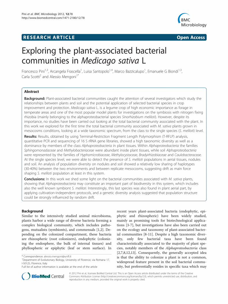
RESEARCH ARTICLE Open Access Exploring the plant-associated bacterial communities in Medicago sativa L Francesco Pini 1,3 , Arcangela Frascella 1 , Luisa Santopolo 1,4 , Marco Bazzicalupo 1 , Emanuele G Biondi 1,3 , Carla Scotti 2 and Alessio Mengoni 1* Abstract Background: Plant-associated bacterial communities caught the attention of several investigators which study the relationships between plants and soil and the potential application of selected bacterial species in crop improvement and protection. Medicago sativa L. is a legume crop of high economic importance as forage in temperate areas and one of the most popular model plants for investigations on the symbiosis with nitrogen fixing rhizobia (mainly belonging to the alphaproteobacterial species Sinorhizobium meliloti). However, despite its importance, no studies have been carried out looking at the total bacterial community associated with the plant. In this work we explored for the first time the total bacterial community associated with M. sativa plants grown in mesocosms conditions, looking at a wide taxonomic spectrum, from the class to the single species (S. meliloti) level. Results: Results, obtained by using Terminal-Restriction Fragment Length Polymorphism (T-RFLP) analysis, quantitative PCR and sequencing of 16 S rRNA gene libraries, showed a high taxonomic diversity as well as a dominance by members of the class Alphaproteobacteria in plant tissues. Within Alphaproteobacteria the families Sphingomonadaceae and Methylobacteriaceae were abundant inside plant tissues, while soil Alphaproteobacteria were represented by the families of Hyphomicrobiaceae, Methylocystaceae, Bradyirhizobiaceae and Caulobacteraceae. At the single species level, we were able to detect the presence of S. meliloti populations in aerial tissues, nodules and soil. An analysis of population diversity on nodules and soil showed a relatively low sharing of haplotypes (30-40%) between the two environments and between replicate mesocosms, suggesting drift as main force shaping S. meliloti population at least in this system. Conclusions: In this work we shed some light on the bacterial communities associated with M. sativa plants, showing that Alphaproteobacteria may constitute an important part of biodiversity in this system, which includes also the well known symbiont S. meliloti. Interestingly, this last species was also found in plant aerial part, by applying cultivation-independent protocols, and a genetic diversity analysis suggested that population structure could be strongly influenced by random drift. Background Similar to the intensively studied animal microbioma, plants harbor a wide range of diverse bacteria forming a complex biological community, which includes patho- gens, mutualists (symbionts), and commensals [1,2]. De- pending on the colonized compartment, these bacteria are rhizospheric (root colonizers), endophytic (coloniz- ing the endosphere, the bulk of internal tissues) and phyllospheric or epiphytic (leaf or stem surface). In recent years plant-associated bacteria (endophytic, epi- phytic and rhizospheric) have been widely studied, mainly as promising tools for biotechnological applica- tions [3-7], but investigations have also been carried out on the ecology and taxonomy of plant-associated bacter- ial communities [8-11]. Despite a high taxonomic diver- sity, only few bacterial taxa have been found characteristically associated to the majority of plant spe- cies, notably members of the Alphaproteobacteria class [2,7,8,12,13]. Consequently, the generally accepted idea is that the ability to colonize a plant is not a common, widespread feature present in the soil bacterial commu- nity, but preferentially resides in specific taxa which may * Correspondence: [email protected] 1 Department of Evolutionary Biology, University of Florence, via Romana 17, I-50125, Florence, Italy Full list of author information is available at the end of the article © 2012 Pini et al.; licensee BioMed Central Ltd. This is an Open Access article distributed under the terms of the Creative Commons Attribution License (http://creativecommons.org/licenses/by/2.0), which permits unrestricted use, distribution, and reproduction in any medium, provided the original work is properly cited. Pini et al. BMC Microbiology 2012, 12:78 http://www.biomedcentral.com/1471-2180/12/78
Transcript of Exploring the plant-associated bacterial communities in Medicago sativa L
Pini et al. BMC Microbiology 2012, 12:78http://www.biomedcentral.com/1471-2180/12/78
RESEARCH ARTICLE Open Access
Exploring the plant-associated bacterialcommunities in Medicago sativa LFrancesco Pini1,3, Arcangela Frascella1, Luisa Santopolo1,4, Marco Bazzicalupo1, Emanuele G Biondi1,3,Carla Scotti2 and Alessio Mengoni1*
Abstract
Background: Plant-associated bacterial communities caught the attention of several investigators which study therelationships between plants and soil and the potential application of selected bacterial species in cropimprovement and protection. Medicago sativa L. is a legume crop of high economic importance as forage intemperate areas and one of the most popular model plants for investigations on the symbiosis with nitrogen fixingrhizobia (mainly belonging to the alphaproteobacterial species Sinorhizobium meliloti). However, despite itsimportance, no studies have been carried out looking at the total bacterial community associated with the plant. Inthis work we explored for the first time the total bacterial community associated with M. sativa plants grown inmesocosms conditions, looking at a wide taxonomic spectrum, from the class to the single species (S. meliloti) level.
Results: Results, obtained by using Terminal-Restriction Fragment Length Polymorphism (T-RFLP) analysis,quantitative PCR and sequencing of 16 S rRNA gene libraries, showed a high taxonomic diversity as well as adominance by members of the class Alphaproteobacteria in plant tissues. Within Alphaproteobacteria the familiesSphingomonadaceae and Methylobacteriaceae were abundant inside plant tissues, while soil Alphaproteobacteriawere represented by the families of Hyphomicrobiaceae, Methylocystaceae, Bradyirhizobiaceae and Caulobacteraceae.At the single species level, we were able to detect the presence of S. meliloti populations in aerial tissues, nodulesand soil. An analysis of population diversity on nodules and soil showed a relatively low sharing of haplotypes(30-40%) between the two environments and between replicate mesocosms, suggesting drift as main forceshaping S. meliloti population at least in this system.
Conclusions: In this work we shed some light on the bacterial communities associated with M. sativa plants,showing that Alphaproteobacteria may constitute an important part of biodiversity in this system, which includesalso the well known symbiont S. meliloti. Interestingly, this last species was also found in plant aerial part, byapplying cultivation-independent protocols, and a genetic diversity analysis suggested that population structurecould be strongly influenced by random drift.
BackgroundSimilar to the intensively studied animal microbioma,plants harbor a wide range of diverse bacteria forming acomplex biological community, which includes patho-gens, mutualists (symbionts), and commensals [1,2]. De-pending on the colonized compartment, these bacteriaare rhizospheric (root colonizers), endophytic (coloniz-ing the endosphere, the bulk of internal tissues) andphyllospheric or epiphytic (leaf or stem surface). In
* Correspondence: [email protected] of Evolutionary Biology, University of Florence, via Romana 17,I-50125, Florence, ItalyFull list of author information is available at the end of the article
© 2012 Pini et al.; licensee BioMed Central LtdCommons Attribution License (http://creativecreproduction in any medium, provided the or
recent years plant-associated bacteria (endophytic, epi-phytic and rhizospheric) have been widely studied,mainly as promising tools for biotechnological applica-tions [3-7], but investigations have also been carried outon the ecology and taxonomy of plant-associated bacter-ial communities [8-11]. Despite a high taxonomic diver-sity, only few bacterial taxa have been foundcharacteristically associated to the majority of plant spe-cies, notably members of the Alphaproteobacteria class[2,7,8,12,13]. Consequently, the generally accepted ideais that the ability to colonize a plant is not a common,widespread feature present in the soil bacterial commu-nity, but preferentially resides in specific taxa which may
. This is an Open Access article distributed under the terms of the Creativeommons.org/licenses/by/2.0), which permits unrestricted use, distribution, andiginal work is properly cited.

Pini et al. BMC Microbiology 2012, 12:78 Page 2 of 10http://www.biomedcentral.com/1471-2180/12/78
be considered more ecologically versatile or geneticallyprone to the association with plants. This last hypothesishas recently been supported by the finding that, at leastin the class of Alphaproteobacteria, a common gene rep-ertoire seems to be present in all of its plant-associatedmembers [14].Medicago sativa L. (alfalfa) is one of the most import-
ant legume crop in temperate areas throughout theworld, commonly used as forage or in crop rotationpractices to contribute organic nitrogen to the soil viaits symbiosis with the nitrogen fixing bacteria [15].Moreover, it is important also for bioenergy production[16] and is one of the most suited plant species for landrestoration [17]. Finally, this species, and the diploidrelative M. truncatula Gaertn. (barrel medic), are amongthe most studied model species regarding the molecularaspects of plant-bacteria symbiosis, particularly in rela-tion with the alphaproteobacterium Sinorhizobium (syn.Ensifer) meliloti [18-20]. Concerning S. meliloti, this spe-cies is present in most temperate soils, and, when condi-tions are suitable, it forms specialized structures, callednodules, in the roots of alfalfa plants where it differenti-ates into bacteroids [18]. It is assumed that a fraction ofbacterial cells is released from dehiscent nodules to soil,giving rise to new free-living rhizobial clones [21]. In thelast years S. meliloti has been found able to also endo-phytically colonize the aerial part of other plant species,as rice [22], suggesting the presence of several ecologicalniches for this species (soil, nodule, other plant tissues).While the plant-associated bacterial flora of M. sativa
has never been investigated at the community level, S.meliloti population genetics have been extensively stud-ied in the past [23-28], but only on strains isolated fromnodules, with a few early studies performed on bacteriadirectly recovered from soil [29,30], due to the lack ofefficient selective culture media. No data have beenreported on the presence in natural conditions of S.meliloti as endophytes in other plant compartments(such as leaves) and no comparison of soil vs. plant-associated populations has been done.Based on the above mentioned considerations, there is
a need to characterize the bacterial community asso-ciated with M. sativa in relation to both the potentiallyimportant role the class of Alphaproteobacteria seemsto have as main component of a “core plant-associatedbacterial community” in several different plant species[13,31-33], and to the relationships of soil vs. plant-associated populations of the symbiotic alphaproteobac-terial partner S. meliloti.In this work we investigated the bacterial communities
associated with the legume M. sativa, focusing on boththe total bacterial community composition and on thepresence and populations structure of the symbioticpartner S. meliloti in soil and plant tissues.
The analysis was conducted by cultivation-independenttechniques on alfalfa (M. sativa) plants grown in meso-cosm pots. The bacterial community associated with M.sativa and that of the surrounding soil were analyzed athigh (class, family) and low (single species, S. meliloti)taxonomic levels by employing Terminal-RestrictionFragment Length Polymorphism (T-RFLP) profiling [33],16 S rRNA library screening and S. meliloti-specificmarkers [34,35]. These approaches allowed us to explorefor the first time the bacterial community compositionof such important plant species and the populations ofS. meliloti without cultivation.
ResultsRibotype variability of the bacterial communityThe ribotype variability of bacterial communities presentin soil and associated to plant tissues (nodules, stemsand leaves) was investigated by T-RFLP analysis. A totalof 43 samples was analyzed: in particular one pooled soilsample for each one of the three pots, one pooled sam-ple from all the nodules found in each pot and fourplants per pot (one stem and 2–3 pools of leaves perplant). T-RFLP profiles on these samples produced 253Terminal-Restriction Fragments (T-RFs) or ribotypesafter the restriction digestion with two restrictionenzymes, HinfI and TaqI. 16 S rRNA gene amplificationand T-RFLP profiling was also performed on DNAextracted from surface-sterilized seeds, but no bands of16 S rRNA gene amplification were recovered (data notshown), suggesting a very low bacterial titre in seeds.Figure 1 shows the pattern of similarity among T-
RFLP profiles from total communities as Non-MetricMultidimensional Scaling (N-MDS). Soil and nodulebacterial communities were strongly differentiated fromstem and leaf communities, forming relatively tight clus-ters. Large heterogeneity was detected in leaf and stemcommunities. To better evaluate the statistical signifi-cance of differentiation of communities we employedAMOVA. Most of the variation (71.75%) was due tointra-environment differences (Additional file 1: TableS1). However, significant differences between environ-ments were found (P< 0.0001), in particular between asoil-nodule group and a stem-leaf group.Interestingly, stem and leaf communities showed a sig-
nificant (P< 0.0001), though small (pairwise FST= 0.05)separation (Additional file 2: Table S2). Moreover,AMOVA on stems and leaves community revealed a sta-tistically significant differentiation between the three pots(P< 0.0001), irrespective of possible grouping (eitherplant genotype-related or unrelated), suggesting a pot-effect over the taxonomic shaping of the leaf-associatedcommunity and no effect of plant genotypes. These dataconfirmed a previous long-term experiment only addres-sing S. meliloti species [23].

-0,24 -0,16 -0,08 0 0,08 0,16 0,24 0,32
Coordinate 1
-0,2
-0,15
-0,1
-0,05
0
0,05
0,1
0,15
0,2
0,25
Coo
rdin
ate
2
Similarity
Pot 1
Pot 2
Pot 3
StemsNodules
Leaves
Soil
Figure 1 Pattern of similarities of individual T-RFLP profiles from total community analysis. The pattern of similarity has been inspected byusing Nonmetric Multidimensional scaling (N-MDS) based on Jaccard similarity matrix. Stress of N-MDS= 0.1896. Stars indicate nodules; squares,soils; circles, leaves; triangles, stems. Grey filling, pot 1; white, pot 2; black, pot 3. Samples of the same environment were grey shaded.
Pini et al. BMC Microbiology 2012, 12:78 Page 3 of 10http://www.biomedcentral.com/1471-2180/12/78
Taxonomic composition of bacterial communities in soil,nodules and plant aerial partsT-RFLP analysis has shown that bacterial communitiesclustered in three groups (soil, nodules and plant aerialparts). In order to elucidate which taxa are mainlyrepresented in the bacterial communities of these as-semblies, three 16 S rRNA gene clone libraries wereconstructed pooling together the DNAs extracted fromthe samples of each environment; additionally, leavesand stems samples were also pooled together due totheir high similarities as mentioned above. Pooled sam-ples did conceivably result in an enrichment of themore shared taxa possibly preventing the detection oftaxa associated only to a few individual samples. DNAwas used as template to construct three 16 S rRNA li-braries; a total of 276 clones (from 78 to 116 per li-brary) were sequenced. Sequence analysis revealed, asexpected, that the soil community was the most diverse(Shannon H’= 4.63; Chao1 = 168), while the nodule-associated community was less diverse (ShannonH’= 1.98; Chao1 = 30), (Additional file 3: Table S3). As aconsequence, the library of nodules showed a coverage(85.9%) higher than those of stems + leaves (74.1%) andsoil (47.1%).The percentages of taxonomic classes detected in the
sequences of the clone libraries are reported in Figure 2.
Seven classes were represented in both soil and stem+leaf communities, and 4 of them were also found innodules. Alphaproteobacteria were dominant in nodules(as expected, due to the presence of high titres of thesymbiotic alphaproteobacterium S. meliloti) and instems + leaves. Also in soil Alphaproteobacteria werehighly prevalent, but Acidobacteria and Crenarchaeotawere also abundant. Flavobacteria were found only innodules, however a low presence in the other environ-ments cannot be excluded, especially in relation to thelower coverage of the respective libraries. Beta- andGammaproteobacteria and Actinobacteria were found inall three libraries.Concerning Alphaproteobacteria, only members of the
Rhizobiaceae family were found in nodules, with allsequences assigned, as expected, to the Sinorhizobium/Ensifer genus (Figure 3). Alphaproteobacteria present insoil belonged to the Rhizobiaceae, Bradyrhizobiaceae,Methylocystaceae, Hypomicrobiaceae and Caulobactera-ceae families. Rhizobiaceae, Aurantimonadaceae andMethylobacteriaceae, all belonging to the Rhizobiales,plus taxa of the order Sphingomonadales, were found inthe stem+ leaf library. The absence of sequencesassigned to the Sinorhizobium/Ensifer genus from stem+leaves and soil libraries, though this species was foundby qPCR in both these environments (see the following

0 10 20 30 40 50 60 70 80 90 100
Soil
Nodule
Stems + Leaves Unclassified bacteriaAlpha-proteobacteriaBeta-proteobacteriaGamma-proteobacteriaDelta-proteobacteriaActinobacteriaBacilli
ClostridiaChloroflexiFlavobacteriaSphingobacteriaAcidobacteriaThermoproteiDeinococci
Percentage of clones
Figure 2 Representation of bacterial divisions in the 16 S rRNA gene clone libraries. The percentage of clones accounting for each divisionwith respect to its origin (nodule, stems+ leaves, soil) is reported.
Pini et al. BMC Microbiology 2012, 12:78 Page 4 of 10http://www.biomedcentral.com/1471-2180/12/78
paragraph), could be due to its low abundance and tothe relatively low coverage of clone libraries.
Detection and diversity of sinorhizobium meliloti in soiland plant tissuesAiming to analyze presence and diversity of S. meliloti,we firstly estimated the population size by qPCR, usingtwo species-specific primer pairs which amplify chromo-somal (rpoE1) and megaplasmidic loci (nodC onpSymA), respectively [35]. The obtained results arereported in Table 1. Relatively higher titers of S. melilotiDNA were detected in root nodules, while lower valueswere obtained in soils, leaves and stems. Interestingly,nodule titers of S. meliloti DNA detected by rpoE mar-ker were higher than those estimated by nodC marker(roughly one order of magnitude). The viable titers of S.meliloti cells from crushed nodules of M. sativa plantsusually ranged from 2.1x10⁸ to 5.0x10⁸cells/g of freshtissue (data not shown), suggesting that the titers fromnodC marker are a better proxy of the number of bac-teria involved in the symbiotic nitrogen-fixing process.Then, to inspect the genetic diversity of S. meliloti
populations present in the different environments, theamplification of the 1.3 kbp long 16 S-23 S ribosomalintergenic spacer (IGS) which proved to be an efficientmarker for the study of S. meliloti populations [34], wasattempted. Only DNAs from nodules and soil gave aPCR product, probably as a result of the low bacterialtiters and high content in inhibitors present in DNAextracted from stems and leaves. Consequently, noduletissue was taken as representative of the plant environ-ment and was compared with soil. A total of 121 differ-ent IGS-T-RFs (16 S-23 S ribosomal intergenic spacer
Terminal-Restriction Fragments) was detected after di-gestion with four restriction enzymes (AluI, MspI, HinfI,HhaI) in the six DNA samples (three from soil, threefrom nodules), after IGS amplification and T-RFLP pro-filing (Additional file 4: Figure S1a). Most of the 121detected IGS-T-RFs (71.9%) were detected in one sam-ple out of 6, while 8 (6.6%) IGS-T-RFs were present inall six samples (Additional file 4: Figure S1b). Moreover,from 25.5 to 53.3% of IGS-T-RFs present in soil werealso detected in nodules and from 31.4 to 40.1% ofIGS-T-RFs present in nodules were detected in the re-spective soil sample. Figure 4 shows the similarity rela-tionships between IGS-T-RFLP profiles. Non-metricMDS plot of IGS-T-RFLP profiles (Figure 4a) showed apossible separation of nodule and soil populations onthe second dimension. In particular, the nodule popula-tion in pot 1 was more separated from the soil popula-tion of the same pot and from the populations of theother pots. On the contrary, nodule populations of pots2 and 3 were the closest ones, with soil population ofpot 3 in the same cluster (Figure 4b), suggesting a pos-sible effect of plant genotype as previously shown[23,36]. However, in agreement with the high numberof single-sample haplotypes detected, an AMOVA car-ried out to evaluate the variance contribution to ahypothetical differentiation of soil and nodule S. meli-loti population showed that 17.37% only of variancewas attributed to a soil-nodule separation, theremaining 82.63% of variance being due to among-nodules and among-soil differences. Additionally, nostatistical significant separation (P< 0.46) was detectedfor groupings based on the two plant genotypes presentin the mesocosms.

Percentage of clones0 10 20 30 40 50 60 70 80 90 100
Soil
Nodule
Stem + Leaves
Rhizobiaceae
Methylobacteriaceae
Aurantimonadaceae
Hyphomicrobiaceae
Methylocystaceae
Bradyrhizobiaceae
Caulobacteraceae
Sphingomonadaceae
Unclassified
Figure 3 Distribution of the recovered families in Alphaproteobacteria with respect to their origin (nodule, stems+ leaves, soil). Thepercentage of clones present in the libraries for each family is reported.
Pini et al. BMC Microbiology 2012, 12:78 Page 5 of 10http://www.biomedcentral.com/1471-2180/12/78
DiscussionIn recent years there has been an increasing interest inexploring the bacterial flora associated with plants[37-41]. A recent field survey indicates [8] that plant aer-ial parts (leaves) harbor complex, but highly variable,bacterial communities, and that only a small number ofbacterial taxa (mainly belonging to Alphaproteobacteria)are plant-specific. In the experiments reported here, asin the majority of the reports on endophytic microflora,
Table 1 Titers of S. meliloti in soil and plant tissues}
Sample Titers
rpoE1-based nodC-based
Pot 1
Soil 4.92 ± 2.82 x 10⁴ 2.78 ± 0.63 x 10⁴
Nodules 3.07 ± 0.67 x 10⁹ 4.25 ±1.24 x 10⁸ **
Stems 2.73 ± 1.21 x 10⁴ 3.22 ±2.4 x 10³ *
Leaves 8.65 ± 4.04 x 10³ 4.28 ± 1.23 x 10³
Pot 2
Soil 1.16 ± 0.33 x 10⁴ 2.88 ± 1.09 x 10⁴
Nodules 1.20 ± 0.50 x 10¹⁰ 1.01 ± 0.10 x 10⁹ **
Stems 2.37 ± 0.49 x 10³ 1.13 ± 0.15 x 10³
Leaves 9.74 ± 5.08 x 10² 2.34 ±0.78 x 10²
Pot 3
Soil 2.70 ± 0.41 x 10⁵ 7.42 ±0.93 x 10⁴ *
Nodules 6.02 ± 1.45 x 10⁹ 2.02 ± 3.22 x 10⁷ **
Stems 4.91 ± 0.95 x 10⁵ 1.07 ± 3.74 x 10⁵
Leaves 5.54 ± 2.83 x 10³ 5.21 ± 3.01 x 10³
}Titers were estimated by qPCR [35] with rpoE1 and nodC markers and areexpressed as n. of gene copies/g of tissue or soil; ± standard deviation fromtriplicate experiments. Asterisks indicate significant differences betweenestimates based on rpoE1 and nodC markers (*, P< 0.05; **, P< 0.01).
we refer to endophytic and epiphytic bacteria indicatingall those that are inside the plant tissue or strongly ad-hering to the plant surface, such as they resist washingand sterilization (or their DNA is retained by plant tis-sue), therefore a more correct definition could be “plant-associated bacteria”.The present study shows that root nodules and aerial
parts of Medicago sativa plants grown in mesocosmconditions, harbor distinct bacterial communities withspecific signatures at the class, family and species levelsand that these communities do not mirror soil bacterialcommunities.Initially, T-RFLP profiles allowed us to show that bac-
terial communities present in the different environments(soil, nodules, stems and leaves) were strongly differen-tiated and in particular that a large heterogeneity waspresent between leaves of individual plants, though soilprofiles were highly similar. Moreover a clear separationbetween above-ground (stem and leaves) and below-ground environments (soil and nodules) was detected.An analysis of the clone libraries, prepared from above-ground and below-ground pooled samples, revealed anuneven distribution of bacterial classes, with a markedpattern highlighting the class of Alphaproteobacteria asthe more abundant in plant tissues (this class repre-sented half of the clones in the stem+ leaf library). Thesame uneven pattern was then observed, at lower taxo-nomic ranks, within the Alphaproteobacteria, withsequences of clones belonging to members of the Methy-lobacteriaceae and Sphingomonadaceae families beingmore abundant in stem than in soil and nodules. Methy-lobacteria and Sphingomonadaceae have been found asendophytes in a number of plants [8,12,31,33,42-45] and

Nodules-3
Soil-3
Nodules-1
Soil-1
Nodules-2
Soil-2
-0,48 -0,36 -0,24 -0,12 0 0,12 0,24 0,36 0,48
Coordinate 1
-0,3
-0,2
-0,1
0
0,1
0,2
0,3
0,4
0,5C
oord
inat
e 2
0 0.8 1.6 2.4 3.2 4 4.8 5.6 6.4
0.2
0.3
0.4
0.5
0.6
0.7
0.8
0.9
1
Sim
ilarit
y (J
acca
rd)
Soi
l - 1
Soi
l -2
Nod
ules
-2
Nod
ules
-3
Soi
l-3
Nod
ules
-1a) b)
Figure 4 a) Non-metric MDS plot of similarities of IGS-T-RFLP profiles from S. meliloti population analysis. a) The pattern of similarity of S.meliloti populations has been inspected by using Non-metric Multidimensional scaling (N-MDS) based on Jaccard similarity matrix. Stress = 0.0898.b) Cluster analysis based on Jaccard similarity matrix. Scale bar represents Jaccard similarity coefficient
Pini et al. BMC Microbiology 2012, 12:78 Page 6 of 10http://www.biomedcentral.com/1471-2180/12/78
it is believed that this group of bacteria may take advan-tage from living as plant-associated, thanks to its abilityto utilize the one-carbon alcohol methanol dischargedby wall-associated pectin metabolism of growing plantcells.Concerning root nodule bacterial communities,
obtained data indicated that very diverse bacterial taxaare associated with nodules, the most represented beingthe specific rhizobial host of M. sativa, the alphaproteo-bacterium S. meliloti. However, additional taxa havebeen found, including members of Actinobacteria, Flavo-bacteria, Gammaproteobacteria and Betaproteobacteria,which may have some additional plant growth-promot-ing activities (see for instance [46,47]).In soil, Acidobacteria was one of the most important
divisions (in terms of number of clones in the library)and was present exclusively in the soil clone library, inagreement with many previous observations [48,49]. Arelatively high presence of Archaea (Thermoprotei) wasalso found. Checking the 16 S rRNA gene sequencespresent in the Ribosomal Database for 799f/pHr primerannealing, we found that PCR amplification from Ther-moprotei was theoretically possible with this primer pair(data not shown). The presence of Archaea in the soil isnot unexpected [50] and could be linked also to the an-oxic or nearly anoxic conditions present in the bottomof the pot. However, since the low coverage of soil clonelibrary, the presence of many other additional taxa, aswell of different proportions of those found here cannotbe excluded. In addition, it should be mentioned thatdifferences between soil and plant-tissues bacterial com-munities could also be ascribed to the different DNA ex-traction protocols we were obliged to use, since a unique
protocol (bead-beading protocol for both soil DNA andplant DNA) failed in a successful extraction of DNAfrom both soil and plant tissues (data not shown). Asimilar technical need was encountered by other authorsalso [33], which renders the study of the relationshipsbetween plant-associated and soil bacterial communitiesstill at its beginning.At the lowest taxonomic rank here investigated, within
the species S. meliloti, we detected the presence of thisspecies in all environment analyzed (soil, nodules andplant aerial tissues). This finding is confirming earlierreports on the ability of S. meliloti to behave as an endo-phytic strain, colonizing all plant compartments, besidesbeing a root symbiont of legumes [22], and suggest a po-tential higher genetic variability of S. meliloti population,and, from the other side, potential new ecological andfunctional roles for this species, not investigated so far[29,51,52]. Unfortunately, the low population size of S.meliloti in stems and leaves and the possible presence ofPCR inhibitors (plant DNA or phenolic compounds, forinstance) did not permit the amplification of 16 S-23 SrRNA intergenic region from plant aerial parts to obtaininformation about the genetic diversity and structure ofS. meliloti population resident in plant aerial part. Nohypothesis could then be drawn about the relationshipsbetween this population and those of soil and nodules.Concerning S. meliloti populations present in soil andnodules, similar values for diversity were detected innodules and in soil, suggesting that both environmentsharbor a consistent fraction of the population’s geneticdiversity. Interestingly, most of the T-RFs were detectedin one sample only, and a very small fraction of T-RFswas shared among all samples, though the original soil

Pini et al. BMC Microbiology 2012, 12:78 Page 7 of 10http://www.biomedcentral.com/1471-2180/12/78
material was homogeneous and should, in theory,contain the same S. meliloti haplotypes. Therefore,S. meliloti populations from all the three mesocosmsinvestigated were highly differentiated between eachother and, as expected from previous studies onS. meliloti [23] and on Bradyrhizobium [53], no statis-tically significant plant genotype- related haplotypeswere detected.A possible explanation of such findings could be
linked to the relatively low titers of S. meliloti in soil(104-105 cells/g), which is roughly 1/10,000 of the totalbacterial community of soil (estimated at ~109 16 SrRNA gene copies/g of soil by qPCR, data not shown).Such estimated S. meliloti titers were similar to thosepreviously observed in other soil and plant tissues [35]and in line with those normally found in soil with viable(Most Probable Number, MPN) estimates [26,54]. As aconsequence of this low population size, founder effectand genetic drift are likely to be among the main shap-ing forces of S. meliloti population in this experimentalset-up, perhaps permitting the fixation of sample-specifichaplotypes by simple chance [55]. Regarding the nodule-soil relationships, though our experiments did not directlyaddress this issue, the reported S. meliloti populationanalysis suggests the presence of somewhat nonoverlap-ping soil and nodule population fractions, even if nospecific patterns of soil and nodule populations weredetected. The presence of different rhizobial haplotypesin nodules and soil was previously found in chickpea[51] and clover [52], though no simple conclusion couldbe drawn, because of limited sampling. However, as fortotal bacterial community analysis, it should be men-tioned that the use of two different DNA extractionprotocols for soil and plant DNA may have producedsome bias in the proportion of the different haplotyesdetected.
ConclusionIn conclusion, we show on M. sativa that its associatedmicroflora, though highly variable, is mainly related tothe presence of Alphaproteobacteria. This class has anuneven presence of families in stems + leaves, nodulesand soil. We then speculated that a sort of “pan-plant-associated bacterial community” may be composed of alarge plethora of “accessory” taxa, which are occasionallyassociated with plants, and a small number of “core”taxa (e.g. Alphaproteobacteria families) which, on thecontrary, are consistently found in the plants. Moreover,within Alphaproteobacteria the specific alfalfa symbioticspecies S. meliloti, abundant as symbiont in rootnodules, was also detected in soil and in leaves, with po-tentially different populations, suggesting a more com-plex interplay of colonization of multiple environments(soil, root nodules, other plant tissues) by this species.
MethodsExperimental design and sampling procedureA controlled experiment was set-up in mesocosmscomposed of three pots (numbered 1, 2, 3) containingMedicago sativa (alfalfa) plants grown at CRA-FLCLodi, Italy, in outdoor conditions. Two of the three potswere planted with the same line of alfalfa (1x5) whilethe third pot was planted with a different line (5x7).The pots (cylinders of 25 cm diameter x 80 cm depth)with a drainage layer on the bottom, were filled with asandy loam non-calcareous soil (57.8% sand, 32% silt,10.2% clay, 1.7% organic matter and 0.09% total N; pH6.7) in which alfalfa has never been grown. Phosphorusand potassium equivalent to 120 Kg ha-1 of P2O5 and180 Kg ha-1 of K2O were distributed into the soil, whileno mineral N was added; irrigation was not limiting.Twenty plants/pot (density equivalent to 400 plantsm-2) were transplanted in March 2008 and allowed togrow until the 2nd year (the end of September 2009),when plant aerial parts of 12 plants were harvested andthe pots were opened to allow sampling of the wholeeye-detectable nodules present (approximately 80–100of various sizes per pot) and of bulk soil. Roots wereexcluded from the analysis since the presence of smallnodules or nodule primordia could not be excluded,possibly inducing a strong bias in the estimation of“non-nodule-associated root colonizers”. The plant sam-ple size was chosen on the basis of a previous analysisof plant-by-plant variation in which the overall diversityof communities did not change from 2 to 30 plants(unpublished data and [8]). Stems, leaves (pools ofaround 10 leaves per plant) and nodules were washedwith water and with 10 mM MgSO4 twice to removemost soil and dust particles and eliminate bacterialoosely adhering to the surface and then surface steri-lized with 1% HClO for 1 min. Samples of soil, nodules,stem and leaves were then stored at −80°C from1–2 weeks before DNA extraction.A control of seed-borne bacteria was also prepared
with seeds of M. sativa surface sterilized with 1% HgCl2.S. meliloti viable titres in sterilized nodules have been
estimated by serial dilution of crushed nodules as previ-ously reported [54].
DNA extraction real-time PCR and T-RFLP profilingDNA was extracted from soil by using a commercial kit(Fast DNA Spin kit for soil, QBiogene, Cambridge, UK)following the manufacturer’s instructions. DNA extrac-tion from plant tissues and surface sterilized controlseeds was performed by a 2X CTAB protocol as previ-ously described [56]. The 16 S rRNA gene pool of totalbacterial community was amplified from the extractedDNA with primer pairs 799f (labeled with HEX) andpHr which allow the amplification of most bacterial

Pini et al. BMC Microbiology 2012, 12:78 Page 8 of 10http://www.biomedcentral.com/1471-2180/12/78
groups without targeting chloroplast DNA [33]. PCRconditions and Terminal-Restriction Fragment LengthPolymorphism (T-RFLP) profiling were as previouslyreported [8], by using HinfI and TaqI restrictionenzymes. For sinorhizobial populations, T-RFLP was car-ried out on 16 S-23 S ribosomal intergenic spacer ampli-fied from total DNA (IGS-T-RFLP) with S. melilotispecific primers and AluI and HhaII restriction enzymes,as already reported [34]. Real-Time PCR (qPCR) forquantification of S. meliloti DNA was carried out onrpoE1 and nodC loci, as previously reported [35]; twodifferent calibration curves were constructed, one forsoil samples and the other one for plant samples, byusing as template DNA extracted from sterile soil (with-out presence of S. meliloti) and from sterile plant (grownin petri dishes), both spiked with serial dilutions ofknown titres of S. meliloti cells, as previously reported[35]. Controls with S. medicae WSM419 DNA wereincluded in both IGS-T-RFLP and qPCR, for S. melilotispecies-specificity check [35].
Library construction and sequencingAmplified (with 799f and pHr primer pair) 16 S rRNAgenes from DNA extracted from soil, nodules, pooledstems and leaves of a 1:1:1 mix of all pots were insertedinto a pGemT vector (Promega, Fitchburg, WI, USA)and cloned in E. coli JM109 cells. Positive clones wereinitially screened by white/blue coloring and the insertedamplified 16SrRNA genes sequenced. Plasmid purifica-tion and sequencing reactions were performed byMacrogen Europe Inc. (Amsterdam, The Netherlands).The nucleotide sequences obtained were deposited inGen- Bank/DDBJ/EMBL databases under accessionnumbers from HQ834968 to HQ835246.
Data processing and statistical analysesFor qPCR data, 1-way ANOVA with Tukey post hoc testwas employed. Analyse-it 2.0 software (Analyse-It, Ldt.,Leeds, UK) was used for both tests. For T-RFLP, chro-matogram files from automated sequencer sizing wereimported into GeneMarker ver. 1.71 software (SoftGe-netics LLC, State College, PA, USA) by filtering with thedefault options of the module for AFLP analysis. Peaksabove 100 fluorescence units and whose size rangedfrom 35 to 500 nt were considered for profile analysis.Only the presence/absence of peaks was considered asinformative data from the chromatograms. Statisticalanalyses were performed on a binary matrix obtained aspreviously reported [8]. Past 2.02 [57] software packagewas used to compute Non-Metric MultidimensionalScaling (N-MDS). To test the distribution of the vari-ance of T-RFLP profiles within plant tissues and amongpots, Analysis of Molecular Variance (AMOVA [58]) wasapplied using Arlequin 3.5.1.2 software (http://cmpg.
unibe.ch/software/arlequin3/). Although developed forpopulation genetic analysis, the general procedureimplemented by AMOVA is flexible enough to estimatethe statistical significance of groups of bacterial commu-nities as reported previously [13,42,59]. Pairwise FST dis-tances [60] between T-RFLP profiles of plant tissues andsoils were used to infer a Neighbor-Joining dendrogramwith the MEGA4 software [61].Partial 16 S rRNA sequences were manually inspected
for quality, then aligned and clustered with the furthestneighboring algorithm with the module present inMothur v.1.12.3 [62]. Diversity indices (Shannon H’ andChao-1) were calculated with the same software. Librarycoverage was estimated with the formula C=1-(n/N)[63], where n is the number of singletons (defined at97% sequence identity in Mothur) that are encounteredonly once in the library and N is the total number ofsequenced clones. Taxonomic assignment was per-formed with the Classifier module present in RibosomalDatabase Project 10 website [64] (http://rdp.cme.msu.edu/) at 80% confidence threshold. Sequences with 97%similarity were treated as a single Operational Taxo-nomic Unit (OTU). Sequences (one for each OTU)were aligned with the 16 S rRNA gene sequences of theclosest match retrieved from NCBI databases, usingMUSCLE [65] and a Neighbor-Joining dendrogram wasconstructed using MEGA4 [61]. Phylogenetic inferenceand evolutionary distance calculations were generatedusing the Maximum Composite Likelihood; 1000 boot-strap replicates were used to obtain confidence estimatesfor the phylogenetic trees.
Additional files
Additional file 1: Table S1. Hierarchical analysis of differentiationbetween bacterial communities. AMOVA was performed with T-RFLPprofiles from samples of the four different environments (soil, nodules,stems and leaves). Data show the degrees of freedom (d.f.), the sum ofsquared deviation, the variance component estimate, the percentage oftotal variance contributed by each component, and the probability (P) ofobtaining a more extreme component estimate by chance alone,estimated from 10,000 permutations.
Additional file 2: Table S2. Matrix of pairwise FST values. Statisticalsignificance (p< 0.05) has been computed after 1000 randompermutation; n.s., not significant. Only below diagonal values arereported. 2
Additional file 3: Table S3. Statistical analysis of 16SrRNA gene clonelibraries. OTUs were arbitrarily defined at 97% sequence identity basedon Mothur clustering. Confidence intervals at 95% are given inparentheses. Coverage is defined C = [1− (n/N)] × 100, where n is thenumber of unique clones, and N is the total number of clonesexamined.
Additional file 4: Figure S1. S. meliloti IGS-T-RFLP profiling of noduleand soil samples. A), the schematic representation of the binary matrixof IGS-T-RF presence (black) and absence (empty cell); the IGS-T-RFnumber is reported on the right side of each row. B) The occurrence of“private” and “public” IGS-T-RFs. The percentage of total number ofscored IGS-T-RFs is reported for T-RFs present from 1 to all 6 samplesanalyzed.

Pini et al. BMC Microbiology 2012, 12:78 Page 9 of 10http://www.biomedcentral.com/1471-2180/12/78
Competing interestsThe authors declare that they have no competing interests.
AcknowledgementsThis work was partially supported by intramural funding of the University ofFlorence to AM and MB and by Soil-Sink (FISR-MIUR) project funding to MB.We are grateful to Mary Forrest, Professor of Scientific English, Department ofPharmacology, Faculty of Medicine and Surgery, University of Florence forediting English language. We acknowledge two anonymous reviewers forhelpful suggestions for the improvement of the manuscript.
Author details1Department of Evolutionary Biology, University of Florence, via Romana 17,I-50125, Florence, Italy. 2Centro di ricerca per le produzioni foraggere elattiero-casearie (CRA-FLC), viale Piacenza 29, I-26900, Lodi, Italy. 3Institut deRecherche Interdisciplinaire - IRI CNRS USR3078, Parc de la Haute Borne 50avenue de Halley, F-59658, Villeneuve d'Ascq Cedex, France. 4Department ofAgricultural Biotechnology, Section of Microbiology, University of Florence,Piazzale delle Cascine 24, 50144, Florence, Italy.
Authors’ contributionsFP performed most of the analyses, prepared the figures and contributed inwriting the draft of the manuscript. This work is part of FP PhD thesis. AFand LS gave technical assistance to the experimental work during thepreparation of their master degree theses. CS settled the mesocosmexperiment and assisted in the samplings. EGB, MB, FP and AM conceivedthe idea and contributed in performing part of the analyses and in draftingthe manuscript. All authors have given final approval of the version to bepublished.
Received: 05 August 2011 Accepted: 20 May 2012Published: 20 May 2012
References1. Ryan RP, Germaine K, Franks A, Ryan DJ, Dowling DN: Bacterial
endophytes: recent developments and applications. FEMS Microbiol Lett2008,278(1):1–9.
2. Mengoni A, Schat H, Vangronsveld J: Plants as extreme environments? Ni-resistant bacteria and Ni-hyperaccumulators of serpentine flora. Plantand Soil 2010, 331:5–16.
3. Danhorn T, Fuqua C: Biofilm formation by plant-associated bacteria. AnnuRev Microbiol 2007, 61:401–422.
4. Lodewyckx C, Vangronsveld J, Porteous F, Moore ERB, Taghavi S, MezgeayM, van der Lelie D: Endophytic bacteria and their potential applications.Crit Rev Plant Sci 2002, 21(6):583–606.
5. Rajkumar M, Ae N, Freitas H: Endophytic bacteria and their potential toenhance heavy metal phytoextraction. Chemosphere 2009, 77(2):153–160.
6. Mastretta C, Taghavi S, Van der Lelie D, Mengoni A, Galardi F, Gonnelli C,Barac T, Boulet J, Weyens N, Vangronsveld J: Endophytic bacteria fromseeds of Nicotiana tabacum can reduce cadmium phytotoxicity. Int JPhytoremediation 2009, 11:251–267.
7. Ikeda S, Okubo T, Anda M, Nakashita H, Yasuda M, Sato S, Kaneko T, TabataS, Eda S, Momiyama A, et al: Community- and Genome-Based Views ofPlant-Associated Bacteria: Plant-Bacterial Interactions in Soybean andRice. Plant Cell Physiol 2010, 51(9):1398–1410.
8. Mengoni A, Pini F, Huang L-N, Shu W-S, Bazzicalupo M: Plant-by-plantvariations of bacterial communities associated with leaves of the nickel-hyperaccumulator Alyssum bertolonii Desv. Microbial Ecol 2009,58:660–667.
9. Manter DK, Delgado JA, Holm DG, Stong RA: Pyrosequencing Reveals aHighly Diverse and Cultivar-Specific Bacterial Endophyte Community inPotato Roots. Microbial Ecol 2010, 60:157–166.
10. Bulgari D, Casati P, Brusetti L, Quaglino F, Brasca M, Daffonchio D, BiancoPA: Endophytic Bacterial Diversity in Grapevine (Vitis vinifera L.) LeavesDescribed by 16S rRNA Gene Sequence Analysis and LengthHeterogeneity-PCR. J Microbiol 2009, 47(4):393–401.
11. Prakamhang J, Minamisawa K, Teamtaisong K, Boonkerd N, Teaumroong N:The communities of endophytic diazotrophic bacteria in cultivated rice(Oryza sativa L.). Applied Soil Ecol 2009, 42(2):141–149.
12. Idris R, Kuffner M, Bodrossy L, Puschenreiter M, Monchy S, Wenzel WW,Sessitsch A: Characterization of Ni-tolerant methylobacteria associatedwith the hyperaccumulating plant Thlaspi goesingense and descriptionof Methylobacterium goesingense sp nov. Syst Applied Microbiol 2006,29(8):634–644.
13. Okubo T, Ikeda S, Kaneko T, Eda S, Mitsui H, Sato S, Tabata S, Minamisawa K:Nodulation-dependent communities of culturable bacterial endophytesfrom stems of field-grown soybeans. Microbes Environ 2009, 24(3):253–258.
14. Pini F, Galardini M, Bazzicalupo M, Mengoni A: Plant-bacteria associationand symbiosis: are there common genomic traits in Alphaproteobacteria?Genes 2011, 2:1017–1032.
15. Sprent JI: Nodulation in legumes. London: Royal Botanic Gardens, Kew; 2001.16. Sanderson MA, Adler PR: Perennial forages as second generation
bioenergy crops. Int J Mol Sci 2008, 9(5):768–788.17. Bradshaw AD, Chadwick MJ: The restoration of land: the ecology and
reclamation of derelict and degraded land. Berkley: University of CaliforniaPress; 1980.
18. Gibson KE, Kobayashi H, Walker GC: Molecular Determinants of aSymbiotic Chronic Infection. Annual Rev Genet 2008, 42:413–441.
19. Oldroyd GED, Downie JM: Coordinating nodule morphogenesis withrhizobial infection in legumes. Annu Rev Plant Biol 2008, 59:519–546.
20. Downie JA: The roles of extracellular proteins, polysaccharides andsignals in the interactions of rhizobia with legume roots. Fems MicrobiolRev 2010, 34(2):150–170.
21. Oono R, Denison RF, Kiers ET: Controlling the reproductive fate ofrhizobia: how universal are legume sanctions? New Phytologist 2009,183(4):967–979.
22. Chi F, Shen SH, Cheng HP, Jing YX, Yanni YG, Dazzo FB: Ascendingmigration of endophytic rhizobia, from roots to leaves, inside rice plantsand assessment of benefits to rice growth physiology. Appl EnvironMicrobiol 2005, 71(11):7271–7278.
23. Carelli M, Gnocchi S, Fancelli S, Mengoni A, Paffetti D, Scotti C, BazzicalupoM: Genetic diversity and dinamics of Sinorhizobium meliloti populationsnodulating different alfalfa varieties in Italian soils. Applied EnvironMicrobiol 2000, 66:4785–4789.
24. Jebara M, Mhamdi R, Aouani ME, Ghrir R, Mars M: Genetic diversity ofSinorhizobium populations recovered from different Medicago varietiescultivated in Tunisian soils. Can J Microbiol 2001, 47(2):139–147.
25. Bailly X, Olivieri I, De Mita S, Cleyet-Marel JC, Bena G: Recombination andselection shape the molecular diversity pattern of nitrogen-fixingSinorhizobium sp. associated to Medicago. Mol Ecol 2006,15(10):2719–2734.
26. Trabelsi D, Mengoni A, Aouani ME, Bazzicalupo M, Mhamdi R: Geneticdiversity and salt tolerance of Sinorhizobium populations from twoTunisian soils. Annals of Microbiol 2010, 60(3):541–547.
27. Roumiantseva ML, Andronov EE, Sharypova LA, Dammann-Kalinowski T,Keller M, Young JPW, Simarov BV: Diversity of Sinorhizobium meliloti fromthe central Asian alfalfa gene center. Applied Environ Microbiol 2002,68(9):4694–4697.
28. Biondi EG, Pilli E, Giuntini E, Roumiantseva ML, Andronov EE, OnichtchoukOP, Kurchak ON, Simarov BV, Dzyubenko NI, Mengoni A, et al: Geneticrelationship of Sinorhizobium meliloti and Sinorhizobium medicae strainsisolated from Caucasian region. FEMS Microbiol Lett 2003, 220(2):207–213.
29. Bromfield ESP, Barran LR, Wheatcroft R: Relattive genetic structure of apopulation of Rhizobium meliloti isolated directly from soil and fromnodules of alfalfa (Medicago sativa) and sweet clover (Melilotus alba).Mol Ecol 1995, 4(2):183–188.
30. Hartmann A, Giraud JJ, Catroux G: Genotypic diversity of Sinorhizobium(formerly Rhizobium) meliloti strains isolated directly from a soil andfrom nodules of alfalfa (Medicago sativa) grown in the same soil. FemsMicrobiol Ecol 1998, 25(2):107–116.
31. Ikeda S, Okubo T, Kaneko T, Inaba S, Maekawa T, Eda S, Sato S, Tabata S,Mitsui H, Minamisawa K: Community shifts of soybean stem-associatedbacteria responding to different nodulation phenotypes and N levels.ISME J 2010, 4(3):315–326.
32. Ikeda S, Rallos LEE, Okubo T, Eda S, Inaba S, Mitsui H, Minamisawa K:Microbial Community Analysis of Field-Grown Soybeans withDifferent Nodulation Phenotypes. Appl Environ Microbiol 2008,74(18):5704–5709.
33. Idris R, Trifonova R, Puschenreiter M, Wenzel WW, Sessitsch A: Bacterialcommunities associated with flowering plants of the Ni

Pini et al. BMC Microbiology 2012, 12:78 Page 10 of 10http://www.biomedcentral.com/1471-2180/12/78
hyperaccumulator Thlaspi goesingense. Appl Environ Microbiol 2004,70(5):2667–2677.
34. Trabelsi D, Pini F, Bazzicalupo M, Biondi EG, Aouani ME, Mengoni A:Development of a cultivation-independent approach for the study ofgenetic diversity of Sinorhizobium meliloti populations. Mol Ecol Res2010, 10(1):170–172.
35. Trabelsi D, Pini F, Aouani ME, Bazzicalupo M, Mengoni A: Development ofreal-time PCR assay for detection and quantification of Sinorhizobiummeliloti in soil and plant tissue. Letters in Applied Microbiol 2009,48(3):355–361.
36. Paffetti D, Daguin F, Fancelli S, Gnocchi S, Lippi F, Scotti C, Bazzicalupo M:Influence of plant genotype on the selection of nodulatingSinorhizobium meliloti strains by Medicago sativa. Antonie VanLeeuwenhoek 1998, 73(1):3–8.
37. Hardoim PR, van Overbeek LS, Elsas JDv: Properties of bacterialendophytes and their proposed role in plant growth. Trends in Microbiol2008, 16(10):463–471.
38. Rosenblueth M, Martinez-Romero E: Bacterial endophytes and theirinteractions with hosts. Mol Plant Microbe Interact 2006, 19(8):827–837.
39. Sessitsch A, Puschenreiter M: Endophytes and Rhizosphere Bacteria ofPlants Growing in Heavy Metal-Containing Soils. In Microbiology ofExtreme Soils Soil Biology 1. Edited by Dion P, Nautiyal CS. Berlin Heidelberg:Springer; 2008.
40. Hartmann A, Stoffels M, Eckert B, Kirchhof G, Schloter M: Analysis of thepresence and diversity of diazotrophic endophytes. In Prokaryoticnitrogen fixation: a model system for the analysis of a biological process.Edited by Triplett EW. Wymondham: Horizon Scientific Press;2000:727–736.
41. Weyens N, van der Lelie D, Taghavi S, Newman L, Vangronsveld J:Exploiting plant-microbe partnerships to improve biomass productionand remediation. Trends in Biotechnology 2009, 27(10):591–598.
42. Mengoni A, Mocali S, Surico G, Tegli S, Fani R: Fluctuation of endophyticbacteria and phytoplasmosis in elm trees. Microbiol Res 2003,158(4):363–369.
43. Van Aken B, Peres CM, Doty SL, Yoon JM, Schnoor JL: Methylobacteriumpopuli sp. nov., a novel aerobic, pink-pigmented, facultativelymethylotrophic, methane-utilizing bacterium isolated from poplar trees(Populus deltoides x nigra DN34). Int J Syst Evol Microbiol 2004,54(Pt 4):1191–1196.
44. Ulrich K, Ulrich A, Ewald D: Diversity of endophytic bacterial communitiesin poplar grown under field conditions. Fems Microbiol Ecol 2008,63:169–180.
45. López-López A, Rogel MA, Ormeño-Orrillo E, Martínez-Romero J, Martínez-Romero E: Phaseolus vulgaris seed-borne endophytic community withnovel bacterial species such as Rhizobium endophyticum sp. nov. SystApplied Microbiol 2010, 33(6):322–327.
46. Hayat R, Ali S, Amara U, Khalid R, Ahmed I: Soil beneficial bacteria andtheir role in plant growth promotion: a review. Annals of Microbiol 2010,60(4):579–584.
47. Lugtenberg B, Kamilova F: Plant-growth-promoting rhizobacteria. Ann RevMicrobiol 2009, 63:541–556.
48. Nunes da Rocha U, Van Overbeek L, Van Elsas JD: Exploration of hitherto-uncultured bacteria from the rhizosphere. Fems Microbiol Ecol 2009,69(3):313–328.
49. Kielak A, Pijl AS, van Veen JA, Kowalchuk GA: Phylogenetic diversity ofAcidobacteria in a former agricultural soil. ISME J 2008, 3(3):378–382.
50. Gubry-Rangin C, Nicol GW, Prosser JI: Archaea rather than bacteria controlnitrification in two agricultural acidic soils. Fems Microbiol Ecol 2010,74(3):566–574.
51. Sarita S, Sharma PK, Priefer UB, Prell J: Direct amplification of rhizobialnodC sequences from soil total DNA and comparison to nodC diversityof root nodule isolates. Fems Microbiol Ecol 2005, 54(1):1–11.
52. Zézé A, Mutch LA, Young JPW: Direct amplification of nodD fromcommunity DNA reveals the genetic diversity of Rhizobiumleguminosarum in soil. Environ Microbiol 2001, 3(6):363–370.
53. Sachs JL, Kembel SW, Lau AH, Simms EL: In Situ Phylogenetic Structureand Diversity of Wild Bradyrhizobium Communities. Appl EnvironMicrobiol 2009, 75(14):4727–4735.
54. Thies JE, Singleton PW, Bohlool BB: Influence of the Size of IndigenousRhizobial Populations on Establishment and Symbiotic Performance of
Introduced Rhizobia on Field-Grown Legumes. Appl Environ Microbiol1991, 57(1):19–28.
55. Koonin EV, Aravind L, Kondrashov AS: The impact of comparativegenomics on our understanding of evolution. Cell 2000, 101:573–576.
56. Mengoni A, Barabesi C, Gonnelli C, Galardi F, Bazzicalupo M: Geneticdiversity of heavy metal-tolerant populations in Silene paradoxa L.(Caryophyllaceae): a chloroplast microsatellite analysis. Mol Ecol 2001,10(8):1909–1916.
57. Hammer �, Harper DAT, Ryan PD: PAST: Paleontological StatisticsSoftware Package for Education and Data Analysis. PalaeontologiaElectronica 2001, 41(1):9.
58. Excoffier L, Smouse PE, Quattro M: Analysis of molecular variance inferredfrom metric distances among DNA haplotypes: application to humanmitochondrial DNA restriction data. Genet 1992, 131:479–491.
59. Mocali S, Bertelli E, Di Cello F, Mengoni A, Sfalanga A, Viliani F, Caciotti A,Tegli S, Surico G, Fani R: Fluctuation of bacteria isolated from elm tissuesduring different seasons and from different plant organs. Res Microbiol2003, 154(2):105–114.
60. Slatkin M: A measure of population subdivision based on microsatelliteallele frequencies. Genet 1995, 139:457–462.
61. Tamura K, Dudley J, Nei M, Kumar S: MEGA4: Molecular EvolutionaryGenetics Analysis (MEGA) software version 4.0. Mol Biol Evol 2007,24:1596–1599.
62. Schloss PD, Westcott SL, Ryabin T, Hall JR, Hartmann M, Hollister EB,Lesniewski RA, Oakley BB, Parks DH, Robinson CJ, et al: Introducing mothur:Open Source, Platform-independent, Community-supported Software forDescribing and Comparing Microbial Communities. Appl Environ Microbiol2009, 75(23):7537–7541.
63. Good IJ: The population frequencies of species and the estimation to thepopulation parameters. Biometrika 1953, 40:237–264.
64. Cole JR, Wang Q, Cardenas E, Fish J, Chai B, Farris RJ, Kulam-Syed-MohideenAS, McGarrell DM, Marsh T, Garrity GM, et al: The Ribosomal DatabaseProject: improved alignments and new tools for rRNA analysis. Nucl AcidsRes 2009, 37(suppl_1):D141–145.
65. Edgar RC: MUSCLE: a multiple sequence alignment method with reducedtime and space complexity. BMC Bioinformatics 2004, 5:113.
doi:10.1186/1471-2180-12-78Cite this article as: Pini et al.: Exploring the plant-associated bacterialcommunities in Medicago sativa L. BMC Microbiology 2012 12:78.
Submit your next manuscript to BioMed Centraland take full advantage of:
• Convenient online submission
• Thorough peer review
• No space constraints or color figure charges
• Immediate publication on acceptance
• Inclusion in PubMed, CAS, Scopus and Google Scholar
• Research which is freely available for redistribution
Submit your manuscript at www.biomedcentral.com/submit