Seed oil bodies from Gevuina avellana and Madia sativa
11
Seed Oil Bodies from Gevuina avellana and Madia sativa Francisca Acevedo,* ,†,‡ Mó nica Rubilar, †,‡ Carolina Shene, †,‡ Patricia Navarrete, # Fernando Romero, # Claudia Rabert, † Pascale Jolivet, §,⊗ Benoît Valot, ⊥,∥,△,▽ and Thierry Chardot §,⊗ † Center of Food Biotechnology and Bioseparations, BIOREN, Universidad de La Frontera, Casilla 54-D, Temuco, Chile ‡ Agriaquaculture Nutritional Genomic Center, CGNA (R10C1001), Technology and Processes Unit, Francisco Salazar 01145, Universidad de La Frontera, Temuco, Chile # Center of Neurosciences and Peptides Biology-CEBIOR, BIOREN, Universidad de La Frontera, Casilla 54-D, Temuco, Chile § INRA, UMR1318, Institut Jean-Pierre Bourgin, Saclay Plant Sciences, RD10, F-78000 Versailles, France ⊗ AgroParisTech, Institut Jean-Pierre Bourgin, Saclay Plant Sciences, RD10, F-78000 Versailles, France ⊥ INRA, UMR0320 Ge ́ ne ́ tique Vé ge ́ tale, Plateforme d’Analyse Prote ́ omique de Paris Sud, Ferme du Moulon, F-91190 Gif-sur-Yvette, France ∥ CNRS, UMR8120 Ge ́ ne ́ tique Vé ge ́ tale, Plateforme d’Analyse Prote ́ omique de Paris Sud, Ferme du Moulon, F-91190 Gif-sur-Yvette, France △ Universite ́ Paris-Sud, Plateforme d’Analyse Prote ́ omique de Paris Sud, Ferme du Moulon, F-91190 Gif-sur-Yvette, France ▽ AgroParisTech, Plateforme d’Analyse Prote ́ omique de Paris Sud, Ferme du Moulon, F-91190 Gif-sur-Yvette, France * S Supporting Information ABSTRACT: In this study, oil bodies (OBs) from Gevuina avellana (OBs-G) and Madia sativa (OBs-M) were isolated and characterized. Microscopic inspection revealed that the monolayer on OB-G was thinner compared to that on OB-M. Cytometric profiles regarding size, complexity, and staining for the two OB sources were similar. Fatty acid to protein mass ratio in both OBs was near 29, indicating high lipid enrichment. OBs-G and OBs-M showed a strong electrostatic repulsion over wide ranges of pH (5.5−9.5) and NaCl concentration (0−150 mM). Proteins displaying highly conserved sequences (steroleosins and aquaporins) in the plant kingdom were identified. The presence of oleosins was immunologically revealed using antibodies raised against Arabidopsis thaliana oleosins. OBs-G and OBs-M exhibited no significant cytotoxicity against the cells. This is the first report about the isolation and characterization of OBs-G and OBs-M, and this knowledge could be used for novel applications of these raw materials. KEYWORDS: oil bodies, oleosins, Gevuina avellana, Madia sativa ■ INTRODUCTION Oleosomes from plant seeds are oil bodies (OBs) that act as energy stores for postgerminative growth. 1 OBs consist of an oil core with a matrix of triacylglycerol (TAG) bound by a phospholipid monolayer embedded with proteins known as oleosins. 1 Oleosins are considered to be highly stable with natural self-emulsifying properties derived from alternating amphipathic and hydrophobic domains. 1 It has been tacitly assumed that oleosins maintain OBs as individual units to provide a high surface-to-volume ratio that would facilitate lipase access during germination. 2 OBs obtained from oilseeds have been exploited for several biotechnological applications 3,4 on the basis of their non- coalescing nature, ease of extraction, and presence of oleosins. 5 In food products, OBs could serve as healthier and more economical alternatives to emulsifying agents, particularly when they have high contents of polyunsaturated fatty acids and antioxidants such as α-tocopherol. 6 OB-based pharmaceutical formulations include therapeutic, diagnostic, and delivery agents. 7−9 OBs have been used as carriers for hydrophobic molecules in nutraceuticals, 10 pesticides, 11 flavors, 12 and pharmaceutical applications; in addition, they have been successfully tested as a biocapsule for probiotics. 13 OBs and oleosins have been purified and characterized in seeds of many plant species, such as Arabidopsis thaliana, 14 Jatropha curcas, 15 maize, 16 sunflower, 17 rapeseed, 18,19 and soybean. 20 Chilean Amerindians (the Mapuche people) have used Gevuina avellana and Madia sativa seeds as oil sources since pre-Columbian times, but there are very few scientific reports that describe their bioactive molecules. 21,22 In addition, both are native species that can be economically exploited. G. avellana Mol., a Chilean hazelnut, belongs to monospecific genera of the Proteacea family found in the native forest of the Andes and Coastal mountains in southern Chile. 23 Their fruits (edible nuts) have a great commercial potential in the cosmetic, pharmacological, and food industries because of the high content of bioactive and nutritive substances. The seeds contain Received: March 31, 2012 Revised: June 20, 2012 Accepted: June 22, 2012 Published: June 22, 2012 Article pubs.acs.org/JAFC © 2012 American Chemical Society 6994 dx.doi.org/10.1021/jf301390d | J. Agric. Food Chem. 2012, 60, 6994−7004
Transcript of Seed oil bodies from Gevuina avellana and Madia sativa

Seed Oil Bodies from Gevuina avellana and Madia sativaFrancisca Acevedo,*,†,‡ Monica Rubilar,†,‡ Carolina Shene,†,‡ Patricia Navarrete,# Fernando Romero,#
Claudia Rabert,† Pascale Jolivet,§,⊗ Benoît Valot,⊥,∥,△,▽ and Thierry Chardot§,⊗
†Center of Food Biotechnology and Bioseparations, BIOREN, Universidad de La Frontera, Casilla 54-D, Temuco, Chile‡Agriaquaculture Nutritional Genomic Center, CGNA (R10C1001), Technology and Processes Unit, Francisco Salazar 01145,Universidad de La Frontera, Temuco, Chile#Center of Neurosciences and Peptides Biology-CEBIOR, BIOREN, Universidad de La Frontera, Casilla 54-D, Temuco, Chile§INRA, UMR1318, Institut Jean-Pierre Bourgin, Saclay Plant Sciences, RD10, F-78000 Versailles, France⊗AgroParisTech, Institut Jean-Pierre Bourgin, Saclay Plant Sciences, RD10, F-78000 Versailles, France⊥INRA, UMR0320 Genetique Vegetale, Plateforme d’Analyse Proteomique de Paris Sud, Ferme du Moulon, F-91190 Gif-sur-Yvette,France∥CNRS, UMR8120 Genetique Vegetale, Plateforme d’Analyse Proteomique de Paris Sud, Ferme du Moulon, F-91190 Gif-sur-Yvette,France△Universite Paris-Sud, Plateforme d’Analyse Proteomique de Paris Sud, Ferme du Moulon, F-91190 Gif-sur-Yvette, France▽AgroParisTech, Plateforme d’Analyse Proteomique de Paris Sud, Ferme du Moulon, F-91190 Gif-sur-Yvette, France
*S Supporting Information
ABSTRACT: In this study, oil bodies (OBs) from Gevuina avellana (OBs-G) and Madia sativa (OBs-M) were isolated andcharacterized. Microscopic inspection revealed that the monolayer on OB-G was thinner compared to that on OB-M. Cytometricprofiles regarding size, complexity, and staining for the two OB sources were similar. Fatty acid to protein mass ratio in both OBswas near 29, indicating high lipid enrichment. OBs-G and OBs-M showed a strong electrostatic repulsion over wide ranges of pH(5.5−9.5) and NaCl concentration (0−150 mM). Proteins displaying highly conserved sequences (steroleosins and aquaporins)in the plant kingdom were identified. The presence of oleosins was immunologically revealed using antibodies raised againstArabidopsis thaliana oleosins. OBs-G and OBs-M exhibited no significant cytotoxicity against the cells. This is the first reportabout the isolation and characterization of OBs-G and OBs-M, and this knowledge could be used for novel applications of theseraw materials.
KEYWORDS: oil bodies, oleosins, Gevuina avellana, Madia sativa
■ INTRODUCTIONOleosomes from plant seeds are oil bodies (OBs) that act asenergy stores for postgerminative growth.1 OBs consist of an oilcore with a matrix of triacylglycerol (TAG) bound by aphospholipid monolayer embedded with proteins known asoleosins.1 Oleosins are considered to be highly stable withnatural self-emulsifying properties derived from alternatingamphipathic and hydrophobic domains.1 It has been tacitlyassumed that oleosins maintain OBs as individual units toprovide a high surface-to-volume ratio that would facilitatelipase access during germination.2
OBs obtained from oilseeds have been exploited for severalbiotechnological applications3,4 on the basis of their non-coalescing nature, ease of extraction, and presence of oleosins.5
In food products, OBs could serve as healthier and moreeconomical alternatives to emulsifying agents, particularly whenthey have high contents of polyunsaturated fatty acids andantioxidants such as α-tocopherol.6 OB-based pharmaceuticalformulations include therapeutic, diagnostic, and deliveryagents.7−9 OBs have been used as carriers for hydrophobicmolecules in nutraceuticals,10 pesticides,11 flavors,12 and
pharmaceutical applications; in addition, they have beensuccessfully tested as a biocapsule for probiotics.13
OBs and oleosins have been purified and characterized inseeds of many plant species, such as Arabidopsis thaliana,14
Jatropha curcas,15 maize,16 sunflower,17 rapeseed,18,19 andsoybean.20
Chilean Amerindians (the Mapuche people) have usedGevuina avellana and Madia sativa seeds as oil sources sincepre-Columbian times, but there are very few scientific reportsthat describe their bioactive molecules.21,22 In addition, bothare native species that can be economically exploited.G. avellanaMol., a Chilean hazelnut, belongs to monospecific
genera of the Proteacea family found in the native forest of theAndes and Coastal mountains in southern Chile.23 Their fruits(edible nuts) have a great commercial potential in the cosmetic,pharmacological, and food industries because of the highcontent of bioactive and nutritive substances. The seeds contain
Received: March 31, 2012Revised: June 20, 2012Accepted: June 22, 2012Published: June 22, 2012
Article
pubs.acs.org/JAFC
© 2012 American Chemical Society 6994 dx.doi.org/10.1021/jf301390d | J. Agric. Food Chem. 2012, 60, 6994−7004

12% proteins, 24% carbohydrates, and a high oil percentage(40−49%).24 In Chile, they are consumed as toasted seeds.M. sativa Mol. has been classified as a weed; it is found in
central Chile. Natives process the seeds to obtain edible oil.22
M. sativa belongs to the Astereacea family, and its seeds presentapproximately 29% protein, 26% lipids, 24% fiber, and 14%total carbohydrates.22 Furthermore, both native seeds are aninteresting source of oil bodies.The objective of this study was to characterize proteins and
lipids of seed oil bodies from the nonsequenced species G.avellana and M. sativa and to evaluate their stability and toxicityin cells.
■ MATERIALS AND METHODSPlant Materials. Mature seeds from G. avellana were obtained
from local producers (Villarrica, La Araucania Region, southern Chile).Seeds from M. sativa were collected in the central region of Chile.Seeds were dried at 30 °C and stored at 4 °C until use. G. avellanaseeds were manually dehulled before the isolation of OBs.lsolation of Oil Bodies. Isolation of OBs was performed according
to the method of Jolivet et al.25 Seeds (400 mg for G. avellana and 300mg forM. sativa) were separately ground four times for 30 s in 5 mL of100 mM sodium carbonate containing 0.6 M sucrose (pH 10.5) usinga glass potter and a Teflon plunger driven by a Heidolph motor (rate7). The samples were cooled in ice between each grinding cycle. Thesuspension was overlaid by 5 mL of sodium carbonate containing 0.4M sucrose and spun at 10000g and 4 °C for 30 min in a swinging-bucket rotor (Beckman Coulter ultracentrifuge Optima L90K). Thefloating OBs fraction was resuspended in 1 mL of 0.4 M sucrosesodium carbonate, overlaid by 100 mM 4-(2-hydroxyethyl)-1-piperazineethanesulfonic acid (HEPES) buffer (pH 7.5) and spun asbefore. The floating OB fraction was once more spun in HEPESbuffer. Finally, the OB fraction was suspended in a minimal volume ofHEPES buffer and stored at 4 °C until further use.Microscopy of Oil Bodies. OB suspension (10% v/v) was
observed using an Olympus CX41 light microscope equipped with a100× oil immersion objective. Images were captured with a coupledcamera (Micropublisher 3.3 RTV) and processed using Q.Capture pro6.0 software. In addition, Nile red (1 mg/mL in acetone) was added toan aliquot of the suspension. After 30 min of incubation at roomtemperature, the OBs were observed through confocal microscopy(Olympus Fluoview 1000, 488/583 nm argon laser, OlympusUPLSAPO 100× (oil immersion) objective). All of the settings forthe confocal microscope and the imaging were computer-controlled(software FV-ASW 2.0).The samples were imaged using transmission electron microscopy
(TEM) (JEOL JEM-1200 EX 11, 120 KVolts, Camera Getan model782, Erlangshen ES500WJEOL). The samples were diluted with 0.1 Msodium cacodylate (pH 6.8) and fixed with 4% glutaraldehyde for 1 hat room temperature followed by three washings for 30 min each in 0.1M sodium cacodylate (pH 6.8).26 The OBs were subjected to asecondary fixing using 1% OsO4 plus 1.3% K3Fe(CN)6 for 1 h at roomtemperature. Ethanol was used to dehydrate the fixed OBs inincreasing concentrations of 30 and 50% v/v, for 1 h each followed byexposure to 70% v/v ethanol over 48 h, and finally up to 100% v/vethanol. Infiltration using SPURR resin (Electron MicroscopySciences, Fort Washington, PA, USA) was instituted in a stepwisefashion proceeding from 25 to 50 to 75% v/v and brought upovernight to 100% (v/v). After embedding, the material waspolymerized for 48 h at 60 °C.26
Cytometric Profile of Oil Bodies. OBs from G. avellana (OBs-G)and M. sativa (OBs-M) were stained with 2 μM 3,3′-dihexylox-acarbocyanine iodide (DiOC6) for 5 min at room temperature.27 Thesize and complexity of nonlabeled OBs (control) and labeled OBswere measured using a BD FACS Canto II TM flow cytometer(Becton-Dickinson, Mountain View, CA, USA) controlled by BDFACSDiva software. DiOC6 was excited with an argon laser at 488 nm,and emission wavelengths were measured between 520 and 550 nm in
FIT-C channel. The suspension circulated at a rate of 600−1000 cells/s, and data for 10000 OBs were collected and analyzed using theCellQuest program Pro Becton-Dickinson.
Analyses of Oil Body Neutral Lipids. Neutral lipids of OBs wereextracted according to the method of Folch et al.28 after the addition ofheptadecanoic acid (C17:0) as an internal standard. Lipids weresaponified in 1 mL of 0.5 M NaOH/MeOH for 10 min at 70 °C.Then, methanolysis was performed in 1 mL of BF3/MeOH (14:86, v/v) for 10 min at 70 °C. The fatty acid methyl esters (FAMEs) wereextracted with pentane and analyzed by GC (7890A Agilent, Massy,France) using a split−splitless injector maintained at 250 °C and aflame ionization detector at 270 °C. FAMEs were separated in a 30 m× 0.25 mm capillary Factor Four VF-23 ms column (Agilent). Thecarrier gas was helium. The column temperature program started at 40°C for 1 min, ramping to 120 °C at 40 °C/min, holding for 1 min at120 °C, ramping to 210 °C at 3 °C/min, and holding for 10 min at210 °C. FAME peaks were identified by comparison with commercialstandards (Sigma-Aldrich) and quantified using C17:0 methyl ester asthe internal standard. Results are the average of three analyses induplicate.
Protein Quantification, Separation, and Identification by LC-MS/MS. Protein quantification was carried out according to themethod of Landry and Delhaye.29 Samples corresponding to 10 μg ofprotein were diluted in a dissociation buffer consisting of 62.5 mMTris-HCl (pH 6.8), 10% v/v glycerol, 5% v/v 2-mercaptoethanol, 2%w/v SDS, and 0.02% w/v bromophenol blue and subjected to SDS-PAGE carried out according to the method of Laemmli,30 using 12%ready-to-use NuPAGE polyacrylamide gels (Novex, San Diego, CA,USA). Electrophoresis was run under 100 V for 180 min using 50 mMMES NuPAGE buffer (pH 7.3). Gel was stained with Coomassie blue(G-250) according to the method of Neuhoff et al.31 Molecular masseswere estimated with Mark 12 standard from Novex.
Protein bands stained with Coomassie blue were excised from thepolyacrylamide gel and stored at −20 °C. Trypsin digestion wascarried out as described by Jolivet et al.14 after reduction with 10 mMdithiothreitol and alkylation in the dark with 55 mM iodoacetamide.After digestion, the resulting peptides were extracted successively withformic acid (HCOOH) (5% v/v), acetonitrile/water (50:50, v/v), andacetonitrile (ACN). Combined extracts were dried and suspended in15 μL of 0.05% (v/v) trifluoroacetic acid, 0.05% (v/v) formic acid, and2% (v/v) ACN. HPLC was performed with a NanoLC-Ultra Eksigentsystem. The sample (4 μL) was loaded at a flow rate of 7.5 μL/mininto a precolumn cartridge (20 mm, 100 μm internal diameter;stationary phase, Biosphere C18, 5 μm; NanoSeparations, Nieuwkoop,The Netherlands) and desalted with 0.1% (v/v) formic acid and 2%ACN. After 3 min, the precolumn cartridge was connected to theseparating column (150 mm, 75 μm internal diameter; stationaryphase, Biosphere C18, 3 μm; NanoSeparations). The buffers used wereH2O (buffer A) and ACN (buffer B) each containing 0.1% (v/v)HCOOH. Peptides were separated using a linear gradient from 5 to95% B for 37 min at 300 nL/min. A single run took 45 min, includingthe regeneration step in 100% buffer B and the equilibration step in100% buffer A. Eluted peptides were analyzed online with an LTQ XLion trap (Thermo Electron) using a nanoelectrospray interface.Ionization (1.5 kV ionization potential) was achieved with a liquidjunction and an uncoated capillary probe (10 μm internal diameter;New Objective, Cambridge, MA, USA). Peptide ions were analyzedusing Xcalibur 2.0.7, with the following data-dependent acquisitionsteps: (1) full MS scan (mass-to-charge ratio m/z 400−1400, centroidmode) and (2) MS/MS (qz = 0.25, activation time = 30 ms, andcollision energy = 35%; centroid mode). Step 2 was repeated for thethree major ions detected in step 1. Dynamic exclusion was set to 45 s.X! Tandem (version 2010.12.01.1; http://www.thegpm.org/tandem/)was the database search engine. Enzymatic cleavage was declared as atrypsin digestion with one possible miscleavage event. Cyscarboxyamidomethylation and Met oxidation were set to static andpossible modifications, respectively. Precursor mass and fragment masstolerance were 2.0 and 0.5, respectively. A refinement search wasadded with similar parameters, except that semitryptic peptide andpossible N-terminal amino acid acetylation, dehydratation, or
Journal of Agricultural and Food Chemistry Article
dx.doi.org/10.1021/jf301390d | J. Agric. Food Chem. 2012, 60, 6994−70046995

deamidation were searched. Searches were performed using theUniProt database restricted to spermatophyta (http://www.uniprot.org; 107,531 entries). Identified proteins were filtered and groupedusing the X! Tandem pipeline (http://pappso.inra.fr/bioinfo/xtandempipeline/, version 3.1.4) according to the followingspecifications: (1) at least two different peptides with an E value of<0.05 and (2) a protein E value (calculated as the product of uniquepeptide E values) of <10−4. In the case of identification with only twoor three MS/MS spectra, the similarity between the experimental andtheoretical MS/MS spectra was checked visually. To take redundancyinto account, proteins with at least one peptide in common weregrouped. This allowed the grouping of proteins of similar function. Inthe absence of positive identification by sequence homology, due tothe fact that G. avellana and M. sativa genomes had not beensequenced, peptide sequences were determined by de novointerpretation of MS/MS spectra using PepNovo software (version2010225). Trypsin digestion, Cys carboxyamidomethylation, and Metoxidation were set to enzymatic cleavage, static, and possiblemodifications, respectively. Only sequences containing a tag of atleast six amino acids with an associated probability of >0.9 wereselected. Sequence similarity searches were performed by Fastssoftware (version 3.4t26) using the MD20-MS matrix. Sequencescorresponding to keratins or trypsin were first removed by querying ahomemade contaminant database. Second, the search computingprocess was carried out using the UniProt-spermatophyta database.Protein identifications were validated with a minimum of twoindependent peptides and an E value of <0.001. In all cases, theautomatic de novo interpretation of MS/MS spectra was confirmedvisually.Immunoblot Analyses of Oleosins. Rabbit sera anti-rS2, anti-
rS3, and anti-rS4, raised against A. thaliana S2, S3, and S4 oleosins,were produced as described previously.32 Proteins resolved by SDS-PAGE were blotted onto Immobilon-P PVDF membrane (Millipore,Molsheim, France). The membrane was probed with anti-rS2, anti-rS3,and anti-rS4 at 1:5000, 1:4000, and 1:2000 dilutions, respectively.Rabbit antibodies were revealed with peroxidase-conjugated goat anti-rabbit IgG from Pierce (Perbio Science, Brebieres, France). Saturationand incubation with antibodies were carried out according to themethod of d’Andrea et al.32 Peroxidase activity was revealed usingSuperSignal West Dura Extended Duration Substrate from Pierceaccording to the manufacturer’s protocol. Luminescence fromperoxidase activity was recorded using the LAS-3000 imaging system.A MagicMark XP Western protein standard from Invitrogen was usedto visualize standard bands.Effect of pH and Ionic Strength on the Stability of Oil Body
Suspensions. The influence of pH and ionic strength on the stabilityof OBs from G. avellana and M. sativa was examined by means of theturbidity test.33 The 600 nm absorbance of the suspension in the lowerportion of the cuvette was measured at 18 °C and time intervals(Optizen 3220 UV spectrophotometer). The oil suspension wasdiluted 5-fold in Tris-HCl buffer at known pH (pH 5.5−9.5) or NaClconcentration (0−150 mM).The turbidity (T) of the suspension was proportional to 10A, and
the relative turbidity was expressed at T/T0 = 10A/10A0. Rate constantsof relative turbidity variation (or stability constant, Kst) were calculatedusing a first-order kinetic model that assumed a decrease of theturbidity over time. Results were submitted to analysis of variance(ANOVA) followed by Duncan’s test of multiple comparisons. Thesignificance was determined at a 5% confidence level. All of the resultsare presented as the average value ± standard deviation of threereplicates.Cytotoxicity Effect of Oil Bodies on Human Umbilical
Venous Endothelial Cells (HUVEC). HUVEC (from the umbilicalcords of newborns) were isolated according to the method of Jaffe etal.34 and grown in dish plates (10 cm). The culture medium was M199modified supplemented with horse serum (10%), bovine fetal serum(10%), and a mixture of penicillin−streptomycin (10000 U/mL). Allcells were maintained at 37 °C in a humidified incubator at 5% CO2.The HUVEC were incubated with 2 mg/mL of OB suspension for 3 h.Finally, the viability of cells was estimated by trypan blue (trypan blue
stain, Sigma-Aldrich, Steinheim, Germany) exclusion staining 0.4% v/v. The colorimetric change determined through optical microscopy isan index of total nonviable cells labeled with the dye and visible withbright-field optics.35
■ RESULTS AND DISCUSSIONOil Bodies Extraction and Droplet Shape. Lipid
contents measured by Soxhlet extraction were 30% in G.avellana seeds and 20% in M. sativa seeds. The fatty acid/totalprotein ratio was close to 2.6 and 0.7 in the mature seeds of G.avellana and M. sativa, respectively, taking into account proteincontent (11.5 and 29% w/w, respectively). In typical OBpreparations with 1 g of fresh weight of desiccated mature seedsas starting material, 1.9 mg of proteins and 54.3 mg of fatty acidin TAG for G. avellana and 1.4 mg of proteins and 41.2 mg offatty acid for M. sativa were obtained. The fatty acid/proteinratio in OBs was near 29 for the two seeds, indicating high lipidenrichment. The extraction yield of OB was 18.3 and 20.5% forG. avellana and M. sativa, respectively. This extraction yield issimilar with that obtained by Iwanaga et al.3 from soybeans.OBs were observed by optical and confocal microscopy and
TEM analysis. As shown in Figure 1, OBs were selectively
stained by Nile red (a neutral lipid stain). The microscopicanalysis revealed a spherical shape of G. avellana and M. sativaOBs; however, some differences were observed depending onthe source. In fact, OBs-M seem to be surrounded by a thickercoat than the OBs-G.
Cytometric Profile of Oil Bodies. Flow cytometricanalysis was used as a complementary tool for the character-ization of OBs.35 To our knowledge this is the first time thatthis technique has been used for this purpose. By means of thisanalysis the complexity and size of OBs-G and OBs-M in a highcell number (10000) were evaluated. Cytometric profiles ofOBs according to (i) size and complexity (dot plot), (ii)intensity of basal fluorescence (histogram), (iii) size andcomplexity of OBs stained with DiOC6 (dot plot), and (iv)intensity of fluorescence of OBs stained with DiOC6 (histo-gram) were obtained (Figure 2). DiOC6, a lipophilicfluorescent stain for labeling membranes and other hydro-phobic structures, is a positively charged (cationic) carbocya-nine dye that binds readily to negatively charged cells.36 Once
Figure 1. Microphotographs of oil bodies from G. avellana and M.sativa by optical microscopy (×100, oil immersion) (A), confocalmicroscopy (B), and TEM (C).
Journal of Agricultural and Food Chemistry Article
dx.doi.org/10.1021/jf301390d | J. Agric. Food Chem. 2012, 60, 6994−70046996

Figure 2. Representative cytometric profiles of oil bodies from G. avellana (A) and M. sativa (B), according to (i) size and complexity (dot plot), (ii)intensity of basal fluorescence (histogram), (iii) size and complexity with DiOC6 (dot plot), and (iv) intensity of fluorescence with DiOC6(histogram).
Journal of Agricultural and Food Chemistry Article
dx.doi.org/10.1021/jf301390d | J. Agric. Food Chem. 2012, 60, 6994−70046997

applied to OBs, DiOC6 diffused within OB, resulting in acomplete staining (data not shown). As shown in Figure 2,OBs-G and OBs-M presented similar cytometric profilesregarding the size, complexity, and staining with DiOC6.However, the dot plots obtained indicate that OBs-M showed asize and complexity lower than those presented in OBs-G. Themean of fluorescence intensity for the control OBs populationand that labeled with DiOC6 were 7 and 25.583 for OBs-G,respectively; in the case of M. sativa, the values were 8 and12.770, respectively. The higher fluorescence intensity in OBs-G can be attributed to a major size and complexity presented inthe population analyzed. These results indicate that the stainingtechnique for both OBs using DiOC6 was appropriate, and theflow cytometry analysis performed in this study constitutes arapid and simple method for the characterization of OBs. As oilbodies are considered to be natural equivalents of liposomesand natural oil-in-water emulsions, we estimate that the data ofsize, complexity, and fluorescence intensity of OBs-G and OBs-M determined by flow cytometry and represented in dot plotand histograms in this study are valid parameters for thecharacterization and comparison of oil bodies extracted fromdifferent seeds.Fatty Acids of Oil Bodies. Fatty acids of neutral lipids
extracted from the OBs-G were highly monounsaturated(84.3%), containing mainly oleic acid (25.5%) and anuncommon monoenoic fatty acid, 16:1(n-5) (26.4%) (Table1); minor amounts of saturated fatty acids (5%) and
polyunsaturated fatty acids (10.1%) such as linoleic acid(10%) and α-linolenic acid (0.1%) were also detected. Bycontrast, OBs-M contained mainly polyunsaturated fatty acids(69.9%) such as linoleic acid (69.7%) and α-linolenic acid(0.2%) and lower amounts of oleic acid (9.3%) and of saturatedfatty acids such as palmitic acid (13.2%), stearic acid (3.6%),eicosanoic acid (0.5%), and oleic acid (9.3%).The fatty acid profile of the lipids from OBs-G is in good
agreement with that reported by Bertoli et al.21 for G. avellana
oil except that these authors observed higher oleic acid content(Table 1). In our experiments, we logically identified twopositional isomers corresponding to the n-9 and n-5 family foreach monounsaturated fatty acid. Bertoli et al.21 mentioned thepresence of two other minor fatty acids (C18:1(n‑6) and C22:1(n‑3))we did not identify. G. avellana oil contains 56% ofmonounsaturated fatty acids of the n-5 family. These unusualfatty acid positional isomers are not widely found in plants buthave been also mentioned in Grevillea robusta, anotherProteaceae known as the silk oak, an Australian shade andtimber tree.37 These authors found the n-5 monoene serieswith chain lengths of C14−C28 and comprising 22.5% of thefatty acids derived from the seed oil of this plant. Theconcentrations of some n-5 isomers occurred in higherconcentration in other Proteaceae.38,39 The fatty acid patternof OBs-M was similar to that reported in seeds by Schmeda-Hirschmann.22
Effect of pH and Ionic Strength on the Stability of OilBody Suspensions. It is known that OBs are remarkablystable either inside the cells or in isolated preparation32 atneutral pH.4 However, the stability could change depending onthe pH and ionic strength. For example, under acidic conditionshistidine residues may be protonated resulting in theneutralization of the OB surface.40 In addition, monovalentcations (Na+) may partially displace divalent cations such asMg2+ or Ca2+ associated with the anionic OB surface at neutralpH, thereby counterbalancing the expected decrease in negativecharge.3
An indirect stability measurement of the OBs extracted fromG. avellana and M. sativa seeds was determined as a function ofpH and ionic strength. As shown in Table 2, the pH did not
influence (p > 0.05) the stability of either OB in the pH rangefrom 5.5 to 9.5. However, the stability of the OBs from M.sativa differed significantly from that of G. avellana OBs (p <0.05); OBs-M have a stronger electrostatic repulsion (lower Kstvalues) that prevents them from aggregating and coalescingcompared to OBs-G (Table 2; Figure 3).With regard to the NaCl effect, no significant differences (p >
0.05) were observed in the stability of the OBs from M. sativaand G. avellana for salt concentration in the range between 0
Table 1. GC Analyses of TAGs from Gevuina avellana andMadia sativa Oil Bodies
total fatty acids (%)
G. avellana M. sativa
fatty acidcomposition
this work(SD)a
Bertoli etal.
(1988)this work(SD)a
Schmeda-Hirschmann(1995)
16:0 1.60 (0.004) 1.9 13.25 (0.096) 13.4516:1(n-5) 26.36 (0.046) 22.718:0 0.39 (0.002) 0.5 3.63 (0.027) 3.8518:1(n-9) 25.52 (0.060) 39.4 9.30 (0.064) 9.0518:1(n-5) 9.36 (0.021)18:1(n-6) 6.218:2(n-6) 10.00 (0.023) 5.6 69.73 (0.368) 71.918:3(n-3) 0.12 (0.002) 0.1 0.20 (0.004)20:0 1.01 (0.003) 1.4 0.52 (0.004) 0.820:1(n-9) 1.83 (0.009) 3.120:1(n-5) 9.14 (0.017) 6.622:0 1.62 (0.008) 2.222:1(n-9) 1.00 (0.006)22:1(n-3) 1.622:1(n-5) 11.09 (0.030) 7.924:0 0.38 (0.006) 0.5
aMean and standard deviation of 6 samples.
Table 2. Stability Constants (Kst) under Variable Conditionsof pH and Ionic Strengtha
stability constant (Kst)
(A) pH G. avellana M. sativa
5.5 10.90 × 10⁻² ± 0.0028 a 5.65 × 10⁻² ± 0.0007 b6.5 15.60 × 10⁻²± 0.0226 a 9.15 × 10⁻² ± 0.0160 b7.5 12.70 × 10⁻²± 0.0085 a 9.40 × 10⁻² ± 0.0141 b8.5 12.45 × 10⁻²± 0.0129 a 8.15 × 10⁻² ± 0.0260 b9.5 11.95 × 10⁻²± 0.0233 a 5.90 × 10⁻² ± 0.0010 b
stability constant (Kst)
(B) NaCl (mM) G. avellana M. sativa
0 10.25 × 10⁻² ± 0.0160 a 6.80 × 10⁻² ± 0.0028 b10 10.80 × 10⁻²± 0.0042 a 5.35 × 10⁻² ± 0.0021 b50 11.65 × 10⁻²± 0.0021 a 7.70 × 10⁻² ± 0.0300 b100 9.20 × 10⁻²± 0.0057 a 8.20 × 10⁻² ± 0.0028 b150 11.75 × 10⁻²± 0.0050 a 7.35 × 10⁻² ± 0.0190 b
aDifferent letters indicate significant differences between means of Kstat different pH values, according to analysis of variance (ANOVA)followed by Duncan’s test of multiple comparisons at 0.05 significance.
Journal of Agricultural and Food Chemistry Article
dx.doi.org/10.1021/jf301390d | J. Agric. Food Chem. 2012, 60, 6994−70046998

and 150 mM. Furthermore, the results indicate that thesuspensions of OBs have a high stability to aggregation andcoalescence in the presence of NaCl. Under these conditions,OBs-M showed stronger electrostatic repulsion (lower Kstvalues) in comparison to the behavior of OBs-M in thepresence of NaCl (Table 2; Figure 3).This indirect stability measurement is a helpful parameter;
however, it does not consider the interaction of ions withproteins and lipid surfaces, an immensely complicated area thatis not fully understood.As oleosins are known to play an important role in the
stability of OBs exposing negatively charged residues to thecytosol, thereby providing the electronegative repulsion forceneeded for preventing aggregation and coalescence,41 weperformed an exhaustive identification of the proteinsassociated with OBs-G and OBs-M.
Identification of OB Proteins by LC-MS/MS. Proteinsfrom the suspensions of OBs were analyzed by SDS-PAGE indenaturing conditions (Figure 4). Rather simple patterns were
obtained with a limited number of protein bands between 14and 100 kDa. However, the patterns corresponding to the twoseeds were very different. All of the bands were excised andsubjected to trypsin proteolysis, and the resulting peptides wereanalyzed by MS. As the genomes of G. avellana and M. sativaare not sequenced, protein identification was performed bysequence homology against a large database restricted tospermatophyta. On a second run, a new query using PepNovosoftware was realized with the mass spectra not used for thefirst identification. The results are presented in Tables 3 and 4and as Supporting Information. Despite the complementarystudy performed with the two pieces of software, only a fewproteins were identified, especially for OBs-M. In particular, itwas not possible to obtain information about the low molecularweight proteins, the most abundant according to the intensityof the Coomassie staining. A total of 40 proteins were identifiedfrom OBs-G and 23 from M. sativa OBs-M. For each protein,the name and the putative molecular function of the proteindisplaying the best sequence homology were reported. Clearly,the two OBs proteomes strongly differed.Some ribosomal proteins were associated with OBs-M, but
their abundance was low due to the fact that they wereidentified with few mass spectra (Table 3). OB preparation
Figure 3. Stability of oil bodies from G. avellana and M. sativa undervariable conditions of pH (A) and ionic strength (B).
Figure 4. SDS-PAGE of proteins from isolated oil bodies from G.avellana and M. sativa seeds (10 μg protein/lane). Molecular massmarker (M) was Mark 12 from Novex. Coomassie-blue stained proteinbands were numbered as listed in Tables 3 and 4 and in theSupporting Information.
Journal of Agricultural and Food Chemistry Article
dx.doi.org/10.1021/jf301390d | J. Agric. Food Chem. 2012, 60, 6994−70046999

using alkaline conditions is able to mimic and simplify thelaborious purification method of Tzen et al.41 but a contrariomay favor the recovery of very basic ribosomal proteins. Themost abundant proteins were homologous to avenin and to 11Sglobulin belonging to the storage protein family and indicatinga low contamination of OB fraction with protein bodies, whichhas been reported elsewhere.18,19 Other largely identifiedproteins were Ras-related proteins involved in the proteintransport between the endoplasmic reticulum and the Golgiapparatus. The presence of these proteins involved inmembrane traffic has been reported in mammalian andmicrobial lipid droplets.42,43 Finally, the unique protein clearlyidentified as closely associated with seed OBs was aquaporin,
belonging to the tonoplast intrinsic protein (TIP) family.14,15
TIP aquaporins are classically water facilitators, but they canalso present glycerol transport activity.44
Proteins involved in storage accumulation, protein biosyn-thesis (ribosomal proteins and elongation factor), and proteintransport were less present in OBs-G (Table 4). Among themost abundant proteins, three belong to cytoskeleton and areinvolved in intracellular trafficking (actin, α and β tubulin).Several proteins are chaperone or stimuli-induced proteins,such as indole-3-acetic acid-amido synthetase or jasmonateinducible protein. Two proteins are involved in proteindegradation process (polyubiquitin and 26S proteasomeregulatory subunit). Contrary to the case of OBs-M, numerous
Table 3. Proteins Identified in OBs Purified from M. sativaa
protein band (observedMW, kDa) protein identificationb
entry name in UniProt databasec
(accession no.)dsupposed molecular
functione log(E value)mean MWf
(kDa)mass
spectra no.
M1 (33 kDa) prohibitin Q5ECI7_PETHY mitochondrialprohibitin
−11.324574 32 7
40S ribosomal protein S3 RS33_ARATH (Q9FJA6) ribosomal protein,RNA binding
−5.514278 27 2
11S globulin seed storageproteing
11S3_HELAN (P19084) seed storage protein −4.494850 56 21
M2 (26 kDa) Ras-related protein RABA1e RAA1E_ARATH (O49513) protein transport, GTPbinding
−4.348722 24 3
M3 (24 kDa) Ras-related protein RABG3 RAG3D_ARATH (Q9C820) protein transport, GTPbinding
−9.733233 24 8
Ras-related protein Rab 11 B4F7 V4_MAIZE protein transport, GTPbinding
−11.862392 24 5
Ras-related protein RABC A5ATB8_VITVI protein transport, GTPbinding
−13.176786 23 4
allergen 11S seed storageglobulin precusor
B7P073_PISVE seed storage protein −7.517698 56 6
40S ribosomal protein S5 B9T025_RICCO ribosomal protein,RNA binding
−7.522879 23 4
M4 (23 kDa) avenin AVEN_AVESA (P27919) seed storage protein −11.289208 24 6GTP-binding protein SAR Q8VYP7_ARATH ER-Golgi transport,
GTP binding−7.617479 22 4
Rab-related small GTP-binding protein
Q84RR9_SIMCH protein transport, GTPbinding
−7.99140 22 3
Ras-related protein RABD2b RAD2B (Q9FPJ4) ER-Golgi transport,GTP binding
−11.088949 22 3
aquaporin TIP3-2 TIP32_ARATH (O22588) water transport −11.631342 28 360S ribosomal protein L18 B9SAT7_RICCO ribosomal protein −7.378824 21 240S ribosomal protein S18 B9S400_RICCO ribosomal protein,
RNA binding−4.206908 17 2
M5 (22 kDa) 11S globulin seed storageprotein
11S3_HELAN (P19084) seed storage protein −22.051775 55 12
glyceraldehyde-3-phosphatedehydrogenase
D9IE12_THYVU oxidoreductase −10.253366 19 4
60S ribosomal protein L11 RL11_ORYSI (A2YDY2) ribosomal protein,rRNA binding
−13.768938 21 5
60S ribosomal protein L12 B4FRM7_MAIZE ribosomal protein −9.154902 18 340S ribosomal protein S15 B9RTE6_RICCO ribosomal protein,
RNA binding−6.405276 17 2
M6 (18 kDa) 40S ribosomal protein S16 B5KV60_HELAN ribosomal protein −7.420217 15 2
M7 (16 kDa) 60S ribosomal protein L14 B9SV21_RICCO ribosomal protein −7.097997 15 3aBand number refers to bands in Figure 4. bProtein identification was performed using X!Tandem and sequence homology in the UniProt databaserestricted to spermatophyta. cEntry name in UniProt database of the protein displaying the best sequence homology. dAccession number in Swiss-Prot if one exists. ePossible function reported by UniProt/Swiss-Prot. fMean molecular mass calculated from all proteins grouped by a similarfunction as explained under Materials and Methods. gProteins in italics were identified using PepNovo software.
Journal of Agricultural and Food Chemistry Article
dx.doi.org/10.1021/jf301390d | J. Agric. Food Chem. 2012, 60, 6994−70047000
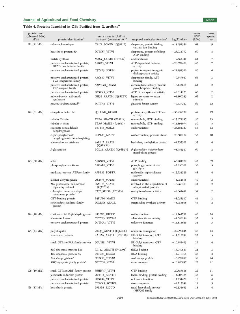
Table 4. Proteins Identified in OBs Purified from G. avellanaa
protein band(observed MW,
kDa) protein identificationbentry name in UniProt
databasec (accession no.)d supposed molecular functione log(E value)
meanMWf
(kDa)
massspectrano.
G1 (81 kDa) calnexin homologue CALX_SOYBN (Q39817) chaperone, protein folding,calcium ion binding
−14.698536 61 9
heat shock protein 60 D7TS57_VITVI chaperone, protein refolding,ATP binding
−23.934792 60 8
malate synthase MASY_GOSHI (P17432) acyltransferase −9.662341 64 7putative uncharacterized protein,DEAD box helicase family
A5BI23_VITVI ATP-dependent helicaseactivity
−20.697400 46 7
putative uncharacterized protein C5Z6P5_SORBI proton transport, inorganicdiphosphatase activity
−21.991360 80 6
putative uncharacterized protein,TCP chaperonin family
A5C537_VITVI chaperonin family, ATPbinding
−9.347947 63 3
putative uncharacterized protein,TPP enzyme family
A2WKY8_ORYSI carboxy-lyase activity, thiaminpyrophosphate binding
−5.142668 64 2
putative uncharacterized protein D7SYK8_VITVI ATP citrate synthase activity −8.014125 66 2indole-3-acetic acid-amidosynthetase
GH32_ARATH (Q9SZT9) ligase, response to auxinstimulus
−4.800245 62 2
putative uncharacterizedg D7TYA2_VITVI glycerone kinase activity −9.337242 62 12
G2 (61 kDa) elongation factor 1-α Q3LUM2_GOSHI protein biosynthesis, GTPaseactivity
−36.939730 49 19
tubulin β chain TBB6_ARATH (P29514) microtubule, GTP binding −23.678587 50 13tubulin α chain TBA6_MAIZE (P33627) microtubule, GTP binding −14.894074 50 8succinate semialdehydedehydrogenase
B6TPI6_MAIZE oxidoreductase −28.181347 56 12
6-phosphogluconatedehydrogenase, decarboxylating
C0PL33_MAIZE oxidoreductase, pentose shunt −20.387102 53 10
adenosylhomocysteinase SAHH2_ARATH(Q9LK36)
hydrolase, methylation control −9.233361 53 4
β-glucosidase BGL23_ARATH (Q9SR37) β-glucosidase, carbohydratemetabolism process
−8.782517 60 2
G3 (50 kDa) actin A5BN09_VITVI ATP binding −65.784770 42 39phosphoglycerate kinase A5CAF6_VITVI phosphoglycerate kinase,
glycolysis−7.856361 50 5
predicted protein, ATPase family A9PB39_POPTR nucleoside triphosphataseactivity
−12.934329 45 4
alcohol dehydrogenase O82478_SOYBN oxidoreductase −4.951558 40 326S proteasome non-ATPaseregulatory subunit
PSMD6_ARATH(Q93Y35)
involved in the degradation ofubiquitinated proteins
−8.765483 44 2
chloroplast inner envelopemembrane protein
IN37_SPIOL (P23525) methyltransferase activity −8.061481 39 2
GTP-binding protein B4FUE0_MAIZE GTP binding −5.055517 44 2strictosidine synthase familyprotein
D7M0S8_ARALL strictosidine synthase activity −8.958608 44 2
G4 (40 kDa) corticosteroid 11-β-dehydrogenase B9SPX3_RICCO oxidoreductase −19.161781 40 24adenosine kinase C6T7F3_SOYBN adenosine kinase activity −8.086186 37 3putative uncharacterized protein D7THA1_VITVI unknown function −11.815649 35 3
G5 (23 kDa) polyubiquitin UBQ8_ARATH (Q39256) ubiquitin conjugation −27.797846 28 14Ras-related protein RAD2A_ARATH (P28188) ER-Golgi transport, GTP
binding−14.315298 23 5
small GTPase/SAR family protein D7U2H1_VITVI ER-Golgi transport, GTPbinding
−10.982425 22 4
60S ribosomal protein L11 RL112_ARATH (P42794) tRNA binding −13.949543 21 340S ribosomal protein S5 B9T025_RICCO RNA binding −12.917358 23 311S storage globuling O82437_COFAR seed storage protein −6.795880 55 18MIP/aquaporin family proteing D7T7C6_VITVI water transport −16.886057 27 11
G6 (19 kDa) small GTPase/ARF family protein F6HHV7_VITVI GTP binding −20.585516 22 11jasmonate inducible protein O04314_ARATH lectin binding, protein folding −14.703335 32 6putative uncharacterized protein D7SY44_VITVI unknown function −11.734428 18 4putative uncharacterized protein C6SVX3_SOYBN stress response −8.213248 18 3
G8 (17 kDa) heat-shock protein B9S3B3_RICCO small heat-shock protein(HSP20) family
−6.531653 18 4
Journal of Agricultural and Food Chemistry Article
dx.doi.org/10.1021/jf301390d | J. Agric. Food Chem. 2012, 60, 6994−70047001

enzymes, especially oxidoreductase, kinase, and hydrolase, wereassociated with OBs-G. One oxidoreductase that wasparticularly abundant because it was identified with 24 massspectra, displayed similarity with corticosteroid 11β-dehydro-genase. This enzyme, comprising an oil body-anchoringsegment, NADPH-binding subdomain, active site, and sterol-binding subdomain, exists in seed OBs of diverse species, whereit is often called steroleosin, and could be involved in sterolmetabolism and in diverse signal transductions.32 In the case ofG. avellana steroleosin, the highly conserved nucleotide bindingsite was identified. Another membrane protein associated toOBs-G was an aquaporin belonging to the MIP family andidentified by de novo sequencing.Proteins displaying highly conserved sequences in the plant
kingdom could be identified despite the fact that the genomesof G. avellana and M. sativa are still not sequenced. It appearedthat among the integral OB proteins, steroleosins andaquaporins are likely the most conserved proteins. By contrast,oleosins, which have been described in many plant species,were not identified by mass spectrometry. In these proteins, thecentral hydrophobic region is highly conserved,19 but containedno trypsin cleavage site, whereas N- and C-terminal endsdisplay variations even within a protein family. The high varietyof OB-associated proteins identified here may reflect the in vivointeractions of OBs with glyoxysomes, protein storage vacuoles,small Golgi vesicles, mitochondria, cytoskeleton, and plasmamembrane,16,18,45 thus reflecting collaboration between organ-elles.46
Immunological Methods Reveal the Presence ofOleosins. Proteins from G. avellana and M. sativa purifiedseed OBs were separated by SDS-PAGE, transferred onto aPVDF membrane, and submitted to Western blotting usingantibodies raised against S2, S3, and S4 oleosins from A.thaliana (Figure 5). It was observed that all of these specificantibodies cross-reacted with OBs-G proteins. Immunoreactionwas particularly high with anti-rS2 and very low, as two faintbands, with anti-rS4. In the case of M. sativa, cross-reactivitywas similar for anti-rS2 and anti-rS3 and lower than thatobserved for G. avellana. No reactivity was observed with anti-rS4 despite a very long time of exposure. These results indicatethat oil bodies from M. sativa and G. avellana seeds containsome proteins recognized by antibodies raised against A.thaliana oleosin. These proteins must display some structuralhomology with oleosins to immunoreact with antibodies, butnot enough sequence homology to be identified by massspectrometry. Immunoblots indicated that molecular masses ofproteins immunologically related to A. thaliana oleosins werevery close, ranging from 15 to 18 kDa, with the S3-relatedprotein being lighter and the S4-related protein being heavier.Oleosins have been classified as high or low Mr isoforms (H-and L-oleosin, respectively) depending on their relativemolecular masses,47 with the A. thaliana S3 oleosin belongingto the L-form and A. thaliana S2 and S4 belonging to the H-form. H- and L-oleosins are immunologically distinct;2 bothforms could coexist in Madia and Gevuina seeds. High-levelimmunoreactions detected with OBs-G indicated that structural
proteins homologous to oleosins were the major proteincomponent of these OBs. In contrast, SDS data (Figures 4 and5) indicated that OBs-M contained some proteins homologousto oleosins and many other proteins. This result supported thefact that a thick coat surrounding OBs-M was visible bymicroscopy.
Effect of Oil Body on HUVEC Viability. OBs have beenexploited as therapeutic, diagnostic, and delivery agents offeringseveral advantages over synthetic carriers such as reduction ofcollateral effects and toxicity due to their natural origin.However, it is necessary to certify that OBs maintain theirsafety after the extraction processing from seeds for their use infood or pharmaceutical applications. For this reason, an in vitrocytotoxicity test48,49 using trypan blue50 was performed in cellsto evaluate the potential toxicity of OBs. HUVEC are relativelyeasy to culture and provide a valuable cell model for manybiology research applications. In viable cells, trypan blue is notabsorbed, whereas it passes through the membrane in deadcells. As shown in Figure 6, no trypan blue staining wasobserved for HUVEC incubated with the OBs, indicating thatcell viability was not compromised. These results are promisingfor possible uses of OBs as a delivery carrier or functionaladditive.Furthermore, in this first study of the isolation and
characterization of OBs from G. avellana and M. sativa, theresults showed that the seeds are sources of proteins andhealthy and nutritive oils very rich in unsaturated fatty acidssuch as oleic acid (omega 9) in G. avellana and linoleic acid(omega 6) in M. sativa. The quality of oils is related to the lipidoxidation during storage and/or food processing. One approach
Table 4. continued
aBand number refers to bands in Figure 4. bProtein identification was performed using X!Tandem and sequence homology in the UniProt databaserestricted to spermatophyta. cEntry name in UniProt database of the protein displaying the best sequence homology. dAccession number in Swiss-Prot if one exists. ePossible function reported by UniProt/Swiss-Prot. fMean molecular mass calculated from all proteins grouped by a similarfunction as explained under Materials and Methods. gProteins in italics were identified using PepNovo software.
Figure 5. Immunological detection of OB proteins from M. sativa (M)or G. avellana (G) seeds with antibodies raised against oleosins S2, S3,and S4 from A. thaliana. Proteins (5 μg) from purified OBs wereresolved by SDS-PAGE before immunoblot analysis. The blots wereprobed with anti-rS2 (1:5000 dilution), anti-rS3 (1:4000), and anti-rS4(1:2000) sera. Detection was performed using chemiluminescence.Exposure time was 6 min for anti-rS2 and anti-rS3 and 10 min for anti-rS4. Molecular masses are given in kDa using MagicMark (MM)protein standard. The panel on the left is the Coomassie-blue stainedprotein gel (10 or 5 μg of protein) with Mark 12 (M12) as molecularmass marker.
Journal of Agricultural and Food Chemistry Article
dx.doi.org/10.1021/jf301390d | J. Agric. Food Chem. 2012, 60, 6994−70047002

to protect lipid from oxidation is through microencapsulationusing a polymeric matrix, which has been widely used in thefood industry. OBs may be considered as a natural protectionsystem against the oxidation of fatty acids because they safelystore lipids in seeds in the form of TAGs for long periods, evenunder unfavorable conditions (drying, rehydration, low or hightemperature, etc.). For this reason, the incorporation ofundisrupted oil bodies into functional foods may preventlipid deterioration.3,4 In this study, it was demonstrated thatOBs from G. avellana and in particular from M. sativa representa stable and natural emulsion system under a wide range of pHand ionic strengths, thus offering a natural alternative forincorporating them into food emulsion systems. Moreover,these isolated OBs did not cause any cytotoxicity in humancells. Therefore, OBs of these native seeds may be useful for thedevelopment of safe and efficient delivery carriers of bioactivemolecules for food and/or pharmaceutical purposes.
■ ASSOCIATED CONTENT*S Supporting InformationX! Tandem and PepNovo identification of OB proteins from G.avellana and M. sativa seeds. This material is available free ofcharge via the Internet at http://pubs.acs.org.
■ AUTHOR INFORMATIONCorresponding Author*Phone: 56-45-325050. Fax: 56-45-325053. E-mail: [email protected].
FundingThis research was supported by funding from Conicyt throughFondecyt Project 3120022 and Project DI11-7001 and GAPtechnical support provided by the Research Office at theUniversidad de La Frontera.
NotesThe authors declare no competing financial interest.
■ ABBREVIATIONS USEDOBs, oil bodies; OBs-G, oil bodies from Gevuina avellana; OBs-M, oil bodies from Madia sativa; TAG, triacylglycerol; HEPES,4-(2-hydroxyethyl)-1-piperazineethanesulfonic acid; TEM,transmission electron microscopy; FAMEs, fatty acid methylesters; GC, gas chromatography; SDS-PAGE, sodium dodecylsulfate−polyacrylamide gel electrophoresis; HPLC-MS/MS,liquid chromatography−tandem mass spectrometry; T, turbid-ity; ANOVA, analysis of variance; HUVEC, human umbilicalvein endothelial cells; Kst, stability constant.
■ REFERENCES(1) Huang, A. H. Oil bodies and oleosins in seeds. Annu. Rev. PlantBiol. 1992, 43, 177−200.(2) Huang, A. H. Oleosins and oil bodies in seeds and other organs.Plant Physiol. 1996, 110, 2063−2069.(3) Iwanaga, D.; Gray, D. A.; Fisk, I. D.; Decker, E. A.; Weiss, J.;McClements, D. J. Extraction and characterization of oil bodies fromsoy beans: a natural source of pre-emulsified soybean oil. J. Agric. FoodChem. 2007, 55, 8711−8716.(4) White, D. A.; Fisk, I. D.; Mitchell, J. R.; Wolf, B.; Hill, S. E.; Gray,D. A. Sunflower-seed oil body emulsions: rheology and stabilityassessment of a natural emulsion. Food Hydrocolloids 2008, 22, 1224−1232.(5) Beisson, F.; Ferte, N.; Bruley, S.; Voultoury, R.; Verger, R.;Arondel, V. Oil-bodies as substrates for lipolytic enzymes. Biochim.Biophys Acta 2001, 1531, 47−58.(6) White, D. A.; Fisk, I. D.; Makkhun, S.; Gray, D. A. In vitroassessment of the bioaccessibility of tocopherol and fatty acids fromsunflower seed oil bodies. J. Agric. Food Chem. 2009, 57, 5720−5726.(7) Delgado-Vargas, F.; Paredes-Lopez, O. Natural Colorants for Foodand Nutraceutical Uses; CRC Press: Boca Raton, FL, 2003; pp 113−166 and 257−309.(8) Deckers, H. M.; van Rooijen, G.; Boothe, J., et al. SembiosysGenetics Inc. Products for topical applications comprising oil bodies.U.S. Patent 6582710, 2003.(9) Deckers, H. M.; Van Rooijen, G.; Boothe, J.; Goll, J.; Moloney,M. M.; Schryvers, A. B. et al. Sembiosys genetics inc. immunogenicformulations comprising oil bodies. U.S. Patent 6761914, 2004.(10) Moloney, M. M. Sembiosys Genetics Inc. Oil-body proteins ascarriers of high-value peptides in plants. U.S. Patent 5650554, 1997.(11) Boucher, J.; Cengelli, F.; Trumbic, D.; Marison, I. W. Sorptionof hydrophobic organic compounds (HOC) in rapeseed oil bodies.Chemosphere 2008, 70, 1452−1458.(12) Fisk, I. D.; Linforth, S. T.; Taylor, A. J.; Gray, D. A. Aromaencapsulation and aroma delivery by oil body suspensions derivedfrom sunflower seeds (Helianthus annus). Eur. Food Res. Technol. 2011,232, 905−910.(13) Hou, R. C. W.; Lin, M. Y.; Wang, M. M. C.; Tzen, J. T. C.Increase of viability of entrapped cells of Lactobacillus delbrueckii ssp.bulgaricus in artificial sesame oil emulsions. J. Dairy Sci. 2003, 86,424−428.(14) Jolivet, P.; Roux, E.; d’Andrea, S.; Davanture, M.; Negroni, L.;Zivy, M.; Chardot, T. Protein composition of oil bodies in Arabidopsisthaliana ecotype WS. Plant Physiol. Biochem. 2004, 42, 501−509.(15) Popluechai, S.; Froissard, M.; Jolivet, P.; Breviario, D.;Gatehouse, A. M. R.; Donnell, A. G. O.; Chardot, T.; Kohli, A.Jatropha curcas oil body proteome and oleosins: L-form JcOle3 as apotential phylogenetic marker. Plant Physiol. Biochem. 2011, 49, 352−356.(16) Tnani, H.; Lopez, I.; Jouenne, T.; Vicient, C. M. Proteincomposition analysis of oil bodies from maize embryos duringgermination. J. Plant Physiol. 2011, 168, 510−513.(17) Cummins, I.; Murphy, D. J. cDNA sequence of a sunfloweroleosin and transcript tissue specificity. Plant Mol. Biol. 1992, 19, 873−876.(18) Katavic, V.; Agrawal, G. K.; Hajduch, M.; Harris, S. L.; Thelen, J.J. Protein and lipid composition analysis of oil bodies from twoBrassica napus cultivars. Proteomics 2006, 6, 4586−4598.(19) Jolivet, P.; Boulard, C.; Bellamy, A.; Larre, C.; Barre, M.;Rogniaux, H.; d’Andrea, S.; Chardot, T.; Nesi, N. Protein compositionof oil bodies from mature Brassica napus seeds. Proteomics 2009, 9,3268−3284.(20) Kalinski, A.; Loer, D. S.; Weisemann, J. M.; Matthews, B. F.;Herman, E. M. Isoforms of soybean seed oil body membrane protein24 kDa oleosin are encoded by closely related cDNAs. Plant Mol. Biol.1991, 17, 1095−1098.(21) Bertoli, C.; Fay, L. B.; Stancanelli, M.; Gumy, D.; Lambelet, P.Characterization of Chilean hazelnut (Gevuina avellana Mol) seed oil.J. Am. Oil Chem. Soc. 1988, 75, 1037−1040.
Figure 6. Effect of oil bodies from G. avellana (A) and M. sativa (B)on HUVEC viability. The images represent HUVEC with oil bodiesusing optical microscopy.
Journal of Agricultural and Food Chemistry Article
dx.doi.org/10.1021/jf301390d | J. Agric. Food Chem. 2012, 60, 6994−70047003

(22) Schmeda-Hisrchmann, G. Madia sativa, a potential oil crop ofcentral Chile. Econ. Bot. 1995, 49, 257−259.(23) Ibaca, R. Monografia de Arboles y Arbustos Chilenos conPropiedades Medicinales y Aromaticas; Facultad de Ciencias Forestales,Universidad de Concepcion: Concepcion, Chile, 2001; p 246(24) Facciola, S. Cornupia-un libro de la Fuente de Plantas Comestibles;Publicaciones de Kampong, 1990; ISBN 0-9628087-0-9.(25) Jolivet, P.; Boulard, C.; Bellamy, A.; Valot, B.; d'Andrea, S.; Zivy,M.; Nesi, N.; Chardot, T. Oil body proteins sequentially accumulatethroughout seed development in Brassica napus. J. Plant Physiol. 2011,168, 2015−2020.(26) Allen, D. K.; Tao, B. Y. Kinetic characterization of enhancedlipid activity on oil bodies. Bioprocess. Biosyst. Eng. 2007, 30, 271−279.(27) Zhuang, X.; Tlalka, M.; Davies, D. S.; Allaway, W. G.;Watkinson, S. C.; Ashford, A. E. Spitzenkorper, vacuoles, ring-likestructures, and mitochondria of Phanerochaete velutina hyphal tipsvisualized with carboxy-DFFDA, CMAC and DiOC6(3). Mycol. Res.2009, 113, 417−431.(28) Folch, J.; Lees, M.; Sloane Stanley, G. H. A simple method forthe isolation and purification of total lipids from animal tissues. J. Biol.Chem. 1957, 226, 497−509.(29) Landry, J.; Delhaye, S. A simple and rapid procedure forhydrolyzing minute amounts of proteins with alcali. Anal. Biochem.1996, 243, 191−194.(30) Laemmli, U. K. Cleavage of structural proteins during theassembly of the head of bacteriophage T4. Nature 1970, 227, 680−685.(31) Neuhoff, V.; Arold, N.; Taube, D.; Ehrhardt, W. Improvedstaining of proteins in polyacrylamide gels including isoelectricfocusing gels with clear background at nanogram sensitivity usingCoomasie Brilliant Blue G-250 and R-250. Electrophoresis 1988, 9,255−262.(32) d’Andrea, S.; Canonge, M.; Beopoulos, A.; Jolivet, P.;Hartmann, M. A.; Miquel, M.; Lepiniec, L.; Chardot, T. At5g50600encodes a member of the short-chain dehydrogenase reductasesuperfamily with 11β- and 17β-hydroxysteroid dehydrogenaseactivities associated with Arabidopsis thaliana seed oil bodies. Biochimie2007, 89, 222−229.(33) Tzen, J. T. C.; Huang, A. H. C. Surface structure and propertiesof plant seed oil bodies. J. Cell Biol. 1992, 117, 327−335.(34) Jaffe, E. A.; Nachman, R. L.; Becker, C. G.; Minic, C. R. Cultureof human endothelial cells derived from umbilical veins. J. Clin. Invest.1973, 52, 2745−2756.(35) Childers, N. K.; Michalek, S. M.; Eldridge, J. H.; Denys, F. R.;Berry, A. K.; McGhee, J. R. Characterization of liposome suspensionsby flow cytometry. J. Immunol. Methods 1989, 119, 135−143.(36) Macey, M. G. Flow Cytometry. Principles and Applications; Macey,M. G., Ed.; Humana Press: Totowa, NJ, 2007; pp 290.(37) Plattner, R. D.; Kleiman, R. Grevillea robusta seed oil: a source ofω-5 monoenes. Phytochemistry 1977, 16, 255−256.(38) Vickery, J. R. The fatty acid composition of the seed oils ofProteaceae: a chemotaxonomic study. Phytochemistry 1971, 10, 123−130.(39) Bombarda, I.; Zongo, C.; McGill, C. R.; Doumenq, P.; Fogliani,B. Fatty acids profile of Alphitonia neocaledonica and Grevillea exul var.rubiginosa seed oils, occurrence of an ω5 series. J. Am. Oil Chem. Soc.2010, 87, 981−986.(40) Frandsen, G.; Mundy, J.; Tzen, T. C. Oil bodies and theirassociated proteins, oleosin and caleosin. Physiol. Plant. 2007, 112, 301.(41) Tzen, J. T. C.; Peng, C. C.; Cheng, D. J.; Chen, E. C. F.; Chiu, J.M. H. A new method for seed oil body purification and examination ofoil body integrity following germination. J. Biochem. 1997, 121, 762−768.(42) Fujimoto, Y.; Itabe, H.; Sakai, J.; Makita, M.; Noda, J.; Mori, M.;Higashi, Y.; Kojima, S.; Takano, T. Identification of major proteins inthe lipid droplet-enriched fraction isolated from the human hepatocytecell line HuH7. Biochim. Biophys. Acta 2004, 1644, 47−59.(43) Athenstaedt, K.; Jolivet, P.; Boulard, C.; Negroni, L.; Zivy, M.;Nicaud, J.-M.; Chardot, T. Lipid particle composition of the yeast
Yarrowia lipolytica depends on the carbon source. Proteomics 2006, 6,1450−1459.(44) Maurel, C.; Verdoucq, L.; Luu, D. T.; Santoni, V. Plantaquaporins: membrane channels with multiple integrated functions.Annu. Rev. Plant Biol. 2008, 59, 595−624.(45) Marmagne, A.; Ferro, M.; Meinnel, T.; Bruley, C.; Kuhn, L.;Garin, J.; Barbier-Brygoo, H.; Ephritikhine, G. A high content in lipid-modified peripheral proteins and integral receptor kinases features inthe arabidopsis plasma membrane proteome. Mol. Cell. Proteomics2007, 6, 1980−1996.(46) Agrawal, G. K.; Bourguignon, J.; Rolland, N.; Ephritikhine, G.;Ferro, M.; Jaquinod, M.; Alexiou, K. G.; Chardot, T.; Chakraborty, N.;Jolivet, P.; Doonan, J. H.; Rakwal, R. Plant organelle proteomics:collaborating for optimal cell function. Mass Spectrom. Rev. 2011, 30,772−853.(47) Tzen, J. T.; Lai, Y. K.; Chan, K. L.; Huang, A. H. Oleosinisoforms of high and low molecular weights are present in the oilbodies of diverse seed species. Plant Physiol. 1990, 94, 1282−1289.(48) Ridolfi, D. M.; Marcato, P. D.; Machato, D.; Silva, R. A.; Justo,G. Z.; Duran, N. In vitro cytotoxicity assays of solid lipid nanoparticlesin epitelial and dermal cells. J. Physics: Conf. Ser. 2011, 304, 012032.(49) Sini, K. R.; Haribabu, Y.; Sajoth, M. S.; Surya Sreekumar, K. Invitro cytotoxic activity of orthosophon thymif lrus Roth, sleensen leadextract against Dalton lymphoma ascites cell lines. J. Chem. Pharm. Res.2012, 4, 917−921.(50) Palama, I. E.; Muraso, M.; Coluucia, A.; D'Amone, S.; Giglil, G.Cell uptake and validation of novel PECs for biomedical applications.J. Drug Delivery 2011, 1−7, DOI: 10.1155/2011/203676.
Journal of Agricultural and Food Chemistry Article
dx.doi.org/10.1021/jf301390d | J. Agric. Food Chem. 2012, 60, 6994−70047004