World Journal of Clinical Pediatrics - BPG Management System
Upload
khangminh22Category
view
0download
0
Published by Baishideng Publishing Group Inc
World Journal of DiabetesWorld J Diabetes 2015 June 10; 6(5): 673-773
ISSN 1948-9358 (online)
EDITOR-IN-CHIEFLu Qi, BostonJingbo Zhao, Aalborg
STRATEGY ASSOCIATE EDITOR-IN-CHIEFUndurti Narasimha Das, Shaker HeightsMin Du, LaramieGregory I Liou, AugustaZhong-Cheng Luo, QuebecDemosthenes B Panagiotakos, Athens
GUEST EDITORIAL BOARD MEMBERSJuei-Tang Cheng, TainanChih-Hsung Chu, KaohsiungLow-Tone (Larry) Ho, TaipeiCheng-Cheng Hsiao, KeelungYung-Hsi Kao, TaoyuanChi Feng Liu, TaipeiShing-Hwa Liu, TaipeiWayne H-H Sheu, TaichungEing-Mei Tsai, KaohsiungChin-Hsiao Tseng, TaipeiYen Tzung-Hai, TaipeiChing-Shuang Wu, KaohsiungWei-Chung Vivian Yang, TaipeiWen-Chin Yang, Taipei
MEMBERS OF THE EDITORIAL BOARD
Argentina
Justo P Castaño, CordobaEduardo Spinedi, La Plata
Australia
Sof Andrikopoulos, Heidelberg HeightsHugh Russell Barrett, PerthBernhard T Baune, TownsvilleGrant Brinkworth, AdelaideLouise Janet Maple Brown, CasuarinaMelinda Therese Coughlan, MelbourneJosephine Maree Forbes, MelbournePaul A Fournier, PerthAngela Gialamas, Adelaide Mark Douglas Gorrell, NewtownGraeme Hankey, PerthAnandwardhan A Hardikar, MelbourneMichael Horowitz, AdelaideKarin Jandeleit-Dahm, MelbourneMartha Lappas, VictoriaPeter J Little, MelbourneXin Liu, BrisbaneDianna Josephine Magliano, CaufieldRobyn McDermott, AdelaideBeverly Sara Muhlhausler, AdelaideChristopher Nolan, CanberraLuciano Pirola, MelbourneMaryam Rashidi, VictoriaKarly Calliopi Sourris, VictoriaGreg Tesch, ClaytonJack Ronald Wall, PenrithOwen Llewellyn Woodman, Bundoora
AustriaChristian Heinz Anderwald, ViennaHelmuth Martin Borkenstein, GrazWalter Hermann Hörl, ViennaAlexandra Kautzky-Willer, Vienna
Friedrich Mittermayer, ViennaMarkus Paulmichl, SalzburgStefan Pilz, GrazGuntram Schernthaner, ViennaHarald Sourij, GrazThomas Michael Stulnig, ViennaLudwig Wagner, Vienna
Belgium
Giovanni Dapri, BrusselsChristophe De Block, AntwerpEkaterine Tskitishvili, LiegeF Andre Van Assche, LeuvenLuc F Van Gaal, Antwerp
Brazil
Monica Levy Andersen, Vila ClementinoClaudia RL Cardoso, Rio de JaneiroRicardo Vitor Cohen, São PauloMarcelo Correia, Rio de JaneiroCassyano Januario Correr, CuritibaMatheus Roriz Cruz, Porto AlegreCintia Chaves Curioni, Rio de JaneiroFreddy Goldberg Eliaschewitz, Rua GoiásRodrigo Jorge, Ribeirão PretoLuciana Ansaneli Naves, Asa NorteJúlio César Voltarelli, Ribeirão PretoBernardo L Wajchenberg, PinheirosJacqueline Nelisis Zanoni, Maringá
Canada
Jean-Luc Ardilouze, Sherbrooke
I
Editorial Board2011-2015
The World Journal of Diabetes Editorial Board now consists of 712 members, representing a team of worldwide experts in diabetes mellitus. They are from 56 countries, including Argentina (2), Australia (27), Austria (11), Belgium (5), Brazil (13), Canada (25), Chile (3), China (40), Cuba (1), Czech Republic (3), Denmark (16), Egypt (3), Finland (5), France (12), Germany (27), Greece (17), Hungary (4), India (28), Iran (8), Iraq (2), Ireland (3), Israel (10), Italy (56), Japan (30), Jordan (1), Kuwait (3), Lebanon (1), Malaysia (1), Malta (1), Mexico (4), Netherlands (9), New Zealand (3), Nigeria (2), Norway (2), Oman (3), Pakistan (2), Poland (7), Portugal (1), Qatar (1), Romania (2), Saudi Arabia (1), Singapore (4), Slovakia (1), South Africa (1), South Korea (15), Spain (24), Sweden (5), Switzerland (4), Thailand (4), Tunisia (1), Turkey (13), United Arab Emirates (3), United Kingdom (27), United States (213), Venezuela (1), and Yemen (1).
December 11, 2012WJD|www.wjgnet.com
World Journal of DiabetesW J D

Subrata Chakrabarti, LondonDavid Cherney, OntarioMervyn Deitel, TorontoJean-Pierre Després, QuebecDavid Joseph Hill, LondonTian-Ru Jin, TorontoArulmozhi D Kandasamy, EdmontonJennifer L Kuk, TorontoIsmail Laher, VancouverRoger S McIntyre, TorontoDavid Meyre, OntarioJoseph Fomusi Ndisang, SaskatoonRaj Padwal, AlbertaCiriaco A Piccirillo, MontrealRemi Rabasa-Lhoret, MontrealAM James Shapiro, EdmontonGuang Sun, St. John’sValerie Taylor, HamiltonCory Toth, CalgaryAndré Tremblay, MontréalVincent C Woo, WinnipegJames Roscoe Wright, CalgaryXi-Long Zheng, Calgary
Chile
Sebastian San Martin, ValparaisoArmando Rojas-Rubio, TalcaLuis Sobrevia, Santiago
China
Pang-Zeng Chang, QingdaoJie Chen, NanjingBernard Man Yung Cheung, Hong KongWilliam Chi-shing Cho, Hong KongTian-Pei Hong, BeijingQin Huang, ShanghaiPo Sing Leung, Hong KongChao Liu, NanjingJian-Kang Liu, Xi’anLie-Gang Liu, WuhanRonald Ching Wan Ma, Hong KongJin-Sheng Qi, ShijiazhuangWing Yee So, Hong KongCheuk Chun Szeto, Hong KongKathryn Tan, Hong KongCheng-Ming Wang, YangzhouCong-Yi Wang, WuhanYu Wang, Hong KongGuang-Da Xiang, WuhanBao-Feng Yang, HarbinShu-Yu Yang, FujianXi-Lin Yang, Hong KongZai-Qing Yang, WuhanShan-Dong Ye, HefeiShi-Sheng Zhou, DalianZhi-Guang Zhou, Changsha
Cuba
Luis Sarmiento-Pérez, Havana
Czech Republic
Martin Haluzik, Prague
Michal Krcma, PlzenTerezie Pelikanova, Prague
Denmark
Charlotte Brøns, GentofteJens Sandahl Christiansen, ArhusFlemming Dela, CopenhagenKristine Færch, GentofteErik L Grove, AarhusLouise Groth Grunnet, GentofteR Scott Heller, GentofteKurt Højlund, Odense CFilip K Knop, HellerupHelle Markholst, MåløvJens D Mikkelsen, CopenhagenOle Hartvig Mortensen, CopenhagenOluf Pedersen, CopenhagenEsben Thyssen Vestergaard, AarhusMilan Zdravkovic, Søborg
Egypt
Mamdouh Moawad Ali Hssan, CairoMoshira Abdel Hakim Rateb, CairoMona Farag Schaalan, Cairo
Finland
Siamak Bidel, HelsinkiGang Hu, HelsinkiThomas Kietzmann, OuluQing Qiao, HelsinkiKaroliina Wehkalampi, Helsinki
France
Jean Claude Ansquer, DijonBertrand Cariou, NantesSylvie Dejager, Rueil-MalmaisonNaim Akhtar Khan, DijonJean-Philippe Lavigne, NîmesMichel Marre, ParisMarie-Claude Morice, MassyRiccardo Perfetti, ParisGérard Said, ParisSophie Visvikis Siest, NancyDominique Simon, ParisDidier Vieau, Villeneuve d’Ascq
Germany
Ioanna Gouni Berthold, CologneChrista Buechler, RegensburgRoland Büttner, HeidelbergMichael Froehner, DresdenHammes Hans-Peter, MannheimNadj Herbach, MunichAndrea Icks, DüsseldorfThomas Jax, NeussUlrich Arthur Julius, DresdenMichael Kluge, MunichFlorian Lang, Tuebingen Matthias Laudes, KölnRalf Lobmann, Stuttgart
Rafael T Mikolajczyk, BremenAndreas Stefan Mueller, Halle (Saale)Karsten Müssig, TübingenNahid Parvizi, Neustadt am RübenbergeThomas Peter Reinehr, DattelnMichael Ristow, JenaSven Schinner, DuesseldorfPeter Egbert Hermann Schwarz, DresdenKonstantinos Stellos, TubingenOvidiu Alin Stirban, Bad OeynhausenDiego J Walther, BerlinSilvia Anette Wein, KielChristian Wrede, BerlinDan Ziegler, Düsseldorf
Greece
George P Chrousos, AthensMoses S Elisaf, IoanninaPanagiotis Georgoulias, LarissaNikolaos Kadoglou, ThessalonikiGerasimos E Krassas, KriniSpilios Manolakopoulos, AttikiNikolaos Papanas, AlexandroupolisDimitrios Papazoglou, AlexandroupolisSokratis Pastromas, AthensMelpomeni Peppa, AthensChristina Piperi, GoudiNicholas K Tentolouris, AthensKonstantinos A Toulis, SalonikaApostolos Tsapas, ThessalonikiKonstantinos Tziomalos, ThessalonikiElias Zintzaras, Thessaly
Hungary
Mária Bagyánszki, SzegedGyörgy Jermendy, BudapestKaroly Racz, BudapestGyula Soltesz, Pécs
India
Deepak Narayan Amrapurkar, MumbaiC V Anuradha, Tamil NaduSarika Arora, New DelhiPitchai Balakumar, SivakasiMuthuswamy Balasubramanyam, ChennaiSubhabrata Chakrabarti, HyderabadAbhay Sankar Chakraborti, KolkataTapan K Chaudhuri, New DelhiKanwaljit Chopra, ChandigarhMalabika Datta, DelhiDebidas Ghosh, West BengalRavinder Goswami, New DelhiPappachan M Joseph, KeralaJothydev Kesavadev, KeralaKVS Hari Kumar, LucknowAnoop Misra, New DelhiAnalava Mitra, KharagpurViswanathan Mohan, ChennaiS P Murthy, BangalorePallavi Panchu, GunturUsharani Pingali, HyderabadAmbady Ramachandran, Egmore ChennaiVadde Ramakrishna, Kadapa
II December 11, 2012WJD|www.wjgnet.com

III December 11, 2012WJD|www.wjgnet.com
Geetha Vani Rayasam, HaryanaRajat Sandhir, ChandigarhManju Sharma, New DelhiSuman Bala Sharma, DelhiTarun Sharma, Chennai
Iran
Mohammad Abdollahi, TehranMohammad Kazemi Arababadi, RafsanjanLeila Azadbakht, IsfahanHamid Baradaran, TehranBehrooz Broumand, TehranAhmad Esmaillzadeh, IsfahanMajid Ghayour-Mobarhan, Mashhad Mohsen Janghorbani, Isfahan
Iraq
Saad Abdul-Rahman Hussain, BaghdadAbbas Ali Mansour, Basrah
Ireland
Amar Agha, DublinMark Philip Hehir, DublinGerald H Tomkin, Dublin
Israel
Michael Aviram, HaifaGal Dubnov-Raz, Tel HashomerShimon Efrat, Tel AvivRaymond Elias Farah, SafedOren Froy, RehovotSaher Hamed, HaifaArid Nakhoul, HaifaOrit Pinhas-Hamiel, Tel HashomerHaim Werner, Tel AvivMarina Shargorodsky Zimlichman, Holon
Italy
Luigi Angrisani, NapoliMoschetta Antonio, BariAntonio Aversa, RomeRoberto Baldelli, RomeGiuseppe Barbaro, RomeAlessandro Bartolomucci, ParmaGiuseppina Basta, PisaSimona Bertoli, MilanoFederico Bilotta, RomeFabio Broglio, TorinoFrancesco G Chiarelli, ChietiSergio Coccheri, BolognaMassimo Collino, TorinoMarco Aristide Comaschi, GenoaRenzo Cordera, GenovaFrancesco Dotta, SienaGagliardini Elena, BergamoStefano Fiorucci, PerugiaMaurizio Galderisi, NaplesAmalia Gastaldelli, Pisa
Ezio Ghigo, TurinCarla Giordano, PalermoPaolo Gisondi, VeronaRiccarda Granata, TurinGiorgio Iervasi, PisaClaudia Kusmic, PisaCarmelo La Rosa, CataniaFrancesco Landi, RomeMonica Rosa Loizzo, Arcavacata RendePaolo Magni, MilanoMariano Malaguarnera, CataniaMelania Manco, RomePiero Marchetti, PisaMassimo Massi-Benedetti, PerugiaAntonio Nicolucci, ImbaroLucia Pacifico, RomeStefano Palomba, CatanzaroGiampaolo Papi, CarpiRenato Pasquali, BolognaPiermarco Piatti, MilanoDario Pitocco, RomeAntonio E Pontiroli, MilanoGiulio Marchesini Reggiani, BolognaGiuseppe Remuzzi, BergamoManfredi Rizzo, PalermoRaffaella Rosso, GenoaGiuseppe Schillaci, PerugiaLeonardo A Sechi, SassariImad Sheiban, TorinoCesare R Sirtori, MilanoGiovanni Tarantino, NaplesGiovanni Targher, VeronaDonadon Valter, PordenoneAlberto Verrotti, ChietiAndrea Viggiano, NapoliGianvincenzo Zuccotti, Milan
Japan
Masato Asahina, ChibaTakuya Awata, SaitamaYuichiro Eguchi, SagaGoji Hasegawa, KyotoSatoshi Inoue, TokyoEiji Ishimura, OsakaMasayuki Iwano, NaraTakashi Kadowaki, TokyoEisuke Kagawa, HiroshimaMasahito Katahira, AichiEiji Kawasaki, NagasakiNoriyuki Koibuchi, GunmaKazuhiko Kotani, TochigiDaisuke Koya, IshikawaNorikazu Maeda, OsakaTakayuki Masaki, OitaYuji Matsuzawa, OsakaKazuaki Nishio, TokyoKenji Okumura, NagoyaMotoaki Saito, YonagoToshiyasu Sasaoka, ToyamaMichio Shimabukuro, OkinawaKohzo Takebayashi, SaitamaHiroyuki Tamemoto, TochigiTakashi Togo, YokohamaJun Udagawa, IzumoYoshinari Uehara, FukuokaTakuya Watanabe, TokyoToshihiko Yada, Tochigi
Tohru Yorifuji, Osaka
Jordan
Yousef S Khader, Irbid
Kuwait
Kamal AA Sulaiman Al-Shoumer, KuwaitIbrahim Fadil Benter, SafatAbu Salim Mustafa, Kuwait
Lebanon
Ramzi F Sabra, Beirut
Malaysia
Mafauzy Mohamed, Kota Bharu
Malta
Charles Savona-Ventura, Msida
Mexico
Manuel González-Ortiz, GuadalajaraFernando Guerrero-Romero, DurangoJesus Alberto Olivares-Reyes, Mexico CityRocío Salceda, Mexico City
Netherlands
Sander Kersten, WageningenNanne Kleefstra, ZwolleEdwin Mariman, MaastrichtDon Poldermans, RotterdamFrançois Pouwer, TilburgHan Roelofsen, GroningenHendrik-Jan Schuurman, UtrechtSuat Simsek, AlkmaarMarcel Twickler, Bergen op Zoom
New Zealand
Paul Hofman, AucklandPeter E Lobie, AucklandElaine Rush, Auckland
Nigeria
Adejuwon A Adeneye, LagosAnthonia Okeoghene Ogbera, Lagos
Norway
Akhtar Hussain, OsloOdd Erik Johansen, Hovik

IV December 11, 2012WJD|www.wjgnet.com
Oman
Mohammed Al Shafaee, MuscatJumana S Saleh, MuscatRadha Shenoy, Muscat
Pakistan
Shahid Hameed, IslamabadJamil A Malik, Islamabad
Poland
Marcin Baranowski, BialystokJerzy Beltowski, LublinAlicia Hubalewska Dydejczyk, KrakowMaciej Owecki, PoznańEwa Pankowska, WarsawAgnieszka Piwowar, WroclawDorota Anna Zieba, Krakow
Portugal
M Graça Pereira, Braga
Qatar
Hong Ding, Doha
Romania
Elena Ganea, BucharestAdriana Georgescu, Bucharest
Saudi Arabia
J Fernando Arevalo, Caracas
Singapore
S Thameem Dheen, SingaporeYung Seng Lee, SingaporeDaniel Ng, SingaporeRob Martinus van Dam, Singapore
Slovakia
Katarína Šebeková, Bratislava
South Africa
Md Shahidul Islam, Durban
South Korea
Huneg-Sik Choi, GwangjuKyung Mook Choi, SeoulWon Mi Hwang, SeoulEui-Bae Jeung, Chungbuk
Ju-Hee Kang, IncheonSin Gon Kim, Seongbuk-GuSung-Jin Kim, SeoulYoung-Gyu Ko, SeoulKang-Beom Kwon, ChonbukMyung Gull Lee, BucheonSoo Lim, SeongnamByung-Hyun Park, JeonbukSeungjoon Park, SeoulKun-Ho Yoon, SeoulJeesuk Yu, Cheonan
Spain
Vivencio Barrios, MadridM Lusia Bonet, Palma de MallorcaManuel Vazquez Carrera, BarcelonaMaria Luz Martinez Chantar, DerioManuel Aguilar Diosdado, CádizJavier Espino, BadajozRicardo V García-Mayor, VigoJosé Manuel Gómez-Sáez, BarcelonaOreste Gualillo, Santiago de CompostelaJ Alfredo Martínez Hernández, PamplonaEmilio Herrera, MadridAmelia Marti, PamplonaMerce Miranda, TarragonaJF Navarro-González, Santa Cruz de TenerifeAlberto Ortiz, MadridMaria Javier Ramirez, PamplonaEugenia Resmini, BarcelonaPedro Romero-Aroca, ReusJordi Salas-Salvadó, ReusGines M Salido, CaceresVictor Sanchez-Margalet, SevilleHelmut Schröder, BarcelonaCarmen Segundo, CádizRafael Simó, Barcelona
Sweden
Joanna Hlebowicz, MalmöKaj S Stenlöf, GöteborgAnn-Britt Wiréhn, LinköpingWeili Xu, StockholmShao-Nian Yang, Stockholm
Switzerland
Kaspar Berneis, ZurichPascal Bovet, LausanneLuc Tappy, LausanneChristian Toso, Geneva
Thailand
Narattaphol Charoenphandhu, BangkokArthorn Riewpaiboon, BangkokRawee Teanpaisan, Hat-YaiViroj Wiwanitkit, Bangkok
Tunisia
Khaled Hamden, Sfax
Turkey
Ugur Cavlak, DenizliTeoman Dogru, AnkaraErsin Fadillioglu, AnkaraAbdurrahman Fatih Fidan, AfyonkarahisarMuammer Karadeniz, Bornova-IzmirCevdet Kaya, IstanbulFahrettin Kelestimur, KayseriAltan Onat, IstanbulSemir Ozdemir, AntalyaMustafa Şahin, AnkaraIlker Tasci, AnkaraBelma Turan, AnkaraSerap Yalın, Mersin
United Arab Emirates
Ernest Akingunola Adeghate, Al AinMukesh M Agarwal, Al AinSamir M Awadallah, Sharjah
United Kingdom
Nisreen Alwan, LeedsAmbika P Ashraf, BirminghamChen Bing, LiverpoolFay Crawford, EdinburghTim M Curtis, BelfastUmesh Dashora, HastingsGareth Davison, BelfastPeter Raymond Flatt, ColeraineKathleen M Gillespie, BristolPeter John Grant, LeedsLorna W Harries, ExeterNigel Hoggard, AberdeenNigel Irwin, ColeraineEdward Jude, LancashireAndreas F Kolb, AberdeenStefan Marciniak, CambridgeMoffat Joha Nyirenda, EdinburghJeetesh Patel, BirminghamSnorri Bjorn Rafnsson, EdinburghThozhukat Sathyapalan, YorkshireLatika Sibal, NewcastleRajagopalan Sriraman, LincolnRamasamyiyer Swaminathan, LondonAbd A Tahrani, BirminghamG Neil Thomas, BirminghamCecil Thompson, LondonPaul Henry Whiting, Leicester
United States
Varun Agrawal, SpringfieldMohamed Al-Shabrawey, AugustaPascale Alard, LouisvilleOmar Ali, MilwaukeeJudith Aponte, New YorkBalamurugan N Appakalai, MinneapolisHwyda A Arafat, PhiladelphiaCarl V Asche, Salt LakeSanford A Asher, PittsburghAnthony Atala, Winston-SalemSami Toufic Azar, Beirut

V December 11, 2012WJD|www.wjgnet.com
George Louis Bakris, ChicagoAlistair J Barber, HersheyDaniel C Batlle, ChicagoDavid SH Bell, BirminghamRita Bortell, WorcesterSebastien G Bouret, Los AngelesDavid Lloyd Brown, Stony BrookLu Cai, LouisvilleJack D Caldwell, ErieAnna C Calkin, Los AngelesRoberto A Calle, GrotonR Keith Campbell, PullmanCarlos Campos, New BraunfelsHeping Cao, New OrleansKrista Casazza, BirminghamAaron Brandon Caughey, PortlandEileen R Chasens, PittsburghMunmun Chattopadhyay, Ann ArborXiao-Li Chen, St PaulSheri Renee Colberg, NorfolkCraig Ian Coleman, HartfordRobert Russell Conley, IndianapolisColleen M Croniger, ClevelandDoyle M Cummings, GreenvilleWilliam C Cushman, MemphisPatricia Ann D’Amore, BostonPatricia Darbishire, West LafayetteGuillaume Darrasse-Jèze, New YorkRavi M Dasu, SacramentoMichael Harvey Davidson, ChicagoPrakash Deedwania, SanFranciscoHong-Wen Deng, Kansas CityTeresa P DiLorenzo, BronxScot E Dowd, LubbockSamuel C Durso, BaltimoreKrystal L Edwards, DallasAlexander M Efanov, IndianapolisAzza B El-Remessy, AugustaAmy Zhihong Fan, AtlantaMelissa Spezia Faulkner, TucsonGeorge S Ferzli, Staten IslandPaolo Fiorina, BostonJames Edward Foley, East HanoverSamuel N Forjuoh, TempleAlessia Fornoni, MiamiMartha M Funnell, Ann ArborTrudy Gaillard, ColumbusPietro Galassetti, IrvineClaudia Gragnoli, HersheyJennifer B Green, DurhamGary J Grover, PiscatawayAlok Kumar Gupta, Baton RougeWerner K Gurr, New HavenSamy L Habib, San AntonioAbdel Rahim Hamad, BaltimoreDaniel M Herron, New YorkTiffany Hilton, RochesterRaimund Hirschberg, TorranceMichael Francis Holick, BostonZhaoyong Hu, HoustonRachel Mary Hudacko, New BrunswickYasuo Ido, BostonBrian K Irons, LubbockPamela Itkin-Ansari, La JollaHieronim Jakubowski, NewarkHong-Lin Jiang, BlacksburgPing Jiao, ProvidenceShengkan Jin, PiscatawayArpita Kalla, St LouisRichard Evers Katholi, Springfield
Melina Rae Kibbe, ChicagoBhumsoo Kim, Ann ArborTomoshige Kino, BethesdaJulienne K Kirk, Winston-SalemRenu A Kowluru, DetroitLewis H Kuller, PittsburghRajesh Kumar, TempleBlandine Laferrère, New YorkSang Yeoup Lee, MayoCong-Jun Li, BeltsvilleChing-Shwun Lin, San FranciscoJulie Lin, BostonShuo Lin, Los AngelesPeter Lindgren, San DiegoJames F List, PrincetonDong-Min Liu, BlacksburgZhen-Qi Liu, CharlottesvilleGeorge William Lyerly, ConwayJian-Xing Ma, Oklahoma CityRong Ma, Fort WorthXin-Laing Ma, PhiladelphiaDavid Maggs, San DiegoKenneth Maiese, DetroitKevin C Maki, Glen EllynSridhar Mani, BronxSuresh Mathews, AuburnLauraar McCabe, East LansingSarah E Messiah, MiamiThomas O Metz, RichlandShannon A Miller, OrlandoMurielle Mimeault, OmahaRaghavendra G Mirmira, IndianapolisPrasun J Mishra, BethesdaReema Mody, GrayslakeArshag D Mooradian, JacksonvilleMohammad Reza Movahed, TucsonJames Mu, RahwayMuraleedharan G Nair, East LansingManuel F Navedo, SeattleCharles B Nemeroff, AtlantaJoshua J Neumiller, SpokaneSteven Nissen, ClevelandHirofumi Noguchi, Fort WorthCraig Nunemake, CharlottesvillePatrick J O’Connor, MinneapolisErin St Onge, ApopkaWei-Hong Pan, Baton RougeNaushira Pandya, Fort LauderdaleMichael R Peluso, CorvallisInga Peter, New YorkAxel Pflueger, RochesterGretchen A Piatt, PittsburghJohn D Piette, Ann ArborLeonid Poretsky, New YorkWalter J Pories, GreenvilleParviz M Pour, OmahaWei Qiao Qiu, BostonTeresa Quattrin, BuffaloCristina Rabadán-Diehl, BethesdaRajendra S Raghow, MemphisSwapnil Rajpathak, BronxArmin Rashidi, NorfolkMohammed S Razzaque, BostonBeverly A S Reyes, PhiladelphiaDavid Rodbard, PotomacHelena W Rodbard, RockvilleJune Hart Romeo, ClevelandRaul J Rosenthal, Fort LauderdaleJuan M Saavedra, BethesdaStephen W Schaffer, Mobile
Frank AJL Scheer, BostonRichard E Scranton, TivertonVallabh (Raj) O Shah, AlbuquerqueAziz Shaibani, HoustonJin-Xiong She, AugustaGuo-Ping Shi, BostonCarol Ann Shively, Winston-SalemAnders AF Sima, DetroitPramil N Singh, Loma LindaRajan Singh, Los AngelesJay S Skyler, MiamiDawn Smiley, AtlantaMatthew D Solomon, StanfordMark A Sperling, PittsburghRakesh K Srivastava, TylerBangyan Stiles, Los AngelesYu-Xiang Sun, HoustonSalim Surani, Corpus ChristiArthur L M Swislocki, MartinezYa-Xiong Tao, AuburnJohn A Tayek, TorranceJohn Gaylord Teeter, New HavenCarlos Marcelo Telleria, VermillionChristopher Gordon Thanos, ProvidenceRonald G Tilton, GalvestonSerena Tonstad, Loma LindaMichael Lawrence Traub, Staten IslandGuillermo E Umpierrez, AtlantaMargrit Urbanek, ChicagoVladimir N Uversky, IndianapolisGabriel I Uwaifo, Baton RougeVolker Vallon, San DiegoShambhu D Varma, BaltimoreMaria Virella, CharlestonHong-Jun Wang, BostonMark E Williams, BostonNathan D Wong, IrvineGuangyu Wu, New OrleansZhong-Jian Xie, San FrancisocoMing-Zhao Xing, BaltimoreHariom Yadav, BethesdaLijun Yang, GainesvilleRuojing Yang, RahwaySubhashini Yaturu, AlbanyJoseph Yeboah, CharlottesvilleDengping Yin, NashvilleYisang Yoon, RochesterYi-Hao Yu, New YorkKevin CJ Yuen, PortlandIan Stuart Zagon, HersheyRobert Yuk-Lung Zee, BostonCui-Lin Zhang, RockvilleJames Xuejie Zhang, RichmondSarah Zhang, OklahomaGuixiang Zhao, AtlantaYang Zhao, IndianapolisMing-Hui Zou, Oklahoma City
Venezuela
Fuad Lechin, Caracas
Yemen
Khaled Abdul-Aziz Ahmed, Ibb
-----=
Contents Biweekly Volume 6 Number 5 June 10, 2015
June 10, 2015|Volume 6|Issue 5|WJD|www.wjgnet.com I
EDITORIAL673 Updateontype2diabetes-relatedosteoporosis
Wongdee K, Charoenphandhu N
REVIEW679 Endothelialdysfunctionasapredictorofcardiovasculardiseaseintype1diabetes
Bertoluci MC, Cé GV, da Silva AMV, Wainstein MV, Boff W, Puñales M
693 Managementofcriticallyillpatientswithtype2diabetes:Theneedforpersonalisedtherapy
Kar P, Jones KL, Horowitz M, Deane AM
707 Co-occurrenceoftype1diabetesmellitusandceliacdisease
Akirov A, Pinhas-Hamiel O
715 Recentprogressinthegeneticsofdiabeticmicrovascularcomplications
Chang YC, Chang EYC, Chuang LM
MINIREVIEWS726 FunctionsofMüllercell-derivedvascularendothelialgrowthfactorindiabeticretinopathy
Wang JJ, Zhu M, Le YZ
734 Whatneonatalcomplicationsshouldthepediatricianbeawareofincaseofmaternalgestationaldiabetes?
Mitanchez D, Yzydorczyk C, Simeoni U
744 Emerginglinksbetweentype2diabetesandAlzheimer’sdisease
Sridhar GR, Lakshmi G, Nagamani G
ORIGINAL ARTICLE
Observational Study
752 Patientattitudesaboutfinancialincentivesfordiabetesself-management:Asurvey
Blondon KS
SYSTEMATIC REVIEWS759 DiabeticnephropathyinAfrica:Asystematicreview
Noubiap JJN, Naidoo J, Kengne AP
ContentsWorld Journal of Diabetes
Volume 6 Number 5 June 10, 2015
FLYLEAF
EDITORS FOR THIS ISSUE
Responsible Assistant Editor: Li Xiang Responsible Science Editor: Fang-Fang JiResponsible Electronic Editor: Dan-Ni Zhang Proofing Editorial Office Director: Xiu-Xia SongProofing Editor-in-Chief: Lian-Sheng Ma
NAMEOFJOURNALWorld Journal of Diabetes
ISSNISSN 1948-9358 (online)
LAUNCHDATEMarch 15, 2010
FREQUENCYBiweekly
EDITORS-IN-CHIEFLu Qi, MD, PhD, Assistant Professor, Department of Nutrition, Harvard School of Public Health, 665 Huntington Ave., Boston, MA 02115, United States
Jingbo Zhao, PhD, Associate Professor, Aalborg Hospital Science and Innovation Centre, Aalborg Hospital, Aarhus University Hospital, Aalborg 9000, Denmark
EDITORIALOFFICEJin-Lei Wang, Director
Xiu-Xia Song, Vice DirectorWorld Journal of DiabetesRoom 903, Building D, Ocean International Center, No. 62 Dongsihuan Zhonglu, Chaoyang District, Beijing 100025, ChinaTelephone: +86-10-85381891Fax: +86-10-85381893E-mail: [email protected] Desk: http://www.wjgnet.com/esps/helpdesk.aspxhttp://www.wjgnet.com
PUBLISHERBaishideng Publishing Group Inc8226 Regency Drive, Pleasanton, CA 94588, USATelephone: +1-925-223-8242Fax: +1-925-223-8243E-mail: [email protected] Desk: http://www.wjgnet.com/esps/helpdesk.aspxhttp://www.wjgnet.com
PUBLICATIONDATEJune 10, 2015
COPYRIGHT© 2015 Baishideng Publishing Group Inc. Articles published by this Open-Access journal are distributed under the terms of the Creative Commons Attribution Non-commercial License, which permits use, distribution, and reproduction in any medium, provided the original work is properly cited, the use is non-commercial and is otherwise in compliance with the license.
SPECIALSTATEMENTAll articles published in journals owned by the Baishideng Publishing Group (BPG) represent the views and opin-ions of their authors, and not the views, opinions or policies of the BPG, except where otherwise explicitly indicated.
INSTRUCTIONSTOAUTHORSFull instructions are available online at http://www.wjgnet.com/1948-9358/g_info_20100107165233.htm
ONLINESUBMISSIONhttp://www.wjgnet.com/esps/
ABOUT COVER
June 10, 2015|Volume 6|Issue 5|WJD|www.wjgnet.com II
AIM AND SCOPE
EditorialBoardMemberofWorldJournalofDiabetes,CevdetKaya,MD,AssociateProfessor,DepartmentofUrology,HaydarpasaNumuneTrainingandResearchHospital,Kadikoy,34000Istanbul,Turkey
World Journal of Diabetes (World J Diabetes, WJD, online ISSN 1948-9358, DOI: 10.4239), is a peer-reviewed open access academic journal that aims to guide clinical practice and improve diagnostic and therapeutic skills of clinicians.
WJD covers topics concerning α, β, δ and PP cells of the pancreatic islet, the effect of insulin and insulinresistance, pancreatic islet transplantation, adipose cells and obesity.
We encourage authors to submit their manuscripts to WJD. We will give priority to manuscripts that are supported by major national and international foundations and those that are of great clinical significance.
World Journal of Diabetes is now indexed in PubMed Central, PubMed, Digital Object Identifier, and Directory of Open Access Journals.
I-V EditorialBoard
INDEXING/ABSTRACTING
EDITORIAL
Update on type 2 diabetes-related osteoporosis
Kannikar Wongdee, Narattaphol Charoenphandhu
Kannikar Wongdee, Narattaphol Charoenphandhu, Center of Calcium and Bone Research (COCAB), Faculty of Science, Mahidol University, Bangkok 10400, ThailandKannikar Wongdee, Office of Academic Management, Faculty of Allied Health Sciences, Burapha University, Chonburi 20131, ThailandNarattaphol Charoenphandhu, Department of Physiology, Faculty of Science, Mahidol University, Bangkok 10400, Thailand
Author contributions: Wongdee K and Charoenphandhu N contributed equally for literature review, data analysis and preparation of the manuscript.
Supported by Grants from the Cluster and Program Management Office (CPMO), National Science and Technology Development Agency (P-11-00639); the Thailand Research Fund (TRF)-Mahidol University through the TRF Senior Research Scholar Grant (RTA5780001 to NC); the Faculty of Allied Health Sciences, Burapha University and Thailand Research Fund through TRF Research Career Development Grant (RSA5780041 to KW); the Research and Development Fund Burapha University (05/2557 to KW); the Faculty of Allied Health Sciences, Burapha University Research Grant of Fiscal Year 2015 (AHS05/2558 to KW).
Conflict-of-interest: Kannikar Wongdee and Narattaphol Charoenphandhu declare no conflicts of interest.
Open-Access: This article is an open-access article which was selected by an in-house editor and fully peer-reviewed by external reviewers. It is distributed in accordance with the Creative Commons Attribution Non Commercial (CC BY-NC 4.0) license, which permits others to distribute, remix, adapt, build upon this work non-commercially, and license their derivative works on different terms, provided the original work is properly cited and the use is non-commercial. See: http://creativecommons.org/licenses/by-nc/4.0/
Correspondence to: Narattaphol Charoenphandhu, MD, PhD, Department of Physiology, Faculty of Science, Mahidol University, Rama VI Road, Bangkok 10400, Thailand. [email protected]: +66-2-3547154Fax: +66-2-3547154
Received: January 11, 2015Peer-review started: January 15, 2015
First decision: February 7, 2015Revised: February 12, 2015Accepted: March 5, 2015Article in press: March 9, 2015Published online: June 10, 2015
AbstractIt was previously understood that body weight gain and obesity observed in type 2 diabetes mellitus (T2DM) could be beneficial since body weight increase elevated bone mineral density and thus helped maintain the skeletal framework. However, a number of recent findings in humans and rodents have revealed that T2DM is not only associated with trabecular defects but also increases cortical porosity, and compromised bone cell function and bone mechanical properties. Hyperglycemia and insulin resistance in T2DM may further induce osteoblast apoptosis and uncoupling bone turnover. Prolonged accumulation of advanced glycation end products and diminished activity of lysyl oxidase, an essential enzyme for collagen cross-link, can lead to structural abnormalities of bone collagen fibrils, brittle matrix, and fragility fractures. Our studies in T2DM rats showed that dyslipidemia, which often occurs in T2DM, could obscure the T2DM-associated changes in bone microstructure and osteopenia. Longi-tudinal bone growth regulated by the growth plate chondrocytes is also impaired by T2DM since diffe-rentiation of growth plate chondrocytes is arrested and retained in the resting state while only a small number of cells undergo hypertrophic differentiation. Such a delayed chondrocyte differentiation may have also resulted from premature apoptosis of the growth plate chondrocytes. Nevertheless, the underlying cellular and molecular mechanisms of insulin resistance in osteoblasts, osteoclasts, osteocytes, and growth plate chondrocytes remain to be investigated.
Key words: Advanced glycation end products; Chondrocyte apoptosis; Collagen; Dyslipidemia; Fracture; Growth plate; Type 2 diabetes mellitus; Osteoporosis
673 June 10, 2015|Volume 6|Issue 5|WJD|www.wjgnet.com
Submit a Manuscript: http://www.wjgnet.com/esps/Help Desk: http://www.wjgnet.com/esps/helpdesk.aspxDOI: 10.4239/wjd.v6.i5.673
World J Diabetes 2015 June 10; 6(5): 673-678ISSN 1948-9358 (online)
© 2015 Baishideng Publishing Group Inc. All rights reserved.
© The Author(s) 2015. Published by Baishideng Publishing Group Inc. All rights reserved.
Core tip: Type 2 diabetes mellitus (T2DM) negatively affects bone density and strength by inducing cellular and extracellular matrix failures. Insulin resistance in T2DM deteriorates osteoblast proliferation and activity, but enhances osteoclast activity, leading to uncoupled bone remodeling. Hyperglycemia also aggravates osteoblast dysfunction, thus contributing to cellular failure. Extracellular matrix failure is caused by abnormal collagen synthesis and aberrant collagen structure and alignment, the latter of which results, in part, from advanced glycation end products (AGEs). With hyperglycemia and AGEs, impaired bone strength may occur despite high bone mineral density. It is, therefore, concluded that T2DM can be considered a cause of osteoporosis and/or poor bone mechanical properties.
Wongdee K, Charoenphandhu N. Update on type 2 diabetes-related osteoporosis. World J Diabetes 2015; 6(5): 673-678 Available from: URL: http://www.wjgnet.com/1948-9358/full/v6/i5/673.htm DOI: http://dx.doi.org/10.4239/wjd.v6.i5.673
INTRODUCTIONAlthough type 1 diabetes mellitus (T1DM) is known to compromise bone microstructure[1,2], how type 2 diabetes mellitus (T2DM) affects bone metabolism has long been debate for decades. It was previously believed that T2DM could be protective against osteoporosis since a number of clinical studies and meta-analyses revealed an increase in bone mineral density (BMD) in T2DM patients[1,2]. However, several recent lines of evidence in both humans and rodents have corroborated that T2DM is indeed detrimental to bone, leading to impaired osteoblast-mediated bone formation, accelerated bone resorption, microstr-uctural defect, and poor bone quality. The previous controversial data stem from the use of low-resolution X-ray-based techniques, including measurement of areal BMD by dual energy X-ray absorptiometry (DXA), rather than evaluation of high-resolution bone microstructure or bone mechanical properties. This article has updated the recent findings of T2DM-related osteopenia and osteoporosis, as summarized in Table 1.
T2DM INCREASES BONE POROSITY AND DECREASES MECHANICAL BONE STRENGTHPrevious investigations in human mostly focus on trabecular bone changes in T2DM, but recent inves-tigations have moved to the study of cortical changes and mechanical properties. By using more advanced
techniques, such as high-resolution 3-dimensional computed tomography and microindentation, it was found that T2DM negatively affected bone strength despite the presence of relatively high BMD. Several cross-sectional studies in T2DM patients using high-resolution peripheral quantitative computed tomography (HR-pQCT) and magnetic resonance imaging (MRI) consistently revealed quality defects in both cortical and trabecular networks that would increase fracture risk[3-6]. For instance, Farr et al[3] by assessing bone quality with HR-pQCT in 30 postmenopausal T2DM patients at distal radius and distal tibia, found lower cortical thickness in T2DM was lower than normal non-diabetic controls, while bone microindentation testing showed lower bone material strength (BMS) in T2DM patients. Moreover, the radius quality evaluated by MRI, showed trabecular network holes being approximately 10% larger in postmenopausal T2DM patients than normal controls[5]. The cortical part was similarly affected by T2DM[4]. Patsch et al[4] investigated changes in bone microarchitecture in postmenopausal T2DM patients with or without fractures at radius and tibia by using DXA and HR-pQCT. Interestingly, they found that T2DM patients with fractures had higher pore-related deficits, i.e., greater cortical pore volume, cortical porosity, and endocortical bone surface, than diabetic patients without fractures[4], consistent with the previous report in the radii of T2DM patients that having greater cortical pore volume (approximately 150%) and cortical porosity (approximately 125%) than normal individuals[6]. These cortical defects were often accompanied by impaired mechanical properties, such as increased failure load and low bone bending strength, that led to reduction in overall bone strength and increase in fracture risk[4,7].
DYSLIPIDEMIA MIGHT OBSCURE T2DM-INDUCED OSTEOPOROSISPreviously it was believed that greater body weight or obesity associated with T2DM could be beneficial to the skeletal system through increasing BMD and bone mass[7,8]. However, our group recently reported the possible masking effects of dyslipidemia on diabetic bone in rats[9]. In our study, we determined the effects of dyslipidemia on bone microstructure were determined in Goto-Kakizaki (GK) diabetic rats treated with high cholesterol diet compared those fed with normal diet. The GK rats-a non-obese T2DM rat model without obesity-induced bone gain-were found to manifest stable fasting hyperglycemia and insulin resistance, while cholesterol-fed GK rats exhibited hypercholesterolemia, hypertriglyceridemia and hyperglycemia without significant weight gain[10]. Bone histomorphometry revealed that GK rats with T2DM manifested several signs of suppressed osteoblast function, such as decreases in osteoblast surface and bone formation rate, whereas the osteoclast-mediated
Wongdee K et al . Type 2 diabetes causes bone fragility
674 June 10, 2015|Volume 6|Issue 5|WJD|www.wjgnet.com
bone resorption was markedly enhanced. It was noted that, these microstructural changes disappeared after 16-wk of high cholesterol consumption, suggesting that high cholesterol diet and perhaps the resultant dyslipidemia could obscure the T2DM-associated osteopenia and changes in bone microstructural defect[9]. Thus, the difficulty in detecting bone deter-ioration in T2DM rats with dyslipidemia could explain, in part, why osteopenia was not observed in some T2DM studies.
T2DM AND LONGITUDINAL BONE GROWTHUp until now, few studies have investigated relation-ship between T2DM and longitudinal bone growth. Generally, longitudinal bone growth is controlled by proliferation and differentiation of chondrocytes in the growth plate, which is histologically divided into 3 zones, i.e., resting zone (RZ), proliferative zone (PZ) and hypertrophic zone (HZ). The RZ consists of low mitotic activity stem-like cells that gradually migrate to the PZ where chondrocytes proliferate and align into vertical columns and eventually reach the mature state in HZ. Thereafter, the hypertrophic chondrocytes in HZ undergo apoptosis and are replaced by capillaries and osteoblasts, which later use cartilaginous scaffold as a template for bone formation and bone elongation[11,12]. Since several investigations reported reduced bone length in diabetic rats compared with normal rats[9,13], T2DM may be a cause of aberrant growth plate function. Lapmanee et al[9] examined changes in the growth plate of diabetic GK rats and found impairment of chondrocyte differentiation as indicated by increased RZ height and decreased HZ height. It was possible that differentiation of chondrocyte precursors in T2DM rats were arrested and cells remained in the resting state, with only a small number of proliferating cells undergoing differentiation into hypertrophic chondrocytes[9].
Aiemlapa et al[14] further demonstrated the underly-ing mechanism of delayed growth plate chondrocyte differentiation. By using terminal deoxynucleotidyl transferase dUTP nick end labeling (TUNEL) assay for apoptosis, they found premature apoptosis of chondrocytes in the HZ and chondro-osseous junction of GK rats. The massive loss of growth plate chondrocy-tes was accompanied by an increase in serum IGF-1 level, and overexpression of parathyroid hormone related protein (PTHrP), runt-related transcription factor (Runx2) and vascular endothelial growth factor (VEGF) in the growth plate, all of which might have been compensatory responses to mitigate excessive loss of chondrocytes due to premature apoptosis[14]. However, it was possible that the effects of T2DM on the growth plate might be dependent on animal strain and model of DM induction. For instance, Wu et al[15] found acceleration of longitudinal bone growth as indicated by bone elongation and increased heights of PZ and HZ in insulin resistant mice induced by high fat diet. In this model, insulin might remain to have a stimulatory effect on bone growth in vivo similar to its reported stimulatory effect on metatarsal linear growth in vitro[15]. Furthermore, high fat diet-induced dyslipidemia could complicate the matter since 7α-hydroxycholesterol and oxidized low-density lipoprotein (LDL) have been shown to modulate osteoblast and osteoclast functions, which, in turn, could have effects on bone elongation[16,17].
POSSIBLE CELLULAR MECHANISMS OF T2DM-RELATED FRAGILITY FRACTURESThe pathogenesis of T2DM-related fragility fracture can be looked upon from 2 aspects, i.e., cellular failure and extracellular matrix failure. At cellular level, T2DM was associated with diminished activities of osteoblasts, osteoclasts and osteocytes, and increased apoptosis of bone cells[9,18-21]. A decrease in osteocyte density (number of osteocyte-occupied lacunae per unit
675 June 10, 2015|Volume 6|Issue 5|WJD|www.wjgnet.com
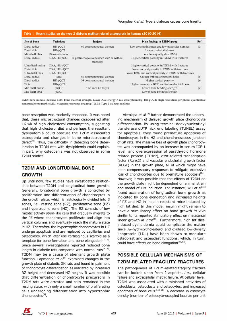
Table 1 Recent studies on the type 2 diabetes mellitus-related osteoporosis in humans (2010-2014)
Site of bone Technique Subjects Main findings in T2DM group Ref.
Distal radius HR-pQCT 60 postmenopausal women Low cortical thickness and low trabecular number [3]Distal tibia HR-pQCT Lower cortical thicknessMid-shaft tibia Microindentation Poor bone quality (low BMS)Distal radius DXA, HR-pQCT 80 postmenopausal women with or without
fracturesHigher cortical porosity in T2DM with fractures [4]
Ultradistal radius DXA, HR-pQCT Higher cortical porosity in T2DM with fracturesDistal tibia DXA, HR-pQCT Lower cortical porosity in T2DM with fracturesUltradistal tibia DXA, HR-pQCT Lower BMD and cortical porosity in T2DM with fracturesDistal radius MRI 60 postmenopausal women Greater trabecular network holes [5]Distal radius HR-pQCT 38 postmenopausal women Higher cortical porosity [6]Tibia HR-pQCT Higher volumetric BMD and trabecular thicknessMid-shaft radius pQCT 1171 men (≥ 65 yr) Lower bone bending strength [7]Mid-shaft tibia pQCT Lower bone bending strength
BMD: Bone mineral density; BMS: Bone material strength; DXA: Dual energy X-ray absorptiometry; HR-pQCT: High resolution-peripheral quantitative computed tomography; MRI: Magnetic resonance imaging; T2DM: Type 2 diabetes mellitus.
Wongdee K et al . Type 2 diabetes causes bone fragility
enzymatic collagen cross-link, toughening of the matrix, or the presence of advanced glycation end products (AGEs).
AGEs are non-enzymatic carbohydrate modifications of extracellular and intracellular proteins accumulated in long-lived tissues, such as skin and bone, and are often present in the plasma proteins of patients with DM and renal failure[28,29]. A number of investigations have revealed that AGEs are considered a factor that provokes fragility fractures in T2DM by inducing abnormal arrangement of collagen[26,28,30]. By using scanning electron microscope (SEM) and transmission electron microscope (TEM), Aoki et al[28] provided evidence that the rats subjected to adenine-induced renal failure exhibited AGEs accumulation and suppre-ssion of osteoblast function, similar to that observed in T2DM. SEM showed irregularity in collagen fibril alignment, while TEM revealed a wider diameter of collagen fibril in adenine-treated rats with renal ost-eodystrophy[28]. Immunohistochemistry also showed greater accumulation of AGEs in peritrabecular osteoblasts of adenine-treated rats than control rats. Further in vitro study in AGEs-treated MC3T3-E1 osteoblast-like cells showed a decrease in protein expression of secreted phosphoprotein 1 and lysyl oxidase, a mature osteoblast marker and essential enzyme for collagen cross-link, respectively. It was thus suggested that suppressed osteoblast differentiation and decreased lysyl oxidase production caused structural abnormalities of bone collagen fibrils leading to bone fragility[28].
Collagen is the most abundant protein in bone organic matrix, and it undergoes intra- and extracellular post-translational modifications[31]. To stabilize collagen fibrils, lysyl oxidase catalyzes intra- and intermolecular cross-link between collagen molecules essential for bone strength[31]. It was reported that glycation of collagen caused abnormal arrangement of collagen leading to brittle matrix and fragile bone[26,28,30], but little is known whether a decrease in lysyl oxidase-dependent collagen cross-link contributes to diabetic bone fragility and osteoporosis. The underlying mechanism of AGEs-attenuated lysyl oxidase activity was explored in mouse and rat primary osteoblasts and it was found that the carboxymethylated collagen, a form of AGEs, was not able to promote lysyl oxidase-mediated cross-linking due to failure of binding between abnormal collagen and discoidin domain receptor-2[30].
CONCLUSIONCurrently, it can be concluded that T2DM compromises bone microstructure by inducing aberrant bone cell function (cellular failure) and abnormal matrix structure (matrix failure). Regarding the cellular effect, T2DM is associated with increased osteoblast apoptosis, diminished osteoblast differentiation, and enhanced osteoclast-mediated bone resorption, which, in part,
area) was conspicuously observed in streptozotocin-induced diabetic rats[21]. Hyperglycemia-induced insulin resistance is another important factor that cause both osteoblast and osteoclast malfunctions[22]. Since insulin is a suppressor of osteoclast-mediated bone resorption[23], T2DM-associated insulin resis-tance could enhance bone resorption. Moreover, high plasma glucose concentration can induce glucotoxicity in cells including osteoblasts, leading to osteoblast apoptosis[24]. In vivo experiment in transgenic liver-specific S503A CEACAM1 mutant (L-SACC1) mice, a model of impaired insulin clearance in the liver causing hyperinsulinemia and insulin resistance, suggested that the abnormally high bone mass in these mice might have resulted from low bone turnover as indicated by decreases in double-labeled surface (as determined by bone histomorphometry) and TRAP-positive osteoclasts, which represent activities of osteoblast-mediated bone formation and osteoclast-mediated bone resorption, respectively[22]. In other words, insulin resistance in this model was associated with a slowdown in bone turnover, which could eventually result in inadequate healing of microcracks, poor bone quality and increased fracture risk[22]. In addition, the experiment in high fat diet-fed Zucker diabetic fatty (ZDF) rats also showed impaired osteoblast function as indicated by downregulation of the expression of osteoblast-specific genes, e.g., bone morphogenetic protein-2 (BMP-2), Runx2, osteocalcin and osteopontin. Suppression of osteoblastogenesis in these ZDF rats possibly compromised bone regeneration capacity. Subcritical bone defect regeneration study further showed that nondiabetic rats filled the defect by 57%, whereas diabetic rats could fill only 21% of bone defect in 12 wk[18].
T2DM not only caused deterioration of bone cell functions (cellular failure), but it also damaged bone extracellular matrix. Most studies suggested that T2DM caused abnormality in the structure of collagen, which is the most abundant protein in organic bone matrix. García-Hernández et al[25] reported that high glucose concentration indeed increased biomineralization in human alveolar bone-derived osteoblasts, but the mineral quality was lower than that in low glucose-exposed group. Determination of mineral quality in term of calcium/phosphate (Ca/Pi) ratio in the mineralized extracellular matrix nodules by energy-dispersive X-ray microanalysis (EDX) showed that high concentration of glucose significantly decreased Ca/Pi ratio on day 7 and 14 of treatment. Hammond et al[26] further studied nanoscale morphology of type Ⅰ collagen in tibiae of ZDF rats by Raman spectroscopy and reference point indentation (RPI), the latter of which applied a force to determine bone mechanical properties by measuring the relative displacement reference position[26,27]. RPI analysis revealed that bone matrix of ZDF diabetic rats was more resistant to plastic deformation, which might have resulted from abnormal formation of non-
676 June 10, 2015|Volume 6|Issue 5|WJD|www.wjgnet.com
Wongdee K et al . Type 2 diabetes causes bone fragility
resulted from hyperglycemia and insulin resistance. Prolonged accumulation of AGEs coexisting with a decrease in lysyl oxidase activity causes abnormal structure and alignment of collagen, leading to bone fragility. Several confounding factors in T2DM, parti-cularly body weight gain, obesity, and dyslipidemia, are able to mask the detrimental effects of T2DM, and may delay diagnosis of diabetic osteoporosis. In other words, bone is already damaged in T2DM despite a relatively high BMD. Although deleterious effects of T2DM on bone have been elucidated, the underlying cellular and molecular mechanisms remain unclear. For example, how does insulin resistance occur in osteoblasts and how do phosphorylation of insulin-receptor substrate isoforms (IRSs) and resultant insulin resistance in osteoblasts, osteoclasts and perhaps osteocytes contribute to diabetic bone loss? Indeed, osteocytes residing inside lacunae play an important role in bone remodeling in health and disease since they are responsible for inducing bone loss under certain conditions, such as during lactation[32,33]. Further investigation is required to demonstrate whether osteocytic dysfunction does exist in T2DM.
ACKNOWLEDGMENTSThe authors thank Professor Nateetip Krishnamra for valuable comments on the manuscript.
REFERENCES1 Wongdee K, Charoenphandhu N. Osteoporosis in diabetes
mellitus: Possible cellular and molecular mechanisms. World J Diabetes 2011; 2: 41-48 [PMID: 21537459 DOI: 10.4239/wjd.v2.i3.41]
2 Hofbauer LC, Brueck CC, Singh SK, Dobnig H. Osteoporosis in patients with diabetes mellitus. J Bone Miner Res 2007; 22: 1317-1328 [PMID: 17501667 DOI: 10.1359/jbmr.070510]
3 Farr JN, Drake MT, Amin S, Melton LJ, McCready LK, Khosla S. In vivo assessment of bone quality in postmenopausal women with type 2 diabetes. J Bone Miner Res 2014; 29: 787-795 [PMID: 24123088 DOI: 10.1002/jbmr.2106]
4 Patsch JM, Burghardt AJ, Yap SP, Baum T, Schwartz AV, Joseph GB, Link TM. Increased cortical porosity in type 2 diabetic postmenopausal women with fragility fractures. J Bone Miner Res 2013; 28: 313-324 [PMID: 22991256 DOI: 10.1002/jbmr.1763]
5 Pritchard JM, Giangregorio LM, Atkinson SA, Beattie KA, Inglis D, Ioannidis G, Punthakee Z, Adachi JD, Papaioannou A. Association of larger holes in the trabecular bone at the distal radius in postmenopausal women with type 2 diabetes mellitus compared to controls. Arthritis Care Res (Hoboken) 2012; 64: 83-91 [PMID: 22213724 DOI: 10.1002/acr.20602]
6 Burghardt AJ, Issever AS, Schwartz AV, Davis KA, Masharani U, Majumdar S, Link TM. High-resolution peripheral quantitative computed tomographic imaging of cortical and trabecular bone microarchitecture in patients with type 2 diabetes mellitus. J Clin Endocrinol Metab 2010; 95: 5045-5055 [PMID: 20719835 DOI: 10.1210/jc.2010-0226]
7 Petit MA, Paudel ML, Taylor BC, Hughes JM, Strotmeyer ES, Schwartz AV, Cauley JA, Zmuda JM, Hoffman AR, Ensrud KE. Bone mass and strength in older men with type 2 diabetes: the Osteoporotic Fractures in Men Study. J Bone Miner Res 2010; 25: 285-291 [PMID: 19594301 DOI: 10.1359/jbmr.090725]
8 Yamaguchi T, Kanazawa I, Yamamoto M, Kurioka S, Yamauchi
M, Yano S, Sugimoto T. Associations between components of the metabolic syndrome versus bone mineral density and vertebral fractures in patients with type 2 diabetes. Bone 2009; 45: 174-179 [PMID: 19446053 DOI: 10.1016/j.bone.2009.05.003]
9 Lapmanee S, Charoenphandhu N, Aeimlapa R, Suntornsaratoon P, Wongdee K, Tiyasatkulkovit W, Kengkoom K, Chaimongkolnukul K, Seriwatanachai D, Krishnamra N. High dietary cholesterol masks type 2 diabetes-induced osteopenia and changes in bone microstructure in rats. Lipids 2014; 49: 975-986 [PMID: 25200330 DOI: 10.1007/s11745-014-3950-3]
10 Kengkoom K, Klinkhamhom A, Sirimontaporn A, Singha O, Ketjareon T, Panavechkijkul Y, Seriwatanachai D, Ukong S, Ampawong S. Effects on high cholesterol-fed to liver, retina, hippocampus, and Harderian gland in Goto-Kakizaki rat. Int J Clin Exp Pathol 2013; 6: 639-649 [PMID: 23573310]
11 Wongdee K, Krishnamra N, Charoenphandhu N. Endochondral bone growth, bone calcium accretion, and bone mineral density: how are they related? J Physiol Sci 2012; 62: 299-307 [PMID: 22627708 DOI: 10.1007/s12576-012-0212-0]
12 Mackie EJ, Ahmed YA, Tatarczuch L, Chen KS, Mirams M. Endochondral ossification: how cartilage is converted into bone in the developing skeleton. Int J Biochem Cell Biol 2008; 40: 46-62 [PMID: 17659995 DOI: 10.1016/j.biocel.2007.06.009]
13 Zhang L, Liu Y, Wang D, Zhao X, Qiu Z, Ji H, Rong H. Bone biomechanical and histomorphometrical investment in type 2 diabetic Goto-Kakizaki rats. Acta Diabetol 2009; 46: 119-126 [PMID: 18843446 DOI: 10.1007/s00592-008-0068-1]
14 Aeimlapa R, Wongdee K, Charoenphandhu N, Suntornsaratoon P, Krishnamra N. Premature chondrocyte apoptosis and compensatory upregulation of chondroregulatory protein expression in the growth plate of Goto-Kakizaki diabetic rats. Biochem Biophys Res Commun 2014; 452: 395-401 [PMID: 25159845 DOI: 10.1016/j.bbrc.2014.08.085]
15 Wu S, Aguilar AL, Ostrow V, De Luca F. Insulin resistance secondary to a high-fat diet stimulates longitudinal bone growth and growth plate chondrogenesis in mice. Endocrinology 2011; 152: 468-475 [PMID: 21106874 DOI: 10.1210/en.2010-0803]
16 Hamel P, Abed E, Brissette L, Moreau R. Characterization of oxidized low-density lipoprotein-induced hormesis-like effects in osteoblastic cells. Am J Physiol Cell Physiol 2008; 294: C1021-C1033 [PMID: 18287334 DOI: 10.1152/ajpcell.00361.2007]
17 Luegmayr E, Glantschnig H, Wesolowski GA, Gentile MA, Fisher JE, Rodan GA, Reszka AA. Osteoclast formation, survival and morphology are highly dependent on exogenous cholesterol/lipoproteins. Cell Death Differ 2004; 11 Suppl 1: S108-S118 [PMID: 15017384 DOI: 10.1038/sj.cdd.4401399]
18 Hamann C, Goettsch C, Mettelsiefen J, Henkenjohann V, Rauner M, Hempel U, Bernhardt R, Fratzl-Zelman N, Roschger P, Rammelt S, Günther KP, Hofbauer LC. Delayed bone regeneration and low bone mass in a rat model of insulin-resistant type 2 diabetes mellitus is due to impaired osteoblast function. Am J Physiol Endocrinol Metab 2011; 301: E1220-E1228 [PMID: 21900121 DOI: 10.1152/ajpendo.00378.2011]
19 Kawashima Y, Fritton JC, Yakar S, Epstein S, Schaffler MB, Jepsen KJ, LeRoith D. Type 2 diabetic mice demonstrate slender long bones with increased fragility secondary to increased osteoclastogenesis. Bone 2009; 44: 648-655 [PMID: 19150422 DOI: 10.1016/j.bone.2008.12.012]
20 Weinberg E, Maymon T, Weinreb M. AGEs induce caspase-mediated apoptosis of rat BMSCs via TNFα production and oxidative stress. J Mol Endocrinol 2014; 52: 67-76 [PMID: 24198288 DOI: 10.1530/JME-13-0229]
21 Villarino ME, Sánchez LM, Bozal CB, Ubios AM. Influence of short-term diabetes on osteocytic lacunae of alveolar bone. A histomorphometric study. Acta Odontol Latinoam 2006; 19: 23-28 [PMID: 17121195]
22 Huang S, Kaw M, Harris MT, Ebraheim N, McInerney MF, Najjar SM, Lecka-Czernik B. Decreased osteoclastogenesis and high bone mass in mice with impaired insulin clearance due to liver-specific inactivation to CEACAM1. Bone 2010; 46: 1138-1145 [PMID:
677 June 10, 2015|Volume 6|Issue 5|WJD|www.wjgnet.com
Wongdee K et al . Type 2 diabetes causes bone fragility
20044046 DOI: 10.1016/j.bone.2009.12.020]23 Thomas DM, Udagawa N, Hards DK, Quinn JM, Moseley JM,
Findlay DM, Best JD. Insulin receptor expression in primary and cultured osteoclast-like cells. Bone 1998; 23: 181-186 [PMID: 9737339]
24 Zhen D, Chen Y, Tang X. Metformin reverses the deleterious effects of high glucose on osteoblast function. J Diabetes Complications 2010; 24: 334-344 [PMID: 19628413 DOI: 10.1016/j.jdiacomp.2009.05.002]
25 García-Hernández A, Arzate H, Gil-Chavarría I, Rojo R, Moreno-Fierros L. High glucose concentrations alter the biomineralization process in human osteoblastic cells. Bone 2012; 50: 276-288 [PMID: 22086137 DOI: 10.1016/j.bone.2011.10.032]
26 Hammond MA, Gallant MA, Burr DB, Wallace JM. Nanoscale changes in collagen are reflected in physical and mechanical properties of bone at the microscale in diabetic rats. Bone 2014; 60: 26-32 [PMID: 24269519 DOI: 10.1016/j.bone.2013.11.015]
27 Gallant MA, Brown DM, Organ JM, Allen MR, Burr DB. Reference-point indentation correlates with bone toughness assessed using whole-bone traditional mechanical testing. Bone 2013; 53: 301-305 [PMID: 23274349 DOI: 10.1016/j.bone.2012.12.015]
28 Aoki C, Uto K, Honda K, Kato Y, Oda H. Advanced glycation
end products suppress lysyl oxidase and induce bone collagen degradation in a rat model of renal osteodystrophy. Lab Invest 2013; 93: 1170-1183 [PMID: 23979426 DOI: 10.1038/labinvest.2013.105]
29 McCarthy AD, Molinuevo MS, Cortizo AM. Ages and bone ageing in diabetes mellitus. J Diabetes Metab 2013; 4: 276 [DOI: 10.4172/2155-6156.1000276]
30 Khosravi R, Sodek KL, Faibish M, Trackman PC. Collagen advanced glycation inhibits its Discoidin Domain Receptor 2 (DDR2)-mediated induction of lysyl oxidase in osteoblasts. Bone 2014; 58: 33-41 [PMID: 24120383 DOI: 10.1016/j.bone.2013.10.001]
31 Saito M, Kida Y, Kato S, Marumo K. Diabetes, collagen, and bone quality. Curr Osteoporos Rep 2014; 12: 181-188 [PMID: 24623537 DOI: 10.1007/s11914-014-0202-7]
32 Qing H, Ardeshirpour L, Pajevic PD, Dusevich V, Jähn K, Kato S, Wysolmerski J, Bonewald LF. Demonstration of osteocytic perilacunar/canalicular remodeling in mice during lactation. J Bone Miner Res 2012; 27: 1018-1029 [PMID: 22308018 DOI: 10.1002/jbmr.1567]
33 Suntornsaratoon P, Kraidith K, Teerapornpuntakit J, Dorkkam N, Wongdee K, Krishnamra N, Charoenphandhu N. Pre-suckling calcium supplementation effectively prevents lactation-induced osteopenia in rats. Am J Physiol Endocrinol Metab 2014; 306: E177-E188 [PMID: 24302005 DOI: 10.1152/ajpendo.00556.2013]
P- Reviewer: Grieco A, Hiwatashi A S- Editor: Ji FF L- Editor: A E- Editor: Zhang DN
678 June 10, 2015|Volume 6|Issue 5|WJD|www.wjgnet.com
Wongdee K et al . Type 2 diabetes causes bone fragility
REVIEW
Endothelial dysfunction as a predictor of cardiovascular disease in type 1 diabetes
Marcello C Bertoluci, Gislaine V Cé, Antônio MV da Silva, Marco V Wainstein, Winston Boff, Marcia Puñales
Marcello C Bertoluci, Marco V Wainstein, Departamento de Medicina Interna, Universidade Federal do Rio Grande do Sul, Porto Alegre RS 90035-003, BrazilMarcello C Bertoluci, Serviço de Medicina Interna do Hospital de Clinicas de Porto Alegre, Porto Alegre RS 90035-903, Brazil Marcello C Bertoluci, Programa de Pós-Graduação em Ciências Médicas, Universidade Federal do Rio Grande do Sul, Porto Alegre RS 90035-003, BrazilGislaine V Cé, Winston Boff, Marcia Puñales, Instituto da Criança com Diabetes - Grupo Hospitalar Conceição, Porto Alegre RS 91350-250, BrasilAntônio MV da Silva, Departamento de Fisioterapia e Reabilitação, Universidade Federal de Santa Maria, Santa Maria RS 97105-900, BrasilMarco V Wainstein, Serviço de Cardiologia do Hospital de Clínicas de Porto Alegre, Porto Alegre RS 90035-903, Brazil
Author contributions: Bertoluci MC, Puñales M, Cé GV, da Silva AMV, Wainstein MV and Boff W contributed to this paper.
Conflict-of-interest: Dr. Wainstein reports grants and other from Biotronik, grants and other from Abbott Vascular, other from BBraun, outside the submitted work. The other authors declare not to have any type of conflict-of-interest related to the present review.
Open-Access: This article is an open-access article which was selected by an in-house editor and fully peer-reviewed by external reviewers. It is distributed in accordance with the Creative Commons Attribution Non Commercial (CC BY-NC 4.0) license, which permits others to distribute, remix, adapt, build upon this work non-commercially, and license their derivative works on different terms, provided the original work is properly cited and the use is non-commercial. See: http://creativecommons.org/licenses/by-nc/4.0/
Correspondence to: Marcello C Bertoluci, MD, DMSc, Serviço de Medicina Interna do Hospital de Clinicas de Porto Alegre, Rua Ramiro Barcellos 2350- Sala 700, Santa Cecília, Porto Alegre 90035-003, Brazil. [email protected]: +55-51-33598000 Fax: +55-51-33598000
Received: November 6, 2014Peer-review started: November 10, 2014
First decision: February 7, 2015Revised: March 11, 2015Accepted: April 1, 2015Article in press: April 7, 2015Published online: June 10, 2015
AbstractMacro and microvascular disease are the main cause of morbi-mortality in type 1 diabetes (T1DM). Although there is a clear association between endothelial dysfunction and atherosclerosis in type 2 diabetes, a cause-effect relationship is less clear in T1DM. Although endothelial dysfunction (ED) precedes atherosclerosis, it is not clear weather, in recent onset T1DM, it may progress to clinical macrovascular disease. Moreover, endothelial dysfunction may either be reversed spontaneously or in response to intensive glycemic control, long-term exercise training and use of statins. Acute, long-term and post-prandial hyperglycemia as well as duration of diabetes and microalbuminuria are all conditions associated with ED in T1DM. The pathogenesis of endothelial dysfunction is closely related to oxidative-stress. NAD(P)H oxidase over activity induces excessive superoxide production inside the mitochondrial oxidative chain of endothelial cells, thus reducing nitric oxide bioavailability and resulting in peroxynitrite formation, a potent oxidant agent. Moreover, oxidative stress also uncouples endothelial nitric oxide synthase, which becomes dysfunctional, inducing formation of superoxide. Other important mechanisms are the activation of both the polyol and protein kinase C pathways as well as the presence of advanced glycation end-products. Future studies are needed to evaluate the potential clinical applicability of endothelial dysfunction as a marker for early vascular complications in T1DM.
Key words: Endothelial dysfunction; Type 1 diabetes; Cardiovascular disease
679 June 10, 2015|Volume 6|Issue 5|WJD|www.wjgnet.com
Submit a Manuscript: http://www.wjgnet.com/esps/Help Desk: http://www.wjgnet.com/esps/helpdesk.aspxDOI: 10.4239/wjd.v6.i5.679
World J Diabetes 2015 June 10; 6(5): 679-692ISSN 1948-9358 (online)
© 2015 Baishideng Publishing Group Inc. All rights reserved.
© The Author(s) 2015. Published by Baishideng Publishing Group Inc. All rights reserved.
Core tip: Endothelial dysfunction is an early finding in the natural history of type 1 diabetes and is predictive for microvascular disease and premature atherosclerosis. Decreased nitric oxide due oxidative stress is the central pathogenetic mechanism. Polyol pathway activation, protein kinase C (PKC) activation and advanced glycation product formation are also important. Long-term hyperglycemia, repeated hypo-glycemia and microalbuminuria are factors associated. Intensive glycemic control and exercise training ameliorate endothelial dysfunction. Statins and renin-angiotensin system blockers are partially effective and may be influenced by hyperglycemia. There is a possible clinical benefit for the use of vitamin E and vitamin C that are still to be confirmed. PKC inhibitors are still investigative.
Bertoluci MC, Cé GV, da Silva AMV, Wainstein MV, Boff W, Puñales M. Endothelial dysfunction as a predictor of cardiovascular disease in type 1 diabetes. World J Diabetes 2015; 6(5): 679-692 Available from: URL: http://www.wjgnet.com/1948-9358/full/v6/i5/679.htm DOI: http://dx.doi.org/10.4239/wjd.v6.i5.679
INTRODUCTIONMicro and macrovascular complications are leading causes of morbidity and mortality in patients with type 1 diabetes mellitus (T1DM)[1,2]. Subjects with T1DM are prone to accelerated atherosclerosis[3] and have 3 to 6 times more risk of cardiovascular death than individuals without diabetes. Endothelial dysfunction (ED) is an early event along the natural history of T1DM, indicating a phenotype in risk for accelerated atherosclerosis, that may be independent of the classical cardiovascular risk factors[4,5]. Interestingly, traditional cardiovascular risk factors altogether can not explain the totality of the cardiovascular risk in T1DM[6,7]. Chronic hyperglycemia per se, although an important predictor of microvascular disease, is a weak predictor of macrovascular complications in both T1DM and T2DM[8,9]. Thus, much of the residual cardiovascular risk still remains unexplained. In this context, ED becomes a new important risk factor that should be debated in the cardiovascular scenario. In the present review, we discuss recent evidences in the pathogenesis of endothelial dysfunction, its role as a risk factor for cardiovascular disease and the potential interventions for reducing endothelial dysfunction in T1DM.
THE NORMAL ENDOTHELIUMThe vascular endothelium forms the cell layer that is
directly in contact with the vascular lumen, separated from the smooth muscle layer of the basement membrane. Its role is to maintain the homeostasis between the blood and the arterial wall through the synthesis of substances that modulate vascular tone, inhibit platelet aggregation and control the proliferation of vascular smooth muscle cells[10].
In endothelial cells, nitric oxide (NO) is essential for the maintenance of integrity and homeostasis of endothelium[11]. NO is synthesized from L-arginine by the action of endothelial nitric oxide synthase (eNOS) in the presence of oxygen, NADP(H) and the NOS co-factor, tetrahydrobiopterin (BH4)[12]. The synthesized NO diffuses itself quickly into the smooth muscle cell layer and into platelets where it activates guanylate cyclase (GCA), with consequent production of cyclic GMP (cGMP). The presence of cGMP, in turn, promotes vascular relaxation and inhibition of platelet aggregation, keeping the equilibrium between pro and anti-thrombotic factors in the blood and arterial wall. However, as the half-life of NO is very brief, rapidly oxidizing into nitrate, the continuous activation of eNOS becomes the key determinant of NO synthesis and tissue bioavailability[11]. Normally, eNOs is activated by the turbulent blood flow against the luminal endothelial wall (shear-stress) as well as by the stretch of vascular wall cells and changes in the oxygen tension, promoting vascular muscle relaxation, an effect known as “endo-thelial-dependent vasodilation”[13,14].
The stability of the endothelium is also dependent on endothelial repair and regeneration, which are determined by migration and proliferation of surrounding mature endothelial cell resident the vascular wall[15]. Recently it has been demonstrated that circulating endothelial progenitor cells (EPCs) are important in the endothelial regeneration. EPCs are circulating bone-marrow-derived cells characterized by the expression of varying surface markers that adhere to the damaged endothelium promoting tissue repair[15]. Circulating EPCs are considered biomarkers of endothelial function and prognostic indicators of cardiovascular morbi-mortality. Endothelial dysfunction represents the breakdown of this endothelium homeostasis, leading to a pro-thrombotic and pro-inflammatory that may lead to progressive atherosclerosis.
METHODS FOR ASSESSMENT OF ENDOTHELIAL FUNCTIONEndothelial function can be investigated through invasively and non-invasively techniques. Coronary arteries can be evaluated invasively through angio-graphy with quantitative measurement of changes in the vascular diameter in response to infusion of acetylcholine[16] and also through invasive venous occlusion plethysmography[17], which measures forearm blood flow in response to acetylcholine infusion in the brachial artery. The invasive nature of
Bertoluci MC et al . Endothelial dysfunction in type 1 diabetes
680 June 10, 2015|Volume 6|Issue 5|WJD|www.wjgnet.com
these techniques, involving artery cannulation and infusion of vasoactive drugs[14], make them unfeasible to widespread use in clinical practice.
Non-invasive techniques, on the other hand, are being increasingly used in clinical settings. The flow mediated dilation (FMD) is the most popular technique currently used[18-20]. The rationale in FMD is based on reactive hyperemia responsive to shear-stress caused by turbulent blood flow, causing NO to be released and promoting endothelium-dependent vasodilation[14,21]. The measurement of vascular dilation can be done by capturing images of the brachial artery using high-resolution ultrasound[18]. Reactive hyperemia occurs after a period of ischemia, induced by occlusion of the brachial artery, with a sphygmomanometer cuff inflated with progressive release of vasodilator mediators such as adenosine and +H ions from ischemic tissue. When the release of blood flow occurs, sudden shear-stress is produced in the brachial vein endothelium, which is a strong stimulus for releasing NO[19]. This mechanism depends on the integrity of eNOs. The lesser the dilation the more severe the dysfunction. Definition of ED is than considered arbitrarily when the increase in dilation is less than 8%[20].
Other non-invasive techniques are used less frequently. Peripheral artery tonometry induced by reactive hyperemia[22] assesses endothelial function by a combination of flow-mediated dilation and measurement of the amplitude of the arterial pulse wave expansion through a pneumatic sensor placed on the index finger. The microvascular reactivity on the forearm skin is evaluated with laser Doppler flowmetry, being the the iontophoresis of acetylcholine the endothelium-dependent vasodilator stimulus. The determination of the complacency of the dorsal hand vein is a minimally invasive method described by Aellig[23] in 1981 which has been used by our group[24]. It consists in a infusion of vasoactive drugs into the vein surface of the dorsum of the hand to measure endothelium-dependent vasodilation in response to acetylcholine, bradykinin or isoproterenol. The venous occlusion plethysmography can also be used to measure changes in forearm blood flow in response to reactive hyperemia. Finally, the measurement of the thickness of the intima-media layer (IMT) of the common carotid by ultrasound is a structural marker of atherosclerosis and correlates inversely with FMD in the brachial artery[25]. Increases in the IMT are indicative of early atherosclerosis.
ENDOTHELIAL CHANGES IN DIABETESChronic sustained hyperglycemia in diabetes promotes important structural and functional modifications in the endothelium, as reported in both experimental and clinical studies[26-29]. In the aorta of rabbits with alloxan-induced diabetes, endothelial changes are visible after 2 wk from the onset of hyperglycemia
and become more severe after 6 wk of the diabetes onset[26]. The findings include adhesion of leukocytes, platelets and fibrin material on the endothelial surface. In mice, 6 wk after the onset of diabetes induced by streptozotocin (STZ), it is possible to observe increased endothelial permeability and endothelial cell apoptosis[27].
In a classic study using samples of human skin and subcutaneous tissue obtained from autopsies and biopsies of 24 patients with T1DM, which were compared to 9 non-diabetic controls, the most important finding was the increase in the thickness of the basement membrane in T1DM patients compared with non-diabetic subjects[28]. In another study[29], the increased thickness of the capillary basement membrane of skeletal muscle from patients with 12 years of T1DM, could be reversed by intensive glycemic control during one year[29]. In studies using electron microscopy, endothelial cells obtained from umbilical cord blood from pregnant women with T1DM show increased mitochondrial area when compared to pregnant women without diabetes[28]. The clinical significance of these structural changes, however, is still not clear to predict future atherosclerosis.
Important functional changes occur in the endo-thelium of T1DM. Hyperglycemia induces excess of electrons that leak from the oxidative chain and are captured by oxygen, generating superoxide excess and oxidative stress. Excessive superoxide production uncouples eNOS, impairing NO production[29]. The net effect is a reduction of NO production in response to shear stress in the inner vascular wall. By this way, the main determinant of ED is the preponderance of vasoconstrictor factors released by the endothelium in detriment of vasodilators factors due to the decreased availability of NO[30]. The dysfunctional endothelium leads to a migration of blood cells into the arterial wall, inducing proliferation of smooth muscle cells, platelet aggregation, LDLc oxidation, monocyte adhesion and synthesis of inflammatory cytokines, all factors contributing to atherogenesis[31].
In patients with T1DM, functional changes in endothelium occurs very early in the natural history of diabetes[32]. The duration of diabetes is a major determinant for the presence of endothelial dysfunction in T1DM, being inversely correlated with the endothelium-dependent dilation. ED generally occurs in the first decade of T1DM, earlier than increases in the carotid intima-media layer thickness (Table 1).
ED is a common finding in T1DM, generally seen after 4 years of disease. In the study by Singh et al[33], 31 adolescents with 6.8 years of T1DM and poor glycemic control presented both ED and increased intima-media layer thickness of carotid artery, compared with individuals without diabetes. The duration of diabetes was inversely correlated with the endothelium-dependent dilation[33]. These results were confirmed by other authors[34-38] and are in accordance
681 June 10, 2015|Volume 6|Issue 5|WJD|www.wjgnet.com
Bertoluci MC et al . Endothelial dysfunction in type 1 diabetes
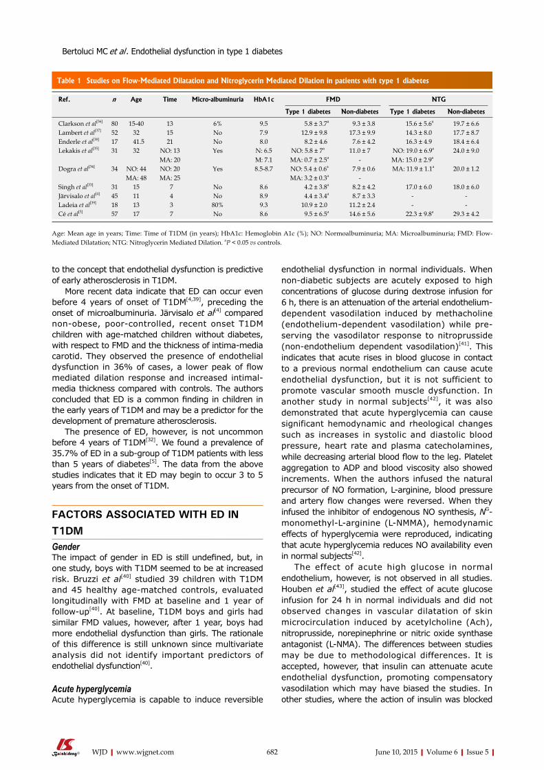
Table 1 Studies on Flow-Mediated Dilatation and Nitroglycerin Mediated Dilation in patients with type 1 diabetes
endothelial dysfunction in normal individuals. When non-diabetic subjects are acutely exposed to high concentrations of glucose during dextrose infusion for 6 h, there is an attenuation of the arterial endothelium-dependent vasodilation induced by methacholine (endothelium-dependent vasodilation) while pre-serving the vasodilator response to nitroprusside
(non-endothelium dependent vasodilation)[41]. This indicates that acute rises in blood glucose in contact to a previous normal endothelium can cause acute endothelial dysfunction, but it is not sufficient to promote vascular smooth muscle dysfunction. In another study in normal subjects[42], it was also demonstrated that acute hyperglycemia can cause significant hemodynamic and rheological changes such as increases in systolic and diastolic blood pressure, heart rate and plasma catecholamines, while decreasing arterial blood flow to the leg. Platelet aggregation to ADP and blood viscosity also showed increments. When the authors infused the natural precursor of NO formation, L-arginine, blood pressure and artery flow changes were reversed. When they infused the inhibitor of endogenous NO synthesis, NG-monomethyl-L-arginine (L-NMMA), hemodynamic effects of hyperglycemia were reproduced, indicating that acute hyperglycemia reduces NO availability even in normal subjects[42].
The effect of acute high glucose in normal endothelium, however, is not observed in all studies. Houben et al[43], studied the effect of acute glucose infusion for 24 h in normal individuals and did not observed changes in vascular dilatation of skin microcirculation induced by acetylcholine (Ach), nitroprusside, norepinephrine or nitric oxide synthase antagonist (L-NMA). The differences between studies may be due to methodological differences. It is accepted, however, that insulin can attenuate acute endothelial dysfunction, promoting compensatory vasodilation which may have biased the studies. In other studies, where the action of insulin was blocked
to the concept that endothelial dysfunction is predictive of early atherosclerosis in T1DM.
More recent data indicate that ED can occur even before 4 years of onset of T1DM[4,39], preceding the onset of microalbuminuria. Järvisalo et al[4] compared non-obese, poor-controlled, recent onset T1DM children with age-matched children without diabetes, with respect to FMD and the thickness of intima-media carotid. They observed the presence of endothelial dysfunction in 36% of cases, a lower peak of flow mediated dilation response and increased intimal-media thickness compared with controls. The authors concluded that ED is a common finding in children in the early years of T1DM and may be a predictor for the development of premature atherosclerosis.
The presence of ED, however, is not uncommon before 4 years of T1DM[32]. We found a prevalence of 35.7% of ED in a sub-group of T1DM patients with less than 5 years of diabetes[5]. The data from the above studies indicates that it ED may begin to occur 3 to 5 years from the onset of T1DM.
FACTORS ASSOCIATED WITH ED IN T1DM GenderThe impact of gender in ED is still undefined, but, in one study, boys with T1DM seemed to be at increased risk. Bruzzi et al[40] studied 39 children with T1DM and 45 healthy age-matched controls, evaluated longitudinally with FMD at baseline and 1 year of follow-up[40]. At baseline, T1DM boys and girls had similar FMD values, however, after 1 year, boys had more endothelial dysfunction than girls. The rationale of this difference is still unknown since multivariate analysis did not identify important predictors of endothelial dysfunction[40].
Acute hyperglycemiaAcute hyperglycemia is capable to induce reversible
682 June 10, 2015|Volume 6|Issue 5|WJD|www.wjgnet.com
Ref. n Age Time Micro-albuminuria HbA1c FMD NTG
Type 1 diabetes Non-diabetes Type 1 diabetes Non-diabetes
Clarkson et al[36] 80 15-40 13 6% 9.5 5.8 ± 3.7a 9.3 ± 3.8 15.6 ± 5.6a 19.7 ± 6.6Lambert et al[37] 52 32 15 No 7.9 12.9 ± 9.8 17.3 ± 9.9 14.3 ± 8.0 17.7 ± 8.7Enderle et al[38] 17 41.5 21 No 8.0 8.2 ± 4.6 7.6 ± 4.2 16.3 ± 4.9 18.4 ± 6.4Lekakis et al[35] 31 32 NO: 13
MA: 20Yes N: 6.5
M: 7.1NO: 5.8 ± 7a
MA: 0.7 ± 2.5a11.0 ± 7
- NO: 19.0 ± 6.9a
MA: 15.0 ± 2.9a24.0 ± 9.0
Dogra et al[34] 34 NO: 44MA: 48
NO: 20MA: 25
Yes 8.5-8.7 NO: 5.4 ± 0.6a
MA: 3.2 ± 0.3a 7.9 ± 0.6
- MA: 11.9 ± 1.1a 20.0 ± 1.2
Singh et al[33] 31 15 7 No 8.6 4.2 ± 3.8a 8.2 ± 4.2 17.0 ± 6.0 18.0 ± 6.0Järvisalo et al[4] 45 11 4 No 8.9 4.4 ± 3.4a 8.7 ± 3.3 - -Ladeia et al[39] 18 13 3 80% 9.3 10.9 ± 2.0 11.2 ± 2.4 - -Cé et al[5] 57 17 7 No 8.6 9.5 ± 6.5a 14.6 ± 5.6 22.3 ± 9.8a 29.3 ± 4.2
Age: Mean age in years; Time: Time of T1DM (in years); HbA1c: Hemoglobin A1c (%); NO: Normoalbuminuria; MA: Microalbuminuria; FMD: Flow-Mediated Dilatation; NTG: Nitroglycerin Mediated Dilation. aP < 0.05 vs controls.
Bertoluci MC et al . Endothelial dysfunction in type 1 diabetes
by octreotide, the effect of acute hyperglycemia alone was evident[41].
Long-term hyperglycemiaThe association between HbA1c and flow-mediated dilation (FMD) is seen in some cross-sectional studies with patients with T1DM. In the study of Ladeia et al[39], with 19 normo and microalbuminuric T1DM patients, there was a moderate positive correlation between FMD and HbA1c. In another study[44], patients with T1DM with HbA1c above 6.0% had significant impairment of endothelial function compared to patients with HbA1c below 6%, indicating that chronic mild increases in mean hyperglycemia are also associated to ED in T1DM.
We studied the impact of chronic glycemic control in endothelial function of T1DM in a historical cohort study[5]. T1DM adolescents under 5 year of disease were evaluated for ED and had their mean HbA1c obtained from medical records in the same institution since their diagnosis. Considering as a whole, the mean historical HbA1c was clearly higher in T1DM patients with endothelial dysfunction compared with T1DM without ED. Interestingly, we observed a moderate inverse correlation between FMD and the historical mean of HbA1c in the first 2 years after the diagnosis of T1DM but not with the more recent HbA1c. The plausible explanation was that endothelial function could be more affected by the long-term than by the short-term glycemic control, supporting the concept of metabolic memory[45]. Glycation of the endothelium in the first years of T1DM seems, by this way, decisive to determine future endothelial dysfunction in T1DM[32].
Post-prandial hyperglycemiaThe effect of postprandial hyperglycemia in endo-thelial function was studied by Giugliano et al[42] in individuals with type 2 diabetes. The combined effect of postprandial glucose and hypertriglyceridemia was associated with increased serum concentrations of adhesion molecules such as ICAM-1, E-selectin and VCAM-1. Markers of oxidative stress such as nitro-tyrosine increased sharply after ingestion of 75 g of oral glucose. This effect was greater when an additional lipid overload was used, indicating that both acute hyperglycemia and hyperlipemia can affect endothelial function in diabetes[46].
In another study, the same authors[47] observed that, after a glucose overload, ED accentuates until the second hour, but returns to basal level after 4 h from the beginning of the overload. Though, lipid overload did not change FMD until the fourth hour. These data suggests that the effect of postprandial glycaemia in endothelial function is independent of the postprandial lipemia, although both might be mediated by increased oxidative stress.
HypoglycemiaRecently, it was demonstrated that repeated episodes of hypoglycemia in subjects with T1DM may be associated with endothelial dysfunction and could be an aggravating factor for preclinical atherosclerosis. In a case-control study[48], T1DM with repeated hypoglycemia episodes were compared with age and sex-matched T1DM controls who did not have frequent hypoglycemias. Vascular function was assessed by FMD, intimal-media carotid artery thickness (IMT) and endothelial dysfunction markers such as von Willebrand factor (vWf). The group with increased hypoglycemia episodes presented lower percentages of FMD in response to ischemia, increased IMT measures and higher endothelial function markers. In another study[49], with cross-sectional design, T1DM children and age and gender-matched healthy children were evaluated for vascular function and continuous glucose monitoring system (CGMS) in order to compare the impact of glucose variability and hypoglycemia episodes in vascular function. Subjects with T1DM had significantly lower FMD compared to healthy children. However, when comparing CGMS parameters, the authors found significant inverse relationship between FMD with hypoglycemia indexes but not with variability indexes.
The biological rationale for hypoglycemia inducing endothelial dysfunction in T1DM is that acute hypo-glycemia can induce rapid pro-inflammatory, platelet aggregatory, anti-fibrinolytic response, and recurrent hypoglycemia may induce changes in hemostatic factors and viscosity which may decrease perfusion in diabetic microangiopathy[50].
Glycemic variabilityGlycemic variability (GV) is a term exclusively related to blood glucose fluctuations and must be differentiated from post-prandial glucose (PPG). GV is related to glucose variability along the day, while PPG is related to the precise time of glucose rise after a meal. PPG effect in vascular function can also be influenced by other confounders such as hypertriglyceridemia.
Whether GV is an important factor to cause ED is still a matter of debate. The effect of glycemic variability in ED was studied in T1DM with normal urinary albumin excretion[51]. Patients were exposed to 48 h of good (mean 113 mg/dL) or poor metabolic control (mean 286 mg/dL) and evaluated with FMD and serological markers of endothelial function. Both endothelium-dependent and endothelium-independent vasodilation were significantly impaired after the poor control period in relation to good glycemic control. There was also a significant increase in vWf levels after the deterioration of control. These results indicate that endothelial function may suffer significant impact of acute variability of blood glucose in T1DM, although it
683 June 10, 2015|Volume 6|Issue 5|WJD|www.wjgnet.com
Bertoluci MC et al . Endothelial dysfunction in type 1 diabetes
can be reversed with improvement of glycemic control. However this effect was not seen in other studies. In the DCCT study, the GV in glycemic data using 7-point self monitoring blood glucose (SMBG) obtained every 3 mo did not correlated to macrovascular complications[52]. We were also not able to detect a relationship between FMD and the standard deviation (SD) of glycemia in T1DM, using day 7-point SMBG for 30 d preceding FMD (data unpublished). Finally, in a cohort study of T1DM[53], the standard deviation of blood glucose, calculated from self-monitoring blood glucose data along 11 years of follow up, was predictive for incident peripheral neuropathy[53]. The influence of GV in vascular function and future micro or macro vascular complications of T1DM is not yet established.
MicroalbuminuriaMicroalbuminuria is strongly associated with ED in T1DM. Dogra et al[34] studied long-term T1DM patients with microalbuminuria with poor glycemic control who were compared to normoalbuminuric T1DM and to non-diabetic individuals. FMD was more severely impaired in T1DM patients with microalbuminuria being albuminuria an independent predictor of ED. In another study, Lekakis et al[35] observed lower FMD values in microalbuminuric compared with normoalbuminuric patients. In a similar study with children and adolescents with T1DM with less than 5 years of disease[39], there was a negative correlation between the percent of endothelium-mediated dilation and albuminuria. Mean FMD was also significant decreased in microalbuminuric. ED is also be present in type 2 diabetes patients with normal albuminuria with long duration T2DM during chronic poor glycemic control[54]. In T1DM patients with normoalbuminuria ED is less common[40].
MECHANISMS OF ED IN T1DMOxidative stress Children with T1DM have increased oxidative stress and reduction of anti-oxidant defense compared to healthy children and to their parents[55,56] and these results are similar in adolescents with T1DM[57]. Moreover, endothelial progenitor cells are also reduced in children with T1DM compared to non-diabetic controls possibly related to oxidative stress[58].
Hyperglycemia can cause excessive production of superoxide in the mitochondria oxidative chain of endothelial cells. The excess of superoxide reacts rapidly with NO, reducing NO bioactivity and producing peroxynitrite (ONOO-). Peroxynitrite is a potent oxidant agent and an activator of the lipid peroxidation which may impair endothelial function by stimulating arachidonic acid metabolism[59]. The overproduction of superoxide and NO favors the formation of peroxynitrite by interfering with the production of the eNOS cofactor,
tetrahydrobiopterin (BH4)[60].NAD(P)H oxidase is a chief determinant enzyme
of superoxide production in animal models of vascular diseases, including diabetes[32]. In arteries of patients with diabetes who were submitted to artery bypass surgery, it was demonstrated that the endothelium can produce superoxide induced by a dysfunctional eNOS. Dysfunctional eNOS is caused by the oxidation of the co-factor BH4 into BH2. The enzymatic uncoupling of eNOS in human endothelium turns eNOS into dysfunctional eNOS which promotes a transition from NO production to superoxide production[60,61].
Protein kinase C pathway activationProtein kinase C (PKC) is a cytoplasmic family of enzymes with a wide variety of actions in intracellular signal transduction. The activation of PKC by decreases endothelium derived nitric oxide synthesis, whereas its inhibition increases NO release. The beta isoforms are activated in response to hyperglycemia[62]. The activation of PKC system is also associated with increased albuminuria in rats[63].
There are several mechanisms in which PKC may decrease the bioavailability of NO. PKC antagonizes activation of eNOS, decreases NO concentration, and induces NAD(P)H oxidase to produce superoxide, which, in turn, uncouples eNOS, inducing the production of even more superoxide. PKC is associated with various vascular disorders such as a decrease of Na+/K+ ATPase, increased extracellular matrix, increased vascular permeability, contractility and cell proliferation. The activation of PKC system is also associated with increa-sed albuminuria in rats[63].
In a randomized double-blind placebo controlled clinical trial, in healthy individuals submitted to acute hyperglycemia with hyperglycemic clamp technique, FMD was attenuated by hyperglycemia and reversed after treatment for 7 d with the PKC beta inhibitor, LY333531, indicating that the PKC-B system is an important regulator of hyperglycemia-induced endo-thelial dysfunction[64].
Advanced glycation products In the presence of sustained hyperglycemia, tissue proteins such as collagen undergo non-enzymatic glycation and formation of cross-links, resulting in advanced glycation end-products (AGEs). AGES promotes a permanent chemical modification of pro-teins, stimulating cellular responses through specific anti-proliferative receptors[65,66]. These receptors were first observed in experiments in mouse peritoneal macrophages[66], showing ability to remove modified glycated proteins. AGEs may reduce the availability of endothelial NO, and reactive AGE intermediates may compromise their anti-proliferative effect.
684 June 10, 2015|Volume 6|Issue 5|WJD|www.wjgnet.com
Bertoluci MC et al . Endothelial dysfunction in type 1 diabetes
Polyol pathway activationChronic hyperglycemia increases the activity of aldose reductase enzyme and leads to activation of polyol pathway, transforming glucose into sorbitol and subsequently into fructose. It also induces the consumption of NADP(H), an important cofactor for NO synthesis[61]. As NADP(H) is an important cofactor for NOS to NO synthesis, its depletion leads to reduction of NO production. It remains uncertain, however, the magnitude of importance in the prevention of human atherosclerosis.
ED AS A MARKER OF CARDIOVASCULAR RISK IN T1DMAlthough endothelial dysfunction and chronic low-grade inflammation have been associated with atherothrombotic cardiovascular disease, independently of traditional cardiovascular risk factors in either individuals with or without diabetes, a clear cause-effect relationship with atherosclerosis is not yet established in the natural history of T1DM.
In non-diabetic patients with coronary disease, endothelial dysfunction is predictive for increasing risk of cardiovascular events[67]. In an observational study, 157 patients with mild coronary disease were classified according to the severity of ED, which was defined by intracoronary ultrasound with vascular reactivity after administration of acetylcholine, adenosine or nitroglycerin[67]. They were followed by a mean of 28 mo for the assessment of cardiovascular outcomes. At the end of follow up, patients with more severe ED presented 14% of cardiovascular events, while those with mild or no ED had no cardiovascular outcomes (P < 0.05)[67]. This study demonstrated, for the first time, that patients with mild coronary disease but with severe ED were at increased risk for cardiovascular events.
Serum markers of EDThe vWf and C-Reactive protein (CRP) are related to ED and inflammation. In the population-based cohort study, the HOORN study[68], the predictive value of the serum ED marker, vWf, was evaluated for cardiovascular mortality in T2DM patients. The cohort including 2.484 caucasian individuals with ages between 50-70 years, in which 27% had T2DM and 27% had impaired glucose tolerance, was followed by 5 years[68]. Patients with vWf levels in the upper tertile had a 3 fold increase in cardiovascular mortality compared to those in the lower tertiles, even after adjustments for age, sex and glucose tolerance status. The relative risk for all-cause mortality associated with vWf was 2.03 (95%CI: 1.19 to 3.47). The predictive value of vWf was not confirmed in ARIC study[69], however, vWF is also an independent predictor of cardiovascular mortality in specific populations[70].
CRP is an inflamatory marker and can be increased in T1DM patients without clinical macroangiopathy, compared with healthy subjects[71]. This increase is greater in the presence of micro or macroalbuminuria compared with normoalbuminuric patients indicating an association between endothelial dysfunction and vascular inflammation[71].
The mechanisms by which the cardiovascular risk is associated with elevated levels of vWf and CRP are not completely understood. It may reflect generalized endothelial dysfunction, increased prothrombotic state[71], inflammation and greater risk for developing atherosclerosis[72]. Von Willebrand factor in combination with t-PA measurement may also be an index for endothelial dysfunction[73].
Markers of endothelial function can also be deter-minants of inflammation. In the EURODIAB Prospective Complications Study[74], a nested case-control study of 543 T1DM participants, the levels of serum markers of ED such as E-selectin, vascular adhesion molecule-1 cell (VCAM-1) and inflammatory markers were determined. In this study, endothelial dysfunction was strongly associated with inflammatory activity suggesting that endothelial dysfunction may interact with vascular inflammation in T1DM which potential consequences in accelerating atherosclerosis[74].
Flow mediated dilation Impaired flow mediated dilation (FMD) may also predispose to early atherosclerosis in T1DM patients, as seen by the development of increased carotid artery thickness (IMT). In a cross-sectional study, 45 children with T1DM and 30 healthy matched in age, gender and body size were evaluated for FMD and IMT[75]. Children with diabetes presented lower peak FMD response and increased IMT compared to non-diabetic children. In another cross-sectional study[75], T1DM adolescents without diabetes complications were compared with healthy age-matched controls in respect to FMD and the presence of diastolic dysfunction with pulse wave Doppler and tissue Doppler echocardiography measurements. ED was associated with segmental diastolic dysfunction. These studies suggest that ED is associated with indirect evidences of premature atherosclerosis, however, long-term prospective studies are still needed to conclude if FMD is predictive for cardiovasclular events in early T1DM.
CLINICAL MANAGEMENT OF ED IN
T1DMIntensive insulin therapy and glycemic controlThere are compelling evidences indicating that optimizing glycemic control with intensive insulin therapy reduces the development and progression of microvascular complications[1]. Although endothelial
685 June 10, 2015|Volume 6|Issue 5|WJD|www.wjgnet.com
Bertoluci MC et al . Endothelial dysfunction in type 1 diabetes
dysfunction and oxidative-stress are early changes in T1DM, both conditions are only partially reversed by insulin therapy alone[76]. In adults with 8 years of T1DM who are in poor glycemic control, acute intravenous insulin infusion can only partially reverse impaired FMD, even after completely normalizing glycaemia. It is likely that a more prolonged treatment is necessary for achieving a reversion to normal functioning of endothelium-dependent vasodilation in T1DM[76].
In a clinical trial, 92 children and adolescents with T1DM in conventional insulin therapy were randomized for either continuing in conventional insulin therapy or to switch to a more intensive insulin therapy, including insulin infusion pump and multiple insulin injections. After 1 year of intensive insulin therapy, the baseline vascular response to acetylcholine and the levels of E-selectin improved significantly in the intensive group, while no effect was seen in the conventional group. Interestingly, in this study the benefit was independent of HbA1c, suggesting that intensive insulin therapy may confer vascular protection in addition to improving glycemic control[76].
ExerciseExercise has a great impact in the mitigation of ED in patients with cardiovascular risk factors both in T1DM and T2DM. In children with T1DM, 30 min of aerobic training, two times a week, for 18 wk can significantly increase flow mediated dilation in around 65%[77]. In adults with T1DM, 60 min of aerobic training, 2 times a week, significantly improves flow mediated dilation in more than 50% after 24 wk[78]. In a cross sectional study, T1DM adolescents who did more than 60 min daily of moderate-to-vigorous physical activity have higher flow mediated dilation than inactive patients with diabetes[79]. Many other studies also show improvement of vasodilator response in T2DM without coronary artery disease, with both aerobic and mixed aerobic/resistance training with 8 to 12 wk of duration[80-82].
The main mechanism underlying the amelioration of vasodilation in response to exercise is largely related to the increased in nitric oxide (NO) bioavailability, resulted from the increased activity and expression of the eNOS and the diminished degradation of NO due to the action of radical oxygen species (ROS). Cultured cells experiments[83] indicate that shear -tress can induce eNOS expression and activity due to stabilization eNOS mRNA or increasing its synthesis. In humans with coronary artery disease, it was also demonstrated a two-fold increase in eNOS expression and a 3.2 increase in the phosphorylation of eNOS after 4 wk of regular training[84]. The increase in antioxidant defenses such as the activity of superoxide dismutase and glutathione peroxidase is another important mechanism underlying the improvement of endothelial function by exercise seen in patients with
heart failure[85].Exercise training can increase the number of
circulations EPCs in healthy subjects[86] and coronary artery disease patients[87] after 4 wk of regular training. EPCs are decreased in patients with T1DM compared to healthy subjects[83,88]. The effect of exercise impact in EPCs in type 1 patients, however, remains to be clarified.
Anti-oxidants Experimental studies demonstrated that antioxidants can modulate the response of the endothelium dependent vasodilation, endothelium-leukocyte interactions and the balance of pro and anti-thrombotic factors[89-91].
In clinical studies, treatment with oral high-dose vitamin E during 6 mo have yielded conflicting results in ED improvement in patients with T1DM. In the first study, Skyrme-Jones et al[92] compared the effect of vitamin E 1000 UI/d with placebo in a double-blind randomized clinical trial in patients with T1DM in FMD. They found significant increases in FMD after 3 mo in the group receiving vitamin E. They considered that vitamin E decreased the LDLc oxidant capacity, thus reducing ED. In a double-blind randomized clinical trial in adults with both T1DM and T2DM, Beckman et al[93] compared the use of a combination of vitamin E (800 UI/d) with vitamin C (1000 mg/d) compared to placebo. After 6 mo, FMD increased significantly in the T1DM group but not in T2DM. These data are promising, however, neither all studies confirm these findings. In a randomized clinical trial, Economides et al[94] studied the effect of high-dose vitamin E (1800 UI) against placebo in T1DM and T2DM along 12 mo but failed to find improvements in ED[94]. The clinical effectiveness of vitamin E in improving ED and reducing the progression to atherosclerosis remains to be established in larger trials in T1DM.
Ascorbic acid infused together with intravenous insulin with near normalization of glycaemia, can rapidly normalize endothelial dysfunction in T1DM[75]. This effect is not completely attained, however, when either intensive glycemic control with insulin or ascorbic acid infusions are used alone, indicating that an additive effect of both treatments exist, an effect that may be limited to new-onset type 1 diabetes[89]. Ascorbic acid can also decrease transcapillary albumin escape[95] and urinary albumin excretion in T1DM adults[96]. In children with T1DM, the combined use of ascorbic acid and vitamin E can increase superoxide-dismutase levels[57].
Low ingestion of antioxidants, especially vitamins, is associated with increased risk of cardiovascular disease and atherosclerosis[97,98]. The inverse correlation between concentrations of antioxidant agents, vitamins and disease risk could be associated to higher requirement of antioxidant molecules during inflammatory diseases.
686 June 10, 2015|Volume 6|Issue 5|WJD|www.wjgnet.com
Bertoluci MC et al . Endothelial dysfunction in type 1 diabetes
Insufficient supply with these compounds may further accelerate disease process[99]. On the other hand, these data are not yet been convincingly established in clinical trials and are still controversial[100-102].
StatinsStatins may have benefic effect on endothelial dys-function in patients with T1DM. In a clinical trial with 204 long-term T1DM randomized to receive ator-vastatin 40 mg plus hypolipemic diet or placebo plus hypolipemic diet for 6 mo, FMD increased 44% and PAI-1 was reduced in atorvastatin group compared to placebo[103]. Similar results were observed in a small cross-over trial including 16 T1DM with microalbuminuria[104] who received atorvastatin 40 mg or placebo for 6 wk with a 4-wk period of washout. FMD and non-endothelium dependent vasodilation increased significantly while using atorvastatin. In a meta-analysis of 10 studies including 845 patients with both T1DM and T2DM[105], statin therapy signi-ficantly ameliorates FMD in patients with diabetes, although heterogeneity among trials was found. Statins however, improved FMD only in patients with better endothelial function. Factors associated with improvements were: T1DM, younger age, lower baseline lipid levels and blood pressure. Mechani-sms enrolled in this effect are not completely known but may be related to reductions in LDLc as well as pleiotropic anti-oxidant effects of statins.
ACE inhibitors Experimental evidences suggest that ACE inhibition may have benefic effects to the endothelium in vitro[106,107]. In a clinical trial, quinapril showed benefit in coronary endothelial function of non-diabetic patients with CAD[108]. ACE inhibitors improve FMD in T1DM with microalbuminuria although not in T1DM with normoalbuminuria[109]. In another small trial with normotensive T1DM patients with microalbuminuria, ACE-I inhibitors improved both FMD and GTN in the femoral artery after 1 wk of treatment[110].
The direct renin blockade was studied in normo-albuminuric T1DM with the use of aliskiren during 4 wk of monotherapy, followed by 4 wk of a combina-tion of aliskiren and ramipril. In both conditions of hyperglycemia and euglycemia of short duration, obtain-ed through euglycemic clamp and hyperglycemic clamp techniques, ED ameliorated with aliskiren alone and improved further when in combination with ramipril. This effect only occurred in the euglycemic phase of the study. Effects were abolished when patients became hyperglycemic[111].
PKC inhibitorsRuboxistaurin (RBX) is an orally administered isoform-selective inhibitor of PKC which was demonstrated to have beneficial effect in experimental models
of diabetic retinopathy[112] and in hemodynamic retinal abnormalities of patients with diabetes[113]. The studies PKC-DRS[114] and PKC DRS2[115] showed a 50% reduction in vision loss of patients treated with RBX. The effect of RBX was than studied in 2 combined phase 3 trials: MBDL and MBCU. Both were randomized, double-blind, placebo-controlled, clinical trials, including T1DM and T2DM with ages above 18 years. Patients had HbA1c below 11%, blood pressure below 160/90 mmHg and diabetic retinopathy. Patients were submitted to pan-photocoagulation or focal photocoagulation after randomization. Patients were than randomized to RBX 32 mg or placebo and followed by 36 mo (in MBDL) and 48 mo (in MBCU). Altogether, 1040 patients were randomized. Sustained moderate visual loss occurred in 4.4% of placebo vs 2.3% of RBX treated patients (P = 0.045). The results were promising, indicating a potential reduction in visual loss of 50% above standard care[116]. RBX has also been studied in diabetic neuropathy in other smaller studies. In a 6 mo clinical trial, RBX vs placebo in patients with both T1DM and T2DM. Patients who were randomized for RBX presented improvement of neuropathic symptoms and ameliorated decreased skin microvascular blood flow[117]. Although promising, RBX is not available for clinical use.
CONCLUSIONED should be a concern for clinicians as an early and common phenomenon in T1DM, which may be predictive for future microvascular disease and early atherosclerosis. The clinical use of endothelial function measurement in clinical practice, specially FMD, is a potential tool to enhance cardiovascular risk prediction. Long-term intensive insulin treatment with optimized glycemic control along with exer-cise training are essential to prevent ED in these patients. Drugs such as statins and ACE-inhibitors are partially effective and may be influenced by the degree of hyperglycemia, with better response in microalbuminuric patients. There is also a possible benefit in using anti-oxidants such as vitamin E and vitamin C, but there is a clear demand for long-term randomized clinical trials to define their role in ED treatment. New agents such as PKC inhibitors are still investigative, but hold promise for future treatment of ED in T1DM.
REFERENCES1 The effect of intensive treatment of diabetes on the development
and progression of long-term complications in insulin-dependent diabetes mellitus. The Diabetes Control and Complications Trial Research Group. N Engl J Med 1993; 329: 977-986 [PMID: 8366922 DOI: 10.1056/NEJM199309303291401]
2 Epidemiology of Diabetes Interventions and Complications (EDIC). Design, implementation, and preliminary results of a long-term follow-up of the Diabetes Control and Complications
687 June 10, 2015|Volume 6|Issue 5|WJD|www.wjgnet.com
Bertoluci MC et al . Endothelial dysfunction in type 1 diabetes
Trial cohort. Diabetes Care 1999; 22: 99-111 [PMID: 10333910 DOI: 10.2337/diacare.22.1.99]
3 Giannini C, Mohn A, Chiarelli F, Kelnar CJ. Macrovascular angiopathy in children and adolescents with type 1 diabetes. Diabetes Metab Res Rev 2011; 27: 436-460 [PMID: 21433262 DOI: 10.1002/dmrr.1195]
4 Järvisalo MJ, Raitakari M, Toikka JO, Putto-Laurila A, Rontu R, Laine S, Lehtimäki T, Rönnemaa T, Viikari J, Raitakari OT. Endothelial dysfunction and increased arterial intima-media thickness in children with type 1 diabetes. Circulation 2004; 109: 1750-1755 [PMID: 15023875 DOI: 10.1161/01.cir.0000124725.46165.2c]
5 Cé GV, Rohde LE, da Silva AM, Puñales MK, de Castro AC, Bertoluci MC. Endothelial dysfunction is related to poor glycemic control in adolescents with type 1 diabetes under 5 years of disease: evidence of metabolic memory. J Clin Endocrinol Metab 2011; 96: 1493-1499 [PMID: 21346068 DOI: 10.1210/jc.2010-2363]
6 Krolewski AS, Kosinski EJ, Warram JH, Leland OS, Busick EJ, Asmal AC, Rand LI, Christlieb AR, Bradley RF, Kahn CR. Magnitude and determinants of coronary artery disease in juvenile-onset, insulin-dependent diabetes mellitus. Am J Cardiol 1987; 59: 750-755 [PMID: 3825934 DOI: 10.1016/0002-9149(87)91086-1]
7 Morrish NJ, Stevens LK, Fuller JH, Keen H, Jarrett RJ. Incidence of macrovascular disease in diabetes mellitus: the London cohort of the WHO Multinational Study of Vascular Disease in Diabetics. Diabetologia 1991; 34: 584-589 [PMID: 1936662 DOI: 10.1007/bf00400278]
8 Nathan DM, Cleary PA, Backlund JY, Genuth SM, Lachin JM, Orchard TJ, Raskin P, Zinman B. Intensive diabetes treatment and cardiovascular disease in patients with type 1 diabetes. N Engl J Med 2005; 353: 2643-2653 [PMID: 16371630 DOI: 10.1056/NEJMoa052187]
9 UKPDS 28: a randomized trial of efficacy of early addition of metformin in sulfonylurea-treated type 2 diabetes. U.K. Prospective Diabetes Study Group. Diabetes Care 1998; 21: 87-92 [PMID: 9538975 DOI: 10.2337/diacare.21.1.87]
10 Palmer RM, Ferrige AG, Moncada S. Nitric oxide release accounts for the biological activity of endothelium-derived relaxing factor. Nature 1987; 327: 524-526 [PMID: 3495737 DOI: 10.1038/327524a0]
11 Moncada S, Higgs A. The L-arginine-nitric oxide pathway. N Engl J Med 1993; 329: 2002-2012 [PMID: 7504210]
12 Palmer RM, Ashton DS, Moncada S. Vascular endothelial cells synthesize nitric oxide from L-arginine. Nature 1988; 333: 664-666 [PMID: 3131684 DOI: 10.1038/333664a0]
13 Pohl U, Busse R. Hypoxia stimulates release of endothelium-derived relaxant factor. Am J Physiol 1989; 256: H1595-H1600 [PMID: 2660596]
14 Hutcheson IR, Griffith TM. Release of endothelium-derived relaxing factor is modulated both by frequency and amplitude of pulsatile flow. Am J Physiol 1991; 261: H257-H262 [PMID: 1858928]
15 Głowińska-Olszewska B, Moniuszko M, Hryniewicz A, Jeznach M, Rusak M, Dąbrowska M, Łuczyński W, Bodzenta-Łukaszyk A, Bossowski A. Relationship between circulating endothelial progenitor cells and endothelial dysfunction in children with type 1 diabetes: a novel paradigm of early atherosclerosis in high-risk young patients. Eur J Endocrinol 2013; 168: 153-161 [PMID: 23111589 DOI: 10.1530/EJE-12-0857]
16 Kato M, Shiode N, Yamagata T, Matsuura H, Kajiyama G. Coronary segmental responses to acetylcholine and bradykinin in patients with atherosclerotic risk factors. Am J Cardiol 1997; 80: 751-755 [PMID: 9315582 DOI: 10.1016/S0002-9149(97)00508-0]
17 Benjamin N, Calver A, Collier J, Robinson B, Vallance P, Webb D. Measuring forearm blood flow and interpreting the responses to drugs and mediators. Hypertension 1995; 25: 918-923 [PMID: 7737727 DOI: 10.1161/01.HYP.25.5.918]
18 Harris RA, Nishiyama SK, Wray DW, Richardson RS. Ultrasound assessment of flow-mediated dilation. Hypertension 2010; 55: 1075-1085 [PMID: 20351340 DOI: 10.1161/HYPERTEN-SIONAHA.110.150821]
19 Higashi Y, Sasaki S, Nakagawa K, Matsuura H, Kajiyama G, Oshima T. A noninvasive measurement of reactive hyperemia that can be used to assess resistance artery endothelial function in humans. Am J Cardiol 2001; 87: 121-125, A9 [PMID: 11137850 DOI: 10.1016/s0002-9149(00)01288-1]
20 Corretti MC, Anderson TJ, Benjamin EJ, Celermajer D, Charbonneau F, Creager MA, Deanfield J, Drexler H, Gerhard-Herman M, Herrington D, Vallance P, Vita J, Vogel R. Guidelines for the ultrasound assessment of endothelial-dependent flow-mediated vasodilation of the brachial artery: a report of the International Brachial Artery Reactivity Task Force. J Am Coll Cardiol 2002; 39: 257-265 [PMID: 11788217 DOI: 10.1016/s0735-1097(01)01746-6]
21 Joannides R, Haefeli WE, Linder L, Richard V, Bakkali EH, Thuillez C, Lüscher TF. Nitric oxide is responsible for flow-dependent dilatation of human peripheral conduit arteries in vivo. Circulation 1995; 91: 1314-1319 [PMID: 7867167 DOI: 10.1161/01.CIR.91.5.1314]
22 Haller MJ, Stein J, Shuster J, Theriaque D, Silverstein J, Schatz DA, Earing MG, Lerman A, Mahmud FH. Peripheral artery tonometry demonstrates altered endothelial function in children with type 1 diabetes. Pediatr Diabetes 2007; 8: 193-198 [PMID: 17659060 DOI: 10.1111/j.1399-5448.2007.00246.x]
23 Aellig WH. A new technique for recording compliance of human hand veins. Br J Clin Pharmacol 1981; 11: 237-243 [PMID: 7213525 DOI: 10.1111/j.1365-2125.2004.02291.x]
24 Silva AM, Schaan BD, Signori LU, Plentz RD, Moreno H, Bertoluci MC, Irigoyen MC. Microalbuminuria is associated with impaired arterial and venous endothelium-dependent vasodilation in patients with Type 2 diabetes. J Endocrinol Invest 2010; 33: 696-700 [PMID: 20354354 DOI: 10.3275/6955]
25 Juonala M, Viikari JS, Laitinen T, Marniemi J, Helenius H, Rönnemaa T, Raitakari OT. Interrelations between brachial endothelial function and carotid intima-media thickness in young adults: the cardiovascular risk in young Finns study. Circulation 2004; 110: 2918-2923 [PMID: 15505080 DOI: 10.1161/01.CIR.0000147540.88559.00]
26 Hadcock S, Richardson M, Winocour PD, Hatton MW. Intimal alterations in rabbit aortas during the first 6 months of alloxan-induced diabetes. Arterioscler Thromb 1991; 11: 517-529 [PMID: 2029493 DOI: 10.1161/01.ATV.11.3.517]
27 Lin SJ, Hong CY, Chang MS, Chiang BN, Chien S. Increased aortic endothelial death and enhanced transendothelial macromolecular transport in streptozotocin-diabetic rats. Diabetologia 1993; 36: 926-930 [PMID: 8243871 DOI: 10.1007/BF02374474]
28 Aagenaes O, Moe H. Light- and electron-microscopic study of skin capillaries of diabetics. Diabetes 1961; 10: 253-259 [PMID: 13680859 DOI: 10.2337/diab.10.4.253]
29 Rosenstock J, Challis P, Strowig S, Raskin P. Improved diabetes control reduces skeletal muscle capillary basement membrane width in insulin-dependent diabetes mellitus. Diabetes Res Clin Pract 1988; 4: 167-175 [PMID: 3359916 DOI: 10.1016/S0168-8227(88)80014-7]
30 Pomilio M, Mohn A, Verrotti A, Chiarelli F. Endothelial dysfunction in children with type 1 diabetes mellitus. J Pediatr Endocrinol Metab 2002; 15: 343-361 [PMID: 12008680 DOI: 10.1515/jpem.2002.15.4.343]
31 Vita JA, Keaney JF. Endothelial function: a barometer for cardiovascular risk? Circulation 2002; 106: 640-642 [PMID: 12163419 DOI: 10.1161/01.cir.0000028581.07992.56]
32 Mahmud FH, Earing MG, Lee RA, Lteif AN, Driscoll DJ, Lerman A. Altered endothelial function in asymptomatic male adolescents with type 1 diabetes. Congenit Heart Dis 2006; 1: 98-103 [PMID: 18377552 DOI: 10.1111/j.1747-0803.2006.00015.
688 June 10, 2015|Volume 6|Issue 5|WJD|www.wjgnet.com
Bertoluci MC et al . Endothelial dysfunction in type 1 diabetes
x]33 Singh TP, Groehn H, Kazmers A. Vascular function and carotid
intimal-medial thickness in children with insulin-dependent diabetes mellitus. J Am Coll Cardiol 2003; 41: 661-665 [PMID: 12598080 DOI: 10.1016/S0735-1097(02)02894-2]
34 Dogra G, Rich L, Stanton K, Watts GF. Endothelium-dependent and independent vasodilation studies at normoglycaemia in type I diabetes mellitus with and without microalbuminuria. Diabetologia 2001; 44: 593-601 [PMID: 11380077 DOI: 10.1007/s001250051665]
35 Lekakis J, Papamichael C, Anastasiou H, Alevizaki M, Desses N, Souvatzoglou A, Stamatelopoulos S, Koutras DA. Endothelial dysfunction of conduit arteries in insulin-dependent diabetes mellitus without microalbuminuria. Cardiovasc Res 1997; 34: 164-168 [PMID: 9217886 DOI: 10.1016/S0008-6363(97)00015-1]
36 Clarkson P, Celermajer DS, Donald AE, Sampson M, Sorensen KE, Adams M, Yue DK, Betteridge DJ, Deanfield JE. Impaired vascular reactivity in insulin-dependent diabetes mellitus is related to disease duration and low density lipoprotein cholesterol levels. J Am Coll Cardiol 1996; 28: 573-579 [PMID: 8772741 DOI: 10.1016/0735-1097(96)82380-1]
37 Lambert J, Aarsen M, Donker AJ, Stehouwer CD. Endothelium-dependent and -independent vasodilation of large arteries in normoalbuminuric insulin-dependent diabetes mellitus. Arterioscler Thromb Vasc Biol 1996; 16: 705-711 [PMID: 8963729 DOI: 10.1161/01.ATV.16.5.705]
38 Enderle MD, Benda N, Schmuelling RM, Haering HU, Pfohl M. Preserved endothelial function in IDDM patients, but not in NIDDM patients, compared with healthy subjects. Diabetes Care 1998; 21: 271-277 [PMID: 9539995 DOI: 10.2337/diacare.21.2.271]
39 Ladeia AM, Ladeia-Frota C, Pinho L, Stefanelli E, Adan L. Endothelial dysfunction is correlated with microalbuminuria in children with short-duration type 1 diabetes. Diabetes Care 2005; 28: 2048-2050 [PMID: 16043758 DOI: 10.2337/diacare.28.8.2048]
40 Bruzzi P, Predieri B, Patianna VD, Salvini A, Rossi R, Modena MG, Iughetti L. Longitudinal evaluation of endothelial function in children and adolescents with type 1 diabetes mellitus: a long-term follow-up study. Pediatr Int 2014; 56: 188-195 [PMID: 24020908 DOI: 10.1111/ped.12220]
41 Williams SB, Goldfine AB, Timimi FK, Ting HH, Roddy MA, Simonson DC, Creager MA. Acute hyperglycemia attenuates endothelium-dependent vasodilation in humans in vivo. Circulation 1998; 97: 1695-1701 [PMID: 9591763 DOI: 10.1161/01.CIR.97.17.1695]
42 Giugliano D, Marfella R, Coppola L, Verrazzo G, Acampora R, Giunta R, Nappo F, Lucarelli C, D’Onofrio F. Vascular effects of acute hyperglycemia in humans are reversed by L-arginine. Evidence for reduced availability of nitric oxide during hyperglycemia. Circulation 1997; 95: 1783-1790 [PMID: 9107164 DOI: 10.1161/01.CIR.95.7.17]
43 Houben AJ, Schaper NC, de Haan CH, Huvers FC, Slaaf DW, de Leeuw PW, Nieuwenhuijzen Kruseman C. Local 24-h hyperglycemia does not affect endothelium-dependent or -independent vasoreactivity in humans. Am J Physiol 1996; 270: H2014-H2020 [PMID: 8764252]
44 Khan F, Elhadd TA, Greene SA, Belch JJ. Impaired skin microvascular function in children, adolescents, and young adults with type 1 diabetes. Diabetes Care 2000; 23: 215-220 [PMID: 10868834 DOI: 10.2337/diacare.23.2.215]
45 Ceriello A, Esposito K, Ihnat M, Thorpe J, Giugliano D. Long-term glycemic control influences the long-lasting effect of hyperglycemia on endothelial function in type 1 diabetes. J Clin Endocrinol Metab 2009; 94: 2751-2756 [PMID: 19491221 DOI: 10.1210/jc.2009-0762]
46 Ceriello A, Quagliaro L, Piconi L, Assaloni R, Da Ros R, Maier A, Esposito K, Giugliano D. Effect of postprandial hypertriglyceridemia and hyperglycemia on circulating adhesion
molecules and oxidative stress generation and the possible role of simvastatin treatment. Diabetes 2004; 53: 701-710 [PMID: 14988255 DOI: 10.2337/diabetes.53.3.701]
47 Ceriello A, Taboga C, Tonutti L, Quagliaro L, Piconi L, Bais B, Da Ros R, Motz E. Evidence for an independent and cumulative effect of postprandial hypertriglyceridemia and hyperglycemia on endothelial dysfunction and oxidative stress generation: effects of short- and long-term simvastatin treatment. Circulation 2002; 106: 1211-1218 [PMID: 12208795 DOI: 10.1161/01.CIR.0000027569.76671.A8]
48 Giménez M, Gilabert R, Monteagudo J, Alonso A, Casamitjana R, Paré C, Conget I. Repeated episodes of hypoglycemia as a potential aggravating factor for preclinical atherosclerosis in subjects with type 1 diabetes. Diabetes Care 2011; 34: 198-203 [PMID: 20929996 DOI: 10.2337/dc10-1371]
49 Peña AS, Couper JJ, Harrington J, Gent R, Fairchild J, Tham E, Baghurst P. Hypoglycemia, but not glucose variability, relates to vascular function in children with type 1 diabetes. Diabetes Technol Ther 2012; 14: 457-462 [PMID: 22313018 DOI: 10.1089/dia.2011.0229]
50 Gogitidze Joy N, Hedrington MS, Briscoe VJ, Tate DB, Ertl AC, Davis SN. Effects of acute hypoglycemia on inflammatory and pro-atherothrombotic biomarkers in individuals with type 1 diabetes and healthy individuals. Diabetes Care 2010; 33: 1529-1535 [PMID: 20587723 DOI: 10.2337/dc09-0354]
51 Sørensen VR, Mathiesen ER, Clausen P, Flyvbjerg A, Feldt-Rasmussen B. Impaired vascular function during short-term poor glycaemic control in Type 1 diabetic patients. Diabet Med 2005; 22: 871-876 [PMID: 15975101 DOI: 10.1111/j.1464-5491.2005.01543.x]
52 Kilpatrick ES, Rigby AS, Atkin SL. Mean blood glucose compared with HbA1c in the prediction of cardiovascular disease in patients with type 1 diabetes. Diabetologia 2008; 51: 365-371 [PMID: 18040661 DOI: 10.1007/s00125-007-0883-x]
53 Bragd J, Adamson U, Bäcklund LB, Lins PE, Moberg E, Oskarsson P. Can glycaemic variability, as calculated from blood glucose self-monitoring, predict the development of complications in type 1 diabetes over a decade? Diabetes Metab 2008; 34: 612-616 [PMID: 18824382 DOI: 10.1016/j.diabet.2008.04.005]
54 Meeking DR, Cummings MH, Thorne S, Donald A, Clarkson P, Crook JR, Watts GF, Shaw KM. Endothelial dysfunction in Type 2 diabetic subjects with and without microalbuminuria. Diabet Med 1999; 16: 841-847 [PMID: 10547211 DOI: 10.1016/jdiabet.2008.04.005]
55 Varvarovská J, Racek J, Stetina R, Sýkora J, Pomahacová R, Rusavý Z, Lacigová S, Trefil L, Siala K, Stozický F. Aspects of oxidative stress in children with type 1 diabetes mellitus. Biomed Pharmacother 2004; 58: 539-545 [PMID: 15589060 DOI: 10.1016/j.biopha.2004.09.011]
56 Varvarovská J, Racek J, Stozický F, Soucek J, Trefil L, Pomahacová R. Parameters of oxidative stress in children with Type 1 diabetes mellitus and their relatives. J Diabetes Complications 2003; 17: 7-10 [PMID: 12505749 DOI: 10.1016/S1056-8727(01)00228-8]
57 Domínguez C, Ruiz E, Gussinye M, Carrascosa A. Oxidative stress at onset and in early stages of type 1 diabetes in children and adolescents. Diabetes Care 1998; 21: 1736-1742 [PMID: 9773740 DOI: 10.2337/diacare.21.10.1736]
58 Loomans CJ, De Koning EJ, Staal FJ, Rabelink TJ, Zonneveld AJ. Endothelial progenitor cell dysfunction in type 1 diabetes: another consequence of oxidative stress? Antioxid Redox Signal 2005; 7: 1468-1475 [PMID: 16356109 DOI: 10.1089/ars.2005.7.1468]
59 Kojda G, Harrison D. Interactions between NO and reactive oxygen species: pathophysiological importance in atherosclerosis, hypertension, diabetes and heart failure. Cardiovasc Res 1999; 43: 562-571 [PMID: 10690328 DOI: 10.1016/S0008-6363(99)00169-8]
60 Beckman JS, Koppenol WH. Nitric oxide, superoxide, and peroxynitrite: the good, the bad, and ugly. Am J Physiol
689 June 10, 2015|Volume 6|Issue 5|WJD|www.wjgnet.com
Bertoluci MC et al . Endothelial dysfunction in type 1 diabetes

1996; 271: C1424-C1437 [PMID: 8944624 DOI: 10.4236/ajac.2013.410A1004]
61 Guzik TJ, Mussa S, Gastaldi D, Sadowski J, Ratnatunga C, Pillai R, Channon KM. Mechanisms of increased vascular superoxide production in human diabetes mellitus: role of NAD(P)H oxidase and endothelial nitric oxide synthase. Circulation 2002; 105: 1656-1662 [PMID: 11940543 DOI: 10.1161/01.CIR.0000012748.58444.08]
62 Inoguchi T, Battan R, Handler E, Sportsman JR, Heath W, King GL. Preferential elevation of protein kinase C isoform beta II and diacylglycerol levels in the aorta and heart of diabetic rats: differential reversibility to glycemic control by islet cell transplantation. Proc Natl Acad Sci USA 1992; 89: 11059-11063 [PMID: 1438315 DOI: 10.1073/pnas.89.22.11059]
63 Wolf BA, Williamson JR, Easom RA, Chang K, Sherman WR, Turk J. Diacylglycerol accumulation and microvascular abnormalities induced by elevated glucose levels. J Clin Invest 1991; 87: 31-38 [PMID: 1985103 DOI: 10.1172/JCI114988]
64 Beckman JA, Goldfine AB, Gordon MB, Garrett LA, Creager MA. Inhibition of protein kinase Cbeta prevents impaired endothelium-dependent vasodilation caused by hyperglycemia in humans. Circ Res 2002; 90: 107-111 [PMID: 11786526 DOI: 10.1161/hh0102.102359]
65 Brownlee M, Cerami A, Vlassara H. Advanced glycosylation end products in tissue and the biochemical basis of diabetic complications. N Engl J Med 1988; 318: 1315-1321 [PMID: 3283558 DOI: 10.1056/NEJM198805193182007]
66 Vlassara H, Brownlee M, Cerami A. High-affinity-receptor-mediated uptake and degradation of glucose-modified proteins: a potential mechanism for the removal of senescent macromolecules. Proc Natl Acad Sci USA 1985; 82: 5588-5592 [PMID: 2994035 DOI: 10.1073/pnas.82.17.5588]
67 Suwaidi JA, Hamasaki S, Higano ST, Nishimura RA, Holmes DR, Lerman A. Long-term follow-up of patients with mild coronary artery disease and endothelial dysfunction. Circulation 2000; 101: 948-954 [PMID: 10704159 DOI: 10.1161/01.CIR.101.9.948]
68 Jager A, van Hinsbergh VW, Kostense PJ, Emeis JJ, Yudkin JS, Nijpels G, Dekker JM, Heine RJ, Bouter LM, Stehouwer CD. von Willebrand factor, C-reactive protein, and 5-year mortality in diabetic and nondiabetic subjects: the Hoorn Study. Arterioscler Thromb Vasc Biol 1999; 19: 3071-3078 [PMID: 10591689 DOI: 10.1161/01.ATV.19.12.3071]
69 Folsom AR, Wu KK, Rosamond WD, Sharrett AR, Chambless LE. Prospective study of hemostatic factors and incidence of coronary heart disease: the Atherosclerosis Risk in Communities (ARIC) Study. Circulation 1997; 96: 1102-1108 [PMID: 9286936 DOI: 10.1161/01.CIR.96.4.1102]
70 Mannucci PM. von Willebrand factor: a marker of endothelial damage? Arterioscler Thromb Vasc Biol 1998; 18: 1359-1362 [PMID: 9743222 DOI: 10.1161/01.ATV.18.9.1359]
71 Schalkwijk CG, Poland DC, van Dijk W, Kok A, Emeis JJ, Dräger AM, Doni A, van Hinsbergh VW, Stehouwer CD. Plasma concentration of C-reactive protein is increased in type I diabetic patients without clinical macroangiopathy and correlates with markers of endothelial dysfunction: evidence for chronic inflammation. Diabetologia 1999; 42: 351-357 [PMID: 10096789 DOI: 10.1007/s001250051162]
72 Thompson SG, Kienast J, Pyke SD, Haverkate F, van de Loo JC. Hemostatic factors and the risk of myocardial infarction or sudden death in patients with angina pectoris. European Concerted Action on Thrombosis and Disabilities Angina Pectoris Study Group. N Engl J Med 1995; 332: 635-641 [PMID: 7845427 DOI: 10.1056/NEJM199503093321003]
73 Romano M, Pomilio M, Vigneri S, Falco A, Chiesa PL, Chiarelli F, Davì G. Endothelial perturbation in children and adolescents with type 1 diabetes: association with markers of the inflammatory reaction. Diabetes Care 2001; 24: 1674-1678 [PMID: 11522718 DOI: 10.2337/diacare.24.9.1674]
74 Schram MT, Chaturvedi N, Schalkwijk C, Giorgino F, Ebeling
P, Fuller JH, Stehouwer CD. Vascular risk factors and markers of endothelial function as determinants of inflammatory markers in type 1 diabetes: the EURODIAB Prospective Complications Study. Diabetes Care 2003; 26: 2165-2173 [PMID: 12832330 DOI: 10.2337/diacare.26.7.2165]
75 Çiftel M , Ertuğ H, Parlak M, Akçurin G, Kardelen F. Investigation of endothelial dysfunction and arterial stiffness in children with type 1 diabetes mellitus and the association with diastolic dysfunction. Diab Vasc Dis Res 2014; 11: 19-25 [PMID: 24169808 DOI: 10.1177/1479164113508564]
76 Franklin VL, Khan F, Kennedy G, Belch JJ, Greene SA. Intensive insulin therapy improves endothelial function and microvascular reactivity in young people with type 1 diabetes. Diabetologia 2008; 51: 353-360 [PMID: 18040663 DOI: 10.1007/s00125-007-0870-2]
77 Seeger JP, Thijssen DH, Noordam K, Cranen ME, Hopman MT, Nijhuis-van der Sanden MW. Exercise training improves physical fitness and vascular function in children with type 1 diabetes. Diabetes Obes Metab 2011; 13: 382-384 [PMID: 21226819 DOI: 10.1111/j.1463-1326.2011.01361.x]
78 Fuchsjäger-Mayrl G, Pleiner J, Wiesinger GF, Sieder AE, Quittan M, Nuhr MJ, Francesconi C, Seit HP, Francesconi M, Schmetterer L, Wolzt M. Exercise training improves vascular endothelial function in patients with type 1 diabetes. Diabetes Care 2002; 25: 1795-1801 [PMID: 12351480 DOI: 10.2337/diacare.25.10.1795]
79 Trigona B, Aggoun Y, Maggio A, Martin XE, Marchand LM, Beghetti M, Farpour-Lambert NJ. Preclinical noninvasive markers of atherosclerosis in children and adolescents with type 1 diabetes are influenced by physical activity. J Pediatr 2010; 157: 533-539 [PMID: 20826281 DOI: 10.1016/j.jpeds.2010.04.023]
80 Maiorana A, O’Driscoll G, Cheetham C, Dembo L, Stanton K, Goodman C, Taylor R, Green D. The effect of combined aerobic and resistance exercise training on vascular function in type 2 diabetes. J Am Coll Cardiol 2001; 38: 860-866 [PMID: 11527646 DOI: 10.1016/S0735-1097(01)01439-5]
81 Okada S, Hiuge A, Makino H, Nagumo A, Takaki H, Konishi H, Goto Y, Yoshimasa Y, Miyamoto Y. Effect of exercise intervention on endothelial function and incidence of cardiovascular disease in patients with type 2 diabetes. J Atheroscler Thromb 2010; 17: 828-833 [PMID: 20467191 DOI: 10.5551/jat.3798]
82 De Filippis E, Cusi K, Ocampo G, Berria R, Buck S, Consoli A, Mandarino LJ. Exercise-induced improvement in vasodilatory function accompanies increased insulin sensitivity in obesity and type 2 diabetes mellitus. J Clin Endocrinol Metab 2006; 91: 4903-4910 [PMID: 17018657 DOI: 10.1210/jc.2006-1142]
83 Ranjan V, Xiao Z, Diamond SL. Constitutive NOS expression in cultured endothelial cells is elevated by fluid shear stress. Am J Physiol 1995; 269: H550-H555 [PMID: 7544542]
84 Hambrecht R, Adams V, Erbs S, Linke A, Kränkel N, Shu Y, Baither Y, Gielen S, Thiele H, Gummert JF, Mohr FW, Schuler G. Regular physical activity improves endothelial function in patients with coronary artery disease by increasing phosphorylation of endothelial nitric oxide synthase. Circulation 2003; 107: 3152-3158 [PMID: 12810615 DOI: 10.1161/01.CIR.0000074229.93804.5C]
85 Ennezat PV, Malendowicz SL, Testa M, Colombo PC, Cohen-Solal A, Evans T, LeJemtel TH. Physical training in patients with chronic heart failure enhances the expression of genes encoding antioxidative enzymes. J Am Coll Cardiol 2001; 38: 194-198 [PMID: 11451274 DOI: 10.1016/S0735-1097(01)01321-3]
86 Laufs U, Werner N, Link A, Endres M, Wassmann S, Jürgens K, Miche E, Böhm M, Nickenig G. Physical training increases endothelial progenitor cells, inhibits neointima formation, and enhances angiogenesis. Circulation 2004; 109: 220-226 [PMID: 14691039 DOI: 10.1161/01.CIR.0000109141.48980.37]
87 Adams V, Lenk K, Linke A, Lenz D, Erbs S, Sandri M, Tarnok A, Gielen S, Emmrich F, Schuler G, Hambrecht R. Increase of circulating endothelial progenitor cells in patients with coronary artery disease after exercise-induced ischemia. Arterioscler
690 June 10, 2015|Volume 6|Issue 5|WJD|www.wjgnet.com
Bertoluci MC et al . Endothelial dysfunction in type 1 diabetes
Thromb Vasc Biol 2004; 24: 684-690 [PMID: 14988094 DOI: 10.1161/01.ATV.0000124104.23702.a0]
88 Hörtenhuber T, Rami-Mehar B, Satler M, Nagl K, Höbaus C, Höllerl F, Koppensteiner R, Schernthaner G, Schober E, Schernthaner GH. Endothelial progenitor cells are related to glycemic control in children with type 1 diabetes over time. Diabetes Care 2013; 36: 1647-1653 [PMID: 23340890 DOI: 10.2337/dc12-1206]
89 Praticò D. Antioxidants and endothelium protection. Atherosclerosis 2005; 181: 215-224 [PMID: 15893757 DOI: 10.1016/j.atherosclerosis.2005.03.005]
90 Murr C, Winklhofer-Roob BM, Schroecksnadel K, Maritschnegg M, Mangge H, Böhm BO, Winkelmann BR, März W, Fuchs D. Inverse association between serum concentrations of neopterin and antioxidants in patients with and without angiographic coronary artery disease. Atherosclerosis 2009; 202: 543-549 [PMID: 18556000 DOI: 10.1016/j.atherosclerosis.2008.04.047]
91 Chrysohoou C, Panagiotakos DB, Pitsavos C, Skoumas J, Economou M, Papadimitriou L, Stefanadis C. The association between pre-hypertension status and oxidative stress markers related to atherosclerot ic disease: the ATTICA study. Atherosclerosis 2007; 192: 169-176 [PMID: 16730734 DOI: 10.1016/j.atherosclerosis.2006.04.030]
92 Skyrme-Jones RA, O’Brien RC, Berry KL, Meredith IT. Vitamin E supplementation improves endothelial function in type I diabetes mellitus: a randomized, placebo-controlled study. J Am Coll Cardiol 2000; 36: 94-102 [PMID: 10898419 DOI: 10.1016/s0735-1097(00)00720-8]
93 Beckman JA, Goldfine AB, Gordon MB, Garrett LA, Keaney JF, Creager MA. Oral antioxidant therapy improves endothelial function in Type 1 but not Type 2 diabetes mellitus. Am J Physiol Heart Circ Physiol 2003; 285: H2392-H2398 [PMID: 12881209 DOI: 10.1152/ajpheart.00403.2003]
94 Economides PA, Khaodhiar L, Caselli A, Caballero AE, Keenan H, Bursell SE, King GL, Johnstone MT, Horton ES, Veves A. The effect of vitamin E on endothelial function of micro- and macrocirculation and left ventricular function in type 1 and type 2 diabetic patients. Diabetes 2005; 54: 204-211 [PMID: 15616030 DOI: 10.2337/diabetes.54.1.204]
95 Juhl B, Klein F, Christiansen JS. Vitamin C treatment reduces transcapillary escape rate of albumin in type 1 diabetes. Eur J Intern Med 2004; 15: 428-435 [PMID: 15581746 DOI: 10.1016/j.ejim.2004.07.009]
96 McAuliffe AV, Brooks BA, Fisher EJ, Molyneaux LM, Yue DK. Administration of ascorbic acid and an aldose reductase inhibitor (tolrestat) in diabetes: effect on urinary albumin excretion. Nephron 1998; 80: 277-284 [PMID: 9807036 DOI: 10.1159/000045187]
97 Rimm EB, Stampfer MJ, Ascherio A, Giovannucci E, Colditz GA, Willett WC. Vitamin E consumption and the risk of coronary heart disease in men. N Engl J Med 1993; 328: 1450-1456 [PMID: 8479464 DOI: 10.1056/NEJM199305203282004]
98 Stampfer MJ, Hennekens CH, Manson JE, Colditz GA, Rosner B, Willett WC. Vitamin E consumption and the risk of coronary disease in women. N Engl J Med 1993; 328: 1444-1449 [PMID: 8479463 DOI: 10.1056/NEJM199305203282003]
99 Mangge H, Becker K, Fuchs D, Gostner JM. Antioxidants, inflammation and cardiovascular disease. World J Cardiol 2014; 6: 462-477 [PMID: 24976919 DOI: 10.4330/wjc.v6.i6.462]
100 Bjelakovic G, Nikolova D, Gluud C. Antioxidant supplements and mortality. Curr Opin Clin Nutr Metab Care 2014; 17: 40-44 [PMID: 24241129 DOI: 10.1097/MCO.0000000000000009]
101 Fuchs D, Sperner-Unterweger B. Can intake of extra antioxidants delay the development and progression of atherosclerosis? Atherosclerosis 2013; 226: 43-44 [PMID: 22989475 DOI: 10.1016/j.atherosclerosis.2012.08.019]
102 Sarmento RA, Silva FM, Sbruzzi G, Schaan BD, Almeida JC. Antioxidant micronutrients and cardiovascular risk in patients with diabetes: a systematic review. Arq Bras Cardiol 2013; 101:
240-248 [PMID: 23877741 DOI: 10.5935/abc.20130146]103 Konduracka E, Galicka-Latala D, Cieslik G, Rostoff P, Fedak
D, Sieradzki J, Naskalski J, Piwowarska W. Effect of atorvastatin on endothelial function and inflammation in long-duration type 1 diabetic patients without coronary heart disease and arterial hypertension. Diabetes Obes Metab 2008; 10: 719-725 [PMID: 17941871 DOI: 10.1111/j.1463-1326.2007.00798.x]
104 Dogra GK, Watts GF, Chan DC, Stanton K. Statin therapy improves brachial artery vasodilator function in patients with Type 1 diabetes and microalbuminuria. Diabet Med 2005; 22: 239-242 [PMID: 15717868 DOI: 10.1111/j.1464-5491.2004.01382.x]
105 Zhang L, Gong D, Li S, Zhou X. Meta-analysis of the effects of statin therapy on endothelial function in patients with diabetes mellitus. Atherosclerosis 2012; 223: 78-85 [PMID: 22326029 DOI: 10.1016/j.atherosclerosis.2012.01.031]
106 Clozel M, Kuhn H, Hefti F, Baumgartner HR. Endothelial dysfunction and subendothelial monocyte macrophages in hypertension. Effect of angiotensin converting enzyme inhibition. Hypertension 1991; 18: 132-141 [PMID: 1653185]
107 Hernández-Presa M, Bustos C, Ortego M, Tuñon J, Renedo G, Ruiz-Ortega M, Egido J. Angiotensin-converting enzyme inhibition prevents arterial nuclear factor-kappa B activation, monocyte chemoattractant protein-1 expression, and macrophage infiltration in a rabbit model of early accelerated atherosclerosis. Circulation 1997; 95: 1532-1541 [PMID: 9118522 DOI: 10.1161/01.cir.95.6.1532]
108 Mancini GB, Henry GC, Macaya C, O’Neill BJ, Pucillo AL, Carere RG, Wargovich TJ, Mudra H, Lüscher TF, Klibaner MI, Haber HE, Uprichard AC, Pepine CJ, Pitt B. Angiotensin-converting enzyme inhibition with quinapril improves endothelial vasomotor dysfunction in patients with coronary artery disease. The TREND (Trial on Reversing ENdothelial Dysfunction) Study. Circulation 1996; 94: 258-265 [PMID: 8759064 DOI: 10.1161/01.cir.94.3.258]
109 Mullen MJ, Clarkson P, Donald AE, Thomson H, Thorne SA, Powe AJ, Furuno T, Bull T, Deanfield JE. Effect of enalapril on endothelial function in young insulin-dependent diabetic patients: a randomized, double-blind study. J Am Coll Cardiol 1998; 31: 1330-1335 [PMID: 9581728 DOI: 10.1016/S0735-1097(98)00099-0]
110 Arcaro G, Zenere BM, Saggiani F, Zenti MG, Monauni T, Lechi A, Muggeo M, Bonadonna RC. ACE inhibitors improve endothelial function in type 1 diabetic patients with normal arterial pressure and microalbuminuria. Diabetes Care 1999; 22: 1536-1542 [PMID: 10480522]
111 Cherney DZ, Scholey JW, Jiang S, Har R, Lai V, Sochett EB, Reich HN. The effect of direct renin inhibition alone and in combination with ACE inhibition on endothelial function, arterial stiffness, and renal function in type 1 diabetes. Diabetes Care 2012; 35: 2324-2330 [PMID: 22837362 DOI: 10.2337/dc12-0773]
112 Yokota T, Ma RC, Park JY, Isshiki K, Sotiropoulos KB, Rauniyar RK, Bornfeldt KE, King GL. Role of protein kinase C on the expression of platelet-derived growth factor and endothelin-1 in the retina of diabetic rats and cultured retinal capillary pericytes. Diabetes 2003; 52: 838-845 [PMID: 12606528 DOI: 10.2337/diabetes.52.3.838]
113 Aiello LP, Clermont A, Arora V, Davis MD, Sheetz MJ, Bursell SE. Inhibition of PKC beta by oral administration of ruboxistaurin is well tolerated and ameliorates diabetes-induced retinal hemodynamic abnormalities in patients. Invest Ophthalmol Vis Sci 2006; 47: 86-92 [PMID: 16384948 DOI: 10.1167/iovs.05-0757]
114 PKC-DRS Study Group. The effect of ruboxistaurin on visual loss in patients with moderately severe to very severe nonproliferative diabetic retinopathy: initial results of the Protein Kinase C beta Inhibitor Diabetic Retinopathy Study (PKC-DRS) multicenter randomized clinical trial. Diabetes 2005; 54: 2188-2197 [PMID: 15983221 DOI: 10.2337/diabetes.54.7.21886]
115 Aiello LP, Davis MD, Girach A, Kles KA, Milton RC, Sheetz MJ, Vignati L, Zhi XE. Effect of ruboxistaurin on visual loss in patients
691 June 10, 2015|Volume 6|Issue 5|WJD|www.wjgnet.com
Bertoluci MC et al . Endothelial dysfunction in type 1 diabetes
with diabetic retinopathy. Ophthalmology 2006; 113: 2221-2230 [PMID: 16989901 DOI: 10.1016/j.ophtha.2006.07.032]
116 Sheetz MJ, Aiello LP, Davis MD, Danis R, Bek T, Cunha-Vaz J, Shahri N, Berg PH. The effect of the oral PKC β inhibitor ruboxistaurin on vision loss in two phase 3 studies. Invest Ophthalmol Vis Sci 2013; 54: 1750-1757 [PMID: 23404115 DOI: 10.1167/iovs.12-11055]
117 Casellini CM, Barlow PM, Rice AL, Casey M, Simmons K, Pittenger G, Bastyr EJ, Wolka AM, Vinik AI. A 6-month, randomized, double-masked, placebo-controlled study evaluating the effects of the protein kinase C-beta inhibitor ruboxistaurin on skin microvascular blood flow and other measures of diabetic peripheral neuropathy. Diabetes Care 2007; 30: 896-902 [PMID: 17392551 DOI: 10.2337/dc06-1699]
P- Reviewer: Magni P S- Editor: Ji FF L- Editor: A E- Editor: Zhang DN
692 June 10, 2015|Volume 6|Issue 5|WJD|www.wjgnet.com
Bertoluci MC et al . Endothelial dysfunction in type 1 diabetes

REVIEW
Management of critically ill patients with type 2 diabetes: The need for personalised therapy
Palash Kar, Karen L Jones, Michael Horowitz, Adam M Deane
Palash Kar, Adam M Deane, Discipline of Acute Care Medicine, Level 5, Eleanor Harrald Building, University of Adelaide, South Australia 5000, AustraliaPalash Kar, Adam M Deane, Intensive Care Unit, Level 4, Emergency Services Building, Royal Adelaide Hospital, South Australia 5000, AustraliaKaren L Jones, Michael Horowitz, Adam M Deane, Centre for Research Excellence, University of Adelaide, South Australia 5000, AustraliaKaren L Jones, Michael Horowitz, Discipline of Medicine, Level 6, Eleanor Harrald Building, University of Adelaide, South Australia 5000, Australia
Author contributions: Kar P was involved in conception and design of manuscript, acquiring and interpretation of data and drafting and revising the manuscript for final submission; Jones KL and Horowitz M co-supervised Kar P and were involved in conception, design and coordination of the manuscript along with drafting and revising the manuscript; Deane AM supervised Kar P, and was involved in conception and design of manuscript, acquiring data, analysis and interpretation of data, and drafting and revising the manuscript for final submission; all authors read and approved the final manuscript.
Conflict-of-interest: The authors declare there are no non-financial competing interests. Horowitz M has participated in advisory boards and/or symposia for Novo/Nordisk, Sanofi-aventis, Novartis, Eli-Lily, Boehringer Ingelheim, AstraZeneca, Satlogen and Meyer Nutraceuticals.
Open-Access: This article is an open-access article which was selected by an in-house editor and fully peer-reviewed by external reviewers. It is distributed in accordance with the Creative Commons Attribution Non Commercial (CC BY-NC 4.0) license, which permits others to distribute, remix, adapt, build upon this work non-commercially, and license their derivative works on different terms, provided the original work is properly cited and the use is non-commercial. See: http://creativecommons.org/licenses/by-nc/4.0/
Correspondence to: Dr. Palash Kar, Intensive Care Unit, Level 4, Emergency Services Building, Royal Adelaide Hospital, North Terrace, Adelaide, South Australia 5000, Australia. [email protected]: +61-8-82224624
Received: December 24, 2014Peer-review started: December 26, 2014First decision: February 7, 2015Revised: February 20, 2015Accepted: April 1, 2015Article in press: April 7, 2015Published online: June 10, 2015
AbstractCritical illness in patients with pre-existing diabetes frequently causes deterioration in glycaemic control. Despite the prevalence of diabetes in patients admit-ted to hospital and intensive care units, the ideal management of hyperglycaemia in these groups is uncertain. There are data that suggest that acute hyperglycaemia in critically ill patients without diabetes is associated with increased mortality and morbidity. Exogenous insulin to keep blood glucose concentrations < 10 mmol/L is accepted as standard of care in this group. However, preliminary data have recently been reported that suggest that chronic hyperglycaemia may result in conditioning, which protects these patients against damage mediated by acute hyperglycaemia. Furthermore, acute glucose-lowering to < 10 mmol/L in patients with diabetes with inadequate glycaemic control prior to their critical illness appears to have the capacity to cause harm. This review focuses on glycaemic control in critically ill patients with type 2 diabetes, the potential for harm from glucose-lowering and the rationale for personalised therapy.
Key words: Diabetes; Critically ill; Intensive care; Management; Personalised therapy
© The Author(s) 2015. Published by Baishideng Publishing Group Inc. All rights reserved.
Core tip: With diabetes increasing in prevalence, the optimal management of glycaemia in critically ill
693 June 10, 2015|Volume 6|Issue 5|WJD|www.wjgnet.com
Submit a Manuscript: http://www.wjgnet.com/esps/Help Desk: http://www.wjgnet.com/esps/helpdesk.aspxDOI: 10.4239/wjd.v6.i5.693
World J Diabetes 2015 June 10; 6(5): 693-706ISSN 1948-9358 (online)
© 2015 Baishideng Publishing Group Inc. All rights reserved.
patients with pre-existing diabetes remains unknown. Recent data has highlighted therapeutic uncertainties specific to these patients with suggestions that targeted blood glucose concentrations may benefit from consideration of a patient’s premorbid glucose state. In patients with uncontrolled type 2 diabetes, it may be safer to target blood glucose concentrations between 1014 mmol/L, however definitive studies of critically ill patients with poorly controlled diabetes are required. In contrast, in patients with CIAH, or those with well-controlled diabetes (HbA1c < 7.0) have data supporting a more conservative target (6-10 mmol/L).
Kar P, Jones KL, Horowitz M, Deane AM. Management of critically ill patients with type 2 diabetes: The need for personalised therapy. World J Diabetes 2015; 6(5): 693-706 Available from: URL: http://www.wjgnet.com/1948-9358/full/v6/i5/693.htm DOI: http://dx.doi.org/10.4239/wjd.v6.i5.693
INTRODUCTIONPatients with diabetes mellitus may develop an acute severe illness that necessitates a level of care that can only be provided within an intensive care unit (ICU)[1]. In the majority of critically ill patients with pre-existing diabetes, the pathophysiological response to the acute illness or injury, and/or the treatments involved, may lead to deterioration in glycaemic control. Despite the high and increasing prevalence of diabetes (both within the community and in the critically ill), the optimal management of glycaemia in critically ill pati-ents with pre-existing diabetes remains unknown. However, recent data has highlighted the therapeutic uncertainties specific to these patients.
The majority of critically ill patients with diabetes have type 2 diabetes[2]. The limited information relating to patients with type 1 diabetes precludes speculation as to whether management of glycaemia in this group should be different from that in type 2 diabetes. Accordingly, this review focuses on critically ill patients with type 2 diabetes addressing issues including prevalence, potential rationale for harm and evidence for personalised therapy.
PREVALENCEIn the community type 2 diabetes occurs frequently with global health expenditure estimated at US $376 billion in 2010, which is expected to rise to US $490 billion by 2030 due to increasing prevalence[3,4]. In Australia it is estimated over the last 15 years, the prevalence has increased from 8.5% to 12.0%[5]. There is a substantial variation in the prevalence of diabetes between countries, peaking in Nauru (31%)[6]. Factors relating to the increase in prevalence include increasing obesity, increasing age and racial region. A limitation in estimating prevalence is that many patients remain unaware of their diagnosis.
For example, the estimated prevalence in the United States is 13% of the population, of which 40% is unrecognised or undiagnosed[7].
Diagnosis of diabetesThe prevalence of recognised and unrecognised diabetes varies according to the definitions used, as well as the location and the populations studied. The current diagnostic criteria used by the American Diabetes Association (ADA) involves one of the following; an HbA1c ≥ 6.5, a fasting glucose ≥ 7 mmol/L, a 2 h post glucose tolerance test following a 75 g oral glucose load of ≥ 11.1 mmol/L, or a random blood glucose ≥ 11.1 mmol/L with symptoms of hyperglycaemia[8]. These criteria were ratified by the World Health Organization (WHO) in 2011[9].
Given each test (HbA1c, fasting, postprandial or random blood glucose) reflects different physiological phenomena, different populations may be diagnosed when using each criterion[10,11]. Each diagnostic test has advantages and disadvantages. Both the fasting glucose and 2 h post glucose tolerance test are established standards, relatively rapid and easy to perform, and predict microvascular complications. However, these tests are subject to day-to-day variability, require patients to fast and only reflect glucose homeostasis at a single point in time[12]. HbA1c is convenient (with no fasting required), can predict microvascular complications, is a better predictor of macrovascular disease (than fasting glucose or 2 h post glucose tolerance test) and has low day-to-day variability[8,12]. Additionally, as the physiological responses to acute illness cause deterioration in glycaemia, estimating glucose control prior to the acute illness - using markers such as HbA1c - to accurately determine which patients have unrecognised diabetes and which patients have “stress hyperglycaemia” is possible[13]. Weaknesses include variations amongst ethnic groups and age, it may be misrepresentative in certain medical conditions (such as certain forms of anaemia and haemoglobinopathies) and the need for a validated, standardised assay[12].
Prevalence of diabetes in hospitalised patients Compared to the general population, the prevalence of diabetes in hospitalised adult patients (i.e., admitted to general wards) is considered to be greater. Depending on the population, estimates range from between 11%-35% of all patients (Table 1).
Numerous studies in the critically ill have evaluated the prevalence of glucose intolerance (Table 1). However, a limitation of the studies reported is that investigators were unable to identify those patients who had so-called “stress hyperglycaemia” (or critical illness associated hyperglycaemia (CIAH) - the condition of acute glucose intolerance that is confined to the period of critical illness) and those who have unrecognised diabetes. Several studies use either fasting blood glucose (≥ 7 mmol/L) and/or random
Kar P et al . Managing ICU patients with type 2 diabetes
694 June 10, 2015|Volume 6|Issue 5|WJD|www.wjgnet.com
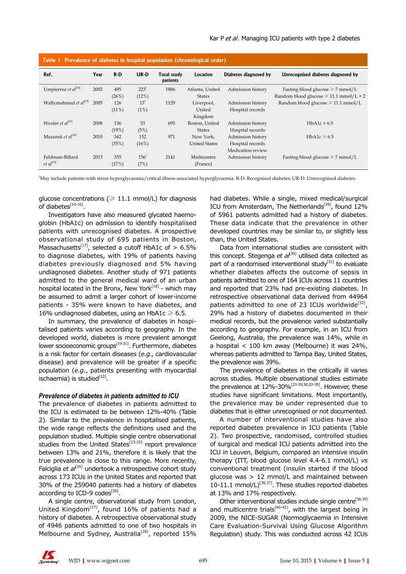
glucose concentrations (≥ 11.1 mmol/L) for diagnosis of diabetes[14-16].
Investigators have also measured glycated haemo-globin (HbA1c) on admission to identify hospitalised patients with unrecognised diabetes. A prospective observational study of 695 patients in Boston, Massachusetts[17], selected a cutoff HbA1c of > 6.5% to diagnose diabetes, with 19% of patients having diabetes previously diagnosed and 5% having undiagnosed diabetes. Another study of 971 patients admitted to the general medical ward of an urban hospital located in the Bronx, New York[18] - which may be assumed to admit a larger cohort of lower-income patients - 35% were known to have diabetes, and 16% undiagnosed diabetes, using an HbA1c ≥ 6.5.
In summary, the prevalence of diabetes in hospi-talised patients varies according to geography. In the developed world, diabetes is more prevalent amongst lower socioeconomic groups[19-21]. Furthermore, diabetes is a risk factor for certain diseases (e.g., cardiovascular disease) and prevalence will be greater if a specific population (e.g., patients presenting with myocardial ischaemia) is studied[22].
Prevalence of diabetes in patients admitted to ICUThe prevalence of diabetes in patients admitted to the ICU is estimated to be between 12%-40% (Table 2). Similar to the prevalence in hospitalised patients, the wide range reflects the definitions used and the population studied. Multiple single centre observational studies from the United States[23-25] report prevalence between 13% and 21%, therefore it is likely that the true prevalence is close to this range. More recently, Falciglia et al[26] undertook a retrospective cohort study across 173 ICUs in the United States and reported that 30% of the 259040 patients had a history of diabetes according to ICD-9 codes[26].
A single centre, observational study from London, United Kingdom[27], found 16% of patients had a history of diabetes. A retrospective observational study of 4946 patients admitted to one of two hospitals in Melbourne and Sydney, Australia[28], reported 15%
had diabetes. While a single, mixed medical/surgical ICU from Amsterdam, The Netherlands[29], found 12% of 5961 patients admitted had a history of diabetes. These data indicate that the prevalence in other developed countries may be similar to, or slightly less than, the United States.
Data from international studies are consistent with this concept. Stegenga et al[30] utilised data collected as part of a randomised interventional study[31] to evaluate whether diabetes affects the outcome of sepsis in patients admitted to one of 164 ICUs across 11 countries and reported that 23% had pre-existing diabetes. In retrospective observational data derived from 44964 patients admitted to one of 23 ICUs worldwide[32], 29% had a history of diabetes documented in their medical records, but the prevalence varied substantially according to geography. For example, in an ICU from Geelong, Australia, the prevalence was 14%, while in a hospital < 100 km away (Melbourne) it was 24%, whereas patients admitted to Tampa Bay, United States, the prevalence was 39%.
The prevalence of diabetes in the critically ill varies across studies. Multiple observational studies estimate the prevalence at 12%-30%[23-29,30,32-35]. However, these studies have significant limitations. Most importantly, the prevalence may be under represented due to diabetes that is either unrecognised or not documented.
A number of interventional studies have also reported diabetes prevalence in ICU patients (Table 2). Two prospective, randomised, controlled studies of surgical and medical ICU patients admitted into the ICU in Leuven, Belgium, compared an intensive insulin therapy (ITT, blood glucose level 4.4-6.1 mmol/L) vs conventional treatment (insulin started if the blood glucose was > 12 mmol/L and maintained between 10-11.1 mmol/L)[36,37]. These studies reported diabetes at 13% and 17% respectively.
Other interventional studies include single centre[38,39] and multicentre trials[40-42], with the largest being in 2009, the NICE-SUGAR (Normoglycaemia in Intensive Care Evaluation-Survival Using Glucose Algorithm Regulation) study. This was conducted across 42 ICUs
695 June 10, 2015|Volume 6|Issue 5|WJD|www.wjgnet.com
Table 1 Prevalence of diabetes in hospital population (chronological order)
Ref. Year R-D UR-D Total study patients
Location Diabetes diagnosed by Unrecognised diabetes diagnosed by
Umpierrez et al[14] 2002 495 (26%)
2231
(12%)1886 Atlanta, United
StatesAdmission history Fasting blood glucose ≥ 7 mmol/L
Random blood glucose ≥ 11.1 mmol/L × 2Wallymahmed et al[15] 2005 126
(11%)131
(1%)1129 Liverpool,
United Kingdom
Admission historyHospital records
Random blood glucose ≥ 11.1 mmol/L
Wexler et al[17] 2008 136 (19%)
33 (5%)
695 Boston, United States
Admission historyHospital records
HbA1c > 6.5
Mazurek et al[18] 2010 342 (35%)
152 (16%)
971 New York, United States
Admission historyHospital records
Medication review
HbA1c ≥ 6.5
Feldman-Billard et al[16]
2013 355 (17%)
1561
(7%)2141 Multicentre
(France)Admission history Fasting blood glucose ≥ 7 mmol/L
1May include patients with stress hyperglycaemia/critical illness associated hyperglycaemia. R-D: Recognised diabetes; UR-D: Unrecognised diabetes.
Kar P et al . Managing ICU patients with type 2 diabetes

body of evidence suggests, chronic glucose control may have implications on optimal acute glucose ranges in the critically ill.
Hospital and ICU prevalence of unrecognised diabetes can be estimated from the studies mentioned (Tables 1 and 2) along with other studies cited below (Table 3). Hospital prevalence is estimated to be between 5%-16%[16-18,43] and ICU prevalence between 6%-14%[34,44]. The prevalence in patients with ischaemic heart disease (e.g., presenting with acute myocardial infarction) appears to be higher[45,46].
In two European studies, patients with an acute myocardial infarct and without a history of diabetes subsequently underwent an oral glucose tolerance test (OGTT) to diagnose diabetes[45,46]. The prevalence of diabetes was found to be over 30% at discharge, and between 25%-31% at 3 mo. In London (United Kingdom), Emergency Department patients were
throughout Australia, New Zealand and Canada[41], and noted 20% of its 6029 patients with a history of diabetes, with the majority (92%) having type 2 diabetes.
It should be recognised that there are limitations to using data from these interventional studies. Inclusion into these studies usually requires hyperglycaemia and therefore leads to selection bias, which artificially increases any estimate of prevalence. The interventional trials estimated ICU prevalence at 13%-40%[36-42].
Prevalence of unrecognised diabetesPatients may have diabetes that is unrecognised prior to admission[2]. This may not represent “stress hyperglycaemia” or CIAH - as the hyperglycaemia is chronic rather than acute. Unrecognised diabetes is important as it not only impacts on estimations for the actual prevalence of the condition, but, as a growing
696 June 10, 2015|Volume 6|Issue 5|WJD|www.wjgnet.com
Table 2 Prevalence of diabetes in the intensive care unit population (chronological order)
Ref. Year Study type R-D UR-D Total study patients
Location Recognised DM diagnosis Unrecognised diabetes diagnosed by
Van den Berghe et al[36] 2001 Interv 204 (13%) N/A 1548 Leuven, Belgium
Admission history N/A
Finney et al[27] 2003 Observ 86 (16%) N/A 523 London, United Kingdom
Unknown N/A
Whitcomb et al[23] 2005 Observ 574 (21%) 3951 (15%) 2713 Baltimore, United States
Admission history Hyperglycaemia without a history of DM
Van den Berghe et al[37] 2006 Interv 203 (17%) N/A 1200 Leuven, Belgium
Admission history N/A
Krinsely[24] 2006 Observ 1110 (21%) N/A 5365 Stamford, United States
Hospital records (ICD-9 codes) for the first 2 yr then all available info
N/A
Egi et al[28] 2008 Observ 728 (15%) N/A 4946 Multicentre (Australia)
Hospital records N/A
Treggiari et al[25] 2008 Observ 1361 (13%) N/A 10456 Seattle, United States
Hospital records N/A
Arabi et al[39] 2008 Interv 208 (40%) N/A 523 Riyadh, Saudi Arabia
Admission historyHospital records
N/A
Bronkhurst et al[38] 2008 Interv 163 (30%) N/A 537 Multicentre (Germany)
Unknown N/A
Del La Rosa et al[42] 2008 Interv 61 (12%) N/A 504 Medellin, Colombia
Admission history N/A
Finfer et al[41] 2009 Interv 1211 (20%) N/A 6029 Multicentre (Australia, NZ,
Canada)
Admission history N/A
Preiser et al[40] 2009 Interv 203 (19%) N/A 1078 Multicentre (Europe)
Admission history N/A
Falciglia et al[26] 2009 Observ 77850 (30%) N/A 259040 Multicentre (United States)
Hospital records (ICD-9 codes)
N/A
Stegenga et al[30] 2010 Observ 188 (23%) N/A 830 Multicentre (Worldwide)
Admission history N/A
Hermanides et al[29] 2010 Observ 699 (12%) N/A 5961 Amsterdam, Netherlands
Hospital records (computerised system)
N/A
Krinsely et al[33] 2011 Observ 669 (21%) N/A 3263 Multicentre (United States,
Europe)
Hospital records (ICU clinical database)
N/A
Krinsley et al[32] 2013 Observ 12880 (29%) N/A 44964 Multicentre (Worldwide)
Admission history N/A
Plummer et al[34] 2014 Observ 220 (22%) 55 (6%) 1000 Adelaide, Australia
Admission historyPhone call to GP
HbA1c ≥ 6.5
HbA1c ≥ 6.5 without a history of DM
1May include patients with stress hyperglycaemia/critical illness associated hyperglycaemia. Interv: Interventional; Observ: Observational; R-D: Recognised diabetes; UR-D: Unrecognised diabetes; NZ: New Zealand; N/A: Not available.
Kar P et al . Managing ICU patients with type 2 diabetes
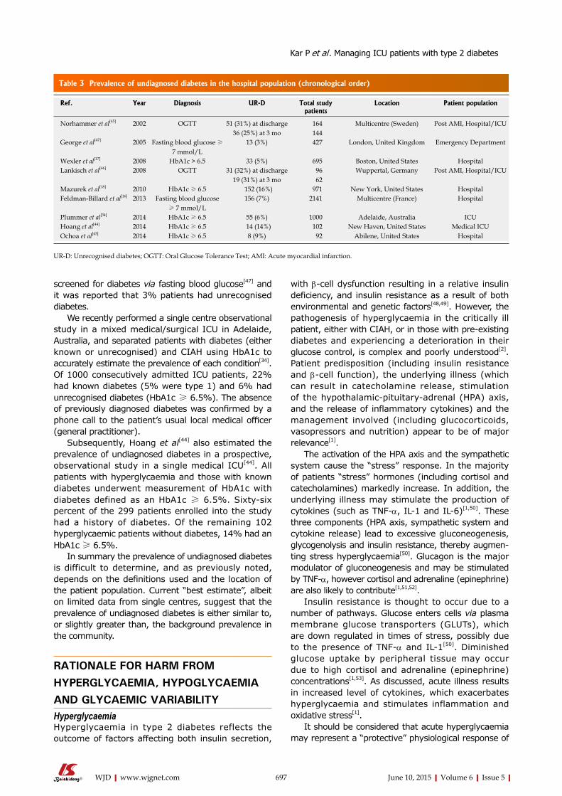
screened for diabetes via fasting blood glucose[47] and it was reported that 3% patients had unrecognised diabetes.
We recently performed a single centre observational study in a mixed medical/surgical ICU in Adelaide, Australia, and separated patients with diabetes (either known or unrecognised) and CIAH using HbA1c to accurately estimate the prevalence of each condition[34]. Of 1000 consecutively admitted ICU patients, 22% had known diabetes (5% were type 1) and 6% had unrecognised diabetes (HbA1c ≥ 6.5%). The absence of previously diagnosed diabetes was confirmed by a phone call to the patient’s usual local medical officer (general practitioner).
Subsequently, Hoang et al[44] also estimated the prevalence of undiagnosed diabetes in a prospective, observational study in a single medical ICU[44]. All patients with hyperglycaemia and those with known diabetes underwent measurement of HbA1c with diabetes defined as an HbA1c ≥ 6.5%. Sixty-six percent of the 299 patients enrolled into the study had a history of diabetes. Of the remaining 102 hyperglycaemic patients without diabetes, 14% had an HbA1c ≥ 6.5%.
In summary the prevalence of undiagnosed diabetes is difficult to determine, and as previously noted, depends on the definitions used and the location of the patient population. Current “best estimate”, albeit on limited data from single centres, suggest that the prevalence of undiagnosed diabetes is either similar to, or slightly greater than, the background prevalence in the community.
RATIONALE FOR HARM FROM HYPERGLYCAEMIA, HYPOGLYCAEMIA AND GLYCAEMIC VARIABILITYHyperglycaemiaHyperglycaemia in type 2 diabetes reflects the outcome of factors affecting both insulin secretion,
with β-cell dysfunction resulting in a relative insulin deficiency, and insulin resistance as a result of both environmental and genetic factors[48,49]. However, the pathogenesis of hyperglycaemia in the critically ill patient, either with CIAH, or in those with pre-existing diabetes and experiencing a deterioration in their glucose control, is complex and poorly understood[2]. Patient predisposition (including insulin resistance and β-cell function), the underlying illness (which can result in catecholamine release, stimulation of the hypothalamic-pituitary-adrenal (HPA) axis, and the release of inflammatory cytokines) and the management involved (including glucocorticoids, vasopressors and nutrition) appear to be of major relevance[1].
The activation of the HPA axis and the sympathetic system cause the “stress” response. In the majority of patients “stress” hormones (including cortisol and catecholamines) markedly increase. In addition, the underlying illness may stimulate the production of cytokines (such as TNF-α, IL-1 and IL-6)[1,50]. These three components (HPA axis, sympathetic system and cytokine release) lead to excessive gluconeogenesis, glycogenolysis and insulin resistance, thereby augmen-ting stress hyperglycaemia[50]. Glucagon is the major modulator of gluconeogenesis and may be stimulated by TNF-α, however cortisol and adrenaline (epinephrine) are also likely to contribute[1,51,52].
Insulin resistance is thought to occur due to a number of pathways. Glucose enters cells via plasma membrane glucose transporters (GLUTs), which are down regulated in times of stress, possibly due to the presence of TNF-α and IL-1[50]. Diminished glucose uptake by peripheral tissue may occur due to high cortisol and adrenaline (epinephrine) concentrations[1,53]. As discussed, acute illness results in increased level of cytokines, which exacerbates hyperglycaemia and stimulates inflammation and oxidative stress[1].
It should be considered that acute hyperglycaemia may represent a “protective” physiological response of
697 June 10, 2015|Volume 6|Issue 5|WJD|www.wjgnet.com
Table 3 Prevalence of undiagnosed diabetes in the hospital population (chronological order)
Ref. Year Diagnosis UR-D Total study patients
Location Patient population
Norhammer et al[45] 2002 OGTT 51 (31%) at discharge36 (25%) at 3 mo
164 144
Multicentre (Sweden) Post AMI, Hospital/ICU
George et al[47] 2005 Fasting blood glucose ≥ 7 mmol/L
13 (3%) 427 London, United Kingdom Emergency Department
Wexler et al[17] 2008 HbA1c > 6.5 33 (5%) 695 Boston, United States HospitalLankisch et al[46] 2008 OGTT 31 (32%) at discharge
19 (31%) at 3 mo 96 62
Wuppertal, Germany Post AMI, Hospital/ICU
Mazurek et al[18] 2010 HbA1c ≥ 6.5 152 (16%) 971 New York, United States HospitalFeldman-Billard et al[16] 2013 Fasting blood glucose
≥ 7 mmol/L156 (7%) 2141 Multicentre (France) Hospital
Plummer et al[34] 2014 HbA1c ≥ 6.5 55 (6%) 1000 Adelaide, Australia ICUHoang et al[44] 2014 HbA1c ≥ 6.5 14 (14%) 102 New Haven, United States Medical ICUOchoa et al[43] 2014 HbA1c ≥ 6.5 8 (9%) 92 Abilene, United States Hospital
UR-D: Unrecognised diabetes; OGTT: Oral Glucose Tolerance Test; AMI: Acute myocardial infarction.
Kar P et al . Managing ICU patients with type 2 diabetes
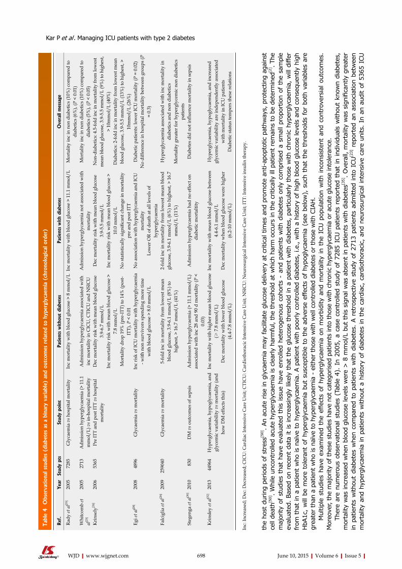
Tabl
e 4 O
bser
vation
al s
tudi
es (
diab
etes
as
a bi
nary
var
iabl
e) a
nd o
utco
mes
rel
ated
to
hype
rgly
caem
ia (
chro
nolo
gica
l ord
er)
the
host
dur
ing
period
s of
str
ess[5
0]. An
acut
e rise
in g
lyca
emia
may
fac
ilita
te g
luco
se d
eliv
ery
at c
ritic
al tim
es a
nd p
rom
ote
anti-
apop
totic
pat
hway
s, p
rote
ctin
g ag
ains
t ce
ll de
ath[5
0]. W
hile
unc
ontr
olle
d ac
ute
hype
rgly
caem
ia is
cle
arly
har
mfu
l, th
e th
resh
old
at w
hich
har
m o
ccur
s in
the
critic
ally
ill p
atie
nt r
emai
ns to
be d
eter
min
ed[2
] . Th
e m
ajor
ity o
f st
udie
s th
at h
ave
eval
uate
d th
is iss
ue h
ave
enro
lled
hete
roge
nous
coh
orts
- a
nd p
atie
nts
with
dia
bete
s on
ly c
ompr
ised
a s
mal
l pr
opor
tion
of t
he s
ampl
e ev
alua
ted.
Bas
ed o
n re
cent
dat
a it
is in
crea
sing
ly li
kely
tha
t th
e gl
ucos
e th
resh
old
in a
pat
ient
with
dia
bete
s, p
artic
ular
ly t
hose
with
chr
onic
hyp
ergl
ycae
mia
, w
ill d
iffer
from
tha
t in
a p
atie
nt w
ho is
naï
ve t
o hy
perg
lyca
emia
. A p
atie
nt w
ith p
oorly
cont
rolle
d di
abet
es, i.e
., w
ith a
his
tory
of hi
gh b
lood
glu
cose
leve
ls a
nd c
onse
quen
tly h
igh
HbA
1c,
will b
e m
ore
tole
rant
of
hype
rgly
caem
ia b
ut s
usce
ptib
le t
o th
e ad
vers
e ef
fect
s of
hyp
ogly
caem
ia (
see
belo
w),
suc
h th
at t
he t
hres
hold
s fo
r bo
th v
aria
bles
are
gr
eate
r th
an a
pat
ient
who
is n
aïve
to
hype
rgly
caem
ia -
eith
er tho
se w
ith w
ell c
ontr
olle
d di
abet
es o
r th
ose
with
CIA
H.
Mul
tipl
e st
udie
s ha
ve e
xam
ined
the
effec
ts o
f hy
perg
lyca
emia
on
mor
bidi
ty a
nd m
orta
lity
in t
he I
CU p
opul
atio
n w
ith
inco
nsis
tent
and
con
trov
ersi
al o
utco
mes
. M
oreo
ver,
the
maj
ority
of th
ese
stud
ies
have
not
cat
egor
ised
pat
ient
s in
to tho
se w
ith c
hron
ic h
yper
glyc
aem
ia o
r ac
ute
gluc
ose
into
lera
nce.
Th
ere
are
num
erou
s ob
serv
atio
nal st
udie
s (T
able
4).
In
2005
, a
case
con
trol
led
stud
y of
728
5 IC
U p
atie
nts
repo
rted
tha
t in
ind
ivid
uals
with
out
know
n di
abet
es,
mor
talit
y w
as in
crea
sed
whe
n bl
ood
gluc
ose
leve
ls w
ere
> 8
mm
ol/L
but
thi
s si
gnal
was
abs
ent
in p
atie
nts
with
dia
bete
s[35]. O
vera
ll, m
orta
lity
was
sig
nific
antly
gre
ater
in
pat
ient
s w
ithou
t di
abet
es w
hen
com
pare
d to
pat
ient
s w
ith d
iabe
tes.
A r
etro
spec
tive
stud
y of
271
3 pa
tient
s ad
mitt
ed int
o IC
U[2
3] r
epor
ted
an a
ssoc
iatio
n be
twee
n m
orta
lity
and
hype
rgly
caem
ia in
patie
nts
with
out
a hi
stor
y of
dia
bete
s in
the
car
diac
, ca
rdio
thor
acic
, an
d ne
uros
urgi
cal in
tens
ive
care
uni
ts.
In a
n au
dit
of 5
365
ICU
698 June 10, 2015|Volume 6|Issue 5|WJD|www.wjgnet.com
Ref
.Yea
rSt
udy
pts
Stud
y po
int
Patien
ts w
itho
ut d
iabe
tes
Patien
ts w
ith
diab
etes
Ove
rall
mes
sage
Rady
et a
l[35]
2005
7
285
Gly
caem
ia v
s hos
pita
l mor
talit
yIn
c m
orta
lity
with
blo
od g
luco
se >
8 m
mol
/LIn
c m
orta
lity
with
blo
od g
luco
se >
11.
1 m
mol
/LM
orta
lity
inc
in n
on d
iabe
tics
(10%
) com
pare
d to
di
abet
ics
(6%
), (P
< 0
.01)
Whi
tcom
b et
al
[23]
2005
2
713
Adm
issi
on h
yper
glyc
aem
ia (>
11.
1 m
mol
/L) v
s in-
hosp
ital m
orta
lity
Adm
issi
on h
yper
glyc
aem
ia a
ssoc
iate
d w
ith
inc
mor
talit
y in
CIC
U, C
TIC
U a
nd N
SIC
UA
dmis
sion
hyp
ergl
ycae
mia
not
ass
ocia
ted
with
m
orta
lity
Mor
talit
y in
c in
non
dia
betic
s (1
0%) c
ompa
red
to
diab
etic
s (5
%),
(P <
0.0
5)K
rins
ely[2
4]20
06
536
5Pr
e IT
T an
d po
st IT
T vs
hos
pita
l m
orta
lity
Dec
mor
talit
y ri
sk w
ith m
ean
bloo
d gl
ucos
e 3.
9-6.
7 m
mol
/LIn
c m
orta
lity
risk
with
mea
n bl
ood
gluc
ose
> 7.
8 m
mol
/LM
orta
lity
drop
19%
(pre
-ITT)
to 1
4% (p
ost-
ITT)
, P <
0.0
1
Dec
mor
talit
y ri
sk w
ith m
ean
bloo
d gl
ucos
e 3.
9-5.
5 m
mol
/LIn
c m
orta
lity
risk
with
mea
n bl
ood
gluc
ose
> 10
.0 m
mol
/LN
o st
atis
tical
ly s
igni
fican
t cha
nge
in m
orta
lity
pre
and
post
ITT
Non
-dia
betic
s: 4
.5-fo
ld in
c in
mor
talit
y fr
om lo
wes
t m
ean
bloo
d gl
ucos
e, 3
.9-5
.5 m
mol
/L (9
%) t
o hi
ghes
t, >
10m
mol
/L (4
0%)
Dia
betic
s: 2
-fold
inc
in m
orta
lity
from
low
est m
ean
bloo
d gl
ucos
e, 3
.9-5
.5 m
mol
/L (1
3%) t
o hi
ghes
t, >
10m
mol
/L (2
6%)
Egi e
t al[2
8]20
08
489
6G
lyca
emia
vs m
orta
lity
Inc
risk
of I
CU
mor
talit
y w
ith h
yper
glyc
aem
ia
- with
non
sur
vivo
rs s
pend
ing
mor
e tim
e w
ith b
lood
glu
cose
> 8
.0 m
mol
/L
No
asso
ciat
ion
with
hyp
ergl
ycae
mia
and
ICU
m
orta
lity
Low
er O
R of
dea
th a
t all
leve
ls o
f hy
perg
lyca
emia
Dia
betic
pat
ient
s: lo
wer
ICU
mor
talit
y (P
= 0
.02)
No
diffe
renc
e in
hos
pita
l mor
talit
y be
twee
n gr
oups
(P
= 0.
3)
Falc
iglia
et a
l[26]
2009
2590
40G
lyca
emia
vs m
orta
lity
5-fo
ld in
c in
mor
talit
y fr
om lo
wes
t mea
n bl
ood
gluc
ose,
3.9
-6.1
mm
ol/L
(8%
) to
high
est,
> 16
.7 m
mol
/L (4
1%)
2-fo
ld in
c in
mor
talit
y fr
om lo
wes
t mea
n bl
ood
gluc
ose,
3.9
-6.1
mm
ol/L
(6%
) to
high
est,
> 16
.7
mm
ol/L
(11%
)
Hyp
ergl
ycae
mia
ass
ocia
ted
with
inc
mor
talit
y in
di
abet
ics
and
non
diab
etic
sM
orta
lity
grea
ter f
or h
yper
glyc
emic
non
dia
betic
s pa
tient
sSt
egen
ga et
al[3
0]20
10
83
0D
M v
s out
com
es o
f sep
sis
Adm
issi
on h
yper
glyc
aem
ia (>
11.
1 m
mol
/L)
asso
ciat
ed w
ith in
c 28
and
90
d m
orta
lity
(P <
0.
03)
Adm
issi
on h
yper
glyc
aem
ia h
ad n
o ef
fect
on
diab
etic
mor
talti
tyD
iabe
tes
did
not i
nflue
nce
mor
talit
y in
sep
sis
Kri
nsle
y et
al[3
2]20
13 4
4964
Hyp
ergl
ycae
mia
, hyp
ogly
caem
ia, a
nd
glyc
emic
var
iabi
lity
vs m
orta
lity
(and
ho
w D
M e
ffect
s th
is)
Inc
mor
talit
y w
ith h
ighe
r mea
n bl
ood
gluc
ose
(≥ 7
.8 m
mol
/L)
Dec
mor
talit
y w
ith lo
wer
blo
od g
luco
se
(4.4
-7.8
mm
ol/L
)
Inc
mor
talit
y w
ith m
ean
bloo
d gl
ucos
e be
twee
n 4.
4-6.
1 m
mol
/LD
ec m
orta
lity
whe
n bl
ood
gluc
ose
wer
e hi
gher
(6
.2-1
0 m
mol
/L)
Hyp
ergl
ycae
mia
, hyp
ogly
caem
ia, a
nd in
crea
sed
glyc
emic
var
iabi
lity
are
inde
pend
ently
ass
ocia
ted
with
mor
talit
y in
ICU
pat
ient
sD
iabe
tic s
tatu
s te
mpe
rs th
ese
rela
tions
Inc:
Incr
ease
d; D
ec: D
ecre
ased
; CIC
U: C
ardi
ac In
tens
ive
Car
e U
nit;
CTI
CU
: Car
diot
hora
cic
Inte
nsiv
e C
are
Uni
t; N
SIC
U: N
euro
surg
ical
Inte
nsiv
e C
are
Uni
t; IT
T: In
tens
ive
insu
lin th
erap
y.
Kar P et al . Managing ICU patients with type 2 diabetes
patients evaluated before and after implementation of an intensive glucose control policy[24], mortality was increased in patients with hyperglycaemia who were not known to have diabetes when compared to those with diabetes. In 2008, Egi et al[28] reported a retrospective study of 4946 patients in which ICU mortality increased with increasing mean blood glucose level in patients without diabetes but this signal of harm was absent in those with pre-existing diabetes[28].
A retrospective cohort study of 259040 ICU ad-missions also reported an association between mortality and hyperglycaemia, with the relationship far stronger in patients without a diagnosis of diabetes when compared to those with pre-existing diabetes[26]. A retrospective analysis of a previous study[31] included 830 patients admitted with severe sepsis (defined as sepsis associated with acute organ dysfunction)[30], and reported that hyperglycaemia was predictive of subsequent death in those patients not known to have diabetes. Additionally, a multicentre retrospective study of 44964 patients divided into 2 cohorts (with and without known diabetes)[32], reported increased mortality with higher mean blood glucose concentrations (≥ 7.8 mmol/L) when compared to blood glucose concentrations 4.4-7.8 mmol/L in patients without diabetes. In contrast, patients with diabetes were more likely to die when mean blood glucose concentrations were between 4.4-6.1 mmol/L when compared to patients with greater blood glucose concentrations (6.2-10 mmol/L).
A number of interventional studies have evaluated the relationship between chronic and acute hyper-glycaemia and outcomes (Table 5). In a pooled analysis of studies conducted in a single centre in Leuven, intensive insulin therapy (ITT, aiming for blood glucose concentrations between 4.4-6.1 mmol/L) was reported to reduce mortality and morbidity in patients without a diagnosis of diabetes, but this was not the case in patients with diabetes, if anything, there was a trend for harm with intensive insulin therapy in patients with diabetes such that mortality was non-significantly greater at a lower mean blood glucose range (6.1-8.3 mmol/L, 21.2% vs < 6.1 mmol/L, 26.2%, P = 0.4 and > 8.3 mmol/L, 21.6%, P = 0.9)[54].
Subsequently, a number of interventional, ran-domised, controlled trials, containing patients with diabetes, comparing ITT to more conventional glucose targets have been published[38-42]. A trial of 523 mixed (medical and surgical) ICU patients[39] reported no survival benefit in patients with diabetes with ITT, but ITT was associated with an increased prevalence of hypoglycaemia. The Efficacy of Volume Substitution and Insulin Therapy in Severe Sepsis (VISEP) study assigned 537 ICU patients with severe sepsis to either ITT or more conventional glucose targets while receiving either 10% pentastarch or a modified Ringers lactate in a two-by-two factorial study[38]. The study was suspended at interim analysis for safety reasons
with ITT being associated with increases in episodes of severe hypoglycaemia and adverse events. De La Rosa et al[42] also evaluated ITT in 504 ICU patients (61 with diabetes) and there was no mortality or morbidity benefit observed, but an associated increased risk of hypoglycaemia, when administering ITT.
In 2009, the NICE-SUGAR study compared ITT with conventional glucose control in 6029 ICU patients and established that the observations from the initial Leuven studies regarding ITT were not generalisable outside that specialised institution[41]. However, amongst the 1211 patients with pre-existing diabetes in the NICE-SUGAR study the administration of ITT did not appear more harmful than in patients without diabetes. The Glucontrol study[40], an international, multicentre trial involving over 1000 ICU patients was stopped early due to protocol violations, and it was, accordingly, underpowered. However, there was no evidence to suggest any benefit with ITT and data in patients with diabetes were not specifically described.
Recently a number of studies have attempted to measure chronic glycaemia as a dynamic (HbA1c), rather than a binary, variable (i.e., presence of diabetes - yes/no) (Table 6). Egi et al[55] performed a retrospective observational study of 415 patients with diabetes (from two Australian ICUs) in whom glycated haemoglobin (HbA1c) had been measured within 3 mo of their critical illness and evaluated how this measure of pre-existing glycaemia impacted on the interaction between acute glycaemia and mortality[55]. It was reported that in patients with elevated preadmission HbA1c levels (> 7%) the number of deaths were significantly fewer when blood glucose concentrations were > 10 mmol/L.
Consistent with this observation, we recently measured HbA1c on admission and glucose concentrations for the first 48 h of ICU admission[34] and observed that acute peak glucose concentrations were associated with increased mortality only in patients with adequate premorbid glycaemic control (defined as HbA1c < 7%), but not in patients with chronic hyperglycaemia (defined as an HbA1c ≥ 7%). This finding was also supported by Hoang et al[44] who assessed the prevalence of undiagnosed diabetes (i.e., HbA1c ≥ 6.5%) among those with hyperglycaemia in a medical ICU. Patients with an HbA1c ≥ 6.5% were found to have significantly lower mortality compared to those with an HbA1c < 6.5% (11.7% vs 19.3%, P = 0.038), despite having greater glucose concentrations.
In summary the outcomes of the largest and most generalisable randomised study are consistent with the concept that the optimal glucose concentrations in unselected critically ill patients are between 6-10 mmol/L[41]. However, observational data, post-hoc analysis of interventional studies and studies measuring chronic glycaemia as a dynamic variable suggest that patients with pre-existing diabetes may warrant higher targets. Indeed, there is increasing data suggesting that targets should be personalised depending on both
699 June 10, 2015|Volume 6|Issue 5|WJD|www.wjgnet.com
Kar P et al . Managing ICU patients with type 2 diabetes
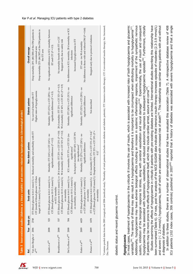
Tabl
e 5 In
terv
ention
al s
tudi
es (
diab
etes
as
a bi
nary
var
iabl
e) a
nd o
utco
mes
rel
ated
to
hype
rgly
caem
ia (
chro
nolo
gica
l ord
er)
diab
etic
sta
tus
and
rece
nt g
lyca
emic
con
trol
.
Hypo
glyc
aem
iaIn
mos
t ca
ses,
tre
atm
ent of
hyp
ergl
ycae
mia
in the
critic
ally
ill i
nvol
ves
the
use
of in
sulin
, w
hich
is a
ssoc
iate
d w
ith in
crea
sed
risk
s of
bot
h hy
pogl
ycae
mia
and
gly
caem
ic
variab
ility
[56]. Th
e se
verity
of illne
ss m
ay a
lso
resu
lt in
a h
ypog
lyca
emia
and
the
refo
re it
is im
port
ant
to b
e ci
rcum
spec
t w
hen
attr
ibut
ing
mor
talit
y to
hyp
ogly
caem
ia[5
7].
Add
ition
ally
, hy
pogl
ycae
mia
may
hav
e ad
vers
e bi
olog
ical
effec
ts inc
ludi
ng a
n in
crea
se in
syst
emic
infl
amm
ator
y re
spon
se,
impa
irm
ent
of t
he s
ympa
thet
ic n
ervo
us
syst
em,
inhi
bition
of
the
biol
ogic
al r
espo
nse
to s
tres
s, a
long
with
cere
bral
vas
odila
tion
and
neu
ral da
mag
e[2,5
8] .
Expe
rim
enta
lly,
the
use
of ins
ulin
and
con
sequ
ent
hypo
glyc
aem
ia m
ay b
e as
soci
ated
with
hyp
oten
sion
, va
sodi
latio
n, a
nd r
educ
ed a
uton
omic
res
pons
es t
o su
bseq
uent
hyp
ogly
caem
ic e
piso
des[5
8]. Fu
rthe
rmor
e, c
ritic
ally
ill p
atie
nts
may
be
mor
e pr
one
to the
effec
ts o
f hy
pogl
ycae
mia
itse
lf, w
hich
may
incl
ude
card
iac
arre
st, se
izur
e an
d co
ma[5
9].
Stu
dies
exa
min
ing
the
effe
cts
of h
ypog
lyca
emia
in c
ritic
ally
ill p
atie
nts
with
pre
-exi
stin
g di
abet
es a
re li
mite
d. I
nter
vent
iona
l stu
dies
des
crib
ing
this
rel
atio
nshi
p ha
ve
been
sum
mar
ised
(Ta
ble
6). O
f no
te, po
st h
oc a
naly
sis
of the
NIC
E-SUG
AR d
ata
indi
cate
tha
t in
tens
ive
insu
lin the
rapy
incr
ease
s ep
isod
es o
f m
oder
ate
(2.3
-3.9
mm
ol/L
) an
d se
vere
(≤
2.2
mm
ol/L
) hy
pogl
ycae
mia
, bo
th o
f w
hich
are
ass
ocia
ted
with
incr
ease
d risk
of de
ath[5
6]. Th
is r
elat
ions
hip
was
sim
ilar
amon
g pa
tient
s w
ith a
nd w
ithou
t a
diag
nosi
s of
dia
bete
s.
In a
dditi
on t
o th
ese
stud
ies,
the
re a
re a
num
ber
of o
bser
vatio
nal st
udie
s th
at h
ave
eval
uate
d th
is a
ssoc
iatio
n (T
able
7).
A r
etro
spec
tive
data
base
rev
iew
of
408
ICU p
atie
nts
(102
ind
ex c
ases
, 30
6 co
ntro
ls)
publ
ishe
d in
200
7[60] r
epor
ted
that
a h
isto
ry o
f di
abet
es w
as a
ssoc
iate
d w
ith s
ever
e hy
pogl
ycae
mia
and
tha
t a
sing
le
700 June 10, 2015|Volume 6|Issue 5|WJD|www.wjgnet.com
Ref
.Yea
rSt
udy
pts
Stud
y po
int
Non
dia
betic
patien
tsD
iabe
tic
patien
tsO
vera
ll m
essa
ge
Van
den
Ber
ghe
et
al[5
4]1
2006
2748
ITT
(blo
od g
luco
se 4
.4-6
.1 m
mol
/L) v
s C
IT (i
nsul
in if
blo
od g
luco
se >
12
then
ta
rget
10-
11.1
mm
ol/L
) on
mor
talit
y
Redu
ced
mor
talit
y an
d m
orbi
dity
with
ITT
No
surv
ival
ben
efit w
ith IT
TH
ighe
r rat
es o
f hyp
ogly
caem
iaH
osp
mor
talit
y 20
% (4
0/20
0) o
f the
DM
pat
ient
s in
co
nven
tiona
l arm
H
osp
mor
talit
y 22
% (4
6/20
7) o
f the
DM
pat
ient
s in
th
e IT
T ar
m
Ara
bi et
al[3
9]20
08 5
23IT
T (b
lood
glu
cose
4.4
-6.1
mm
ol/L
) vs
CIT
(blo
od g
luco
se 1
0-11
.1 m
mol
/L)
on IC
U m
orta
lity
Mor
talit
y: IT
T (1
4%) v
s CIT
(14%
) - n
o si
gnifi
cant
diff
eren
ce (P
= 0
.2)
Mor
talit
y: IT
T (1
3%) v
s CIT
(20%
) - n
o si
gnifi
cant
diff
eren
ce (P
= 0
.3)
No
sign
ifica
nt d
iffer
ence
in IC
U m
orta
lity
betw
een
IIT
and
CIT
(P =
0.3
)
Brun
khor
st et
al[3
8]20
08 5
37IT
T (b
lood
glu
cose
4.4
-6.1
mm
ol/L
) vs
CIT
(blo
od g
luco
se 1
0-11
.1 m
mol
/L)
on m
orta
lity
28 d
mor
talit
y: IT
T 25
% v
s CIT
23%
(P =
0.8
)90
d m
orta
lity:
ITT
40%
vs C
IT 3
2% (P
= 0
.2)
28 d
mor
talit
y: IT
T 25
% v
s CIT
32%
(P =
0.3
)90
d m
orta
lity:
ITT
40%
vs C
IT 4
2% (P
= 0
.9)
No
mor
talit
y be
nefit
with
ITT
vs C
ITSt
oppe
d ea
rly
due
to s
afet
y ri
sk
Del
La
Rosa
et a
l[42]
2008
504
ITT
(blo
od g
luco
se 4
.4-6
.1 m
mol
/L) v
s C
IT (b
lood
glu
cose
10-
11.1
mm
ol/L
) on
mor
bidi
ty a
nd m
orta
lity
ICU
mor
talit
y IT
T 37
% v
s CIT
32%
(no
sign
ifica
nce)
2
In-h
ospi
tal m
orta
lity:
ITT
40%
vs C
IT 3
9%
(no
sign
ifica
nce)
2
Mor
talit
y: IT
T (3
8%) v
s CIT
(31%
) - n
o si
gnifi
cant
diff
eren
ceN
o di
ffere
nce
in IC
U m
orta
lity,
28
d m
orta
lity
or IC
U
infe
ctio
nsIn
crea
sed
hypo
glyc
aem
ia in
ITT
Finf
er et
al[4
1]20
0960
29IT
T (b
lood
glu
cose
4.4
-6.1
mm
ol/L
) vs
CIT
(blo
od g
luco
se <
10
mm
ol/L
) on
mor
talit
y
Mor
talit
y: IT
T (2
7%) v
s CIT
(24%
) - n
o si
gnifi
cant
diff
eren
ceM
orta
lity:
ITT
(32%
) vs C
IT (2
8%) -
no
sign
ifica
nt d
iffer
ence
ITT
arm
- in
c 90
d m
orta
lity
No
diffe
renc
e in
thos
e w
ith a
nd w
ithou
t DM
(P =
0.6
0)
Prei
ser e
t al[4
0]20
0910
78IT
T (b
lood
glu
cose
4.4
-6.1
mm
ol/L
) vs
CIT
(blo
od g
luco
se 7
.8-1
0 m
mol
/L)
on m
orta
lity
ICU
mor
talit
y IT
T 17
% v
s CIT
15%
(P =
0.4
) 2
Hos
pita
l mor
talit
y: IT
T 23
% v
s CIT
19%
(P =
0.
1)2
Not
des
crib
edSt
oppe
d ea
rly
due
to p
roto
col v
iola
tions
1 Con
tain
s po
oled
dat
a fr
om th
e 20
01 (s
urgi
cal)
and
2006
(med
ical
) stu
dy; 2 M
orta
lity
of to
tal p
atie
nts
(incl
udes
non
-dia
betic
and
dia
betic
pat
ient
s). I
TT: I
nten
sive
insu
lin th
erap
y; C
IT: C
onve
ntio
nal i
nsul
in th
erap
y; In
c: In
crea
sed;
D
ec: D
ecre
ase.
Kar P et al . Managing ICU patients with type 2 diabetes

hypoglycaemic episode was associated with an increased risk of mortality (compared with those without an episode of severe hypoglycaemia). Egi et al[61] reported mild or moderate hypoglycaemia was associated with mortality in critically ill patients - with mortality substantially increasing according to severity of hypoglycaemia - and patients with diabetes were more likely to suffer from insulin-associated hypoglycaemia.
The blood glucose threshold that adverse events occur may be greater in patients with pre-existing diabetes. In a retrospective multi-centre observational study[32] increased mortality was reported in 12880 patients with pre-existing diabetes who had mean glucose concentrations between 4.4-6.2 mmol/L. While the investigators were not able to differentiate between patients with well-controlled or poorly-controlled diabetes, these data support the concept that the threshold for “hypoglycaemia” may be increased in critically ill patients with diabetes when compared to non diabetic patients. For example, if a patient typically has blood glucose concentrations above 10 mmol/L, and, in hospital, insulin is administered to achieve blood glucose concentration of about 6 mmol/L, this may result in a “relative” hypoglycaemia.
Glycaemic variabilityGlycaemic variability (GV) describes the fluctuations in blood glucose concentrations, as marked fluctua-tions may be associated with multiple adverse effects such as apoptosis, cytokine production and increased markers of oxidative stress[59]. Oxidative stress markers have been shown to increase with glucose fluctuations[62,63]. GV may be assessed by a number of methods. Techniques to quantify variability are reviewed
elsewhere[64].Multiple studies in the critically ill have established
as an association with poor outcomes and GV[44,65-71], however the evidence in patients with pre-existing diabetes is limited and inconsistent (Table 8). In 2006, Egi et al[65] published a retrospective, electronic database analysis of 7049 ICU patients in 4 centres around Australia, using standard deviation as a marker of glucose variability, and focusing on the association of blood glucose variability and mortality[65]. Both mean and standard deviation of blood glucose were independently associated with mortality.
A retrospective, single center cohort study of patients admitted with sepsis reported that GV was also independently associated with increased mortality and importantly, that this was independent of hypoglycaemia and the presence of diabetes[66]. Another retrospective study of 3252 patients reported that increased GV was associated with mortality[67] and diabetes was associated with greater GV. A prospective, observational study of 42 patients used non-lineal dynamics to measure glycaemia in time series[69]. Patients underwent continuous glucose monitoring system measuring interstitial glucose concentrations every 5 min for 48 h. The authors reported greater variability was associated with increasing mortality, even in patients with diabetes. However, given the small cohort, these results must be treated with caution.
Other studies have reported no relationship between mortality and GV in patients with diabetes. A retro-spective, observational study of 4084 critically ill patients (942 with known diabetes)[68] reported that GV was associated with mortality in patients without diabetes, but not in patients with diabetes. More
701 June 10, 2015|Volume 6|Issue 5|WJD|www.wjgnet.com
Table 6 Observational studies that have recorded chronic glycaemia as a dynamic variable (chronological order)
Ref. Year Study pts Study point Non diabetic patients Diabetic patients Overall message
Egi et al[55] 2011 415 Does preexisting hyperglycaemia modulate the association between glycemia and outcome in ICU patients
with DM
N/A Patients with elevated preadmission HbA1c levels (> 7%) showed a
mortality benefit when mean ICU glucose concentrations were > 10
mmol/L
Relationship between HbA1c and mortality changed
according to the levels of time-weighted average of blood
glucose concentrations
Plummer et al[34]
2014 1000 Prevalence of CIAH and recognized/unrecognized DM in ICU and to evaluate
the premorbid glycaemia on the association between acute hyperglycaemia and mortality
50% had CIAHRisk of death inc by
20% for each increase in acute glycaemia of
1 mmol/L
Well controlled DM (HbA1c < 6%) and adequately controlled (DM
6%-7%) - risk of death as per non diabetic patient
HbA1c ≥ 7% (insufficiently controlled DM) had no significance
between mortality and acute glycaemia
22% had recognised DM6% had unrecognised diabetes
Hoang et al[44]
2014 299 Prevalance of unrecognized DM amongst those with CIAH
and the association between baseline glycaemia and
mortality
102 (34%) had no history of DM
14/102 (14%) had unrecognized DM (diagnosed with
HbA1c ≥ 6.5)
197 (66%) had a history of DM Lower HbA1c had inc mortality (in this population of CIAH
patients) despite lower median glucose values and less glucose
variabilityMortality in HbA1c < 6.5 (19%) vs HbA1c ≥ 6.5 (12%), P = 0.04
Inc: Increased; Dec: Decreased; N/A: Not available.
Kar P et al . Managing ICU patients with type 2 diabetes

recently in the study by Hoang et al[44] of 299 patients there was no association between GV and mortality in their entire cohort, however the group with diabetes (128 patients) had a lower rate of mortality despite having a higher GV. Additionally, a retrospective analysis of 2782 ICU patients, comparing different GV indices and mean glucose concentrations to predict mortality and ICU acquired infections[70] reported that while GV was associated with infections and mortality in patients without pre-existing diabetes, in those with diabetes GV was greater but was not associated with either mortality or infection.
In summary, there is a strong relationship between GV and mortality in critically ill patients that has been confirmed in multiple studies. However, with respect to patients with diabetes, data are inconsistent. This may be due a number of factors, including small numbers studied resulting in lack of power, or that patients with chronic hyperglycaemia are protected somewhat by glycaemic excursions during acute illness. Research is warranted to further understand whether GV is harmful in patients with pre-existing diabetes.
RATIONALE FOR PERSONALISED THERAPY AND THAT THE HARM FROM EACH OF THESE DOMAINS MAY VARY ACCORDING TO PRE EXISTING PHYSIOLOGYDiabetes is known to be associated with a large burden
of illness in the outpatient setting and is associated with increased mortality[72]. Paradoxically, as discussed, multiple studies exist suggesting that acute hyper-glycaema in critically ill patients without diabetes (i.e., patients with CIAH) is associated with increased mortality and morbidity when compared to those with known diabetes[73]. There is growing evidence that chronic hyperglycaemia may lead to cellular conditioning, and that in fact, may be protective against acute hyperglycaemia mediated damage during an episode critical illness[1]. These outcomes suggest that current target glucose levels in patients naïve to hyperglycaemia, or those suffering from CIAH, may be harmful to those with chronic hyperglycaemia or poorly controlled diabetes.
CONCLUSIONThis review articulates the need for further research to be done to identify the ideal glucose targets in critically ill patient with pre-existing diabetes. Not only does hyperglycaemia occur frequently in this group, but, recent data suggests that targeted blood glucose concentrations may benefit from consideration of a patient’s premorbid glucose state.
Our recommendations are to avoid treating patients with diabetes as a homogenous group. Treatment of the critically ill patient with type 2 diabetes should be personalised to their internal milieu. There is preliminary evidence suggesting that higher blood glucose concentrations (e.g., up to 14 mmol/L) in patients with uncontrolled type 2 diabetes may not be
702 June 10, 2015|Volume 6|Issue 5|WJD|www.wjgnet.com
Table 7 Observational studies and outcomes related to hypoglycaemia (chronological order)
Ref. Year Study pts Study point Non diabetic patients Diabetic patients Overall message
Krinsley and Grover[60]
2007 408 Risk factors for developing
hypoglycaemia in ICU and outcomes
Severe hypoglycaemia associated with septic shock.
Renal insufficiency, mechanical ventilation, illness severity and use
of ITT
Associated with inc risk of severe hypoglycaemia
(P < 0.01)DM had no association
with mortality
Mortality in severe hypoglycaemia cohort 56% vs control cohort 40%, P < 0.01
Egi et al[61] 2010 4946 Hypoglycaemia vs risk of death in
critically ill patients
Mild or moderate hypoglycaemia was associated with mortality in
critically ill patientsMortality increases as severity of
hypoglycaemia increases
Diabetic patients more likely to suffer from insulin-associated
hypoglycaemia
22% of total patients had one episode of hypoglycaemia
Hospital mortality: hypoglycaemic cohort 37% vs control cohort 20%, P < 0.01
Krinsely et al[33] 2011 62401 Mild hypoglycaemia (blood glucose level
< 3.9 mmol/L) vs risk of mortality in
critically ill patients.
Mild hypoglycaemia was associated with a significantly increased risk of
mortality
The association between hypoglycaemia
and mortality was independent of diabetic
status
Inc severity of hypoglycaemia was associated with inc risk of
mortalityHypoglycemic patients had
higher mortality regardless of diagnostic category and ICU LOS
Krinsley et al[32] 2013 44964 Hyperglycaemia, hypoglycaemia, and glycemic variability
vs mortality (and how DM effects this)
Inc mortality with higher mean blood glucose (≥ 7.8 mmol/L)Dec mortality with lower blood
glucose (4.4-7.8 mmol/L)
Inc mortality with mean blood glucose between
4.4-6.1 mmol/LDec mortality when blood glucose were
higher (6.2-10 mmol/L)
Hyperglycaemia, hypoglycaemia, and increased
glycemic variability are independently associated with
mortality in ICU patientsDiabetic status tempers these
relations
1Contains partial data from one prospective RCT (Glucontrol trial) and complete data from two observational cohorts (United States and The Netherlands). Inc: Increased; Dec: Decrease; LOS: Length of stay.
Kar P et al . Managing ICU patients with type 2 diabetes
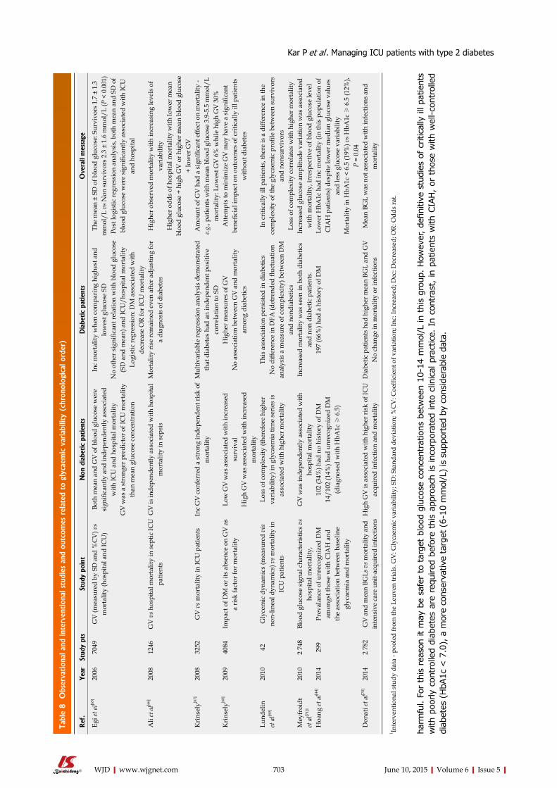
Tabl
e 8 O
bser
vation
al a
nd in
terv
ention
al s
tudi
es a
nd o
utco
mes
rel
ated
to
glyc
aem
ic v
aria
bilit
y (c
hron
olog
ical
ord
er)
harm
ful.
For
this
rea
son
it m
ay b
e sa
fer
to t
arge
t bl
ood
gluc
ose
conc
entr
atio
ns b
etw
een
10-1
4 m
mol
/L in
thi
s gr
oup.
How
ever
, de
finiti
ve s
tudi
es o
f cr
itica
lly il
l pat
ient
s w
ith p
oorly
cont
rolle
d di
abet
es a
re r
equi
red
befo
re t
his
appr
oach
is
inco
rpor
ated
int
o cl
inic
al p
ract
ice.
In
cont
rast
, in
pat
ient
s w
ith C
IAH
, or
tho
se w
ith w
ell-co
ntro
lled
diab
etes
(H
bA1c
< 7
.0),
a m
ore
cons
erva
tive
targ
et (
6-10
mm
ol/L
) is
sup
port
ed b
y co
nsid
erab
le d
ata.
703 June 10, 2015|Volume 6|Issue 5|WJD|www.wjgnet.com
Ref
.Yea
rSt
udy
pts
Stud
y po
int
Non
dia
betic
patien
tsD
iabe
tic
patien
tsO
vera
ll m
essa
ge
Egi e
t al[6
5]20
0670
49G
V (m
easu
red
by S
D a
nd %
CV
) vs
mor
talit
y (h
ospi
tal a
nd IC
U)
Both
mea
n an
d G
V o
f blo
od g
luco
se w
ere
sign
ifica
ntly
and
inde
pend
ently
ass
ocia
ted
with
ICU
and
hos
pita
l mor
talit
yG
V w
as a
str
onge
r pre
dict
or o
f IC
U m
orta
lity
than
mea
n gl
ucos
e co
ncen
trat
ion
Inc
mor
talit
y w
hen
com
pari
ng h
ighe
st a
nd
low
est g
luco
se S
DN
o ot
her s
igni
fican
t rel
atio
n w
ith b
lood
glu
cose
(S
D a
nd m
ean)
and
ICU
/hos
pita
l mor
talit
y Lo
gist
ic re
gres
sion
: DM
ass
ocia
ted
with
de
crea
se O
R fo
r IC
U m
orta
lity
The
mea
n ±
SD o
f blo
od g
luco
se: S
urvi
vors
1.7
± 1
.3
mm
ol/L
vs N
on s
urvi
vors
2.3
± 1
.6 m
mol
/L (P
< 0
.001
)Po
st lo
gist
ic re
gres
sion
ana
lysi
s, b
oth
mea
n an
d SD
of
bloo
d gl
ucos
e w
ere
sign
ifica
ntly
ass
ocia
ted
with
ICU
an
d ho
spita
l
Ali
et a
l[66]
2008
1246
GV
vs h
ospi
tal m
orta
lity
in s
eptic
ICU
pa
tient
sG
V is
inde
pend
ently
ass
ocia
ted
with
hos
pita
l m
orta
lity
in s
epsi
sM
orta
lity
rise
rem
aine
d ev
en a
fter a
djus
ting
for
a di
agno
sis
of d
iabe
tes
Hig
her o
bser
ved
mor
talit
y w
ith in
crea
sing
leve
ls o
f va
riab
ility
Hig
her o
dds
of h
ospi
tal m
orta
lity
with
low
er m
ean
bloo
d gl
ucos
e +
high
GV
or h
ighe
r mea
n bl
ood
gluc
ose
+ lo
wer
GV
Kri
nsel
y[67]
2008
3252
GV
vs m
orta
lity
in IC
U p
atie
nts
Inc
GV
con
ferr
ed a
str
ong
inde
pend
ent r
isk
of
mor
talit
yM
ultiv
aria
ble
regr
essi
on a
naly
sis
dem
onst
rate
d th
at d
iabe
tes
had
an in
depe
nden
t pos
itive
co
rrel
atio
n to
SD
Am
ount
of G
V h
ad a
sig
nific
ant e
ffect
on
mor
talit
y -
e.g.,
patie
nts
with
mea
n bl
ood
gluc
ose
3.9-
5.5
mm
ol/L
m
orta
lity:
Low
est G
V 6
% w
hile
hig
h G
V 3
0%K
rins
ely[6
8]20
0940
84Im
pact
of D
M o
r its
abs
ence
on
GV
as
a ri
sk fa
ctor
for m
orta
lity
Low
GV
was
ass
ocia
ted
with
incr
ease
d su
rviv
alH
igh
GV
was
ass
ocia
ted
with
incr
ease
d m
orta
lity
Hig
her m
easu
res
of G
VN
o as
soci
atio
n be
twee
n G
V a
nd m
orta
lity
amon
g di
abet
ics
Atte
mpt
s to
min
imiz
e G
V m
ay h
ave
a si
gnifi
cant
be
nefic
ial i
mpa
ct o
n ou
tcom
es o
f cri
tical
ly il
l pat
ient
s w
ithou
t dia
bete
s
Lund
elin
et
al[6
9]20
1042
Gly
cem
ic d
ynam
ics
(mea
sure
d vi
a no
n-lin
eal d
ynam
ics)
vs m
orta
lity
in
ICU
pat
ient
s
Loss
of c
ompl
exity
(the
refo
re h
ighe
r va
riab
ility
) in
glyc
aem
ia ti
me
seri
es is
as
soci
ated
with
hig
her m
orta
lity
This
ass
ocia
tion
pers
iste
d in
dia
betic
sN
o di
ffere
nce
in D
FA (d
etre
nded
fluc
tuat
ion
anal
ysis
a m
easu
re o
f com
plex
ity) b
etw
een
DM
an
d no
ndia
betic
s
In c
ritic
ally
ill p
atie
nts,
ther
e is
a d
iffer
ence
in th
e co
mpl
exity
of t
he g
lyca
emic
pro
file
betw
een
surv
ivor
s an
d no
nsur
vivo
rsLo
ss o
f com
plex
ity c
orre
late
s w
ith h
ighe
r mor
talit
yM
eyfr
oidt
et
al[7
1]1
2010
2 74
8Bl
ood
gluc
ose
sign
al c
hara
cter
istic
s vs
hosp
ital m
orta
lity,
GV
was
inde
pend
ently
ass
ocia
ted
with
ho
spita
l mor
talit
yIn
crea
sed
mor
talit
y w
as s
een
in b
oth
diab
etic
s an
d no
n di
abet
ic p
atie
nts.
Incr
ease
d gl
ucos
e am
plitu
de v
aria
tion
was
ass
ocia
ted
with
mor
talit
y, ir
resp
ectiv
e of
blo
od g
luco
se le
vel
Hoa
ng et
al[4
4]20
1429
9Pr
eval
ance
of u
nrec
ogni
zed
DM
am
ongs
t tho
se w
ith C
IAH
and
th
e as
soci
atio
n be
twee
n ba
selin
e gl
ycae
mia
and
mor
talit
y
102
(34%
) had
no
hist
ory
of D
M14
/102
(14%
) had
unr
ecog
nize
d D
M
(dia
gnos
ed w
ith H
bA1c
≥ 6
.5)
197
(66%
) had
a h
isto
ry o
f DM
Low
er H
bA1c
had
inc
mor
talit
y (in
this
pop
ulat
ion
of
CIA
H p
atie
nts)
des
pite
low
er m
edia
n gl
ucos
e va
lues
an
d le
ss g
luco
se v
aria
bilit
yM
orta
lity
in H
bA1c
< 6
.5 (1
9%) v
s HbA
1c ≥
6.5
(12%
), P
= 0.
04D
onat
i et a
l[70]
2014
2 78
2G
V a
nd m
ean
BGLs
vs m
orta
lity
and
inte
nsiv
e ca
re u
nit-a
cqui
red
infe
ctio
nsH
igh
GV
is a
ssoc
iate
d w
ith h
ighe
r ris
k of
ICU
ac
quir
ed in
fect
ion
and
mor
talit
yD
iabe
tic p
atie
nts
had
high
er m
ean
BGL
and
GV
No
chan
ge in
mor
talit
y or
infe
ctio
nsM
ean
BGL
was
not
ass
ocia
ted
with
infe
ctio
ns a
nd
mor
talit
y
1 Inte
rven
tiona
l stu
dy d
ata
- poo
led
from
the
Leuv
en tr
ials
. GV
: Gly
caem
ic v
aria
bilit
y; S
D: S
tand
ard
devi
atio
n; %
CV
: Coe
ffici
ent o
f var
iatio
n; In
c: In
crea
sed;
Dec
: Dec
reas
ed; O
R: O
dds
rat.
Kar P et al . Managing ICU patients with type 2 diabetes
REFERENCES1 Dungan KM, Braithwaite SS, Preiser JC. Stress hyperglycaemia.
Lancet 2009; 373: 1798-1807 [PMID: 19465235 DOI: 10.1016/S0140-6736(09)60553-5]
2 Deane AM, Horowitz M. Dysglycaemia in the critically ill - significance and management. Diabetes Obes Metab 2013; 15: 792-801 [PMID: 23368662 DOI: 10.1111/dom.12078]
3 Zhang P, Zhang X, Brown J, Vistisen D, Sicree R, Shaw J, Nichols G. Global healthcare expenditure on diabetes for 2010 and 2030. Diabetes Res Clin Pract 2010; 87: 293-301 [PMID: 20171754 DOI: 10.1016/j.diabres.2010.01.026]
4 Danaei G, Finucane MM, Lu Y, Singh GM, Cowan MJ, Paciorek CJ, Lin JK, Farzadfar F, Khang YH, Stevens GA, Rao M, Ali MK, Riley LM, Robinson CA, Ezzati M. National, regional, and global trends in fasting plasma glucose and diabetes prevalence since 1980: systematic analysis of health examination surveys and epidemiological studies with 370 country-years and 2·7 million participants. Lancet 2011; 378: 31-40 [PMID: 21705069 DOI: 10.1016/S0140-6736(11)60679-X]
5 Tanamas SK, Lynch B, Sethi P, Willenberg L, Polkinghorne KR, Chadban S, Dunstan D, Shaw JE. AusDiab 2012. The Australian Diabetes, Obesity and Lifestyle Study. Melbourne: Baker IDI Heart and Diabetes Institute, 2013
6 Shaw JE, Sicree RA, Zimmet PZ. Global estimates of the prevalence of diabetes for 2010 and 2030. Diabetes Res Clin Pract 2010; 87: 4-14 [PMID: 19896746 DOI: 10.1016/j.diabres.2009.10.007]
7 Cowie CC, Rust KF, Ford ES, Eberhardt MS, Byrd-Holt DD, Li C, Williams DE, Gregg EW, Bainbridge KE, Saydah SH, Geiss LS. Full accounting of diabetes and pre-diabetes in the U.S. population in 1988-1994 and 2005-2006. Diabetes Care 2009; 32: 287-294 [PMID: 19017771 DOI: 10.2337/dc08-1296]
8 American Diabetes Association. Diagnosis and classification of diabetes mellitus. Diabetes Care 2010; 33 Suppl 1: S62-S69 [PMID: 20042775 DOI: 10.2337/dc10-S062]
9 Colagiuri S. Glycated haemoglobin (HbA1c) for the diagnosis of diabetes mellitus--practical implications. Diabetes Res Clin Pract 2011; 93: 312-313 [PMID: 21820751 DOI: 10.1016/j.diabres.2011.06.025]
10 Carson AP, Reynolds K, Fonseca VA, Muntner P. Comparison of A1C and fasting glucose criteria to diagnose diabetes among U.S. adults. Diabetes Care 2010; 33: 95-97 [PMID: 19808920 DOI: 10.2337/dc09-1227]
11 Cowie CC, Rust KF, Byrd-Holt DD, Gregg EW, Ford ES, Geiss LS, Bainbridge KE, Fradkin JE. Prevalence of diabetes and high risk for diabetes using A1C criteria in the U.S. population in 1988-2006. Diabetes Care 2010; 33: 562-568 [PMID: 20067953 DOI: 10.2337/dc09-1524]
12 Sacks DB. A1C versus glucose testing: a comparison. Diabetes Care 2011; 34: 518-523 [PMID: 21270207 DOI: 10.2337/dc10-1546]
13 Lippi G, Targher G. Glycated hemoglobin (HbA1c): old dogmas, a new perspective? Clin Chem Lab Med 2010; 48: 609-614 [PMID: 20464776]
14 Umpierrez GE, Isaacs SD, Bazargan N, You X, Thaler LM, Kitabchi AE. Hyperglycemia: an independent marker of in-hospital mortality in patients with undiagnosed diabetes. J Clin Endocrinol Metab 2002; 87: 978-982 [PMID: 11889147 DOI: 10.1210/jcem.87.3.8341]
15 Wallymahmed ME, Dawes S, Clarke G, Saunders S, Younis N, MacFarlane IA. Hospital in-patients with diabetes: increasing prevalence and management problems. Diabet Med 2005; 22: 107-109 [PMID: 15606701 DOI: 10.1111/j.1464-5491.2004.01355.x]
16 Feldman-Billard S, Joubert M, Morello R, Dorey F, Seret-Begue D, Getin-Bouyer F, Jan P, Colobert A, Verlet E, Roques M, Reznik Y. High prevalence of diabetes mellitus and hospital-related hyperglycaemia in French general wards. Diabetes Metab 2013; 39: 454-458 [PMID: 23726314 DOI: 10.1016/j.diabet.2013.04.002]
17 Wexler DJ, Nathan DM, Grant RW, Regan S, Van Leuvan AL, Cagliero E. Prevalence of elevated hemoglobin A1c among patients admitted to the hospital without a diagnosis of diabetes.
J Clin Endocrinol Metab 2008; 93: 4238-4244 [PMID: 18697862 DOI: 10.1210/jc.2008-1090]
18 Mazurek JA, Hailpern SM, Goring T, Nordin C. Prevalence of hemoglobin A1c greater than 6.5% and 7.0% among hospitalized patients without known diagnosis of diabetes at an urban inner city hospital. J Clin Endocrinol Metab 2010; 95: 1344-1348 [PMID: 20080838 DOI: 10.1210/jc.2009-1151]
19 Cunningham J, O’Dea K, Dunbar T, Weeramanthri T, Shaw J, Zimmet P. Socioeconomic status and diabetes among urban Indigenous Australians aged 15-64 years in the DRUID study. Ethn Health 2008; 13: 23-37 [PMID: 18066736 DOI: 10.1080/13557850701803130]
20 Saydah SH, Imperatore G, Beckles GL. Socioeconomic status and mortality: contribution of health care access and psychological distress among U.S. adults with diagnosed diabetes. Diabetes Care 2013; 36: 49-55 [PMID: 22933434 DOI: 10.2337/dc11-1864]
21 Connolly V, Unwin N, Sherriff P, Bilous R, Kelly W. Diabetes prevalence and socioeconomic status: a population based study showing increased prevalence of type 2 diabetes mellitus in deprived areas. J Epidemiol Community Health 2000; 54: 173-177 [PMID: 10746110]
22 Sarwar N, Gao P, Seshasai SR, Gobin R, Kaptoge S, Di Angelantonio E, Ingelsson E, Lawlor DA, Selvin E, Stampfer M, Stehouwer CD, Lewington S, Pennells L, Thompson A, Sattar N, White IR, Ray KK, Danesh J. Diabetes mellitus, fasting blood glucose concentration, and risk of vascular disease: a collaborative meta-analysis of 102 prospective studies. Lancet 2010; 375: 2215-2222 [PMID: 20609967 DOI: 10.1016/S0140-6736(10)60484-9]
23 Whitcomb BW, Pradhan EK, Pittas AG, Roghmann MC, Perencevich EN. Impact of admission hyperglycemia on hospital mortality in various intensive care unit populations. Crit Care Med 2005; 33: 2772-2777 [PMID: 16352959]
24 Krinsley JS. Glycemic control, diabetic status, and mortality in a heterogeneous population of critically ill patients before and during the era of intensive glycemic management: six and one-half years experience at a university-affiliated community hospital. Semin Thorac Cardiovasc Surg 2006; 18: 317-325 [PMID: 17395028 DOI: 10.1053/j.semtcvs.2006.12.003]
25 Treggiari MM, Karir V, Yanez ND, Weiss NS, Daniel S, Deem SA. Intensive insulin therapy and mortality in critically ill patients. Crit Care 2008; 12: R29 [PMID: 18312617 DOI: 10.1186/cc6807]
26 Falciglia M, Freyberg RW, Almenoff PL, D’Alessio DA, Render ML. Hyperglycemia-related mortality in critically ill patients varies with admission diagnosis. Crit Care Med 2009; 37: 3001-3009 [PMID: 19661802 DOI: 10.1097/CCM.0b013e3181b083f7]
27 Finney SJ, Zekveld C, Elia A, Evans TW. Glucose control and mortality in critically ill patients. JAMA 2003; 290: 2041-2047 [PMID: 14559958 DOI: 10.1001/jama.290.15.2041]
28 Egi M, Bellomo R, Stachowski E, French CJ, Hart GK, Hegarty C, Bailey M. Blood glucose concentration and outcome of critical illness: the impact of diabetes. Crit Care Med 2008; 36: 2249-2255 [PMID: 18664780 DOI: 10.1097/CCM.0b013e318181039a]
29 Hermanides J, Bosman RJ, Vriesendorp TM, Dotsch R, Rosendaal FR, Zandstra DF, Hoekstra JB, DeVries JH. Hypoglycemia is associated with intensive care unit mortality. Crit Care Med 2010; 38: 1430-1434 [PMID: 20386307 DOI: 10.1097/CCM.0b013e3181de562c]
30 Stegenga ME, Vincent JL, Vail GM, Xie J, Haney DJ, Williams MD, Bernard GR, van der Poll T. Diabetes does not alter mortality or hemostatic and inflammatory responses in patients with severe sepsis. Crit Care Med 2010; 38: 539-545 [PMID: 19851093 DOI: 10.1097/CCM.0b013e3181c02726]
31 Bernard GR, Vincent JL, Laterre PF, LaRosa SP, Dhainaut JF, Lopez-Rodriguez A, Steingrub JS, Garber GE, Helterbrand JD, Ely EW, Fisher CJ. Efficacy and safety of recombinant human activated protein C for severe sepsis. N Engl J Med 2001; 344: 699-709 [PMID: 11236773 DOI: 10.1056/NEJM200103083441001]
32 Krinsley JS, Egi M, Kiss A, Devendra AN, Schuetz P, Maurer PM, Schultz MJ, van Hooijdonk RT, Kiyoshi M, Mackenzie IM, Annane D, Stow P, Nasraway SA, Holewinski S, Holzinger U,
704 June 10, 2015|Volume 6|Issue 5|WJD|www.wjgnet.com
Kar P et al . Managing ICU patients with type 2 diabetes
Preiser JC, Vincent JL, Bellomo R. Diabetic status and the relation of the three domains of glycemic control to mortality in critically ill patients: an international multicenter cohort study. Crit Care 2013; 17: R37 [PMID: 23452622 DOI: 10.1186/cc12547]
33 Krinsley JS, Schultz MJ, Spronk PE, Harmsen RE, van Braam Houckgeest F, van der Sluijs JP, Mélot C, Preiser JC. Mild hypoglycemia is independently associated with increased mortality in the critically ill. Crit Care 2011; 15: R173 [PMID: 21787410 DOI: 10.1186/cc10322]
34 Plummer MP, Bellomo R, Cousins CE, Annink CE, Sundararajan K, Reddi BA, Raj JP, Chapman MJ, Horowitz M, Deane AM. Dysglycaemia in the critically ill and the interaction of chronic and acute glycaemia with mortality. Intensive Care Med 2014; 40: 973-980 [PMID: 24760120 DOI: 10.1007/s00134-014-3287-7]
35 Rady MY, Johnson DJ, Patel BM, Larson JS, Helmers RA. Influence of individual characteristics on outcome of glycemic control in intensive care unit patients with or without diabetes mellitus. Mayo Clin Proc 2005; 80: 1558-1567 [PMID: 16342648 DOI: 10.4065/80.12.1558]
36 van den Berghe G, Wouters P, Weekers F, Verwaest C, Bruyninckx F, Schetz M, Vlasselaers D, Ferdinande P, Lauwers P, Bouillon R. Intensive insulin therapy in critically ill patients. N Engl J Med 2001; 345: 1359-1367 [PMID: 11794168 DOI: 10.1056/NEJMoa011300]
37 Van den Berghe G, Wilmer A, Hermans G, Meersseman W, Wouters PJ, Milants I, Van Wijngaerden E, Bobbaers H, Bouillon R. Intensive insulin therapy in the medical ICU. N Engl J Med 2006; 354: 449-461 [PMID: 16452557 DOI: 10.1056/NEJMoa052521]
38 Brunkhorst FM, Engel C, Bloos F, Meier-Hellmann A, Ragaller M, Weiler N, Moerer O, Gruendling M, Oppert M, Grond S, Olthoff D, Jaschinski U, John S, Rossaint R, Welte T, Schaefer M, Kern P, Kuhnt E, Kiehntopf M, Hartog C, Natanson C, Loeffler M, Reinhart K. Intensive insulin therapy and pentastarch resuscitation in severe sepsis. N Engl J Med 2008; 358: 125-139 [PMID: 18184958 DOI: 10.1056/NEJMoa070716]
39 Arabi YM, Dabbagh OC, Tamim HM, Al-Shimemeri AA, Memish ZA, Haddad SH, Syed SJ, Giridhar HR, Rishu AH, Al-Daker MO, Kahoul SH, Britts RJ, Sakkijha MH. Intensive versus conventional insulin therapy: a randomized controlled trial in medical and surgical critically ill patients. Crit Care Med 2008; 36: 3190-3197 [PMID: 18936702 DOI: 10.1097/CCM.0b013e31818f21aa]
40 Preiser JC, Devos P, Ruiz-Santana S, Mélot C, Annane D, Groeneveld J, Iapichino G, Leverve X, Nitenberg G, Singer P, Wernerman J, Joannidis M, Stecher A, Chioléro R. A prospective randomised multi-centre controlled trial on tight glucose control by intensive insulin therapy in adult intensive care units: the Glucontrol study. Intensive Care Med 2009; 35: 1738-1748 [PMID: 19636533 DOI: 10.1007/s00134-009-1585-2]
41 Finfer S, Chittock DR, Su SY, Blair D, Foster D, Dhingra V, Bellomo R, Cook D, Dodek P, Henderson WR, Hébert PC, Heritier S, Heyland DK, McArthur C, McDonald E, Mitchell I, Myburgh JA, Norton R, Potter J, Robinson BG, Ronco JJ. Intensive versus conventional glucose control in critically ill patients. N Engl J Med 2009; 360: 1283-1297 [PMID: 19318384 DOI: 10.1056/NEJMoa0810625]
42 De La Rosa Gdel C, Donado JH, Restrepo AH, Quintero AM, González LG, Saldarriaga NE, Bedoya M, Toro JM, Velásquez JB, Valencia JC, Arango CM, Aleman PH, Vasquez EM, Chavarriaga JC, Yepes A, Pulido W, Cadavid CA. Strict glycaemic control in patients hospitalised in a mixed medical and surgical intensive care unit: a randomised clinical trial. Crit Care 2008; 12: R120 [PMID: 18799004 DOI: 10.1186/cc7017]
43 Ochoa PS, Terrell BT, Vega JA, Mnjoyan SZ, Lu C, Klein MS, Binkley GW. Identification of previously undiagnosed diabetes and prediabetes in the inpatient setting using risk factor and hemoglobin A1C screening. Ann Pharmacother 2014; 48: 1434-1439 [PMID: 25124691 DOI: 10.1177/1060028014547383]
44 Hoang QN, Pisani MA, Inzucchi S, Hu B, Honiden S. The prevalence of undiagnosed diabetes mellitus and the association of baseline glycemic control on mortality in the intensive care unit: a
prospective observational study. J Crit Care 2014; 29: 1052-1056 [PMID: 25092614 DOI: 10.1016/j.jcrc.2014.06.007]
45 Norhammar A, Tenerz A, Nilsson G, Hamsten A, Efendíc S, Rydén L, Malmberg K. Glucose metabolism in patients with acute myocardial infarction and no previous diagnosis of diabetes mellitus: a prospective study. Lancet 2002; 359: 2140-2144 [PMID: 12090978 DOI: 10.1016/S0140-6736(02)09089-X]
46 Lankisch M, Füth R, Gülker H, Lapp H, Bufe A, Haastert B, Martin S, Rathmann W. Screening for undiagnosed diabetes in patients with acute myocardial infarction. Clin Res Cardiol 2008; 97: 753-759 [PMID: 18491170 DOI: 10.1007/s00392-008-0674-5]
47 George PM, Valabhji J, Dawood M, Henry JA. Screening for Type 2 diabetes in the accident and emergency department. Diabet Med 2005; 22: 1766-1769 [PMID: 16401327 DOI: 10.1111/j.1464-5491.2005.01674.x]
48 Stumvoll M, Goldstein BJ, van Haeften TW. Type 2 diabetes: principles of pathogenesis and therapy. Lancet 2005; 365: 1333-1346 [PMID: 15823385 DOI: 10.1016/S0140-6736(05)61032-X]
49 Weyer C, Bogardus C, Mott DM, Pratley RE. The natural history of insulin secretory dysfunction and insulin resistance in the pathogenesis of type 2 diabetes mellitus. J Clin Invest 1999; 104: 787-794 [PMID: 10491414 DOI: 10.1172/JCI7231]
50 Marik PE, Bellomo R. Stress hyperglycemia: an essential survival response! Crit Care 2013; 17: 305 [PMID: 23470218 DOI: 10.1186/cc12514]
51 Lang CH, Bagby GJ, Blakesley HL, Spitzer JJ. Importance of hyperglucagonemia in eliciting the sepsis-induced increase in glucose production. Circ Shock 1989; 29: 181-191 [PMID: 2574079]
52 Blumberg D, Hochwald S, Burt M, Donner D, Brennan MF. Tumor necrosis factor alpha stimulates gluconeogenesis from alanine in vivo. J Surg Oncol 1995; 59: 220-224; discussion 220-224 [PMID: 7630167]
53 Mizock BA. Alterations in fuel metabolism in critical illness: hyperglycaemia. Best Pract Res Clin Endocrinol Metab 2001; 15: 533-551 [PMID: 11800522 DOI: 10.1053/beem.2001.0168]
54 Van den Berghe G, Wilmer A, Milants I, Wouters PJ, Bouckaert B, Bruyninckx F, Bouillon R, Schetz M. Intensive insulin therapy in mixed medical/surgical intensive care units: benefit versus harm. Diabetes 2006; 55: 3151-3159 [PMID: 17065355 DOI: 10.2337/db06-0855]
55 Egi M, Bellomo R, Stachowski E, French CJ, Hart GK, Taori G, Hegarty C, Bailey M. The interaction of chronic and acute glycemia with mortality in critically ill patients with diabetes. Crit Care Med 2011; 39: 105-111 [PMID: 20975552 DOI: 10.1097/CCM.0b013e3181feb5ea]
56 Finfer S, Liu B, Chittock DR, Norton R, Myburgh JA, McArthur C, Mitchell I, Foster D, Dhingra V, Henderson WR, Ronco JJ, Bellomo R, Cook D, McDonald E, Dodek P, Hébert PC, Heyland DK, Robinson BG. Hypoglycemia and risk of death in critically ill patients. N Engl J Med 2012; 367: 1108-1118 [PMID: 22992074 DOI: 10.1056/NEJMoa1204942]
57 Vriesendorp TM, DeVries JH, van Santen S, Moeniralam HS, de Jonge E, Roos YB, Schultz MJ, Rosendaal FR, Hoekstra JB. Evaluation of short-term consequences of hypoglycemia in an intensive care unit. Crit Care Med 2006; 34: 2714-2718 [PMID: 16943734 DOI: 10.1097/01.CCM.0000241155.36689.91]
58 Egi M, Finfer S, Bellomo R. Glycemic control in the ICU. Chest 2011; 140: 212-220 [PMID: 21729892 DOI: 10.1378/chest.10-1478]
59 Fahy BG, Sheehy AM, Coursin DB. Glucose control in the intensive care unit. Crit Care Med 2009; 37: 1769-1776 [PMID: 19325461 DOI: 10.1097/CCM.0b013e3181a19ceb]
60 Krinsley JS, Grover A. Severe hypoglycemia in critically ill patients: risk factors and outcomes. Crit Care Med 2007; 35: 2262-2267 [PMID: 17717490 DOI: 10.1097/01.CCM.0000282073.98414.4B]
61 Egi M, Bellomo R, Stachowski E, French CJ, Hart GK, Taori G, Hegarty C, Bailey M. Hypoglycemia and outcome in critically ill patients. Mayo Clin Proc 2010; 85: 217-224 [PMID: 20176928 DOI: 10.4065/mcp.2009.0394]
705 June 10, 2015|Volume 6|Issue 5|WJD|www.wjgnet.com
Kar P et al . Managing ICU patients with type 2 diabetes
62 Schiekofer S, Andrassy M, Chen J, Rudofsky G, Schneider J, Wendt T, Stefan N, Humpert P, Fritsche A, Stumvoll M, Schleicher E, Häring HU, Nawroth PP, Bierhaus A. Acute hyperglycemia causes intracellular formation of CML and activation of ras, p42/44 MAPK, and nuclear factor kappaB in PBMCs. Diabetes 2003; 52: 621-633 [PMID: 12606501]
63 Monnier L, Mas E, Ginet C, Michel F, Villon L, Cristol JP, Colette C. Activation of oxidative stress by acute glucose fluctuations compared with sustained chronic hyperglycemia in patients with type 2 diabetes. JAMA 2006; 295: 1681-1687 [PMID: 16609090 DOI: 10.1001/jama.295.14.1681]
64 Rodbard D. Interpretation of continuous glucose monitoring data: glycemic variability and quality of glycemic control. Diabetes Technol Ther 2009; 11 Suppl 1: S55-S67 [PMID: 19469679 DOI: 10.1089/dia.2008.0132]
65 Egi M, Bellomo R, Stachowski E, French CJ, Hart G. Variability of blood glucose concentration and short-term mortality in critically ill patients. Anesthesiology 2006; 105: 244-252 [PMID: 16871057]
66 Ali NA, O’Brien JM, Dungan K, Phillips G, Marsh CB, Lemeshow S, Connors AF, Preiser JC. Glucose variability and mortality in patients with sepsis. Crit Care Med 2008; 36: 2316-2321 [PMID: 18596625 DOI: 10.1097/CCM.0b013e3181810378]
67 Krinsley JS. Glycemic variability: a strong independent predictor of mortality in critically ill patients. Crit Care Med 2008; 36: 3008-3013 [PMID: 18824908 DOI: 10.1097/CCM.0b013e31818b38d2]
68 Krinsley JS. Glycemic variability and mortality in critically ill patients: the impact of diabetes. J Diabetes Sci Technol 2009; 3: 1292-1301 [PMID: 20144383]
69 Lundelin K, Vigil L, Bua S, Gomez-Mestre I, Honrubia T, Varela M. Differences in complexity of glycemic profile in survivors and nonsurvivors in an intensive care unit: a pilot study. Crit Care Med 2010; 38: 849-854 [PMID: 20068460 DOI: 10.1097/CCM.0b013e3181ce49cf]
70 Donati A, Damiani E, Domizi R, Botticelli L, Castagnani R, Gabbanelli V, Nataloni S, Carsetti A, Scorcella C, Adrario E, Pelaia P, Preiser JC. Glycaemic variability, infections and mortality in a medical-surgical intensive care unit. Crit Care Resusc 2014; 16: 13-23 [PMID: 24588431]
71 Meyfroidt G, Keenan DM, Wang X, Wouters PJ, Veldhuis JD, Van den Berghe G. Dynamic characteristics of blood glucose time series during the course of critical illness: effects of intensive insulin therapy and relative association with mortality. Crit Care Med 2010; 38: 1021-1029 [PMID: 20124887 DOI: 10.1097/CCM.0b013e3181cf710e]
72 Krinsley JS, Fisher M. The diabetes paradox: diabetes is not independently associated with mortality in critically ill patients. Hosp Pract (1995) 2012; 40: 31-35 [PMID: 22615076 DOI: 10.3810/hp.2012.04.967]
73 Smith FG, Sheehy AM, Vincent JL, Coursin DB. Critical illness-induced dysglycaemia: diabetes and beyond. Crit Care 2010; 14: 327 [PMID: 21067560 DOI: 10.1186/cc9266]
P- Reviewer: Bestas A, Casadesus D S- Editor: Ji FF L- Editor: A E- Editor: Zhang DN
706 June 10, 2015|Volume 6|Issue 5|WJD|www.wjgnet.com
Kar P et al . Managing ICU patients with type 2 diabetes
REVIEW
Co-occurrence of type 1 diabetes mellitus and celiac disease
Amit Akirov, Orit Pinhas-Hamiel
Amit Akirov, Beilinson Campus, Rabin Medical Center, Petach-Tikva 49100, IsraelAmit Akirov, Orit Pinhas-Hamiel, Sackler School of Medicine, Tel-Aviv University, Tel Aviv 69978, IsraelOrit Pinhas-Hamiel, Pediatric Endocrine and Diabetes Unit, Safra Children’s Hospital, Sheba Medical Center, Ramat-Gan 52621, Israel
Author contributions: Akirov A reviewed the literature and drafted the manuscript; Pinhas-Hamiel O helped with the review of the literature and the retrieval of the data from the articles and helped draft the paper; all authors read and approved the final manuscript.
Conflict-of-interest: The authors report no conflict of interest.
Open-Access: This article is an open-access article which was selected by an in-house editor and fully peer-reviewed by external reviewers. It is distributed in accordance with the Creative Commons Attribution Non Commercial (CC BY-NC 4.0) license, which permits others to distribute, remix, adapt, build upon this work non-commercially, and license their derivative works on different terms, provided the original work is properly cited and the use is non-commercial. See: http://creativecommons.org/licenses/by-nc/4.0/
Correspondence to: Amit Akirov, MD, Beilinson Campus, Rabin Medical Center, 1 Kaplan Street, Petach-Tikva 49100, Israel. [email protected]: +972-3-524650760Fax: +972-3-9221605
Received: August 28, 2014Peer-review started: August 30, 2014First decision: December 17, 2014Revised: December 30, 2014Accepted: March 16, 2015Article in press: March 18, 2015Published online: June 10, 2015
AbstractThe co-occurrence of celiac disease (CD) and type 1 diabetes (T1DM) has been reported as 5-7 times more
prevalent than CD alone. The clinical presentation and natural history of CD in patients with T1DM may vary considerably. Less than 10% of patients with T1DM and CD show gastrointestinal symptoms. Therefore, experts support screening for CD in T1DM patients, though there is no consensus as to the recommended frequency of screening. When stratified by time since CD diagnosis, longer follow-up and coexistence of CD are associated with significant increased risk of diabetic associated morbidity and mortality. Early CD diagnosis and treatment with a gluten-free diet are essential.
Key words: Type 1 diabetes mellitus; Celiac disease; Glycemic control; Gluten free diet; Pediatrics
© The Author(s) 2015. Published by Baishideng Publishing Group Inc. All rights reserved.
Core tip: Increased prevalence rates of celiac disease (CD) are described among individuals with type 1 dia-betes mellitus (T1DM). Specifically celiac disease is more prevalent in females with T1DM. Less than 10% of patients with T1DM and CD show gastrointestinal symptoms therefore screening is necessary. The significant increase of diabetic associated morbidity and mortality, emphasize the importance of early diagnosis of CD and appropriate treatment with gluten-free diet.
Akirov A, Pinhas-Hamiel O. Co-occurrence of type 1 diabetes mellitus and celiac disease. World J Diabetes 2015; 6(5): 707-714 Available from: URL: http://www.wjgnet.com/1948-9358/full/v6/i5/707.htm DOI: http://dx.doi.org/10.4239/wjd.v6.i5.707
INTRODUCTIONCeliac disease Celiac disease (CD), previously known as celiac sprue, affects 0.6%-1.0% of the population worldwide, with wide geographic variation, for unknown reasons[1,2]. The autoimmune disorder is triggered in genetically
707 June 10, 2015|Volume 6|Issue 5|WJD|www.wjgnet.com
Submit a Manuscript: http://www.wjgnet.com/esps/Help Desk: http://www.wjgnet.com/esps/helpdesk.aspxDOI: 10.4239/wjd.v6.i5.707
World J Diabetes 2015 June 10; 6(5): 707-714ISSN 1948-9358 (online)
© 2015 Baishideng Publishing Group Inc. All rights reserved.
predisposed patients by gluten ingestion[1-4]. Symptoms of CD include malabsorption and malnutrition, vitamin deficiencies, iron deficiency anemia, failure to thrive, short stature, diarrhea, anorexia, constipation, vomiting, and abdominal distension. Other complications asso-ciated with untreated CD include osteoporosis, obste-tric complications, and neurologic disorders, as well as enteropathy-associated T-cell lymphoma and ad-enocarcinoma of the jejunum[5,6]. However, several reports in the literature state that many cases of CD are asymptomatic or associated with mild symptoms[7-9]. Diagnosis of CD is based on intestinal biopsy and the presence of specific antibodies; however, most cases of CD remain undiagnosed[10,11]. Currently, the only effective treatment for CD is a lifelong gluten-free diet (GFD), which results in resolution or improvement for most individuals[12].
Epidemiology of T1DM and CDThe association between CD and T1DM was first described in the late 1960s[13]. Studies published during the last few years have demonstrated elevated prevalence rates of CD among individuals with T1DM: 4.4% in the United Kingdom, 3.7% in Israel, 4.8% in Greece, and 6.4% in Germany; and as high as 10.5% in Brazil and 11.1% in India[14-19].
The incidence of T1DM is rapidly increasing in children and adolescents, with a reported increase of 3% annually[20,21]. Similarly, a longitudinal study documented an increase in the prevalence of CD in the mid 1990s, from 3.3% to 10.6%, most probably due to changes in environmental factors[22].
CD is a female predominant disease, and is 2-3 times more common among females[23]. Although there is no gender difference in the prevalence rates of T1DM, CD is also more prevalent in females than in males with T1DM. The etiological risk factors for developing antibodies against the small bowel are thought to be different from those for T1DM[24-26].
GeneticsGenetic background plays a key role in the predisposition to CD, as suggested by higher prevalence among family members and higher concordance rates in monozygotic than dizygotic twins (over 80% compared with 11%)[27].
The human leukocyte antigen (HLA) plays a key role in the genetic predisposition to CD, as there is a strong association between both HLA-DQ2 and HLA-DQ8, and between CD. The negative predictive value of HLA typing is high, as CD is extremely rare in patients carrying neither DQ2 nor DQ8 alleles[28,29].
An overlap in the genetic susceptibility conferred by HLA-DQ2 is the basis for the increased prevalence of CD in patients with T1DM. Over 90% of those with CD express HLA-DR3/DQ2 haplotype, as well as 55% of those with T1DM, compared with less than 25% of the general population[30]. Bakker et al[31] confirmed
the high prevalence of HLA-DQ2 haplotypes in patients with both T1DM and CD, and reported that HLA-DQ2 homozygosity confers the highest risk for CD among patients with CD. DQ2 has been cited by a number of studies as the major susceptibility factor for CD. HLA-DQ8, another important allele for CD, is considered a stronger susceptibility factor for T1DM. DQ8 heterozygosity is claimed to be the strongest risk factor for the development of T1DM[32,33]. Trynka et al[34] reported 57 independent CD association signals from 39 non-HLA genes that confer a predisposition to CD. However, although genetic predisposition is essential, it is not sufficient for the development of CD, as the pathogenesis of CD involves an external trigger, namely gluten.
SCREENING AND DIAGNOSISIn the general population According to the modified guidelines of the European Society of Pediatric Gastroenterology, Hepatology and Nutrition (ESPGHAN)[10], testing for CD is recommended in children and adolescents with otherwise unexplained signs and symptoms suggestive of CD, and among asymptomatic children and adolescents with an increa-sed risk for CD, such as patients with T1DM or first-degree relatives with CD.
Diagnosis of CD is based on the presence of villous atrophy and crypt hyperplasia by intestinal biopsy and the presence of antibodies against tissue transglu-taminase (TTG) or endomysium (EMA). The diagnosis is confirmed by an antibody decline to a GFD. According to ESPGHAN guidelines, in patients with suspected CD and certain conditions (typical symptoms, high titer of TTG antibodies and predisposing HLA genotypes), there is no obligation to complete duodenal biopsy and histology[10].
Finally, potential CD, a term coined by Ferguson in the 1990s, refers to patients with positive CD-asso-ciated antibodies, but with normal, or almost normal, jejuna mucosa[35]. These patients usually have mild symptoms, if any, of CD. The number of patients diag-nosed with potential CD is increasing, following the raised attention for CD and screening tests of high-risk populations[36]. Numerous studies in the general population demonstrate that CD is often only diagnosed after several years of CD related complaints[37-39]. Some reported that diagnosis of irritable bowel syndrome preceded the correct diagnosis of CD in many pati-ents[40,41].
CD in T1DM patientsLess than 10% of patients with T1DM and CD show gastrointestinal symptoms[42]. Therefore, most profe-ssional societies recommend screening of patients with T1DM for CD. However, there is no consensus regarding the recommended screening tests and the frequency of screening[43].
Akirov A et al . Co-occurrence of T1DM and CD
708 June 10, 2015|Volume 6|Issue 5|WJD|www.wjgnet.com
Recommended screening test: Most guidelines support screening based on TTG IgA (confirmed by EMA), or TTG IgG in patients with IgA deficiency, because of its high sensitivity and specificity[44,45]. Most experts argue that in patients with CD-associated antibodies, it is mandatory to perform esophagoduo-denoscopy with small bowel biopsies to confirm the diagnosis[10].
Timing and frequency of screening: Neither in the guidelines issued by ESPGHAN nor those issued by the National Institute for Health and Clinical Excellence, is the timing of screening specified. As for the frequency of screening, ESPGHAN guidelines recommend retesting at intervals, with no firm evidence, but opinion is every 2-3 years[10]. NICE guidelines state that the evidence is insufficient to make a recommendation regarding the frequency of screening for CD in patients with T1DM[10,46]. The International Society for Pediatric and Adolescent Diabetes recommends screening for CD at diagnosis of T1DM, every year in the first five years of follow-up, and less frequently in successive years[4,47,48].
Bakker et al[31] reported that almost 50% of T1DM patients diagnosed with CD in adulthood had CD related complaints for over 5 years prior to the diagnosis of CD. Furthermore, their findings demonstrated a bimodal distribution of the age of diagnosis of CD in patients with T1DM, with peak incidence rates at the ages of 10 and 45 years.
CLINICAL PRESENTATION OF CD IN PATIENTS WITH T1DMCD diagnosis most often follows the diagnosis of T1DM, and only a minority of patients were diagnosed first with CD[7,31,49].
Age of onsetThe mean age at onset of T1DM is younger in those with both T1DM and CD, than in those with only T1DM[25,50]. In an observational cohort study of 4379 people aged ≤ 18 years from Australia, the mean age at T1DM onset was 6.6 ± 4.0 years in those with T1DM and CD, compared with 8.4 ± 4.1 years in those without CD[51].
Signs and symptomsThe natural history of CD in patients with T1DM may vary considerably, as the diagnosis of CD can precede the diagnosis of T1DM, or be established at the onset of T1DM, during routine screening tests at follow-up. Accordingly, the presentation of CD varies greatly, from asymptomatic or mild symptoms to poor growth and considerable morbidity[7-9,44]. In individuals with diabetes, symptoms of CD may be divided into two main categories, those directly associated with CD and those related to the impact of CD on diabetes.
Signs and symptoms directly associated with CD These include malabsorption and malnutrition, vitamin deficiencies, iron deficiency anemia, failure to thrive, short stature, diarrhea, anorexia, constipation, vomiting, and abdominal distension.
Growth in children with CD and T1DM compared to children with T1DM alone: As stated above, differences between reports may be due to whether CD diagnosis results from routine screening or is prompted from signs and symptoms.
Body weight was found to be significantly lower among children with T1DM with screening-identified CD compared to those with T1DM only; however, there was no difference in height[52]. Of 41951 children and adolescents surveyed in Germany, only 22273 (53%) had been screened for CD. Those with both T1DM and CD had a significantly lower weight standard deviation and height standard deviation score (SDS)[50]. In a subgroup of 183 patients, those with both diseases had significantly lower height and weight SDS after 5-year follow-up[53]. Previously, we demonstrated a higher prevalence of growth impairment among patients with both CD and T1DM, compared to patients with T1DM alone. Patients with CD were, on average, significantly shorter than their genetic target height potential, compared to patients with T1DM alone. Furthermore, poor adherence to GFD resulted in continuous growth impairment, compared to steady improvement among those with good adherence to a GFD[16]. Of note, patients with CD who do not improve their growth velocity after GFD should be evaluated for growth hormone deficiency secondary to autoimmune hypophysitis[54].
Signs related to the impact of CD on diabetes at diagnosis of CDGlycemic control: Data remain inconsistent regarding glycemic control in patients with dual diagnosis of CD and T1DM. Data may differ based on the points of time HbA1c levels were assessed (at diagnosis vs at follow-up), whether diagnosis was based on routine screening or on symptoms, and in longitudinal studies whether adherence to GFD was assessed in parallel.
(1) HbA1c levels at diagnosis of CD. Malabsorption of nutrients may cause a reduction in HbA1c levels. In a controlled study in children mean age 10 years with T1DM duration of about 4 years, HbA1c levels at baseline did not differ significantly between patients with T1DM and CD, and between those with T1DM alone[55]. Yet, among adult T1DM patients who were newly diagnosed with CD, glycemic control was significantly worse than for those with T1DM alone, 8.2% vs 7.5%, P = 0.05[56]. The difference between these studies may reflect the impact of delayed diagnosis of CD.
(2) HbA1c levels at follow-up. In a controlled prospective 2-year follow-up study, mean HbA1c
709 June 10, 2015|Volume 6|Issue 5|WJD|www.wjgnet.com
Akirov A et al . Co-occurrence of T1DM and CD

Prevalence of complications in patients with T1DM and newly diagnosed CD Among adults with T1DM duration of over 20 years, those with newly diagnosed CD had worse glycemic control and a significantly higher prevalence of retinopathy (58.3% vs 25%), nephropathy (41.6% vs 4.2%), and peripheral neuropathy (41.6% vs 16.6%)[56]. In contrast, Picarelli et al[61] reported significantly lower prevalence of nephropathy and retinopathy among adult T1DM aged about 50 years, with T1DM duration of about 18 years and newly diagnosed CD. The difference between these studies may be due to the unknown duration of undiagnosed CD, and to the difference in HbA1c levels between studies. In the latter, only those with HbA1c levels < 7.5% were included.
The prevalence of complications in patients with T1DM and CD: Long-term follow-upA lower prevalence of retinopathy was reported in individuals with median durations of T1DM and CD of 27 and 3 years respectively, compared with controls (38.7% vs 67.4%). However, no difference in the prevalence of nephropathy was found between the groups[59]. The duration of CD was found to be correlated with the risk of diabetic retinopathy. When stratified by time since CD diagnosis, individuals with T1DM and CD were at a lower risk of retinopathy in the first 5 years after CD diagnosis, followed by a neutral risk in years 5 to 10. However, with longer follow-up, coexisting CD was a 2.83 increased risk factor for diabetic retinopathy at 10 to 15 years of follow-up, and a three-fold risk after 15 years of follow-up[62].
Patients with both T1DM and CD were reported to have more severe subclinical atherosclerosis than those presenting with only T1DM or CD. Among patients with both T1DM and CD, mean age of 39 years, mean T1DM duration of 18 years and CD duration of 8.5 years, carotid intima-media thickness was significantly greater in those with both T1DM and CD than in those with either T1DM or CD, suggesting that the association of these autoimmune diseases might accelerate the atherosclerotic process[63].
Finally, mortality in patients with both T1DM and CD was studied in 960 individuals aged 30 years, compared with 4608 with T1DM alone, matched for sex, age and disease duration. CD was not a risk factor for death in patients with T1DM during the first 5 years after CD diagnosis, but thereafter the hazard ratio for mortality increased as a function of follow-up time. Having a CD diagnosis for > 15 years was associated with a 2.80-fold increased risk of death in individuals with T1DM[64].
TREATMENTThe standard therapy for CD is GFD, which requires
levels did not differ significantly between patients with both T1DM and CD and between those with T1DM alone[57]. Similarly, in a large cohort from 297 centers in Germany and Austria, no statistically significant differences were found in mean HbA1c levels, between children with and without CD, mean age of 13.7 after 5 years of follow-up[50].
Acute events-hypoglycemic and diabetes keto-acidosis: CD is associated with mucosal changes that may interfere with the absorption of carbohydrates, even without leading to true malabsorption. An increased risk for symptomatic hypoglycemia was reported in the 6 mo before and after diagnosis of CD[58]. However, during long-term follow-up and under GFD, no differences were found in the numbers of severe hypoglycemic episodes[50]. There are no reports of increased risk of DKA episodes in individuals with both T1DM and CD[50].
Insulin requirements: One study reported significantly lower insulin requirements among patients with T1DM and CD than among those with T1DM alone[52]; yet the mean insulin requirement increased significantly from 0.88 to 1.1 units/kg per day after 12 mo GFD. In another study, there was no difference in insulin dosage per kilogram per day between patients with both T1DM and CD, mean CD duration of 3 years, and those without CD[59].
Other autoimmune diseases: Patients with CD are at increased risk for other autoimmune diseases, such as autoimmune thyroid disorders. Thyroid disorders have been reported to be an important risk factor for the development of CD among patients with T1DM[49,60].
COMPLICATIONS IN PATIENTS WITH T1DM AND CDComplications may be divided into two main categories, those directly associated with CD and those related to the impact of CD on diabetes.
The long term complications associated with untrea-ted CD include osteoporosis, obstetric complications, and neurologic disorders, as well as enteropathy-associated T-cell lymphoma and adenocarcinoma of the jejunum[2,6].
Diabetes associated complications in patients with CDAs for long-term complications among patients with T1DM and CD, the data are conflicting: some report that CD increases rates of complications[56], some show no difference, and others suggest lower incidence of complications[61]. These discrepancies may be due to differences in duration of undiagnosed CD.
710 June 10, 2015|Volume 6|Issue 5|WJD|www.wjgnet.com
Akirov A et al . Co-occurrence of T1DM and CD
avoiding wheat, rye, barley and oats. Patients with CD must follow this strict diet for their entire life. Delay in starting GFD increases the risk of osteoporosis, gastrointestinal malignancies, iron deficiency anemia, infertility, and other autoimmune disorders. Adherence to GFD augments the restrictions required by a diabetic dietary regimen.
GFD impact on glycemic controlGood glycemic control is essential to reduce the risks of T1DM related complications. However, many specially prepared gluten-free foods have high glycemic indices, and thus affect glucose levels, insulin requirements, lipid profiles and body mass indices (BMI). GFD may worsen glycemic control and can thus increase the difficulties of disease management for patients with T1DM and CD[42]. Numerous studies have evaluated the effect of CD and GFD on the metabolic control of patients with T1DM. Some reported better metabolic control with GFD among CD patients with T1DM[65,66]. Others did not show any change in HbA1c levels with GFD[67-72], and some reported worse glycemic control with GFD[73].
GFD impact on weight and heightData on weight gain in patients with CD are inconsistent. Some studies report that treatment with GFD promotes a significant catch-up growth while others show no difference. The time of follow-up, age and stage of puberty of patients in different studies may explain the discrepancies. Twelve months after commencement of GFD, one study showed no statistically significant change in the SDS for height, weight and BMI of the 23 children assessed[74]. In a separate study, children with T1DM and CD had lower SDS for height and weight at CD diagnosis. After 2 years of follow-up, SDS was significantly increased for weight, and for height in prepubertal children[57].
Adherence to GFDThe compliance rates to GFD among patients with CD and T1DM is less than 60%, compared with about 80% among those with CD only[75]. The more severe problems of GFD adherence usually occur during adolescence[44].
QUALITY OF LIFE IN CHILDREN WITH T1DMFamilies of children with both CD and T1DM report a higher burden than those affected by T1DM only. Similarly, health care providers perceived family burden to increase over time[76]. Yet, among children aged 8-18 years, no significant differences in quality of life were observed. However, parents of children with both CD and T1D did express greater concern about their children’s social functioning. Adults (mean age 49 years) with both CD and T1DM scored lower in
general health perception, social functioning and role limitation, as a result of physical health and emotional problems. In addition, concerns about diabetes related and social problems were significantly higher in those with both diagnoses[77].
ACKNOWLEDGMENTSThe authors are in debts to Mrs Cindy Cohen for her editorial assistance.
REFERENCES1 Green PH, Cellier C. Celiac disease. N Engl J Med 2007; 357:
1731-1743 [PMID: 17960014 DOI: 10.1056/NEJMra071600]2 Fasano A, Berti I, Gerarduzzi T, Not T, Colletti RB, Drago S,
Elitsur Y, Green PH, Guandalini S, Hill ID, Pietzak M, Ventura A, Thorpe M, Kryszak D, Fornaroli F, Wasserman SS, Murray JA, Horvath K. Prevalence of celiac disease in at-risk and not-at-risk groups in the United States: a large multicenter study. Arch Intern Med 2003; 163: 286-292 [PMID: 12578508]
3 Mäki M, Mustalahti K, Kokkonen J, Kulmala P, Haapalahti M, Karttunen T, Ilonen J, Laurila K, Dahlbom I, Hansson T, Höpfl P, Knip M. Prevalence of Celiac disease among children in Finland. N Engl J Med 2003; 348: 2517-2524 [PMID: 12815137 DOI: 10.1056/NEJMoa021687]
4 Sud S, Marcon M, Assor E, Palmert MR, Daneman D, Mahmud FH. Celiac disease and pediatric type 1 diabetes: diagnostic and treatment dilemmas. Int J Pediatr Endocrinol 2010; 2010: 161285 [PMID: 20652072 DOI: 10.1155/2010/161285]
5 Fasano A, Catassi C. Current approaches to diagnosis and treatment of celiac disease: an evolving spectrum. Gastroenterology 2001; 120: 636-651 [PMID: 11179241]
6 Sharaiha RZ, Lebwohl B, Reimers L, Bhagat G, Green PH, Neugut AI. Increasing incidence of enteropathy-associated T-cell lymphoma in the United States, 1973-2008. Cancer 2012; 118: 3786-3792 [PMID: 22169928 DOI: 10.1002/cncr.26700.Increasing]
7 Valerio G, Maiuri L, Troncone R, Buono P, Lombardi F, Palmieri R, Franzese A. Severe clinical onset of diabetes and increased prevalence of other autoimmune diseases in children with coeliac disease diagnosed before diabetes mellitus. Diabetologia 2002; 45: 1719-1722 [PMID: 12488963 DOI: 10.1007/s00125-002-0923-5]
8 Ventura A, Neri E, Ughi C, Leopaldi A, Città A, Not T. Gluten-dependent diabetes-related and thyroid-related autoantibodies in patients with celiac disease. J Pediatr 2000; 137: 263-265 [PMID: 10931424]
9 Holmes GK. Potential and latent coeliac disease. Eur J Gastroenterol Hepatol 2001; 13: 1057-1060 [PMID: 11564955]
10 Husby S, Koletzko S, Korponay-Szabó IR, Mearin ML, Phillips A, Shamir R, Troncone R, Giersiepen K, Branski D, Catassi C, Lelgeman M, Mäki M, Ribes-Koninckx C, Ventura A, Zimmer KP. European Society for Pediatric Gastroenterology, Hepatology, and Nutrition guidelines for the diagnosis of coeliac disease. J Pediatr Gastroenterol Nutr 2012; 54: 136-160 [PMID: 22197856 DOI: 10.1097/MPG.0b013e31821a23d0]
11 West J, Logan RF, Hill PG, Lloyd A, Lewis S, Hubbard R, Reader R, Holmes GK, Khaw KT. Seroprevalence, correlates, and characteristics of undetected coeliac disease in England. Gut 2003; 52: 960-965 [PMID: 12801951]
12 NIH Consensus Development Conference on Celiac Disease. NIH Consens State Sci Statements 2004; 21: 1-23 [PMID: 17308551]
13 Walker-Smith JA, Vines R, Grigor W. Coeliac disease and diabetes. Lancet 1969; 2: 650 [PMID: 4185626]
14 Goh C, Banerjee K. Prevalence of coeliac disease in children and adolescents with type 1 diabetes mellitus in a clinic based population. Postgrad Med J 2007; 83: 132-136 [PMID: 17308219 DOI: 10.1136/pgmj.2006.049189]
711 June 10, 2015|Volume 6|Issue 5|WJD|www.wjgnet.com
Akirov A et al . Co-occurrence of T1DM and CD
15 Karavanaki K, Kakleas K, Paschali E, Kefalas N, Konstantopoulos I, Petrou V, Kanariou M, Karayianni C. Screening for associated autoimmunity in children and adolescents with type 1 diabetes mellitus (T1DM). Horm Res 2009; 71: 201-206 [PMID: 19258711 DOI: 10.1159/000201108]
16 Akirov A, Hirsch G, Boyko V, Barel C, Lerner-Geva L, Pinhas-Hamiel O. Celiac in type 1 diabetes children and youth–prevalence, metabolic control and growth parameters. Tel Aviv: Israel Society of Clinical Pediatrics, 2010
17 Sanchez-Albisua I, Wolf J, Neu A, Geiger H, Wäscher I, Stern M. Coeliac disease in children with Type 1 diabetes mellitus: the effect of the gluten-free diet. Diabet Med 2005; 22: 1079-1082 [PMID: 16026376 DOI: 10.1111/j.1464-5491.2005.01609.x]
18 Rosário N, Landgraf LF. Serum prevalence of celiac disease in children and adolescents with type 1 diabetes mellitus. J Pediatr (Rio J) 2006; 82: 400-401; author reply 401 [PMID: 17028766 DOI: 10.2223/JPED.1542]
19 Bhadada SK, Kochhar R, Bhansali A, Dutta U, Kumar PR, Poornachandra KS, Vaiphei K, Nain CK, Singh K. Prevalence and clinical profile of celiac disease in type 1 diabetes mellitus in north India. J Gastroenterol Hepatol 2011; 26: 378-381 [PMID: 21261730 DOI: 10.1111/j.1440-1746.2010.06508.x]
20 Patterson CC, Dahlquist GG, Gyürüs E, Green A, Soltész G. Incidence trends for childhood type 1 diabetes in Europe during 1989-2003 and predicted new cases 2005-20: a multicentre prospective registration study. Lancet 2009; 373: 2027-2033 [PMID: 19481249 DOI: 10.1016/S0140-6736(09)60568-7]
21 Dabelea D, Mayer-Davis EJ, Saydah S, Imperatore G, Linder B, Divers J, Bell R, Badaru A, Talton JW, Crume T, Liese AD, Merchant AT, Lawrence JM, Reynolds K, Dolan L, Liu LL, Hamman RF. Prevalence of type 1 and type 2 diabetes among children and adolescents from 2001 to 2009. JAMA 2014; 311: 1778-1786 [PMID: 24794371 DOI: 10.1001/jama.2014.3201]
22 Salardi S, Volta U, Zucchini S, Fiorini E, Maltoni G, Vaira B, Cicognani A. Prevalence of celiac disease in children with type 1 diabetes mellitus increased in the mid-1990 s: an 18-year longitudinal study based on anti-endomysial antibodies. J Pediatr Gastroenterol Nutr 2008; 46: 612-614 [PMID: 18493223 DOI: 10.1097/MPG.0b013e31815d697e]
23 Bai D, Brar P, Holleran S, Ramakrishnan R, Green PH. Effect of gender on the manifestations of celiac disease: evidence for greater malabsorption in men. Scand J Gastroenterol 2005; 40: 183-187 [PMID: 15764149 DOI: 10.1080/00365520510011498]
24 Picarelli A, Sabbatella L, Di Tola M, Vetrano S, Casale C, Anania MC, Porowska B, Vergari M, Schiaffini R, Gargiulo P. Anti-endomysial antibody of IgG1 isotype detection strongly increases the prevalence of coeliac disease in patients affected by type I diabetes mellitus. Clin Exp Immunol 2005; 142: 111-115 [PMID: 16178863 DOI: 10.1111/j.1365-2249.2005.02866.x]
25 Kaspers S, Kordonouri O, Schober E, Grabert M, Hauffa BP, Holl RW. Anthropometry, metabolic control, and thyroid autoimmunity in type 1 diabetes with celiac disease: A multicenter survey. J Pediatr 2004; 145: 790-795 [PMID: 15580203 DOI: 10.1016/j.jpeds.2004.07.031]
26 Greco D, Pisciotta M, Gambina F, Maggio F. Celiac disease in subjects with type 1 diabetes mellitus: a prevalence study in western Sicily (Italy). Endocrine 2013; 43: 108-111 [PMID: 22707396 DOI: 10.1007/s12020-012-9718-8]
27 Greco L, Romino R, Coto I, Di Cosmo N, Percopo S, Maglio M, Paparo F, Gasperi V, Limongelli MG, Cotichini R, D’Agate C, Tinto N, Sacchetti L, Tosi R, Stazi MA. The first large population based twin study of coeliac disease. Gut 2002; 50: 624-628 [PMID: 11950806]
28 Karell K, Louka AS, Moodie SJ, Ascher H, Clot F, Greco L, Ciclitira PJ, Sollid LM, Partanen J. HLA types in celiac disease patients not carrying the DQA1*05-DQB1*02 (DQ2) heterodimer: results from the European Genetics Cluster on Celiac Disease. Hum Immunol 2003; 64: 469-477 [PMID: 12651074]
29 Lavant EH, Carlson JA. A new automated human leukocyte antigen genotyping strategy to identify DR-DQ risk alleles for
celiac disease and type 1 diabetes mellitus. Clin Chem Lab Med 2009; 47: 1489-1495 [PMID: 19929553 DOI: 10.1515/CCLM.2009.346]
30 Rewers M, Eisenbarth GS. Autoimmunity: Celiac disease in T1DM-the need to look long term. Nat Rev Endocrinol 2012; 8: 7-8 [PMID: 22064502 DOI: 10.1038/nrendo.2011.193.Celiac]
31 Bakker SF, Tushuizen ME, Stokvis-Brantsma WH, Aanstoot HJ, Winterdijk P, van Setten PA, von Blomberg BM, Mulder CJ, Simsek S. Frequent delay of coeliac disease diagnosis in symptomatic patients with type 1 diabetes mellitus: clinical and genetic characteristics. Eur J Intern Med 2013; 24: 456-460 [PMID: 23414771 DOI: 10.1016/j.ejim.2013.01.016]
32 Dezsofi A, Szebeni B, Hermann CS, Kapitány A, Veres G, Sipka S, Körner A, Madácsy L, Korponay-Szabó I, Rajczy K, Arató A. Frequencies of genetic polymorphisms of TLR4 and CD14 and of HLA-DQ genotypes in children with celiac disease, type 1 diabetes mellitus, or both. J Pediatr Gastroenterol Nutr 2008; 47: 283-287 [PMID: 18728522 DOI: 10.1097/MPG.0b013e31816de885]
33 Koeleman BP, Lie BA, Undlien DE, Dudbridge F, Thorsby E, de Vries RR, Cucca F, Roep BO, Giphart MJ, Todd JA. Genotype effects and epistasis in type 1 diabetes and HLA-DQ trans dimer associations with disease. Genes Immun 2004; 5: 381-388 [PMID: 15164102 DOI: 10.1038/sj.gene.6364106]
34 Trynka G, Hunt KA, Bockett NA, Romanos J, Mistry V, Szperl A, Bakker SF, Bardella MT, Bhaw-Rosun L, Castillejo G, de la Concha EG, de Almeida RC, Dias KR, van Diemen CC, Dubois PC, Duerr RH, Edkins S, Franke L, Fransen K, Gutierrez J, Heap GA, Hrdlickova B, Hunt S, Plaza Izurieta L, Izzo V, Joosten LA, Langford C, Mazzilli MC, Mein CA, Midah V, Mitrovic M, Mora B, Morelli M, Nutland S, Núñez C, Onengut-Gumuscu S, Pearce K, Platteel M, Polanco I, Potter S, Ribes-Koninckx C, Ricaño-Ponce I, Rich SS, Rybak A, Santiago JL, Senapati S, Sood A, Szajewska H, Troncone R, Varadé J, Wallace C, Wolters VM, Zhernakova A, Thelma BK, Cukrowska B, Urcelay E, Bilbao JR, Mearin ML, Barisani D, Barrett JC, Plagnol V, Deloukas P, Wijmenga C, van Heel DA. Dense genotyping identifies and localizes multiple common and rare variant association signals in celiac disease. Nat Genet 2011; 43: 1193-1201 [PMID: 22057235 DOI: 10.1038/ng.998.Dense]
35 Ferguson A, Arranz E, O’Mahony S. Clinical and pathological spectrum of coeliac disease--active, silent, latent, potential. Gut 1993; 34: 150-151 [PMID: 8432463]
36 Tosco A, Salvati VM, Auricchio R, Maglio M, Borrelli M, Coruzzo A, Paparo F, Boffardi M, Esposito A, D’Adamo G, Malamisura B, Greco L, Troncone R. Natural history of potential celiac disease in children. Clin Gastroenterol Hepatol 2011; 9: 320-325; quiz e326 [PMID: 20851213 DOI: 10.1016/j.cgh.2010.09.006]
37 Cranney A, Zarkadas M, Graham ID, Butzner JD, Rashid M, Warren R, Molloy M, Case S, Burrows V, Switzer C. The Canadian Celiac Health Survey. Dig Dis Sci 2007; 52: 1087-1095 [PMID: 17318390 DOI: 10.1007/s10620]
38 Gray AM, Papanicolas IN. Impact of symptoms on quality of life before and after diagnosis of coeliac disease: results from a UK population survey. BMC Health Serv Res 2010; 10: 105 [PMID: 20423498 DOI: 10.1186/1472-6963-10-105]
39 Green PHR SN, Panagi SG, Goldstein SL, Mcmahon DJ, Absan H, Neugut AI. Characteristics of adult celiac disease in the USA: results of a national survey. Am J Gastroenterol 2001; 96: 126-131 [PMID: 11197241 DOI: 10.1111/j.1572-0241.2001.03462.x]
40 Corazza GR, Brusco G, Andreani ML, Biagi F, Stefano MD, Gasbarrini G. Previous misdiagnosis and diagnostic delay in adult celiac sprue. J Clin Gastroenterol 1996; 22: 324-325 [PMID: 8771434]
41 Barratt SM, Leeds JS, Robinson K, Lobo AJ, McAlindon ME, Sanders DS. Prodromal irritable bowel syndrome may be responsible for delays in diagnosis in patients presenting with unrecognized Crohn’s disease and celiac disease, but not ulcerative colitis. Dig Dis Sci 2011; 56: 3270-3275 [PMID: 21695401 DOI: 10.1007/s10620-011-1783-y]
42 Scaramuzza AE, Mantegazza C, Bosetti A, Zuccotti GV. Type
712 June 10, 2015|Volume 6|Issue 5|WJD|www.wjgnet.com
Akirov A et al . Co-occurrence of T1DM and CD
1 diabetes and celiac disease: The effects of gluten free diet on metabolic control. World J Diabetes 2013; 4: 130-134 [PMID: 23961323 DOI: 10.4239/wjd.v4.i4.130]
43 Porter JA, MacKenzie K, Darlow B, Day AS. Looking for coeliac disease in children with type 1 diabetes mellitus. J Paediatr Child Health 2014; 50: 811-816 [PMID: 25041529 DOI: 10.1111/jpc.12643]
44 Camarca ME, Mozzillo E, Nugnes R, Zito E, Falco M, Fattorusso V, Mobilia S, Buono P, Valerio G, Troncone R, Franzese A. Celiac disease in type 1 diabetes mellitus. Ital J Pediatr 2012; 38: 10 [PMID: 22449104 DOI: 10.1186/1824-7288-38-10]
45 Zintzaras E, Germenis AE. Performance of antibodies against tissue transglutaminase for the diagnosis of celiac disease: meta-analysis. Clin Vaccine Immunol 2006; 13: 187-192 [PMID: 16467324 DOI: 10.1128/CVI.13.2.187]
46 Group CGD. Coeliac disease. London: National Institute for Health and Care Excellence, 2009
47 Freemark M, Levitsky LL. Screening for celiac disease in children with type 1 diabetes: two views of the controversy. Diabetes Care 2003; 26: 1932-1939 [PMID: 12766138]
48 International Diabetes Federation. The global IDF/ISPAD guideline for diabetes in childhood and adolescence. Available from: URL: http://www.idf.org/global-idfispad-guideline-diabetes-childhood-and-adolescence
49 Cerutti F, Bruno G, Chiarelli F, Lorini R, Meschi F, Sacchetti C. Younger age at onset and sex predict celiac disease in children and adolescents with type 1 diabetes: an Italian multicenter study. Diabetes Care 2004; 27: 1294-1298 [PMID: 15161778]
50 Fröhlich-Reiterer EE, Kaspers S, Hofer S, Schober E, Kordonouri O, Pozza SB, Holl RW. Anthropometry, metabolic control, and follow-up in children and adolescents with type 1 diabetes mellitus and biopsy-proven celiac disease. J Pediatr 2011; 158: 589-593.e2 [PMID: 21051047 DOI: 10.1016/j.jpeds.2010.09.050]
51 Pham-Short A, Donaghue KC, Ambler G, Chan AK, Craig ME. Coeliac disease in Type 1 diabetes from 1990 to 2009: higher incidence in young children after longer diabetes duration. Diabet Med 2012; 29: e286-e289 [PMID: 22672045 DOI: 10.1111/j.1464-5491.2012.03720.x]
52 Simmons JH, Klingensmith GJ, McFann K, Rewers M, Taylor J, Emery LM, Taki I, Vanyi S, Liu E, Hoffenberg EJ. Impact of celiac autoimmunity on children with type 1 diabetes. J Pediatr 2007; 150: 461-466 [PMID: 17452216 DOI: 10.1016/j.jpeds.2006.12.046]
53 Mackinder M, Allison G, Svolos V, Buchanan E, Johnston A, Cardigan T, Laird N, Duncan H, Fraser K, Edwards CA, Craigie I, McGrogan P, Gerasimidis K. Nutritional status, growth and disease management in children with single and dual diagnosis of type 1 diabetes mellitus and coeliac disease. BMC Gastroenterol 2014; 14: 99 [PMID: 24885742 DOI: 10.1186/1471-230X-14-99]
54 Iughetti L, De Bellis A, Predieri B, Bizzarro A, De Simone M, Balli F, Bellastella A, Bernasconi S. Growth hormone impaired secretion and antipituitary antibodies in patients with coeliac disease and poor catch-up growth after a long gluten-free diet period: a causal association? Eur J Pediatr 2006; 165: 897-903 [PMID: 16896648 DOI: 10.1007/s00431-006-0182-4]
55 Simmons JH, Klingensmith GJ, McFann K, Rewers M, Ide LM, Taki I, Liu E, Hoffenberg EJ. Celiac autoimmunity in children with type 1 diabetes: a two-year follow-up. J Pediatr 2011; 158: 276-81.e1 [PMID: 20817171 DOI: 10.1016/j.jpeds.2010.07.025.Celiac]
56 Leeds JS, Hopper AD, Hadjivassiliou M, Tesfaye S, Sanders DS. High prevalence of microvascular complications in adults with type 1 diabetes and newly diagnosed celiac disease. Diabetes Care 2011; 34: 2158-2163 [PMID: 21911773 DOI: 10.2337/dc11-0149]
57 Hansen D, Brock-Jacobsen B, Lund E, Bjørn C, Hansen LP, Nielsen C, Fenger C, Lillevang ST, Husby S. Clinical benefit of a gluten-free diet in type 1 diabetic children with screening-detected celiac disease: a population-based screening study with 2 years’ follow-up. Diabetes Care 2006; 29: 2452-2456 [PMID: 17065683 DOI: 10.2337/dc06-0990]
58 Mohn A, Cerruto M, Iafusco D, Prisco F, Tumini S, Stoppoloni O,
Chiarelli F. Celiac disease in children and adolescents with type I diabetes: importance of hypoglycemia. J Pediatr Gastroenterol Nutr 2001; 32: 37-40 [PMID: 11176322]
59 Bakker SF, Tushuizen ME, von Blomberg ME, Mulder CJ, Simsek S. Type 1 diabetes and celiac disease in adults: glycemic control and diabetic complications. Acta Diabetol 2013; 50: 319-324 [PMID: 22539236 DOI: 10.1007/s00592-012-0395-0]
60 Collin P, Kaukinen K, Välimäki M, Salmi J. Endocrinological disorders and celiac disease. Endocr Rev 2002; 23: 464-483 [PMID: 12202461 DOI: 10.1210/er.2001-0035]
61 Picarelli A, Di Tola M, D’Amico T, Mercuri V, Gargiulo P. Type 1 diabetes mellitus and celiac disease: endothelial dysfunction. Acta Diabetol 2014; 51: 333 [PMID: 21953422 DOI: 10.1007/s00592-011-0327-4]
62 Mollazadegan K, Kugelberg M, Montgomery SM, Sanders DS, Ludvigsson J, Ludvigsson JF. A population-based study of the risk of diabetic retinopathy in patients with type 1 diabetes and celiac disease. Diabetes Care 2013; 36: 316-321 [PMID: 22966098 DOI: 10.2337/dc12-0766]
63 Pitocco D, Giubilato S, Martini F, Zaccardi F, Pazzano V, Manto A, Cammarota G, Di Stasio E, Pedicino D, Liuzzo G, Crea F, Ghirlanda G. Combined atherogenic effects of celiac disease and type 1 diabetes mellitus. Atherosclerosis 2011; 217: 531-535 [PMID: 21601206 DOI: 10.1016/j.atherosclerosis.2011.04.042]
64 Mollazadegan K, Sanders DS, Ludvigsson J, Ludvigsson JF. Long-term coeliac disease influences risk of death in patients with type 1 diabetes. J Intern Med 2013; 274: 273-280 [PMID: 23700968 DOI: 10.1111/joim.12092]
65 Amin R, Murphy N, Edge J, Ahmed ML, Acerini CL, Dunger DB. A longitudinal study of the effects of a gluten-free diet on glycemic control and weight gain in subjects with type 1 diabetes and celiac disease. Diabetes Care 2002; 25: 1117-1122 [PMID: 12087007 DOI: 10.2337/diacare.25.7.1117]
66 Acerini CL, Ahmed ML, Ross KM, Sullivan PB, Bird G, Dunger DB. Coeliac disease in children and adolescents with IDDM: clinical characteristics and response to gluten-free diet. Diabet Med 1998; 15: 38-44 [PMID: 9472862]
67 Westman E, Ambler GR, Royle M, Peat J, Chan A. Children with coeliac disease and insulin dependent diabetes mellitus--growth, diabetes control and dietary intake. J Pediatr Endocrinol Metab 1999; 12: 433-442 [PMID: 10821223 DOI: 10.1515/JPEM.1999.12.3.433]
68 Saukkonen T, Väisänen S, Akerblom HK, Savilahti E. Coeliac disease in children and adolescents with type 1 diabetes: a study of growth, glycaemic control, and experiences of families. Acta Paediatr 2002; 91: 297-302 [PMID: 12022302 DOI: 10.1111/j.1651-2227.2002.tb01718.x]
69 Saadah OI, Zacharin M, O’Callaghan A, Oliver MR, Catto-Smith AG. Effect of gluten-free diet and adherence on growth and diabetic control in diabetics with coeliac disease. Arch Dis Child 2004; 89: 871-876 [PMID: 15321869 DOI: 10.1136/adc.2002.012799]
70 Valletta E, Ulmi D, Mabboni I, Tomasselli F, Pinelli L. Early diagnosis and treatment of celiac disease in type 1 diabetes. A longitudinal, case-control study. Pediatr Med Chir 2007; 29: 99-104 [PMID: 17461097]
71 Taler I, Phillip M, Lebenthal Y, de Vries L, Shamir R, Shalitin S. Growth and metabolic control in patients with type 1 diabetes and celiac disease: a longitudinal observational case-control study. Pediatr Diabetes 2012; 13: 597-606 [PMID: 22564209 DOI: 10.1111/j.1399-5448.2012.00878.x]
72 Savilahti E, Simell O, Koskimies S, Rilva A, Akerblom HK. Celiac disease in insulin-dependent diabetes mellitus. J Pediatr 1986; 108: 690-693 [PMID: 3701514]
73 Sun S, Puttha R, Ghezaiel S, Skae M, Cooper C, Amin R. The effect of biopsy-positive silent coeliac disease and treatment with a gluten-free diet on growth and glycaemic control in children with Type 1 diabetes. Diabet Med 2009; 26: 1250-1254 [PMID: 20002477 DOI: 10.1111/j.1464-5491.2009.02859.x]
74 Abid N, McGlone O, Cardwell C, McCallion W, Carson D.
713 June 10, 2015|Volume 6|Issue 5|WJD|www.wjgnet.com
Akirov A et al . Co-occurrence of T1DM and CD
Clinical and metabolic effects of gluten free diet in children with type 1 diabetes and coeliac disease. Pediatr Diabetes 2011; 12: 322-325 [PMID: 21615651 DOI: 10.1111/j.1399-5448.2010.00700.x]
75 Errichiello S, Esposito O, Di Mase R, Camarca ME, Natale C, Limongelli MG, Marano C, Coruzzo A, Lombardo M, Strisciuglio P, Greco L. Celiac disease: predictors of compliance with a gluten-free diet in adolescents and young adults. J Pediatr Gastroenterol Nutr 2010; 50: 54-60 [PMID: 19644397 DOI: 10.1097/
MPG.0b013e31819de82a]76 Sponzilli I, Chiari G, Iovane B, Scarabello C, Gkliati D, Monti
G, Fanciullo L, de’Angelis GL, Vanelli M. Celiac disease in children with type 1 diabetes: impact of gluten free diet on diabetes management. Acta Biomed 2010; 81: 165-170 [PMID: 22530453]
77 Bakker SF, Pouwer F, Tushuizen ME, Hoogma RP, Mulder CJ, Simsek S. Compromised quality of life in patients with both Type 1 diabetes mellitus and coeliac disease. Diabet Med 2013; 30: 835-839 [PMID: 23534496 DOI: 10.1111/dme.12205]
P- Reviewer: Chang HY S- Editor: Gong XM L- Editor: A E- Editor: Zhang DN
714 June 10, 2015|Volume 6|Issue 5|WJD|www.wjgnet.com
Akirov A et al . Co-occurrence of T1DM and CD
REVIEW
Recent progress in the genetics of diabetic microvascular complications
Yi-Cheng Chang, Emily Yun-Chia Chang, Lee-Ming Chuang
Yi-Cheng Chang, Graduate Institute of Medical Genomics and Proteomics, National Taiwan University, Taipei 100, Taiwan Yi-Cheng Chang, Department of Medicine, National Taiwan University Hospital, HsinChu branch, HsinChu 300, Taiwan Yi-Cheng Chang, Lee-Ming Chuang, Department of Medicine, College of Medicine, National Taiwan University, Taipei 100, Taiwan Emily Yun-Chia Chang, Lee-Ming Chuang, Department of Internal Medicine, National Taiwan University Hospital, Taipei 100, Taiwan Lee-Ming Chuang, Institute of Preventive Medicine, College of Public Health, National Taiwan University, Taipei 100, Taiwan
Author contributions: Chang YC and Chang EYC drafted the manuscript and drew figures; Chuang LM conceived the outlines and approved the final manuscript.
Conflict-of-interest: None.
Open-Access: This article is an open-access article which was selected by an in-house editor and fully peer-reviewed by external reviewers. It is distributed in accordance with the Creative Commons Attribution Non Commercial (CC BY-NC 4.0) license, which permits others to distribute, remix, adapt, build upon this work non-commercially, and license their derivative works on different terms, provided the original work is properly cited and the use is non-commercial. See: http://creativecommons.org/licenses/by-nc/4.0/
Correspondence to: Dr. Lee-Ming Chuang, MD, PhD, Department of Internal Medicine, National Taiwan University Hospital, 7, Chung Shan S. Rd, Taipei 100, Taiwan. [email protected]: +886-2-23123456Fax: +886-2-23938859
Received: September 9, 2014Peer-review started: September 9, 2014First decision: December 17, 2014Revised: March 2, 2015Accepted: March 16, 2015Article in press: March 18, 2015Published online: June 10, 2015
AbstractDiabetic complications including diabetic nephropathy, retinopathy, and neuropathy are as major causes of morbidity and mortality in diabetes individuals worldwide and current therapies are still unsatisfactory. One of the reasons for failure to develop effective treatment is the lack of fundamental understanding for underlying mechanisms. Genetic studies are powerful tools to dissect disease mechanism. The heritability (h 2) was estimated to be 0.3-0.44 for diabetic nephropathy and 0.25-0.50 for diabetic retinopathy respectively. Previous linkage studies for diabetic nephropathy have identified overlapped linkage regions in 1q43-44, 3q21-23, 3q26, 10p12-15, 18q22-23, 19q13, 22q11-12.3 in multiple ethnic groups. Genome-wide association studies (GWAS) of diabetic nephropathy have been conducted in several populations. However, most of the identified risk loci could not be replicated by independent studies with a few exceptions including those in ELMO1 , FRMD3 , CARS , MYO16/IRS2 , and APOL3-MYH9 genes. Functional studies of these genes revealed the involvement of cytoskeleton reorganization (especially non-muscle type myosin), phagocytosis of apoptotic cells, fibroblast migration, insulin signaling, and epithelial clonal expansion in the pathogenesis of diabetic nephropathy. Linkage analyses of diabetic retinopathy overlapped only in 1q36 region and current results from GWAS for diabetic retinopathy are inconsistent. Conclusive results from genetic studies for diabetic neuropathy are lacking. For now, small sample sizes, confounding by population stratification, different phenotype definitions between studies, ethnic-specific associations, the influence of environmental factors, and the possible contribution of rare variants may explain the inconsistencies between studies.
Key words: Microvascular complications; Nephropathy; Retionopathy; Neuropathy; Diabetes
715 June 10, 2015|Volume 6|Issue 5|WJD|www.wjgnet.com
Submit a Manuscript: http://www.wjgnet.com/esps/Help Desk: http://www.wjgnet.com/esps/helpdesk.aspxDOI: 10.4239/wjd.v6.i5.715
World J Diabetes 2015 June 10; 6(5): 715-725ISSN 1948-9358 (online)
© 2015 Baishideng Publishing Group Inc. All rights reserved.
© The Author(s) 2015. Published by Baishideng Publishing Group Inc. All rights reserved.
Core tip: Most risk genetic loci identified by genome-wide association studies (GWAS) for diabetic nephropathy could not be replicated by independent studies with a few exceptions including those in ELMO1, FRMD3, CARS, MYO16/IRS2 , and APOL3-MYH9 genes. These findings highlighted the importance of cytoskeleton reorganization, phagocytosis of apoptotic cells, fibroblast migration, insulin signaling, and epithelial clonal expansion in the pathogenesis of diabetic nephropathy. Conclusive results from GWAS for diabetic retinopathy and diabetic neuropathy are currently lacking.
Chang YC, Chang EYC, Chuang LM. Recent progress in the genetics of diabetic microvascular complications. World J Diabetes 2015; 6(5): 715-725 Available from: URL: http://www.wjgnet.com/1948-9358/full/v6/i5/715.htm DOI: http://dx.doi.org/10.4239/wjd.v6.i5.715
INTRODUCTIONThe prevalence of diabetes mellitus is increasing globally, especially in developing countries[1]. This surge of diabetes mellitus prevalence poses a serious threat to the public and diabetic complications are ranked as major causes of morbidity and mortality worldwide. Several common mechanisms underlying these microvascular complications including the polyol pathway, advanced glycation end products pathway, protein kinase C pathway, the hexosamine pathway, and cytokines such as nuclear factorκB, tumor growth factorβ, and vascular endothelial growth factor are well described and the unifying mechanism of superoxide production have been proposed[2]. Nevertheless, therapies targeting these pathways have not been very successful[35]. One of the reasons is the lack of fundamental understanding for underlying mechanisms.
Genetic studies provide a powerful tool to the understanding of disease mechanism. Previous family linkage analyses have successfully identified mutations responsible for highpenetrating monogenetic disease. Some discoveries, for example, the identification of PCSK9 mutation through linkage analyses in hypercholesterolemic families, have resulted in major breakthroughs in therapy[6,7]. However, family linkage analysis is generally not adequately powered to detect genetic loci of complex disease. Over the last few years, the advent of genomewide association studies (GWAS) have launched a great leap toward the genetic basis of complex diseases such type 2 diabetes mellitus, cancers, and psychiatric diseases. Intriguingly, many of the identified genetic loci were not previously considered to be related to these diseases and the discoveries indeed illuminated
important pathophysiological pathways to these complex diseases. Diabetic microvascular complications are complex traits influenced by both environmental and genetic factors, and compelling evidences indicate that diabetic microvascular complications are heritable[812]. Here in this review, we only summarized the progress in the genetics for diabetic microvascular complications.
GENETICS STUDIES OF DIABETIC NEPHROPATHYLinkage studies of diabetic nephropathyThe heritability (h2) of diabetic nephropathy (DN) defined by reduced glomerular filtration rate (GFR) or albuminuria was estimated to be 0.30.44 in multiple Caucasian diabetic populations[810]. Previous linkage studies have repeatedly identified linkage region in 1q4344, 3q2123, 3q26, 10p1215, 18q2223, 19q13, 22q11-12.3 in multiple ethnic groups (Figure 1, Table 1)[1324]. However, these linkage regions usually spanned over megabases and therefore exact locus or risk gene is unclear. In contrast, the resolution of linkage disequilibrium mapping (also called association mapping) is much higher than linkage studies. The distinction between linkage and association mapping is that family linkage mapping use the small amount of recombination events that occurs in each generation within a pedigree to localize a chromosomal region, which usually contains hundreds of genes; while populationbased casecontrol association mapping uses large amount of recombinations that occurred during the evolutional history of a population to locate the risk loci, which generally did not extend over a few genes. However, populationbased casecontrol association studies are susceptible to the population stratification and independent replication is essential to confirm the result of association studies.
Association studies of DN in type 2 diabetic patientsSeveral GWAS of DN have been conducted in several ethnic populations (Table 1, Figure 1). ELMO1 (the engulfment and cell motility 1 gene) was first found to be associated with diabetic nephropathy in a GWAS in Japanese 2 diabetic patients (546 DN cases and 334 type 2 diabetic controls)[25]. Replication studies in the GoKinD collection (558 DN cases and 820 type 2 diabetes controls)[26], two African American cohorts [1136 end-stage renal diseae (ESRD) diabetes cases and type 2 diabetic 1160 controls][27], a Chinese population (123 DN cases and 77 type 2 diabetic controls)[28], and a Caucasian GWAS (547 ESRD and 549 type 1 diabetic controls)[29] confirmed this finding although the risk SNPs are not exactly the same with those reported in the original Japanese population (intron 1620 in original Japanese GWAS, intron 1620 in GoKinD, intron 13 in African Americans, intron 18 in Chinese). In a large metaanalysis of the GENIE
Chang YC et al . Progress in genetics of diabetic complications
716 June 10, 2015|Volume 6|Issue 5|WJD|www.wjgnet.com
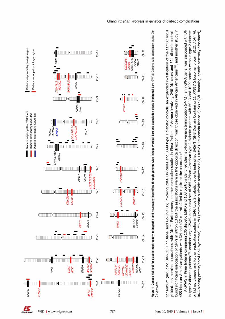
cons
ortiu
m (
incl
udin
g UK-
RO
I, F
innD
iane
, an
d G
oKin
D U
S)
invo
lvin
g 29
66 D
N c
ases
and
339
9 ty
pe 1
dia
betic
con
trol
s, a
n ex
pand
ed in
vest
igat
ion
of t
he E
LMO
1 lo
cus
yiel
ded
only
nom
inal
ass
ocia
tions
with
DN
[30].
Furt
herm
ore,
ano
ther
rep
licat
ion
stud
ies
in P
ima
Indi
ans
of A
rizo
na inv
olvi
ng 2
48 D
N c
ases
and
524
dia
betic
con
trol
s fo
und
sign
ifica
nt a
ssoc
iatio
n of
SN
Ps in
intr
on 1
3 bu
t th
e as
soci
atio
ns w
ere
in the
opp
osite
direc
tion
from
tho
se o
bser
ved
in A
fric
an A
mer
ican
s[31], an
d an
othe
r st
udy
in
455
Mex
ican
Am
eric
an p
atie
nts
with
DN
and
437
con
trol
s fa
iled
to rep
licat
e th
e as
soci
atio
n[32].
A G
WAS in
Pim
a In
dian
s co
mpa
ring
105
diab
etic E
SRD
and
103
con
trol
s id
entifi
ed p
lasm
acyt
oma
varia
nt tra
nslo
catio
n (P
VT1
), a
n ln
cRNA g
ene,
was
ass
ocia
ted
with
DN
in typ
e 2
diab
etic p
atie
nts[3
3]. Ano
ther
larg
e G
WAS in
an
initi
al s
et o
f 96
5 African
Am
erican
typ
e 2
diab
etic p
atie
nts
with
ESRD
and
102
9 co
ntro
ls w
ithou
t ty
pe 2
dia
bete
s or
kid
ney
dise
ase
and
furt
her
repl
icat
ion
stud
ies
in 1
246
type
2 d
iabe
tic p
atie
nts
inde
ntifi
ed S
ASH1
(SH3
Dom
ain
Con
tain
ing
1), RPS
12 (
ribos
omal
pro
tein
S12
), A
UH (
AU
RNA b
indi
ng p
rote
in/e
noyl
-CoA
hyd
rata
se),
MSRB3
(met
hion
ine
sulfo
xide
red
ucta
se B
3), LI
MK2
(LIM
dom
ain
kina
se 2
)-SFI
1 (S
fi1 h
omol
og, sp
indl
e as
sem
bly
asso
ciat
ed),
717 June 10, 2015|Volume 6|Issue 5|WJD|www.wjgnet.com
GFR
A2
MYS
M1
AKT3
/ZN
F238
MSR
B3
LRP2
/BB
S5
ARL4
C/SH
3BP4
AFF3
ERBB
4
AKAP
11/
FABP
3P2
TBC1
D4/
COM
MD
6/UC
HL3 HS6
ST3
MYO
16/
IRS2
FSTL
5
GLR
A3
FMN
1
RGW
A/M
CTP2
SULT
1A1
ZNRF
1
CAM
K4
C5or
f21
RPS1
2
SASH
1
LOC
7282
75/
LOC7
2816
FAM
18B
PVT1
ELM
O1
CPVL
/CH
N2
CO
X5B
L1/
LOC4
4102
6FR
MD
3AU
H
C10o
rf11
2PL
XDC2
ARH
GAP
22
ZMIZ
1
CARS
CCD
C101
/N
UPR1
LIM
K2-S
FI1
APO
L3
PCSK
2
PSD
3/SH
2D44
GFR
A2
MYH
9
Dia
betic
nep
hrop
athy
GW
AS lo
ciD
iabe
tic r
etin
opat
hy G
WAS
loci
Dia
betic
neu
ropa
thy
GW
AS lo
ci
Dia
betic
nep
hrop
athy
link
age
regi
on
Dia
betic
ret
inop
athy
link
age
regi
on
Chr1
Chr2
Chr3
Chr4
Chr5
Chr6
Chr7
Chr8
Chr9
Chr1
0Ch
r11
Chr1
2Ch
r13
Chr1
4Ch
r15
Chr1
6Ch
r17
Chr1
8Ch
r19
Chr2
0Ch
r21
Chr2
2
Figu
re 1
Gen
etic
risk
loci
for d
iabet
ic ne
phro
path
y, re
tinop
athy
, and
neu
ropa
thy
inde
ntifi
ed th
roug
h ge
nom
e-wi
de li
nkag
e (v
ertic
al ba
r) an
d as
socia
tion
scan
s (h
orizo
ntal
bar).
GW
AS: G
enom
e-wi
de a
ssoc
iation
stud
y; Ch
r: Ch
romo
some
.
Chang YC et al . Progress in genetics of diabetic complications
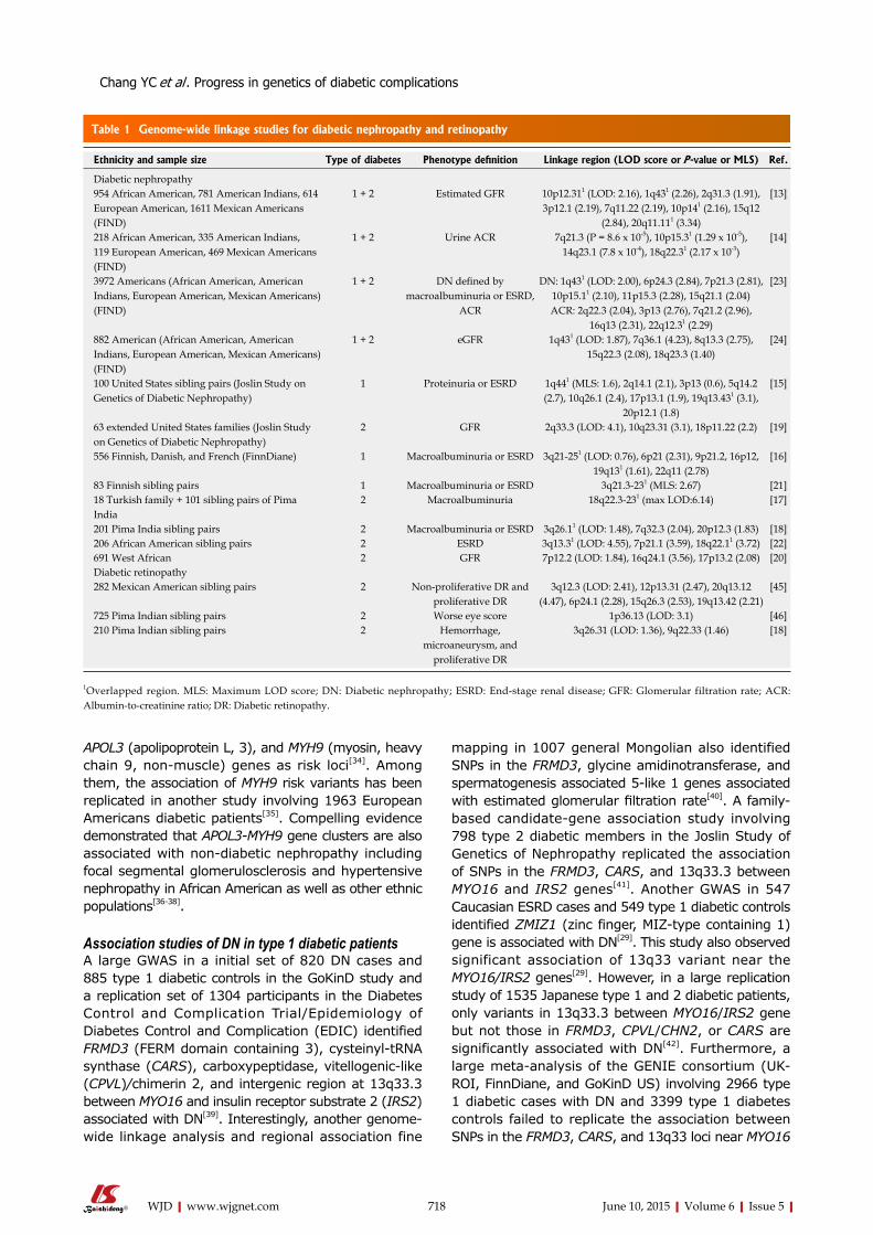
Table 1 Genome-wide linkage studies for diabetic nephropathy and retinopathy
mapping in 1007 general Mongolian also identified SNPs in the FRMD3, glycine amidinotransferase, and spermatogenesis associated 5like 1 genes associated with estimated glomerular filtration rate[40]. A familybased candidategene association study involving 798 type 2 diabetic members in the Joslin Study of Genetics of Nephropathy replicated the association of SNPs in the FRMD3, CARS, and 13q33.3 between MYO16 and IRS2 genes[41]. Another GWAS in 547 Caucasian ESRD cases and 549 type 1 diabetic controls identified ZMIZ1 (zinc finger, MIZtype containing 1) gene is associated with DN[29]. This study also observed significant association of 13q33 variant near the MYO16/IRS2 genes[29]. However, in a large replication study of 1535 Japanese type 1 and 2 diabetic patients, only variants in 13q33.3 between MYO16/IRS2 gene but not those in FRMD3, CPVL/CHN2, or CARS are significantly associated with DN[42]. Furthermore, a large metaanalysis of the GENIE consortium (UKROI, FinnDiane, and GoKinD US) involving 2966 type 1 diabetic cases with DN and 3399 type 1 diabetes controls failed to replicate the association between SNPs in the FRMD3, CARS, and 13q33 loci near MYO16
APOL3 (apolipoprotein L, 3), and MYH9 (myosin, heavy chain 9, nonmuscle) genes as risk loci[34]. Among them, the association of MYH9 risk variants has been replicated in another study involving 1963 European Americans diabetic patients[35]. Compelling evidence demonstrated that APOL3-MYH9 gene clusters are also associated with nondiabetic nephropathy including focal segmental glomerulosclerosis and hypertensive nephropathy in African American as well as other ethnic populations[3638].
Association studies of DN in type 1 diabetic patientsA large GWAS in a initial set of 820 DN cases and 885 type 1 diabetic controls in the GoKinD study and a replication set of 1304 participants in the Diabetes Control and Complication Trial/Epidemiology of Diabetes Control and Complication (EDIC) identified FRMD3 (FERM domain containing 3), cysteinyl-tRNA synthase (CARS), carboxypeptidase, vitellogeniclike (CPVL)/chimerin 2, and intergenic region at 13q33.3 between MYO16 and insulin receptor substrate 2 (IRS2) associated with DN[39]. Interestingly, another genomewide linkage analysis and regional association fine
718 June 10, 2015|Volume 6|Issue 5|WJD|www.wjgnet.com
Ethnicity and sample size Type of diabetes Phenotype definition Linkage region (LOD score or P -value or MLS) Ref.
Diabetic nephropathy954 African American, 781 American Indians, 614 European American, 1611 Mexican Americans (FIND)
1 + 2 Estimated GFR 10p12.311 (LOD: 2.16), 1q431 (2.26), 2q31.3 (1.91), 3p12.1 (2.19), 7q11.22 (2.19), 10p141 (2.16), 15q12
(2.84), 20q11.111 (3.34)
[13]
218 African American, 335 American Indians, 119 European American, 469 Mexican Americans (FIND)
1 + 2 Urine ACR 7q21.3 (P = 8.6 x 10⁻⁵), 10p15.31 (1.29 x 10⁻⁵), 14q23.1 (7.8 x 10⁻⁴), 18q22.31 (2.17 x 10⁻³)
[14]
3972 Americans (African American, American Indians, European American, Mexican Americans) (FIND)
1 + 2 DN defined by macroalbuminuria or ESRD,
ACR
DN: 1q431 (LOD: 2.00), 6p24.3 (2.84), 7p21.3 (2.81), 10p15.11 (2.10), 11p15.3 (2.28), 15q21.1 (2.04)ACR: 2q22.3 (2.04), 3p13 (2.76), 7q21.2 (2.96),
16q13 (2.31), 22q12.31 (2.29)
[23]
882 American (African American, American Indians, European American, Mexican Americans) (FIND)
1 + 2 eGFR 1q431 (LOD: 1.87), 7q36.1 (4.23), 8q13.3 (2.75), 15q22.3 (2.08), 18q23.3 (1.40)
[24]
100 United States sibling pairs (Joslin Study on Genetics of Diabetic Nephropathy)
1 Proteinuria or ESRD 1q441 (MLS: 1.6), 2q14.1 (2.1), 3p13 (0.6), 5q14.2 (2.7), 10q26.1 (2.4), 17p13.1 (1.9), 19q13.431 (3.1),
20p12.1 (1.8)
[15]
63 extended United States families (Joslin Study on Genetics of Diabetic Nephropathy)
2 GFR 2q33.3 (LOD: 4.1), 10q23.31 (3.1), 18p11.22 (2.2) [19]
556 Finnish, Danish, and French (FinnDiane) 1 Macroalbuminuria or ESRD 3q21-251 (LOD: 0.76), 6p21 (2.31), 9p21.2, 16p12, 19q131 (1.61), 22q11 (2.78)
[16]
83 Finnish sibling pairs 1 Macroalbuminuria or ESRD 3q21.3-231 (MLS: 2.67) [21]18 Turkish family + 101 sibling pairs of Pima India
2 Macroalbuminuria 18q22.3-231 (max LOD:6.14) [17]
201 Pima India sibling pairs 2 Macroalbuminuria or ESRD 3q26.11 (LOD: 1.48), 7q32.3 (2.04), 20p12.3 (1.83) [18]206 African American sibling pairs 2 ESRD 3q13.31 (LOD: 4.55), 7p21.1 (3.59), 18q22.11 (3.72) [22]691 West African 2 GFR 7p12.2 (LOD: 1.84), 16q24.1 (3.56), 17p13.2 (2.08) [20]Diabetic retinopathy282 Mexican American sibling pairs 2 Non-proliferative DR and
proliferative DR 3q12.3 (LOD: 2.41), 12p13.31 (2.47), 20q13.12
(4.47), 6p24.1 (2.28), 15q26.3 (2.53), 19q13.42 (2.21)[45]
725 Pima Indian sibling pairs 2 Worse eye score 1p36.13 (LOD: 3.1) [46]210 Pima Indian sibling pairs 2 Hemorrhage,
microaneurysm, and proliferative DR
3q26.31 (LOD: 1.36), 9q22.33 (1.46) [18]
1Overlapped region. MLS: Maximum LOD score; DN: Diabetic nephropathy; ESRD: End-stage renal disease; GFR: Glomerular filtration rate; ACR: Albumin-to-creatinine ratio; DR: Diabetic retinopathy.
Chang YC et al . Progress in genetics of diabetic complications
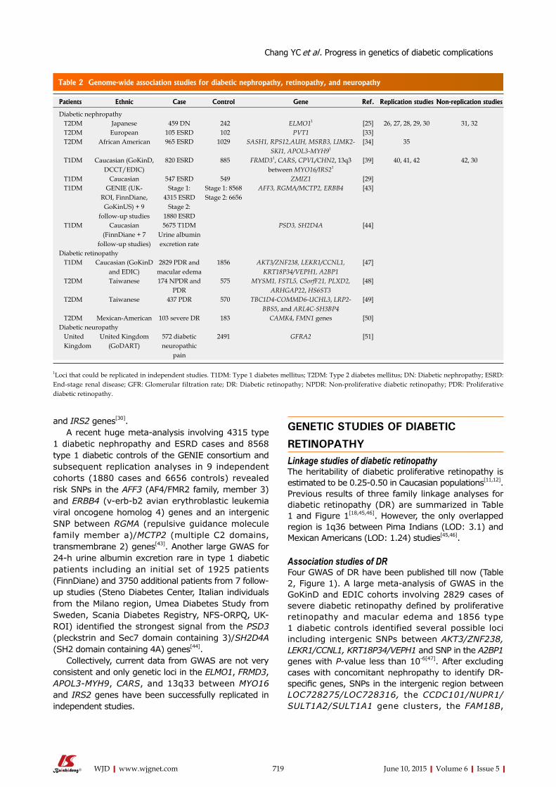
and IRS2 genes[30]. A recent huge metaanalysis involving 4315 type
1 diabetic nephropathy and ESRD cases and 8568 type 1 diabetic controls of the GENIE consortium and subsequent replication analyses in 9 independent cohorts (1880 cases and 6656 controls) revealed risk SNPs in the AFF3 (AF4/FMR2 family, member 3) and ERBB4 (verbb2 avian erythroblastic leukemia viral oncogene homolog 4) genes and an intergenic SNP between RGMA (repulsive guidance molecule family member a)/MCTP2 (multiple C2 domains, transmembrane 2) genes[43]. Another large GWAS for 24h urine albumin excretion rare in type 1 diabetic patients including an initial set of 1925 patients (FinnDiane) and 3750 additional patients from 7 follow-up studies (Steno Diabetes Center, Italian individuals from the Milano region, Umea Diabetes Study from Sweden, Scania Diabetes Registry, NFS-ORPQ, UK-ROI) identified the strongest signal from the PSD3 (pleckstrin and Sec7 domain containing 3)/SH2D4A (SH2 domain containing 4A) genes[44].
Collectively, current data from GWAS are not very consistent and only genetic loci in the ELMO1, FRMD3, APOL3-MYH9, CARS, and 13q33 between MYO16 and IRS2 genes have been successfully replicated in independent studies.
GENETIC STUDIES OF DIABETIC
RETINOPATHYLinkage studies of diabetic retinopathyThe heritability of diabetic proliferative retinopathy is estimated to be 0.250.50 in Caucasian populations[11,12]. Previous results of three family linkage analyses for diabetic retinopathy (DR) are summarized in Table 1 and Figure 1[18,45,46]. However, the only overlapped region is 1q36 between Pima Indians (LOD: 3.1) and Mexican Americans (LOD: 1.24) studies[45,46].
Association studies of DRFour GWAS of DR have been published till now (Table 2, Figure 1). A large meta-analysis of GWAS in the GoKinD and EDIC cohorts involving 2829 cases of severe diabetic retinopathy defined by proliferative retinopathy and macular edema and 1856 type 1 diabetic controls identified several possible loci including intergenic SNPs between AKT3/ZNF238, LEKR1/CCNL1, KRT18P34/VEPH1 and SNP in the A2BP1 genes with Pvalue less than 106[47]. After excluding cases with concomitant nephropathy to identify DR-specific genes, SNPs in the intergenic region between LOC728275/LOC728316, the CCDC101/NUPR1/SULT1A2/SULT1A1 gene clusters, the FAM18B,
719 June 10, 2015|Volume 6|Issue 5|WJD|www.wjgnet.com
Table 2 Genome-wide association studies for diabetic nephropathy, retinopathy, and neuropathy
Patients Ethnic Case Control Gene Ref. Replication studies Non-replication studies
Diabetic nephropathy T2DM Japanese 459 DN 242 ELMO11 [25] 26, 27, 28, 29, 30 31, 32 T2DM European 105 ESRD 102 PVT1 [33] T2DM African American 965 ESRD 1029 SASH1, RPS12,AUH, MSRB3, LIMK2-
SKI1, APOL3-MYH91[34] 35
T1DM Caucasian (GoKinD, DCCT/EDIC)
820 ESRD 885 FRMD31, CARS, CPVL/CHN2, 13q3 between MYO16/IRS21
[39] 40, 41, 42 42, 30
T1DM Caucasian 547 ESRD 549 ZMIZ1 [29] T1DM GENIE (UK-
ROI, FinnDiane, GoKinUS) + 9
follow-up studies
Stage 1: 4315 ESRD
Stage 2: 1880 ESRD
Stage 1: 8568Stage 2: 6656
AFF3, RGMA/MCTP2, ERBB4 [43]
T1DM Caucasian(FinnDiane + 7
follow-up studies)
5675 T1DMUrine albumin excretion rate
PSD3, SH2D4A [44]
Diabetic retinopathy T1DM Caucasian (GoKinD
and EDIC)2829 PDR and
macular edema1856 AKT3/ZNF238, LEKR1/CCNL1,
KRT18P34/VEPH1, A2BP1[47]
T2DM Taiwanese 174 NPDR and PDR
575 MYSM1, FSTL5, C5orfF21, PLXD2, ARHGAP22, HS6ST3
[48]
T2DM Taiwanese 437 PDR 570 TBC1D4-COMMD6-UCHL3, LRP2-BBS5, and ARL4C-SH3BP4
[49]
T2DM Mexican-American 103 severe DR 183 CAMK4, FMN1 genes [50]Diabetic neuropathy United Kingdom
United Kingdom (GoDART)
572 diabetic neuropathic
pain
2491 GFRA2 [51]
1Loci that could be replicated in independent studies. T1DM: Type 1 diabetes mellitus; T2DM: Type 2 diabetes mellitus; DN: Diabetic nephropathy; ESRD: End-stage renal disease; GFR: Glomerular filtration rate; DR: Diabetic retinopathy; NPDR: Non-proliferative diabetic retinopathy; PDR: Proliferative diabetic retinopathy.
Chang YC et al . Progress in genetics of diabetic complications
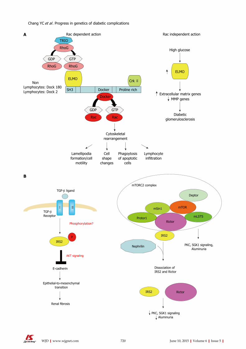
720 June 10, 2015|Volume 6|Issue 5|WJD|www.wjgnet.com
Rac dependent action
TRIO
RhoG
GDP GTP
RhoG RhoG
ELMONon
Lymphocytes: Dock 180Lymphocytes: Dock 2 SH3 Docker Proline rich
Crk Ⅱ
Docker
GDP GTP
RacRac
Cytoskeletal rearrangement
Cell shape
changes
Phagoytosis of apoptotic
cells
Lamellipodia formation/cell
motility
Lymphocyte infiltration
ELMO
High glucose
Extracellular matrix genesMMP genes
Diabetic glomerulosclerosis
Rac independent action
B
A
TGF-β ligand
TGF-β Receptor
Phosphorylation?
Ⅰ Ⅱ
IRS2P
E-cadherin
Epithelial-to-mesenchymaltransition
Renal fibrosis
mTORC2 complex
Deptor
mTORmSin1
Protor1Rictor
mLSTS
IRS2
Nephrilin
IRS2 Rictor
Dissociation of IRS2 and Rictor
PKC, SGK1 signaling,Aluminuria
PKC, SGK1 signalingAluminuria
Chang YC et al . Progress in genetics of diabetic complications
AKT signaling
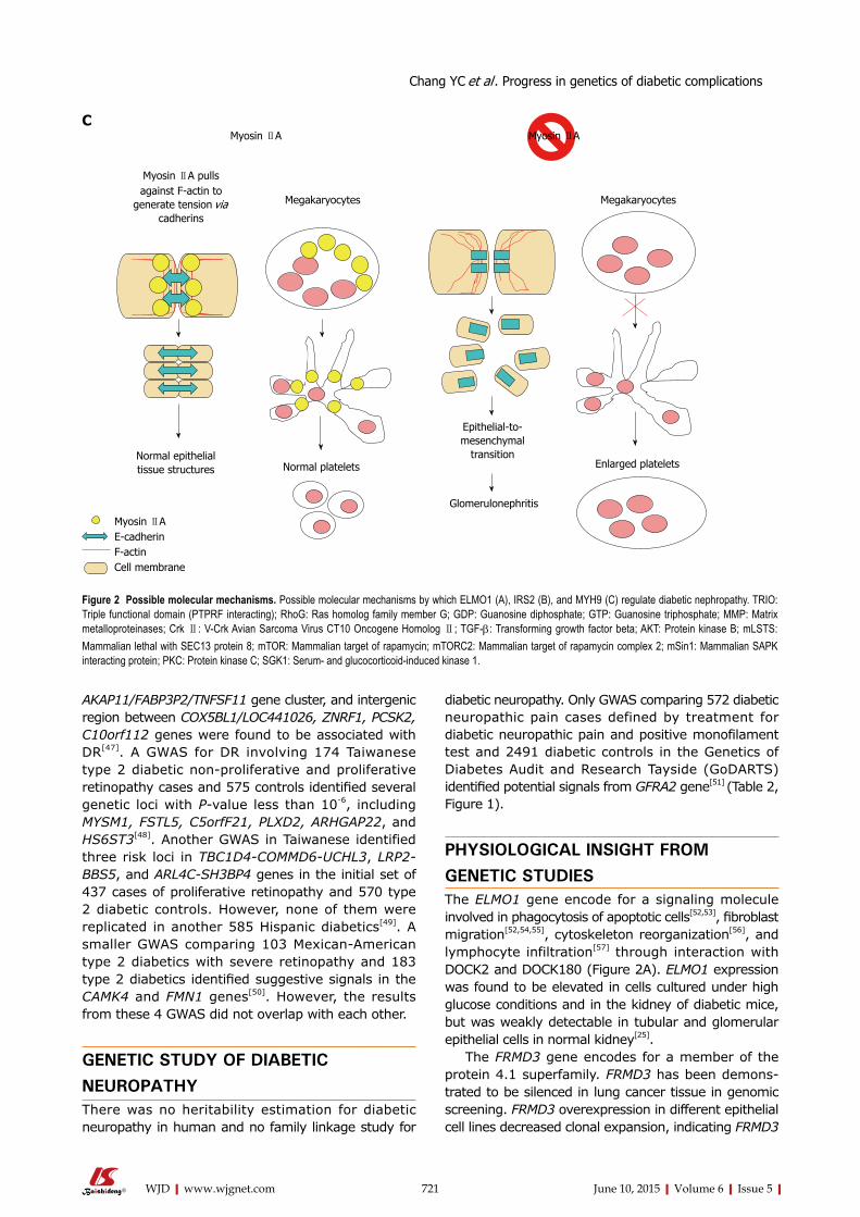
AKAP11/FABP3P2/TNFSF11 gene cluster, and intergenic region between COX5BL1/LOC441026, ZNRF1, PCSK2, C10orf112 genes were found to be associated with DR[47]. A GWAS for DR involving 174 Taiwanese type 2 diabetic nonproliferative and proliferative retinopathy cases and 575 controls identified several genetic loci with Pvalue less than 106, including MYSM1, FSTL5, C5orfF21, PLXD2, ARHGAP22, and HS6ST3[48]. Another GWAS in Taiwanese identified three risk loci in TBC1D4-COMMD6-UCHL3, LRP2-BBS5, and ARL4C-SH3BP4 genes in the initial set of 437 cases of proliferative retinopathy and 570 type 2 diabetic controls. However, none of them were replicated in another 585 Hispanic diabetics[49]. A smaller GWAS comparing 103 MexicanAmerican type 2 diabetics with severe retinopathy and 183 type 2 diabetics identified suggestive signals in the CAMK4 and FMN1 genes[50]. However, the results from these 4 GWAS did not overlap with each other.
GENETIC STUDY OF DIABETIC NEUROPATHYThere was no heritability estimation for diabetic neuropathy in human and no family linkage study for
diabetic neuropathy. Only GWAS comparing 572 diabetic neuropathic pain cases defined by treatment for diabetic neuropathic pain and positive monofilament test and 2491 diabetic controls in the Genetics of Diabetes Audit and Research Tayside (GoDARTS) identified potential signals from GFRA2 gene[51] (Table 2, Figure 1).
PHYSIOLOGICAL INSIGHT FROM GENETIC STUDIESThe ELMO1 gene encode for a signaling molecule involved in phagocytosis of apoptotic cells[52,53], fibroblast migration[52,54,55], cytoskeleton reorganization[56], and lymphocyte infiltration[57] through interaction with DOCK2 and DOCK180 (Figure 2A). ELMO1 expression was found to be elevated in cells cultured under high glucose conditions and in the kidney of diabetic mice, but was weakly detectable in tubular and glomerular epithelial cells in normal kidney[25].
The FRMD3 gene encodes for a member of the protein 4.1 superfamily. FRMD3 has been demonstrated to be silenced in lung cancer tissue in genomic screening. FRMD3 overexpression in different epithelial cell lines decreased clonal expansion, indicating FRMD3
721 June 10, 2015|Volume 6|Issue 5|WJD|www.wjgnet.com
Figure 2 Possible molecular mechanisms. Possible molecular mechanisms by which ELMO1 (A), IRS2 (B), and MYH9 (C) regulate diabetic nephropathy. TRIO: Triple functional domain (PTPRF interacting); RhoG: Ras homolog family member G; GDP: Guanosine diphosphate; GTP: Guanosine triphosphate; MMP: Matrix metalloproteinases; Crk Ⅱ: V-Crk Avian Sarcoma Virus CT10 Oncogene Homolog Ⅱ; TGF-β: Transforming growth factor beta; AKT: Protein kinase B; mLSTS: Mammalian lethal with SEC13 protein 8; mTOR: Mammalian target of rapamycin; mTORC2: Mammalian target of rapamycin complex 2; mSin1: Mammalian SAPK interacting protein; PKC: Protein kinase C; SGK1: Serum- and glucocorticoid-induced kinase 1.
Myosin ⅡA Myosin ⅡA
Myosin ⅡA pulls against F-actin to
generate tension via cadherins
Megakaryocytes Megakaryocytes
Normal epithelial tissue structures
Myosin ⅡA E-cadherinF-actinCell membrane
Normal platelets
Epithelial-to-mesenchymal
transition
Glomerulonephritis
Enlarged platelets
C
Chang YC et al . Progress in genetics of diabetic complications
as a potential tumor suppressor gene[58]. The CARS encodes for a cysteinyl-tRNA synthetase, which is a frequent gene fusion partner of anaplastic lymphoma kinase found in anaplastic largecell lymphoma and inflammatory myofibroblastic tumor[59,60]. However, the link between FRMD3 or CARS and diabetic nephropathy is currently poorly understood.
The 13q33 risk loci lie between the MYO16 and IRS2 genes. The MYO16 gene encodes a novel unconventional myosin with divergent tails that is presumed to bind to membranous compartments and interact with actin filaments. MYO16 has also been shown to be expressed during brain development and regulate neuronal morphogenesis through interaction with protein phosphatase and modulation of phosphoinositide 3kinase signaling[61]. A GWAS for autism has identified risk loci within an intergenic region between the MYO16 and IRS2 genes[62]. A genomewide linkage study and regional fine mapping for schizophrenia[63] and another GWAS of the Framingham Heart Study for pulse pressure[64] have identified MYO16 as risk loci, indicating MYO16 may play pleiotropic functions.
The IRS2 gene encodes for an adaptor protein that interacts directly with the insulin receptors and the insulinlike growth factor Ⅰ receptor and is a key mediator of insulin signaling. IRS2 was expressed in renal epithelial and tubular cells. Deletion of Irs2 causes reduced kidney size and reduced glomerular number in mice[65]. A study of transcriptome and metabolome profiles of the primary cultured inner medullary collecting duct cells grown in hyperosmolar culture medium identified IRS2 levels to be significantly altered[66]. IRS2 expression in kidney tubules has also been shown to be elevated nine fold in human diabetic nephropathy patients[67]. Transforming growth factor (TGF)-β1 is the primary cytokine shown to induce fibrosis. IRS2 has been shown to mediate TGF-β1 signals in kidney epithelial cells[68]. IRS2 has also been shown to interact with nuclear complex of rictor to regulate albuminuria in diabetic mice[69] (Figure 2B).
Mutations in MYH9 results in a familial autosomal dominant syndrome characterized by a variety of clinical features, including macrothrombocytopenia, deafness, nephritis, and cataract[70]. GWAS also identified common MYH9 polymorphism as risk loci for nondiabetic nephropathy including focal segmental glomerulosclerosis and hypertensive nephropathy[36,27]. MYH9 encodes the nonmuscle myosin heavy chain 9, which, with other subunits, forms myosin Ⅱ. Myosin Ⅱ is a motor protein that binds actin to regulate cellular motility. MYH9 is expressed in the podocytes, as well as in mesangial cells and arteriolar and peritubular capillaries in kidneys[71]. Classical deletion of Myh9 in mice results in embryonic lethality due to loss of cellcell adhesion and loss of cell movement during gastrulation. Podocytespecfic deletion of Myh9 in C57BL/6 mice results in susceptibility to experimental doxorubicin hydrochloride glomerulopathy[71]. Several
strains of Myh9 knockin mice showed macrothrombocytopenia, premature cataract formation, kidney abnormalities, including albuminuria, focal segmental glomerulosclerosis and progressive kidney disease, and mild hearing loss[72,73] (Figure 2C).
LIMITATIONS AND PROSPECTIVEThe major limitation of family linkage studies is their low resolution and power to detect variants with small effects, especially for complex genetic diseases. GWAS is a hypothesisfree and unbiased tool with finer resolution and greater power to detect risk loci. However, false positivity often results from population admixture or stratification in GWAS. Therefore, independent replications are essential for genetic association studies. However, current results from GWAS are not consistent since most identified loci are not reproducible except for a few genes such as ELMO1, CARS, FRMD3, MYO16/IRS2, and APOL3/MYH9. Small sample sizes, different phenotype definitions between studies, populationspecific associations, and strong influence of environmental factors (medications, comorbidities) may explain the failure of GWAS for diabetic complications. While GWAS are usually designed for common variants, rare variants with intermediate effects within should also be pursued with nextgeneration sequencing. The interaction with environmental factors should also be taken into account.
REFERENCES1 Wild S, Roglic G, Green A, Sicree R, King H. Global prevalence
of diabetes: estimates for the year 2000 and projections for 2030. Diabetes Care 2004; 27: 1047-1053 [PMID: 15111519 DOI: 10.2337/diacare.27.5.1047]
2 Brownlee M. The pathobiology of diabetic complications: a unifying mechanism. Diabetes 2005; 54: 1615-1625 [PMID: 15919781 DOI: 10.2337/diabetes.54.6.1615]
3 Shamsi HN, Masaud JS, Ghazi NG. Diabetic macular edema: New promising therapies. World J Diabetes 2013; 4: 324-338 [PMID: 24379924 DOI: 10.4239/wjd.v4.i6.324]
4 Gosmanov AR, Wall BM, Gosmanova EO. Diagnosis and treatment of diabetic kidney disease. Am J Med Sci 2014; 347: 406-413 [PMID: 24553399 DOI: 10.1097/MAJ.0000000000000185]
5 Spallone V, Lacerenza M, Rossi A, Sicuteri R, Marchettini P. Painful diabetic polyneuropathy: approach to diagnosis and management. Clin J Pain 2012; 28: 726-743 [PMID: 22209797 DOI: 10.1097/AJP.0b013e318243075c]
6 Abifadel M, Varret M, Rabès JP, Allard D, Ouguerram K, Devillers M, Cruaud C, Benjannet S, Wickham L, Erlich D, Derré A, Villéger L, Farnier M, Beucler I, Bruckert E, Chambaz J, Chanu B, Lecerf JM, Luc G, Moulin P, Weissenbach J, Prat A, Krempf M, Junien C, Seidah NG, Boileau C. Mutations in PCSK9 cause autosomal dominant hypercholesterolemia. Nat Genet 2003; 34: 154-156 [PMID: 12730697 DOI: 10.1038/ng1161]
7 Stein EA, Mellis S, Yancopoulos GD, Stahl N, Logan D, Smith WB, Lisbon E, Gutierrez M, Webb C, Wu R, Du Y, Kranz T, Gasparino E, Swergold GD. Effect of a monoclonal antibody to PCSK9 on LDL cholesterol. N Engl J Med 2012; 366: 1108-1118 [PMID: 22435370 DOI: 10.1056/NEJMoa1105803]
8 Forsblom CM, Kanninen T, Lehtovirta M, Saloranta C, Groop LC. Heritability of albumin excretion rate in families of patients
722 June 10, 2015|Volume 6|Issue 5|WJD|www.wjgnet.com
Chang YC et al . Progress in genetics of diabetic complications
with Type II diabetes. Diabetologia 1999; 42: 1359-1366 [PMID: 10550421 DOI: 10.1007/s001250051450]
9 Fogarty DG, Rich SS, Hanna L, Warram JH, Krolewski AS. Urinary albumin excretion in families with type 2 diabetes is heritable and genetically correlated to blood pressure. Kidney Int 2000; 57: 250-257 [PMID: 10620206 DOI: 10.1046/j.1523-1755.2000.00833.x]
10 Langefeld CD, Beck SR, Bowden DW, Rich SS, Wagenknecht LE, Freedman BI. Heritability of GFR and albuminuria in Caucasians with type 2 diabetes mellitus. Am J Kidney Dis 2004; 43: 796-800 [PMID: 15112169]
11 Arar NH, Freedman BI, Adler SG, Iyengar SK, Chew EY, Davis MD, Satko SG, Bowden DW, Duggirala R, Elston RC, Guo X, Hanson RL, Igo RP, Ipp E, Kimmel PL, Knowler WC, Molineros J, Nelson RG, Pahl MV, Quade SR, Rasooly RS, Rotter JI, Saad MF, Scavini M, Schelling JR, Sedor JR, Shah VO, Zager PG, Abboud HE. Heritability of the severity of diabetic retinopathy: the FIND-Eye study. Invest Ophthalmol Vis Sci 2008; 49: 3839-3845 [PMID: 18765632 DOI: 10.1167/iovs.07-1633]
12 Hietala K, Forsblom C, Summanen P, Groop PH. Heritability of proliferative diabetic retinopathy. Diabetes 2008; 57: 2176-2180 [PMID: 18443200 DOI: 10.2337/db07-1495]
13 Thameem F, Igo RP, Freedman BI, Langefeld C, Hanson RL, Schelling JR, Elston RC, Duggirala R, Nicholas SB, Goddard KA, Divers J, Guo X, Ipp E, Kimmel PL, Meoni LA, Shah VO, Smith MW, Winkler CA, Zager PG, Knowler WC, Nelson RG, Pahl MV, Parekh RS, Kao WH, Rasooly RS, Adler SG, Abboud HE, Iyengar SK, Sedor JR. A genome-wide search for linkage of estimated glomerular filtration rate (eGFR) in the Family Investigation of Nephropathy and Diabetes (FIND). PLoS One 2013; 8: e81888 [PMID: 24358131 DOI: 10.1371/journal.pone.0081888]
14 Iyengar SK, Abboud HE, Goddard KA, Saad MF, Adler SG, Arar NH, Bowden DW, Duggirala R, Elston RC, Hanson RL, Ipp E, Kao WH, Kimmel PL, Klag MJ, Knowler WC, Meoni LA, Nelson RG, Nicholas SB, Pahl MV, Parekh RS, Quade SR, Rich SS, Rotter JI, Scavini M, Schelling JR, Sedor JR, Sehgal AR, Shah VO, Smith MW, Taylor KD, Winkler CA, Zager PG, Freedman BI. Genome-wide scans for diabetic nephropathy and albuminuria in multiethnic populations: the family investigation of nephropathy and diabetes (FIND). Diabetes 2007; 56: 1577-1585 [PMID: 17363742 DOI: 10.2337/db06-1154]
15 Rogus JJ, Poznik GD, Pezzolesi MG, Smiles AM, Dunn J, Walker W, Wanic K, Moczulski D, Canani L, Araki S, Makita Y, Warram JH, Krolewski AS. High-density single nucleotide polymorphism genome-wide linkage scan for susceptibility genes for diabetic nephropathy in type 1 diabetes: discordant sibpair approach. Diabetes 2008; 57: 2519-2526 [PMID: 18559660 DOI: 10.2337/db07-1086]
16 Wessman M, Forsblom C, Kaunisto MA, Söderlund J, Ilonen J, Sallinen R, Hiekkalinna T, Parkkonen M, Maxwell AP, Tarnow L, Parving HH, Hadjadj S, Marre M, Peltonen L, Groop PH. Novel susceptibility locus at 22q11 for diabetic nephropathy in type 1 diabetes. PLoS One 2011; 6: e24053 [PMID: 21909410 DOI: 10.1371/journal.pone.0024053]
17 Vardarli I, Baier LJ, Hanson RL, Akkoyun I, Fischer C, Rohmeiss P, Basci A, Bartram CR, Van Der Woude FJ, Janssen B. Gene for susceptibility to diabetic nephropathy in type 2 diabetes maps to 18q22.3-23. Kidney Int 2002; 62: 2176-2183 [PMID: 12427143 DOI: 10.1046/j.1523-1755.2002.00663.x]
18 Imperatore G, Hanson RL, Pettitt DJ, Kobes S, Bennett PH, Knowler WC. Sib-pair linkage analysis for susceptibility genes for microvascular complications among Pima Indians with type 2 diabetes. Pima Diabetes Genes Group. Diabetes 1998; 47: 821-830 [PMID: 9588456]
19 Placha G, Poznik GD, Dunn J, Smiles A, Krolewski B, Glew T, Puppala S, Schneider J, Rogus JJ, Rich SS, Duggirala R, Warram JH, Krolewski AS. A genome-wide linkage scan for genes controlling variation in renal function estimated by serum cystatin C levels in extended families with type 2 diabetes. Diabetes 2006; 55: 3358-3365 [PMID: 17130480 DOI: 10.2337/db06-0781]
20 Chen G, Adeyemo AA, Zhou J, Chen Y, Doumatey A, Lashley
K, Huang H, Amoah A, Agyenim-Boateng K, Eghan BA, Okafor G, Acheampong J, Oli J, Fasanmade O, Johnson T, Rotimi C. A genome-wide search for linkage to renal function phenotypes in West Africans with type 2 diabetes. Am J Kidney Dis 2007; 49: 394-400 [PMID: 17336700 DOI: 10.1053/j.ajkd.2006.12.011]
21 Osterholm AM, He B, Pitkaniemi J, Albinsson L, Berg T, Sarti C, Tuomilehto J, Tryggvason K. Genome-wide scan for type 1 diabetic nephropathy in the Finnish population reveals suggestive linkage to a single locus on chromosome 3q. Kidney Int 2007; 71: 140-145 [PMID: 17021601 DOI: 10.1038/sj.ki.5001933]
22 Bowden DW, Colicigno CJ, Langefeld CD, Sale MM, Williams A, Anderson PJ, Rich SS, Freedman BI. A genome scan for diabetic nephropathy in African Americans. Kidney Int 2004; 66: 1517-1526 [PMID: 15458446 DOI: 10.1111/j.1523-1755.2004.00915.x]
23 Igo RP, Iyengar SK, Nicholas SB, Goddard KA, Langefeld CD, Hanson RL, Duggirala R, Divers J, Abboud H, Adler SG, Arar NH, Horvath A, Elston RC, Bowden DW, Guo X, Ipp E, Kao WH, Kimmel PL, Knowler WC, Meoni LA, Molineros J, Nelson RG, Pahl MV, Parekh RS, Rasooly RS, Schelling JR, Shah VO, Smith MW, Winkler CA, Zager PG, Sedor JR, Freedman BI. Genomewide linkage scan for diabetic renal failure and albuminuria: the FIND study. Am J Nephrol 2011; 33: 381-389 [PMID: 21454968 DOI: 10.1159/000326763]
24 Schelling JR, Abboud HE, Nicholas SB, Pahl MV, Sedor JR, Adler SG, Arar NH, Bowden DW, Elston RC, Freedman BI, Goddard KA, Guo X, Hanson RL, Ipp E, Iyengar SK, Jun G, Kao WH, Kasinath BS, Kimmel PL, Klag MJ, Knowler WC, Nelson RG, Parekh RS, Quade SR, Rich SS, Saad MF, Scavini M, Smith MW, Taylor K, Winkler CA, Zager PG, Shah VO. Genome-wide scan for estimated glomerular filtration rate in multi-ethnic diabetic populations: the Family Investigation of Nephropathy and Diabetes (FIND). Diabetes 2008; 57: 235-243 [PMID: 18003762 DOI: 10.2337/db07-0313]
25 Shimazaki A, Kawamura Y, Kanazawa A, Sekine A, Saito S, Tsunoda T, Koya D, Babazono T, Tanaka Y, Matsuda M, Kawai K, Iiizumi T, Imanishi M, Shinosaki T, Yanagimoto T, Ikeda M, Omachi S, Kashiwagi A, Kaku K, Iwamoto Y, Kawamori R, Kikkawa R, Nakajima M, Nakamura Y, Maeda S. Genetic variations in the gene encoding ELMO1 are associated with susceptibility to diabetic nephropathy. Diabetes 2005; 54: 1171-1178 [PMID: 15793258 DOI: 10.2337/diabetes.54.4.1171]
26 Pezzolesi MG, Katavetin P, Kure M, Poznik GD, Skupien J, Mychaleckyj JC, Rich SS, Warram JH, Krolewski AS. Confirmation of genetic associations at ELMO1 in the GoKinD collection supports its role as a susceptibility gene in diabetic nephropathy. Diabetes 2009; 58: 2698-2702 [PMID: 19651817 DOI: 10.2337/db09-0641]
27 Leak TS, Perlegas PS, Smith SG, Keene KL, Hicks PJ, Langefeld CD, Mychaleckyj JC, Rich SS, Kirk JK, Freedman BI, Bowden DW, Sale MM. Variants in intron 13 of the ELMO1 gene are associated with diabetic nephropathy in African Americans. Ann Hum Genet 2009; 73: 152-159 [PMID: 19183347 DOI: 10.1111/j.1469-1809.2008.00498.x]
28 Wu HY, Wang Y, Chen M, Zhang X, Wang D, Pan Y, Li L, Liu D, Dai XM. Association of ELMO1 gene polymorphisms with diabetic nephropathy in Chinese population. J Endocrinol Invest 2013; 36: 298-302 [PMID: 22842811 DOI: 10.3275/8525]
29 Craig DW, Millis MP, DiStefano JK. Genome-wide SNP genotyping study using pooled DNA to identify candidate markers mediating susceptibility to end-stage renal disease attributed to Type 1 diabetes. Diabet Med 2009; 26: 1090-1098 [PMID: 19929986 DOI: 10.1111/j.1464-5491.2009.02846.x]
30 Williams WW, Salem RM, McKnight AJ, Sandholm N, Forsblom C, Taylor A, Guiducci C, McAteer JB, McKay GJ, Isakova T, Brennan EP, Sadlier DM, Palmer C, Söderlund J, Fagerholm E, Harjutsalo V, Lithovius R, Gordin D, Hietala K, Kytö J, Parkkonen M, Rosengård-Bärlund M, Thorn L, Syreeni A, Tolonen N, Saraheimo M, Wadén J, Pitkäniemi J, Sarti C, Tuomilehto J, Tryggvason K, Österholm AM, He B, Bain S, Martin F, Godson C, Hirschhorn JN, Maxwell AP, Groop PH, Florez JC. Association
723 June 10, 2015|Volume 6|Issue 5|WJD|www.wjgnet.com
Chang YC et al . Progress in genetics of diabetic complications
testing of previously reported variants in a large case-control meta-analysis of diabetic nephropathy. Diabetes 2012; 61: 2187-2194 [PMID: 22721967 DOI: 10.2337/db11-0751]
31 Hanson RL, Millis MP, Young NJ, Kobes S, Nelson RG, Knowler WC, DiStefano JK. ELMO1 variants and susceptibility to diabetic nephropathy in American Indians. Mol Genet Metab 2010; 101: 383-390 [PMID: 20826100 DOI: 10.1016/j.ymgme.2010.08.014]
32 Kim S, Abboud HE, Pahl MV, Tayek J, Snyder S, Tamkin J, Alcorn H, Ipp E, Nast CC, Elston RC, Iyengar SK, Adler SG. Examination of association with candidate genes for diabetic nephropathy in a Mexican American population. Clin J Am Soc Nephrol 2010; 5: 1072-1078 [PMID: 20299368 DOI: 10.2215/CJN.06550909]
33 Hanson RL, Craig DW, Millis MP, Yeatts KA, Kobes S, Pearson JV, Lee AM, Knowler WC, Nelson RG, Wolford JK. Identification of PVT1 as a candidate gene for end-stage renal disease in type 2 diabetes using a pooling-based genome-wide single nucleotide polymorphism association study. Diabetes 2007; 56: 975-983 [PMID: 17395743 DOI: 10.2337/db06-1072]
34 McDonough CW, Palmer ND, Hicks PJ, Roh BH, An SS, Cooke JN, Hester JM, Wing MR, Bostrom MA, Rudock ME, Lewis JP, Talbert ME, Blevins RA, Lu L, Ng MC, Sale MM, Divers J, Langefeld CD, Freedman BI, Bowden DW. A genome-wide association study for diabetic nephropathy genes in African Americans. Kidney Int 2011; 79: 563-572 [PMID: 21150874 DOI: 10.1038/ki.2010.467]
35 Cooke JN, Bostrom MA, Hicks PJ, Ng MC, Hellwege JN, Comeau ME, Divers J, Langefeld CD, Freedman BI, Bowden DW. Polymorphisms in MYH9 are associated with diabetic nephropathy in European Americans. Nephrol Dial Transplant 2012; 27: 1505-1511 [PMID: 21968013 DOI: 10.1093/ndt/gfr522]
36 Kao WH, Klag MJ, Meoni LA, Reich D, Berthier-Schaad Y, Li M, Coresh J, Patterson N, Tandon A, Powe NR, Fink NE, Sadler JH, Weir MR, Abboud HE, Adler SG, Divers J, Iyengar SK, Freedman BI, Kimmel PL, Knowler WC, Kohn OF, Kramp K, Leehey DJ, Nicholas SB, Pahl MV, Schelling JR, Sedor JR, Thornley-Brown D, Winkler CA, Smith MW, Parekh RS. MYH9 is associated with nondiabetic end-stage renal disease in African Americans. Nat Genet 2008; 40: 1185-1192 [PMID: 18794854 DOI: 10.1038/ng.232]
37 Kopp JB, Smith MW, Nelson GW, Johnson RC, Freedman BI, Bowden DW, Oleksyk T, McKenzie LM, Kajiyama H, Ahuja TS, Berns JS, Briggs W, Cho ME, Dart RA, Kimmel PL, Korbet SM, Michel DM, Mokrzycki MH, Schelling JR, Simon E, Trachtman H, Vlahov D, Winkler CA. MYH9 is a major-effect risk gene for focal segmental glomerulosclerosis. Nat Genet 2008; 40: 1175-1184 [PMID: 18794856 DOI: 10.1038/ng.226]
38 Freedman BI, Hicks PJ, Bostrom MA, Cunningham ME, Liu Y, Divers J, Kopp JB, Winkler CA, Nelson GW, Langefeld CD, Bowden DW. Polymorphisms in the non-muscle myosin heavy chain 9 gene (MYH9) are strongly associated with end-stage renal disease historically attributed to hypertension in African Americans. Kidney Int 2009; 75: 736-745 [PMID: 19177153 DOI: 10.1038/ki.2008.701]
39 Pezzolesi MG, Poznik GD, Mychaleckyj JC, Paterson AD, Barati MT, Klein JB, Ng DP, Placha G, Canani LH, Bochenski J, Waggott D, Merchant ML, Krolewski B, Mirea L, Wanic K, Katavetin P, Kure M, Wolkow P, Dunn JS, Smiles A, Walker WH, Boright AP, Bull SB, Doria A, Rogus JJ, Rich SS, Warram JH, Krolewski AS. Genome-wide association scan for diabetic nephropathy susceptibility genes in type 1 diabetes. Diabetes 2009; 58: 1403-1410 [PMID: 19252134 DOI: 10.2337/db08-1514]
40 Park H, Kim HJ, Lee S, Yoo YJ, Ju YS, Lee JE, Cho SI, Sung J, Kim JI, Seo JS. A family-based association study after genome-wide linkage analysis identified two genetic loci for renal function in a Mongolian population. Kidney Int 2013; 83: 285-292 [PMID: 23254893 DOI: 10.1038/ki.2012.389]
41 Pezzolesi MG, Jeong J, Smiles AM, Skupien J, Mychaleckyj JC, Rich SS, Warram JH, Krolewski AS. Family-based association analysis confirms the role of the chromosome 9q21.32 locus in the susceptibility of diabetic nephropathy. PLoS One 2013; 8: e60301
[PMID: 23555951]42 Maeda S, Araki S, Babazono T, Toyoda M, Umezono T, Kawai K,
Imanishi M, Uzu T, Watada H, Suzuki D, Kashiwagi A, Iwamoto Y, Kaku K, Kawamori R, Nakamura Y. Replication study for the association between four Loci identified by a genome-wide association study on European American subjects with type 1 diabetes and susceptibility to diabetic nephropathy in Japanese subjects with type 2 diabetes. Diabetes 2010; 59: 2075-2079 [PMID: 20460425 DOI: 10.2337/db10-0067]
43 Sandholm N, Salem RM, McKnight AJ, Brennan EP, Forsblom C, Isakova T, McKay GJ, Williams WW, Sadlier DM, Mäkinen VP, Swan EJ, Palmer C, Boright AP, Ahlqvist E, Deshmukh HA, Keller BJ, Huang H, Ahola AJ, Fagerholm E, Gordin D, Harjutsalo V, He B, Heikkilä O, Hietala K, Kytö J, Lahermo P, Lehto M, Lithovius R, Osterholm AM, Parkkonen M, Pitkäniemi J, Rosengård-Bärlund M, Saraheimo M, Sarti C, Söderlund J, Soro-Paavonen A, Syreeni A, Thorn LM, Tikkanen H, Tolonen N, Tryggvason K, Tuomilehto J, Wadén J, Gill GV, Prior S, Guiducci C, Mirel DB, Taylor A, Hosseini SM, Parving HH, Rossing P, Tarnow L, Ladenvall C, Alhenc-Gelas F, Lefebvre P, Rigalleau V, Roussel R, Tregouet DA, Maestroni A, Maestroni S, Falhammar H, Gu T, Möllsten A, Cimponeriu D, Ioana M, Mota M, Mota E, Serafinceanu C, Stavarachi M, Hanson RL, Nelson RG, Kretzler M, Colhoun HM, Panduru NM, Gu HF, Brismar K, Zerbini G, Hadjadj S, Marre M, Groop L, Lajer M, Bull SB, Waggott D, Paterson AD, Savage DA, Bain SC, Martin F, Hirschhorn JN, Godson C, Florez JC, Groop PH, Maxwell AP. New susceptibility loci associated with kidney disease in type 1 diabetes. PLoS Genet 2012; 8: e1002921 [PMID: 23028342 DOI: 10.1371/journal.pgen.1002921]
44 Sandholm N, Forsblom C, Mäkinen VP, McKnight AJ, Osterholm AM, He B, Harjutsalo V, Lithovius R, Gordin D, Parkkonen M, Saraheimo M, Thorn LM, Tolonen N, Wadén J, Tuomilehto J, Lajer M, Ahlqvist E, Möllsten A, Marcovecchio ML, Cooper J, Dunger D, Paterson AD, Zerbini G, Groop L, Tarnow L, Maxwell AP, Tryggvason K, Groop PH. Genome-wide association study of urinary albumin excretion rate in patients with type 1 diabetes. Diabetologia 2014; 57: 1143-1153 [PMID: 24595857 DOI: 10.1007/s00125-014-3202-3]
45 Hallman DM, Boerwinkle E, Gonzalez VH, Klein BE, Klein R, Hanis CL. A genome-wide linkage scan for diabetic retinopathy susceptibility genes in Mexican Americans with type 2 diabetes from Starr County, Texas. Diabetes 2007; 56: 1167-1173 [PMID: 17251272 DOI: 10.2337/db06-1373]
46 Looker HC, Nelson RG, Chew E, Klein R, Klein BE, Knowler WC, Hanson RL. Genome-wide linkage analyses to identify Loci for diabetic retinopathy. Diabetes 2007; 56: 1160-1166 [PMID: 17395753 DOI: 10.2337/db06-1299]
47 Grassi MA, Tikhomirov A, Ramalingam S, Below JE, Cox NJ, Nicolae DL. Genome-wide meta-analysis for severe diabetic retinopathy. Hum Mol Genet 2011; 20: 2472-2481 [PMID: 21441570 DOI: 10.1093/hmg/ddr121]
48 Huang YC, Lin JM, Lin HJ, Chen CC, Chen SY, Tsai CH, Tsai FJ. Genome-wide association study of diabetic retinopathy in a Taiwanese population. Ophthalmology 2011; 118: 642-648 [PMID: 21310492 DOI: 10.1016/j.ophtha.2010.07.020]
49 Sheu WH, Kuo JZ, Lee IT, Hung YJ, Lee WJ, Tsai HY, Wang JS, Goodarzi MO, Klein R, Klein BE, Ipp E, Lin SY, Guo X, Hsieh CH, Taylor KD, Fu CP, Rotter JI, Chen YD. Genome-wide association study in a Chinese population with diabetic retinopathy. Hum Mol Genet 2013; 22: 3165-3173 [PMID: 23562823 DOI: 10.1093/hmg/ddt161]
50 Fu YP, Hallman DM, Gonzalez VH, Klein BE, Klein R, Hayes MG, Cox NJ, Bell GI, Hanis CL. Identification of Diabetic Retinopathy Genes through a Genome-Wide Association Study among Mexican-Americans f rom Starr County, Texas. J Ophthalmol 2010; 2010: [PMID: 20871662 DOI: 10.1155/2010/861291]
51 Meng W, Deshmukh HA, van Zuydam NR, Liu Y, Donnelly LA, Zhou K, Morris AD, Colhoun HM, Palmer CN, Smith BH. A genome-wide association study suggests an association of
724 June 10, 2015|Volume 6|Issue 5|WJD|www.wjgnet.com
Chang YC et al . Progress in genetics of diabetic complications
Chr8p21.3 (GFRA2) with diabetic neuropathic pain. Eur J Pain 2015; 19: 392-399 [PMID: 24974787 DOI: 10.1002/ejp.560]
52 Gumienny TL, Brugnera E, Tosello-Trampont AC, Kinchen JM, Haney LB, Nishiwaki K, Walk SF, Nemergut ME, Macara IG, Francis R, Schedl T, Qin Y, Van Aelst L, Hengartner MO, Ravichandran KS. CED-12/ELMO, a novel member of the CrkII/Dock180/Rac pathway, is required for phagocytosis and cell migration. Cell 2001; 107: 27-41 [PMID: 11595183 DOI: 10.1016/S0092-8674(01)00520-7]
53 deBakker CD, Haney LB, Kinchen JM, Grimsley C, Lu M, Klingele D, Hsu PK, Chou BK, Cheng LC, Blangy A, Sondek J, Hengartner MO, Wu YC, Ravichandran KS. Phagocytosis of apoptotic cells is regulated by a UNC-73/TRIO-MIG-2/RhoG signaling module and armadillo repeats of CED-12/ELMO. Curr Biol 2004; 14: 2208-2216 [PMID: 15620647]
54 Grimsley CM, Kinchen JM, Tosello-Trampont AC, Brugnera E, Haney LB, Lu M, Chen Q, Klingele D, Hengartner MO, Ravichandran KS. Dock180 and ELMO1 proteins cooperate to promote evolutionarily conserved Rac-dependent cell migration. J Biol Chem 2004; 279: 6087-6097 [PMID: 14638695 DOI: 10.1074/jbc.M307087200]
55 Yokoyama N, deBakker CD, Zappacosta F, Huddleston MJ, Annan RS, Ravichandran KS, Miller WT. Identification of tyrosine residues on ELMO1 that are phosphorylated by the Src-family kinase Hck. Biochemistry 2005; 44: 8841-8849 [PMID: 15952790 DOI: 10.1021/bi0500832]
56 Sanui T, Inayoshi A, Noda M, Iwata E, Stein JV, Sasazuki T, Fukui Y. DOCK2 regulates Rac activation and cytoskeletal reorganization through interaction with ELMO1. Blood 2003; 102: 2948-2950 [PMID: 12829596 DOI: 10.1182/blood-2003-01-0173]
57 Janardhan A, Swigut T, Hill B, Myers MP, Skowronski J. HIV-1 Nef binds the DOCK2-ELMO1 complex to activate rac and inhibit lymphocyte chemotaxis. PLoS Biol 2004; 2: E6 [PMID: 14737186 DOI: 10.1371/journal.pbio.0020006]
58 Haase D, Meister M, Muley T, Hess J, Teurich S, Schnabel P, Hartenstein B, Angel P. FRMD3, a novel putative tumour suppressor in NSCLC. Oncogene 2007; 26: 4464-4468 [PMID: 17260017]
59 Cools J, Wlodarska I, Somers R, Mentens N, Pedeutour F, Maes B, De Wolf-Peeters C, Pauwels P, Hagemeijer A, Marynen P. Identification of novel fusion partners of ALK, the anaplastic lymphoma kinase, in anaplastic large-cell lymphoma and inflammatory myofibroblastic tumor. Genes Chromosomes Cancer 2002; 34: 354-362 [PMID: 12112524 DOI: 10.1002/gcc.10033]
60 Debelenko LV, Arthur DC, Pack SD, Helman LJ, Schrump DS, Tsokos M. Identification of CARS-ALK fusion in primary and metastatic lesions of an inflammatory myofibroblastic tumor. Lab Invest 2003; 83: 1255-1265 [PMID: 13679433]
61 Patel KG, Liu C, Cameron PL, Cameron RS. Myr 8, a novel unconventional myosin expressed during brain development associates with the protein phosphatase catalytic subunits 1alpha and 1gamma1. J Neurosci 2001; 21: 7954-7968 [PMID: 11588169]
62 Chang SC, Pauls DL, Lange C, Sasanfar R, Santangelo SL. Sex-specific association of a common variant of the XG gene with autism spectrum disorders. Am J Med Genet B Neuropsychiatr
Genet 2013; 162B: 742-750 [PMID: 24132906 DOI: 10.1002/ajmg.b.32165]
63 Rodriguez-Murillo L, Xu B, Roos JL, Abecasis GR, Gogos JA, Karayiorgou M. Fine mapping on chromosome 13q32-34 and brain expression analysis implicates MYO16 in schizophrenia. Neuropsychopharmacology 2014; 39: 934-943 [PMID: 24141571 DOI: 10.1038/npp.2013.293]
64 Basson J, Sung YJ, Schwander K, Kume R, Simino J, de las Fuentes L, Rao D. Gene-education interactions identify novel blood pressure loci in the Framingham Heart Study. Am J Hypertens 2014; 27: 431-444 [PMID: 24473254 DOI: 10.1093/ajh/hpt283]
65 Carew RM, Sadagurski M, Goldschmeding R, Martin F, White MF, Brazil DP. Deletion of Irs2 causes reduced kidney size in mice: role for inhibition of GSK3beta? BMC Dev Biol 2010; 10: 73 [PMID: 20604929 DOI: 10.1186/1471-213X-10-73]
66 Choi HJ, Yoon YJ, Kwon YK, Lee YJ, Chae S, Hwang D, Hwang GS, Kwon TH. Patterns of gene and metabolite define the effects of extracellular osmolality on kidney collecting duct. J Proteome Res 2012; 11: 3816-3828 [PMID: 22686594 DOI: 10.1021/pr300309d]
67 Hookham MB, O’Donovan HC, Church RH, Mercier-Zuber A, Luzi L, Curran SP, Carew RM, Droguett A, Mezzano S, Schubert M, White MF, Crean JK, Brazil DP. Insulin receptor substrate-2 is expressed in kidney epithelium and up-regulated in diabetic nephropathy. FEBS J 2013; 280: 3232-3243 [PMID: 23617393 DOI: 10.1111/febs.12305]
68 Carew RM, Browne MB, Hickey FB, Brazil DP. Insulin receptor substrate 2 and FoxO3a signalling are involved in E-cadherin expression and transforming growth factor-β1-induced repression in kidney epithelial cells. FEBS J 2011; 278: 3370-3380 [PMID: 21777391 DOI: 10.1111/j.1742-4658.2011.08261.x]
69 Singh BK, Singh A, Mascarenhas DD. A nuclear complex of rictor and insulin receptor substrate-2 is associated with albuminuria in diabetic mice. Metab Syndr Relat Disord 2010; 8: 355-363 [PMID: 20545557 DOI: 10.1089/met.2010.0011]
70 Bostrom MA, Freedman BI. The spectrum of MYH9-associated nephropathy. Clin J Am Soc Nephrol 2010; 5: 1107-1113 [PMID: 20299374 DOI: 10.2215/CJN.08721209]
71 Johnstone DB, Zhang J, George B, Léon C, Gachet C, Wong H, Parekh R, Holzman LB. Podocyte-specific deletion of Myh9 encoding nonmuscle myosin heavy chain 2A predisposes mice to glomerulopathy. Mol Cell Biol 2011; 31: 2162-2170 [PMID: 21402784 DOI: 10.1128/MCB.05234-11]
72 Suzuki N, Kunishima S, Ikejiri M, Maruyama S, Sone M, Takagi A, Ikawa M, Okabe M, Kojima T, Saito H, Naoe T, Matsushita T. Establishment of mouse model of MYH9 disorders: heterozygous R702C mutation provokes macrothrombocytopenia with leukocyte inclusion bodies, renal glomerulosclerosis and hearing disability. PLoS One 2013; 8: e71187 [PMID: 23976996 DOI: 10.1371/journal.pone.0071187]
73 Zhang Y, Conti MA, Malide D, Dong F, Wang A, Shmist YA, Liu C, Zerfas P, Daniels MP, Chan CC, Kozin E, Kachar B, Kelley MJ, Kopp JB, Adelstein RS. Mouse models of MYH9-related disease: mutations in nonmuscle myosin II-A. Blood 2012; 119: 238-250 [PMID: 21908426 DOI: 10.1182/blood-2011-06-358853]
P- Reviewer: Das UN, Ido Y, Kaya C, Uehara Y, Verrotti A S- Editor: Gong XM L- Editor: A E- Editor: Zhang DN
725 June 10, 2015|Volume 6|Issue 5|WJD|www.wjgnet.com
Chang YC et al . Progress in genetics of diabetic complications
MINIREVIEWS
Functions of Müller cell-derived vascular endothelial growth factor in diabetic retinopathy
Juan-Juan Wang, Meili Zhu, Yun-Zheng Le
Juan-Juan Wang, Meili Zhu, Yun-Zheng Le, Department of Medicine Endocrinology, University of Oklahoma Health Sciences Center, Oklahoma City, OK 73104, United StatesJuan-Juan Wang, Department of Ophthalmology, Huizhou Municipal Central Hospital, Huizhou 516000, Guangdong Province, ChinaYun-Zheng Le, Department of Cell Biology, University of Oklahoma Health Sciences Center, Oklahoma City, OK 73104, United StatesYun-Zheng Le, Department of Ophthalmology, University of Oklahoma Health Sciences Center, Oklahoma City, OK 73104, United StatesYun-Zheng Le, Harold Hamm Diabetes Center, University of Oklahoma Health Sciences Center, Oklahoma City, OK 73104, United States
Author contributions: All the authors equally contributed to this work.
Supported by The NIH grants, Nos. GM104934, EY020900 and EY021725 (NEI Core); Chinese National Natural Science Foundation grant, No. 81200699; grants from Presbyterian Health Foundation and Oklahoma Center for Adult Stem Cell Research, and an endowment from Choctaw Nation (to Le YZ).
Conflict-of-interest: None.
Open-Access: This article is an open-access article which was selected by an in-house editor and fully peer-reviewed by external reviewers. It is distributed in accordance with the Creative Commons Attribution Non Commercial (CC BY-NC 4.0) license, which permits others to distribute, remix, adapt, build upon this work non-commercially, and license their derivative works on different terms, provided the original work is properly cited and the use is non-commercial. See: http://creativecommons.org/licenses/by-nc/4.0/
Correspondence to: Yun-Zheng Le, PhD, Department of Medicine Endocrinology, University of Oklahoma Health Sciences Center, 941 S. L. Young Blvd., BSEB 302G, Oklahoma City, OK 73104, United States. [email protected]: +1-405-2711087Fax: +1-405-2713973
Received: September 18, 2014Peer-review started: September 18, 2014
First decision: January 8, 2015Revised: February 21, 2015Accepted: March 16, 2015Article in press: March 18, 2015Published online: June 10, 2015
AbstractMüller cells are macroglia and play many essential roles as supporting cells in the retina. To respond to pathological changes in diabetic retinopathy (DR), a major complication in the eye of diabetic patients, retinal Müller glia produce a high level of vascular endothelial growth factor (VEGF or VEGF-A). As VEGF is expressed by multiple retinal cell-types and Müller glia comprise only a small portion of cells in the retina, it has been a great challenge to reveal the function of VEGF or other globally expressed proteins produced by Müller cells. With the development of conditional gene targeting tools, it is now possible to dissect the function of Müller cell-derived VEGF in vivo . By using conditional gene targeting approach, we demonstrate that Müller glia are a major source of retinal VEGF in diabetic mice and Müller cell-derived VEGF plays a significant role in the alteration of protein expression and peroxynitration, which leads to retinal inflammation, neovascularization, vascular leakage, and vascular lesion, key pathological changes in DR. Therefore, Müller glia are a potential cellular target for the treatment of DR, a leading cause of blindness.
Key words: Müller glia; Vascular endothelial growth factor; Protein modification; Inflammation; Blood-retina barriers; Diabetic retinopathy
© The Author(s) 2015. Published by Baishideng Publishing Group Inc. All rights reserved.
Core tip: Diabetic retinopathy is a disorder of blood-retina barriers (BRBs) and neurons. Anti-vascular endothelial growth factor (VEGF) drugs are explored
726 June 10, 2015|Volume 6|Issue 5|WJD|www.wjgnet.com
Submit a Manuscript: http://www.wjgnet.com/esps/Help Desk: http://www.wjgnet.com/esps/helpdesk.aspxDOI: 10.4239/wjd.v6.i5.726
World J Diabetes 2015 June 10; 6(5): 726-733ISSN 1948-9358 (online)
© 2015 Baishideng Publishing Group Inc. All rights reserved.
for treating BRB breakdown in the disease. As VEGF is also potentially beneficial, it is essential to understand the cellular and molecular mechanisms of VEGF action in the retina. Discussion is centered on the usefulness of conditional gene targeting mice in dissecting the function of globally expressed VEGF and in identifying significant roles for Müller glia-derived VEGF in diabetes-induced changes in protein expression/modification, inflammation, and BRB lesions and leakage.
Wang JJ, Zhu M, Le YZ. Functions of Müller cell-derived vascular endothelial growth factor in diabetic retinopathy. World J Diabetes 2015; 6(5): 726-733 Available from: URL: http://www.wjgnet.com/1948-9358/full/v6/i5/726.htm DOI: http://dx.doi.org/10.4239/wjd.v6.i5.726
INTRODUCTIONMüller cells, the principal macroglia of mammalian retina, span from the vitreal surface to subretinal space and cover all retinal layers. This structural arrangement is ideal for them to serve as major supporting cells in regulating physiological and pathological responses in retinal vasculature and neurons. In the retina, Müller glia play many essential roles in metabolism, functions, maintenance, and protection by providing trophic factors, removing metabolic wastes, controlling extracellular space volumes and ion and water homeostasis, participating visual cycles, releasing neurotransmitters, regulating bloodretina barrier (BRB) function, and modulating innate immunity[1]. Müller glia is also a major respondent to various stresses by reactive gliosis which involves in morphological, biochemical, and physiological changes. Under some pathological conditions, uncontrollable alteration in growth factor production, such as overexpression of vascular endothelial growth factor (VEGFA or VEGF) in diabetic retinopathy (DR), results in a detrimental effect and causes vision loss. In this minireview, we will discuss the general role of VEGF in DR and the function of Müller cellderivedVEGF (MCD-VEGF) in protein expression/modification, inflammation, BRB function, and vascular and neuronal integrity in diabetic animals.
DIABETIC RETINOPATHYDR is a leading cause of blindness in workingage people in industrialized countries and is traditionally regarded as a microvascular complication in the eye of diabetic patients due to an apparent breakdown of the endothelial barrier, which is formed by orderly arranged tight junction proteins. The structural interactions between these tight junction proteins control the fluid flow through the barrier[2]. Patients with endothelial barrier breakdown demonstrate the following key clinical characteristics: retinal
hemorrhages, microaneurysms, cottonwool spots, lipid exudates, macular edema, capillary occlusion, and retinal neovascularization[2]. Studies with retinal pigment epithelium (RPE) barrier by others and us suggest that the breakdown of the tight junctions in the RPE barrier may contribute substantially to diabetesinduced bloodcontent leakage[35], which is responsible for at least some form of macular edema[6]. Therefore, DR is not just a microvascular disease, but rather a disorder of BRB. Macular edema resulted from BRB breakdown and retinal neovascularization are two most devastating causes of vision loss in diabetic patients. On the other hand, it is increasingly recognized that the loss of retinal neuronal function and viability occurs before the onset of BRB abnormalities in diabetic patients and in experimental animals[711]. Perhaps neuronal and BRB disorder is a more appropriate description for DR.
VEGF IN REGULATING BLOOD-RETINA BARRIER FUNCTIONVEGFA or VEGF, a heparinbinding homodimeric glycoprotein[12,13], belongs to a family of seven members, including VEGFA to F and placental growth factor and (PlGF). Each of them may also have several isoforms due to alternative splicing, which affects their solubility, and thus is responsible for their cellular localization. The most intensively studied and predominant isoform of VEGFA in humans is VEGFA165. VEGFs exert their function through complicated receptor and coreceptormediated signaling cascades, which involves VEGF receptor1 (VEGFR1), VEGFR2, VEGFR3, neuropilin1, neuropilin2, vascular endothelial cadherin, and integrin[14]. Much of the literature information regarding VEGFs in the eye is centered on the pathobiology of VEGFA due to its high relevance to the pathogenesis of DR, retinopathy of prematurity (ROP), and agerelated macular degeneration (AMD), leading causes of blindness.
VEGF is a potent mitogenic factor for endothelial proliferation and migration and tube formation during vessel development and is a major stimulator for embryogenesis, vasculogenesis, and angiogenesis[13,15]. Disruption of a single VEGF allele is lethal in mice at embryonic day 1112[16,17]. VEGF is a major regulator of pathological neovascularization in proliferative DR[18]. VEGF blockade has been shown to inhibit hypoxiainduced retinal angiogenesis[1921]. Due to its potent activity in inducing blood barrier hyperpermeability[2225], VEGF is regarded as a major contributor to the high level of bloodcontent leakage in DR[26,27]. Overexpression of VEGF or its receptors, which causes disorganization of endothelial and RPE tight junctions[25,28,29], is associated with diabetic macular edema[30].
A major regulator for VEGF signaling is oxygen tension and VEGF expression is induced by hypoxia,
Wang JJ et al . Müller cell-derived VEGF in diabetic retinopathy
727 June 10, 2015|Volume 6|Issue 5|WJD|www.wjgnet.com
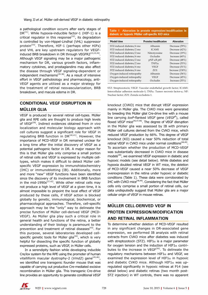
a pathological condition occurrs after early stages of DR[31]. While hypoxiainducible factor1 (HIF1) is a critical regulator in this response[32], its degradation is controlled by vonHippelLindhal (VHL) suppressor protein[33]. Therefore, HIF1 (perhaps other HIFs) and VHL are key upstream regulators for VEGFinduced BRB breakdown in DR through VEGFR2[13,15,34]. Although VEGF signaling may be a major pathogenic mechanism for DR, various growth factors, inflammatory cytokines, and prostaglandins may also affect the disease through VEGF signalingdependent or independent mechanisms[3538]. As a result of intensive effort in VEGF pathobiology and pharmacology, antiVEGF agents are utilized as a major strategy for the treatment of retinal neovascularization, BRB breakdown, and macula edema in DR.
CONDITIONAL VEGF DISRUPTION IN MÜLLER GLIA VEGF is produced by several retinal celltypes. Müller glia and RPE cells are thought to produce high levels of VEGF[39]. Indirect evidences obtained from in vivo localization and molecular biology approach with cell cultures suggest a significant role for VEGF in regulating BRB function in DR[40,41]. However, the importance of MCDVEGF in DR remained unclear for a long time after the initial discovery of VEGF as a potential pathogenic factor in DR. A major reason for this is that Müller glia only comprise a small portion of retinal cells and VEGF is expressed by multiple celltypes, which makes it difficult to detect Müller cellspecific VEGF expression by immunohistochemistry (IHC) or immunoblotting (IB). Additionally, more and more “new” VEGF functions have been identified since the discovery of its involvement in BRB function in the mid1990s[27,42]. While other retinal cells may not produce a high level of VEGF at a given time, it is almost impossible to pinpoint the local effect of VEGF produced by these cells, if VEGF action is blocked globally by genetic, immunological, biochemical, or pharmacological approaches. Therefore, cellspecific approach may be the “only” way to delineate the precise function of Müller cellderived VEGF (MCDVEGF). As Müller glia play such a critical role in general health and functions of the retina, a better understanding of their biology is paramount to the prevention and treatment of retinal diseases[43]. For this purpose, several laboratories developed cellspecific genetic tools for Müller glia[44], which is very helpful for dissecting the specific function of globally expressed proteins, such as VEGF, in Müller cells.
In a serendipity fashion while developing inducible Cre/lox system for the RPE using the promoter of human vitelliform macular dystrophy2 (Vmd2) gene[45,46], we identified one transgenic founder mouse that was capable of carrying out productive Cremediated excisive recombination in Müller glia. This transgenic Credrive line provides an opportunity to generate conditional VEGF
knockout (CVKO) mice that disrupt VEGF expression mainly in Müller glia. The CVKO mice were generated by breeding this Müller glial Credrive line with a mouse line carrying loxPflanked VEGF gene (VEGFff), called floxed VEGF mice[45,47,48]. The degree of VEGF disruption in the Müller glia was assessed by IB with primary Müller cell cultures derived from the CVKO mice, which reduced VEGF production by 66%. This degree of VEGF knockout (KO) caused a near 50% reduction of total retinal VEGF in CVKO mice under normal conditions[48,49]. To ascertain whether the production of MCDVEGF was substantially decreased in CVKO mice in disease models[49], we examined VEGF expression in diabetic and hypoxic models (see detail below). While diabetes and hypoxia doubled retinal VEGF in WT mice, the deletion of MCDVEGF caused a near 50% decrease of VEGF overexpression in the retina under hypoxic or diabetic conditions (Table 1). These data were corroborated by IHC with CVKO mice[48,49]. Considering the fact that Müller cells only comprise a small portion of retinal cells, our data undisputedly suggest that Müller glia are a major cellular origin of VEGF in mouse retinas.
MÜLLER CELL-DERIVED VEGF IN PROTEIN EXPRESSION/MODIFICATION AND RETINAL INFLAMMATIONTo determine whether deletion of MCDVEGF resulted in any significant changes in DRassociated gene expression, we performed IB analysis with retinal extracts from CVKO mice after diabetes was induced with streptozotocin (STZ). HIF1α is a major parameter for oxygen tension and the induction of HIF1α contributes to the increase in VEGF[50]. To delineate the regulatory mechanisms between HIF1α and VEGF, we examined the expression level of HIF1α in hypoxic and diabetic CVKO mice. Although HIF1α was upregulated significantly in hypoxic retinas at P14 (see detail below) and diabetic retinas (two month postSTZ injection) in WT controls, there was no apparent
728 June 10, 2015|Volume 6|Issue 5|WJD|www.wjgnet.com
Table 1 Alteration in protein expression/modification in diabetic or hypoxic Müller cell-specific KO mice
Model/time Proteins/modification Alteration
STZ-induced diabetes/6 mo Albumin Decrease (59%)STZ-induced diabetes/2 mo ICAM1 Decrease (62%)STZ-induced diabetes/2 mo Nitrotyrosine Decrease (19%)STZ-induced diabetes/6 mo Occludin Increase (60%)STZ-induced diabetes/2 mo pNF-κB p65 Decrease (48%)STZ-induced diabetes/2 mo TNF1α Decrease (53%)STZ-induced diabetes/6 mo VEGF Decrease (51%)STZ-induced diabetes/6 mo ZO1 Increase (130%)Oxygen-induced retinopathy Albumin Decrease (56%)Oxygen-induced retinopathy VEGF Decrease (45%)Oxygen-induced retinopathy Occludin Increase (35%)
STZ: Streptozotocin; VEGF: Vascular endothelial growth factor; ICAM1: Intercellular adhesion molecule-1; TNFα: Tumor necrosis factor-α; NF: Nuclear factor; ZO1: Zonula occludens-1.
Wang JJ et al . Müller cell-derived VEGF in diabetic retinopathy

showing a 75.0% reduction of adherent leukocytes, a cardinal feature of retinal inflammation in DR, in the retinal microvasculature of the CVKO mice 2 mo after STZ injection (Table 2). Collectively, these data point to a major role for MCD-VEGF in developing inflammation in DR.
MÜLLER CELL-DERIVED VEGF IN BRB FUNCTION AND INTEGRITYAs diabetic rodents usually do not develop retinal neovascularization[58], the closest way to investigate this is to utilize oxygen induced retinopathy (OIR), a model mimicking an infant blinding disease, ROP. To examine the effect of MCDVEGF on BRB function, the severity of retinal neovascularization was examined in CVKO mice subjected to OIR. The CVKO mice were placed in hyperoxia (75% oxygen) from postnatal day 7 (P7) to P12 and were then kept under normoxia for up to 5 d. In OIR model, the retina was relatively hypoxic at P14 (compared with that at P712), as judged by the abundance of HIF1α. The level of retinal VEGF in hypoxic CVKO mice was decreased by 45% (Table 1). At P17, fluorescein angiography was performed and OIRtreated CVKO mice demonstrated 40% and 30% reductions in areas of retinal neovascularization and of vasoobliteration, respectively (Table 2). Retinal sections from OIRtreated CVKO mice also showed a 34% reduction in the number of preretinal neovascular endothelia (Table 2). As a consequence of decreasing retinal neovascularization and unhealthy microvasculature, we observed a 56% reduction of OIRinduced vascular leakage in CVKO mice (Table 1), judged by IB for albumin. This observation was supported by IHC data[48].
IB analysis with retinal and vitreous extracts prepared from PBSperfused diabetic WT mice demonstrated a 1.5fold increase in extravascular albumin 6 mo after STZinjection. However, the disruption of MCDVEGF caused a near 60% reduction of albumin leakage in agematched diabetic mice (Table 1). Such a reduction can be visualized in the retinal flat-mounts of diabetic CVKO mice by intravenously injected fluorescein isothiocyanatelabeled albumin[49].
To delineate the mechanistic insights of MCDVEGF in diabetesinduced vascular leakage, we analyzed the levels of occludin and Zonula occludens1 (ZO1), two major tight junction proteins, in the retina[49]. While diabetes resulted in 39% and 58% decreases of occludin and ZO1, respectively, in WT animals, no alteration in occludin and ZO1 expression was observed in diabetic CVKO mice. As a result, the diabetic CVKO mice had 60% and 130% upregulation of occludin and ZO1 (Table 1), compared with that of diabetic WT controls. Our data indicated that the disruption of MCDVEGF resulted in a significant reduction of diabetesinduced retinal vascular leakage by attenuating the depletion of occludin and ZO1. This result was supported by a 36% increase in the level
difference in the levels of retinal HIF1α between the CVKO mice and WT controls under hypoxic and diabetic conditions[48,49], suggesting that hypoxia/HIF1α is an upstream regulator of VEGF produced by Müller glia.
Nuclear factorkappaB (NFκB) is a transcription factor and a major player in inducing early pathological changes, such as inflammation, in DR[51,52]. To explore if MCDVEGF regulated NFκB in diabetic retina, we examined the expression/phosphorylation of NFκB p65 subunit. While there was no detectable change in the total NFκB p65 level between the CVKO mice and WT controls, the loss of MCDVEGF caused a dramatic decrease (48%) of phosphorylated (activated) NFκB p65 in the retina 2 mo after STZinjection (Table 1). This result suggests that activated p65 is downstream of MCDVEGF in DR[49]. Nitric oxide (NO) is an important inflammatory mediator and its level is a representation of oxidative stress in the retina of diabetic patients and animals[5355]. Peroxynitrite is a highly reactive oxidant, which is formed by the rapid combination of NO with superoxide. Increased peroxynitrite formation may be directly linked to diabetesinduced VEGF overexpression and there is a possible loop effect of VEGF signaling and peroxynitrite formation[56,57]. To determine the role of MCDVEGF in protein nitration, we examined the level of nitrotyrosine, a biomarker of peroxynitrite, in retinal protein extracts from CVKO and control mice 2 mo after STZinjection. The retinal extracts from diabetic CVKO mice demonstrated a 19% decrease of proteins with nitrotyrosine (Table 1), indicating that the disruption of MCDVEGF reduced oxidative stress.
Inflammation is an early pathological response in DR. To identify the role of MCDVEGF in retinal inflammation in DR, we examined the levels of proinflammatory markers in CVKO mice by IB analysis for intercellular adhesion molecule1 (ICAM1) and tumor necrosis factorα (TNFα), 2 mo after STZinjection. Compared with controls, the CVKO mice showed 62.3% and 52.9% reduction of ICAM1 and TNFα (Table 1), respectively. These results suggest that the deletion of MCDVEGF substantially inhibits inflammation in diabetic retinas[49]. This notion is reinforced by the result from the leukostasis assay
729 June 10, 2015|Volume 6|Issue 5|WJD|www.wjgnet.com
Table 2 Pathological changes in diabetic or hypoxic Müller cell-specific KO mice
Model Pathological changes Alteration
STZ-induced diabetes Leukocytosis Decrease (75%)STZ-induced diabetes Vascular leakage Decrease (60%)STZ-induced diabetes Acellular capillaries Decrease (45%)STZ-induced diabetes Vascular leakage Decrease (60.0%)Oxygen-induced retinopathy Pre-retinal
neovascularization Decrease (34%)
Oxygen-induced retinopathy Neovascularization area
Decrease (40%)
Oxygen-induced retinopathy Vaso-obliteration area Decrease (30%)
STZ: Streptozotocin.
Wang JJ et al . Müller cell-derived VEGF in diabetic retinopathy
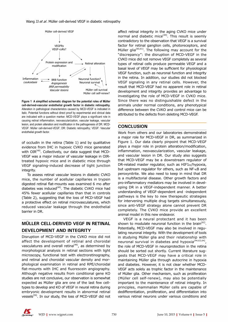
of occludin in the retina (Table 1) and by qualitative evidence from IHC in hypoxic CVKO mice generated with OIR[48]. Collectively, our data suggest that MCDVEGF was a major inducer of vascular leakage in OIRtreated hypoxic mice and in diabetic mice through VEGF signalinginduced decrease of tight junction integrity.
To assess retinal vascular lesions in diabetic CVKO mice, the number of acellular capillaries in trypsin digested retinal flat-mounts was examined 6 mo after diabetes was induced[49]. The diabetic CVKO mice had 45% fewer acellular capillaries than that in controls (Table 2), suggesting that the loss of MCDVEGF had a protective effect on retinal microvasculatures, which reduced vascular leakage through the endothelial barrier in DR.
MÜLLER CELL-DERIVED VEGF IN RETINAL DEVELOPMENT AND INTEGRITYDisruption of MCDVEGF in the CVKO mice did not affect the development of retinal and choroidal vasculatures and overall retina[48], as determined by morphological analysis in retinal sections with light microscopy, functional test with electroretinography, and retinal and choroidal vascular density and morphological examination in retinal and RPE/choroidal flatmounts with IHC and fluorescein angiography. Although negative results from conditional gene KO studies are not conclusive, our observation is somewhat expected as Müller glia are one of the last few celltypes to develop and KO of VEGF in neural retina during embryonic development results in abnormal retinal vessels[59]. In our study, the loss of MCDVEGF did not
affect retinal integrity in the aging CVKO mice under normal and diabetic mice[49]. This result is seemly contradictory to the observation that VEGF is a survival factor for retinal ganglion cells, photoreceptors, and Müller glia[60,61]. The following may account for the “discrepancy”: the disruption of MCDVEGF in the CVKO mice did not remove VEGF completely as several types of retinal cells produce permeable VEGF and a basal level of VEGF may be sufficient for physiological VEGF function, such as neuronal function and integrity in the retina. In addition, our studies did not blocked VEGF signaling in any retinal cells. However, the result that MCDVEGF had no apparent role in retinal development and integrity provides an advantage to investigating the role of MCDVEGF in CVKO mice. Since there was no distinguishable defect in the animals under normal conditions, any phenotypical difference between the CVKO and control mice can be attributed to the defects from deleting MCDVEGF.
CONCLUSIONWork from others and our laboratories demonstrated a major role for MCDVEGF in DR, as summarized in Figure 1. Our data clearly pinpoint that MCDVEGF plays a major role in protein alteration/modification, inflammation, neovascularization, vascular leakage, and vascular lesion in DR. Our study also suggests that MCDVEGF may be a downstream regulator of DRrelated master regulator, such as HIF1α/hypoxia, but upstream regulator for others, such as NFκB and peroxynitrite. We also need to keep in mind that DR is a multifactorial disease. Other growth factors and pro-inflammatory mediators may be involved in devel-oping DR in a VEGFindependent manner. A better understanding of VEGFdependent and independent pathways is the key to new therapeutic strategies for intervening multiple drug targets simultaneously, since antiVEGF strategy alone cannot prevent DR completely. The CVKO mice provide an excellent animal model in this new endeavor.
VEGF is a neural protectant and it has been shown to modulate neuronal function in the brain[62]. Potentially, MCDVEGF may also be involved in regulating neuronal integrity. With the development of tools in studying Müller glia and their relationship with neuronal survival in diabetes and hypoxia[44,63,64], the role of MCDVEGF in neuroprotection in the retina should be sorted out shortly. Current literature suggests that MCDVEGF may have a critical role in maintaining Müller glia through autocrine in hypoxia and diabetes. However, it is not clear whether MCDVEGF acts solely as trophic factor in the maintenance of Müller glia. Other mechanism, such as proliferation (Müller cell selfrenew), may also be potentially important to the maintenance of retinal integrity. In principles, mammalian Müller cells are capable of dedifferentiation, proliferation, and differentiation into various retinal neurons under various conditions and
730 June 10, 2015|Volume 6|Issue 5|WJD|www.wjgnet.com
Müller cell-derived VEGF
VEGFRsVEGF-coRs?
Protein expression and modification
BRB functionAngiogenesis
BRB permeabilityVascular lesions
Retinal alteration
Neuronal function? Neuronal survival
InflammationLeukostasis
Müller cell survivalMüller cell self-renew?
Figure 1 A simplified schematic diagram for the potential roles of Müller cell-derived-vascular endothelial growth factor in diabetic retinopathy. Alteration in pathological characteristics caused by MCD-VEGF is indicated in italic. Potential functions without direct proof by experimental and clinical data are indicated with a question marker. MCD-VEGF plays a significant role in causing retinal inflammation, neovascularization, vascular leakage, vascular lesion, and protein alteration and modification in the pathogenesis of DR. MCD-VEGF: Müller cell-derived-VEGF; DR: Diabetic retinopathy; VEGF: Vascular endothelial growth factor.
Wang JJ et al . Müller cell-derived VEGF in diabetic retinopathy
are considered as a major retinal stem cell[65]. It will be fascinating if MCDVEGF can act similarly as other growth factors in differentiating Müller cells to neurons for neuroprotection[66].
Although there are many publications on VEGF or MCDVEGF in DR, little is known about its actually signaling pathways. As discussed earlier, the presence of at least seven VEGF receptors and coreceptors accounts for the difficulties in revealing their mechanisms. Additionally, VEGF is a secreted protein and loss of VEGF produced by a single celltype can be compensated by others. Delineating detailed mechanisms may greatly enhance our understanding to the pathogenesis, treatment, and neuronal function in DR, which is critical to the improvement and safety of current antiVEGF strategy and to the design of new treatments for the disease.
ACKNOWLEDGMENTSWe thank Dr. Gennadiy P Moiseyev for critical reading of this manuscript and members of our laboratories for providing data and helpful discussions.
REFERENCES1 Reichenbach A, Bringmann A. New functions of Müller cells. Glia
2013; 61: 651-678 [PMID: 23440929 DOI: 10.1002/glia.22477]2 Gardner TW, Antonetti DA, Barber AJ, LaNoue KF, Levison SW.
Diabetic retinopathy: more than meets the eye. Surv Ophthalmol 2002; 47 Suppl 2: S253-S262 [PMID: 12507627 DOI: 10.1016/S0039-6257(02)00387-9]
3 Xu HZ, Le YZ. Significance of outer blood-retina barrier breakdown in diabetes and ischemia. Invest Ophthalmol Vis Sci 2011; 52: 2160-2164 [PMID: 21178141 DOI: 10.1167/iovs.10-6518]
4 Xu HZ, Song Z, Fu S, Zhu M, Le YZ. RPE barrier breakdown in diabetic retinopathy: seeing is believing. J Ocul Biol Dis Infor 2011; 4: 83-92 [PMID: 23275801 DOI: 10.1007/s12177-011-9068-4]
5 Ablonczy Z, Prakasam A, Fant J, Fauq A, Crosson C, Sambamurti K. Pigment epithelium-derived factor maintains retinal pigment epithelium function by inhibiting vascular endothelial growth factor-R2 signaling through gamma-secretase. J Biol Chem 2009; 284: 30177-30186 [PMID: 19723623 DOI: 10.1074/jbc.M109.032391]
6 Ozdemir H, Karacorlu M, Karacorlu S. Serous macular detachment in diabetic cystoid macular oedema. Acta Ophthalmol Scand 2005; 83: 63-66 [PMID: 15715559 DOI: 10.1111/j.1600-0420.2005.00387.x]
7 Martin PM, Roon P, Van Ells TK, Ganapathy V, Smith SB. Death of retinal neurons in streptozotocin-induced diabetic mice. Invest Ophthalmol Vis Sci 2004; 45: 3330-3336 [PMID: 15326158 DOI: 10.1167/iovs.04-0247]
8 Gardner TW, Antonetti DA, Barber AJ, LaNoue KF, Nakamura M. New insights into the pathophysiology of diabetic retinopathy: potential cell-specific therapeutic targets. Diabetes Technol Ther 2000; 2: 601-608 [PMID: 11469624 DOI: 10.1089/15209150050502023]
9 Terasaki H, Hirose H, Miyake Y. S-cone pathway sensitivity in diabetes measured with threshold versus intensity curves on flashed backgrounds. Invest Ophthalmol Vis Sci 1996; 37: 680-684 [PMID: 8595969]
10 Aspinall PA. Rod-cone interaction: some indirect evidence. Acta Ophthalmol (Copenh) 1977; 55: 294-302 [PMID: 577101 DOI: 10.1111/j.1755-3768.1977.tb01311.x]
11 Barber AJ, Lieth E, Khin SA, Antonetti DA, Buchanan AG,
Gardner TW. Neural apoptosis in the retina during experimental and human diabetes. Early onset and effect of insulin. J Clin Invest 1998; 102: 783-791 [PMID: 9710447 DOI: 10.1172/JCI2425]
12 Ferrara N, Henzel WJ. Pituitary follicular cells secrete a novel heparin-binding growth factor specific for vascular endothelial cells. Biochem Biophys Res Commun 1989; 161: 851-858 [PMID: 2735925 DOI: 10.1016/0006-291X(89)92678-8]
13 Ferrara N, Gerber HP, LeCouter J. The biology of VEGF and its receptors. Nat Med 2003; 9: 669-676 [PMID: 12778165 DOI: 10.1038/nm0603-669]
14 Penn JS, Madan A, Caldwell RB, Bartoli M, Caldwell RW, Hartnett ME. Vascular endothelial growth factor in eye disease. Prog Retin Eye Res 2008; 27: 331-371 [PMID: 18653375 DOI: 10.1016/j.preteyeres.2008.05.001]
15 Shibuya M. Structure and dual function of vascular endothelial growth factor receptor-1 (Flt-1). Int J Biochem Cell Biol 2001; 33: 409-420 [PMID: 11312109 DOI: 10.1247/csf.26.25]
16 Carmeliet P, Ferreira V, Breier G, Pollefeyt S, Kieckens L, Gertsenstein M, Fahrig M, Vandenhoeck A, Harpal K, Eberhardt C, Declercq C, Pawling J, Moons L, Collen D, Risau W, Nagy A. Abnormal blood vessel development and lethality in embryos lacking a single VEGF allele. Nature 1996; 380: 435-439 [PMID: 8602241 DOI: 10.1038/380435a0]
17 Ferrara N, Carver-Moore K, Chen H, Dowd M, Lu L, O’Shea KS, Powell-Braxton L, Hillan KJ, Moore MW. Heterozygous embryonic lethality induced by targeted inactivation of the VEGF gene. Nature 1996; 380: 439-442 [PMID: 8602242 DOI: 10.1038/380439a0]
18 Adamis AP, Miller JW, Bernal MT, D’Amico DJ, Folkman J, Yeo TK, Yeo KT. Increased vascular endothelial growth factor levels in the vitreous of eyes with proliferative diabetic retinopathy. Am J Ophthalmol 1994; 118: 445-450 [PMID: 7943121 DOI: 10.1016/S0002-9394(14)75794-0]
19 Adamis AP , Shima DT, Tolentino MJ, Gragoudas ES, Ferrara N, Folkman J, D’Amore PA, Miller JW. Inhibition of vascular endothelial growth factor prevents retinal ischemia-associated iris neovascularization in a nonhuman primate. Arch Ophthalmol 1996; 114: 66-71 [PMID: 8540853 DOI: 10.1001/archopht.1996.01100130062010]
20 Aiello LP, Pierce EA, Foley ED, Takagi H, Chen H, Riddle L, Ferrara N, King GL, Smith LE. Suppression of retinal neovascularization in vivo by inhibition of vascular endothelial growth factor (VEGF) using soluble VEGF-receptor chimeric proteins. Proc Natl Acad Sci USA 1995; 92: 10457-10461 [PMID: 7479819 DOI: 10.1073/pnas.92.23.10457]
21 Ozaki H, Seo MS, Ozaki K, Yamada H, Yamada E, Okamoto N, Hofmann F, Wood JM, Campochiaro PA. Blockade of vascular endothelial cell growth factor receptor signaling is sufficient to completely prevent retinal neovascularization. Am J Pathol 2000; 156: 697-707 [PMID: 10666398 DOI: 10.1016/S0002-9440(10)64773-6]
22 Senger DR, Connolly DT, Van de Water L, Feder J, Dvorak HF. Purification and NH2-terminal amino acid sequence of guinea pig tumor-secreted vascular permeability factor. Cancer Res 1990; 50: 1774-1778 [PMID: 2155059]
23 Dvorak HF, Brown LF, Detmar M, Dvorak AM. Vascular permeability factor/vascular endothelial growth factor, microvascular hyperpermeability, and angiogenesis. Am J Pathol 1995; 146: 1029-1039 [PMID: 7538264]
24 Senger DR, Galli SJ, Dvorak AM, Perruzzi CA, Harvey VS, Dvorak HF. Tumor cells secrete a vascular permeability factor that promotes accumulation of ascites fluid. Science 1983; 219: 983-985 [PMID: 6823562 DOI: 10.1126/science.6823562]
25 Aiello LP, Bursell SE, Clermont A, Duh E, Ishii H, Takagi C, Mori F, Ciulla TA, Ways K, Jirousek M, Smith LE, King GL. Vascular endothelial growth factor-induced retinal permeability is mediated by protein kinase C in vivo and suppressed by an orally effective beta-isoform-selective inhibitor. Diabetes 1997; 46: 1473-1480 [PMID: 9287049 DOI: 10.2337/diab.46.9.1473]
26 Miller JW, Adamis AP, Shima DT, D’Amore PA, Moulton RS, O’
731 June 10, 2015|Volume 6|Issue 5|WJD|www.wjgnet.com
Wang JJ et al . Müller cell-derived VEGF in diabetic retinopathy
Reilly MS, Folkman J, Dvorak HF, Brown LF, Berse B. Vascular endothelial growth factor/vascular permeability factor is temporally and spatially correlated with ocular angiogenesis in a primate model. Am J Pathol 1994; 145: 574-584 [PMID: 7521577]
27 Aiello LP, Avery RL, Arrigg PG, Keyt BA, Jampel HD, Shah ST, Pasquale LR, Thieme H, Iwamoto MA, Park JE. Vascular endothelial growth factor in ocular fluid of patients with diabetic retinopathy and other retinal disorders. N Engl J Med 1994; 331: 1480-1487 [PMID: 7526212 DOI: 10.1056/NEJM199412013312203]
28 Hartnett ME, Lappas A, Darland D, McColm JR, Lovejoy S, D’Amore PA. Retinal pigment epithelium and endothelial cell interaction causes retinal pigment epithelial barrier dysfunction via a soluble VEGF-dependent mechanism. Exp Eye Res 2003; 77: 593-599 [PMID: 14550401 DOI: 10.1016/S0014-4835(03)00189-1]
29 Qaum T, Xu Q, Joussen AM, Clemens MW, Qin W, Miyamoto K, Hassessian H, Wiegand SJ, Rudge J, Yancopoulos GD, Adamis AP. VEGF-initiated blood-retinal barrier breakdown in early diabetes. Invest Ophthalmol Vis Sci 2001; 42: 2408-2413 [PMID: 11527957]
30 Funatsu H, Yamashita H, Noma H, Mimura T, Yamashita T, Hori S. Increased levels of vascular endothelial growth factor and interleukin-6 in the aqueous humor of diabetics with macular edema. Am J Ophthalmol 2002; 133: 70-77 [PMID: 11755841 DOI: 10.1016/S0002-9394(01)01269-7]
31 Dor Y, Porat R, Keshet E. Vascular endothelial growth factor and vascular adjustments to perturbations in oxygen homeostasis. Am J Physiol Cell Physiol 2001; 280: C1367-C1374 [PMID: 11350731]
32 Semenza G. Signal transduction to hypoxia-inducible factor 1. Biochem Pharmacol 2002; 64: 993-998 [PMID: 12213597 DOI: 10.1016/S0006-2952(02)01168-1]
33 Mole DR, Maxwell PH, Pugh CW, Ratcliffe PJ. Regulation of HIF by the von Hippel-Lindau tumour suppressor: implications for cellular oxygen sensing. IUBMB Life 2001; 52: 43-47 [PMID: 11795592 DOI: 10.1080/15216540252774757]
34 Matsumoto T, Claesson-Welsh L. VEGF receptor signal transduction. Sci STKE 2001; 2001: re21 [PMID: 11741095]
35 Kolch W, Martiny-Baron G, Kieser A, Marmé D. Regulation of the expression of the VEGF/VPS and its receptors: role in tumor angiogenesis. Breast Cancer Res Treat 1995; 36: 139-155 [PMID: 8534863 DOI: 10.1007/BF00666036]
36 Michiels C, Minet E, Michel G, Mottet D, Piret JP, Raes M. HIF-1 and AP-1 cooperate to increase gene expression in hypoxia: role of MAP kinases. IUBMB Life 2001; 52: 49-53 [PMID: 11795593 DOI: 10.1080/15216540252774766]
37 Berra E, Milanini J, Richard DE, Le Gall M, Viñals F, Gothié E, Roux D, Pagès G, Pouysségur J. Signaling angiogenesis via p42/p44 MAP kinase and hypoxia. Biochem Pharmacol 2000; 60: 1171-1178 [PMID: 11007955 DOI: 10.1016/S0006-2952(00)00423-8]
38 Pagès G, Milanini J, Richard DE, Berra E, Gothié E, Viñals F, Pouysségur J. Signaling angiogenesis via p42/p44 MAP kinase cascade. Ann N Y Acad Sci 2000; 902: 187-200 [PMID: 10865838 DOI: 10.1111/j.1749-6632.2000.tb06313.x]
39 Blaauwgeers HG, Holtkamp GM, Rutten H, Witmer AN, Koolwijk P, Partanen TA, Alitalo K, Kroon ME, Kijlstra A, van Hinsbergh VW, Schlingemann RO. Polarized vascular endothelial growth factor secretion by human retinal pigment epithelium and localization of vascular endothelial growth factor receptors on the inner choriocapillaris. Evidence for a trophic paracrine relation. Am J Pathol 1999; 155: 421-428 [PMID: 10433935 DOI: 10.1016/S0002-9440(10)65138-3]
40 Pierce EA, Avery RL, Foley ED, Aiello LP, Smith LE. Vascular endothelial growth factor/vascular permeability factor expression in a mouse model of retinal neovascularization. Proc Natl Acad Sci USA 1995; 92: 905-909 [PMID: 7846076 DOI: 10.1073/pnas.92.3.905]
41 Jingjing L, Xue Y, Agarwal N, Roque RS. Human Müller cells express VEGF183, a novel spliced variant of vascular endothelial growth factor. Invest Ophthalmol Vis Sci 1999; 40: 752-759 [PMID: 10067980]
42 Alon T, Hemo I, Itin A, Pe’er J, Stone J, Keshet E. Vascular endothelial growth factor acts as a survival factor for newly formed retinal vessels and has implications for retinopathy of prematurity. Nat Med 1995; 1: 1024-1028 [PMID: 7489357 DOI: 10.1038/nm1095-1024]
43 Koury CB. Genetic system offer option for studying the role of retinal müller cells. Retina Today 2007; I: 31
44 Le YZ. Conditional gene targeting: dissecting the cellular mechanisms of retinal degenerations. J Ophthalmol 2011; 2011: 806783 [PMID: 21253511 DOI: 10.1155/2011/806783]
45 Ueki Y, Ash JD, Zhu M, Zheng L, Le YZ. Expression of Cre recombinase in retinal Müller cells. Vision Res 2009; 49: 615-621 [PMID: 19948109 DOI: 10.1016/j.visres.2009.01.012]
46 Zhu M , Zheng L, Ueki Y, Ash JD, Le YZ. Unexpected transcriptional activity of the human VMD2 promoter in retinal development. Adv Exp Med Biol 2010; 664: 211-216 [PMID: 20238019]
47 Gerber HP, Hillan KJ, Ryan AM, Kowalski J, Keller GA, Rangell L, Wright BD, Radtke F, Aguet M, Ferrara N. VEGF is required for growth and survival in neonatal mice. Development 1999; 126: 1149-1159 [PMID: 10021335]
48 Bai Y, Ma JX, Guo J, Wang J, Zhu M, Chen Y, Le YZ. Müller cell-derived VEGF is a significant contributor to retinal neovascularization. J Pathol 2009; 219: 446-454 [PMID: 19768732 DOI: 10.1002/path.2611]
49 Wang J, Xu X, Elliott MH, Zhu M, Le YZ. Müller cell-derived VEGF is essential for diabetes-induced retinal inflammation and vascular leakage. Diabetes 2010; 59: 2297-2305 [PMID: 20530741 DOI: 10.2337/db09-1420]
50 Kondo T, Kahn CR. Altered insulin signaling in retinal tissue in diabetic states. J Biol Chem 2004; 279: 37997-38006 [PMID: 15201286 DOI: 10.1074/jbc.M401339200]
51 Kowluru RA, Koppolu P, Chakrabarti S, Chen S. Diabetes-induced activation of nuclear transcriptional factor in the retina, and its inhibition by antioxidants. Free Radic Res 2003; 37: 1169-1180 [PMID: 14703729 DOI: 10.1080/10715760310001604189]
52 Kowluru RA, Engerman RL, Kern TS. Abnormalities of retinal metabolism in diabetes or experimental galactosemia VIII. Prevention by aminoguanidine. Curr Eye Res 2000; 21: 814-819 [PMID: 11120572 DOI: 10.1076/ceyr.21.4.814.5545]
53 Du Y, Smith MA, Miller CM, Kern TS. Diabetes-induced nitrative stress in the retina, and correction by aminoguanidine. J Neurochem 2002; 80: 771-779 [PMID: 11948240 DOI: 10.1046/j.0022-3042.2001.00737.x]
54 Abu El-Asrar AM, Desmet S, Meersschaert A, Dralands L, Missotten L, Geboes K. Expression of the inducible isoform of nitric oxide synthase in the retinas of human subjects with diabetes mellitus. Am J Ophthalmol 2001; 132: 551-556 [PMID: 11589878 DOI: 10.1016/S0002-9394(01)01127-8]
55 Leal EC, Manivannan A, Hosoya K, Terasaki T, Cunha-Vaz J, Ambrósio AF, Forrester JV. Inducible nitric oxide synthase isoform is a key mediator of leukostasis and blood-retinal barrier breakdown in diabetic retinopathy. Invest Ophthalmol Vis Sci 2007; 48: 5257-5265 [PMID: 17962481 DOI: 10.1167/iovs.07-0112]
56 El-Remessy AB, Al-Shabrawey M, Platt DH, Bartoli M, Behzadian MA, Ghaly N, Tsai N, Motamed K, Caldwell RB. Peroxynitrite mediates VEGF’s angiogenic signal and function via a nitration-independent mechanism in endothelial cells. FASEB J 2007; 21: 2528-2539 [PMID: 17384142 DOI: 10.1096/fj.06-7854com]
57 El-Remessy AB, Behzadian MA, Abou-Mohamed G, Franklin T, Caldwell RW, Caldwell RB. Experimental diabetes causes breakdown of the blood-retina barrier by a mechanism involving tyrosine nitration and increases in expression of vascular endothelial growth factor and urokinase plasminogen activator receptor. Am J Pathol 2003; 162: 1995-2004 [PMID: 12759255 DOI: 10.1016/S0002-9440(10)64332-5]
58 Smith LE, Wesolowski E, McLellan A, Kostyk SK, D’Amato R, Sullivan R, D’Amore PA. Oxygen-induced retinopathy in the mouse. Invest Ophthalmol Vis Sci 1994; 35: 101-111 [PMID:
732 June 10, 2015|Volume 6|Issue 5|WJD|www.wjgnet.com
Wang JJ et al . Müller cell-derived VEGF in diabetic retinopathy
7507904]59 Haigh JJ, Morelli PI, Gerhardt H, Haigh K, Tsien J, Damert
A, Miquerol L, Muhlner U, Klein R, Ferrara N, Wagner EF, Betsholtz C, Nagy A. Cortical and retinal defects caused by dosage-dependent reductions in VEGF-A paracrine signaling. Dev Biol 2003; 262: 225-241 [PMID: 14550787 DOI: 10.1016/S0012-1606(03)00356-7]
60 Nishijima K, Ng YS, Zhong L, Bradley J, Schubert W, Jo N, Akita J, Samuelsson SJ, Robinson GS, Adamis AP, Shima DT. Vascular endothelial growth factor-A is a survival factor for retinal neurons and a critical neuroprotectant during the adaptive response to ischemic injury. Am J Pathol 2007; 171: 53-67 [PMID: 17591953 DOI: 10.2353/ajpath.2007.061237]
61 Saint-Geniez M, Maharaj AS, Walshe TE, Tucker BA, Sekiyama E, Kurihara T, Darland DC, Young MJ, D’Amore PA. Endogenous VEGF is required for visual function: evidence for a survival role on müller cells and photoreceptors. PLoS One 2008; 3: e3554 [PMID: 18978936 DOI: 10.1371/journal.pone.0003554]
62 McCloskey DP, Croll SD, Scharfman HE. Depression of synaptic
transmission by vascular endothelial growth factor in adult rat hippocampus and evidence for increased efficacy after chronic seizures. J Neurosci 2005; 25: 8889-8897 [PMID: 16192378 DOI: 10.1523/JNEUROSCI.2577-05.2005]
63 Dong S, Liu Y, Zhu M, Xu X, Le YZ. Simplified system to investigate alteration of retinal neurons in diabetes. Adv Exp Med Biol 2014; 801: 139-143 [PMID: 24664691 DOI: 10.1007/978-1-4614-3209-8_18]
64 Fu S, Zhu M, Ash JD, Wang Y, Le YZ. Investigating the role of retinal Müller cells with approaches in genetics and cell biology. Adv Exp Med Biol 2014; 801: 401-405 [PMID: 24664724 DOI: 10.1007/978-1-4614-3209-8_51]
65 Karl MO, Hayes S, Nelson BR, Tan K, Buckingham B, Reh TA. Stimulation of neural regeneration in the mouse retina. Proc Natl Acad Sci USA 2008; 105: 19508-19513 [PMID: 19033471 DOI: 10.1073/pnas.0807453105]
66 Karl MO, Reh TA. Studying the generation of regenerated retinal neuron from Müller glia in the mouse eye. Methods Mol Biol 2012; 884: 213-227 [PMID: 22688709 DOI: 10.1007/978-1-61779-848-1_15]
P- Reviewer: Das UN, García Mayor R, McNally RJQ, Saleh J, Swierczynski J S- Editor: Tian YL L- Editor: A
E- Editor: Zhang DN
733 June 10, 2015|Volume 6|Issue 5|WJD|www.wjgnet.com
Wang JJ et al . Müller cell-derived VEGF in diabetic retinopathy
MINIREVIEWS
What neonatal complications should the pediatrician be aware of in case of maternal gestational diabetes?
Delphine Mitanchez, Catherine Yzydorczyk, Umberto Simeoni
Delphine Mitanchez, Division of Neonatology, Department of Perinatology, Armand Trousseau Hospital, 75012 Paris, FranceCatherine Yzydorczyk, Umberto Simeoni, Division of Pediatrics and DOHaD Laboratory, CHUV University Hospital and UNIL, 1011 Lausanne, Switzerland
Author contributions: All the authors made substantial contributions to conception and design of the review, made critical revisions and final approval of the version of the article to be published.
Conflict-of-interest: None.
Open-Access: This article is an openaccess article which was selected by an inhouse editor and fully peerreviewed by external reviewers. It is distributed in accordance with the Creative Commons Attribution Non Commercial (CC BY-NC 4.0) license, which permits others to distribute, remix, adapt, build upon this work noncommercially, and license their derivative works on different terms, provided the original work is properly cited and the use is non-commercial. See: http://creativecommons.org/licenses/by-nc/4.0/
Correspondence to: Delphine Mitanchez, MD, PhD, Division of Neonatology, Department of Perinatology, Armand Trousseau Hospital, 26 Avenue du Docteur Arnold Netter, 75012 Paris, France. [email protected]: +33144736191Fax: +33144736892
Received: August 28, 2014Peer-review started: August 28, 2014First decision: December 17, 2014Revised: January 29, 2015Accepted: March 16, 2015Article in press: March 18, 2015Published online: June 10, 2015
AbstractIn the epidemiologic context of maternal obesity and type 2 diabetes (T2D), the incidence of gestational diabetes has significantly increased in the last decades. Infants of diabetic mothers are prone to various
neonatal adverse outcomes, including metabolic and hematologic disorders, respiratory distress, cardiac disorders and neurologic impairment due to perinatal asphyxia and birth traumas, among others. Macrosomia is the most constant consequence of diabetes and its severity is mainly influenced by maternal blood glucose level. Neonatal hypoglycemia is the main metabolic disorder that should be prevented as soon as possible after birth. The severity of macrosomia and the maternal health condition have a strong impact on the frequency and the severity of adverse neonatal outcomes. Pregestational T2D and maternal obesity significantly increase the risk of perinatal death and birth defects. The high incidence of maternal hyperglycemia in developing countries, associated with the scarcity of maternal and neonatal care, seriously increase the burden of neonatal complications in these countries.
Key words: Birth defects; Hypoglycemia; Respiratory distress; Preterm; Perinatal mortality; Type 2 diabetes; Obesity
© The Author(s) 2015. Published by Baishideng Publishing Group Inc. All rights reserved.
Core tip: Increased mortality and morbidity are histo-rically attributed to neonates of diabetic mothers. A discerning analysis of the literature shows that these adverse outcomes are uncommon among infants born from “pure” gestational diabetes mellitus (GDM) mothers, well managed during pregnancy. Macrosomia is the predominant adverse outcome and the main factor linked to neonatal complications. Poor maternal glycemic control, especially in the context of maternal type 2 diabetes and obesity increases the risk of all adverse neonatal outcomes, most strikingly the risk of perinatal mortality and birth defects. Developing strategies for screening and managing women with GDM must be encouraged notably in middle and low income countries and, also to limit the adverse effects on global health population in the future.
734 June 10, 2015|Volume 6|Issue 5|WJD|www.wjgnet.com
Submit a Manuscript: http://www.wjgnet.com/esps/Help Desk: http://www.wjgnet.com/esps/helpdesk.aspxDOI: 10.4239/wjd.v6.i5.734
World J Diabetes 2015 June 10; 6(5): 734-743ISSN 1948-9358 (online)
© 2015 Baishideng Publishing Group Inc. All rights reserved.
Mitanchez D, Yzydorczyk C, Simeoni U. What neonatal complications should the pediatrician be aware of in case of maternal gestational diabetes? World J Diabetes 2015; 6(5): 734743 Available from: URL: http://www.wjgnet.com/1948-9358/full/v6/i5/734.htm DOI: http://dx.doi.org/10.4239/wjd.v6.i5.734
INTRODUCTIONGestational diabetes mellitus (GDM) is defined as a glucose intolerance of any degree with onset or first recognition during pregnancy. In high income countries, but also in middle and low income countries, because of the spreading of industrialized lifestyle, the incidence of obesity and type 2 diabetes (T2D) has dramatically increased, and subsequently the incidence of GDM[1].
In high-resource countries, progress has been made during the past fifty years regarding preconceptional care, screening and management of GDM. However, in low and middle-income countries, quality of antenatal care to detect and manage GDM, are often poorly available. As a consequence, the prenatal and neonatal burden of GDM may be paradoxically higher in these countries, although this point is not well documented[2].
Much of the currently available knowledge on the consequences of maternal diabetes on the offspring has been provided by studies on type 1 diabetes (T1D), while the risks related to GDM, which is much more frequent, need to be clarified in order to improve and to adapt neonatal management[3]. Moreover, extensive data suggest that the offspring of diabetic mothers is furthermore exposed to an increased risk of developing chronic, non-communicable diseases at adulthood[4].
Neonatologists are facing first-line this new epide-miologic setting. This review addresses the currently available knowledge on short term consequences of GDM in neonates and focuses on situations with increased risks of neonatal adverse outcomes.
SHORT TERM OUTCOMESMacrosomiaMacrosomia is the most constant complication in GDM. The concept of excessive fetal growth is expressed either by the word “macrosomia” or by the expression “large for gestational age” (LGA). Macrosomia is defined by a birth weight (BW) of 4000 or 4500 g and more, depending on the authors. However, in this definition, gestational age (GA) is not taken into account. The term LGA corresponds to a BW ≥ 90th percentile or > +2SD (> 97th percentile) for GA. This definition allows premature newborns with excessive fetal growth to be identified. Macrosomia in newborns of diabetic mothers is characterized by excess body fat, an increased muscle mass and organomegaly, without increase in brain size.
The Pedersen-Freinkel’s hypothesis, expressed sixty years ago, suggested that fetal overgrowth is related to increased transplacental transfer of maternal glucose, which stimulates the release of insulin by fetal pancreatic beta cells[5]. Insulin is a major factor of fetal growth and it up-regulates the Insulin-like Growth Factor (IGF) system, subsequently leading to fetal macrosomia. According to this hypothesis, different studies have characterized the link between maternal glycemia and neonatal macrosomia or fat mass[6,7].
The HAPO study showed a continuous, positive association between maternal glycemia, fetal hyper-insulinism and BW[8]. A linear and continuous rela-tionship between body fat percentage in newborns, maternal glycaemia and fetal insulin levels has been found in this study[9]. More recently, other mechanisms that may also contribute to fetal overgrowth were evoked, like maternal metabolic environment and placental modifications[10]. In particular, maternal lipids availability and transport to the fetus may be enhanced in case of maternal diabetes[11].
Hence, all types of maternal diabetes are risk factors for macrosomia. As discussed below, macrosomia is per se a cause increased neonatal adverse outcome and this point emphasizes the importance of recognizing the excess of growth, even in preterm infants. Treatment of GDM significantly reduces the rate of macrosomia[12,13].
Preterm birthA number of studies have reported an increased risk of preterm births in case of diabetes. However, data are not always available on the respective proportion of induced and spontaneous births, considering the increased maternal and fetal morbidity of diabetes during pregnancy. The benefits of early delivery to avoid fetal death or shoulder dystocia must be balan-ced against the morbidity linked to preterm birth, especially the respiratory morbidity.
The link between GDM and spontaneous preterm birth is still controversial. Hedderson et al[14] showed in a large cohort study that GDM was an independent risk factor for spontaneous preterm birth (RR = 1.42, 95%CI: 1.15-1.77). On the other hand, Yogev et al[15] found that the rate of spontaneous preterm delivery was not increased in GDM compared to non-GDM patients. Nevertheless, both studies found a relationship between higher glucose values in the oral glucose tolerance test (OGTT) or higher mean blood glucose levels and preterm birth.
Metabolic disordersHypoglycemia: The link between macrosomia, increased cord C-peptide levels that reflects fetal insulin secretion, and neonatal hypoglycemia has long been known. The data collected by the HAPO study confirmed this relationship: neonatal hypoglycemia was strongly associated with elevated cord serum C-peptide levels[16]. The infant of a diabetic mother is
Mitanchez D et al . Neonatal complications and maternal gestational diabetes
735 June 10, 2015|Volume 6|Issue 5|WJD|www.wjgnet.com
at risk of transient hyperinsulinism, which prevents at birth the normal activation of metabolic pathways producing glucose and ketone bodies, and causes increased glucose consumption by tissues[17].
The exact incidence of hypoglycemia in case of maternal diabetes is difficult to assess due to the various definitions used for neonatal hypoglycemia in the literature. The rate of intravenously treated hypoglycemia was reported between 5% to 7% in two large studies[18,19]. Comparisons with the risk observed in healthy newborns are difficult also because monitoring of blood glucose at birth was different according to the mother was diabetic or not in most of the studies. At last, in many studies, blood glucose level in neonates is checked soon after birth, although the pathologic significance of low blood glucose levels immediately after birth, in the absence of specific symptoms, is still questioned. Indeed, an immediate fall in blood glucose concentration is observed after birth because of the interruption of placental supply, reaching a nadir between 1 and 2 h in healthy term infants[20]. Normal levels at this period cannot be distinguished from abnormal ones in asymptomatic infants and the incidence of hypoglycemia is likely to be overestimated[21]. From 3 h of age, blood glucose then rises spontaneously, even in the absence of any nutritional intake, due to the activation of metabolic regulatory pathways. Therefore, in the absence of abnormal clinical signs, the first blood glucose measurement is recommended after the second feed, which generally allows infants who cannot manage adequate early glucose homoeostasis to be identified[21].
There is currently no consensus on the indications for systematic glucose blood monitoring in asymptomatic infants born to diabetic mothers. It seems reasonable to consider that LGA or growth restricted infants (< 10th percentile) born to diabetic mother may benefit from blood glucose concentration check at 3 to 6 h intervals during the first day of life. On the other hand, normal-grown infants of mothers with diet-controlled GDM should not be monitored[22].
For newborns with no clinical signs, therapeutic intervention can be considered starting at a threshold value of 0.36 g/L (2.0 mmol/L). Early and frequent breastfeeding remains the key in preventing hypo-glycemia, whatever the infant’s BW, as far as he/she is able to feed autonomously. Therefore, infants of diabetic mothers should be kept aside their mother, in the absence of significant complications requiring a transfer to a special care neonatal unit. Even in mildly or moderately symptomatic infants with low blood glucose levels, sustained breastfeeding, or eventually formula supplements should be tried first, provided a satisfactory clinical response is obtained[22]. In case the infant is unable to feed, an IV glucose supplementation (3-6 mg/kg per hour) should be provided at constant rate of infusion, in order to avoid rebound hypoglycemia.
Hypocalcemia: Hypocalcemia can be defined by
plasma calcium concentration below 2 mmol/L or ionized calcium concentration below 1.1 mmol/L, regardless of GA or BW. Transient neonatal hypo-calcemia has been mainly reported in neonates of pre-gestational insulin dependent- diabetic mothers and may be partly related to maternal hypomagnesemia and subsequent fetal hypomagnesemia. The severity of hypocalcemia also appeared to be related to the severity of maternal diabetes, as calcium concentration in the neonates was negatively related to maternal HbA1c levels[23].
It seems that hypocalcemia is rarely of clinical significance, particularly in case of GDM, unless other complications are associated[24].
The mechanism is still unclear but seems to involve an abnormal calcium phosphorus metabolism during pregnancy with a decrease in calcium and vitamin D concentrations especially during the third trimester. Some studies have reported an association between GDM and low maternal vitamin D status, particularly with poor blood glucose control. Conversely, there are growing evidences that women who develop GDM are more likely to be vitamin D deficient[25]. Other factors like prematurity and perinatal asphyxia can contribute to low calcium levels[26].
Therefore, there is no indication to screen healthy baby for hypocalcemia and hypomagnesemia. When treatment is indicated, it consists to give oral vitamin D supplements and calcium gluconate orally or intravenously (40-60 mg/kg per day) and magnesium treatment according to plasma level.
Hyperbilirubinemia: Hyperbilirubinemia is more frequently observed in infants born to diabetic mothers. It is not a serious complication if non-toxic levels are diagnosed and treated, which is usually the case. The risk of nuclear icterus, the severe form of hyperbilirubinemia, is not reported in cases of diabetes as being more frequent. In the HAPO study, hyperbilirubinemia was weakly associated with maternal blood glucose levels[8]. Polycythemia could be one of the reasons, but additional mechanisms, such as preterm birth, poor liver conjugation are likely to be involved.
Hematologic disorders It has been reported that infants of diabetic mothers may have polycythemia [hematocrit (Ht) higher than 65%]. Mechanisms evoked are reduced transplacental oxygen transport to the fetus and increased fetal oxygen consumption due to fetal hyperinsulinism. This may lead to fetal hypoxia and increased levels of fetal erythropoietin. However, no consistent correlation between plasma erythropoietin level and polycythemia has been reported in human. Increased insulin and IGFs levels can also increase red blood cells production. A strong positive correlation between maternal β-hydroxybutyrate levels and polycythemia was observed in a small observational study[27].
736 June 10, 2015|Volume 6|Issue 5|WJD|www.wjgnet.com
Mitanchez D et al . Neonatal complications and maternal gestational diabetes
according to the severity and the extension of cardiac hypertrophy. Major septal hypertrophy can lead to subaortic stenosis and secondary mitral insufficiency. It is usually considered that heart hypertrophy resolves anatomically within few months. However, the long term effect of diabetic cardiomyopathy on heart function remains to be elucidated.
Cardiac malformations: Some data supports that GDM carries a small but significantly increased of congenital defects (ORs between 1.1 and 1.3), but it is much lower than in women with pregestational diabetes[37]. The malformations described are similar to those reported in pregestational diabetes, especially cardiovascular defects and anomalies involving the musculo-skeletal and central nervous systems[38,39].
The most commonly reported cardiac malformations include transposition of the great arteries, double outlet right ventricle, truncus arteriosus, hypoplastic left heart syndrome and ventricular septal defects[40].
Antenatal ultrasounds play an important role in monitoring fetal cardiac anatomy and function. Antenatal diagnosis of cardiac malformation is helpful to decide the place of birth when specific neonatal cardiologic care is needed. Babies of women with GDM should have an echocardiogram in the presence of clinical signs at birth associated with congenital heart malformations (cyanosis, murmur) or cardiomyopathy (heart failure).
Neurological impairmentsInfants of diabetic mothers are prone to neurologic impairments, mainly due to perinatal asphyxia, birth traumas and metabolic disorders.
Perinatal asphyxia: Increased risk of perinatal asphyxia has been reported in diabetic pregnancies in a number of studies. The risks of perinatal asphyxia are increased in case of macrosomia, particularly when there is a shoulder dystocia[28,41]. Impaired fetal environment characterized by fetal hypoxia has also been evoked as a contributing factor.
However, in cases of GDM, the incidence of perinatal asphyxia, defined by a 5-min Apgar score < 7, was very low (1%-2%) in a study including more than a thousand neonates of GDM mothers[18]. In another study, umbilical arterial pH < 7.2 was about 15% in GDM group, comparable to the non-diabetic group[42]. In both studies, the incidence was not influenced by the treatment of maternal diabetes.
Metabolic disorders: Glucose is the main energy substrate for the brain. In newborns, hypoglycemia can lead to the situation where brain energy metabolism cannot be sustained. The consequences of low blood glucose levels depend on the availability of other substrates, such as lactate and ketone bodies also used by the brain to provide energy. These alternative substrates are not routinely measured, and the normal
Normovolemic polycythemia seen in infants from diabetic mother can lead to hyperviscosity. Early symptoms are unspecific, feeding problems, plethoric aspect, acro-cyanosis, lethargy, hypotonia, respiratory distress, jitteriness and irritability, seizure (due to multiple cerebral infarcts), necrotizing enterocolitis, hyperbilirubinaemia and hypoglycemia have all been found associated. Polycythemia may also favor deep vessels thrombosis. Hypoglycemia may be aggravated in infants from diabetic mothers in case of polycythemia, due to increased glucose consumption by the increased red cell mass. Partial exchange transfusion with saline solution should be performed in symptomatic infants according to the formula (volume exchanged in mL): (Ht-55) x weight (kg) x 80/Ht.
Respiratory disordersThe rate and the risk of respiratory distress syndrome (RDS) in cases of GDM cannot be accurately established, due to insufficient precise data[28]. In a recent study from the French birth cohort in 2011, including 474 614 births, the risk for neonatal respiratory disorders was slightly but significantly increased in case of GDM [OR adjusted on mother’s age and gestational age, 1.2 (1.1-1.3)] (personal data not yet published).
It is generally recognized that, besides RDS, infants born to diabetic mothers are exposed to increased risk of transient tachypnea of the newborn. This is more likely to happen after caesarian section due to delayed reduction of alveolar fluid at birth and when the infants have macrosomia. This was clearly showed in infants of T1D mothers[29].
Diabetes, but also maternal body mass index (BMI), is associated with a higher risk of persistent pulmonary hypertension (PPH). However, other independent risk factors like macrosomia and caesarean deliveries might be in the causal pathway between diabetes, overweight and PPH[30].
Cardiac disordersHypertrophic cardiomyopathy: Fetuses exposed to maternal hyperglycemia and hyperinsulinism, are prone to develop hypertrophic cardiomyopathy. It primarily affects the interventricular septum, but can extend to the myocardium in more severe cases[31].
Myocardial hypertrophy has been reported in both pregestational diabetes and GDM with a wide range of frequencies (between 25% to 75% of infants born to diabetic mothers)[32,33]. The incidence was lower in case of pure GDM comparing to pregestational diabetes[34]. The most recent studies showed that good maternal glycemic control does not entirely prevent interventricular septum hypertrophy and minor fetal cardiac function impairment, regardless of the type of diabetes[35,36]. Although myocardial hypertrophy is associated with an overall decrease in ventricular compliance and an increase in contractility of the left and right ventricles, it is most often asymptomatic. It can sometimes lead to severe morbidity and mortality,
737 June 10, 2015|Volume 6|Issue 5|WJD|www.wjgnet.com
Mitanchez D et al . Neonatal complications and maternal gestational diabetes

threshold values are unknown. Their availability also depends on the clinical and nutritional status of the infant.
For infants presenting with clinical signs compatible with hypoglycemia, like apnoea, hypotonia, jitteriness, apathy, hypothermia, tremors and seizures, treatment must ensure that blood glucose levels remain above 0.45 g/L (2.5 mmol/L). An IV bolus dose of glucose (150-200 mg/kg) should be administered urgently, followed by a constant rate infusion. It is necessary to check that thereafter blood glucose concentrations stabilize within normal ranges (20). In case of clinical signs, Cornblath et al[43] have suggested that the Whipple triad should be fulfilled: a low blood glucose concentration; signs consistent with hypoglycemia; and resolution of signs and symptoms after restoring blood glucose concentrations to normal values. Therefore, if symptoms persist despite adequate treatment, other causes should be investigated, since these symptoms are not specific.
Symptoms from hypocalcemia are similar to those observed in hypoglycemia, but usually present later, between 24-72 h of life[31]. Then, blood calcium con-centration should also be measured in the presence of symptoms suggestive of hypocalcemia.
Brachial plexus injuries: The spinal cord is vulnerable to birth trauma with symptoms related to palsies of the brachial plexus.
The most common type of brachial plexus injury, also called Erb’s palsy, involved the cervical roots C5 to C7. The infant presents with internally rotated arm and flexed wrist. The second most common type called total plexus palsy involves cervical roots C5-C8 and sometimes thoracic root T1. The infant presents with a flaccid and insensitive arm and with clawed hand. Paralysis of the hemi-diaphragm is also observed when phrenic nerve is involved, leading to respiratory insufficiency and requirement of mechanical ventilation[44].
Incidence of brachial plexus palsy in newborns of diabetic mothers is low, between 0.2% and 3%. As a consequence, the risk could not be accurately measured[28].
Poor suckling: It has been shown in the early nineties that maternal GDM may impair neonatal behavior, leading to lethargy and hypotonia related to delayed neural maturation. More recently, poorer suckling patterns were found at day 3 only among infants of insulin-managed GDM mothers, but not in the diet-managed mothers[45]. This study confirmed some degree of neurologic immaturity during the early neonatal period. Digestive impairmentApart from the difficulties to feed because of poor suckling pattern, neonates of diabetic mothers may also exhibit neonatal small left colon, a cause of
functional lower intestinal obstruction that can mimic Hirschsprung disease. The pathophysiology is unknown but it is significantly associated with maternal diabetes. The treatment is always conservative as long as intestinal perforation does not happen. Contrast enema is both diagnostic (abrupt transition zone at the splenic flexure) and curative, promoting the evacuation of meconium relieving the intestinal obstruction[46].
FACTORS THAT INFLUENCE THE SEVER-ITY OF NEONATAL ADVERSE OUTCOME IN GDM The adverse neonatal outcomes described above are not constant in all cases but they are significantly influenced by the quality of maternal care and by maternal health. Furthermore, most of these complications are more likely to happen in macrosomic infants.
Maternal conditions and neonatal outcomesImpact of maternal blood glucose levels and pregestational T2D on neonatal outcomes: Perinatal death, malformations and prematurity are mainly influenced by maternal glucose levels. As discussed above, there is also a linear relationship between glucose maternal level and the frequency of macrosomia[8]. Furthermore, the analysis of the risk of fetal malformation and perinatal death in case of GDM shows that undiagnosed pre-pregnancy T2D has a substantial impact on these serious perinatal complications.
There is a relationship between the malformation rate and maternal fasting blood glucose level[39,47]. This risk also increases with maternal BMI, and when GDM is diagnosed during early pregnancy[48,49]. Most major malformations occur very early in gestation during the embryonic stage. In diabetic pregnancies, they are attributable to unstable periconceptional glycemia. Maternal hyperglycemia results in excess glucose metabolism in the developing embryo that may alter various molecular chain reactions: (1) altered cell lipid metabolism, notably the production of prostaglandin E2 involved in the patency of the ductus arteriosus in utero[50]; (2) high glucose levels induce an excess production of reactive oxygen species which has been shown to cause oxidative stress and subsequently increase the risk for fetal malformations, notably neural tube defect[51]; and also (3) high glucose levels induce the activation of many proteins involved in apoptotic cell death, including members of the caspase families[52]. Although data on the molecular basis of diabetic embryopathy have improved during the last years, mechanisms are still incompletely understood[53].
These clinical and physiopathologic data suggest that the increased risk of congenital defects in GDM reported in some studies is likely to be related to the inclusion of women with undiagnosed T2D in the GDM
738 June 10, 2015|Volume 6|Issue 5|WJD|www.wjgnet.com
Mitanchez D et al . Neonatal complications and maternal gestational diabetes
groups[54].Unlike in pregestational diabetes, the increased
rate of fetal deaths in the 2nd and 3rd trimesters of pregnancy is debatable in cases of GDM[55,56]. In a large cohort study, the rate of mortality was 16.2/1000 in the GDM group vs 12.5/1000 in the general population. Six weeks after delivery, women diagnosed with GDM were reclassified by a postpartum glucose tolerance test. Women having diabetes on post-partum test were considered as “newly presenting T2D”. When those women were excluded from the GDM group, perinatal mortality was 8.9/1000 in the “true” GDM group, which was similar to the general population. Mortality was the highest in the groups with T2D diagnosed before and after pregnancy (respectively, 39.1/1000 and 56.2/1000)[57]. These data demonstrated that the increased risk of perinatal death reported in case of GDM in some studies, seems to be attributable to undiagnosed T2D.
Prematurity is one of the leading causes of neonatal death. As discuss above, higher maternal glucose levels were observed in case of prematurity in GDM pregnancies. Furthermore, one of the main causes of induced preterm delivery is maternal pre-eclampsia which is more commonly associated with T2D preg-nancies[58].
Impact of maternal obesity in the complications of GDM: Maternal obesity is associated with worse perinatal outcome even in glucose-tolerant women. Macrosomia is the main complication reported in overweight or obese women, independently of diabetes[59-62]. It is well recognized that neonates born to obese women, even if normal glucose tolerant, have increased fat mass[63]. As in GDM, increased adiposity at birth is related to maternal excess of glucose and lipids availability, and placental transfer to the fetus[11].
The risk of fetal and infant deaths are two to three times greater for women with preconceptional obesity, after excluding pregnancies affected by congenital anomalies or pregestational diabetes[64]. It was recently showed that even modest increases in maternal BMI were associated with increased risk of fetal death, stillbirth, and neonatal, perinatal, and infant death. The relative risk per 5-unit increase in maternal BMI ranged from 1.15 to 1.24[65]. Maternal obesity is also associated with an increased risk of a range of structural anomalies, with the higher risk for neural tube defects[66,67]. It is interesting to note that the risk of particular malformations such as omphalocele and diaphragmatic hernia is increased in obese pregnant women, but not in case of diabetes[68,69].
There is a tight link between maternal obesity and diabetes in pregnancy. Indeed, the risk of GDM increases with maternal BMI[70]. The overall population-attributable fraction of GDM related to overweight was estimated at 46.2%[71].
The benefit effect of treatment of diabetes on neonatal outcomes is lower in obese women, even
if targeted levels of glycemic control are achieved. Furthermore, when GDM is untreated or poorly controlled, overweight and obese women have a higher risk of poor neonatal outcome, compared to normal weight GDM women[42,72].
The combination of GDM and obesity shows a greater impact on pregnancy outcomes than either GDM or obesity alone. This cumulative risk was shown for macrosomia, newborn percent body fat and birth trauma[73]. It was also reported for a composite neonatal outcome (BW > 4000 g, birth trauma, shoulder dystocia, hypoglycemia, or jaundice)[74].
Effects of macrosomia on neonatal outcomesIt has long been reported that the delivery of macrosomic infants is associated with a higher risk for adverse neonatal morbidity such as birth injury, respiratory distress and hypoglycemia. Macrosomia (BW > 4500 g), regardless of the cause, is also in itself a risk factor for asphyxia and perinatal death[75].
Macrosomia increases the risk of shoulder dystocia, regardless of the cause. In the study by Zhang et al[75], the risk of birth injury was the highest for infants with a birth weight 4500-4999 g and ≥ 5000 g, [ORs 2.4 (2.2-2.5) and 3.5 (3.0-4.2), respectively].
In the case of GDM, there is a particularly high risk of respiratory distress in newborns with a BW ≥ 4000 g, compared to those with a BW of less than 4000 g [OR = 3.1 (1.11-8.65)][76]. In the other study, the risk of respiratory complications increased with increasing BW ≥ 4000 g, irrespective of maternal diabetic status[22]. Furthermore, it seems that clinically significant hypertrophic cardiomyopathy without concomitant fetal macrosomia is rarely observed[40].
The analysis of the data collected by the HAPO study showed that neonatal hypoglycemia was strongly related to elevated cord C-peptide levels. High C-peptide levels are related to the importance of fetal hyperinsulinemia that favors fetal excess of growth. Therefore, infants with excessive size at birth are prone to develop hypoglycemia[16]. It was shown that when BW was ≥ 4000 g the risk of hypoglycemia increased, but the risk was higher when BW ≥ 4000 g was associated with maternal GDM[76]. In another study, the risk of hypoglycemia increased with increasing BW, irrespective of maternal diabetic status[77].
SIZE OF THE BURDEN IN LOW INCOME COUNTRIESThe prevalence of risk factors for diabetes during pregnancy are increasing all around the world because of increasing incidence of T2D and obesity and the shift of age at onset of diabetes to younger age groups. T2D is an occult disease that can remain undiagnosed, especially in young women of reproductive age. A recent study reported an estimated global prevalence of hyperglycemia in pregnancy worldwide of 170/1000
739 June 10, 2015|Volume 6|Issue 5|WJD|www.wjgnet.com
Mitanchez D et al . Neonatal complications and maternal gestational diabetes
live births in 2013. A majority of cases occurred in low and middle income countries (91.6%). The prevalence varies widely around the world. The South-East Asia region had the highest prevalence with 23% of live births, followed by the Middle East and North Africa region with 22%[78].
A community-based prospective program in India, with universal screening for GDM, showed that the prevalence of GDM was 13.9%. The frequency varied widely across urban, semi-urban and rural areas, respectively 17.8%, 13.8% and 9.9%. The prevalence also varied according to maternal BMI. For BMI ≥ 25 mg/m2, the incidence was up to 28.4%, 23.8% and 16.1% in urban, semi-urban and rural areas, respectively[79].
A recent analysis of data from World Health Orga-nization (WHO)’s Global Survey on maternal and perinatal outcomes in 23 developing countries described the prevalence of macrosomia, one of the main com-plications of maternal diabetes and obesity[80]. There was a large variation in the prevalence of babies with BW ≥ 4 kg, ranging from 0.5% in India, to 15% in Algeria. Maternal diabetes and increased gestational BMI were significantly associated with macrosomia in all regions. For example, in Algeria, where 15% of the babies had a BW ≥ 4 kg, 25% of the mothers were obese (BMI ≥ 30 kg/m2). In in Latin America countries, frequency of maternal obesity was more than 30% (Argentina, Mexico, and Paraguay).
It can then be estimated that the burden of neo-natal complications is higher in developing countries than in high-income countries, because of the high incidence of maternal hyperglycemia and the absence of screening and treatment of maternal diabetes, and finally because of substandard neonatal care. This is probably even worst within the rural areas, because of limited financial and human resources.
CONCLUSIONIt is indisputable that diabetes during pregnancy exposes the fetus and the neonates to increased adverse outcomes. These risks mainly depend on maternal health condition. Thus, awareness of maternal health prior and during pregnancy is essential to pediatricians to anticipate the severity of neonatal adverse outcome. Health systems in low income countries are often insufficiently structured to provide adequate screening and care to diabetic mothers. Such situations seriously increase the burden of adverse fetal and neonatal outcomes, probably still underestimated.
The current definition of GDM does not allow identifying pregestational diabetes from true GDM. The WHO recently proposed new criteria for the diagnosis and definition of hyperglycemia first de-tected in pregnancy which distinguishes the more serious diabetes in pregnancy from GDM[81]. This is a considerable advance as we say that risks of serious complications for fetuses and the neonates are much
higher in true diabetes than in GDM. This will help to better understand the burden of hyperglycemia in pregnancy and its relationship with the growing prevalence of T2D. This will also probably allow in the future to determine precisely the risks linked to GDM compared to those linked to T2D. Such distinction will subsequently help better identifying risks in the neonatal period, but also later in life. Indeed, offspring of diabetic and obese women, or macrosomic infants, are more likely to be obese and to have diabetes and cardiovascular diseases in adulthood[3,4]. These long-term consequences of diabetes in pregnancy are going to be the burden of further generations.
REFERENCES1 Roglic G. Diabetes in women: the global perspective. Int J
Gynaecol Obstet 2009; 104 Suppl 1: S11-S13 [PMID: 19154995 DOI: 10.1016/j.ijgo.2008.11.022]
2 Wang Z, Kanguru L, Hussein J, Fitzmaurice A, Ritchie K. Incidence of adverse outcomes associated with gestational diabetes mellitus in low- and middle-income countries. Int J Gynaecol Obstet 2013; 121: 14-19 [PMID: 23321368 DOI: 10.1016/j.ijgo.2012.10.032]
3 Mitanchez D, Burguet A, Simeoni U. Infants born to mothers with gestational diabetes mellitus: mild neonatal effects, a long-term threat to global health. J Pediatr 2014; 164: 445-450 [PMID: 24331686 DOI: 10.1016/j.jpeds.2013.10.076]
4 Simeoni U, Barker DJ. Offspring of diabetic pregnancy: long-term outcomes. Semin Fetal Neonatal Med 2009; 14: 119-124 [PMID: 19208505 DOI: 10.1016/j.siny.2009.01.002]
5 Pedersen J. Weight and length at birth of infants of diabetic mothers. Acta Endocrinol (Copenh) 1954; 16: 330-342 [PMID: 13206643]
6 Ostlund I, Hanson U, Björklund A, Hjertberg R, Eva N, Nordlander E, Swahn ML, Wager J. Maternal and fetal outcomes if gestational impaired glucose tolerance is not treated. Diabetes Care 2003; 26: 2107-2111 [PMID: 12832321 DOI: 10.2337/diacare.26.7.2107]
7 Ong KK, Diderholm B, Salzano G, Wingate D, Hughes IA, MacDougall J, Acerini CL, Dunger DB. Pregnancy insulin, glucose, and BMI contribute to birth outcomes in nondiabetic mothers. Diabetes Care 2008; 31: 2193-2197 [PMID: 18697902 DOI: 10.2337/dc08-1111]
8 Metzger BE, Lowe LP, Dyer AR, Trimble ER, Chaovarindr U, Coustan DR, Hadden DR, McCance DR, Hod M, McIntyre HD, Oats JJ, Persson B, Rogers MS, Sacks DA. Hyperglycemia and adverse pregnancy outcomes. N Engl J Med 2008; 358: 1991-2002 [PMID: 18463375 DOI: 10.1056/NEJMoa0707943]
9 HAPO Study Cooperative Research Group. Hyperglycemia and Adverse Pregnancy Outcome (HAPO) Study: associations with neonatal anthropometrics. Diabetes 2009; 58: 453-459 [PMID: 19011170 DOI: 10.2337/db08-1112]
10 Vambergue A, Fajardy I. Consequences of gestational and pregestational diabetes on placental function and birth weight. World J Diabetes 2011; 2: 196-203 [PMID: 22087356 DOI: 10.4239/wjd.v2.i11.196]
11 Catalano PM, Hauguel-De Mouzon S. Is it time to revisit the Pedersen hypothesis in the face of the obesity epidemic? Am J Obstet Gynecol 2011; 204: 479-487 [PMID: 21288502 DOI: 10.1016/j.ajog.2010.11.039]
12 Falavigna M, Schmidt MI, Trujillo J, Alves LF, Wendland ER, Torloni MR, Colagiuri S, Duncan BB. Effectiveness of gestational diabetes treatment: a systematic review with quality of evidence assessment. Diabetes Res Clin Pract 2012; 98: 396-405 [PMID: 23031412 DOI: 10.1016/j.diabres.2012.09.002]
13 Horvath K, Koch K, Jeitler K, Matyas E, Bender R, Bastian
740 June 10, 2015|Volume 6|Issue 5|WJD|www.wjgnet.com
Mitanchez D et al . Neonatal complications and maternal gestational diabetes
H, Lange S, Siebenhofer A. Effects of treatment in women with gestational diabetes mellitus: systematic review and meta-analysis. BMJ 2010; 340: c1395 [PMID: 20360215 DOI: 10.1136/bmj.c1395]
14 Hedderson MM, Ferrara A, Sacks DA. Gestational diabetes mellitus and lesser degrees of pregnancy hyperglycemia: association with increased risk of spontaneous preterm birth. Obstet Gynecol 2003; 102: 850-856 [PMID: 14551018 DOI: 10.1016/S0029-7844(03)00661-6]
15 Yogev Y, Langer O. Spontaneous preterm delivery and gestational diabetes: the impact of glycemic control. Arch Gynecol Obstet 2007; 276: 361-365 [PMID: 17429669 DOI: 10.1007/s00404-007-0359-8]
16 Metzger BE, Persson B, Lowe LP, Dyer AR, Cruickshank JK, Deerochanawong C, Halliday HL, Hennis AJ, Liley H, Ng PC, Coustan DR, Hadden DR, Hod M, Oats JJ, Trimble ER. Hyperglycemia and adverse pregnancy outcome study: neonatal glycemia. Pediatrics 2010; 126: e1545-e1552 [PMID: 21078733 DOI: 10.1542/peds.2009-2257]
17 Hawdon JM. Babies born after diabetes in pregnancy: what are the short and longterm risks and how can we minimise them? Best Pract Res Clin Obstet Gynaecol 2011; 25: 91-104 [PMID: 21237719 DOI: 10.1016/j.bpobgyn.2010.10.005]
18 Crowther CA, Hiller JE, Moss JR, McPhee AJ, Jeffries WS, Robinson JS. Effect of treatment of gestational diabetes mellitus on pregnancy outcomes. N Engl J Med 2005; 352: 2477-2486 [PMID: 15951574 DOI: 10.1056/NEJMoa042973]
19 Landon MB, Spong CY, Thom E, Carpenter MW, Ramin SM, Casey B, Wapner RJ, Varner MW, Rouse DJ, Thorp JM, Sciscione A, Catalano P, Harper M, Saade G, Lain KY, Sorokin Y, Peaceman AM, Tolosa JE, Anderson GB. A multicenter, randomized trial of treatment for mild gestational diabetes. N Engl J Med 2009; 361: 1339-1348 [PMID: 19797280 DOI: 10.1056/NEJMoa0902430]
20 Hay WW, Rozance PJ. Continuous glucose monitoring for diagnosis and treatment of neonatal hypoglycemia. J Pediatr 2010; 157: 180-182 [PMID: 20472249 DOI: 10.1016/j.jpeds.2010.04.007]
21 Deshpande S, Ward Platt M. The investigation and management of neonatal hypoglycaemia. Semin Fetal Neonatal Med 2005; 10: 351-361 [PMID: 15922680 DOI: 10.1016/j.siny.2005.04.002]
22 Mitanchez D. Management of infants born to mothers with gestational diabetes. Paediatric environment. Diabetes Metab 2010; 36: 587-594 [PMID: 21163423 DOI: 10.1016/j.diabet.2010.11.012]
23 Demarini S, Mimouni F, Tsang RC, Khoury J, Hertzberg V. Impact of metabolic control of diabetes during pregnancy on neonatal hypocalcemia: a randomized study. Obstet Gynecol 1994; 83: 918-922 [PMID: 8190431 DOI: 10.1097/00006250-199406000-00003]
24 Cordero L, Treuer SH, Landon MB, Gabbe SG. Management of infants of diabetic mothers. Arch Pediatr Adolesc Med 1998; 152: 249-254 [PMID: 9529462 DOI: 10.1001/archpedi.152.3.249]
25 Alzaim M, Wood RJ. Vitamin D and gestational diabetes mellitus. Nutr Rev 2013; 71: 158-167 [PMID: 23452283 DOI: 10.1111/nure.12018]
26 Mimouni F, Loughead J, Miodovnik M, Khoury J, Tsang RC. Early neonatal predictors of neonatal hypocalcemia in infants of diabetic mothers: an epidemiologic study. Am J Perinatol 1990; 7: 203-206 [PMID: 2372324 DOI: 10.1055/s-2007-999481]
27 Cetin H, Yalaz M, Akisu M, Kultursay N. Polycythaemia in infants of diabetic mothers: βhydroxybutyrate stimulates erythropoietic activity. J Int Med Res 2011; 39: 815-821 [PMID: 21819713 DOI: 10.1177/147323001103900314]
28 Mitanchez D. Foetal and neonatal complications in gestational diabetes: perinatal mortality, congenital malformations, macrosomia, shoulder dystocia, birth injuries, neonatal complications. Diabetes Metab 2010; 36: 617-627 [PMID: 21163425 DOI: 10.1016/j.diabet.2010.11.013]
29 Al-Agha R, Kinsley BT, Finucane FM, Murray S, Daly S, Foley M, Smith SC, Firth RG. Caesarean section and macrosomia increase transient tachypnoea of the newborn in type 1 diabetes pregnancies. Diabetes Res Clin Pract 2010; 89: e46-e48 [PMID: 20576305 DOI: 10.1016/j.diabres.2010.05.016]
30 Hernández-Díaz S, Van Marter LJ, Werler MM, Louik C, Mitchell AA. Risk factors for persistent pulmonary hypertension of the newborn. Pediatrics 2007; 120: e272-e282 [PMID: 17671038 DOI: 10.1542/peds.2006-3037]
31 Hay WW. Care of the infant of the diabetic mother. Curr Diab Rep 2012; 12: 4-15 [PMID: 22094826 DOI: 10.1007/s11892-011-0243-6]
32 Veille JC, Sivakoff M, Hanson R, Fanaroff AA. Interventricular septal thickness in fetuses of diabetic mothers. Obstet Gynecol 1992; 79: 51-54 [PMID: 1727586]
33 Oberhoffer R, Högel J, Stoz F, Kohne E, Lang D. Cardiac and extracardiac complications in infants of diabetic mothers and their relation to parameters of carbohydrate metabolism. Eur J Pediatr 1997; 156: 262-265 [PMID: 9128807 DOI: 10.1007/s004310050596]
34 Ullmo S, Vial Y, Di Bernardo S, Roth-Kleiner M, Mivelaz Y, Sekarski N, Ruiz J, Meijboom EJ. Pathologic ventricular hypertrophy in the offspring of diabetic mothers: a retrospective study. Eur Heart J 2007; 28: 1319-1325 [PMID: 17158827 DOI: 10.1093/eurheartj/ehl416]
35 Garcia-Flores J, Jañez M, Gonzalez MC, Martinez N, Espada M, Gonzalez A. Fetal myocardial morphological and functional changes associated with well-controlled gestational diabetes. Eur J Obstet Gynecol Reprod Biol 2011; 154: 24-26 [PMID: 20800336 DOI: 10.1016/j.ejogrb.2010.08.002]
36 Chu C, Gui YH, Ren YY, Shi LY. The impacts of maternal gestational diabetes mellitus (GDM) on fetal hearts. Biomed Environ Sci 2012; 25: 15-22 [PMID: 22424622 DOI: 10.3967/0895-3988.2012.01.003]
37 Balsells M, García-Patterson A, Gich I, Corcoy R. Major congenital malformations in women with gestational diabetes mellitus: a systematic review and meta-analysis. Diabetes Metab Res Rev 2012; 28: 252-257 [PMID: 22052679 DOI: 10.1002/dmrr.1304]
38 Aberg A, Westbom L, Källén B. Congenital malformations among infants whose mothers had gestational diabetes or preexisting diabetes. Early Hum Dev 2001; 61: 85-95 [PMID: 11223271 DOI: 10.1016/S0378-3782(00)00125-0]
39 Schaefer-Graf UM, Buchanan TA, Xiang A, Songster G, Montoro M, Kjos SL. Patterns of congenital anomalies and relationship to initial maternal fasting glucose levels in pregnancies complicated by type 2 and gestational diabetes. Am J Obstet Gynecol 2000; 182: 313-320 [PMID: 10694330 DOI: 10.1016/S0002-9378(00)70217-1]
40 Corrigan N, Brazil DP, McAuliffe F. Fetal cardiac effects of maternal hyperglycemia during pregnancy. Birth Defects Res A Clin Mol Teratol 2009; 85: 523-530 [PMID: 19180650 DOI: 10.1002/bdra.20567]
41 Anoon SS, Rizk DE, Ezimokhai M. Obstetric outcome of excessively overgrown fetuses (& gt; or = 5000 g): a case-control study. J Perinat Med 2003; 31: 295-301 [PMID: 12951884 DOI: 10.1515/JPM.2003.041]
42 Langer O, Yogev Y, Most O, Xenakis EM. Gestational diabetes: the consequences of not treating. Am J Obstet Gynecol 2005; 192: 989-997 [PMID: 15846171 DOI: 10.1016/j.ajog.2004.11.039]
43 Cornblath M, Hawdon JM, Williams AF, Aynsley-Green A, Ward-Platt MP, Schwartz R, Kalhan SC. Controversies regarding definition of neonatal hypoglycemia: suggested operational thresholds. Pediatrics 2000; 105: 1141-1145 [PMID: 10790476 DOI: 10.1542/peds.105.5.1141]
44 Zafeiriou DI, Psychogiou K. Obstetrical brachial plexus palsy. Pediatr Neurol 2008; 38: 235-242 [PMID: 18358400 DOI: 10.1016/j.pediatrneurol.2007.09.013]
45 Bromiker R, Rachamim A, Hammerman C, Schimmel M, Kaplan M, Medoff-Cooper B. Immature sucking patterns in infants of mothers with diabetes. J Pediatr 2006; 149: 640-643 [PMID: 17095335 DOI: 10.1016/j.jpeds.2006.07.034]
46 Ellis H, Kumar R, Kostyrka B. Neonatal small left colon syndrome in the offspring of diabetic mothers-an analysis of 105 children. J Pediatr Surg 2009; 44: 2343-2346 [PMID: 20006023 DOI: 10.1016/j.jpedsurg.2009.07.054]
741 June 10, 2015|Volume 6|Issue 5|WJD|www.wjgnet.com
Mitanchez D et al . Neonatal complications and maternal gestational diabetes

47 Sheffield JS, Butler-Koster EL, Casey BM, McIntire DD, Leveno KJ. Maternal diabetes mellitus and infant malformations. Obstet Gynecol 2002; 100: 925-930 [PMID: 12423854 DOI: 10.1016/S0029-7844(02)02242-1]
48 Correa A, Gilboa SM, Besser LM, Botto LD, Moore CA, Hobbs CA, Cleves MA, Riehle-Colarusso TJ, Waller DK, Reece EA. Diabetes mellitus and birth defects. Am J Obstet Gynecol 2008; 199: 237.e1-237.e9 [PMID: 18674752 DOI: 10.1016/j.ajog.2008.06.028]
49 García-Patterson A, Erdozain L, Ginovart G, Adelantado JM, Cubero JM, Gallo G, de Leiva A, Corcoy R. In human gestational diabetes mellitus congenital malformations are related to pre-pregnancy body mass index and to severity of diabetes. Diabetologia 2004; 47: 509-514 [PMID: 14770278 DOI: 10.1007/s00125-004-1337-3]
50 Schneider DJ, Moore JW. Patent ductus arteriosus. Circulation 2006; 114: 1873-1882 [PMID: 17060397 DOI: 10.1161/CIRCULATIO-NAHA.105.592063]
51 Chang TI, Horal M, Jain SK, Wang F, Patel R, Loeken MR. Oxidant regulation of gene expression and neural tube development: Insights gained from diabetic pregnancy on molecular causes of neural tube defects. Diabetologia 2003; 46: 538-545 [PMID: 12739027]
52 Zhao Z, Yang P, Eckert RL, Reece EA. Caspase-8: a key role in the pathogenesis of diabetic embryopathy. Birth Defects Res B Dev Reprod Toxicol 2009; 86: 72-77 [PMID: 19194987 DOI: 10.1002/bdrb.20185]
53 Reece EA. Diabetes-induced birth defects: what do we know? What can we do? Curr Diab Rep 2012; 12: 24-32 [PMID: 22167469 DOI: 10.1007/s11892-011-0251-6]
54 Allen VM, Armson BA, Wilson RD, Allen VM, Blight C, Gagnon A, Johnson JA, Langlois S, Summers A, Wyatt P, Farine D, Armson BA, Crane J, Delisle MF, KeenanLindsay L, Morin V, Schneider CE, Van Aerde J. Teratogenicity associated with pre-existing and gestational diabetes. J Obstet Gynaecol Can 2007; 29: 927-944 [PMID: 17977497]
55 Dudley DJ. Diabetic-associated stillbirth: incidence, pathophysiology, and prevention. Obstet Gynecol Clin North Am 2007; 34: 293-307, ix [PMID: 17572273 DOI: 10.1016/j.ogc.2007.03.001]
56 Silver RM, Varner MW, Reddy U, Goldenberg R, Pinar H, Conway D, Bukowski R, Carpenter M, Hogue C, Willinger M, Dudley D, Saade G, Stoll B. Work-up of stillbirth: a review of the evidence. Am J Obstet Gynecol 2007; 196: 433-444 [PMID: 17466694 DOI: 10.1016/j.ajog.2006.11.041]
57 Cundy T, Gamble G, Townend K, Henley PG, MacPherson P, Roberts AB. Perinatal mortality in Type 2 diabetes mellitus. Diabet Med 2000; 17: 33-39 [PMID: 10691157 DOI: 10.1046/j.1464-5491.2000.00215.x]
58 Melamed N, Chen R, Soiberman U, Ben-Haroush A, Hod M, Yogev Y. Spontaneous and indicated preterm delivery in pregestational diabetes mellitus: etiology and risk factors. Arch Gynecol Obstet 2008; 278: 129-134 [PMID: 18193440 DOI: 10.1007/s00404-007-0541-z]
59 Ehrenberg HM, Mercer BM, Catalano PM. The influence of obesity and diabetes on the prevalence of macrosomia. Am J Obstet Gynecol 2004; 191: 964-968 [PMID: 15467573 DOI: 10.1016/j.ajog.2004.06.057]
60 Jolly MC, Sebire NJ, Harris JP, Regan L, Robinson S. Risk factors for macrosomia and its clinical consequences: a study of 350,311 pregnancies. Eur J Obstet Gynecol Reprod Biol 2003; 111: 9-14 [PMID: 14557004]
61 HAPO Study Cooperative Research Group. Hyperglycaemia and Adverse Pregnancy Outcome (HAPO) Study: associations with maternal body mass index. BJOG 2010; 117: 575-584 [PMID: 20089115 DOI: 10.1111/j.1471-0528.2009.02486.x]
62 Owens LA, O’Sullivan EP, Kirwan B, Avalos G, Gaffney G, Dunne F. ATLANTIC DIP: the impact of obesity on pregnancy outcome in glucose-tolerant women. Diabetes Care 2010; 33: 577-579 [PMID: 20067952 DOI: 10.2337/dc09-0911]
63 Sewell MF, Huston-Presley L, Super DM, Catalano P. Increased
neonatal fat mass, not lean body mass, is associated with maternal obesity. Am J Obstet Gynecol 2006; 195: 1100-1103 [PMID: 16875645 DOI: 10.1016/j.ajog.2006.06.014]
64 Tennant PW, Rankin J, Bell R. Maternal body mass index and the risk of fetal and infant death: a cohort study from the North of England. Hum Reprod 2011; 26: 1501-1511 [PMID: 21467206 DOI: 10.1093/humrep/der052]
65 Aune D, Saugstad OD, Henriksen T, Tonstad S. Maternal body mass index and the risk of fetal death, stillbirth, and infant death: a systematic review and meta-analysis. JAMA 2014; 311: 1536-1546 [PMID: 24737366 DOI: 10.1001/jama.2014.2269]
66 Stothard KJ, Tennant PW, Bell R, Rankin J. Maternal overweight and obesity and the risk of congenital anomalies: a systematic review and meta-analysis. JAMA 2009; 301: 636-650 [PMID: 19211471 DOI: 10.1001/jama.2009.113]
67 Rasmussen SA, Chu SY, Kim SY, Schmid CH, Lau J. Maternal obesity and risk of neural tube defects: a metaanalysis. Am J Obstet Gynecol 2008; 198: 611-619 [PMID: 18538144 DOI: 10.1016/j.ajog.2008.04.021]
68 Watkins ML, Rasmussen SA, Honein MA, Botto LD, Moore CA. Maternal obesity and risk for birth defects. Pediatrics 2003; 111: 1152-1158 [PMID: 12728129]
69 Waller DK, Shaw GM, Rasmussen SA, Hobbs CA, Canfield MA, Siega-Riz AM, Gallaway MS, Correa A. Prepregnancy obesity as a risk factor for structural birth defects. Arch Pediatr Adolesc Med 2007; 161: 745-750 [PMID: 17679655 DOI: 10.1001/archpedi.161.8.745]
70 Torloni MR, Betrán AP, Horta BL, Nakamura MU, Atallah AN, Moron AF, Valente O. Prepregnancy BMI and the risk of gestational diabetes: a systematic review of the literature with meta-analysis. Obes Rev 2009; 10: 194-203 [PMID: 19055539 DOI: 10.1111/j.1467-789X.2008.00541.x]
71 Kim SY, England L, Wilson HG, Bish C, Satten GA, Dietz P. Percentage of gestational diabetes mellitus attributable to overweight and obesity. Am J Public Health 2010; 100: 1047-1052 [PMID: 20395581 DOI: 10.2105/AJPH.2009.172890]
72 Langer O, Yogev Y, Xenakis EM, Brustman L. Overweight and obese in gestational diabetes: the impact on pregnancy outcome. Am J Obstet Gynecol 2005; 192: 1768-1776 [PMID: 15970805 DOI: 10.1016/j.ajog.2004.12.049]
73 Catalano PM, McIntyre HD, Cruickshank JK, McCance DR, Dyer AR, Metzger BE, Lowe LP, Trimble ER, Coustan DR, Hadden DR, Persson B, Hod M, Oats JJ. The hyperglycemia and adverse pregnancy outcome study: associations of GDM and obesity with pregnancy outcomes. Diabetes Care 2012; 35: 780-786 [PMID: 22357187 DOI: 10.2337/dc11-1790]
74 Roman AS, Rebarber A, Fox NS, Klauser CK, Istwan N, Rhea D, Saltzman D. The effect of maternal obesity on pregnancy outcomes in women with gestational diabetes. J Matern Fetal Neonatal Med 2011; 24: 723-727 [PMID: 21366395 DOI: 10.3109/14767058.2010.521871]
75 Zhang X, Decker A, Platt RW, Kramer MS. How big is too big? The perinatal consequences of fetal macrosomia. Am J Obstet Gynecol 2008; 198: 517.e1-517.e6 [PMID: 18455528 DOI: 10.1016/j.ajog.2007.12.005]
76 Esakoff TF, Cheng YW, Sparks TN, Caughey AB. The association between birthweight 4000 g or greater and perinatal outcomes in patients with and without gestational diabetes mellitus. Am J Obstet Gynecol 2009; 200: 672.e1-672.e4 [PMID: 19376489 DOI: 10.1016/j.ajog.2009.02.035]
77 Das S, Irigoyen M, Patterson MB, Salvador A, Schutzman DL. Neonatal outcomes of macrosomic births in diabetic and nondiabetic women. Arch Dis Child Fetal Neonatal Ed 2009; 94: F419-F422 [PMID: 19531522 DOI: 10.1136/adc.2008.156026]
78 Guariguata L, Linnenkamp U, Beagley J, Whiting DR, Cho NH. Global estimates of the prevalence of hyperglycaemia in pregnancy. Diabetes Res Clin Pract 2014; 103: 176-185 [PMID: 24300020 DOI: 10.1016/j.diabres.2013.11.003]
79 Seshiah V, Balaji V, Balaji MS, Paneerselvam A, Kapur A. Pregnancy and diabetes scenario around the world: India. Int J
742 June 10, 2015|Volume 6|Issue 5|WJD|www.wjgnet.com
Mitanchez D et al . Neonatal complications and maternal gestational diabetes

Gynaecol Obstet 2009; 104 Suppl 1: S35-S38 [PMID: 19154999 DOI: 10.1016/j.ijgo.2008.11.035]
80 Koyanagi A, Zhang J, Dagvadorj A, Hirayama F, Shibuya K, Souza JP, Gülmezoglu AM. Macrosomia in 23 developing countries: an analysis of a multicountry, facility-based, cross-sectional survey. Lancet 2013; 381: 476-483 [PMID: 23290494
DOI: 10.1016/S0140-6736(12)61605-5]81 World Health Organisation. Diagnostic criteria and classification
of hyperglycemia first detected in pregnancy. Geneva, Switzerland: World Health Organisation; 2013. Available form: URL: http://www.who.int/diabetes/publications/Hyperglycaemia_In_Pregnancy/en/
P- Reviewer: Tskitishvili E S- Editor: Tian YL L- Editor: A E- Editor: Zhang DN
743 June 10, 2015|Volume 6|Issue 5|WJD|www.wjgnet.com
Mitanchez D et al . Neonatal complications and maternal gestational diabetes
MINIREVIEWS
Emerging links between type 2 diabetes and Alzheimer’s disease
Gumpeny R Sridhar, Gumpeny Lakshmi, Gumpeny Nagamani
Gumpeny R Sridhar, Gumpeny Lakshmi, Gumpeny Nagamani, Endocrine and Diabetes Centre, Department of Obstetrics and Gynecology, Andhra Medical College, Visakhapatnam 530002, India
Author contributions: Sridhar GR, Lakshmi G and Nagamani G contributed equally to this paper.
Conflict-of-interest: None.
Open-Access: This article is an open-access article which was selected by an in-house editor and fully peer-reviewed by external reviewers. It is distributed in accordance with the Creative Commons Attribution Non Commercial (CC BY-NC 4.0) license, which permits others to distribute, remix, adapt, build upon this work non-commercially, and license their derivative works on different terms, provided the original work is properly cited and the use is non-commercial. See: http://creativecommons.org/licenses/by-nc/4.0/
Correspondence to: Gumpeny R Sridhar, MD, DM, FACE, FRCP, Endocrine and Diabetes Centre, Department of Obstetrics and Gynecology, Andhra Medical College, 15-12-15 Krishnanagar, Visakhapatnam 530002, India. [email protected]: +91-891-2566301Fax: +91-891-2509427
Received: August 29, 2014Peer-review started: August 30, 2014First decision: November 27, 2014Revised: December 4, 2014Accepted: March 16, 2015Article in press: March 18, 2015Published online: June 10, 2015
AbstractType 2 diabetes mellitus and Alzheimer’s disease are both associated with increasing age, and each increases the risk of development of the other. Epidemiological, clinical, biochemical and imaging studies have shown that elevated glucose levels and diabetes are associated
with cognitive dysfunction, the most prevalent cause of which is Alzheimer’s disease. Cross sectional studies have clearly shown such an association, whereas longitudinal studies are equivocal, reflecting the many complex ways in which the two interact. Despite the dichotomy, common risk and etiological factors (obesity, dyslipidemia, insulin resistance, and sedentary habits) are recognized; correction of these by lifestyle changes and pharmacological agents can be expected to prevent or retard the progression of both diseases. Common pathogenic factors in both conditions span a broad sweep including chronic hyperglycemia per se , hyperinsulinemia, insulin resistance, acute hypoglycemic episodes, especially in the elderly, microvascular disease, fibrillar deposits (in brain in Alzheimer’s disease and in pancreas in type 2 diabetes), altered insulin processing, inflammation, obesity, dyslipidemia, altered levels of insulin like growth factor and occurrence of variant forms of the protein butyrylcholinesterase. Of interest not only do lifestyle measures have a protective effect against the development of cognitive impairment due to Alzheimer’s disease, but so do some of the pharmacological agents used in the treatment of diabetes such as insulin (especially when delivered intranasally), metformin, peroxisome proliferator-activated receptors γ agonists, glucagon-like peptide-1 receptor agonists and dipeptidyl peptidase-4 inhibitors. Diabetes must be recognized as a risk for development of Alzheimer’s disease; clinicians must ensure preventive care be given to control and postpone both conditions, and to identify cognitive impairment early to manage it appropriately.
Key words: Cognition; Insulin resistance; Insulin; Butyryl-cholinesterase; Dementia
© The Author(s) 2015. Published by Baishideng Publishing Group Inc. All rights reserved.
Core tip: Type 2 diabetes mellitus is a risk factor for future development of Alzheimer’s disease, the most prominent cause of cognitive failure in the elderly. Common pathogenic mechanisms underpin both
744 June 10, 2015|Volume 6|Issue 5|WJD|www.wjgnet.com
Submit a Manuscript: http://www.wjgnet.com/esps/Help Desk: http://www.wjgnet.com/esps/helpdesk.aspxDOI: 10.4239/wjd.v6.i5.744
World J Diabetes 2015 June 10; 6(5): 744-751ISSN 1948-9358 (online)
© 2015 Baishideng Publishing Group Inc. All rights reserved.
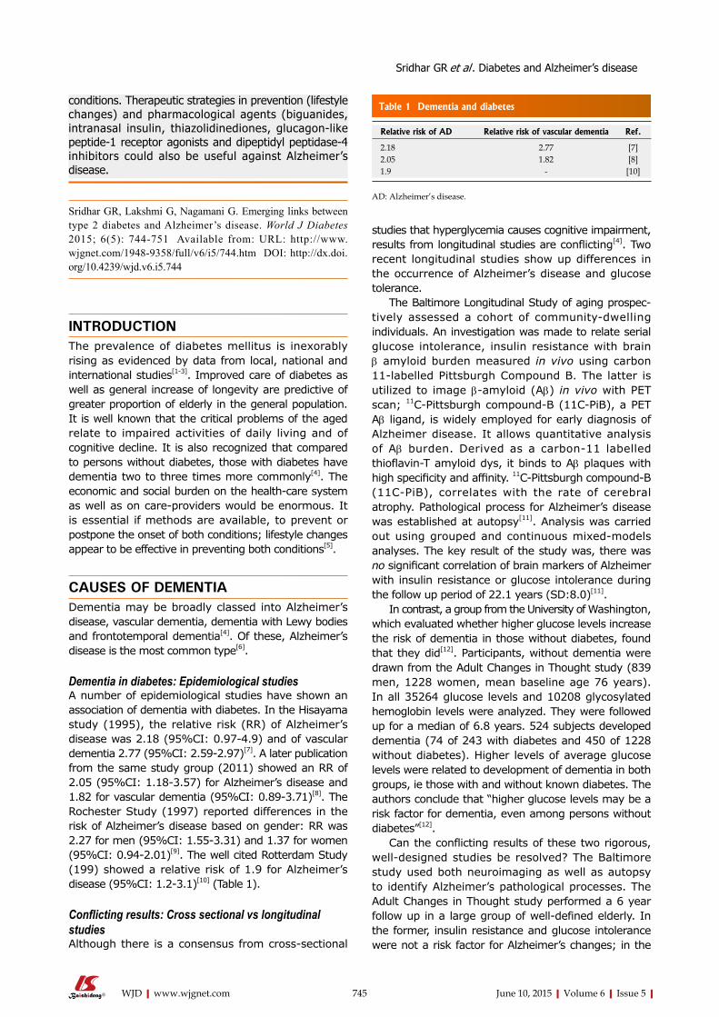
conditions. Therapeutic strategies in prevention (lifestyle changes) and pharmacological agents (biguanides, intranasal insulin, thiazolidinediones, glucagon-like peptide-1 receptor agonists and dipeptidyl peptidase-4 inhibitors could also be useful against Alzheimer’s disease.
Sridhar GR, Lakshmi G, Nagamani G. Emerging links between type 2 diabetes and Alzheimer’s disease. World J Diabetes 2015; 6(5): 744-751 Available from: URL: http://www.wjgnet.com/1948-9358/full/v6/i5/744.htm DOI: http://dx.doi.org/10.4239/wjd.v6.i5.744
INTRODUCTIONThe prevalence of diabetes mellitus is inexorably rising as evidenced by data from local, national and international studies[1-3]. Improved care of diabetes as well as general increase of longevity are predictive of greater proportion of elderly in the general population. It is well known that the critical problems of the aged relate to impaired activities of daily living and of cognitive decline. It is also recognized that compared to persons without diabetes, those with diabetes have dementia two to three times more commonly[4]. The economic and social burden on the health-care system as well as on care-providers would be enormous. It is essential if methods are available, to prevent or postpone the onset of both conditions; lifestyle changes appear to be effective in preventing both conditions[5].
CAUSES OF DEMENTIADementia may be broadly classed into Alzheimer’s disease, vascular dementia, dementia with Lewy bodies and frontotemporal dementia[4]. Of these, Alzheimer’s disease is the most common type[6].
Dementia in diabetes: Epidemiological studiesA number of epidemiological studies have shown an association of dementia with diabetes. In the Hisayama study (1995), the relative risk (RR) of Alzheimer’s disease was 2.18 (95%CI: 0.97-4.9) and of vascular dementia 2.77 (95%CI: 2.59-2.97)[7]. A later publication from the same study group (2011) showed an RR of 2.05 (95%CI: 1.18-3.57) for Alzheimer’s disease and 1.82 for vascular dementia (95%CI: 0.89-3.71)[8]. The Rochester Study (1997) reported differences in the risk of Alzheimer’s disease based on gender: RR was 2.27 for men (95%CI: 1.55-3.31) and 1.37 for women (95%CI: 0.94-2.01)[9]. The well cited Rotterdam Study (199) showed a relative risk of 1.9 for Alzheimer’s disease (95%CI: 1.2-3.1)[10] (Table 1).
Conflicting results: Cross sectional vs longitudinal studiesAlthough there is a consensus from cross-sectional
studies that hyperglycemia causes cognitive impairment, results from longitudinal studies are conflicting[4]. Two recent longitudinal studies show up differences in the occurrence of Alzheimer’s disease and glucose tolerance.
The Baltimore Longitudinal Study of aging prospec-tively assessed a cohort of community-dwelling individuals. An investigation was made to relate serial glucose intolerance, insulin resistance with brain β amyloid burden measured in vivo using carbon 11-labelled Pittsburgh Compound B. The latter is utilized to image β-amyloid (Aβ) in vivo with PET scan; 11C-Pittsburgh compound-B (11C-PiB), a PET Aβ ligand, is widely employed for early diagnosis of Alzheimer disease. It allows quantitative analysis of Aβ burden. Derived as a carbon-11 labelled thioflavin-T amyloid dys, it binds to Aβ plaques with high specificity and affinity. 11C-Pittsburgh compound-B (11C-PiB), correlates with the rate of cerebral atrophy. Pathological process for Alzheimer’s disease was established at autopsy[11]. Analysis was carried out using grouped and continuous mixed-models analyses. The key result of the study was, there was no significant correlation of brain markers of Alzheimer with insulin resistance or glucose intolerance during the follow up period of 22.1 years (SD:8.0)[11].
In contrast, a group from the University of Washington, which evaluated whether higher glucose levels increase the risk of dementia in those without diabetes, found that they did[12]. Participants, without dementia were drawn from the Adult Changes in Thought study (839 men, 1228 women, mean baseline age 76 years). In all 35264 glucose levels and 10208 glycosylated hemoglobin levels were analyzed. They were followed up for a median of 6.8 years. 524 subjects developed dementia (74 of 243 with diabetes and 450 of 1228 without diabetes). Higher levels of average glucose levels were related to development of dementia in both groups, ie those with and without known diabetes. The authors conclude that “higher glucose levels may be a risk factor for dementia, even among persons without diabetes”[12].
Can the conflicting results of these two rigorous, well-designed studies be resolved? The Baltimore study used both neuroimaging as well as autopsy to identify Alzheimer’s pathological processes. The Adult Changes in Thought study performed a 6 year follow up in a large group of well-defined elderly. In the former, insulin resistance and glucose intolerance were not a risk factor for Alzheimer’s changes; in the
Sridhar GR et al . Diabetes and Alzheimer’s disease
745 June 10, 2015|Volume 6|Issue 5|WJD|www.wjgnet.com
Table 1 Dementia and diabetes
Relative risk of AD Relative risk of vascular dementia Ref.
2.18 2.77 [7]2.05 1.82 [8]1.9 - [10]
AD: Alzheimer’s disease.
latter, higher glucose levels even among those without diabetes may be a risk factor for dementia.
The apparent differences can be attributed to the variety of pathological changes in diabetes leading to dementia including Alzheimer’s disease: chronic hyperglycemia, hypoglycemia (acute and recurrent), glycosylated of proteins, vascular disease, endothelial dysfunction, inflammation, altered blood brain barrier, dyslipidemia, insulin resistance, genetic predisposition, amyloid deposition and depression, among others[13]. More sensitive methods of measuring brain volume as a surrogate of cognitive function may throw light[4]. In addition, there is a flaw in using brain markers such as plaques in diagnosis of Alzheimer’s disease: subjects may have amyloid plaques, yet display no symptoms of Alzheimer’s disease throughout their life. This discrepancy between the presence of plaques and Alzheimers could play a role for a lack of finding a link between diabetes and Alzheimer’s disease.
HyperglycemiaHyperglycemia is a recognized risk factor for cognitive impairment as shown by the ACCORD-MIND study and others[14,15]. Biological reasons for such changes were ascribed to neural damage following advanced glycosylated end products and oxidative stress, osmotic stress damaging the blood brain barrier and resultant leak of toxic substances leading to further damage of nervous structures[4]. In addition to chronic hyperglycemia as assessed by glycated hemoglobin, glycemic variability was also proposed to contribute to cognitive dysfunction. Measurements by continuous glucose monitoring revealed cognitive function was better correlated with diurnal variation in blood glu-cose[16]. Post prandial glucose levels could also be a contributing factor, acting via oxidative stress[4].
HypoglycemiaAlthough severe hypoglycemia was shown to be associa-ted with dementia in the elderly[17], when hypoglycemia is avoided by careful treatment as in the DCCT/EDIC study, there was no association between hypoglycemia and cognitive dysfunction[18]. In the elderly however, hypoglycemia, when coupled with atherosclerosis leads to organic brain damage which is often irreversible[4].
Role of insulin in brainCognition may be affected not only by alterations in the level of glucose, but also via the action of insulin. Upon transport through the blood brain barrier, insulin binds to its receptors, and is involved in modulating cognitive function. A large number of insulin receptors occur in brain areas related to memory such as the hippocampus and cerebral cortex. In addition it also aids the release of β-amyloid peptide extracellularly, and increases the expression of the enzyme which degrades insulin, insulin degrading enzyme (IDE)[4]. As the latter also degrades β-amyloid peptide, insulin deficiency results in accumulation of β-amyloid peptide.
Both hyperinsulinemia and hyperglycemia were shown to increase neuritic plaque formation[19].
Information is being available about the origin of insulin in the brain and its role in cognition. Originally, brain was considered to be insulin insensitive because insulin did not influence the glucose uptake by the bulk brain. However insulin has been shown to be a neuroregulatory peptide playing a role in food intake and in monitoring the energy stores of the body[20]. Interestingly a role for insulin in modulation of memory and cognition is also emerging. Its action has been observed in regions associated with reward recognition such as hippocampus, and in global cognition and memory. Rather than passing across the blood brain barrier from the periphery, insulin appears to be produced locally for action as a neurotransmitter, regulated by glucose levels. In addition a para-arteriolar pathway for transport at the level of micro-vasculature has been proposed[20].
The role of insulin in the pathogenesis of Alzheimer disease has been described. Insulin can modulate Aβ peptide in vitro. The peptide is well known as a neuropathological hallmark of AD. Low levels of insulin in the brain can decrease the Aβ release into extracellular compartments. In addition hypoinsulinemia in the central nervous sytem can lower the levels of insulin-degrading enzyme, thereby impairing Aβ clearance. In all, chronic hyperinsulinemia in the peripheral circulation, along with decreased uptake of insulin into the brain can lead to dysregulation of Aβ and inflammation[21,22].
Role of microvascular disease in cognitive declineStudies have shown that retinopathy and nephropa-thy are associated with impairment of cognition[22,23]. Small vessels in both organs arise from a similar embryonic antecedent and share similar structures; it is conceivable therefore that insults (e.g., increased polyol pathway activity, myo-inositol dysmetabolism) result in similar adverse reactions in frontal lobe of the cerebral cortex leading to cognitive decline[4]. However, evidence is not unequivocal. Retinopathy is related to microvascular changes in diabetes. Because the retina shares many features with the brain, both developmental, anatomical (e.g., microvascular bed) and physiological (e.g., blood-tissue barrier), changes in retina were suggested to presage brain pathological processes. Alzheimer’s disease is known to involve the retina, such as the macula and the optic disc. It has been suggested that pathological changes in the retina such as macular deposits, reduced thickness of retinal nerve, cupping of optic disc and retinal microvascular changes may be related to cognitive dysfunction and Alzheimer disease[24].
Insulin resistance and Alzheimer’s diseaseDownstream insulin signaling acts through a complex interplay involving phosphatidylinositol 3-kinase (P13K0 and mitogen activation protein kinase (MAPK).
746 June 10, 2015|Volume 6|Issue 5|WJD|www.wjgnet.com
Sridhar GR et al . Diabetes and Alzheimer’s disease

consists of 37 amino acids.
ApoE-ε 4Expression of ApoE-ε4, which is related to diabetes as well, increases the risk of early onset Alzheimer’s disease. It has increased ability to deposit Aβ, which is neurotoxic, and also impair its clearance[35]. ApoE-ε4 is less protective against oxidative stress and leads to cholinergic dysfunction seen in Alzheimer’s disease, besides modifying the cholesterol transporter protein ABCA1.
Other potential associationsIn addition other associations are also being recog-nized as risk factors for both diabetes and Alzheimer’s disease: weight gain, acting perhaps through defective leptin signaling and increased formation of advanced glycation end products, which could have a pathogenic role in the amyloid plaques deposited in the brain[35]. Other emerging associations include disturbances in sleep and the circadian rhythm[36,37], and iron overload in brain among persons with obesity[38].
Insulin like growth factorAnimal studies have provided intriguing evidence that loss of insulin-like growth factors, along with insulin could lead to age-dependent brain atrophy with cognitive decline[39]. Insulin and its growth factors maintain brain protein content; their replacement can prevent brain protein loss, cell degeneration and demyelination. Lack of insulin and growth factors, which are common to type 2 diabetes and to Alzheimer’s disease could therefore play a role in their pathogenesis and provide therapeutic targets in their treatment[39].
ButyrylcholinesteraseButyrylcholinesterase, belonging to the esterase family of enzymes that also contain acetylcholinesterase[40] has been evaluated in relation to insulin resistance, cardiovascular disease, obesity and dyslipidemia[36]. Variant forms of the enzyme with little or no activity exist in isolated geographic populations[41] with apparently no adverse health effects[42]. Butrylcholinesterase was studied in relation to both type 2 diabetes mellitus and to Alzheimer’s disease[43-45]. with studies suggesting a possible protective effect against Alzheimer’s disease and risk for fronto-temporal dementia[46].
A number of hypothesis were put forward for the association of butyrylcholinesterase with type 2 diabetes mellitus and Alzheimer’s disease[33,36]. Plaques in the brains of individuals with dementia had higher levels of butyrylcholinesterase, which is also localized in neurofibrillary tangles. It was also shown to attenuate amyloid formation[47].
Similarly in type 2 diabetes, butyrylcholinesterase may modify the expression of insulin resistance or by way of amyloid fibril deposition in β cells of the pancreas[33]. It was shown to interact with amylin
The latter is associated with most metabolic effects of insulin. Unlike its peripheral effects, the action of insulin in the central nervous system is dependent on its crossing the blood brain barrier via direct transfer[25] through an insulin receptor protein, which is selectively distributed. In addition there is also local synthesis of insulin in the CNS. Unlike peripheral insulin receptors, those in the CNS differ in terms of structure, function and size. They are highly populated in the olfactory bulb, hypothalamus, cortex, cerebellum, hippocampus and are expressed in both neurons and glia[26].
The physiological effects of insulin in the brain are unlike those in the periphery: in an animal model it suppressed food intake and increased the level of glucose[27], acting in a way as its own counterregulation[25]. Compared to its weight, the brain depends on a larger amount of glucose for its metabolic needs compared to other tissues. Glucose reaches it via facilitated diffusion transported by glucose transport proteins. In the brain, insulin does not have a major effect on either the transport of glucose to the brain or its basal metabolism[28,29]. While it is not a major regulator of glucose metabolism in the brain, insulin indirectly affects neurons by modulating neurotransmitter release, neural growth, tubulin activity, nerve survival and synaptic plasticity[26]. In humans, insulin improves cognition independent of its effects on peripheral glucose[30].
Whereas acute increases of insulin improve cog-nition, chronic hyperinsulinemia can adversely affect neuronal function in vitro by increasing susceptibility to toxin and stress-induced effects[31]. Glycated proteins and inflammatory mediators could also have a pathogenic role[25].
Protein aggregation, diabetes and Alzheimer’s diseaseProtein aggregation has been suggested to be an underlying pathogenic factor between type 2 diabetes and Alzheimer’s disease[32]. A number of hypotheses were proposed to explain why a biological protein can be transformed into a pathological entity with the ability to self-assemble: aging, high concentrations of the protein, mutation of amino acids or abnormal post-translational modification, modulated by environmental factors[32].
Alzheimer’s disease is associated with accumulation of neurofibrillary tangles and amyloid fibers leading to neuronal cell loss. Amyloid β peptides form from cleavage of amyloid β-protein precursor seen in plaques. It is organized as amyloid fibrils, which are linear aggregates[32]. Diabetes is also characterized by localized and progressive amyloid deposition in the pancreatic β cell islets[33]. Common features of amyloid deposited in both Alzheimer’s disease and diabetes include: linear appearance, with a beta-sheet structure, which begin to form from spherical oligomers that can self-assemble[34]. The islet amyloid peptide is secreted by β cells of the pancreas and
747 June 10, 2015|Volume 6|Issue 5|WJD|www.wjgnet.com
Sridhar GR et al . Diabetes and Alzheimer’s disease

and attenuate the formation of amylin fibril as well as its oligomer. When applied to cultured β cells, it was protective against amylin cytotoxicity[47]. Butyrylcholinesterase was shown to participate in the progression of metabolic syndrome to type 2 diabetes mellitus. A majority of subjects with type 2 diabetes show extracellular deposits formed by islet amyloid polypeptide (IAP), adjacent to the β cells. Elevated levels of IAP are found in conditions of insulin resistance. Disturbed balance of sympathetic and parasympathetic nervous system could participate in metabolic syndrome. Lower vagal activity could in part be caused by increased hydrolysis of acetylcholine mediated by increased butyrylcholinesterase[48]. Lowering of acetylcholinesterase results in reduced parasympathetic signals and increased ratio of sympathetic signals[48]. In addition BChE was reported to attenuate the formation of Aβ amyloid fibrils[47]. Essentially subjects with metabolic syndrome had elevated levels of BChE compared to those with type 2 diabetes and with controls. In vitro interaction of BChE was observed with amylin. It interacted with amylin, and attenuated the formation of both amylin fibril and oligomer formation, showing that it can protect cultured β cells from cytotoxicity due to amylin[47]. Thus increased BChE seen in metabolic syndrome could protect pancreatic β cells by reducing toxic amylin oligomer formation[47].
A bioinformatics study suggested the following sequences (E < e-5) were associated with both type 2 diabetes and Alzheimer’s disease: butyrylcholinesterase precursor K allele (NP_000046.1), acetylcholinesterase isoform E4-6 precorsor (NP_000656.1) and apoptosis-related acetylcholinesterase (1B41|A). In an animal study, streptozotocin-induced diabetes was associated with elevated butyrylcholinesterase activity, lowered superoxide dismutase and impaired cognitive function assessed by Morris water maze method[49]. Being low-grade inflammatory conditons, both type 2 diabetes mellitus and Alzheimer’s may be target conditions for utilization of butyrylcholinesterae as a biomarker[50], as well as a treatment target[33].
The principal drugs currently available to manage
Alzheimer’s disease act via modifying butyrylcholines-terase levels. Renewed interest in the possible role of “missing genes” in people who are apparently healthy may aid in uncovering new treatment modalities[51]. Individuals with variant butyrylcholinesterae activity genes may be a potential group for such long-term follow up studies vis a vis their propensity or protection against diabetes and Alzheimer’s disease[40].
THERAPEUTIC IMPLICATIONSThe interest of linking type 2 diabetes mellitus and Alzheimer’s disease lies not in science, but more in translational science: how understanding the common pathogenesis can help in prevention and treatment of both conditions. Other than the scope for future therapies, evidence is now available for the currently available drugs used in diabetes[52] to have a modulatory effect on the cognitive decline due to Alzheimer’s disease.Along with its action on AMPK, metformin has been recently shown to influence the incretin system by increasing the secretion of glucagon-like peptide-1 (GLP-1)[53]. GLP-1 and GIP receptors are known to be expressed in the brain, and direct activation of these receptors may be a potential strategy to treat Allzheimer’s disease[54]. In addition, metformin also influences gut microbiome along with its other putative actions in the management of diabetes mellitus[55]. The use of drugs used in diabetes mellitus such as metformin, GLP-1 mimetics (exenatide and liraglutide) and peroxisome proliferator-activated receptors γ agonists may all be of potential benefit in the prevention and management of Alzheimer disease[56].
When a theoretical basis for insulin to affect brain responses was studied in clinical practice, a recent review of 8 published studies on effect of intranasal insulin on cognition, comprising 328 participants showed first of all, no significant adverse effects. Generally the authors concluded that “the limited clinical experience suggests potential beneficial cognitive effects of intranasal insulin”[57]. In a single study, use of 20 IU intranasal insulin showed improved immediate recall in Apoε4(-) subjects but not in Apoε4(+) subjects.
Other than correcting hyperglycemia, some of the conventional antidiabetic agents were shown to affect cognition. Metformin protected brain against oxidative stress in rats, and by preventing apoptosis[54]. However caution must be exercised because of met-formin leading to increased biogenesis of Alzheimer’s amyloid peptides[58] and increased risk of cognitive impairment[59]. Thiazolidinediones, which act at the nuclear receptors of insulin-sensitive tissues, affect transcription of genes affecting lipid and glucose metabolism. Early studies suggested that this group of drugs may favourably affect cognition, before the clinical use tapered due to their adverse drug effect profile.
Pharmacological agents modulating the incretin
748 June 10, 2015|Volume 6|Issue 5|WJD|www.wjgnet.com
Sridhar GR et al . Diabetes and Alzheimer’s disease
Pathogenesis
Management
HyperglycemiaInsulin resistanceMicrovascular involvementProtein aggregatesApoE-ε4Insulin like growth factorButyrylcholinesterase
Physical exercise, liifestyleNasal insulinMetforminGLP-modulatorsPPARγ agonists
Figure 1 Links between Alzheimer’s disease and type 2 diabetes mellitus.
system have now become mainstream in many markets world-wide. Pathological studies showed that sitagliptin lowered APP and Aβ deposition in the hippocampus of transgenic Alzheimer’s disease mice[53]. However whether it does so by lowering glucose levels or independently remains to be clarified. Interest arises because liraglutide, another drug in the same group had similar protective effects (Figure 1).
CONCLUSIONIn conclusion, diabetes mellitus and Alzheimer’s disease are both common and increasing in incidence in the aging population. Recent evidence has shown common pathogenic factors operating in both conditions. Thereby common preventive and therapeutic agents may be used in their prevention and treatment. Physicians caring for the elderly must be aware of the increased risk of the other when one condition is present. Common pathogenesis and therapeutic agents make it possible to manage both using similar lifestyle changes and pharmacological agents.
REFERENCES1 Sridhar GR, Putcha V, Lakshmi G. Time trends in the prevalence
of diabetes mellitus: ten year analysis from southern India (1994-2004) on 19,072 subjects with diabetes. J Assoc Physicians India 2010; 58: 290-294 [PMID: 21117347]
2 Anjana RM , Pradeepa R, Deepa M, Datta M, Sudha V, Unnikrishnan R, Bhansali A, Joshi SR, Joshi PP, Yajnik CS, Dhandhania VK, Nath LM, Das AK, Rao PV, Madhu SV, Shukla DK, Kaur T, Priya M, Nirmal E, Parvathi SJ, Subhashini S, Subashini R, Ali MK, Mohan V. Prevalence of diabetes and prediabetes (impaired fasting glucose and/or impaired glucose tolerance) in urban and rural India: phase I results of the Indian Council of Medical Research-INdia DIABetes (ICMR-INDIAB) study. Diabetologia 2011; 54: 3022-3027 [PMID: 21959957 DOI: 10.1007/s00125-011-2291-5]
3 Wild S, Roglic G, Green A, Sicree R, King H. Global prevalence of diabetes: estimates for the year 2000 and projections for 2030. Diabetes Care 2004; 27: 1047-1053 [PMID: 15111519 DOI: 10.2337/diacare.27.5.1047]
4 Kawamura T, Umemura T, Hotta N. Cognitive impairment in diabetic patients: Can diabetic control prevent cognitive decline? J Diabetes Investig 2012; 3: 413-423 [PMID: 24843599 DOI: 10.1111/j.2040-1124.2012.00234.x]
5 Rodríguez-Gómez O, Palacio-Lacambra ME, Palasí A, Ruiz-Laza A, Boada-Rovira M. Prevention of Alzheimer’s disease: a global challenge for next generation neuroscientists. J Alzheimers Dis 2014; 42 Suppl 4: S515-S523 [PMID: 25351111]
6 Inestrosa NC, Alvarez A, Pérez CA, Moreno RD, Vicente M, Linker C, Casanueva OI, Soto C, Garrido J. Acetylcholinesterase accelerates assembly of amyloid-beta-peptides into Alzheimer’s fibrils: possible role of the peripheral site of the enzyme. Neuron 1996; 16: 881-891 [PMID: 8608006 DOI: 10.1016/S0896-6273(00)80108-7]
7 Yoshitake T, Kiyohara Y, Kato I, Ohmura T, Iwamoto H, Nakayama K, Ohmori S, Nomiyama K, Kawano H, Ueda K. Incidence and risk factors of vascular dementia and Alzheimer’s disease in a defined elderly Japanese population: the Hisayama Study. Neurology 1995; 45: 1161-1168 [PMID: 7783883 DOI: 10.1212/WNL.45.6.1161]
8 Ohara T, Doi Y, Ninomiya T, Hirakawa Y, Hata J, Iwaki T, Kanba S, Kiyohara Y. Glucose tolerance status and risk of dementia in the community: the Hisayama study. Neurology 2011; 77: 1126-1134 [PMID: 21931106 DOI: 10.1212/WNL.0b013e31822f0435]
9 Leibson CL, Rocca WA, Hanson VA, Cha R, Kokmen E, O’Brien PC, Palumbo PJ. Risk of dementia among persons with diabetes mellitus: a population-based cohort study. Am J Epidemiol 1997; 145: 301-308 [PMID: 9054233 DOI: 10.1093/oxfordjournals.aje.a009106]
10 Ott A, Stolk RP, van Harskamp F, Pols HA, Hofman A, Breteler MM. Diabetes mellitus and the risk of dementia: The Rotterdam Study. Neurology 1999; 53: 1937-1942 [PMID: 10599761 DOI: 10.1212/WNL.53.9.1937]
11 Thambisetty M, Jeffrey Metter E, Yang A, Dolan H, Marano C, Zonderman AB, Troncoso JC, Zhou Y, Wong DF, Ferrucci L, Egan J, Resnick SM, O’Brien RJ. Glucose intolerance, insulin resistance, and pathological features of Alzheimer disease in the Baltimore Longitudinal Study of Aging. JAMA Neurol 2013; 70: 1167-1172 [PMID: 23897112 DOI: 10.1001/jamaneurol.2013.284]
12 Crane PK, Walker R, Hubbard RA, Li G, Nathan DM, Zheng H, Haneuse S, Craft S, Montine TJ, Kahn SE, McCormick W, McCurry SM, Bowen JD, Larson EB. Glucose levels and risk of dementia. N Engl J Med 2013; 369: 540-548 [PMID: 23924004 DOI: 10.1056/NEJMoa1215740]
13 McCrimmon RJ, Ryan CM, Frier BM. Diabetes and cognitive dysfunction. Lancet 2012; 379: 2291-2299 [PMID: 22683129 DOI: 10.1016/S0140-6736(12)60360-2]
14 Cukierman-Yaffe T, Gerstein HC, Williamson JD, Lazar RM, Lovato L, Miller ME, Coker LH, Murray A, Sullivan MD, Marcovina SM, Launer LJ. Relationship between baseline glycemic control and cognitive function in individuals with type 2 diabetes and other cardiovascular risk factors: the action to control cardiovascular risk in diabetes-memory in diabetes (ACCORD-MIND) trial. Diabetes Care 2009; 32: 221-226 [PMID: 19171735 DOI: 10.2337/dc08-1153]
15 Umegaki H, Kawamura T, Mogi N, Umemura T, Kanai A, Sano T. Glucose control levels, ischaemic brain lesions, and hyperinsulinaemia were associated with cognitive dysfunction in diabetic elderly. Age Ageing 2008; 37: 458-461 [PMID: 18339615 DOI: 10.1093/ageing/afn051]
16 Rizzo MR, Marfella R, Barbieri M, Boccardi V, Vestini F, Lettieri B, Canonico S, Paolisso G. Relationships between daily acute glucose fluctuations and cognitive performance among aged type 2 diabetic patients. Diabetes Care 2010; 33: 2169-2174 [PMID: 20573753 DOI: 10.2337/dc10-0389]
17 Whitmer RA, Karter AJ, Yaffe K, Quesenberry CP, Selby JV. Hypoglycemic episodes and risk of dementia in older patients with type 2 diabetes mellitus. JAMA 2009; 301: 1565-1572 [PMID: 19366776 DOI: 10.1001/jama.2009.460]
18 Jacobson AM, Musen G, Ryan CM, Silvers N, Cleary P, Waberski B, Burwood A, Weinger K, Bayless M, Dahms W, Harth J. Long-term effect of diabetes and its treatment on cognitive function. N Engl J Med 2007; 356: 1842-1852 [PMID: 17476010]
19 Matsuzaki T, Sasaki K, Tanizaki Y, Hata J, Fujimi K, Matsui Y, Sekita A, Suzuki SO, Kanba S, Kiyohara Y, Iwaki T. Insulin resistance is associated with the pathology of Alzheimer disease: the Hisayama study. Neurology 2010; 75: 764-770 [PMID: 20739649 DOI: 10.1212/WNL.0b013e3181eee25f]
20 Gray SM, Meijer RI, Barrett EJ. Insulin regulates brain function, but how does it get there? Diabetes 2014; 63: 3992-3997 [PMID: 25414013 DOI: 10.2337/db14-0340]
21 Craft S. The role of insulin dysregulation in aging and Alzheimer’s disease. In: Craft S, Christen Y, editors. Diabetes, Insulin and Alzheimer’s Disease. Berlin: Springer-Verlag Berlin Heidelberg, 2010: 109-127
22 Ding J, Strachan MW, Reynolds RM, Frier BM, Deary IJ, Fowkes FG, Lee AJ, McKnight J, Halpin P, Swa K, Price JF. Diabetic retinopathy and cognitive decline in older people with type 2 diabetes: the Edinburgh Type 2 Diabetes Study. Diabetes 2010; 59: 2883-2889 [PMID: 20798334 DOI: 10.2337/db10-0752]
23 de Bresser J, Reijmer YD, van den Berg E, Breedijk MA, Kappelle LJ, Viergever MA, Biessels GJ. Microvascular determinants of cognitive decline and brain volume change in elderly patients with
749 June 10, 2015|Volume 6|Issue 5|WJD|www.wjgnet.com
Sridhar GR et al . Diabetes and Alzheimer’s disease
type 2 diabetes. Dement Geriatr Cogn Disord 2010; 30: 381-386 [PMID: 20962529 DOI: 10.1159/000321354]
24 Ikram MK, Cheung CY, Wong TY, Chen CP. Retinal pathology as biomarker for cognitive impairment and Alzheimer’s disease. J Neurol Neurosurg Psychiatry 2012; 83: 917-922 [PMID: 22733082 DOI: 10.1136/jnnp-2011-301628]
25 Neumann KF, Rojo L, Navarrete LP, Farías G, Reyes P, Maccioni RB. Insulin resistance and Alzheimer’s disease: molecular links & amp; clinical implications. Curr Alzheimer Res 2008; 5: 438-447 [PMID: 18855585 DOI: 10.2174/156720508785908919]
26 Banks WA. The source of cerebral insulin. Eur J Pharmacol 2004; 490: 5-12 [PMID: 15094069 DOI: 10.1016/j.ejphar.2004.02.040]
27 Florant GL, Singer L, Scheurink AJ, Park CR, Richardson RD, Woods SC. Intraventricular insulin reduces food intake and body weight of marmots during the summer feeding period. Physiol Behav 1991; 49: 335-338 [PMID: 1905822 DOI: 10.1016/0031-9384(91)90053-Q]
28 Marfaing P, Penicaud L, Broer Y, Mraovitch S, Calando Y, Picon L. Effects of hyperinsulinemia on local cerebral insulin binding and glucose utilization in normoglycemic awake rats. Neurosci Lett 1990; 115: 279-285 [PMID: 2234506 DOI: 10.1016/0304-3940(90)90469-P]
29 Hasselbalch SG, Knudsen GM, Videbaek C, Pinborg LH, Schmidt JF, Holm S, Paulson OB. No effect of insulin on glucose blood-brain barrier transport and cerebral metabolism in humans. Diabetes 1999; 48: 1915-1921 [PMID: 10512354 DOI: 10.2337/diabetes.48.10.1915]
30 Kern W, Born J, Schreiber H, Fehm HL. Central nervous system effects of intranasally administered insulin during euglycemia in men. Diabetes 1999; 48: 557-563 [PMID: 10078556 DOI: 10.2337/diabetes.48.3.557]
31 Schäfer M, Erdö SL. Development of glutamate neurotoxicity in cortical cultures: induction of vulnerability by insulin. Brain Res Dev Brain Res 1991; 62: 293-296 [PMID: 1685107 DOI: 10.1016/0165-3806(91)90179-M]
32 Di Carlo M, Pasquale Picone P, Carrotta R, Giacomazza D, Biagio S. Alzheimer’s disease and type 2 diabetes: different pathologies and same features. In: Zimering M, editor. Topics in the prevention, treatment and complications of type 2 diabetes. Croatia: INTECH Open Access Publisher, 2011 [DOI: 10.5772/23490]
33 Sridhar GR, Thota H, Allam AR, Suresh Babu C, Siva Prasad A, Divakar Ch. Alzheimer’s disease and type 2 diabetes mellitus: the cholinesterase connection? Lipids Health Dis 2006; 5: 28 [PMID: 17096857 DOI: 10.1186/1476-511X-5-28]
34 Xu S. Aggregation drives “misfolding” in protein amyloid fiber formation. Amyloid 2007; 14: 119-131 [PMID: 17577685 DOI: 10.1080/13506120701260059]
35 Akter K, Lanza EA, Martin SA, Myronyuk N, Rua M, Raffa RB. Diabetes mellitus and Alzheimer’s disease: shared pathology and treatment? Br J Clin Pharmacol 2011; 71: 365-376 [PMID: 21284695 DOI: 10.1111/j.1365-2125.2010.03830.x]
36 Sridhar GR, Rao AA, Srinivas K, Nirmala G, Lakshmi G, Suryanarayna D, Rao PV, Kaladhar DG, Kumar SV, Devi TU, Nitesh T, Hanuman T. Butyrylcholinesterase in metabolic syndrome. Med Hypotheses 2010; 75: 648-651 [PMID: 20797821 DOI: 10.1016/j.mehy.2010.08.008]
37 Kang JE, Lim MM, Bateman RJ, Lee JJ, Smyth LP, Cirrito JR, Fujiki N, Nishino S, Holtzman DM. Amyloid-beta dynamics are regulated by orexin and the sleep-wake cycle. Science 2009; 326: 1005-1007 [PMID: 19779148 DOI: 10.1126/science.1180962]
38 Blasco G, Puig J, Daunis-I-Estadella J, Molina X, Xifra G, Fernández-Aranda F, Pedraza S, Ricart W, Portero-Otín M, Fernández-Real JM. Brain iron overload, insulin resistance, and cognitive performance in obese subjects: a preliminary MRI case-control study. Diabetes Care 2014; 37: 3076-3083 [PMID: 25125507 DOI: 10.2337/dc14-0664]
39 Serbedžija P, Ishii DN. Insulin and insulin-like growth factor prevent brain atrophy and cognitive impairment in diabetic rats. Indian J Endocrinol Metab 2012; 16: S601-S610 [PMID: 23565496 DOI: 10.4103/2230-8210.105578]
40 Sridhar GR, Nirmala G, Apparao A, Madhavi AS, Sreelatha S, Rani JS, Vijayalakshmi P. Serum butyrylcholinesterase in type 2 diabetes mellitus: a biochemical and bioinformatics approach. Lipids Health Dis 2005; 4: 18 [PMID: 16150144 DOI: 10.1186/1476-511X-4-18]
41 Sridhar GR, Nirmala G. Inborn errors in lipid metabolism. In: Lipid disorders. Tripathy BB, Das S, editors. Association of Physicians of India. Mumbai: API College of Physicians, 2002: 59-80
42 Manoharan I, Boopathy R, Darvesh S, Lockridge O. A medical health report on individuals with silent butyrylcholinesterase in the Vysya community of India. Clin Chim Acta 2007; 378: 128-135 [PMID: 17182021 DOI: 10.1016/j.cca.2006.11.005]
43 Lehmann DJ, Johnston C, Smith AD. Synergy between the genes for butyrylcholinesterase K variant and apolipoprotein E4 in late-onset confirmed Alzheimer’s disease. Hum Mol Genet 1997; 6: 1933-1936 [PMID: 9302273 DOI: 10.1093/hmg/6.11.1933]
44 Johansen A, Nielsen EM, Andersen G, Hamid YH, Jensen DP, Glümer C, Drivsholm T, Borch-Johnsen K, Jørgensen T, Hansen T, Pedersen O. Large-scale studies of the functional K variant of the butyrylcholinesterase gene in relation to Type 2 diabetes and insulin secretion. Diabetologia 2004; 47: 1437-1441 [PMID: 15258737 DOI: 10.1007/s00125-004-1459-7]
45 Pohanka M. Butyrylcholinesterase as a biochemical marker. Bratisl Lek Listy 2013; 114: 726-734 [PMID: 24329513]
46 Diamant S, Podoly E, Friedler A, Ligumsky H, Livnah O, Soreq H. Butyrylcholinesterase attenuates amyloid fibril formation in vitro. Proc Natl Acad Sci USA 2006; 103: 8628-8633 [PMID: 16731619 DOI: 10.1073/pnas.0602922103]
47 Shenhar-Tsarfaty S, Bruck T, Bennett ER, Bravman T, Aassayag EB, Waiskopf N, Rogowski O, Bornstein N, Berliner S, Soreq H. Butyrylcholinesterase interactions with amylin may protect pancreatic cells in metabolic syndrome. J Cell Mol Med 2011; 15: 1747-1756 [PMID: 20807286 DOI: 10.1111/j.1582-4934.2010.01165.x]
48 Rao AA, Siva Reddy C, Sridhar GR, Annapurna A, Hanuman T, Prameela M, Suresh K, Prasannalaxmi S, Das UN. Enhanced butyrylcholinesterase activity may be the common link in triggering low-grade systemic inflammation and decrease in cognitive function in diabetes mellitus and Alzheimer’s disease. CurrNutr Food Sci 2008; 4: 213-216 [DOI: 10.2174/157340108785133310]
49 Rao AA, Sridhar GR, Das UN. Elevated butyrylcholinesterase and acetylcholinesterase may predict the development of type 2 diabetes mellitus and Alzheimer’s disease. Med Hypotheses 2007; 69: 1272-1276 [PMID: 17553629 DOI: 10.1016/j.mehy.2007.03.032]
50 Sridhar GR. Proteins of the esterase family: patents for some proteins in search of metabolic functions. Recent Patents on Biomarkers 2011; 1: 205-212 [DOI: 10.2174/2210309011101030205]
51 Kaiser J. The hunt for missing genes. Science 2014; 344: 687-689 [PMID: 24833372 DOI: 10.1126/science.344.6185.687]
52 Cummings JL. Use of cholinesterase inhibitors in clinical practice: evidence-based recommendations. Am J Geriatr Psychiatry 2003; 11: 131-145 [PMID: 12611743 DOI: 10.1176/appi.ajgp.11.2.131]
53 Shemesh E, Rudich A, Harman-Boehm I, Cukierman-Yaffe T. Effect of intranasal insulin on cognitive function: a systematic review. J Clin Endocrinol Metab 2012; 97: 366-376 [PMID: 22162476 DOI: 10.1210/jc.2011-1802]
54 Vardarli I, Arndt E, Deacon CF, Holst JJ, Nauck MA. Effects of sitagliptin and metformin treatment on incretin hormone and insulin secretory responses to oral and “isoglycemic” intravenous glucose. Diabetes 2014; 63: 663-674 [PMID: 24186866 DOI: 10.2337/db13-0805]
55 Napolitano A, Miller S, Nicholls AW, Baker D, Van Horn S, Thomas E, Rajpal D, Spivak A, Brown JR, Nunez DJ. Novel gut-based pharmacology of metformin in patients with type 2 diabetes mellitus. PLoS One 2014; 9: e100778 [PMID: 24988476]
56 Strittmatter WJ. Alzheimer’s disease: the new promise. J Clin Invest 2012; 122: 1191 [PMID: 22754953 DOI: 10.1172/JCI62745]
750 June 10, 2015|Volume 6|Issue 5|WJD|www.wjgnet.com
Sridhar GR et al . Diabetes and Alzheimer’s disease

57 Sebastião I, Candeias E, Santos MS, de Oliveira CR, Moreira PI, Duarte AI. Insulin as a Bridge between Type 2 Diabetes and Alzheimer Disease - How Anti-Diabetics Could be a Solution for Dementia. Front Endocrinol (Lausanne) 2014; 5: 110 [PMID: 25071725 DOI: 10.3389/fendo.2014.00110]
58 Chen Y, Zhou K, Wang R, Liu Y, Kwak YD, Ma T, Thompson RC, Zhao Y, Smith L, Gasparini L, Luo Z, Xu H, Liao FF. Antidiabetic drug metformin (GlucophageR) increases biogenesis of Alzheimer’s amyloid
peptides via up-regulating BACE1 transcription. Proc Natl Acad Sci USA 2009; 106: 3907-3912 [PMID: 19237574 DOI: 10.1073/pnas.0807991106]
59 Moore EM, Mander AG, Ames D, Kotowicz MA, Carne RP, Brodaty H, Woodward M, Boundy K, Ellis KA, Bush AI, Faux NG, Martins R, Szoeke C, Rowe C, Watters DA. Increased risk of cognitive impairment in patients with diabetes is associated with metformin. Diabetes Care 2013; 36: 2981-2987 [PMID: 24009301]
P- Reviewer: Hssan M, Hwu CM, Laher I S- Editor: Tian YL L- Editor: A E- Editor: Zhang DN
751 June 10, 2015|Volume 6|Issue 5|WJD|www.wjgnet.com
Sridhar GR et al . Diabetes and Alzheimer’s disease
752 June 10, 2015|Volume 6|Issue 5|WJD|www.wjgnet.com
Submit a Manuscript: http://www.wjgnet.com/esps/Help Desk: http://www.wjgnet.com/esps/helpdesk.aspxDOI: 10.4239/wjd.v6.i5.752
World J Diabetes 2015 June 10; 6(5): 752-758ISSN 1948-9358 (online)
© 2015 Baishideng Publishing Group Inc. All rights reserved.
Patient attitudes about financial incentives for diabetes self-management: A survey
Katherine S Blondon
Katherine S Blondon, Division of General Internal Medicine, University Hospitals of Geneva, 1205 Geneva, Switzerland
Author contributions: Blondon KS solely contributed to this paper.
Ethics approval: By the Institutional Review Board of the University of Washington.Informed consent: Statement available online, participants gave their online consent by accepting to participate.Conflict-of-interest: No funding to disclose.Data sharing: No additional data are available.
Open-Access: This article is an open-access article which was selected by an in-house editor and fully peer-reviewed by external reviewers. It is distributed in accordance with the Creative Commons Attribution Non Commercial (CC BY-NC 4.0) license, which permits others to distribute, remix, adapt, build upon this work non-commercially, and license their derivative works on different terms, provided the original work is properly cited and the use is non-commercial. See: http://creativecommons.org/licenses/by-nc/4.0/
Correspondence to: Katherine S Blondon, MD, PhD, Division of General Internal Medicine, University Hospitals of Geneva, Rue Gabrielle-Perret-Gentil 4, 1205 Geneva, Switzerland. [email protected]: +41-79-5534323Fax: +41-22-3729235
Received: November 27, 2014 Peer-review started: November 28, 2014First decision: January 20, 2015Revised: March 3, 2015Accepted: April 1, 2015Article in press: April 7, 2015Published online: June 10, 2015
AbstractAIM: To study the acceptability of incentives for behavior
changes in individuals with diabetes, comparing financial incentives to self-rewards and non-financial incentives.
METHODS: A national online survey of United States adults with diabetes was conducted in March 2013 (n = 153). This survey was designed for this study, with iterative testing and modifications in a pilot population. We measured the demographics of individuals, their interest in incentives, as well as the perceived challenge of diabetes self-management tasks, and expectations of incentives to improve diabetes self-management (financial, non-financial and self-rewards). Using an ordered logistic regression model, we assessed the association between a 32-point score of the perceived challenge of the self-management tasks and the three types of rewards.
RESULTS: Ninety-six percent of individuals were interested in financial incentives, 60% in non-financial incentives and 72% in self-rewards. Patients were less likely to use financial incentives when they perceived the behavior to be more challenging (odds ratio of using financial incentives of 0.82 (95%CI: 0.72-0.93) for each point of the behavior score). While the effectiveness of incentives may vary according to the perceived level of challenge of each behavior, participants did not expect to need large amounts to motivate them to modify their behavior. The expected average amounts needed to motivate a 5 lb weight loss in our population and to maintain this weight change for a year was $258 (interquartile range of $10-100) and $713 (interquartile range of $25-250) for a 15 lb weight loss. The difference in mean amount estimates for 5 lb and 15 lb weight loss was significant (P < 0.001).
CONCLUSION: Individuals with diabetes are willing to consider financial incentives to improve diabetes self-management. Future studies are needed to explore incentive programs and their effectiveness for diabetes.
Observational Study
ORIGINAL ARTICLE

Key words: Patient incentives; Diabetes self-management; Motivation; Weight loss; Patient engagement
© The Author(s) 2015. Published by Baishideng Publishing Group Inc. All rights reserved.
Core tip: Patient incentives have shown potential in modifying behaviors such as smoking cessation or weight loss. This online survey for individuals with diabetes explores their attitude towards incentives (financial, non-financial and self-rewards) for diabetes self-management. Although nearly all participants showed positive expectations about financial incentives, they favored financial incentives for less challenging behaviors, and non-financial incentives for more challenging behaviors. This survey also enquired about expected amount of incentives, in particular for a 5 lb weight loss, maintained over a year.
Blondon KS. Patient attitudes about financial incentives for diabetes self-management: A survey. World J Diabetes 2015; 6(5): 752-758 Available from: URL: http://www.wjgnet.com/1948-9358/full/v6/i5/752.htm DOI: http://dx.doi.org/10.4239/wjd.v6.i5.752
INTRODUCTIONBehavioral changes are key part of diabetes self-management[1], but they are difficult to implement and maintain. Setting goals is a key step to behavior change. Findings from a group of Swedish investigators about patients’ willingness to pay to improve their diabetes self-management emphasize the importance of certain goals for patients, such as weight loss, less frequent or severe hypoglycemic events and lower HbA1c levels[2].
Financial incentives have shown potential in suppor-ting patients to modify their behaviors[3], such as for smoking cessation[4] or weight loss[5,6]. Acceptability and feasibility of incentives may vary with different health behaviors, as the complexity of the behaviors vary widely. The effect and type of incentives for one-time vaccinations[7] may differ from incentives for repeated, constant efforts for weight loss[8]. Financial incentives for health and wellness are being used by a rapidly growing number of employers, with nearly 90% companies using such measures in the 2013 survey from Fidelity Investments and the National Business Group on Health (57% increase since 2009)[9]. While some authors have suggested using financial incentives for diabetes self-management behaviors, evidence for incentivized diabetes self-care is still scarce[10]. There is to our knowledge only one study on patient incentives for diabetes self-management, comparing incentives to peer mentors or usual care[11]. Although this study did not find a significant benefit, the sample was small and the reward design was solely based on the outcome (change in HbA1c value).
Incentives programs can reward different types of goals. Some programs provide outcome-based incentives, where rewards are given for achieved final results (e.g., HbA1c or BMI, body mass index). These outcome-based rewards can favor individuals who are healthier at baseline. Other programs reward the total amount of behavior change-related results, thus favoring individuals who are more obese, for example, who then have more weight to lose. Finally, some incentive programs reward the process, such as attendance at group sessions, or tracking and reporting results. These are more equitable, because it is more attainable for all participants. Furthermore, prior research suggests that process-based rewards may be more effective than outcome-based rewards, and that associating them could have an additional effect.
Incentives programs also differ in the rewards that are given. Some programs have financial incentives, others offer vouchers and discounts[12], and yet others propose badges and stars without financial stakes. Self-rewards are another type of reward, which one gives oneself for reaching self-defined goals. Rewards are typically used to address present bias, the tendency to value small immediate rewards over large rewards in the distant future. Controversies about using financial incentives for long-term behavior change have been raised due to the “undermining effect[13,14]”. Long-term behavior changes are driven by intrinsic motivation, or inherent motivation, rather than by extrinsic motivation, which are rewards that are external to the behavior[15]. Prior studies on rewards have shown that the removal of extrinsic rewards can results in a decrease in intrinsic motivation, the so-called undermining effect[13,14].
The goal of this study was to explore patients’ expected responses to financial and non-financial incentives to improve their diabetes self-management. We hypothesized that expected responses to the type of incentive (financial, non-financial or self-reward) would differ with the perceived level of challenge of the incentivized behavior. We also wanted to explore the amount of money participants considered necessary to motivate behavior change. These results can help guide the creation and implementation of new patient-centered approaches for diabetes self-management.
MATERIALS AND METHODSSettingThe survey was developed by the authors through an iterative process of editing and was tested on two groups of altogether 15 students and faculty at the University of Washington. The feedback led to modifications to simplify the survey, and resulted in a final set of 10 questions in addition to demographic information. Two individuals of the test groups had diabetes. The survey was available in English.
This online survey was launched to a panel of
Blondon KS. Financial incentives for diabetes: A survey
753 June 10, 2015|Volume 6|Issue 5|WJD|www.wjgnet.com
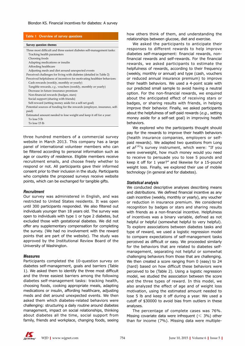
Table 1 Overview of survey questions
three hundred members of a commercial survey website in March 2013. This company has a large panel of international volunteer members who can be filtered according to personal information such as age or country of residence. Eligible members receive recruitment emails, and choose freely whether to respond or not. All participants gave their informed consent prior to their inclusion in the study. Participants who complete the proposed surveys receive website points, which can be exchanged for tangible gifts.
RecruitmentOur survey was administered in English, and was restricted to United States residents. It was open until 300 participants responded. We also filtered out individuals younger than 18 years old. The survey was open to individuals with type 1 or type 2 diabetes, but excluded those with gestational diabetes. We did not offer any supplementary compensation for completing the survey. (We had no involvement with the reward points that are part of the website). The study was approved by the Institutional Review Board of the University of Washington.
MeasuresParticipants completed the 10-question survey on diabetes self-management, goals and barriers (Table 1). We asked them to identify the three most difficult and the three easiest barriers among the following diabetes self-management tasks: tracking health, choosing foods, cooking appropriate meals, adapting medications or insulin, affording healthcare, adjusting meds and diet around unexpected events. We then asked them which diabetes-related behaviors were challenging: structuring a daily routine around diabetes management, impact on social relationships, thinking about diabetes all the time, social support from family, friends and workplace, changing foods, seeing
how others think of them, and understanding the relationships between glucose, diet and exercise.
We asked the participants to anticipate their responses to different rewards to help improve diabetes self-management: financial rewards, non-financial rewards and self-rewards. For the financial rewards, we asked participants to estimate the helpfulness of rewards, according to their frequency (weekly, monthly or annual) and type (cash, vouchers or reduced annual insurance premium) to improve their health behaviors. We used a 4-point scale with our predicted small sample to avoid having a neutral option. For the non-financial rewards, we enquired about the anticipated effect of receiving stars or badges, or sharing results with friends, in helping improve their behavior. Finally, we asked participants about the helpfulness of self-paid rewards (e.g., setting money aside for a self-set goal) in improving health behaviors.
We explored who the participants thought should pay for the rewards to improve their health behaviors (health insurance companies, employers or self-paid rewards). We adapted two questions from Long et al[16]’s survey instrument, which were: “If you were overweight, how much money would you need to receive to persuade you to lose 5 pounds and keep it off for 1 year?” and likewise for a 15-pound weight loss. Finally, we explored their use of mobile technology (in general and for diabetes). Statistical analysisWe conducted descriptive analyses describing means and distributions. We defined financial incentive as any cash incentive (weekly, monthly or yearly), any voucher or reduction in insurance premium. We considered recognition by badges or stars and sharing results with friends as a non-financial incentive. Helpfulness of incentives was a binary variable, defined as not helpful or helpful (somewhat helpful to very helpful). To explore associations between diabetes tasks and type of reward, we used a logistic regression model to compare expectations of self-management tasks perceived as difficult or easy. We proceeded similarly for the behaviors that are related to diabetes self-management, separating not helpful or somewhat challenging behaviors from those that are challenging. We then created a score ranging from 0 (easy) to 24 (hard) based on how difficult these behaviors were perceived to be (Table 2). Using a logistic regression model, we studied the association between the score and the three types of reward. In this model, we also analyzed the effect of age and of weight loss motivation, using the estimated amount needed to lose 5 lb and keep it off during a year. We used a cutoff of $30000 to avoid bias from outliers in these analyses.
The percentage of complete cases was 76%. Missing covariate data were infrequent (≤ 3%) other than for income (7%). Missing data were multiple-
754 June 10, 2015|Volume 6|Issue 5|WJD|www.wjgnet.com
Survey question themes
Three most difficult and three easiest diabetes self-management tasks: Tracking health parameters Choosing foods Adapting medications or insulin Affording healthcare Adjusting meds and diet around unexpected eventsPerceived challenges for living with diabetes (detailed in Table 2)Perceived helpfulness of incentives for motivating healthier behaviors: Cash rewards (weekly, monthly or yearly) Tangible rewards, e.g., vouchers (weekly, monthly or yearly) Decrease in future insurance premium Non-financial rewards (badges, stars) Social support (sharing with friends) Self-reward (setting money aside for a self-set goal) Potential sources of funding for the rewards (employer, insurance, self-paid)Estimated amount needed to lose weight and keep it off for a year: To lose 5 lb To lose 15 lb
Blondon KS. Financial incentives for diabetes: A survey
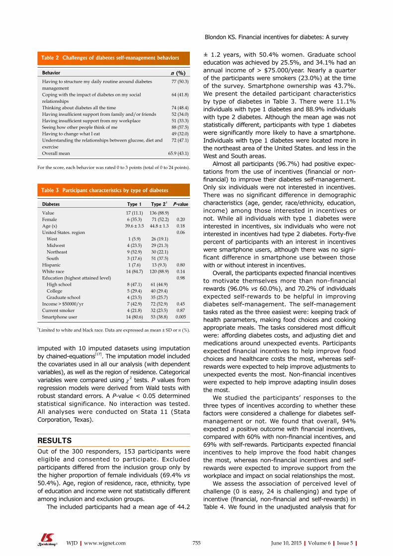
± 1.2 years, with 50.4% women. Graduate school education was achieved by 25.5%, and 34.1% had an annual income of > $75.000/year. Nearly a quarter of the participants were smokers (23.0%) at the time of the survey. Smartphone ownership was 43.7%. We present the detailed participant characteristics by type of diabetes in Table 3. There were 11.1% individuals with type 1 diabetes and 88.9% individuals with type 2 diabetes. Although the mean age was not statistically different, participants with type 1 diabetes were significantly more likely to have a smartphone. Individuals with type 1 diabetes were located more in the northeast area of the United States. and less in the West and South areas.
Almost all participants (96.7%) had positive expec-tations from the use of incentives (financial or non-financial) to improve their diabetes self-management. Only six individuals were not interested in incentives. There was no significant difference in demographic characteristics (age, gender, race/ethnicity, education, income) among those interested in incentives or not. While all individuals with type 1 diabetes were interested in incentives, six individuals who were not interested in incentives had type 2 diabetes. Forty-five percent of participants with an interest in incentives were smartphone users, although there was no signi-ficant difference in smartphone use between those with or without interest in incentives.
Overall, the participants expected financial incentives to motivate themselves more than non-financial rewards (96.0% vs 60.0%), and 70.2% of individuals expected self-rewards to be helpful in improving diabetes self-management. The self-management tasks rated as the three easiest were: keeping track of health parameters, making food choices and cooking appropriate meals. The tasks considered most difficult were: affording diabetes costs, and adjusting diet and medications around unexpected events. Participants expected financial incentives to help improve food choices and healthcare costs the most, whereas self-rewards were expected to help improve adjustments to unexpected events the most. Non-financial incentives were expected to help improve adapting insulin doses the most.
We studied the participants’ responses to the three types of incentives according to whether these factors were considered a challenge for diabetes self-management or not. We found that overall, 94% expected a positive outcome with financial incentives, compared with 60% with non-financial incentives, and 69% with self-rewards. Participants expected financial incentives to help improve the food habit changes the most, whereas non-financial incentives and self-rewards were expected to improve support from the workplace and impact on social relationships the most.
We assess the association of perceived level of challenge (0 is easy, 24 is challenging) and type of incentive (financial, non-financial and self-rewards) in Table 4. We found in the unadjusted analysis that for
imputed with 10 imputed datasets using imputation by chained-equations[17]. The imputation model included the covariates used in all our analysis (with dependent variables), as well as the region of residence. Categorical variables were compared using χ2 tests. P values from regression models were derived from Wald tests with robust standard errors. A P-value < 0.05 determined statistical significance. No interaction was tested. All analyses were conducted on Stata 11 (Stata Corporation, Texas).
RESULTSOut of the 300 responders, 153 participants were eligible and consented to participate. Excluded participants differed from the inclusion group only by the higher proportion of female individuals (69.4% vs 50.4%). Age, region of residence, race, ethnicity, type of education and income were not statistically different among inclusion and exclusion groups.
The included participants had a mean age of 44.2
755 June 10, 2015|Volume 6|Issue 5|WJD|www.wjgnet.com
Behavior n (%)Having to structure my daily routine around diabetes management
77 (50.3)
Coping with the impact of diabetes on my social relationships
64 (41.8)
Thinking about diabetes all the time 74 (48.4)Having insufficient support from family and/or friends 52 (34.0)Having insufficient support from my workplace 51 (33.3)Seeing how other people think of me 88 (57.5)Having to change what I eat 49 (32.0)Understanding the relationships between glucose, diet and exercise
72 (47.1)
Overall mean 65.9 (43.1)
For the score, each behavior was rated 0 to 3 points (total of 0 to 24 points).
Table 2 Challenges of diabetes self-management behaviors
Table 3 Participant characteristics by type of diabetes
Diabetes Type 1 Type 21 P -value
Value 17 (11.1) 136 (88.9)Female 6 (35.3) 71 (52.2) 0.20Age (x) 39.6 ± 3.5 44.8 ± 1.3 0.18United States. region 0.06 West 1 (5.9) 26 (19.1) Midwest 4 (23.5) 29 (21.3) Northeast 9 (52.9) 30 (22.1) South 3 (17.6) 51 (37.5)Hispanic 1 (7.6) 13 (9.3) 0.80White race 14 (84.7) 120 (88.9) 0.14Education (highest attained level) 0.98 High school 8 (47.1) 61 (44.9) College 5 (29.4) 40 (29.4) Graduate school 4 (23.5) 35 (25.7)Income > $50000/yr 7 (42.9) 72 (52.9) 0.45Current smoker 4 (21.8) 32 (23.5) 0.87Smartphone user 14 (80.6) 53 (38.8) 0.005
1limited to white and black race. Data are expressed as mean ± SD or n (%).
Blondon KS. Financial incentives for diabetes: A survey
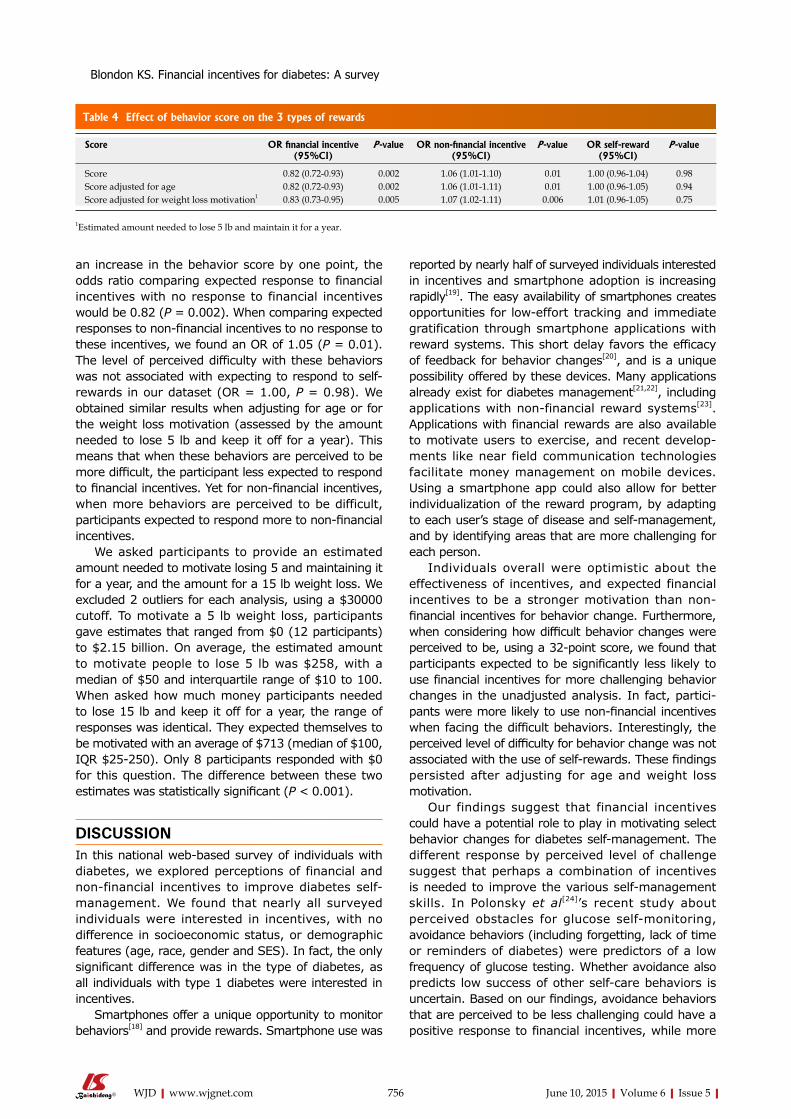
an increase in the behavior score by one point, the odds ratio comparing expected response to financial incentives with no response to financial incentives would be 0.82 (P = 0.002). When comparing expected responses to non-financial incentives to no response to these incentives, we found an OR of 1.05 (P = 0.01). The level of perceived difficulty with these behaviors was not associated with expecting to respond to self-rewards in our dataset (OR = 1.00, P = 0.98). We obtained similar results when adjusting for age or for the weight loss motivation (assessed by the amount needed to lose 5 lb and keep it off for a year). This means that when these behaviors are perceived to be more difficult, the participant less expected to respond to financial incentives. Yet for non-financial incentives, when more behaviors are perceived to be difficult, participants expected to respond more to non-financial incentives.
We asked participants to provide an estimated amount needed to motivate losing 5 and maintaining it for a year, and the amount for a 15 lb weight loss. We excluded 2 outliers for each analysis, using a $30000 cutoff. To motivate a 5 lb weight loss, participants gave estimates that ranged from $0 (12 participants) to $2.15 billion. On average, the estimated amount to motivate people to lose 5 lb was $258, with a median of $50 and interquartile range of $10 to 100. When asked how much money participants needed to lose 15 lb and keep it off for a year, the range of responses was identical. They expected themselves to be motivated with an average of $713 (median of $100, IQR $25-250). Only 8 participants responded with $0 for this question. The difference between these two estimates was statistically significant (P < 0.001).
DISCUSSIONIn this national web-based survey of individuals with diabetes, we explored perceptions of financial and non-financial incentives to improve diabetes self-management. We found that nearly all surveyed individuals were interested in incentives, with no difference in socioeconomic status, or demographic features (age, race, gender and SES). In fact, the only significant difference was in the type of diabetes, as all individuals with type 1 diabetes were interested in incentives.
Smartphones offer a unique opportunity to monitor behaviors[18] and provide rewards. Smartphone use was
reported by nearly half of surveyed individuals interested in incentives and smartphone adoption is increasing rapidly[19]. The easy availability of smartphones creates opportunities for low-effort tracking and immediate gratification through smartphone applications with reward systems. This short delay favors the efficacy of feedback for behavior changes[20], and is a unique possibility offered by these devices. Many applications already exist for diabetes management[21,22], including applications with non-financial reward systems[23]. Applications with financial rewards are also available to motivate users to exercise, and recent develop-ments like near field communication technologies facilitate money management on mobile devices. Using a smartphone app could also allow for better individualization of the reward program, by adapting to each user’s stage of disease and self-management, and by identifying areas that are more challenging for each person.
Individuals overall were optimistic about the effectiveness of incentives, and expected financial incentives to be a stronger motivation than non-financial incentives for behavior change. Furthermore, when considering how difficult behavior changes were perceived to be, using a 32-point score, we found that participants expected to be significantly less likely to use financial incentives for more challenging behavior changes in the unadjusted analysis. In fact, partici-pants were more likely to use non-financial incentives when facing the difficult behaviors. Interestingly, the perceived level of difficulty for behavior change was not associated with the use of self-rewards. These findings persisted after adjusting for age and weight loss motivation.
Our findings suggest that financial incentives could have a potential role to play in motivating select behavior changes for diabetes self-management. The different response by perceived level of challenge suggest that perhaps a combination of incentives is needed to improve the various self-management skills. In Polonsky et al[24]’s recent study about perceived obstacles for glucose self-monitoring, avoidance behaviors (including forgetting, lack of time or reminders of diabetes) were predictors of a low frequency of glucose testing. Whether avoidance also predicts low success of other self-care behaviors is uncertain. Based on our findings, avoidance behaviors that are perceived to be less challenging could have a positive response to financial incentives, while more
756 June 10, 2015|Volume 6|Issue 5|WJD|www.wjgnet.com
Table 4 Effect of behavior score on the 3 types of rewards
Score OR financial incentive (95%CI)
P -value OR non-financial incentive (95%CI)
P -value OR self-reward (95%CI)
P -value
Score 0.82 (0.72-0.93) 0.002 1.06 (1.01-1.10) 0.01 1.00 (0.96-1.04) 0.98Score adjusted for age 0.82 (0.72-0.93) 0.002 1.06 (1.01-1.11) 0.01 1.00 (0.96-1.05) 0.94Score adjusted for weight loss motivation1 0.83 (0.73-0.95) 0.005 1.07 (1.02-1.11) 0.006 1.01 (0.96-1.05) 0.75
1Estimated amount needed to lose 5 lb and maintain it for a year.
Blondon KS. Financial incentives for diabetes: A survey

challenging behaviors would require the use of non-financial incentives.
The amounts of financial incentives are important to consider. An interesting finding from our survey is the relatively low amounts of money that participants expected as incentive for weight loss ($258 for the 5 lb weight loss and $713 for the 15 lb weight loss), particularly if we consider that current out-of-pocket costs for diabetes are estimated at $350-500/mo[25,26]. This concurs with findings from another study which explored the use of financial incentives in diabetes, where participants suggested $25 per month for tracking and reporting glucose results[27]. Employers typically employ similar amounts for action-based incentives, or rewards for taking action (joining a weight loss program, for example) after going through a risk assessment[28]. Prior research has found that very large amounts can lead to lower performances, because the individual feels pressure to perform well. Likewise, amounts that are too small lead to lower performances, even lower than those who do not have any incentive[29]. These considerations suggest that our participants’ intuitions about the reward amounts are in the right ballpark, although this needs to be evaluated empirically.
The strengths of our study include its focus on the use of incentives, both financial and non-financial, in a diabetic population. Although the relatively moderate number of participants may limit its generalizability, its web-based modality allowed us to recruit nationwide. This modality however also has its limitations, as the participants are self-selected, and received tokens in exchange for taking part in the survey (no compensation was given by the investigators, this was solely a feature of the survey company). Our population may therefore be disproportionately biased in their interest in incentives. A final limitation to our study lies in its design as a survey, as we assess the participants’ expected response to incentives, which may differ from them their actual response to incentives. Future studies are needed to confirm our findings, and to further explore the acceptability and feasibility of incentives for diabetes self-management in a larger population.
According to our nationwide survey, patients with diabetes are willing to consider using incentives, both financial and non-financial, to improve diabetes self-management. While the financial incentives may be more effective for behavior changes that are perceived as less challenging, non-financial incentives may be useful for the more challenging behaviors. Participants did not expect to need large amounts to motivate them to modify their behavior. Our findings suggest that the effectiveness of incentives could vary, and may depend on the perceived difficulty of the incentivized task. Future studies are needed to confirm these results in interventional studies on larger populations.
ACKNOWLEDGMENTSI acknowledge Klasnja P, Pratt W and the rest of the iMed team for their help in preparing the survey.
COMMENTSBackgroundFinancial incentives have shown potential for modifying behaviors such as smoking cessation. Diabetes self-management requires many behavior changes, which may benefit from such incentives. Little is known about patient attitudes in response to incentives for diabetes self-management. Research frontiersAlthough incentives have been studied for certain behaviors, the current research hotspot is patient acceptance of incentives for diabetes self-management, in particular for the goals, type (financial, non-financial or self-reward) and amount of incentives.Innovations and breakthroughsBeyond patient attitudes and expectations about their response to incentives, this survey provides suggestions about goal-setting in reward programs, in relation to the perceived level of challenge of certain behaviors. Individualization of goals and rewards could be feasible with the exponential adoption of mobile devices, which could both track and reward behaviors ubiquitously. ApplicationsThe results of this study can help guide future interventional studies with incentives, both in goal-setting in terms of types of behaviors, types of rewards, and amounts for financial incentives.TerminologyFinancial incentives include all rewards relating to monetary rewards or equivalents such as vouchers (cash or discount for a future purchase, for example). Non-financial incentives are rewards such as badges or stars in social networks that provide recognition from others for an achieved feat. Self-rewards are rewards that a person gives themselves when they reach a predefined goal, often planned as small rewards that accumulate to reach a large, final reward.Peer-reviewIn this well-written paper, the author investigates the patient willingness to change diabetes self-management behaviors in response to rewards. The reviewers found this approach to improve patient engagement interesting. The results can help guide the creation and implementation of new patient-centered approaches for diabetes self-management.
REFERENCES1 Executive summary: Standards of medical care in diabetes--2013.
Diabetes Care 2013; 36 Suppl 1: S4-10 [PMID: 23264424 DOI: 10.2337/dc13-S004]
2 Jendle J, Torffvit O, Ridderstråle M, Lammert M, Ericsson A, Bøgelund M. Willingness to pay for health improvements associated with anti-diabetes treatments for people with type 2 diabetes. Curr Med Res Opin 2010; 26: 917-923 [PMID: 20163195 DOI: 10.1185/03007991003657867]
3 Giuffrida A, Torgerson DJ. Should we pay the patient? Review of financial incentives to enhance patient compliance. BMJ 1997; 315: 703-707 [PMID: 9314754]
4 Sigmon SC, Patrick ME. The use of financial incentives in promoting smoking cessation. Prev Med 2012; 55 Suppl: S24-S32 [PMID: 22525802 DOI: 10.1016/j.ypmed.2012.04.007]
5 Kullgren JT, Troxel AB, Loewenstein G, Asch DA, Norton LA, Wesby L, Tao Y, Zhu J, Volpp KG. Individual- versus group-based financial incentives for weight loss: a randomized, controlled trial. Ann Intern Med 2013; 158: 505-514 [PMID: 23546562 DOI: 10.7326/0003-4819-158-7-201304020-00002]
6 Volpp KG, John LK, Troxel AB, Norton L, Fassbender J, Loewenstein G. Financial incentive-based approaches for weight
757 June 10, 2015|Volume 6|Issue 5|WJD|www.wjgnet.com
COMMENTS
Blondon KS. Financial incentives for diabetes: A survey
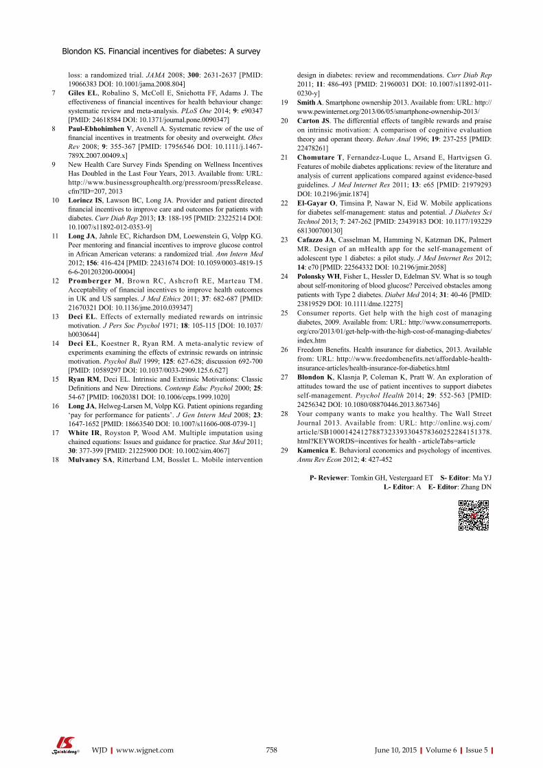
loss: a randomized trial. JAMA 2008; 300: 2631-2637 [PMID: 19066383 DOI: 10.1001/jama.2008.804]
7 Giles EL, Robalino S, McColl E, Sniehotta FF, Adams J. The effectiveness of financial incentives for health behaviour change: systematic review and meta-analysis. PLoS One 2014; 9: e90347 [PMID: 24618584 DOI: 10.1371/journal.pone.0090347]
8 Paul-Ebhohimhen V, Avenell A. Systematic review of the use of financial incentives in treatments for obesity and overweight. Obes Rev 2008; 9: 355-367 [PMID: 17956546 DOI: 10.1111/j.1467-789X.2007.00409.x]
9 New Health Care Survey Finds Spending on Wellness Incentives Has Doubled in the Last Four Years, 2013. Available from: URL: http://www.businessgrouphealth.org/pressroom/pressRelease.cfm?ID=207, 2013
10 Lorincz IS, Lawson BC, Long JA. Provider and patient directed financial incentives to improve care and outcomes for patients with diabetes. Curr Diab Rep 2013; 13: 188-195 [PMID: 23225214 DOI: 10.1007/s11892-012-0353-9]
11 Long JA, Jahnle EC, Richardson DM, Loewenstein G, Volpp KG. Peer mentoring and financial incentives to improve glucose control in African American veterans: a randomized trial. Ann Intern Med 2012; 156: 416-424 [PMID: 22431674 DOI: 10.1059/0003-4819-156-6-201203200-00004]
12 Promberger M , Brown RC, Ashcroft RE, Marteau TM. Acceptability of financial incentives to improve health outcomes in UK and US samples. J Med Ethics 2011; 37: 682-687 [PMID: 21670321 DOI: 10.1136/jme.2010.039347]
13 Deci EL. Effects of externally mediated rewards on intrinsic motivation. J Pers Soc Psychol 1971; 18: 105-115 [DOI: 10.1037/h0030644]
14 Deci EL, Koestner R, Ryan RM. A meta-analytic review of experiments examining the effects of extrinsic rewards on intrinsic motivation. Psychol Bull 1999; 125: 627-628; discussion 692-700 [PMID: 10589297 DOI: 10.1037/0033-2909.125.6.627]
15 Ryan RM, Deci EL. Intrinsic and Extrinsic Motivations: Classic Definitions and New Directions. Contemp Educ Psychol 2000; 25: 54-67 [PMID: 10620381 DOI: 10.1006/ceps.1999.1020]
16 Long JA, Helweg-Larsen M, Volpp KG. Patient opinions regarding ‘pay for performance for patients’. J Gen Intern Med 2008; 23: 1647-1652 [PMID: 18663540 DOI: 10.1007/s11606-008-0739-1]
17 White IR, Royston P, Wood AM. Multiple imputation using chained equations: Issues and guidance for practice. Stat Med 2011; 30: 377-399 [PMID: 21225900 DOI: 10.1002/sim.4067]
18 Mulvaney SA, Ritterband LM, Bosslet L. Mobile intervention
design in diabetes: review and recommendations. Curr Diab Rep 2011; 11: 486-493 [PMID: 21960031 DOI: 10.1007/s11892-011-0230-y]
19 Smith A. Smartphone ownership 2013. Available from: URL: http://www.pewinternet.org/2013/06/05/smartphone-ownership-2013/
20 Carton JS. The differential effects of tangible rewards and praise on intrinsic motivation: A comparison of cognitive evaluation theory and operant theory. Behav Anal 1996; 19: 237-255 [PMID: 22478261]
21 Chomutare T, Fernandez-Luque L, Arsand E, Hartvigsen G. Features of mobile diabetes applications: review of the literature and analysis of current applications compared against evidence-based guidelines. J Med Internet Res 2011; 13: e65 [PMID: 21979293 DOI: 10.2196/jmir.1874]
22 El-Gayar O, Timsina P, Nawar N, Eid W. Mobile applications for diabetes self-management: status and potential. J Diabetes Sci Technol 2013; 7: 247-262 [PMID: 23439183 DOI: 10.1177/193229681300700130]
23 Cafazzo JA, Casselman M, Hamming N, Katzman DK, Palmert MR. Design of an mHealth app for the self-management of adolescent type 1 diabetes: a pilot study. J Med Internet Res 2012; 14: e70 [PMID: 22564332 DOI: 10.2196/jmir.2058]
24 Polonsky WH, Fisher L, Hessler D, Edelman SV. What is so tough about self-monitoring of blood glucose? Perceived obstacles among patients with Type 2 diabetes. Diabet Med 2014; 31: 40-46 [PMID: 23819529 DOI: 10.1111/dme.12275]
25 Consumer reports. Get help with the high cost of managing diabetes, 2009. Available from: URL: http://www.consumerreports.org/cro/2013/01/get-help-with-the-high-cost-of-managing-diabetes/index.htm
26 Freedom Benefits. Health insurance for diabetics, 2013. Available from: URL: http://www.freedombenefits.net/affordable-health-insurance-articles/health-insurance-for-diabetics.html
27 Blondon K, Klasnja P, Coleman K, Pratt W. An exploration of attitudes toward the use of patient incentives to support diabetes self-management. Psychol Health 2014; 29: 552-563 [PMID: 24256342 DOI: 10.1080/08870446.2013.867346]
28 Your company wants to make you healthy. The Wall Street Journal 2013. Available from: URL: http://online.wsj.com/article/SB10001424127887323393304578360252284151378.html?KEYWORDS=incentives for health - articleTabs=article
29 Kamenica E. Behavioral economics and psychology of incentives. Annu Rev Econ 2012; 4: 427-452
P- Reviewer: Tomkin GH, Vestergaard ET S- Editor: Ma YJ L- Editor: A E- Editor: Zhang DN
758 June 10, 2015|Volume 6|Issue 5|WJD|www.wjgnet.com
Blondon KS. Financial incentives for diabetes: A survey
SYSTEMATIC REVIEWS
Diabetic nephropathy in Africa: A systematic review
Jean Jacques N Noubiap, Jashira Naidoo, Andre P Kengne
Jean Jacques N Noubiap, Internal Medicine Unit, Edéa Regional Hospital, PO BOX 100 Edéa, CameroonJashira Naidoo, Department of Medicine, Groote Schuur Hospital, University of Cape Town, 7925 Observatory, Cape Town, South AfricaJashira Naidoo, Andre P Kengne, Non-Communicable Diseases Research Unit, South African Medical Research Council, 7505 Cape Town, South Africa
Author contributions: All authors contributed to this work.
Conflict-of-interest: None for all co-authors.
Open-Access: This article is an open-access article which was selected by an in-house editor and fully peer-reviewed by external reviewers. It is distributed in accordance with the Creative Commons Attribution Non Commercial (CC BY-NC 4.0) license, which permits others to distribute, remix, adapt, build upon this work non-commercially, and license their derivative works on different terms, provided the original work is properly cited and the use is non-commercial. See: http://creativecommons.org/licenses/by-nc/4.0/
Correspondence to: Andre P Kengne, Professor, Medical Research Council of South Africa, PO Box 19070 Tygerberg, 7505 Cape Town, South Africa. [email protected]: +27-21-9380529Fax: +27-21-9380460
Received: December 8, 2014Peer-review started: December 9, 2014First decision: January 20, 2015Revised: February 18, 2015Accepted: March 16, 2015Article in press: March 18, 2015Published online: June 10, 2015
AbstractAIM: To determine the prevalence and incidence of diabetic nephropathy in Africa.
METHODS: We performed a systematic narrative review of published literature following the MOOSE Guidelines for Meta-Analysis and Systematic Reviews
of Observational Studies. We searched PubMed-MEDLINE for all articles published in English and French languages between January 1994 and July 2014 using a predefined strategy based on the combination of relevant terms and the names of each of the 54 African countries and African sub-regions to capture the largest number of studies, and hand-searched the reference lists of retrieved articles. Included studies reported on the prevalence, incidence or determinants of chronic kidney disease (CKD) in people with diabetes within African countries.
RESULTS: Overall, we included 32 studies from 16 countries; two being population-based studies and the remaining being clinic-based surveys. Most of the studies (90.6%) were conducted in urban settings. Methods for assessing and classifying CKD varied widely. Measurement of urine protein was the most common method of assessing kidney damage (62.5% of studies). The overall prevalence of CKD varied from 11% to 83.7%. Incident event rates were 94.9% for proteinuria at 10 years of follow-up, 34.7% for end-stage renal disease at 5 years of follow-up and 18.4% for mortality from nephropathy at 20 years of follow-up. Duration of diabetes, blood pressure, advancing age, obesity and glucose control were the common determinants of kidney disease.
CONCLUSION: The burden of CKD is important among people with diabetes in Africa. High quality data from large population-based studies with validated measures of kidney function are still needed to better capture the magnitude and characteristics of diabetic nephropathy in Africa.
Key words: Diabetes; Diabetes nephropathy; Chronic kidney disease; Epidemiology; Prevalence; Incidence; Mortality; Africa; Systematic review
© The Author(s) 2015. Published by Baishideng Publishing Group Inc. All rights reserved.
Core tip: Chronic kidney disease is a serious health threat for people with diabetes in Africa, with prevalence
759 June 10, 2015|Volume 6|Issue 5|WJD|www.wjgnet.com
Submit a Manuscript: http://www.wjgnet.com/esps/Help Desk: http://www.wjgnet.com/esps/helpdesk.aspxDOI: 10.4239/wjd.v6.i5.759
World J Diabetes 2015 June 10; 6(5): 759-773ISSN 1948-9358 (online)
© 2015 Baishideng Publishing Group Inc. All rights reserved.
figures ranging from 11% to 83.7%. The incidence estimates suggest that 95% of people with diabetes may have proteinuria after 10 years from diabetes diagnosis; about 35% may develop end-stage renal disease after 5 years and 18% die from nephropathy after 20 years of disease duration. Hypertension, obesity, poor glycemic control and diabetes duration are the main risk factors of chronic kidney disease among diabetic patients in Africa. High quality data are needed to refine the epidemiology of diabetic nephropathy on the continent.
Noubiap JJN, Naidoo J, Kengne AP. Diabetic nephropathy in Africa: A systematic review. World J Diabetes 2015; 6(5): 759-773 Available from: URL: http://www.wjgnet.com/1948-9358/full/v6/i5/759.htm DOI: http://dx.doi.org/10.4239/wjd.v6.i5.759
INTRODUCTIONAfrica, like the rest of the world, is experiencing an increasing prevalence of diabetes alongside other non-communicable diseases, mainly as a result of urbanization, sedentary lifestyles, obesity and popu-lation growth and ageing[1]. Estimates for 2013 by the International Diabetes Federation (IDF) indicate that the number of adults with diabetes in the world will expand by 55%, from 381.8 million in 2013 to 591.9 million in 2035[2]. The largest increase of the population with diabetes will occur in sub-Saharan Africa, with a projected growth of 109.6%, from 19.8 million in 2013 to 41.5 million in 2035[2].
Diabetes causes significant morbidity, disability and early mortality. Diabetes has been identified as a major contributor in several other important diseases, both non-communicable diseases such as cardiovascular disease and renal disease[3,4], and communicable diseases such as invasive bacterial infections[5,6]. Mortality attributable to diabetes in sub-Saharan Africa was estimated to account for 8.6% of the total death in 2013[7]. Diabetic nephropathy (DN) is one of the most common complications of diabetes. The prevalence of DN is increasing steeply along with the diabetes epidemic[8]. Approximately one third to half of patients with diabetes develops renal manifestations[8-11]. DN is associated with increased premature mortality, end-stage renal disease and need to renal replacement therapy, cardiovascular diseases, and escalating health-care costs[8].
DN has been suggested to be more frequent among patients with diabetes in Africa as compared to those in the developed world due to delayed diagnosis, limited screening and diagnostic resources, poor control of blood sugar and other risk factors, and inadequate treatment at an early stage[7,12,13]. However, evidence to support the burden of kidney diseases in people with diabetes in Africa remains very patchy, and
we are not aware of any effort to synthesize existing data on the occurrence of kidney disease in African populations with diabetes. Accordingly, the aim of this review is to provide a comprehensive overview of the published evidence on the occurrence of nephropathy in African people with diabetes.
MATERIALS AND METHODSData sources and search strategyA systematic narrative review of published literature was performed following the MOOSE Guidelines for Meta-Analyses and Systematic Reviews of Observational Studies[14]. We searched MEDLINE via PubMed for articles published in English and French on DN in Africa between January 1994 and July 2014, using a predefined strategy based on the combination of relevant terms and the names of each of the 54 African countries and African sub-regions to capture the largest number of studies. The data search was limited to human studies. The last search date was October 22, 2014. Search histories are provided in Table 1. Once duplicate references were removed the titles and abstracts of the references were screened. The references of included articles were scanned to identify additional articles of interest.
Study selection and data extractionWe included cross-sectional, case-control or cohort studies of subjects with diabetes mellitus resident in African countries reporting the prevalence or incidence or progression of DN. We excluded studies of populations of African origin residing outside Africa; case series (sample size less than 50 subjects), letters, comments and editorials; studies not published in English or French. Two investigators (JJNN, APK) independently identified articles and sequentially screened them for inclusion (Figure 1). Disagreements were solved by a third investigator (JN). Full text articles were reviewed by two investigators (JJNN and APK) who independently extracted data regarding study setting and design, study population characteristics and prevalence or incidence of DN.
RESULTSWe identified 730 articles, of which 73 were reviewed in full-text; 32 met the inclusion criteria (Figure 1)[15-46].
Characteristics of included studies Characteristics of the included studies are summarized in Table 2. The 32 studies were performed in 16 countries, with a geographical distribution covering all the African regions. However, more than half the studies [18 (56.3%)] were from South Africa (five), Nigeria (four), DR Congo (three) and Ethiopia (three).
Only two population-based studies were identified. In Democratic Republic of Congo, between March and April 2007, Makulo et al[35] studied pathologic
Noubiap JJN et al . Diabetic nephropathy in Africa
760 June 10, 2015|Volume 6|Issue 5|WJD|www.wjgnet.com

albuminuria among 81 diabetic patients identified through a population-based survey on the prevalence of diabetes involving 1898 participants[35]. Pruijm et al[39] in Seychelles in 2004, conducted a large-scale population-based estimate of the prevalence of microalbuminuria among 1218 adults. All other studies were clinic-based surveys conducted mostly in
diabetic clinics. There were three cohort studies (two prospective and one retrospective), one case-control study and the other 28 studies were cross-sectional with non-random sampling. Only three (9.4%) studies were conducted in rural settings.
Methods of assessment and classification of chronic kidney disease (CKD) varied widely. The studies assessed kidney function by urine protein [20 (62.5%) studies], urine albumin-to-creatinine ration (ACR) [9 (28.1%) studies], and estimation of glomerular filtration rate (GFR) by Cockcroft-Gault formula [3 (9.4%) studies] or by MDRD formula [4 (12.4%) studies]. Six studies (18.8%) measured kidney function by two methods, and renal biopsy was not performed in any study.
Prevalence of CKDAs depicted in Table 3, the overall prevalence of CKD varied from 11% in Tunisia to 83.7% in Tanzania[20,29]. In studies where proteinuria was used to assess CKD, the prevalence varied from 5.3% in South Africa to 53.1% in Cameroon (study with a small sample size)[32,44]. When considering the estimation of the GFR, the prevalence ranged from 4.6% in Tanzania to 43.1% in Nigeria (study with a small sample size)[15,33].
Incidence of CKDA study in South Africa investigated the long-term incidence of proteinuria among T2DM patients. After 12 years of follow-up or death, 94.9% (56/59) had a proteinuria with a mean duration from diabetes onset to proteinuria of 9.7 (5.9) years[31]. In another study in South Africa, found that 18.4% of T1DM patients had
761 June 10, 2015|Volume 6|Issue 5|WJD|www.wjgnet.com
Table 1 Search history PubMed
Search Search terms Hits
1 Diabetes[tw] OR Diabetes mellitus[tw] OR Type 1 diabetes[tw] OR Type 1 diabetes mellitus[tw] OR T1DM[tw] OR Type 2 diabetes[tw] OR Type 2 diabetes mellitus[tw] OR T2DM[tw] OR Hyperglycemia[tw] OR Glucose intolerance[tw]
445204
2 Renal insufficiency[tw] OR Renal failure[tw] OR Renal injury[tw] OR Renal disease[tw] Kidney insufficiency[tw] OR Kidney failure[tw] OR Kidney injury[tw] OR Kidney disease[tw] OR End-stage renal disease[tw] OR End-stage renal failure[tw] OR End-stage kidney disease[tw] OR End-stage kidney failure [tw] OR End stage renal disease[tw] OR End stage renal failure[tw] OR End stage kidney disease[tw] OR End stage kidney failure [tw] OR Microalbuminuria [tw] OR Micro-albuminuria OR Macroalbuminuria [tw] or Macro-albuminuria [tw]
154354
3 # 1 AND # 2 203884 Diabetic nephropathy [MeSH Terms] 194065 # 3 OR # 4 342216 ((((("Africa"[MeSH] OR Africa*[tw] OR Algeria[tw] OR Angola[tw] OR Benin[tw] OR Botswana[tw] OR "Burkina Faso"[tw] OR
Burundi[tw] OR Cameroon[tw] OR "Canary Islands"[tw] OR "Cape Verde"[tw] OR "Central African Republic"[tw] OR Chad[tw] OR Comoros[tw] OR Congo[tw] OR "Democratic Republic of Congo"[tw] OR Djibouti[tw] OR Egypt[tw] OR "Equatorial Guinea"[tw] OR Eritrea[tw] OR Ethiopia[tw] OR Gabon[tw] OR Gambia[tw] OR Ghana[tw] OR Guinea[tw] OR "Guinea Bissau"[tw] OR "Ivory Coast"[tw] OR "Cote d'Ivoire"[tw] OR Jamahiriya[tw] OR Jamahiryia[tw] OR Kenya[tw] OR Lesotho[tw] OR Liberia[tw] OR Libya[tw] OR Libia[tw] OR Madagascar[tw] OR Malawi[tw] OR Mali[tw] OR Mauritania[tw] OR Mauritius[tw] OR Mayote[tw] OR Morocco[tw] OR Mozambique[tw] OR Mocambique[tw] OR Namibia[tw] OR Niger[tw] OR Nigeria[tw] OR Principe[tw] OR Reunion[tw] OR Rwanda[tw] OR "Sao Tome"[tw] OR Senegal[tw] OR Seychelles[tw] OR "Sierra Leone"[tw] OR Somalia[tw] OR "South Africa"[tw] OR "St Helena"[tw] OR Sudan[tw] OR Swaziland[tw] OR Tanzania[tw] OR Togo[tw] OR Tunisia[tw] OR Uganda[tw] OR "Western Sahara"[tw] OR Zaire[tw] OR Zambia[tw] OR Zimbabwe[tw] OR "Central Africa"[tw] OR "Central African"[tw] OR "West Africa"[tw] OR "West African"[tw] OR "Western Africa"[tw] OR "Western African"[tw] OR "East Africa"[tw] OR "East African"[tw] OR "Eastern Africa"[tw] OR "Eastern African"[tw] OR "North Africa"[tw] OR "North African"[tw] OR "Northern Africa"[tw] OR "Northern African"[tw] OR "South African"[tw] OR "Southern Africa"[tw] OR "Southern African"[tw] OR "sub Saharan Africa"[tw] OR "sub Saharan African"[tw] OR "subSaharan Africa"[tw] OR "subSaharan African"[tw]) NOT ("guinea pig"[tw] OR "guinea pigs"[tw] OR "aspergillus niger"[tw])))))
354928
7 # 5 AND # 6 10658 #4 Limits: 1994/01/01 to 2014/10/22 and studies done in Humans 918
Figure 1 Flow diagram of study selection.
918 articles retrieved from MEDLINE and screened based
on title and abstract
836 articles did not meet inclusion criteria
7 full-texts of potentially relevant
articles were not found
75 full-text articles assessed for eligibility
6 articles from review of bibliographies
49 articles excluded
32 articles included in the systematic review
Noubiap JJN et al . Diabetic nephropathy in Africa
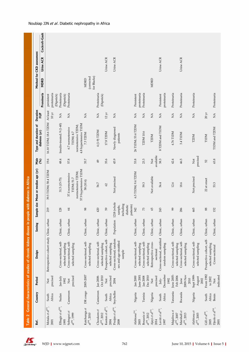
Tabl
e 2 G
ener
al c
hara
cter
istics
of
stud
ies
of c
hron
ic k
idne
y di
seas
e in
peo
ple
with
diab
etes
in A
fric
a
762 June 10, 2015|Volume 6|Issue 5|WJD|www.wjgnet.com
Ref
.C
ount
ryPe
riod
Des
ign
Sett
ing
Sam
ple
size
Mea
n or
med
ian
age
(yr)
Mal
e (%
)Ty
pe a
nd d
urat
ion
of
diab
etes
(yr
)D
urat
ion
FUP
Met
hod
for
CK
D a
sses
smen
t
Prot
einu
ria
MD
RD
Urine
AC
RC
ockr
oft-
Gau
lt
Mot
ala
et a
l[37],
2001
So
uth
Afr
ica
Not
pr
ecis
edRe
tros
pect
ive
coho
rt s
tudy
Clin
ic, u
rban
21
939
.5 T
1DM
; 58.
4 T2
DM
19.6
16.1
0 T1
DM
; 18.
6 T2
DM
At l
east
10
yr
pers
iste
nt
prot
einu
ria
(Dip
stic
k)El
bagi
r et a
l[26],
1995
Su
dan
Jan-
July
19
92C
ross
-sec
tiona
l, se
lf-se
lect
ed s
ampl
ing
Clin
ic, u
rban
128
31.5
(15-
75)
48.4
Insu
lin-tr
eate
d; 9
(1-4
0)N
APr
otei
nuri
a (D
ipst
ick)
Sobn
gwi e
t al
[44], 1
999
Cam
eroo
nN
ot
prec
ised
Cro
ss-s
ectio
nal,
self-
sele
cted
sam
plin
gC
linic
, urb
an 6
437
.4 n
orm
oten
sive
T1
DM
; 51.
7 no
rmot
ensi
ve T
2DM
; 57
.9 h
yper
tens
ive
T1D
M
57.8
6.7
norm
oten
sive
T1
DM
; 4.7
no
rmot
ensi
ve T
2DM
; 4.
8 hy
pert
ensi
ve T
1DM
NA
Prot
einu
ria
Kat
chun
ga et
al
[30], 2
010
DR
cong
o20
05-2
007
Cro
ss-s
ectio
nal,
self-
sele
cted
sam
plin
gC
linic
, urb
an 9
858
(10.
4)35
.77.
3 T2
DM
NA
MD
RD
(cor
rect
ed
for B
lack
s)C
houk
em et
al
[22], 2
012
Cam
eroo
nJa
n 20
08-
Oct
201
0C
ross
-sec
tiona
l, se
lf-se
lect
ed s
ampl
ing
Clin
ic, u
rban
420
56.7
494
(1-9
) T2D
MN
APr
otei
nuri
a (D
ipst
ick)
K
eeto
n et
al[3
1],
2004
So
uth
Afr
ica
Not
pr
ecis
edPr
ospe
ctiv
e co
hort
, sel
f-se
lect
ed s
ampl
ing
Clin
ic, u
rban
59
62
35.6
17.8
T2D
M12
yr
Uri
ne A
CR
Prui
jm et
al[3
9],
2008
Se
yche
lles
2004
Cro
ss-s
ectio
nal;
rand
om
sex
and
age-
stra
tified
sa
mpl
e
Popu
latio
n12
18 (w
hole
sa
mpl
e,
incl
udin
g di
abet
ic
patie
nts)
Not
pre
cise
d45
.9N
ewly
dia
gnos
ed
patie
nts
NA
Uri
ne A
CR
Ale
bios
u[16],
2003
N
iger
iaJa
n 20
00
June
200
1C
ross
-sec
tiona
l, se
lf-se
lect
ed s
ampl
ing
Clin
ic, u
rban
342
6.5
T1D
M; 9
.4 T
2DM
53.8
26 T
1DM
; 53.
4 T2
DM
NA
Pers
iste
nt
prot
einu
ria
Boua
ziz
et
al[2
0], 2
012
Tuni
sia
Jan
2008
Dec
201
0C
ross
-sec
tiona
l, se
lf-se
lect
ed s
ampl
ing
Clin
ic, u
rban
73
59.3
23.3
T2D
M 1
0.6
NA
Prot
einu
ria
Aja
yi et
al[1
5],
2014
N
iger
iaN
ot
prec
ised
Retr
ospe
ctiv
e cr
oss-
sect
iona
lC
linic
, urb
an 6
5N
ot a
vaila
ble
Not
av
aila
ble
T2D
MN
AM
DRD
Levi
tt et
al[3
2],
1997
So
uth
Afr
ica
July
-D
ecem
ber
1992
Cro
ss-s
ectio
nal,
stra
tified
ra
ndom
sam
plin
gC
linic
, urb
an24
356
.4
38.3
8 T2
DM
and
T1D
MN
APe
rsis
tent
pr
otei
nuri
aU
rine
AC
R
Maj
aliw
a et
al
[34], 2
007
Tanz
ania
June
200
5-
Feb
2006
Cro
ss-s
ectio
nal,
self-
sele
cted
sam
plin
gC
linic
, urb
an 9
912
.6
42.4
4.76
T1D
MN
APr
otei
nuri
a
Mar
shal
l et
al[3
6], 2
013
Rwan
daJu
ne
2009
-Nov
20
10
Cro
ss-s
ectio
nal,
self-
sele
cted
sam
plin
gC
linic
, urb
an28
618
.6
46.5
3.4
T1D
MN
APr
otei
nuri
aU
rine
AC
R
Ale
bios
u et
al
[18], 2
003
Nig
eria
Sept
199
9-
Aug
ust
2002
Cro
ss-s
ectio
nal,
self-
sele
cted
sam
plin
gC
linic
, urb
an46
5N
ot p
reci
sed
Not
pr
ecis
edT2
DM
NA
Prot
einu
ria
Gill
et a
l[28],
2005
So
uth
Afr
ica
From
198
2 to
200
2Pr
ospe
ctiv
e co
hort
, sel
f-se
lect
ed s
ampl
ing
Clin
ic, u
rban
88
22 a
t ons
et52
T1D
M20
yr
Djro
lo et
al[2
4],
2001
Be
nin
Not
in
dica
ted
Cro
ss-s
ectio
nal
Clin
ic, u
rban
152
53.3
65.8
T1D
M a
nd T
2DM
NA
Prot
einu
ria
Noubiap JJN et al . Diabetic nephropathy in Africa
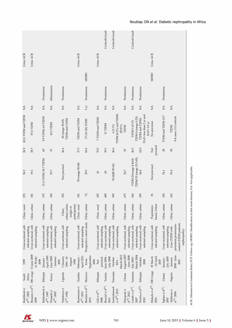
763 June 10, 2015|Volume 6|Issue 5|WJD|www.wjgnet.com
Rotc
hfor
d et
al
[43], 2
002
Sout
h A
fric
a19
99C
ross
-sec
tiona
l, se
lf-se
lect
ed s
ampl
ing
Clin
ic, r
ural
253
56.5
26
.942
.2; T
1DM
and
T2D
MN
A
U
rine
AC
R
Riss
assi
et a
l[42],
2009
D
R co
ngo
11 ju
ne 2
008
to 3
0 ju
ly
2008
Cro
ss-s
ectio
nal,
self-
sele
cted
sam
plin
gC
linic
, urb
an18
119
.1
38.7
57.6
T1D
MN
AU
rine
AC
R
Rahl
enbe
ck et
al
[40], 1
997
Ethi
opia
Janu
ary
- A
pril
1995
Cro
ss-s
ectio
nal,
self-
sele
cted
sam
plin
gC
linic
, urb
an17
031
.4 T
1DM
; 56.
7 T2
DM
605.
9 T1
DM
; 6.0
T2D
MN
APr
otei
nuri
a
Wan
johi
et
al[4
5], 2
002
Ken
yaJu
ne 2
000
- Jan
uary
20
01
cros
s-se
ctio
nal,
self-
sele
cted
sam
plin
gC
linic
, urb
an10
053
.7
3710
.3 T
2DM
NA
Alb
umin
uria
Nam
buya
et
al[3
8], 1
996
Uga
nda
1 Ja
nuar
y 19
93 -
10
Aug
ust
1994
Cro
ss-s
ectio
nal,
self-
sele
cted
sam
plin
gC
linic
, ur
ban/
urba
n (o
rigi
n of
pa
rtic
ipan
ts)
252
Not
pre
cise
d46
.445
(ran
ge 3
0-69
)T2
DM
and
T1D
MN
APr
otei
nuri
a
Rasm
usse
n et
al
[41], 2
013
Zam
bia
Febr
uary
- A
pril
2011
Cro
ss-s
ectio
nal,
self-
sele
cted
sam
plin
gC
linic
, rur
al10
150
(ran
ge 5
0-68
)37
.3T2
DM
and
T1D
MN
AU
rine
AC
R
Bent
ata
et a
l[19],
2013
M
oroc
coFr
om
Sept
embe
r 20
06
Pros
pect
ive
coho
rt s
tudy
Clin
ic, u
rban
72
29.5
69
.417
(11-
20) T
1DM
5 yr
Prot
einu
ria
MD
RD
Gill
et a
l[27],
2008
Et
hiop
iaN
ot
prec
ised
Cro
ss-s
ectio
nal,
self-
sele
cted
sam
plin
gC
linic
, rur
al10
541
70
.57
T1D
M a
nd T
2DM
NA
Uri
ne A
CR
Bouz
id et
al[2
1],
2011
Tu
nisi
aJu
ne 2
006
- Ju
ly 2
008
Cro
ss-s
ectio
nal,
self-
sele
cted
sam
plin
gC
linic
, urb
an68
960
39
.311
T2D
MN
APr
otei
nuri
aC
ockr
oft-G
ault
Janm
oham
ed
et a
l[29], 2
013
Tanz
ania
Oct
ober
20
11 -
Mar
ch 2
012
Cro
ss-s
ectio
nal,
self-
sele
cted
sam
plin
gC
linic
, urb
an36
954
(IQ
R 45
-62)
46.6
6 (3
-11)
T1D
M (6
.2%
) and
T2D
M
(93.
8%)
NA
Coc
krof
t-Gau
lt
Dan
quah
et
al[2
3], 2
012
Gha
naA
ugus
t 200
7 - J
une
2008
Cro
ss-s
ectio
nal,
self-
sele
cted
sam
plin
gC
linic
, urb
an67
554
.7
25T2
DM
NA
Prot
einu
ria
Luta
le et
al[3
3],
2007
Tanz
ania
July
200
3 -
Mar
ch 2
004
Cro
ss-s
ectio
nal,
self-
sele
cted
sam
plin
gC
linic
, urb
an24
4T1
DM
21(
rang
e 4-
44.8
)T2
DM
53
(ran
ge 2
3.5-
85)
46.3
T1D
M 3
(0-1
7)T2
DM
4 (r
ange
0-2
5)N
APr
otei
nuri
aC
ockr
oft-G
ault
Wor
ku et
al[4
6],
2010
Et
hiop
iaO
ctob
er
2008
Cro
ss-s
ectio
nal,
self-
sele
cted
sam
plin
gC
linic
, urb
an30
544
.4
62.9
T1D
M a
nd T
2DM
; 53
.4%
less
than
5 y
r and
33
.8%
5-9
yr
NA
Prot
einu
ria
Mak
ulo
et a
l[35],
2010
D
R C
ongo
30 M
arch
- 2
4 A
pril
2007
Cro
ss-s
ectio
nal,
self-
sele
cted
sam
plin
gPo
pula
tion-
base
d, U
rban
81
Not
pre
cise
dN
ot
prec
ised
No
prec
isio
nN
AM
DRD
Uri
ne A
CR
Egha
n et
al[2
5],
2007
G
hana
Janu
ary
- Ju
ly 2
005
Cro
ss-s
ectio
nal,
self-
sele
cted
sam
plin
gC
linic
, urb
an10
954
.1
28T1
DM
and
T2D
M 1
0.7
NA
Prot
einu
ria
Ale
bios
u et
al
[17], 2
004
Nig
eria
Janu
ary
2000
- Ju
ne
2001
Cas
e (T
2DM
with
pe
rsis
tent
pro
tein
uria
-co
ntro
l (T2
DM
pat
ient
s ne
phro
path
y)
Clin
ic, u
rban
162
53.4
50
T2D
M9.
4 ca
ses,
5.5
con
trol
sN
A
AC
R: A
lbum
in-to
-Cre
atin
ine
Ratio
; FU
P: F
ollo
w-u
p; M
DRD
: Mod
ifica
tion
of d
iet r
enal
dis
ease
; NA
: Not
app
licab
le.
Noubiap JJN et al . Diabetic nephropathy in Africa
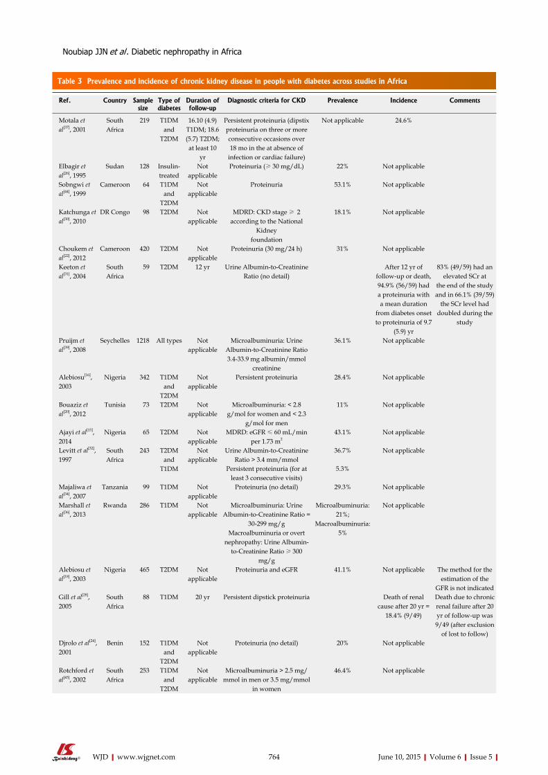
Table 3 Prevalence and incidence of chronic kidney disease in people with diabetes across studies in Africa
764 June 10, 2015|Volume 6|Issue 5|WJD|www.wjgnet.com
Ref. Country Sample size
Type of diabetes
Duration of follow-up
Diagnostic criteria for CKD Prevalence Incidence Comments
Motala et al[37], 2001
South Africa
219 T1DM and
T2DM
16.10 (4.9) T1DM; 18.6 (5.7) T2DM; at least 10
yr
Persistent proteinuria (dipstix proteinuria on three or more consecutive occasions over 18 mo in the at absence of
infection or cardiac failure)
Not applicable 24.6%
Elbagir et al[26], 1995
Sudan 128 Insulin-treated
Not applicable
Proteinuria (≥ 30 mg/dL) 22% Not applicable
Sobngwi et al[44], 1999
Cameroon 64 T1DM and
T2DM
Not applicable
Proteinuria 53.1% Not applicable
Katchunga et al[30], 2010
DR Congo 98 T2DM Not applicable
MDRD: CKD stage ≥ 2 according to the National
Kidneyfoundation
18.1% Not applicable
Choukem et al[22], 2012
Cameroon 420 T2DM Not applicable
Proteinuria (30 mg/24 h) 31% Not applicable
Keeton et al[31], 2004
South Africa
59 T2DM 12 yr Urine Albumin-to-Creatinine Ratio (no detail)
After 12 yr of follow-up or death, 94.9% (56/59) had a proteinuria with a mean duration
from diabetes onset to proteinuria of 9.7
(5.9) yr
83% (49/59) had an elevated SCr at
the end of the study and in 66.1% (39/59)
the SCr level had doubled during the
study
Pruijm et al[39], 2008
Seychelles 1218 All types Not applicable
Microalbuminuria: Urine Albumin-to-Creatinine Ratio 3.4-33.9 mg albumin/mmol
creatinine
36.1% Not applicable
Alebiosu[16], 2003
Nigeria 342 T1DM and
T2DM
Not applicable
Persistent proteinuria 28.4% Not applicable
Bouaziz et al[20], 2012
Tunisia 73 T2DM Not applicable
Microalbuminuria: < 2.8 g/mol for women and < 2.3
g/mol for men
11% Not applicable
Ajayi et al[15], 2014
Nigeria 65 T2DM Not applicable
MDRD: eGFR ≤ 60 mL/min per 1.73 m2
43.1% Not applicable
Levitt et al[32], 1997
South Africa
243 T2DM and
T1DM
Not applicable
Urine Albumin-to-Creatinine Ratio > 3.4 mm/mmol
36.7% Not applicable
Persistent proteinuria (for at least 3 consecutive visits)
5.3%
Majaliwa et al[34], 2007
Tanzania 99 T1DM Not applicable
Proteinuria (no detail) 29.3% Not applicable
Marshall et al[36], 2013
Rwanda 286 T1DM Not applicable
Microalbuminuria: Urine Albumin-to-Creatinine Ratio =
30-299 mg/gMacroalbuminuria or overt
nephropathy: Urine Albumin-to-Creatinine Ratio ≥ 300
mg/g
Microalbuminuria: 21%;
Macroalbuminuria: 5%
Not applicable
Alebiosu et al[18], 2003
Nigeria 465 T2DM Not applicable
Proteinuria and eGFR 41.1% Not applicable The method for the estimation of the
GFR is not indicated Gill et al[28], 2005
South Africa
88 T1DM 20 yr Persistent dipstick proteinuria Death of renal cause after 20 yr =
18.4% (9/49)
Death due to chronic renal failure after 20 yr of follow-up was 9/49 (after exclusion
of lost to follow)Djrolo et al[24], 2001
Benin 152 T1DM and
T2DM
Not applicable
Proteinuria (no detail) 20% Not applicable
Rotchford et al[43], 2002
South Africa
253 T1DM and
T2DM
Not applicable
Microalbuminuria > 2.5 mg/mmol in men or 3.5 mg/mmol
in women
46.4% Not applicable
Noubiap JJN et al . Diabetic nephropathy in Africa

765 June 10, 2015|Volume 6|Issue 5|WJD|www.wjgnet.com
Rissassi et al[42], 2009
DR congo 181 T1DM Not applicable
Microalbuminuria: Urine Albumin-to-Creatinine Ratio =
30-299 mg/gMacroalbuminuria: Urine
Albumin-to-Creatinine Ratio ≥ 300 mg/g
21.9% (microalbuminuria)
and 7.3% (macroalbuminuria)
Not applicable
Rahlenbeck et al[40], 1997
Ethiopia 170 T1DM and
T2DM
Not applicable
Microalbuminuria: > 30 mg/LMacroalbuminuria: > 300
mg/L
T1DM: 32% (microalbuminuria)
and 15% (macroalbuminuria)
T2DM: 37% (microalbuminuria)
and 20% (macroalbuminuria)
Not applicable
Wanjohi et al[45], 2002
Kenya 100 T2DM Not applicable
Proteinuria ≥ 20 mg 26% Not applicable
Nambuya et al[38], 1996
Uganda 252 T1DM and
T2DM
Not applicable
Proteinuria (no detail) 17.1% Not applicable Newly diagnosed patients
Rasmussen et al[41], 2013
Zambia 101 T1DM and
T2DM
Not applicable
Microalbuminuria: ACR = 3.5-35.0 for women and
2.5-25.0 mg/mmol for menMacroalbuminuria were ACR> 35.0 for women and > 25.0
for men
Microalbuminuria: 23.8%
Macroalbuminuria: 8.9%
Not applicable There were 33 patients with
diabetes alone, and 68 patients
with diabetes and hypertension
Bentata et al[19], 2013
Morocco 72 T1DM 5 yr Microalbuminuria: albumin excretion rate 30-300 mg/24 hMacroalbuminuria: albumin excretion rate > 300 mg/24 h
Nephrotic proteinuria: albumin excretion rate ≥ 3000
mg/24 hRenal failure: eGFR < 60 mL/
min (MDRD)
At the time of enrollement
Microalbuminuria: 48.6%
Macroalbuminuria: 36.1%
Nephrotic proteinuria: 15.3%
The incidence of end stage renal
disease after 5 yr: 34.7%
Urinary assays done on
admission were repeated on three
specimens atthree-monthly
intervals
Gill et al[27], 2008
Ethiopia 105 T1DM and
T2DM
Not applicable
Nephropathy: ACR> 25.0 mg/mmol and retinopathy present
Microalbuminuria: ACR> 2.5 and < 25.0 mg/mmol in
men and > 3.5 and< 25.0 mg/mmol in women
Nephropathy: 2%Microalbuminuria:
51%
Urinary ACR levels (to
assess microalbuminuria and nephropathy)
were done on59 patients, as those
with haematuria and/or urinary infection were
excludedBouzid et al[21], 2011
Tunisia 689 T2DM Not applicable
CKD: eGFR < 60 mL/min per 1.73 m2 (Cockroft-Gault)
Microalbuminuria: albumin excretion rate 30-300 mg/24 hMacroalbuminuria: albumin excretion rate > 300 mg/24 h
CKD: 19.8%Microalbuminuria:
13%Macroalbuminuria:
10.1%
Not applicable Macroalbuminuria was significantly
associated with CKD (P < 0.00001)
Janmohamed et al[29], 2013
Tanzania 369 T1DM and
T2DM
Not applicable
CKD: eGFR < 60 mL/min per 1.73 m2 (Cockroft-Gault) or
microalbuminuria (> 20 mg/L) or overt protienuria
CKD: 83.7%eGFR < 60 mL/min per 1.73 m2: 24.7%Microalbuminuria:
45.8%Overt proteinuria:
34.1%
Not applicable
Danquah et al[23], 2012
Ghana 671 T2DM Not applicable
Proteinuria ≥ 20 mg/L 43% Not applicable
Lutale et al[33], 2007
Tanzania 244 T1DM and
T2DM
Not applicable
Microalbuminuria: AER 20-200 μg/min
Macroalbuminuria: AER > 200 μg/min
Renal failure: eGFR < 60 mL/min per 1.73 m2:
Microalbuminuria: 12.1% (T1DM); 9.8%
(T2DM)Macroalbuminuria: 1.1% (T1DM); 7.2%
(T2DM)Renal failure:
4.6% (T1DM); 22% (T2DM)
Not applicable
Noubiap JJN et al . Diabetic nephropathy in Africa
died from renal nephropathy after 20 years of follow-up[28]. In a recent study in Morocco, the incidence of end-stage renal disease after 5 years was 34.7%[19].
Risk factors of CKDTwenty studies (62.5%) reported factors associated with CKD in diabetic patients (Table 4). However, in most studies the method to assess this association was imprecise. In cross-sectional studies, correlates of CKD included systolic and diastolic high blood pressure, long duration of diabetes, older age, dyslipidemia, obesity[16-22,25,26,29-31,33,36,40,42-44,46]. In a study in Cameroon, T2DM patients with systolic hypertension and diastolic hypertension were respectively 1.45 (95%CI: 1.15-1.84; P = 0.006) and 1.33 (95%CI: 1.06-1.66; P = 0.026) times more likely to have nephropathy[22]. Two studies in Rwanda and South Africa respectively showed that a one year increase in the duration of T1DM increased by 0.86 (95%CI: 0.77-0.96; P = 0.008) the odds of microalbuminuria[36], and that T1DM and T2DM patients with a duration of diabetes greater than 10 years were 4.19 times (95%CI: 1.93-9.10; P < 0.001) more likely to have microalbuminuria[43]. Poor glycemic control as measured by HbA1c was also a strong predictor of nephropathy. For instance, HbA1c level greater than 10% and 14% were respectively associated with a 2.6 fold (95%CI: 1.1-6.4) and a 4.69 (95%CI: 1.65-13.3; P = 0.004)[42,43]. A 1 g/dL decrease in hemoglobin level has been found to be associated with end-stage renal disease (OR 3.18, 95%CI: 1.47-6.87; P = 0.003)[19]. Studies in Nigeria showed that left ventricular hypertrophy, stroke, myocardial infarction and peripheral arterial disease were more frequent in T2DM patients with nephropathy, especially those with advanced stages[17,18].
DISCUSSIONDiabetic nephropathy is a common and morbid com-
plication of diabetes and the leading cause of CKD in the developed world. The lack of renal registries means that there are no reliable statistics about the burden of CKD in people with diabetes in the majority of African countries. The current systematic review identified 32 relevant studies published over the last 20 years on kidney diseases in people with diabetes residing in Africa. Prevalence rates ranged from 11% to 83.7% for the overall CKD, 5.3% to 53.1% for CKD based on proteinuria, and 4.6% to 43.1% for CKD based on eGFR. Incident event rates were 94.9% for proteinuria at 10 years for follow-up, 34.7% for ERSD at 5 years of follow-up and 18.4% for mortality from nephropathy at 20 years of follow-up. Diagnosed duration of diabetes, blood pressure variables, advancing age, obesity and to some ext-end glucose control were the common determinants of kidney disease in people with diabetes. Studies were overwhelmingly hospital-based studies; half of them originated from four countries while variable definitions and methods for assessing nephropathy had been used across studies.
The most recent overview of CKD in populations within Africa was completed in 2012, and was restricted to sub-Saharan African Countries[47]. This review identified 90 articles representing data from 21 countries, with over half of the studies originating from South Africa, Nigeria and Ethiopia alones. Across 21 studies deemed to be of medium to high quality by the investigators, the pooled prevalence of CKD was 13.9% (95%CI: 12.2-15.7), with substantial heterogeneity across studies. The prevalence in people with diabetes ranged from 4% to 24% based essentially on proteinuria defined CKD[47]. In our review without applying quality criteria, we found much higher prevalence of CKD, regardless of the definition. In four studies published in 2013 for instance, the prevalence of microalbuminuria ranged between 21% and 45%. Although issues with the quality of the studies preclude direct comparisons, it is likely that nephropathy is
766 June 10, 2015|Volume 6|Issue 5|WJD|www.wjgnet.com
Worku et al[46], 2010
Ethiopia 305 T1DM (38%) and
T2DM (62%)
Not applicable
Proteinuria (no detail) 15.7% Not applicable
Makulo et al[35], 2010
DR Congo 81 No precision
Not applicable
Microalbuminuria: ACR 30-299 mg/g
Macroalbuminuria: ACR ≥ 300 mg/g
Renal failure: eGFR < 60 mL/min per 1.73 m2
Microalbuminuria: 43.5%
Macroalbuminuria: 12%
Renal failure: 21.4%
Not applicable
Eghan et al[25], 2007
Ghana 109 T1DM and
T2DM
Not applicable
Microalbuminuria: ACR 30-300 mg/g
43.1% Not applicable
Alebiosu et al[17], 2004
Nigeria 162 T2DM Not applicable
Not applicable Not applicable Not applicable The study did not assess the prevalence or
incidence of diabetic nephropathy, but its
predictors
Noubiap JJN et al . Diabetic nephropathy in Africa
ACR: Albumin-to-Creatinine Ratio; T1DM: Type 1 diabetes mellitus; T2DM: Type 2 diabetes mellitus; CKD: Chronic kidney disease; eGFR: Epidermal growth factor receptor.
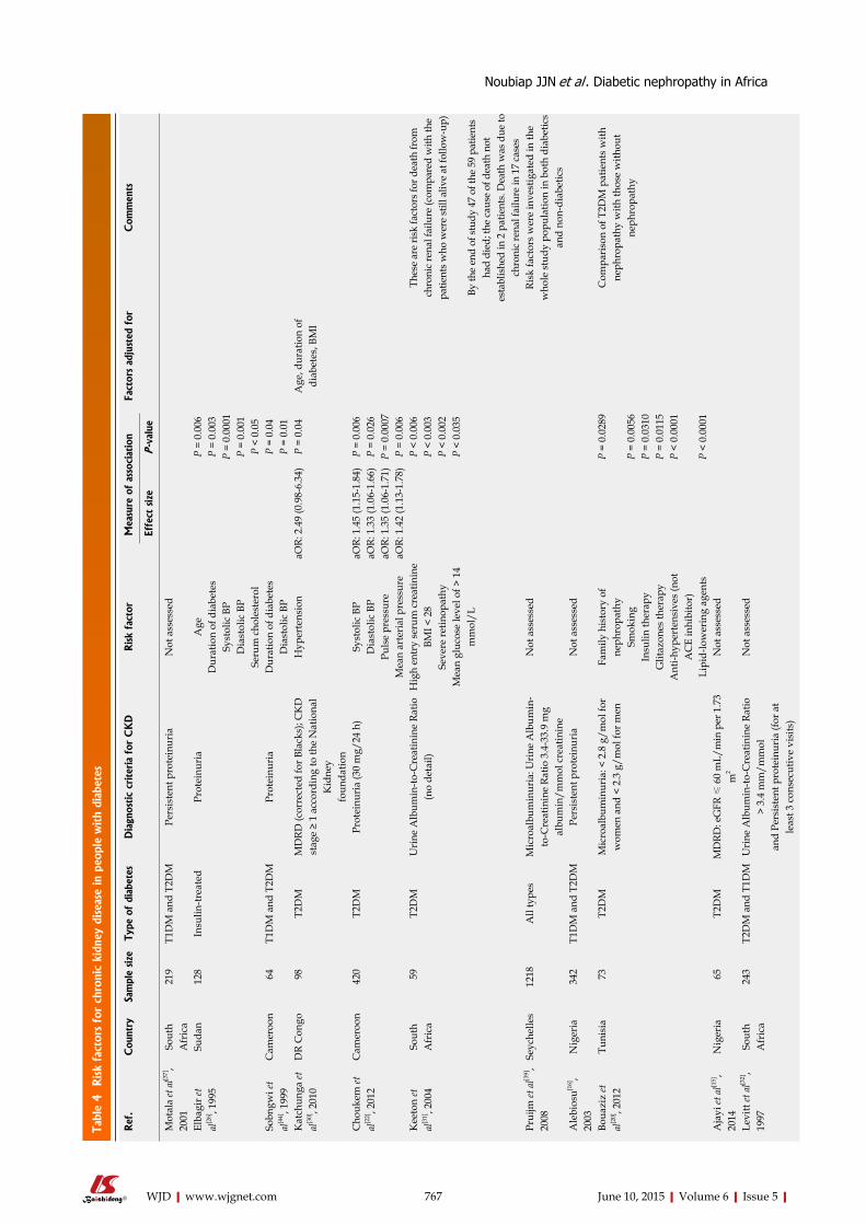
Tabl
e 4 Risk
fact
ors
for
chro
nic
kidn
ey d
isea
se in
peo
ple
with
diab
etes
767 June 10, 2015|Volume 6|Issue 5|WJD|www.wjgnet.com
Ref
.C
ount
rySa
mpl
e size
Type
of
diab
etes
Dia
gnos
tic
criter
ia f
or C
KD
Risk
fact
orM
easu
re o
f as
soci
atio
nFa
ctor
s ad
just
ed f
orC
omm
ents
Effe
ct s
ize
P-v
alue
Mot
ala
et a
l[37],
2001
So
uth
Afr
ica
219
T1D
M a
nd T
2DM
Pers
iste
nt p
rote
inur
ia
Not
ass
esse
d
Elba
gir e
t al
[26], 1
995
Suda
n12
8In
sulin
-trea
ted
Prot
einu
ria
Age
P =
0.00
6D
urat
ion
of d
iabe
tes
P =
0.00
3Sy
stol
ic B
PP
= 0.
0001
Dia
stol
ic B
PP
= 0.
001
Seru
m c
hole
ster
olP
< 0.
05So
bngw
i et
al[4
4], 1
999
Cam
eroo
n 6
4T1
DM
and
T2D
MPr
otei
nuri
aD
urat
ion
of d
iabe
tes
P =
0.04
Dia
stol
ic B
PP
= 0.
01K
atch
unga
et
al[3
0], 2
010
DR
Con
go 9
8T2
DM
MD
RD (c
orre
cted
for B
lack
s); C
KD
st
age
≥ 1
acco
rdin
g to
the
Nat
iona
l K
idne
yfo
unda
tion
Hyp
erte
nsio
naO
R: 2
.49
(0.9
8-6.
34)
P =
0.04
Age
, dur
atio
n of
di
abet
es, B
MI
Cho
ukem
et
al[2
2], 2
012
Cam
eroo
n42
0T2
DM
Prot
einu
ria
(30
mg/
24 h
)Sy
stol
ic B
PaO
R: 1
.45
(1.1
5-1.
84)
P =
0.00
6D
iast
olic
BP
aOR:
1.3
3 (1
.06-
1.66
)P
= 0.
026
Puls
e pr
essu
reaO
R: 1
.35
(1.0
6-1.
71)
P =
0.00
07M
ean
arte
rial
pre
ssur
eaO
R: 1
.42
(1.1
3-1.
78)
P =
0.00
6K
eeto
n et
al
[31], 2
004
Sout
h A
fric
a 5
9T2
DM
Uri
ne A
lbum
in-to
-Cre
atin
ine
Ratio
(n
o de
tail)
Hig
h en
try
seru
m c
reat
inin
eP
< 0.
006
Thes
e ar
e ris
k fa
ctor
s for
dea
th fr
om
chro
nic
rena
l fai
lure
(com
pare
d w
ith th
e pa
tient
s who
wer
e st
ill a
live
at fo
llow
-up)
By th
e en
d of
stud
y 47
of t
he 5
9 pa
tient
s ha
d di
ed; t
he c
ause
of d
eath
not
es
tabl
ishe
d in
2 p
atie
nts.
Dea
th w
as d
ue to
ch
roni
c re
nal f
ailu
re in
17
case
s
BMI <
28
P <
0.00
3Se
vere
retin
opat
hyP
< 0.
002
Mea
n gl
ucos
e le
vel o
f > 1
4 m
mol
/LP
< 0.
035
Prui
jm et
al[3
9],
2008
Se
yche
lles
1218
All
type
sM
icro
albu
min
uria
: Uri
ne A
lbum
in-
to-C
reat
inin
e Ra
tio 3
.4-3
3.9
mg
albu
min
/mm
ol c
reat
inin
e
Not
ass
esse
dRi
sk fa
ctor
s w
ere
inve
stig
ated
in th
e w
hole
stu
dy p
opul
atio
n in
bot
h di
abet
ics
and
non-
diab
etic
sA
lebi
osu[1
6],
2003
N
iger
ia34
2T1
DM
and
T2D
MPe
rsis
tent
pro
tein
uria
Not
ass
esse
d
Boua
ziz
et
al[2
0], 2
012
Tuni
sia
73
T2D
MM
icro
albu
min
uria
: < 2
.8 g
/mol
for
wom
en a
nd <
2.3
g/m
ol fo
r men
Fam
ily h
isto
ry o
f ne
phro
path
yP
= 0.
0289
Com
pari
son
of T
2DM
pat
ient
s w
ith
neph
ropa
thy
with
thos
e w
ithou
t ne
phro
path
ySm
okin
gP
= 0.
0056
Insu
lin th
erap
yP
= 0.
0310
Glit
azon
es th
erap
yP
= 0.
0115
Ant
i-hyp
erte
nsiv
es (n
ot
AC
E in
hibi
tor)
P <
0.00
01
Lipi
d-lo
wer
ing
agen
tsP
< 0.
0001
Aja
yi et
al[1
5],
2014
N
iger
ia 6
5T2
DM
MD
RD: e
GFR
≤ 6
0 m
L/m
in p
er 1
.73
m2
Not
ass
esse
d
Levi
tt et
al[3
2],
1997
So
uth
Afr
ica
243
T2D
M a
nd T
1DM
Uri
ne A
lbum
in-to
-Cre
atin
ine
Ratio
>
3.4
mm
/mm
ol
Not
ass
esse
d
and
Pers
iste
nt p
rote
inur
ia (f
or a
t le
ast 3
con
secu
tive
visi
ts)
Noubiap JJN et al . Diabetic nephropathy in Africa
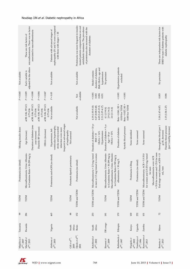
768 June 10, 2015|Volume 6|Issue 5|WJD|www.wjgnet.com
Maj
aliw
a et
al
[34], 2
007
Tanz
ania
99
T1D
MPr
otei
nuri
a (n
o de
tail)
Mis
sing
insu
lin d
oses
P =
0.04
5N
ot a
vaila
ble
Mar
shal
l et
al[3
6], 2
013
Rwan
da28
6T1
DM
Mic
roal
bum
inur
ia: U
rine
Alb
umin
-to
-Cre
atin
ine
Ratio
= 3
0-29
9 m
g/g
Age
(inc
reas
e)aO
R: 0
.86,
95%
CI:
0.77
-0.9
6P
= 0.
009
Each
var
iabl
e is
ad
just
ed fo
r the
oth
ers
Thes
e ar
e ri
sk fa
ctor
s of
m
icro
albu
min
uria
. The
re w
as n
o fa
ctor
as
soci
ated
to m
acro
albu
min
uria
Dur
atio
n of
dia
bete
s (o
ne
year
incr
ease
)aO
R: 0
.86,
95%
CI:
0.77
-0.9
6P
= 0.
008
Dia
stol
ic B
P (in
crea
se)
aOR:
0.8
6, 9
5%C
I: 0.
77-0
.96
P =
0.00
4
HBA
1c (i
ncre
ase)
aOR:
0.8
6, 9
5%C
I: 0.
77-0
.96
P =
0.04
7
Ale
bios
u et
al
[18], 2
003
Nig
eria
465
T2D
MPr
otei
nuri
a an
d eG
FR (n
o de
tail)
Hyp
erte
nsio
n, le
ft ve
ntri
cula
r hyp
ertr
ophy
, st
roke
and
myo
card
ial
infa
rctio
n w
ere
mor
e fr
eque
nt in
adv
ance
d st
ages
of
nep
hrop
athy
Not
ava
ilabl
eP
< 0.
05N
ot a
vaila
ble
Patie
nts
with
adv
ance
d st
ages
of
neph
ropa
thy
(IV a
nd V
) wer
e co
mpa
red
with
thos
e w
ith s
tage
s ≤
III
Gill
et a
l[28],
2005
So
uth
Afr
ica
88
T1D
MPe
rsis
tent
dip
stic
k pr
otei
nuri
aN
ot a
sses
sed
Djro
lo et
al[2
4],
2001
Be
nin
152
T1D
M a
nd T
2DM
Prot
einu
ria
(no
deta
il)N
ot a
vaila
ble
Not
av
aila
ble
Not
ava
ilabl
ePr
otei
nuri
a w
as m
ore
freq
uent
in in
sulin
-tr
eate
d pa
tient
s co
mpa
red
thos
e on
ora
l an
tidia
betic
trea
tmen
t. Th
e pr
eval
ence
of
pro
tein
uria
als
o in
crea
sed
with
the
dura
tion
of d
iabe
tes
Rotc
hfor
d et
al
[43], 2
002
Sout
h A
fric
a25
3T1
DM
and
T2D
MM
icro
albu
min
uria
> 2
.5 m
g/m
mol
in
men
or 3
.5 m
g/m
mol
in w
omen
Dur
atio
n of
dia
bete
s >
10 y
r4.
19 (1
.93-
9.10
)<
0.00
1M
odel
con
tain
s du
ratio
n of
dia
bete
s,
BMI,
HbA
1c, a
ge a
ndhy
pert
ensi
on
BMI >
33
0.27
(0.0
8-0.
48)
0.00
2H
bA1c
> 1
4%4.
69 (1
.65-
13.3
)0.
004
Hyp
erte
nsio
n2.
11 (1
.07-
4.17
)0.
031
Riss
assi
et
al[4
2], 2
009
DR
cong
o18
1T1
DM
Mic
roal
bum
inur
ia: U
rine
Alb
umin
-to
-Cre
atin
ine
Ratio
= 3
0-29
9 m
g/g
Mac
roal
bum
inur
ia: U
rine
Alb
umin
-to
-Cre
atin
ine
Ratio
≥ 3
00 m
g/g
Dur
atio
n of
dia
bete
s >
5 yr
4.1
(1.9
-8.4
)N
o pr
ecis
ion
Age
> 1
8 yr
2.9
(1.3
-6.2
)H
bA1c
> 1
0%2.
6 (1
.1-6
.4)
Rahl
enbe
ck et
al
[40], 1
997
Ethi
opia
170
T1D
M a
nd T
2DM
albu
min
uria
: > 3
0 m
g/L
Dur
atio
n of
dia
bete
sBe
ta =
0.0
61, S
E =
0.01
8 fo
r T1D
M<
0.00
1H
yper
tens
ive
patie
nts
excl
uded
Syst
olic
blo
od p
ress
ure
Beta
= 0
.027
, SE
= 0.
005
for T
1DM
< 0.
001
Wan
johi
et
al[4
5], 2
002
Ken
ya10
0T2
DM
Prot
einu
ria
≥ 20
mg
Non
e id
entifi
ed
Nam
buya
et
al[3
8], 1
996
Uga
nda
252
T1D
M a
nd T
2DM
Prot
einu
ria
(no
deta
il)N
one
asse
ssed
Rasm
usse
n et
al
[41], 20
13
Zam
bia
101
T1D
M a
nd T
2DM
Mic
roal
bum
inur
ia: A
CR
= 3.
5-35
.0
for w
omen
and
2.5
-25.
0 m
g/m
mol
fo
r men
Mac
roal
bum
inur
ia w
ere
AC
R>
35.0
for w
omen
and
> 2
5.0
for m
en
Non
e as
sess
ed
Bent
ata
et
al[1
9], 2
013
Mar
oc 7
2 T1
DM
End-
stag
e re
nal d
isea
se: e
GFR
< 1
5 m
L/m
inH
emog
lobi
n bl
ood
(per
1
g/dL
dec
reas
e)3.
18 (1
.47-
6.87
)0.
003
No
prec
isio
nTh
ese
are
inde
pend
ent r
isk
fact
ors
for
ESRD
in ty
pe-1
dia
bete
s pa
tient
s w
ith
diab
etic
nep
hrop
athy
Dia
stol
ic b
lood
pre
ssur
e (p
er 1
mm
Hg
incr
ease
)1.
15 (1
.04-
1.27
)0.
006
Noubiap JJN et al . Diabetic nephropathy in Africa
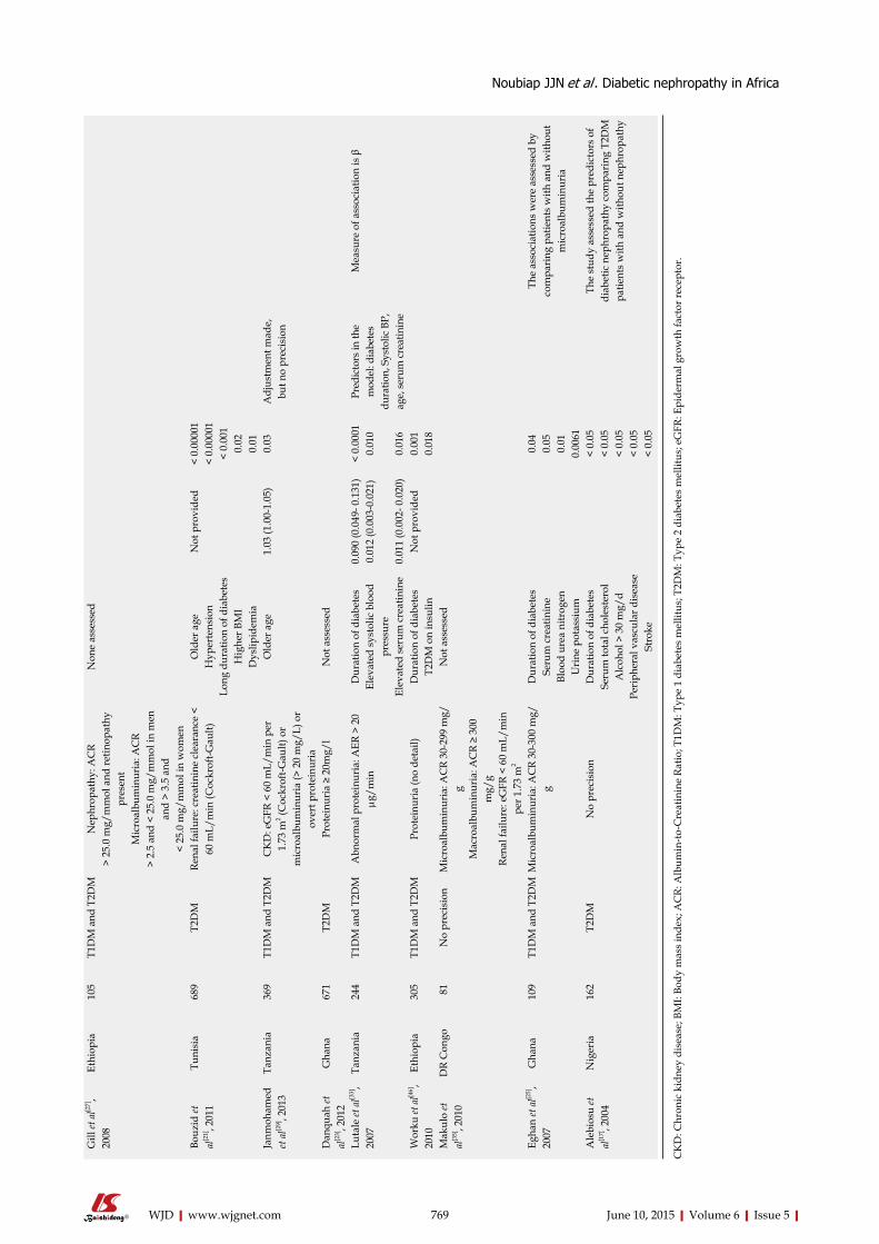
769 June 10, 2015|Volume 6|Issue 5|WJD|www.wjgnet.com
Gill
et a
l[27],
2008
Et
hiop
ia10
5T1
DM
and
T2D
MN
ephr
opat
hy: A
CR
> 25
.0 m
g/m
mol
and
retin
opat
hy
pres
ent
Mic
roal
bum
inur
ia: A
CR
> 2.
5 an
d <
25.0
mg/
mm
ol in
men
an
d >
3.5
and
< 25
.0 m
g/m
mol
in w
omen
Non
e as
sess
ed
Bouz
id et
al
[21], 2
011
Tuni
sia
689
T2D
MRe
nal f
ailu
re: c
reat
inin
e cl
eara
nce
< 60
mL/
min
(Coc
krof
t-Gau
lt)O
lder
age
Not
pro
vide
d<
0.00
001
Hyp
erte
nsio
n<
0.00
001
Long
dur
atio
n of
dia
bete
s<
0.00
1H
ighe
r BM
I0.
02D
yslip
idem
ia0.
01Ja
nmoh
amed
et
al[2
9], 2
013
Tanz
ania
369
T1D
M a
nd T
2DM
CK
D: e
GFR
< 6
0 m
L/m
in p
er
1.73
m2 (C
ockr
oft-G
ault)
or
mic
roal
bum
inur
ia (>
20
mg/
L) o
r ov
ert p
rote
inur
ia
Old
er a
ge1.
03 (1
.00-
1.05
)0.
03A
djus
tmen
t mad
e,
but n
o pr
ecis
ion
Dan
quah
et
al[2
3], 2
012
Gha
na67
1T2
DM
Prot
einu
ria
≥ 20
mg/
lN
ot a
sses
sed
Luta
le et
al[3
3],
2007
Ta
nzan
ia24
4T1
DM
and
T2D
MA
bnor
mal
pro
tein
uria
: AER
> 2
0 μg
/min
Dur
atio
n of
dia
bete
s0.
090
(0.0
49- 0
.131
)<
0.00
01Pr
edic
tors
in th
e m
odel
: dia
bete
s du
ratio
n, S
ysto
lic B
P,
age,
seru
m c
reat
inin
e
Mea
sure
of a
ssoc
iatio
n is
βEl
evat
ed s
ysto
lic b
lood
pr
essu
re0.
012
(0.0
03-0
.021
)0.
010
Elev
ated
ser
um c
reat
inin
e0.
011
(0.0
02- 0
.020
)0.
016
Wor
ku et
al[4
6],
2010
Et
hiop
ia30
5T1
DM
and
T2D
M
Prot
einu
ria
(no
deta
il)D
urat
ion
of d
iabe
tes
Not
pro
vide
d0.
001
T2D
M o
n in
sulin
0.01
8M
akul
o et
al
[35], 2
010
DR
Con
go 8
1N
o pr
ecis
ion
Mic
roal
bum
inur
ia: A
CR
30-2
99 m
g/g
Mac
roal
bum
inur
ia: A
CR
≥ 30
0 m
g/g
Rena
l fai
lure
: eG
FR <
60
mL/
min
pe
r 1.7
3 m
2
Not
ass
esse
d
Egha
n et
al[2
5],
2007
G
hana
109
T1D
M a
nd T
2DM
Mic
roal
bum
inur
ia: A
CR
30-3
00 m
g/g
Dur
atio
n of
dia
bete
s0.
04Th
e as
soci
atio
ns w
ere
asse
ssed
by
com
pari
ng p
atie
nts
with
and
with
out
mic
roal
bum
inur
iaSe
rum
cre
atin
ine
0.05
Bloo
d ur
ea n
itrog
en0.
01U
rine
pot
assi
um0.
0061
Ale
bios
u et
al
[17], 2
004
Nig
eria
162
T2D
MN
o pr
ecis
ion
Dur
atio
n of
dia
bete
s<
0.05
The
stud
y as
sess
ed th
e pr
edic
tors
of
diab
etic
nep
hrop
athy
com
pari
ng T
2DM
pa
tient
s w
ith a
nd w
ithou
t nep
hrop
athy
Seru
m to
tal c
hole
ster
ol<
0.05
Alc
ohol
> 3
0 m
g/d
< 0.
05Pe
riph
eral
vas
cula
r dis
ease
< 0.
05St
roke
< 0.
05
CK
D: C
hron
ic k
idne
y di
seas
e; B
MI:
Body
mas
s in
dex;
AC
R: A
lbum
in-to
-Cre
atin
ine
Ratio
; T1D
M: T
ype
1 di
abet
es m
ellit
us; T
2DM
: Typ
e 2
diab
etes
mel
litus
; eG
FR: E
pide
rmal
gro
wth
fact
or re
cept
or.
Noubiap JJN et al . Diabetic nephropathy in Africa
more frequent in population with diabetes within Africa than in developed countries. The review by Stanifer et al[47] also identified many challenges and limitations, which largely apply to the current study.
The most important aspect in assessing incidence and prevalence of diabetic nephropathy in Africa is currently different diagnostic criteria for CKD. There are no clear definitions on DN. The 2012 KDIGO CKD classification assesses diabetes related kidney changes according to urinary albumin-to-creatinine ratio based on early morning spot urine samples[48]. Quantification of proteinuria in assessing CKD is controversial as no optimal test exists. The National Institute for Health and Clinical Excellence (NICE) guidance has recommended that an early morning urinary ACR should be preferred to other tests of proteinuria, because ACR offers greater sensitivity for the detecting lower, but clinically significant, levels of proteinuria[49]. Almost all the studies included in our review utilized urine tests to diagnose CKD, but only nine studies used ACR. Inconsistencies in the way and manner of reaching a diagnosis of DN in Africans are explained at least in part by issues relating to availability and accessibility of screening or diagnostic tools. Swanepoel et al[50] have reviewed in detail some of the problems associated with nephrology in Africa and discussed the role of lack of amenities in diagnosing renal diseases. Another challenge to making the diagnosis of diabetic nephropathy in Africa is the degree to which other causes of chronic kidney disease have been excluded. A standard armamentarium of tests would include tests looking for HIV, hepatitis B and C, brief collagen screen, syphilis exclusion and other tests would have to be based on history and physical exam.
The classification of CKD is important in the definition of DN and has a few limitations that are universally acknowledged: eGFR underestimates kidney function and there is discordance in the estimates across different estimators[51]; isolated microalbuminuria is a normal feature of aging, inflammation, vascular pathologies, smoking, diet and obesity which are all frequent in diabetes; decline in kidney function is an expected phenomenon with advanced age, just like diabetes risk increases with age. Further considerations to CKD classifications and DN definition limitations is that current guidelines take no notice of the single most important risk factor associated with CKD namely hypertension, which is present in over 50% of people with type 2 diabetes.
Risk factor association was not assessed in 12 of the 32 studies, however common risk factors included were hypertension, raised BMI, HbA1c and duration of diabetes. Despite advances in management over the last three decades, many people with diabetes still develop CKD. This may be partly explained by the poor achievement of blood pressure and blood glucose targets. Recently the JNC 8 guidelines have added to the controversy of various blood pressure targets needed for diabetic patients that would
assist in preventing progression to CKD. Optimal targets when reached, however have shown to aid in progression to progression. Another risk factor pertinent to the developing world is the socioeconomic status of individuals in the causative role of diabetic nephropathy. Weil et al[52], in 2010 reviewed factors associated with disadvantage that may increase the risk of diabetic kidney disease, and the barriers to care that hinder attempts to provide an adequate therapeutic response[52].
Several mechanisms underlying the pathogenesis of diabetic nephropathy have been suggested and include glomerular hyperfiltration; hyperglycemia and the increased production of advanced glycation end products; hypoxia-inflammation and the activation of cytokines. Hyperfiltration commonly occur in early in the course of diabetes and involves glucose-dependent dilation of the afferent arteriolar dilation, and the enhanced filtration area secondary to the increase in the number of mesangial cells and capillary loops. Molecular level action involves vasoactive mediators like insulin-like growth factor 1, transforming growth factor beta, nitric oxide, prostaglandin, glucagon and vascular endothelial growth factor[53]. Other hallmarks of diabetic nephropathy include nodular diabetic glomerulosclerosis and diffuse glomerulosclerosis, mediated at least in part by inflammatory processes and immune cells activity[53]. Interstitial fibrosis and tubular atrophy are also seen early in DN, with the underlying pathogenetic mechanism being similar to those in progressive non diabetic renal disease[54].
Diabetic nephropathy ultimately occurs only in susceptible individuals with diabetes; which suscep-tibility is determined by the combined effect of gene-tic predisposition and non-genetic factors. Genetic susceptibility to diabetic nephropathy is by nature polygenetic. Whole-genome scanning studies have identified several chromosomal regions linked with diabetic nephropathy; however, the pathophysiologic function of such genetic regions has yet to be fully elucidated. Genetic polymorphisms may explain the familial clustering of diabetic nephropathy[55]. Some studies have suggested some detrimental effect of the double-deletion (DD) polymorphism of the angiotensin-converting enzyme (ACE) genotype on disease progression[56]. Non-genetic determinants of diabetic nephropathy include among others socioeconomic factors, dietary factors, poor hyperglycemic control, hypertension, obesity and early life factors[57,58]. Hypertension appears to be a strong correlate of disease progression in Black people[59,60].
The current review has some limitations. Included studies were mostly based on small samples, with different study designs and most of the studies were cross sectional with only two being retrospective cohorts and one case-control. A large proportion were based in urban clinics with and most of the populations studied were that attending a general diabetic clinic and the results may not be generalizable
770 June 10, 2015|Volume 6|Issue 5|WJD|www.wjgnet.com
Noubiap JJN et al . Diabetic nephropathy in Africa

to primary care populations. Ideally chronic kidney disease should not be diagnosed on the basis of single measurements of serum creatinine and albuminuria, and standard baseline investigations are needed to exclude other causative kidney disease, although there is precedence for this in other studies in the West as well. Finally, detection of microalbuminuria was one the most frequent method to assess the presence of diabetic nephropathy. As microalbuminuria is more a quantitative estimate of endothelial/vascular dysfunction than of diabetic nephropathy, the incidence and prevalence rate of diabetic nephropathy have probably been overestimated when assessing kidney function by urine protein.
In conclusion, the current review gives a small glimpse of the larger numbers of CKD in diabetics in Africa compared to Western society. CKD is a substantial health burden among diabetic patients on the African continent, with prevalence varying from 11% to 83.7% depending on the method of assessment. Estimates suggest that 95% of diabetics may have proteinuria after a 10 years duration of diabetes, about 35% may have an end-stage renal disease after 5 years and 18% die from nephropathy after 20 years of disease duration. Risk factors of CKD include mainly hypertension, obesity, poor glycemic control and disease duration. Better surveillance of diabetes is a necessary first step toward its prevention and control, which is now recognized as an urgent priority. An electronic database in African regions would be ideal to assist in this entity although it is presumed that we are light years away from that. At a primary care level it is very plausible that with early detection, proper screening, and management, the impact of diabetic nephropathy may be better mitigated to lessen its impact on society and healthcare.
COMMENTSBackgroundAfrican countries are experiencing an epidemics of diabetes mellitus. Diabetic nephropathy is one the most frequent complications of diabetes mellitus. Several studies on the epidemiology of diabetic nephropathy have been conducted in Africa, but there is no previous published work which synthesizes evidences from this study to provide an overview of the disease on the continent. Research frontiersEpidemiological data on diabetic nephropathy in Africa are sparse. These data are important to quantify the magnitude of the disease and assist the formulation of strategies to reduce the impact of nephropathy on people with diabetes in Africa.Innovations and breakthroughsThis review is the first to synthesize relevant data on diabetic nephropathy in Africa. The authors performed extensive electronic and manual bibliographic searches to determine the prevalence and incidence of diabetic nephropathy on the continent. Although the quality of data was not optimal, estimates suggest that the prevalence of diabetic nephropathy vary between 11%-83.7%. About one third of diabetic patients have end-stage renal disease after 5 years and about one fifth die from nephropathy after 20 years of disease duration. Hypertension, obesity, poor glycemic control and disease duration are the main risk factors of chronic kidney disease among diabetic patients in Africa.
ApplicationsThis review shows that the burden of chronic kidney disease is important among people with diabetes in Africa. The findings will have implications for policy, practice and future research on diabetic nephropathy on the continent.TerminologyDiabetic nephropathy is an alteration of the function of the kidneys due to diabetes mellitus. It is associated with substantial morbidity and mortality.Peer-reviewThe authors of the present manuscript performed extensive electronic and manual bibliographic research to determine the prevalence and incidence of kidney disease in people with diabetes mellitus within countries in Africa. Overall the review is well written.
REFERENCES1 Zimmet P, Alberti KG, Shaw J. Global and societal implications
of the diabetes epidemic. Nature 2001; 414: 782-787 [PMID: 11742409 DOI: 10.1038/414782a]
2 Aguiree F, Brown A, Cho NH, Dahlquist G, Dodd S, Dunning T, Hirst M, Hwang C, Magliano D, Patterson C, Scott C, Shaw J, Soltesz G, Usher-Smith J, Whiting D. IDF Diabetes Atlas: sixth edition. 6th ed. Basel, Switzerland: International Diabetes Federation, 2013
3 Seshasai SR, Kaptoge S, Thompson A, Di Angelantonio E, Gao P, Sarwar N, Whincup PH, Mukamal KJ, Gillum RF, Holme I, Njølstad I, Fletcher A, Nilsson P, Lewington S, Collins R, Gudnason V, Thompson SG, Sattar N, Selvin E, Hu FB, Danesh J. Diabetes mellitus, fasting glucose, and risk of cause-specific death. N Engl J Med 2011; 364: 829-841 [PMID: 21366474 DOI: 10.1056/NEJMoa1008862]
4 Fox CS, Matsushita K, Woodward M, Bilo HJ, Chalmers J, Heerspink HJ, Lee BJ, Perkins RM, Rossing P, Sairenchi T, Tonelli M, Vassalotti JA, Yamagishi K, Coresh J, de Jong PE, Wen CP, Nelson RG. Associations of kidney disease measures with mortality and end-stage renal disease in individuals with and without diabetes: a meta-analysis. Lancet 2012; 380: 1662-1673 [PMID: 23013602 DOI: 10.1016/S0140-6736(12)61350-6]
5 Jeon CY, Murray MB. Diabetes mellitus increases the risk of active tuberculosis: a systematic review of 13 observational studies. PLoS Med 2008; 5: e152 [PMID: 18630984 DOI: 10.1371/journal.pmed.0050152]
6 Muller LM, Gorter KJ, Hak E, Goudzwaard WL, Schellevis FG, Hoepelman AI, Rutten GE. Increased risk of common infections in patients with type 1 and type 2 diabetes mellitus. Clin Infect Dis 2005; 41: 281-288 [PMID: 16007521 DOI: 10.1086/431587]
7 Kengne AP, Echouffo-Tcheugui JB, Sobngwi E, Mbanya JC. New insights on diabetes mellitus and obesity in Africa-part 1: prevalence, pathogenesis and comorbidities. Heart 2013; 99: 979-983 [PMID: 23680891 DOI: 10.1136/heartjnl-2012-303316]
8 Harjutsalo V, Groop PH. Epidemiology and risk factors for diabetic kidney disease. Adv Chronic Kidney Dis 2014; 21: 260-266 [PMID: 24780453 DOI: 10.1053/j.ackd.2014.03.009]
9 Assogba GF, Couchoud C, Roudier C, Pornet C, Fosse S, Romon I, Druet C, Stengel B, Fagot-Campagna A. Prevalence, screening and treatment of chronic kidney disease in people with type 2 diabetes in France: the ENTRED surveys (2001 and 2007). Diabetes Metab 2012; 38: 558-566 [PMID: 23036461 DOI: 10.1016/j.diabet.2012.08.004]
10 Bakris GL. Recognition, pathogenesis, and treatment of different stages of nephropathy in patients with type 2 diabetes mellitus. Mayo Clin Proc 2011; 86: 444-456 [PMID: 21531886 DOI: 10.4065/mcp.2010.0713]
11 Thomas MC, Weekes AJ, Broadley OJ, Cooper ME, Mathew TH. The burden of chronic kidney disease in Australian patients with type 2 diabetes (the NEFRON study). Med J Aust 2006; 185: 140-144 [PMID: 16893353]
12 Kengne AP, Sobngwi E, Echouffo-Tcheugui JB, Mbanya JC. New insights on diabetes mellitus and obesity in Africa-Part 2:
771 June 10, 2015|Volume 6|Issue 5|WJD|www.wjgnet.com
COMMENTS
Noubiap JJN et al . Diabetic nephropathy in Africa
prevention, screening and economic burden. Heart 2013; 99: 1072-1077 [PMID: 23680890 DOI: 10.1136/heartjnl-2013-303773]
13 Mbanya JC, Motala AA, Sobngwi E, Assah FK, Enoru ST. Diabetes in sub-Saharan Africa. Lancet 2010; 375: 2254-2266 [PMID: 20609971 DOI: 10.1016/S0140-6736(10)60550-8]
14 Stroup DF, Berlin JA, Morton SC, Olkin I, Williamson GD, Rennie D, Moher D, Becker BJ, Sipe TA, Thacker SB. Meta-analysis of observational studies in epidemiology: a proposal for reporting. Meta-analysis Of Observational Studies in Epidemiology (MOOSE) group. JAMA 2000; 283: 2008-2012 [PMID: 10789670]
15 Ajayi S, Mamven M, Ojji D. eGFR and chronic kidney disease stages among newly diagnosed asymptomatic hypertensives and diabetics seen in a tertiary health center in Nigeria. Ethn Dis 2014; 24: 220-225 [PMID: 24804370]
16 Alebiosu CO. Clinical diabetic nephropathy in a tropical African population. West Afr J Med 2003; 22: 152-155 [PMID: 14529227]
17 Alebiosu CO , Odusan O, Familoni OB, Jaiyesimi AE. Cardiovascular risk factors in type 2 diabetic Nigerians with clinical diabetic nephropathy. Cardiovasc J S Afr 2004; 15: 124-128 [PMID: 15258622]
18 Alebiosu CO, Odusan O, Jaiyesimi A. Morbidity in relation to stage of diabetic nephropathy in type-2 diabetic patients. J Natl Med Assoc 2003; 95: 1042-1047 [PMID: 14651370]
19 Bentata Y, Haddiya I, Latrech H, Serraj K, Abouqal R. Progression of diabetic nephropathy, risk of end-stage renal disease and mortality in patients with type-1 diabetes. Saudi J Kidney Dis Transpl 2013; 24: 392-402 [PMID: 23538374]
20 Bouaziz A, Zidi I, Zidi N, Mnif W, Zinelabidine HT. Nephropathy following type 2 diabetes mellitus in Tunisian population. West Indian Med J 2012; 61: 881-889 [PMID: 24020228]
21 Bouzid C, Smida H, Kacem A, Turki Z, Ben Salem L, Ben Rayana C, Slama BC. [Renal failure in Tunisian patients with type 2 diabetes: frequency and related factors]. Tunis Med 2011; 89: 10-15 [PMID: 21267820]
22 Choukem SP, Dzudie A, Dehayem M, Halle MP, Doualla MS, Luma H, Kengne AP. Comparison of different blood pressure indices for the prediction of prevalent diabetic nephropathy in a sub-Saharan African population with type 2 diabetes. Pan Afr Med J 2012; 11: 67 [PMID: 22655101]
23 Danquah I, Bedu-Addo G, Terpe KJ, Micah F, Amoako YA, Awuku YA, Dietz E, van der Giet M, Spranger J, Mockenhaupt FP. Diabetes mellitus type 2 in urban Ghana: characteristics and associated factors. BMC Public Health 2012; 12: 210 [PMID: 22429713 DOI: 10.1186/1471-2458-12-210]
24 Djrolo F, Attolou VG, Avode DG, Houngbe F, Akpona S, Addra B, Kodjoh N. [Diabetic nephropathy: an epidemiological study based on proteinuria in a population of black African diabetics in Cotonou, Benin]. Sante 2001; 11: 105-109 [PMID: 11440886]
25 Eghan BA, Frempong MT, Adjei-Poku M. Prevalence and predictors of microalbuminuria in patients with diabetes mellitus: a cross-sectional observational study in Kumasi, Ghana. Ethn Dis 2007; 17: 726-730 [PMID: 18072386]
26 Elbagir MN, Eltom MA, Mahadi EO, Berne C. Pattern of long-term complications in Sudanese insulin-treated diabetic patients. Diabetes Res Clin Pract 1995; 30: 59-67 [PMID: 8745207]
27 Gill G, Gebrekidan A, English P, Wile D, Tesfaye S. Diabetic complications and glycaemic control in remote North Africa. QJM 2008; 101: 793-798 [PMID: 18687702 DOI: 10.1093/qjmed/hcn096]
28 Gill GV, Huddle KR, Monkoe G. Long-term (20 years) outcome and mortality of Type 1 diabetic patients in Soweto, South Africa. Diabet Med 2005; 22: 1642-1646 [PMID: 16401306 DOI: 10.1111/j.1464-5491.2005.01712.x]
29 Janmohamed MN, Kalluvya SE, Mueller A, Kabangila R, Smart LR, Downs JA, Peck RN. Prevalence of chronic kidney disease in diabetic adult out-patients in Tanzania. BMC Nephrol 2013; 14: 183 [PMID: 24228774 DOI: 10.1186/1471-2369-14-183]
30 Katchunga P, Hermans MP, Manwa B, Lepira F, Kashongwe Z, M’Buyamba-Kabangu JR. [Hypertension, insulin resistance and chronic kidney disease in type 2 diabetes patients from South Kivu,
DR Congo]. Nephrol Ther 2010; 6: 520-525 [PMID: 20605543 DOI: 10.1016/j.nephro.2010.04.002]
31 Keeton GR, Smit Rv, Bryer A. Renal outcome of type 2 diabetes in South Africa--a 12-year follow-up study. S Afr Med J 2004; 94: 771-775 [PMID: 15487844]
32 Levitt NS, Bradshaw D, Zwarenstein MF, Bawa AA, Maphumolo S. Audit of public sector primary diabetes care in Cape Town, South Africa: high prevalence of complications, uncontrolled hyperglycaemia, and hypertension. Diabet Med 1997; 14: 1073-1077 [PMID: 9455936 DOI: 10.1002/(sici)1096-9136(199712)14]
33 Lutale JJ, Thordarson H, Abbas ZG, Vetvik K. Microalbuminuria among Type 1 and Type 2 diabetic patients of African origin in Dar Es Salaam, Tanzania. BMC Nephrol 2007; 8: 2 [PMID: 17224056 DOI: 10.1186/1471-2369-8-2]
34 Majaliwa ES, Munubhi E, Ramaiya K, Mpembeni R, Sanyiwa A, Mohn A, Chiarelli F. Survey on acute and chronic complications in children and adolescents with type 1 diabetes at Muhimbili National Hospital in Dar es Salaam, Tanzania. Diabetes Care 2007; 30: 2187-2192 [PMID: 17563337 DOI: 10.2337/dc07-0594]
35 Makulo R, Nseka MN, Jadoul M, Mvitu M, Muyer MT, Kimenyembo W, Mandja M, Bieleli E, Mapatano MA, Epira FB, Sumaili EK, Kaimbo WK, Nge O, Buntinx F, Muls E. [Albuminuria during the screening for diabetes in a semi-rural area (Kisantu City, DR Congo)]. Nephrol Ther 2010; 6: 513-519 [PMID: 20627763 DOI: 10.1016/j.nephro.2010.04.006]
36 Marshall SL, Edidin D, Sharma V, Ogle G, Arena VC, Orchard T. Current clinical status, glucose control, and complication rates of children and youth with type 1 diabetes in Rwanda. Pediatr Diabetes 2013; 14: 217-226 [PMID: 23279222 DOI: 10.1111/pedi.12007]
37 Motala AA, Pirie FJ, Gouws E, Amod A, Omar MA. Microvascular complications in South African patients with long-duration diabetes mellitus. S Afr Med J 2001; 91: 987-992 [PMID: 11847923]
38 Nambuya AP, Otim MA, Whitehead H, Mulvany D, Kennedy R, Hadden DR. The presentation of newly-diagnosed diabetic patients in Uganda. QJM 1996; 89: 705-711 [PMID: 8917747]
39 Pruijm MT, Madeleine G, Riesen WF, Burnier M, Bovet P. Prevalence of microalbuminuria in the general population of Seychelles and strong association with diabetes and hypertension independent of renal markers. J Hypertens 2008; 26: 871-877 [PMID: 18398328 DOI: 10.1097/HJH.0b013e3282f624d9]
40 Rahlenbeck SI, Gebre-Yohannes A. Prevalence and epidemiology of micro- and macroalbuminuria in Ethiopian diabetic patients. J Diabetes Complications 1997; 11: 343-349 [PMID: 9365876]
41 Rasmussen JB, Thomsen JA, Rossing P, Parkinson S, Christensen DL, Bygbjerg IC. Diabetes mellitus, hypertension and albuminuria in rural Zambia: a hospital-based survey. Trop Med Int Health 2013; 18: 1080-1084 [PMID: 23763632 DOI: 10.1111/tmi.12139]
42 Rissassi JR, Nseka M, Jadoul M, Lepira FB, Mvitu M, Mbenza G, Yekoladio D, Aloni M, Nge OO. [Prevalence and determinants of microalbuminuria and macroalbuminuria in children and young adults with type 1 diabetes in Kinshasa]. Nephrol Ther 2010; 6: 40-46 [PMID: 19853548 DOI: 10.1016/j.nephro.2009.08.001]
43 Rotchford AP, Rotchford KM. Diabetes in rural South Africa--an assessment of care and complications. S Afr Med J 2002; 92: 536-541 [PMID: 12197196]
44 Sobngwi E, Mbanya JC, Moukouri EN, Ngu KB. Microalbuminuria and retinopathy in a diabetic population of Cameroon. Diabetes Res Clin Pract 1999; 44: 191-196 [PMID: 10462142]
45 Wanjohi FW, Otieno FC, Ogola EN, Amayo EO. Nephropathy in patients with recently diagnosed type 2 diabetes mellitus in black Africans. East Afr Med J 2002; 79: 399-404 [PMID: 12638839]
46 Worku D, Hamza L, Woldemichael K. Patterns of diabetic complications at jimma university specialized hospital, southwest ethiopia. Ethiop J Health Sci 2010; 20: 33-39 [PMID: 22434958]
47 Stanifer JW, Jing B, Tolan S, Helmke N, Mukerjee R, Naicker S, Patel U. The epidemiology of chronic kidney disease in sub-Saharan Africa: a systematic review and meta-analysis. Lancet Glob Health 2014; 2: e174-e181 [PMID: 25102850 DOI: 10.1016/S2214-109X(14)70002-6]
48 Levin A, Stevens PE. Summary of KDIGO 2012 CKD Guideline:
772 June 10, 2015|Volume 6|Issue 5|WJD|www.wjgnet.com
Noubiap JJN et al . Diabetic nephropathy in Africa
behind the scenes, need for guidance, and a framework for moving forward. Kidney Int 2014; 85: 49-61 [PMID: 24284513 DOI: 10.1038/ki.2013.444]
49 National Collaborating Centre for Chronic Conditions. Chronic kidney disease: Early identification and management of chronic kidney disease in adults in primary and secondary care. London: NICE, 2008. Available from: URL: http://www.nice.org.uk/nicemedia/live/12069/42117/42117.pdf
50 Swanepoel CR, Wearne N, Okpechi IG. Nephrology in Africa--not yet uhuru. Nat Rev Nephrol 2013; 9: 610-622 [PMID: 23958719 DOI: 10.1038/nrneph.2013.168]
51 Matsha TE, Yako YY, Rensburg MA, Hassan MS, Kengne AP, Erasmus RT. Chronic kidney diseases in mixed ancestry south African populations: prevalence, determinants and concordance between kidney function estimators. BMC Nephrol 2013; 14: 75 [PMID: 23547953 DOI: 10.1186/1471-2369-14-75]
52 Weil EJ, Curtis JM, Hanson RL, Knowler WC, Nelson RG. The impact of disadvantage on the development and progression of diabetic kidney disease. Clin Nephrol 2010; 74 Suppl 1: S32-S38 [PMID: 20979961]
53 Ruster C, Wolf G. The role of chemokines and chemokine receptors in diabetic nephropathy. Front Biosci 2008; 13: 944-955 [PMID: 17981602]
54 Iwano M, Plieth D, Danoff TM, Xue C, Okada H, Neilson EG. Evidence that fibroblasts derive from epithelium during tissue fibrosis. J Clin Invest 2002; 110: 341-350 [PMID: 12163453 DOI: 10.1172/JCI15518]
55 Seaquist ER, Goetz FC, Rich S, Barbosa J. Familial clustering of diabetic kidney disease. Evidence for genetic susceptibility to diabetic nephropathy. N Engl J Med 1989; 320: 1161-1165 [PMID: 2710189 DOI: 10.1056/NEJM198905043201801]
56 Marre M, Jeunemaitre X, Gallois Y, Rodier M, Chatellier G, Sert C, Dusselier L, Kahal Z, Chaillous L, Halimi S, Muller A, Sackmann H, Bauduceau B, Bled F, Passa P, Alhenc-Gelas F. Contribution of genetic polymorphism in the renin-angiotensin system to the development of renal complications in insulin-dependent diabetes: Genetique de la Nephropathie Diabetique (GENEDIAB) study group. J Clin Invest 1997; 99: 1585-1595 [PMID: 9120002 DOI: 10.1172/JCI119321]
57 Nelson RG, Morgenstern H, Bennett PH. Intrauterine diabetes exposure and the risk of renal disease in diabetic Pima Indians. Diabetes 1998; 47: 1489-1493 [PMID: 9726239]
58 Zandi-Nejad K, Luyckx VA, Brenner BM. Adult hypertension and kidney disease: the role of fetal programming. Hypertension 2006; 47: 502-508 [PMID: 16415374 DOI: 10.1161/01.HYP.0000198544.099-09.1a]
59 Brancati FL, Whittle JC, Whelton PK, Seidler AJ, Klag MJ. The excess incidence of diabetic end-stage renal disease among blacks. A population-based study of potential explanatory factors. JAMA 1992; 268: 3079-3084 [PMID: 1433738]
60 Chaiken RL, Palmisano J, Norton ME, Banerji MA, Bard M, Sachimechi I, Behzadi H, Lebovitz HE. Interaction of hypertension and diabetes on renal function in black NIDDM subjects. Kidney Int 1995; 47: 1697-1702 [PMID: 7643539]
P- Reviewer: de Oliveira JMF, Laghmani K S- Editor: Ji FF L- Editor: A E- Editor: Zhang DN
773 June 10, 2015|Volume 6|Issue 5|WJD|www.wjgnet.com
Noubiap JJN et al . Diabetic nephropathy in Africa

© 2015 Baishideng Publishing Group Inc. All rights reserved.
Published by Baishideng Publishing Group Inc8226 Regency Drive, Pleasanton, CA 94588, USA
Telephone: +1-925-223-8242Fax: +1-925-223-8243
E-mail: [email protected] Desk: http://www.wjgnet.com/esps/helpdesk.aspx
http://www.wjgnet.com
Copyright © 2022 FDOKUMEN