Clinical Pearls: Hepatology - American College of Physicians
Upload
khangminh22Category
view
0download
0
World Journal of HepatologyWorld J Hepatol 2015 October 18; 7(23): 2427-2496
Published by Baishideng Publishing Group Inc
ISSN 1948-5182 (online)

EDITORS-IN-CHIEFClara Balsano, RomeWan-Long Chuang, Kaohsiung
GUEST EDITORIAL BOARD MEMBERSKing-Wah Chiu, KaohsiungTai-An Chiang, TainanChi-Tan Hu, HualienSen-Yung Hsieh, TaoyuanWenya Huang, TainanLiang-Yi Hung, TainanJih RU Hwu, HsinchuJing-Yi Lee, TaipeiMei-Hsuan Lee, TaipeiChih-Wen Lin, KaohsiungChun-Che Lin, TaichungWan-Yu Lin, TaichungTai-Long Pan, Tao-YuanSuh-Ching Yang, TaipeiChun-Yan Yeung, Taipei
MEMBERS OF THE EDITORIAL BOARD
Algeria
Samir Rouabhia, Batna
Argentina
Fernando O Bessone, RosarioMaria C Carrillo, RosarioMelisa M Dirchwolf, Buenos AiresBernardo Frider, Buenos Aires
Jorge Quarleri, Buenos AiresAdriana M Torres, Rosario
Armenia
Narina Sargsyants, Yerevan
Australia
Mark D Gorrell, Sydney
Austria
Harald Hofer, ViennaGustav Paumgartner, ViennaMatthias Pinter, ViennaThomas Reiberger, Vienna
Bangladesh
Shahinul Alam, DhakaMamun Al Mahtab, Dhaka
Belgium
Nicolas Lanthier, BrusselsPhilip Meuleman, GhentLuisa Vonghia, Antwerp
Botswana
Francesca Cainelli, Gaborone
Sandro Vento, Gaborone
Brazil
Edson Abdala, Sao PauloIlka FSF Boin, CampinasNiels OS Camara, Sao PauloAna Carolina FN Cardoso, Rio de JaneiroRoberto J Carvalho-Filho, Sao PauloJulio CU Coelho, CuritibaFlavio Henrique Ferreira Galvao, São PauloJanaina L Narciso-Schiavon, FlorianopolisSílvia HC Sales-Peres, BauruLeonardo L Schiavon, FlorianópolisLuciana D Silva, Belo HorizonteVanessa Souza-Mello, Rio de JaneiroJaques Waisberg, Santo André
Bulgaria
Mariana P Penkova-Radicheva, Stara ZagoraMarieta Simonova, Sofia
Canada
Runjan Chetty, TorontoMichele Molinari, HalifaxGiada Sebastiani, Montreal
Chile
Luis A Videla, Santiago
I
Editorial Board2014-2017
The World Journal of Hepatology Editorial Board consists of 469 members, representing a team of worldwide experts in hepatology. They are from 53 countries, including Algeria (1), Argentina (6), Armenia (1), Australia (1), Austria (4), Bangladesh (2), Belgium (3), Botswana (2), Brazil (13), Bulgaria (2), Canada (3), Chile (1), China (98), Czech Repoublic (1), Denmark (2), Egypt (12), France (6), Germany (19), Greece (11), Hungary (5), India (15), Indonesia (2), Iran (4), Israel (1), Italy (52), Japan (35), Jordan (1), Malaysia (2), Mexico (3), Moldova (1), Netherlands (3), Nigeria (1), Pakistan (1), Philippines (2), Poland (1), Portugal (2), Qatar (1), Romania (6), Russia (2), Saudi Arabia (4), Singapore (1), South Korea (11), Spain (20), Sri Lanka (1), Sudan (1), Sweden (1), Switzerland (1), Thailand (4), Turkey (21), Ukraine (3), United Kingdom (17), and United States (56).
January 27, 2014WJH|www.wjgnet.com
World Journal of HepatologyW J H

ChinaGuang-Wen Cao, ShanghaiEn-Qiang Chen, ChengduGong-Ying Chen, HangzhouJin-lian Chen, ShanghaiJun Chen, ChangshaAlfred Cheng, Hong KongChun-Ping Cui, BeijingShuang-Suo Dang, Xi’an Ming-Xing Ding, JinhuaZhi-Jun Duang, DalianHe-Bin Fan, WuhanXiao-Ming Fan, ShanghaiJames Yan Yue Fung, Hong Kong Yi Gao, GuangzhouZuo-Jiong Gong, WuhanZhi-Yong Guo, GuangzhouShao-Liang Han, WenzhouTao Han, TianjinJin-Yang He, GuangzhouMing-Liang He, Hong KongCan-Hua Huang, ChengduBo Jin, BeijingShan Jin, Hohhot Hui-Qing Jiang, ShijiazhuangWan-Yee Joseph Lau, Hong KongGuo-Lin Li, ChangshaJin-Jun Li, ShanghaiQiang Li, JinanSheng Li, JinanZong-Fang Li, Xi'anXu Li, Guangzhou Xue-Song Liang, Shanghai En-Qi Liu, Xi‘anPei Liu, ShenyangZhong-Hui Liu, ChangchunGuang-Hua Luo, ChangzhouYi Lv, Xi'anGuang-Dong Pan, LiuzhouWen-Sheng Pan, HangzhouJian-Min Qin, Shanghai Wai-Kay Seto, Hong KongHong Shen, ChangshaXiao Su, ShanghaiLi-Ping Sun, BeijingWei-Hao Sun, NanjingXue-Ying Sun, HarbinHua Tang, TianjinLing Tian, ShanghaiEric Tse, Hong KongGuo-Ying Wang, ChangzhouYue Wang, BeijingShu-Qiang Wang, ChengduMary MY Waye, Hong KongHong-Shan Wei, BeijingDanny Ka-Ho Wong, Hong KongGrace Lai-Hung Wong, Hong KongBang-Fu Wu, DongguanFeng Wu, ChongqingXiong-Zhi Wu, Tianjin Chun-Fang Xu, SuzhouRui-An Xu, QuanzhouRui-Yun Xu, GuangzhouWei-Li Xu, ShijiazhuangShi-Ying Xuan, Qingdao Ming-Xian Yan, JinanLv-Nan Yan, ChengduJin Yang, HangzhouJi-Hong Yao, DalianWinnie Yeo, Hong Kong
Zheng Zeng, BeijingQi Zhang, HangzhouShi-Jun Zhang, GuangzhouXiao-Lan Zhang, ShijiazhuangXiao-Yong Zhang, GuangzhouXin-Chen Zhang, HarbinYong Zhang, Xi'anHong-Chuan Zhao, HefeiMing-Hua Zheng, WenzhouYu-Bao Zheng, GuangzhouRen-Qian Zhong, ShanghaiFan Zhu, WuhanXiao Zhu, Dongguan
Czech Repoublic
Kamil Vyslouzil, Olomouc
Denmark
Henning Gronbaek, AarhusChristian Mortensen, Hvidovre
Egypt
Ihab T Abdel-Raheem, DamanhourNGB G Bader EL Din, CairoHatem Elalfy, MansouraMahmoud M El-Bendary, MansouraMona El SH El-Raziky, CairoMohammad El-Sayed, CairoYasser M Fouad, MiniaMohamed AA Metwally, BenhaHany Shehab, CairoMostafa M Sira, Shebin El-koomAshraf Taye, MiniaMA Ali Wahab, Mansoura
France
Laurent Alric, ToulouseSophie Conchon, NantesDaniel J Felmlee, StrasbourgHerve Lerat, CreteilDominique Salmon, ParisJean-Pierre Vartanian, Paris
Germany
Laura E Buitrago-Molina, HannoverEnrico N De Toni, MunichOliver Ebert, MuenchenRolf Gebhardt, LeipzigJanine V Hartl, RegensburgSebastian Hinz, KielBenjamin Juntermanns, EssenRoland Kaufmann, JenaViola Knop, FrankfurtVeronika Lukacs-Kornek, HomburgBenjamin Maasoumy, HannoverJochen Mattner, ErlangenNadja M Meindl-Beinker, MannheimUlf P Neumann, AachenMargarete Odenthal, CologneYoshiaki Sunami, Munich
Christoph Roderburg, AachenFrank Tacke, AachenYuchen Xia, Munich
Greece
Alex P Betrosian, AthensGeorge N Dalekos, LarissaIoanna K Delladetsima, AthensNikolaos K Gatselis, LarissaStavros Gourgiotis, AthensChristos G Savopoulos, ThessalonikiTania Siahanidou, AthensEmmanouil Sinakos, ThessalonikiNikolaos G Symeonidi, ThessalonikiKonstantinos C Thomopoulos, LarissaKonstantinos Tziomalos, Thessaloniki
Hungary
Gabor Banhegyi, BudapestPeter L Lakatos, BudapestMaria Papp, DebrecenFerenc Sipos, BudapestZsolt J Tulassay, Budapest
India
Deepak N Amarapurkar, Mumbai Girish M Bhopale, PuneSibnarayan Datta, TezpurNutan D Desai, MumbaiSorabh Kapoor, MumbaiJaswinder S Maras, New DelhiNabeen C Nayak, New DelhiC Ganesh Pai, ManipalAmit Pal, ChandigarhK Rajeshwari, New DelhiAnup Ramachandran, VelloreD Nageshwar Reddy, HyderabadShivaram P Singh, CuttackAjith TA, ThrissurBalasubramaniyan Vairappan, Pondicherry
Indonesia
Cosmas RA Lesmana, JakartaNeneng Ratnasari, Yogyakarta
Iran
Seyed M Jazayeri, TehranSedigheh Kafi-Abad, TehranIradj Maleki, SariFakhraddin Naghibalhossaini, Shiraz
Israel
Stephen DH Malnick, Rehovot
Italy
Francesco Angelico, Rome
II January 27, 2014WJH|www.wjgnet.com

III January 27, 2014WJH|www.wjgnet.com
Alfonso W Avolio, RomeFrancesco Bellanti, FoggiaMarcello Bianchini, ModenaGuglielmo Borgia, NaplesMauro Borzio, MilanoEnrico Brunetti, PaviaValeria Cento, RomaBeatrice Conti, RomeFrancesco D'Amico, PadovaSamuele De Minicis, FermoFabrizio De Ponti, BolognaGiovan Giuseppe Di Costanzo, NapoliLuca Fabris, PadovaGiovanna Ferraioli, PaviaAndrea Galli, FlorenceeMatteo Garcovich, RomeEdoardo G Giannini, GenovaRossano Girometti, UdineAlessandro Granito, BolognaAlberto Grassi, RiminiAlessandro Grasso, SavonaSalvatore Gruttadauria, PalermoFrancesca Guerrieri, RomeQuirino Lai, AquilaAndrea Lisotti, BolognaMarcello F Maida, PalermoLucia Malaguarnera, CataniaAndrea Mancuso, PalermoLuca Maroni, AnconaFrancesco Marotta, MilanoPierluigi Marzuillo, NaplesSara Montagnese, PadovaGiuseppe Nigri, RomeClaudia Piccoli, FoggiaCamillo Porta, PaviaChiara Raggi, Rozzano (MI)Maria Rendina, BariMaria Ripoli, San Giovanni RotondoKryssia I Rodriguez-Castro, PaduaRaffaella Romeo, MilanAmedeo Sciarra, MilanoAntonio Solinas, SassariAurelio Sonzogni, BergamoGiovanni Squadrito, MessinaSalvatore Sutti, NovaraValentina Svicher, RomeLuca Toti, RomeElvira Verduci, MilanUmberto Vespasiani-Gentilucci, RomeMaria A Zocco, Rome
Japan
Yasuhiro Asahina, TokyoNabil AS Eid, TakatsukiKenichi Ikejima, TokyoShoji Ikuo, KobeYoshihiro Ikura, TakatsukiShinichi Ikuta, NishinomiyaKazuaki Inoue, YokohamaToshiya Kamiyama, SapporoTakanobu Kato, TokyoSaiho Ko, NaraHaruki Komatsu, SakuraMasanori Matsuda, Chuo-city Yasunobu Matsuda, NiigataYoshifumi Nakayama, KitakyushuTaichiro Nishikawa, Kyoto
Satoshi Oeda, SagaKenji Okumura, UrayasuMichitaka Ozaki, SapporoTakahiro Sato, SapporoJunichi Shindoh, TokyoRyo Sudo, YokohamaAtsushi Suetsugu, GifuHaruhiko Sugimura, HamamatsuReiji Sugita, SendaiKoichi Takaguchi, TakamatsuShinji Takai, TakatsukiAkinobu Takaki, OkayamaYasuhito Tanaka, NagoyaTakuji Tanaka, Gifu CityAtsunori Tsuchiya, NiigataKoichi Watashi, TokyoHiroshi Yagi, TokyoTaro Yamashita, KanazawaShuhei Yoshida, ChibaHitoshi Yoshiji, Kashihara
Jordan
Kamal E Bani-Hani, Zarqa
Malaysia
Peng Soon Koh, Kuala LumpurYeong Yeh Lee, Kota Bahru
Mexico
Francisco J Bosques-Padilla, MonterreyMaría de F Higuera-de la Tijera, Mexico CityJosé A Morales-Gonzalez, México City
Moldova
Angela Peltec, Chishinev
Netherlands
Wybrich R Cnossen, NijmegenFrank G Schaap, MaastrichtFareeba Sheedfar, Groningen
Nigeria
CA Asabamaka Onyekwere, Lagos
Pakistan
Bikha Ram Devrajani, Jamshoro
Philippines
Janus P Ong, PasigJD Decena Sollano, Manila
Poland
Jacek Zielinski, Gdansk
Portugal
Rui T Marinho, LisboaJoao B Soares, Braga
Qatar
Reem Al Olaby, Doha
Romania
Bogdan Dorobantu, BucharestLiana Gheorghe, BucharestGeorge S Gherlan, BucharestRomeo G Mihaila, SibiuBogdan Procopet, Cluj-NapocaStreba T Streba, Craiova
Russia
Anisa Gumerova, KazanPavel G Tarazov, St.Petersburg
Saudi Arabia
Abdulrahman A Aljumah, RiyadhIhab MH Mahmoud, RiyadhIbrahim Masoodi, RiyadhMhoammad K Parvez, Riyadh
Singapore
Ser Yee Lee, Singapore
South Korea
Young-Hwa Chung, SeoulDae-Won Jun, SeoulBum-Joon Kim, SeoulDo Young Kim, SeoulJi Won Kim, SeoulMoon Young Kim, WonuMi-Kyung Lee, SuncheonKwan-Kyu Park, DaeguYoung Nyun Park, SeoulJae-Hong Ryoo, SeoulJong Won Yun, Kyungsan
Spain
Ivan G Marina, MadridJuan G Acevedo, BarcelonaJavier Ampuero, SevillaJaime Arias, MadridAndres Cardenas, BarcelonaAgustin Castiella, MendaroIsrael Fernandez-Pineda, SevillaRocio Gallego-Duran, SevillaRita Garcia-Martinez, Barcelona

IV January 27, 2014WJH|www.wjgnet.com
José M González-Navajas, AlicanteJuan C Laguna, BarcelonaElba Llop, MadridLaura Ochoa-Callejero, La Rioja Albert Pares, BarcelonaSonia Ramos, MadridFrancisco Rodriguez-Frias, CórdobaManuel L Rodriguez-Peralvarez, CórdobaMarta R Romero, Salamanca Carlos J Romero, Madrid Maria Trapero-Marugan, Madrid
Sri Lanka
Niranga M Devanarayana, Ragama
Sudan
Hatim MY Mudawi, Khartoum
Sweden
Evangelos Kalaitzakis, Lund
Switzerland
Christoph A Maurer, Liestal
Thailand
Taned Chitapanarux, Chiang maiTemduang Limpaiboon, Khon KaenSith Phongkitkarun, BangkokYong Poovorawan, Bangkok
Turkey
Osman Abbasoglu, AnkaraMesut Akarsu, IzmirUmit Akyuz, IstanbulHakan Alagozlu, SivasYasemin H Balaban, IstanbulBulent Baran, VanMehmet Celikbilek, Yozgat
Levent Doganay, IstanbulFatih Eren, IstanbulAbdurrahman Kadayifci, GaziantepAhmet Karaman, KayseriMuhsin Kaya, DiyarbakirOzgur Kemik, VanSerdar Moralioglu, UskudarA Melih Ozel, Gebze - KocaeliSeren Ozenirler, AnkaraAli Sazci, KocaeliGoktug Sirin, KocaeliMustafa Sunbul, SamsunNazan Tuna, SakaryaOzlem Yonem, Sivas
Ukraine
Rostyslav V Bubnov, KyivNazarii K Kobyliak, KyivIgor N Skrypnyk, Poltava
United Kingdom
Safa Al-Shamma, BournemouthJayantha Arnold, SouthallMarco Carbone, CambridgeRajeev Desai, BirminghamAshwin Dhanda, BristolMatthew Hoare, CambridgeStefan G Hubscher, BirminghamNikolaos Karidis, LondonLemonica J Koumbi, LondonPatricia Lalor, BirminghamJi-Liang Li, OxfordEvaggelia Liaskou, BirminghamRodrigo Liberal, LondonWei-Yu Lu, EdinburghRichie G Madden, TruroChristian P Selinger, LeedsEsther Una Cidon, Bournemouth
United States
Naim Alkhouri, Cleveland Robert A Anders, BaltimoreMohammed Sawkat Anwer, North GraftonKalyan Ram Bhamidimarri, Miami
Brian B Borg, JacksonRonald W Busuttil, Los AngelesAndres F Carrion, MiamiSaurabh Chatterjee, ColumbiaDisaya Chavalitdhamrong, GainesvilleMark J Czaja, BronxJonathan M Fenkel, PhiladelphiaCatherine Frenette, La JollaLorenzo Gallon, ChicagoKalpana Ghoshal, ColumbusGrigoriy E Gurvits, New YorkHie-Won L Hann, PhiladelphiaShuang-Teng He, Kansas CityWendong Huang, DuarteRachel Hudacko, SuffernLu-Yu Hwang, HoustonIjaz S Jamall, SacramentoNeil L Julie, BethesdaHetal Karsan, AtlantaAhmed O Kaseb, HoustonZeid Kayali, PasadenaKusum K Kharbanda, OmahaTimothy R Koch, WashingtonGursimran S Kochhar, ClevelandSteven J Kovacs, East HanoverMary C Kuhns, Abbott ParkJiang Liu, Silver SpringLi Ma, StanfordFrancisco Igor Macedo, SouthfieldSandeep Mukherjee, OmahaNatalia A Osna, OmahaJen-Jung Pan, HoustonChristine Pocha, MinneapolisYury Popov, BostonDavide Povero, La JollaPhillip Ruiz, MiamiTakao Sakai, ClevelandNicola Santoro, New HavenEva Schmelzer, PittsburghZhongjie Shi, PhiladelphiaNathan J Shores, New OrleansSiddharth Singh, RochesterVeysel Tahan, Iowa CityMehlika Toy, BostonHani M Wadei, JacksonvilleGulam Waris, North ChicagoRuliang Xu, New YorkJun Xu, Los AngelesMatthew M Yeh, SeattleXuchen Zhang, West HavenLixin Zhu, BuffaloSasa Zivkovic, Pittsburgh

Contents Three issues per month Volume 7 Number 23 October 18, 2015
October 18, 2015|Volume 7|Issue 23|WJH|www.wjgnet.com I
TOPIC HIGHLIGHT2427 AntiviraltherapyforchronichepatitisB:Combinationofnucleosideanalogsandinterferon
Hagiwara S, Nishida N, Kudo M
2432 Transoesophagealechocardiographyduringlivertransplantation
De Pietri L, Mocchegiani F, Leuzzi C, Montalti R, Vivarelli M, Agnoletti V
REVIEW2449 Programmeddeath-1/programmeddeath-L1signalingpathwayanditsblockadeinhepatitisCvirus
immunotherapy
Salem ML, El-Badawy A
2459 Contributionofthetoxicadvancedglycationend-products-receptoraxisinnonalcoholicsteatohepatitis-
relatedhepatocellularcarcinoma
Takino J, Nagamine K, Hori T, Sakasai-Sakai A, Takeuchi M
MINIREVIEWS2470 Trainingvs practice:Ataleofoppositioninacutecholecystitis
Patel PP, Daly SC, Velasco JM
2474 Roleofbiomarkersinthepredictionanddiagnosisofhepatocellularcarcinoma
Khattab M, Fouad M, Ahmed E
SYSTEMATIC REVIEWS2482 DetectionofhepatitisBvirusinfection:Asystematicreview
Ghosh M, Nandi S, Dutta S, Saha MK
CASE REPORT2492 Arterialischemiainthedeportalizedliverfollowingassociatingliverpartitionandportalveinligationfor
stagedhepatectomy
Sanjeevi S, Sparrelid E, Gilg S, Jonas E, Isaksson B

ContentsWorld Journal of Hepatology
Volume 7 Number 23 October 18, 2015
FLYLEAF
EDITORS FOR THIS ISSUE
Responsible Assistant Editor: Xiang Li Responsible Science Editor: Fang-Fang JiResponsible Electronic Editor: Su-Qing Liu Proofing Editorial Office Director: Xiu-Xia SongProofing Editor-in-Chief: Lian-Sheng Ma
NAMEOFJOURNALWorld Journal of Hepatology
ISSNISSN 1948-5182 (online)
LAUNCHDATEOctober 31, 2009
FREQUENCY36 Issues/Year (8th, 18th, and 28th of each month)
EDITORS-IN-CHIEFClara Balsano, PhD, Professor, Departement of Biomedicine, Institute of Molecular Biology and Pathology, Rome 00161, Italy
Wan-Long Chuang, MD, PhD, Doctor, Professor, Hepatobiliary Division, Department of Internal Medicine, Kaohsiung Medical University Hospital, Kaohsiung Medical University, Kaohsiung 807, Taiwan
EDITORIALOFFICEJin-Lei Wang, Director
Xiu-Xia Song, Vice DirectorWorld Journal of HepatologyRoom 903, Building D, Ocean International Center, No. 62 Dongsihuan Zhonglu, Chaoyang District, Beijing 100025, ChinaTelephone: +86-10-59080039Fax: +86-10-85381893E-mail: [email protected] Desk: http://www.wjgnet.com/esps/helpdesk.aspxhttp://www.wjgnet.com
PUBLISHERBaishideng Publishing Group Inc8226 Regency Drive, Pleasanton, CA 94588, USATelephone: +1-925-223-8242Fax: +1-925-223-8243E-mail: [email protected] Desk: http://www.wjgnet.com/esps/helpdesk.aspxhttp://www.wjgnet.com
PUBLICATIONDATEOctober 18, 2015
COPYRIGHT© 2015 Baishideng Publishing Group Inc. Articles pub-lished by this Open Access journal are distributed under the terms of the Creative Commons Attribution Non-commercial License, which permits use, distribution, and reproduction in any medium, provided the original work is properly cited, the use is non commercial and is otherwise in compliance with the license.
SPECIALSTATEMENTAll articles published in journals owned by the Baishideng Publishing Group (BPG) represent the views and opinions of their authors, and not the views, opinions or policies of the BPG, except where other-wise explicitly indicated.
INSTRUCTIONSTOAUTHORSFull instructions are available online at http://www.wjgnet.com/1948-5182/g_info_20100316080002.htm
ONLINESUBMISSIONhttp://www.wjgnet.com/esps/
October 18, 2015|Volume 7|Issue 23|WJH|www.wjgnet.com II
ABOUT COVER
AIM AND SCOPE
EditorialBoardMemberofWorldJournalofHepatology ,Dr.ChristianMortensen,MD,PhD,DepartmentofGastroenterology,HvidovreUniversityHospital,DK-2650Hvidovre,Denmark
World Journal of Hepatology (World J Hepatol, WJH, online ISSN 1948-5182, DOI: 10.4254), is a peer-reviewed open access academic journal that aims to guide clinical practice and improve diagnostic and therapeutic skills of clinicians.
WJH covers topics concerning liver biology/pathology, cirrhosis and its complications, liver fibrosis, liver failure, portal hypertension, hepatitis B and C and inflammatory disorders, steatohepatitis and metabolic liver disease, hepatocellular carcinoma, biliary tract disease, autoimmune disease, cholestatic and biliary disease, transplantation, genetics, epidemiology, microbiology, molecular and cell biology, nutrition, geriatric and pediatric hepatology, diagnosis and screening, endoscopy, imaging, and advanced technology. Priority publication will be given to articles concerning diagnosis and treatment of hepatology diseases. The following aspects are covered: Clinical diagnosis, laboratory diagnosis, differential diagnosis, imaging tests, pathological diagnosis, molecular biological diagnosis, immunological diagnosis, genetic diagnosis, functional diagnostics, and physical diagnosis; and comprehensive therapy, drug therapy, surgical therapy, interventional treatment, minimally invasive therapy, and robot-assisted therapy.
We encourage authors to submit their manuscripts to WJH. We will give priority to manuscripts that are supported by major national and international foundations and those that are of great basic and clinical significance.
World Journal of Hepatology is now indexed in PubMed Central, PubMed, Digital Object Identifier, Directory of Open Access Journals, and Scopus.
I-IV EditorialBoard
INDEXING/ABSTRACTING

Satoru Hagiwara, Naoshi Nishida, Masatoshi Kudo
Satoru Hagiwara, Naoshi Nishida, Masatoshi Kudo, Department of Gastroenterology and Hepatology, Kinki University School of Medicine, OsakaSayama, Osaka 5898511, Japan
Author contributions: Hagiwara S drafted the manuscript and wrote the final version; Nishida N revised the manuscript; Nishida N and Kudo M approved the final version of the manuscript.
Supported by GrantinAid for Scientific Research (in part, KAKENHI: 24590997) from the Japanese Society for the Promotion of Science (to Nishida N); and a grant from the Smoking Research Foundation (to Nishida N).
Conflict-of-interest statement: There are no conflicts of interest to declare.
Data sharing statement: No additional data are available.
Open-Access: This article is an openaccess article which was selected by an inhouse editor and fully peerreviewed by external reviewers. It is distributed in accordance with the Creative Commons Attribution Non Commercial (CC BYNC 4.0) license, which permits others to distribute, remix, adapt, build upon this work noncommercially, and license their derivative works on different terms, provided the original work is properly cited and the use is noncommercial. See: http://creativecommons.org/licenses/bync/4.0/
Correspondence to: Naoshi Nishida, MD, PhD, Department of Gastroenterology and Hepatology, Kinki University School of Medicine, 3772 OhnoHigashi, OsakaSayama, Osaka 5898511, Japan. [email protected]: +817236602213525Fax: +81723670288
Received: May 13, 2015Peer-review started: May 15, 2015First decision: June 2, 2015Revised: June 18, 2015Accepted: September 14, 2015 Article in press: September 21, 2015Published online: October 18, 2015
AbstractThe ideal goal of chronic hepatitis B (CHB) treatment should be suppression of emergence of hepatocellular carcinoma through the disappearance of hepatitis B s antigen (HBsAg) rather than the control of serum hepatitis B virus-DNA level. For this purpose, various types of combination therapies using nucleoside analogs (NAs) and interferon (IFN) have been conducted. The therapeutic effects of combination of two different kinds of agents are better than those of the monotherapy using NAs or IFN alone, probably because different pharmaceutical properties might act in a coordinated manner. Recently, combination therapies with NAs and IFN and sequential therapies with NAs administration followed by IFN therapy have been routinely employed. We previously reported that combination therapy using entecavir (ETV) and pegylated (PEG)-IFN showed antiviral effects in 71% of CHB patients; the effect of this combination was better than that using lamivudine (LAM) and PEG-IFN. This is partially explained by the better antiviral effects of ETV than those of LAM. In our analysis, the cohort of CHB consisted of the patients who showed a flare-up of hepatitis before antiviral therapy, and their baseline HBsAg levels were relatively low. Therefore, in addition to the combination of the agents, the appropriate selection of patients is critical to achieve a good viral response.
Key words: Hepatitis B virus; Interferon; Sequential therapy; Combination therapy; Nucleoside analog
© The Author(s) 2015. Published by Baishideng Publishing Group Inc. All rights reserved.
Core tip: For the suppression of emergence of hepato-cellular carcinoma, disappearance of hepatitis B s antigen (HBsAg) is necessary, which is an important goal for the treatment of chronic hepatitis B. In order to achieve HBsAg clearance, combination therapies with nucleoside
Submit a Manuscript: http://www.wjgnet.com/esps/Help Desk: http://www.wjgnet.com/esps/helpdesk.aspxDOI: 10.4254/wjh.v7.i23.2427
2427 October 18, 2015|Volume 7|Issue 23|WJH|www.wjgnet.com
World J Hepatol 2015 October 18; 7(23): 2427-2431ISSN 1948-5182 (online)
© 2015 Baishideng Publishing Group Inc. All rights reserved.
TOPIC HIGHLIGHT
Antiviral therapy for chronic hepatitis B: Combination of nucleoside analogs and interferon
2015 Advances in Hepatitis B virus

analogs (NAs) and interferon (IFN) and sequential therapies with NAs administration followed by IFN therapy have been routinely employed. The combination of antiviral agents, and the appropriate selection of patients are critical to obtain a good response for HBsAg clearance.
Hagiwara S, Nishida N, Kudo M. Antiviral therapy for chronic hepatitis B: Combination of nucleoside analogs and interferon. World J Hepatol 2015; 7(23): 24272431 Available from: URL: http://www.wjgnet.com/19485182/full/v7/i23/2427.htm DOI: http://dx.doi.org/10.4254/wjh.v7.i23.2427
INTRODUCTIONHepatitis B virus (HBV) is a DNA virus, and it is characterized by reverse transcription for replication in infected hepatocytes[1]. Various nucleoside analogs (NAs) have been employed as antiviral agents for chronic hepatitis B (CHB) patients. Lamivudine (LAM)[2,3], adefovir[4,5], and entecavir (ETV)[6,7] have been used to inhibit HBV replication by blocking DNA chain elongation. However, resistant viruses that appear after longterm administration of NAs[810] should be taken into consideration. HBV infects liver cells and forms stable circular doublestranded DNA [covalently closed circular DNA (cccDNA)] using DNA polymerase in the liver cell nuclei, providing a template for viral proliferation[11,12]. NAs do not directly act on cccDNA but inhibit HBV proliferation by blocking reverse transcription. Thus, discontinuing the administration may well cause viral rebound that lead to the reactivation of hepatitis. On the other hand, interferon (IFN) induces natural killer (NK) cell and reduces cccDNA through the elimination of infected liver cells. However, IFN is rarely indicated for the cirrhotic patients with deteriorated liver functions. In addition, it actually exerts a weak inhibitory effect on virus proliferation compared to the NAs[13]. In general, unlike NAs, IFN shows different therapeutic effects depending on the HBV genotypes; patients with the genotypes C and D are more resistant to IFN than those with the genotypes A and B. Instead of IFN, pegylatedIFN, whose metabolism and elimination are suppressed by linear polyethylene glycol (PEG) modification, was developed, achieving stronger antiviral effects[14].
In order to utilize the synergic effects of these agents, NAs have been combined with IFN. Combination therapies with IFN and NAs and socalled “sequential therapies”, which are characterized by the NAs administration followed by the IFN therapy, have been routinely employed. In this article, we focus on the recent advancement of antiviral therapy for CHB in the context of combination therapy using NAs and IFNs.
CONCURRENT ADMINISTRATION OF IFN AND NAS AND THEIR COMBINATIONSeveral randomized largescale trials have been
reported on PEGIFNα2a and LAM combination therapy. In hepatitis B e antigen (HBeAg)negative cases, 48wk PEGIFNα2a monotherapy was compared with 48wk PEGIFNα2a and LAM combination therapy, demonstrating that the combination therapy showed relatively stronger antiviral effects, although no difference was noted at 24 wk after the therapy in terms of viral response (VR rates: 43% vs 44%, respectively)[15]. In HBeAgpositive patients, 48wk PEGIFNα2b monotherapy was also compared with 48wk PEGIFNα2b and LAM combination therapy, again demonstrating no difference in the therapeutic effects (VR rates: 32% vs 27%, respectively)[16]. Therefore, PEGIFN monotherapy is recommended as the firsline treatment because there was no advantage of 48wk PEGIFNα2a and LAM combination in therapeutic effects of CHB[17].
We have conducted another clinical trial wherein we combined ETV that exerted a stronger antiviral effect than LAM, with PEGIFN for 48 wk[18]. Seventeen CHB patients with genotype C received ETV and PEGIFNα2b combination for 48 wk, and were observed for additional 24 wk to know the virological and biochemical response. Our results showed that serum HBVDNA levels continued to reduce after the 48wk administration. At 24 wk after the administration, low viral loads were sustained at < 4 log copies/mL in 12 cases (71%). Of 11 HBeAgpositive cases, four (36%) and eight (73%) showed HBeAg seroconversion at the end of the treatment and at 24 wk after the administration, respectively. Hepatitis B s antigen (HBsAg) disappeared in one case. Since only a small number of patients were analyzed in this study, the good antiviral effect of this combination should be confirmed with a larger sample size. However, the virological and biochemical effects observed in this study were superior to those reported in the previous study of HBeAgpositive CHB patients using PEGIFNα2a monotherapy or combination therapy with LAM[18]. This is partially explained by the use of ETV that has more potent antiviral effects than LAM. On the other hand, our cohort of CHB consisted of the patients who developed flareup hepatitis before the treatment with low baseline HBsAg levels. Therefore background condition of hepatitis with active immune response should be an important factor to achieve a good antiviral effect.
A recent report showed randomized control trial of monotherapy vs combination therapy: Comparison of the antiviral response between HBeAgpositive patients who received ETV alone for 24 wk vs patients treated with PEGIFN for 24 wk after 24wk ETV administration[19]. HBeAg loss with an HBV DNA < 200 IU/mL (18% vs 32%, respectively, P = 0.032) rates were significantly higher in the 24wk PEGIFN combination group, and the relapse rate after ETV discontinuation was also lower in the 24wk PEGIFN combination group than ETVmonotherapy group. Thus, ETV and PEGIFN combination may provide favorable outcomes. It is also known that tenofovir (TDF) also exerts a good antiviral effect to HBV. Therefore, TDF and PEGIFN combination should be conducted in a largescale study.
2428 October 18, 2015|Volume 7|Issue 23|WJH|www.wjgnet.com
Hagiwara S et al . Antiviral combination therapy for HBV

SEQUENTIAL THERAPYAdministration of NAs followed by IFN therapy is known as sequential therapy. The results of the treatment with the 20wk LMV administration, followed by the 4wk combination of LMV with IFN and subsequent 24wk IFN monotherapy were reported[20]. Fourteen CHB patients received the therapy, resulting in HBeAg seroconversion in 45% and negative for HBVDNA in 57%. Thus, this sequential method could be a promising antiviral therapy. However, the majority of the patients examined in this trial had a HBVgenotype A. Therefore, antiviral effect for other genotypes is still unknown.
Other trials were performed using different protocols at many facilities. Sequential therapy was conducted in 36 HBeAgnegative patients using 6mo LMV monotherapy, followed by 6mo combination of LAM with IFN, and additional 12mo IFN monotherapy[21]. At 12 mo after the therapy, biological effects and HBVDNAnegative rates were not significantly different from those of the age and sexmatched IFNalone control group. The antiviral response reported in this study were markedly different from those reported in the previous report[20]. Sequential therapy was also conducted in 24 HBeAgpositive patients using 16 to 32wk LMV therapy, followed by 4wk combination with IFNβ and additional 20wk IFN monotherapy[22]. Virological effects were noted in 29% of the patients, which is also much lower than those reported by Serfaty et al[20]. In this trial, the majority of the CHB patients carried genotype C virus that was known as a resistant genotype to IFN therapy. Therefore, these controversial results among the studies may be explained by the difference of the HBV genotype in addition to the differences of sex, and ethnic groups analyzed. In this report, background factor that associated with therapeutic effects were also analyzed: IFN therapy is markedly effective in young patients with low HBVDNA levels before the therapy[23]. Sequential therapy with ETV and IFNα was also reported, demonstrating no additional therapeutic advantage as compared to those with LMV[24]. However, this therapy is more likely to be effective in patients who achieved clearance of HBeAg during ETV administration. Therefore, the effects of sequential therapy should be enhanced through the appropriate patient selection.
IMMUNE RESPONSE AND THERAPEUTIC EFFECTSIn the early stage of HBV infection, viruses are controlled by natural immunity, mainly consisting of NKT cells, and activated NKT cells activate NK, T, and B cells to ameliorate the HBV infection and eliminate infected hepatocytes[25,26]. Thus, activated NKT cells are essential for viral clearance in acute hepatitis B. On the other hand, attempts have been made to treat chronic hepatitis B by activating NKT cells. In a clinical trial on chronic hepatitis B treatment with αGalCer, a ligand for type 1 NKT cells, no marked antiviral effects were
noted[27]. This should be further investigated in the future.
The expression levels of activation markers in NK cells from the peripheral blood and liver were higher in chronic hepatitis B patients with high alanine transaminase (ALT) levels than in those with low ALT levels, with the degree of activation being correlated with the severity of hepatocyte damage[28]. As described above, IFN exerts antiviral effects through the activation of NK cells[25,26]. Hence, in patients with high ALT levels, antiviral effects may be increased by IFN intervention through the increased activation of NK cells. The marked therapeutic effects of the combination therapy with PEGIFN and ETV can be explained by the selection of patients with high ALT levels (157 ± 143 IU/L) before the intervention[18]. A high viral load has been reported as an IFNresistance factor. The combination with ETV may have increased the IFN effects by reducing viruses early. The activities of interleukin 15 and 6 and CD8 were increased by the combination with tenofovir and PEGIFN as compared to PEGIFN monotherapy, suggesting that tenofovir improves the immune response to IFN[29]. This should be further examined in the future.
The numbers of NKT cells in chronic hepatitis B patients were lower than those in healthy subjects. However, the numbers of NKT cells were restored in patients successfully treated with telbivudine[30]. In addition, the IFNγ production capacity of NK cells was improved in patients successfully treated with ETV[31]. Before analog treatment, sequential therapy is conducted to reduce viruses, followed by IFN treatment. IFN intervention may exert effects in patients with the numbers and functions of NKT cells restored by analog treatment.
In both combination therapy with PEGIFN and analog and sequential therapy, immunocompetent cells, mainly NKT cells, may be associated with the therapeutic effects. From this viewpoint, therapeutic indications and effectiveness should be examined.
CONCLUSIONThe concurrent administration of IFN and NAs is intended to enhance the effect of IFN. Baseline viral loads are considered to be associated with IFN resistance, while flare up of hepatitis lead to susceptibility to IFN treatment because active immune response should accelerate the antiviral action of IFN. Thus, IFN exerts maximum effects in a conflicting situation with low HBV-DNA level accompanied by active hepatitis. From this point of view, IFN should be started during an initial decreasing phase of HBVDNA under the administration of NAs in patients with active hepatitis. To achieve this condition, simultaneous initiation of NAs and IFN should be ideal. Indeed, in addition to the types of combinations of NAs and IFN, various factors, such as ALT, HBVDNA, and HBsAg levels before the treatment may affect the therapeutic effects.
Sequential therapy is aimed at enhancing thera
2429 October 18, 2015|Volume 7|Issue 23|WJH|www.wjgnet.com
Hagiwara S et al . Antiviral combination therapy for HBV
2430 October 18, 2015|Volume 7|Issue 23|WJH|www.wjgnet.com
11 Arase Y, Tsubota A, Saitoh S, Suzuki Y, Kobayashi M, Suzuki F, Someya T, Akuta N, Ikeda K, Kobayashi M, Kumada H. Randomized, controlled trial of natural interferonalpha therapy for eantigenpositive chronic hepatitis B patients. Hepatol Res 2002; 23: 98104 [PMID: 12048063 DOI: 10.1016/s13866346(01)001693]
12 Newbold JE, Xin H, Tencza M, Sherman G, Dean J, Bowden S, Locarnini S. The covalently closed duplex form of the hepadnavirus genome exists in situ as a heterogeneous population of viral minichromosomes. J Virol 1995; 69: 33503357 [PMID: 7745682]
13 Wu TT, Coates L, Aldrich CE, Summers J, Mason WS. In hepatocytes infected with duck hepatitis B virus, the template for viral RNA synthesis is amplified by an intracellular pathway. Virology 1990; 175: 255261 [PMID: 2155510 DOI: 10.1016/00426822(90)902067]
14 Cooksley WG, Piratvisuth T, Lee SD, Mahachai V, Chao YC, Tanwandee T, Chutaputti A, Chang WY, Zahm FE, Pluck N. Peginterferon alpha2a (40 kDa): an advance in the treatment of hepatitis B e antigenpositive chronic hepatitis B. J Viral Hepat 2003; 10: 298305 [PMID: 12823597 DOI: 10.1046/j.13652893.2003.00450.x]
15 Marcellin P, Lau GK, Bonino F, Farci P, Hadziyannis S, Jin R, Lu ZM, Piratvisuth T, Germanidis G, Yurdaydin C, Diago M, Gurel S, Lai MY, Button P, Pluck N. Peginterferon alfa2a alone, lamivudine alone, and the two in combination in patients with HBeAgnegative chronic hepatitis B. N Engl J Med 2004; 351: 12061217 [PMID: 15371578]
16 Janssen HL, van Zonneveld M, Senturk H, Zeuzem S, Akarca US, Cakaloglu Y, Simon C, So TM, Gerken G, de Man RA, Niesters HG, Zondervan P, Hansen B, Schalm SW. Pegylated interferon alfa2b alone or in combination with lamivudine for HBeAgpositive chronic hepatitis B: a randomised trial. Lancet 2005; 365: 123129 [PMID: 15639293 DOI: 10.1016/s07395930(08)704458]
17 Lau GK, Piratvisuth T, Luo KX, Marcellin P, Thongsawat S, Cooksley G, Gane E, Fried MW, Chow WC, Paik SW, Chang WY, Berg T, Flisiak R, McCloud P, Pluck N. Peginterferon Alfa2a, lamivudine, and the combination for HBeAgpositive chronic hepatitis B. N Engl J Med 2005; 352: 26822695 [PMID: 15987917 DOI: 10.1016/s00843954(08)700873]
18 Hagiwara S, Kudo M, Osaki Y, Matsuo H, Inuzuka T, Matsumoto A, Tanaka E, Sakurai T, Ueshima K, Inoue T, Yada N, Nishida N. Impact of peginterferon alpha2b and entecavir hydrate combination therapy on persistent viral suppression in patients with chronic hepatitis B. J Med Virol 2013; 85: 987995 [PMID: 23588724 DOI: 10.1002/jmv.23564]
19 Brouwer WP, Xie Q, Sonneveld MJ, Zhang N, Zhang Q, Tabak F, StreinuCercel A, Wang JY, Idilman R, Reesink HW, Diculescu M, Simon K, Voiculescu M, Akdogan M, Mazur W, Reijnders JG, Verhey E, Hansen BE, Janssen HL. Adding pegylated interferon to entecavir for hepatitis B e antigenpositive chronic hepatitis B: A multicenter randomized trial (ARES study). Hepatology 2015; 61: 15121522 [PMID: 25348661 DOI: 10.1002/hep.27586]
20 Serfaty L, Thabut D, Zoulim F, Andreani T, Chazouillères O, Carbonell N, Loria A, Poupon R. Sequential treatment with lamivudine and interferon monotherapies in patients with chronic hepatitis B not responding to interferon alone: results of a pilot study. Hepatology 2001; 34: 573577 [PMID: 11526544 DOI: 10.1053/jhep.2001.26819]
21 Manesis EK, Papatheodoridis GV, Hadziyannis SJ. A partially overlapping treatment course with lamivudine and interferon in hepatitis B e antigennegative chronic hepatitis B. Aliment Pharmacol Ther 2006; 23: 99106 [PMID: 16393286 DOI: 10.1111/j.13652036.2006.02731.x]
22 Enomoto M, Tamori A, Kohmoto MT, Hayashi T, Jomura H, Habu D, Sakaguchi H, Takeda T, Kawada N, Seki S, Shiomi S, Koh N, Nishiguchi S. Lamivudine and IFNbeta sequential therapy in HBe antigenpositive patients with chronic hepatitis B virus genotype C infection. J Interferon Cytokine Res 2007; 27: 201207 [PMID: 17348818 DOI: 10.1089/jir.2006.0140]
23 Enomoto M, Nishiguchi S, Tamori A, Kozuka R, Hayashi T, Kohmoto MT, Jomura H, Morikawa H, Murakami Y, Shiomi
peutic effects and safety discontinuing of the NAs administration. However, it should also induce inactive hepatitis after a longterm administration of NAs, which could affect the effect of IFN. On the other hand, many studies reported the effectiveness of sequential therapy specifically in patients with low serum HBV-DNA levels and negative HBeAg after administration of NA. The factors that predict the effectiveness of sequential therapy should be investigated.
Combination therapy and sequential therapy are based on the different treatment concepts. However, both are basically aimed at drugfree treatment. We should take the advantages as well as the risk of treatment failure into reconsideration for the treatment of CHB.
REFERENCES1 Summers J, Mason WS. Replication of the genome of a hepatitis
Blike virus by reverse transcription of an RNA intermediate. Cell 1982; 29: 403415 [PMID: 6180831 DOI: 10.1016/00928674(82)90157x]
2 Dienstag JL, Perrillo RP, Schiff ER, Bartholomew M, Vicary C, Rubin M. A preliminary trial of lamivudine for chronic hepatitis B infection. N Engl J Med 1995; 333: 16571661 [PMID: 7477217 DOI: 10.2165/0000201820022507000004]
3 Leung NW, Lai CL, Chang TT, Guan R, Lee CM, Ng KY, Lim SG, Wu PC, Dent JC, Edmundson S, Condreay LD, Chien RN. Extended lamivudine treatment in patients with chronic hepatitis B enhances hepatitis B e antigen seroconversion rates: results after 3 years of therapy. Hepatology 2001; 33: 15271532 [PMID: 11391543 DOI: 10.1097/0004273720010700000026]
4 Marcellin P, Chang TT, Lim SG, Tong MJ, Sievert W, Shiffman ML, Jeffers L, Goodman Z, Wulfsohn MS, Xiong S, Fry J, Brosgart CL. Adefovir dipivoxil for the treatment of hepatitis B e antigenpositive chronic hepatitis B. N Engl J Med 2003; 348: 808816 [PMID: 12606735 DOI: 10.2165/0012841320081636000035]
5 Hadziyannis SJ, Tassopoulos NC, Heathcote EJ, Chang TT, Kitis G, Rizzetto M, Marcellin P, Lim SG, Goodman Z, Wulfsohn MS, Xiong S, Fry J, Brosgart CL. Adefovir dipivoxil for the treatment of hepatitis B e antigennegative chronic hepatitis B. N Engl J Med 2003; 348: 800807 [PMID: 12606734]
6 Chang TT, Gish RG, de Man R, Gadano A, Sollano J, Chao YC, Lok AS, Han KH, Goodman Z, Zhu J, Cross A, DeHertogh D, Wilber R, Colonno R, Apelian D. A comparison of entecavir and lamivudine for HBeAgpositive chronic hepatitis B. N Engl J Med 2006; 354: 10011010 [PMID: 16525137 DOI: 10.1016/s00843954(08)701544]
7 Lai CL, Shouval D, Lok AS, Chang TT, Cheinquer H, Goodman Z, DeHertogh D, Wilber R, Zink RC, Cross A, Colonno R, Fernandes L. Entecavir versus lamivudine for patients with HBeAgnegative chronic hepatitis B. N Engl J Med 2006; 354: 10111020 [PMID: 16525138 DOI: 10.1016/s00843954(08)701532]
8 Lok AS, Lai CL, Leung N, Yao GB, Cui ZY, Schiff ER, Dienstag JL, Heathcote EJ, Little NR, Griffiths DA, Gardner SD, Castiglia M. Longterm safety of lamivudine treatment in patients with chronic hepatitis B. Gastroenterology 2003; 125: 17141722 [PMID: 14724824 DOI: 10.1053/j.gastro.2003.09.033]
9 Yuen MF, Sablon E, Hui CK, Yuan HJ, Decraemer H, Lai CL. Factors associated with hepatitis B virus DNA breakthrough in patients receiving prolonged lamivudine therapy. Hepatology 2001; 34: 785791 [PMID: 11584376 DOI: 10.1053/jhep.2001.27563]
10 Yuen MF, Seto WK, Chow DH, Tsui K, Wong DK, Ngai VW, Wong BC, Fung J, Yuen JC, Lai CL. Longterm lamivudine therapy reduces the risk of longterm complications of chronic hepatitis B infection even in patients without advanced disease. Antivir Ther 2007; 12: 12951303 [PMID: 18240869]
Hagiwara S et al . Antiviral combination therapy for HBV

2431 October 18, 2015|Volume 7|Issue 23|WJH|www.wjgnet.com
S, Kawada N. LongTerm Outcome of Sequential Therapy with Lamivudine Followed by Interferon-β in Nucleoside-Naive, Hepatitis B eAntigenPositive Patients with Chronic Hepatitis B Virus Genotype C Infection. J Interferon Cytokine Res 2015; 35: 613620 [PMID: 25884105 DOI: 10.1089/jir.2014.0234]
24 Enomoto M, Nishiguchi S, Tamori A, Kobayashi S, Sakaguchi H, Shiomi S, Kim SR, Enomoto H, Saito M, Imanishi H, Kawada N. Entecavir and interferon-α sequential therapy in Japanese patients with hepatitis B e antigenpositive chronic hepatitis B. J Gastroenterol 2013; 48: 397404 [PMID: 22850869 DOI: 10.1007/s0053501206455]
25 Bertoletti A, Ferrari C. Kinetics of the immune response during HBV and HCV infection. Hepatology 2003; 38: 413 [PMID: 12829979 DOI: 10.1053/jhep.2003.50310]
26 Rehermann B, Nascimbeni M. Immunology of hepatitis B virus and hepatitis C virus infection. Nat Rev Immunol 2005; 5: 215229 [PMID: 15738952 DOI: 10.1038/nri1573]
27 Woltman AM, Ter Borg MJ, Binda RS, Sprengers D, von Blomberg BM, Scheper RJ, Hayashi K, Nishi N, Boonstra A, van der Molen R, Janssen HL. Alphagalactosylceramide in chronic hepatitis B infection: results from a randomized placebocontrolled
Phase I/II trial. Antivir Ther 2009; 14: 809818 [PMID: 19812443 DOI: 10.3851/1295]
28 Zhang Z, Zhang S, Zou Z, Shi J, Zhao J, Fan R, Qin E, Li B, Li Z, Xu X, Fu J, Zhang J, Gao B, Tian Z, Wang FS. Hypercytolytic activity of hepatic natural killer cells correlates with liver injury in chronic hepatitis B patients. Hepatology 2011; 53: 7385 [PMID: 21254163 DOI: 10.1002/hep.23977]
29 Tan AT, Hoang LT, Chin D, Rasmussen E, Lopatin U, Hart S, Bitter H, Chu T, Gruenbaum L, Ravindran P, Zhong H, Gane E, Lim SG, Chow WC, Chen PJ, Petric R, Bertoletti A, Hibberd ML. Reduction of HBV replication prolongs the early immunological response to IFNα therapy. J Hepatol 2014; 60: 5461 [PMID: 23994382 DOI: 10.1016/j.jhep.2013.08.020]
30 Jiang X, Zhang M, Lai Q, Huang X, Li Y, Sun J, Abbott WG, Ma S, Hou J. Restored circulating invariant NKT cells are associated with viral control in patients with chronic hepatitis B. PLoS One 2011; 6: e28871 [PMID: 22194934 DOI: 10.1371/journal.pone.0028871]
31 Tjwa ET, van Oord GW, Hegmans JP, Janssen HL, Woltman AM. Viral load reduction improves activation and function of natural killer cells in patients with chronic hepatitis B. J Hepatol 2011; 54: 209218 [PMID: 21095036 DOI: 10.1016/j.jhep.2010.07.009]
P- Reviewer: Gehring AJ, Ohkoshi S, Parvez MK, Pandey VN S- Editor: Yu J L- Editor: A E- Editor: Liu SQ
Hagiwara S et al . Antiviral combination therapy for HBV

First decision: June 2, 2015Revised: June 22, 2015 Accepted: September 2, 2015 Article in press: September 9, 2015Published online: October 18, 2015
AbstractLiver transplantation (LT) has become the standard of care for patients with end stage liver disease. The allocation of organs, which prioritizes the sickest patients, has made the management of liver transplant candidates more complex both as regards their comorbidities and their higher risk of perioperative complications. Patients undergoing LT frequently display considerable physiological changes during the procedures as a result of both the disease process and the surgery. Transoesophageal echocardiography (TEE), which visualizes dynamic cardiac function and overall contractility, has become essential for perioperative LT management and can optimize the anaesthetic management of these highly complex patients. Moreover, TEE can provide useful information on volume status and the adequacy of therapeutic interventions and can diagnose early intraoperative complications, such as the embolization of large vessels or development of pulmonary hypertension. In this review, directed at clinicians who manage TEE during LT, we show why the procedure merits a place in challenging anaesthetic environment and how it can provide essential information in the perioperative management of compromised patients undergoing this very complex surgical procedure.
Key words: Liver transplantation; Transoesophageal echocardiography; Cirrhotic cardiomyopathy; Liver cirrhosis; Perioperative anaesthesia management
© The Author(s) 2015. Published by Baishideng Publishing Group Inc. All rights reserved.
Lesley De Pietri, Federico Mocchegiani, Chiara Leuzzi, Roberto Montalti, Marco Vivarelli, Vanni Agnoletti
Lesley De Pietri, Division of Anaesthesiology and Intensive Care Unit, Azienda Ospedaliero-Universitaria di Modena-Policlinico, 41100 Modena, Italy
Federico Mocchegiani, Roberto Montalti, Marco Vivarelli, Transplantation Unit, Polytechnic University of Marche, 60126 Ancona, Italy
Chiara Leuzzi, Unit of Interventional Cardiology, Arcispedale Santa Maria Nuova, IRCCS, 42100 Reggio Emilia, Italy
Vanni Agnoletti, Division of Anaesthesiology and Intensive Care Unit, Arcispedale Santa Maria Nuova, IRCCS, 42100 Reggio Emilia, Italy
Author contributions: De Pietri L conceived, designed and performed the review; Mocchegiani F, Montalti R, Vivarelli M and Agnoletti V revised the manuscript; Leuzzi C performed the research.
Conflictofinterest statement: All authors of the work submitted for consideration of publication have no conflicting interests (including but not limited to commercial, personal, political, intellectual, or religious interests) to declare.
OpenAccess: This article is an open-access article which was selected by an in-house editor and fully peer-reviewed by external reviewers. It is distributed in accordance with the Creative Commons Attribution Non Commercial (CC BY-NC 4.0) license, which permits others to distribute, remix, adapt, build upon this work non-commercially, and license their derivative works on different terms, provided the original work is properly cited and the use is non-commercial. See: http://creativecommons.org/licenses/by-nc/4.0/
Correspondence to: Dr. Lesley De Pietri, Division of Anaesthesiology and Intensive Care Unit, Azienda Ospedaliero-Universitaria di Modena-Policlinico, No. 71 via del Pozzo, 41100 Modena, Italy. [email protected]: +39-59-4225684Fax: +39-59-4224100
Received: April 26, 2015 Peerreview started: April 28, 2015
Submit a Manuscript: http://www.wjgnet.com/esps/Help Desk: http://www.wjgnet.com/esps/helpdesk.aspxDOI: 10.4254/wjh.v7.i23.2432
2432 October 18, 2015|Volume 7|Issue 23|WJH|www.wjgnet.com
World J Hepatol 2015 October 18; 7(23): 2432-2448ISSN 1948-5182 (online)
© 2015 Baishideng Publishing Group Inc. All rights reserved.
TOPIC HIGHLIGHT
Transoesophageal echocardiography during liver transplantation
2015 Advances in Liver Transplantation

Core tip: The allocation of organs to the sickest liver transplant candidates has made their management more complex and anaesthesia for perioperative liver transplantation (LT) more challenging. Transoesophageal echocardiography, which can visualize dynamic cardiac function and overall contractility and provide realtime feedback on the adequacy of therapeutic interventions, has gained an irreplaceable role in the perioperative management of LT. We believe that echocardiography can play a key role in the care of and decision making for compromised liver transplant candidates undergoing this complex surgical procedure.
De Pietri L, Mocchegiani F, Leuzzi C, Montalti R, Vivarelli M, Agnoletti V. Transoesophageal echocardiography during liver transplantation. World J Hepatol 2015; 7(23): 2432-2448 Available from: URL: http://www.wjgnet.com/1948-5182/full/v7/i23/2432.htm DOI: http://dx.doi.org/10.4254/wjh.v7.i23.2432
INTRODUCTIONLiver transplantation (LT) is a life-saving procedure for patients with end-stage liver disease not responsive to other medical treatment. Unfortunately, many trans-plant candidates will die on the waiting list because of the marked shortage of donor organs[1]. In an effort to reduce waiting list mortality, organ allocation is based on the Model of End-Stage Liver Disease (MELD). This score prioritizes allocation to the sickest patients, and for this reason, patients undergoing LT today have more severe end stage liver disease (ESLD), are older (> 60 years), and are more complex regard both their comorbidity burden and their risk for perioperative com-plications[2]. The patient’s first examination will confirm his status as a LT candidate, and intraoperative trans-oesophageal echocardiography (TEE) will help optimize the anaesthetic management of these highly complex patients.
Patients with cirrhosis requiring LT have an increased cardiac output and a decreased peripheral vascular resistance and arterial pressure but a compromised ventricular response to stressors, such as haemorrhage, vasoactive drugs, vascular clamping, volume overload, and reperfusion. This condition is defined as cirrhotic cardiomyopathy[3,4] and is associated with increased left ventricular wall thickness and cardiac chamber enlar-gement. Assessing the optimal volaemia and excluding left or right ventricular dysfunction in the perioperative course of LT is a challenge for the anaesthesiologist. TEE, by providing a rapid visualization of dynamic cardiac function, volume status, overall contractility, regional wall motion, embolization of large vessels, and pericardial effusion, can provide irreplaceable help[5].
PReOPeRaTIve Tee assessmeNT IN lIveR TRaNsPlaNT CaNDIDaTesLiver transplant candidates usually have cardiac changes
associated with their advanced age and the presence of several comorbidities that increase the potential for cardiovascular complications, particularly during the haemodynamic stresses that characterize the perioperative period. The preoperative cardiac evaluation usually includes a trans thoracic echocardiography which is essential to assess cardiac function and to look for the effects on the heart of the two main pulmonary syndromes caused by the end-stage liver disease: Hepatopulmonary syndrome and portopulmonary hyper-tension (POPH).
Echocardiography with agitated saline contrast is commonly used to detect intrapulmonary arteriovenous shunts, which are common in patients with ESLD. Microbubbles that appear late (after a time delay of 4 to 8 cardiac cycles) in the left side of the heart after agitated saline injection into the venous system are consistent with the diagnosis of hepatopulmonary syndrome. Immediate or early shunting (1-2 cardiac cycles) is more consistent with an atrial septal defect or patent foramen ovale[6].
If diagnostic questions remain after a transthoracic study, a TEE can provide increased sensitivity and can directly visualize bubbles entering the left atrium from the pulmonary veins rather than crossing the interatrial septum[7]. TEE is believed to be the test of choice for the diagnosis of patent foramen ovale or interatrial shunts[8].
For a preoperative patent foramen ovale (PFO) diagnosis, it is important to assess the severity of the problem and decide whether preoperative correction is needed to avoid significant amounts of venous air entering the systemic circulation at the time of reper-fusion.
PFO PFO, usually a benign and silent lesion (present in approximately 25% of adults in the general population), can cause hypoxemia and paradoxical embolic pheno-mena under circumstances when right atrial pressure exceeds left atrial pressure[9]. These circumstances may occur perioperatively as a result of mechanical ventilation, changes in intra-abdominal pressure, hypotension, and/or severe reperfusion syndromes. Numerous case reports implicate PFO as a cause of perioperative hypoxemia and systemic thromboembolism in LT[10]. Air from the right atrium can embolize the coronary arteries, particularly the right coronary artery, resulting in acute myocardial ischaemia, ventricular fibrillation, and severe right ventricular hypokinesis. Even in the absence of systematic study, screening for PFO before surgery has been suggested for high-risk patients to reduce the risk of paradoxical embolization[10,11].
Other authors, in contrast, state that LT can be performed safely in patients with a PFO and other types of intra-cardiac shunts because the overall incidence of this complication appears to be quite low (isolated case reports)[12]. They argue that although TEE offers the best sensitivity and specificity for PFO diagnosis, it is semi-invasive and expensive and that most patients
2433 October 18, 2015|Volume 7|Issue 23|WJH|www.wjgnet.com
De Pietri L et al . Transoesophageal echocardiography during liver transplantation
with portal hypertension and oesophageal varices are at higher risk of oesophageal bleeding provoked by the TEE probe. Therefore, these authors argue that a TEE should be reserved for LT patients with specific indications[13]. Although a PFO is not a contraindication to LT, extra care should be taken to prevent thrombus formation and air entry into the venous system during surgery. Further studies are needed to determine impact of a PFO on LT morbidity and the potential role, if any, for percutaneous PFO closure in liver transplant candidates.
Contraindication to TEE The value of a TEE must of course be balanced against the risk of performing the procedure. The insertion and manipulation of a transoesophageal echocardiographic probe may, even if infrequently, cause arrhythmias, respiratory distress, hemodynamic effects, provoke dental injuries, pharyngeal and/or laryngeal, esophageal and/or gastric trauma, and of course bleeding, that can be more severe and dangerous in cirrhotic patients with gastric varices or coagulopathy. Gastro-oesophageal varices are very common in patients listed for LT, and their presence is indicated by worse laboratory parameters and MELD scores. Varices are present in 73% of patients with ESLD awaiting LT[14,15], and 5% will develop new varices; moreover, up to 28% of patients will have oesophageal varices in the three years following a diagnosis of cirrhosis[16]. Preoperative endoscopy to evaluate the grade of the varices, oroph-aryngeal examination, limited probe manipulation and exam performance by an experienced operator is, recommended for patients with ESLD prior to TEE[17,18].
Patients with oesophageal stricture, cancer, diver-ticulum, and recent oesophageal surgery are generally considered to have near absolute contraindications for TEE.
Gastro-oesophageal varices are considered a relative contraindication to TEE, and the transgastric view is sometimes avoided to prevent damaging gastro-oeso-phageal varices in the distal oesophagus[19]; however, concerns over damage to these varices have been shown to be largely unfounded[20]. On the other hand, the right message should be that TEE is not completely safe in patients with oesophageal varices because their presence still remains a relative contraindication for TEE.
There is a paucity of data related to the manipulation of the TEE probe in patients with gastro-oesophageal varices, but a recent retrospective analysis by Spier et al[17] specifically analysed this cohort and found no major bleeding complications, even in higher-risk patients. No bleeding episodes were reported in a small study of 23 patients with oesophageal varices undergoing intraoperative TEE during LT[21].
An editorial by Spencer[22] accompanying Spier’s study provides some recommendations to aid decision-making regarding the use of TEE in patients with gastro-oesophageal varices and concludes by saying that “if a patient has an important indication for TEE, which cannot
be answered first with TTE or any other noninvasive technique, then TEE should not be contraindicated by the presence of oesophageal varices”.
Physicians who perform TEE need to make individual decisions on the risk/benefit ratio of performing TEE in patients with known or suspected varices. Burger-Klepp et al[14] agreed, suggesting that TEE is a relatively safe method for monitoring cardiac performance of LT patients with a moderate MELD score and documented gastro-oesophageal varices but a low risk of major haemorrhagic complications. Markin et al[23], based on their retrospective study, state that intraoperative TEE is a relatively safe method for monitoring cardiac performance in liver transplant patients, with a major complication rate of 0.86%. Although not without risk, TEE provided valuable information that would not otherwise be detected, such as pulmonary thromboem-bolism occurring at the time of graft reperfusion, uniden-tified PFO and the presence of ventricular dysfunction after graft reperfusion[23].
INTRaOPeRaTIve Tee assessmeNT IN lIveR TRaNsPlaNT PaTIeNTsIntraoperative TEE use during LT has been increasing because of its unique ability to rapidly visualize the dimensions and function of the heart chambers; to detect intra-cardiac air or thrombus, myocardial ischaemia, or pulmonary thromboembolism[24]; and for its invaluable role in intraoperative haemodynamic management[25]. It is an invasive medical procedure that is focused on intra-operative monitoring rather than specific diagnosis. It carries rare but potentially life threatening complications and therefore must be performed by only qualified physicians.
Training and certificationAlthough TEE is reported to be routine in 40%-72% of high-volume liver transplant centres[13,26], formal TEE certification is the exception. A thorough understanding of anatomy, physiology, and the surgical procedure is critical to its proper use. The intraoperative use of TEE is limited by the need for advanced training and the lack of credentialed anaesthesiologists. Most anaesthesiologists learn to use TEE in LT in an informal manner after com-pleting training[26]. A significantly smaller proportion of anaesthesiologists who work in low volume liver transplant centres do not use perioperative TEE because they are unfamiliar with the procedure and the data it provides[27]. The American Society of Anesthesiology, in cooperation with the National Board of Echocardiography (NBE), defined the components of basic perioperative TEE training as including independent clinical experience, supervision, and continuing education requirements. The NBE’s Basic perioperative TEE training pathways require an extensive training, at least 150 basic intrao-perative procedures, and a written exam to obtain certification[28,29].
2434 October 18, 2015|Volume 7|Issue 23|WJH|www.wjgnet.com
De Pietri L et al . Transoesophageal echocardiography during liver transplantation

2435 October 18, 2015|Volume 7|Issue 23|WJH|www.wjgnet.com
from the anhepatic to the reperfusion phases of LT, and rapid fluid assessments and adjustments are needed to optimize the outcome. TEE analysis based on the close approximation of the papillary muscles in the transgastric mid papillary view (TG Mid SAX) allows for a rapid qualitative assessment of ventricular filling so that fluids can be adjusted for the desired preload.
Cirrhotic patients have an increased risk for right ventricular failure, so aggressive fluid repletion or blood transfusion or the increased blood flow to the right heart at reperfusion can cause volume overload and precipitate pulmonary oedema due to occult cardiac disease[3]. In these circumstances, TEE allows the right heart chambers to be visualized, allowing for a real-time diagnosis of right ventricle failure (due to pulmonary embolism or overload), pulmonary hypertension, or even a reduced preload. Right and left ventricular dysfunction can be exacerbated during the transplant by the clamping and unclamping of major vessels, such as the inferior vena cava or portal vein, and the sudden fluid shifts associated with such manoeuvres can be assessed during surgery by TEE.
TEE can be useful not only in assessing the patient’s volume status and intraoperative fluid management but also when evaluating any left ventricular hypertrophy or hyperdynamic systolic function typical of ESLD, which may result in haemodynamically significant left ventricular outflow tract obstruction (LVOTO) during LT. TEE makes the diagnosis of an haemodynamically significant LVOTO possible intraoperatively together with the recognition and management of refractory hypotension through inotropic agents and careful volume administration[34].
Monitoring cardiac output (CO) during LT is parti-cularly important because it is considered, despite its limitations, one of the main determinants of oxygen transport and wrongly considered to be a surrogate for left ventricular function. TEE seems to be a valid alternative to standard methods for measuring CO, providing both a numerical value for CO and separate qualitative determinations of right and left ventricular function and ejection fraction.
Because a normal CO value does not always imply an adequate peripheral perfusion, it may be more useful to monitor CO variations over time, especially under conditions of haemodynamic instability or after therapeutic interventions rather than considering a single numerical value.
Comparison of cardiac output measurements by TEE or pulmonary artery catheterThe thermodilution method is the most common techni-que used at the bedside to monitor CO during LT; this method uses a pulmonary artery catheter (PAC), which is considered a gold standard due to its extensive past use[35]. PAC measurements are based on changes in the temperature of the blood surrounding the catheter, but the large core body temperature shifts that are frequently witnessed before and after revascularization
The American Society of Echocardiography (ASE) and the Society of Cardiovascular Anesthesiologists (SCA) state that because of the risks, technical complexity, and potential impact of TEE on perioperative management, the basic TEE echocardiographer must be a licensed physician[28].
Although a basic perioperative TEE echocardio-grapher should be familiar with the 20 classical views necessary to obtain a comprehensive intraoperative transoesophageal echocardiogram[30], recent ASE and SCA guidelines state that it is more realistic to expect an anaesthesiologist to be familiar with the 11 most relevant TEE views (Figure 1), which can provide him with the necessary information for an aetiologic diagnosis of haemodynamic instability during surgery[28].
The basic perioperative TEE examination should be performed using three primary positions of the probe within the gastrointestinal tract: The mid-oesophageal level, the transgastric level, and the upper oesophageal level.
TEE assessment of haemodynamic alterations during LTThe haemodynamic instability typical of LT can result from heart failure, real hypovolaemia, or reduced peripheral vascular resistance[31].
Hemodynamic instability and rapid changes in volume status, mainly due to acute blood loss or vascular clamping, are the most serious complications and challenges that the anaesthetist has to manage during LT.
TEE allows the intraoperative causes of hypotension to be identified and can help optimize volaemia and avoid impaired organ perfusion and ischaemia. Identifying the cause of hypotension is the key to treatment. It is the inability of the cirrhotic patient to respond to cardiac stress together with their hypovolaemia and the massive fluid shifts that accompany clamping of the inferior vena cava and subsequent reperfusion of the liver graft that make intraoperative fluid management so difficult[32,33]. Unique haemodynamic changes occur in the transition
ME four chamber ME two chamber ME LAX ME Asc Aortic LAX
ME Asc Aortic SAX ME AV SAX ME RV inflowoutflow ME bicaval
TG Mid SAX Desc Aortic SAX Desc Aortic LAX
A B C D
E F G H
I J K
Figure 1 List of the 11 views suggested by the American Society of Echocardiography and Society of Cardiovascular Anesthesiologists guidelines on basic perioperative transoesophageal echocardiography. Modified from Reeves et al[28]. ME: Mid esophageal; LAX: Long axis view.
De Pietri L et al . Transoesophageal echocardiography during liver transplantation

2436 October 18, 2015|Volume 7|Issue 23|WJH|www.wjgnet.com
of the new graft can affect the reliability of both con-tinuous and bolus determination of CO with PAC[36,37]. Massive peripheral or central venous infusion of fluids or unheated blood from a veno-venous by-pass (VVB) may affect the accuracy of thermodilution by increasing thermal noise and lead to erroneous measures of CO as well[38]. Using right-heart catheterization for volumetric left ventricular preload assessment highlights some limitations. Right ventricular function differs considerably from left ventricular one. The major deter-minant of left ventricular function is myocardial wall tension, whereas for the right it is ventricular afterload. Therefore, the relationship between right ventricular preload assessment and cardiac output readings may be weak[39]. Another underlined PAC limitation comes from a possible delayed reactivity to rapid changes in cardiac output and intravascular volume detection. CO monitored continuously by the thermodilution technique yields values averaged over a period of time (3-6 min or longer)[36], so changes in the left ventricular stroke volume (SV) or CO cannot be assessed with a high time resolution[36]. TEE cardiac output monitoring instead seems to detect changes in output during LT, as can occur during acute haemodynamic changes, more rapidly than thermodilution[40]. Other authors argue instead that a sudden change in filling pressures or SvO2, as indirect indicator of cardiac output, is an extremely valuable information provided by PAC, that allows the proper detection and identification of certain intraoperative events that TEE cannot detect[41]. TEE application does not guarantee a continuous monitoring and it’s not good at trending information (especially preload): No quantitative online evaluation of right ventricular function is available, and only sporadic right ventricular ejection fraction values can be obtained[42]. On the other hand TEE, unlike PAC, is not affected by blood temperature changes and provides a calculated numerical value for left ventricular volume and cardiac output (e.g., by Simpson’s rule) as well as a qualitative determination of right and left ventricular filling and ejection fraction.
In experienced hands, the correlation between echocardiographic and thermodilution measurements of cardiac output is generally acceptable[43,44] even if there is no general agreement on this subject. Other authors have declared that CO measurements by TEE are not interchangeable with PAC thermodilution because of limited agreement and a large percentage of errors[45]. Unlike PAC, TEE provides volumetric rather than pressure data, which can be misleading in the setting of pulmonary hypertension, valvular dysfunction or ventricular failure. The unique shape and function of the right ventricle may delay changes in PAC readings until the right ventricle is significantly dilated as central venous pressure readings do not necessarily correlate with right ventricular preload or ejection[21].
Despite PAC limits, transoesophageal echocar-diography for monitoring left ventricular preload has some limitations which should be emphasized as well.
Determination of the left ventricular end diastolic area
(LVEDA) index provides a measure of left ventricular filling that has been shown to correlate with changes in SV during volume therapy[46] only if, the compliance and contractility of the left ventricle remain unchanged[47]. Quantitative assessment of LVEDA may be altered by dislocation of the probe from the midpapillary level as well[47]. The technical complexity of TEE performance can be increased by the difficulty in obtaining short-axis visualization of the left ventricle which is limited due to the common posterior retraction of the stomach during LT[48]. The apparent reduced invasiveness of TEE compared to PAC, is however offset by a greater complexity of the technique. TEE users should be qualified in displaying standardized cross-sections, and skilled to interpret findings in order to avoid potential serious misinterpretation of the images[49]. Beside this technical difficulty this method is either not practicable in a perioperative setting or cannot be routinely performed for logistic and economic reasons.
In summary PAC allows for the nearly continuous measurement of CO, right ventricle ejection fraction, and right ventricle end diastolic volume showing some great advantages like its continuous nature and the relative lack of user input. TEE, besides aiding in the estimation of preload, is very valuable in the overall assessment of cardiac function, detection of air embolism or intracardiac clot formation, diagnosis of hepatopulmonary syndrome, and management of pulmonary hypertension[50]. Both methods have several limitations which make the actual available literature inconclusive about how to best monitor the hemodynamics during LT and whether a single monitoring device is absolutely superior to the others in terms of accuracy, validity and reproducibility of data. Nowadays a complete, accurate, non-invasive device, suitable for instantaneously detecting hemody-namic alterations typical of LT is still unavailable, and the integration of various data from different monitoring systems probably remains the only way to properly manage haemodynamic instability.
How to measure CO with TEEIn the clinical setting, SV is an important parameter of cardiac performance. Assessment of CO is an important measure of responses to medical and surgical therapies, such as administration of inotropic agents to treat right and left heart failure[51]. SV and CO are most reliably and easily measured at the left ventricular outflow tract (LVOT) or at the level of the aortic valve. SV and CO can also be measured at the level of the mitral valve or the pulmonary artery, but this is less commonly done because, unlike the mitral or pulmonary valves, the cross sectional area of the LVOT and ascending aorta (because they are circular structures) change very little throughout the cardiac cycle.
SV and CO measurement at LVOT: The TEE-derived CO can be calculated as the product of SV and heart rate, where left ventricular SV is calculated by multiplying the time-velocity integral at the left ventricular outflow
De Pietri L et al . Transoesophageal echocardiography during liver transplantation

2437 October 18, 2015|Volume 7|Issue 23|WJH|www.wjgnet.com
tract by the LVOT area. It is important to remember that area and flow measurements must be made at the same anatomical site. This calculation assumes that flow is laminar (i.e., not turbulent) and that the conduit being measured is an unchanging circular orifice such that it has the area of πr2.
Stroke volume measured at LVOT level is simplified by the following equations:
SV = VTI × CSALVOT
CSA: Cross-sectional area; VTI: Velocity-time integral.
The CSA (LVOT) is calculated from the LVOT diameter as follows: CSALVOT = 0.785 × diameter2
The CSA of the LVOT is usually obtained from the mid-oesophageal long axis (ME LAX) view at 110°-140° (Figure 2). Errors in diameter measurements are quadrupled because the formula requires squaring the diameter. Therefore, very small errors in measurement make a dramatic difference to the calculation.
The diameter should be measured multiple (usually three) times at mid-systole in the mid oesophageal aortic valve long axis (ME AV LAX) view, using the inner edge to inner edge technique, and then averaged. This measurement assumes that the annular size does not vary much throughout the cardiac cycle, so the timing of this measurement is not crucial.
VTI measured at the level of the LVOT using pulse wave Doppler requires the sample volume to be positioned in the LVOT just proximal to the aortic valve. Because the blood flow is nearly parallel to the ultrasound beam, the best transoesophageal views for this measurement are the transgastric long axis (TG LAX) and the deep transgastric long axis (deep TG LAX) views with PW Doppler sample volume placed in the LVOT (Figure 3)[45,52].
When aortic stenosis is present, the CW Doppler signal shows a characteristic flow image with two densities[53]. The most intense part of the time velocity integral is the SV, whereas the outer contour shows the speed of the peak that allows the pressure gradient at the level of aortic valve to be calculated using the modified Bernoulli equation. This technique cannot be used when there is a significant aortic regurgitation.
SV and CO measurement at aortic valve: If measuring at the level of the aortic valve, the CSA of the valve can be measured using planimetry of a short axis view of the aortic valve in mid systole (Figure 4). VTI is measured using continuous wave Doppler with the Doppler beam directed through the valve orifice; the TG LAX view or the deep TG LAX view are the most helpful views for this purpose.
The stroke volume measured at aortic valve is simplified by the following equation:
SV = VTI × aortic valve area
It is important to remember that if significant aortic stenosis is present, flow distal to the valve is not laminar and SV measurements will therefore be inaccurate.
ME AV LAX
LA
LV
RV
Figure 2 Transoesophageal echocardiography view (mid esophageal long axis view) used to measure left ventricular outflow tract diameter usually best imaged at a multiplane angle of 110°140°. AV: Aortic valve; LA: Left atrium; LV: Left ventricle; RV: Right ventricle; ME AV LAX: Mid esophageal aortic valve long axis view. Modified by Møller-Sørensen et al[45].
Deep TG LAX
LV
AV LA
aA
Figure 3 Transoesophageal echocardiography view (deep transgastric long axis view), used to measure velocity time integral. AV: Aortic valve; LA: Left atrium; LV: Left ventricle; aA: Ascending aorta; TG LAX: Transgastric long axis view. Modified by Møller-Sørensen et al[45].
Figure 4 Transesophageal measurement of aortic valve planimetry from mid esophageal aortic valve SAX, usually best imaged at a multiplane angle of 40°60°.
De Pietri L et al . Transoesophageal echocardiography during liver transplantation
2438 October 18, 2015|Volume 7|Issue 23|WJH|www.wjgnet.com
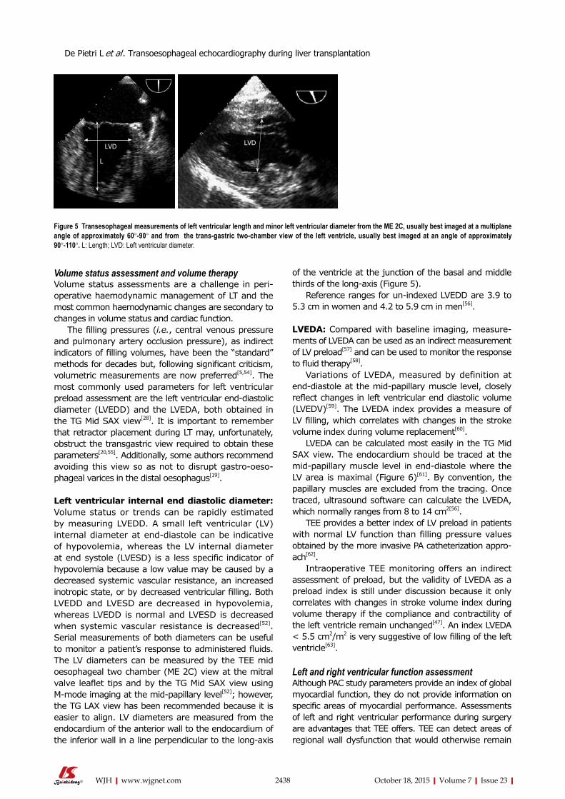
Volume status assessment and volume therapyVolume status assessments are a challenge in peri-operative haemodynamic management of LT and the most common haemodynamic changes are secondary to changes in volume status and cardiac function.
The filling pressures (i.e., central venous pressure and pulmonary artery occlusion pressure), as indirect indicators of filling volumes, have been the “standard” methods for decades but, following significant criticism, volumetric measurements are now preferred[5,54]. The most commonly used parameters for left ventricular preload assessment are the left ventricular end-diastolic diameter (LVEDD) and the LVEDA, both obtained in the TG Mid SAX view[28]. It is important to remember that retractor placement during LT may, unfortunately, obstruct the transgastric view required to obtain these parameters[20,55]. Additionally, some authors recommend avoiding this view so as not to disrupt gastro-oeso-phageal varices in the distal oesophagus[19].
Left ventricular internal end diastolic diameter: Volume status or trends can be rapidly estimated by measuring LVEDD. A small left ventricular (LV) internal diameter at end-diastole can be indicative of hypovolemia, whereas the LV internal diameter at end systole (LVESD) is a less specific indicator of hypovolemia because a low value may be caused by a decreased systemic vascular resistance, an increased inotropic state, or by decreased ventricular filling. Both LVEDD and LVESD are decreased in hypovolemia, whereas LVEDD is normal and LVESD is decreased when systemic vascular resistance is decreased[52]. Serial measurements of both diameters can be useful to monitor a patient’s response to administered fluids. The LV diameters can be measured by the TEE mid oesophageal two chamber (ME 2C) view at the mitral valve leaflet tips and by the TG Mid SAX view using M-mode imaging at the mid-papillary level[52]; however, the TG LAX view has been recommended because it is easier to align. LV diameters are measured from the endocardium of the anterior wall to the endocardium of the inferior wall in a line perpendicular to the long-axis
of the ventricle at the junction of the basal and middle thirds of the long-axis (Figure 5).
Reference ranges for un-indexed LVEDD are 3.9 to 5.3 cm in women and 4.2 to 5.9 cm in men[56].
LVEDA: Compared with baseline imaging, measure-ments of LVEDA can be used as an indirect measurement of LV preload[57] and can be used to monitor the response to fluid therapy[58].
Variations of LVEDA, measured by definition at end-diastole at the mid-papillary muscle level, closely reflect changes in left ventricular end diastolic volume (LVEDV)[59]. The LVEDA index provides a measure of LV filling, which correlates with changes in the stroke volume index during volume replacement[60].
LVEDA can be calculated most easily in the TG Mid SAX view. The endocardium should be traced at the mid-papillary muscle level in end-diastole where the LV area is maximal (Figure 6)[61]. By convention, the papillary muscles are excluded from the tracing. Once traced, ultrasound software can calculate the LVEDA, which normally ranges from 8 to 14 cm2[56].
TEE provides a better index of LV preload in patients with normal LV function than filling pressure values obtained by the more invasive PA catheterization appro-ach[62].
Intraoperative TEE monitoring offers an indirect assessment of preload, but the validity of LVEDA as a preload index is still under discussion because it only correlates with changes in stroke volume index during volume therapy if the compliance and contractility of the left ventricle remain unchanged[47]. An index LVEDA < 5.5 cm2/m2 is very suggestive of low filling of the left ventricle[63].
Left and right ventricular function assessment Although PAC study parameters provide an index of global myocardial function, they do not provide information on specific areas of myocardial performance. Assessments of left and right ventricular performance during surgery are advantages that TEE offers. TEE can detect areas of regional wall dysfunction that would otherwise remain
Figure 5 Transesophageal measurements of left ventricular length and minor left ventricular diameter from the ME 2C, usually best imaged at a multiplane angle of approximately 60°90° and from the transgastric twochamber view of the left ventricle, usually best imaged at an angle of approximately 90°110°. L: Length; LVD: Left ventricular diameter.
L
LVD LVD
De Pietri L et al . Transoesophageal echocardiography during liver transplantation

2439 October 18, 2015|Volume 7|Issue 23|WJH|www.wjgnet.com
undiagnosed. Clearly, time consuming methods requiring multiple equations or measurements are not helpful in an acute setting, such as with the hemodynamic instability of LT. So, despite many published quantitative measures of global left ventricular function[28,64], most basic echocardiographers rely on qualitative, visual estimations of systolic function. This approach is far from precise but allows a basic echocardiographer to differentiate those patients who might benefit from inotropic therapies from those who simply need more fluid volume.
Left ventricular function: The American Society of Echocardiography and by the Society of Cardiovascular Anaesthesiologists[28] has recommended that ventricular function is assessed by a regional wall motion analysis based on a 17-segment wall motion score, as described in the ASE guidelines[64]. This approach suggests that a physician trained in basic transoesophageal echocar-diography obtains mid oesophageal four-chamber (ME 4C), ME 2C and ME LAX views for a more comprehensive evaluation and to monitor global and regional LV function. However, visualization of 6 mid-papillary segments from the TG Mid SAX view may suffice and is important for prognosis[65].
The TG Mid SAX view provides significant diagnostic information regarding regional and global ventricular function to allow for efficient patient care and to minimize any distraction under intraoperative conditions while the patient is haemodynamically unstable.
Quantitative measurements of left ventricular func-tion can also be obtained by measuring the Fractional Shortening, fractional area change and Ejection Fraction.
Fractional shortening: Fractional shortening (FS) expresses the percentage change between the LVEDD and the LVESD according to the following formula:
FS (%) = (LVEDD - LVESD)/LVEDD × 100
The LV internal diameters are measured at the ends of diastole and systole on an M-mode tracing of a TG Mid SAX view taken just above the papillary muscles
and sometimes from an M-mode tracing of a TG LAX view from the inner edge to the inner edge of the endocardial borders (Figure 7).
Fractional shortening can be used to determine ventricular function, with normal values ranging from 25% to 45%[56].
Although FS gives a rapid and simple estimate of LV systolic function, it is not representative of global ventricular function if there are ventricular regional wall abnormalities or aneurysmal deformities[56]. It assesses only the selected cross section of the left ventricle, so there should be no alterations in regional LV contractility either at the apex or at the base if FS is to reflect global LV function accurately.
Fractional area change: Fractional area change (FAC) is a two-dimensional measurement that is easily obtained from the TG Mid SAX view. It expresses the percentage change between the LVEDA and the left ventricular end-systolic area (LVESA) according to the following formula:
FAC (%) = (LVEDA - LVESA)/LVEDA × 100
Normal values range from approximately 55% to 65%[56].
This approach requires both the LVEDA and LVESA measurements, tracing the left ventricular endo-cardium during the end of diastole and systole. The measurements are usually made in the TG Mid SAX view, but when this view is suboptimal, long axis views can also be used. The endocardium can be traced manually traced around the LV cavity, ignoring the papillary muscles, or the endocardial borders can be detected automatically (Figure 6)[61].
FAC can provide a reasonable global estimate of LV function but, like the FS, it has its limitations. It may represent global LV function poorly in cases of myocardial infarction or aneurysmal dilatation in areas of the ventricle other than the mid papillary level, where FAC is evaluated. Changes in loading condition may also influence the FAC.
A B
Figure 6 Transesophageal measurements of fractional area change. Transgastric Mid SAX view of the left ventricle showing measurement of left ventricular end diastolic area (A) and left ventricular end-systolic area (B). The bidimensional image is usually best imaged at a multiplane angle of 0°. Modified by Guarracino Fabio et al[61].
De Pietri L et al . Transoesophageal echocardiography during liver transplantation

2440 October 18, 2015|Volume 7|Issue 23|WJH|www.wjgnet.com
Ejection fraction: This measure requires the appli-cation of algorithms that can approximate the left ventricle to a conventional solid. The ejection fraction (EF) is the most widely used index in clinical practice to describe left ventricular function, even if it measures ejection ability rather than the contractility of the left ventricle. The B-mode evaluation is most widely used to study systolic function because it is reliable even in the presence of geometric distortion or wall motion abnormalities and because it correlates well with radionuclide ventriculography and scintigraphy measurements[66]. In contrast to fractional shortening, which depends on a single cavity dimension in systole and diastole, the ejection fraction examines the entirety of myocardial contraction by expressing stroke volume as a percentage of LV end-diastolic volume:
LVEF% = (LVEDV - LVESV)/LVEDV × 100
Where LVEF is the left ventricle ejection fraction, LVEDV is the LV end-diastolic volume, and LV end-systolic volume (LVESV).
The LVEF represents a composite of cardiac per-formance involving preload, contractility, and afterload. It is widely regarded to be a predictor of outcome and survival. Normal EF values range from 55% to 70%.
The area-length and the Simpson’s methods are among the commonest used to calculate the EF. The previously used Teichholz method of calculating LV ejection fraction from LV linear dimensions is not recommended for clinic practice because inaccuracies can arise from the geometric assumptions required to convert a linear measurement to a 3-D volume[67].
The area-length method is an alternative method to calculate LV volumes when the apical endocardial definition precludes an accurate tracing. This method assumes that the LV is bullet shaped[68], and the volumes are obtained by measuring LV areas and lengths at both end-diastole and end-systole. The measurements require a long-axis view of the chamber without foreshortening of the LV. This view may be difficult
to obtain from the standard ME 4C view, and some retroflexion of the probe may be required to visualize the true apex and prevent any foreshortening of the LV.
The Simpson method is the most widespread EF calculation method and consists of 2-D measurements of volume with the biplane method of discs (modified Simpson’s rule). The EF is calculated from the summation of a series (20) of overlapping slices from apex to base, each of which is assumed to be an elliptical disc. This is the currently recommended method of choice of the American Society of Echocardiography and the European Association of Echocardiography[56]. Modern ultrasound is equipped with software to calculate the EF by using the area of the LV and its diameter measured from the apex to the mitral floor in the two phases of the cardiac cycle. The EF is calculated from projections of the ME 4C and 2C views (Figure 8).
Right ventricular function A normal right ventricle (RV) has an oblong shape and a complex architecture and, in the ME 4C and TG Mid SAX views, it is approximately two-thirds the size of the LV. In the ME 4C view, the RV extends more than half way to but does not normally share the LV apex.
The RV cavity on the short axis has a half-moon shape with the concave side of the interventricular septum towards the VS. A first echocardiographic sight provides information regarding RV function because, at the onset of right ventricular dysfunction, the ventricular chamber enlarges and the usually convex septal wall of the RV, facing the crescent shaped RV cavity, loses its classical shape and anatomical relationships with VS. An increase in RV pressure or volume overload can cause flattening or leftward deviation of the septal wall, producing an elliptical or circular short-axis shape of the RV cavity. The normal right ventricle is accustomed to a low pulmonary resistance and hence low afterload; thus, normal RV pressure is low and right ventricular compliance is high. Elevations in RV afterload result acutely in RV dilatation, whereas chronic elevations cause RV hypertrophy. Right ventricular size is best
Figure 7 Transesophageal measurements of fractional shortening. A: TG Mid SAX view of the left ventricle showing M-mode measurement of LVEDD and LVESD normalized for LVEDD. The bidimensional image is usually best imaged at a multiplane angle of 0°; B: TG LAX view of left ventricle showing M-Mode measurement of LVEDD and LVESD normalized for LVEDD usually best imaged at an angle of approximately 80°-110°. LAX: Long axis view; TG: Transgastric; LVEDD: Left ventricle end diastolic diameter; LVESD: Left ventricle end systolic diameter. Modified by Guarracino Fabio et al[61].
0
5
1077 bpm75 mm/s
0 1 2 3 4
0
2
4
6
8
10
12
14
A B
De Pietri L et al . Transoesophageal echocardiography during liver transplantation

2441 October 18, 2015|Volume 7|Issue 23|WJH|www.wjgnet.com
estimated by TEE from a right ventricle focused ME 4C view, with the multiplane angle adjusted to maximize the tricuspid annulus diameter, usually between 10° and 20°[56]. Care should be taken to obtain the image of the maximal right ventricular diameter without foreshortening[69].
Many techniques have been described to obtain quantitative measurements of the overall function of the right ventricle, but in most cases, the physician relies on qualitative measures, and on visual estimates of systolic right ventricular function.
Assessments of right ventricular function are re-quired whenever a patient presents with unexplained or refractory hypotension. Patients undergoing LT, for example, can present with hypotension secondary to right ventricular failure[70]. If their RV dysfunction is related to acute changes in pulmonary pressures associated with lung volume shifts or acid-base changes during the transplant, they can also be at risk for pulmonary hypertension[21].
TEE has the significant advantage that right ven-tricular failure can be identified by dynamic rather than the pressure changes, which can be easily missed in PAC measurements. Because right ventricular failure is an important complication during the reperfusion phase of LT, TEE monitoring confers very significant advantages at this stage.
When the evaluation is based on a quantitative assessment, a number of echocardiographic techniques may be used to assess RV function.
Tricuspidal annular plane systolic excursion: In systole, the tricuspid annulus will normally descend towards the apex 1.6-2.0 cm. A tricuspidal annular plane systolic excursion (TAPSE) of less than 1.6 cm has been associated with a poor prognosis in a variety of cardiovascular diseases; it is highly specific for RV dysfunction and can be used to monitor RV systolic function serially. It is important to place the cursor at the annulus side and to apply the M-Mode. Once the image is frozen, the TAPSE is the difference between the lowest and highest excursion points of the tricuspidal
annulus. It has the advantage of easy reproducibility, speed of measurement and less dependence on optimal-quality images. There are some disadvantages: The measurement is angle dependent, and the displacement in the ME 4C view is representative of the function of the entire right ventricle only if there are no regional RV wall motion abnormalities. The TAPSE may also be load-dependent, even if less preload-dependent than other markers of RV function[69].
Right ventricular fractional area change: The FAC provides an estimate of the systolic function of the right ventricle. Usually measured in the ME 4C view, it is a simple method to assess RV function that correlates with RV ejection fractions measured by magnetic resonance imaging and has been related to outcome in a number of diseases[56,71]. It is important to verify that the entire right ventricle is in the view, including the apex and the lateral wall, in both systole and diastole. Care must be taken to exclude trabeculations while tracing the RV area. The normal range is 35%-60% and a two-dimensional FAC < 35% indicates RV systolic dysfunction[69].
UTIlITy OF Tee eXamINaTION aND sURGICal PHases OF lTIn addition to all the pathophysiological processes that characterize ESLD, the three major stages of LT (pre-anhepatic, anhepatic and reperfusion phases) pose particular challenges in terms of anaesthetic mana-gement. TEE permits accurate monitoring of these clinical phases with rapid anaesthetic management during complicated procedures, maximizing the opportunities for a successful outcome.
Pre-anhepatic phaseThe pre-anhepatic phase begins with surgical incision and concludes with cross clamping of the vascular inflow to the liver. The conventional technique involves clamping of the portal vein, the suprahepatic inferior
A B
EDV (BP) 81 mLESV (A4C) 55 mL
Figure 8 Calculation of ejection fraction using the disc method (Simpson's rule). Transoesophageal echocardiography ME 4C view in diastole (A) and systole (B). The bidimensional image is usually best imaged at a multiplane angle of 0°. Modified by Guarracino Fabio et al[61]. EDV: End diastolic volume; ESV: End-systolic volume.
De Pietri L et al . Transoesophageal echocardiography during liver transplantation

2442 October 18, 2015|Volume 7|Issue 23|WJH|www.wjgnet.com
vena cava, the infrahepatic inferior vena cava, and the hepatic artery. The piggyback technique requires temporary clamping of portal flow only and tangential clamping of the retrohepatic inferior vena cava, which allows venous return to the heart.
Surgical bleeding, when present, could be the main issue during the pre-anhepatic phase. Dissection may be complicated by steady and sometimes rapid haemorrhage from varices in the abdominal wall or adhesions within the abdominal cavity. Fluid shifts and third space fluid losses may result from the drainage of ascitic fluid and from venous congestion. Bleeding during this phase of surgery is related to the degree of pre-existing coagulopathy, the presence and severity of portal hypertension, and the duration and complexity of the surgical procedure[72,73].
Vascular clamping and manipulation of the liver, together with an inadequate volume resuscitation, result in decreased venous return and reduced cardiac output, resulting in critical organ hypoperfusion[74].
Sudden haemodynamic variations and a reduced preload are the most serious complications and chall-enges that the anaesthetist has to manage during LT.
TEE allows circulatory volumes to be optimized, avoiding impaired organ perfusion, ischaemia and, at the same time, volume overload. Identifying the cause of any hypotension is critical for successful treatment and aggressive fluid repletion leading to overload must be avoided to prevent pulmonary oedema or right ventricular failure due to unrecognized preoperative cardiac disease[3,75].
Close approximation of the papillary muscles and decreases in LVEDA and LVEDD in the TG Mid SAX view signal a reduced preload due to haemorrhage or vascular clamping: After a fluid challenge; TEE allows the anaesthetist to assess the adequacy the interventions rapidly. Signs of RV and right atrial dilation together with RV hypokinesis[76] or atypical regional wall motion abnormalities of the RV free wall[77] can draw attention to right ventricular failure.
If the conventional surgical technique with inferior vena cava and portal vein clamping is poorly tolerated, the VVB might be warranted to guarantee venous return to right atrium and to decompress the portal venous system, reducing bleeding, vascular congestion to the intestines, and injury to the bowel capillary bed[78].
In this case, TEE can assist in the placement of transcutaneous VVB lines, confirm the correct location of guide wires in the venous system, and detect the tip of the cannula in the superior vena cava[79]. Air embolism, thromboembolism, and inadvertent decannulation are among the reported and feared complications of VVB and can increase its morbidity[80].
Anhepatic phaseThe anhepatic phase starts after complete occlusion of vascular inflow to the liver; includes the removal of the native liver and the completion of the vascular anastomoses; and ends with the reperfusion of the
newly grafted liver. This phase is mainly characterized by hemodynamic changes induced by the cross-clamping of the inferior vena cava and portal vein, which reduce venous return, cardiac output and renal perfusion pressure while increasing the splanchnic and lower caval pressures[25]. VVB is used routinely in some centres to facilitate return of blood from the portal system and lower body to the heart via a centrifugal pump to the axillary vein[78]. The TG Mid SAX view or ME 4C view can be ideal for monitoring ventricular function and volume status continuously during this phase. If preload is diminished or systemic vascular resistance is reduced, the left ventricular papillary muscles will approximate each other during systole and LVEDA will be reduced.
Reperfusion phaseLT represents a special case of acute right ventricular stress. Cardiac output increases acutely at the time of reperfusion (up to 3-fold in 15 min) and this increased blood flow to the right heart can result in volume overload and pulmonary oedema due to occult cardiac disease[3].
This phase extends from the period immediately after the reperfusion of the graft to the end of surgery and includes the arterial anastomosis and biliary tract reconstruction. Cardiovascular instability is greatest during this phase of the operation, and it is as accom-panied by a decrease in mean arterial pressure of 30% or more from baseline for at least 1 min’s duration, and occurring within 5 min of reperfusion[81,82].
The reperfusion syndrome is typically characterized by severe hypotension, decreased heart rate, a signi-ficant reduction in systemic vascular resistance, and increases in pulmonary arterial pressure and wedge pressure. All these changes are thought to result from the sudden release of cold, acidotic and hyperkalaemic preservation fluid into the circulation while myocardial dysfunction, often observed after reperfusion, is caused by several vasoactive mediators released into the circulation by the re-perfused graft[83].
The characteristic echocardiographic features of the reperfusion phase may include acute right ventricular systolic dysfunction, left ventricular systolic dysfunction or both; new global or focal wall motion abnormalities; decreased FAC; and because of rapid cardiac influx after vascular unclamping, increases in ventricular end-diastolic volume and LVEDA are common. Selecting and maintaining TEE, typically either in the ME 4C or TG Mid SAX views, allows for real time monitoring of all the effects of reperfusion on the heart. During transition from the anhepatic to the reperfusion phase, TEE views can also be useful for detecting intracardiac air, thrombosis, mitral or tricuspid valve regurgitation; severe diastolic dysfunction; or a previously undia-gnosed outflow obstruction that can occur during reperfusion.
Following reperfusion TEE may be useful to detect the temporary opening of a foramen ovale[84] or elevation of pulmonary arterial pressures, facilitating prompt
De Pietri L et al . Transoesophageal echocardiography during liver transplantation

2443 October 18, 2015|Volume 7|Issue 23|WJH|www.wjgnet.com
management of these complications.Unique haemodynamic changes occur from the
anhepatic to the reperfusion phases, and a real time TEE allows for a rapid assessment and adjustment of fluid shifts and an optimal outcome.
UseFUlNess OF Tee IN THe DIFFeReNTIal DIaGNOsIs OF RaRe CaRDIOvasCUlaR CONDITIONs OFTeN assOCIaTeD WITH HaemODyNamIC INsTaBIlITyPulmonary embolism Although patients with liver disease were long assumed to have a natural bleeding tendency and to be pro-tected from thrombosis, the real coagulation state of the cirrhotic patients combines changes in both pro- and anti-haemostatic pathways in a new haemostatic balance[85]. However, the occurrence of both bleeding and thrombotic complications in a significant proportion of patients shows that this haemostatic balance is relatively unstable[86]. Although LT is associated with increased bleeding and altered coagulation, a prothrombotic state may also occur. Intravascular thrombus formation and subsequent embolization is a potentially fatal complication that most often occurs after reperfusion. Both surgery (vascular clamping) and trauma pose an increased risk for pulmonary embolism (PE) and an incidental cardiac thrombosis, in particular, may lead to serious complications in LT, including intraoperative death[87]. Thus, anaesthesiologists may be responsible for both PE diagnosis and treatment, even if the diagnostic sensitivity of TEE for PE by direct visualization of a thrombus in the pulmonary artery is actually quite low[88]. Although TEE is not the gold standard for PE diagnosis, it can compare with the sensitivity of a TC scan when the PE is acute, central and characterized by severe haemodynamic instability[89,90]. Notably, only 30% obstruction is needed for RV dysfunction to be recognized on TEE[91] but the echocardiographic diagnosis of a PE using direct evidence often requires advanced TEE skills.
The Consensus Statement of the American Society of Echocardiography and the Society of Cardiovascular Anesthesiologists recommends that “a physician trained in basic perioperative TEE at least should be able to use ME 4C, ME AV SAX, and ME RV inflow-outflow views to identify indirect echocardiographic findings consistent with a PE”[28]. Before initiating any specific treatment, the presence of thrombus and/or signs of RV dysfunction, typically due to elevated right-sided pressures, should be identified. The direct visualization of intracardiac thrombus or emboli in the main pulmonary artery or its right or left branches by TEE allows for diagnosis, though it is important to remember that the left pulmonary artery is not completely visualized with TEE due to the
interposition of the left bronchus.Echocardiographic findings consistent with acute PE
include: Signs of RV dilation, RV hypokinesis[76], atypical regional wall motion abnormalities in the RV free wall[77], and decreased TAPSE.
Another typical sign of elevated right-sided pressures is the leftward shift of the interatrial septum or interventricular septum, which is easily seen in the ME 4C view. Although the RV is dysfunctional, the LV may appear hyperkinetic and underfilled, due to a leftward shift of the interventricular septum[92].
LVOTO Cirrhosis is associated with increased left ventricular wall thickness, cardiac chamber enlargement, and a significantly impaired systolic and diastolic response to stress, especially in the setting of volume overload. The hypertrophic cardiomyopathy, which characterizes the cirrhotic patient, makes these patients particularly susceptible to LVOTO especially when hypovolemia, tachycardia, and increased ventricular contraction can cause apposition of the mitral valve anterior leaflet and the septal wall during systole[93]. The pre-anhepatic and anhepatic phases of LT are usually associated with decreases in the left ventricular preload secondary to intraoperative surgical or medical bleeding and to vascular clamping, whereas the post-reperfusion phase is usually associated with a marked decrease in systemic vascular resistance. Therefore, the patients undergoing LT often have several risk factors for dynamic LVOTO[94]. TEE, by virtue of its unique features, guarantees a continuous monitoring of cardiac function and structures, especially during the anhepatic and reperfusion phases[34] when the occurrence of dynamic LVOTO is more common, and can facilitate appropriate management and therapeutic interventions[95].
Dynamic LVOTO can be assessed readily in the ME LAX view at an approximately 120° angle. The characteristic features to look for are turbulence through the left ventricular outflow tract, hypercontractility of the left ventricle, systolic anterior leaflet motion of the mitral valve, and some degree of late mitral regurgitation[96]. A severe mitral regurgitation due to the systolic anterior leaflet motion represents a rapid qualitative method to quantify the degree of LVOTO. The degree of mitral regurgitation is a qualitative method to determine the severity of LVOTO because it usually correlates with the degree of outflow tract obstruction. Another rapid qualitative measure of LVOTO can be obtained by Colour-Doppler, where a mosaic pattern indicates turbulence associated with an elevated LVOT gradient. A quantitative assessment of the obstruction is otherwise obtained by a Doppler quantification of blood flow velocities through the LVOT, using the TG LAX view or the deep TG LAX view. Blood flow velocity in the LVOT is measured by positioning the continuous wave Doppler sample volume in the centre of the LVOT just proximal to the AV. Normal LVOT and AV flow velocities
De Pietri L et al . Transoesophageal echocardiography during liver transplantation

2444 October 18, 2015|Volume 7|Issue 23|WJH|www.wjgnet.com
are less than 1.5 m/s[97]. Colour flow Doppler imaging of the LVOT and AV is useful in directing the Doppler beam through the area of maximum flow when these velocity measurements are made. Portopulmonary hypertension Liver disease and portal hypertension can be associated with pulmonary vascular complications, such as POPH, which is characterized by the presence of portal hypertension, a mean pulmonary artery pressure > 25 mmHg at rest, a mean pulmonary capillary wedge pressure < 15 mmHg, and a pulmonary vascular resistance > 240 dynxsxcm-5[98]. The prevalence of POPH in liver transplant candidates is reported to be 6.3%[99] and 8.5%[100]
Severe pulmonary hypertension and elevated right ventricular systolic pressure predicted a high risk of morbidity and mortality from fulminant right ventricular failure among patients undergoing LT[101]. Patients presenting for liver transplantation with pulmonary hypertension have an additional risk for RV dysfunction secondary to acute changes in pulmonary pressures associated with the volume shifts and acid base distur-bances that characterize LT.
For this reason, most transplant centres consider severe POPH an absolute contraindication to trans-plantation[102]. On the other hand, a number of reports have confirmed that LT can be performed safely if the patient haemodynamic state is suitably controlled[102].
The most important test to screen for POPH is the two dimensional TTE, which is a routine part of an LT evaluation[103].
Unfortunately, the TTE cannot fully discriminate between increased PVR due to true vaso-occlusive arteriopathy, a hyperdynamic state, or fluid overload, with normal/low PVR. Therefore, right heart catheteri-zation is the gold standard for the diagnosis of POPH[104].
For this reason, POPH may be missed by pre-operative echocardiography and may be recognized only during right heart catheterization in the intraoperative phase.
The diagnosis of unexpected POPH on the operating table may still be best handled through TEE. Continuous intraoperative transoesophageal echocardiography has been recommended for following right heart function, and in the event of a pulmonary hypertensive crisis, the anaesthetist has to be ready to address acute pulmonary hypertension with effective agents such as inhaled or intravenous vasodilators[102,105].
The echocardiographic intraoperative findings of severe pulmonary hypertension include right ventricular hypertrophy, dilatation and dysfunction, as well as right atrial enlargement. A paradoxical septal movement can be seen as well.
The best views to visualize the right heart chambers are ME 4C and ME RV inflow-outflow.
CONClUsIONTEE has evolved as an important diagnostic tool outside
the field of cardiac anaesthesia and it has gained increasing importance as a monitoring tool in liver anae-sthesia. One of the most advantageous features of the TEE over PAC is the direct visualization of the heart in real time, which allows for instantaneous assessment of the state of the cardiovascular system, changes in global and regional contractility, and the rapid diagnosis of ventricular dilatation and failure. TEE can overcome the limitations of PAC measurements arising from the large core body temperature shifts typical of massive fluid infusion or revascularization of the new graft during LT.
TEE allows the intraoperative causes of hypotensive episodes during LT to be identified and can help rapidly optimize volaemia, avoiding organ perfusion impairment and ischaemia. TEE also allows for the intraoperative diagnosis and management of specific cardiovascular conditions that like portopulmonary hypertension, air embolism and thromboembolism, can complicate LT and the management of patients with hypertrophic cardiomyopathy, who may experience LVOTO.
Notwithstanding the published guidelines that define the basic and advanced competency requirements for TEE users, transoesophageal echocardiography is being used to monitor haemodynamics and for direct therapy in liver transplant patients, and clinically useful interpretations are possible even during the skill-acquisition phase of TEE training.
The value of TEE must of course be balanced against the risk of performing the procedure. Gastro-oesophageal varices are very common in patients listed for LT; thus, patients with ESLD should have a preoperative endoscopic surveillance and oropharyngeal examination, and probe manipulation should be limited to experienced operators.
Despite these limitations, the intraoperative TEE is a relatively safe method for monitoring cardiac performance in liver transplant patients and should not be contraindicated by the presence of oesophageal varices if the indications for the exam are important.
Although the interpretation of TEE remains mostly subjective, TEE has been helpful for assessing haemo-dynamic alterations, guiding fluid replacement and inotropic therapy, and identifying potential complications during LT, allowing for better management of patients.
ReFeReNCes1 Wertheim JA, Petrowsky H, Saab S, Kupiec-Weglinski JW,
Busuttil RW. Major challenges limiting liver transplantation in the United States. Am J Transplant 2011; 11: 1773-1784 [PMID: 21672146 DOI: 10.1111/j.1600-6143.2011.03587.x]
2 Petrowsky H, Rana A, Kaldas FM, Sharma A, Hong JC, Agopian VG, Durazo F, Honda H, Gornbein J, Wu V, Farmer DG, Hiatt JR, Busuttil RW. Liver transplantation in highest acuity recipients: identifying factors to avoid futility. Ann Surg 2014; 259: 1186-1194 [PMID: 24263317 DOI: 10.1097/SLA.0000000000000265]
3 Gaskari SA, Honar H, Lee SS. Therapy insight: Cirrhotic cardiomyopathy. Nat Clin Pract Gastroenterol Hepatol 2006; 3: 329-337 [PMID: 16741552 DOI: 10.1038/ncpgasthep0498]
4 Alqahtani SA, Fouad TR, Lee SS. Cirrhotic cardiomyopathy.
De Pietri L et al . Transoesophageal echocardiography during liver transplantation

2445 October 18, 2015|Volume 7|Issue 23|WJH|www.wjgnet.com
Semin Liver Dis 2008; 28: 59-69 [PMID: 18293277 DOI: 10.1055/s-2008-1040321]
5 De Wolf A. Transesophageal echocardiography and orthotopic liver transplantation: general concepts. Liver Transpl Surg 1999; 5: 339-340 [PMID: 10388509 DOI: 10.1002/lt.500050408]
6 Gudavalli A, Kalaria VG, Chen X, Schwarz KQ. Intrapulmonary arteriovenous shunt: diagnosis by saline contrast bubbles in the pulmonary veins. J Am Soc Echocardiogr 2002; 15: 1012-1014 [PMID: 12221424]
7 Vedrinne JM, Duperret S, Bizollon T, Magnin C, Motin J, Trepo C, Ducerf C. Comparison of transesophageal and transthoracic contrast echocardiography for detection of an intrapulmonary shunt in liver disease. Chest 1997; 111: 1236-1240 [PMID: 9149575]
8 Mojadidi MK, Winoker JS, Roberts SC, Msaouel P, Gevorgyan R, Zolty R. Two-dimensional echocardiography using second harmonic imaging for the diagnosis of intracardiac right-to-left shunt: a meta-analysis of prospective studies. Int J Cardiovasc Imaging 2014; 30: 911-923 [PMID: 24740212 DOI: 10.1007/s10554-014-0426-8]
9 Meissner I, Whisnant JP, Khandheria BK, Spittell PC, O’Fallon WM, Pascoe RD, Enriquez-Sarano M, Seward JB, Covalt JL, Sicks JD, Wiebers DO. Prevalence of potential risk factors for stroke assessed by transesophageal echocardiography and carotid ultrasonography: the SPARC study. Stroke Prevention: Assessment of Risk in a Community. Mayo Clin Proc 1999; 74: 862-869 [PMID: 10488786]
10 Yerlioglu E, Krishnamoorthy V, Jeon H, Gustin A, Nicolau-Raducu R. Patent foramen ovale and intracardiac thrombus identified by transesophageal echocardiography during liver transplantation. J Cardiothorac Vasc Anesth 2012; 26: 1069-1073 [PMID: 21757375 DOI: 10.1053/j.jvca.2011.05.003]
11 Garg A, Armstrong WF. Echocardiography in liver transplant candidates. JACC Cardiovasc Imaging 2013; 6: 105-119 [PMID: 23328568 DOI: 10.1016/j.jcmg.2012.11.002]
12 Alba AC, Verocai Flaman F, Granton J, Delgado DH. Patent foramen ovale does not have a negative impact on early outcomes in patients undergoing liver transplantation. Clin Transplant 2011; 25: 151-155 [PMID: 20156223 DOI: 10.1111/j.1399-0012.2010.01210.x]
13 Schumann R, Mandell MS, Mercaldo N, Michaels D, Robertson A, Banerjee A, Pai R, Klinck J, Pandharipande P, Walia A. Anesthesia for liver transplantation in United States academic centers: intraoperative practice. J Clin Anesth 2013; 25: 542-550 [PMID: 23994704 DOI: 10.1016/j.jclinane.2013.04.017]
14 Burger-Klepp U, Karatosic R, Thum M, Schwarzer R, Fuhrmann V, Hetz H, Bacher A, Berlakovich G, Krenn CG, Faybik P. Transesophageal echocardiography during orthotopic liver transplantation in patients with esophagoastric varices. Transplantation 2012; 94: 192-196 [PMID: 22728290 DOI: 10.1097/TP.0b013e31825475c2]
15 Cárdenas A, Ginès P. Management of patients with cirrhosis awaiting liver transplantation. Gut 2011; 60: 412-421 [PMID: 21193458 DOI: 10.1136/gut.2009.179937]
16 Merli M, Nicolini G, Angeloni S, Rinaldi V, De Santis A, Merkel C, Attili AF, Riggio O. Incidence and natural history of small esophageal varices in cirrhotic patients. J Hepatol 2003; 38: 266-272 [PMID: 12586291]
17 Spier BJ, Larue SJ, Teelin TC, Leff JA, Swize LR, Borkan SH, Satyapriya A, Rahko PS, Pfau PR. Review of complications in a series of patients with known gastro-esophageal varices undergoing transesophageal echocardiography. J Am Soc Echocardiogr 2009; 22: 396-400 [PMID: 19231133 DOI: 10.1016/j.echo.2009.01.002]
18 Garcia-Tsao G, Sanyal AJ, Grace ND, Carey WD. Prevention and management of gastroesophageal varices and variceal hemorrhage in cirrhosis. Am J Gastroenterol 2007; 102: 2086-2102 [PMID: 17727436 DOI: 10.1111/j.1572-0241.2007.01481.x]
19 Hilberath JN, Oakes DA, Shernan SK, Bulwer BE, D’Ambra MN, Eltzschig HK. Safety of transesophageal echocardiography. J Am Soc Echocardiogr 2010; 23: 1115-1127; quiz 1220-1221 [PMID: 20864313 DOI: 10.1016/j.echo.2010.08.013]
20 Krenn CG, De Wolf AM. Current approach to intraoperative monitoring in liver transplantation. Curr Opin Organ Transplant 2008; 13: 285-290 [PMID: 18685319 DOI: 10.1097/MOT.0b013e3283005832]
21 Suriani RJ, Cutrone A, Feierman D, Konstadt S. Intraoperative transesophageal echocardiography during liver transplantation. J Cardiothorac Vasc Anesth 1996; 10: 699-707 [PMID: 8910147 DOI: 10.1016/S1053-0770(96)80193-5]
22 Spencer KT. Transesophageal echocardiography in patients with esophageal varices. J Am Soc Echocardiogr 2009; 22: 401-403 [PMID: 19345308 DOI: 10.1016/j.echo.2009.02.011]
23 Markin NW, Sharma A, Grant W, Shillcutt SK. The safety of transesophageal echocardiography in patients undergoing orthotopic liver transplantation. J Cardiothorac Vasc Anesth 2015; 29: 588-593 [PMID: 25622974 DOI: 10.1053/j.jvca.2014.10.012]
24 Robertson AC, Eagle SS. Transesophageal echocardiography during orthotopic liver transplantation: maximizing information without the distraction. J Cardiothorac Vasc Anesth 2014; 28: 141-154 [PMID: 23642888 DOI: 10.1053/j.jvca.2012.11.016]
25 Ozier Y , Klinck JR. Anesthetic management of hepatic transplantation. Curr Opin Anaesthesiol 2008; 21: 391-400 [PMID: 18458561 DOI: 10.1097/ACO.0b013e3282ff85f4]
26 Wax DB, Torres A, Scher C, Leibowitz AB. Transesophageal echocardiography utilization in high-volume liver transplantation centers in the United States. J Cardiothorac Vasc Anesth 2008; 22: 811-813 [PMID: 18834818 DOI: 10.1053/j.jvca.2008.07.007]
27 Schumann R. Intraoperative resource utilization in anesthesia for liver transplantation in the United States: a survey. Anesth Analg 2003; 97: 21-28, table of contents [PMID: 12818937]
28 Reeves ST, Finley AC, Skubas NJ, Swaminathan M, Whitley WS, Glas KE, Hahn RT, Shanewise JS, Adams MS, Shernan SK. Basic perioperative transesophageal echocardiography examination: a consensus statement of the American Society of Echocardiography and the Society of Cardiovascular Anesthesiologists. J Am Soc Echocardiogr 2013; 26: 443-456 [PMID: 23622926 DOI: 10.1016/j.echo.2013.02.015]
29 Practice Guidelines for Perioperative Transesophageal Echocardiography. An Updated Report by the American Society of Anesthesiologists and the Society of Cardiovascular Anesthe-siologists Task Force on Transesophageal Echocardiography. Anesthesiology 2010; 112: 1084-1096
30 Shanewise JS, Cheung AT, Aronson S, Stewart WJ, Weiss RL, Mark JB, Savage RM, Sears-Rogan P, Mathew JP, Quiñones MA, Cahalan MK, Savino JS. ASE/SCA guidelines for perfor-ming a comprehensive intraoperative multiplane transesophageal echocardiography examination: recommendations of the American Society of Echocardiography Council for Intraoperative Echocardiography and the Society of Cardiovascular Anesthesio-logists Task Force for Certification in Perioperative Transesophageal Echocardiography. Anesth Analg 1999; 89: 870-884 [PMID: 10512257]
31 Oxorn DC. Intraoperative echocardiography. Heart 2008; 94: 1236-1243 [PMID: 18703702 DOI: 10.1136/hrt.2008.148510]
32 Della Rocca G, Costa MG, Pompei L, Chiarandini P. The liver transplant recipient with cardiac disease. Transplant Proc 2008; 40: 1172-1174 [PMID: 18555141 DOI: 10.1016/j.transproceed.2008.03.070]
33 Feltracco P, Biancofiore G, Ori C, Saner FH, Della Rocca G. Limits and pitfalls of haemodynamic monitoring systems in liver transplantation surgery. Minerva Anestesiol 2012; 78: 1372-1384 [PMID: 22858882]
34 Cywinski JB, Argalious M, Marks TN, Parker BM. Dynamic left ventricular outflow tract obstruction in an orthotopic liver transplant recipient. Liver Transpl 2005; 11: 692-695 [PMID: 15915494 DOI: 10.1002/lt.20440]
35 Reuter DA, Huang C, Edrich T, Shernan SK, Eltzschig HK. Cardiac output monitoring using indicator-dilution techniques: basics, limits, and perspectives. Anesth Analg 2010; 110: 799-811 [PMID: 20185659 DOI: 10.1213/ANE.0b013e3181cc885a]
36 Bao FP, Wu J. Continuous versus bolus cardiac output monitoring
De Pietri L et al . Transoesophageal echocardiography during liver transplantation

2446 October 18, 2015|Volume 7|Issue 23|WJH|www.wjgnet.com
during orthotopic liver transplantation. Hepatobiliary Pancreat Dis Int 2008; 7: 138-144 [PMID: 18397847]
37 Böttiger BW, Sinner B, Motsch J, Bach A, Bauer H, Martin E. Continuous versus intermittent thermodilution cardiac output measurement during orthotopic liver transplantation. Anaesthesia 1997; 52: 207-214 [PMID: 9124659]
38 Greim CA, Roewer N, Thiel H, Laux G, Schulte am Esch J. Continuous cardiac output monitoring during adult liver trans-plantation: thermal filament technique versus bolus thermo-dilution. Anesth Analg 1997; 85: 483-488 [PMID: 9296398]
39 Hurford WE, Zapol WM. The right ventricle and critical illness: a review of anatomy, physiology, and clinical evaluation of its function. Intensive Care Med 1988; 14 Suppl 2: 448-457 [PMID: 3042829]
40 Boucaud C, Bouffard Y, Dumortier J, Gaillac N, Sagnard P, Graber MC, Adham M, Boillot O. Transoesophageal echo-Doppler vs. thermodilution cardiac output measurement during hepatic vascular exclusion in liver transplantation. Eur J Anaesthesiol 2008; 25: 485-489 [PMID: 18298871 DOI: 10.1017/S0265021508003670]
41 De Wolf AM. Pulmonary artery catheter: rest in peace? Not just quite yet... Liver Transpl 2008; 14: 917-918 [PMID: 18581507 DOI: 10.1002/lt.21543]
42 Zink W, Nöll J, Rauch H, Bauer H, Desimone R, Martin E, Böttiger BW. Continuous assessment of right ventricular ejection fraction: new pulmonary artery catheter versus transoesophageal echocardiography. Anaesthesia 2004; 59: 1126-1132 [PMID: 15479324 DOI: 10.1111/j.1365-2044.2004.03876.x]
43 Savino JS, Troianos CA, Aukburg S, Weiss R, Reichek N. Measurement of pulmonary blood flow with transesophageal two-dimensional and Doppler echocardiography. Anesthesiology 1991; 75: 445-451 [PMID: 1888051]
44 Muhiudeen IA, Kuecherer HF, Lee E, Cahalan MK, Schiller NB. Intraoperative estimation of cardiac output by transesophageal pulsed Doppler echocardiography. Anesthesiology 1991; 74: 9-14 [PMID: 1986664]
45 Møller-Sørensen H, Graeser K, Hansen KL, Zemtsovski M, Sander EM, Nilsson JC. Measurements of cardiac output obtained with transesophageal echocardiography and pulmonary artery thermodilution are not interchangeable. Acta Anaesthesiol Scand 2014; 58: 80-88 [PMID: 24192143 DOI: 10.1111/aas.12227]
46 Della Rocca G, Costa MG, Coccia C, Pompei L, Salandin V, Pierangelo DM, Pietropaoli P. Continuous right ventricular end-diastolic volume in comparison with left ventricular end-diastolic area. Eur J Anaesthesiol 2009; 26: 272-278 [PMID: 19276913 DOI: 10.1097/EJA.0b013e328319be8e]
47 Cheung AT, Savino JS, Weiss SJ, Aukburg SJ, Berlin JA. Echocardiographic and hemodynamic indexes of left ventricular preload in patients with normal and abnormal ventricular function. Anesthesiology 1994; 81: 376-387 [PMID: 8053588]
48 Della Rocca G, Brondani A, Costa MG. Intraoperative hemo-dynamic monitoring during organ transplantation: what is new? Curr Opin Organ Transplant 2009; 14: 291-296 [PMID: 19448537 DOI: 10.1097/MOT.0b013e32832d927d]
49 Practice guidelines for perioperative transesophageal echocar-diography. A report by the American Society of Anesthesiologists and the Society of Cardiovascular Anesthesiologists Task Force on Transesophageal Echocardiography. Anesthesiology 1996; 84: 986-1006 [PMID: 8638856]
50 Vanatta JM, Dryn O, Berkley T, Nair S, Eason JD. Liver transplantation at the University of Tennessee Health Science Center in Memphis, Tennessee: the current era 2006-2012. Clin Transpl 2012: 103-109 [PMID: 23721013]
51 Lahm T, McCaslin CA, Wozniak TC, Ghumman W, Fadl YY, Obeidat OS, Schwab K, Meldrum DR. Medical and surgical treatment of acute right ventricular failure. J Am Coll Cardiol 2010; 56: 1435-1446 [PMID: 20951319 DOI: 10.1016/j.jacc.2010.05.046]
52 Porter TR, Shillcutt SK, Adams MS, Desjardins G, Glas KE, Olson JJ, Troughton RW. Guidelines for the use of echocar-diography as a monitor for therapeutic intervention in adults: a report from the American Society of Echocardiography. J Am Soc
Echocardiogr 2015; 28: 40-56 [PMID: 25559474 DOI: 10.1016/j.echo.2014.09.009]
53 Maslow AD, Mashikian J, Haering JM, Heindel S, Douglas P, Levine R. Transesophageal echocardiographic evaluation of native aortic valve area: utility of the double-envelope technique. J Cardiothorac Vasc Anesth 2001; 15: 293-299 [PMID: 11426358 DOI: 10.1053/jcan.2001.23272]
54 De Wolf AM, Begliomini B, Gasior TA, Kang Y, Pinsky MR. Right ventricular function during orthotopic liver transplantation. Anesth Analg 1993; 76: 562-568 [PMID: 8452268]
55 De Wolf AM, Aggarwal S. Monitoring preload during liver transplantation. Liver Transpl 2008; 14: 268-269 [PMID: 18306387 DOI: 10.1002/lt.21316]
56 Lang RM, Bierig M, Devereux RB, Flachskampf FA, Foster E, Pellikka PA, Picard MH, Roman MJ, Seward J, Shanewise J, Solomon S, Spencer KT, St John Sutton M, Stewart W. Recom-mendations for chamber quantification. Eur J Echocardiogr 2006; 7: 79-108 [PMID: 16458610 DOI: 10.1016/j.euje.2005.12.014]
57 Schmidlin D, Jenni R, Schmid ER. Transesophageal echo-cardiographic area and Doppler flow velocity measurements: comparison with hemodynamic changes in coronary artery bypass surgery. J Cardiothorac Vasc Anesth 1999; 13: 143-149 [PMID: 10230946]
58 Swenson JD, Harkin C, Pace NL, Astle K, Bailey P. Transesophageal echocardiography: an objective tool in defining maximum ventricular response to intravenous fluid therapy. Anesth Analg 1996; 83: 1149-1153 [PMID: 8942577]
59 Appleyard RF, Glantz SA. Two dimensions describe left ventricular volume change during hemodynamic transients. Am J Physiol 1990; 258: H277-H284 [PMID: 2301611]
60 Denault AY, Couture P, McKenty S, Boudreault D, Plante F, Perron R, Babin D, Buithieu J. Perioperative use of transesophageal echocardiography by anesthesiologists: impact in noncardiac surgery and in the intensive care unit. Can J Anaesth 2002; 49: 287-293 [PMID: 11861348 DOI: 10.1007/BF03020529]
61 Guarracino F. Ecocardiografia transesofagea in area critica. Elsevier Masson Italia, 2008: 213-250
62 Thys DM, Hillel Z, Goldman ME, Mindich BP, Kaplan JA. A comparison of hemodynamic indices derived by invasive monitoring and two-dimensional echocardiography. Anesthesiology 1987; 67: 630-634 [PMID: 3499831]
63 Skarvan K, Lambert A, Filipovic M, Seeberger M. Reference values for left ventricular function in subjects under general anaesthesia and controlled ventilation assessed by two-dimensional transoesophageal echocardiography. Eur J Anaesthesiol 2001; 18: 713-722 [PMID: 11580777]
64 Lang RM, Bierig M, Devereux RB, Flachskampf FA, Foster E, Pellikka PA, Picard MH, Roman MJ, Seward J, Shanewise JS, Solomon SD, Spencer KT, Sutton MS, Stewart WJ. Recom-mendations for chamber quantification: a report from the American Society of Echocardiography’s Guidelines and Standards Committee and the Chamber Quantification Writing Group, developed in conjunction with the European Association of Echocardiography, a branch of the European Society of Cardiology. J Am Soc Echocardiogr 2005; 18: 1440-1463 [PMID: 16376782 DOI: 10.1016/j.echo.2005.10.005]
65 Reichert CL, Visser CA, van den Brink RB, Koolen JJ, van Wezel HB, Moulijn AC, Dunning AJ. Prognostic value of biventricular function in hypotensive patients after cardiac surgery as assessed by transesophageal echocardiography. J Cardiothorac Vasc Anesth 1992; 6: 429-432 [PMID: 1498297]
66 Naik MM, Diamond GA, Pai T, Soffer A, Siegel RJ. Corres-pondence of left ventricular ejection fraction determinations from two-dimensional echocardiography, radionuclide angiography and contrast cineangiography. J Am Coll Cardiol 1995; 25: 937-942 [PMID: 7884101 DOI: 10.1016/0735-1097(94)00506-L]
67 Teichholz LE, Kreulen T, Herman MV, Gorlin R. Problems in echocardiographic volume determinations: echocardiographic-angiographic correlations in the presence of absence of asynergy. Am J Cardiol 1976; 37: 7-11 [PMID: 1244736]
De Pietri L et al . Transoesophageal echocardiography during liver transplantation

2447 October 18, 2015|Volume 7|Issue 23|WJH|www.wjgnet.com
68 Hozumi T, Shakudo M, Shah PM. Quantitation of left ventricular volumes and ejection fraction by biplane transesophageal echo-cardiography. Am J Cardiol 1993; 72: 356-359 [PMID: 8342520]
69 Rudski LG, Lai WW, Afilalo J, Hua L, Handschumacher MD, Chandrasekaran K, Solomon SD, Louie EK, Schiller NB. Guidelines for the echocardiographic assessment of the right heart in adults: a report from the American Society of Echocardiography endorsed by the European Association of Echocardiography, a registered branch of the European Society of Cardiology, and the Canadian Society of Echocardiography. J Am Soc Echocardiogr 2010; 23: 685-713; quiz 786-788 [PMID: 20620859 DOI: 10.1016/j.echo.2010.05.010]
70 Ellis JE, Lichtor JL, Feinstein SB, Chung MR, Polk SL, Broelsch C, Emond J, Thistlethwaite JR, Roizen MF. Right heart dysfunction, pulmonary embolism, and paradoxical embolization during liver transplantation. A transesophageal two-dimensional echocardiographic study. Anesth Analg 1989; 68: 777-782 [PMID: 2660629]
71 Maslow AD, Regan MM, Panzica P, Heindel S, Mashikian J, Comunale ME. Precardiopulmonary bypass right ventricular function is associated with poor outcome after coronary artery bypass grafting in patients with severe left ventricular systolic dysfunction. Anesth Analg 2002; 95: 1507-1518, table of contents [PMID: 12456409]
72 Hannaman MJ, Hevesi ZG. Anesthesia care for liver trans-plantation. Transplant Rev (Orlando) 2011; 25: 36-43 [PMID: 21126662 DOI: 10.1016/j.trre.2010.10.004]
73 Steadman RH. Anesthesia for liver transplant surgery. Anesthesiol Clin North America 2004; 22: 687-711 [PMID: 15541931 DOI: 10.1016/j.atc.2004.06.009]
74 Møller S, Dümcke CW, Krag A. The heart and the liver. Expert Rev Gastroenterol Hepatol 2009; 3: 51-64 [PMID: 19210113 DOI: 10.1586/17474124.3.1.51]
75 Burtenshaw AJ, Isaac JL. The role of trans-oesophageal echocar-diography for perioperative cardiovascular monitoring during orthotopic liver transplantation. Liver Transpl 2006; 12: 1577-1583 [PMID: 17058248 DOI: 10.1002/lt.20929]
76 Torbicki A, Perrier A, Konstantinides S, Agnelli G, Galiè N, Pruszczyk P, Bengel F, Brady AJ, Ferreira D, Janssens U, Klepetko W, Mayer E, Remy-Jardin M, Bassand JP. Guidelines on the diagnosis and management of acute pulmonary embolism: the Task Force for the Diagnosis and Management of Acute Pulmonary Embolism of the European Society of Cardiology (ESC). Eur Heart J 2008; 29: 2276-2315 [PMID: 18757870 DOI: 10.1093/eurheartj/ehn310]
77 McConnell MV, Solomon SD, Rayan ME, Come PC, Goldhaber SZ, Lee RT. Regional right ventricular dysfunction detected by echocardiography in acute pulmonary embolism. Am J Cardiol 1996; 78: 469-473 [PMID: 8752195]
78 Miranda LE, de Melo PS, Sabat BD, Tenório AL, Lima DL, Neto OC, Amorim AG, Fernandez JL, de Macedo FI, Lacerda CM. Orthotopic liver transplantation without venovenous bypass: 125 cases from a single center. Transplant Proc 2012; 44: 2416-2422 [PMID: 23026610 DOI: 10.1016/j.transproceed.2012.07.038]
79 Planinsic RM, Nicolau-Raducu R, Caldwell JC, Aggarwal S, Hilmi I. Transesophageal echocardiography-guided placement of internal jugular percutaneous venovenous bypass cannula in orthotopic liver transplantation. Anesth Analg 2003; 97: 648-649 [PMID: 12933376]
80 Budd JM, Isaac JL, Bennett J, Freeman JW. Morbidity and mortality associated with large-bore percutaneous venovenous bypass cannulation for 312 orthotopic liver transplantations. Liver Transpl 2001; 7: 359-362 [PMID: 11303297 DOI: 10.1053/jlts.2001.22708]
81 Aggarwal S, Kang Y, Freeman JA, Fortunato FL, Pinsky MR. Postreperfusion syndrome: cardiovascular collapse following hepatic reperfusion during liver transplantation. Transplant Proc 1987; 19: 54-55 [PMID: 3303534]
82 Chung IS, Kim HY, Shin YH, Ko JS, Gwak MS, Sim WS, Kim GS, Lee SK. Incidence and predictors of post-reperfusion syndrome in living donor liver transplantation. Clin Transplant 2012; 26:
539-543 [PMID: 22168355 DOI: 10.1111/j.1399-0012.2011.01568.x]
83 Nanashima A, Pillay P, Crawford M, Nakasuji M, Verran DJ, Painter D. Analysis of postrevascularization syndrome after orthotopic liver transplantation: the experience of an Australian liver transplantation center. J Hepatobiliary Pancreat Surg 2001; 8: 557-563 [PMID: 11956908 DOI: 10.1007/s005340100025]
84 Saada M, Liu N, Cherqui D, Beydon L, Maurel C, Catoire P, Bonnet F, Duvaldestin P. [Opening of a foramen ovale during liver transplantation. The value of transesophageal echocardiography]. Ann Fr Anesth Reanim 1990; 9: 412-414 [PMID: 2240693]
85 Weeder PD, Porte RJ, Lisman T. Hemostasis in liver disease: implications of new concepts for perioperative management. Transfus Med Rev 2014; 28: 107-113 [PMID: 24721432 DOI: 10.1016/j.tmrv.2014.03.002]
86 Lisman T, Porte RJ. Rebalanced hemostasis in patients with liver disease: evidence and clinical consequences. Blood 2010; 116: 878-885 [PMID: 20400681 DOI: 10.1182/blood-2010-02-261891]
87 Xia VW, Ho JK, Nourmand H, Wray C, Busuttil RW, Steadman RH. Incidental intracardiac thromboemboli during liver transplantation: incidence, risk factors, and management. Liver Transpl 2010; 16: 1421-1427 [PMID: 21117252 DOI: 10.1002/lt.22182]
88 Rosenberger P, Shernan SK, Body SC, Eltzschig HK. Utility of intraoperative transesophageal echocardiography for diagnosis of pulmonary embolism. Anesth Analg 2004; 99: 12-16 [PMID: 15281493]
89 Pruszczyk P, Torbicki A, Kuch-Wocial A, Chlebus M, Miśkiewicz ZC, Jedrusik P. Transoesophageal echocardiography for definitive diagnosis of haemodynamically significant pulmonary embolism. Eur Heart J 1995; 16: 534-538 [PMID: 7671900]
90 Krivec B, Voga G, Zuran I, Skale R, Pareznik R, Podbregar M, Noc M. Diagnosis and treatment of shock due to massive pulmonary embolism: approach with transesophageal echocardiography and intrapulmonary thrombolysis. Chest 1997; 112: 1310-1316 [PMID: 9367474]
91 Wolfe MW, Lee RT, Feldstein ML, Parker JA, Come PC, Goldhaber SZ. Prognostic significance of right ventricular hypo-kinesis and perfusion lung scan defects in pulmonary embolism. Am Heart J 1994; 127: 1371-1375 [PMID: 8172067]
92 Matthews JC, McLaughlin V. Acute right ventricular failure in the setting of acute pulmonary embolism or chronic pulmonary hypertension: a detailed review of the pathophysiology, diagnosis, and management. Curr Cardiol Rev 2008; 4: 49-59 [PMID: 19924277 DOI: 10.2174/157340308783565384]
93 Ramaraj R. Hypertrophic cardiomyopathy: etiology, diagnosis, and treatment. Cardiol Rev 2008; 16: 172-180 [PMID: 18562807 DOI: 10.1097/CRD.0b013e318178e525]
94 Lee AR, Kim YR, Ham JS, Lee SM, Kim GS. Dynamic left ventricular outflow tract obstruction in living donor liver trans-plantation recipients -A report of two cases-. Korean J Anesthesiol 2010; 59 Suppl: S128-S132 [PMID: 21286421 DOI: 10.4097/kjae.2010.59.S.S128]
95 Roy D, Ralley FE. Anesthetic management of a patient with dynamic left ventricular outflow tract obstruction with systolic anterior movement of the mitral valve undergoing redo-orthotopic liver transplantation. J Cardiothorac Vasc Anesth 2012; 26: 274-276 [PMID: 21514844 DOI: 10.1053/j.jvca.2011.02.014]
96 Ashidagawa M, Ohara M, Koide Y. An intraoperative diagnosis of dynamic left ventricular outflow tract obstruction using transesophageal echocardiography leads to the treatment with intravenous disopyramide. Anesth Analg 2002; 94: 310-312, table of contents [PMID: 11812689]
97 Shanewise JS, Cheung AT, Aronson S, Stewart WJ, Weiss RL, Mark JB, Savage RM, Sears-Rogan P, Mathew JP, Quiñones MA, Cahalan MK, Savino JS. ASE/SCA guidelines for performing a comprehensive intraoperative multiplane transesophageal echocardiography examination: recommendations of the American Society of Echocardiography Council for Intraoperative Echocardiography and the Society of Cardiovascular Anesthesiologists Task Force for Certification in Perioperative Transesophageal Echocardiography. J
De Pietri L et al . Transoesophageal echocardiography during liver transplantation

2448 October 18, 2015|Volume 7|Issue 23|WJH|www.wjgnet.com
Am Soc Echocardiogr 1999; 12: 884-900 [PMID: 10511663]98 Simonneau G, Robbins IM, Beghetti M, Channick RN, Delcroix
M, Denton CP, Elliott CG, Gaine SP, Gladwin MT, Jing ZC, Krowka MJ, Langleben D, Nakanishi N, Souza R. Updated clinical classification of pulmonary hypertension. J Am Coll Cardiol 2009; 54: S43-S54 [PMID: 19555858 DOI: 10.1016/j.jacc.2009.04.012]
99 Kawut SM, Krowka MJ, Trotter JF, Roberts KE, Benza RL, Badesch DB, Taichman DB, Horn EM, Zacks S, Kaplowitz N, Brown RS, Fallon MB. Clinical risk factors for portopulmonary hypertension. Hepatology 2008; 48: 196-203 [PMID: 18537192 DOI: 10.1002/hep.22275]
100 Ramsay MA, Simpson BR, Nguyen AT, Ramsay KJ, East C, Klintmalm GB. Severe pulmonary hypertension in liver transplant candidates. Liver Transpl Surg 1997; 3: 494-500 [PMID: 9346791]
101 Porres-Aguilar M, Zuckerman MJ, Figueroa-Casas JB, Krowka MJ. Portopulmonary hypertension: state of the art. Ann Hepatol 2008; 7: 321-330 [PMID: 19034231]
102 Ramsay M. Portopulmonary hypertension and right heart failure in
patients with cirrhosis. Curr Opin Anaesthesiol 2010; 23: 145-150 [PMID: 20124995 DOI: 10.1097/ACO.0b013e32833725c4]
103 Galiè N, Hoeper MM, Humbert M, Torbicki A, Vachiery JL, Barbera JA, Beghetti M, Corris P, Gaine S, Gibbs JS, Gomez-Sanchez MA, Jondeau G, Klepetko W, Opitz C, Peacock A, Rubin L, Zellweger M, Simonneau G. Guidelines for the diagnosis and treatment of pulmonary hypertension: the Task Force for the Diagnosis and Treatment of Pulmonary Hypertension of the European Society of Cardiology (ESC) and the European Respiratory Society (ERS), endorsed by the International Society of Heart and Lung Transplantation (ISHLT). Eur Heart J 2009; 30: 2493-2537 [PMID: 19713419 DOI: 10.1093/eurheartj/ehp297]
104 Duarte-Rojo A, Altamirano JT, Feld JJ. Noninvasive markers of fibrosis: key concepts for improving accuracy in daily clinical practice. Ann Hepatol 2012; 11: 426-439 [PMID: 22700624]
105 Safdar Z, Bartolome S, Sussman N. Portopulmonary hypertension: an update. Liver Transpl 2012; 18: 881-891 [PMID: 22674534 DOI: 10.1002/lt.23485]
P- Reviewer: Niu ZS S- Editor: Ma YJ L- Editor: A E- Editor: Liu SQ
De Pietri L et al . Transoesophageal echocardiography during liver transplantation

Mohamed L Salem, Ahmed El-Badawy
Mohamed L Salem, Center of Excellence in Cancer Research, Tanta University, Tanta 31527, Egypt
Mohamed L Salem, Immunology and Biotechnology Division, Zoology Department, Faculty of Science, Tanta 31527, Egypt
Ahmed El-Badawy, Center of Excellence for Stem Cells and Regenerative Medicine, Zewail City of Science and Technology, 6th of October City, Giza 12588, Egypt
Author contributions: Salem ML and El-Badawy A contributed equally to this work.
Supported by Science and Technology Development Fund (STDF; grants No. 1469 and No. 5245); and Tanta University Fund, Egypt to Mohamed L Salem, the Principal investigator of these projects.
Conflict-of-interest statement: We declare that authors have no conflict of interest.
Open-Access: This article is an open-access article which was selected by an in-house editor and fully peer-reviewed by external reviewers. It is distributed in accordance with the Creative Commons Attribution Non Commercial (CC BY-NC 4.0) license, which permits others to distribute, remix, adapt, build upon this work non-commercially, and license their derivative works on different terms, provided the original work is properly cited and the use is non-commercial. See: http://creativecommons.org/licenses/by-nc/4.0/
Correspondence to: Mohamed L Salem, PhD, Professor, Center of Excellence in Cancer Research, Tanta University, Tanta 31527, Egypt. [email protected]: +20-12-74272624
Received: April 11, 2015 Peer-review started: April 15, 2015First decision: June 2, 2015Revised: June 29, 2015Accepted: September 2, 2015Article in press: September 7, 2015Published online: October 18, 2015
AbstractChronic hepatitis C virus (HCV) infection is a public health issue that often progresses to life-threatening complications, including liver cirrhosis, fibrosis, and hepatocellular carcinoma. Impaired immune responses to HCV are key features of chronic HCV infection. There-fore, intervention strategies usually involve enhancing the immune responses against HCV. Cytotoxic CD8+ T lymphocytes (CTLs) play a critical role in the control of HCV infection. However, their cytolytic function can be impaired by the expression of co-inhibitory molecules. Programmed death-1 (PD-1) receptor and its ligand PD-L1 function in a T cell co-inhibitory pathway, which either blocks the function of CTLs or the differentiation of CD8+ T cells. During chronic HCV infection, the immune inhibitory receptor PD-1 is upregulated on dysfunctional HCV-specific CD8+ T cells. As such, blockade of the PD-1/PD-L1 pathway in these CD8+ T cells might restore their functional capabilities. Indeed, clinical trials using therapies to block this pathway have shown promise in the fostering of anti-HCV immunity. Understanding how chronic HCV infection induces upregulation of PD-1 on HCV specific T cells and how the PD-1/PD-L1 interaction develops HCV specific T cell dysfunction will accelerate the development of an efficacious prophylactic and therapeutic vaccination against chronic HCV infections, which will significantly improve HCV treatments and patient survival. In this review, we discuss the relationship between PD-1 expression and clinical responses and the potential use of PD-1 blockade for anti-HCV therapy.
Key words: Hepatitis C virus; Programmed death-1; Hepatitis C virus immunotherapy; Exhausted T cells; Hepatitis C virus immune escape
© The Author(s) 2015. Published by Baishideng Publishing Group Inc. All rights reserved.
Core tip: The programmed death-1 (PD-1)/PD-L1
REVIEW
Submit a Manuscript: http://www.wjgnet.com/esps/Help Desk: http://www.wjgnet.com/esps/helpdesk.aspxDOI: 10.4254/wjh.v7.i23.2449
2449 October 18, 2015|Volume 7|Issue 23|WJH|www.wjgnet.com
World J Hepatol 2015 October 18; 7(23): 2449-2458ISSN 1948-5182 (online)
© 2015 Baishideng Publishing Group Inc. All rights reserved.
Programmed death-1/programmed death-L1 signaling pathway and its blockade in hepatitis C virus immunotherapy

pathway is an attractive target for anti-hepatitis C virus (HCV) immunotherapy because it restores the functional capacities of HCV-specific T cells. This is an extremely promising development in anti-HCV vaccines research since restoration of exhausted anti-HCV T cells is a major challenge when developing either prophylactic or therapeutic vaccines. This review will discuss the correlation between PD-1 expression and the clinical outcome in HCV patients and how this information can be potentially applied to block PD-1/PD-L1 pathway for HCV immunotherapy.
Salem ML, El-Badawy A. Programmed death-1/programmed death-L1 signaling pathway and its blockade in hepatitis C virus immunotherapy. World J Hepatol 2015; 7(23): 2449-2458 Available from: URL: http://www.wjgnet.com/1948-5182/full/v7/i23/2449.htm DOI: http://dx.doi.org/10.4254/wjh.v7.i23.2449
INTRODUCTIONChronic viral infections, including hepatitis C virus (HCV), hepatitis B virus (HBV), and human immunodeficiency virus (HIV), are among the main causes of death worldwide[1]. While most viral infections prompt successful T cell responses that remove the infections, HCV, HBV, and HIV have acquired mechanisms to avoid immune elimination, permitting them to persist in many, if not all, infected individuals. These escape mechanisms lower the responsiveness of patients to antiviral therapy.
HCV is found in nearly every region of the world, affecting an estimated 170 million patients and 1%2% of the overall population in most infected countries[2]. HCV not only causes hepatitis C, but it also provides the perfect infection setting to study viral evasion mechanisms, since the infection persists in most infected individuals while 25% of infected patients effectively clear the virus. This allows for the comparison of immune responses between responders and nonresponders. How these immune responses determine whether a patient eliminates infection or develops a chronic infection is not completely understood. Accordingly, viral escape from immune cells has been suggested as a contributing factor to HCV as well as HIV and HBV infection.
In the acute stage of HCV infection, 20%40% of patients improve spontaneously[3], and this recovery is associated with a robust, HCV-specific T cell responses[46]. The discriminating role of the HCV specific CD8+ cells responses in the unprompted recovery of acute HCV infection was demonstrated in chimpanzees[5,6], the only animal model for the study of HCV. Even in chimpanzees with chronically developing, acute HCV infection, intrahepatic infusion of CD8+ T cells promoted a partial decline in the HCV load[7,8]. In chimpanzees, vaccination with an experimental prophylactic vaccine induced HCVspecific CD8+ cell responses and suppression of acute HCV infection[9]. These studies led to the hypothesis that
recovery from HCV may be due to induction of HCVspecific T cell responses. Hence, research efforts for the development of novel treatments for chronic HCV infection have focused on T cell responses.
Dysfunction of virusspecific CD8+ cells is a fundamental property of persistent viral infections like HCV; consequently, restoration of T cell capacity is a major aim in the generation of immunebased therapies for persistent infection of viruses[10]. Many factors are known to contribute to T cell dysfunction, including inhibitory cytokines, regulatory T cells, and inhibitory receptors expressed on T cells[10]. Accordingly, removal or blockade of these inhibitory factors may be a promising approach for the treatment of persistent viral infection.
Overall, the mechanisms that have been proposed to date to explain impaired immunity in chronic HCV infection are summarized as follows: (1) HCV escapes immune responses by developing mutations; (2) primary T cell exhaustion after an extensive response; (3) impaired antigen presentation of dendritic cells (DCs); (4) impaired natural killer (NK) cell activities; (5) skewing the Th1 type cytokine to a Th2 type; (6) suppression by HCV proteins; (7) impaired T cell maturation; (8) suppression by regulatory T cells; (9) the nature of the tolerogenic environment in the liver; and (10) the expression of coinhibitory molecules on immune cells[11].
An important inhibitory receptor that downregulates T cell function is programmed death1 (PD1)[12]. PD1, with its two known ligands B7H1/PDL1 and B7DC/PDL2, has recently been shown to be upregulated on HCV- and HIV-specific CD8+ cells, indicating that PD1 upregulation may be an essential mechanism for viral immune escape in chronic HCV and HIV infections[1317].
Here, we provide up to date review on the role of PD1 in HCV immune evasion and the potential use of PD1 blockade for antiHCV therapy. Understanding the relationship between PD1/PDL1 and T cell dysfunction and its role in HCV persistence will accelerate the development of an efficacious prophylactic and thera-peutic vaccination against chronic HCV infection.
HCV TREATMENT AND FAILURECombination therapy with pegylated interferon (PEGIFN) and ribavirin (RBV) is the current standard therapy for individuals with chronic HCV infection[1821]. Treatment duration is 48 wk for HCV genotypes 1 and 4, and 24 wk for genotypes 2 and 3. The dominant majority of treated patients, especially those with HCV genotypes 2 or 3, show a significant virologic response. Almost 66% of patients with HCV genotype 2 or 3 accomplish rapid virologic response (RVR), characterized by untraceable HCV RNA within 4 wk of starting treatment, and 97% have undetectable HCV RNA within 1224 wk of starting treatment. Seventysix percent attain sustained virologic response (SVR)[21,22]. Unfortunately, only approximately half of all patients accomplish SVR with 2448 wk of therapy with PEGIFN and RBV[18,20].
2450 October 18, 2015|Volume 7|Issue 23|WJH|www.wjgnet.com
Salem ML et al . PD-1/PD-L1 pathway in HCV
Factors that contribute to nonresponsiveness, other than genotype, are high baseline HCV viralload, high fibrosis stage in the liver, male gender, old age, race, obesity, alcohol intake, insulin resistance, liver steatosis, and alterations in the host immune response, such as high interleukin (IL)8 and IL10 serum levels[2325].
Current IFNbased therapy does not work in many patients, possibly due to a combination of viral and host factors. Innate immunity to HCV is activated by cellular sensors that identify the presence of pathogenassociated molecular patterns (PAMPs). Key fundamental cellular sensors for HCV infection are tolllike receptor 3 (TLR3), which recognizes doublestranded RNA (dsRNA),and the RNA helicase retinoic acidinducible gene 1 (RIG-1). The HCV PAMP sensors TLR3 and RIG1 signal through the adaptor proteins TIRdomaincontaining adapterinducing interferonβ (TRIF) and Cardif, respectively. Remarkably, the HCV NS34A serine protease relieves both Cardif[26,27] and TRIF[28] to disable signals initiated by RIG1 and TLR3. In addition to blockade of the upstream events of IFNβ transcription by NS34A, there is evidence that other HCV proteins block IFN signaling downstream of the IFNα/β receptor. Overexpression of HCV core protein causes activation of suppressors of cytokine signaling (SOCS) 3 protein[29], which in turn hinders signal transducer and activator of transcription 1 (STAT1) phosphorylation by janus kinase 1 (Jak1). These mechanisms support viral persistence even in the face of IFNbased therapies. Further understanding of the molecular mechanisms underlying HCV resistance to the host immune response will lead to generation of novel therapeutic strategies. Moreover, host factors, such as insulin resistance and race, have considerable effects on treatment responsiveness. Adjustment of adverse host factors, whenever possible, may be a feasible alternative for the optimization of HCV therapy.
PEGIFN is contraindicated in decompensated cirrhosis[30] and is associated with constitutional, autoimmune, neuropsychiatric, and hematological side effects[31], whereas RBV is contraindicated in renal failure[32] and is associated with rash, cough, hemolysis, and teratogenesis[31]. Therefore, many patients are ineligible for or intolerant to PEGIFN and RBV therapy. However, the approval of sofosbuvir (Sovaldi®, Gilead Sciences), which is a direct acting pyrimidine nucleotide analog that represents the first NS5B HCV polymerase inhibitor, is considered a key step towards a new era in chronic hepatitis C therapy. It was among the first approved antiviral agents with strong activity and high genetic barrier against all HCV genotypes. Additionally, its safety profile is highly favorable, even when it is prescribed to patients with very advanced liver disease and high risk of complications (e.g., cirrhosis with portal hypertension and liver transplant recipients).
ANTI-HCV IMMUNITyImpaired immune responses to HCV are hallmarks of
chronic HCV infection. Therefore, intervention approaches commonly include those that can boost the immune responses against HCV. These immunotherapies for chronic HCV infections include antiHCV neutralizing antibodies, antagonists of T cell inhibitory factors, therapeutic vaccines, agonists for TLRs, and cytokines[33]. These therapies can be utilized alone or in combination with other antiviral drugs for chronic HCV therapy.
To date, immunebased therapies have not demonstrated satisfactory efficacy. In general, a virologic response was shown only in a small group of patients, and in these cases, the effect was marginal and transient. A critical reason for the poor efficacy is the inadequate activation and stimulation of immune responses. It should be mentioned, however, that the virologic responders showed the strongest T cell responses in a late study that tested the peptide vaccine IC41[34]. This observation demonstrated that a sufficient virologic response might be accomplished by sufficient activation and stimulation of the immune system. Thus, enhancement of the protocol/regimen is needed to improve the efficacy of immune-based therapies.
One possible critical mechanism underlying the inability of HCV patients to resolve the infection is the imbalance between the stimulatory and regulatory immune cells[35]. The poor adequacy of immunebased therapies may be due to various factors. First, many individuals with chronic HCV infection have delayed impairment of the antiHCV immune response, and it seems unlikely that longstanding immune dysfunction can be repaired by immunebased therapies. Second, HCV advances quickly, and persistent HCV infection brings about the specific survival of viruses that are most proficient at evading host immune responses. Accordingly, these viruses may have the capacity to resist clearance despite the improvement of immune responses by immunotherapies. Finally, the poor adequacy may be credited, in part, to the selection of patients.
Combination therapy might be an efficient strategy for improving the efficiency of immunotherapies For instance, the impact of therapeutic vaccines could be enhanced by combining them with antagonists of T cell inhibitory factors and/or agonists of TLRs. Interestingly, combining antagonists of IL10R or PD1 with a therapeutic vaccine strengthened the effects in a murine model of persistent lymphocytic choriomeningitis virus (LCMV) infection[36]. In general, immunotherapies have been wellendured and have not been associated with severe adverse effects. However, improvements in immunotherapies aiming to prompt stronger immune responses may aggravate liver injury and cause severe hepatitis in extreme situations. In this regard, it would be useful to demonstrate the differences between cytotoxic virusclearing and tissuedamaging T cell responses. A better understanding of the cellular and molecular mechanisms implicated in T cell dysfunction will pave the way for highly efficacious immunotherapies for chronic HCV.
2451 October 18, 2015|Volume 7|Issue 23|WJH|www.wjgnet.com
Salem ML et al . PD-1/PD-L1 pathway in HCV
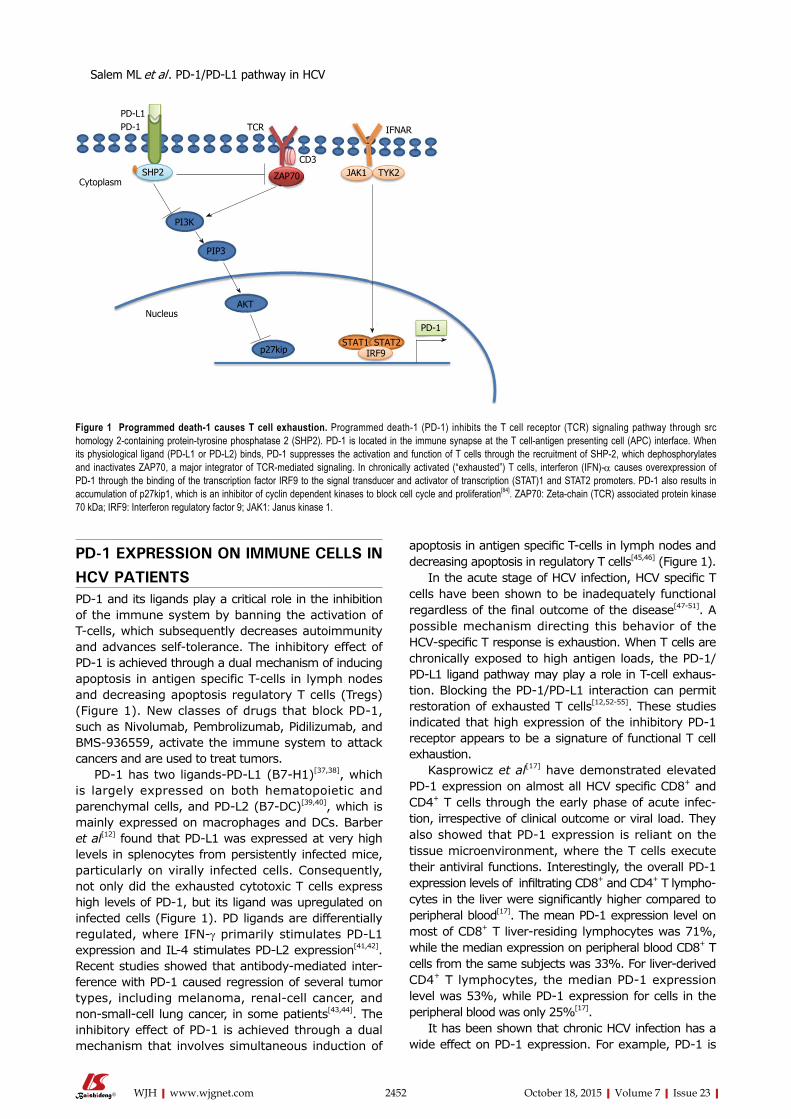
2452 October 18, 2015|Volume 7|Issue 23|WJH|www.wjgnet.com
apoptosis in antigen specific T-cells in lymph nodes and decreasing apoptosis in regulatory T cells[45,46] (Figure 1).
In the acute stage of HCV infection, HCV specific T cells have been shown to be inadequately functional regardless of the final outcome of the disease[4751]. A possible mechanism directing this behavior of the HCVspecific T response is exhaustion. When T cells are chronically exposed to high antigen loads, the PD1/PDL1 ligand pathway may play a role in Tcell exhaustion. Blocking the PD1/PDL1 interaction can permit restoration of exhausted T cells[12,5255]. These studies indicated that high expression of the inhibitory PD1 receptor appears to be a signature of functional T cell exhaustion.
Kasprowicz et al[17] have demonstrated elevated PD1 expression on almost all HCV specific CD8+ and CD4+ T cells through the early phase of acute infection, irrespective of clinical outcome or viral load. They also showed that PD1 expression is reliant on the tissue microenvironment, where the T cells execute their antiviral functions. Interestingly, the overall PD1 expression levels of infiltrating CD8+ and CD4+ T lymphocytes in the liver were significantly higher compared to peripheral blood[17]. The mean PD1 expression level on most of CD8+ T liverresiding lymphocytes was 71%, while the median expression on peripheral blood CD8+ T cells from the same subjects was 33%. For liverderived CD4+ T lymphocytes, the median PD1 expression level was 53%, while PD1 expression for cells in the peripheral blood was only 25%[17].
It has been shown that chronic HCV infection has a wide effect on PD1 expression. For example, PD1 is
PD-1 EXPRESSION ON IMMUNE CELLS IN HCV PATIENTSPD1 and its ligands play a critical role in the inhibition of the immune system by banning the activation of Tcells, which subsequently decreases autoimmunity and advances selftolerance. The inhibitory effect of PD1 is achieved through a dual mechanism of inducing apoptosis in antigen specific Tcells in lymph nodes and decreasing apoptosis regulatory T cells (Tregs) (Figure 1). New classes of drugs that block PD1, such as Nivolumab, Pembrolizumab, Pidilizumab, and BMS936559, activate the immune system to attack cancers and are used to treat tumors.
PD1 has two ligandsPDL1 (B7H1)[37,38], which is largely expressed on both hematopoietic and parenchymal cells, and PDL2 (B7DC)[39,40], which is mainly expressed on macrophages and DCs. Barber et al[12] found that PDL1 was expressed at very high levels in splenocytes from persistently infected mice, particularly on virally infected cells. Consequently, not only did the exhausted cytotoxic T cells express high levels of PD1, but its ligand was upregulated on infected cells (Figure 1). PD ligands are differentially regulated, where IFNγ primarily stimulates PDL1 expression and IL4 stimulates PDL2 expression[41,42]. Recent studies showed that antibodymediated interference with PD1 caused regression of several tumor types, including melanoma, renalcell cancer, and nonsmallcell lung cancer, in some patients[43,44]. The inhibitory effect of PD1 is achieved through a dual mechanism that involves simultaneous induction of
PD-L1PD-1 TCR
CD3
IFNAR
SHP2 ZAP70 JAK1 TYK2Cytoplasm
PI3K
PIP3
AKT
p27kip
Nucleus
STAT1 STAT2IRF9
PD-1
Figure 1 Programmed death-1 causes T cell exhaustion. Programmed death-1 (PD-1) inhibits the T cell receptor (TCR) signaling pathway through src homology 2-containing protein-tyrosine phosphatase 2 (SHP2). PD-1 is located in the immune synapse at the T cell-antigen presenting cell (APC) interface. When its physiological ligand (PD-L1 or PD-L2) binds, PD-1 suppresses the activation and function of T cells through the recruitment of SHP-2, which dephosphorylates and inactivates ZAP70, a major integrator of TCR-mediated signaling. In chronically activated (“exhausted”) T cells, interferon (IFN)-α causes overexpression of PD-1 through the binding of the transcription factor IRF9 to the signal transducer and activator of transcription (STAT)1 and STAT2 promoters. PD-1 also results in accumulation of p27kip1, which is an inhibitor of cyclin dependent kinases to block cell cycle and proliferation[84]. ZAP70: Zeta-chain (TCR) associated protein kinase 70 kDa; IRF9: Interferon regulatory factor 9; JAK1: Janus kinase 1.
Salem ML et al . PD-1/PD-L1 pathway in HCV
2453 October 18, 2015|Volume 7|Issue 23|WJH|www.wjgnet.com
highly expressed on peripheral B cells and monocytes, since it is induced upon activation[56]. In patients with chronic HCV, CD56high NK cells expressed greater levels of PD1, convenient considering their greater functional deficiency and less mature CD56low differentiation state[57]. In addition, PD1 is expressed on Kupffer cells in the liver, other monocytederived cells, as well as epithelial, endothelial, and tumor cells[52,58,59].
Although HCVspecific CD8+ Tcells are generally dysfunctional in HCV persistence, their level of impairment varied considerably among patients depending on PD1 expression. Within an individual patient, the function and PD1 expression of HCVspecific CD8+ Tcells varied between the liver and peripheral blood[60]. Additional studies on the expression patterns of diverse splice variants of PD1, PDL1, and receptorligand interactions in diseased tissue will be important in determining a more comprehensive estimation of the level of the inhibitory signal and its effect on the outcome of human infection. Such studies will not only provide a superior mechanistic understanding of the PD1 pathway in controlling T cell responses but will also encourage specific manipulation of this pathway therapeutically.
CORRELATION OF PD-1 EXPRESSION AND CLINICAL RESPONSESThe identification of cellular and molecular factors predicting clinical response to immunotherapy is strongly desirable, not only to aid in the design of therapies that overcome and enhance the inhibitory and stimulatory mechanisms, but also to preselect patients most likely to benefit from therapy and spare others from unnecessary exposure to possible side effects. Similar to its inhibitory role in anticancer immunity, the PD1 signaling pathway has also been found to shape the overall immunity in HCV infection. For instance, PD1 expression in acute HCV infection was found to be a signature of functional HCVspecific CD8 T cell exhaustion[16]. In this study, and as reported for the acute infection of HBV[61], the expression of PD-1 by HCV specific cytotoxic T cells was decreased in selflimited infections after the acute stage of infection in conjunction with CD8+ T cell differentiation towards a memory CD127 phenotype[61]. In contrast, HCV specific CD8+ T cells maintained high levels of PD1 in patients with chronic advancement of infection and remained functionally impeded with no change from an effector to a memory phenotype[16]. Other studies,
however, reported high levels of PD1 expression (60%100%) on all HCV specific CD8+ and CD4+ cells during the early stage of acute infection, irrespective of the clinical outcome or viral load[17]. Taken together, these results suggested that a role for the PD1/PDL1 interaction in regulating CD8+ T cell function may exist under conditions of continuous high levels of HCV antigen stimulation.
Consistent with this suggestion, another study showed that the level of PD1 expression in early phases of HCV infection was significantly more on HCV-specific T cells from patients who advanced to chronic HCV infection than from those who eliminated infection; and this correlation was independent of HCV RNA titer levels[62]. The reason for this difference is unclear, but it may be due to differences between the routes of infection between the two studies. This suggests that some of the biological differences that cause the development of symptoms likewise affect PD1 expression. Additionally, the duration of infection in patients defined as acutely infected could be different between studies. Table 1 provides a list of studies involving PD1 expression and the role of PD1 in HCV infection.
PD1 expression was investigated in 72 treatment naïve patients with persistent HCV[57]. In this study, PD1 expression was upregulated significantly not only on CD4+ and CD8+ T cells but also on NK cells, connecting with failed early and persistent virologic response to therapy. In contrast, patients with SVR demonstrated decreases in PD1 after therapy completion, demonstrating that PD1 expressed by NK cells is critical in persistent HCV[57].
PD1+ HCVspecific CD8+ T cells in chronic HCV infection have a tendency to coexpress Tim3[63], 2B4, CD160 and other inhibitory molecules[64], particularly in the liver[65] since intrahepatic T cells showed a more exhausted phenotype than in the blood. In addition, the level of TIM3 from patients with persistent HCV infection was greater than those who resolved the infection.
The results from other studies, however, are inconsistent regarding the differences in levels of PD1/PDL1 on HCVspecific CD8+ cells contrasts between those who clear HCV infection and those with persistent infection[16,17,62]. Most of these studies though concluded that PD1 expression levels are elevated on HCVspecific T cells vs naïve CD8+ T cells or on T cells specific for some control antigens in the acute stage of infection, regardless of the outcome.
Table 1 Differences between the studies made on the correlation between programmed death-1 expression and clinical outcome
Ref. No. of patients Mode of transfection Symptoms PD-1 expression levels in acute infection
PD-1 expression levels in resolved patients
Urbani et al[16] 19 Sexually transmission Symptomatic High DecreasedKasprowicz et al[17] 37 Sexually transmission Symptomatic High IrrespectiveRutebemberwa et al[62] 20 Injection drug use Asymptomatic High Decreased
PD-1: Programmed death-1.
Salem ML et al . PD-1/PD-L1 pathway in HCV

2454 October 18, 2015|Volume 7|Issue 23|WJH|www.wjgnet.com
RESTORATION OF ANTI-HCV RESPONSES IN VITRO By BLOCKING PD-1During the acute phase of HCV infection, HCV specific T cells have been characterized as poorly functional, regardless of the outcome of the disease[4751]. A possible explanation for this behavior of the HCV-specific CD8+ T response is exhaustion, which is supported by the initial rapid kinetics of HCV spread, replication after infection, and later on, by the continuous exposure of CD8+ T cells to high antigen load. Several studies demonstrated that the PD1/PDL1 pathway plays a role in Tcell exhaustion when CD8+ cells are chronically exposed to high level antigen loads. Blockade of this pathway can permit restoration of exhausted CD8+ T cells[12,5255] and results in expansion of HCV specific T cell proliferation[15,16,66] (Figure 2).
For instance, PD-L1 blockade improved HCV-specific T cell proliferation in a dosedependent manner. However, proliferation of cytomegalovirus (CMV)specific CD8+ T cells was not affected by PD1/PDL1 blockade, consistent with their low expression levels of PD1. Similar to its effects on CD8+ T cells in HCV, blocking the PD1/PDL1 interaction in vitro restored effector function and enhanced the proliferative ability of exhausted CD8+ T cells in many chronic infections, including HIV, HBV, simian immunodeficiency virus (SIV), LCMV, and EpsteinBarr virus (EBV)[13,15,6771].
This functional T cell restoration by blocking the PD1/PDL1 pathway is a hierarchical phenomenon that appears to reflect the different sensitivities to exhaustion of the diverse T cell functions[72]. Restoration of proliferation capacity is relatively faster than the restoration of IFNγ and IL2 production but with no effect on cytotoxicity. In line with this, treatment with antiPDL1 antibodies does not always predict the expected positive effect of PD1 blockade[73], suggesting that PDL1 binds at least one additional receptor
other than PD1 to mediate its costimulatory function. Moreover, exhaustion was more effectively overcomed in HCV, than in HIV[68] by PD1/PDL1 blockade, as demonstrated by the increased capability of HCVspecific CD8+ T cells to expand and to produce IFN and IL2 after incubation with anti PDL1 antibodies[15].
Collectively, these in vitro data suggest that PD1 signaling on T cells is a significant inhibitory pathway during chronic HCV infection. Consequently, the possibility of partially restoring CD8+ T cell function by blocking PD1/PDL1 interaction may provide a valuable tool for the enhancement of available therapies to cure chronic hepatitis C.
POTENTIAL USE OF IN VIVO PD-1 BLOCKING FOR ANTI-HCV THERAPyAs demonstrated above, in vitro blockade of PD1 can restore the functionality of HCVspecific T cells. Blockade of PD1 signaling was tested in vivo in both chimpanzees[74] and in patients with chronic HCV infection[75]. In the chimpanzee study, an increase in HCV specific CD8+ cell responses and a considerable, although transient, reduction in HCV viremia was only seen in one of three chimpanzees. This chimpanzee had the strongest and broadest CD4+ and CD8+ T cell response before the development of chronic infection, which suggested that PD1 blockade alone is not sufficient to attain viral clearance[74]. In the patient study, a single dose (10 mg/kg) of the PD1 blocking antibody BMS936558 was followed by a greater than 0.5 log10 IU/mL decrease in HCV RNA titer in five of 45 (11%) patients. At the highest dose given (10 mg/kg), a > 4 log10 IU/mL decrease in HCV RNA titer was seen in three of 20 (15%) patients. This decrease of HCV replication continued for more than 8 wk in most patients[75].
Interestingly, in vivo PDL1 blockade does not appear to affect IL10 level during chronic infection, although
ProliferationProliferation
Anti PD-1
Normal T cell
Partially exhausted T cell
Severely exhausted T cell
Figure 2 Proliferation of exhausted T cells and blockage strategies to reverse exhaustion. Severely exhausted T cells (red cells) proliferate poorly in comparison to partially exhausted and normal T cells (yellow and green cells). Antibody blockade of the pathway with PD-1 and its ligand reverses exhaustion and restores the functional capacities of exhausted T cells. PD-1: Programmed death-1.
Salem ML et al . PD-1/PD-L1 pathway in HCV

2455 October 18, 2015|Volume 7|Issue 23|WJH|www.wjgnet.com
it downregulated IL10 and upregulated IL2 and IFNγ during in vitro stimulation[76,77]. Another ex vivo study showed that intrahepatic T cells were significantly dysfunctional and insusceptible to PD1 blockade[60]. Therefore, the effect of PD1 blockade is characterized by T cell compartmentalization. Additionally, PD1 expression on HCV specific CD8+ cells is affected by viral immuneevasion in chronic HCV infections[62].
Indeed, an efficient helper T cell function is compulsory for the development of virus specific CD8 cells[78]. Thus, the synergistic effect of CD8 and CD4mediated T cell functions enhanced by antiPDL1 may represent a strategy to enhance the effect of available antiHCV drugs. Consequently, it is conceivable that utilizing PD1/PDL1 blockade can enhance the antiviral effect of IFN/RBV therapy. Additional studies are required, however, to survey whether the enhancement of the T cell function induced by PD1/PDL1 blockade and favored by IFNα therapy in patients with a recent HCV infection can be also accomplished in chronic infections of longer duration, where the effect of long lasting exhaustion may be more difficult to succeed. Therefore, the potential of partially restoring CD8+ cells function by blocking PD1/PDL1 interaction could provide an additional tool to enhance available therapies to cure chronic HCV.
CONCLUSIONIn spite of much progress in our understanding of T cell responses against HCV over the past decade, many critical research questions remain to be answered. It will be important to determine whether there is a causal correlation between the outcome of HCV infection and PD-1 expression. Functionally deficient exhausted HCV specific T cells are a significant cause and outcome of chronic viral infection. This is a great challenge to vaccine designs that either aim to eliminate or prevent such infections by interceding the T cell response. Whether developing a therapeutic vaccine to eliminate disease in infected patients or a prophylactic vaccine to prevent HCV in healthy individuals, it is imperative to keep this considerable obstacle in mind.
The possibility of restoring the function of HCV specific T cells by blocking the PD1/PDL1 pathway and reverting Tcell dysfunction in chronic HCV seems a worthy direction for antiHCV immunotherapy. While the therapeutic application of this strategy in HCV infection is constrained by the recent, ongoing development of highly efficacious new treatments, it is promising that further investigation of PD1 pathway blockade during antiviral therapies is warranted.
However, the systemic administration of PDL1/PD1 blocking antibodies carries the high risk of violating peripheral tolerance. Most tissues depend on PDL1 expression to decrease Tcell effector activities that might cause autoimmune attack. To demonstrate this, knocking out PD1 or PDL1 pathways in mouse models causes severe, deadly autoimmunity[79,80]. In humans,
singlenucleotide polymorphisms (SNP) of the PDCD1 gene (encoding PD1) are connected with systemic lupus erythematosus[81]. Therefore, efforts have to be made to enhance this promising strategy to maximize antiHCV therapeutic activities while minimizing toxicity. We propose that one possible attractive alternative to the systemic blockade of PD1 for HCV immunotherapy is to target the suppression of PD1/PDL1 costimulation during antigen presentation. This was demonstrated in mouse antigen presenting cells (APCs)[82,83]. This local and transient blockade may provide the positive effects needed to adequately boost antiHCV immunity while restricting possible side effects to a minimum.
Overall, continuing work to understand better how the PD1/PDL1 pathway functions is imperative and will encourage development of new vaccination approaches that can overcome HCV specific T cell exhaustion.
REFERENCES 1 Weiss RA, McMichael AJ. Social and environmental risk factors in
the emergence of infectious diseases. Nat Med 2004; 10: S70-S76 [PMID: 15577934 DOI: 10.1038/nm1150]
2 Alter MJ. Epidemiology of hepatitis C in the West. Semin Liver Dis 1995; 15: 5-14 [PMID: 7597444 DOI: 10.1055/s-2007-1007259]
3 Seeff LB. Natural history of chronic hepatitis C. Hepatology 2002; 36: S35-S46 [PMID: 12407575 DOI: 10.1053/jhep.2002.36806]
4 Huang L, Koziel MJ. Immunology of hepatitis C virus infection. Curr Opin Gastroenterol 2000; 16: 558-564 [PMID: 17031138]
5 Rehermann B, Nascimbeni M. Immunology of hepatitis B virus and hepatitis C virus infection. Nat Rev Immunol 2005; 5: 215-229 [PMID: 15738952 DOI: 10.1038/nri1573]
6 Dustin LB, Rice CM. Flying under the radar: the immunobiology of hepatitis C. Annu Rev Immunol 2007; 25: 71-99 [PMID: 17067278 DOI: 10.1146/annurev.immunol.25.022106.141602]
7 Shin EC, Capone S, Cortese R, Colloca S, Nicosia A, Folgori A, Rehermann B. The kinetics of hepatitis C virus-specific CD8 T-cell responses in the blood mirror those in the liver in acute hepatitis C virus infection. J Virol 2008; 82: 9782-9788 [PMID: 18667501 DOI: 10.1128/JVI.00475-08]
8 Shin EC, Seifert U, Kato T, Rice CM, Feinstone SM, Kloetzel PM, Rehermann B. Virus-induced type I IFN stimulates generation of immunoproteasomes at the site of infection. J Clin Invest 2006; 116: 3006-3014 [PMID: 17039255 DOI: 10.1172/JCI29832]
9 Folgori A, Capone S, Ruggeri L, Meola A, Sporeno E, Ercole BB, Pezzanera M, Tafi R, Arcuri M, Fattori E, Lahm A, Luzzago A, Vitelli A, Colloca S, Cortese R, Nicosia A. A T-cell HCV vaccine eliciting effective immunity against heterologous virus challenge in chimpanzees. Nat Med 2006; 12: 190-197 [PMID: 16462801 DOI: 10.1038/nm1353]
10 Shin EC, Rehermann B. Taking the brake off T cells in chronic viral infection. Nat Med 2006; 12: 276-277 [PMID: 16520769 DOI: 10.1038/nm0306-276]
11 Kanto T, Hayashi N. Immunopathogenesis of hepatitis C virus infection: multifaceted strategies subverting innate and adaptive immunity. Intern Med 2006; 45: 183-191 [PMID: 16543687]
12 Barber DL, Wherry EJ, Masopust D, Zhu B, Allison JP, Sharpe AH, Freeman GJ, Ahmed R. Restoring function in exhausted CD8 T cells during chronic viral infection. Nature 2006; 439: 682-687 [PMID: 16382236 DOI: 10.1038/nature04444]
13 Day CL, Kaufmann DE, Kiepiela P, Brown JA, Moodley ES, Reddy S, Mackey EW, Miller JD, Leslie AJ, DePierres C, Mncube Z, Duraiswamy J, Zhu B, Eichbaum Q, Altfeld M, Wherry EJ, Coovadia HM, Goulder PJ, Klenerman P, Ahmed R, Freeman GJ, Walker BD. PD-1 expression on HIV-specific T cells is associated with T-cell exhaustion and disease progression. Nature 2006; 443: 350-354 [PMID: 16921384 DOI: 10.1038/nature05115]
Salem ML et al . PD-1/PD-L1 pathway in HCV

2456 October 18, 2015|Volume 7|Issue 23|WJH|www.wjgnet.com
14 Radziewicz H, Ibegbu CC, Fernandez ML, Workowski KA, Obideen K, Wehbi M, Hanson HL, Steinberg JP, Masopust D, Wherry EJ, Altman JD, Rouse BT, Freeman GJ, Ahmed R, Grakoui A. Liver-infiltrating lymphocytes in chronic human hepatitis C virus infection display an exhausted phenotype with high levels of PD-1 and low levels of CD127 expression. J Virol 2007; 81: 2545-2553 [PMID: 17182670 DOI: 10.1128/JVI.02021-06]
15 Penna A, Pilli M, Zerbini A, Orlandini A, Mezzadri S, Sacchelli L, Missale G, Ferrari C. Dysfunction and functional restoration of HCV-specific CD8 responses in chronic hepatitis C virus infection. Hepatology 2007; 45: 588-601 [PMID: 17326153 DOI: 10.1002/hep.21541]
16 Urbani S, Amadei B, Tola D, Massari M, Schivazappa S, Missale G, Ferrari C. PD-1 expression in acute hepatitis C virus (HCV) infection is associated with HCV-specific CD8 exhaustion. J Virol 2006; 80: 11398-11403 [PMID: 16956940 DOI: 10.1128/JVI.01177-06]
17 Kasprowicz V, Schulze Zur Wiesch J, Kuntzen T, Nolan BE, Longworth S, Berical A, Blum J, McMahon C, Reyor LL, Elias N, Kwok WW, McGovern BG, Freeman G, Chung RT, Klenerman P, Lewis-Ximenez L, Walker BD, Allen TM, Kim AY, Lauer GM. High level of PD-1 expression on hepatitis C virus (HCV)-specific CD8+ and CD4+ T cells during acute HCV infection, irrespective of clinical outcome. J Virol 2008; 82: 3154-3160 [PMID: 18160439 DOI: 10.1128/JVI.02474-07]
18 Hadziyannis SJ, Sette H, Morgan TR, Balan V, Diago M, Marcellin P, Ramadori G, Bodenheimer H, Bernstein D, Rizzetto M, Zeuzem S, Pockros PJ, Lin A, Ackrill AM. Peginterferon-alpha2a and ribavirin combination therapy in chronic hepatitis C: a randomized study of treatment duration and ribavirin dose. Ann Intern Med 2004; 140: 346-355 [PMID: 14996676]
19 Manns MP, McHutchison JG, Gordon SC, Rustgi VK, Shiffman M, Reindollar R, Goodman ZD, Koury K, Ling M, Albrecht JK. Peginterferon alfa-2b plus ribavirin compared with interferon alfa-2b plus ribavirin for initial treatment of chronic hepatitis C: a randomised trial. Lancet 2001; 358: 958-965 [PMID: 11583749]
20 McHutchison JG, Lawitz EJ, Shiffman ML, Muir AJ, Galler GW, McCone J, Nyberg LM, Lee WM, Ghalib RH, Schiff ER, Galati JS, Bacon BR, Davis MN, Mukhopadhyay P, Koury K, Noviello S, Pedicone LD, Brass CA, Albrecht JK, Sulkowski MS. Peginterferon alfa-2b or alfa-2a with ribavirin for treatment of hepatitis C infection. N Engl J Med 2009; 361: 580-593 [PMID: 19625712 DOI: 10.1056/NEJMoa0808010]
21 Shiffman ML, Suter F, Bacon BR, Nelson D, Harley H, Solá R, Shafran SD, Barange K, Lin A, Soman A, Zeuzem S. Peginterferon alfa-2a and ribavirin for 16 or 24 weeks in HCV genotype 2 or 3. N Engl J Med 2007; 357: 124-134 [PMID: 17625124 DOI: 10.1056/NEJMoa066403]
22 Parise E, Cheinquer H, Crespo D, Meirelles A, Martinelli A, Sette H, Gallizi J, Silva R, Lacet C, Correa E, Cotrim H, Fonseca J, Paraná R, Spinelli V, Amorim W, Tatsch F, Pessoa M. Peginterferon alfa-2a (40KD) (PEGASYS) plus ribavirin (COPEGUS) in retreatment of chronic hepatitis C patients, nonresponders and relapsers to previous conventional interferon plus ribavirin therapy. Braz J Infect Dis 2006; 10: 11-16 [PMID: 16767309]
23 Muir AJ, Bornstein JD, Killenberg PG. Peginterferon alfa-2b and ribavirin for the treatment of chronic hepatitis C in blacks and non-Hispanic whites. N Engl J Med 2004; 350: 2265-2271 [PMID: 15163776 DOI: 10.1056/NEJMoa032502]
24 Poynard T, Ratziu V, McHutchison J, Manns M, Goodman Z, Zeuzem S, Younossi Z, Albrecht J. Effect of treatment with peginterferon or interferon alfa-2b and ribavirin on steatosis in patients infected with hepatitis C. Hepatology 2003; 38: 75-85 [PMID: 12829989 DOI: 10.1053/jhep.2003.50267]
25 Gao B, Hong F, Radaeva S. Host factors and failure of interferon-alpha treatment in hepatitis C virus. Hepatology 2004; 39: 880-890 [PMID: 15057887 DOI: 10.1002/hep.20139]
26 Meylan E, Curran J, Hofmann K, Moradpour D, Binder M, Bartenschlager R, Tschopp J. Cardif is an adaptor protein in the RIG-I antiviral pathway and is targeted by hepatitis C virus. Nature 2005;
437: 1167-1172 [PMID: 16177806 DOI: 10.1038/nature04193]27 Li XD, Sun L, Seth RB, Pineda G, Chen ZJ. Hepatitis C virus
protease NS3/4A cleaves mitochondrial antiviral signaling protein off the mitochondria to evade innate immunity. Proc Natl Acad Sci USA 2005; 102: 17717-17722 [PMID: 16301520 DOI: 10.1073/pnas.0508531102]
28 Li K, Foy E, Ferreon JC, Nakamura M, Ferreon AC, Ikeda M, Ray SC, Gale M, Lemon SM. Immune evasion by hepatitis C virus NS3/4A protease-mediated cleavage of the Toll-like receptor 3 adaptor protein TRIF. Proc Natl Acad Sci USA 2005; 102: 2992-2997 [PMID: 15710891 DOI: 10.1073/pnas.0408824102]
29 Bode JG, Ludwig S, Ehrhardt C, Albrecht U, Erhardt A, Schaper F, Heinrich PC, Häussinger D. IFN-alpha antagonistic activity of HCV core protein involves induction of suppressor of cytokine signaling-3. FASEB J 2003; 17: 488-490 [PMID: 12551851 DOI: 10.1096/fj.02-0664fje]
30 Hézode C, Fontaine H, Dorival C, Larrey D, Zoulim F, Canva V, de Ledinghen V, Poynard T, Samuel D, Bourlière M, Zarski JP, Raabe JJ, Alric L, Marcellin P, Riachi G, Bernard PH, Loustaud-Ratti V, Métivier S, Tran A, Serfaty L, Abergel A, Causse X, Di Martino V, Guyader D, Lucidarme D, Grando-Lemaire V, Hillon P, Feray C, Dao T, Cacoub P, Rosa I, Attali P, Petrov-Sanchez V, Barthe Y, Pawlotsky JM, Pol S, Carrat F, Bronowicki JP. Triple therapy in treatment-experienced patients with HCV-cirrhosis in a multicentre cohort of the French Early Access Programme (ANRS CO20-CUPIC) - NCT01514890. J Hepatol 2013; 59: 434-441 [PMID: 23669289 DOI: 10.1016/j.jhep.2013.04.035]
31 Ward RP, Kugelmas M. Using pegylated interferon and ribavirin to treat patients with chronic hepatitis C. Am Fam Physician 2005; 72: 655-662 [PMID: 16127955]
32 Bruchfeld A, Lindahl K, Ståhle L, Söderberg M, Schvarcz R. Interferon and ribavirin treatment in patients with hepatitis C-associated renal disease and renal insufficiency. Nephrol Dial Transplant 2003; 18: 1573-1580 [PMID: 12897097]
33 Wen Y, feng LY, Jing Y, Xin L, Zhikai X. Controlling hepatitis C with immunotherapy. Lancet Infect Dis 2009; 9: 652-653 [PMID: 19850221 DOI: 10.1016/S1473-3099(09)70258-0]
34 Klade CS, Wedemeyer H, Berg T, Hinrichsen H, Cholewinska G, Zeuzem S, Blum H, Buschle M, Jelovcan S, Buerger V, Tauber E, Frisch J, Manns MP. Therapeutic vaccination of chronic hepatitis C nonresponder patients with the peptide vaccine IC41. Gastroenterology 2008; 134: 1385-1395 [PMID: 18471515 DOI: 10.1053/j.gastro.2008.02.058]
35 Salem ML, El-Demellawy M, El-Azm AR. The potential use of Toll-like receptor agonists to restore the dysfunctional immunity induced by hepatitis C virus. Cell Immunol 2010; 262: 96-104 [PMID: 20338549 DOI: 10.1016/j.cellimm.2010.03.002]
36 Penaloza-MacMaster P, Kamphorst AO, Wieland A, Araki K, Iyer SS, West EE, O’Mara L, Yang S, Konieczny BT, Sharpe AH, Freeman GJ, Rudensky AY, Ahmed R. Interplay between regulatory T cells and PD-1 in modulating T cell exhaustion and viral control during chronic LCMV infection. J Exp Med 2014; 211: 1905-1918 [PMID: 25113973 DOI: 10.1084/jem.20132577]
37 Freeman GJ, Long AJ, Iwai Y, Bourque K, Chernova T, Nishimura H, Fitz LJ, Malenkovich N, Okazaki T, Byrne MC, Horton HF, Fouser L, Carter L, Ling V, Bowman MR, Carreno BM, Collins M, Wood CR, Honjo T. Engagement of the PD-1 immunoinhibitory receptor by a novel B7 family member leads to negative regulation of lymphocyte activation. J Exp Med 2000; 192: 1027-1034 [PMID: 11015443]
38 Dong H, Zhu G, Tamada K, Chen L. B7-H1, a third member of the B7 family, co-stimulates T-cell proliferation and interleukin-10 secretion. Nat Med 1999; 5: 1365-1369 [PMID: 10581077 DOI: 10.1038/70932]
39 Sharpe AH, Freeman GJ. The B7-CD28 superfamily. Nat Rev Immunol 2002; 2: 116-126 [PMID: 11910893 DOI: 10.1038/nri727]
40 Chen L. Co-inhibitory molecules of the B7-CD28 family in the control of T-cell immunity. Nat Rev Immunol 2004; 4: 336-347 [PMID: 15122199 DOI: 10.1038/nri1349]
41 Liang SC, Latchman YE, Buhlmann JE, Tomczak MF, Horwitz
Salem ML et al . PD-1/PD-L1 pathway in HCV

2457 October 18, 2015|Volume 7|Issue 23|WJH|www.wjgnet.com
BH, Freeman GJ, Sharpe AH. Regulation of PD-1, PD-L1, and PD-L2 expression during normal and autoimmune responses. Eur J Immunol 2003; 33: 2706-2716 [PMID: 14515254 DOI: 10.1002/eji.200324228]
42 Loke P, Allison JP. PD-L1 and PD-L2 are differentially regulated by Th1 and Th2 cells. Proc Natl Acad Sci USA 2003; 100: 5336-5341 [PMID: 12697896 DOI: 10.1073/pnas.0931259100]
43 Brahmer JR, Tykodi SS, Chow LQ, Hwu WJ, Topalian SL, Hwu P, Drake CG, Camacho LH, Kauh J, Odunsi K, Pitot HC, Hamid O, Bhatia S, Martins R, Eaton K, Chen S, Salay TM, Alaparthy S, Grosso JF, Korman AJ, Parker SM, Agrawal S, Goldberg SM, Pardoll DM, Gupta A, Wigginton JM. Safety and activity of anti-PD-L1 antibody in patients with advanced cancer. N Engl J Med 2012; 366: 2455-2465 [PMID: 22658128 DOI: 10.1056/NEJMoa1200694]
44 Topalian SL, Hodi FS, Brahmer JR, Gettinger SN, Smith DC, McDermott DF, Powderly JD, Carvajal RD, Sosman JA, Atkins MB, Leming PD, Spigel DR, Antonia SJ, Horn L, Drake CG, Pardoll DM, Chen L, Sharfman WH, Anders RA, Taube JM, McMiller TL, Xu H, Korman AJ, Jure-Kunkel M, Agrawal S, McDonald D, Kollia GD, Gupta A, Wigginton JM, Sznol M. Safety, activity, and immune correlates of anti-PD-1 antibody in cancer. N Engl J Med 2012; 366: 2443-2454 [PMID: 22658127 DOI: 10.1056/NEJMoa1200690]
45 Fife BT, Pauken KE. The role of the PD-1 pathway in autoim-munity and peripheral tolerance. Ann N Y Acad Sci 2011; 1217: 45-59 [PMID: 21276005 DOI: 10.1111/j.1749-6632.2010.05919.x]
46 Francisco LM, Sage PT, Sharpe AH. The PD-1 pathway in tolerance and autoimmunity. Immunol Rev 2010; 236: 219-242 [PMID: 20636820 DOI: 10.1111/j.1600-065X.2010.00923.x]
47 Gruener NH, Lechner F, Jung MC, Diepolder H, Gerlach T, Lauer G, Walker B, Sullivan J, Phillips R, Pape GR, Klenerman P. Sustained dysfunction of antiviral CD8+ T lymphocytes after infection with hepatitis C virus. J Virol 2001; 75: 5550-5558 [PMID: 11356962 DOI: 10.1128/JVI.75.12.5550-5558.2001]
48 Lechner F, Wong DK, Dunbar PR, Chapman R, Chung RT, Dohrenwend P, Robbins G, Phillips R, Klenerman P, Walker BD. Analysis of successful immune responses in persons infected with hepatitis C virus. J Exp Med 2000; 191: 1499-1512 [PMID: 10790425]
49 Thimme R, Oldach D, Chang KM, Steiger C, Ray SC, Chisari FV. Determinants of viral clearance and persistence during acute hepatitis C virus infection. J Exp Med 2001; 194: 1395-1406 [PMID: 11714747]
50 Urbani S, Boni C, Missale G, Elia G, Cavallo C, Massari M, Raimondo G, Ferrari C. Virus-specific CD8+ lymphocytes share the same effector-memory phenotype but exhibit functional differences in acute hepatitis B and C. J Virol 2002; 76: 12423-12434 [PMID: 12438568]
51 Urbani S, Amadei B, Fisicaro P, Tola D, Orlandini A, Sacchelli L, Mori C, Missale G, Ferrari C. Outcome of acute hepatitis C is related to virus-specific CD4 function and maturation of antiviral memory CD8 responses. Hepatology 2006; 44: 126-139 [PMID: 16799989 DOI: 10.1002/hep.21242]
52 Brown JA, Dorfman DM, Ma FR, Sullivan EL, Munoz O, Wood CR, Greenfield EA, Freeman GJ. Blockade of programmed death-1 ligands on dendritic cells enhances T cell activation and cytokine production. J Immunol 2003; 170: 1257-1266 [PMID: 12538684]
53 Cai G, Karni A, Oliveira EM, Weiner HL, Hafler DA, Freeman GJ. PD-1 ligands, negative regulators for activation of naive, memory, and recently activated human CD4+ T cells. Cell Immunol 2004; 230: 89-98 [PMID: 15598424 DOI: 10.1016/j.cellimm.2004.09.004]
54 Latchman YE, Liang SC, Wu Y, Chernova T, Sobel RA, Klemm M, Kuchroo VK, Freeman GJ, Sharpe AH. PD-L1-deficient mice show that PD-L1 on T cells, antigen-presenting cells, and host tissues negatively regulates T cells. Proc Natl Acad Sci USA 2004; 101: 10691-10696 [PMID: 15249675 DOI: 10.1073/pnas.0307252101]
55 Okazaki T, Honjo T. The PD-1-PD-L pathway in immunological tolerance. Trends Immunol 2006; 27: 195-201 [PMID: 16500147 DOI: 10.1016/j.it.2006.02.001]
56 Riley JL, June CH. The road to recovery: translating PD-1 biology into clinical benefit. Trends Immunol 2007; 28: 48-50 [PMID: 17188572 DOI: 10.1016/j.it.2006.12.001]
57 Golden-Mason L, Klarquist J, Wahed AS, Rosen HR. Cutting edge: programmed death-1 expression is increased on immunocytes in chronic hepatitis C virus and predicts failure of response to antiviral therapy: race-dependent differences. J Immunol 2008; 180: 3637-3641 [PMID: 18322167]
58 Dong H, Strome SE, Salomao DR, Tamura H, Hirano F, Flies DB, Roche PC, Lu J, Zhu G, Tamada K, Lennon VA, Celis E, Chen L. Tumor-associated B7-H1 promotes T-cell apoptosis: a potential mechanism of immune evasion. Nat Med 2002; 8: 793-800 [PMID: 12091876 DOI: 10.1038/nm730]
59 Selenko-Gebauer N, Majdic O, Szekeres A, Höfler G, Guthann E, Korthäuer U, Zlabinger G, Steinberger P, Pickl WF, Stockinger H, Knapp W, Stöckl J. B7-H1 (programmed death-1 ligand) on dendritic cells is involved in the induction and maintenance of T cell anergy. J Immunol 2003; 170: 3637-3644 [PMID: 12646628]
60 Nakamoto N, Kaplan DE, Coleclough J, Li Y, Valiga ME, Kaminski M, Shaked A, Olthoff K, Gostick E, Price DA, Freeman GJ, Wherry EJ, Chang KM. Functional restoration of HCV-specific CD8 T cells by PD-1 blockade is defined by PD-1 expression and compartmentalization. Gastroenterology 2008; 134: 1927-1937, 1937.e1-2 [PMID: 18549878 DOI: 10.1053/j.gastro.2008.02.033]
61 Boettler T, Panther E, Bengsch B, Nazarova N, Spangenberg HC, Blum HE, Thimme R. Expression of the interleukin-7 receptor alpha chain (CD127) on virus-specific CD8+ T cells identifies functionally and phenotypically defined memory T cells during acute resolving hepatitis B virus infection. J Virol 2006; 80: 3532-3540 [PMID: 16537621 DOI: 10.1128/JVI.80.7.3532-3540.2006]
62 Rutebemberwa A, Ray SC, Astemborski J, Levine J, Liu L, Dowd KA, Clute S, Wang C, Korman A, Sette A, Sidney J, Pardoll DM, Cox AL. High-programmed death-1 levels on hepatitis C virus-specific T cells during acute infection are associated with viral persistence and require preservation of cognate antigen during chronic infection. J Immunol 2008; 181: 8215-8225 [PMID: 19050238]
63 Golden-Mason L, Palmer BE, Kassam N, Townshend-Bulson L, Livingston S, McMahon BJ, Castelblanco N, Kuchroo V, Gretch DR, Rosen HR. Negative immune regulator Tim-3 is overexpressed on T cells in hepatitis C virus infection and its blockade rescues dysfunctional CD4+ and CD8+ T cells. J Virol 2009; 83: 9122-9130 [PMID: 19587053 DOI: 10.1128/JVI.00639-09]
64 Bengsch B, Seigel B, Ruhl M, Timm J, Kuntz M, Blum HE, Pircher H, Thimme R. Coexpression of PD-1, 2B4, CD160 and KLRG1 on exhausted HCV-specific CD8+ T cells is linked to antigen recognition and T cell differentiation. PLoS Pathog 2010; 6: e1000947 [PMID: 20548953 DOI: 10.1371/journal.ppat.1000947]
65 Kroy DC, Ciuffreda D, Cooperrider JH, Tomlinson M, Hauck GD, Aneja J, Berger C, Wolski D, Carrington M, Wherry EJ, Chung RT, Tanabe KK, Elias N, Freeman GJ, de Kruyff RH, Misdraji J, Kim AY, Lauer GM. Liver environment and HCV replication affect human T-cell phenotype and expression of inhibitory receptors. Gastroenterology 2014; 146: 550-561 [PMID: 24148617 DOI: 10.1053/j.gastro.2013.10.022]
66 Golden-Mason L, Palmer B, Klarquist J, Mengshol JA, Caste-lblanco N, Rosen HR. Upregulation of PD-1 expression on circulating and intrahepatic hepatitis C virus-specific CD8+ T cells associated with reversible immune dysfunction. J Virol 2007; 81: 9249-9258 [PMID: 17567698 DOI: 10.1128/JVI.00409-07]
67 Boni C, Fisicaro P, Valdatta C, Amadei B, Di Vincenzo P, Giuberti T, Laccabue D, Zerbini A, Cavalli A, Missale G, Bertoletti A, Ferrari C. Characterization of hepatitis B virus (HBV)-specific T-cell dysfunction in chronic HBV infection. J Virol 2007; 81: 4215-4225 [PMID: 17287266 DOI: 10.1128/JVI.02844-06]
68 Trautmann L, Janbazian L, Chomont N, Said EA, Gimmig S, Bessette B, Boulassel MR, Delwart E, Sepulveda H, Balderas RS, Routy JP, Haddad EK, Sekaly RP. Upregulation of PD-1 expression on HIV-specific CD8+ T cells leads to reversible immune
Salem ML et al . PD-1/PD-L1 pathway in HCV

2458 October 18, 2015|Volume 7|Issue 23|WJH|www.wjgnet.com
dysfunction. Nat Med 2006; 12: 1198-1202 [PMID: 16917489 DOI: 10.1038/nm1482]
69 Velu V, Kannanganat S, Ibegbu C, Chennareddi L, Villinger F, Freeman GJ, Ahmed R, Amara RR. Elevated expression levels of inhibitory receptor programmed death 1 on simian immuno-deficiency virus-specific CD8 T cells during chronic infection but not after vaccination. J Virol 2007; 81: 5819-5828 [PMID: 17376899]
70 Ou R, Zhou S, Huang L, Moskophidis D. Critical role for alpha/beta and gamma interferons in persistence of lymphocytic choriomeningitis virus by clonal exhaustion of cytotoxic T cells. J Virol 2001; 75: 8407-8423 [PMID: 11507186]
71 Boutboul F, Puthier D, Appay V, Pellé O, Ait-Mohand H, Combadière B, Carcelain G, Katlama C, Rowland-Jones SL, Debré P, Nguyen C, Autran B. Modulation of interleukin-7 receptor expression characterizes differentiation of CD8 T cells specific for HIV, EBV and CMV. AIDS 2005; 19: 1981-1986 [PMID: 16260904]
72 Wherry EJ, Blattman JN, Murali-Krishna K, van der Most R, Ahmed R. Viral persistence alters CD8 T-cell immunodominance and tissue distribution and results in distinct stages of functional impairment. J Virol 2003; 77: 4911-4927 [PMID: 12663797]
73 Salama AD, Chitnis T, Imitola J, Ansari MJ, Akiba H, Tushima F, Azuma M, Yagita H, Sayegh MH, Khoury SJ. Critical role of the programmed death-1 (PD-1) pathway in regulation of experimental autoimmune encephalomyelitis. J Exp Med 2003; 198: 71-78 [PMID: 12847138 DOI: 10.1084/jem.20022119]
74 Fuller MJ, Callendret B, Zhu B, Freeman GJ, Hasselschwert DL, Satterfield W, Sharpe AH, Dustin LB, Rice CM, Grakoui A, Ahmed R, Walker CM. Immunotherapy of chronic hepatitis C virus infection with antibodies against programmed cell death-1 (PD-1). Proc Natl Acad Sci USA 2013; 110: 15001-15006 [PMID: 23980172 DOI: 10.1073/pnas.1312772110]
75 Gardiner D, Lalezari J, Lawitz E, DiMicco M, Ghalib R, Reddy KR, Chang KM, Sulkowski M, Marro SO, Anderson J, He B, Kansra V, McPhee F, Wind-Rotolo M, Grasela D, Selby M, Korman AJ, Lowy I. A randomized, double-blind, placebo-controlled assessment of BMS-936558, a fully human monoclonal antibody to programmed death-1 (PD-1), in patients with chronic hepatitis C virus infection. PLoS One 2013; 8: e63818 [PMID: 23717490 DOI: 10.1371/journal.pone.0063818]
76 Chen L, Zhang Z, Chen W, Zhang Z, Li Y, Shi M, Zhang J, Chen L, Wang S, Wang FS. B7-H1 up-regulation on myeloid dendritic
cells significantly suppresses T cell immune function in patients with chronic hepatitis B. J Immunol 2007; 178: 6634-6641 [PMID: 17475895]
77 Curiel TJ, Wei S, Dong H, Alvarez X, Cheng P, Mottram P, Krzysiek R, Knutson KL, Daniel B, Zimmermann MC, David O, Burow M, Gordon A, Dhurandhar N, Myers L, Berggren R, Hemminki A, Alvarez RD, Emilie D, Curiel DT, Chen L, Zou W. Blockade of B7-H1 improves myeloid dendritic cell-mediated antitumor immunity. Nat Med 2003; 9: 562-567 [PMID: 12704383 DOI: 10.1038/nm863]
78 Harari A, Dutoit V, Cellerai C, Bart PA, Du Pasquier RA, Pantaleo G. Functional signatures of protective antiviral T-cell immunity in human virus infections. Immunol Rev 2006; 211: 236-254 [PMID: 16824132]
79 Nishimura H, Okazaki T, Tanaka Y, Nakatani K, Hara M, Matsumori A, Sasayama S, Mizoguchi A, Hiai H, Minato N, Honjo T. Autoimmune dilated cardiomyopathy in PD-1 receptor-deficient mice. Science 2001; 291: 319-322 [PMID: 11209085 DOI: 10.1126/science.291.5502.319]
80 Nishimura H, Nose M, Hiai H, Minato N, Honjo T. Development of lupus-like autoimmune diseases by disruption of the PD-1 gene encoding an ITIM motif-carrying immunoreceptor. Immunity 1999; 11: 141-151 [PMID: 10485649]
81 Prokunina L, Castillejo-López C, Oberg F, Gunnarsson I, Berg L, Magnusson V, Brookes AJ, Tentler D, Kristjansdóttir H, Gröndal G, Bolstad AI, Svenungsson E, Lundberg I, Sturfelt G, Jönssen A, Truedsson L, Lima G, Alcocer-Varela J, Jonsson R, Gyllensten UB, Harley JB, Alarcón-Segovia D, Steinsson K, Alarcón-Riquelme ME. A regulatory polymorphism in PDCD1 is associated with susceptibility to systemic lupus erythematosus in humans. Nat Genet 2002; 32: 666-669 [PMID: 12402038 DOI: 10.1038/ng1020ng1020]
82 Song MY, Park SH, Nam HJ, Choi DH, Sung YC. Enhancement of vaccine-induced primary and memory CD8(+) T-cell responses by soluble PD-1. J Immunother 2011; 34: 297-306 [PMID: 21389868 DOI: 10.1097/CJI.0b013e318210ed0e]
83 He YF, Zhang GM, Wang XH, Zhang H, Yuan Y, Li D, Feng ZH. Blocking programmed death-1 ligand-PD-1 interactions by local gene therapy results in enhancement of antitumor effect of secondary lymphoid tissue chemokine. J Immunol 2004; 173: 4919-4928 [PMID: 15470033]
84 Riley JL. PD-1 signaling in primary T cells. Immunol Rev 2009; 229: 114-125 [PMID: 19426218 DOI: 10.1111/j.1600-065X.2009.00767.x]
P- Reviewer: Jin B S- Editor: Ma YJ L- Editor: A E- Editor: Liu SQ
Salem ML et al . PD-1/PD-L1 pathway in HCV

Jun-ichi Takino, Kentaro Nagamine, Takamitsu Hori, Akiko Sakasai-Sakai, Masayoshi Takeuchi
Jun-ichi Takino, Kentaro Nagamine, Takamitsu Hori, Department of Biochemistry, Faculty of Pharmaceutical Sciences, Hiroshima International University, Hiroshima 737-0112, Japan
Akiko Sakasai-Sakai, Masayoshi Takeuchi, Department of Advanced Medicine, Medical Research Institute, Kanazawa Medical University, Ishikawa 920-0293, Japan
Author contributions: All authors contributed to this study.
Supported by The Japan Society for the Promotion of Science (JSPS) KAKENHI, Grant No. 22300264 and No. 25282029 (to Takeuchi M); the Ministry of Education, Culture, Sports, Science; Technology (MEXT), Regional Innovation Strategy Support Program (to Takeuchi M); and Kanazawa Medical University, No. SR2012-04.
Conflict-of-interest statement: The authors declare no conflict of interest associated with this manuscript.
Open-Access: This article is an open-access article which was selected by an in-house editor and fully peer-reviewed by external reviewers. It is distributed in accordance with the Creative Commons Attribution Non Commercial (CC BY-NC 4.0) license, which permits others to distribute, remix, adapt, build upon this work non-commercially, and license their derivative works on different terms, provided the original work is properly cited and the use is non-commercial. See: http://creativecommons.org/licenses/by-nc/4.0/
Correspondence to: Masayoshi Takeuchi, PhD, Department of Advanced Medicine, Medical Research Institute, Kanazawa Medical University, 1-1 Daigaku, Uchinada-machi, Kahoku, Ishikawa 920-0293, Japan. [email protected]: +81-76-2862211Fax: +81-76-2863652
Received: March 18, 2015Peer-review started: March 18, 2015First decision: April 23, 2015Revised: May 7, 2015Accepted: August 29, 2015 Article in press: September 7, 2015Published online: October 18, 2015
AbstractHepatocellular carcinoma (HCC) is one of the most common malignancies worldwide. The main etiologies of HCC are hepatitis B virus and hepatitis C virus (HCV), and non-hepatitis B/non-hepatitis C HCC (NBNC-HCC) has also been identified as an etiological factor. Although the incidence of HCV-related HCC in Japan has decreased slightly in recent years, that of NBNC-HCC has increased. The onset mechanism of NBNC-HCC, which has various etiologies, remains unclear; however, nonalcoholic steatohepatitis (NASH), a severe form of nonalcoholic fatty liver disease, is known to be an important risk factor for NBNC-HCC. Among the different advanced glycation end-products (AGEs) formed by the Maillard reaction, glyceraldehyde-derived AGEs, the predominant components of toxic AGEs (TAGE), have been associated with NASH and NBNC-HCC, including NASH-related HCC. Furthermore, the expression of the receptor for AGEs (RAGE) has been correlated with the malignant progression of HCC. Therefore, TAGE induce oxidative stress by binding with RAGE may, in turn, lead to adverse effects, such as fibrosis and malignant transformation, in hepatic stellate cells and tumor cells during NASH or NASH-related HCC progression. The aim of this review was to examine the contribution of the TAGE-RAGE axis in NASH-related HCC.
Key words: Hepatocellular carcinoma; Nonalcoholic steatohepatitis; Advanced glycation end-products; Toxic advanced glycation end-products; Receptor for advanced glycation end-products; Hepatic stellate cells
© The Author(s) 2015. Published by Baishideng Publishing Group Inc. All rights reserved.
Core tip: Expression of the receptor for advanced glycation end-products (RAGE), which is a multi-ligand cell surface receptor, is correlated with the poor therapeutic outcomes and malignancy of hepatocellular
REVIEW
Submit a Manuscript: http://www.wjgnet.com/esps/Help Desk: http://www.wjgnet.com/esps/helpdesk.aspxDOI: 10.4254/wjh.v7.i23.2459
2459 October 18, 2015|Volume 7|Issue 23|WJH|www.wjgnet.com
World J Hepatol 2015 October 18; 7(23): 2459-2469ISSN 1948-5182 (online)
© 2015 Baishideng Publishing Group Inc. All rights reserved.
Contribution of the toxic advanced glycation end-products-receptor axis in nonalcoholic steatohepatitis-related hepatocellular carcinoma

carcinoma (HCC). The synthesis of toxic advanced glycation end-products (TAGE), ligands of RAGE, is increased in nonalcoholic steatohepatitis (NASH) as well as in NASH-related HCC. Interactions between TAGE and RAGE induce oxidative stress, which may, in turn, lead to adverse effects in tumor cells and hepatic stellate cells during NASH or NASH-related HCC progression. Therefore, these findings prompted us to suggest that the TAGE-RAGE axis may be a treatment target in NASH-related HCC.
Takino J, Nagamine K, Hori T, Sakasai-Sakai A, Takeuchi M. Contribution of the toxic advanced glycation end-products-receptor axis in nonalcoholic steatohepatitis-related hepatocellular carcinoma. World J Hepatol 2015; 7(23): 2459-2469 Available from: URL: http://www.wjgnet.com/1948-5182/full/v7/i23/2459.htm DOI: http://dx.doi.org/10.4254/wjh.v7.i23.2459
INTRODUCTIONHepatocellular carcinoma (HCC), which accounts for approximately 90% of all primary liver cancers, is one of the most common malignancies in men and women, and is the third leading cause of cancer-related mortality worldwide[1-3]. Hepatitis B virus (HBV) and hepatitis C virus (HCV) are known to be the main risk factors for HCC, accounting for over 75% of HCC worldwide[4]. The incidence of HCC is particularly high in Asia[3,4], and HBV infection is endemic in eastern/south-eastern Asia, while HCV infection is prevalent in Japan[5-7]. Among diagnosed HCC patients in Japan in 2006-2009, 84.1% had virus-related HCC (HCV: 66.3%, HBV: 14.1%, HCV + HBV: 3.7%) and 15.9% had non-hepatitis B/non-hepatitis C HCC (NBNC-HCC) [alcoholic: 7.2%, etiology unknown: 5.1%, nonalcoholic fatty liver disease (NAFLD): 2.0%, Others: 1.6%][7,8]. Although the incidence of HCV-related HCC has decreased slightly in recent years, that of NBNC-HCC has increased[9,10]. Nonalcoholic steatohepatitis (NASH), a severe form of NAFLD, has been identified as an important risk factor among the etiological factors of NBNC-HCC. The incidence of NASH-related HCC is expected to increase in the future as the number of patients with NAFLD is increasing worldwide.
Advanced glycation end-products (AGEs) formed by the Maillard reaction, a nonenzymatic reaction between the ketone or aldehyde groups of sugars and the amino groups of proteins, have been implicated in aging and diabetes-related pathological complications[11,12]. This reaction begins with the conversion of reversible Schiff base adducts to more stable covalently bound Amadori rearrangement products. Over the course of days to weeks, these Amadori products undergo further rearrangement reactions to form irreversibly bound moieties known as AGEs[13]. Recent studies have suggested that AGEs are formed not only from sugars, but also from carbonyl compounds produced as a result of the autoxidation of sugars and from other
metabolic pathways[14,15]. There is evidence to suggest that glyceraldehyde-derived AGEs (Glycer-AGEs), the predominant components of toxic AGEs (TAGE), are closely associated with insulin resistance, obesity, hypertension, diabetes complications, cardiovascular diseases, dementia, NASH, and cancer[16-24]. We recently demonstrated that TAGE were present at significantly higher concentrations in the sera of patients with NASH than in those with simple steatosis or healthy controls, and that TAGE accumulated in the livers of patients with NASH[25]. Extracellular TAGE induce oxidative stress by binding with the receptor for AGEs (RAGE), which, in turn, causes adverse effects in various types of cells[26-31]. The TAGE-RAGE axis has been shown to increase the malignancy of various types of cancer cells[18,32-35].
These findings suggest that TAGE play an important role in the development and progression of NASH and NASH-related HCC. In this review, we discuss the contribution of the TAGE-RAGE axis in NASH-related HCC.
BaCkgROUND Of NaSH aND NBNC-HCCNAFLD, which is the most common liver disease world-wide, is a disease that ranges from simple steatosis to NASH[36-41]. Approximately 20%-30% of the population has evidence of fatty liver disease attributed to NAFLD, and approximately 10% of patients with NAFLD progress to NASH[42]. NASH, which is a disease that has the typical histopathological findings of alcoholic liver disease in patients without a history of significant alcohol abuse, is recognized as a component of the metabolic syndrome and has been closely associated with insulin resistance as well as glucose and lipid metabolic disorders[43-46]. Although simple steatosis appears to be a benign and non-progressive condition, NASH is a potentially progressive disease that can lead to fibrosis, cirrhosis, and HCC[47,48]. Approximately 8%-26% of patients with NASH progress to cirrhosis, and approximately 10% of patients with cirrhotic NASH transform to HCC after 5 years[47,49]. Several case series have recently been published on NASH-related HCC[50,51]. Furthermore, NASH was shown to increase the risk of HCC without the development of cirrhosis[52]. While NASH is a risk factor for HCC, the cirrhosis caused by NASH is also considered to be an important risk factor.
The etiology of NBNC-HCC is often cryptogenic cirrhosis (CC). Most cases of CC are considered to be end-stage NASH because the prevalence of obesity and diabetes among patients with CC is similar to that of patients with NASH[53]. In addition, patients who undergo orthotopic liver transplantation for CC often develop NAFLD and NASH after transplant[54]. However, the histopathological features of NASH often disappear when cirrhosis is established[55]. Marrero et al[56] reported that HCV (51%) and CC (29%) were the first and second most common etiologies among 105
2460 October 18, 2015|Volume 7|Issue 23|WJH|www.wjgnet.com
Takino J et al . Contribution of TAGE in HCC

patients with HCC in the United States, respectively, that 50% of patients with CC had a prior histological diagnosis of NASH or clinical features associated with NAFLD, and that NAFLD-related CC accounted for 13% of patients with HCC. These findings suggested the existence of NASH-related HCC.
alTeRNaTIve ROUTeS fOR THe fORmaTION Of ageS in vivoThe formation of AGEs, which occurs through a non-enzymatic glycation reaction, is known to result from not only glucose, but also the actions of various metabolites that are primarily located intracellularly[13,15,57].
We previously reported the contribution of fructose, α-hydroxyaldehydes (glyceraldehyde and glycola-ldehyde), and dicarbonyl compounds (methylglyoxal, glyoxal, and 3-deoxyglucosone) as well as glucose in the glycation of proteins. Seven immunochemically distinct
classes of AGEs (Glu-AGEs, glucose-derived AGEs; Fru-AGEs, fructose-derived AGEs; Glycer-AGEs, glycera-ldehyde-derived AGEs; Glycol-AGEs, glycolaldehyde-derived AGEs; MGO-AGEs, methylglyoxal-derived AGEs; GO-AGEs, glyoxal-derived AGEs; and 3-DG-AGEs, 3-deoxyglucosone-derived AGEs) were detected in the sera of type 2 diabetic subjects undergoing hemodialysis[13,58-61]. These findings suggested that all seven forms of AGEs were synthesized in vivo (Figure 1).
PaTHway fOR THe in vivo fORmaTION Of TageGlyceraldehyde, a precursor of TAGE, is produced by two pathways (the glycolytic pathway and fructose metabolic pathway)[17,21,22,24,62]. In the glycolytic pathway (glycolysis), the intermediate glyceraldehyde-3-phosphate (G-3-P) is metabolized by glyceraldehyde-3-phosphate dehydrogenase (GAPDH), G-3-P accu-
2461 October 18, 2015|Volume 7|Issue 23|WJH|www.wjgnet.com
Takino J et al . Contribution of TAGE in HCC
Figure 1 Alternative routes for the formation of advanced glycation end-products in vivo. Reducing sugars, such as glucose, fructose, and glyceraldehyde are known to react non-enzymatically with the amino groups of proteins to form reversible Schiff bases and Amadori product/Heyns products. These early glycation products undergo further complex reactions such as rearrangement, dehydration, and condensation to become irreversibly cross-linked, heterogeneous fluorescent derivatives, termed advanced glycation end-products (AGEs). Glu-AGEs: Glucose-derived AGEs; Fru-AGEs: Fructose-derived AGEs; Glycer-AGEs: Glyceraldehyde-derived AGEs; Glycol-AGEs: Glycolaldehyde-derived AGEs; MGO-AGEs: Methylglyoxal-derived AGEs; GO-AGEs: Glyoxal-derived AGEs; 3-DG-AGEs: 3-deoxyglucosone-derived AGEs; CML: Nε-(carboxymethyl)lysine; P-NH2: Free amino residue of a protein; AR: Aldose reductase; SDH: Sorbitol dehydrogenase; FK: Fructokinase; HFCS: High-fructose corn syrup; HbA1c: Hemoglobin A1c; TAGE: Toxic advanced glycation end-products.
AR
Sorbitol
SDH Polyolpathway
HC O
HC O
Glyoxal
H2N PGO-AGEs
CH O
CH2OH
Glycolaldehyde
H2N PGlycol-AGEs
CML
OH
O
OHOH
HOHO
HC O
HCOH
HOCH
HCOH
HCOH
CH2OH
H2N P
HC N
HCOH
HOCH
HCOH
HCOH
CH2OH
P H2C NH
C O
HOCH
HCOH
HCOH
CH2OH
P
H2N PGlu-AGEs
D-glucose Schiff base Amadori product(HbA1c)
HC O
C O
CH2
HCOH
HCOH
CH2OH
H2N P3-DG-AGEs
3-deoxyglucosoneHC O
HCOH
CH2-O-P
Glyceraldehyde3-phosphate
HC O
C O
CH3
Methylglyoxal
H2N PMGO-AGEs
HFCSsucrose
FKFructose1-phosphate Aldolase B
Glyceraldehyde
HC O
HCOH
CH2OH
H2N P
H2N P
Schiff base Heyns products Fru-AGEs H2N P
D-fructoseGlycer-AGEs
(TAGE)
Glycolysis

2462 October 18, 2015|Volume 7|Issue 23|WJH|www.wjgnet.com
xidation[14]. CML may also be formed independently of the presence of fructose-lysine during the metal-catalyzed oxidation of low-density lipoproteins and peroxidation of polyunsaturated fatty acids[68]. A recently supported concept is that CML is a marker of oxidation rather than glycation.
Sebeková et al[69] initially suggested that the cata-bolism and clearance of circulating CML was impaired by various liver diseases. In their study, plasma CML levels were measured in 51 patients with liver cirrhosis (five of whom were followed for 36 mo after liver transplantation) and 19 healthy controls. The main findings obtained were that: (1) plasma CML levels were markedly elevated in patients with liver cirrhosis and positively correlated with severity of the disease; (2) plasma CML levels were inversely associated with residual liver function in patients, as estimated by serum albumin and plasma bilirubin levels; and (3) plasma CML levels were markedly decreased (to approximately 50% of those before treatment) within 3 mo of liver transplantation. These findings suggested that the liver may play an important role in the removal of circulating CML and that the hepatic clearance of circulating CML may be impaired due to liver cirrhosis. Yagmur et al[70] also reported that serum CML levels were significantly higher in patients with liver cirrhosis than in patients without cirrhosis, and were positively associated with the severity of cirrhosis defined by the Child-Pugh score.
mulates intracellularly due to a decrease in GAPDH enzyme activity. Accumulated G-3-P then shifts to another metabolic route, and the amount of glyceral-dehyde is increased. In the fructose metabolic pathway (fructolysis), fructose is mainly metabolized in the liver. Fructose is phosphorylated to fructose-1-phosphate (F-1-P) by a fructokinase, and liver aldolase B cleaves F-1-P to produce dihydroxyacetone phosphate and glyceraldehyde. The accumulated glyceraldehyde due to a metabolic disorder is then transported or leaks passively across the plasma membrane, thereby promoting the intracellular and extracellular formation of TAGE (Figure 2).
SeRUm Tage levelS IN NaSH aND NBNC-HCC AGEs were originally characterized by their ability to form cross-links with and between amino groups and have a yellow brown fluorescent color. However, this term is now used for a broad range of advanced products of the glycation process, including Nε-(carboxymethyl)lysine (CML), Nε-(carboxyethyl)lysine, and pyrraline, which are not cross-linked proteins and do not have color or fluorescence[13,63-67]. CML was previously shown to be formed from precursors such as glycolaldehyde and glyoxal via the intra-molecular Cannizzaro reaction, a process that is largely independent of glucose auto-
Liver
Glycolysis
(Polyol pathway)
FK
F-1-P
(Non-enzymatic)G-3-P
GAPDH
PyruvateProtein-NH2
TG
DHA-P
Aldolase B
Fructolysis
Serum TAGE level ( ↑ )
TAGE production/accumulation ( ↑ )
Glyceraldehyde ( ↑ )
Glucose Fructose
HFCS/sucrose
Figure 2 In vivo production routes of glycer-advanced glycation end-products (toxic advanced glycation end-products). The chronic and excessive ingestion of sugar-sweetened beverages (HFCS/sucrose) increases the levels of the sugar metabolite, glyceraldehyde in the liver. The glycolytic intermediate glyceraldehyde-3-phosphate (G-3-P) is normally catabolized (glycolysis) by the enzyme glyceraldehyde-3-phosphate dehydrogenase (GAPDH). G-3-P accumulates intracellularly with a decline in GAPDH activity. The metabolism of G-3-P then shifts to another route, resulting in an increase in the amount of glyceraldehyde, which promotes the formation of glycer-advanced glycation end-products (AGEs) (TAGE). Fructose from the daily diet and polyol pathway is phosphorylated to fructose-1-phosphate (F-1-P) by fructokinase and is then catabolized to glyceraldehyde and dihydroxyacetone phosphate by aldolase B (fructolysis). The newly synthesized glyceraldehyde is then transported or leaks passively across the plasma membrane. Glyceraldehyde promotes the formation of TAGE both intracellularly and extracellularly. DHA-P: Dihydroxyacetone-phosphate; FK: Fructokinase; HFCS: High-fructose corn syrup; TAGE: Toxic advanced glycation end-products; TG: Triglyceride; Protein-NH2: Free amino residue of a protein.
Takino J et al . Contribution of TAGE in HCC

2463 October 18, 2015|Volume 7|Issue 23|WJH|www.wjgnet.com
These findings suggested that circulating CML levels may be a useful biomarker for evaluating residual liver function. However, Moy et al[71] recently reported that serum CML levels were inversely correlated with the risk of HCC. They measured serum CML levels in 145 patients with HCC and 340 control patients, who were male Finnish smokers, and found that high serum CML levels correlated with a lower risk of HCC. Furthermore, this relationship did not change in the case of NBNC-HCC. Therefore, the relationship between circulating CML levels and liver disease needs to be examined in more detail in future studies.
Our clinical data indicated that TAGE played a role in the etiology of NASH and that serum TAGE levels were a useful clinical tool for discriminating between NASH-related HCC, NASH, and simple steatosis[25,72,73]. We measured serum AGE levels (CML, Glu-AGEs, and TAGE) in 66 NASH patients without cirrhosis, 10 patients with simple steatosis, and 30 control patients. We found that serum TAGE levels were significantly higher in NASH patients than in those with simple steatosis and the controls; however, no significant difference was observed in CML and Glu-AGE levels between the groups[25]. We measured serum TAGE levels in 43 NASH patients with dyslipidemia in order to determine whether they played a role in the treatment of NASH. Serum TAGE levels were measured and clinical laboratory tests were performed periodically during the administration of atorvastatin (10 mg daily), a hydroxymethylglutaryl-CoA reductase inhibitor, for 12 mo. This treatment significantly decreased serum TAGE levels, and significantly im-proved biochemical and histological findings[72]. We also measured serum TAGE levels in 90 patients with NBNC-HCC, 56 NASH patients without HCC, and 27 control patients, and found that serum TAGE levels were significantly higher in NBNC-HCC patients than in those with NASH without HCC and controls. Among the patients with NBNC-HCC, 10 had NASH-related HCC, 49 alcoholic-related HCC, and 31 etiology unknown HCC, and no significant differences were observed in serum TAGE levels between these groups[73]. These findings suggested that the formation of TAGE, but not CML, was enhanced by the development and progression of NASH, and that enhanced TAGE may influence the development and progression of NBNC-HCC. Brenner et al[74] previously reported that increases in AGE-RAGE-mediated inflammation in patients with end-stage liver diseases including HCC following liver transplantation were dependent on reactive carbonyl species-derived AGEs, but not CML. Therefore, TAGE may play a more important role in the development and progression of NASH-related HCC than CML[24,75].
Rage exPReSSION IN HCCRAGE, a multi-ligand cell surface receptor, interacts with distinct molecules that have been implicated in homeostasis, development, and inflammation. RAGE binding by ligands such as AGEs, high mobility group
box 1, and S100/calgranulins has been shown to trigger the activation of key cell signaling pathways, thereby reprogramming cellular properties[76]. Previous studies suggested that the expression of RAGE was associated with the malignant progression of cancer [77-80].
In the liver, RAGE is expressed in hepatocytes and hepatic stellate cells (HSCs)[81,82]. Liver damage caused by various factors such as inflammation, drugs, and hepatic ischemia/reperfusion (I/R) is known to increase the expression of RAGE, which induces further liver failure[83-89]. For example, Kuhla et al[89] reported that exposure to galactosamine/lipopolysaccharides induced RAGE expression, leading to inflammation, and a pre- or post-treatment with an anti-RAGE antibody attenuated enhanced inflammation, apoptosis, and necrosis. There-fore, RAGE is crucially involved in the exacerbation of liver disease and may also play a role in HCC, which occurs following liver failure. A previous study reported that the expression of RAGE mRNA was higher in the liver cells of hepatitis and HCC patients than in normal liver cells[81], and RAGE expression correlated with the poor therapeutic outcomes and malignancy of HCC[90,91]. Ito et al[90] investigated the relationship between RAGE expression and clinical outcomes in 65 patients who underwent initial hepatectomy for HCC. The number of patients that expressed RAGE was significantly higher among those with well and poorly differentiated HCC than those with moderately differentiated HCC, and the 5-year survival rate was significantly lower in the RAGE-positive group than in the RAGE-negative group. Yang et al[91] investigated the relationship between RAGE expression and clinicopathological features in 75 patients with HCC. HCC tissues expressed significantly higher levels of RAGE than non-cancerous tissues, and the expression of RAGE was closely associated with pathological staging and lymph-vascular space invasion. Furthermore, recent studies suggested that RAGE expression increased under hypoxic conditions in HCC cell lines[79,81]. The survival of RAGE-transfected cells was significantly prolonged under hypoxic conditions, and an anti-RAGE siRNA treatment eliminated this influence[81]. Therefore, inhibitors of RAGE expression may be effective as new HCC therapeutic drugs. Koh et al[88] reported that losartan, a peroxisome proliferator-activated receptor-γ (PPAR-γ) activator, attenuated the enhanced expression of RAGE in I/R and attenuated liver damage-inducing factors such as aspartate or alanine aminotransferase, tumor necrosis factor-α, and interleukin-6. Yang et al[91] demonstrated that pioglitazone, a PPAR-γ agonist, decreased the expression of RAGE in HCC cell lines, suppressed cell proliferation and cell invasion, and induced apoptosis and cell cycle arrest. We also previously reported that telmisartan may down-regulate the expression of RAGE through its PPAR-γ-modulating activity in the human HCC cell line, Hep3B. Telmisartan, but not candesartan, decreased RAGE mRNA and protein expression levels, and GW9662, an inhibitor of PPAR-γ, blocked the inhibitory effects of telmisartan on RAGE mRNA and protein expression.
Takino J et al . Contribution of TAGE in HCC

2464 October 18, 2015|Volume 7|Issue 23|WJH|www.wjgnet.com
Troglitazone and ciglitazone, which are full agonists of PPAR-γ, mimicked the effects of telmisartan[92]. Therefore, PPAR-γ activators may become important targets as inhibitors of RAGE expression in treatment strategies for HCC.
Full-length RAGE, which is generally referred to as RAGE, induces adverse effects, whereas the soluble form of RAGE (sRAGE) attenuates these effects. sRAGE, a circulating isoform of RAGE, has been detected in plasma and consists of an endogenous secretory RAGE (esRAGE), which is a splice variant, as well as a proteolytically cleaved isoform of cell surface RAGE. sRAGE, including esRAGE, is known to act as a decoy receptor of RAGE by binding with AGEs and other ligands competitively[93,94]. Zeng et al[83] reported that the survival of mice treated with sRAGE after hepatic I/R injury was better than that of mice treated with PBS, and that the blockade of RAGE signaling by sRAGE attenuated hepatic I/R injury. In order to elucidate the relationship between serum sRAGE levels and NASH, Yilmaz et al[95] measured serum sRAGE levels in 48 patients with NASH (definite NASH, n = 40, and borderline NASH, n = 8) and 14 control patients, and found that serum sRAGE levels were significantly lower in patients with NASH than in controls. Furthermore, regarding the relationship between serum sRAGE levels and HCC, Moy et al[71] reported that serum sRAGE levels inversely correlated with the risk of HCC or NBNC-HCC, in addition to serum CML levels, as discussed above. Kohles et al[96] also recently demonstrated that serum sRAGE levels were significantly lower in patients with progressive HCC than in patients without progressive HCC. On the other hand, we, along with others, recently found that serum sRAGE levels were positively, rather than inversely, associated with serum TAGE levels in non-diabetic and diabetic subjects[97,98]. These findings suggested that since TAGE up-regulates the expression of RAGE, increases in sRAGE levels may reflect the expression of full-length RAGE[99]. However, the relationship between sRAGE and TAGE in NASH or NASH-related HCC has not yet been elucidated in detail; therefore, further analyses will be necessary in the future.
THe Tage-Rage axIS IN HCC aND HSCSWe previously described the effects of the TAGE-RAGE axis in HCC[100]. TAGE induced the expression of C-reactive protein (CRP), an inflammatory marker, via the activation of Rac-1 in Hep3B, and its induction was attenuated by a pretreatment with anti-RAGE anti-serum. Signal transducer and activator of transcription 3 - and nuclear factor-kappa B-dependent pathways and a reactive oxygen species (ROS)-dependent pathway exist in this signaling pathway, and have been suggested to participate with each other in the early and late stages of CRP induction[100]. A previous study reported a
relationship between increases in the expression of CRP and the malignancy of HCC. Kinoshita et al[101] analyzed the relationship between serum CRP levels and poor prognoses in 186 patients with HCC, and demonstrated that serum CRP levels correlated with a poor prognosis in HCC patients. Furthermore, Kim et al[102] examined serum CRP levels in 83 HCC patients with malignant portal vein invasion and 1056 HCC patients without portal vein invasion who underwent liver resection. They found that CRP levels were significantly higher in HCC patients with malignant portal vein invasion than in HCC patients without portal vein invasion, and that CRP levels correlated with the risk of tumor recurrence in HCC patients with malignant portal vein invasion. TAGE significantly enhanced cell proliferation in the human HCC cell line HuH7, which expressed RAGE on the cell surface, but not in the human HCC cell line HepG2, which does not express RAGE. Flow cytometry with anti-RAGE antibody staining demonstrated the expression of membrane-bound RAGE in both HuH7 and HepG2 cells at 24.3% and 6.2%, respectively. Furthermore, MK615, an extract of the Japanese apricot, was shown to suppress TAGE-induced cell proliferation by decreasing the expression of RAGE on the cell surface[34]. The expression of vascular endothelial growth factor mRNA and protein was significantly greater in Hep3B cells treated with TAGE than with the control non-glycated bovine serum albumin (BSA). Furthermore, the proliferation and migration of as well as tube for-mation by human umbilical vein endothelial cells was significantly greater with the conditioned medium of TAGE-treated Hep3B cells than with the conditioned medium of control non-glycated BSA-treated Hep3B cells. On the other hand, TAGE did not influence HepG2 cells[34]. This may have been due to differences in the expression of RAGE on cell surfaces. Glu-AGEs were also found to have no influence on Hep3B or HepG2 cells because they are known to have lower binding affinity with RAGE than TAGE. The findings suggested that the TAGE-RAGE axis played an important role in the malignant transformation of HCC (Figure 3).
We previously reported the effects of the TAGE-RAGE axis in HSCs. TAGE induced the expression of transforming growth factor-β1 and collagen type Iα2, which are fibrogenic factors, as well as that of monocyte chemoattractant protein-1, an inflammatory factor, via the generation of NADPH oxidase-derived ROS in the human stellate cell line LI90[103]. The activation of HSCs, which mainly produce the extracellular matrix, has been shown to play a pivotal role in liver fibrogenesis[104], and promotes the onset and progression of HCC[105,106]. Amann et al[107] found that activated HSCs increased the malignancy of HCC. The main findings of their study were: (1) the conditioned medium of activated HSCs significantly increased the proliferation and migration of human HCC cell lines (HepG2, Hep3B, and PLC); (2) activated HSCs significantly increased the volumes of spheroids formed in the three-dimensional coculture of HSCs and HCC, and these spheroids showed smaller
Takino J et al . Contribution of TAGE in HCC
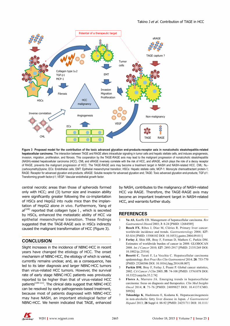
2465 October 18, 2015|Volume 7|Issue 23|WJH|www.wjgnet.com
central necrotic areas than those of spheroids formed only with HCC; and (3) tumor size and invasion ability were significantly greater following the co-implantation of HSCs and HepG2 into nude mice than the implan-tation of HepG2 alone in vivo. Furthermore, Yang et al[108] reported that collagen type Ⅰ, which is secreted by HSCs, enhanced the metastatic ability of HCC via epithelial mesenchymal transition. These findings suggested that the TAGE-RAGE axis in HSCs indirectly caused the malignant transformation of HCC (Figure 3).
CONClUSIONSlight increases in the incidence of NBNC-HCC in recent years have changed the etiology of HCC. The onset mechanism of NBNC-HCC, the etiology of which is varied, currently remains unclear, and, as a consequence, has led to its later diagnosis and larger NBNC-HCC tumors than virus-related HCC tumors. However, the survival rate of early stage NBNC-HCC patients was previously reported to be higher than that of virus-related HCC patients[109-111]. The clinical data suggest that NBNC-HCC can be resolved by early pathogenesis-based treatment, because most of patients diagnosed with NBNC-HCC may have NASH, an important etiological factor of NBNC-HCC. We herein indicated that TAGE, enhanced
by NASH, contributes to the malignancy of NASH-related HCC via RAGE. Therefore, the TAGE-RAGE axis may become an important treatment target in NASH-related HCC, and warrants further study.
RefeReNCeS1 Yu AS, Keeffe EB. Management of hepatocellular carcinoma. Rev
Gastroenterol Disord 2003; 3: 8-24 [PMID: 12684589]2 Bosch FX, Ribes J, Díaz M, Cléries R. Primary liver cancer:
worldwide incidence and trends. Gastroenterology 2004; 127: S5-S16 [PMID: 15508102 DOI: 10.1053/j.gastro.2004.09.011]
3 Ferlay J, Shin HR, Bray F, Forman D, Mathers C, Parkin DM. Estimates of worldwide burden of cancer in 2008: GLOBOCAN 2008. Int J Cancer 2010; 127: 2893-2917 [PMID: 21351269 DOI: 10.1002/ijc.25516]
4 Bosetti C, Turati F, La Vecchia C. Hepatocellular carcinoma epidemiology. Best Pract Res Clin Gastroenterol 2014; 28: 753-770 [PMID: 25260306 DOI: 10.1016/j.bpg.2014.08.007]
5 Parkin DM, Bray F, Ferlay J, Pisani P. Global cancer statistics, 2002. CA Cancer J Clin 2005; 55: 74-108 [PMID: 15761078 DOI: 10.3322/canjclin.55.2.74]
6 Flores A, Marrero JA. Emerging trends in hepatocellular carcinoma: focus on diagnosis and therapeutics. Clin Med Insights Oncol 2014; 8: 71-76 [PMID: 24899827 DOI: 10.4137/CMO.S9926]
7 Tokushige K, Hashimoto E, Kodama K. Hepatocarcinogenesis in non-alcoholic fatty liver disease in Japan. J Gastroenterol Hepatol 2013; 28 Suppl 4: 88-92 [PMID: 24251711 DOI: 10.1111/
Collagen type Iα2TGF-β1MCP-1
Tumor cells
EMT
InvasionMigrationProliferation
HCC malignancy
HSCs
Inverse correlationAngiogenesis
ECs
CML
VEGF
TAGE RAGE
Non-malignancy
TAGE capture ?
sRAGE
Potential of a therapeutic target
Fibrosis
Figure 3 Proposed model for the contribution of the toxic advanced glycation end-products-receptor axis in nonalcoholic steatohepatitis-related hepatocellular carcinoma. The interaction between TAGE and RAGE alters intracellular signaling in tumor cells and hepatic stellate cells, and induces angiogenesis, invasion, migration, proliferation, and fibrosis. This cooperation by the TAGE-RAGE axis may lead to the malignant progression of nonalcoholic steatohepatitis (NASH)-related hepatocellular carcinoma (HCC). CML and sRAGE inversely correlate with the risk of HCC, and sRAGE, which plays the role of a decoy receptor of RAGE, prevents the malignant progression of HCC. The TAGE-RAGE axis may become a treatment target in NASH and NASH-related HCC. CML: Nε-(carboxymethyl)lysine; ECs: Endothelial cells; EMT: Epithelial mesenchymal transition; HSCs: Hepatic stellate cells; MCP-1: Monocyte chemoattractant protein-1; RAGE: Receptor for advanced glycation end-products; sRAGE: Soluble receptor for advanced glycation end; TAGE: Toxic advanced glycation end-products; TGF-β1: Transforming growth factor-β1; VEGF: Vascular endothelial growth factor.
Takino J et al . Contribution of TAGE in HCC

2466 October 18, 2015|Volume 7|Issue 23|WJH|www.wjgnet.com
jgh.12239]8 Tokushige K, Hashimoto E, Horie Y, Taniai M, Higuchi S.
Hepatocellular carcinoma in Japanese patients with nonalcoholic fatty liver disease, alcoholic liver disease, and chronic liver disease of unknown etiology: report of the nationwide survey. J Gastroenterol 2011; 46: 1230-1237 [PMID: 21748549 DOI: 10.1007/s00535-011-0431-9]
9 Hatanaka K, Kudo M, Fukunaga T, Ueshima K, Chung H, Minami Y, Sakaguchi Y, Hagiwara S, Orino A, Osaki Y. Clinical characteristics of NonBNonC- HCC: Comparison with HBV and HCV related HCC. Intervirology 2007; 50: 24-31 [PMID: 17164554 DOI: 10.1159/000096309]
10 Nagaoki Y, Hyogo H, Aikata H, Tanaka M, Naeshiro N, Nakahara T, Honda Y, Miyaki D, Kawaoka T, Takaki S, Hiramatsu A, Waki K, Imamura M, Kawakami Y, Takahashi S, Chayama K. Recent trend of clinical features in patients with hepatocellular carcinoma. Hepatol Res 2012; 42: 368-375 [PMID: 22151896 DOI: 10.1111/j.1872-034X.2011.00929.x]
11 al-Abed Y, Kapurniotu A, Bucala R. Advanced glycation end products: detection and reversal. Methods Enzymol 1999; 309: 152-172 [PMID: 10507023 DOI: 10.1016/S0076-6879(99)09013-8]
12 Vlassara H, Palace MR. Diabetes and advanced glycation endproducts. J Intern Med 2002; 251: 87-101 [PMID: 11905595 DOI: 10.1046/j.1365-2796.2002.00932.x]
13 Takeuchi M, Makita Z. Alternative routes for the formation of immunochemically distinct advanced glycation end-products in vivo. Curr Mol Med 2001; 1: 305-315 [PMID: 11899079 DOI: 10.2174/1566524013363735]
14 Glomb MA, Monnier VM. Mechanism of protein modification by glyoxal and glycolaldehyde, reactive intermediates of the Maillard reaction. J Biol Chem 1995; 270: 10017-10026 [PMID: 7730303 DOI: 10.1074/jbc.270.17.10017]
15 Thornalley PJ, Langborg A, Minhas HS. Formation of glyoxal, methylglyoxal and 3-deoxyglucosone in the glycation of proteins by glucose. Biochem J 1999; 344 Pt 1: 109-116 [PMID: 10548540]
16 Takeuchi M, Yamagishi S. TAGE (toxic AGEs) hypothesis in various chronic diseases. Med Hypotheses 2004; 63: 449-452 [PMID: 15288366 DOI: 10.1016/j.mehy.2004.02.042]
17 Sato T, Iwaki M, Shimogaito N, Wu X, Yamagishi S, Takeuchi M. TAGE (toxic AGEs) theory in diabetic complications. Curr Mol Med 2006; 6: 351-358 [PMID: 16712480 DOI: 10.2174/156652406776894536]
18 Abe R, Yamagishi S. AGE-RAGE system and carcinogenesis. Curr Pharm Des 2008; 14: 940-945 [PMID: 18473843 DOI: 10.2174/138161208784139765]
19 Hyogo H, Yamagishi S. Advanced glycation end products (AGEs) and their involvement in liver disease. Curr Pharm Des 2008; 14: 969-972 [PMID: 18473847 DOI: 10.2174/138161208784139701]
20 Takeuchi M, Yamagishi S. Possible involvement of advanced glycation end-products (AGEs) in the pathogenesis of Alzheimer’s disease. Curr Pharm Des 2008; 14: 973-978 [PMID: 18473848 DOI: 10.2174/138161208784139693]
21 Takeuchi M, Yamagishi S. Involvement of toxic AGEs (TAGE) in the pathogenesis of diabetic vascular complications and Alzheimer’s disease. J Alzheimers Dis 2009; 16: 845-858 [PMID: 19387117 DOI: 10.3233/JAD-2009-0974]
22 Takeuchi M, Takino J, Yamagishi S. Involvement of the toxic AGEs (TAGE)-RAGE system in the pathogenesis of diabetic vascular complications: a novel therapeutic strategy. Curr Drug Targets 2010; 11: 1468-1482 [PMID: 20583971 DOI: 10.2174/1389450111009011468]
23 Yamagishi S, Maeda S, Matsui T, Ueda S, Fukami K, Okuda S. Role of advanced glycation end products (AGEs) and oxidative stress in vascular complications in diabetes. Biochim Biophys Acta 2012; 1820: 663-671 [PMID: 21440603 DOI: 10.1016/j.bbagen.2011.03.014]
24 Takeuchi M, Takino J, Sakasai-Sakai A, Takata T, Ueda T, Tsutsumi M, Hyogo H, Yamagishi S. Involvement of the TAGE-RAGE system in non-alcoholic steatohepatitis: Novel treatment strategies. World J Hepatol 2014; 6: 880-893 [PMID: 25544875 DOI: 10.4254/wjh.v6.i12.880]
25 Hyogo H, Yamagishi S, Iwamoto K, Arihiro K, Takeuchi M, Sato T, Ochi H, Nonaka M, Nabeshima Y, Inoue M, Ishitobi T, Chayama K, Tazuma S. Elevated levels of serum advanced glycation end products in patients with non-alcoholic steatohepatitis. J Gastroenterol Hepatol 2007; 22: 1112-1119 [PMID: 17559366 DOI: 10.1111/j.1440-1746.2007.04943.x]
26 Schmidt AM, Stern D. Atherosclerosis and diabetes: the RAGE connection. Curr Atheroscler Rep 2000; 2: 430-436 [PMID: 11122775 DOI: 10.1007/s11883-000-0082-4]
27 Wendt T, Bucciarelli L, Qu W, Lu Y, Yan SF, Stern DM, Schmidt AM. Receptor for advanced glycation endproducts (RAGE) and vascular inflammation: insights into the pathogenesis of macro-vascular complications in diabetes. Curr Atheroscler Rep 2002; 4: 228-237 [PMID: 11931721 DOI: 10.1007/s11883-002-0024-4]
28 Rahbar S, Figarola JL. Novel inhibitors of advanced glycation endproducts. Arch Biochem Biophys 2003; 419: 63-79 [PMID: 14568010 DOI: 10.1016/j.abb.2003.08.009]
29 Yamagishi S, Takeuchi M, Inagaki Y, Nakamura K, Imaizumi T. Role of advanced glycation end products (AGEs) and their receptor (RAGE) in the pathogenesis of diabetic microangiopathy. Int J Clin Pharmacol Res 2003; 23: 129-134 [PMID: 15224502]
30 Yamagishi S, Imaizumi T. Diabetic vascular complications: pathophysiology, biochemical basis and potential therapeutic strategy. Curr Pharm Des 2005; 11: 2279-2299 [PMID: 16022668 DOI: 10.2174/1381612054367300]
31 Takenaka K, Yamagishi S, Matsui T, Nakamura K, Imaizumi T. Role of advanced glycation end products (AGEs) in thrombogenic abnormalities in diabetes. Curr Neurovasc Res 2006; 3: 73-77 [PMID: 16472128 DOI: 10.2174/156720206775541804]
32 Abe R, Shimizu T, Sugawara H, Watanabe H, Nakamura H, Choei H, Sasaki N, Yamagishi S, Takeuchi M, Shimizu H. Regulation of human melanoma growth and metastasis by AGE-AGE receptor interactions. J Invest Dermatol 2004; 122: 461-467 [PMID: 15009731 DOI: 10.1046/j.0022-202X.2004.22218.x]
33 Takino J, Yamagishi S, Takeuchi M. Cancer malignancy is enhanced by glyceraldehyde-derived advanced glycation end-products. J Oncol 2010; 2010: 739852 [PMID: 20631911 DOI: 10.1155/2010/739852]
34 Sakuraoka Y, Sawada T, Okada T, Shiraki T, Miura Y, Hiraishi K, Ohsawa T, Adachi M, Takino J, Takeuchi M, Kubota K. MK615 decreases RAGE expression and inhibits TAGE-induced proliferation in hepatocellular carcinoma cells. World J Gastroenterol 2010; 16: 5334-5341 [PMID: 21072897 DOI: 10.3748/wjg.v16.i42.5334]
35 Takino J, Yamagishi S, Takeuchi M. Glycer-AGEs-RAGE signaling enhances the angiogenic potential of hepatocellular carcinoma by upregulating VEGF expression. World J Gastroenterol 2012; 18: 1781-1788 [PMID: 22553402 DOI: 10.3748/wjg.v18.i15.1781]
36 Angulo P. Nonalcoholic fatty liver disease. N Engl J Med 2002; 346: 1221-1231 [PMID: 11961152 DOI: 10.1056/NEJMra011775]
37 Tappy L, Lê KA. Metabolic effects of fructose and the worldwide increase in obesity. Physiol Rev 2010; 90: 23-46 [PMID: 20086073 DOI: 10.1152/physrev.00019.2009]
38 Dekker MJ, Su Q, Baker C, Rutledge AC, Adeli K. Fructose: a highly lipogenic nutrient implicated in insulin resistance, hepatic steatosis, and the metabolic syndrome. Am J Physiol Endocrinol Metab 2010; 299: E685-E694 [PMID: 20823452 DOI: 10.1152/ajpendo.00283.2010]
39 Vos MB, Lavine JE. Dietary fructose in nonalcoholic fatty liver disease. Hepatology 2013; 57: 2525-2531 [PMID: 23390127 DOI: 10.1002/hep.26299]
40 Attar BM, Van Thiel DH. Current concepts and management approaches in nonalcoholic fatty liver disease. ScientificWorldJournal 2013; 2013: 481893 [PMID: 23576902 DOI: 10.1155/2013/481893]
41 Conlon BA, Beasley JM, Aebersold K, Jhangiani SS, Wylie-Rosett J. Nutritional management of insulin resistance in nonalcoholic fatty liver disease (NAFLD). Nutrients 2013; 5: 4093-4114 [PMID: 24152749 DOI: 10.3390/nu5104093]
42 Bedogni G, Miglioli L, Masutti F, Tiribelli C, Marchesini G, Bellentani S. Prevalence of and risk factors for nonalcoholic fatty liver disease: the Dionysos nutrition and liver study. Hepatology
Takino J et al . Contribution of TAGE in HCC
2467 October 18, 2015|Volume 7|Issue 23|WJH|www.wjgnet.com
2005; 42: 44-52 [PMID: 15895401 DOI: 10.1002/hep.20734]43 Ludwig J, Viggiano TR, McGill DB, Oh BJ. Nonalcoholic
steatohepatitis: Mayo Clinic experiences with a hitherto unnamed disease. Mayo Clin Proc 1980; 55: 434-438 [PMID: 7382552]
44 Marchesini G, Brizi M, Bianchi G, Tomassetti S, Bugianesi E, Lenzi M, McCullough AJ, Natale S, Forlani G, Melchionda N. Nonalcoholic fatty liver disease: a feature of the metabolic syndrome. Diabetes 2001; 50: 1844-1850 [PMID: 11473047 DOI: 10.2337/diabetes.50.8.1844]
45 Chitturi S, Abeygunasekera S, Farrell GC, Holmes-Walker J, Hui JM, Fung C, Karim R, Lin R, Samarasinghe D, Liddle C, Weltman M, George J. NASH and insulin resistance: Insulin hypersecretion and specific association with the insulin resistance syndrome. Hepatology 2002; 35: 373-379 [PMID: 11826411 DOI: 10.1053/jhep.2002.30692]
46 Marchesini G, Bugianesi E, Forlani G, Cerrelli F, Lenzi M, Manini R, Natale S, Vanni E, Villanova N, Melchionda N, Rizzetto M. Nonalcoholic fatty liver, steatohepatitis, and the metabolic syndrome. Hepatology 2003; 37: 917-923 [PMID: 12668987 DOI: 10.1053/jhep.2003.50161]
47 Matteoni CA, Younossi ZM, Gramlich T, Boparai N, Liu YC, McCullough AJ. Nonalcoholic fatty liver disease: a spectrum of clinical and pathological severity. Gastroenterology 1999; 116: 1413-1419 [PMID: 10348825 DOI: 10.1016/S0016-5085(99)70506-8]
48 Dam-Larsen S, Franzmann M, Andersen IB, Christoffersen P, Jensen LB, Sørensen TI, Becker U, Bendtsen F. Long term prognosis of fatty liver: risk of chronic liver disease and death. Gut 2004; 53: 750-755 [PMID: 15082596 DOI: 10.1136/gut.2003.019984]
49 Yatsuji S, Hashimoto E, Tobari M, Taniai M, Tokushige K, Shiratori K. Clinical features and outcomes of cirrhosis due to non-alcoholic steatohepatitis compared with cirrhosis caused by chronic hepatitis C. J Gastroenterol Hepatol 2009; 24: 248-254 [PMID: 19032450 DOI: 10.1111/j.1440-1746.2008.05640.x]
50 Starley BQ, Calcagno CJ, Harrison SA. Nonalcoholic fatty liver disease and hepatocellular carcinoma: a weighty connection. Hepatology 2010; 51: 1820-1832 [PMID: 20432259 DOI: 10.1002/hep.23594]
51 Takuma Y, Nouso K. Nonalcoholic steatohepatitis-associated hepatocellular carcinoma: our case series and literature review. World J Gastroenterol 2010; 16: 1436-1441 [PMID: 20333782 DOI: 10.3748/wjg.v16.i12.1436]
52 Ertle J, Dechêne A, Sowa JP, Penndorf V, Herzer K, Kaiser G, Schlaak JF, Gerken G, Syn WK, Canbay A. Non-alcoholic fatty liver disease progresses to hepatocellular carcinoma in the absence of apparent cirrhosis. Int J Cancer 2011; 128: 2436-2443 [PMID: 21128245 DOI: 10.1002/ijc.25797]
53 Caldwell SH, Oelsner DH, Iezzoni JC, Hespenheide EE, Battle EH, Driscoll CJ. Cryptogenic cirrhosis: clinical characterization and risk factors for underlying disease. Hepatology 1999; 29: 664-669 [PMID: 10051466 DOI: 10.1002/hep.510290347]
54 Ong J, Younossi ZM, Reddy V, Price LL, Gramlich T, Mayes J, Boparai N. Cryptogenic cirrhosis and posttransplantation non-alcoholic fatty liver disease. Liver Transpl 2001; 7: 797-801 [PMID: 11552214 DOI: 10.1053/jlts.2001.24644]
55 Poonawala A, Nair SP, Thuluvath PJ. Prevalence of obesity and diabetes in patients with cryptogenic cirrhosis: a case-control study. Hepatology 2000; 32: 689-692 [PMID: 11003611 DOI: 10.1053/jhep.2000.17894]
56 Marrero JA, Fontana RJ, Su GL, Conjeevaram HS, Emick DM, Lok AS. NAFLD may be a common underlying liver disease in patients with hepatocellular carcinoma in the United States. Hepatology 2002; 36: 1349-1354 [PMID: 12447858 DOI: 10.1002/hep.1840360609]
57 Thornalley PJ. Pharmacology of methylglyoxal: formation, modi-fication of proteins and nucleic acids, and enzymatic detoxification--a role in pathogenesis and antiproliferative chemotherapy. Gen Pharmacol 1996; 27: 565-573 [PMID: 8853285 DOI: 10.1016/0306-3623(95)02054-3]
58 Takeuchi M, Makita Z, Yanagisawa K, Kameda Y, Koike T.
Detection of noncarboxymethyllysine and carboxymethyllysine advanced glycation end products (AGE) in serum of diabetic patients. Mol Med 1999; 5: 393-405 [PMID: 10415164]
59 Takeuchi M, Makita Z, Bucala R, Suzuki T, Koike T, Kameda Y. Immunological evidence that non-carboxymethyllysine advanced glycation end-products are produced from short chain sugars and dicarbonyl compounds in vivo. Mol Med 2000; 6: 114-125 [PMID: 10859028]
60 Takeuchi M, Yanase Y, Matsuura N, Yamagishi Si S, Kameda Y, Bucala R, Makita Z. Immunological detection of a novel advanced glycation end-product. Mol Med 2001; 7: 783-791 [PMID: 11788793]
61 Takeuchi M, Iwaki M, Takino J, Shirai H, Kawakami M, Bucala R, Yamagishi S. Immunological detection of fructose-derived advanced glycation end-products. Lab Invest 2010; 90: 1117-1127 [PMID: 20212455 DOI: 10.1038/labinvest.2010.62]
62 Takeuchi M, Yamagishi S. Alternative routes for the formation of glyceraldehyde-derived AGEs (TAGE) in vivo. Med Hypotheses 2004; 63: 453-455 [PMID: 15288367 DOI: 10.1016/j.mehy.2004.03.005]
63 Bucala R, Cerami A. Advanced glycosylation: chemistry, biology, and implications for diabetes and aging. Adv Pharmacol 1992; 23: 1-34 [PMID: 1540533 DOI: 10.1016/S1054-3589(08)60961-8]
64 Vlassara H, Bucala R, Striker L. Pathogenic effects of advanced glycosylation: biochemical, biologic, and clinical implications for diabetes and aging. Lab Invest 1994; 70: 138-151 [PMID: 8139257]
65 Brownlee M. Advanced protein glycosylation in diabetes and aging. Annu Rev Med 1995; 46: 223-234 [PMID: 7598459 DOI: 10.1146/annurev.med.46.1.223]
66 Brownlee M, Cerami A, Vlassara H. Advanced glycosylation end products in tissue and the biochemical basis of diabetic complications. N Engl J Med 1988; 318: 1315-1321 [PMID: 3283558 DOI: 10.1056/NEJM198805193182007]
67 Monnier VM, Cerami A. Nonenzymatic browning in vivo: possible process for aging of long-lived proteins. Science 1981; 211: 491-493 [PMID: 6779377 DOI: 10.1126/science.6779377]
68 Fu MX, Requena JR, Jenkins AJ, Lyons TJ, Baynes JW, Thorpe SR. The advanced glycation end product, Nepsilon-(carboxymethyl)lysine, is a product of both lipid peroxidation and glycoxidation reactions. J Biol Chem 1996; 271: 9982-9986 [PMID: 8626637 DOI: 10.1074/jbc.271.17.9982]
69 Sebeková K, Kupcová V, Schinzel R, Heidland A. Markedly elevated levels of plasma advanced glycation end products in patients with liver cirrhosis - amelioration by liver transplantation. J Hepatol 2002; 36: 66-71 [PMID: 11804666]
70 Yagmur E, Tacke F, Weiss C, Lahme B, Manns MP, Kiefer P, Trautwein C, Gressner AM. Elevation of Nepsilon-(carboxy-methyl)lysine-modified advanced glycation end products in chronic liver disease is an indicator of liver cirrhosis. Clin Biochem 2006; 39: 39-45 [PMID: 16321365 DOI: 10.1016/j.clinbiochem.2005.07.016]
71 Moy KA, Jiao L, Freedman ND, Weinstein SJ, Sinha R, Virtamo J, Albanes D, Stolzenberg-Solomon RZ. Soluble receptor for advanced glycation end products and risk of liver cancer. Hepatology 2013; 57: 2338-2345 [PMID: 23325627 DOI: 10.1002/hep.26264]
72 Kimura Y, Hyogo H, Yamagishi S, Takeuchi M, Ishitobi T, Nabeshima Y, Arihiro K, Chayama K. Atorvastatin decreases serum levels of advanced glycation endproducts (AGEs) in nonalcoholic steatohepatitis (NASH) patients with dyslipidemia: clinical usefulness of AGEs as a biomarker for the attenuation of NASH. J Gastroenterol 2010; 45: 750-757 [PMID: 20112031 DOI: 10.1007/s00535-010-0203-y]
73 Kan H, Yamagishi SI, Ojima A, Fukami K, Ueda S, Takeuchi M, Hyogo H, Aikata H, Chayama K. Elevation of Serum Levels of Advanced Glycation End Products in Patients With Non-B or Non-C Hepatocellular Carcinoma. J Clin Lab Anal 2014; Epub ahead of print [PMID: 25252033 DOI: 10.1002/jcla.21797]
74 Brenner T, Fleming TH, Spranz D, Schemmer P, Bruckner T, Uhle F, Martin EO, Weigand MA, Hofer S. Reactive metabolites and AGE-RAGE-mediated inflammation in patients following liver
Takino J et al . Contribution of TAGE in HCC

2468 October 18, 2015|Volume 7|Issue 23|WJH|www.wjgnet.com
transplantation. Mediators Inflamm 2013; 2013: 501430 [PMID: 23766560 DOI: 10.1155/2013/501430]
75 Takeuchi M, Sakasai-Sakai A, Takata T, Ueda T, Takino J, Tsutsumi M, Hyogo H, Yamagishi S. Serum levels of toxic AGEs (TAGE) may be a promising novel biomarker in development and progression of NASH. Med Hypotheses 2015; 84: 490-493 [PMID: 25697114 DOI: 10.1016/j.mehy.2015.02.002]
76 Schmidt AM, Yan SD, Yan SF, Stern DM. The multiligand receptor RAGE as a progression factor amplifying immune and inflammatory responses. J Clin Invest 2001; 108: 949-955 [PMID: 11581294 DOI: 10.1172/JCI14002]
77 Kuniyasu H, Oue N, Wakikawa A, Shigeishi H, Matsutani N, Kuraoka K, Ito R, Yokozaki H, Yasui W. Expression of receptors for advanced glycation end-products (RAGE) is closely associated with the invasive and metastatic activity of gastric cancer. J Pathol 2002; 196: 163-170 [PMID: 11793367 DOI: 10.1002/path.1031]
78 Bhawal UK, Ozaki Y, Nishimura M, Sugiyama M, Sasahira T, Nomura Y, Sato F, Fujimoto K, Sasaki N, Ikeda MA, Tsuji K, Kuniyasu H, Kato Y. Association of expression of receptor for advanced glycation end products and invasive activity of oral squamous cell carcinoma. Oncology 2005; 69: 246-255 [PMID: 16127291 DOI: 10.1159/000087910]
79 Yan W, Chang Y, Liang X, Cardinal JS, Huang H, Thorne SH, Monga SP, Geller DA, Lotze MT, Tsung A. High-mobility group box 1 activates caspase-1 and promotes hepatocellular carcinoma invasiveness and metastases. Hepatology 2012; 55: 1863-1875 [PMID: 22234969 DOI: 10.1002/hep.25572]
80 Chen RC, Yi PP, Zhou RR, Xiao MF, Huang ZB, Tang DL, Huang Y, Fan XG. The role of HMGB1-RAGE axis in migration and invasion of hepatocellular carcinoma cell lines. Mol Cell Biochem 2014; 390: 271-280 [PMID: 24510323 DOI: 10.1007/s11010-014-1978-6]
81 Hiwatashi K, Ueno S, Abeyama K, Kubo F, Sakoda M, Maruyama I, Hamanoue M, Natsugoe S, Aikou T. A novel function of the receptor for advanced glycation end-products (RAGE) in association with tumorigenesis and tumor differentiation of HCC. Ann Surg Oncol 2008; 15: 923-933 [PMID: 18080716 DOI: 10.1245/s10434-007-9698-8]
82 Fehrenbach H, Weiskirchen R, Kasper M, Gressner AM. Up-regulated expression of the receptor for advanced glycation end products in cultured rat hepatic stellate cells during transdifferentiation to myofibroblasts. Hepatology 2001; 34: 943-952 [PMID: 11679965 DOI: 10.1053/jhep.2001.28788]
83 Zeng S, Feirt N, Goldstein M, Guarrera J, Ippagunta N, Ekong U, Dun H, Lu Y, Qu W, Schmidt AM, Emond JC. Blockade of receptor for advanced glycation end product (RAGE) attenuates ischemia and reperfusion injury to the liver in mice. Hepatology 2004; 39: 422-432 [PMID: 14767995]
84 Cataldegirmen G, Zeng S, Feirt N, Ippagunta N, Dun H, Qu W, Lu Y, Rong LL, Hofmann MA, Kislinger T, Pachydaki SI, Jenkins DG, Weinberg A, Lefkowitch J, Rogiers X, Yan SF, Schmidt AM, Emond JC. RAGE limits regeneration after massive liver injury by coordinated suppression of TNF-alpha and NF-kappaB. J Exp Med 2005; 201: 473-484 [PMID: 15699076 DOI: 10.1084/jem.20040934]
85 Ekong U, Zeng S, Dun H, Feirt N, Guo J, Ippagunta N, Guarrera JV, Lu Y, Weinberg A, Qu W, Ramasamy R, Schmidt AM, Emond JC. Blockade of the receptor for advanced glycation end products attenuates acetaminophen-induced hepatotoxicity in mice. J Gastroenterol Hepatol 2006; 21: 682-688 [PMID: 16677153]
86 Higuchi S, Yano A, Takai S, Tsuneyama K, Fukami T, Nakajima M, Yokoi T. Metabolic activation and inflammation reactions involved in carbamazepine-induced liver injury. Toxicol Sci 2012; 130: 4-16 [PMID: 22790970 DOI: 10.1093/toxsci/kfs222]
87 Goodwin M, Herath C, Jia Z, Leung C, Coughlan MT, Forbes J, Angus P. Advanced glycation end products augment experimental hepatic fibrosis. J Gastroenterol Hepatol 2013; 28: 369-376 [PMID: 23173780 DOI: 10.1111/jgh.12042]
88 Koh EJ, Yoon SJ, Lee SM. Losartan protects liver against ischaemia/reperfusion injury through PPAR-γ activation and
receptor for advanced glycation end-products down-regulation. Br J Pharmacol 2013; 169: 1404-1416 [PMID: 23647130 DOI: 10.1111/bph.12229]
89 Kuhla A, Norden J, Abshagen K, Menger MD, Vollmar B. RAGE blockade and hepatic microcirculation in experimental endotoxaemic liver failure. Br J Surg 2013; 100: 1229-1239 [PMID: 23804133 DOI: 10.1002/bjs.9188]
90 Ito R, Ishii Y, Wakiyama S, Shiba H, Fujioka S, Misawa T, Ishida Y, Hano H, Yanaga K. Prognostic significance of receptor for advanced glycation end products expression in hepatocellular carcinoma after hepatectomy. J Surg Res 2014; 192: 503-508 [PMID: 25043528 DOI: 10.1016/j.jss.2014.06.028]
91 Yang Y, Zhao LH, Huang B, Wang RY, Yuan SX, Tao QF, Xu Y, Sun HY, Lin C, Zhou WP. Pioglitazone, a PPARγ agonist, inhibits growth and invasion of human hepatocellular carcinoma via blockade of the rage signaling. Mol Carcinog 2014; Epub ahead of print [PMID: 25307746 DOI: 10.1002/mc.22231]
92 Yoshida T, Yamagishi S, Nakamura K, Matsui T, Imaizumi T, Takeuchi M, Koga H, Ueno T, Sata M. Telmisartan inhibits AGE-induced C-reactive protein production through downregulation of the receptor for AGE via peroxisome proliferator-activated receptor-gamma activation. Diabetologia 2006; 49: 3094-3099 [PMID: 17004092 DOI: 10.1007/s00125-006-0437-7]
93 Yonekura H, Yamamoto Y, Sakurai S, Petrova RG, Abedin MJ, Li H, Yasui K, Takeuchi M, Makita Z, Takasawa S, Okamoto H, Watanabe T, Yamamoto H. Novel splice variants of the receptor for advanced glycation end-products expressed in human vascular endothelial cells and pericytes, and their putative roles in diabetes-induced vascular injury. Biochem J 2003; 370: 1097-1109 [PMID: 12495433 DOI: 10.1042/BJ20021371]
94 Geroldi D, Falcone C, Emanuele E. Soluble receptor for advanced glycation end products: from disease marker to potential therapeutic target. Curr Med Chem 2006; 13: 1971-1978 [PMID: 16842191 DOI: 10.2174/092986706777585013]
95 Yilmaz Y, Ulukaya E, Gul OO, Arabul M, Gul CB, Atug O, Oral AY, Aker S, Dolar E. Decreased plasma levels of soluble receptor for advanced glycation endproducts (sRAGE) in patients with nonalcoholic fatty liver disease. Clin Biochem 2009; 42: 802-807 [PMID: 19217891 DOI: 10.1016/j.clinbiochem.2009.02.003]
96 Kohles N, Nagel D, Jüngst D, Stieber P, Holdenrieder S. Predictive value of immunogenic cell death biomarkers HMGB1, sRAGE, and DNase in liver cancer patients receiving transarterial chemoembolization therapy. Tumour Biol 2012; 33: 2401-2409 [PMID: 22965881 DOI: 10.1007/s13277-012-0504-2]
97 Nakamura K, Yamagishi SI, Matsui T, Adachi H, Takeuchi M, Imaizumi T. Serum levels of soluble form of receptor for advanced glycation end products (sRAGE) are correlated with AGEs in both diabetic and non-diabetic subjects. Clin Exp Med 2007; 7: 188-190 [PMID: 18188534 DOI: 10.1007/s10238-007-0146-7]
98 Yamagishi S, Adachi H, Nakamura K, Matsui T, Jinnouchi Y, Takenaka K, Takeuchi M, Enomoto M, Furuki K, Hino A, Shigeto Y, Imaizumi T. Positive association between serum levels of advanced glycation end products and the soluble form of receptor for advanced glycation end products in nondiabetic subjects. Metabolism 2006; 55: 1227-1231 [PMID: 16919543 DOI: 10.1016/j.metabol.2006.05.007]
99 Yamagishi S, Imaizumi T. Serum levels of soluble form of receptor for advanced glycation end products (sRAGE) may reflect tissue RAGE expression in diabetes. Arterioscler Thromb Vasc Biol 2007; 27: e32; author reply e33-e34 [PMID: 17522392 DOI: 10.1161/ATVBAHA.107.139923]
100 Yoshida T, Yamagishi S, Nakamura K, Matsui T, Imaizumi T, Takeuchi M, Ueno T, Sata M. Pigment epithelium-derived factor (PEDF) inhibits advanced glycation end product (AGE)-induced C-reactive protein expression in hepatoma cells by suppressing Rac-1 activation. FEBS Lett 2006; 580: 2788-2796 [PMID: 16674952 DOI: 10.1016/j.febset.2006.04.050]
101 Kinoshita A, Onoda H, Imai N, Iwaku A, Oishi M, Tanaka K, Fushiya N, Koike K, Nishino H, Matsushima M, Tajiri H. The addition of C-reactive protein to validated staging systems
Takino J et al . Contribution of TAGE in HCC

2469 October 18, 2015|Volume 7|Issue 23|WJH|www.wjgnet.com
improves their prognostic ability in patients with hepatocellular carcinoma. Oncology 2014; 86: 308-317 [PMID: 24924697 DOI: 10.1159/000360704]
102 Kim JM, Kwon CH, Joh JW, Ko JS, Park JB, Lee JH, Kim SJ, Paik SW, Park CK. C-reactive protein may be a prognostic factor in hepatocellular carcinoma with malignant portal vein invasion. World J Surg Oncol 2013; 11: 92 [PMID: 23618082 DOI: 10.1186/1477-7819-11-92]
103 Iwamoto K, Kanno K, Hyogo H, Yamagishi S, Takeuchi M, Tazuma S, Chayama K. Advanced glycation end products enhance the proliferation and activation of hepatic stellate cells. J Gastroenterol 2008; 43: 298-304 [PMID: 18458846 DOI: 10.1007/s00535-007-2152-7]
104 Moreira RK. Hepatic stellate cells and liver fibrosis. Arch Pathol Lab Med 2007; 131: 1728-1734 [PMID: 17979495]
105 Carloni V, Luong TV, Rombouts K. Hepatic stellate cells and extracellular matrix in hepatocellular carcinoma: more complicated than ever. Liver Int 2014; 34: 834-843 [PMID: 24397349 DOI: 10.1111/liv.12465]
106 Coulouarn C, Clément B. Stellate cells and the development of liver cancer: therapeutic potential of targeting the stroma. J Hepatol 2014; 60: 1306-1309 [PMID: 24530649 DOI: 10.1016/j.jhep.2014.02.003]
107 Amann T, Bataille F, Spruss T, Mühlbauer M, Gäbele E, Schö-
lmerich J, Kiefer P, Bosserhoff AK, Hellerbrand C. Activated hepatic stellate cells promote tumorigenicity of hepatocellular carcinoma. Cancer Sci 2009; 100: 646-653 [PMID: 19175606 DOI: 10.1111/j.1349-7006.2009.01087.x]
108 Yang MC, Wang CJ, Liao PC, Yen CJ, Shan YS. Hepatic stellate cells secretes type I collagen to trigger epithelial mesenchymal transition of hepatoma cells. Am J Cancer Res 2014; 4: 751-763 [PMID: 25520865]
109 Akahoshi H, Taura N, Ichikawa T, Miyaaki H, Akiyama M, Miuma S, Ozawa E, Takeshita S, Muraoka T, Matsuzaki T, Ohtani M, Isomoto H, Matsumoto T, Takeshima F, Nakao K. Differences in prognostic factors according to viral status in patients with hepatocellular carcinoma. Oncol Rep 2010; 23: 1317-1323 [PMID: 20372846]
110 Takuma Y, Nouso K, Makino Y, Gotoh T, Toshikuni N, Morimoto Y, Shimomura H, Yamamoto H. Outcomes after curative treatment for cryptogenic cirrhosis-associated hepatocellular carcinoma satisfying the Milan criteria. J Gastroenterol Hepatol 2011; 26: 1417-1424 [PMID: 21884248 DOI: 10.1111/j.1440-1746.2011.06775.x]
111 Kaibori M, Ishizaki M, Matsui K, Kwon AH. Clinicopathologic characteristics of patients with non-B non-C hepatitis virus hepatocellular carcinoma after hepatectomy. Am J Surg 2012; 204: 300-307 [PMID: 22591698 DOI: 10.1016/j.amjsurg.2011.11.014]
P- Reviewer: Ampuero J, Gallego-Duran R, Lukacs-Kornek V S- Editor: Yu J L- Editor: Webster JR E- Editor: Liu SQ
Takino J et al . Contribution of TAGE in HCC
imaging, and management of bile duct stones despite growing evidence in the literature defining best practice. Management of patients with acute cholecystitis with early laparoscopic cholecystectomy (LC) results in better patient outcomes when compared with delayed surgical management techniques including antibiotic therapy or percutaneous cholecystostomy. Regardless of this data, many surgeons still prefer to utilize antibiotic therapy and complete an interval LC to manage acute cholecystitis. The use of intraoperative biliary imaging by cholangiogram or laparoscopic ultrasound has been demonstrated to facilitate the safe completion of cholecystectomy, minimizing the risk for inadvertent injury to surrounding structures, and lowering conversion rates, however it is rarely utilized. Choledocholithiasis used to be a diagnosis managed exclusively by surgeons but current practice favors referral to gastroenterologists for performance of preoperative endoscopic removal. Yet, there is evidence that intraoperative laparoscopic stone extraction is safe, feasible and may have added advantages. This review aims to highlight the differences between existing management of acute cholecystitis and evidence supported in the literature regarding best practice with the goal to change surgical practice to adopt these current recommendations.
Key words: Cholangiography; Acute cholecystitis; Ultrasound; Laparoscopy; Cholecystectomy; Evidence based
© The Author(s) 2015. Published by Baishideng Publishing Group Inc. All rights reserved.
Core tip: General surgeons commonly perform laparoscopic cholecystectomy for acute cholecystitis; however, current practices in biliary surgery often vary regarding timing, intraoperative biliary imaging, and management of bile duct stones. In spite of growing evidence in the literature defining best practice and societal guidelines supporting early cholecystectomy, intraoperative cholangiogram and ultrasound, and laparoscopic bile
Purvi P Patel, Shaun C Daly, Jose M Velasco
Purvi P Patel, Shaun C Daly, Jose M Velasco, Department of General Surgery, Rush University Medical Center, Chicago, IL 60612, United States
Author contributions: Patel PP, Daly SC and Velasco JM contributed equally to this work.
Conflictofinterest statement: The authors disclose no conflict of interests.
OpenAccess: This article is an open-access article which was selected by an in-house editor and fully peer-reviewed by external reviewers. It is distributed in accordance with the Creative Commons Attribution Non Commercial (CC BY-NC 4.0) license, which permits others to distribute, remix, adapt, build upon this work non-commercially, and license their derivative works on different terms, provided the original work is properly cited and the use is non-commercial. See: http://creativecommons.org/licenses/by-nc/4.0/
Correspondence to: Jose M Velasco, MD, FACS, FCCM, Department of General Surgery, Rush University Medical Center, 1735 West Harrison Street, Suite 810, Chicago, IL 60612, United States. [email protected]: +1-312-9425000Fax: +1-312-5632080
Received: April 27, 2015Peerreview started: May 1, 2015First decision: July 6, 2015Revised: September 1, 2015Accepted: September 7, 2015Article in press: September 8, 2015Published online: October 18, 2015
AbstractAcute cholecystitis is one of the most common surgical diagnoses encountered by general surgeons. Despite its high incidence there remains a range of treatment of approaches. Current practices in biliary surgery vary as to timing, intraoperative utilization of biliary
MINIREVIEWS
Submit a Manuscript: http://www.wjgnet.com/esps/Help Desk: http://www.wjgnet.com/esps/helpdesk.aspxDOI: 10.4254/wjh.v7.i23.2470
2470 October 18, 2015|Volume 7|Issue 23|WJH|www.wjgnet.com
World J Hepatol 2015 October 18; 7(23): 2470-2473ISSN 1948-5182 (online)
© 2015 Baishideng Publishing Group Inc. All rights reserved.
Training vs practice: A tale of opposition in acute cholecystitis

duct exploration utilizing laparoscopic ultrasound and performing common bile duct exploration, an overwhelming number of surgeons still perform delayed operations, rarely perform intraoperative imaging and defer treatment of common bile duct stones. Efforts should be made to adopt the evidencebased data supported in the literature.
Patel PP, Daly SC, Velasco JM. Training vs practice: A tale of opposition in acute cholecystitis. World J Hepatol 2015; 7(23): 2470-2473 Available from: URL: http://www.wjgnet.com/1948-5182/full/v7/i23/2470.htm DOI: http://dx.doi.org/10.4254/wjh.v7.i23.2470
THE DICHOTOMY OF LAPAROSCOPIC CHOLECYSTECTOMY TIMINGThe timing of cholecystectomy in the management of patients presenting with acute cholecystitis is variable and controversial in spite of well-published guidelines from surgical societies: SAGES and SSAT advocating early cholecystectomy during same hospital admission[1]. Two current approaches exist in the treatment of acute cholecystitis: (1) the traditional, conservative approach consisting of initial antibiotic therapy or percutaneous cholecystostomy followed by delayed cholecystectomy (DC) once inflammation has resolved; and (2) the preferred approach of early surgical intervention, ideally utilizing laparoscopic cholecystectomy (EC) within three to seven days of admission. Many surgeons continue to practice the former due to a number of reasons, such as a belief that an operation will be technically easier after a two to six week delay. Patient comorbidities, and the thought that laparoscopic cholecystectomy may be relatively contraindicated in acute cholecystitis for it may lead to high conversion rates have led to a resurgence of percutaneous cholecystostomy beyond the accepted indications in medically compromised patients. However, a wide range of practicing surgeons in the United States, ranging from 20% to 65% favor performing a laparoscopic cholecystectomy early during the initial presentation of acute cholecystitis[2-4]. Current surgical training has shifted this paradigm more towards early surgical intervention based on strong evidence of its benefit. However, practicing surgeons have been slow to embrace the change for a variety of reasons[3,5,6].
The concern for increased morbidity by operating in a surgical field of acute inflammation still persists. While significantly decreased morbidity rates for patients undergoing EC (12.0%) compared to DC (33.3%) was demonstrated in the ACDC trial, these results were not replicated on a recent meta-analysis where similar morbidity rates between EC patients and DC patients (0.12% vs 0.27%) were observed[7,8]. In the elderly population, where the number of comorbidities is often higher, similar morbidity rates were shown between EC patients and DC patients[9]. However, decreased rates of
cholangitis and persistent cholecystitis (1.3% vs 10.3%) and a decreased rate of septic shock (0.0% vs 1.3%) have been demonstrated in EC patients[7]. Most studies with the highest level of evidence demonstrated similar morbidity rates between EC and DC patients while some even show improved outcomes for EC patients[4,7-9]. A recent study analyzing the management of acute cholecystitis in the elderly did not show any significant differences in outcomes between the early and late cholecystostomy groups. Furthermore, percutaneous cholecystostomy did not confer any significant benefit as a bridge to cholecystectomy except in medically compromised patients[1].
The concern for an increased risk of bile duct injuries when operating in acute inflammation may deter some surgeons from early cholecystectomy. However, a meta-analysis, which included seven trials and over 1106 patients, demonstrated a similar common bile duct injury rate between patients undergoing an EC vs a DC (0.002% vs 0.004%)[8]. In addition to the meta-analysis, a database review by Zafar et al[4], which included over 95000 patients, failed to show a difference in observed bile duct injury rates. The literature supports equal rates of bile duct injury irrespective of laparoscopic cholecystectomy timing.
Many surgeons may choose to delay laparoscopic cholecystectomy in times of acute inflammation to lessen the chance of converting to an open procedure. The current literature does not support this concern. There exists no difference in conversion to open operations in patients undergoing an EC, with rates as low as 0.14% in a large meta-analysis to ranges of 5.0%-9.9% in elsewhere in the literature[4,7,8]. These rates compare to patients undergoing DC with rates as lows as 0.16% in meta-analysis and ranges of 1.7%-11.9% elsewhere in the literature[4,7,8]. Results were similar in the elderly population[9]. An increase in conversion rates after 5 d of symptoms to open cholecystectomy was shown by Zafar et al[4], further supports EC.
Some surgeons may be hesitant to perform a laparoscopic cholecystectomy because such an operation may be thought to lead to longer hospital stays, higher costs and decreased patient satisfaction. Significantly shorter hospital stays in EC patients, when compared to DC patients, have been demonstrated and includes the elderly population[4,9,10]. Zafar et al[4] demonstrated an increased postoperative stay of two days in patients whose laparoscopic cholecystectomy was performed after 5 d. Decreased hospital stay in one factor that has led to decreased costs in EC. A cost savings of nearly 31% has been observed in the literature[7]. In addition, Johner et al[11] demonstrated not only a $2028 (2009 Canadian dollar) cost savings but a gain of 0.03 quality-adjusted life year (QALY) gain in patients undergoing EC. Compared to EC, delayed cholecystectomy led to a significantly increased rate of persistent abdominal pain, 10.0% vs 2.3% in EC patients, and increased rates of persistent fever, 3.3% vs 0.3% in the EC group[7]. When mean patients satisfaction scores were determined, EC patients had significantly higher satisfaction (92.7)
2471 October 18, 2015|Volume 7|Issue 23|WJH|www.wjgnet.com
Patel PP et al . Current treatment trends in acute cholecystitis

compared to DC patients (75.3)[10]. This difference was attributable to persistent and recurrent biliary attacks in patients undergoing DC[10].
THE DICHOTOMY OF BILIARY DUCT IMAGING IN ACUTE CHOLECYSTITISTwo current approaches exist regarding intraoperative evaluation of the biliary tree: One that routinely evaluates biliary ductal anatomy during a laparoscopic cholecystectomy and the alternative, a selective approach, that completes intraoperative imaging based on individual clinical factors. To date, no randomized controlled study has been appropriately powered to endorse routine biliary imaging, however many studies have demonstrated a trend towards decreased biliary ductal injuries with routine evaluation[12,13]. Despite this trend in data and despite a bile duct injury being one of the most dreaded complications of biliary surgeons, few training programs endorse a curriculum of routine imaging and few surgeons have adopted it as part of their practice.
When a surgeon undertakes biliary imaging, the next decision to be made is which method of imaging should be employed. Many techniques have been described in the literature but the two most popular are intraoperative transcystic cholangiography (IOC) and laparoscopic ultrasound (LUS). Intraoperative cholangiogram is com-pleted by cannulating the transected cystic duct with a small lumen catheter and is the most common method utilized. Alternatively, laparoscopic ultrasound uses a flexible probe dressed in a sterile sheath to evaluate both ductal anatomy and the hepatic vasculature. In an attempt to try and demonstrate one method superior to the other, Aziz et al[14] performed a meta-analysis including 11 studies whose results demonstrated no significant difference in either sensitivity or specificity between each method. However, more recent studies have been able to demonstrate a higher specificity, in some cases nearly 100%, using laparoscopic ultrasound[15]. Additional advantages to LUS include that it is efficient, does not require cannulating the biliary system, and it can be accomplished prior to a complete and sometimes tedious dissection as it is easily repeated as needed during the course of an operation. Furthermore, LUS does not require fluoroscopy, and it has a lower failure rate than IOC, being extremely useful in defining the hepaticoduodenal anatomy[15-17]. In spite of these many benefits, LUS is rarely taught and rarely utilized in practice because of the technique’s large learning curve and the traditional acceptance of IOC as best care.
THE DICHOTOMY OF TREATING COMMON BILE DUCT STONES IN ACUTE CHOLECYSTITIS The rate of common bile duct stones in acute chole-
cystitis ranges from 3%-18%[18]. Many algorithms have been established to manage choledocholithiasis but controversy exists to which is the best method. When open cholecystectomy was standard of care, the majority of common bile duct stones were removed at the time of surgery by means of a common bile duct exploration via a choledochotomy or an indirect transcystic method. Early in the laparoscopic era, there were limited capabilities of laparoscopic instruments and surgeons’ lacked the expertise to complete a common bile duct exploration. As such, endoscopic retrograde cholangiopancreatography (ERCP) became standard practice. With improved laparoscopic instruments and advanced training, laparoscopic common bile duct exploration (LCBDE) rates are on the rise. In conjunction with intraoperative biliary ductal imaging, LCBDE allows for the management of common bile duct stones during one procedure. This technique has been shown to have a statistically significant reduction in total costs, length of hospital stay from 98 h with ERCP to 55 h, and number of procedures performed[19,20]. In addition, a trend towards better ductal clearance has been demonstrated[19]. Although utilization of LCBDE is gaining popularity, use is largely limited to fellowship trained minimally invasive or hepatobiliary surgeons and has not yet been readily adopted by most general surgeons despite improved outcomes.
THE FUTURE OF BILIARY SURGERYEven though laparoscopic cholecystectomy for acute cholecystitis is one of the most commonly performed operations by general surgeons, current practices in biliary surgery remain varied despite growing evidence in the literature defining best practice. Despite improved outcomes by performing an early cholecystectomy in acute cholecystitis and current training mirror such re-commendations, an overwhelming number of surgeons still perform delayed operations. Despite superior outcomes when LUS and IOC are utilized, a very limited number of residents will be proficient at these techniques upon completion of training and thus will not incorporate them into clinical practice. Due to this lack of expertise, many surgeons have come to rely on other tools including MRCP for preoperative definition of anatomy and ERCP to diagnose and remove common bile duct stones. Although these alternative methods have utility in a majority of cases, they come with additional potential morbidity and costs. Efforts should be made for surgical practice to catch up to surgical training and the evidence supported in the current literature.
REFERENCES1 Cull JD, Velasco JM, Czubak A, Rice D, Brown EC. Management
of acute cholecystitis: prevalence of percutaneous cholecystostomy and delayed cholecystectomy in the elderly. J Gastrointest Surg 2014; 18: 328-333 [PMID: 24197550 DOI: 10.1007/s11605-013-2341-z]
2 Wilson P, Leese T, Morgan WP, Kelly JF, Brigg JK. Elective
2472 October 18, 2015|Volume 7|Issue 23|WJH|www.wjgnet.com
Patel PP et al . Current treatment trends in acute cholecystitis
2473 October 18, 2015|Volume 7|Issue 23|WJH|www.wjgnet.com
12 Flum DR, Dellinger EP, Cheadle A, Chan L, Koepsell T. Intraoperative cholangiography and risk of common bile duct injury during cholecystectomy. JAMA 2003; 289: 1639-1644 [PMID: 12672731]
13 Metcalfe MS, Ong T, Bruening MH, Iswariah H, Wemyss-Holden SA, Maddern GJ. Is laparoscopic intraoperative cholangiogram a matter of routine? Am J Surg 2004; 187: 475-481 [PMID: 15041494]
14 Aziz O, Ashrafian H, Jones C, Harling L, Kumar S, Garas G, Holme T, Darzi A, Zacharakis E, Athanasiou T. Laparoscopic ultrasonography versus intra-operative cholangiogram for the detection of common bile duct stones during laparoscopic cholec-ystectomy: a meta-analysis of diagnostic accuracy. Int J Surg 2014; 12: 712-719 [PMID: 24861544 DOI: 10.1016/j.ijsu.2014.05.038]
15 Tranter SE, Thompson MH. A prospective single-blinded controlled study comparing laparoscopic ultrasound of the common bile duct with operative cholangiography. Surg Endosc 2003; 17: 216-219 [PMID: 12457223]
16 Perry KA, Myers JA, Deziel DJ. Laparoscopic ultrasound as the primary method for bile duct imaging during cholecystectomy. Surg Endosc 2008; 22: 208-213 [PMID: 17721807]
17 Gwinn EC, Daly S, Deziel DJ. The use of laparoscopic ultrasound in difficult cholecystectomy cases significantly decreases morbidity. Surgery 2013; 154: 909-915; discussion 915-917 [PMID: 24074430 DOI: 10.1016/j.surg.2013.04.041]
18 Dasari BV, Tan CJ, Gurusamy KS, Martin DJ, Kirk G, McKie L, Diamond T, Taylor MA. Surgical versus endoscopic treatment of bile duct stones. Cochrane Database Syst Rev 2013; 12: CD003327 [PMID: 24338858 DOI: 10.1002/14651858.CD003327.pub4]
19 Noble H, Tranter S, Chesworth T, Norton S, Thompson M. A randomized, clinical trial to compare endoscopic sphincterotomy and subsequent laparoscopic cholecystectomy with primary laparoscopic bile duct exploration during cholecystectomy in higher risk patients with choledocholithiasis. J Laparoendosc Adv Surg Tech A 2009; 19: 713-720 [PMID: 19792866 DOI: 10.1089/lap.2008.0428]
20 Rogers SJ, Cello JP, Horn JK, Siperstein AE, Schecter WP, Campbell AR, Mackersie RC, Rodas A, Kreuwel HT, Harris HW. Prospective randomized trial of LC+LCBDE vs ERCP/S+LC for common bile duct stone disease. Arch Surg 2010; 145: 28-33 [PMID: 20083751 DOI: 10.1001/archsurg.2009.226]
P- Reviewer: Garg P S- Editor: Song XX L- Editor: A E- Editor: Liu SQ
laparoscopic cholecystectomy for “all-comers”. Lancet 1991; 338: 795-797 [PMID: 1681169]
3 Livingston EH, Rege RV. A nationwide study of conversion from laparoscopic to open cholecystectomy. Am J Surg 2004; 188: 205-211 [PMID: 15450821]
4 Zafar SN, Obirieze A, Adesibikan B, Cornwell EE, Fullum TM, Tran DD. Optimal time for early laparoscopic cholecystectomy for acute cholecystitis. JAMA Surg 2015; 150: 129-136 [PMID: 25517723 DOI: 10.1001/jamasurg.2014.2339]
5 Cameron IC, Chadwick C, Phillips J, Johnson AG. Management of acute cholecystitis in UK hospitals: time for a change. Postgrad Med J 2004; 80: 292-294 [PMID: 15138321]
6 Yamashita Y, Takada T, Hirata K. A survey of the timing and approach to the surgical management of patients with acute cholecystitis in Japanese hospitals. J Hepatobiliary Pancreat Surg 2006; 13: 409-415 [PMID: 17013715]
7 Gutt CN, Encke J, Köninger J, Harnoss JC, Weigand K, Kipfmüller K, Schunter O, Götze T, Golling MT, Menges M, Klar E, Feilhauer K, Zoller WG, Ridwelski K, Ackmann S, Baron A, Schön MR, Seitz HK, Daniel D, Stremmel W, Büchler MW. Acute cholecystitis: early versus delayed cholecystectomy, a multicenter randomized trial (ACDC study, NCT00447304). Ann Surg 2013; 258: 385-393 [PMID: 24022431 DOI: 10.1097/SLA.0b013e3182a1599b]
8 Zhou MW, Gu XD, Xiang JB, Chen ZY. Comparison of clinical safety and outcomes of early versus delayed laparoscopic cholecystec-tomy for acute cholecystitis: a meta-analysis. ScientificWorldJournal 2014; 2014: 274516 [PMID: 25133217 DOI: 10.1155/2014/274516]
9 Haltmeier T, Benjamin E, Inaba K, Lam L, Demetriades D. Early versus delayed same-admission laparoscopic cholecystectomy for acute cholecystitis in elderly patients with comorbidities. J Trauma Acute Care Surg 2015; 78: 801-807 [PMID: 25742252 DOI: 10.1097/TA.0000000000000577]
10 Saber A, Hokkam EN. Operative outcome and patient satisfaction in early and delayed laparoscopic cholecystectomy for acute cholecystitis. Minim Invasive Surg 2014; 2014: 162643 [PMID: 25197568 DOI: 10.1155/2014/162643]
11 Johner A, Raymakers A, Wiseman SM. Cost utility of early versus delayed laparoscopic cholecystectomy for acute cholecystitis. Surg Endosc 2013; 27: 256-262 [PMID: 22773234 DOI: 10.1007/s00464-012-2430-1]
Patel PP et al . Current treatment trends in acute cholecystitis

Mahmoud Khattab, Magdy Fouad, Elham Ahmed
Mahmoud Khattab, Elham Ahmed, Internal Medicine Department, Minia University, Minia 61111, Egypt
Magdy Fouad, Tropical Medicine Department, Minia University, Minia 61111, Egypt
Author contributions: All authors equally contributed to acquiring the data, reviewing the literature, drafting the article, making critical revisions to the manuscript, and approving of the final version of the article to be published.
Conflict-of-interest statement: All authors declare that they have no conflict of interest to be declared.
Open-Access: This article is an openaccess article which was selected by an inhouse editor and fully peerreviewed by external reviewers. It is distributed in accordance with the Creative Commons Attribution Non Commercial (CC BYNC 4.0) license, which permits others to distribute, remix, adapt, build upon this work noncommercially, and license their derivative works on different terms, provided the original work is properly cited and the use is noncommercial. See: http://creativecommons.org/licenses/bync/4.0/
Correspondence to: Mahmoud Khattab, MD, PhD, Professor of Medicine, Head of Liver Unit, Internal Medicine Department, Minia University, Main Road Shalaby Land, Minia 61111, Egypt. [email protected]: +2025197818Fax: +20862319040
Received: December 1, 2014Peer-review started: December 2, 2014First decision: January 12, 2015Revised: August 24, 2015Accepted: September 10, 2015Article in press: September 16, 2015Published online: October 18, 2015
AbstractThe prevalence of hepatocellular carcinoma (HCC) has progressively increased in recent years and is now the
fifth and the second most common cancer in the World and in Egypt, respectively. Much work has focused in the development of assays for detecting hepatic carcinogensis before the observance of hepatic focal lesions. Particular attention has been directed towards HCC-specific biomarkers for use in the early diagnosis of HCC and in the confirmation of radiological studies. Although a number of biomarkers have been identified, none have been considered reliable indicators of early HCC lesions. This review presents a few of the most relevant HCC biomarkers and suggests improvements to the accuracy of diagnostic assays through their combined use. Furthermore, we present an algorithm for the biomarker-based diagnosis of HCC and highlight its important role in the early prediction of HCC.
Key words: Hepatocellular carcinoma; Epidemiology; Pathogenesis; Biomarkers; Diagnosis
© The Author(s) 2015. Published by Baishideng Publishing Group Inc. All rights reserved.
Core tip: Alpha-fetoprotein (AFP) has been widely used as a reference biomarker to validate the diagnosis of hepatocellular carcinoma (HCC). However, normal physiological-levels of AFP are observed in approximately one third of HCC cases. Furthermore, a number of HCC positive patients have AFP levels less than the threshold value of 400 ng/mL. These factors make an AFP-based diagnosis of HCC far from reliable. However, high diagnostic accuracy indices have been reported when AFP is combined with other biomarkers such as midkine, golgi protein 73, des-γ-carboxyprothrombin, glypican-3, and gamma-glutamyl transferase.
Khattab M, Fouad M, Ahmed E. Role of biomarkers in the prediction and diagnosis of hepatocellular carcinoma. World J Hepatol 2015; 7(23): 24742481 Available from: URL: http://www.wjgnet.com/19485182/full/v7/i23/2474.htm DOI: http://dx.doi.org/10.4254/wjh.v7.i23.2474
MINIREVIEWS
Submit a Manuscript: http://www.wjgnet.com/esps/Help Desk: http://www.wjgnet.com/esps/helpdesk.aspxDOI: 10.4254/wjh.v7.i23.2474
2474 October 18, 2015|Volume 7|Issue 23|WJH|www.wjgnet.com
World J Hepatol 2015 October 18; 7(23): 2474-2481ISSN 1948-5182 (online)
© 2015 Baishideng Publishing Group Inc. All rights reserved.
Role of biomarkers in the prediction and diagnosis of hepatocellular carcinoma

EPIDEMOLOGYHepatocellular carcinoma (HCC) has an annual incidence of 7.9% in men and 6.5% in women (fifth and seventh worldwide respectively)[1]. In regions with a high prevalence of hepatitis C virus (HCV) and hepatitis B virus (HBV) infections, the incidence and prevalence of HCC has progressively increased[2]. In Egypt, due to the high prevalence of HCV and HBV infections, the incidence rate of HCC has doubled in the past ten years[3].
Pathogenesis and risk factorsImportant environmental risk factors for HCC include chronic hepatitis virus infections, alcohol abuse, and nonalcoholic steatohepatitis (NASH). These risk factors are also relevant aetiologic factors for cirrhosis[4]. In Eastern Asia and subSaharan Africa the main risk factors for HCC are chronic hepatitis B infection and exposure to aflatoxin B1. In North America, Europe, and Japan, the main risk factors are chronic hepatitis C (CHC) infection and alcohol consumption[5]. HCV increases HCC risk by promoting cirrhosis and causing specific genetic lesions to the infected liver cells[6]. Clinically relevant hepatitis viral infections has been shown to predispose individuals to HCC, similarly, occult hepatitis B infection has been associated with the development of HCC[7].
Higher viral loads with prolonged infections have been correlated with the occurrence of HCC and may be due to accumulated risks from chronic oncogenic damage[8]. Daily alcohol consumption and Aflatoxin food contamination have been associated with increased risks of HCC[9,10]. Patients with CHC, increased insulin resistance and high serum adiponectin levels are more likely to develop liver cancer[11]. Metabolic syndrome, a combination of phenomena, including obesity, dyslipidaemia, insulin resistance and type 2 diabetes mellitus (DM), is a known potential risk factor for HCC. Due to obesity or other causes, NASH has been associated with increased risks of HCC[12,13]. DM has been associated with a two to three fold increased risk of HCC. Additionally, DM has been shown to affect the prognosis of HCC after curative therapies[14]. Furthermore, DM type 2 can lead to HCC caused by carcinogenic effects on the liver and other tissues from insulinlike growth factor1 (IGF1) due to hyperinsulinaemia and insulinresistance (Figure 1)[15,16]. The risk for HCC was particularly higher in diabetic patients treated with insulin[17]. However, Yamamoto et al[18], 2012, reported a case in which a dramatic regression of HCC was observed after four weeks of treatment with a dipeptidyl peptidase4 enzyme (DPP4) inhibitor in a patient with HCVrelated chronic hepatitis. CD8+ Tcells were shown to accumulate around the HCC tissue, indicating that a DPP4 inhibitor may safely exert beneficial effects on HCV-related HCC through immunity modulation[19]. A 1.7fold increase in the incidence rates of HCC was reported for individuals with hereditary haemochromatosis confirming preliminary observations
in smaller studies in related populations[20]. A correlation was also observed for alcoholics presenting with liver iron overload for increased risks of HCC and C282Y mutation in haemochromatosis[21].
MOLECULAR PATHOGENESISThe pathogenesis of HCC remains undetermined. Evidence exists supporting the notion that DNA damage occurs, resulting in the dysregulation of DNA methylation, chromosomal instability, protooncogene activation, and tumour suppressor gene inactivation. The renin angiotensin system signalling pathways have been observed to activate, which leads to cell proliferation (Figure 2)[22,23]. Major risk factors for HCC typically lead to liver cirrhosis and the accumulation of genetic and epigenetic changes, such as the activation of oncogenes and the inactivation of tumour suppressor genes. The signalling pathways (e.g., Raf/MEK/ERK, PI3K/AKT, and Wn/βcatenin pathways) are activated through various growth factors. Growth factor receptor signalling typically results in abnormal hepatocyte proliferation and subsequently, tumour angiogenesis (Figure 3)[24,25]. Furthermore, multiple mutations at the chromosomales, genetics and epigenetics levels have been implicated as pathogenetic mechanisms in the development of HCC[26,27]. MicroRNA has been shown to aid in the transcription of HCC oncogenes and (Wnt) signaling, and it plays important roles in the development of HCC through the activation of β catenin, the overexpression of Wnt receptors and the inactivation of Ecadherin[28].
BIOMARKERS IN DIAGNOSIS OF HCCAlpha-fetoprotein and alpha-fetoprotein-L3There are three forms of alphafetoprotein (AFP) according to electrophoresis lectinreactivity (AFPL1, AFPL2, and AFPL3). A high percentage of AFPL3 seems to differentiate HCC from chronic liver diseases and may be an indicator of HCC when the total serum AFP levels are ≥ 200 ng/mL[29].
AFP has been traditionally used as a reference biomarker to screen and support the diagnoses of HCC. However, approximately onethird of HCC patients have normal, physiologicallevels of AFP. AFP levels > 200 ng/mL are specific for HCC, and levels > 500 ng/mL correlate with tumour size. AFPL3 levels above 10% to 15% threshold level (percentage of AFPL3 over AFP) have been detected in approximately onethird of HCC patients[30]. The use of AFP levels to diagnose HCC patients relies on a threshold value of 200 ng/mL, a sensitivity of 0.310, a specificity of 0.960 and an area under the curve (AUC) of 0.835[31,32].
Des-γ -carboxyprothrombinDesγcarboxyprothrombin (DCP) is produced in HCC cell lines, and it is found at significantly higher concentrations than normal in 50% to 60% of all HCC patients and in 15% to 30% of early HCC cases. DCP
2475 October 18, 2015|Volume 7|Issue 23|WJH|www.wjgnet.com
Khattab M et al . Serum biomarkers and diagnosis of HCC

can be used together with AFPL3 to diagnose HCC[33]. At a 125 mAU/mL threshold, DCP has high sensitivity (89%), specificity (95%) and AUC (0.797)[34,35] in the prediction of HCC.
MidkineAT a 654 ng/mL threshold level, midkine (MDK) serum has higher sensitivity (86.9% vs 51.9%), specificity (86.3% vs 83.9%) and AUC (0.915 vs 0.754) compared with AFP. Therefore, MDK can be used in the diagnosis of AFPnegative HCCs and very earlystages of HCCs. Furthermore, MDK can be used in HCC patients after curative resections to diagnose tumour recurrence[36].
Dickkopf-1Dickkopf1 (DKK1) can be used together with AFP for the diagnosis of HCC and especially for HCC cases with low levels of AFP. DKK1 can distinguish HCC from nonmalignant chronic liver diseases and has sensitivity of 69.1% and a specificity of 90.6%[37].
Golgi protein 73Golgi protein 73 (GP73) serum levels increase in patients with liver disease and HCC[38]. At a threshold value of RU 10 units, GP73 sensitivity, specificity and AUC are 69%, 75% and 0.914 respectively[39].
The combination of GP73 and AFP (with a 35 ng/mL threshold for AFP and an 8.5 RU threshold for GP73) increased the HCC diagnostic sensitivity to 89.2%, specificity to 85.2%, with an AUC of 0.914[40].
Glypican-3Glypican3 (GLP3) is a 60 kDa cell surfacelinked heparin sulfate proteoglycan and is not expressed in adult livers[41]. GLP3 serum levels are higher in HCC patients than in patients with HCVinduced cirrhosis. Furthermore, GLP3 is more sensitive than AFP for the detection of smaller HCC. The combined use of GLP3
and AFP has been shown to provide higher sensitivity and specificity than the individual use of each marker[42].
Gamma-glutamyl transferaseIn HCC and liver diseases, significant changes occur in gammaglutamyl transferase (GGT) serum activity. GGTⅡ is a hepatoma-specific GGT and an early enzyme marker of precancerous and cancerous processes[43]. At threshold levels of 100 U/L and 100 IU/mL for GGT and AFP, respectively, GGT had a higher sensitivity and a lower specificity than AFP (69.5% vs 43.5%) and (41.9% vs 96.4%), respectively, for the diagnosis of HCC. When AFP and GGT were combined, the sensitivity was 56.5% and specificity was 69.5%[44].
Alpha-l-fucosidaseAlphalfucosidase (AFU) is a lysosomal enzyme. Its serum levels have been shown to increase in patients with cirrhosis and HCC[45]. At a threshold of 2.3005 µmol/L per minute, AFU yielded a sensitivity and specificity of 90% and 97.5%, respectively[46].
Transforming growth factor beta-1Transforming growth factor beta1 (TGFbeta1) is a cytokine with multiple biological functions. It has a role in cell growth and extracellular matrix formation[47]. With a threshold of 64.33 ng/mL, TGFbeta1 has a sensitivity of 78.3% and a specificity of 29.5% for the diagnosis of HCC. The combined use of AFP and TGFbeta1 altered the specificity and the sensitivity to 86.6% and 30.4%, respectively[48].
IGFIGFsⅠ and Ⅱ are polypeptides that play important roles in hepatic carcinogenesis. The serum levels of IGFⅠ are significantly lower in patients with HCC compared with patients without HCC[49]. At a threshold of 4.1 mg/g IGFⅠ has a 63% sensitivity and a 90% specificity for diagnosings of HCC. The combination of IGFⅠ and AFP increased the sensitivity to 80% and the specificity to 90%[50].
2476 October 18, 2015|Volume 7|Issue 23|WJH|www.wjgnet.com
Hyperinsulinemia (HOMA-IR ↑)
Activation of hepatic stellate cells
Accumulation ofextracellular matrix
Development and proliferation of HCC
CTGF ↑ Insulin receptor IGF1 receptor
Raf-MEK-MAPK axisIRS-MAPK-ERK axis
Figure 1 Insulin resistance and the development of hepatocellular carcinoma (cited from Eslam et al[16], 2011). HCC: Hepatocellular carcinoma; HOMA-IR: Homeostasis model of insulin resistance; IRS: Insulin receptor; MAPK: Mitogen-activated protein kinase; IGF1: Insulin like growth factor 1; CTGF: Connective tissue growth factor.
ANG-(1-7)
↑ Apoptosis
↓ Proliferation
↓ Angiogenesis
ACE2
ACE
↑ Proliferation
↑ Angiogenesis ANG Ⅱ
Figure 2 The balancing effects of the renin-angiotensin system on tumouri-genesis (cited from Ager et al[23], 2008). The renin-angiotensin system can promote or inhibit angiogenesis and cellular proliferation and thereby, support or block tumour neovascularisation, growth and metastasis. ANG: Angiopoietin; ACE: Angiotensin converting enzyme.
Khattab M et al . Serum biomarkers and diagnosis of HCC

2477 October 18, 2015|Volume 7|Issue 23|WJH|www.wjgnet.com
Furthermore, OPN was more valuable when combined AFPL3[57].
Heat shock proteinHeat shock proteins (HSP) are stressinduced proteins and belong to the glucoseregulated proteins (GRPs) family. In neoplasms, the expression of HSP has been associated with apoptosis regulation and tumour immune response. The expressions of HSP27, HSP70, HSP90, GRP78, and GRP94 increased in a stepwise pattern as HCC developed from a dysplastic nodules to early HCC and, finally to advanced HCC[58]. In HBVinfected patients, the expressions of GRP78, GRP94, or HSP90 have been significantly correlated with vascular invasion and intrahepatic metastasis. HSP27 has been detected in 90% HCC patients sera and two HBV patients sera, but in none of normal sera[59]. The optimal diagnostic threshold for HSP27 was 456.5 pg/mL. This yielded a sensitivity of 70% and a specificity of 73%, with an AUC
Squamous cell carcinoma antigenSquamous cell carcinoma antigen (SCCA) levels are persistently elevated in patients with HCC displaying normal, physiologicalAFP levels. This property is useful in the early detection and followup diagnoses for patients treated for HCC[51]. At a threshold of 0.368 ng/mL, SCCA has an AUC of 0.705, a sensitivity of 84.2% and a specificity of 48.9%[52].
OsteopontinOsteopontin (OPN) is an integrinbinding glycophosphoprotein involved in many cellular functions, such as regulating the survival, migration, invasion, and metastasis of tumour cells[53]. When compared with cirrhosis, CHC, chronic hepatitis B or healthy controls, OPN plasma levels were significantly elevated in HCC patients[54]. At a serum of 557 ng/mL, OPN has sensitivity, specificity and AUC of 26%, 92.5% and 51%, respectively, for the diagnosis of HCC[55,56].
HCV HBV
EGFR IGFR1 VEGFR PDGFR
Insulin
Sulphonylureas
CytosolLKB1 PI3K
PTEN
AMPKMetformin
MDM2 mTOR
Akt COX2
Aspirin
Statins
MycCyclin DP53
P21
CDK2
Apoptosis
Statins
Proliferationcell survival
Nucleus
ERK
TZDs
HBV
ERK
MEK
Raf
Ras Statins
Cellcycle
Figure 3 Pathogenesis of hepatocellular carcinoma and targets for chemopreventive agents (cited from Singh et al[25], 2014). AMPK: Adenosine monophosphate-activated protein kinase; IGFR1: Insulin-like growth factor receptor 1; MAPK: Ras mitogen-activated protein kinase; mTOR: Mammalian target of rapamycin; PI3K: Phosphatidylinositol 3-kinase; HBV: Hepatitis B virus; HCV: Hepatitis C virus; PTEN: Phosphatase/Tensin homolog deleted on chromosome 10; P53: A tumor suppressor protein; P21: Cyclin-dependent kinase 2 inhibitor; CDK2: Cyclin-dependent kinase 2; ERK: Extracellular signal-regulated kinase protein; MEK: Minase that phosphorylate mitogen activated protein (MAP); PDGFR: Platelet derived growth factor receptor; VEGFR: Vascular endothelial growth factor receptor; IGFR1: Insulin like growth factor receptor 1; EGFR: Epidermal growth factor receptor; Ras: Prototypical member of the RAS superfamily of proteins; Raf: A MAP kinase kinase kinase (a serine/threonine specific kinase); Akt: A protein kinase family of genes involved in regulating cell survival.
Khattab M et al . Serum biomarkers and diagnosis of HCC

2478 October 18, 2015|Volume 7|Issue 23|WJH|www.wjgnet.com
of 0.749[60].
Interleukin-6 and syndecan-1Syndican: The serum level of syndecan-1 significantly increased in HCC patients when compared with cirrhotic and control groups. Syndecan1 levels significantly increased with progressive stages of BarcelonaClinic Liver Cancer Group[61]. The diagnostic value of the test was significantly increased when combined with AFP.
Interleukin-6: At a threshold of 7.9 pg/mL, interleukin-6 (IL-6) has a sensitivity of 0.83, a specificity of 0.83, and an AUC of 0.810[62].
Higher Creactive protein and IL6 levels correlated well with larger tumour sizes, poorer ChildPugh functions, shorter survival times and more predictable outcomes in patients with HCC receiving locoregional therapy[63].
Soluble CD25In a study by Cabrera et al[64], 2012, sCD25 was found
be elevated in HCC patients when compared with those with cirrhosis. At a threshold of 2180 pg/mL, sCD25 based diagnoses of HCC had a sensitivity of 92.3% and a specificity of 37.7%; for the early detection of HCC. sCD25 based diagnoses had a sensitivity of 89.6% and specificity of 39.3%[64].
Vascular endothelial growth factorVascular endothelial growth factor (VEGF), especially VEGFA, was found to be elevated in HCC, particularly in advanced tumour stages and metastasis[65]. High serum levels of VEGF indicate poor HCC prognosis[66].
Glucoregulatory enzymesIn experimental studies, the sera and tissue levels of the alpha feto protein and glucoregulatory enzymes, HK, GAPDH and G6PD of hexose monophosphate shunt were significantly higher in HCC-bearing animals when compared with normal controls[67]. Glucoregulatory enzymes are promising targets as biomarkers for the prediction of HCC in humans (Table 1).
Biopsy
Positive
Negative PositiveNegative Positive
Arterial hypervascularisation and venous or delayed-phase washout
Index biomarkerOther imaging modality (CT or MRI)
Negative Positive
Arterial hypervascularisation and venous or delayed-phase washout
4-phase MDCT/dynamic MR
Proceed according to lesion sizeReturn to slandered surveillance
Enlarging and/or rising index biomarker
Stable over 18-24 mo
Repeat ultrasonography every 3 mo
Positive Negative
Index biomarker
< 1 cm ≥ 1 cmMass on surveillance ultrasonography and/or
raised serum level of index biomarker in cirrhotic patient or chronic hepatitis B patient
Treat as HCC
Index biomarker = A biomarker with a high AUC:GP73 AUC = 0.914AFP + DCP AUC = 0.874AFP + GP73 AUC = 0.932AFP + AFPL3 AUC = 0.748
Figure 4 Diagnostic algorithm for hepatocellular carcinoma. MDCT: Multidetector CT; MR: Magnetic resonance; AFP: Alpha-fetoprotein; DCP: Des-γ-carboxyprothrombin; GP73: Golgi protein 73; AUC: Area under the curve; MRI: MR imaging; CT: Computed tomography; HCC: Hepatocellular carcinoma.
Khattab M et al . Serum biomarkers and diagnosis of HCC

2479 October 18, 2015|Volume 7|Issue 23|WJH|www.wjgnet.com
Guideline diagnostic work-up for HCCHCC nodules 1 cm or smaller are difficult to diagnose by imaging and require further tested. Nodules exceeding 1 cm can be diagnosed by imaging computed tomography (CT) or magnetic resonance imaging (MRI) with contrast. The uptake during the arterial phase and contrast washout during the venous or delayed phases provides diagnostic clues for HCC[68]. However, the conventional practices and methods for diagnosing HCC are cumbersome and invasive. Nodular lesions showing an atypical imaging patterns indicative of HCC on one of the dynamic scans (CT or MRI) are validated with the other dynamic scan (CT or MRI). Any liver nodules ≥ 2 cm showing atypical imaging pattern on both dynamic scans (CT and MRI) subsequently require histological confirmation[69]. However, the use of biomarkers with high HCC diagnoses accuracy indices can preclude the need for these invasive and hazardous liver biopsies (Figure 4).
CONCLUSIONMultiple biological markers are available to aid in the diagnosis of HCC. However, their individual use does not provide sufficient sensitivity and specificity. As presented in this review, the combined use of more than one biomarker may increase the predictive accuracy of HCC diagnoses in cirrhotic patients.
REFERENCES1 Jemal A, Bray F, Center MM, Ferlay J, Ward E, Forman D. Global
cancer statistics. CA Cancer J Clin 2011; 61: 6990 [PMID: 21296855 DOI: 10.3322/caac.20107]
2 El-Serag HB. Epidemiology of viral hepatitis and hepatocellular carcinoma. Gastroenterology 2012; 142: 12641273.e1 [PMID: 22537432 DOI: 10.1053/j.gastro.2011.12.061]
3 El-Garem H, Hafez HA, Foaud A, Akel A, Eldien MA, EldienAtia M, Salah M, Khatteb H, Osman H, Ragab K, Zayed N. Tissue Biomarkers in the Early Detection of Hepatocellular Carcinoma among Egyptian Patients with Chronic Hepatitis C: A Possible Genetic Profile. Br J Med Med Res 2013; 3: 18581870 [DOI:
10.9734/BJMMR/2013/4187]4 Bosch FX, Ribes J, Díaz M, Cléries R. Primary liver cancer:
worldwide incidence and trends. Gastroenterology 2004; 127: S5S16 [PMID: 15508102]
5 El-Serag HB. Hepatocellular carcinoma. N Engl J Med 2011; 365: 11181127 [PMID: 21992124 DOI: 10.1056/NEJMra1001683]
6 Yu ML, Lin SM, Lee CM, Dai CY, Chang WY, Chen SC, Lee LP, Lin ZY, Hsieh MY, Wang LY, Chuang WL, Liaw YF. A simple noninvasive index for predicting longterm outcome of chronic hepatitis C after interferonbased therapy. Hepatology 2006; 44: 10861097 [PMID: 17058238 DOI: 10.1002/hep.21363]
7 Huang X, Hollinger FB. Occult hepatitis B virus infection and hepatocellular carcinoma: a systematic review. J Viral Hepat 2014; 21: 153162 [PMID: 24438677 DOI: 10.1111/jvh.12222]
8 Chen CJ, Yang HI, Su J, Jen CL, You SL, Lu SN, Huang GT, Iloeje UH. Risk of hepatocellular carcinoma across a biological gradient of serum hepatitis B virus DNA level. JAMA 2006; 295: 6573 [PMID: 16391218 DOI: 10.1001/jama.295.1.65]
9 Schwartz LM, Persson EC, Weinstein SJ, Graubard BI, Freedman ND, Männistö S, Albanes D, McGlynn KA. Alcohol consumption, onecarbon metabolites, liver cancer and liver disease mortality. PLoS One 2013; 8: e78156 [PMID: 24205137 DOI: 10.1371/journal.pone.0078156]
10 Magnussen A, Parsi MA. Aflatoxins, hepatocellular carcinoma and public health. World J Gastroenterol 2013; 19: 15081512 [PMID: 23539499 DOI: 10.3748/wjg.v19.i10.1508]
11 Khattab MA, Eslam M, Mousa YI, Elaadawy N, Fathy S, Shatat M, AbdAalhalim H, Kamal A, Sharawe MA. Association between metabolic abnormalities and hepatitis Crelated hepatocellular carcinoma. Ann Hepatol 2012; 11: 487494 [PMID: 22700630]
12 Loomba R, Yang HI, Su J, Brenner D, BarrettConnor E, Iloeje U, Chen CJ. Synergism between obesity and alcohol in increasing the risk of hepatocellular carcinoma: a prospective cohort study. Am J Epidemiol 2013; 177: 333342 [PMID: 23355498 DOI: 10.1093/aje/kws252]
13 Thompson KJ, Lau KN, Johnson S, Martinie JB, Iannitti DA, McKillop IH, Sindram D. Leptin inhibits hepatocellular carcinoma proliferation via p38MAPKdependent signalling. HPB (Oxford) 2011; 13: 225233 [PMID: 21418127 DOI: 10.1111/j.14772574.2010.00259.x]
14 Zhang H, Gao C, Fang L, Yao SK. Increased international normalized ratio level in hepatocellular carcinoma patients with diabetes mellitus. World J Gastroenterol 2013; 19: 23952403 [PMID: 23613635 DOI: 10.3748/wjg.v19.i15.2395]
15 Donadon V, Balbi M, Perciaccante A, Casarin P, Zanette. Insulin Resistance and Hyperinsulinemia in Patients with Chronic Liver Disease and Hepatocellular Carcinoma Clinical Medicine. Endocrinol Diabetes 2009; 2: 2533
Table 1 Threshold values, sensitivity, specificity and area under the curve of a few biomarkers
Marker Threshold Sensitivity Specificity AUC Ref.
AFP 200 ng/mL 0.310 0.960 0.835 [31,32]DCP 7.5 ng/mL 0.600 0.940 0.797 [34,35]GP73 10 RU 0.69 0.75 0.914 [39]AFP-L3 10% 0.410 0.990 0.710 [30]MDK 654 ng/mL 86.9% 86.3% 0.915 [36]AFU 2.3005 µmol/L 90% 0.975 [46]AFP + GP73 7.4 RU 0.770 0.840 0.932 [40]GGT + AFP 100 U/L + 100 IU/mL 0.57 0.70 - [44]SCCA 0.368 ng/mL 0.84 0.49 0.705 [52]HSP27 456.5 pg/mL 0.70 0.73 0.649 [70]IL-6 7.9 pg/mL 0.83 0.83 0.810 [62]
AUC: Area under the curve; AFP: Alpha-fetoprotein; DCP: Des-γ-carboxyprothrombin; GP73: Golgi protein 73; MDK: Midkine; AFU: Alpha-l-fucosidase; GGT: Gamma-glutamyl transferase; SCCA: Squamous cell carcinoma antigen; IL-6: Interleukin-6.
Khattab M et al . Serum biomarkers and diagnosis of HCC

2480 October 18, 2015|Volume 7|Issue 23|WJH|www.wjgnet.com
16 Eslam M, Kawaguchi T, Del Campo JA, Sata M, Khattab MA, RomeroGomez M. Use of HOMAIR in hepatitis C. J Viral Hepat 2011; 18: 675684 [PMID: 21914084 DOI: 10.1111/j.13652893.2011.01474.x]
17 Schlesinger S, Aleksandrova K, Pischon T, Jenab M, Fedirko V, Trepo E, Overvad K, Roswall N, Tjønneland A, BoutronRuault MC, Fagherazzi G, Racine A, Kaaks R, Grote VA, Boeing H, Trichopoulou A, Pantzalis M, Kritikou M, Mattiello A, Sieri S, Sacerdote C, Palli D, Tumino R, Peeters PH, BuenodeMesquita HB, Weiderpass E, Quirós JR, ZamoraRos R, Sánchez MJ, Arriola L, Ardanaz E, Tormo MJ, Nilsson P, Lindkvist B, Sund M, Rolandsson O, Khaw KT, Wareham N, Travis RC, Riboli E, Nöthlings U. Diabetes mellitus, insulin treatment, diabetes duration, and risk of biliary tract cancer and hepatocellular carcinoma in a European cohort. Ann Oncol 2013; 24: 24492455 [PMID: 23720454 DOI: 10.1093/annonc/mdt204]
18 Yamamoto S, Tokuhara T, Nishikawa M, Nishizawa S, Nishioka T, Nozawa A, Takahashi A, Watanabe Y, Wada R, Wakasa K, Kubo S. Spontaneous regression of hepatocellular carcinoma after improving diabetes mellitus: possibly responsible for immune system. Kanzo 2012; 53: 164167
19 Itou M, Kawaguchi T, Taniguchi E, Sata M. Dipeptidyl peptidase4: a key player in chronic liver disease. World J Gastroenterol 2013; 19: 22982306 [PMID: 23613622 DOI: 10.3748/wjg.v19.i15.2298]
20 Fracanzani AL, Fargion S, Stazi MA, Valenti L, Amoroso P, Cariani E, Sangiovanni A, Tommasini M, Rossini A, Bertelli C, Fatta E, Patriarca V, Brescianini S, Stroffolini T. Association between heterozygosity for HFE gene mutations and hepatitis viruses in hepatocellular carcinoma. Blood Cells Mol Dis 2005; 35: 2732 [PMID: 15894495]
21 Nahon P, Sutton A, Rufat P, Ziol M, Thabut G, Schischmanoff PO, Vidaud D, Charnaux N, Couvert P, GanneCarrie N, Trinchet JC, Gattegno L, Beaugrand M. Liver iron, HFE gene mutations, and hepatocellular carcinoma occurrence in patients with cirrhosis. Gastroenterology 2008; 134: 102110 [PMID: 18061182]
22 Aucejo F, Hanouneh I, Carey WD. Hepatocellular Carcinoma Textbased CME cases. Disease Management Project Clinical Decisions, 2013. Available from: URL: http://www.clevelandclinicmeded.com/medicalpubs/diseasemanagement/hematologyoncology/hepatocellularcarcinoma/Default.htm
23 Ager EI, Neo J, Christophi C. The reninangiotensin system and malignancy. Carcinogenesis 2008; 29: 16751684 [PMID: 18632755 DOI: 10.1093/carcin/bgn171]
24 Levrero M, Pediconi N, vossio S, Schinzari V, Guerrieri F, Palescandolo E. Molecular pathogensis. Hepatocellular Carcinoma A Practical Approach. CRC Press, 2009: 925 [DOI: 10.3109/97802030928803]
25 Singh S, Singh PP, Roberts LR, Sanchez W. Chemopreventive strategies in hepatocellular carcinoma. Nat Rev Gastroenterol Hepatol 2014; 11: 4554 [PMID: 23938452 DOI: 10.1038/nrgastro.2013.143]
26 Villanueva A, Newell P, Chiang DY, Friedman SL, Llovet JM. Genomics and signaling pathways in hepatocellular carcinoma. Semin Liver Dis 2007; 27: 5576 [PMID: 17295177 DOI: 10.1055/s2006960171]
27 Toffanin S, Hoshida Y, Lachenmayer A, Villanueva A, Cabellos L, Minguez B, Savic R, Ward SC, Thung S, Chiang DY, Alsinet C, Tovar V, Roayaie S, Schwartz M, Bruix J, Waxman S, Friedman SL, Golub T, Mazzaferro V, Llovet JM. MicroRNAbased classification of hepatocellular carcinoma and oncogenic role of miR517a. Gastroenterology 2011; 140: 16181628.e16 [PMID: 21324318 DOI: 10.1053/j.gastro.2011.02.009]
28 Zhang Y, Wei W, Cheng N, Wang K, Li B, Jiang X, Sun S. Hepatitis C virusinduced upregulation of microRNA155 promotes hepatocarcinogenesis by activating Wnt signaling. Hepatology 2012; 56: 16311640 [PMID: 22610915 DOI: 10.1002/hep.25849]
29 Mayo Clinic. Test ID: L3AFP. AlphaFetoprotein (AFP) L3% and Total, Hepatocellular Carcinoma Tumor Marker, Serum. Available from: URL: http://www.mayomedicallaboratories.com/testcatalog/
Clinical and Interpretive/8887830 Malaguarnera G, Giordano M, Paladina I, Berretta M, Cappellani
A, Malaguarnera M. Serum markers of hepatocellular carcinoma. Dig Dis Sci 2010; 55: 27442755 [PMID: 20339916 DOI: 10.1007/s1062001011847]
31 Hu B, Tian X, Sun J, Meng X. Evaluation of individual and combined applications of serum biomarkers for diagnosis of hepatocellular carcinoma: a metaanalysis. Int J Mol Sci 2013; 14: 2355923580 [PMID: 24317431 DOI: 10.3390/ijms141223559]
32 Trevisani F, D’Intino PE, MorselliLabate AM, Mazzella G, Accogli E, Caraceni P, Domenicali M, De Notariis S, Roda E, Bernardi M. Serum alphafetoprotein for diagnosis of hepatocellular carcinoma in patients with chronic liver disease: influence of HBsAg and antiHCV status. J Hepatol 2001; 34: 570575 [PMID: 11394657 DOI: 10.1016/S01688278(00)000532]
33 Stefaniuk P, Cianciara J, WiercinskaDrapalo A. Present and future possibilities for early diagnosis of hepatocellular carcinoma. World J Gastroenterol 2010; 16: 418424 [PMID: 20101765 DOI: 10.3748/wjg.v16.i4.418]
34 Marrero JA, Su GL, Wei W, Emick D, Conjeevaram HS, Fontana RJ, Lok AS. Desgamma carboxyprothrombin can differentiate hepatocellular carcinoma from nonmalignant chronic liver disease in american patients. Hepatology 2003; 37: 11141121 [PMID: 12717392 DOI: 10.1053/jhep.2003.50195]
35 Ishii M, Gama H, Chida N, Ueno Y, Shinzawa H, Takagi T, Toyota T, Takahashi T, Kasukawa R. Simultaneous measurements of serum alphafetoprotein and protein induced by vitamin K absence for detecting hepatocellular carcinoma. South Tohoku District Study Group. Am J Gastroenterol 2000; 95: 10361040 [PMID: 10763956 DOI: 10.1111/j.15720241.2000.01978.x]
36 Zhu WW, Guo JJ, Guo L, Jia HL, Zhu M, Zhang JB, Loffredo CA, Forgues M, Huang H, Xing XJ, Ren N, Dong QZ, Zhou HJ, Ren ZG, Zhao NQ, Wang XW, Tang ZY, Qin LX, Ye QH. Evaluation of midkine as a diagnostic serum biomarker in hepatocellular carcinoma. Clin Cancer Res 2013; 19: 39443954 [PMID: 23719264 DOI: 10.1158/10780432.CCR123363]
37 Prieto PA, Cha CH. DKK1 as a serum biomarker for hepatocellular carcinoma. Hepatobiliary Surg Nutr 2013; 2: 127128 [PMID: 24570929 DOI: 10.3978/j.issn.23043881.2012.10.12]
38 Ba MC, Long H, Tang YQ, Cui SZ. GP73 expression and its significance in the diagnosis of hepatocellular carcinoma: a review. Int J Clin Exp Pathol 2012; 5: 874881 [PMID: 23119104]
39 Zhou Y, Yin X, Ying J, Zhang B. Golgi protein 73 versus alphafetoprotein as a biomarker for hepatocellular carcinoma: A diagnostic metaanalysis. BMC Cancer 2012; 12: 1217 [DOI: 10.1186/14712407]
40 Mao Y, Yang H, Xu H, Lu X, Sang X, Du S, Zhao H, Chen W, Xu Y, Chi T, Yang Z, Cai J, Li H, Chen J, Zhong S, Mohanti SR, LopezSoler R, Millis JM, Huang J, Zhang H. Golgi protein 73 (GOLPH2) is a valuable serum marker for hepatocellular carcinoma. Gut 2010; 59: 16871693 [PMID: 20876776 DOI: 10.1136/gut.2010.214916]
41 Liu H, Li P, Zhai Y, Qu CF, Zhang LJ, Tan YF, Li N, Ding HG. Diagnostic value of glypican3 in serum and liver for primary hepatocellular carcinoma. World J Gastroenterol 2010; 16: 44104415 [PMID: 20845507 DOI: 10.3748/wjg.v16.i35.4410]
42 Badr EA, Korah TE, Ghani AA, ElSayed S, Badr S. Role of serum glypican3 in the diagnosis and differentiation of small hepatocellular carcinoma from hepatitisC virus cirrhosis. Alexandria J Med 2014; 50: 221226 [DOI: 10.1016/j.ajme.2014.01.002]
43 Zhu J, Jiang F, Ni HB, Xiao MB, Chen BY, Ni WK, Lu CH, Ni RZ. Combined analysis of serum γ-glutamyl transferase isoenzyme II, α-L-fucosidase and α-fetoprotein detected using a commercial kit in the diagnosis of hepatocellular carcinoma. Exp Ther Med 2013; 5: 8994 [PMID: 23251247 DOI: 10.3892/etm.2012.783]
44 Stefaniuk P, Mikuła T, Krygier R, Dusza M, Cianciara J, Wiercińska-Drapało A. Combination of alpha-fetoprotein with gammaglutamyl transferase as the complementary biomarkers useful in early diagnosis of hepatocellular carcinoma. Hepatology 2010; 6: 4044
45 Fawzy Montaser M, Amin Sakr M, Omar Khalifa M. AlphaL
Khattab M et al . Serum biomarkers and diagnosis of HCC

2481 October 18, 2015|Volume 7|Issue 23|WJH|www.wjgnet.com
fucosidase as a tumour marker of hepatocellular carcinoma. Arab J Gastroenterol 2012; 13: 913 [PMID: 22560818 DOI: 10.1016/j.ajg.2012.03.006]
46 Gan Y, Liang Q, Song X. Diagnostic value of alphaLfucosidase for hepatocellular carcinoma: a metaanalysis. Tumour Biol 2014; 35: 39533960 [PMID: 24395655 DOI: 10.1007/s1327701315638]
47 Hussein MM, Ibrahim AA, Khattab NF, El Fouly RF, ElFouly NF. Serum Transforming Growth Factor Beta1 in Hepatitis C Virus Related Chronic Liver Disease and Hepatocellular Carcinoma Patients. Med J Cairo Univ 2010; 78: 279286
48 Yasmin Anum MY, Looi ML, Nor Aini AH, Merican I, Wahidah A, Mohd Radzi AH, Nor Azizah A, Othman NH. Combined assessment of TGFbeta1 and alphafetoprotein values improves specificity in the diagnosis of hepatocellular carcinoma and other chronic liver diseases in Malaysia. Med J Malaysia 2009; 64: 223227 [PMID: 20527273]
49 Mazziotti G, Sorvillo F, Morisco F, Carbone A, Rotondi M, Stornaiuolo G, Precone DF, Cioffi M, Gaeta GB, Caporaso N, Carella C. Serum insulinlike growth factor I evaluation as a useful tool for predicting the risk of developing hepatocellular carcinoma in patients with hepatitis C virusrelated cirrhosis: a prospective study. Cancer 2002; 95: 25392545 [PMID: 12467068]
50 Lopez JB. Recent developments in the first detection of hepatocellular carcinoma. Clin Biochem Rev 2005; 26: 6579 [PMID: 16450014]
51 Hussein MM, Ibrahim AA, Abdella HM, Montasser IF, Hassan MI. Evaluation of serum squamous cell carcinoma antigen as a novel biomarker for diagnosis of hepatocellular carcinoma in Egyptian patients. Indian J Cancer 2008; 45: 167172 [PMID: 19112206 DOI: 10.4103/0019509X.44666]
52 Giannelli G, Marinosci F, Trerotoli P, Volpe A, Quaranta M, Dentico P, Antonaci S. SCCA antigen combined with alphafetoprotein as serologic markers of HCC. Int J Cancer 2005; 117: 506509 [PMID: 15906357 DOI: 10.1002/ijc.21189]
53 Shevde LA, Das S, Clark DW, Samant RS. Osteopontin: an effector and an effect of tumor metastasis. Curr Mol Med 2010; 10: 7181 [PMID: 20205680 DOI: 10.2174/156652410791065381]
54 Shang S, Plymoth A, Ge S, Feng Z, Rosen HR, Sangrajrang S, Hainaut P, Marrero JA, Beretta L. Identification of osteopontin as a novel marker for early hepatocellular carcinoma. Hepatology 2012; 55: 483490 [PMID: 21953299 DOI: 10.1002/hep.24703]
55 Wan HG, Xu H, Gu YM, Wang H, Xu W, Zu MH. Comparison osteopontin vs AFP for the diagnosis of HCC: a metaanalysis. Clin Res Hepatol Gastroenterol 2014; 38: 706714 [DOI: 10.1016/j.clinre.2014.06.008]
56 Lee HJ, Yeon JE, Suh SJ, Lee SJ, Yoon EL, Kang K, Yoo YJ, Kim JH, Seo YS, Yim HJ, Byun KS. Clinical utility of plasma glypican3 and osteopontin as biomarkers of hepatocellular carcinoma. Gut Liver 2014; 8: 177185 [DOI: 10.5009/gnl.2014.8.2.177]
57 Hafez SS, El Sebai1 AA, Abd AL Aziz MM, Mahmoud MAB, El Maraghy MO, Khalifa MO and Musa NI. AlfaFetoprotein L3 Subfraction and Osteopontin: Novel Markers for the Diagnosis of Hepatocellular Carcinoma. J Am Sci 2013; 9: 322328
58 Lim SO, Park SG, Yoo JH, Park YM, Kim HJ, Jang KT, Cho JW, Yoo BC, Jung GH, Park CK. Expression of heat shock proteins (HSP27, HSP60, HSP70, HSP90, GRP78, GRP94) in hepatitis B virusrelated hepatocellular carcinomas and dysplastic nodules. World J Gastroenterol 2005; 11: 20722079 [PMID: 15810071 DOI: 10.3748/wjg.v11.i14.2072]
59 Feng JT, Liu YK, Song HY, Dai Z, Qin LX, Almofti MR, Fang CY, Lu HJ, Yang PY, Tang ZY. Heatshock protein 27: a potential biomarker for hepatocellular carcinoma identified by serum proteome analysis. Proteomics 2005; 5: 45814588 [PMID: 16240287 DOI: 10.1002/pmic.200401309]
60 Gruden G, Carucci P, Lolli V, Cosso L, Dellavalle E, Rolle E, Cantamessa A, Pinach S, Abate ML, Campra D, Brunello F, Bruno G, Rizzetto M, Perin PC. Serum heat shock protein 27 levels in patients with hepatocellular carcinoma. Cell Stress Chaperones 2013; 18: 235241 [PMID: 23073653 DOI: 10.1007/s1219201203778]
61 Metwaly HA, AlGayyar MM, Eletreby S, Ebrahim MA, ElShishtawy MM. Relevance of serum levels of interleukin6 and syndecan1 in patients with hepatocellular carcinoma. Sci Pharm 2012; 80: 179188 [PMID: 22396913 DOI: 10.3797/scipharm.111007]
62 Porta C, De Amici M, Quaglini S, Paglino C, Tagliani F, Boncimino A, Moratti R, Corazza GR. Circulating interleukin6 as a tumor marker for hepatocellular carcinoma. Ann Oncol 2008; 19: 353358 [PMID: 17962206 DOI: 10.1093/annonc/mdm448]
63 Jang JW, Oh BS, Kwon JH, You CR, Chung KW, Kay CS, Jung HS. Serum interleukin6 and Creactive protein as a prognostic indicator in hepatocellular carcinoma. Cytokine 2012; 60: 686693 [PMID: 22906998 DOI: 10.1016/j.cyto.2012.07.017]
64 Cabrera R, Fitian AI, Ararat M, Xu Y, Brusko T, Wasserfall C, Atkinson MA, Liu C, Nelson DR. Serum levels of soluble CD25 as a marker for hepatocellular carcinoma. Oncol Lett 2012; 4: 840846 [PMID: 23205111 DOI: 10.3892/ol.2012.826]
65 Kemik O, Sumer A, Kemik SA, Purisa S, Tuzun S. Circulating levels of VEGF family and their receptors in hepatocellular carcinoma. Bratisl Lek Listy 2010; 111: 485488 [PMID: 21180261]
66 Zhan P, Qian Q, Yu LK. Serum VEGF level is associated with the outcome of patients with hepatocellular carcinoma: a metaanalysis. Hepatobiliary Surg Nutr 2013; 2: 209215 [PMID: 24570945 DOI: 10.3978/j.issn.23043881.2013.06.07]
67 Abdel-Hamid NM, Ramadan MF, Amgad SW. Glycoregulatory Enzymes as Early Diagnostic Markers during Premalignant Stage in Hepatocellular Carcinoma. Am J Canc Prev 2013; 1: 1419
68 Forner A, Llovet JM, Bruix J. Hepatocellular carcinoma. Lancet 2012; 379: 12451255 [PMID: 22353262 DOI: 10.1016/S01406736(11)613470]
69 Kumar A, Acharyay SK, Singhz SP, Saraswatx VA, Arora A, Dusejak A, Jain, Premashish Kar MK, Kumaryy M, Kumaranzz V, Mohandasxx KM, Pandakk D, Paul SP, Ramachandran J, Ramesh H, Raoyyy PN, Shahzzz SR, Sharmay H, Ragesh B. The Indian National Association for Study of the Liver (INASL) Consensus on Prevention, Diagnosis and Management of Hepatocellular Carcinoma in India: The Puri Recommendations. J Clin Exp Hepatol 2014; 4 S3: S3S26 [DOI: 10.1016/j.jceh.2014.04.003]
P- Reviewer: Chen JL, Passadakis PS, Romanelli RG, Wang B S- Editor: Ji FF L- Editor: A E- Editor: Liu SQ
Khattab M et al . Serum biomarkers and diagnosis of HCC

Mallika Ghosh, Srijita Nandi, Shrinwanti Dutta, Malay Kumar Saha
Mallika Ghosh, Srijita Nandi, Shrinwanti Dutta, Malay Kumar Saha, National Institute of Cholera and Enteric Diseases, Kolkata 700010, India
Author contributions: Ghosh M, Nandi S, Dutta S and Saha MK contributed equally to the work; all conceptualized and designed the review, drafted the manuscript, reviewed and approved the final manuscript as submitted.
Conflict-of-interest statement: All the authors declare that they have no competing interest.
Data sharing statement: Technical appendix, statistical code, and dataset available from the corresponding author at [email protected]. No additional data are available. Open-Access: This article is an open-access article which was selected by an in-house editor and fully peer-reviewed by external reviewers. It is distributed in accordance with the Creative Commons Attribution Non Commercial (CC BY-NC 4.0) license, which permits others to distribute, remix, adapt, build upon this work non-commercially, and license their derivative works on different terms, provided the original work is properly cited and the use is non-commercial. See: http://creativecommons.org/licenses/by-nc/4.0/
Correspondence to: Malay Kumar Saha, PhD, National Institute of Cholera and Enteric Diseases, P-33, C.I.T. Road, Scheme XM, Beleghata, Kolkata 700010, India. [email protected]: +91-94-33081013 Fax: +91-33-23632398
Received: April 28, 2015Peer-review started: May 6, 2015First decision: July 29, 2015Revised: August 18, 2015Accepted: September 29, 2015Article in press: September 30, 2015Published online: October 18, 2015
Abstract AIM: To review published methods for detection of hepatitis B virus (HBV) infection.
METHODS: A thorough search on Medline database was conducted to find original articles describing different methods or techniques of detection of HBV, which are published in English in last 10 years. Articles outlining methods of detection of mutants or drug resistance were excluded. Full texts and abstracts (if full text not available) were reviewed thoroughly. Manual search of references of retrieved articles were also done. We extracted data on different samples and techniques of detection of HBV, their sensitivity (Sn), specificity (Sp) and applicability.
RESULTS: A total of 72 studies were reviewed. HBV was detected from dried blood/plasma spots, hepa-tocytes, ovarian tissue, cerumen, saliva, parotid tissue, renal tissue, oocytes and embryos, cholangiocarcinoma tissue, etc . Sensitivity of dried blood spot for detecting HBV was > 90% in all the studies. In case of sero-negative patients, HBV DNA or serological markers have been detected from hepatocytes or renal tissue in many instances. Enzyme linked immunosorbent assay and Chemiluminescent immunoassay (CLIA) are most commonly used serological tests for detection. CLIA systems are also used for quantitation. Molecular techniques are used qualitatively as well as for quan-titative detection. Among the molecular techniques version 2.0 of the CobasAmpliprep/CobasTaqMan assay and Abbott’s real time polymerase chain reaction kit were found to be most sensitive with a lower detection limit of only 6.25 IU/mL and 1.48 IU/mL respectively.
CONCLUSION: Serological and molecular assays are predominant and reliable methods for HBV detection. Automated systems are highly sensitive and quantify HBV DNA and serological markers for monitoring.
Key words: Chemiluminescent immunoassay; Serology; Automated detection; Molecular assay; Hepatitis B virus
© The Author(s) 2015. Published by Baishideng Publishing Group Inc. All rights reserved.
Core tip: The article was aimed to review published
SYSTEMATIC REVIEWS
Submit a Manuscript: http://www.wjgnet.com/esps/Help Desk: http://www.wjgnet.com/esps/helpdesk.aspxDOI: 10.4254/wjh.v7.i23.2482
2482 October 18, 2015|Volume 7|Issue 23|WJH|www.wjgnet.com
World J Hepatol 2015 October 18; 7(23): 2482-2491ISSN 1948-5182 (online)
© 2015 Baishideng Publishing Group Inc. All rights reserved.
Detection of hepatitis B virus infection: A systematic review

methods of detection of hepatitis B virus (HBV) infection. A thorough search on medline database was conducted and 72 studies were included. It was observed that HBV can be detected reliably from dried blood spot (sensitivity > 90%). Serological and Molecular assays are predominant and reliable methods. Chemiluminescent immunoassay is more sensitive than Enzyme linked immunosorbent assay. Rapid tests are useful for screening. Real time polymerase chain reaction (PCR), branched DNA probe assays are principal methods for quantitation. Automated systems are more sensitive compared to in house assays. Abbott real time PCR was found to be most sensitive with a lower detection limit of only 1.48 IU/mL.
Ghosh M, Nandi S, Dutta S, Saha MK. Detection of hepatitis B virus infection: A systematic review. World J Hepatol 2015; 7(23): 2482-2491 Available from: URL: http://www.wjgnet.com/1948-5182/full/v7/i23/2482.htm DOI: http://dx.doi.org/10.4254/wjh.v7.i23.2482
INTRODUCTIONThe enigma of hepatitis started long back in 3rd millennium B.C. in Sumeria with the first description of jaundice. Epidemic icterus was reported initially by Hippocrates (460 to 375 B.C.) followed by various vague descriptions by Greeks and Romans. But the perception of transmissibility came into acceptance with the spread of syphilis by Columbus and crew in 1494[1]. Further innumerable epidemics occurred in recipients of vaccines containing human serum or lymph. The largest was in 1942 among United States Army personnel, who received yellow fever vaccine containing human serum[2]. In 1940’s several experiments in human volunteers by Cameron (1943)[3], Mac Callam (1944)[2,3], Paul et al[4] (1945) confirmed the viral etiology of hepatitis. 2 distinct clinicoepidemiological forms of viral hepatitis: Serum hepatitis and infectious hepatitis was evidenced by the study of Krugman[3] in the late 1950’s and 1960’s at WillowBrook State Schools, NewYork[4]. But the most important exploration in the history of viral hepatitis was of Sir B. Blumberg in the year 1960’s. He observed an unusual reaction between the serum of hemophiliac patient and that of Australian aborigine in immunodiffusion gel and named this unusual protein Australia Antigen (Au Ag) which was further linked to viral hepatitis[5]. In 1968 Alfred Prince also described a serum antigen (SH Ag) in the serum of post transfusion patients[6]. These Au Antigen and SH Ag were soon found to be identical[1]. In the year 1970, Dane et al[7] discovered 42 nm sized virus like particles while observing Au Ag immune complexes under Electron Microscope. It was obvious that Au Ag was the surface antigen, whereas the Dane particles were actual virus. Hence the Au Ag was named hepatitis B surface antigen (HBsAg). By treating these “Dane particles” with mild detergents
core particles were released by Almeida et al[8]. Antibody present in post hepatitis serum reacted with these inner/core particles. Researchers could comprehend soon that to assess the infectivity of the disease mere presence of HBsAg is not sufficient. In 1972 hepatitis B e antigen (HBeAg) was identified by Magnius et al[9] which helped to differentiate between highly infectious and less infectious forms. Simultaneously hepatitis B virus (HBV) DNA was identified by Robinson et al[10]. In earlier days infection with HBV was detected by demonstration of antibody titer by Complement Fixation Test[2]. The first solid phase sandwich radio immunoassay named Ausria 125 was developed by Ling et al[11] at Abbott Laboratories (North Chicago). This highly sensitive detection method became a major discovery in the diagnosis of viral transfusion hepatitis and screening of blood donors[2]. Since then innumerable serological and molecular methods have been developed for diagnosing HBV. This article provides an overview of detection of HBV infection employing different techniques.
MATERIALS AND METHODSLiterature searchThe review followed the Preferred Reporting Items for Systematic Reviews and Meta-Analyses (PRISMA) guidelines[12]. A protocol was developed and pertinent studies were identified as per inclusion and exclusion criteria (Figure 1). A thorough search on Medline database was conducted for articles related to diagnosis of HBV infection. The search was based on the following keywords or medical subject heading terms in the database: (detection[All Fields] AND (“hepatitis B virus”[MeSH Terms] OR “hepatitis B virus”[All Fields]) AND (“infection”[MeSH Terms] OR “infection”[All Fields])) AND (“2005/04/02”[PDat] : “2015/03/30”[PDat]).
Inclusion and exclusion criteriaThe inclusion criteria were (1) articles describing methods or techniques of diagnosis of HBV; (2) published in English language; and (3) published in last 10 years. Articles were excluded if (1) study not original (review or editorial or case report); (2) studies describing methods of detection of drug resistance or mutants; (3) studies describing non microbiological serum biomarkers for diagnosing hepatitis only; (4) studies describing diagnosis of patients coinfected with other viruses [hepatitis C virus (HCV), human immunodeficiency virus, etc.] or bacteria (Mycobacterium tuberculosis); and (5) full text or abstract not available in Medline.
RESULTSDetection of HBV from samples other than serum or whole bloodHBV is most commonly detected in serum or whole blood. But we retrieved total 17 studies, which have been published in MEDLINE in last 10 years, discussing about detection of HBV from samples other than
2483 October 18, 2015|Volume 7|Issue 23|WJH|www.wjgnet.com
Ghosh M et al . Detection of HBV
serum or whole blood. Researchers have detected HBV from dried blood/plasma spots[13-16], hepatocytes[17-20], ovarian tissue[21], cerumen[22,23], saliva[24], parotid tissue[25], renal tissue[26], oocytes and embryos[27,28], cholangiocarcinoma tissue[29], etc. (Table 1).
Dried blood spots were first used in medical diagnostics by Guthrie et al[30] to detect Phenylketonuria. Dried blood spot (DBS) collection is much easier than taking venous blood. More over different antibodies, medications, metabolites, and nucleic acids remain stable for a longer period in these samples[15]. As researchers have validated this sample in diagnosis of HBV, it has been used much conveniently in field settings or resource poor settings. This review highlights that serological markers and nucleic acid of HBV can be detected from this sample by Point Of Care Tests (POCT), Enzyme Linked Immunosorbent Assay (ELISA) or Nucleic Acid Amplification Techniques with high sensitivity (Table 1). The combination of DBS and POCT is even more advantageous to use in resource poor settings. Sn of detection of HBsAg from saliva was 74.29% in the study of Arora et al[24] Presence of viral antigen in saliva makes dentistry personnel more vulnerable. Saliva can also be collected very easily without technical expertise and with the help of POCTs diagnosis can be made in resource poor settings rapidly.
In certain cases of chronic infection with low level viremia or seronegative patients, HBV DNA has been detected from hepatocytes by polymerase chain reaction (PCR) - In situ hybridization, while couldn’tbe detected from blood[18]. Other novel and highly sensitive techniques like flowcytometric quantitation, droplet digital PCR has increased the sensitivity of HBV detection from hepatocytes even more. This is especially important in diagnosing the etiology of chronic hepatitis/
hepatocellular carcinoma in seronegative or low viremic patients. Again persistent detection of covalently closed circular DNA (cccDNA) helps to predict recurrence of the disease[19]. Detection of serological markers and HBV DNA from ovarian tissue, oocytes or embryo becomes important in case of in vitro fertilization[21,28]. Though in one study nucleic acid couldn’t be detected after culture and vitrification of oocytes or embryos from seropositive mothers during the procedure[27]. In the study of Kong et al[26], HBsAg and HBcAg were detected in frozen renal tissue by immunohistochemistry in 1.9% of seronegative patients with glomerulonephritis. As it is a common extrahepatic manifestation of viral hepatitis, in occult infections renal tissues can be used to detect the presence of virus.
Different methods of detection of HBV infectionThe detection of HBV is very important in controlling its spread. After the discovery of Ausria 125 various serological, molecular and automated detection methods have been introduced and validated by different researchers. While searching Medline database in last 10 years total 55 studies were found describing different methods of detection.
Serological methodsSerological methods are most common, rapid and cost effective methods to detect different markers like HBsAg, anti-HBsAg, anti-HBcAg, HBeAg, anti-HBeAg, etc.
ELISA: ELISA is a type of solid phase immunoassay in which antigens or antibodies are covalently bound with suitable enzymes that can catalyze the conversion of a substrate into colored products. It is a validated method
2484 October 18, 2015|Volume 7|Issue 23|WJH|www.wjgnet.com
Total 770 studies were identified in MEDLINE as per search strategy
247 studies were found to be relevant as per title and abstract
Studies included in review (n = 72)
Studies excluded (n = 175): Reasons: Studies not original (n 1 = 33)Studies detecting drug resistance or mutants only (n 2 = 66)Studies describing only non microbiological methods for detection of hepatitis (n 3 = 10)Studies describing diagnosis of HBV patients co infected with other pathogen (n 4 = 30)Full text or abstract not available (n 5 = 2)Studies describing non human experiments (n 6 = 9)Duplicate study (n 7 = 5)Article not in English (n 8 =20)
Figure 1 Flow Chart for selection of relevant articles. HBV: Hepatitis B virus.
Ghosh M et al . Detection of HBV

2485 October 18, 2015|Volume 7|Issue 23|WJH|www.wjgnet.com
of kit of Span Diagnostics Ltd. Though in most of the cases kits are evaluated against a pretested panel, when the results are projected to a population, PPV and NPVs depend widely on the prevalence of that infection. Different researchers have modified this method even. Yazdani et al[32], 2010 used novel monoclonal antibodies as capture layer and a polyclonal biotinylated antibody as detector phase to develop one new ELISA system. Sensitivity and specificity of the assay were 98.98%
to detect different serological markers. Various ELISA kits are commercially available. Maity et al[31], 2012 evaluated 3 ELISA kits (Span diagnostics Ltd., J. Mitra and Co. Pvt. Ltd., and Transasia Biomedicals Ltd.) in 300 samples. All the kits were found to be good at screening having higher specificity. Positive predictive value (PPV) and negative predictive value (NPV) were 100% when panels were tested by kits of J. Mitra and Co. Pvt Ltd. and Transasia Biomedicals Ltd, though little less in case
Table 1 Studies describing detection of hepatitis B virus from samples other than serum or whole blood
Ref. Year of publication Sample used Method used Comments
Mendy et al[13] 2005 Dried blood spots along with serum
HBsAg detected by determine (TM) HBsAg
Comparison of DBS results with serum testing results: Sn 96%, Sp 100%
Chen et al[21] 2005 Ovarian tissue HBsAg and HBcAg detected by immunocytochemistry and HBV
DNA by PCR
Positivity rate of HBV DNA was 58.3%
van der Laan et al[17] 2007 Hepatocytes Flow cytometric quantitation A significant correlation was found between the percentage of infected hepatocytes and the intracellular expression level of HBsAg
(R = 0.841, P < 0.001)Goh et al[22] 2008 Cerumen and otorrhoea
samples along with serumHBsAg and HBeAg were
detected by Enzyme Immunoassay and HBV DNA was detected by quantitative
PCR
HBV DNA was detected in 66.7% of cerumen samples and 100% of otorrhoea
samples
Chen et al[25] 2009 Parotid tissue Serological markers by immunocytrochemistry and
HBV DNA by PCR
Overall positivity rate was 54.5% to 58.3%
Nuriya et al[18] 2010 Hepatocyres PCR - In situ hybridisation All hepatocytes were infected with HBV in chronic liver disease
Villar et al[14] 2011 Dried blood spots HBsAg, anti-HBc, and anti-HBs were detected by ELISA
Sn was 90.5%, 97.6%, and 78% for anti-HBc, HBsAg, anti-HBs assays, and Sp was 92.6%, 96.7%, and 97.3% for anti-HBc, HBsAg, and
anti-HBs assays, respectivelyWu et al[29] 2012 Paraffin embedded intrahepatic
and extrahepaticcholangiocarcinoma tissue
HBV DNA by nested PCR and HBV related antigens by
immunohistochemistry method
HBV DNA and HBV antigens were detected significantly in cases of intrahepatic
cholangiocarcinomaArora et al[24] 2012 Saliva HBsAg was detected by ELISA Sn 74.29% and Sp 100%Cobo et al[27] 2012 Spent culture media and liquid
nitrogen samples of oocytes and embryos
Reverse transcriptase PCR Viral sequences were not detected in these samples from seropositive patients
Ye et al[28] 2013 Discarded test tube embryos from mothers with chronic HBV
infection undergoing in vitro fertilization treatment
Single cell reverse transcriptase PCR
Detection rate was 13.2%
Eftekharian et al[23] 2013 Cerumen along with serum HBV DNA was detected by PCR HBV DNA was detected in 6.6% of HBsAg positive patients
Kong et al[26] 2013 Frozen renal tissue HBsAg and HBcAg detected by immunohistochemistry
Found positive in 9 out of 500 patients of glomerulonephritis without serological
evidenceRoss et al[15] 2013 Dried blood spots HBsAG, anti HBcAg, anti
HBsAg detected by Abbott Architect and HBV DNA by
artus HBV LC PCR
Sensitivity was 98.6%, 97.1%, 97.5%, 93%
Alidjinou et al[16] 2014 Dried plasma spots HBsAg and HBV DNA detected by ELISA and PCR
Sn and Sp 100% for serological markers and Sn 96%, Sp 100% for HBV DNA
Zhong et al[19] 2014 Hepatocytes Covalently closed circular HBV DNA detected by in situ PCR
Helps to detect recurrence of HBV
Huang et al[20] 2015 Formalin fixed paraffin embedded hepatocellular
carcinoma tissue
Droplet digital PCR to detect HBV copy number
Highly sensitive method
HBsAg: Hepatitis B surface antigen; DBS: Dried blood spots; Sn: Sensitivity; Sp: Specificity; HBcAg: Hepatitis B core antigen; HBV DNA: Hepatitis B virus deoxy ribonucleic acid; PCR: Polymerase chain reaction; ELISA: Enzyme linked immunosorbent assay; Anti-HBc: Antibody to HBcAg; Anti-HBs: Antibody to HBsAg.
Ghosh M et al . Detection of HBV

2486 October 18, 2015|Volume 7|Issue 23|WJH|www.wjgnet.com
and 99.6%, respectively when compared to established commercial kit. The performance of ELISA depends on concentration of coating antibody, conjugates and sera. Using different concentrations by checkerboard titration method Fatema et al[33], found that, optimal concentration of coating antibody to be 0.25 ng/mL and 1 in 9 diution of both conjugate and sera. Poly L lysine coated magnetic beads were used to concentrate the virus by Satoh et al[34]. HBsAg and Anti HBc were tested by Enzyme Immuno Assay (AxSYM, Abbott), and haemaglutination inhibition test. By HBsAg EIA they were able to detect 27 out of 40 occult HBV infection. Antigen/ antibody quality is very important for diagnostic accuracy. Recombinant HBcAg is expressed in Escherichia coli and Pichia pastoris (P. pastoris) by Li et al[35], 2007 and used in ELISA for detection of anti HBcAg. P. pastoris derived antigen was more specific and sensitive in detection than the other counterpart. Chemiluminescent enzyme immunoassay and its modifications: This rapid immunoassay method uses antigen or antibodies labeled with luminescent molecules. This is more sensitive than ELISA. In comparative studies with PCR the sensitivity of chemil-uminescent enzyme immunoassay (CLEIA/ CLIA) is 96%[36]. Its sensitivity is even more enhanced by different modifications by researchers. Matsubara et al[37], 2009 developed a highly sensitive CLEIA method for quantitative detection of HBsAg by a combination of monoclonal antibodies each specific for epitopes of HBsAg. This method was 230 fold more sensitive than existing CLIA methods. Incorporating firefly luciferase as labelling enzyme a bioluminescent enzyme immunoassay was developed by Minekawa et al[38]. This became 50 fold more sensitive than conventional CLIAs. Liu et al[39], 2013 developed an amplified luminescent proximity homogeneous assay (AlphaLISA) for HBsAg. The detection sensitivity was as low as 0.01 IU/mL, when compared with the commercial light-initiated chemiluminescence assay. The correlation coefficient of this assay was 0.921.
Automated systems: AxSYM (Abbott) is the first automated third generation immunoassay system. Abbott PRISM HBsAg assay is an in vitro chemiluminescent immunoassay. A new prototype assay based on magnetic micro particle was developed in this sysyem to increase its sensitivity and ability to detect mutants. Lou et al[40] demonstrated that it can detect more commercially available seroconversion panel members (185 of 384) than PRISM (181). Researchers have evaluated different automated CLIA systems across the world. Elecsys (Roche) and Architect (Abbott) gave comparable results for quantitation of HBsAg when assessed by Gupta et al[41]. Beckman Coulter’s anti-HBs chemiluminescence immunoassay (Access AbHBsII) was evaluated in 1207 routine samples prescreened with AxSYM (Abbott) for detection of anti HBsAg by Motte et al[42]. Sn, Sp, PPV and NPV were 97.8%, 98.1%, 96%, and 99%,
respectively. ADVIA centaur CP Immunoassay System is based on chemiluminescent with advanced acridinium ester technology. van Helden et al[43] compared its performance with AxSYM, Abbott. It’s Sn and Sp was 100% and 99.5%. The automated chemiluminescent micro particle immunoassay of Abbott (Architect) detects anti-HBc. Borderline reactivity in this system was reassessed by 2 other tests: Microparticle enzyme immunoassay (MEIA, AxSYM, Abbott), and enzyme linked fluorescent assay (ELFA, VIDAS Anti-HBc Total Ⅱ, bioMérieux) by Ollier et al[44]. 42.99% of borderline reactive samples were found to be positive by MEIA, ELFA. So, other confirmatory tests should be done in this scenario. This commonly used Abbott’s Architect system was also compared with another fully automated and closed DiaSorinLIAISON(®)XL by Krawczyk et al[45] and Kinn et al[46]. The two tests were in > 95% agreement in both the studies. In a multicentre study, automated VIDAS HBsAg Ultra [long (L) and short (S)] incubation protocol (Biomérieux) was compared to AxSYM (Abbott) by Weber et al[47]. Sn of the VIDAS HBsAg Ultra (L), (S) and the AxSYM HBsAg v2 were 99.07%, 97.87% and 94.14% respectively. Sp was 100% for VIDAS. The mean time of the diagnostic window was shortened with the VIDAS HBsAg Ultra (L) and (S) when compared with the AxSYM HBsAg v2 by 1.06 and 0.66 d, respectively. Sn for the VIDAS HBsAg Ultra (L), (S) and AxSYM HBsAg v2 were 99.07%, 97.87% and 94.14%. The Sp were 100% (VIDAS HBsAg Ultra L and S) and 99.6% (AxSYM HBsAg v2)[47].
Other methods: A biosensor based imaging ellip-sometry was developed and validated for 169 patients by Qi et al[48]. They concluded that this method could detect 5 markers within 1 h with acceptable agreement when compared to ELISA. Another novel assay based on magnetic beads and time resolved fluroimmunoassay (TR FIA) was developed by Ren et al[49], 2014. The detection antibodies were europium labeled and capturing monoclonal antibodies were immobilized on magnetic beads. The test results had correlation with CLIA (Y = 1.182X - 0.017, R = 0.989). The same TRFIA method was also used to detect HBV Pre S1 antigen by Hu et al[50] and HBsAg by Myyryläinen et al[51]. Burbelo et al[52] used Luciferase Immmunoprecipitation system to detect HBV infection. This could correctly predict the HBV status in all but 2 of 99 assays. Fletcher et al[53] standardised an in house neutralization test for confirmation of HBsAg. Six hundred and fifteen HBsAg samples were subjected to the test. 100% of high reactive samples and 93% of low reactive samples were neutralized by this method, whereas 100% of grey zone reactive samples were negative.
POCT: POCTs are developed to make diagnosis more rapid and accessible to patients. Njai et al[54] validated 3 POCTs (Determine, Vikia and Espline) for detecting HBsAg in field or laboratory setting in Gambia, Western Africa. All the 3 tests gave acceptable result when
Ghosh M et al . Detection of HBV

2487 October 18, 2015|Volume 7|Issue 23|WJH|www.wjgnet.com
compared to AxSYM HBsAg ELISA as reference test. Rapid kits (J. Mitra and Co. Pvt. Ltd., Span diagnostic Ltd., Standard Diag. Inc.) were also evaluated by Maity et al[31], 2012. Sn, Sp, PPV and NPV of all the kits were 100%.
Comparison of different methodsLiu et al[55] compared test results of 4 different types of serological tests in 116455 samples. Chemilu-minescent microparticle immunoassay (CMIA), electro-chemiluminescent immunoassay (ECLIA), ELISA and golden immunochromato-graphic assay (GICA) were used to test the HBsAg level. For qualitative results GICA was significantly less specific than the other 3 tests. Compared to CMIA the false negativity rate of ECLIA, ELISA and GICA were 0.2%, 1.3%, 12.3%.
Molecular methods: Molecular methods used in diagnosis can be categorized as nucleic acid hybri-dization, nucleic acid amplification, sequencing and enzymatic digestion of nucleic acids.
Hybridization technique: Conventional hybridization technique, though highly specific, lacks sensitivity. Yao et al[56] constructed a peptide nucleic acid probe which combined with target DNA sequences more efficiently than DNA probes. The detection limit was 8.6 pg/L and Sp was 94.4%.
Nucleic acid amplification technique: Amplification techniques can be: (1) target amplification: PCR, nucleic acid sequence based amplification, transcription mediated amplification, Strand Displacement amp-
lification, etc.; (2) Signal amplification: Branched DNA probe (bDNA); and (3) probe amplification: Ligase chain reaction. These techniques can qualitatively or quantitatively detect minute amount of HBV DNA present in the sample. Some researchers have even combined 2 different methods to increase Sn. Combination of bDNA and HBV PCR helped in detection of HBe Ag positive chronic HBV patients by Ozdarendeli et al[57].
Quantitative detection is very important for moni-toring of HBV infection. Molecular methods have been used for quantitation by different researchers (Table 2). In this review, of the entire in house and automated molecular techniques version 2.0 (v2.0) of the Cobas-AmpliPrep/CobasTaqMan (CAP/CTM) assay was found to be most sensitive, with a lower detection limit of only 6.25 IU/mL[63]. Commercial assays were more sensitive than in house assays.
Park et al[69] evaluated Magicplex™ HepaTrio Real-time Detection test, a multiplex PCR assay for the detection of hepatitis A virus, HBV and HCV. Sn and Sp was 93.8% and 98.2%. Monjezi et al[70] developed a Taq Man real time detection assay based on the concept of phage display mediated immune PCR for the detection of HBcAg. This method was able to detect about 10 ng of HBcAg.
A rapid real time micro scale chip based PCR system consisting of 6 individual thermal cycling modules was developed by Cho et al[71]. It took less than 20 min to complete 40 thermal cycles. They conducted large clinical evaluation study to detect HBV infection. The sn and sp was 94% and 93% respectively.
The persistence of HBV can be detected by demon-stration of covalently cccDNA. Takkenberg et al[72]
Table 2 Studies describing different quantitative molecular methods
Ref. Year of publication Method of quantitation Detection limit
Garson et al[58] 2005 FRET based real time PCR assay Sn at 95% detection level was 24.2 IU/mLWelzel et al[59] 2006 Novel real time PCR Sn at 95% detection level was 56 IU/mLMazet-Wagner et al[60] 2006 Real time PCR assay to detect total HBV DNA
and cccDNA from serum and peripheral blood mononuclear cells
27 IU/mL
McCormick et al[61] 2006 Procleix Ultrio Assay (Multiplex PCR) to detect HIV 1, HCV RNA and HBV DNA simultaneously
Sp ≥ 99.5%, Sn is > 95% with detection limit for HBV DNA of 15 IU/mL
Cai et al[62] 2008 Real time fluorogenic Loop Mediated Isothermal Amplification (RtF-LAMP)
At 95% detection level 210 copies/mL
Paraskevis et al[63] 2010 New ultrasensitive in house real time PCR assay Sn at 95% and 50% detection level: 22.2 IU/mL and 8.4 IU/mL
Chevaliez et al[64] 2010 v2.0 of the CAP/CTM assay Highly Sn, could even detect 6.25 IU/mL HBV DNA; Sp is 99%, Intra-assay and interassay coefficients of
variation ranged from 0.21% to 2.67% and from 0.65% to 2.25%, respectively
Sun et al[65] 2011 Duplex real-time PCR assay using two sets of primers/probes and a specific armored DNA as internal control
Detection limit 29.5 IU/mL; Sp 100%
Cha et al[66] 2013 ExiStation HBV diagnostic system 9.55 IU/mLYang et al[67] 2014 Colorimetric PCR with DNA zyme containing probe Broad range of linearity and high sensitivityKania et al[68] 2014 2 in house real time PCR targeting X (qPCR1) or S
(qPCR2) genesqPCR1: 104 IU/mL; qPCR2: 91 IU/mL
FRET: Fluoresence resonant energy transfer; PCR: Polymerase chain reaction; cccDNA: Covalently closed circular DNA; Sn: Sensitivity; Sp: Specificity; HBV DNA: Hepatitis B virus deoxy ribonucleic acid; v2.0: Version 2.0; HCV: Hepatitis C virus; CAP/CTM: CobasAmpliPrep/CobasTaqMan.
Ghosh M et al . Detection of HBV

2488 October 18, 2015|Volume 7|Issue 23|WJH|www.wjgnet.com
developed a sensitive, specific and reproducible Real Time PCR to detect and quantitate cccDNA in chronic HBV patients. The lower limit of detection was 15 copies/PCR. cccDNA is detected by Southern blot analysis in cell cultures by Cai et al[73]. Guo et al[74] developed magnetic capture hybridization and quantitative PCR assay to detect cccDNA with a detection limit of 90 IU/mL.
Studies have been conducted to compare different methods (Table 3). Abbott’s real time PCR kit was most sensitive with lower limit of detection of only 1.48 IU/mL. In comparison most of the automated systems had good agreement.
DISCUSSIONHBV can be detected reliably from DBS (Sn > 90% in all cases). In certain cases of occult infections or seronegative patients, HBV have been detected from hepatocytes or renal tissues also. Serological and Molecular assays are predominant and reliable methods for HBV detection. CLIA is more sensitive than ELISA. Rapid tests are also dependable and useful for screening purpose, especially in resource poor settings. Quantitation is important for monitoring. Real time PCR, bDNA assays are principal methods used for this purpose. Automated systems are more sensitive when compared to in house assays. Among the molecular techniques v2.0 of the CAP/CTM assay and Abbott real time PCR were found to be most sensitive with a lower detection limit of only 6.25 IU/mL and 1.48 IU/mL respectively.
ACKNOWLEDGMENTS We acknowledge staff of Virology Division, National Institute of Cholera and Enteric Diseases for providing
support.
COMMENTSBackgroundIn earlier days infection with hepatitis B virus (HBV) was detected by demonstration of antibody titer by Complement Fixation Test. The first solid phase sandwich radio immunoassay named Ausria 125 was developed by Ling et al at Abbott Laboratories (North Chicago). This highly sensitive detection method became a major discovery in the diagnosis of viral transfusion hepatitis and screening of blood donors. Since then innumerable serological and molecular methods have been developed for diagnosing HBV.
Research frontiersThis article provides an overview of detection of HBV infection employing different techniques.
Innovations and breakthroughsBeside serum/plasma, HBV can be detected reliably from dried blood spots (DBS) (Sn > 90% in all cases). In occult infections or seronegative patients, HBV was detected from hepatocytes or renal tissues. Serological and Molecular assays are predominant and reliable methods. Chemiluminescent immunoassay is more sensitive than enzyme Linked Immunosorbent Assay. Rapid tests are useful for screening. Real time polymerase chain reaction (PCR), branched DNA assays are principal methods for quantitation. Automated systems are more sensitive compared to in house assays. CobasAmpliprep/CobasTaqMan version 2.0 assay and Abbott real time PCR were found to be most sensitive with a lower detection limit of only 6.25 IU/mL and 1.48 IU/mL respectively. Rapid tests are also highly sensitive and specific as evaluated by different researchers.
ApplicationsUse of DBS and validated rapid tests can aid in initial diagnosis in resource poor settings. Quantitation is important for monitoring and prognostic evaluation and automated systems are highly sensitive and efficient for this purpose.
Peer-reviewThe authors have performed a good study, the manuscript is interesting.
Table 3 Comparison of different methods
Ref. Year of publication Comparison between Remarks
Hochberger et al[75] 2006 Automated COBAS AmpliPrep/COBAS TaqMan system (real time PCR) and Versant HBV 3.0
Good correlation between two
Juman Awadh et al[76] 2008 Versant HBV 3.0 (Bayer, branched DNA mediated assay) and Biotitre B (real time PCR variant)
Both were highly specific, though reproducibility of Versant HBV 3.0 was higher
Yang et al[77] 2009 Real Art HBV PCR Kit (Abbott, real time PCR) and Versant bDNA 3.0
Abbott's kit was more sn, detection limit 27 IU/mL
Louisirirotchanakul et al[78] 2010 fully automated ElecsysHBsAg Ⅱ assay, Architect, AxSYM and Advia Centaur HBsAg assays
The later 2 tests appeared less sensitive in detecting early HBV infection
Berger et al[79] 2010 CAP/CTM assay v2.0 and v1.0 Comparable results for all 278 tested samples Lunel-Fabiani et al[80] 2010 Access immunoassay system from Beckman coulter
with Abbott AxSYM and PRISM HBsAg assays; VIDAS was used to conclude discrepant results
Sn: 100%, Sp: 99.96%
Caliendo et al[81] 2011 Abbott RealTime HBV IUO, the Roche CAP/CTM HBV test, the Roche CobasTaqMan HBV test with HighPure
system, and the Qiagen artus HBV TM ASR
Limit of detection of artus 1.5 log(10) IU/mL, of other 3 tests 1.0 log(10) IU/mL
Ismail et al[82] 2011 Abbott HBV real-time PCR (Abbott PCR), artus HBV real-time PCR with QIAamp DNA blood kit
purification (artus-DB), and artus HBV real-time PCR with the QIAamp DSP virus kit purification (artus-DSP)
Lower limit of detection against WHO standards were 1.43, 82 and 9 IU/mL
respectively
Yeh et al[83] 2014 Abbott real time HBV (RealTime assay) and CAP/CTM HBV assays 2.0 (TaqMan assay)
Real time assay's Sn: 98.2%, Sp: 100%. Good level of agreement between the two
PCR: Polymerase chain reaction; Sn: Sensitivity; Sp: Specificity; WHO: World Health Organization; v2.0: Version 2.0; CAP/CTM: CobasAmpliPrep/CobasTaqMan; HBV: Hepatitis B virus; bDNA: Branched DNA; HBsAg: Hepatitis B surface antigen.
Ghosh M et al . Detection of HBV
COMMENTS

2489 October 18, 2015|Volume 7|Issue 23|WJH|www.wjgnet.com
REFERENCES1 Trepo C. A brief history of hepatitis milestones. Liver Int 2014; 34
Suppl 1: 29-37 [PMID: 24373076 DOI: 10.1111/liv.12409]2 Gerlich WH. Medical virology of hepatitis B: how it began and
where we are now. Virol J 2013; 10: 239 [PMID: 23870415 DOI: 10.1186/1743-422X-10-239]
3 Krugman S. Viral hepatitis: overview and historical perspectives. Yale J Biol Med 1976; 49: 199-203 [PMID: 785825]
4 Paul JR, Havens WP, Sabin AB, Philip CB. Transmission experiments in serum jaundice and infectious hepatitis. J Am Med Assoc 1945; 128: 911 [DOI: 10.1001/jama.1945.02860300001001]
5 Blumberg BS, Alter HJ, Visnich S. A “new” antigen in leukemia sera. JAMA 1965; 191: 541-546 [PMID: 14239025 DOI: 10.1001/jama.1965.03080070025007]
6 Prince AM. An antigen detected in the blood during the incubation period of serum hepatitis. Proc Natl Acad Sci USA 1968; 60: 814-821 [PMID: 4970112 DOI: 10.1073/pnas.60.3.814]
7 Dane DS, Cameron CH, Briggs M. Virus-like particles in serum of patients with Australia-antigen-associated hepatitis. Lancet 1970; 1: 695-698 [PMID: 4190997 DOI: 10.1016/S0140-6736(70)90926-8]
8 Almeida JD, Rubenstein D, Stott EJ. New antigen-antibody system in Australia-antigen-positive hepatitis. Lancet 1971; 2: 1225-1227 [PMID: 4143591 DOI: 10.1016/S0140-6736(71)90543-5]
9 Magnius LO, Espmark A. A new antigen complex co-occurring with Australia antigen. Acta Pathol Microbiol Scand B Microbiol Immunol 1972; 80: 335-337 [PMID: 4624538 DOI: 10.1111/j.1699-0463.1972.tb00167.x]
10 Robinson WS, Clayton DA, Greenman RL. DNA of a human hepatitis B virus candidate. J Virol 1974; 14: 384-391 [PMID: 4847328]
11 Ling CM, Overby LR. Prevalence of hepatitis B virus antigen as revealed by direct radioimmune assay with 125 I-antibody. J Immunol 1972; 109: 834-841 [PMID: 4627513]
12 PRISMA 2009 checklist [Internet]. [Accessed 2015 Mar 25]. Available from: URL: http://www.prismastatement.org/2.1.2PRISMA 2009 Checklist.pdf
13 Mendy M, Kirk GD, van der Sande M, Jeng-Barry A, Lesi OA, Hainaut P, Sam O, McConkey S, Whittle H. Hepatitis B surface antigenaemia and alpha-foetoprotein detection from dried blood spots: applications to field-based studies and to clinical care in hepatitis B virus endemic areas. J Viral Hepat 2005; 12: 642-647 [PMID: 16255766 DOI: 10.1111/j.1365-2893.2005.00641.x]
14 Villar LM, de Oliveira JC, Cruz HM, Yoshida CF, Lampe E, Lewis-Ximenez LL. Assessment of dried blood spot samples as a simple method for detection of hepatitis B virus markers. J Med Virol 2011; 83: 1522-1529 [PMID: 21739441 DOI: 10.1002/-jmv.22138]
15 Ross RS, Stambouli O, Grüner N, Marcus U, Cai W, Zhang W, Zimmermann R, Roggendorf M. Detection of infections with hepatitis B virus, hepatitis C virus, and human immunodeficiency virus by analyses of dried blood spots--performance characteristics of the ARCHITECT system and two commercial assays for nucleic acid amplification. Virol J 2013; 10: 72 [PMID: 23497102 DOI: 10.1186/1743-422X-10-72]
16 Alidjinou EK, Moukassa D, Sané F, Twagirimana Nyenyeli S, Akoko EC, Mountou MV, Bocket L, Ibara JR, Hober D. Detection of hepatitis B virus infection markers in dried plasma spots among patients in Congo-Brazzaville. Diagn Microbiol Infect Dis 2014; 78: 229-231 [PMID: 24342801 DOI: 10.1016/j.diagmicrobio.2013.09.019]
17 van der Laan LJ, Taimr P, Kok A, Sprengers D, Zondervan PE, Tilanus HW, Janssen HL. Flowcytometric quantitation of hepatitis B viral antigens in hepatocytes from regular and fine-needle biopsies. J Virol Methods 2007; 142: 189-197 [PMID: 17328969 DOI: 10.1016/j.jviromet.2007.01.027]
18 Nuriya H, Inoue K, Tanaka T, Hayashi Y, Hishima T, Funata N, Kaji K, Hayashi S, Kaneko S, Kohara M. Detection of hepatitis B and C viruses in almost all hepatocytes by modified PCR-based in situ hybridization. J Clin Microbiol 2010; 48: 3843-3851 [PMID: 20739486 DOI: 10.1128/JCM.00415-10]
19 Zhong Y, Hu S, Xu C, Zhao Y, Xu D, Zhao Y, Zhao J, Li Z, Zhang X, Zhang H, Li J. A novel method for detection of HBVcccDNA in hepatocytes using rolling circle amplification combined with in situ PCR. BMC Infect Dis 2014; 14: 608 [PMID: 25465805 DOI: 10.1186/s12879-014-0608-y]
20 Huang JT, Liu YJ, Wang J, Xu ZG, Yang Y, Shen F, Liu XH, Zhou X, Liu SM. Next generation digital PCR measurement of hepatitis B virus copy number in formalin-fixed paraffin-embedded hepatocellular carcinoma tissue. Clin Chem 2015; 61: 290-296 [PMID: 25361948 DOI: 10.1373/clinchem.2014.230227]
21 Chen LZ, Fan XG, Gao JM. Detection of HBsAg, HBcAg, and HBV DNA in ovarian tissues from patients with HBV infection. World J Gastroenterol 2005; 11: 5565-5567 [PMID: 16222757 DOI: 10.3748/wjg.v11.i35.5565]
22 Goh EK, Son BH, Kong SK, Chon KM, Cho KS. Analysis of hepatitis B virus in the cerumen and otorrhea of chronic HBV-infected patients: is there a hepatitis B virus infectivity? Otol Neurotol 2008; 29: 929-932 [PMID: 18665006 DOI: 10.1097/MAO.0b013e31817fdfc3]
23 Eftekharian A, Moghaddasi H, Gachkar L, Amlashi SS. Detection of hepatitis B virus in the cerumen of patients with chronic hepatitis B infection. J Laryngol Otol 2013; 127: 1065-1066 [PMID: 24131958 DOI: 10.1017/S0022215113002314]
24 Arora G, Sheikh S, Pallagatti S, Singh B, Singh VA, Singh R. Saliva as a tool in the detection of hepatitis B surface antigen in patients. Compend Contin Educ Dent 2012; 33: 174-176, 178; quiz 180, 182 [PMID: 22479783]
25 Chen L, Liu F, Fan X, Gao J, Chen N, Wong T, Wu J, Wen SW. Detection of hepatitis B surface antigen, hepatitis B core antigen, and hepatitis B virus DNA in parotid tissues. Int J Infect Dis 2009; 13: 20-23 [PMID: 18539066 DOI: 10.1016/j.ijid.2008.03.015]
26 Kong D, Wu D, Wang T, Li T, Xu S, Chen F, Jin X, Lou G. Detection of viral antigens in renal tissue of glomerulonephritis patients without serological evidence of hepatitis B virus and hepatitis C virus infection. Int J Infect Dis 2013; 17: e535-e538 [PMID: 23474175 DOI: 10.1016/j.ijid.2013.01.017]
27 Cobo A, Bellver J, de los Santos MJ, Remohí J. Viral screening of spent culture media and liquid nitrogen samples of oocytes and embryos from hepatitis B, hepatitis C, and human immunodeficiency virus chronically infected women undergoing in vitro fertilization cycles. Fertil Steril 2012; 97: 74-78 [PMID: 22035968 DOI: 10.1016/j.fertnstert.2011.10.006]
28 Ye F, Jin Y, Kong Y, Shi JZ, Qiu HT, Zhang X, Zhang SL, Lin SM. The presence of HBV mRNA in the fertilized in vitro embryo of HBV patients confirms vertical transmission of HBV via the ovum. Epidemiol Infect 2013; 141: 926-930 [PMID: 22877604 DOI: 10.1017/S0950268812001690]
29 Wu Y, Wang T, Ye S, Zhao R, Bai X, Wu Y, Abe K, Jin X. Detection of hepatitis B virus DNA in paraffin-embedded intra-hepatic and extrahepatic cholangiocarcinoma tissue in the northern Chinese population. Hum Pathol 2012; 43: 56-61 [PMID: 21777950 DOI: 10.1016/j.humpath.2011.04.004]
30 Guthrie R, Susi A. A simple phenylalanine method for detecting phenylketonuria in large populations of newborn infants. Pediatrics 1963; 32: 338-343 [PMID: 14063511]
31 Maity S, Nandi S, Biswas S, Sadhukhan SK, Saha MK. Per-formance and diagnostic usefulness of commercially available enzyme linked immunosorbent assay and rapid kits for detection of HIV, HBV and HCV in India. Virol J 2012; 9: 290 [PMID: 23181517 DOI: 10.1186/1743-422X-9-290]
32 Yazdani Y, Roohi A, Khoshnoodi J, Shokri F. Development of a sensitive enzyme-linked immunosorbent assay for detection of hepatitis B surface antigen using novel monoclonal antibodies. Avicenna J Med Biotechnol 2010; 2: 207-214 [PMID: 23408744]
33 Fatema K, Tabassum S, Nessa A, Jahan M. Development and evaluation of an in-house ELISA to detect hepatitis B virus surface antigen in resource-limited settings. Bangladesh Med Res Counc Bull 2013; 39: 65-68 [PMID: 24930194]
34 Satoh K, Iwata-Takakura A, Yoshikawa A, Gotanda Y, Tanaka T, Yamaguchi T, Mizoguchi H. A new method of concentrating
Ghosh M et al . Detection of HBV

2490 October 18, 2015|Volume 7|Issue 23|WJH|www.wjgnet.com
hepatitis B virus (HBV) DNA and HBV surface antigen: an application of the method to the detection of occult HBV infection. Vox Sang 2008; 95: 174-180 [PMID: 19121181 DOI: 10.1111/j.1423-0410.2008.01091.x]
35 Li ZX, Hong GQ, Hu B, Liang MJ, Xu J, Li L. Suitability of yeast- and Escherichia coli-expressed hepatitis B virus core antigen derivatives for detection of anti-HBc antibodies in human sera. Protein Expr Purif 2007; 56: 293-300 [PMID: 17897838 DOI: 10.1016/j.pep.2007.08.004]
36 Khadem-Ansari MH, Omrani MD, Rasmi Y, Ghavam A. Diagnostic validity of the chemiluminescent method compared to polymerase chain reaction for hepatitis B virus detection in the routine clinical diagnostic laboratory. Adv Biomed Res 2014; 3: 116 [PMID: 24949287 DOI: 10.4103/2277-9175.133178]
37 Matsubara N, Kusano O, Sugamata Y, Itoh T, Mizuii M, Tanaka J, Yoshizawa H. A novel hepatitis B virus surface antigen immunoassay as sensitive as hepatitis B virus nucleic acid testing in detecting early infection. Transfusion 2009; 49: 585-595 [PMID: 19192255 DOI: 10.1111/j.1537-2995.2008.02026.x]
38 Minekawa T, Ohkuma H, Abe K, Maekawa H, Arakawa H. Development of ultra-high sensitivity bioluminescent enzyme immunoassay for hepatitis B virus surface antigen using firefly luciferase. Luminescence 2009; 24: 394-399 [PMID: 19424965 DOI: 10.1002/bio.1125]
39 Liu TC, Huang H, Dong ZN, He A, Li M, Wu YS, Xu WW. Development of an amplified luminescent proximity homogeneous assay for quantitative determination of hepatitis B surface antigen in human serum. Clin Chim Acta 2013; 426: 139-144 [PMID: 24064424 DOI: 10.1016/j.cca.2013.09.013]
40 Lou SC, Pearce SK, Lukaszewska TX, Taylor RE, Williams GT, Leary TP. An improved Abbott ARCHITECT assay for the detection of hepatitis B virus surface antigen (HBsAg). J Clin Virol 2011; 51: 59-63 [PMID: 21367654 DOI: 10.1016/j.jcv.2011.01.019]
41 Gupta E, Pandey P, Kumar A, Sharma MK, Sarin SK. Correlation between two chemiluminescence based assays for quantification of hepatitis B surface antigen in patients with chronic hepatitis B infection. Indian J Med Microbiol 2015; 33: 96-100 [PMID: 25560010 DOI: 10.4103/0255-0857.148400]
42 Motte A, Colson P, Tamalet C. Evaluation of the clinical perfor-mance of the Beckman Coulter Access AbHBsII immunoassay for the detection of hepatitis B surface antibodies. J Clin Virol 2006; 37: 213-217 [PMID: 17010665 DOI: 10.1016/j.jcv.2006.08.004]
43 van Helden J, Cornely C, Dati F, Levy HR, Bal T, Seeger M, Wright T, Baker L. Performance evaluation of the ADVIA Centaur anti-HBe and HBeAg assays. J Clin Virol 2008; 43: 169-175 [PMID: 18635392 DOI: 10.1016/j.jcv.2008.05.008]
44 Ollier L, Laffont C, Kechkekian A, Doglio A, Giordanengo V. Detection of antibodies to hepatitis B core antigen using the Abbott ARCHITECT anti-HBc assay: analysis of borderline reactive sera. J Virol Methods 2008; 154: 206-209 [PMID: 18848582 DOI: 10.1016/j.jviromet.2008.09.006]
45 Krawczyk A, Hintze C, Ackermann J, Goitowski B, Trippler M, Grüner N, Neumann-Fraune M, Verheyen J, Fiedler M. Clinical performance of the novel DiaSorin LIAISON(®) XL murex: HBsAg Quant, HCV-Ab, HIV-Ab/Ag assays. J Clin Virol 2014; 59: 44-49 [PMID: 24268764 DOI: 10.1016/j.jcv.2013.10.009]
46 Kinn S, Akhavan S, Agut H, Thibault V. Performance of the DiaSorin LIAISON(®) anti-HBs II for the detection of hepatitis B surface antibodies: comparison with the Abbott Architect anti-HBs assay. J Clin Virol 2011; 50: 297-302 [PMID: 21330194 DOI: 10.1016/j.jcv.2011.01.009]
47 Weber B, Van der Taelem-Brulé N, Berger A, Simon F, Geudin M, Ritter J. Evaluation of a new automated assay for hepatitis B surface antigen (HBsAg) detection VIDAS HBsAg Ultra. J Virol Methods 2006; 135: 109-117 [PMID: 16567005 DOI: 10.1016/j.jviromet.2006.02.009]
48 Qi C, Zhu W, Niu Y, Zhang HG, Zhu GY, Meng YH, Chen S, Jin G. Detection of hepatitis B virus markers using a biosensor based on imaging ellipsometry. J Viral Hepat 2009; 16: 822-832 [PMID: 19486471 DOI: 10.1111/j.1365-2893.2009.01123.x]
49 Ren ZQ, Liu TC, Hou JY, Chen MJ, Chen ZH, Lin GF, Wu YS. A rapid and sensitive method based on magnetic beads for the detection of hepatitis B virus surface antigen in human serum. Luminescence 2014; 29: 591-597 [PMID: 24136927 DOI: 10.1002/bio.2587]
50 Hu Z, Li M, Huang B, Liu J, Yu L, Chen G. Detection of hepatitis B virus PreS1 antigen using a time-resolved fluoroimmunoassay. J Immunoassay Immunochem 2012; 33: 156-165 [PMID: 22471606 DOI: 10.1080/15321819.2011.609576]
51 Myyryläinen T, Talha SM, Swaminathan S, Vainionpää R, Soukka T, Khanna N, Pettersson K. Simultaneous detection of Human Immunodeficiency Virus 1 and Hepatitis B virus infections using a dual-label time-resolved fluorometric assay. J Nanobiotechnology 2010; 8: 27 [PMID: 21108849 DOI: 10.1186/1477-3155-8-27]
52 Burbelo PD, Ching KH, Mattson TL, Light JS, Bishop LR, Kovacs JA. Rapid antibody quantification and generation of whole proteome antibody response profiles using LIPS (luciferase immunoprecipitation systems). Biochem Biophys Res Commun 2007; 352: 889-895 [PMID: 17157815 DOI: 10.1016/j.bbrc.2006.11.140]
53 Fletcher GJ, Gnanamony M, David J, Ismail AM, Subramani T, Abraham P. Do we need an ‘in-house’ neutralization assay for confirmation of hepatitis B surface antigen? Answers from a tertiary care hospital in India. J Gastroenterol Hepatol 2010; 25: 942-945 [PMID: 19929924 DOI: 10.1111/j.1440-1746.2009.06088.x]
54 Njai HF, Shimakawa Y, Sanneh B, Ferguson L, Ndow G, Mendy M, Sow A, Lo G, Toure-Kane C, Tanaka J, Taal M, D’alessandro U, Njie R, Thursz M, Lemoine M. Validation of rapid point-of-care (POC) tests for detection of hepatitis B surface antigen in field and laboratory settings in the Gambia, Western Africa. J Clin Microbiol 2015; 53: 1156-1163 [PMID: 25631805 DOI: 10.1128/JCM.02980-14]
55 Liu C, Chen T, Lin J, Chen H, Chen J, Lin S, Yang B, Shang H, Ou Q. Evaluation of the performance of four methods for detection of hepatitis B surface antigen and their application for testing 116,455 specimens. J Virol Methods 2014; 196: 174-178 [PMID: 24239632 DOI: 10.1016/j.jviromet.2013.10.039]
56 Yao C, Zhu T, Tang J, Wu R, Chen Q, Chen M, Zhang B, Huang J, Fu W. Hybridization assay of hepatitis B virus by QCM peptide nucleic acid biosensor. Biosens Bioelectron 2008; 23: 879-885 [PMID: 17951045 DOI: 10.1016/j.bios.2007.09.003]
57 Ozdarendeli A, Toroman ZA, Bulut Y, Demirbag K, Kalkan A, Ozden M, Kilic SS. Combined branched-DNA and conventional HBV PCR assays for detection of serum HBV-DNA in hepatitis B e antigen-positive chronic hepatitis B patients. Hepatogastroenterology 2006; 53: 106-109 [PMID: 16506386]
58 Garson JA, Grant PR, Ayliffe U, Ferns RB, Tedder RS. Real-time PCR quantitation of hepatitis B virus DNA using automated sample preparation and murine cytomegalovirus internal control. J Virol Methods 2005; 126: 207-213 [PMID: 15847939 DOI: 10.1016/j.jviromet.2005.03.001]
59 Welzel TM, Miley WJ, Parks TL, Goedert JJ, Whitby D, Ortiz-Conde BA. Real-time PCR assay for detection and quantification of hepatitis B virus genotypes A to G. J Clin Microbiol 2006; 44: 3325-3333 [PMID: 16954268 DOI: 10.1128/JCM.00024-06]
60 Mazet-Wagner AA, Baclet MC, Loustaud-Ratti V, Denis F, Alain S. Real-time PCR quantitation of hepatitis B virus total DNA and covalently closed circular DNA in peripheral blood mononuclear cells from hepatitis B virus-infected patients. J Virol Methods 2006; 138: 70-79 [PMID: 16962180]
61 McCormick MK, Dockter J, Linnen JM, Kolk D, Wu Y, Giachetti C. Evaluation of a new molecular assay for detection of human immunodeficiency virus type 1 RNA, hepatitis C virus RNA, and hepatitis B virus DNA. J Clin Virol 2006; 36: 166-176 [PMID: 16427802 DOI: 10.1016/j.jcv.2005.12.003]
62 Cai T, Lou G, Yang J, Xu D, Meng Z. Development and evaluation of real-time loop-mediated isothermal amplification for hepatitis B virus DNA quantification: a new tool for HBV management. J Clin Virol 2008; 41: 270-276 [PMID: 18296109 DOI: 10.1016/j.jcv.2007.11.025]
63 Paraskevis D, Beloukas A, Haida C, Katsoulidou A, Moschidis Z,
Ghosh M et al . Detection of HBV

2491 October 18, 2015|Volume 7|Issue 23|WJH|www.wjgnet.com
Hatzitheodorou H, Varaklioti A, Sypsa V, Hatzakis A. Development of a new ultra sensitive real-time PCR assay (ultra sensitive RTQ-PCR) for the quantification of HBV-DNA. Virol J 2010; 7: 57 [PMID: 20226057 DOI: 10.1186/1743-422X-7-57]
64 Chevaliez S, Bouvier-Alias M, Laperche S, Hézode C, Pawlotsky JM. Performance of version 2.0 of the Cobas AmpliPrep/Cobas TaqMan real-time PCR assay for hepatitis B virus DNA quantification. J Clin Microbiol 2010; 48: 3641-3647 [PMID: 20720031 DOI: 10.1128/JCM.01306-10]
65 Sun S, Meng S, Zhang R, Zhang K, Wang L, Li J. Development of a new duplex real-time polymerase chain reaction assay for hepatitis B viral DNA detection. Virol J 2011; 8: 227 [PMID: 21569595 DOI: 10.1186/1743-422X-8-227]
66 Cha YJ, Yoo SJ, Sohn YH, Kim HS. Performance evaluation of ExiStation HBV diagnostic system for hepatitis B virus DNA quantitation. J Virol Methods 2013; 193: 492-497 [PMID: 23892129 DOI: 10.1016/j.jviromet.2013.07.027]
67 Yang L, Du F, Chen G, Yasmeen A, Tang Z. A novel colorimetric PCR-based biosensor for detection and quantification of hepatitis B virus. Anal Chim Acta 2014; 840: 75-81 [PMID: 25086896 DOI: 10.1016/j.aca.2014.05.032]
68 Kania D, Ottomani L, Meda N, Peries M, Dujols P, Bolloré K, Rénier W, Viljoen J, Ducos J, Van de Perre P, Tuaillon E. Performance of two real-time PCR assays for hepatitis B virus DNA detection and quantitation. J Virol Methods 2014; 201: 24-30 [PMID: 24560781 DOI: 10.1016/j.jviromet.2014.01.015]
69 Park Y, Kim BS, Choi KH, Shin DH, Lee MJ, Cho Y, Kim HS. A novel multiplex real-time PCR assay for the concurrent detection of hepatitis A, B and C viruses in patients with acute hepatitis. PLoS One 2012; 7: e49106 [PMID: 23145085 DOI: 10.1371/journal.pone.0049106]
70 Monjezi R, Tan SW, Tey BT, Sieo CC, Tan WS. Detection of hepatitis B virus core antigen by phage display mediated TaqMan real-time immuno-PCR. J Virol Methods 2013; 187: 121-126 [PMID: 23022731 DOI: 10.1016/j.jviromet.2012.09.017]
71 Cho YK, Kim J, Lee Y, Kim YA, Namkoong K, Lim H, Oh KW, Kim S, Han J, Park C, Pak YE, Ki CS, Choi JR, Myeong HK, Ko C. Clinical evaluation of micro-scale chip-based PCR system for rapid detection of hepatitis B virus. Biosens Bioelectron 2006; 21: 2161-2169 [PMID: 16290126 DOI: 10.1016/j.bios.2005.10.005]
72 Takkenberg RB, Zaaijer HL, Molenkamp R, Menting S, Terpstra V, Weegink CJ, Dijkgraaf MG, Jansen PL, Reesink HW, Beld MG. Validation of a sensitive and specific real-time PCR for detection and quantitation of hepatitis B virus covalently closed circular DNA in plasma of chronic hepatitis B patients. J Med Virol 2009; 81: 988-995 [PMID: 19382261 DOI: 10.1002/jmv.21477]
73 Cai D, Nie H, Yan R, Guo JT, Block TM, Guo H. A southern blot assay for detection of hepatitis B virus covalently closed circular DNA from cell cultures. Methods Mol Biol 2013; 1030: 151-161 [PMID: 23821267 DOI: 10.1007/978-1-62703-484-5_13]
74 Guo Y, Sheng S, Nie B, Tu Z. Development of magnetic capture hybridization and quantitative polymerase chain reaction for hepatitis B virus covalently closed circular DNA. Hepat Mon 2015; 15: e23729 [PMID: 25741372 DOI: 10.5812/hepatmon.23729]
75 Hochberger S, Althof D, Gallegos de Schrott R, Nachbaur N, Röck H, Leying H. Fully automated quantitation of hepatitis B virus (HBV) DNA in human plasma by the COBAS AmpliPrep/COBAS TaqMan system. J Clin Virol 2006; 35: 373-380 [PMID: 16461000 DOI: 10.1016/j.jcv.2006.01.003]
76 Juman Awadh A, Kyuregyan KK, Isaeva OV, Mikhailov MI. Comparative characterization of two tests for measurement of hepatitis B virus DNA in the blood serum and plasma, based on the use of two different detection methods. Bull Exp Biol Med 2008; 146: 246-249 [PMID: 19145329 DOI: 10.1007/s10517-008-0260-x]
77 Yang JF, Lin YY, Huang JF, Liu SF, Chu PY, Hsieh MY, Lin ZY, Chen SC, Wang LY, Dai CY, Chuang WL, Yu ML. Comparison of clinical application of the Abbott HBV PCR kit and the VERSANT HBV DNA 3.0 test to measure serum hepatitis B virus DNA in Taiwanese patients. Kaohsiung J Med Sci 2009; 25: 413-422 [PMID: 19605335 DOI: 10.1016/S1607-551X(09)70536-4]
78 Louisirirotchanakul S, Khupulsup K, Akraekthalin S, Chan KP, Saw S, Aw TC, Cho DH, Shin MG, Lim J. Comparison of the technical and clinical performance of the Elecsys HBsAg II assay with the Architect, AxSym, and Advia Centaur HBsAg screening assays. J Med Virol 2010; 82: 755-762 [PMID: 20336717 DOI: 10.1002/jmv.21706]
79 Berger A, Gohl P, Stürmer M, Rabenau HF, Nauck M, Doerr HW. Detection and quantitation of HBV DNA in miniaturized samples: multi centre study to evaluate the performance of the COBAS ® AmpliPrep/COBAS ® TaqMan ® hepatitis B virus (HBV) test v2.0 by the use of plasma or serum specimens. J Virol Methods 2010; 169: 404-408 [PMID: 20728470 DOI: 10.1016/j.jviromet.2010.07.025]
80 Lunel-Fabiani F, Duburcq X, Levayer T, Descamps F, Maniez-Montreuil M, Pivert A, Ducancelle A, Pouzet A, Marant L, Clèment A, Duhamel D, Leirens Y, Margotteau F, Falcou-Briatte R, Heinen C, Bouniort F, Taskar S, Artus A, Woodrum D, Flecheux O. Multi-center evaluation of the hepatitis B surface antigen (HBsAg) assay and HbsAg confirmatory assay for the family of Access immunoassay systems. Clin Lab 2010; 56: 281-290 [PMID: 20857892]
81 Caliendo AM, Valsamakis A, Bremer JW, Ferreira-Gonzalez A, Granger S, Sabatini L, Tsongalis GJ, Wang YF, Yen-Lieberman B, Young S, Lurain NS. Multilaboratory evaluation of real-time PCR tests for hepatitis B virus DNA quantification. J Clin Microbiol 2011; 49: 2854-2858 [PMID: 21697326 DOI: 10.1128/JCM.00471-11]
82 Ismail AM, Sivakumar J, Anantharam R, Dayalan S, Samuel P, Fletcher GJ, Gnanamony M, Abraham P. Performance chara-cteristics and comparison of Abbott and artus real-time systems for hepatitis B virus DNA quantification. J Clin Microbiol 2011; 49: 3215-3221 [PMID: 21795507 DOI: 10.1128/JCM.00915-11]
83 Yeh ML, Huang CF, Hsieh MY, Huang JF, Dai CY, Yu ML, Chuang WL. Comparison of the Abbott RealTime HBV assay with the Roche Cobas AmpliPrep/Cobas TaqMan HBV assay for HBV DNA detection and quantification. J Clin Virol 2014; 60: 206-214 [PMID: 24809730 DOI: 10.1016/j.jcv.2014.04.008]
P- Reviewer: Changotra H S- Editor: Tian YL L- Editor: A E- Editor: Liu SQ
Ghosh M et al . Detection of HBV

Srinivas Sanjeevi, Ernesto Sparrelid, Stefan Gilg, Eduard Jonas, Bengt Isaksson
Srinivas Sanjeevi, Department of Surgical Gastroenterology, Karolinska University Hospital, 14186 Stockholm, Sweden
Ernesto Sparrelid, Stefan Gilg, Eduard Jonas, Bengt Isaksson, Division of Surgery, Department of Clinical Science, Intervention and Technology (CLINTEC), Karolinska Institutet, 14186 Stockholm, Sweden
Author contributions: All authors contributed to this manuscript.
Institutional review board statement: This case report has been approved by the Karolinska University Hospital’s Hepatic Surgery Board.
Informed consent statement: The patient described in the case report provided verbal consent prior to submission for publication.
Conflict-of-interest statement: The authors have no conflicts of interest to declare. This work did not receive any support or funding.
Open-Access: This article is an open-access article which was selected by an in-house editor and fully peer-reviewed by external reviewers. It is distributed in accordance with the Creative Commons Attribution Non Commercial (CC BY-NC 4.0) license, which permits others to distribute, remix, adapt, build upon this work non-commercially, and license their derivative works on different terms, provided the original work is properly cited and the use is non-commercial. See: http://creativecommons.org/licenses/by-nc/4.0/
Correspondence to: Bengt Isaksson, MD, PhD, Division of Surgery, Department of Clinical Science, Intervention and Technology (CLINTEC), Karolinska Institutet, Gastrocentrum, K53, 14186 Stockholm, Sweden. [email protected]: +46-07-36994477Fax: +46-08-58582340
Received: June 10, 2015 Peer-review started: June 12, 2015 First decision: July 25, 2015Revised: August 3, 2015
Accepted: August 30, 2015 Article in press: August 31, 2015Published online: October 18, 2015
AbstractAssociating liver partition and portal vein ligation for staged hepatectomy (ALPPS) is a novel 2-stage technique intended to induce rapid growth of the future liver remnant (FLR). Initial reports of a 12% mortality rate have sparked debate regarding the safety of the procedure. A 64 years old male was planned for a right-sided hemi-hepatectomy due to colorectal cancer liver metastases. Intra-operatively it was decided to convert to an ALPPS due to unexpectedly small segments 2-4. Post-operative serum laboratory tests indicated an acute liver failure and radiological imaging showed no sign of arterial blood flow to the right hemi-liver. A computed tomography examination on post-operative day 3 revealed that the FLR had increased from 290 to 690 mL in 3 d (138% growth). In the following days serum values gradually improved and stage 2 was carried out on post-operative day 7. The rest of the hospital stay was uneventful and the patient made a full recovery. ALPPS is a fascinating advancement in liver surgery. Despite severe post-operative complications, in properly selected cases it provides successful outcomes that other modalities of treatment cannot offer.
Key words: Associating liver partition and portal vein ligation for staged hepatectomy; Liver surgery; Acute liver failure; Future liver remnant
© The Author(s) 2015. Published by Baishideng Publishing Group Inc. All rights reserved.
Core tip: Associating liver partition and portal vein ligation for staged hepatectomy (ALPPS) is a novel
CASE REPORT
Submit a Manuscript: http://www.wjgnet.com/esps/Help Desk: http://www.wjgnet.com/esps/helpdesk.aspxDOI: 10.4254/wjh.v7.i23.2492
2492 October 18, 2015|Volume 7|Issue 23|WJH|www.wjgnet.com
World J Hepatol 2015 October 18; 7(23): 2492-2496ISSN 1948-5182 (online)
© 2015 Baishideng Publishing Group Inc. All rights reserved.
Arterial ischemia in the deportalized liver following associating liver partition and portal vein ligation for staged hepatectomy

2-stage technique intended to induce rapid growth of the future liver remnant. Initial reports of a 12% mortality rate have sparked debate regarding the safety of the procedure. We here present a complication following ALPPS that to our knowledge has never been described before. Yet proper patient selection resulted in a full recovery following a potentially life threatening liver failure.
Sanjeevi S, Sparrelid E, Gilg S, Jonas E, Isaksson B. Arterial ischemia in the deportalized liver following associating liver partition and portal vein ligation for staged hepatectomy. World J Hepatol 2015; 7(23): 2492-2496 Available from: URL: http://www.wjgnet.com/1948-5182/full/v7/i23/2492.htm DOI: http://dx.doi.org/10.4254/wjh.v7.i23.2492
INTRODUCTIONAssociating liver partition and portal vein ligation for staged hepatectomy (ALPPS) is a novel two-stage technique intended to induce rapid growth of the future liver remnant (FLR). Classically the technique is applied in the setting of an extended right-sided hemi-hepatectomy. The stage 1 operation involves transection of the right portal vein, followed by division of the parenchyma between the portal flow-deprived segments 4-8 and the normally vascularized FLR (segments 1-3). After a short interval (in general 7 d at our institution) and having confirmed adequate growth of the FLR, stage 2 is performed with removal of the deportalized liver[1]. Viable alternatives to ALPPS include portal vein embolization, portal vein ligation and two-stage hepatectomy[2]. The decision to perform an ALPPS procedure can also be made intra-operatively when detection of additional disease makes other forms of therapy unfeasible[3]. The procedure is applicable in a variety of liver tumors, but is utilized most often for colorectal cancer liver metastasis (CRLM)[4]. The question whether ALPPS is a superior choice compared to previously established surgical procedures is still unanswered. Initial reports of a 12% mortality rate have sparked debate regarding the safety of the procedure[5]. Large multicenter retrospective studies have studied the morbidity and mortality associated with ALPPS, but a prospective randomized control trial is yet to be performed[3,4,6].
In the following case report, we present a post-operative complication following ALPPS for CRLM, to our knowledge not previously described.
CASE REPORTA 64 years old male with no comorbidities was dia-gnosed with rectal cancer at a screening colonoscopy. The tumor was staged as a T3cN0M0 and was surgically resected. A scheduled follow-up at 2 mo revealed elevated carcinoembryonic antigen levels and a com-
puted tomography (CT) thorax/abdomen showed 9 metastatic lesions in segments 5, 6, 7 and 8. The patient received 4 cycles of FOLFOX and subsequent imaging showed marked regress of the hepatic lesions (Figure 1A). It was also noted that the right hepatic artery arose directly from the superior mesenteric artery (type Ⅲ according to the Michel classification). Following 2 additional cycles of chemotherapy, the patient was planned for a right-sided hemi-hepatectomy.
Intra-operatively, pronounced chemotherapy induced liver changes were evident with signs of sinusoidal obstruction; i.e., blue liver syndrome. Further exploration showed that segments 2, 3 and 4 were unusually small (post-operative calculation of the FLR (segments 1-4) 290 mL, giving a FLR/BW-ratio of 0.34% and a ratio of FLR/total estimated liver volume (sFLR) of 16.1% (Figure 1A). It was decided intra-operatively to deviate from the initial planned one-stage right-sided hemi-hepatectomy and to perform an ALPPS procedure with segments 1-4 as FLR. The right hepatic vein, a large inferior hepatic vein (“Makuuchi vein”), right hepatic artery and right bile duct were isolated and marked with rubber vessel loops (Figure 2). The right portal vein was divided and the liver was transected with a cavitron ultrasonic surgical aspiration. Intra-operative bleeding was 400 mL, no blood was administered intra-operatively and no hypotensive episodes were noticed during the operation.
On post-operative day two, serum laboratory tests showed signs of acute liver failure and hepatic ischemia [international normalised ratio (INR) of 2.1 (ref < 1.2), bilirubin 61 μmol/L (ref < 26 μmol/L), aspartaat aminotransferase (ASAT) 131 ukat/L (ref < 0.76 ukat/L), alanine aminotransferase (ALAT) 94.2 ukat/L (ref < 1.20 ukat/L) and creatinine 140 μmol/L (ref < 100 μmol/L)] (Table 1). An ultrasound of the liver showed no sign of arterial blood flow to the right hemi-liver. Following proper rehydration, a CT examination was performed on post-operative day 3, which confirmed occlusion of the right hepatic artery and showed an ischemic right hemi-liver. It was, however, noted that the FLR had increased from 290 to 690 mL in only 3 d translating into a 138% increase of the FLR volume (Figure 1B). Already at this stage the FLR/BW-ratio was 0.8% and sFLR 38.3%. Surprisingly, on the late venous phase of the CT there was contrast enhancement in the hepatic venous system of the deportalized segments, not only in the large veins close to the inferior vena cava, but also in the peripheral vessels (Figure 3A).
Despite impressive growth in terms of volume, the patient was clearly still in severe liver failure. The decision was taken to observe the patient and postpone stage 2 of the ALPPS procedure for as long as possible, closely monitoring the patient’s liver function during the following days. On post-operative day 6 the serum values had gradually improved and were as follows: INR 1.7, bilirubin 52 μmol/L, ASAT 1.54 ukat/L, ALAT 11.85 ukat/L (Table 1). At this point the patient fulfilled the Balzan 50-50 criteria for post-hepatectomy liver failure[7]. However, apart from some mild confusion
2493 October 18, 2015|Volume 7|Issue 23|WJH|www.wjgnet.com
Sanjeevi S et al . A case report and literary review
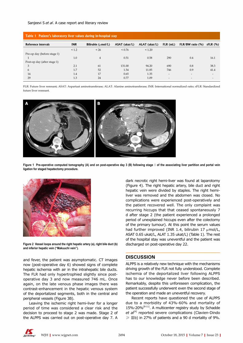
and fever, the patient was asymptomatic. CT images now (post-operative day 6) showed signs of complete hepatic ischemia with air in the intrahepatic bile ducts. The FLR had only hypertrophied slightly since post-operative day 3 and now measured 746 mL. Once again, on the late venous phase images there was contrast-enhancement in the hepatic venous system of the deportalized segments, both in the central and peripheral vessels (Figure 3B).
Leaving the ischemic right hemi-liver for a longer period of time was considered a clear risk and the decision to proceed to stage 2 was made. Stage 2 of the ALPPS was carried out on post-operative day 7. A
dark necrotic right hemi-liver was found at laparotomy (Figure 4). The right hepatic artery, bile duct and right hepatic vein were divided by staples. The right hemi-liver was removed and the abdomen was closed. No complications were experienced post-operatively and the patient recovered well. The only complaint was recurring hiccups that that ceased spontaneously 7 d after stage 2 (the patient experienced a prolonged period of unexplained hiccups even after the colectomy of the primary tumour). At this point the serum values had further improved (INR 1.4, bilirubin 17 μmol/L, ASAT 0.65 ukat/L, ALAT 1.35 ukat/L) (Table 1). The rest of the hospital stay was uneventful and the patient was discharged on post-operative day 22.
DISCUSSIONALPPS is a relatively new technique with the mechanisms driving growth of the FLR not fully understood. Complete ischemia of the deportalized liver following ALPPS has to our knowledge never before been described. Remarkably, despite this unforeseen complication, the patient successfully underwent even the second stage of the operation and made an uneventful recovery.
Recent reports have questioned the use of ALPPS due to a morbidity of 43%-60% and mortality of 15%-20%[8-11]. A multicenter registry study by Schadde et al[4] reported severe complications (Clavien-Dindo ≥ Ⅲb) in 27% of patients and a 90 d mortality of 9%.
2494 October 18, 2015|Volume 7|Issue 23|WJH|www.wjgnet.com
Figure 1 Pre-operative computed tomography (A) and on post-operative day 3 (B) following stage Ⅰ of the associating liver partition and portal vein ligation for staged hepatectomy procedure.
A B
Table 1 Patient’s laboratory liver values during in-hospital stay
Reference intervals INR Bilirubin (μmol/L) ASAT (ukat/L) ALAT (ukat/L) FLR (mL) FLR/BW ratio (%) sFLR (%)
< 1.2 < 26 < 0.76 < 1.20Pre-op day (before stage 1) 1 1.0 4 0.51 0.58 290 0.4 16.1Post-op day (after stage 1) 3 2.1 61 131.00 94.20 690 0.8 38.3 6 1.7 52 1.54 11.85 746 0.9 41.4 14 1.4 17 0.65 1.35 - - - 29 1.3 24 0.77 1.09 - - -
FLR: Future liver remnant; ASAT: Aspartaat aminotransferase; ALAT: Alanine aminotransferase; INR: International normalized ratio; sFLR: Standardized future liver remnant.
Figure 2 Vessel loops around the right hepatic artery (a), right bile duct (b) and inferior hepatic vein (“Makuuchi vein”).
ab
c
Sanjeevi S et al . A case report and literary review

2495 October 18, 2015|Volume 7|Issue 23|WJH|www.wjgnet.com
operative venous flow noted in the deportalized right hemi-liver (Figure 3). This phenomenon may explain the subsequent improvement of serum liver values between stage 1 and 2. Additionally, an increase in venous flow was noted between post-operative days 3 and 6 (Figure 3). The source of the venous blood is however not clearly understood, considering that blood flow in the right hepatic artery was absent on imaging. A standing theory is that the right hemi-lobe received arterial blood from bile duct associated vessels with retrograde filling of the portal veins from the hepatic sinusoids[14]. Retrograde filling of the right hepatic and inferior hepatic veins could also have been possible[15].
The present case was extensively analyzed at the institutional morbidity and mortality review board. A point of criticism was the initial decision to proceed with a right-sided hemi-hepatectomy despite the small FLR. If this was pre-operatively noticed the patient may have benefited from portal vein embolization. Despite the misjudgment, the intra-operative decision to proceed with ALPPS was considered the best option for the patient. The reasoning behind the arterial occlusion was also discussed. A technical error was considered unlikely as optimal flow was palpated in the right hepatic artery prior to closure of the abdomen. It could be hypothesized that trauma during the surgery precipitated the formation of an arterial thrombus. Occlusion of the right hepatic artery has been described following liver transplantations[16]. Damage during the Pringle maneuver and the formation of a pseudoaneurysms have been proposed as underlying mechanisms.
To conclude, ALPPS is a fascinating advancement in liver surgery, requiring further studies to better our under-standing. Yet, despite post-operative complications, in properly selected cases it provides successful outcomes that other modalities of treatment cannot offer.
COMMENTSCase characteristicsA 64 years old male that went into severe liver failure following associating liver partition and portal vein ligation for staged hepatectomy (ALPPS) due to arterial ischemia in the deportalized liver.
Significantly better outcomes were noted in patients with CRLM and patients under the age of 60. Although our patient was 64 years old, he was previously healthy with no recorded comorbidities. Other independent risk factors included red blood cell transfusions and stage Ⅰ operating times longer than 300 min, two factors our patient did not fulfill. Truant et al[6] hypothesize that complications occurring between stage 1 and 2 of ALPPS play a significant role in morbidity and mortality. More specifically, post stage 1 biliary fistula, ascites and infected bile collections worsen overall outcome. Again, our patient experienced none of the abovementioned complications and had an uncomplicated post-operative course apart from post-hepatectomy liver failure and the described liver ischemia.
Post-operative imaging showed that the FLR of the patient increased by 157% over a period of 6 d. Of greater significance is that the major part of the growth (136%) occurred in less than 3 d after stage 1. This is an impressive increase in size, considering that metabolic support from the deportalized liver was sub-optimal. Reported increases in the FLR range from an average of 49%-80% over 7 d[3,4,12]. It should be emphasized that the increase in volume does not necessarily correlate with an increase in function of the FLR[13].
Another unique aspect of this case was the post-
Figure 3 Post-operative late venous phase computed tomography with contrast enhancement of the hepatic venous system of the ischemic right hemi-liver on (A) day 3 and (B) day 6.
A B
Figure 4 Ischemic right liver lobe following stage 2 of associating liver partition and portal vein ligation for staged hepatectomy.
COMMENTS
Sanjeevi S et al . A case report and literary review

2496 October 18, 2015|Volume 7|Issue 23|WJH|www.wjgnet.com
Clinical diagnosisLiver failure following stage Ⅰ of ALPPS due to arterial ischemia.
Differential diagnosisThe diagnosis of liver failure was well established by imaging and laboratory tests. The exact mechanism for the full recovery of the patient is not completely understood.
Laboratory diagnosisAn acute rise in international normalized ratio and serum bilirubin, alanine aminotransferase and aspartaat aminotransferase which gradually improved over time.
Imaging diagnosisAn ultrasound of the liver showed no sign of arterial blood flow to the right hemi-liver and a computed tomography examination confirmed occlusion of the right hepatic artery and an ischemic right hemi-liver.
Pathological diagnosisPathology following stage Ⅱ of ALPPS confirmed the underlying disease to be colorectal liver metastases.
TreatmentSupportive therapy.
Related reportsThis is the first report to describe arterial ischemia in the deportalized liver following ALPPS.
Term explanation ALPPS: Associated liver partition and portal vein ligation for staged hepatectomy; FLR: Future liver remnant; CRLM: Colorectal liver metastasis.
Experiences and lessonsALPPS is a fascinating advancement in liver surgery requiring further studies to better our understanding. Despite post-operative complications, in properly selected cases it provides successful outcomes that other modalities of treatment cannot offer.
Peer-reviewALPPS is an interesting novel two stage surgical procedure for extended liver tumors with more and more applications described into literature: This article describes a case report of application of ALPPS procedure in a patient intraoperatively judged as not eligible to a right hepatectomy due to unexpected little remnant volume: The attention is focalized on a specific particular postoperative complication, never described previously into literature.
REFERENCES1 Alvarez FA, Ardiles V, Sanchez Claria R, Pekolj J, de Santibañes
E. Associating liver partition and portal vein ligation for staged hepatectomy (ALPPS): tips and tricks. J Gastrointest Surg 2013; 17: 814-821 [PMID: 23188224 DOI: 10.1007/s11605-012-2092-2]
2 Hasselgren K, Sandström P, Björnsson B. Role of associating liver partition and portal vein ligation for staged hepatectomy in colorectal liver metastases: a review. World J Gastroenterol 2015; 21: 4491-4498 [PMID: 25914457 DOI: 10.3748/wjg.v21.i15.4491]
3 Jackson T, Siegel KA, Siegel CT. Rescue ALPPS: Intraoperative Conversion to ALPPS during Synchronous Resection of Rectal Cancer and Liver Metastasis. Case Rep Surg 2014; 2014: 487852 [PMID: 25506458 DOI: 10.1155/2014/487852]
4 Schadde E, Hernandez-Alejandro R, Lang H, de Santibaňes E, Clavien PA. ALPPS Offers a Better Chance of Complete Resection in Patients with Primarily Unresectable Liver Tumors. Results of a Multicentre Analysis: Reply. World J Surg 2015; 39: 1850-1851 [PMID: 25743483 DOI: 10.1007/s00268-015-2964-1]
5 Schnitzbauer AA, Lang SA, Goessmann H, Nadalin S, Baumgart J, Farkas SA, Fichtner-Feigl S, Lorf T, Goralcyk A, Hörbelt R, Kroemer A, Loss M, Rümmele P, Scherer MN, Padberg W, Königsrainer A, Lang H, Obed A, Schlitt HJ. Right portal vein ligation combined with in situ splitting induces rapid left lateral liver lobe hypertrophy enabling 2-staged extended right hepatic resection in small-for-size settings. Ann Surg 2012; 255: 405-414 [PMID: 22330038 DOI: 10.1097/SLA.0b013e31824856f5]
6 Truant S, Scatton O, Dokmak S, Regimbeau JM, Lucidi V, Laurent A, Gauzolino R, Castro Benitez C, Pequignot A, Donckier V, Lim C, Blanleuil ML, Brustia R, Le Treut YP, Soubrane O, Azoulay D, Farges O, Adam R, Pruvot FR. Associating liver partition and portal vein ligation for staged hepatectomy (ALPPS): impact of the inter-stages course on morbi-mortality and implications for management. Eur J Surg Oncol 2015; 41: 674-682 [PMID: 25630689 DOI: 10.1016/j.ejso.2015.01.004]
7 Balzan S, Belghiti J, Farges O, Ogata S, Sauvanet A, Delefosse D, Durand F. The “50-50 criteria” on postoperative day 5: an accurate predictor of liver failure and death after hepatectomy. Ann Surg 2005; 242: 824-828, discussion 828-829 [PMID: 16327492]
8 Sotiropoulos GC, Kouraklis G. The ALPPS procedure for extended indications in liver surgery: an old finding applied in surgical oncology. Ann Surg 2013; 257: e26 [PMID: 23629529 DOI: 10.1097/SLA.0b013e3182942e4a]
9 Aloia TA, Vauthey JN. Associating liver partition and portal vein ligation for staged hepatectomy (ALPPS): what is gained and what is lost? Ann Surg 2012; 256: e9; author reply e16-e19 [PMID: 22868369 DOI: 10.1097/SLA.0b013e318265fd3e]
10 Kokudo N, Shindoh J. How can we safely climb the ALPPS? Updates Surg 2013; 65: 175-177 [PMID: 23715830 DOI: 10.1007/s13304-013-0215-2]
11 Rohatgi S, Harrison EM, Powell JJ, Wigmore SJ. ALPPS: Adverse Outcomes Demand Clear Justification in an Era of Improving Survival for Colorectal Liver Metastases. World J Surg 2015; 39: 1848-1849 [PMID: 25145819 DOI: 10.1007/s00268-014-2726-5]
12 Michels NA. Newer anatomy of the liver and its variant blood supply and collateral circulation. Am J Surg 1966; 112: 337-347 [PMID: 5917302]
13 Pagano D, Gruttadauria S. Impact of future remnant liver volume on post-hepatectomy regeneration in non-cirrhotic livers. Front Surg 2014; 1: 10 [PMID: 25593935 DOI: 10.3389/fsurg.2014.00010]
14 Northover JM, Terblanche J. A new look at the arterial supply of the bile duct in man and its surgical implications. Br J Surg 1979; 66: 379-384 [PMID: 466017]
15 Pedersen JF, Dakhil AZ, Jensen DB, Søndergaard B, Bytzer P. Abnormal hepatic vein Doppler waveform in patients without liver disease. Br J Radiol 2005; 78: 242-244 [PMID: 15730989 DOI: 10.1259/bjr/15227254]
16 Tanabe G, Kawaida K, Hamanoue M, Kihara K, Hirata S, Maemura M, Ueno S, Aikou T. Treatment for accidental occlusion of the hepatic artery after hepatic resection: report of two cases. Surg Today 1999; 29: 268-272 [PMID: 10192741 DOI: 10.1007/bf02483020]
P- Reviewer: Campos RR, Sguinzi R, Vennarecci G S- Editor: Ji FF L- Editor: A E- Editor: Liu SQ
Sanjeevi S et al . A case report and literary review

© 2015 Baishideng Publishing Group Inc. All rights reserved.
Published by Baishideng Publishing Group Inc8226 Regency Drive, Pleasanton, CA 94588, USA
Telephone: +1-925-223-8242Fax: +1-925-223-8243
E-mail: [email protected] Desk: http://www.wjgnet.com/esps/helpdesk.aspx
http://www.wjgnet.com
Copyright © 2022 FDOKUMEN