World Journal of Transplantation - BPG Management System
206
World Journal of Transplantation World J Transplant 2016 June 24; 6(2): 255-450 Published by Baishideng Publishing Group Inc ISSN 2220-3230 (online)
-
Upload
khangminh22 -
Category
Documents
-
view
6 -
download
0
Transcript of World Journal of Transplantation - BPG Management System

World Journal of TransplantationWorld J Transplant 2016 June 24; 6(2): 255-450
Published by Baishideng Publishing Group Inc
ISSN 2220-3230 (online)

World Journal of TransplantationW J T
EDITOR-IN-CHIEFMaurizio Salvadori, Florence
GUEST EDITORIAL BOARD MEMBERSChao-Long Chen, KaohsiungYu-Fan Cheng, KaohsiungBor-Luen Chiang, TaipeiYang-Jen Chiang, TaoyuanShiaw-Min Hwang, HsinchuTang-Her Jaing, TaoyuanChih-Cheng Lai, TainanSteven Shoei-Lung Li, KaohsiungSyh-Jae Lin, TaoyuanYa-Chung Tian, LinkouChia-Chao Wu, Taipei
MEMBERS OF THE EDITORIAL BOARD
Argentina
Walter Douthat, Cordoba
Australia
Julianne Bayliss, MelbourneNeil C Boudville, WaZoltan H Endre, SydneyGW McCaughan, CamperdownSteven E Mutsaers, NedlandsNA Shackel, SydneyDeborah J Verran, Nsw
Austria
Kyra A Borchhardt, Vienna
Johannes Clausen, InnsbruckRaimund Margreiter, Innsbruck
Belgium
Olivier Detry, LiegeEvelyne Lerut, LeuvenMaarten Naesens, LeuvenEtienne M Sokal, Bruxelles
Brazil
Luiz Alves, ManguinhosIlka FSF Boin, CampinasNiels OS Camara, Cidade UniversitáriaEleazar Chaib, Sao PauloKatherine AT de Carvalho, CuritibaMF F Silva, Porto AlefreRenato Silva, Sao Paulo
Bulgaria
Papantchev Vassil Gueorguiev, Sofia
Canada
Subrata Chakrabarti, LondonHuifang Chen, MontrealThomas Churchill, AlbertaCaigan Du, VancouverReginald M Gorczynski, TorontoPaul A Keown, VancouverTatsuya Kin, AlbertaMichele Molinari, HalifaxEberhard L Renner, Ontario
AM James Shapiro, EdmontonGeorge Therapondos, EdinburghChandini Thirukkumaran, AlbertaSerdar Yilmaz, Calgary
China
Wing Y Au, Hong KongGodfrey CF Chan, Hong KongDaniel KL Cheuk, Hong KongJun He, SuzhouJanette Kwok, Hong KongJanette SY Kwok, Hong KongAnskar Yu-Hung Leung, Hong KongPo-Sing Leung, Hong KongTing-Bo Liang, HangzhouKai-Yan Liu, BeijingHai-Yan Liu, SuzhouZe-Zhou Song, HangzhouJing-Ping Sun, Hong KongMeng-Qun Tan, ShenzhenChang-Xi Wang, GuangzhouShi-Xia Xu, BeijingLv-Nan Yan, ChengduFeng Yin, Beijing Peng Zhang, Xi'anBin Zhu, HangzhouHe-Qun Zou, Guangzhou
Cuba
OS Leon, Havana
Czech Republic
Holan Vladimir, Videnska
I
Editorial Board2016-2019
The World Journal of Transplantation Editorial Board consists of 361 members, representing a team of worldwide experts in transplantation. They are from 43 countries, including Argentina (1), Australia (7), Austria (3), Belgium (4), Brazil (7), Bulgaria (1), Canada (13), China (32), Cuba (1), Czech Republic (1), Denmark (1), Finland (1), France (4), Georgia (1), Germany (14), Greece (5), Hungary (2), India (7), Iran (7), Israel (4), Italy (33), Japan (18), Jordan (1), Macedonia (1), Mexico (2), Morocco (1), Netherlands (4), Nigeria (1), Norway (1), Pakistan (1), Poland (2), Qatar (1), Saudi Arabia (3), Singapore (1), South Korea (16), Spain (9), Sweden (1), Switzerland (3), Thailand (2), Tunisia (1), Turkey (6), United Kingdom (17), and United States (120).
February 26, 2016WJT|www.wjgnet.com

Denmark
Klaus Muller, Copenhagen
Finland
Andreas Scherer, Kontiolahti
France
Felix Cantarovich, ParisRoussel Christian, NantesBertrand Dupont, ParisLoic Fouillard, Cergy-Pontoise
Georgia
Archil Chkhotua, Tbilisi
Germany
Elisenda Banon-Maneus, MunichSusanne Beckebaum, EssenAndres Beiras-Fernandez, MunichRainer Birck, MannheimHassan Dihazi, GoettingenChristoph Eisenbach, HeidelbergFrieder Keller, UlmAlfred Konigsrainer, TuebingenThomas Minor, BonnPeter Schemmer, HeidelbergMeinolf Suttorp, DresdenRene H Tolba, AachenWolfgang Wagner, AachenMin-Min Wang, Berlin
Greece
Costas Fourtounas, Rio-PatrasEvgenios Goussetis, AthensMaria Koukoulaki, RionSophia Lionaki, AthensAnna Petropoulou, Athens
Hungary
Andrea Ferencz, BudapestPeter Hamar, Budapest
India
Sanjay K Agarwal, New DelhiSuraksha Agrawal, LucknowB George, VellorePravin Mhatre, MumbaiGeeta Ravindran, MunbaiAvnish K Seth, New DelhiMalancha Ta, Bangalore
Iran
Parisa Badiee, Shiraz
Seyed M Dehghani, ShirazAhad Eshraghian, ShirazAli Ghafari, UrmiaMitra Mahdavi-Mazdeh, TehranSaeed Taheri, TehranRamin Yaghoobi, Shiraz
Israel
Nimer Assy, SafedEsther Granot, JerusalemInna Sinuani, ZerifinShimon Slavin, Tel Aviv
Italy
Gian Adani, UdineUmberto Baccarani, UdineBruno Bonetti, VeronaAlessandro Busca, TurinCristina Costa, TorinoStefano Faenza, BolognaGian M Ghiggeri, GenoaGiovanni Camussi, TurinGrandaliano Giuseppe, FoggiaAndrea Giusti, GenovaPaola Gremigni, BolognaWalter F Grigioni, BolognaAlessandro Isidori, PesaroRenzo Mignani, RiminiLuca Neri, MilanPietro Andreone, BolognaLuciano Potena, BolognaMatteo Ravaioli, BolognaGiampiero La Rocca, PalermoGiulio Romano, UdineVito Ruggiero, PomeziaFabrizio Sansone, TurinMichele Santangelo, NaplesSergio Rutella, RomeAntonino Sessa, NaplesAurelio Sonzogni, BergamoGiovanni Stallone, FoggiaLamponi Stefania, SienaGiovanni Luigi Tripepi, Reggio CalabriaCornelio Uderzo, MilanMassimiliano Veroux, CataniaGiovanni Li Volti, Catania
Japan
Junya Kanda, SaitamaHiroshi Kanno, YokohamaMureo Kasahara, TokyoXiao-Kang Li, TokyoShinichi Miyagawa, MatsumotoShugo Mizuno, TsuWalid El Moghazy, KyotoTakehiko Mori, TokyoDaisuke Morioka, YokohamaHirofumi Noguchi, OkinawaMasahiko Okamoto, KyotoYasuhiko Sugawara, TokyoS Sumi, KyotoMasahiko Taniguchi, AsahikawaShintaro Yamazaki, TokyoKotaro Yoshimura, Tokyo Katsutoshi Yoshizato, Higashihiroshima
Kenji Yuzawa, Ibaraki-ken
Jordan
Mahmoud M Sarhan, Juabaiha
Macedonia
Goce Spasovski, Skopje
Mexico
Rene Drucker-Colln, MexicoGustavo Martinez-Mier, Veracruz
Morocco
Faissal Tarrass, Casablanca
Netherlands
Michiel GH Betjes, RotterdamFrank JMF Dor, RotterdamIrma Joosten, NijmegenLuc JW van der Laan, Rotterdam
Nigeria
Anthony A Oyekunle, Ile-Ife
Norway
Lars L Gullestad, Oslo
Pakistan
Tahir Shmasi, Karachi
Poland
Piotr Czubkowski, WarsawAndrzej Rydzewski, Warszawa
Qatar
Moutaz Derbala, Doha
Saudi Arabia
Ali Al-Ahmari, RiyadhMohamed Mabed, Jeddah Mohamed M Sayed-Ahmed, Riyadh
Singapore
Seng H Quak, Singapore
II February 26, 2016WJT|www.wjgnet.com

III February 26, 2016WJT|www.wjgnet.com
South KoreaCurie Ahn, Seoul Jong Wook Chang, Seoul Baik Cho, Jeonju Hyeon Jeong, SeoulKoo-Jeong Kang, DaeguChang Nyung Kim, Gyeonggi-doKyung Mo Kim, SeoulYon S Kim, SeoulGaab S Kim, SeoulJong W Lee, SeoulSang-Oh Lee, Seoul Eun-Jee Oh, SeoulKwon M Park, DaeguChul W Yang, SeoulKun-Ho Yoon, SeoulSeung Kwon You, Seoul
Spain
Manuel Arias, MadridRuben Ciria, CordobaLuis Fontana, GranadaMaria Marco, BarcelonaJose AP Minano, El Palmar-MurciaAlberto Ortiz, MadridJulio Pascual, BarcelonaCarmen Peralta, BarcelonaJesus Vaquero, Majadahonda
Sweden
Tobias Larsson, Stockholm
Switzerland
C Deffert, GenevaAndrea De Gottardi, Berne-InselspitalChristian Toso, Geneva
Thailand
Suradej Hongeng, BangkokWeekitt Kittisupamongkol, Bangkok
Tunisia
Kais Harzallah, Tunis
Turkey
Elvan C Citak, MersinEmir B Denkbas, BeytepeIhsan Ergün, AnkaraMurat Kilic, CigliOner Ozdemir, SakaryaBaris Yildiz, Ankara
United Kingdom
Jacob A Akoh, Plymouth
Atul Bagul, LeicesterRicky H Bhogal, BirminghamRichard J Borrows, BirminghamEric Chemla, LondonSarah Hosgood, Leicester Stefan G Hubscher, BirminghamAlireza Jahromi, LondonAlan Jardine, GlasgowSanjeev Kanoria, LondonMichel Modo, LondonPaolo Muiesan, BirminghamGH Neild, LondonMagdi Shehata, LeicesterAfshin Tavakoli, ManchesterAlexander Woywodt, PrestonQihe Xu, London
United States
Arshak R Alexanian, MilwaukeeSharif Ali, DetroitJaime Aranda-Michel, JacksonvilleRobert M Aris, Chapel HillReto Baertschiger, IndianapolisDavid A Baran, NewarkGerald Brandacher, BaltimoreJoseph F Buell, New OrleansYan Chen, NashvilleHerman S Cheung, Coral GablesGaetano Ciancio, MiamiDiane Cibrik, Ann ArborLuca Cicalese, GalvestonAri Cohen, New OrleansDarshana Dadhania, New YorkGraciela De Boccardo, New YorkCataldo Doria, PhiladelphiaAmrita Dosanjh, San DiegoS H Emre, New HavenSherif S Farag, IndianapolisRoberto Firpi, GainesvilleRobert A Fisher, RichmondAmy Friedman, SyracuseTibor Fulop, JacksonG Ian Gallicano, WashingtonWenda Gao, BostonRoberto Gedaly, LexingtonW Scott Goebel, IndianapolisRujun Gong, ProvidenceChad R Gordon, BostonAngelika Gruessner, TucsonGregg Hadley, ColumbusJeffrey B Halldorson, SeattleMehdi Hamadani, MilwaukeeKaren L Hardinger, Kansas Imed Helal, AuroraAllan D Hess, BaltimoreIbtesam Hilmi, PittsburghAndres Jaramillo, ItascaRandeep S Kashyap, RochesterTatsuo Kawai, BostonImran Khalid, JeddahAjai Khanna, San DiegoDean Y Kim, DetroitKatsuhiro Kita, New YorkDavid J Kramer, JacksonvilleJW Kupiec-Weglinski, Los Angeles
Paul Y Kwo, IndianapolisTechung Lee, BuffaloJosh Levitsky, ChicagoXian C Li, BostonSuthat Liangpunsakul, IndianapolisSeah H Lim, AmarilloJulie Lin, BostonChing-Shwun Lin, San FranciscoDelong Liu, WestchesterAndrew Lobashevsky, IndianapolisPaul Lucas, ValhallaXunrong Luo, ChicagoDidier A Mandelbrot, BostonMartin Mangino, RichmondRichard S Mangus, IndianapolisIgnazio R Marino, PhiladelphiaPaulo Ney Aguiar Martins, BostonAndrew S Mathis, Long BranchJames Millis, ChicagoTamir Miloh, PhoenixAyse L Mindikoglu, BaltimoreAmr El-Husseini Mohamed, LexingtonSandeep Mukherjee, OmahaTibor Nadasdy, ColumbusAtsunori Nakao, PittsburghSingh Neeraj, ColumbusJustin H Nguyen, FloridaVolker Nickeleit, Chapel HillChristopher Niyibizi, HersheyMacaulay Onuigbo, Eau ClaireJorge A Ortiz, PhiladelphiaRaymond M Planinsic, PittsburghQi Qian, RochesterRajalingam Raja, Los AngelesMichael A Ramsay, DallasRaymund R Razonable, RochesterMohammed S Razzaque, BostonPavan Reddy, Ann ArborCamillo Ricordi, MiamiHoracio Rilo, TucsonDA Rizzieri, DurhamKenneth Rolston, HoustonPhilip Rosenthal, San FranciscoPhillip Ruiz, MiamiT Sakai, PittsburghBipin N Savani, NashvilleJan D Schmitto, BostonRoman Schumann, BostonMouin G Seikaly, DallasFuad Shihab, Salt LakeJeffrey H Shuhaiber, Cincinnati OhioMark S Slaughter, LouisvilleAndrey Staruschenko, MilwaukeeKK Sureshkumar, PittsburghHenkie P Tan, PittsburghBurcin Taner, JacksonvilleAJ Tector, IndianapolisVivian Tellis, BronxJohn Thornton, ClevelandJose Torrealba, MadisonJames F Trotter, DallasAndreas G Tzakis, MiamiRocco C Venuto, BuffaloMichael Voigt, DriveMatthew R Weir, BaltimoreVictor Xia, Los Angeles

IV February 26, 2016WJT|www.wjgnet.com
Hongzhi Xu, BostonHe Xu, Atlanta
Dengping Yin, NashvilleRubin Zhang, Louisiana
Zhi Zhong, CharlestonJoseph Zwischenberger, Lexington

FIELD OF VISION
255 Exocrinedrainageinvascularizedpancreastransplantationinthenewmillennium
El-Hennawy H, Stratta RJ, Smith F
272 Hepatoduodenalligamentdissectiontechniqueduringrecipienthepatectomyforlivertransplantation:How
Idoit?
Kayaalp C, Tolan K, Yilmaz S
REVIEW
278 Livertransplantationandthemanagementofprogressivefamilialintrahepaticcholestasisinchildren
Mehl A, Bohorquez H, Serrano MS, Galliano G, Reichman TW
291 Massivehaemorrhageinlivertransplantation:Consequences,predictionandmanagement
Cleland S, Corredor C, Ye JJ, Srinivas C, McCluskey SA
MINIREVIEWS
306 Loco-regionaltherapiesforpatientswithhepatocellularcarcinomaawaitinglivertransplantation:Selecting
anoptimaltherapy
Byrne TJ, Rakela J
314 Potentialapproachestoimprovetheoutcomesofdonationaftercardiacdeathlivergrafts
Mahboub P, Bozorgzadeh A, Martins PN
321 Firstlinevs delayedtransplantationinmyeloma:Certaintiesandcontroversies
Brioli A
331 StateofdeceaseddonortransplantationinIndia:Amodelfordevelopingcountriesaroundtheworld
Abraham G, Vijayan M, Gopalakrishnan N, Shroff S, Amalorpavanathan J, Yuvaraj A, Nair S, Sundarrajan S
ORIGINAL ARTICLE
Basic Study
336 Roleofcytomegalovirusonthematurationandfunctionofmonocytederiveddendriticcellsofliver
transplantpatients
Karimi MH, Shariat A, Yaghobi R, Mokhtariazad T, Moazzeni SM
W JContents
IWJT|www.wjgnet.com June 24, 2016|Volume 6|Issue 2|
World Journal of TransplantationT
Quarterly Volume 6 Number 2 June 24, 2016

ContentsWorld Journal of Transplantation
Volume 6 Number 2 June 24, 2016
IIWJT|www.wjgnet.com June 24, 2016|Volume 6|Issue 2|
Retrospective Cohort Study
347 Single-lungtransplantationinemphysema:Retrospectivestudyanalyzingsurvivalandwaitinglist
mortality
Borro JM, Delgado M, Coll E, Pita S
356 Impactofbodymassindexonoutcomesof48281patientsundergoingfirsttimecadavericliver
transplantation
Ayloo S, Hurton S, Cwinn M, Molinari M
370 Riskfactorsforfractureinadultkidneytransplantrecipients
Naylor KL, Zou G, Leslie WD, Hodsman AB, Lam NN, McArthur E, Fraser LA, Knoll GA, Adachi JD, Kim SJ, Garg AX
Retrospective Study
380 Longtermoutcomesofcardiactransplantforimmunoglobulinlightchainamyloidosis:TheMayoClinic
experience
Grogan M, Gertz M, McCurdy A, Roeker L, Kyle R, Kushwaha S, Daly R, Dearani J, Rodeheffer R, Frantz R, Lacy M,
Hayman S, McGregor C, Edwards B, Dispenzieri A
389 Ventilatorassociatedpneumoniafollowinglivertransplantation:Etiology,riskfactorsandoutcome
Siniscalchi A, Aurini L, Benini B, Gamberini L, Nava S, Viale P, Faenza S
396 LivertransplantationforhepatocellularcarcinomainIreland:Pre-operativealpha-fetoproteinpredicts
tumourrecurrenceina14-yearsingle-centrenationalexperience
O’Connor DB, Burke JP, Hegarty J, McCormick AP, Nolan N, Hoti E, Maguire D, Geoghegan J, Traynor O
403 Higherplasmabilirubinpredictsveno-occlusivediseaseinearlychildhoodundergoinghematopoieticstem
celltransplantationwithcyclosporine
Kim KS, Moon A, Kang HJ, Shin HY, Choi YH, Kim HS, Kim SG
411 Proposalofnewexpandedselectioncriteriausingtotaltumorsizeand18F-fluorodeoxyglucose-positron
emissiontomography/computedtomographyforlivingdonorlivertransplantationinpatientswith
hepatocellularcarcinoma:TheNationalCancerCenterKoreacriteria
Lee SD, Lee B, Kim SH, Joo J, Kim SK, Kim YK, Park SJ
Observational Study
423 DeceaseddonororganprocurementinjuriesintheUnitedStates
Taber TE, Neidlinger NA, Mujtaba MA, Eidbo EE, Cauwels RL, Hannan EM, Miller JR, Paramesh AS

ContentsWorld Journal of Transplantation
Volume 6 Number 2 June 24, 2016
IIIWJT|www.wjgnet.com June 24, 2016|Volume 6|Issue 2|
Randomized Controlled Trial
429 Exercisemanualforliverdiseasepatients
Limongi V, Dos Santos DC, Oliveira da Silva AM, Boin IFSF, Stucchi RSB
CASE REPORT
437 Isletautotransplantationinapatientwithhypercoagulabledisorder
Desai CS, Khan KM, Cui W
442 Acutebacterialsternoclavicularosteomyelitisinalong-termrenaltransplantrecipient
Dounousi E, Duni A, Xiromeriti S, Pappas C, Siamopoulos KC
447 Cavitarylunglesion6yearsafterrenaltransplantation
Subbiah AK, Arava S, Bagchi S, Madan K, Das CJ, Agarwal SK

ContentsWorld Journal of Transplantation
Volume 6 Number 2 June 24, 2016
EDITORS FOR THIS ISSUE
Responsible Assistant Editor: Xiang Li Responsible Science Editor: Fang-Fang Ji Responsible Electronic Editor: Su-Qing Liu Proofing Editorial Office Director: Xiu-Xia SongProofing Editor-in-Chief: Lian-Sheng Ma
Room 903, Building D, Ocean International Center,No. 62 Dongsihuan Zhonglu, Chaoyang District, Beijing 100025, ChinaTelephone: +86-10-85381891Fax: +86-10-85381893E-mail: [email protected] Desk: http://www.wjgnet.com/esps/helpdesk.aspxhttp://www.wjgnet.com
PUBLISHERBaishideng Publishing Group Inc8226 Regency Drive, Pleasanton, CA 94588, USATelephone: +1-925-223-8242Fax: +1-925-223-8243E-mail: [email protected] Desk: http://www.wjgnet.com/esps/helpdesk.aspxhttp://www.wjgnet.com
PUBLICATIONDATEJune 24, 2016
COPYRIGHT© 2016 Baishideng Publishing Group Inc. Articles pub-lished by this Open-Access journal are distributed under the terms of the Creative Commons Attribution Non-commercial License, which permits use, distribution, and reproduction in any medium, provided the original work is properly cited, the use is non commercial and is other-wise in compliance with the license.
SPECIALSTATEMENTAll articles published in journals owned by the Baishideng Publishing Group (BPG) represent the views and opinions of their authors, and not the views, opinions or policies of the BPG, except where otherwise explicitly indicated.
INSTRUCTIONSTOAUTHORSFull instructions are available online at http://www.wjgnet.com/bpg/g_info_20160116143427.htm
ONLINESUBMISSIONhttp://www.wjgnet.com/esps/
IVWJT|www.wjgnet.com
ABOUT COVER
AIM AND SCOPE
AIM AND SCOPE
FLYLEAF
June 24, 2016|Volume 6|Issue 2|
NAMEOFJOURNALWorld Journal of Transplantation
ISSNISSN 2220-3230 (online)
LAUNCHDATEDecember 24, 2011
FREQUENCYQuarterly
EDITOR-IN-CHIEFMaurizio Salvadori, MD, Professor, Renal Unit, Careggi University Hospital, Viale Pieraccini 18, Florence 50139, Italy
EDITORIALOFFICEJin-Lei Wang, DirectorXiu-Xia Song, Vice DirectorWorld Journal of Transplantation
EditorialBoardMemberofWorldJournalofTransplantation,JeffreyBHalldorson,MD,PhD,AssistantProfessor,DepartmentofSurgery,UniversityofWashington,DivisionTransplantat,Seattle,WA98195,UnitedStates
World Journal of Transplantation (World J Transplant, WJT, online ISSN 2220-3230, DOI: 10.5500) is a peer-reviewed open access academic journal that aims to guide clinical practice and improve diagnostic and therapeutic skills of clinicians.
WJT covers topics concerning organ and tissue donation and preservation; tissue injury, repair, inflammation, and aging; immune recognition, regulation, effector mecha-nisms, and opportunities for induction of tolerance, thoracic transplantation (heart, lung), abdominal transplantation (kidney, liver, pancreas, islets), transplantation of tissues, cell therapy and islet transplantation, clinical transplantation, experimental transplantation, immunobiology and genomics, and xenotransplantation. The current columns of WJT include editorial, frontier, diagnostic advances, therapeutics advances, field of vision, mini-reviews, review, topic highlight, medical ethics, original articles, case report, clinical case conference (Clinicopathological conference), and autobiography.
World Journal of Transplantation is now indexed in PubMed, PubMed Central.
I-IV EditorialBoard

Hany El-Hennawy, Robert J Stratta, Fowler Smith
Hany El-Hennawy, Robert J Stratta, Fowler Smith, Department of Surgery, Wake Forest School of Medicine, WinstonSalem, NC 27157, United States
Author contributions: ElHennawy H and Stratta RJ studier conception, designs, acquisition of data, analysis and interpretation of data; all the authors do drafting of manuscript and critical revision.
Conflict-of-interest statement: The authors acknowledge that the above manuscript represents original work that has not been previously published or submitted for publication. There are no conflicts of interest, grant support, sponsorship, or other financial arrangements to report by any of the authors.
Open-Access: This article is an openaccess article which was selected by an inhouse editor and fully peerreviewed by external reviewers. It is distributed in accordance with the Creative Commons Attribution Non Commercial (CC BYNC 4.0) license, which permits others to distribute, remix, adapt, build upon this work noncommercially, and license their derivative works on different terms, provided the original work is properly cited and the use is noncommercial. See: http://creativecommons.org/licenses/bync/4.0/
Manuscript source: Invited manuscript
Correspondence to: Dr. Robert J Stratta, MD, Department of Surgery, Wake Forest School of Medicine, One Medical Center Blvd, WinstonSalem, NC 27157, United States. [email protected]: +13367160548Fax: +13367135055
Received: February 16, 2016Peer-review started: February 16, 2016First decision: March 1, 2016Revised: May 6, 2016Accepted: May 31, 2016Article in press: June 2, 2016Published online: June 24, 2016
AbstractThe history of vascularized pancreas transplantation largely parallels developments in immunosuppression and technical refinements in transplant surgery. From the late-1980s to 1995, most pancreas transplants were whole organ pancreatic grafts with insulin delivery tothe iliac vein and diversion of the pancreatic ductal secretions to the urinary bladder (systemic-bladder technique). The advent of bladder drainage revolu-tionized the safety and improved the success of pan-creas transplantation. However, starting in 1995, a seismic change occurred from bladder to bowel exocrine drainage coincident with improvements in immuno-suppression, preservation techniques, diagnostic mo-nitoring, general medical care, and the success and frequency of enteric conversion. In the new millennium, pancreas transplants are performed predominantly as pancreatico-duodenal grafts with enteric diversion of the pancreatic ductal secretions coupled with iliac vein provision of insulin (systemic-enteric technique) although the systemic-bladder technique endures as a preferred alternative in selected cases. In the early 1990s, a novel technique of venous drainage into the superior mesenteric vein combined with bowel exocrine diversion (portal-enteric technique) was designed and subsequently refined over the next ≥ 20 years to re-create the natural physiology of the pancreas with first-pass hepatic processing of insulin. Enteric drainage usually refers to jejunal or ileal diversion of the exocrine secretions either with a primary enteric anastomosis or with an additional Roux limb. The portal-enteric technique has spawned a number of newer and revi-sited techniques of enteric exocrine drainage including duodenal or gastric diversion. Reports in the literaturesuggest no differences in pancreas transplant outcomes irrespective of type of either venous or exocrine diver-sion. The purpose of this review is to examine the
FIELD OF VISION
255 June 24, 2016|Volume 6|Issue 2|WJT|www.wjgnet.com
Exocrine drainage in vascularized pancreas transplantation in the new millennium
World J Transplant 2016 June 24; 6(2): 255-271ISSN 2220-3230 (online)
© 2016 Baishideng Publishing Group Inc. All rights reserved.
Submit a Manuscript: http://www.wjgnet.com/esps/Help Desk: http://www.wjgnet.com/esps/helpdesk.aspxDOI: 10.5500/wjt.v6.i2.255
World Journal of TransplantationW J T

literature on exocrine drainage in the new millennium (the purported “enteric drainage” era) with special atten-tion to technical variations and nuances in vascularized pancreas transplantation that have been proposed and studied in this time period.
Key words: Pancreas transplantation; Portal-enteric drainage; Simultaneous pancreas-kidney transplant; Systemic-bladder drainage; Enteric conversion; Solitary pancreas transplant; Systemic-enteric drainage
© The Author(s) 2016. Published by Baishideng Publishing Group Inc. All rights reserved.
Core tip: The history of vascularized pancreas trans-plantation largely parallels advances in surgical tech-niques. Prior to 1995, most pancreas transplants were performed with delivery of insulin to the iliac vein and diversion of the pancreatic ductal secretions to the urinary bladder (systemic-bladder technique). Starting in 1995, however, a seismic change occurred from bladder to bowel drainage of the pancreatic secretions that was spurred in part by the success of enteric conversion. In the new millennium, most pancreas transplants are performed as pancreatico-duodenal grafts with either iliac vein and bowel exocrine diversion (systemic-enteric technique) or portal-enteric drainage. With refine-ments in surgical techniques, exocrine drainage is no longer considered the “Achilles’ heel” of pancreas trans-plantation.
ElHennawy H, Stratta RJ, Smith F. Exocrine drainage in vascularized pancreas transplantation in the new millennium. World J Transplant 2016; 6(2): 255271 Available from: URL: http://www.wjgnet.com/22203230/full/v6/i2/255.htm DOI: http://dx.doi.org/10.5500/wjt.v6.i2.255
INTRODUCTIONSince the inception of the International Pancreas Transplant Registry (IPTR) in 1984, data on > 48000 pancreas transplants has been captured in the ensuing 30 years[1]. There exist 3 major types of vascularized pancreas transplantation; simultaneous pancreas-kidney (SPK), sequential pancreas after kidney (PAK), and pancreas transplantation alone (PTA). Solitary pancreas transplants refer to the PAK and PTA types. They are usually analyzed together because of similar outcomes coupled with the fact that these procedures are performed in the absence of uremia. However, the state of kidney function is quite different; post-uremic in PAK compared to non-uremic in PTA. In the past 3 decades, the results of SPK transplantation have been superior to solitary pancreas transplantation although the disparity in outcomes has decreased over time. In the United States, solitary pancreas transplants (PAK-17%, PTA-9%) represent the minority of activity while 74% are characterized as SPK transplants[1-3].
In uremic patients with type 1 diabetes mellitus, SPK transplantation is a highly regarded treatment alternative because it addresses both kidney failure and diabetes[3]. The number of United States annual pancreas trans-plants reached a high of 1484 in 2004 and had dropped to < 1000 by 2014[1-3]. The number of annual pancreas transplants reported to the Eurotransplant Network has similarly declined in the past decade whereas annual activity in the United Kingdom has remained relatively stable and activity elsewhere in the world has increased[1-3]. In spite of declining numbers, outcomes have continued to improve and include higher risk groups such as African-Americans, patients with a phenotype suggesting “type 2 diabetes” and solitary pancreas transplant recipients[1-5]. Five year patient survival rates are now nearly 90% across all three transplant types and 10-year patient survival is > 70% in all three groups. Moreover, insulin independence is sustained at 5 years in 73% of SPK, 64% of PAK, and 53% of PTA recipients. The pancreas graft half-life is currently 10-15 years, which is amongst the lengthiest for extra-renal transplants[2].
Evolution in surgical techniques has characterized and paralleled the growth and development of pancr-eas transplantation. In late 1966 at the University of Minnesota, Kelly et al[6] reported the first human pancreas transplant. The initial case was an SPK trans-plant with a segmental pancreas graft implanted in the iliac fossa with ligation of the pancreatic duct. In the ensuing 13 cases performed between 1966 and 1973, however, Lillehei et al[7] transplanted a pancreatico-duodenal graft with either an external ostomy/cutan-eous fistula or connection between the recipient bowel and graft duodenum for exocrine drainage. Conse-quently, optimal management of the pancreatic ductal secretions was identified as a controversy very early in the development of pancreas transplantation. In the late 1970s and early 1908s, partial or segmental pancreatic grafts (based on the body and tail of the pancreas) with pancreatic ductal ligation or occlusion were the preferred methods of controlling the pancreatic secretions[8,9]. During this developmental phase, exocrine drainage techniques were considered to be the “Achilles’ heel” of pancreas transplantation. The introduction of bladder diversion of the exocrine secretions into clinical trans-plantation in the mid-1980s revolutionized the safety and improved the success of pancreas transplanta-tion[10]. From this point in time onward, whole organ pancreaticoduodenal largely replaced segmental pan-creas grafts as the preferred method of transplantation. However, segmental pancreas grafts remain the only surgical option in pancreas transplantation from living donors[9,11]. From 1988 to 1995, > 90% of pancreas transplants in the United States were whole organ pancreatic grafts with iliac vein and bladder exocrine diversion (systemic-bladder technique), usually using a trimmed segment of donor duodenum inclusive of the ampulla of Vater as a channel for drainage of the exocrine pancreas[12].
256 June 24, 2016|Volume 6|Issue 2|WJT|www.wjgnet.com
El-Hennawy H et al . Exocrine drainage in pancreas transplants

To this day, there remains controversy regarding the optimal method for managing the pancreatic exocrine secretions. By review of data provided by the IPTR, it is evident that the overwhelming majority of pancreas transplants involve whole organ pancreatico-duodenal grafts with either bowel (systemic-enteric) or bladder diversion of the pancreatic ductal secretions coupled with systemic venous delivery of insulin[1,2]. However, starting in 1995, a seismic change from bladder to bowel exocrine diversion transpired coincident with improvements in immunosuppression, preservation techniques, diagnostic monitoring, general medical care, and the success and frequency of enteric con-version[13,14]. Enteric drainage usually refers to jejunal or ileal diversion of the exocrine secretions either as a direct anastomosis or in the presence of a defunc-tionalized Roux en y limb. By 1998, > 50% of SPK transplants were accomplished with bowel diversion and by 2003 this figure had risen to > 80% of cases in the United States although the systemic-bladder technique was still deployed in 50% of solitary pancreas transplants[13,15]. At present, pancreas transplantation with primary enteric exocrine drainage is performed in 90% of cases in the United States from 2010-2014 although the systemic-bladder technique is a reasonable alternative in selected cases and a preferred option at specific centers[1]. Roux limb diversion is performed in a minority of cases including 21% of SPK and 15% of solitary pancreas transplants[1].
To mimic the natural physiology of the endocrine pancreas, an innovative method of portal vein delivery of insulin (by anastomosing the donor portal vein to the recipient superior mesenteric vein for venous outflow) and bowel diversion of the exocrine secretions (portal-enteric technique) was pioneered in the early 1990s and refined over the past ≥ 20 years[16,17]. At present, the proportions of enteric-drained cases with portal venous delivery of insulin are 22% in SPK, 11% in PAK, and 13% in PTA cases. Consequently, > 80% of bowel drained pancreas transplants in the United States are performed without a decompressing Roux limb of small bowel and with systemic (iliac or vena cava)
venous delivery of insulin[1]. Although the promise of the portal-enteric technique has not been achieved, it has spawned a number of newer and revisited techniques of enteric exocrine drainage including duodenal or gastric diversion[18-32]. Previous reports have not shown any main variances in outcomes for bladder- or enteric-diverted pancreas transplants regardless of method of venous drainage[33-55]. Although one of the three described techniques is deployed in nearly all pancreas transplants at present, the prevailing viewpoint is that the most appropriate procedure to be used is best determined both by recipient and donor anatomy as well as the practicing surgeon’s comfort level and experience. A number of previous excellent reviews have emphasized technical aspects of pancreas trans-plantation but few have been published in the past 6 years[52,56-64]. The purpose of this review is to examine the prevailing literature on exocrine drainage in the past 20 years (the purported “enteric drainage” era) with special attention to surgical techniques that have been introduced over time and with experience in pancreas transplantation.
Bladder drainage of the exocrine secretions (systemic-bladder technique)Following the groundbreaking studies of Sollinger et al[65] and Nghiem et al[66] in the 1980s, bladder drainage with a donor duodenal segment became the preferred method of handling the pancreatic ductal secretions in pancreas transplantation until the mid- to late-1990s (Table 1)[67-74]. With this technique, the donor duodenum functions as an exocrine conduit and is anastomosed to the vesical dome either using a 2-layer hand sewn technique or a circular stapled anastomosis[75] (Figure 1). Bladder diversion gained wide acceptance owing to its safety, sterility, convenience, and ease of perfor-mance. In addition, bladder drainage enabled direct monitoring of the pancreatic secretions in the urine, permitted a direct approach for trans-cystoscopic biopsy of either the allograft duodenum or pancreatic parenchyma, and provided easy diagnosis and mana-gement of anastomotic problems with cystography and urethral catheter drainage[76]. Similar to the use of low pressure cystography to diagnose urine leaks following kidney transplantation, cystography facilitated the detection of anastomotic or duodenal segment leaks following pancreas transplantation with bladder drainage. Prolonged urethral catheter drainage in effect decompressed the anastomosis and enabled control of the exocrine leakage while promoting healing.
Bladder diversion of the pancreatic ductal secretions avoided the inherent bacterial contamination (e.g., peritonitis) that occurred with bowel diversion leaks, contamination that lead to substantial morbidity and even mortality[77]. Consequently, it was associated with a lower risk of intra-abdominal infections and sepsis (because of the sterility of the lower urinary tract) compared to previous techniques of either segmental or whole organ pancreas transplantation with enteric
257 June 24, 2016|Volume 6|Issue 2|WJT|www.wjgnet.com
Figure 1 Technique of systemic-bladder drainage with creation of an anastomosis between the allograft duodenal segment and vesical dome of the recipient bladder.
El-Hennawy H et al . Exocrine drainage in pancreas transplants

258 June 24, 2016|Volume 6|Issue 2|WJT|www.wjgnet.com
to anastomotic bleeding, however, administration of octreotide, bladder clot removal by cystoscopy with direct fulguration of bleeding sites, or enteric conversion might be indicated. Rates of hematuria are noted in Table 3.
In addition, bladder drainage resulted in obligatory fluid (up to 1-2 L/d of pancreatic exocrine secretions) losses and urinary bicarbonate wasting with consequent changes in the acid-base balance and enzyme-free environment of the lower genitourinary tract. Many patients were prone to dehydration, metabolic acidosis, erythrocytosis, and orthostasis, particularly in the setting of severe autonomic neuropathy secondary to diabetes. For these reasons, the length of donor duodenum transplanted with the pancreas was progressively shortened over time in an attempt to minimize protein
diversion. In addition, bladder drainage also provided a means to monitor for pancreas allograft rejection by measuring urinary parameters such as amylase, insulin or cytology[78]. However, bladder diversion created an abnormal linkage between the allograft pancreas with intervening donor duodenal conduit and the urinary bladder, which resulted in a number of unique metabolic, urologic, infectious, and miscellaneous complications. Disadvantages and advantages of bladder diversion are specified in Table 2.
With bladder drainage, anastomotic bleeding could be easily diagnosed by the presence of hematuria and usually managed non-operatively with urethral catheter drainage, alkalinization of the urine, administration of blood products, and correction of coagulation parameters. In refractory or persistent cases of hematuria secondary
Center, authors, year, ref., study design, and follow-up
Number and type of transplant
Complications Enteric conversion
1 yr patient survival
1 yr pancreas graft survival
University of Minnesota, Hakim et al[67], Retrospective, mean follow-up 55 mo
n = 425 with bladder drainage, SPK - 53%;
PAK - 23%; PTA - 24%
Duodenal stump complications - 20%;Duodenal leak - 10%;Recurrent UTI - 9%;
Hematuria - 6% (19% required surgery);Bladder stone - 0.5%;
CMV duodenitis - 1.5%;Graft loss - 9%
16% ND ND
University of Nebraska, Stratta et al[68], Retrospective, mean follow-up 44 mo
n = 201 with bladder drainage
Duodenal stump complications - 19%;Duodenal leak - 6% (all required surgery);Hematuria - 13% ( 30% required surgery);
CMV duodenitis - 3%
13% 94% 80%
University of Wisconsin, Sollinger et al[69], Retrospective
n = 500; 338 with bladder drainage, 112 with enteric drainage
Duodenal leak - 15.4%;Graft Thrombosis - 0.7%;
Hematuria - 3%;UTI - 52.5%;
Graft loss - 13%;Death with a functioning graft - 8%
24% 96.4% 87.5%
The Ohio State University, Henry et al[70], Retrospective, mean follow-up 16 mo
n = 300 with bladder drainage
CMV - 2%;Intra-abdominal infection - 15%;
Wound infection - 8%;Rejection - 55%;
Hematuria - 14%;Bladder leak - 10%
4% 92% 82%
University of Maryland, Del Pizzo et al[71], Retrospective, mean follow-up 35 mo
n = 140; SPK - 68%, PAK - 25%, PTA - 7%
Urological complication - 50%;Bladder stone - 10%;
Duodenitis - 11%;Retained foreign bodies - 12%;
Bladder tumor - 2%
21% ND ND
Mayo Clinic Rochester, Gettman et al[72], Retrospective, mean follow-up 44 mo
n = 65 UTI - 59%;Hematuria - 26%;
Allograft pancreatitis - 19%;Duodenal leaks 17%, (all required surgery);
Ureteral lesions - 9%
ND 92% 86%
Hospital Universitario Spain, Medina Polo et al[73], Retrospective, mean follow-up 52 mo
n = 107, all SPK, bladder drainage in 58, enteric
drainage in 49
UTI - 72%;Hematuria - 20%;
Bladder stone - 8%;Reflux pancreatitis - 48%
10% 92.7% 78.1%
University of Nebraska, Sudan et al[74], Retrospective, mean follow-up 60 mo
n = 57, all with bladder drainage
UTI - 15%;Dehydration - 20%;
Rejection - 1%
ND 95% 88%
SPK: Simultaneous pancreas-kidney; PAK: Pancreas after kidney; PTA: Pancreas transplantation alone; UTI: Urinary tract infection; CMV: Cytomegalovirus; ND: Not determined/no data.
Table 1 Bladder drainage: Literature review
El-Hennawy H et al . Exocrine drainage in pancreas transplants

259 June 24, 2016|Volume 6|Issue 2|WJT|www.wjgnet.com
and bicarbonate loss from the allograft duodenal mucosa. In some patients, intractable, recurrent, or refractory complications would occur, which were then treated with open conversion from bladder to bowel diversion (enteric conversion) (Figure 2). Paradoxically, the success of “enteric conversion” paved the way for renewed enthusiasm in primary enteric drainage. Enteric conversion frequency ranged from 10% to 40% (Table 3)[79-86]. Several authors reported that enteric conversion resulted in superb long-term graft function coupled with marked symptom improvement even when performed several years following SPK transplant[84,87,88]. Despite urological morbidity and the finite risk of enteric conversion, 5-year actuarial patient and graft survival rates with bladder drainage were excellent and most complications could be managed with conservative (non-operative) therapy.
For diabetic patients with neurogenic bladders, episodes of reflux pancreatitis (managed with urethral catheter drainage) and recurrent urinary tract infections were not uncommon. In the setting of urinary tract infection, the pH of urine would become more acidic, which led to pancreatic enzyme activation and a variety of complications including hematuria, duodenitis, cystitis, urethritis, urethral stricture or disruption, and balanitis. In severe cases, some investigators even reported reduction cystoplasty and bladder re-anastomosis in an attempt to control persistent urologic problems.
Most patients required daily oral sodium bicarbonate supplementation and some received chronic suppres-sive antibiotics to limit the morbidity attributable to the abnormal physiology. Alternative treatments to reduce exocrine drainage side effects included the use of oral pancreatic enzymes or long-acting somatostatin analogues. Other late complications comprised duode-nal leaks, stone formation, and the risk of urothelial dysplasia.
At present, bladder drainage remains an important option in selected cases, such as those in which pan-creas graft quality in general or viability of the allograft duodenum in particular is suspect. In cases of duodenal ischemia or severe reperfusion injury, the bladder anastomosis can be performed by invaginating the duodenum into the bladder in order to minimize leaks (Figure 1). In addition, if the recipient has severe adhesions from multiple previous intra-abdominal pro-cedures or sclerosing peritonitis, then a bowel anasto-mosis may be risky. Moreover, until recently, bladder drainage was preferred by many centers in solitary pancreas transplantation (PAK, PTA) because of the increased incidence of acute rejection (early and late) in this setting coupled with the established difficulty in the timely detection of pancreas rejection in the absence of either a urinary marker (with bladder drainage) or serum creatinine monitoring (with an SPK transplant).
A number of centers have reported excellent long-term outcomes in pancreas transplantation with the systemic-bladder technique[9,52,69,70,74,80,89]. For a period of time, the bladder drainage technique was also associated with lower incidences of thrombosis, early technical complications, and graft loss in IPTR reports compared to enteric drainage[12,13,15]. Consequently, many new centers (including those in developing countries) elected to embark on their experience in pancreas trans-plantation with systemic-bladder drainage owing to its technical simplicity and purported lower technical com-plication rate. In some instances, centers have adopted a 2-stage approach in which primary bladder diversion is followed by planned enteric conversion in order to avoid the immediate complications of primary enteric diversion
Table 2 Advantages and disadvantages of bladder drainage of the exocrine secretions
Advantages Safety Reduced infection rate because of relative sterility of lower urinary tract Control of anastomosis by urethral catheter decompression Technical considerations Relative simplicity because of favorable anatomic location of bladder Bladder mobilization permits tension-free, multi-layer anastomosis Bladder vasculature and urothelium promote healing Direct access to exocrine secretions for monitoring pancreas allograft function Detection of rejection by urinary parameters (amylase, lipase, insulin, cytology) Cystoscopic access for either duodenal or pancreatic parenchymal biopsyDisadvantages Urologic problems Hematuria, dysuria, cystitis, urethritis, urethral stricture or disruption, balanitis Increased risk of lower urinary tract infections, stone formation, and urine leaks (either from bladder or duodenum) Metabolic and volume problems Dehydration, orthostasis, constipation, erythrocytosis Metabolic acidosis Miscellaneous problems Reflux-associated hyperamylasemia or pancreatitis Transitional cell (urothelial) dysplasia Need for enteric conversion for refractory, persistent, or recurrent problems Medication burden (massive amounts of bicarbonate supplementation)
Figure 2 Technique of conversion from bladder to enteric exocrine drainage (enteric conversion) for persistent metabolic, urologic, or other problems.
El-Hennawy H et al . Exocrine drainage in pancreas transplants

260 June 24, 2016|Volume 6|Issue 2|WJT|www.wjgnet.com
(intra-abdominal infections, early graft loss) and the long-term metabolic and urologic problems related to bladder diversion[84,87]. For example, Marang-van de Mheen et al[87] routinely used a two-step approach in SPK transplant; primary bladder diversion followed by planned enteric conversion (Figure 2). They found that this approach resulted in urological complication rates similar to bowel-drained grafts with subsequent excellent survival rates. Conversions were performed by separating the graft duodeno-cystostomy, then re-establishing continuity and diversion by a side-to-side recipient jejunal-graft duodenal-anastomosis either without (most commonly) or with a diverting Roux limb.
The drawback to planned conversion is loss of urinary amylase as an immunological biomarker, espe-cially in PAK and PTA recipients. In SPK transplant reci-pients, however, the renal allograft and serum creatinine can still be monitored as a biomarker for allograft rejection. Contrary to previous IPTR reports, however, there is no longer a survival, technical complication, or immunological monitoring advantage associated with bladder drainage, so the practice of “intentional” enteric conversion has been largely supplanted by primary bowel diversion[1-3].
Bowel diversion of the pancreatic ductal secretions (systemic-enteric technique)Initial attempts at bowel exocrine diversion in the 1970-80s were fraught with complications including intra-abdominal sepsis and mortality because of limitations in preservation techniques, immunosuppression, diagnostic monitoring, and general medical care. However, the introduction of University of Wisconsin solution (that was initially developed as a pancreas preservation solution), tacrolimus, mycophenolate mofetil, ganciclovir, newer
monoclonal and polyclonal antibody agents, biopsy-directed surveillance, and improvements in general medical and critical care (including higher resolution computerized tomographic scanning, more effective antibiotics, and the development of safe and more sophisticated percutaneous interventions) were pivotal in the re-emergence of primary bowel drainage as an alternative to bladder drainage. During the transitional phase from primary bladder to enteric drainage in the late 1990s to early 2000s, several studies (both prospective and retrospective) reported comparable outcomes with either technique although primary enteric drainage was not associated with the requisite long-term metabolic and urologic complications unique to bladder drainage (Table 4)[90]. In addition, the success of enteric conversion corroborated the safety and feasi-bility of primary enteric drainage following pancreas transplantation, which in essence eliminated the need for re-operation in 10%-40% patients with urinary bladder diversion. Moreover, bowel diversion of the pan-creatic ductal secretions was much more acceptable to the medical community at large because it was more “physiologic” and logical to drain the pancreatico-duodenal secretions into the small bowel. Disadvantages and advantages of primary bowel diversion are noted in Table 5.
Potential risk variables for early bowel leaks in-clude poor characteristics of the allograft duodenum (related to donor hemodynamic instability or trauma), ischemia-reperfusion and preservation injury (related to preservation solution as well as warm and cold ischemia), complications with either the vascular or bowel anas-tomosis because of adhesions or other technical issues, higher donor or recipient age or body mass index, peri-toneal dialysis, and deconditioning in the recipient. In
Table 3 Enteric conversion: Literature review
Center, authors, year, ref., and study design
Overall rate (%) Urologic indications # (%)
Metabolic indications # (%)
Pancreatitis/other indications # (%)
Operative complications # (%)
University of Wisconsin, Van der Werf et al[79], Retrospective
95/449 (21%) 90 (95) 1 (1) 4 (4) 21 (22)
Sollinger et al[80], Retrospective 160/390 (41%) 93 (58) 1 (0.6) 47 (29) NDUniversity of Minnesota, West et al[81], Retrospective
79/500 (16%) 43 (54) 26 (33) 15 (19) 12 (15)
University of Nebraska, Sindhi et al[82], Retrospective
25/195 (13%) 7 (28) 18 (72) 0 3 (12)
University of Barcelona, Spain, Fernandez-Cruz et al[83], Retrospective
16/74 (22%) 0 0 16 (100) Death 1 (6);Wound infection 2 (12);Anastomotic leak 3 (18)
Leiden University Medical Center, Netherlands, van de Linde et al[84], Retrospective
51/ND 39 (76) 23 (45) Pancreatitis 2 (3);Fistula 1 (1)
UTI 7 (13);Minor bleeding 1 (0.5);
Phlebitis 1 (0.5);Paralytic ileus 1 (0.5);Relaparotomy 2 (3)
University of Cincinnati, Kaplan et al[85], Retrospective
26 (32%) 13 (50) 13 (50) 0 Death 1 (3);Anastomotic bleeding 1 (3)
Beaumont Hospital, Ireland, Connolly et al[86], Retrospective
6/ND 3 (50);2 hematuria;
1 UTI
3 (50) ND Pulmonary edema 1 (16)
UTI: Urinary tract infection; ND: Not determined/no data.
El-Hennawy H et al . Exocrine drainage in pancreas transplants

261 June 24, 2016|Volume 6|Issue 2|WJT|www.wjgnet.com
Table 4 Bladder vs enteric drainage: Literature review
Center, authors, year, ref., and study design
Number and type of transplant
Complication/enteric conversion
Acute rejection/graft loss
Reoperation and readmissions
1 yr patient survival
1 yr pancreas (and kidney) graft survival
University of Maryland, Kuo et al[35], Retrospective
23 SPK ED ED: Fewer UTIs and urologic complications
ND ND ED 100%; BD 96%
ED 88%;BD 91%
University of Chicago, Newell et al[33], Retrospective
SPK;ED 12;BD 12
Acidosis and dehydration less with ED (P < 0.005);
Hematuria;BD 25%;ED 0%;
No anastomotic leaks in either group;
No intra-abdominal infection in either group;Enteric conversion: 33%
ND BD: 4 patients underwent enteric conversion
BD 100%;ED 83.3%
BD 91.7%;ED 83.3%
University of Wisconsin, Sollinger et al[80]; Retrospective
1000 SPK;BD 390;ED 610
Pancreas graft thrombosis;
BD 2.3% ED 3.6%;Infection;
BD 1.8% ED 0.8%;Pancreatitis;
BD 1.3% ED 0.5%;Pancreatic leak BD: 12%
ED: 5% (P = 0.06)
Kidney rejection;BD 29%;ED 19%;
Pancreas rejection;BD 12.1%;ED 5.4%
ND Similar in both groups
Similar kidney, and pancreas graft
survival in both groups
Pirsch et al[37], Retrospective
48 BD;78 ED
Opportunistic infections;ED: 12% BD: 31% (P =
0.002);CMV;
BD 21% ED 4% (P = 0.04);Fungal infection;BD 17% ED 4%;
UTI BD 63% ED 20% (P = 0.0001)
Kidney rejection;BD 38%;ED 30%;
Steroid-resistant rejection;BD 19%;ED 17%
University of Washington, Friedrich et al[90], Retrospective
34;ED 17;BD 17
ED 41%;BD 53%;
Enteric conversion: 5%
ED 29%;BD 24%
Readmissions:ED 41%;BD 47%
ND ND
University of Tennessee-Memphis, Stratta et al[41], Prospective
BD 16;ED 16
UTI BD 50% ED 19%;Urologic complications;
BD 25% ED 12.5%;Dehydration BD 100% ED
44%
BD 44%;ED 31% P = NS
BD 25%;ED 25%;
Readmissions:BD 2.6 ± 1.8;ED 1.75 ± 1.2
BD 88%;ED 94%
Kidney survival;BD 92%;ED 93%;
Pancreas survival BD 81%;ED 88%
Albert Einstein Medical Center, Bloom et al[34], Retrospective
71 SPK;BD 37;ED 34
Dehydration BD 34% ED 3.4%;
Acidosis BD 41% ED 0% Pancreatitis BD 40% ED
3.4% UTI BD 71% ED 27% (P < 0.005) Enteric
conversion: 19%
BD: 13.5%;ED: 14.7%
Similar between groups
Pancreas allograft survival was similar
between groups
Emory University, Pearson et al[36], Retrospective
SPK;BD 55;ED 11
BD;UTI 78%;
Hematuria 27%;Dehydration 38%;
ED no complicationUniversity of Pittsburgh Corry et al[43], Retrospective
BD 44;ED 199
Overall BD 41% ED 26%;Anastomotic bleeding;
BD 16% ED 5%;Fistula BD 14% ED 6%
BD 24%;ED 16%
BD 44%;ED 69%
Toronto General Hospital, Cattral et al[40], Retrospective
SPK;BD 20;ED 20
UTI: Similar in both groups;
CMV infections were significantly less in the
ED group
BD 37%;ED 15%;(P = 0.20)
BD 1 patient to ligate an arteriovenous fistula in
the pancreas graft;ED 4 patients;
(bleeding in one, partial wound dehiscence in
one, negative laparotomy in two)
BD 95%;ED 100%
Kidney graft survival;BD 95%;ED 100%;
Pancreas graft survival;BD 95%;ED 100%
El-Hennawy H et al . Exocrine drainage in pancreas transplants

262 June 24, 2016|Volume 6|Issue 2|WJT|www.wjgnet.com
addition, late intra-peritoneal infectious complications may occur in bowel-drained transplants[91-93]. In more recent series, however, the incidence of and outcomes associated with surgical complications following enteric diversion are similar to those following bladder drainage and the rates of early graft loss with either technique are comparable[1-3,52,62-64]. The incidence of surgical complications is also similar by type of transplant (SPK compared to solitary pancreas transplantation)[1-3]. Leaks from the allograft duodenum have been reported to occur in 5%-20% of bladder-drained and 5%-8% of bowel-drained pancreas transplants[9,33-52,67-73,80,91-95]. Increasing experience with enteric exocrine drainage is likewise associated with a decreased rate of technical complications[9,38,80,96-103].
Because of lingering concerns regarding the safety of enteric drainage based on historical precedent, the use of diverting Roux limbs was not uncommon in the late 1990s and many centers continued to direct the head and duodenum of the pancreas allograft toward the pelvis just in case “bladder conversion” was required.
Techniques that incorporated diverting Roux limbs with temporary external ostomies were also described in an attempt to permit direct endoscopic access and provide decompression of the enteric anastomosis and allograft duodenum[23]. However, with time and experience, most pancreas transplant surgeons evolved to directing the head and duodenum of the pancreas allograft away from the pelvis to simplify the enteric anastomosis, which was typically performed side-to-side between the allograft duodenum and either the recipient proximal jejunum or ileum without a Roux limb (Table 6)[104-108] (Figure 3). Safe techniques of using either the circular or linear stapler were described to simplify the enteric anastomosis[109,110]. If a Meckel’s diverticulum was iden-tified, some surgeons would excise the diverticulum and then use this site for the bowel anastomosis[111]. Placement ipsilateral of the kidney and pancreas allo-grafts in SPK transplantation was also introduced to limit the dissection and expedite the procedure[106]. A potential side benefit of enteric drainage was elimination of the need to construct a duodenal segment, which meant less dissection during back bench preparation, less risk of devascularizing the head of the pancreas or duodenum by collateral disruption, and less time spent with the pancreas ex vivo and exposed. By transplanting the pancreas as a complete pancreatico-duodenal graft, collateral circulation to the pancreas and duodenum was preserved. Maintaining full duodenal length also facilitated numerous possibilities for performing the bowel anastomosis in the recipient. In addition, the distal donor duodenum could be used as access for stapler
Wake Forest University, Stratta et al[46], Retrospective
297 SPK;SE 171 (58%);PE 96 (32%);
SB;30 (10%)
No differences were seen in surgical complications
including pancreas thrombosis;Infections:
SE 49%;PE 85%;BD 63%
SE 19%;PE 26%;BD 30%
Readmissions:SE 61%;
PE 63.5%;BD 63%
SE 97%;PE 99%;BD 97%
Kidney;SE 94%;PE 98%;BD 93%;Pancreas;SE 87%;PE 92%;BD 87%
BD: Bladder drainage; ED: Enteric drainage; SB: Systemic-bladder; SE: Systemic-enteric; PE: Portal-enteric; UTI: Urinary tract infection; CMV: Cytomegalovirus; ND: Not determined/no data.
Table 5 Advantages and disadvantages of enteric drainage of the exocrine secretions
Advantages Safety Lower rates of urinary tract infections and urologic complications More “physiologic”; fewer metabolic and volume problems Fewer readmissions Technical considerations Treats exocrine insufficiency (in patients following total pancreatectomy or in patients with cystic fibrosis Avoidance of need for enteric conversion; lower relaparotomy rate Can be used with either systemic or portal venous outflow Disadvantages Safety Higher incidence of leakage of pancreatic enzymes, pancreatitis, peri-pancreatic fluid collections Higher incidence of intra-abdominal abscess, peritonitis, sepsis Anastomotic leaks, GI bleeding Increased risk of wound infections, wound healing problems (contaminated case with GI tract breach) Technical considerations Selective need for enterolysis or diverting Roux en y limb Loss of direct access to anastomosis and allograft for diagnosis and treatment Miscellaneous problems Inability to directly monitor exocrine secretions
GI: Gastrointestinal.
Figure 3 Technique of systemic-enteric drainage with side-to-side anasto-mosis between allograft duodenum and recipient small bowel.
El-Hennawy H et al . Exocrine drainage in pancreas transplants

263 June 24, 2016|Volume 6|Issue 2|WJT|www.wjgnet.com
placement to perform the enteric anastomosis[109,110].
Bowel drainage of the pancreatic ductal secretions (portal-enteric technique)To address the unusual anatomy of pancreas trans-plantation, Gaber et al[16] introduced a new technique in which an anterior intraperitoneal approach to the recipient superior mesenteric vein (SMV) was deployed for venous drainage. This procedure was later modified to a “retroperitoneal” approach to the SMV by Boggi’s group in Pisa. Both of these techniques combined bowel drainage of the pancreatic ductal secretions with portal venous delivery of insulin (portal-enteric technique)[16,17,112,113]. Alternative methods to achieve portal venous delivery of insulin have been reported using either the recipient portal vein directly, the inferior mesenteric vein, or splenic vein. However, in most cases, “portal venous” drainage usually infers that the
allograft has a vertical orientation with the body and tail directed towards the pelvis, the head and duodenum directed cephalad, and the recipient SMV as the site for the venous anastomosis[18-22] (Figure 4). The bowel anastomosis is most commonly performed to a bowel loop that is not excluded from the transit of intestinal contents[4,16,17,33,39-42,44-46,49-53,112-121]. Alternatively, the allograft duodenum can be connected directly into the native stomach or duodenum, to a diverting Roux limb without or with a venting jejunostomy, or to an omega loop[23-32,122] (Table 7). Utilizing the native stomach or duodenum affords straightforward access to the allograft duodenum and pancreas for biopsy and surveillance by endoscopic techniques and also expands the possibilities for exocrine drainage sites, particularly in cases of pancreas retransplantation (Table 8)[25-32,123]. However, because up to 5%-10% of transplanted pancreata are at risk for early technical failure that may lead to leaks,
Table 6 Systemic-enteric drainage: Literature review
Center, authors, year, ref., and study design
Number and type of transplant
Complications Readmission/reoperation/length of stay
1 yr patient survival
1 yr kidney/pancreas survival
Medical University of South Carolina, Douzdjian et al[105], Retrospective
ED 16;BD 26
Recurrent/persistent urinary complications
BD 46% ED 6% (P = 0.01);Dehydration
BD 27% ED 6% (P = 0.05);Pancreatitis
BD 8% ED 6% (P = NS);Wound infection
BD 12% ED 19% (P = 0.5)
Readmissions BD: 1.7 ± 1.5;ED 1.2 ± 1.2 d (P = 0.2)
ReoperationsBD 23% ED 0
(P = 0.04);Length of stay
BD: 12.9 ± 5.6 ED: 20.4 ± 9.6 d, P = 0.007
BD 96%;ED 94%;P = 0.6
KidneyBD 85%;ED 87%;PancreasBD 90%;ED 85%(P = 0.6)
Institut de Malaties Digestives, Spain, Heredia et al[94], Retrospective
205 SPK;ED 97
Duodenal leaks: (n = 11);Acute rejection (n = 6);CMV infection (n = 3);
Technical failure (n = 2);Death: (n = 2) as a consequence of
sepsis
Reoperation for duodenal leak:
Roux-en-Y technique: (n = 3)DJ technique: (n = 2)
Transplantectomy: (n = 6)
ND ND
Toronto General Hospital, Spetzler et al[95], Retrospective
Total 284;191 SPK (67.3%);93 PAK (32.7%)
Duodenal leak (incidence 6.3%), 12 (67%) occurred within the first 100
d after transplantation
Six grafts (33%) were rescued by duodenal segment
resection;
ND ND
Innsbruck University Hospital, Austria, Steurer et al[92], Retrospective
40 ED Intra-abdominal infection - 11 (27.5%)
Reoperation for intra-abdominal infectionPancreatectomy: 5
Necrosectomy and drainage: 5 Percutaneous drainage: 1
ND ND
Ruhr-University Bochum, Germany, Ziaja et al[104], Retrospective
30 SPK Perioperative mortality 3.3% Early relaparotomy was required in 20%;
pancreatectomy in 10%
ND ND
Indiana University, Fridell et al[106], Retrospective
49;SPK;
All ED
Death: (n = 2) (1 patient died from multi-system organ failure and a
second from graft vs host disease);Pancreatic graft failures: (2);
renal graft failure: (1)
Relaparotomies: (n = 5)bowel obstructions: (2)
anastomotic leak: (1) ureteral stricture: (1)
96% Kidney 94%;Pancreas
University of Pittsburgh, Corry et al[107], Retrospective
104 SPK Graft loss in 6 patients, Death in one patient
Splenic artery hemorrhage: (1)ND
98% 92%;Kidney 95%, Pancreas 83%
University of Maryland, Bartlett et al[108], Prospective
27; Solitary pancreas
transplants
One graft lost to acute rejection in the tacrolimus group because of
patient noncompliance
ND ND 90% in patients receiving
tacrolimus, 53% in patients receiving cyclosporine (P =
0.002)
BD: Bladder drainage; ED: Enteric drainage; CMV: Cytomegalovirus; ND: Not determined/no data; DJ: Duodeno-jejunostomy.
El-Hennawy H et al . Exocrine drainage in pancreas transplants

264 June 24, 2016|Volume 6|Issue 2|WJT|www.wjgnet.com
many centers are reluctant to perform enteric diversion either to the native stomach or duodenum. Following reperfusion of the transplanted pancreas, if the allograft duodenum does not appear well vascularized, bowel
drainage with creation of a diverting Roux limb may be preferred to bypass the enteric stream and promote healing even though this procedure mandates an addi-tional bowel anastomosis.
Although the rate of bleeding at the may be higher, some surgeons prefer to use either a circular or linear stapling device to create the bowel anastomosis[109,110]. However, most commonly, the connection between the allograft duodenum and recipient small bowel is performed using a 2-layer hand sewn technique that comprises a running continuous inner layer of inter-locking absorbable suture coupled with an interrupted seromuscular outer layer of simple interrupted non-absorbable sutures to create a “watertight” and hemostatic closure[121]. The bowel anastomosis can be located anywhere between the distal ileum and native stomach although most commonly is performed as a primary side-to-side connection to the proximal jejunum (Figure 4). Other methods of reconstruction may include either an end-to-side or end-to-end anasto-mosis between the allograft duodenum and recipient gastrointestinal tract. When using portal-enteric drain-
Table 7 Portal-enteric drainage: Literature review
Center, authors, year, ref., study design and follow-up
Number and type of transplant
Complications Readmissions, reoperation, length of stay
1 yr patient survival
1 yr kidney and pancreas graft survival
University of Tennessee, Stratta et al[122], Retrospective, mean follow-up 3 yr
PE 126;90 SPK;18 PAK;18 PTA;
Era 1 (10/90-6/95);Era 2 (7/95-5/98);Era 3 (6/98-12/99)
In 3 successive eras, rates of acute rejection were 63%, 33%, and 39%, respectively;
rates of major infection were 60%, 43%, and 44%,
respectively
In 3 successive eras, rates of relaparotomy were 47%, 31%, and 33%, respectively;
rates of thrombosis were 20%, 7%, and 6%,
respectively. Mean length of stay: 12.5 d
In 3 successive eras, patient survival was
77%, 93%, and 100%,
respectively
In 3 successive eras, kidney graft survival was 77%, 93%, and 94%, respectively;
pancreas graft survival was 60%, 83%, and 83%, respectively
Università di Pisa, Italy, Boggi et al[17], Retrospective, mean follow-up 21 ± 20 mo
PE 110 10 grafts were lost; 3 acute rejection, 2 chronic
rejection, 2 venous thrombosis, 2 deaths, 1 late thrombosis (6 mo).
Incidence of pancreas acute rejection was 6%
Relaparotomy rate was 13.6%;
Mean length of stay was 26 ± 14 d; One month
readmission rate was 13%
98% Pancreas graft survival was 91%
University of Chicago, Bruce et al[116], Retrospective, mean follow-up 16 mo
PE 70 Pancreas graft losses: Thrombosis (3), acute
rejection (5), late duodenal perforation (2)
Total 1st year hospitalization: 37 ± 28 d; Relaparotomy in 14 (70%)
88% Kidney 78%;Pancreas 79%
Louisiana State University, Zibari et al[23], Retrospective, mean follow-up 25 mo
PE 21 Postoperative Bleeding in 4, wound infections
in 4, acute rejection in 9, pancreas graft loss in 2
Mean length of stay was 16 d
100% Kidney 90%;Pancreas 90%
Wake Forest Baptist Medical Center, Rogers et al[4], Retrospective, mean follow-up 6 ± 3 yr
202;SPK 162, PAK 35,
PTA 5;PE 179;SE 23
Thrombosis rate was 8%; acute rejection rate was
28%; major infection rate was 50%
Mean length of stay was 13 d;
Relaparotomy rate was 38%
Overall patient survival was
87%; one-year patient survival
was 97%
Overall kidney and pancreas graft survival
rates are 76% and 65%; death-censored graft survival rates
are 84% and 72%, and one year graft survival rates are 94% and 88%,
respectivelyMonash Medical Centre, Victoria, Australia, Kave et al[118], Retrospective, mean follow-up 2 yr
SB 37;PE 27
Pancreas graft thrombosis rates SB 10.8%, PE 7.4% (P
= NS)
Two-year patient survival was
SB 94.3% vs PE 96.0%
Two year kidney (SB 89.2% vs PE 85.2%);
pancreas (SB 77.9% vs PE 71.4%)
SB: Systemic-bladder; SE: Systemic-enteric; PE: Portal-enteric.
Figure 4 Technique of portal-enteric drainage with side-to-side anasto-mosis between allograft duodenum and small bowel; this technique is also amenable to using the native duodenum or stomach for exocrine diversion.
El-Hennawy H et al . Exocrine drainage in pancreas transplants

265 June 24, 2016|Volume 6|Issue 2|WJT|www.wjgnet.com
age, the recipient ileum can be anastomosed to the distal graft duodenum whereas the recipient jejunum can be anastomosed to the proximal graft duodenum. We prefer the former technique with the location of the bowel anastomosis on the posterior aspect of the 3rd or 4th portion of the graft duodenum to promote dependent drainage of the atonic, denervated graft duodenum when the patient is either in the erect or supine position[121]. Anastomotic length can be variable but usually ranges from 3-5 cm.
Unlike bladder drainage, however, anastomotic bleed-ing with enteric drainage is more occult and harder to diagnose in the absence of gastric, duodenal, or extreme proximal jejunal diversion or in the absence of a diverting jejunostomy. Because most enteric anastomoses are performed in the middle third of the gastrointestinal tract, endoscopic confirmation and treatment are not available. Consequently, the true incidence of anastomotic bleeding with enteric drainage is probably under-reported and the severity may be under-appreciated because of other causes of anemia in the immediate post-operative period. Fortunately, most cases are self-limited and
respond to supportive measures such as decompression of the gastrointestinal tract, administration of blood products, and correction of coagulation parameters. In cases of persistent and significant lower (or rarely upper) gastrointestinal bleeding, administration of octreotide may be helpful by inducing vasoconstriction. Rarely, re-operation with revision of the enteric ana-stomosis (with or without Roux limb diversion) may be indicated for anastomotic bleeding. For severe gastro-intestinal bleeding that occurs more than one week post-transplant, however, one must not assume it is secondary to anastomotic bleeding. In this setting, it is imperative to rule out a leaking pseudoaneurysm, which is best diagnosed and treated with angiographic techniques[124].
When using the retroperitoneal approach to the SMV for portal-enteric drainage, in order to perform an anastomosis to the small bowel, one must make a window in the mesentery of the right colon. Bowel drainage can then be accomplished without or with a diverting Roux limb in a standard side-to-side man-ner[17,113]. If one initially performs a side-to-side bowel
Table 8 Portal-duodenal/gastric drainage: Literature review
Center, authors, year, ref., and study design
Number and type of transplant
Complications Readmissions and reoperations
1 yr patient survival
1 yr pancreas survival
New York Medical College, Westchester Medical Center, Gunasekaran et al[28], Retrospective
DJ: 36;DD: 21; stapled 14, hand-
sewn 7
Thrombosis: None in DJ, 2 in DD (P = NS);
Enteric leak and small-bowel obstruction: 3 in DJ, 2 in DD (P = NS);Gastrointestinal bleeding: None in DJ,
4 in DD (P = 0.015)
ND 94% with DJ, 95% with DD
89% with DJ, 86% with DD
Louisiana State University, Shokouh-Amiri et al[27], Retrospective
Group 1: Allograft jejunum to stomach, n = 30;Group 2: Allograft
duodenum to jejunum with Roux-en-Y venting
jejunostomy, n = 30
In Group 1: Pancreatectomy in 3, CMV in 7, acute rejection in 4, death in 3;
In Group 2: Pancreatectomy in 1, CMV in 2, acute rejection in 6, death in 2 (all
P = NS)
Major complications: 4 in group 1, 10 in group 2
94% in group 1, 96% in group 2
85% in group 1, 83% in group 2
Bandeirantes Hospital, Sao Paulo, Brazil, Perosa et al[30], Retrospective
43 PAK, 10 PTA with DD Thrombosis in 5 (9%);4 additional pancreas graft losses
(including 2 deaths with functioning grafts);
Acute rejection in 9 (17%); major infection in 24 (45%)
Readmissions: Mean 1.1;Mean length of hospital
stay: 11.8 d;Reoperations in 9 (17%)
96% 83%
University Hospital Bochum, Germany, Walter et al[31], Retrospective
DD in 125 (64% with portal outflow);
DJ in 116 (12% with portal outflow)
GI bleeding in 14 with DD, 4 with DJ;Thrombosis in 5 with DD, 18 with DJ (P
= 0.002);Acute rejection in 29% in DD vs 31% in
DJ
2 anastomotic leaks with DD, 6 with DJ;
Pancreatectomy in 14 with DD, 21 with DJ;
Early relaparotomy in 42% DD vs 48% DJ, all P = NS
96% in both groups
82% with DD, 78% with DJ
Oslo University Hospital, Rikshospitalet, Norway, Horneland et al[32], Retrospective
20 SPK, 17 PTA, 3 PAK with DD (n = 40);
30 SPK 7 PTA, 3 APK with DJ (n = 40);
In sequential eras
Thrombosis in 13% DD vs 5% DJ;Acute rejection in 23% DD vs 28% DJ,
both P = NS
Reoperations in 40% DD vs 30% DJ;
Mean length of hospital stay 19 d DD vs 16 d DJ,
both P = NS
97.5% DD vs 92.5% DJ
Overall pancreas survival was 80% with DD, 87.5% with DJ (P = NS)
Scientific-Research Institute of Sklifosovsky, Moscow, Russia, Khubutia et al[123], retrospective
Group 1: 15 DJ;Group 2: 17 DD
Acute reject ion in 13% DJ vs 12% DD;Major infections in 20% DJ vs 6% DD,
both P = NS
Surgical complications in 20% DJ vs 23.5% DD, P =
NS
93% DJ vs 94% DD
Pancreas survival 93% DJ vs 94%
DD; kidney survival 93% DJ vs
88% DD
DD: Duodeno-duodenostomy; CMV: Cytomegalovirus; ND: Not determined/no data; DJ: Duodeno-jejunostomy; NS: Not significant.
El-Hennawy H et al . Exocrine drainage in pancreas transplants

266 June 24, 2016|Volume 6|Issue 2|WJT|www.wjgnet.com
anastomosis, it is relatively straightforward to convert to a diverting Roux limb for whatever reason by separating the afferent limb with a gastrointestinal stapler just
proximal to the anastomosis. The stapled and divided proximal limb can then be placed 40 cm or more distal to the anastomosis on the efferent limb and the second
Table 9 Systemic vs portal-enteric drainage: Literature review
Center, authors, year, ref., study design and follow up
Number and types of transplant
Complications Length of stay, readmissions and reoperations
1 yr patient survival
1 yr kidney and pancreas survival
University of Tennessee, Memphis, Stratta et al[44], Prospective, mean follow-up 17 mo
SE 27;PE 27
Incidences of acute rejection (33%) and major infection (52%) similar
in both groups;Intraabdominal infections were slightly greater in the SE group
(26% SE vs 11% PE);2 deaths in SE group compared to
one in PE groupPancreas Graft loss: 7 in SE
compared to 4 in PE group, all P = NS
Readmissions (mean 2.8 SE vs 2.2 PE);
Mean length of hospital stay:
SE: 12.4 d;PE: 12.8 d;
Relaparotomy: 8 in SE compared to 7 in PE group,
all P = NS
SE 96%;PE 93%
Pancreas SE 74%;PE 85%;
Kidney SE 96%;PE 93%
University of Maryland, Philosophe et al[45], Retrospective
SE: 63 SPK, 42 PAK, 26 PTA
Acute rejection: At 36 mo, the pancreas rejection rates were 21% for PE vs 52% for SE (P < 0.0001);
the kidney rejection rates following SPK were 26% PE vs 43% SE (P =
0.017)
ND 36-mo patient survival
rates were similar in both groups, 89%
for PE vs 93% for SE
36-mo graft survival rates for all pancreas transplants were 79%
with PE vs 65% with SE (P = 0.008)
Hospital Juan Canalejo, Coruña, Spain, Alonso et al[49] and Quintela et al[51], Retrospective, mean follow-up 23 mo
PE: 54 SPK, 55 PAK, 40 PTA;
SE 18;PE 20
Incidences of intraabdominal infection and acute rejection episodes were not different
between groups
Early relaparotomy no difference:SE: 34 d;PE: 20 d
PE: 80% vs SE: 86%
Death-censored pancreas (SKP and PAK)
graftsurvival was 73% for PE and 81% for SE (P = NS)
Toronto General Hospital, Bazerbachi et al[53], Retrospective
SE 147;PE 45
In both groups, a complication occurred in 38% of patients in the
first year;Major infections were not different
between groups;3-mo rejection rate was identical
(6%) and the 1-yr rejection rate was 12.2% SE vs 13.3% PE;
Most common reasons for pancreas graft loss in both groups were
death with functioning graft (25%), graft thrombosis (13%), rejection
(11%) and duodenal leak (9%)
Length of stay - mean 11 d vs 10 d in the SE vs PE;
Most common causes of death in both groups were
myocardial infarction (35%), cerebrovascular accident (13%) and cancer (13%);Most common causes of kidney graft loss in both groups were death with
functioning graft (61%) and acute rejection (11%)
Patient survival did
not differ at 5 yr (94% SE vs 89% PE) and 10 yr (85% SE vs 84% PE, P =
NS)
Pancreas survival was similar at 5 yr (82% SE
vs 76% PE) and 10 years (65% SE vs 60% PE);Kidney survival was
similar at 5 yr (93% SE vs 84% PE) and 10 yr (82% SE vs 76% PE)
Medical University Innsbruck, Austria, Ollinger et al[120], Retrospective, Mean follow-up 8.3 yr
509 transplants in 4 eras including 34 PE and 146 SE (with DJ) in most recent era
(2004-2011)
Thrombosis: 9% PE vs 5% SE, P = NS
5-yr patient survival 94%
5-yr pancreas survival 77% PE vs 74% SE
Hôpital Edouard Herriot, Lyon, France, Petruzzo et al[50], Retrospective
SE 36;PE 44;
All SPK
No significant differences in long-term outcomes but the SE group
had a higher incidence of pancreas graft loss secondary to thrombosis
No difference in total surgical complications
Patient survival rates
92% SE vs 95.5% PE
One-, 3-, 5-, and 8-yr pancreas survival rates
were 75%, 60.6%, 56.7%, and 44%, respectively, in the SE group compared to 88.6%, 84.1%, 78.4%,
and 31.3% in the PE group;
One- 3-, 5-, and 8-yr kidney survival rates were 91.7%, 78.1%, 74.1%, and 57.9%,
respectively, in the SE group compared to
93.2%, 88.6%, 78.4%, and 38.9% in the PE group
SE: Systemic enteric; PE: Portal enteric; ND: Not determined/no data; DJ: Duodeno-jejunostomy; NS: Not significant.
El-Hennawy H et al . Exocrine drainage in pancreas transplants

267 June 24, 2016|Volume 6|Issue 2|WJT|www.wjgnet.com
bowel anastomosis can be constructed either in a side-to-side or end-to-side manner with either sutures or a stapler. A potential advantage of accessing the SMV for venous drainage is that the procedure is no longer pelvic but rather mid-abdominal in location, which is helpful in cases of retransplantation or in patients who have had previous pelvic irradiation or procedures[121].
With any method of enteric drainage, the efferent limb must be placed so as to remove any tension or traction on the bowel anastomosis. By careful posi-tioning, an anastomotic “blow-out” or enteric leak can be averted by preventing bowel angulation just distal to the anastomosis. In addition, it is important close any mesenteric defects and to position the pancreas in such a way that the risk of internal hernia is minimized. Although some surgeons prefer to “wrap” omentum around the bowel anastomosis, we do not advocate this practice because of the concern for liquefaction necrosis that may develop from any fat that comes in direct contact with the pancreas following reperfusion. Fat necrosis may result in peri-pancreatic fluid collections that could subsequently require drainage or become infected.
Alleged gains of pancreas transplantation with portal venous delivery of insulin include immunological, technical, and metabolic, “advantages”. However, neither large registry analyses nor prospective cohort studies have been able to corroborate these purported benefits (Table 9)[1,33,39-42,44-46,49-53,112-123]. Conversely, when comparing the three major techniques of pancreas transplantation, there are likewise no well controlled studies to suggest any major drawbacks of portal-enteric vs either systemic-bladder or systemic-enteric drainage.
One of most recent and exciting innovations in pancreas transplantation is the advent of laparoscopic pancreas transplantation with robotic support[125-127]. With the da Vinci Robotic system, Boggi et al[125] reported the first three whole pancreas transplants performed by using this technology. Their experience constitutes a proof of concept for pancreas transplantation with robotic-assisted laparoscopic surgery. In these cases, enteric drainage of was accomplished using a circular stapler to create an anastomosis between the proximal recipient small bowel and donor duodenum[126]. However,Boggi et al[127] have raised concerns regarding the influence of longer warm ischemia duration on viability of the graft because maintaining a cold graft temperature prior to reperfusion is difficult to accomplish laparo-scopically. Although several “variations on a theme” exist in the procedural methodology of pancreas transplan-tation and novel approaches continue to be described, the prevailing viewpoint upholds that the technique with which the individual surgeon feels most confident and comfortable is the best one to be implemented based on donor pancreas quality and recipient anatomic considerations. With improved surgical outcomes over time, exocrine drainage techniques are no longer the “Achilles’ heel” of vascularized pancreas transplantation.
REFERENCES1 Gruessner AC, Gruessner RW. Pancreas Transplantation of US
and NonUS Cases from 2005 to 2014 as Reported to the United Network for Organ Sharing (UNOS) and the International Pancreas Transplant Registry (IPTR). Rev Diabet Stud 2016; Epub ahead of print [PMID: 26982345 DOI: 10.1900/RDS.2016.13.e2016002]
2 Gruessner RW, Gruessner AC. The current state of pancreas transplantation. Nat Rev Endocrinol 2013; 9: 555562 [PMID: 23897173 DOI: 10.1038/nrendo.2013.138]
3 Kandaswamy R, Skeans MA, Gustafson SK, Carrico RJ, Tyler KH, Israni AK, Snyder JJ, Kasiske BL. OPTN/SRTR 2013 Annual Data Report: pancreas. Am J Transplant 2015; 15 Suppl 2: 120 [PMID: 25626343]
4 Rogers J, Farney AC, Orlando G, Iskandar SS, Doares W, Gautreaux MD, Kaczmorski S, ReevesDaniel A, Palanisamy A, Stratta RJ. Pancreas transplantation: The Wake Forest experience in the new millennium. World J Diabetes 2014; 5: 951961 [PMID: 25512802 DOI: 10.4239/wjd.v5.i6.951]
5 Stratta RJ, Rogers J, Farney AC, Orlando G, ElHennawy H, Gautreaux MD, ReevesDaniel A, Palanisamy A, Iskandar SS, Bodner JK. Pancreas transplantation in Cpeptide positive patients: does “type” of diabetes really matter? J Am Coll Surg 2015; 220: 716727 [PMID: 25667140 DOI: 10.1016/j.jamcollsurg.2014.12.020]
6 Kelly WD, Lillehei RC, Merkel FK, Idezuki Y, Goetz FC. Allotransplantation of the pancreas and duodenum along with the kidney in diabetic nephropathy. Surgery 1967; 61: 827837 [PMID: 5338113]
7 Lillehei RC, Simmons RL, Najarian JS, Weil R, Uchida H, Ruiz JO, Kjellstrand CM, Goetz FC. Pancreaticoduodenal allotransplantation: experimental and clinical experience. Ann Surg 1970; 172: 405436 [PMID: 4918002 DOI: 10.1097/0000065819700900000010]
8 Sutherland DE, Gores PF, Farney AC, Wahoff DC, Matas AJ, Dunn DL, Gruessner RW, Najarian JS. Evolution of kidney, pancreas, and islet transplantation for patients with diabetes at the University of Minnesota. Am J Surg 1993; 166: 456491 [PMID: 8238742 DOI: 10.1016/S00029610(05)811420]
9 Sutherland DE, Gruessner RW, Dunn DL, Matas AJ, Humar A, Kandaswamy R, Mauer SM, Kennedy WR, Goetz FC, Robertson RP, Gruessner AC, Najarian JS. Lessons learned from more than 1,000 pancreas transplants at a single institution. Ann Surg 2001; 233: 463501 [PMID: 11303130 DOI: 10.1097/0000065820010400000003]
10 Sollinger HW, Knechtle SJ, Reed A, D’Alessandro AM, Kalayoglu M, Belzer FO, Pirsch J. Experience with 100 consecutive simultaneous kidneypancreas transplants with bladder drainage. Ann Surg 1991; 214: 703711 [PMID: 1741650 DOI: 10.1097/0000065819911200000010]
11 Gruessner RW, Sutherland DE. Simultaneous kidney and segmental pancreas transplants from living related donors the first two successful cases. Transplantation 1996; 61: 12651268 [PMID: 8610429 DOI: 10.1097/0000789019960427000025]
12 Sutherland DE, MoudryMunns K, Gruessner A. Pancreas transplant results in United Network for Organ Sharing (UNOS) United States of America (USA) Registry with a comparison to nonUSA data in the International Registry. Clin Transpl 1993: 4769 [PMID: 7918182]
13 Gruessner AC, Sutherland DE. Analysis of United States (US) and nonUS pancreas transplants as reported to the International Pancreas Transplant Registry (IPTR) and to the United Network for Organ Sharing (UNOS). Clin Transpl 1998: 5373 [PMID: 10503085]
14 Stephanian E, Gruessner RW, Brayman KL, Gores P, Dunn DL, Najarian JS, Sutherland DE. Conversion of exocrine secretions from bladder to enteric drainage in recipients of whole pancreaticoduodenal transplants. Ann Surg 1992; 216: 663672 [PMID: 1466620 DOI: 10.1097/0000065819921200000008]
15 Gruessner AC, Sutherland DE. Pancreas transplant outcomes for
El-Hennawy H et al . Exocrine drainage in pancreas transplants

268 June 24, 2016|Volume 6|Issue 2|WJT|www.wjgnet.com
United States (US) and nonUS cases as reported to the United Network for Organ Sharing (UNOS) and the International Pancreas Transplant Registry (IPTR) as of June 2004. Clin Transplant 2005; 19: 433455 [PMID: 16008587 DOI: 10.1111/j.13990012.2005.00378.x]
16 Gaber AO, ShokouhAmiri H, Grewal HP, Britt LG. A technique for portal pancreatic transplantation with enteric drainage. Surg Gynecol Obstet 1993; 177: 417419 [PMID: 8211589]
17 Boggi U, Vistoli F, Signori S, Del Chiaro M, Campatelli A, Amorese G, Marciano E, Coppelli A, Tregnaghi C, Rizzo G, Marchetti P, Mosca F. A technique for retroperitoneal pancreas transplantation with portalenteric drainage. Transplantation 2005; 79: 11371142 [PMID: 15880057 DOI: 10.1097/01.TP.0000157279.39761.CC]
18 Mühlbacher F, Gnant MF, Auinger M, Steininger R, Klauser R, Prager R, Karnel F. Pancreatic venous drainage to the portal vein: a new method in human pancreas transplantation. Transplant Proc 1990; 22: 636637 [PMID: 2183432]
19 Gil-Vernet JM, FernándezCruz L, Caralps A, Andreu J, Figuerola D. Whole organ and pancreaticoureterostomy in clinical pancreas transplantation. Transplant Proc 1985; 17: 20192022 [PMID: 2411032]
20 Calne RY. Paratopic segmental pancreas grafting: a technique with portal venous drainage. Lancet 1984; 1: 595597 [PMID: 6142308 DOI: 10.1016/S01406736(84)90998X]
21 Rosenlof LK, Earnhardt RC, Pruett TL, Stevenson WC, Douglas MT, Cornett GC, Hanks JB. Pancreas transplantation. An initial experience with systemic and portal drainage of pancreatic allografts. Ann Surg 1992; 215: 586595; discussion 596597 [PMID: 1632680 DOI: 10.1097/0000065819920600000005]
22 Sutherland DE, Goetz FC, Moudry KC, Abouna GM, Najarian JS. Use of recipient mesenteric vessels for revascularization of segmental pancreas grafts: technical and metabolic considerations. Transplant Proc 1987; 19: 23002304 [PMID: 3547925]
23 Zibari GB, Aultman DF, Abreo KD, Lynn ML, Gonzalez E, McMillan RW, Dies D, Work J, McDonald JC. RouxenY venting jejunostomy in pancreatic transplantation: a novel approach to monitor rejection and prevent anastomotic leak. Clin Transplant 2000; 14: 380385 [PMID: 10946775 DOI: 10.1034/j.13990012.2000.14040402.x]
24 Losanoff JE, Harland RC, Thistlethwaite JR, Garfinkel MR, Williams JW, Milner J, Millis JM. Omega jejunoduodenal anastomosis for pancreas transplant. J Am Coll Surg 2006; 202: 10211024 [PMID: 16735221 DOI: 10.1016/j.jamcollsurg.2006.02.025]
25 De Roover A, Coimbra C, Detry O, Van Kemseke C, Squifflet JP, Honore P, Meurisse M. Pancreas graft drainage in recipient duodenum: preliminary experience. Transplantation 2007; 84: 795797 [PMID: 17893615 DOI: 10.1097/01.tp.0000281401.88231.da]
26 Hummel R, Langer M, Wolters HH, Senninger N, Brockmann JG. Exocrine drainage into the duodenum: a novel technique for pancreas transplantation. Transpl Int 2008; 21: 178181 [PMID: 18005088 DOI: 10.1111/j.14322277.2007.00591.x]
27 Shokouh-Amiri H, Zakhary JM, Zibari GB. A novel technique of portalendocrine and gastricexocrine drainage in pancreatic transplantation. J Am Coll Surg 2011; 212: 730738; discussion 738739 [PMID: 21463823 DOI: 10.1016/j.jamcollsurg.2010.12.045]
28 Gunasekaran G, Wee A, Rabets J, Winans C, Krishnamurthi V. Duodenoduodenostomy in pancreas transplantation. Clin Transplant 2012; 26: 550557 [PMID: 22126588 DOI: 10.1111/j.13990012.2011.01563.x]
29 Linhares MM, Beron RI, Gonzalez AM, Tarazona C, Salzedas A, Rangel EB, Sá JR, Melaragno C, Goldman SM, Souza MG, Sato NY, Matos D, LopesFilho GJ, Medina JO. Duodenumstomach anastomosis: a new technique for exocrine drainage in pancreas transplantation. J Gastrointest Surg 2012; 16: 10721075 [PMID: 22258867 DOI: 10.1007/s1160501118061]
30 Perosa M, Noujaim H, Ianhez LE, Oliveira RA, Mota LT, Branez JR, Paredes MM, Giacaglia L, Genzini T. Experience with 53 portal
duodenal drained solitary pancreas transplants. Clin Transplant 2014; 28: 198204 [PMID: 24382212 DOI: 10.1111/ctr.12297]
31 Walter M, Jazra M, Kykalos S, Kuehn P, Michalski S, Klein T, Wunsch A, Viebahn R, Schenker P. 125 Cases of duodenoduodenostomy in pancreas transplantation: a singlecentre experience of an alternative enteric drainage. Transpl Int 2014; 27: 805815 [PMID: 24750305 DOI: 10.1111/tri.12337]
32 Horneland R, Paulsen V, Lindahl JP, Grzyb K, Eide TJ, Lundin K, Aabakken L, Jenssen T, Aandahl EM, Foss A, Øyen O. Pancreas transplantation with enteroanastomosis to native duodenum poses technical challengesbut offers improved endoscopic access for scheduled biopsies and therapeutic interventions. Am J Transplant 2015; 15: 242250 [PMID: 25394773 DOI: 10.1111/ajt.12953]
33 Newell KA, Bruce DS, Cronin DC, Woodle ES, Millis JM, Piper JB, Huss E, Thistlethwaite JR. Comparison of pancreas transplantation with portal venous and enteric exocrine drainage to the standard technique utilizing bladder drainage of exocrine secretions. Transplantation 1996; 62: 13531356 [PMID: 8932284]
34 Bloom RD, Olivares M, Rehman L, Raja RM, Yang S, Badosa F. Longterm pancreas allograft outcome in simultaneous pancreaskidney transplantation: a comparison of enteric and bladder drainage. Transplantation 1997; 64: 16891695 [PMID: 9422403]
35 Kuo PC, Johnson LB, Schweitzer EJ, Bartlett ST. Simultaneous pancreas/kidney transplantationa comparison of enteric and bladder drainage of exocrine pancreatic secretions. Transplantation 1997; 63: 238243 [PMID: 9020324]
36 Pearson TC, Santamaria PJ, Routenberg KL, O’Brien DP, Whelchel JD, Neylan JF, Larsen CP. Drainage of the exocrine pancreas in clinical transplantation: comparison of bladder versus enteric drainage in a consecutive series. Clin Transplant 1997; 11: 201205 [PMID: 9193842]
37 Pirsch JD, Odorico JS, D’Alessandro AM, Knechtle SJ, Becker BN, Sollinger HW. Posttransplant infection in enteric versus bladderdrained simultaneous pancreaskidney transplant recipients. Transplantation 1998; 66: 17461750 [PMID: 9884271]
38 Sugitani A, Gritsch HA, Shapiro R, Bonham CA, Egidi MF, Corry RJ. Surgical complications in 123 consecutive pancreas transplant recipients: comparison of bladder and enteric drainage. Transplant Proc 1998; 30: 293294 [PMID: 9532047 DOI: 10.1016/S00411345(97)012761]
39 Petruzzo P, Da Silva M, Feitosa LC, Dawahra M, Lefrançois N, Dubernard JM, Martin X. Simultaneous pancreaskidney transplantation: portal versus systemic venous drainage of the pancreas allografts. Clin Transplant 2000; 14: 287291 [PMID: 10945198 DOI: 10.1034/j.13990012.2000.140403.x]
40 Cattral MS, Bigam DL, Hemming AW, Carpentier A, Greig PD, Wright E, Cole E, Donat D, Lewis GF. Portal venous and enteric exocrine drainage versus systemic venous and bladder exocrine drainage of pancreas grafts: clinical outcome of 40 consecutive transplant recipients. Ann Surg 2000; 232: 688695 [PMID: 11066141]
41 Stratta RJ, Gaber AO, ShokouhAmiri MH, Reddy KS, Egidi MF, Grewal HP, Gaber LW. A prospective comparison of systemicbladder versus portalenteric drainage in vascularized pancreas transplantation. Surgery 2000; 127: 217226 [PMID: 10686988 DOI: 10.1067/msy.2000.103160]
42 Martin X, Petruzzo P, Dawahra M, Feitosa Tajra LC, Da Silva M, Pibiri L, Chapuis F, Dubernard JM, Lefrançois N. Effects of portal versus systemic venous drainage in kidneypancreas recipients. Transpl Int 2000; 13: 6468 [PMID: 10743692 DOI: 10.1111/j.14322277.2000.tb01038.x]
43 Corry RJ, Chakrabarti P, Shapiro R, Jordan ML, Scantlebury VP, Vivas CA. Comparison of enteric versus bladder drainage in pancreas transplantation. Transplant Proc 2001; 33: 16471651 [PMID: 11267454 DOI: 10.1016/S00411345(00)026269]
44 Stratta RJ, ShokouhAmiri MH, Egidi MF, Grewal HP, Kizilisik AT, Nezakatgoo N, Gaber LW, Gaber AO. A prospective comparison of simultaneous kidneypancreas transplantation with systemicenteric versus portalenteric drainage. Ann Surg 2001; 233: 740751 [PMID: 11371732]
El-Hennawy H et al . Exocrine drainage in pancreas transplants

269 June 24, 2016|Volume 6|Issue 2|WJT|www.wjgnet.com
45 Philosophe B, Farney AC, Schweitzer EJ, Colonna JO, Jarrell BE, Krishnamurthi V, Wiland AM, Bartlett ST. Superiority of portal venous drainage over systemic venous drainage in pancreas transplantation: a retrospective study. Ann Surg 2001; 234: 689696 [PMID: 11685034]
46 Stratta RJ, Alloway RR, Lo A, Hodge EE. Does surgical technique influence outcomes after simultaneous kidneypancreas transplantation? Transplant Proc 2004; 36: 10761077 [PMID: 15194373 DOI: 10.1016/j.transproceed.2004.04.051]
47 Adamec M, Janouŝek L, Lipár K, Tosenovský P, Hampl F, Saudek F, Koznarová R, Boucek P, Havrdová T. A prospective comparison of bladder versus enteric drainage in vascularized pancreas transplantation. Transplant Proc 2004; 36: 15241525 [PMID: 15251375 DOI: 10.1016/j.transproceed.2004.04.074]
48 Monroy-Cuadros M, Salazar A, Yilmaz S, McLaughlin K. Bladder vs enteric drainage in simultaneous pancreaskidney transplantation. Nephrol Dial Transplant 2006; 21: 483487 [PMID: 16286430 DOI: 10.1093/ndt/gfi252]
49 Alonso A, Fernández C, Cillero S, Gómez M, Aguirrezabalaga J, Valdés F. Effects of portal versus systemic venous drainage in pancreas and kidneypancreas transplantation. Transplant Proc 2007; 39: 23352337 [PMID: 17889181 DOI: 10.1016/j.transproceed.2007.06.065]
50 Petruzzo P, Lefrancois N, Berthillot C, Danjou F, Contu P, Codas R, Morelon E, Dubernard JM, Martin X, Badet L. Impact of pancreatic venous drainage site on longterm patient and graft outcome in simultaneous pancreaskidney transplantation. Clin Transplant 2008; 22: 107112 [PMID: 18217911 DOI: 10.1111/j.13990012.2007.00773.x]
51 Quintela J, Aguirrezabalaga J, Alonso A, Fernandez C, Corbal G, Gomez M. Portal and systemic venous drainage in pancreas and kidneypancreas transplantation: early surgical complications and outcomes. Transplant Proc 2009; 41: 24602462 [PMID: 19715951 DOI: 10.1016/j.transproceed.2009.06.046]
52 Young CJ. Are there still roles for exocrine bladder drainage and portal venous drainage for pancreatic allografts? Curr Opin Organ Transplant 2009; 14: 9094 [PMID: 19337153 DOI: 10.1097/MOT.0b013e328320a8d9]
53 Bazerbachi F, Selzner M, Marquez MA, Norgate A, Aslani N, McGilvray ID, Schiff J, Cattral MS. Portal venous versus systemic venous drainage of pancreas grafts: impact on longterm results. Am J Transplant 2012; 12: 226232 [PMID: 22054257 DOI: 10.1111/j.16006143.2011.03756.x]
54 Senaratne NV, Norris JM. Bladder vs enteric drainage following pancreatic transplantation: How best to support graft survival? A best evidence topic. Int J Surg 2015; 22: 149152 [PMID: 26343973 DOI: 10.1016/j.ijsu.2015.08.072]
55 Oliver JB, Beidas AK, Bongu A, Brown L, Shapiro ME. A comparison of longterm outcomes of portal versus systemic venous drainage in pancreatic transplantation: a systematic review and metaanalysis. Clin Transplant 2015; 29: 882892 [PMID: 26172035 DOI: 10.1111/ctr.12588]
56 Bartlett ST. Techniques of pancreatic duct implantation. Curr Opin Organ Transplant 1998; 3: 248252
57 Kaufman DB, Leventhal JR, Gallon LG, Parker MA, Elliott MD, Gheorghiade M, Koffron AJ, Fryer JP, Abecassis MM, Stuart FP. Technical and immunologic progress in simultaneous pancreaskidney transplantation. Surgery 2002; 132: 545553; discussion 553554 [PMID: 12407337 DOI: 10.1067/msy.2002.127547]
58 Odorico JS, Sollinger HW. Technical and immunosuppressive advances in transplantation for insulindependent diabetes mellitus. World J Surg 2002; 26: 194211 [PMID: 11865350 DOI: 10.1007/s0026800102070]
59 Leeser DB, Bartlett ST. Evolving surgical strategies for pancreas transplantation. Curr Diab Rep 2004; 4: 310315 [PMID: 15265475]
60 Stratta RJ. Surgical nuances in pancreas transplantation. Transplant Proc 2005; 37: 12911293 [PMID: 15848700 DOI: 10.1016/j.transproceed.2004.12.210]
61 Boggi U, Vistoli F, Del Chiaro M, Signori S, Candio GD, Amorese G, Coppelli A. Surgical techniques for pancreas transplantation.
Curr Opin Organ Transplant 2005; 10: 15516862 White SA, Shaw JA, Sutherland DE. Pancreas transplantation.
Lancet 2009; 373: 18081817 [PMID: 19465236 DOI: 10.1016/S01406736(09)606097]
63 Boggi U, Amorese G, Marchetti P. Surgical techniques for pancreas transplantation. Curr Opin Organ Transplant 2010; 15: 102111 [PMID: 20010103 DOI: 10.1097/MOT.0b013e32833553de]
64 Lam VW, Pleass HC, Hawthorne W, Allen RD. Evolution of pancreas transplant surgery. ANZ J Surg 2010; 80: 411418 [PMID: 20618193 DOI: 10.1111/j.14452197.2010.05309.x]
65 Sollinger HW, Cook K, Kamps D, Glass NR, Belzer FO. Clinical and experimental experience with pancreaticocystostomy for exocrine pancreatic drainage in pancreas transplantation. Transplant Proc 1984; 16: 749751 [PMID: 6375041]
66 Nghiem DD, Corry RJ. Technique of simultaneous renal pancreatoduodenal transplantation with urinary drainage of pancreatic secretion. Am J Surg 1987; 153: 405406 [PMID: 3551646 DOI: 10.1016/00029610(87)905885]
67 Hakim NS, Gruessner AC, Papalois BE, Troppmann C, Dunn DL, Sutherland DE, Gruessner RW. Duodenal complications in bladderdrained pancreas transplantation. Surgery 1997; 121: 618624 [PMID: 9186461 DOI: 10.1016/S00396060(97)900490]
68 Stratta RJ, Sindhi R, Sudan D, Jerius JT, Radio SJ. Duodenal segment complications in vascularized pancreas transplantation. J Gastrointest Surg 1997; 1: 534544 [PMID: 9834389]
69 Sollinger HW, Odorico JS, Knechtle SJ, D’Alessandro AM, Kalayoglu M, Pirsch JD. Experience with 500 simultaneous pancreaskidney transplants. Ann Surg 1998; 228: 284296 [PMID: 9742912]
70 Henry ML, Elkhammas EA, Bumgardner GL, Pelletier RP, Ferguson RM. Outcome of 300 consecutive pancreaskidney transplants. Transplant Proc 1998; 30: 291 [PMID: 9532045 DOI: 10.1016/S00411345(97)012748]
71 Del Pizzo JJ, Jacobs SC, Bartlett ST, Sklar GN. Urological complications of bladderdrained pancreatic allografts. Br J Urol 1998; 81: 543547 [PMID: 9598625]
72 Gettman MT, Levy JB, Engen DE, Nehra A. Urological complications after kidneypancreas transplantation. J Urol 1998; 159: 3842; discussion 4243 [PMID: 9400432 DOI: 10.1016/S00225347(01)640059]
73 Medina Polo J, Morales JM, Blanco M, Aguirre JF, Andrés A, Díaz R, Jiménez C, Leiva O, Meneu JC, Moreno E, Pamplona M, Passas J, Rodríguez A, de la Rosa F. Urological complications after simultaneous pancreaskidney transplantation. Transplant Proc 2009; 41: 24572459 [PMID: 19715950 DOI: 10.1016/j.transproceed.2009.06.065]
74 Sudan D, Sudan R, Stratta R. Longterm outcome of simultaneous kidneypancreas transplantation: analysis of 61 patients with more than 5 years followup. Transplantation 2000; 69: 550555 [PMID: 10708110 DOI: 10.1097/0000789020000227000015]
75 Pescovitz MD, Dunn DL, Sutherland DE. Use of the circular stapler in construction of the duodenoneocystostomy for drainage into the bladder in transplants involving the whole pancreas. Surg Gynecol Obstet 1989; 169: 169171 [PMID: 2667177]
76 Perkins JD, Munn SR, Marsh CL, Barr D, Engen DE, Carpenter HA. Safety and efficacy of cystoscopically directed biopsy in pancreas transplantation. Transplant Proc 1990; 22: 665666 [PMID: 2327014]
77 Prieto M, Sutherland DE, Goetz FC, Rosenberg ME, Najarian JS. Pancreas transplant results according to the technique of duct management: bladder versus enteric drainage. Surgery 1987; 102: 680691 [PMID: 3310300]
78 Radio SJ, Stratta RJ, Taylor RJ, Linder J. The utility of urine cytology in the diagnosis of allograft rejection after combined pancreaskidney transplantation. Transplantation 1993; 55: 509516 [PMID: 8384382 DOI: 10.1097/0000789019930300000011]
79 Van der Werf WJ, Odorico JS, D’Alessandro AM, Knechtle SJ, Pirsch JD, Kalayoglu M, Sollinger HW. Enteric conversion of bladderdrained pancreas allografts: experience in 95 patients.
El-Hennawy H et al . Exocrine drainage in pancreas transplants

270 June 24, 2016|Volume 6|Issue 2|WJT|www.wjgnet.com
Transplant Proc 1998; 30: 441442 [PMID: 9532119 DOI: 10.1016/S00411345(97)01347X]
80 Sollinger HW, Odorico JS, Becker YT, D’Alessandro AM, Pirsch JD. One thousand simultaneous pancreaskidney transplants at a single center with 22year followup. Ann Surg 2009; 250: 618630 [PMID: 19730242 DOI: 10.1097/SLA.0b013e3181b76d2b]
81 West M, Gruessner AC, Metrakos P, Sutherland DE, Gruessner RW. Conversion from bladder to enteric drainage after pancreaticoduodenal transplantations. Surgery 1998; 124: 883893 [PMID: 9823403]
82 Sindhi R, Stratta RJ, Lowell JA, Sudan D, Cushing KA, Castaldo P, Jerius JT. Experience with enteric conversion after pancreatic transplantation with bladder drainage. J Am Coll Surg 1997; 184: 281289 [PMID: 9060926]
83 Fernandez-Cruz L, Ricart MJ, Astudillo E, Sabater L, Fondevila C, Prados M. Enteric drainage as primary procedure and after cystoenteric conversion in whole pancreaticoduodenal transplantation. Transplant Proc 1997; 29: 643644 [PMID: 9123458 DOI: 10.1016/S00411345(96)003764]
84 van de Linde P, van der Boog PJ, Baranski AG, de Fijter JW, Ringers J, Schaapherder AF. Pancreas transplantation: advantages of both enteric and bladder drainage combined in a twostep approach. Clin Transplant 2006; 20: 253257 [PMID: 16640535 DOI: 10.1111/j.13990012.2005.00477.x]
85 Kaplan AJ, Valente JF, First MR, Demmy AM, Munda R. Early operative intervention for urologic complications of kidneypancreas transplantation. World J Surg 1998; 22: 890894 [PMID: 9673565]
86 Connolly EM, Baktavatsalam R, O’Malley K, Little DM, Hickey DP. Enteric conversion after bladderdrained pancreatic transplantation; a simple and safe salvage procedure. Eur J Surg 2001; 167: 371374 [PMID: 11419554]
87 Marang-van de Mheen PJ, Nijhof HW, Khairoun M, Haasnoot A, van der Boog PJ, Baranski AG. Pancreaskidney transplantations with primary bladder drainage followed by enteric conversion: graft survival and outcomes. Transplantation 2008; 85: 517523 [PMID: 18347529 DOI: 10.1097/TP.0b013e31816361f7]
88 Kleespies A, Mikhailov M, Khalil PN, Preissler G, Rentsch M, Arbogast H, Illner WD, Bruns CJ, Jauch KW, Angele MK. Enteric conversion after pancreatic transplantation: resolution of symptoms and longterm results. Clin Transplant 2011; 25: 549560 [PMID: 21114534 DOI: 10.1111/j.13990012.2010.01363.x]
89 Ciancio G, Sageshima J, Chen L, Gaynor JJ, Hanson L, Tueros L, MontenoraVelarde E, Gomez C, Kupin W, Guerra G, Mattiazzi A, Fornoni A, Pugliese A, Roth D, Wolf M, Burke GW. Advantage of rapamycin over mycophenolate mofetil when used with tacrolimus for simultaneous pancreas kidney transplants: randomized, singlecenter trial at 10 years. Am J Transplant 2012; 12: 33633376 [PMID: 22946986 DOI: 10.1111/j.16006143.2012.04235.x]
90 Friedrich J, Charpentier K, Marsh CL, Bakthavatsalam R, Levy AE, Kuhr CS. Outcomes with the selective use of enteric exocrine drainage in pancreas transplantation. Transplant Proc 2004; 36: 31013104 [PMID: 15686705 DOI: 10.1016/j.transproceed.2004.11.007]
91 Nymann T, ShokouhAmiri MH, Elmer DS, Stratta RJ, Gaber AO. Diagnosis, management, and outcome of late duodenal complications in portalenteric pancreas transplantation: case reports. J Am Coll Surg 1997; 185: 560566 [PMID: 9404880 DOI: 10.1016/S10727515(97)001105]
92 Steurer W, Bonatti H, Obrist P, Spechtenhauser B, Ladurner R, Mark W, Gardetto A, Margreiter R, Königsrainer A. Incidence of intraabdominal infection in a consecutive series of 40 entericdrained pancreas transplants with FK506 and MMF immunosuppression. Transpl Int 2000; 13 Suppl 1: S195S198 [PMID: 11111995 DOI: 10.1111/j.14322277.2000.tb02018.x]
93 Nath DS, Gruessner A, Kandaswamy R, Gruessner RW, Sutherland DE, Humar A. Late anastomotic leaks in pancreas transplant recipients clinical characteristics and predisposing factors. Clin Transplant 2005; 19: 220224 [PMID: 15740558 DOI: 10.1111/j.13990012.2005.00322.x]
94 Heredia EN, Ricart MJ, Astudillo E, LopezBoado M, Delgado S, Amador A, Perez M, Esmatjes E, FernándezCruz L. Pancreas transplantation with enteric drainage: duodenal segment leak. Transplant Proc 2002; 34: 215 [PMID: 11959253 DOI: 10.1016/S00411345(01)027312]
95 Spetzler VN, Goldaracena N, Marquez MA, Singh SK, Norgate A, McGilvray ID, Schiff J, Greig PD, Cattral MS, Selzner M. Duodenal leaks after pancreas transplantation with enteric drainage characteristics and risk factors. Transpl Int 2015; 28: 720728 [PMID: 25647150 DOI: 10.1111/tri.12535]
96 Gruessner RW, Sutherland DE, Troppmann C, Benedetti E, Hakim N, Dunn DL, Gruessner AC. The surgical risk of pancreas transplantation in the cyclosporine era: an overview. J Am Coll Surg 1997; 185: 128144 [PMID: 9249080]
97 Troppmann C, Gruessner AC, Dunn DL, Sutherland DE, Gruessner RW. Surgical complications requiring early relaparotomy after pancreas transplantation: a multivariate risk factor and economic impact analysis of the cyclosporine era. Ann Surg 1998; 227: 255268 [PMID: 9488525]
98 Reddy KS, Stratta RJ, ShokouhAmiri MH, Alloway R, Egidi MF, Gaber AO. Surgical complications after pancreas transplantation with portalenteric drainage. J Am Coll Surg 1999; 189: 305313 [PMID: 10472932 DOI: 10.1016/S10727515(99)001350]
99 Humar A, Kandaswamy R, Granger D, Gruessner RW, Gruessner AC, Sutherland DE. Decreased surgical risks of pancreas transplantation in the modern era. Ann Surg 2000; 231: 269275 [PMID: 10674620]
100 Humar A, Kandaswamy R, Drangstveit MB, Parr E, Gruessner AG, Sutherland DE. Surgical risks and outcome of pancreas retransplants. Surgery 2000; 127: 634640 [PMID: 10840358 DOI: 10.1067/msy.2000.105034]
101 Humar A, Ramcharan T, Kandaswamy R, Gruessner RW, Gruessner AC, Sutherland DE. Technical failures after pancreas transplants: why grafts fail and the risk factorsa multivariate analysis. Transplantation 2004; 78: 11881192 [PMID: 15502718]
102 Ziaja J, Król R, Pawlicki J, Heitzman M, Wilk J, Kowalik A, Bożek-Pająk D, Sekta S, Cierpka L. Donor-dependent risk factors for early surgical complications after simultaneous pancreaskidney transplantation. Transplant Proc 2011; 43: 30923096 [PMID: 21996234 DOI: 10.1016/j.transproceed.2011.08.072]
103 Finger EB, Radosevich DM, Dunn TB, Chinnakotla S, Sutherland DE, Matas AJ, Pruett TL, Kandaswamy R. A composite risk model for predicting technical failure in pancreas transplantation. Am J Transplant 2013; 13: 18401849 [PMID: 23711225 DOI: 10.1111/ajt.12269]
104 Ziaja J, Wullstein C, Woeste G, Bechstein WO. High duodenojejunal anastomosis as a safe method of enteric drainage in simultaneous pancreas and kidney transplantation. Ann Transplant 2002; 7: 4649 [PMID: 12465433]
105 Douzdjian V, Rajagopalan PR. Primary enteric drainage of the pancreas allograft revisited. J Am Coll Surg 1997; 185: 471475 [PMID: 9358092]
106 Fridell JA, Shah A, Milgrom ML, Goggins WC, Leapman SB, Pescovitz MD. Ipsilateral placement of simultaneous pancreas and kidney allografts. Transplantation 2004; 78: 10741076 [PMID: 15480177]
107 Corry RJ, Shapiro R, Egidi MF, Jordan ML, Scantlebury V, Vivas C, Gritsch HA, Starzl TE. Pancreas transplantation without antibody therapy. Transplant Proc 1998; 30: 299300 [PMID: 9532050 DOI: 10.1016/S00411345(97)014152]
108 Bartlett ST, Schweitzer EJ, Johnson LB, Kuo PC, Papadimitriou JC, Drachenberg CB, Klassen DK, HoehnSaric EW, Weir MR, Imbembo AL. Equivalent success of simultaneous pancreas kidney and solitary pancreas transplantation. A prospective trial of tacrolimus immunosuppression with percutaneous biopsy. Ann Surg 1996; 224: 440449; discussion 449452 [PMID: 8857849]
109 Fridell JA, Milgrom ML, Henson S, Pescovitz MD. Use of the endtoend anastomotic circular stapler for creation of the duodenoenterostomy for enteric drainage of the pancreas allograft [corrected]. J Am Coll Surg 2004; 198: 495497 [PMID: 15008165]
El-Hennawy H et al . Exocrine drainage in pancreas transplants

271 June 24, 2016|Volume 6|Issue 2|WJT|www.wjgnet.com
110 Lam VW, Wong K, Hawthorne W, Ryan B, Lau H, Robertson P, Allen RD, Pleass H. The linear cutting stapler for enteric anastomosis: a new technique in pancreas transplantation. Transpl Int 2006; 19: 915918 [PMID: 17018127 DOI: 10.1111/j.14322277.2006.00368.x]
111 Light TD, Light JA. Pancreas transplant and incidental Meckel’s diverticulum: not always a straightforward decision. Clin Transplant 2009; 23: 765768 [PMID: 19563483 DOI: 10.1111/j.13990012.2009.01029.x]
112 Gaber AO, ShokouhAmiri MH, Hathaway DK, Hammontree L, Kitabchi AE, Gaber LW, Saad MF, Britt LG. Results of pancreas transplantation with portal venous and enteric drainage. Ann Surg 1995; 221: 613622; discussion 622624 [PMID: 7794066]
113 Boggi U, Vistoli F, Del Chiaro M, Signori S, Marchetti P, Coppelli A, Giannarelli R, Rizzo G, Mosca F. Retroperitoneal pancreas transplantation with portalenteric drainage. Transplant Proc 2004; 36: 571574 [PMID: 15110597 DOI: 10.1016/j.transproceed.2004.02.033]
114 Nymann T, Elmer DS, ShokouhAmiri MH, Gaber AO. Improved outcome of patients with portalenteric pancreas transplantation. Transplant Proc 1997; 29: 637638 [PMID: 9123454 DOI: 10.1016/S00411345(96)003715]
115 Eubanks JW, ShokouhAmiri MH, Elmer D, Hathaway D, Gaber AO. Solitary pancreas transplantation using the portalenteric technique. Transplant Proc 1998; 30: 446447 [PMID: 9532122 DOI: 10.1016/S00411345(97)01350X]
116 Bruce DS, Newell KA, Woodle ES, Cronin DC, Grewal HP, Millis JM, Ruebe M, Josephson MA, Thistlethwaite JR. Synchronous pancreaskidney transplantation with portal venous and enteric exocrine drainage: outcome in 70 consecutive cases. Transplant Proc 1998; 30: 270271 [PMID: 9532030 DOI: 10.1016/S00411345(97)012591]
117 Nghiem DD. Ipsilateral portal enteric drained pancreaskidney transplantation: a novel technique. Transplant Proc 2008; 40: 15551556 [PMID: 18589150 DOI: 10.1016/j.transproceed.2008.04.013]
118 Kave B, Yii M, Bell R, Kanellis J, Scott D, Saunder A. Initial Australasian experience with portalenteric drainage in simultaneous pancreaskidney transplantation. ANZ J Surg 2010; 80: 722727 [PMID: 21040333 DOI: 10.1111/j.14452197.2009.05083.x]
119 Rogers J, Farney AC, AlGeizawi S, Iskandar SS, Doares W, Gautreaux MD, Hart L, Kaczmorski S, ReevesDaniel A, Winfrey
S, Ghanta M, Adams PL, Stratta RJ. Pancreas transplantation: lessons learned from a decade of experience at Wake Forest Baptist Medical Center. Rev Diabet Stud 2011; 8: 1727 [PMID: 21720669 DOI: 10.1900/RDS.2011.8.17]
120 Ollinger R, Margreiter C, Bösmüller C, Weissenbacher A, Frank F, Schneeberger S, Mark W, Margreiter R, Pratschke J. Evolution of pancreas transplantation: longterm results and perspectives from a highvolume center. Ann Surg 2012; 256: 780786; discussion 786787 [PMID: 23095622 DOI: 10.1097/SLA.0b013e31827381a8]
121 Rogers J, Farney AC, Orlando G, Farooq U, AlShraideh Y, Stratta RJ. Pancreas transplantation with portal venous drainage with an emphasis on technical aspects. Clin Transplant 2014; 28: 1626 [PMID: 24410731 DOI: 10.1111/ctr.12275]
122 Stratta RJ, Gaber AO, ShokouhAmiri MH, Egidi MF, Grewal HP, Kizilisik AT, Alloway RR, Hathaway DK, Gaber LW. Nineyear experience with 126 pancreas transplants with portalenteric drainage. Transplant Proc 2001; 33: 16871688 [PMID: 11267469 DOI: 10.1016/S00411345(00)026415]
123 Khubutia M, Pinchuk A, Dmitriev I, Storozhev R. Simultaneous pancreaskidney transplantation with duodenoduodenal anastomosis. Transplant Proc 2014; 46: 19051909 [PMID: 25131067 DOI: 10.1016/j.transproceed.2014.05.070]
124 Fridell JA, Johnson MS, Goggins WC, Beduschi T, Mujtaba MA, Goble ML, Powelson JA. Vascular catastrophes following pancreas transplantation: an evolution in strategy at a single center. Clin Transplant 2012; 26: 164172 [PMID: 22129039 DOI: 10.1111/j.13990012.2011.01560.x]
125 Boggi U, Vistoli F, Egidi FM, Marchetti P, De Lio N, Perrone V, Caniglia F, Signori S, Barsotti M, Bernini M, Occhipinti M, Focosi D, Amorese G. Transplantation of the pancreas. Curr Diab Rep 2012; 12: 568579 [PMID: 22828824 DOI: 10.1007/s1189201202934]
126 Boggi U, Signori S, Vistoli F, D’Imporzano S, Amorese G, Consani G, Guarracino F, Marchetti P, Focosi D, Mosca F. Laparoscopic robotassisted pancreas transplantation: first world experience. Transplantation 2012; 93: 201206 [PMID: 22245834 DOI: 10.1097/TP.0b013e318238daec]
127 Boggi U, Signori S, Vistoli F, Amorese G, Consani G, De Lio N, Perrone V, Croce C, Marchetti P, Cantarovich D, Mosca F. Current perspectives on laparoscopic robotassisted pancreas and pancreaskidney transplantation. Rev Diabet Stud 2011; 8: 2834 [PMID: 21720670 DOI: 10.1900/RDS.2011.8.28]
P- Reviewer: Chen ZS, Keller F, Marino IR, Rydzewski A, Salvadori M S- Editor: Qiu S L- Editor: A E- Editor: Liu SQ
El-Hennawy H et al . Exocrine drainage in pancreas transplants

Cuneyt Kayaalp, Kerem Tolan, Sezai Yilmaz
Cuneyt Kayaalp, Kerem Tolan, Sezai Yilmaz, Liver Transplantation Institute, Turgut Ozal Medical Center, Inonu University, 44315 Malatya, Turkey
Author contributions: Kayaalp C performed the procedure; Kayaalp C, Tolan K and Yilmaz S contributed to writing, editing and revising of this paper.
Conflict-of-interest statement: The authors declare no conflict of interest regarding our manuscript.
Open-Access: This article is an openaccess article which was selected by an inhouse editor and fully peerreviewed by external reviewers. It is distributed in accordance with the Creative Commons Attribution Non Commercial (CC BYNC 4.0) license, which permits others to distribute, remix, adapt, build upon this work noncommercially, and license their derivative works on different terms, provided the original work is properly cited and the use is noncommercial. See: http://creativecommons.org/licenses/bync/4.0/
Correspondence to: Cuneyt Kayaalp, MD, Professor, Liver Transplantation Institute, Turgut Ozal Medical Center, Inonu University, Elazig Cad, 44315 Malatya, Turkey. [email protected]: +904223410660Fax: +904223410229
Received: October 20, 2015Peer-review started: October 21, 2015First decision: December 28, 2015Revised: April 2, 2016Accepted: April 14, 2016Article in press: April 18, 2016Published online: June 24, 2016
AbstractAccurate dissection of the hepatoduodenal ligament in the recipient is vital for the success of liver transplan-tation surgery. High incidence of anatomic variations at the hepatic artery, portal vein and biliary ducts in
the hepatoduodenal ligament is well known. Surgical experience is important to be able to foresee the most common anatomic diversities and the possible varia-tions, in order to make a safe and accurate dissection in the hepatic hilum. Before anastomosis, all these hilar structures must be well identified, safely dissected and must also have a sufficient length for the coming implantation process. At the beginning of our program, we were starting the hepatic hilum dissection close to the liver. In time, however, we modified our surgical technique, preferring to start further away from the liver (closer to the duodenum). This length increased progressively over 1500 liver transplantations (80% living donor liver transplantation). During this process, our main purpose was the early control of the hepatic artery (artery first approach). In this paper, our aim is to share our latest version of the hepatoduodenal ligament dissection technique. We also describe alter-native approaches used in extraordinary situations.
Key words: Liver transplantation; Living donor liver transplantation; Surgical technique
© The Author(s) 2016. Published by Baishideng Publishing Group Inc. All rights reserved.
Core tip: The hepatic artery is one of the main com-ponents of the hepatoduodenal ligament and exhi-bits high anatomic variability, which may change the outcome and success of liver transplantation. In our experience, early control of the hepatic artery (artery first approach) and by the guidance of the hepatic artery, dissection of the rest of the hepatoduodenal ligament components is more practical. In this paper, we share our latest version of the hepatoduodenal ligament dissection technique, developed over the course of 1500 liver transplantations (80% living donor liver transplantation) in our clinic.
Kayaalp C, Tolan K, Yilmaz S. Hepatoduodenal ligament
FIELD OF VISION
272 June 24, 2016|Volume 6|Issue 2|WJT|www.wjgnet.com
Hepatoduodenal ligament dissection technique during recipient hepatectomy for liver transplantation: How I do it?
World J Transplant 2016 June 24; 6(2): 272-277ISSN 2220-3230 (online)
© 2016 Baishideng Publishing Group Inc. All rights reserved.
Submit a Manuscript: http://www.wjgnet.com/esps/Help Desk: http://www.wjgnet.com/esps/helpdesk.aspxDOI: 10.5500/wjt.v6.i2.272
World Journal of TransplantationW J T

dissection technique during recipient hepatectomy for liver transplantation: How I do it? World J Transplant 2016; 6(2): 272277 Available from: URL: http://www.wjgnet.com/22203230/full/v6/i2/272.htm DOI: http://dx.doi.org/10.5500/wjt.v6.i2.272
LAPAROTOMYThe Mercedes incision is probably the most widely preferred incision technique for liver transplantation in the world. When we first started performing liver transplantations, we used the Mercedes incision as well. However, we observed a high incidence of incisional hernia with this technique[1]. In time, we reduced the size of the incision and started to use the “reverse L” right upper quadrant incision. Nowadays, we prefer the Mercedes incision only in special occasions (obesity, extensive adhesions due to previous surgery). The extension of “reverse L” incision on the right should extend laterally enough to permit the exposure of the segment Ⅵ of the liver. The tip of the incision on the midline extends up to the xiphoid process, high enough for exposure of the suprahepatic vena cava. In some selected patients, we performed the liver transplantation only through a supraumbilical median incision[2]. After laparotomy, the falciform ligament is divided, trimmed and ligated. A sternum lifting mechanical retractor is placed after the suturing of the skin flap to the drape on the right.
MOBILIZATION OF THE LIVERThe left triangular ligament is divided and the gastrohepatic ligament is examined for an accessory left hepatic artery (HA) arising from the left gastric artery; if there is one, it should first be controlled by a vascular bulldog clamp and then cut close to the liver. We do not prefer to use ligamentum Teres for traction of a cirrhotic liver, which usually tends to bleed from the liver capsule during traction. To achieve better exposure of the hepatoduodenal ligament in a cirrhotic liver, we prefer to first mobilize the right lobe of the liver and place a large piece of gauze behind the liver to move the hepatoduodenal ligament anteriorly. In other words, we position the hepatoduodenal ligament closer to the surgeon. Mobilization of the right liver lobe at the beginning of the procedure provides exposure of the retrohepatic vena cava at full length. This also allows for total hepatic vascular occlusion when necessary, particularly in emergency conditions. One or two blades of the automatic liver retractors are placed on the visceral surface of the right and/or left lobes of the liver. Then we can easily and clearly expose the hepatoduodenal ligament. A dilated gall bladder may sometimes lay over the hepatoduodenal ligament, preventing good exposure, and its tractions can result in hemorrhage from the liver capsule. In these situations, a partial cholecystectomy may be useful[3].
THE PHILOSOPHY OF THE ARTERY FIRST APPROACHAt the beginning of our liver transplantation program, we began the hepatoduodenal ligament dissection as close as possible to the liver. This was done to avoid injuries to the proximal parts of the components of the hepatoduodenal ligament. However, we experienced some difficulties while working closer to the liver hilum. At first, it was difficult to perform a dissection in such a small area and increased risk of liver capsule bleeding. Secondly, there was difficulty in identifying the arteries from their distal ends, and we observed more intimal injuries during the dissection of these small caliber arteries if there was no proximal vascular control by a vascular bulldog clamp.
HA is one of the three main components of the hepatoduodenal ligament. It has the highest rates of variation, which may change the outcome and success of the liver transplantation. The surgeon performing the recipient hepatectomy is responsible for the protection of all the arteries that may have a potential use during the implantation process. It is obvious that the arteries that must be protected during the dissection are not limited to two (right and left). In every case, five potential arteries (right, left, segment Ⅳ, right HA from the superior mesenteric artery, left HA from the left gastric artery, Figure 1) must be encountered. In our experience, early control of the hepatic artery (artery first approach) and by the guidance of it, dissection of the rest of the hepatoduodenal ligament components is more practical.
STARTING NEAR TO THE DUODENUMThe hepatoduodenal dissection is started just above the duodenal margin (Figure 2). We proceed from laterally to medially with the ligation of the peritoneum and the vessels under the peritoneum (Figures 3 and 4). Trimming of the anteriorinferior leaf of the hepatoduodenal ligament makes it possible to identify the common bile duct with the help of 3 and 9 o’clock vessels. The direction of the dissection is towards the common HA, which is the main point of our hilar dissection at this stage. Above the duodenum, while dissecting medially, the right gastric artery that arises from the hilum and travels to the stomach must be ligated and transected. Inexperienced surgeons may worry about transecting this artery for fear of harming the common HA. However, the right gastric artery is more superficial than the HA, and it runs to the stomach and not into the hepatoduodenal ligament.
LYMPH NODES AS LANDMARKSOn the medial side of the hepatoduodenal ligament, just above the duodenum, the largest lymph node of
273 June 24, 2016|Volume 6|Issue 2|WJT|www.wjgnet.com
Kayaalp C et al . Hepatoduodenal ligament dissection

the superficial hepatoduodenal lymph nodes can be seen (Figures 5 and 6). This lymph node is located on the trace of the common HA, acting as a landmark for hilar dissection. Once this lymph node is identified, it should be removed carefully after palpation of the common HA under this lymph node. In terms of the newest hemostatic technologies, like Ligasure or Ultrascission[4], we generally prefer to use the suture ligation with low voltage adjusted (25 Watt) monopolar and bipolar electrocautery for hepatoduodenal ligament
dissection. Clamps and scissors can also be used for dissection of the common HA to avoid intimal injury due to thermal effects. The common HA is separated from the upper border of the pancreas and completely mobilized. At this stage, the gastroduodenal artery is searched for in the triangle formed by the medial aspect of the distal common bile duct, the trace of the
274 June 24, 2016|Volume 6|Issue 2|WJT|www.wjgnet.com
Figure 1 All the potential arterial branches that should be preserved during the recipient hepatectomy.
Figure 2 Opening small windows on the peritoneum of the distal hepatoduodenal ligament.
Figure 3 Dissection started just close to the duodenum. Figure 4 Vessels under the peritoneum were transected after ligation. Avoiding electrocautery at this stage protects the duodenum from thermal injury.
Figure 5 The lymph node located at the inferomedial part of the hepatoduodenal ligament is an important landmark. It locates along the upper border of the pancreas.
Figure 6 The landmark lymph node is one of the largest lymph nodes of the hepatoduodenal ligament. Its Identification enables to find out the arteria hepatica communis which is just under this lymph node. The dissection is extended to the infero-lateral part of the hepatoduodenal ligament by jumping over the distal common bile duct. Another large counterpart lymph node is exposed near to the distal common bile duct.
Kayaalp C et al . Hepatoduodenal ligament dissection

275 June 24, 2016|Volume 6|Issue 2|WJT|www.wjgnet.com
help in visualizing the distal branches of the proper HA. This dissection meticulously moves toward the bifurcations of the right, left and segment Ⅳ hepatic arteries. There may be some small arterial branches going toward the lymph nodes. However, because the common HA is clamped by a bulldog, these will not cause any major bleedings. Even so, it is advisable to perform careful dissection to prevent hemorrhage. The left HA and segment segment Ⅳ hepatic arteries artery should be followed as far as their entrance into the liver parenchyma. The spatial relationship between the right HA and the bile duct should be evaluated. Generally, the right HA crosses posterior to the common bile duct (Figure 11). However, it sometimes crosses anteriorly and we cut this HA as closely as possible to the liver and continue on to bile duct dissection later. If the right HA is passing posteriorly, then the common bile duct dissection can be started before arterial transections.
BILE DUCTSThe cystic duct is identified, clipped and divided. The common bile duct and ductus choledochus can be identified with the help of the up traction of the cystic duct stump (Figure 12). The lateral side of the common hepatic duct is dissected caudally and cranially.
For better and safer exposure of the extrahepatic
HA, and the upper side of the duodenum (Figure 7). Ligation and transection of the gastroduodenal artery makes the portal vein visible just beneath it (Figures 8 and 9). After this step, the main three components of the hepatoduodenal ligament can be partly identified. We place an atraumatic bulldog clamp to the common HA to decrease the intraluminal pressure in the arterial lumen and to prevent intimal dissection[5] (Figure 10). The gastroduodenal artery is usually divided for several reasons, such as prolonging the HA for a living donor liver transplantation, avoiding steal syndrome through the gastroduodenal artery, and performing an arterial anastomosis to the bifurcation of the gastroduodenal artery and common HA during a deceased liver transplantation.
FOLLOW THE ANTERIOR SIDE OF THE COMMON HAGenerally, there are no main branches arising on the anterior side of the common HA. This knowledge is particularly valuable when dissecting of the hepatoduodenal ligament by the arterial route. Hanging the tissues on the common HA with the help of a rightangle clamp and cutting them via electrocautery will
Figure 8 Ligation and transaction of the gastroduodenal artery. Figure 7 The gastroduodenal artery is identified in the triangle composed of; distal common bile duct, hepatic artery and upper border of the duodenum.
Figure 9 Division of the gastroduodenal artery makes the portal vein visible.
Figure 10 Bulldog clamp applied to the proximal common hepatic artery to prevent the intimal damage in the artery during further dissections.
Kayaalp C et al . Hepatoduodenal ligament dissection

276 June 24, 2016|Volume 6|Issue 2|WJT|www.wjgnet.com
bile ducts, we peel back the peritoneal sheet that covers the common bile duct, starting from the side of the duodenum and moving to the liver hilum. In our daily surgical practice, we call this “undressing the coat” of the hepatoduodenal ligament. This allows us to safely go underneath the hilar plate that covers the main anatomic contents of the portal hilum. Here, so as not to damage the vascular supply of the choledochus AA and common bile duct, bleeding must be controlled with
fine suturing (Figure 13). In this way, the 3 and 9 o’clock arteries become more visible. These are the landmarks for the medial and the lateral margins of the common bile duct. The common bile duct can be lifted using a rightangle clamp (Figure 14). The pericholedochal plexus supplying the bile ducts from the 3 and 9 o’clockarteries should be preserved. Electrocautery and excessive skeletonization must be avoided here in order to preserve the blood supply of the remnant bile duct, preventing future anastomosis strictures[6]. The remaining tissues along the lateral part of the common bile duct and the portal vein should be divided (Figure 15). However, if there is an accessory right HA arising from the superior mesenteric artery, the surgeon must be careful not to harm it. We prefer to dissect all the lymph nodes around the hepatoduodenal ligament. In this way, the portal vein, bile duct and arteries can be better identified. It also preserves the length of bile duct, arterial branches and the portal vein, which is particularly important for living donor liver transplantations (Figure 16).
ALTERNATIVE APPROACHSAfter identification of all hilar contents, we first cut the HA and then the main bile duct as closely as possible to the liver. The portal vein is then cut just before the un
Figure 11 Right hepatic artery is crossing the common bile duct from the posterior.
Figure 12 Traction of the cystic duct stump ensures a safer dissection of the lateral border of the common bile duct.
Figure 13 Removing the sheet over the extrahepatic bile ducts provides the identification of the medial and lateral borders of the bile ducts.
Figure 14 Common bile duct is further liberated and hanged by the right angle clamp.
Figure 15 Medial traction applied to the bile duct by a rubber band and the lymphatics are transected. This transaction should be done after being sure that there is no accessory right hepatic artery arising from the superior mesenteric artery.
Kayaalp C et al . Hepatoduodenal ligament dissection

277 June 24, 2016|Volume 6|Issue 2|WJT|www.wjgnet.com
hepatic phase. In cases in which the hepatoduodenal ligament cannot be dissected easily due to fibrosis or adhesions, we use alternative techniques. In such cases, we perform a double Pringle’s maneuver to control the hepatoduodenal ligament, and then resect the ligament together with the contents as closely to the liver as possible. Next, we try to retrospectively identify the contents one by one. Once all of the contents have been identified, the Pringle’s maneuvers are released. Also, in cases of portal vein thrombosis, we make available a Foley urinary catheter on the operating table for any unexpected bleeding from the portomesenteric veins during endovenectomy[7].
CONCLUSIONExperience and expertise, especially in the surgical field, where many variations can be seen, is extremely
important for performing safe and successful dissection. We are still improving our technique for recipient hepatectomy day by day. We hope that these technical details will be helpful to our colleagues dealing with the liver transplantation and hepatobiliary surgery.
REFERENCES 1 Ozgor D, Dirican A, Ates M, Yilmaz M, Isik B, Yilmaz S.
Incisional hernia in recipients of adult to adult living donor liver transplantation. World J Surg 2014; 38: 21222125 [PMID: 24705805 DOI: 10.1007/s0026801425289]
2 Kayaalp C, Aydin C, Unal B, Baskiran A, Ozgor D, Aydinli B, Yilmaz S. Liver transplantation from an upper midline incision. Exp Clin Transplant 2011; 9: 273276 [PMID: 21819374]
3 Ara C, Ozdemir F, Ateş M, Ozgör D, Kutlutürk K. Partial cholecystectomy: a technique that makes hilar dissection easier in recipient hepatectomy. Transplant Proc 2014; 46: 216218 [PMID: 24507054 DOI: 10.1016/j.transproceed.2013.05.016]
4 Olmez A , Karabulut K, Aydin C, Kayaalp C, Yilmaz S. Comparison of harmonic scalpel versus conventional knot tying for transection of short hepatic veins at liver transplantation: prospective randomized study. Transplant Proc 2012; 44: 17171719 [PMID: 22841252 DOI: 10.1016/j.transproceed.2012.05.035]
5 Duffy JP, Hong JC, Farmer DG, Ghobrial RM, Yersiz H, Hiatt JR, Busuttil RW. Vascular complications of orthotopic liver transplantation: experience in more than 4,200 patients. J Am Coll Surg 2009; 208: 896903; discussion 903905 [PMID: 19476857 DOI: 10.1016/j.jamcollsurg.2008.12.032]
6 Vellar ID. Preliminary study of the anatomy of the venous drainage of the intrahepatic and extrahepatic bile ducts and its relevance to the practice of hepatobiliary surgery. ANZ J Surg 2001; 71: 418422 [PMID: 11450918 DOI: 10.1046/j.14401622.2001.02150.x]
7 Aydin C, Ersan V, Baskiran A, Unal B, Kayaalp C, Yilmaz S. Controlling massive hemorrhage from the retropancreatic portal vein as a complication of thromboendovenectomy during liver transplantation with balloon catheter tamponade: how to do it. Surg Today 2014; 44: 792794 [PMID: 23812900 DOI: 10.1007/s0059501306479]
P- Reviewer: Hilmi I, Konigsrainer A S- Editor: Qiu S L- Editor: A E- Editor: Liu SQ
Figure 16 Removing the lymph nodes and the transection of the arteries enables a well visualized and prolonged common bile duct and portal vein.
Kayaalp C et al . Hepatoduodenal ligament dissection

Ashley Mehl, Humberto Bohorquez, Maria-Stella Serrano, Gretchen Galliano, Trevor W Reichman
Ashley Mehl, University of Queensland-Ochsner Clinical School of Medicine, Jefferson, LA 70118, United States
Humberto Bohorquez, Trevor W Reichman, Multi-Organ Transplant Institute, Ochsner Medical Center, New Orleans, LA 70118, United States
Maria-Stella Serrano, Section of Pediatric Gastroenterology, Hepatology, and Nutrition, Department of Pediatrics, Ochsner Medical Center, New Orleans, LA 70118, United States
Gretchen Galliano, Department of Pathology, Ochsner Medical Center, New Orleans, LA 70118, United States
Author contributions: All authors contributed to this manuscript.
Conflict-of-interest statement: The authors have indicated they have no potential conflicts of interest to disclose.
Open-Access: This article is an open-access article which was selected by an in-house editor and fully peer-reviewed by external reviewers. It is distributed in accordance with the Creative Commons Attribution Non Commercial (CC BY-NC 4.0) license, which permits others to distribute, remix, adapt, build upon this work non-commercially, and license their derivative works on different terms, provided the original work is properly cited and the use is non-commercial. See: http://creativecommons.org/licenses/by-nc/4.0/
Correspondence to: Trevor W Reichman, MD, PhD, FACS, Multi-Organ Transplant Institute, Ochsner Medical Center, 1514 Jefferson Hwy, New Orleans, LA 70118, United States. [email protected]: +1-504-8423925Fax: +1-504-8425746
Received: December 14, 2015 Peer-review started: December 18, 2015First decision: January 18, 2016Revised: February 24, 2016 Accepted: March 9, 2016Article in press: March 14 2016Published online: June 24, 2016
AbstractProgressive familial intrahepatic cholestasis (PFIC) is a constellation of inherited disorders that result in the impairment of bile flow through the liver that predominantly affects children. The accumulation of bile results in progressive liver damage, and if left untreated leads to end stage liver disease and death. Patients often present with worsening jaundice and pruritis within the first few years of life. Many of these patients will progress to end stage liver disease and require liver transplantation. The role and timing of liver transplantation still remains debated especially in the management of PFIC1. In those patients who are appropriately selected, liver transplantation offers an excellent survival benefit. Appropriate timing and selection of patients for liver transplantation will be discussed, and the short and long term management of patients post liver transplantation will also be described.
Key words: Pediatric liver transplant; Progressive familial intrahepatic cholestasis; Familial intrahepatic cholestasis protein 1; Cholestasis; Multidrug resistance protein 3; Pediatric jaundice; Bile salt excretion protein
© The Author(s) 2016. Published by Baishideng Publishing Group Inc. All rights reserved.
Core tip: Progressive familial intrahepatic cholestasis is a rare disorder that predominantly affects young children. If left untreated, children develop debilitating cholestasis and eventually progress to liver failure. Liver transplantation is curative of symptoms related to liver disease but in some cases worsens the extrahepatic symptoms. A multidisciplinary approach is critical to obtaining good long-term outcomes.
Mehl A, Bohorquez H, Serrano MS, Galliano G, Reichman TW. Liver transplantation and the management of progressive
REVIEW
278 June 24, 2016|Volume 6|Issue 2|WJT|www.wjgnet.com
Liver transplantation and the management of progressive familial intrahepatic cholestasis in children
World J Transplant 2016 June 24; 6(2): 278-290ISSN 2220-3230 (online)
© 2016 Baishideng Publishing Group Inc. All rights reserved.
Submit a Manuscript: http://www.wjgnet.com/esps/Help Desk: http://www.wjgnet.com/esps/helpdesk.aspxDOI: 10.5500/wjt.v6.i2.278
World Journal of TransplantationW J T

familial intrahepatic cholestasis in children. World J Transplant 2016; 6(2): 278-290 Available from: URL: http://www.wjgnet.com/2220-3230/full/v6/i2/278.htm DOI: http://dx.doi.org/10.5500/wjt.v6.i2.278
INTRODUCTIONCholestasis in children is caused by many different entities. Progressive familial intrahepatic cholestasis (PFIC), which is also referred to as Byler’s disease, Byler’s syndrome, or Greenland-Eskimo familial cholestasis, is an autosomal recessive inherited disease that disrupts the genes encoding protein transporters responsible for bile formation[1]. These mutant proteins result in the impairment of bile flow through the liver leading to severe intrahepatic cholestasis and progressive chronic liver disease[2]. Recently, mutations in a gene important for the formation of tight junctions was also reported that leads to progressive intrahepatic cholestasis[3].
Familial conditions of cholestasis were first reported in the 1950s with Ahrens et al[4] reporting 4 patients with congenital absence of their intrahepatic bile ducts. These patients had persistent jaundice very early in life, severe growth retardation, malabsorption, pruritus and xanthomatosis with marked hypercholesterolemia. Liver biopsies of these patients revealed complete absence of interlobular bile ducts and bile stasis, despite a normal lobular architecture and extra hepatic biliary system. All four of these children died at an early age[4]. Similarly, in 1966, Gray et al[5] reported two sisters with jaundice, marked growth retardation, malabsorption, and pruritus. The course was progressive for both sisters and they died before the age 3[5]. Clayton et al[6], Juberg et al[7], and Sharp et al[8] also reported additional cases of children with progressive cholestasis and liver failure resulting in death. Similarities among these early reported cases were described in an early review on PFIC by Ballow et al[9] and included: A familial occurrence, a clinical history of fluctuating jaundice, pruritus, malabsorption, growth retardation early in life and hepatosplenomegaly. Similar biochemical findings included conjugated hyperbilirubinemic obstructive cholestasis with normal blood cholesterol levels[9].
SEARCH STRATEGYA literature search of English language publications from 1990-2014 was used to identity published data on liver transplantation for PFIC using the Patients Intervention Comparator Outcomes outline (Table 1)[10]. Databases searched were PubMed, Ovid MEDLINE, and Cochrane Reviews. Terms used in the search were “liver transplantation” AND one of the following terms “progressive familial intrahepatic cholestasis”, “PFIC”, “PFIC1”, “PFIC2”, “PFIC3”, “Byler’s Syndrome” or “Byler’s Disease”.
EPIDEMIOLOGY The incidence of any of the defective genes involved in the development of PFIC is 1:50000-100000 births and has not shown predominance in any specific geographical area[2,11]. However, there have been communities that have noted cohorts of patients including Faeroe Islands, Inuit (Eskimo) Indians (Greenland and Canada), and the Amish[6,12-15]. PFIC is responsible for 10%-15% of cases of neonatal cholestasis syndrome and is one of the leading indications for pediatric liver transplantation[16,17].
PATHOPHYSIOLOGYBile formation at the level of the hepatocytes involves active transport of bile salts, phospholipids, and chole-sterol from the portal blood at the basolateral mem-brane. In PFIC, these transporters function abnormally (Figure 1). Bile then flows from the bile canaliculi lined by adjacent hepatocytes into the canals of Hering that are lined on one side by hepatocytes and one side by cholangiocytes. From there, bile drains into the larger bile ductules.
PFIC1PFIC1 is an autosomal recessive condition. The mutant gene responsible for the disorder is the ATP8B1 gene en-coding the FIC1 protein[18,19]. The gene locus for ATP8B1is located on chromosome 18 (18q21-22). FIC1 is a member of the type 4 subfamily of P type adenosine tri-phosphatase transporters and is involved in phospholipid translocation. The protein is located on the canalicular membrane of hepatocytes and facilitates movement of phosphatidylserine and phosphatidylethanolamine from the outer to inner leaflet of the plasma membrane of the hepatocyte. In addition, it helps to protect the membrane from high bile salt concentration in the canalicular lumen[20].
Mutation of this protein significantly impairs bile salt secretion. The exact mechanism for how deficiency of FIC1 leads to cholestasis is not fully understood[1].
Varying severities of PFIC1 are however noted[11].
PFIC2PFIC2 is caused by mutation of the ATP binding cassette family B member 11 (ABCB11) gene encoding the bile salt excretion protein (BSEP) protein. The gene locus is on chromosome 2 (2q24) and is similarly inherited in an autosomal recessive fashion. BSEP, like FIC1, is a transporter protein that is expressed at the canalicular membrane of hepatocytes, and is the primary exporter of bile acids[21]. BSEP malfunction leads to failure of bile salt secretion from hepatocytes into bile canaliculi and accumulation of bile inside the hepatocytes. This results in severe impaired bile flow and hepatocellular damage[1].
On immunohistochemical staining, BSEP
is usually not detectable in PFIC2, and if there is any protein present, it is usually non-functional[22-26].
279 June 24, 2016|Volume 6|Issue 2|WJT|www.wjgnet.com
Mehl A et al . Peds LTx for PFIC

PFIC3A mutation in adenosine triphosphate-binding cassette subfamily B member 4 (ABCB4) gene encoding the MDR3 protein leads to the development of PFIC3[27,28]. The gene locus is on chromosome 7 (7q21). MDR3 protein is a p-glycoprotein that secretes phospholipids, primarily phosphatidylcholine within bile acid. Dys-
function leads to a decrease in phospholipid excretion[28]. MDR3 defects results in biliary epithelium injury and bile canaliculi injury as well as cholestasis. In addition, there is destabilization of micelles and promotion of cholesterol crystallization that results in increased biliary lithogenicity. This subtype of PFIC is usually present on both alleles and yields complete loss of the MDR3 protein either from a truncated MDR3 from a premature stop codon or missense mutations. All mutations result in severe defective transport of phospholipids and intracellular misprocessing[29].
PFIC4PFIC4 is a recently described genetic mutation involving the TJP2 gene that encodes for the tight junction protein 2[3]. TJP2 is a cytosolic protein that interacts with several cytoskeletal proteins and integral membrane proteins and plays an important role in localizing proteins such as Claudins (e.g., CLDN1) to these structures[30]. Patients who presented with PFIC were found to have protein-truncating mutations that resulted in inappropriate localization and disruption of the tight junctions[31].
HISTOLOGIC ALTERATIONS IN PFIC Even within the different subtypes of PFIC, there are common features and some distinct features. Specific
280 June 24, 2016|Volume 6|Issue 2|WJT|www.wjgnet.com
APPSPE FIC1
BSEPBA BA
MDR3PC PC
Canalicularlumen Hepatocyte
PFIC-1
PFIC-2
PFIC-3
mFICP1
mBSEP
mMDR3
APPSPE
BA
PC
Figure 1 Disruption of bile flow and progressive familial intrahepatic cholestasis. AP: Aminophospholipids; PS: Phosphatidylserine; PE: Phosphatidylethinolamine; BA: Bile acids; PC: Phosphatidylcholine; FIC1: Familial intrahepatic cholestasis protein 1; BSEP: Bile salt exporter pump; MDR3: Multidrug resistance protein 3; mFIC1: Mutant familial intrahepatic cholestasis protein 1; mBSEP: Mutant bile salt exporter pump; mMDR3: Mutant multidrug resistance protein; PFIC: Progressive familial intrahepatic cholestasis.
Table 1 Patients Intervention Comparator Outcomes table for assessment of progressive familial intrahepatic cholestasis
P I C O
Pediatric patients with PFIC Liver transplantation Biliary diversion and medical management Patients survival, graft survival, post operative morbidity
PFIC: Progressive familial intrahepatic cholestasis; P: Patients; I: Intervention; C: Comparator; O: Outcomes.
Figure 2 Progressive familial intrahepatic cholestasis type 1 with severe bland lobular cholestasis and lobular disarray. The image shows bile plugging with surrounding pseudorosette formation (arrows). In PFIC1, the canalicular bile is course on electronic microscopy and also referred to as “Byler bile”. Thick bile is seen within the pseudorosette here on H and E stain. There is an absence of lobular inflammation and typically no features of neonatal giant cell hepatitis. PFIC: Progressive familial intrahepatic cholestasis.
Mehl A et al . Peds LTx for PFIC

281 June 24, 2016|Volume 6|Issue 2|WJT|www.wjgnet.com
feration, inflammatory infiltrate and biliary fibrosis with mild expansion of portal tracts due to a ductular reaction[32]. Canalicular cholestasis is present in cen-trilobular areas, and biliary/micro nodular cirrhosis supervenes with a biliary halo around cirrhotic nodules. There is also often the presence of ductular reaction and bile plugs[1].
Immunohistochemistry (IHC), electron microscopy (EM) and bile analysis can also provide important information regarding the different subtypes of PFIC. IHC for the different proteins associated with the different PFIC phenotypes is typically performed. However, normal IHC does not necessarily rule out a diagnosis of PFIC since some mutations are solely functional mutations and do not alter protein synthesis or expression[22]. Immunohistochemical stains can be particularly helpful in the identification of the BSEP protein at the canalicular membrane[22]. In PFIC3 patients, canalicular MDR3 immu-noreactivity is typically detectable and the diagnosis of PFIC3 requires gene sequencing[24].
EM is also useful in differentiating the different PFIC subtypes. In PFIC1, the EM is coarse and granular in
signs on biopsy of PFIC1 (Figure 2) include bland cholestasis, mild lobular fibrosis, and centrilobular canalicular cholestasis with acinar or pseudo rosette formation[1,32]. Early in the disease, the initial biopsy typically demonstrates hypoplastic and threadlike inter-lobular bile ducts. With progression of the disease, centrilobular hepatocyte loss occurs with resulting pericanilicular and periportal fibrosis. Over time, there is progression to portal-portal and portal-central bridging fibrosis that leads ultimately to micronodular cirrhosis. Interestingly, fibrosis progresses in the absence of significant inflammation and ductular reaction[32].
Findings in the PFIC2 subtype (Figure 3) include cholestasis, giant cell hepatitis, hepatocellular necrosis, portal fibrosis and neonatal giant cell hepatitis with hepatocellular and canalicular cholestasis[32]. The fibrosis begins both in the portal tracts and in centrilobular regions and progresses through a biliary pattern type cirrhosis leading to micro nodular cirrhosis with slight ductular reaction[32]. Both PFIC1 and PFIC2 can show a paucity of bile ducts (Figure 4).
In PFIC3 (Figure 5), there is bile ductular proli-
Figure 3 Progressive familial intrahepatic cholestasis type 2 is characterized by mutations in the ABCB11 gene. A: Patients with progressive familial intrahepatic cholestasis type 2 (PFIC2) can initially present clinically similarly to PFIC1, but with more rapid progression of liver disease. Early on in the disease patients may present with neonatal giant cell hepatitis and lobular inflammation. However, there can be rapid progression with prominent duct reaction and progression to cirrhosis. This figure demonstrates prominent duct reaction in a patient with PFIC2 and advancing fibrosis (arrow). Duct reaction and cholestasis can also occur in patients with extrahepatic biliary obstruction so correlation with clinical findings is required; B: PFIC2 is also called BSEP disease and is characterized by mutations in the ABCB11 gene. ABCB11 encodes for the major canalicular bile salt exporter BSEP. Patients with normal BSEP expression show positive immunohistochemistry for BSEP with a canalicular pattern of staining (arrow). In some cases of PFIC2, there is complete lack of staining for BSEP. BSEP: Bile salt exporter pump.
A B
BSEP
Mehl A et al . Peds LTx for PFIC
Figure 4 Progressive familial intrahepatic cholestasis type 1 and 2 can also present with duct paucity. A and B: The portal tracts show an absence of bile duct with periportal duct reaction; B: A higher power view of the portal tract with vein on the left artery on the right (arrow) and no appreciable bile duct. Keratin 7 is negative in this portal tract in B and positive in the bile duct reaction (arrow) with some bile duct progenitor cells (paler brown staining arrowhead).
A B

282 June 24, 2016|Volume 6|Issue 2|WJT|www.wjgnet.com
appearance that is the characteristic “Byler’s bile”. In contrast, PFIC2 EM has an amorphous appearance[33,34]. EM findings in PFIC3 patients have not been reported. In PFIC4, EM of the liver tissue of these patients demon-strated elongated tight junctions that lacked the densest part of the zona occludens[3].
CLINICAL PRESENTATIONThe hallmark sign and symptom of the disease is jaundice and pruritus. For children and their parents, pruritus is an extremely distressing manifestation of disease and its relief is often the goal of early therapy. Significant pruritus leads to cutaneous mutilation, loss of sleep, irritability, poor attention and impaired school performance. In addition to pruritus, other symptoms include icterus, hepatosplenomegaly, excoriations, hyper-pigmentation of the skin, shiny nails, growth retardation, pale stools, and fat malabsorption[1,11]. Most cases of PFIC present in infancy or early childhood with jaundice, and progress rapidly to fibrosis and end-stage liver disease. If left untreated, end stage liver disease will result in death.
There are many similarities and few distinct diffe-rences between the different PFIC subtypes[35,36]. Signs specific to PFIC1 include presentation in early infancy as opposed to neonatal period or later in childhood. Foul-smelling, high volume stools and failure to thrive are also hallmarks for PFIC1[35]. Gastrointestinal involvement even after liver transplant with secretory diarrhea can be significant[35,37]. Hemorrhage is also a possible sequelae and is potentiated by vitamin K deficiency and similarly can be the first clinical manifestation[1]. Classic biochemical signs include low or normal gamma-glutamyl transpeptidase (GGT), high alkaline phos-phatase and a lower serum albumin as compared to PFIC2. Additionally, there is typically more severe chole-stasis and recurrent jaundice, extrahepatic disease and portal hypertension. These sequelae often lead to decom-pensation in early childhood.
In contrast to PFIC1, PFIC2 tends to present in the
neonatal period rather than later in infancy or childhood and tends to progress more rapidly. Biochemically, patients generally have a low or normal GGT, higher serum aminotransferases, higher serum bile acids and higher α-fetoprotein[35]. Patients present with severe cholestasis and persistent jaundice typically within the first month of life. Consistent with the restricted expression of ABCB11 to the liver, there are no extra-hepatic manifestations of PFIC2. Progression to end stage liver disease results in portal hypertension and other manifestations of end stage liver disease. PFIC2 tends to progress to end-stage liver disease more rapidly, with cirrhosis, liver failure and death in the first decade of life, most commonly in the first year of life, if a liver transplant is not performed[35].
PFIC3 usually presents in adulthood or late adole-scence[38,39]. It is characterized by cholestasis and gastrointestinal bleeds secondary to cirrhosis and portal hypertension. Gastrointestinal bleeding may be the first presenting symptoms in older children or young adults. Biochemically, PFIC3 patients tend to have an elevated GGT. There is also an increased risk of cholesterol and drug induced cholestasis in patients with MDR3 muta-tions and PFIC3[40,41].
INVESTIGATIONS AND DIFFERENTIAL DIAGNOSISInitial investigations of the jaundiced child include a combination of clinical, radiological, and laboratory testing with the goal of ruling out biliary obstruction and extra hepatic causes of jaundice. In addition, infectious or metabolic etiologies should also be ruled out. Important screening and confirmatory laboratory tests include a complete blood count, chemistries including electrolytes, serum glucose, liver enzymes, total and direct bilirubin, GGT, thyroid function studies, C-reactive protein, ferritin, and coagulation studies. In addition to the above labs, serum bile acids, urinary bile acids, lactic acid, alpha-1-antitrypsin phenotype,
Figure 5 Clinical presentation of progressive familial intrahepatic cholestasis type 3. A: Progressive familial intrahepatic cholestasis type 3 (PFIC3) has a variable clinical presentation and may show nonspecific biliary pattern of injury that can mimic extrahepatic biliary atresia such as bile duct proliferation and cholestasis. In this patient with PFIC3 there is cholestasis, inflammation, and bile duct proliferation; B: Biliary type cirrhosis in a patient with PFIC3 with severe cholestasis (arrow) and micronodular cirrhosis.
A B
Mehl A et al . Peds LTx for PFIC

283 June 24, 2016|Volume 6|Issue 2|WJT|www.wjgnet.com
alpha-fetoprotein, ammonia, cortisol, viral serologies, carnitine and acyl carnitine profile, and plasma amino acids levels should also be considered[14]. GGT levels not only assist in the differentiation of the type of PFIC, but may also be a helpful prognostic indicator[42]. A serum albumin, which if low, may indicate advanced disease or malnutrition[10]. The presence of coagulopathy may also increase the suspicion of advanced disease[10]. Genetic studies for JAG1 mutations as well as for the described PFIC mutations should also be performed to clarify the etiology of cholestasis. Once a diagnosis of PFIC is made, differentiating between the subtypes, such as PFIC1 and 2 in newborns and young infants, is important since options for optimal treatment may differ between subtypes. Genetic testing is the gold standard for diagnosis using a “gene chip”. One chip allows for the analysis of 27 coding regions and their splice junctions from 5 different genes known to be involved in inherited syndromes of intrahepatic cholestasis[43].
In addition to laboratory testing, radiologic investi-gations are also critical and almost always include an initial abdominal ultrasound. In addition, magnetic reso-nance cholangiopancreatography can provide additional information especially in older children and help exclude other diagnoses such as primary sclerosing cholangitis that may be high on the differential list particularly in patients with high levels of GGT and cholestasis.
ROLE OF LIVER TRANSPLANTATIONLiver transplantation is currently the only definitive treatment available for PFIC. It corrects the genetic defect and reverses many if not all of the effects of chronic liver disease. Several series have been published examining the outcomes of liver transplantation for PFIC (Table 2). Of the cumulative 131 patients of all subtypes documented, graft survival and patient survival was 76.6% and 85.2% respectively with the
Table 2 Review of documented liver transplantation outcomes for progressive familial intrahepatic cholestasis patients (≥ 3 patients)
Ref. PFIC type Age at transplant (years old)
Previous management Graft survival Patient survival Notes
Soubrane et al[45] 14 “byler disease” PFIC type
unspecified
6.5 (0.4-13) NR 93.3% 92.8% Consanguineous to the 2nd degree in 8 cases
Emond et al[91] 11 PFIC unspecified type
4.6 ± 3.4 2 had previous partial biliary diversion
procedures
76.9% 73% LT performed on those with advanced cirrhosis
(6 received diversion procedures only)
Ismail et al[80] 8 PFIC of unspecified type
Unknown 1 patient PEBD, all received
cholestyramine, phenobarbital,
rifampicin, UDCA
100% 85.7% 6 cadaver livers, 2 living donors
Kondo et al[63] 4 PFIC of unspecified type
2-7 NR 75% 75%
Bassas et al[56] 5 PFIC3 8 “low GGT PFIC”
PFIC1/2
10-40 mo NR 84.6% 84.6% Parents of 12 out of 13 were 1st cousins
Cutillo et al[57] 6 PFIC1/2 1 PFIC3
4-53 mo NR 100% 75%
Englert et al[44] 33 patients PFIC2 and 3
Unknown UDCA 10 of 33 received biliary diversion then
LT
100% with prior diversion
89% without prior diversion
100%
Aydogdu et al[52] 10 PFIC1/2 2 PFIC3
43.2 ± 27 mo UDCA 69.2% 75% Surviving patients show good quality of life,
exacerbation of diarrhea as the exception, mix of
LDLT and cadaveric Hori et al[50,51] 11 PFIC1 0.6-18.2 years old Total external biliary
diversion performed at time of re-
transplantation in one PFIC1 patient
82.4% total graft survival
(14/17)
PFIC1 - 90.9% at 5 yr, 72.7% at 10 yr, 54.5% at 15 yr; PFIC2 - 100% at 5 yr
Digestive symptoms in 10 out of 11 PFIC1; 8 out
of 11 PFIC1 recipients exhibited steatosis; 9 out
of 11 PFIC1 recipients exhibited fibrosis
Miyagawa-Hayashino et al[54], Egawa et al[53]
3 PFIC2
Kaur et al[58] 2 PFIC3 2 PFIC1/2
2, 2.5, 6 and 9 years old males
UDCA, phenobarbital and ondansetron
100% 75%
LT: Liver transplantation; PEBD: Partial external biliary diversion; UDCA: Ursodeoxycholic acid; PFIC: Progressive familial intrahepatic cholestasis; GGT: Gamma-glutamyl transpeptidase; LDLT: Living donor liver transplantation; NR: Not reported.
Mehl A et al . Peds LTx for PFIC

284 June 24, 2016|Volume 6|Issue 2|WJT|www.wjgnet.com
longest reported follow up interval being 19 years post-transplantation.
In the largest series by Englert et al[44], 23 patients (PFIC2 or 3) underwent orthotopic liver transplantation as their first line of treatment and 10 received liver transplantation after an initial biliary diversion procedure. The graft survival rate of those who received a liver transplant initially was 89%, whereas graft survival rates of those who first received biliary diversion and subsequent transplantation were 100%. Patient survival between the two groups was 100%[44]. Soubrane et al[45] reported similar excellent outcomes. Of the 14 patients transplanted, 13 patients were alive at was an average follow up of 17 mo with normal family life and all children returning to school[45].
Earlier transplantation for PFIC2 appears to be warranted as this subtype appears to progress to cirr-hosis faster and also carries an increased risk for the development of primary liver cancers. Hepatoblastoma, hepatocellular carcinoma, and cholangiocarcinoma have all been reported in PFIC2[46-48]. Transplantation in these patients is well tolerated with high graft and patient survival rates as well as great improvements in quality of life. Shimizu et al[49] reports two PFIC2 patients that were transplanted prior to the development of end-stage liver disease. Both siblings presented with jaundice and pruritus before 1 year of age. The elder sibling also demonstrated symptoms including acholic stools and failure to thrive. Histopathology revealed the classic findings of PFIC2 but no cirrhotic or malignant changes were identified. Neither sibling experienced major postsurgical complications.
Unlike in PFIC2, early transplantation in PFIC1 is controversial. Although liver transplantation corrects the FIC1 gene in the liver and theoretically reverses the symptoms related to liver disease, the outcomes post-transplant are mixed. Hori et al[50,51] reported one of the largest series for patients that underwent liver transplantation for PFIC1. Eleven PFIC1 patients who received living-donor liver transplants were reported. Post-transplant steatosis was significant (moderate-severe) in 8 of the PFIC1 recipients (72.7%). Four of the 11 recipients eventually showed signs of cirr-hosis post-transplant such as esophageal varices and splenomegaly[50]. Two of the 11 PFIC1 patients suffered graft losses, and 10 of 11 patients (90.9%) reported digestive symptoms post liver transplantation. The survival rates of the PFIC1 patients at 5, 10 and 15 years liver transplantation were 90.9%, 72.7% and 54.5% respectively. Additional studies have also highlighted the presence or aggravation of severe digestive symptoms in addition to higher mortality rates following transplantation for PFIC1[52-55]. Therefore, an attempt at medical management of symptoms and/or biliary diversion in PFIC1 patients should be considered prior to transplant. In additions, medical and/or surgical procedures to post liver transplantation should also be considered[52-55].
In addition to considering delaying liver transplan-
tation in PFIC1 patients, the exact mutation specific to the PFIC1 patient may play a role in the development of steatohepatitis in the transplanted liver graft. Three of the 11 patients in this study had distinct mutations in the FIC1 gene that did not result in persistent post-transplant diarrhea or steatosis[54]. Lykavieris et al[37] reported two PFIC1 patients with specific mutations that both resulted in diarrhea exacerbation, appearance of liver steatosis and no catch-up of stature growth at 11 and 7.5 years post-transplant. Nicastro et al[55] similarly reported a PFIC1 patient upon whom gene analysis was done and was found to have double heterozygosity for two missense mutations. This mutation was associated with unremitting diarrhea, steatohepatitis and pro-gressive fibrosis.
There is less data reporting on the outcomes of transplantation for patients with PFIC3. In patients that require transplantation, small series have reported excellent graft and patient survival[44,52,56-58]. Like with PFIC2, liver transplantation is curative with resolution of pruritus and other manifestations of chronic liver disease. There are no reported cases of worsening of extrahepatic symptoms. The only post transplantation complications noted specifically for a PFIC3 patient was documented by Kaur et al[58] who noted grade 1 acute rejection in 1 post-operative patient. Greater than 80% patient survival rates in the groups that included known PFIC3 transplant recipients have been reported however post-operative quality of life for these patients needs to be further investigated.
In conclusion, liver transplantation is typically viewed as an option when patients have failed medical treatment and/or biliary diversion and have a poor quality of life due to refractory pruritus. Liver transplantation is also considered when patients have end stage liver disease or carcinoma. In regions where wait times potentially are shorter and/or living donation is available, liver transplantation can be considered earlier with excellent long-term survival and quality of life without the need to perform a biliary diversion. However, in cases of PFIC1, liver transplantation can be associated with an increase in extra hepatic manifestations, in particular chronic watery diarrhea and continued growth failure. Transplantation in this setting should be weighed against other options.
LIVING DONOR LIVER TRANSPLANTATIONLiving donor liver transplantation (LDLT) has been shown to have outcomes equivalent to deceased donor liver transplantation[59-61]. There is a significant survival advantage to patients transplanted with living donors as compared to those patients on the deceased donor waiting list by preventing death on the waiting list. This can be as high as 20% at some United States centers[62]. In other parts of the world where deceased donation is non-existent, LDLT is the only option for patients with
Mehl A et al . Peds LTx for PFIC

285 June 24, 2016|Volume 6|Issue 2|WJT|www.wjgnet.com
ESLD. However, given that PFIC is an inherited disease, there was some concern that outcomes post transplant might be compromised when compared with deceased donor grafts from non-related donors. There have been several reports examining outcomes from LDLT that have refuted this notion.
All 13 PFIC patients who received a liver trans-plantation reported by Bassas et al[56] received a living related donor transplant. Eleven of the 13 patients survived and were without complications. The authors commented on the success of the grafts being due to adequate matching and graft size rather than the presence or lack of heterozygosity of gene variants in the donor. Similarly, of the 12 patients reported by Aydogdu et al[52], 6 received left lateral segment from living donors. All donors were biological parents. Four of the 6 patients were alive (66.7%) at 1 year follow-up. One patient death was due to hepatic artery thrombosis requiring re-transplantation and subsequent early post-operative death and the other patient developed post-transplant lympho-proliferative disease at 6 mo.
Several other smaller series and case reports have also corroborated these findings[49,57,58,63]. Cutillo et al[57] reported 7 PFIC patients who received living related donor transplantation from parental donors. A previous family history of PFIC was found in three families and parental consanguinity in one family. Parental donors had normal liver functions tests and no personal past history of liver disease, gallstones, jaundice or chole-stasis of pregnancy. They were alive and well at the time of follow up.
There is a natural concern for living related donor liver transplantation in patients with an inheritable intrahepatic cholestatic disease. However, grafts from related donors do not appear to be at higher risk for failure from PFIC-related causes. Living donation provides an excellent alternative to deceased donation and can provide timely liver transplant to patients.
ADVERSE OUTCOMES FOLLOWING LIVER TRANSPLANTATIONLike liver transplantation for other pediatric disorders, several well known complications have also been recorded such infection and rejection after transpl-antation for PFIC. These do not appear to occur at increased frequency post-transplant[64]. In addition to the general complications associated with transplantation, there are some that are specifically associated with PFIC.
In patients with PFIC1, an undesired effect of liver transplant is the potential worsening of the extra hepatic manifestations like diarrhea and short stature[52-55]. However, the manifestation and severity of these symptoms is unpredictable[54]. The diarrhea is almost always associated with steatosis on liver histology as well[50]. When these patients are treated with liver
transplantation, the impairment of bile salt secretion is corrected, and subsequently, there is a large increase in bile acid secretion relative to what the patient’s body is accustomed to. The intestinal manifestations after transplant may reflect an important role for FIC1 in the intestine, where it is highly expressed. This increase in bile acids in the stool causes high volume osmotic diarrhea that has a significant impact on quality of life.
Bile acid resins and partial biliary diversion procedures have been shown to improve these symptoms. Chole-styramine has been reported to be very effective in these patients for managing post-operative diarrhea as well as aiding in overall growth progression[37,50,53,54]. External biliary diversion post-transplantation in patients with PFIC1 who are experiencing an exacerbation of watery diarrhea has also been shown to improve symptoms as well as improve the steatosis on liver histology[55,65].
PFIC2 patients with subtypes that have no imm-unodetectable BSEP in their native liver also appear to be at risk for the development of recurrent disease[66,67]. Certain patients have developed antibodies against the BSEP protein in the donor liver[66,68,69]. These antibodies cause similar symptoms of cholestasis, steatosis and fibrosis that were present in the original disease process. In some cases, these antibodies have resulted in recurrent graft failure[70]. When allo-antibodies are detected, changes in immunosuppression and implemen-ting plasmapheresis/molecular adsorbent recirculating system therapies have been shown to improve chole-static episodes post-transplant in some of these PFIC2 patients[70]. The use of rituximab has also been reported and shown to improve symptoms[71].
ALTERNATIVES TO LIVER TRANSPLANTATION: MEDICAL AND SURGICAL THERAPIESBoth medical and surgical therapies play important roles in the management of patients with PFIC both as definitive therapy and as a bridge to transplant. In some cases, they have also been used to manage post-transplant complications.
Medical treatment for portal hypertension includes βblockers and endoscopic management of esophageal and gastric varices when amenable. Fat-soluble vitamin supplementation and aggressive nutritional support with medium chain triglyceride - rich and high calorie concentrated formulas in infants is also important for the treatment of these patients as well.
Urso-deoxycholic acid (UDCA) increases hepatocyte excretion of bile acids and limits return to the liver by inhibiting their intestinal reabsorption. UDCA has been shown to improve symptoms and liver function tests in some patients with PFIC and is typically viewed as frontline therapy[72-74]. Patients who experience the greatest benefit typically have milder forms of the disease, whereas patients with a total defect in MDR3
Mehl A et al . Peds LTx for PFIC

286 June 24, 2016|Volume 6|Issue 2|WJT|www.wjgnet.com
tend to be the non-responders to UDCA treatment. Recently, the degree of floppase activity in MDR3 was linked to response to UDCA treatment[39]. In some cases, reversal of fibrosis with long term UDCA therapy has been noted[75]. Combining 4-phenylbutyrate (4-PB) and UDCA treatment together has also been shown to be a promising pre transplant therapy for patients with PFIC2 in an effort to increase BSEP presence at the canalicular membrane[76].
Cholestyramine and rifampicin are also used to provide symptomatic relief. Cholestyramine is a resin that binds bile salts in the intestinal lumen and thus reduces absorption and increases fecal bile salt excretion. Cholestyramine is the first line oral management for pruritus and is effective in up to 80%. Rifampicin aids in the excretion of bile salts and bilirubin in the urine, and aids in the treatment of pruritis.
Recently, Engelmann et al[77] documented the use of steroids in PFIC2. These two patients were reported who were incidentally started on steroids for other medical reasons and who subsequently had complete resolution of symptoms and resolution of elevated bile salts.
Biliary diversion proceduresBiliary diversion procedures decrease the enterohepatic circulation of bile reducing its toxic effects. When offered early, biliary diversion is successful in reducing symptoms from pruritus and also slowing the pro-gression of fibrosis[11]. There are both partial external and internal biliary diversions that have been described. Nasobiliary drainage procedures when performed preoperatively can be helpful in the selection of patients that will have the highest success rate from the surgical diversion procedure[17].
Partial external biliary diversion which was first described by Whitington uses a 10-15 cm jejunal con-duit between the gallbladder and the abdominal wall creating a permanent biliary stoma[78]. This procedure has been shown to improve growth, normalize liver function, reduce serum bile acids and improve liver histology[79]. In many cases, this procedure is the first line surgical option and should be offered prior to the development of cirrhosis. However, once cirrhosis has been documented, these patients have poorer outcomes and should undergo liver transplantation[80]. Success as documented by not progressing to liver transplantation is reported to be 23%-75%[44,79-83]. This technique is also associated with significant complications including prolapse of the anastomosis, infection, and high volume bile excretion[84]. Additionally, 1/3 of patients experience moderate to severe dehydration and hyponatremia[84]. Modifications of this technique have included the use of a button cholecystostomy and also the use of the appendix in place of the jejunum as a conduit[85,86].
Partial internal biliary diversion has the advantage in that it avoids an external stoma and the complications associated with it. The most common partial internal
biliary drainage links the gallbladder drainage to the colon[87-89]. A modification of this procedure involving a laparoscopic cholecystocolostomy has also been descri-bed[90]. Initial results from these techniques have been promising, but longer follow-up is needed. Internal diversion to bypass the distal 15% of the small intestine by creating an ileal colonic bypass has also been attem-pted but outcomes were poor[82].
CONCLUSIONUntil more research regarding targeted gene therapies and an increase in the development of the medical management for PFIC, liver transplant remains the most definitive treatment for those with PFIC. However, it is also important to consider current medical therapies and additional surgical interventions like biliary diver-sion that can potentially create a synergistic outcome. In particular, in patients with PFIC1, often the best clinical outcome and quality of life is an appropriate combination of all three of these therapies. Identification and better understanding of certain mutations in FIC1 gene might lead to better patient selection. Similarly, in patients with PFIC2, the need for additional medical management can best be determined by pre-operative immunohistochemical studies which can help provide better clinical outcomes. Although the data for liver transplantation for PFIC3 is still lacking, it appears to be the preferred method of treatment with excellent long-term outcomes. There is currently no available clinical data regarding transplantation in the setting of mutations in TJP2 gene (PFIC4).
REFERENCES1 Nakanishi Y, Saxena R. Pathophysiology and Diseases of the
Proximal Pathways of the Biliary System. Arch Pathol Lab Med 2015; 139: 858-866 [PMID: 26125426 DOI: 10.5858/arpa.2014-0229-RA]
2 Nguyen KD, Sundaram V, Ayoub WS. Atypical causes of cholestasis. World J Gastroenterol 2014; 20: 9418-9426 [PMID: 25071336 DOI: 10.3748/wjg.v20.i28.9418]
3 Sambrotta M, Strautnieks S, Papouli E, Rushton P, Clark BE, Parry DA, Logan CV, Newbury LJ, Kamath BM, Ling S, Grammatikopoulos T, Wagner BE, Magee JC, Sokol RJ, Mieli-Vergani G, Smith JD, Johnson CA, McClean P, Simpson MA, Knisely AS, Bull LN, Thompson RJ. Mutations in TJP2 cause progressive cholestatic liver disease. Nat Genet 2014; 46: 326-328 [PMID: 24614073 DOI: 10.1038/ng.2918]
4 Ahrens EH, Harris RC, Macmahon HE. Atresia of the intrahepatic dile ducts. Pediatrics 1951; 8: 628-647 [PMID: 14891320]
5 Gray OP, Saunders RA. Familial intrahepatic cholestatic jaundice in infancy. Arch Dis Child 1966; 41: 320-328 [PMID: 5940621]
6 Clayton RJ, Iber FL, Ruebner BH, McKusick VA. Byler disease. Fatal familial intrahepatic cholestasis in an Amish kindred. Am J Dis Child 1969; 117: 112-124 [PMID: 5762004]
7 Juberg RC, Holland-Moritz RM, Henley KS, Gonzalez CF. Fami-lial intrahepatic cholestasis with mental and growth retardation. Pediatrics 1966; 38: 819-836 [PMID: 5954222]
8 Sharp HL, Carey JB, White JG, Krivit W. Cholestyramine therapy in patients with a paucity of intrahepatic bile ducts. J Pediatr 1967; 71: 723-736 [PMID: 6054760]
Mehl A et al . Peds LTx for PFIC

287 June 24, 2016|Volume 6|Issue 2|WJT|www.wjgnet.com
9 Ballow M, Margolis CZ, Schachtel B, Hsia YE. Progressive familial intrahepatic cholestasis. Pediatrics 1973; 51: 998-1007 [PMID: 4710460]
10 Haroon M, Phillips R. “There is nothing like looking, if you want to find something” - asking questions and searching for answers - the evidence based approach. Arch Dis Child Educ Pract Ed 2010; 95: 34-39 [PMID: 20351149 DOI: 10.1136/adc.2009.161570]
11 Srivastava A. Progressive familial intrahepatic cholestasis. J Clin Exp Hepatol 2014; 4: 25-36 [PMID: 25755532 DOI: 10.1016/j.jceh.2013.10.005]
12 Klomp LW, Vargas JC, van Mil SW, Pawlikowska L, Strautnieks SS, van Eijk MJ, Juijn JA, Pabón-Peña C, Smith LB, DeYoung JA, Byrne JA, Gombert J, van der Brugge G, Berger R, Jankowska I, Pawlowska J, Villa E, Knisely AS, Thompson RJ, Freimer NB, Houwen RH, Bull LN. Characterization of mutations in ATP8B1 associated with hereditary cholestasis. Hepatology 2004; 40: 27-38 [PMID: 15239083 DOI: 10.1002/hep.20285]
13 Nielsen IM, Ornvold K, Jacobsen BB, Ranek L. Fatal familial cholestatic syndrome in Greenland Eskimo children. Acta Paediatr Scand 1986; 75: 1010-1016 [PMID: 3564958]
14 Ornvold K, Nielsen IM, Poulsen H. Fatal familial cholestatic syndrome in Greenland Eskimo children. A histomorphological analysis of 16 cases. Virchows Arch A Pathol Anat Histopathol 1989; 415: 275-281 [PMID: 2503928]
15 Tygstrup N, Steig BA, Juijn JA, Bull LN, Houwen RH. Recurrent familial intrahepatic cholestasis in the Faeroe Islands. Phenotypic heterogeneity but genetic homogeneity. Hepatology 1999; 29: 506-508 [PMID: 9918928 DOI: 10.1002/hep.510290214]
16 Hori T, Nguyen JH, Uemoto S. Progressive familial intrahepatic cholestasis. Hepatobiliary Pancreat Dis Int 2010; 9: 570-578 [PMID: 21134824]
17 Jacquemin E. Progressive familial intrahepatic cholestasis. Clin Res Hepatol Gastroenterol 2012; 36 Suppl 1: S26-S35 [PMID: 23141890 DOI: 10.1016/S2210-7401(12)70018-9]
18 Bull LN, van Eijk MJ, Pawlikowska L, DeYoung JA, Juijn JA, Liao M, Klomp LW, Lomri N, Berger R, Scharschmidt BF, Knisely AS, Houwen RH, Freimer NB. A gene encoding a P-type ATPase mutated in two forms of hereditary cholestasis. Nat Genet 1998; 18: 219-224 [PMID: 9500542 DOI: 10.1038/ng0398-219]
19 van Mil SW, Klomp LW, Bull LN, Houwen RH. FIC1 disease: a spectrum of intrahepatic cholestatic disorders. Semin Liver Dis 2001; 21: 535-544 [PMID: 11745041 DOI: 10.1055/s-2001-19034]
20 Paulusma CC, Folmer DE, Ho-Mok KS, de Waart DR, Hilarius PM, Verhoeven AJ, Oude Elferink RP. ATP8B1 requires an accessory protein for endoplasmic reticulum exit and plasma membrane lipid flippase activity. Hepatology 2008; 47: 268-278 [PMID: 17948906 DOI: 10.1002/hep.21950]
21 Strautnieks SS, Kagalwalla AF, Tanner MS, Knisely AS, Bull L, Freimer N, Kocoshis SA, Gardiner RM, Thompson RJ. Identi-fication of a locus for progressive familial intrahepatic cholestasis PFIC2 on chromosome 2q24. Am J Hum Genet 1997; 61: 630-633 [PMID: 9326328 DOI: 10.1086/515501]
22 Evason K, Bove KE, Finegold MJ, Knisely AS, Rhee S, Rosenthal P, Miethke AG, Karpen SJ, Ferrell LD, Kim GE. Morphologic findings in progressive familial intrahepatic cholestasis 2 (PFIC2): correlation with genetic and immunohistochemical studies. Am J Surg Pathol 2011; 35: 687-696 [PMID: 21490445 DOI: 10.1097/PAS.0b013e318212ec87]
23 Jansen PL, Strautnieks SS, Jacquemin E, Hadchouel M, Sokal EM, Hooiveld GJ, Koning JH, De Jager-Krikken A, Kuipers F, Stellaard F, Bijleveld CM, Gouw A, Van Goor H, Thompson RJ, Müller M. Hepatocanalicular bile salt export pump deficiency in patients with progressive familial intrahepatic cholestasis. Gastroenterology 1999; 117: 1370-1379 [PMID: 10579978]
24 Keitel V, Burdelski M, Warskulat U, Kühlkamp T, Keppler D, Häussinger D, Kubitz R. Expression and localization of hepatobiliary transport proteins in progressive familial intrahepatic cholestasis. Hepatology 2005; 41: 1160-1172 [PMID: 15841457 DOI: 10.1002/hep.20682]
25 Strautnieks SS, Byrne JA, Pawlikowska L, Cebecauerová D, Rayner A, Dutton L, Meier Y, Antoniou A, Stieger B, Arnell H, Ozçay F, Al-Hussaini HF, Bassas AF, Verkade HJ, Fischler B, Németh A, Kotalová R, Shneider BL, Cielecka-Kuszyk J, McClean P, Whitington PF, Sokal E, Jirsa M, Wali SH, Jankowska I, Pawłowska J, Mieli-Vergani G, Knisely AS, Bull LN, Thompson RJ. Severe bile salt export pump deficiency: 82 different ABCB11 mutations in 109 families. Gastroenterology 2008; 134: 1203-1214 [PMID: 18395098 DOI: 10.1053/j.gastro.2008.01.038]
26 Wang L, Soroka CJ, Boyer JL. The role of bile salt export pump mutations in progressive familial intrahepatic cholestasis type II. J Clin Invest 2002; 110: 965-972 [PMID: 12370274 DOI: 10.1172/JCI15968]
27 de Vree JM, Jacquemin E, Sturm E, Cresteil D, Bosma PJ, Aten J, Deleuze JF, Desrochers M, Burdelski M, Bernard O, Oude Elferink RP, Hadchouel M. Mutations in the MDR3 gene cause progressive familial intrahepatic cholestasis. Proc Natl Acad Sci USA 1998; 95: 282-287 [PMID: 9419367]
28 Deleuze JF, Jacquemin E, Dubuisson C, Cresteil D, Dumont M, Erlinger S, Bernard O, Hadchouel M. Defect of multidrug-resistance 3 gene expression in a subtype of progressive familial intrahepatic cholestasis. Hepatology 1996; 23: 904-908 [PMID: 8666348 DOI: 10.1002/hep.510230435]
29 Jacquemin E. Role of multidrug resistance 3 deficiency in pediatric and adult liver disease: one gene for three diseases. Semin Liver Dis 2001; 21: 551-562 [PMID: 11745043 DOI: 10.1055/s-2001-19033]
30 Fanning AS, Van Itallie CM, Anderson JM. Zonula occludens-1 and -2 regulate apical cell structure and the zonula adherens cytoskeleton in polarized epithelia. Mol Biol Cell 2012; 23: 577-590 [PMID: 22190737 DOI: 10.1091/mbc.E11-09-0791]
31 Sambrotta M, Thompson RJ. Mutations in TJP2, encoding zona occludens 2, and liver disease. Tissue Barriers 2015; 3: e1026537 [PMID: 26451340 DOI: 10.1080/21688370.2015.1026537]
32 Morotti RA, Suchy FJ, Magid MS. Progressive familial intrahe-patic cholestasis (PFIC) type 1, 2, and 3: a review of the liver patho-logy findings. Semin Liver Dis 2011; 31: 3-10 [PMID: 21344347 DOI: 10.1055/s-0031-1272831]
33 Bull LN, Carlton VE, Stricker NL, Baharloo S, DeYoung JA, Freimer NB, Magid MS, Kahn E, Markowitz J, DiCarlo FJ, McLoughlin L, Boyle JT, Dahms BB, Faught PR, Fitzgerald JF, Piccoli DA, Witzleben CL, O’Connell NC, Setchell KD, Agostini RM, Kocoshis SA, Reyes J, Knisely AS. Genetic and morphological findings in progressive familial intrahepatic cholestasis (Byler disease [PFIC-1] and Byler syndrome): evidence for heterogeneity. Hepatology 1997; 26: 155-164 [PMID: 9214465 DOI: 10.1002/hep.510260121]
34 Kurbegov AC, Setchell KD, Haas JE, Mierau GW, Narkewicz M, Bancroft JD, Karrer F, Sokol RJ. Biliary diversion for progressive familial intrahepatic cholestasis: improved liver morphology and bile acid profile. Gastroenterology 2003; 125: 1227-1234 [PMID: 14517804]
35 Davit-Spraul A, Fabre M, Branchereau S, Baussan C, Gonzales E, Stieger B, Bernard O, Jacquemin E. ATP8B1 and ABCB11 analysis in 62 children with normal gamma-glutamyl transferase progressive familial intrahepatic cholestasis (PFIC): phenotypic differences between PFIC1 and PFIC2 and natural history. Hepatology 2010; 51: 1645-1655 [PMID: 20232290 DOI: 10.1002/hep.23539]
36 Pawlikowska L, Strautnieks S, Jankowska I, Czubkowski P, Emerick K, Antoniou A, Wanty C, Fischler B, Jacquemin E, Wali S, Blanchard S, Nielsen IM, Bourke B, McQuaid S, Lacaille F, Byrne JA, van Eerde AM, Kolho KL, Klomp L, Houwen R, Bacchetti P, Lobritto S, Hupertz V, McClean P, Mieli-Vergani G, Shneider B, Nemeth A, Sokal E, Freimer NB, Knisely AS, Rosenthal P, Whitington PF, Pawlowska J, Thompson RJ, Bull LN. Differences in presentation and progression between severe FIC1 and BSEP deficiencies. J Hepatol 2010; 53: 170-178 [PMID: 20447715 DOI: 10.1016/j.jhep.2010.01.034]
37 Lykavieris P, van Mil S, Cresteil D, Fabre M, Hadchouel
Mehl A et al . Peds LTx for PFIC

288 June 24, 2016|Volume 6|Issue 2|WJT|www.wjgnet.com
M, Klomp L, Bernard O, Jacquemin E. Progressive familial intrahepatic cholestasis type 1 and extrahepatic features: no catch-up of stature growth, exacerbation of diarrhea, and appearance of liver steatosis after liver transplantation. J Hepatol 2003; 39: 447-452 [PMID: 12927934]
38 Davit-Spraul A, Gonzales E, Baussan C, Jacquemin E. The spectrum of liver diseases related to ABCB4 gene mutations: pathophysiology and clinical aspects. Semin Liver Dis 2010; 30: 134-146 [PMID: 20422496 DOI: 10.1055/s-0030-1253223]
39 Gordo-Gilart R, Andueza S, Hierro L, Martínez-Fernández P, D’Agostino D, Jara P, Alvarez L. Functional analysis of ABCB4 mutations relates clinical outcomes of progressive familial intrahepatic cholestasis type 3 to the degree of MDR3 floppase activity. Gut 2015; 64: 147-155 [PMID: 24594635 DOI: 10.1136/gutjnl-2014-306896]
40 Ganne-Carrié N, Baussan C, Grando V, Gaudelus J, Cresteil D, Jacquemin E. Progressive familial intrahepatic cholestasis type 3 revealed by oral contraceptive pills. J Hepatol 2003; 38: 693-694 [PMID: 12713886]
41 Pasmant E, Goussard P, Baranes L, Laurendeau I, Quentin S, Ponsot P, Consigny Y, Farges O, Condat B, Vidaud D, Vidaud M, Chen JM, Parfait B. First description of ABCB4 gene deletions in familial low phospholipid-associated cholelithiasis and oral contraceptives-induced cholestasis. Eur J Hum Genet 2012; 20: 277-282 [PMID: 21989363 DOI: 10.1038/ejhg.2011.186]
42 Lu FT, Wu JF, Hsu HY, Ni YH, Chang MH, Chao CI, Chen HL. γ-Glutamyl transpeptidase level as a screening marker among diverse etiologies of infantile intrahepatic cholestasis. J Pediatr Gastroenterol Nutr 2014; 59: 695-701 [PMID: 25141230 DOI: 10.1097/MPG.0000000000000538]
43 Liu C, Aronow BJ, Jegga AG, Wang N, Miethke A, Mourya R, Bezerra JA. Novel resequencing chip customized to diagnose mutations in patients with inherited syndromes of intrahepatic cholestasis. Gastroenterology 2007; 132: 119-126 [PMID: 17241866 DOI: 10.1053/j.gastro.2006.10.034]
44 Englert C, Grabhorn E, Richter A, Rogiers X, Burdelski M, Ganschow R. Liver transplantation in children with progressive familial intrahepatic cholestasis. Transplantation 2007; 84: 1361-1363 [PMID: 18049123 DOI: 10.1097/01.tp.00002828-69.94152.4f]
45 Soubrane O, Gauthier F, DeVictor D, Bernard O, Valayer J, Houssin D, Chapuis Y. Orthotopic liver transplantation for Byler disease. Transplantation 1990; 50: 804-806 [PMID: 2238055]
46 Knisely AS, Strautnieks SS, Meier Y, Stieger B, Byrne JA, Portmann BC, Bull LN, Pawlikowska L, Bilezikçi B, Ozçay F, László A, Tiszlavicz L, Moore L, Raftos J, Arnell H, Fischler B, Németh A, Papadogiannakis N, Cielecka-Kuszyk J, Jankowska I, Pawłowska J, Melín-Aldana H, Emerick KM, Whitington PF, Mieli-Vergani G, Thompson RJ. Hepatocellular carcinoma in ten children under five years of age with bile salt export pump deficiency. Hepatology 2006; 44: 478-486 [PMID: 16871584 DOI: 10.1002/hep.21287]
47 Richter A, Grabhorn E, Schulz A, Schaefer HJ, Burdelski M, Ganschow R. Hepatoblastoma in a child with progressive familial intrahepatic cholestasis. Pediatr Transplant 2005; 9: 805-808 [PMID: 16269056 DOI: 10.1111/j.1399-3046.2005.00380.x]
48 Scheimann AO, Strautnieks SS, Knisely AS, Byrne JA, Thompson RJ, Finegold MJ. Mutations in bile salt export pump (ABCB11) in two children with progressive familial intrahepatic cholestasis and cholangiocarcinoma. J Pediatr 2007; 150: 556-559 [PMID: 17452236 DOI: 10.1016/j.jpeds.2007.02.030]
49 Shimizu H, Migita O, Kosaki R, Kasahara M, Fukuda A, Sakamoto S, Shigeta T, Uemoto S, Nakazawa A, Kakiuchi T, Arai K. Living-related liver transplantation for siblings with progressive familial intrahepatic cholestasis 2, with novel genetic findings. Am J Transplant 2011; 11: 394-398 [PMID: 21219577 DOI: 10.1111/j.1600-6143.2010.03397.x]
50 Hori T, Egawa H, Miyagawa-Hayashino A, Yorifuji T, Yonekawa Y, Nguyen JH, Uemoto S. Living-donor liver transplantation for
progressive familial intrahepatic cholestasis. World J Surg 2011; 35: 393-402 [PMID: 21125272 DOI: 10.1007/s00268-010-0869-6]
51 Hori T, Egawa H, Takada Y, Ueda M, Oike F, Ogura Y, Sakamoto S, Kasahara M, Ogawa K, Miyagawa-Hayashino A, Yonekawa Y, Yorifuji T, Watanabe K, Doi H, Nguyen JH, Chen F, Baine AM, Gardner LB, Uemoto S. Progressive familial intrahepatic cholestasis: a single-center experience of living-donor liver transplantation during two decades in Japan. Clin Transplant 2011; 25: 776-785 [PMID: 21158920 DOI: 10.1111/j.1399-0012.2010.01368.x]
52 Aydogdu S, Cakir M, Arikan C, Tumgor G, Yuksekkaya HA, Yilmaz F, Kilic M. Liver transplantation for progressive familial intrahepatic cholestasis: clinical and histopathological findings, outcome and impact on growth. Pediatr Transplant 2007; 11: 634-640 [PMID: 17663686 DOI: 10.1111/j.1399-3046.2007.00722.x]
53 Egawa H, Yorifuji T, Sumazaki R, Kimura A, Hasegawa M, Tanaka K. Intractable diarrhea after liver transplantation for Byler’s disease: successful treatment with bile adsorptive resin. Liver Transpl 2002; 8: 714-716 [PMID: 12149765 DOI: 10.1053/jlts.2002.34384]
54 Miyagawa-Hayashino A, Egawa H, Yorifuji T, Hasegawa M, Haga H, Tsuruyama T, Wen MC, Sumazaki R, Manabe T, Uemoto S. Allograft steatohepatitis in progressive familial intrahepatic cholestasis type 1 after living donor liver transplantation. Liver Transpl 2009; 15: 610-618 [PMID: 19479804 DOI: 10.1002/lt.21686]
55 Nicastro E, Stephenne X, Smets F, Fusaro F, de Magnée C, Reding R, Sokal EM. Recovery of graft steatosis and protein-losing enteropathy after biliary diversion in a PFIC 1 liver transplanted child. Pediatr Transplant 2012; 16: E177-E182 [PMID: 21672103 DOI: 10.1111/j.1399-3046.2011.01514.x]
56 Bassas A, Chehab M, Hebby H, Al Shahed M, Al Husseini H, Al Zahrani A, Wali S. Living related liver transplantation in 13 cases of progressive familial intrahepatic cholestasis. Transplant Proc 2003; 35: 3003-3005 [PMID: 14697961]
57 Cutillo L, Najimi M, Smets F, Janssen M, Reding R, de Ville de Goyet J, Sokal EM. Safety of living-related liver transplantation for progressive familial intrahepatic cholestasis. Pediatr Transplant 2006; 10: 570-574 [PMID: 16856993 DOI: 10.1111/j.1399-3046.2006.00524.x]
58 Kaur S, Sharma D, Wadhwa N, Gupta S, Chowdhary SK, Sibal A. Therapeutic interventions in progressive familial intrahepatic cholestasis: experience from a tertiary care centre in north India. Indian J Pediatr 2012; 79: 270-273 [PMID: 21769524 DOI: 10.1007/s12098-011-0516-8]
59 Freise CE, Gillespie BW, Koffron AJ, Lok AS, Pruett TL, Emond JC, Fair JH, Fisher RA, Olthoff KM, Trotter JF, Ghobrial RM, Everhart JE. Recipient morbidity after living and deceased donor liver transplantation: findings from the A2ALL Retrospective Cohort Study. Am J Transplant 2008; 8: 2569-2579 [PMID: 18976306 DOI: 10.1111/j.1600-6143.2008.02440.x]
60 Hoehn RS, Wilson GC, Wima K, Hohmann SF, Midura EF, Woodle ES, Abbott DE, Singhal A, Shah SA. Comparing living donor and deceased donor liver transplantation: A matched national analysis from 2007 to 2012. Liver Transpl 2014; 20: 1347-1355 [PMID: 25044564 DOI: 10.1002/lt.23956]
61 Reichman TW, Katchman H, Tanaka T, Greig PD, McGilvray ID, Cattral MS, Renner EL, Selzner M, Ghanekar A, Levy G, Grant DR. Living donor versus deceased donor liver transplantation: a surgeon-matched comparison of recipient morbidity and outcomes. Transpl Int 2013; 26: 780-787 [PMID: 23746118 DOI: 10.1111/tri.12127]
62 Shah SA, Levy GA, Greig PD, Smith R, McGilvray ID, Lilly LB, Girgrah N, Cattral MS, Grant DR. Reduced mortality with right-lobe living donor compared to deceased-donor liver transplantation when analyzed from the time of listing. Am J Transplant 2007; 7: 998-1002 [PMID: 17391140 DOI: 10.1111/j.1600-6143.2006.01692.x]
Mehl A et al . Peds LTx for PFIC

289 June 24, 2016|Volume 6|Issue 2|WJT|www.wjgnet.com
63 Kondo S, Hashimoto T, Suzuki T, Nakamura T, Shimizu Y, Nakamura Y, Hayashi S, Itoh K, Manabe T. Living related liver transplantation in two Byler disease families. Transplant Proc 2000; 32: 2185-2186 [PMID: 11120125]
64 Berumen J, Feinberg E, Todo T, Bonham CA, Concepcion W, Esquivel C. Complications following liver transplantation for progressive familial intrahepatic cholestasis. Dig Dis Sci 2014; 59: 2649-2652 [PMID: 24879297 DOI: 10.1007/s10620-014-3220-5]
65 Usui M, Isaji S, Das BC, Kobayashi M, Osawa I, Iida T, Sakurai H, Tabata M, Yorifuji T, Egawa H, Uemoto S. Liver retransplantation with external biliary diversion for progressive familial intrahepatic cholestasis type 1: a case report. Pediatr Transplant 2009; 13: 611-614 [PMID: 18785905 DOI: 10.1111/j.1399-3046.2008.00878.x]
66 Jara P, Hierro L, Martínez-Fernández P, Alvarez-Doforno R, Yánez F, Diaz MC, Camarena C, De la Vega A, Frauca E, Muñoz-Bartolo G, López-Santamaría M, Larrauri J, Alvarez L. Recurrence of bile salt export pump deficiency after liver transplantation. N Engl J Med 2009; 361: 1359-1367 [PMID: 19797282 DOI: 10.1056/NEJMoa0901075]
67 Maggiore G, Gonzales E, Sciveres M, Redon MJ, Grosse B, Stieger B, Davit-Spraul A, Fabre M, Jacquemin E. Relapsing features of bile salt export pump deficiency after liver trans-plantation in two patients with progressive familial intrahepatic cholestasis type 2. J Hepatol 2010; 53: 981-986 [PMID: 20800306 DOI: 10.1016/j.jhep.2010.05.025]
68 Kubitz R, Dröge C, Kluge S, Stross C, Walter N, Keitel V, Häussinger D, Stindt J. Autoimmune BSEP disease: disease recurrence after liver transplantation for progressive familial intrahepatic cholestasis. Clin Rev Allergy Immunol 2015; 48: 273-284 [PMID: 25342496 DOI: 10.1007/s12016-014-8457-4]
69 Stindt J, Kluge S, Dröge C, Keitel V, Stross C, Baumann U, Brinkert F, Dhawan A, Engelmann G, Ganschow R, Gerner P, Grabhorn E, Knisely AS, Noli KA, Pukite I, Shepherd RW, Ueno T, Schmitt L, Wiek C, Hanenberg H, Häussinger D, Kubitz R. Bile salt export pump-reactive antibodies form a polyclonal, multi-inhibitory response in antibody-induced bile salt export pump deficiency. Hepatology 2016; 63: 524-537 [PMID: 26516723 DOI: 10.1002/hep.28311]
70 Keitel V, Burdelski M, Vojnisek Z, Schmitt L, Häussinger D, Kubitz R. De novo bile salt transporter antibodies as a possible cause of recurrent graft failure after liver transplantation: a novel mechanism of cholestasis. Hepatology 2009; 50: 510-517 [PMID: 19642168 DOI: 10.1002/hep.23083]
71 Lin HC, Alvarez L, Laroche G, Melin-Aldana H, Pfeifer K, Schwarz K, Whitington PF, Alonso EM, Ekong UD. Rituximab as therapy for the recurrence of bile salt export pump deficiency after liver transplantation. Liver Transpl 2013; 19: 1403-1410 [PMID: 24115678 DOI: 10.1002/lt.23754]
72 Dinler G, Koçak N, Ozen H, Yüce A, Gürakan F. Ursodeoxycholic acid treatment in children with Byler disease. Pediatr Int 1999; 41: 662-665 [PMID: 10618887]
73 Jacquemin E, Hermans D, Myara A, Habes D, Debray D, Hadchouel M, Sokal EM, Bernard O. Ursodeoxycholic acid therapy in pediatric patients with progressive familial intrahepatic cholestasis. Hepatology 1997; 25: 519-523 [PMID: 9049190 DOI: 10.1002/hep.510250303]
74 Wanty C, Joomye R, Van Hoorebeek N, Paul K, Otte JB, Reding R, Sokal EM. Fifteen years single center experience in the management of progressive familial intrahepatic cholestasis of infancy. Acta Gastroenterol Belg 2004; 67: 313-319 [PMID: 15727074]
75 Frider B, Castillo A, Gordo-Gilart R, Bruno A, Amante M, Alvarez L, Mathet V. Reversal of advanced fibrosis after long-term ursodeoxycholic acid therapy in a patient with residual expression of MDR3. Ann Hepatol 2015; 14: 745-751 [PMID: 26256905]
76 Gonzales E, Grosse B, Schuller B, Davit-Spraul A, Conti F, Guettier C, Cassio D, Jacquemin E. Targeted pharmacotherapy in progressive familial intrahepatic cholestasis type 2: Evidence for
improvement of cholestasis with 4-phenylbutyrate. Hepatology 2015; 62: 558-566 [PMID: 25716872 DOI: 10.1002/hep.27767]
77 Engelmann G, Wenning D, Herebian D, Sander O, Dröge C, Kluge S, Kubitz R. Two Case Reports of Successful Treatment of Cholestasis With Steroids in Patients With PFIC-2. Pediatrics 2015; 135: e1326-e1332 [PMID: 25847799 DOI: 10.1542/peds.2014-2376]
78 Whitington PF, Whitington GL. Partial external diversion of bile for the treatment of intractable pruritus associated with intrahepatic cholestasis. Gastroenterology 1988; 95: 130-136 [PMID: 3371608]
79 Schukfeh N, Metzelder ML, Petersen C, Reismann M, Pfister ED, Ure BM, Kuebler JF. Normalization of serum bile acids after partial external biliary diversion indicates an excellent long-term outcome in children with progressive familial intrahepatic cholestasis. J Pediatr Surg 2012; 47: 501-505 [PMID: 22424345 DOI: 10.1016/j.jpedsurg.2011.08.010]
80 Ismail H, Kaliciński P, Markiewicz M, Jankowska I, Pawłowska J, Kluge P, Eliadou E, Kamiński A, Szymczak M, Drewniak T, Revillon Y. Treatment of progressive familial intrahepatic chole-stasis: liver transplantation or partial external biliary diversion. Pediatr Transplant 1999; 3: 219-224 [PMID: 10487283]
81 Arnell H, Bergdahl S, Papadogiannakis N, Nemeth A, Fischler B. Preoperative observations and short-term outcome after partial external biliary diversion in 13 patients with progressive familial intrahepatic cholestasis. J Pediatr Surg 2008; 43: 1312-1320 [PMID: 18639688 DOI: 10.1016/j.jpedsurg.2007.10.055]
82 Kaliciński PJ, Ismail H, Jankowska I, Kamiński A, Pawłowska J, Drewniak T, Markiewicz M, Szymczak M. Surgical treatment of progressive familial intrahepatic cholestasis: comparison of partial external biliary diversion and ileal bypass. Eur J Pediatr Surg 2003; 13: 307-311 [PMID: 14618520 DOI: 10.1055/s-2003-43570]
83 Yang H, Porte RJ, Verkade HJ, De Langen ZJ, Hulscher JB. Partial external biliary diversion in children with progressive familial intrahepatic cholestasis and Alagille disease. J Pediatr Gastroenterol Nutr 2009; 49: 216-221 [PMID: 19561545 DOI: 10.1097/MPG.0b013e31819a4e3d]
84 Mousavi SA, Karami H. Partial internal biliary diversion in progressive familial intrahepatic cholestasis: introduction of a new approach. Hepat Mon 2014; 14: e13549 [PMID: 24693315 DOI: 10.5812/hepatmon.13549]
85 Rebhandl W, Felberbauer FX, Turnbull J, Paya K, Barcik U, Huber WD, Whitington PF, Horcher E. Biliary diversion by use of the appendix (cholecystoappendicostomy) in progressive familial intrahepatic cholestasis. J Pediatr Gastroenterol Nutr 1999; 28: 217-219 [PMID: 9932861]
86 Schukfeh N, Gerner P, Paul A, Kathemann S, Metzelder M. Laparoscopic button cholecystostomy for progressive familial intrahepatic cholestasis in two children. Eur J Pediatr Surg 2014; 24: 433-436 [PMID: 24327224 DOI: 10.1055/s-0033-1360457]
87 Bustorff-Silva J, Sbraggia Neto L, Olímpio H, de Alcantara RV, Matsushima E, De Tommaso AM, Brandão MA, Hessel G. Partial internal biliary diversion through a cholecystojejunocolonic anastomosis--a novel surgical approach for patients with progressive familial intrahepatic cholestasis: a preliminary report. J Pediatr Surg 2007; 42: 1337-1340 [PMID: 17706492 DOI: 10.1016/j.jpedsurg.2007.03.029]
88 Gün F, Erginel B, Durmaz O, Sökücü S, Salman T, Celik A. An outstanding non-transplant surgical intervention in progressive familial intrahepatic cholestasis: partial internal biliary diversion. Pediatr Surg Int 2010; 26: 831-834 [PMID: 20563871 DOI: 10.1007/s00383-010-2638-x]
89 Ramachandran P, Shanmugam NP, Sinani SA, Shanmugam V, Srinivas S, Sathiyasekaran M, Tamilvanan V, Rela M. Outcome of partial internal biliary diversion for intractable pruritus in children with cholestatic liver disease. Pediatr Surg Int 2014; 30: 1045-1049 [PMID: 25064227 DOI: 10.1007/s00383-014-3559-x]
90 Diao M, Li L, Zhang JS, Ye M, Cheng W. Laparoscopic cholecystocolostomy: a novel surgical approach for the treatment of progressive familial intrahepatic cholestasis. Ann Surg 2013; 258: 1028-1033 [PMID: 23187749 DOI: 10.1097/SLA.0b013e318
Mehl A et al . Peds LTx for PFIC

290 June 24, 2016|Volume 6|Issue 2|WJT|www.wjgnet.com
27905eb]91 Emond JC, Whitington PF. Selective surgical management of
progressive familial intrahepatic cholestasis (Byler’s disease). J Pediatr Surg 1995; 30: 1635-1641 [PMID: 8749912]
P- Reviewer: Al Mehaidib A, Dehghani SM, Qin JM S- Editor: Ji FF L- Editor: A E- Editor: Liu SQ
Mehl A et al . Peds LTx for PFIC

Stuart Cleland, Carlos Corredor, Jia Jia Ye, Coimbatore Srinivas, Stuart A McCluskey
Stuart Cleland, Carlos Corredor, Jia Jia Ye, Coimbatore Srinivas, Stuart A McCluskey, Department of Anesthesia and Pain Management, Toronto General Hospital, University Health Network, Toronto, ON M5G 2C4, Canada
Stuart Cleland, Carlos Corredor, Jia Jia Ye, Coimbatore Srinivas, Stuart A McCluskey, Department of Anesthesia, University of Toronto, Toronto, ON M5S 1A1, Canada
Author contributions: All authors contributed to the conception and design of the review as well as giving final approval of the final version; Cleland S, Corredor C and Ye JJ performed the literature review as well as drafting of initial version and subsequent revisions up to final version; Srinivas C and McCluskey SA provided critical revision and editing of initial and all subsequent versions.
Supported by Department of Anesthesia and Pain Management academic program support.
Conflict-of-interest statement: No potential conflicts of interest. No financial support.
Open-Access: This article is an openaccess article which was selected by an inhouse editor and fully peerreviewed by external reviewers. It is distributed in accordance with the Creative Commons Attribution Non Commercial (CC BYNC 4.0) license, which permits others to distribute, remix, adapt, build upon this work noncommercially, and license their derivative works on different terms, provided the original work is properly cited and the use is noncommercial. See: http://creativecommons.org/licenses/bync/4.0/
Correspondence to: Stuart A McCluskey, MD, PHD, FRCPC, Department of Anesthesia and Pain Management, Toronto General Hospital, University Health Network, 200 Elizabeth Street, EN3438, Toronto, ON M5G 2C4, Canada. [email protected] Telephone: +14163405164Fax: +14163403698
Received: November 10, 2015 Peer-review started: November 12, 2015 First decision: February 2, 2016Revised: March 22, 2016
Accepted: April 7, 2016Article in press: April 11, 2016Published online: June 24, 2016
AbstractFrom its inception the success of liver transplantation has been associated with massive blood loss. Massive transfusion is classically defined as > 10 units of red blood cells within 24 h, but describing transfusion rates over a shorter period of time may reduce the potential for survival bias. Both massive haemorrhage and transfusion are associated with increased risk of mortality and morbidity (need for dialysis/surgical site infection) following liver transplantation although causality is difficult to prove due to the observational design of most trials. The blood loss associated with liver transplantation is multifactorial. Portal hypertension secondary to cirrhosis results in extensive collateral circulation, which can bleed during hepatectomy parti-cular if portal pressures are increased. Avoiding volume loading and maintenance of a low central venous pressure together with the use of vasopressors have been shown to reduce blood loss and transfusion during liver transplantation, but may increase the risk of renal impairment post-operatively. Coagulation defects may be present pre-transplant, but haemosta-sis is often re-balanced due to a deficit in both pro- and anti-coagulation factors. Further derangement of haemostasis may develop in the anhepatic and neohepatic phases due to absent hepatic metabolic function, hyperfibrinolysis and platelet sequestration in the donor liver. Point-of-care tests of coagulation such as the viscoelastic tests rotation thromboelastometry/thromboelastometry allow and more accurate and rapid assessment of these derangements in coagulation and guide the use of factor replacement and antifibrinoly-tics. Transfusion protocols guided by these tests have been shown to reduce transfusion rates compared with conventional coagulation tests, but have not shown
REVIEW
291 June 24, 2016|Volume 6|Issue 2|WJT|www.wjgnet.com
Massive haemorrhage in liver transplantation: Consequences, prediction and management
World J Transplant 2016 June 24; 6(2): 291-305ISSN 2220-3230 (online)
© 2016 Baishideng Publishing Group Inc. All rights reserved.
Submit a Manuscript: http://www.wjgnet.com/esps/Help Desk: http://www.wjgnet.com/esps/helpdesk.aspxDOI: 10.5500/wjt.v6.i2.291
World Journal of TransplantationW J T

improvements in mortality or morbidity. Pre-operative factors associated with massive transfusion include previous surgery, re-do transplantation, the aetiology and severity of liver disease. Intra-operatively the use of piggy-back technique and avoiding veno-veno bypass has been shown to reduced blood loss.
Key words: Liver transplantation; Massive transfusion; Coagulopathy
© The Author(s) 2016. Published by Baishideng Publishing Group Inc. All rights reserved.
Core tip: The management of bleeding during liver transplantation requires an understanding of the unique coagulopathy of liver failure and the ability to recognize the risk factors for massive transfusion. By avoiding massive haemorrhage and transfusion, patients’ out-comes after transplantation are likely to benefit.
Cleland S, Corredor C, Ye JJ, Srinivas C, McCluskey SA. Massive haemorrhage in liver transplantation: Consequences, prediction and management. World J Transplant 2016; 6(2): 291305 Available from: URL: http://www.wjgnet.com/22203230/full/v6/i2/291.htm DOI: http://dx.doi.org/10.5500/wjt.v6.i2.291
INTRODUCTIONThe first human solid organ transplantation was performed in 1954 when Dr. Joseph Murray led a team in successfully transplanting a kidney between identical twin brothers[1]. Liver transplantation proved far more difficult as patient decompensation was inevitable and the challenges of operating with massive and uncontrollable haemorrhage were substantial[2]. In 1963, Starzl et al[3] published the first case series of 3 patients, two of whom died shortly after the procedure and one bleeding to death on the operating table. Throughout the remainder of the 1960’s liver transplantation was an experimental procedure with the first survival beyond a year not coming till 1967[4]. Improvements in surgical outcomes became possible with the dramatic improvement in the graft quality due to the acceptance of the concept of brain death[5], and with the introduction an effective immunosuppressive agent, cyclosporine[4,6]. Survival after liver transplantation has steadily improved[7,8], and orthotopic liver transplantation (OLT) is now an accepted treatment of advanced liver failure.
With the expansion of OLT programs in the early 1980’s, there was an increasing demand on blood transfusion services. Butler et al[9] reported red blood cell (RBC) transfusion rates in the range of 6254 units per person in the first few years of their programme. With experience the same group was able to reduce their mean RBC, fresh frozen plasma (FFP) and platelet transfusion rates from 40 to 20 units per patient[10],
which was comparable with other groups at the time[11]. The reduction in transfusion rates was attributed to improved surgical technique and faster laboratory processing times to allow more rapid diagnosis and treatment of developing coagulopathies[11]. Despite these advances, liver transplant recipients accounted for up to 25% of all the blood transfused in a hospital[10] and had by far the greatest requirement for blood products of any solid organ transplants[12].
Outcomes following liver transplantation have dramatically improved with 5year graft survival rates in the United States of at least 70% despite transplants being performed on patients with a worse clinical condition due to the Model for EndStage Liver Disease (MELD) score based organ allocation system[13]. MELD was adapted by the United Network for Organ Sharing from a survival model used for patients undergoing transjugular intrahepatic portosystemic shunts[14,15], and objectively predicts 3mo mortality and therefore the need for transplantation[16]. There has been an equally impressive decline in blood product use over the same period[17,18] with case series describing OLT without the use of any blood products[1923]. Yet despite the notable improvements made in the management of blood loss and transfusion there remains a large variability in transfusion practices[24]. This variability in transfusion practice of a precious resource is an important consideration as there may be implications for transplant morbidity and mortality[2529]. The impact of blood transfusion on surgical outcomes is an area of active debate, but the impact of massive transfusion is more convincing.
Recent reviews have discussed prediction of blood loss during liver transplantation[30,31], and summarised strategies to reduce blood loss[32,33]. This review will focus on massive haemorrhage in liver transplantation including consequence, prediction, and management as well as considering some of the lessons learned from other surgical specialties such as trauma and obstetrics.
DEFINITIONThe classical definition of massive haemorrhage is the loss of one blood volume within a 24h period[34]. Correspondingly massive transfusion in an adult has commonly been defined as 10 or more units of packed red cells in a 24h period, which approximates to replacement of one blood volume based on the approximate blood volume of a 70kg male[35].
These definitions are retrospective and often used as the basis for risk prediction models for massive blood loss and the implementation of resuscitative transfusion strategies and protocols. Their use has been questioned particularly in the setting of trauma as it excludes information regarding the patient’s condition, institutional transfusion practices and the risk of survival bias as patients who die from exsanguination before receiving 10 units will not be included in the massive transfusion group[36]. Haemorrhage is the main cause
292 June 24, 2016|Volume 6|Issue 2|WJT|www.wjgnet.com
Cleland S et al . Massive haemorrhage in liver transplantation

of death following major injury in patients surviving to hospital admission with the highest incidence 1 to 3 h following admission[37]. To address this researchers in trauma suggested more dynamic definitions of massive transfusion including the use of 4 red cell concentrates within one hour with likely ongoing need[38], 5plus units within first four hours of admission[39] or 10 units within 6 h[35]. The PROMPTT trial investigators suggested two different approaches. Rahbar et al[35] demonstrated that resuscitation with four or more units (with 1 L crystalloid classed as 1 unit) of fluid within the first 30 min of admission for trauma was significantly associated with 6h mortality and was a surrogate for sickness in severely bleeding patients. Alternatively Rahbar et al[36] using baseline admission characteristics (systolic blood pressure < 90 mmHg, HR > 120 bpm, pH < 7.25 and haemoglobin < 9) were able to develop a latent class model for those at risk of severe haemorrhage and in need a massive transfusion protocol (MTP). The British Committee for Standards in Haematology have suggested a similar dynamic definition as “bleeding which leads to a heart rate of more than 110 beats/min and/or systolic blood pressure less than 90 mmHg”[40] in their most recent guidelines.
In obstetrics massive haemorrhage remains an important cause of morbidity and mortality with 13 death per 100000 maternities in the United Kingdom reported in the most recent confidential enquiry into maternal deaths. Postpartum haemorrhage (PPH) is defined as more than 500 mL from the genital tract within 24 h of birth and subdivided into minor (5001000 mL), moderate (10002000 mL) and severe (> 2000 mL)[41]. These definitions form the basis for activating protocols of resuscitation measures. The Royal College of Obstetrics and Gynaecology state that moderate PPH (1000 mL plus) with ongoing bleeding or signs of shock should trigger such measures[41].
Liver transplantation surgery in contrast to trauma and obstetrics is largely an elective or semielective procedure where blood loss can be anticipated and a strategized around. Death from exsanguination, common in the early days of transplantation is now a rare event and therefore the traditional definitions of massive haemorrhage/transfusion are less at risk of survivor bias. Defining massive transfusion as 6 unit or more in 24 h has been used in a number risk prediction studies for massive transfusion[4244].
EPIDEMIOLOGY OF HAEMORRHAGE DURING LIVER TRANSPLANTATIONLiver transplantation requires operating on patients with the pathophysiological changes associated with advanced cirrhotic liver disease. The presence of portal hypertension and the haemostatic changes that occur both as a consequence of hepatocyte death and during the stages of liver transplantation itself are important causes of bleeding that are unique to this procedure.
Portal hypertensionAs chronic liver disease progresses hepatocyte death leads to inflammation and the subsequent generation of fibrosis that marks the onset of cirrhosis[45]. Increased intrahepatic vascular resistance (HVR) with maintained portal blood flow requires increased portal pressures. Approximately, 70% of the portal hypertension is attributed to structural factors (fibrosis, vascular remodelling, vascular occlusion, nodule formation) whilst the remaining 30% is thought to be due to dynamic functional abnormalities in the liver microvasculature[46]. A reduction in intrahepatic vasodilators (of which nitric oxide may be the most important) combined with an increased activity and sensitivity to endogenous vasoconstrictors contribute to the dysfunctional nature of sinusoidal endothelial cells with vasoconstriction of microvasculature and increased HVR[46]. As portal hypertension develops portosystemic collateral vessels form and blood from the splanchnic circulation is diverted into these collateral vessels[46]. In addition to increased portal blood flow, thinning of arterial walls in these circulatory beds increase the susceptibility for blood loss.
Coagulopathy of liver diseaseThe liver synthesises most of the circulating proteins of coagulation needed in haemostasis, therefore there is a decreased level of many of these proteins in liver failure[47]. Conventional tests of coagulation are often deranged in advanced liver disease reflecting the deficiency in procoagulant factors. The prothrombin time (PT) and international normalised ratio (INR) are useful markers of hepatic synthetic function. The INR is also used in combination with recipient age, bilirubin and creatinine to calculate the MELD score.
Conventional coagulation tests are, however, poor predicators of periprocedural bleeding in endstage liver disease with no increase in bleeding seen in patients undergoing invasive procedures such as cardiac catheterisation[48] or dental extraction[49]. The main source of bleeding seen in liver disease pretransplant is secondary to variceal haemorrhage, with portal hypertension and splanchnic haemodynamics the proposed mechanism for bleeding rather than coagulopathy.
The haemostasis in liver failure is neither shifted towards bleeding nor thrombosis, but has been referred to as a balanced coagulopathy[50]. Thrombocytopenia and reduced platelet function is offset by elevated levels of von Willebrand factor (vWF) and decreased levels of ADAMTS 13 (a metalloprotease which cleaves vWF)[51]. All procoagulant proteins are reduced in hepatic insufficiency with the exception of factor Ⅷ, but so too are the levels of anticoagulants antithrombin and protein C and S[50]. It has been suggested that the relative excess of plasma coagulation factors in health provides a “margin of safety” to account for physiological or pathological stresses to the system[50]. Without this excess of coagulation factors the balanced coagulopathy of liver failure can be thought of as more susceptible
293 June 24, 2016|Volume 6|Issue 2|WJT|www.wjgnet.com
Cleland S et al . Massive haemorrhage in liver transplantation

294 June 24, 2016|Volume 6|Issue 2|WJT|www.wjgnet.com
transfusion of > 28 units was as significant risk factor for decreased 3 mo survival in a study of 233 consecutive liver transplant recipients performed by the same experienced surgeon. Intraoperative blood transfusion greater than 5 units was independently associated with reduced 3 and 5 years survival in a study of 102 living donor liver transplant patients[60].
Observational studies have demonstrated a link between blood loss and transfusion requirements and increased morbidity in OLT patients. Transfusion requirements of > 17.5 packed red cell units and > 3.5 platelet units in a study including 291 consecutive OLT patients were found to accurately predict the requirement for posttransplant renal replacement therapy[29]. Transfusion of > 2 units of packed red cells was identified as a risk factor for development of surgical site infections in liver transplant recipients[61]. Intraoperative blood loss was also found to be the main determinant of early surgical reintervention after OLT[62].
It is important to highlight that studies investigating outcomes following liver transplantation are limited by their observational nature in that they demonstrate association and not causality between blood loss, transfusion requirements and morbidity and mortality outcomes.
PREDICTION OF MASSIVE TRANSFUSION IN LIVER TRANSPLANTATIONA number of studies have identified factors associated with massive blood loss and transfusion requirements in liver transplant patient populations (Table 1)[4244,60,6379]. Risk factors can be classified based on the perioperative period and surgical factors.
Preoperative risk factorsPatient, donor organ or other factors that increase the duration or technical difficulty of the surgical procedure such as previous abdominal surgery[25,60,73,80] or redo transplantation[42] are independently associated with higher blood loss and transfusion requirements. Observational studies suggest that haemostasis, coagulopathy and risk of bleeding differ according to the cause of liver failure. For instance, patients with primary biliary cirrhosis exhibit a preserved capacity for thrombin generation and less fibrinolytic activation during the anhepatic phase compared with other cirrhotic states[81]. Case series of patient with portal vein thrombosis undergoing liver transplantation report greater operation times and consumption of blood products[80,82]. Increasing age of the recipient has been reported as predictor of MBT in a number of studies[42,72,79]. McCluskey et al[42] found age to be a weak predictor and the authors remarked that age is likely to be a surrogate for other unidentified risk factors.
Severity indexes of liver disease have been investigated as predictors of blood loss during liver transplant surgery. The ChildTurcottePugh (CTP) score uses
to the perturbations associated with the perioperative period.
This revised understanding of the coagulopathy of liver failure challenges the ubiquitous use of plasma to correct abnormal blood tests and should focus the use of blood products to manage overt microangiopathic bleeding[2]. In fact, the aggressive correction of derangements in INR without supportive evidence of impaired clotting may not only be unnecessary, but harmful in and of itself. In portal hypertensive rats subjected to a period of haemorrhage, replacing the exact volume lost with blood results in an increase in portal pressures by 20%[52], higher rates of haemorrhage and worse outcome[53]. This has subsequently been demonstrated in patients with severe acute upper GI bleeds. Those treated with a restrictive transfusion strategy had lower portal pressures, lower rates of further bleeding and higher rates of survival compared to those treated with a liberal stratergy[54].
Phases of transplantationDuring the preanhepatic phase of transplantation the surgeon has to perform a hepatectomy whilst contending with the numerous portosystemic collaterals and the hyperdynamic, dilated, thin walled splanchnic circulation. Adhesions from previous surgery can be another source of blood loss[2]. During the anhepatic phase, hepatic synthesis and clearance is absent, and hyperfibrinolysis can increase rapidly with the accumulation of tissue plasminogen (tPA)[55]. Plasma tPA increases the conversion of plasminogen to plasmin. The end result is that during the anhepatic phase fibrinogen production is stopped and the consumption of fibrin is promoted leading to a rapid consumption of the primary building block of clot formation and increased blood loss[56]. In the neohepatic phase, fibrinolysis is further stimulated by the release of tPA from the ischaemically injured endothelium of the donor liver[57]. Platelet counts commonly decrease due to sequestration into the sinusoids, extravasation of platelets into disse spaces and phagocytosis by Kupffer cells[55].
CONSEQUENCES OF MASSIVE BLOOD LOSS AND MASSIVE TRANSFUSIONTransfusion of RBCs and blood products has been linked to adverse outcomes in OLT patients[28,58]. Even modest transfusion requirements have been linked to prolonged lengths of hospital stay, with the use of of more than 6 units of red cells having the greater impact in decreased survival rates[44]. de Boer et al[59] demonstrated a dose related effect in one year survival rates, with a HR of 1.37 per unit of platelets and 1.07 per unit of packed red blood cells, in their multivariate analysis of a cohort of 433 adult OLT patients.
Both short and longterm survival appears to be affected by intraoperative massive blood transfusion (MBT). Rana et al[28] found that an intraoperative blood
Cleland S et al . Massive haemorrhage in liver transplantation

Table 1 Studies evaluating red blood cell transfusion requirements and prediction variables in adult liver transplantation
Ref. No. of patients Population Data methodology Outcomes Final model prediction variables Performance of model
Motschman et al[63] 83 OLT Retrospective univariate and multivariate
RBC transfusion requirement
History of previous GI bleed, Previous RUQ surgery
Deakin et al[64] 300 OLT Retrospective univariate and stepwise multivariate
≥ 7 units RBC Urea levels and platelet count Specificity 62%Sensitivity 68%
Findlay et al[65] 583 OLT Retrospective univariate and multiple linear regression analysis
RBC transfusion requirement
Age, creatinine and bilirubin R = 0.22
Steib et al[66] 410 OLT Retrospective univarite and stepwise multivariate
analysis
High blood loss ≥ 12 units RBC
Preoperative Hb, previous abdominal surgery, preoperative
FDP
Sensitivity 18% Specificity 98%
Pirat et al[67] 40 OLT Bivariate and multiple linear regression
RBC transfusion requirement
Preoperative albumin R = 0.48
Ramos et al[44] 122 OLT Univariate and multivariate regression
> 6 units RBC UNOS class and placement of caval shunt
Massicotte et al[68] 206 OLT Retrospective univariate and multivariate logistic
regression
> 4 units RBC Starting INR, platelet count and duration of surgery
Yuasa et al[69] 635 LDLT (adult and pediatric)
Univariate Arbitrary high blood loss
quartile (344 ± 272 mL/kg)
Univariate = age < 1 yr, Hct < 30%, T-Bil > 20 mg/dL, BUN >
30 mg/dL. Dx Pre-op atresia, Re transplantation
McCluskey et al[42] 460 OLT Multivariate regression. Risk index internally
validated
> 6 units RBC in 24 h
Age > 40, Hb < 10 g/dL, NR 1.21-1.99 and > 2, platelet <
70, creatinine > 110 mmol/L female and > 120 mmol/L males,
albumin < 28 h/L and redo transplant
C statistic model = 0.79
Mangus et al[70] 526 OLT “piggy back”
Univariate and multivariate regression
RBC transfusion requirements
Pre-op Hb MELD score, Initial CVP
Massicotte et al[71] 505 OLT Nomogram risk model based on multivariate
regression analysis
FFP transfusion. High starting Hb and phlebotomy protective
for blood loss
Bootstrapped AUC prediction model =
89.8%Araújo et al[72] 758 OLT Retrospective univariate
and multivariate regression
RBC requirements
PT, Hb, age, liver malignancy R = 0.30
Bang et al[73] 555 LDLT Multivariate regression Intraoperative blood loss >
1000 mL
MELD, albumin, ascites and previous abdominal surgery
Roullet et al[74] 148 OLT Univariate and multivariate regression
> 8 units RBC and loss of > 1 blood volume
Preoperative Hb and Child-Pugh A protective for blood loss > 1
blood volumeEsmat Gamil et al[43]
286 OLT Univariate and multivariate logistic
regression
> 6 units RBC INR > 1.6, Ascites
Li et al[60] 181 LDLT Univariate and multivariate regression
> 6 units RBC Platelet count < 70 × 109/L, Hb < 100 g/L, fibrinogen < 1.5 g/L
and previous abdominal surgeryWu et al[75] 522 LDLT Univariate and
multivariate regression Re-exploration for hemostasis
> 10 mL/kg FFP transfusion
Varotti et al[76] 219 OLT Univariate and multivariate regression
RBC transfusion requirements
MELD
[77] 291 OLT (no malignancy
or re-transplant)
Multivariate logistic regression
RBC transfusion requirements
Baseline Hb and Fibrinogen
De Santis et al[78] 166 OLT “piggy back”
Univariate and multivariate regression
Blood product requirements
Child-Pugh, preoperative Hb and INR, graft ischemia time
Cywinski et al[79] 804 OLT Multivariate regression Bootstrapping for prediction model
RBC and cell saver
requirement, > 20 and > 30 RBC
units usage
MELD and preoperative platelet count
RBC + CS > 20 units c = 0.70 (RBC + CS > 30
units c = 0.67
OLT: Orthotopic liver transplantation; LDLT: Living donor liver transplantation; RBC: Red blood cells; GI: Gastrointestinal; RUQ: Right upper quadrant; UNOS: United Network for Organ Sharing; INR: International normalized ratio for prothrombin activity; FFP: Fresh frozen plasma; Hct: Hematocrit; Hb: Hemoglobin; PT: Prothrombin time; MELD: Model for End-Stage Liver Disease; CVP: Central venous pressure; AUC: Area under the curves.
295 June 24, 2016|Volume 6|Issue 2|WJT|www.wjgnet.com
Cleland S et al . Massive haemorrhage in liver transplantation

296 June 24, 2016|Volume 6|Issue 2|WJT|www.wjgnet.com
levels of serum bilirubin, albumin, PT and the presence of ascites and encephalopathy to quantify of disease severity. Multiple studies have included the CTP score in multivariate analyses of factors associated with increased blood loss during liver transplantation with diverging results[44,60,68,78]. De Santis et al[78] found in a population of 166 “piggyback” OLT that the CTP score together with haemoglobin and graft ischaemia time to be associated with blood and blood products transfusion requirements. A CTP class A was found to be a protective risk factor for bleeding more than one blood volume in a study including 148 OLT patients[74].
Multivariate analysis found an association between preoperative MELD scores and blood products usage or massive blood loss in different liver transplant patient populations such as hepatitis B related cirrhosis[83], living donor[73], piggyback[70] and mixed OLT populations[76,79]. MELD was significantly associated with patients requiring the use of blood products, but failed to predict those requiring massive blood transfusions[79]. MELD was also a poor predictor of blood loss or blood transfusion requirements in a series of 350 patients with mean MELD scores of 20 ± 10[71]. It is important to note to mention that the reported mean transfusion requirement was only 0.5 ± 1.3 unit which is lower than the reported by other studies in similar populations[76].
Preoperative haemoglobin is an important predictor of blood transfusion in a number of multivariate models[42,60,66,70,72,77,83]. Preoperative haemoglobin of more than 12.6 g/dL was found to be a protective factor for blood loss of one blood volume or more in a series of 148 patients receiving OLT[74]. Thrombocytopenia pretransplant is also associated with massive blood transfusion requirements[60,80].
Coagulation variables such as the INR and fibrinogen are predictors of blood loss and transfusion requirements. A cutoff INR of ≥ 1.6 was found to be predictor of > 6 units blood transfusion requirement in an study of 286 patients receiving OLT[43]. Preoperative INR values were also found to be independent predictors of risk for MBT in a study of 460 liver transplant recipients[42]. Fibrinogen levels below 1.5 g/dL were associated with increased risk for transfusion of > 6 units of RBC in living donor related transplant patients[60].
The presence of ascites was found to be predictive of a transfusion requirement of > 6 units RBC[43] and of high intraoperative blood loss (> 1000 mL)[73]. The development of ascites may serve as a marker of portal hypertension with an associated increase in collateral circulation and dilated blood vessels that may be transected during surgical dissection.
Models to improve prediction of blood loss and MBT requirements have been developed from preoperative risk predictor variables that are readily accessible to the clinician during the preoperative assessment. The McCluskey risk index for MBT includes seven preoperative variables: Age > 40 years, haemoglobin concentration (≤ 10.0 g/dL), INR 1.21.99 and > 2), platelet count ≤ 70 × 109/L), creatinine (> 110 μmol/L
for female subjects and > 120 μmol/L for male subjects,) and repeat transplantation. The model was internally validated achieving a high c statistic (0.79)[42]. External validation of the McCluskey index attained reasonable sensitivity (80%) and specificity (84.21%)[84]. However, more recently, Cywinski et al[79] also attempted to create a prediction model for intraoperative blood product requirements based on preoperative variables. The authors used several advanced statistical techniques to analyse data from 804 primary OLTs performed during a 9year period. Although, they found a strong relationship between transfusion and postoperative mortality, the model proved to be an unreliable predictor of transfusion requirements[79].
Surgical factorsAdvances in surgical techniques and experience have been crucial for the reduction in blood loss. The piggyback technique involves a single anastomosis of the donor vena cava to the recipient inferior vena cava and a shortened warm ischemic time[85]. Additionally, the preservation of the recipient’s vena cava reduces the requirement for extensive resection of the retroperitoneum. Large case series of patients undergoing OLT using the piggyback technique report a reduction in transfusion requirements[8688] compared with the classic technique or use of venovenous bypass. Venovenousbypass has been found to be an independent predictor for increased blood loss and transfusion requirements[44,89]. It is thought that the contact with the bypass circuits triggers fibrinolysis, haemolysis and platelet activation, thus impairing or worsening haemostasis. Despite the encouraging data from case series, a Cochrane review that included two trials with high risk of bias comparing the piggyback with the conventional method of liver transplantation did not find enough evidence to recommend or refute the use of the piggyback method[85].
MANAGEMENT OF MASSIVE BLOOD LOSSLessons from the BattlefieldMany of the developments in the management of the exsanguinating patient have come from the trauma literature and the experience gained by treating military casualties in the Iraq and Afghanistan wars. Haemorrhage is the leading cause of death in the first hour following traumatic injury and causes 40% of all trauma deaths[90]. Treatment of massive haemorrhage was historically concerned with restoration of the circulating volume using crystalloids until a transfusion trigger was met (commonly 6 g/L) after which packed red cells were to be given. Both British and American guidelines advised only giving FFP after the loss of approximately one blood volume and aiming for an INR < 1.5[34,91]. Coagulation abnormalities with trauma patients were thought to be as a result of closed head injury or iatro
Cleland S et al . Massive haemorrhage in liver transplantation

297 June 24, 2016|Volume 6|Issue 2|WJT|www.wjgnet.com
genic due to massive blood transfusion or excessive fluid resuscitation. Two papers from 2003 challenged this concept and demonstrated that patients presenting with major trauma commonly had a significant coagulopathy that was present before resuscitation had commenced and was an independent predictor of mortality[92,93]. This coagulopathy was termed acute coagulopathy of trauma.
Acute coagulopathy of trauma is characterised by oozetype bleeding from mucosal regions, serosal surfaces and vascular access sites distinct from simple massive bleeding[94]. It consists of endogenous primary pathologies disseminated intravascular coagulation (DIC) and acute coagulopathy trauma shock (ACOTS), and exogenous secondary pathologies that mimic DIC and ACOTS hypothermia, acidosis, anaemia and dilutional coagulopathies[95]. Similarities between the pathophysiological changes that occur in liver transplantation have been discussed in a recent review on haemostasis in liver transplantation[96]. Derangements in thrombinthrombomodulinprotein C system lead to anticoagulation in both trauma and liver transplantation patients[96]. Catecholamine release during traumatic injury is thought to directly damage the endothelium resulting in progressive deendothelialisation. High levels of syndecan1, a marker of endothelial degradation is association with inflammation, coagulopathy and increased mortality in trauma patients[97], and patients with endstage liver disease have recently been demonstrated to have significantly higher levels than controls[98]. These levels are further elevated following graft reperfusion during liver transplantation.
MTPs with fixed ratios of red cells to plasma more closely approximating whole blood transfusions came to the fore following a retrospective analysis of United States army combat patients requiring massive transfusion. Those that were treated with a high plasma to RBC ratio had a significantly improved survival to hospital discharge compared with those treated with low ratio transfusion, primarily through decreasing death from haemorrhage[99]. These results led to a proliferation of studies reporting beneficial outcomes from high plasma:RBC ratio MTPs in trauma[100,101] as well as obstetrics[102,103]. Part of the benefit must be attributed to the decreased delay in obtaining blood products and improved communication between the laboratory and the team treating the patient. One criticism of the studies investigating MTPs is that they are largely retrospective before and after studies that are subject to survivor bias. Given the lack of high quality trials the Canadian National Advisory Committee on Blood and Blood products took the decision in 2011 that fixed ratio formula based care could not be recommended as a standard of care[104]. In an attempt to address these concerns two large concurrent prospective multicentre trials have been conducted in severely injured adult civilian trauma patients.
The observational trial PROMMTT, demonstrated reduced 30d mortality in patients treated with a higher
FFP/Platelet to red cell ratio early in resuscitation and went on to inform the design of the randomised control trial PROPPRR[105,106]. Here, while 30d mortality was not improved in patients treated with a 1:1:1 ratio vs 1:1:2 (plasma:platelets:red cells), fewer patients died from exsanguination in the first 24 h[107]. Criticism of the use of fixed ration protocols cite the potential waste of blood products and the one-size fits all approach to massive haemorrhage. MTPs promote the early use of plasma and platelets, which might otherwise be delayed if waiting for conventional laboratory coagulation test results to guide treatment. The increasing availability of point of care (POC) haemostatic tests such as the viscoelastic assays, rotational thromboelastometry (ROTEM™) and thromboelastometry (TEG™), provide an alternative. Tapia et al[108] demonstrated that TEG™guided resuscitation was superior to standardized MTP resuscitation of penetrating trauma patient and Karkoutiet al[109] were able to demonstrate a significant reduc-tion in transfusion rates for all blood products for patients undergoing cardiac surgery through a ROTEM™based algorithm. Recent state of the art papers on the management of traumatic haemorrhage have viscoelastic tests integrated into MTPs[38,110112]. In the presence of uncontrolled haemorrhage, fixed ratio transfusion packages are instigated converting to viscoelastic testguided goaldriven resuscitation once bleeding slows[110]. While trials comparing fixed ratioguided resuscitation with viscoelastic testguided in liver transplantation are lacking it is usually a wellcontrolled procedure and most centres have access to POC coagulation monitors to guide transfusion, the fixed ration MTP’s are possibly only required in the most uncontrolled of settings.
Fluid managementAnother strategy to reduce blood loss is fluid restriction similar to liver resection surgery. However, excessive fluid restriction may have deleterious consequences including hemodynamic instability and postoperative renal impairment. Schroeder et al[113] conducted a retrospective record review comparing two liver transplant centres using “low” central venous pressure (CVP) (< 5 mmHg) and “normal” CVP (710 mmHg) targets during liver transplant. Even though transfusion rates were reduced, increased rates of postoperative renal failure and 30 d mortality were observed in the “low” CVP group.
Reduction of blood loss through maintenance of a low CVP must be balanced against adequate tissue perfusion. Static pressure measurements such as CVP are unreliable indicators of volume status and adequacy of organ perfusion[114]. Dynamic (pulse and stroke volume variation) and thermodynamic (Intrathoracic Blood Volume Index) have demonstrated superior performance compared to static pressure measurements in terms of volume status assessment and preload dependence prediction in critical care and perioperative settings[115]. Studies looking at the performance of dynamic parameters during liver transplant surgery have
Cleland S et al . Massive haemorrhage in liver transplantation

298 June 24, 2016|Volume 6|Issue 2|WJT|www.wjgnet.com
produced mixed results[116,117] and their impact on liver transplantation outcomes requires further research.
VasopressorsA variety of pharmacological agents can produce selective vasoconstriction of the splanchnic vascular bed and reduce portal blood flow. Vasopressin, octreotide and phenylephrine are examples of agents that have been studied as potential interventions for blood loss reduction during OLT. Use of low dose vasopressin (0.04 U/min) infusion during the dissection phase was associated with reduce blood loss compared with control group in a retrospective non randomised study of 110 OLT patients[118].
The effect of an octreotide infusion was studied in a randomised controlled trial of 79 patients undergoing OLT. The study found that an octreotide infusion was associated with an increased urine output during the operation compared to control, but it failed to show any significant difference in terms of blood loss or blood transfusion requirements[119].
Phenylephrine administration was found to be associated with decreased blood loss and lower lactate levels compared to patients receiving inotropes (dobutamine or dopamine) for cardiovascular support during liver transplant[120]. Phenylephrine was also found to be useful in restoring systemic arterial pressure following phlebotomy aimed at reduced portal venous pressure and thus blood loss during the dissection phase of OLT[121].
Transfusion thresholds and coagulation monitoringThere is significant variability among liver transplantation centres in methods of coagulation monitoring, transfusion triggers and transfusion protocols[24]. Thereis no evidence supporting specific haemoglobin or haematocrit triggers for packed RBC transfusion in OLT. However, data from other surgical and critical care populations indicates that transfusion strategies targeting lower perioperative haemoglobin levels are safe and can lead to a reduction in RBC transfusion. A transfusion threshold of 70 g/L for hemodynamically stable critically ill is suggested by data from the Transfusion Requirements in Critical Care trial[122]. The Transfusion Reduction Threshold Reduction Trial (TITRe2) compared the outcomes of a large population of cardiac surgical patients finding no evidence of harm with the use of a restrictive threshold of 75 g/L compared with a “liberal” threshold of 90 g/L[123]. Similarly, results from a randomized surgical trials of hip surgery patients with preexisting cardiovascular disease indicate that a restrictive RBC transfusion strategy is not associated with harm[124]. Some guidance can also be extrapolated from a randomize study performed in the setting of severe acute gastrointestinal bleeding excluding massive exsanguinating bleeding, concurrent acute coronary syndrome, stroke or peripheral vascular disease. All patients received endoscopic and treatment for bleeding
within 6 h if required. Patients were randomized to a “liberal” RBC transfusion threshold of 90 g/L or “restrictive” of 70 g/L. Thirtyone percent of patients in both groups had cirrhosis and bleeding was due to oesophageal varices in 21% of the patients. The authors observed improved mortality rates, reduced risk of further bleeding, and less complications such as pulmonary oedema, in patients randomised to the restrictive strategy.
There is some evidence that erythrocytes stimulate thrombin generation and play a concentration dependant role in accelerating the initial coagulation reaction[125]. Therefore, higher haemoglobin concentrations may be desirable during acute bleeding associated with hemodynamic instability.
Blood loss during liver transplant surgery can occur in a slow and protracted manner or can be rapid and cause severe hemodynamic instability limiting the applicability of haemoglobin thresholds. During exsanguinating blood loss transfusion should be guided by the rate of bleeding and the likelihood of surgical control: Guided by transfusion indicators and POC testing where possible and guided by fixed ratio transfusion of RBC, plasma and platelets when bleeding is acute and time does not permit real time assessment of the coagulation status.
Viscoelastic tests of coagulation (TEG™, ROTEM™) provide a dynamic picture of the interaction of the whole blood coagulation and fibrinolytic systems. Viscoelastic methods have faster turnaround times compared to traditional tests and are POC or bedside tests, performed in close proximity to the patient in the operating room or critical care areas.
The use of POC viscoelastic methods of coagulation monitoring and their inclusion in blood and blood products transfusion algorithms has been found to be associated with reduced blood and blood products requirements in cardiac surgery[126]. A Cochrane review including 9 RCTs concluded that the use of ROTEM™ or TEG™ to guide transfusion strategies in patients with massive bleeding appears to reduce the amount of bleeding and requirement for blood and blood products, but found no evidence of benefit in terms of morbidity and mortality[127].
Another Cochrane review studying interventions to reduce blood loss in liver transplantation analysed two randomised studies using thromboelastography in liver transplant populations[128]. The studies were both single centre and included a population of adults undergoing OLT[129,130]. The authors concluded that thromboelastographyguided transfusion was associated with a reduction in FFP transfusion requirements but had no impact on 3year survival rates, RBC or platelet transfusion requirements. The trials were however deemed to have a high risk of bias by the Cochrane reviewers.
Viscoelastic tests can detect the presence and degree of fibrinolysis at different stages of the transplant procedure and can be used effectively to guide the need
Cleland S et al . Massive haemorrhage in liver transplantation

299 June 24, 2016|Volume 6|Issue 2|WJT|www.wjgnet.com
for and response to anti-fibrinolytic therapy[131].
AntifibrinolyticsThere are 2 major classifications of antifibrinolytic agents, the lysine analogues [aminocaproic acid, Amicar and cyclokapron, tranexamic acid (TXA)], and the trypsin inhibitor (aprotinin, Trasylol). Hyperfibrinolysis may lead to significant blood loss due to diffuse micro-vascular bleeding, however, much of the fibrinolysis is selflimiting which might help to explain why our ability to predict massive transfusion is difficult and it calls into question the routine prophylactic use of antifibrinolytic therapy. In most circumstances the risk of thromboembolic complications with an antifibrinolytic is low providing an excellent therapeutic index, but in liver failure our inability to identify thromboembolic risk is also limited[132] and therefore the judicious use of these agents is recommended. Patients with a prothrombotic state, such as primary biliary cirrhosis, primary sclerosing cholangitis, hepatocellular carcinoma, portal vein thrombosis and BuddChiari syndrome, may be at particularly increased risk of thromboembolic complications.
In 1987 Royston demonstrated a dramatic reduction in blood loss with aprotinin in patients under undergoing repeat open heart surgery and its use in cardiac surgery was approved by the Food and Drug Administration in 1993. Concerns regarding an increased risk of renal dysfunction were raised in several observational trials[133,134]. The publication of the Blood Conservation Using Antifibrinolytics in a Randomized Trial (BART) trial raised additional concerns where patients undergoing high risk cardiac surgery were shown to have a significantly higher 30d mortality when given aprotinin vs tranexamic acid or aminocaproic acid[135] led to its licence being withdrawn in a number of countries. A number of concerns regarding the methodology of the BART trial have subsequently been raised and a review by Health Canada found that the trial was too small to reliably assess mortality and concluded that the benefits of aprotinin outweighed it’s risks[136]. Studies investigating the aprotinin ban on blood loss in liver transplantation give mixed results with both an increase in blood transfusion rates following its withdrawal[137] and no change[138] being reported.
Several systematic reviews have investigated the use of antifibrinolytics in liver surgery. A recent Cochrane review focused on methods to decrease blood loss and transfusion requirements in liver resection surgery including 33 trials involving 1913 patients with interventions comparing aprotinin vs control, TXA vs control and TXA vs aprotinin[139]. There was no significant difference in 60d mortality or thromboembolic episodes and while aprotinin was associated with a significantly lower allogenic blood transfusion requirements, it did not confer any outcome benefit. Importantly, the reviewers deemed all the trials to have high risk of bias thus further weakening the strength of the conclusions[139].
In liver transplantation recipients a systematic review
and metaanalysis of 23 studies including 1407 patients analysed the effect of either TXA or aprotinin on blood loss, transfusion requirements and incidence of thromboembolic[132]. Blood loss and transfusion requirements were lower with TXA compared to controls, but the thromboembolic risk was unchanged in groups of patient receiving anti-fibrinolytic therapy[132].
In OLT, thromboembolic events are relatively rare and as such trials studying TXA lack statistical power to detect clinically significant important increases on thromboembolic risk[140]. However, it would be prudent to treat with TXA only in presence of fibrinolysis, observed clinically as microvascular bleeding or evidenced by POC test such as TEG™ or ROTEM™. Routine used is no longer recommended in international guidelines[141] and should be carefully considered in patients at risk of thromboembolic complications.
Cell salvageIntraoperative cell salvage has been adopted in a variety of surgical settings in an effort to reduce allogeneic blood transfusion rates and thus potential complications and cost associated with the transfusion of allogeneic blood[142]. Controversy exists surrounding the use of cell salvage in liver transplantation. The washed RBCs are devoid of clotting factors and platelets and there is potential for accumulation of fibrinolytic factors released by the processed RBC or the transplanted liver. Older studies appeared to substantiate these concerns suggesting that transfusion of salvaged blood was associated with increase blood loss and requirement for blood products[143]. The cost effectiveness of cell salvage has also being questioned[144]. More recent studies have demonstrated the efficacy of cell salvage in reducing the need for allogeneic blood transfusion for both OLT[145] and living donor liver transplantation[146]. The cost effectiveness of cell salvage was also established in a large prospective study including 660 liver transplant patients where a total cost saving of $188618 United States dollars was achieved over the study period[147].
Malignant disease is a relative contraindication for cell salvage due to the risk of metastasis arising from cancerous cells that are not eliminated by the cell salvage process. Intraoperative cell salvage has however been used in the setting of hepatocellular carcinoma with no apparent increase in recurrence rates[148]. Leucocyte depletion filters incorporated into cell salvage circuits have shown to effectively remove malignant cells when used during liver transplantation of patients with nonruptured hepatocellular tumours[149].
Bacteria can contaminate salvaged red cells when suctioned blood is mixed with biliary, bowel secretions or is in contact with the skin. A study analysing bacterial contamination of salvaged blood during liver transplant found that even though microorganisms can be observed in to up to 70% of the processed and reinfused units, none of the postoperative blood cultures revealed growth of the same microorganisms[150]. It is however, advisable to avoid aspiration of blood after initiation
Cleland S et al . Massive haemorrhage in liver transplantation

300 June 24, 2016|Volume 6|Issue 2|WJT|www.wjgnet.com
of the biliary anastomosis stage of the liver transplant procedure.
CONCLUSIONThe management of bleeding associated with liver transplantation remains an important area of investigation and no one change in clinical practice will have a dramatic impact. What is required is a concerted effort including the identification of patients at risk for massive blood loss, POC evaluation of medically manageable bleeding, and cost effective blood conservation strategies designed specifically for each patient. The beneficiaries of our efforts will be the transplant recipients in prolonged disease free survival and our health care systems in reduce cost per patient by both reducing blood product utilization and hospital length of stay.
REFERENCES1 Merrill JP, Murray JE, Harrison JH, Guild WR. Successful
homotransplantation of the human kidney between identical twins. J Am Med Assoc 1956; 160: 277282 [PMID: 13278189 DOI: 10.1001/jama.1956.02960390027008]
2 Calne RY. Surgical aspects of clinical liver transplantation in 14 cases. Br J Surg 1969; 56: 729736 [PMID: 4899842 DOI: 10.1002/bjs.1800561009]
3 Starzl TE, Marchioro TL, Vonkaulla KN, Hermann G, Brittain RS, Waddell WR. Homotransplantation of the liver in humans. Surg Gynecol Obstet 1963; 117: 659676 [PMID: 14100514]
4 Starzl TE, Iwatsuki S, Van Thiel DH, Gartner JC, Zitelli BJ, Malatack JJ, Schade RR, Shaw BW, Hakala TR, Rosenthal JT, Porter KA. Evolution of liver transplantation. Hepatology 2007; 2: 614636 [PMID: 6749635 DOI: 10.1002/hep.1840020516]
5 Song AT, AvelinoSilva VI, Pecora RA, Pugliese V, D’Albuquerque LA, Abdala E. Liver transplantation: fifty years of experience. World J Gastroenterol 2014; 20: 53635374 [PMID: 24833866 DOI: 10.3748/wjg.v20.i18.5363]
6 Calne RY, White DJ, Rolles K, Smith DP, Herbertson BM. Prolonged survival of pig orthotopic heart grafts treated with cyclosporin A. Lancet 1978; 1: 11831185 [PMID: 77948 DOI: 10.1016/S01406736(78)909716]
7 Macdougall BR, Calne RY, McMaster P, Williams R. Survival and rehabilitation after orthotopic liver transplantation. Lancet 1980; 1: 13261328 [PMID: 6104130 DOI: 10.1016/S01406736(80)917857]
8 Scharschmidt BF. Human liver transplantation: analysis of data on 540 patients from four centers. Hepatology 1984; 4: 95S101S [PMID: 6363266 DOI: 10.1002/hep.1840040723]
9 Butler P, Israel L, Nusbacher J, Jenkins DE, Starzl TE. Blood transfusion in liver transplantation. Transfusion 1985; 25: 120123 [PMID: 3885484 DOI: 10.1046/j.15372995.1985.25285169201.x]
10 Lewis JH, Bontempo FA, Cornell F, Kiss JE, Larson P, Ragni MV, Rice EO, Spero JA, Starzl TE. Blood use in liver transplantation. Transfusion 1987; 27: 222225 [PMID: 3296340 DOI: 10.1046/j.15372995.1987.27387235624.x]
11 Farrar RP, Hanto DW, Flye MW, Chaplin H. Blood component use in orthotopic liver transplantation. Transfusion 1988; 28: 474478 [PMID: 3047921 DOI: 10.1046/j.15372995.1988.28588337341.x]
12 Danielson CF, Filo RS, O’Donnell JA, McCarthy LJ. Institutional variation in hemotherapy for solid organ transplantation. Transfusion 1996; 36: 263267 [PMID: 8604514 DOI: 10.1046/j.15372995.1996.36396182147.x]
13 Kim WR, Lake JR, Smith JM, Skeans MA, Schladt DP, Edwards EB, Harper AM, Wainright JL, Snyder JJ, Israni AK, Kasiske BL. OPTN/SRTR 2013 Annual Data Report: liver. Am J Transplant
2015; 15 Suppl 2: 128 [PMID: 25626341 DOI: 10.1111/ajt.13197]14 Malinchoc M, Kamath PS, Gordon FD, Peine CJ, Rank J, ter
Borg PC. A model to predict poor survival in patients undergoing transjugular intrahepatic portosystemic shunts. Hepatology 2000; 31: 864871 [PMID: 10733541 DOI: 10.1053/he.2000.5852]
15 Freeman RB, Wiesner RH, Harper A, McDiarmid SV, Lake J, Edwards E, Merion R, Wolfe R, Turcotte J, Teperman L. The new liver allocation system: moving toward evidencebased transplantation policy. Liver Transpl 2002; 8: 851858 [PMID: 12200791 DOI: 10.1053/jlts.2002.35927]
16 Wiesner R, Edwards E, Freeman R, Harper A, Kim R, Kamath P, Kremers W, Lake J, Howard T, Merion RM, Wolfe RA, Krom R. Model for endstage liver disease (MELD) and allocation of donor livers. Gastroenterology 2003; 124: 9196 [PMID: 12512033 DOI: 10.1053/gast.2003.50016]
17 Ozier Y, Tsou MY. Changing trends in transfusion practice in liver transplantation. Curr Opin Organ Transplant 2008; 13: 304309 [PMID: 18685322 DOI: 10.1097/MOT.0b013e3282faa0dd]
18 Porte RJ, Hendriks HG, Slooff MJ. Blood conservation in liver transplantation: The role of aprotinin. J Cardiothorac Vasc Anesth 2004; 18: 31S37S [PMID: 15368204 DOI: 10.1053/j.jvca.2004.05.004]
19 Ramos HC, Todo S, Kang Y, Felekouras E, Doyle HR, Starzl TE. Liver transplantation without the use of blood products. Arch Surg 1994; 129: 528532; discussion 532533 [PMID: 8185476 DOI: 10.1001/archsurg.1994.01420290074011]
20 Massicotte L, Thibeault L, Roy A. Classical Notions of Coagulation Revisited in Relation with Blood Losses, Transfusion Rate for 700 Consecutive Liver Transplantations. Semin Thromb Hemost 2015; 41: 538546 [PMID: 26080304 DOI: 10.1055/s00351550428]
21 Stoye A, Chapin JW, Botha J, Grant W. Bloodless liver transplantation in a Jehovah’s Witness. Int Anesthesiol Clin 2011; 49: 108115 [PMID: 21441809 DOI: 10.1097/AIA.0b013e3181fa1482]
22 Jabbour N, Gagandeep S, Shah H, Mateo R, Stapfer M, Genyk Y, Sher L, Zwierzchoniewska M, Selby R, Zeger G. Impact of a transfusionfree program on nonJehovah’s Witness patients undergoing liver transplantation. Arch Surg 2006; 141: 913917 [PMID: 17001788 DOI: 10.1001/archsurg.141.9.913]
23 Cacciarelli TV, Keeffe EB, Moore DH, Burns W, Chuljian P, Busque S, Concepcion W, So SK, Esquivel CO. Primary liver transplantation without transfusion of red blood cells. Surgery 1996; 120: 698704; discussion 704705 [PMID: 8862380 DOI: 10.1016/S00396060(96)800195]
24 Ozier Y, Pessione F, Samain E, Courtois F. Institutional variability in transfusion practice for liver transplantation. Anesth Analg 2003; 97: 671679 [PMID: 12933381 DOI: 10.1213/01.ANE.0000073354.38695.7C]
25 Cywinski JB, You J, Argalious M, Irefin S, Parker BM, Fung JJ, Koch CG. Transfusion of older red blood cells is associated with decreased graft survival after orthotopic liver transplantation. Liver Transpl 2013; 19: 11811188 [PMID: 23813754 DOI: 10.1002/lt.23695]
26 Chung HS, Jung DH, Park CS. Intraoperative predictors of shortterm mortality in living donor liver transplantation due to acute liver failure. Transplant Proc 2013; 45: 236240 [PMID: 23375307 DOI: 10.1016/j.transproceed.2012.06.077]
27 Ozkardesler S, AvkanOguz V, Akan M, Unek T, Ozbilgin M, Meseri R, Cimen M, Karademir S. Effects of blood products on nosocomial infections in liver transplant recipients. Exp Clin Transplant 2013; 11: 530536 [PMID: 23901878 DOI: 10.6002/ect.2012.0286]
28 Rana A, Petrowsky H, Hong JC, Agopian VG, Kaldas FM, Farmer D, Yersiz H, Hiatt JR, Busuttil RW. Blood transfusion requirement during liver transplantation is an important risk factor for mortality. J Am Coll Surg 2013; 216: 902907 [PMID: 23478547 DOI: 10.1016/j.jamcollsurg.2012.12.047]
29 Reichert B, Kaltenborn A, Becker T, Schiffer M, Klempnauer J, Schrem H. Massive blood transfusion after the first cut in liver transplantation predicts renal outcome and survival. Langenbecks Arch Surg 2014; 399: 429440 [PMID: 24682384 DOI: 10.1007/
Cleland S et al . Massive haemorrhage in liver transplantation

301 June 24, 2016|Volume 6|Issue 2|WJT|www.wjgnet.com
s004230141181y]30 Feltracco P, Brezzi M, Barbieri S, Galligioni H, Milevoj M,
Carollo C, Ori C. Blood loss, predictors of bleeding, transfusion practice and strategies of blood cell salvaging during liver transplantation. World J Hepatol 2013; 5: 115 [PMID: 23383361 DOI: 10.4254/wjh.v5.i1.1]
31 Pandey CK, Singh A, Kajal K, Dhankhar M, Tandon M, Pandey VK, Karna ST. Intraoperative blood loss in orthotopic liver transplantation: The predictive factors. World J Gastrointest Surg 2015; 7: 8693 [PMID: 26131330 DOI: 10.4240/wjgs.v7.i6.86]
32 Donohue CI, Mallett SV. Reducing transfusion requirements in liver transplantation. World J Transplant 2015; 5: 165182 [PMID: 26722645 DOI: 10.5500/wjt.v5.i4.165]
33 Clevenger B, Mallett SV. Transfusion and coagulation management in liver transplantation. World J Gastroenterol 2014; 20: 61466158 [PMID: 24876736 DOI: 10.3748/wjg.v20.i20.6146]
34 Stainsby D, MacLennan S, Hamilton PJ. Management of massive blood loss: a template guideline. Br J Anaesth 2000; 85: 487491 [PMID: 11103199 DOI: 10.1093/bja/85.3.487]
35 Rahbar E, Fox EE, del Junco DJ, Harvin JA, Holcomb JB, Wade CE, Schreiber MA, Rahbar MH, Bulger EM, Phelan HA, Brasel KJ, Alarcon LH, Myers JG, Cohen MJ, Muskat P, Cotton BA. Early resuscitation intensity as a surrogate for bleeding severity and early mortality in the PROMMTT study. J Trauma Acute Care Surg 2013; 75: S16S23 [PMID: 23778506 DOI: 10.1097/TA.0b013e31828fa535]
36 Rahbar MH, del Junco DJ, Huang H, Ning J, Fox EE, Zhang X, Schreiber MA, Brasel KJ, Bulger EM, Wade CE, Cotton BA, Phelan HA, Cohen MJ, Myers JG, Alarcon LH, Muskat P, Holcomb JB. A latent class model for defining severe hemorrhage: experience from the PROMMTT study. J Trauma Acute Care Surg 2013; 75: S82S88 [PMID: 23778516 DOI: 10.1097/TA.0b013e31828fa3d3]
37 Holcomb JB, Fox EE, Wade CE. The PRospective Observational Multicenter Major Trauma Transfusion (PROMMTT) study. J Trauma Acute Care Surg 2013; 75: S1S2 [PMID: 23778505 DOI: 10.1097/TA.0b013e3182983876]
38 Hayter MA, Pavenski K, Baker J. Massive transfusion in the trauma patient: Continuing Professional Development. Can J Anaesth 2012; 59: 11301145 [PMID: 23076727 DOI: 10.1007/s1263001297954]
39 Mitra B, Olaussen A, Cameron PA, O’Donohoe T, Fitzgerald M. Massive blood transfusions post trauma in the elderly compared to younger patients. Injury 2014; 45: 12961300 [PMID: 24560872 DOI: 10.1016/j.injury.2014.01.016]
40 Hunt BJ, Allard S, Keeling D, Norfolk D, Stanworth SJ, Pendry K. A practical guideline for the haematological management of major haemorrhage. Br J Haematol 2015; 170: 788803 [PMID: 26147359 DOI: 10.1111/bjh.13580]
41 Royal College of Obstetricians and Gynaecologists (RCOG). Prevention and Management of PostPartum Haemorrhage. Green Top Guidel 52. 2011: 124. Available from: URL: http://www.rcog.org.uk/files/rcogcorp/GT52PostpartumHaemorrhage0411.pdf
42 McCluskey SA, Karkouti K, Wijeysundera DN, Kakizawa K, Ghannam M, Hamdy A, Grant D, Levy G. Derivation of a risk index for the prediction of massive blood transfusion in liver transplantation. Liver Transpl 2006; 12: 15841593 [PMID: 16952177 DOI: 10.1002/lt.20868]
43 Esmat Gamil M, Pirenne J, Van Malenstein H, Verhaegen M, Desschans B, Monbaliu D, Aerts R, Laleman W, Cassiman D, Verslype C, Van Steenbergen W, Van Pelt J, Nevens F. Risk factors for bleeding and clinical implications in patients undergoing liver transplantation. Transplant Proc 2012; 44: 28572860 [PMID: 23146542 DOI: 10.1016/j.transproceed.2012.09.085]
44 Ramos E, Dalmau A, Sabate A, Lama C, Llado L, Figueras J, Jaurrieta E. Intraoperative red blood cell transfusion in liver transplantation: influence on patient outcome, prediction of requirements, and measures to reduce them. Liver Transpl 2003; 9: 13201327 [PMID: 14625833 DOI: 10.1016/jlts.2003.50204]
45 Starr SP, Raines D. Cirrhosis: diagnosis, management, and
prevention. Am Fam Physician 2011; 84: 13531359 [PMID: 22230269]
46 García-Pagán JC, GraciaSancho J, Bosch J. Functional aspects on the pathophysiology of portal hypertension in cirrhosis. J Hepatol 2012; 57: 458461 [PMID: 22504334 DOI: 10.1016/j.jhep.2012.03.007]
47 Tripodi A, Mannucci PM. The coagulopathy of chronic liver disease. N Engl J Med 2011; 365: 147156 [PMID: 21751907 DOI: 10.1056/NEJMra1011170]
48 Townsend JC, Heard R, Powers ER, Reuben A. Usefulness of international normalized ratio to predict bleeding complications in patients with endstage liver disease who undergo cardiac catheterization. Am J Cardiol 2012; 110: 10621065 [PMID: 22728001 DOI: 10.1016/j.amjcard.2012.05.043]
49 Perdigão JP, de Almeida PC, Rocha TD, Mota MR, Soares EC, Alves AP, Sousa FB. Postoperative bleeding after dental extraction in liver pretransplant patients. J Oral Maxillofac Surg 2012; 70: e177e184 [PMID: 22374059 DOI: 10.1016/j.joms.2011.10.033]
50 Hoffman M. Coagulation in Liver Disease. Semin Thromb Hemost 2015; 41: 447454 [PMID: 26049068 DOI: 10.1055/s00351550435]
51 Lisman T, Stravitz RT. Rebalanced Hemostasis in Patients with Acute Liver Failure. Semin Thromb Hemost 2015; 41: 468473 [PMID: 26049071 DOI: 10.1055/s00351550430]
52 Kravetz D, Sikuler E, Groszmann RJ. Splanchnic and systemic hemodynamics in portal hypertensive rats during hemorrhage and blood volume restitution. Gastroenterology 1986; 90: 12321240 [PMID: 3956942]
53 Castañeda B, Morales J, Lionetti R, Moitinho E, Andreu V, PérezDelPulgar S, Pizcueta P, Rodés J, Bosch J. Effects of blood volume restitution following a portal hypertensiverelated bleeding in anesthetized cirrhotic rats. Hepatology 2001; 33: 821825 [PMID: 11283845 DOI: 10.1053/jhep.2001.23437]
54 Villanueva C, Colomo A, Bosch A, Concepción M, HernandezGea V, Aracil C, Graupera I, Poca M, AlvarezUrturi C, Gordillo J, GuarnerArgente C, Santaló M, Muñiz E, Guarner C. Transfusion strategies for acute upper gastrointestinal bleeding. N Engl J Med 2013; 368: 1121 [PMID: 23281973 DOI: 10.1056/NEJMoa1211801]
55 Groenland TH, Porte RJ, Metselaar HJ. Liver transplantation and risk of bleeding. Curr Opin Organ Transplant 2007; 12: 287293 [DOI: 10.1097/MOT.0b013e32814e6bb0]
56 Kong HY, Huang SQ, Zhu SM, Wen XH. Role of anhepatic time in endothelialrelated coagulation in liver transplantation. Minerva Anestesiol 2013; 79: 391397 [PMID: 23419336]
57 de Boer MT, Molenaar IQ, Hendriks HG, Slooff MJ, Porte RJ. Minimizing blood loss in liver transplantation: progress through research and evolution of techniques. Dig Surg 2005; 22: 265275 [PMID: 16174983 DOI: 10.1159/000088056]
58 Pereboom IT, de Boer MT, Haagsma EB, Hendriks HG, Lisman T, Porte RJ. Platelet transfusion during liver transplantation is associated with increased postoperative mortality due to acute lung injury. Anesth Analg 2009; 108: 10831091 [PMID: 19299765 DOI: 10.1213/ane.0b013e3181948a59]
59 de Boer MT, Christensen MC, Asmussen M, van der Hilst CS, Hendriks HG, Slooff MJ, Porte RJ. The impact of intraoperative transfusion of platelets and red blood cells on survival after liver transplantation. Anesth Analg 2008; 106: 3244, table of contents [PMID: 18165548 DOI: 10.1213/01.ane.0000289638.26666.ed]
60 Li C, Mi K, Wen TF, Yan LN, Li B, Wei YG, Yang JY, Xu MQ, Wang WT. Risk factors and outcomes of massive red blood cell transfusion following living donor liver transplantation. J Dig Dis 2012; 13: 161167 [PMID: 22356311 DOI: 10.1111/j.17512980.2011.00570.x]
61 Freire MP, Soares Oshiro IC, Bonazzi PR, Guimarães T, Ramos Figueira ER, Bacchella T, Costa SF, Carneiro D’Albuquerque LA, Abdala E. Surgical site infections in liver transplant recipients in the model for endstage liver disease era: an analysis of the epidemiology, risk factors, and outcomes. Liver Transpl 2013; 19: 10111019 [PMID: 23744748 DOI: 10.1002/lt.23682]
Cleland S et al . Massive haemorrhage in liver transplantation

302 June 24, 2016|Volume 6|Issue 2|WJT|www.wjgnet.com
62 Hendriks HG, van der Meer J, de Wolf JT, Peeters PM, Porte RJ, de Jong K, Lip H, Post WJ, Slooff MJ. Intraoperative blood transfusion requirement is the main determinant of early surgical reintervention after orthotopic liver transplantation. Transpl Int 2005; 17: 673679 [PMID: 15717214 DOI: 10.1007/s0014700407935]
63 Motschman TL, Taswell HF, Brecher ME, Rakela J, Grambsch PM, LarsonKeller JJ, Rettke SR, Krom RA. Intraoperative blood loss and patient and graft survival in orthotopic liver transplantation: their relationship to clinical and laboratory data. Mayo Clin Proc 1989; 64: 346355 [PMID: 2539541 DOI: 10.1016/S00256196(12)652562]
64 Deakin M, Gunson BK, Dunn JA, McMaster P, Tisone G, Warwick J, Buckels JA. Factors influencing blood transfusion during adult liver transplantation. Ann R Coll Surg Engl 1993; 75: 339344 [PMID: 8215151]
65 Findlay JY, Rettke SR. Poor prediction of blood transfusion requirements in adult liver transplantations from preoperative variables. J Clin Anesth 2000; 12: 319323 [PMID: 10960206 DOI: 10.1097/0013258620010400000049]
66 Steib A, Freys G, Lehmann C, Meyer C, Mahoudeau G. Intraoperative blood losses and transfusion requirements during adult liver transplantation remain difficult to predict. Can J Anaesth 2001; 48: 10751079 [PMID: 11744582 DOI: 10.1007/BF03020372]
67 Pirat A, Sargin D, Torgay A, Arslan G. Identification of preoperative predictors of intraoperative blood transfusion requirement in orthotopic liver transplantation. Transplant Proc 2002; 34: 21532155 [PMID: 12270349 DOI: 10.1016/s00411345(02)028877]
68 Massicotte L, Sassine MP, Lenis S, Roy A. Transfusion predictors in liver transplant. Anesth Analg 2004; 98: 12451251, table of contents [PMID: 15105195 DOI: 10.1213/01.ANE.0000111184.21278.07]
69 Yuasa T, Niwa N, Kimura S, Tsuji H, Yurugi K, Egawa H, Tanaka K, Asano H, Maekawa T. Intraoperative blood loss during living donor liver transplantation: an analysis of 635 recipients at a single center. Transfusion 2005; 45: 879884 [PMID: 15934985 DOI: 10.1111/j.15372995.2005.04330.x]
70 Mangus RS, Kinsella SB, Nobari MM, Fridell JA, Vianna RM, Ward ES, Nobari R, Tector AJ. Predictors of blood product use in orthotopic liver transplantation using the piggyback hepatectomy technique. Transplant Proc 2007; 39: 32073213 [PMID: 18089355 DOI: 10.1016/j.transproceed.2007.09.029]
71 Massicotte L, Beaulieu D, Roy JD, Marleau D, Vandenbroucke F, Dagenais M, Lapointe R, Roy A. MELD score and blood product requirements during liver transplantation: no link. Transplantation 2009; 87: 16891694 [PMID: 19502961 DOI: 10.1097/TP.0b013e3181a5e5f1]
72 Araújo T, Cordeiro A, Proença P, Perdigoto R, Martins A, Barroso E. Predictive variables affecting transfusion requirements in orthotopic liver transplantation. Transplant Proc 2010; 42: 17581759 [PMID: 20620517 DOI: 10.1016/j.transproceed.2009.10.007]
73 Bang SR, Ahn HJ, Kim GS, Yang M, Gwak MS, Ko JS, Kim SH, Lee SK. Predictors of high intraoperative blood loss derived by simple and objective method in adult living donor liver transplantation. Transplant Proc 2010; 42: 41484150 [PMID: 21168648 DOI: 10.1016/j.transproceed.2010.10.017]
74 Roullet S, Biais M, Millas E, Revel P, Quinart A, Sztark F. Risk factors for bleeding and transfusion during orthotopic liver transplantation. Ann Fr Anesth Reanim 2011; 30: 349352 [PMID: 21353450 DOI: 10.1016/j.annfar.2011.01.008]
75 Wu SC, Chen CL, Wang CH, Huang CJ, Cheng KW, Shih TH, Yang JC, Jawan B. Predictive factors associated with reexploration for hemostasis in living donor liver transplantation. Ann Transplant 2012; 17: 6471 [PMID: 23274326 DOI: 10.12659/aot.883696]
76 Varotti G, Santori G, Andorno E, Morelli N, Ertreo M, Strada P, Porcile E, Casaccia M, Centanaro M, Valente U. Impact of Model for EndStage Liver Disease score on transfusion rates in liver transplantation. Transplant Proc 2013; 45: 26842688 [PMID: 24034024 DOI: 10.1016/j.transproceed.2013.07.006]
77 Abstracts of the ILTS (International Liver Transplantation Society) 19th Annual International Congress. June 1114, 2013. Liver Transpl 2013; 19 Suppl 1: S86334 [PMID: 23716446 DOI: 10.1002/lt.23661]
78 De Santis GC, Brunetta DM, Nardo M, Oliveira LC, Souza FF, Cagnolati D, Mente ÊD, Sankarankutty AK, Covas DT, de Castro e Silva O. Preoperative variables associated with transfusion requirements in orthotopic liver transplantation. Transfus Apher Sci 2014; 50: 99105 [PMID: 24291115 DOI: 10.1016/j.transci.2013.10.006]
79 Cywinski JB, Alster JM, Miller C, Vogt DP, Parker BM. Prediction of intraoperative transfusion requirements during orthotopic liver transplantation and the influence on postoperative patient survival. Anesth Analg 2014; 118: 428437 [PMID: 24445640 DOI: 10.1213/ANE.0b013e3182a76f19]
80 Tao YF, Teng F, Wang ZX, Guo WY, Shi XM, Wang GH, Ding GS, Fu ZR. Liver transplant recipients with portal vein thrombosis: a single center retrospective study. Hepatobiliary Pancreat Dis Int 2009; 8: 3439 [PMID: 19208512]
81 Segal H, Cottam S, Potter D, Hunt BJ. Coagulation and fibrinolysis in primary biliary cirrhosis compared with other liver disease and during orthotopic liver transplantation. Hepatology 1997; 25: 683688 [PMID: 9049219 DOI: 10.1002/hep.510250332]
82 Suarez Artacho G, Barrera Pulido L, Alamo Martinez JM, Serrano DiezCanedo J, Bernal Bellido C, Marín Gomez LM, Padillo Ruiz J, Gómez Bravo MA. Outcomes of liver transplantation in candidates with portal vein thrombosis. Transplant Proc 2010; 42: 31563158 [PMID: 20970634 DOI: 10.1016/j.transproceed.2010.05.057]
83 Feng ZY, Jin XD, Chen YZ. [Predictors of massive blood transfusion in liver transplantation for patients with benign endstage liver disease]. Zhonghua YiXue ZaZhi 2008; 88: 30403044 [PMID: 19192401]
84 Escoresca Ortega AM, Mogollón Jiménez MV, Hinojosa Pérez R, Ferrándiz Millón CM, Salgado Algarrada JC, Herruzo Avilés A, Porras López FM, Perez Bernal JB, Gómez Bravo MA. Application of the McCluskey Index to predict blood product requirements during liver transplantation. Transplant Proc 2008; 40: 29812982 [PMID: 19010166 DOI: 10.1016/j.transproceed.2008.08.091]
85 Gurusamy KS, Pamecha V, Davidson BR. Piggyback graft for liver transplantation. Cochrane Database Syst Rev 2011; (1): CD008258 [PMID: 21249703 DOI: 10.1002/14651858.CD008258.pub2]
86 Nishida S, Nakamura N, Vaidya A, Levi DM, Kato T, Nery JR, Madariaga JR, Molina E, Ruiz P, Gyamfi A, Tzakis AG. Piggyback technique in adult orthotopic liver transplantation: an analysis of 1067 liver transplants at a single center. HPB (Oxford) 2006; 8: 182188 [PMID: 18333273 DOI: 10.1080/13651820500542135]
87 Busque S, Esquivel CO, Concepcion W, So SK. Experience with the piggyback technique without caval occlusion in adult orthotopic liver transplantation. Transplantation 1998; 65: 7782 [PMID: 9448148 DOI: 10.1097/0000789019980115000015]
88 Hosein Shokouh-Amiri M, Osama Gaber A, Bagous WA, Grewal HP, Hathaway DK, Vera SR, Stratta RJ, Bagous TN, Kizilisik T. Choice of surgical technique influences perioperative outcomes in liver transplantation. Ann Surg 2000; 231: 814823 [PMID: 10816624 DOI: 10.1097/0000065820000600000005]
89 Fan ST, Yong BH, Lo CM, Liu CL, Wong J. Right lobe living donor liver transplantation with or without venovenous bypass. Br J Surg 2003; 90: 4856 [PMID: 12520574 DOI: 10.1002/bjs.4026]
90 Riha GM, Schreiber MA. Update and new developments in the management of the exsanguinating patient. J Intensive Care Med 2013; 28: 4657 [PMID: 21747123 DOI: 10.1177/0885066611403273]
91 American Society of Anesthesiologists Task Force on Perio-perative Blood Transfusion and Adjuvant Therapies. Practice guidelines for perioperative blood transfusion and adjuvant therapies: an updated report by the American Society of Anesthesiologists Task Force on Perioperative Blood Transfusion and Adjuvant Therapies. Anesthesiology 2006; 105: 198208 [PMID: 16810012 DOI: 10.1097/0000054220060700000030]
92 MacLeod JB, Lynn M, McKenney MG, Cohn SM, Murtha M.
Cleland S et al . Massive haemorrhage in liver transplantation

303 June 24, 2016|Volume 6|Issue 2|WJT|www.wjgnet.com
Early coagulopathy predicts mortality in trauma. J Trauma 2003; 55: 3944 [PMID: 12855879 DOI: 10.1097/01.TA.0000075338.21177.EF]
93 Brohi K, Singh J, Heron M, Coats T. Acute traumatic coagulopathy. J Trauma 2003; 54: 11271130 [PMID: 12813333 DOI: 10.1097/01.TA.0000069184.82147.06]
94 Oshiro A, Yanagida Y, Gando S, Henzan N, Takahashi I, Makise H. Hemostasis during the early stages of trauma: comparison with disseminated intravascular coagulation. Crit Care 2014; 18: R61 [PMID: 24708802 DOI: 10.1186/cc13816]
95 Gando S, Hayakawa M. Pathophysiology of TraumaInduced Coagulopathy and Management of Critical Bleeding Requiring Massive Transfusion. Semin Thromb Hemost 2016; 42: 155165 [PMID: 26716498 DOI: 10.1055/s00351564831]
96 Hartmann M, Szalai C, Saner FH. Hemostasis in liver transplantation: Pathophysiology, monitoring, and treatment. World J Gastroenterol 2016; 22: 15411550 [PMID: 26819521 DOI: 10.3748/wjg.v22.i4.1541]
97 Johansson PI, Stensballe J, Rasmussen LS, Ostrowski SR. A high admission syndecan1 level, a marker of endothelial glycocalyx degradation, is associated with inflammation, protein C depletion, fibrinolysis, and increased mortality in trauma patients. Ann Surg 2011; 254: 194200 [PMID: 21772125 DOI: 10.1097/SLA.0b013e318226113d]
98 Schiefer J, LebherzEichinger D, Erdoes G, Berlakovich G, Bacher A, Krenn CG, Faybik P. Alterations of Endothelial Glycocalyx During Orthotopic Liver Transplantation in Patients With EndStage Liver Disease. Transplantation 2015; 99: 21182123 [PMID: 25757215 DOI: 10.1097/TP.0000000000000680]
99 Cap AP, Spinella PC, Borgman MA, Blackbourne LH, Perkins JG. Timing and location of blood product transfusion and outcomes in massively transfused combat casualties. J Trauma Acute Care Surg 2012; 73: S89S94 [PMID: 22847102 DOI: 10.1097/TA.0b013e318260625a]
100 Kutcher ME, Kornblith LZ, Narayan R, Curd V, Daley AT, Redick BJ, Nelson MF, Fiebig EW, Cohen MJ. A paradigm shift in trauma resuscitation: evaluation of evolving massive transfusion practices. JAMA Surg 2013; 148: 834840 [PMID: 23864019 DOI: 10.1001/jamasurg.2013.2911]
101 Ball CG, Dente CJ, Shaz B, Wyrzykowski AD, Nicholas JM, Kirkpatrick AW, Feliciano DV. The impact of a massive transfusion protocol (1:1:1) on major hepatic injuries: does it increase abdominal wall closure rates? Can J Surg 2013; 56: E128E134 [PMID: 24067528 DOI: 10.1503/cjs.020412]
102 Schorn MN, Phillippi JC. Volume replacement following severe postpartum hemorrhage. J Midwifery Womens Health 2014; 59: 336343 [PMID: 24751109 DOI: 10.1111/jmwh.12186]
103 Gutierrez MC, Goodnough LT, Druzin M, Butwick AJ. Postpartum hemorrhage treated with a massive transfusion protocol at a tertiary obstetric center: a retrospective study. Int J Obstet Anesth 2012; 21: 230235 [PMID: 22647592 DOI: 10.1016/j.ijoa.2012.03.005]
104 Dzik WH, Blajchman MA, Fergusson D, Hameed M, Henry B, Kirkpatrick AW, Korogyi T, Logsetty S, Skeate RC, Stanworth S, MacAdams C, Muirhead B. Clinical review: Canadian National Advisory Committee on Blood and Blood ProductsMassive transfusion consensus conference 2011: report of the panel. Crit Care 2011; 15: 242 [PMID: 22188866 DOI: 10.1186/cc10498]
105 Holcomb JB, del Junco DJ, Fox EE, Wade CE, Cohen MJ, Schreiber MA, Alarcon LH, Bai Y, Brasel KJ, Bulger EM, Cotton BA, Matijevic N, Muskat P, Myers JG, Phelan HA, White CE, Zhang J, Rahbar MH. The prospective, observational, multicenter, major trauma transfusion (PROMMTT) study: comparative effectiveness of a timevarying treatment with competing risks. JAMA Surg 2013; 148: 127136 [PMID: 23560283 DOI: 10.1001/2013.jamasurg.387]
106 del Junco DJ, Holcomb JB, Fox EE, Brasel KJ, Phelan HA, Bulger EM, Schreiber MA, Muskat P, Alarcon LH, Cohen MJ, Cotton BA, Wade CE, Myers JG, Rahbar MH. Resuscitate early with plasma and platelets or balance blood products gradually: findings from the PROMMTT study. J Trauma Acute Care Surg 2013; 75:
S24S30 [PMID: 23778507 DOI: 10.1097/TA.0b013e31828fa3b9]107 Holcomb JB, Tilley BC, Baraniuk S, Fox EE, Wade CE,
Podbielski JM, del Junco DJ, Brasel KJ, Bulger EM, Callcut RA, Cohen MJ, Cotton BA, Fabian TC, Inaba K, Kerby JD, Muskat P, O’Keeffe T, Rizoli S, Robinson BR, Scalea TM, Schreiber MA, Stein DM, Weinberg JA, Callum JL, Hess JR, Matijevic N, Miller CN, Pittet JF, Hoyt DB, Pearson GD, Leroux B, van Belle G. Transfusion of plasma, platelets, and red blood cells in a 1: 1: 1 vs a 1: 1: 2 ratio and mortality in patients with severe trauma: the PROPPR randomized clinical trial. JAMA 2015; 313: 471482 [PMID: 25647203 DOI: 10.1001/jama.2015.12]
108 Tapia NM, Chang A, Norman M, Welsh F, Scott B, Wall MJ, Mattox KL, Suliburk J. TEGguided resuscitation is superior to standardized MTP resuscitation in massively transfused penetrating trauma patients. J Trauma Acute Care Surg 2013; 74: 378385; discussion 385386 [PMID: 23354228 DOI: 10.1097/TA.0b013e31827e20e0]
109 Karkouti K, McCluskey SA, Callum J, Freedman J, Selby R, Timoumi T, Roy D, Rao V. Evaluation of a novel transfusion algorithm employing pointofcare coagulation assays in cardiac surgery: a retrospective cohort study with interrupted timeseries analysis. Anesthesiology 2015; 122: 560570 [PMID: 25485470 DOI: 10.1097/ALN.0000000000000556]
110 Johansson PI, Stensballe J, Oliveri R, Wade CE, Ostrowski SR, Holcomb JB. How I treat patients with massive hemorrhage. Blood 2014; 124: 30523058 [PMID: 25293771 DOI: 10.1182/blood201405575340]
111 Johansson PI, Sørensen AM, Larsen CF, Windeløv NA, Stensballe J, Perner A, Rasmussen LS, Ostrowski SR. Low hemorrhagerelated mortality in trauma patients in a Level I trauma center employing transfusion packages and early thromboelastographydirected hemostatic resuscitation with plasma and platelets. Transfusion 2013; 53: 30883099 [PMID: 23614333 DOI: 10.1111/trf.12214]
112 Durila M, Malošek M. Rotational thromboelastometry along with thromboelastography plays a critical role in the management of traumatic bleeding. Am J Emerg Med 2014; 32: 288.e1288.e3 [PMID: 24286666 DOI: 10.1016/j.ajem.2013.09.038]
113 Schroeder RA, Collins BH, TuttleNewhall E, Robertson K, Plotkin J, Johnson LB, Kuo PC. Intraoperative fluid management during orthotopic liver transplantation. J Cardiothorac Vasc Anesth 2004; 18: 438441 [PMID: 15365923 DOI: 10.1053/j.jvca.2004.05.020]
114 Marik PE, Cavallazzi R. Does the central venous pressure predict fluid responsiveness? An updated metaanalysis and a plea for some common sense. Crit Care Med 2013; 41: 17741781 [PMID: 23774337 DOI: 10.1097/CCM.0b013e31828a25fd]
115 Marik PE, Monnet X, Teboul JL. Hemodynamic parameters to guide fluid therapy. Ann Intensive Care 2011; 1: 1 [PMID: 21906322 DOI: 10.1186/2110582011]
116 Su BC, Tsai YF, Cheng CW, Yu HP, Yang MW, Lee WC, Lin CC. Stroke volume variation derived by arterial pulse contour analysis is a good indicator for preload estimation during liver transplantation. Transplant Proc 2012; 44: 429432 [PMID: 22410035 DOI: 10.1016/j.transproceed.2011.12.037]
117 Gouvêa G, Diaz R, Auler L, Toledo R, Martinho JM. Evaluation of the pulse pressure variation index as a predictor of fluid responsiveness during orthotopic liver transplantation. Br J Anaesth 2009; 103: 238243 [PMID: 19454548 DOI: 10.1093/bja/aep123]
118 Vitin AA, Martay K, Vater Y, Dembo G, Maziarz M. Effects of Vasoactive Agents on Blood Loss and Transfusion Requirements During PreReperfusion Stages of the Orthotopic Liver Transplantation. J Anesth Clin Res 2010; 1: 104 [DOI: 10.4172/21556148.1000104]
119 Sahmeddini MA, Amini A, Naderi N. The effect of octreotide on urine output during orthotopic liver transplantation and early postoperative renal function; a randomized, doubleblind, placebocontrolled trial. Hepat Mon 2013; 13: e12787 [PMID: 24282425 DOI: 10.5812/hepatmon.12787]
120 Hong SH, Park CS, Jung HS, Choi H, Lee SR, Lee J, Choi JH.
Cleland S et al . Massive haemorrhage in liver transplantation

304 June 24, 2016|Volume 6|Issue 2|WJT|www.wjgnet.com
A comparison of intraoperative blood loss and acidbase balance between vasopressor and inotrope strategy during living donor liver transplantation: a randomised, controlled study. Anaesthesia 2012; 67: 10911100 [PMID: 22950390 DOI: 10.1111/j.13652044.2012.07198.x]
121 Massicotte L, Perrault MA, Denault AY, Klinck JR, Beaulieu D, Roy JD, Thibeault L, Roy A, McCormack M, Karakiewicz P. Effects of phlebotomy and phenylephrine infusion on portal venous pressure and systemic hemodynamics during liver transplantation. Transplantation 2010; 89: 920927 [PMID: 20216483 DOI: 10.1097/TP.0b013e3181d7c40c]
122 Hébert PC, Wells G, Blajchman MA, Marshall J, Martin C, Pagliarello G, Tweeddale M, Schweitzer I, Yetisir E. A multicenter, randomized, controlled clinical trial of transfusion requirements in critical care. Transfusion Requirements in Critical Care Investigators, Canadian Critical Care Trials Group. N Engl J Med 1999; 340: 409417 [PMID: 9971864 DOI: 10.1056/NEJM199902113400601]
123 Murphy GJ, Pike K, Rogers CA, Wordsworth S, Stokes EA, Angelini GD, Reeves BC. Liberal or restrictive transfusion after cardiac surgery. N Engl J Med 2015; 372: 9971008 [PMID: 25760354 DOI: 10.1056/NEJMoa1403612]
124 Carson JL, Terrin ML, Noveck H, Sanders DW, Chaitman BR, Rhoads GG, Nemo G, Dragert K, Beaupre L, Hildebrand K, Macaulay W, Lewis C, Cook DR, Dobbin G, Zakriya KJ, Apple FS, Horney RA, Magaziner J. Liberal or restrictive transfusion in highrisk patients after hip surgery. N Engl J Med 2011; 365: 24532462 [PMID: 22168590 DOI: 10.1056/NEJMoa1012452]
125 Sagesaka T, Juen H, Hayashi M. Influence of red blood cell concentration on the initiation time of blood coagulation: risk of thrombus formation in pregnant females with anemia. Clin Hemorheol Microcirc 2007; 36: 155161 [PMID: 17325439]
126 Corredor C, Wasowicz M, Karkouti K, Sharma V. The role of pointofcare platelet function testing in predicting postoperative bleeding following cardiac surgery: a systematic review and metaanalysis. Anaesthesia 2015; 70: 715731 [PMID: 25916344 DOI: 10.1111/anae.13083]
127 Afshari A, Wikkelsø A, Brok J, Møller AM, Wetterslev J. Thrombelastography (TEG) or thromboelastometry (ROTEM) to monitor haemotherapy versus usual care in patients with massive transfusion. Cochrane Database Syst Rev 2011; (3): CD007871 [PMID: 21412912 DOI: 10.1002/14651858.CD007871.pub2]
128 Gurusamy KS, Pissanou T, Pikhart H, Vaughan J, Burroughs AK, Davidson BR. Methods to decrease blood loss and transfusion requirements for liver transplantation. Cochrane Database Syst Rev 2011; (12): CD009052 [PMID: 22161443 DOI: 10.1002/14651858.CD009052.pub2]
129 Wang SC, Shieh JF, Chang KY, Chu YC, Liu CS, Loong CC, Chan KH, Mandell S, Tsou MY. Thromboelastographyguided transfusion decreases intraoperative blood transfusion during orthotopic liver transplantation: randomized clinical trial. Transplant Proc 2010; 42: 25902593 [PMID: 20832550 DOI: 10.1016/j.transproceed.2010.05.144]
130 Abstracts of the 16th Annual International Congress of the International Liver Transplantation Society. June 1619, 2010. Hong Kong, China. Liver Transpl 2010; 16 Suppl 1: S69263 [PMID: 20517922 DOI: 10.1002/lt.22086]
131 Trzebicki J, Flakiewicz E, Kosieradzki M, Blaszczyk B, Kołacz M, Jureczko L, Pacholczyk M, Chmura A, Lagiewska B, Lisik W, Wasiak D, Kosson D, Kwiatkowski A, Lazowski T. The use of thromboelastometry in the assessment of hemostasis during orthotopic liver transplantation reduces the demand for blood products. Ann Transplant 2010; 15: 1924 [PMID: 20877262]
132 Molenaar IQ, Warnaar N, Groen H, Tenvergert EM, Slooff MJ, Porte RJ. Efficacy and safety of antifibrinolytic drugs in liver transplantation: a systematic review and metaanalysis. Am J Transplant 2007; 7: 185194 [PMID: 17227567 DOI: 10.1111/j.16006143.2006.01591.x]
133 Karkouti K, Beattie WS, Dattilo KM, McCluskey SA, Ghannam M, Hamdy A, Wijeysundera DN, Fedorko L, Yau TM. A propensity
score casecontrol comparison of aprotinin and tranexamic acid in hightransfusionrisk cardiac surgery. Transfusion 2006; 46: 327338 [PMID: 16533273 DOI: 10.1111/j.15372995.2006.00724.x]
134 Mangano DT, Tudor IC, Dietzel C. The risk associated with aprotinin in cardiac surgery. N Engl J Med 2006; 354: 353365 [PMID: 16436767 DOI: 10.1056/NEJMoa051379]
135 Fergusson DA, Hébert PC, Mazer CD, Fremes S, MacAdams C, Murkin JM, Teoh K, Duke PC, Arellano R, Blajchman MA, Bussières JS, Côté D, Karski J, Martineau R, Robblee JA, Rodger M, Wells G, Clinch J, Pretorius R. A comparison of aprotinin and lysine analogues in highrisk cardiac surgery. N Engl J Med 2008; 358: 23192331 [PMID: 18480196 DOI: 10.1056/NEJMoa0802395]
136 Government of Canada HCHP and FBHIIOCE and CD #1, DGIU. MedEffect Health Canada’s Response to the Final Report of the Expert Advisory Panel on Trasylol (aprotinin) [Internet]. 2011. [Accessed 2015 Nov 6]. Available from: URL: http://www.hcsc.gc.ca/dhpmps/medeff/res/hcsc_resreptrasyloleng.php
137 Trzebicki J, Kosieradzki M, Flakiewicz E, Kuzminska G, Wasiak D, Pacholczyk M, Lagiewska B, Lisik W, Kosson D, Kulik A, Chmura A, Lazowski T. Detrimental effect of aprotinin ban on amount of blood loss during liver transplantation: singlecenter experience. Transplant Proc 2011; 43: 17251727 [PMID: 21693266 DOI: 10.1016/j.transproceed.2011.01.182]
138 Schofield N, Sugavanam A, Thompson K, Mallett SV. No increase in blood transfusions during liver transplantation since the withdrawal of aprotinin. Liver Transpl 2014; 20: 584590 [PMID: 24481770 DOI: 10.1002/lt.23839]
139 Gurusamy KS, Li J, Sharma D, Davidson BR. Cardiopulmonary interventions to decrease blood loss and blood transfusion requirements for liver resection. Cochrane Database Syst Rev 2009; (4): CD007338 [PMID: 19821405 DOI: 10.1002/14651858.CD007338.pub2]
140 Ker K, Edwards P, Perel P, Shakur H, Roberts I. Effect of tranexamic acid on surgical bleeding: systematic review and cumulative metaanalysis. BMJ 2012; 344: e3054 [PMID: 22611164 DOI: 10.1136/bmj.e3054]
141 Kozek-Langenecker SA, Afshari A, Albaladejo P, Santullano CA, De Robertis E, Filipescu DC, Fries D, Görlinger K, Haas T, Imberger G, Jacob M, Lancé M, Llau J, Mallett S, Meier J, RaheMeyer N, Samama CM, Smith A, Solomon C, Van der Linden P, Wikkelsø AJ, Wouters P, Wyffels P. Management of severe perioperative bleeding: guidelines from the European Society of Anaesthesiology. Eur J Anaesthesiol 2013; 30: 270382 [PMID: 23656742 DOI: 10.1097/EJA.0b013e32835f4d5b]
142 Carless PA, Henry DA, Moxey AJ, O’Connell D, Brown T, Fergusson DA. Cell salvage for minimising perioperative allogeneic blood transfusion. Cochrane Database Syst Rev 2010; (4): CD001888 [PMID: 20393932 DOI: 10.1002/14651858.CD001888.pub4]
143 Hendriks HG, van der Meer J, Klompmaker IJ, Choudhury N, Hagenaars JA, Porte RJ, de Kam PJ, Slooff MJ, de Wolf JT. Blood loss in orthotopic liver transplantation: a retrospective analysis of transfusion requirements and the effects of autotransfusion of cell saver blood in 164 consecutive patients. Blood Coagul Fibrinolysis 2000; 11 Suppl 1: S87S93 [PMID: 10850571 DOI: 10.1097/0000172120000400100017]
144 Kemper RR, Menitove JE, Hanto DW. Cost analysis of intraoperative blood salvage during orthotopic liver transplantation. Liver Transpl Surg 1997; 3: 513517 [PMID: 9346794 DOI: 10.1002/lt.500030506]
145 Massicotte L, Thibeault L, Beaulieu D, Roy JD, Roy A. Evaluation of cell salvage autotransfusion utility during liver transplantation. HPB (Oxford) 2007; 9: 5257 [PMID: 18333113 DOI: 10.1080/13651820601090596]
146 Kırnap M, Tezcaner T, Ayvazoğlu Soy HE, Akdur A, Yıldırım S, Torgay A, Moray G, Haberal M. Efficacy of cell saver use in livingdonor liver transplant. Exp Clin Transplant 2015; 13 Suppl 1: 315317 [PMID: 25894181 DOI: 10.6002/ect.mesot2014.p153]
Cleland S et al . Massive haemorrhage in liver transplantation

305 June 24, 2016|Volume 6|Issue 2|WJT|www.wjgnet.com
147 Phillips SD, Maguire D, Deshpande R, Muiesan P, Bowles MJ, Rela M, Heaton ND. A prospective study investigating the cost effectiveness of intraoperative blood salvage during liver transplantation. Transplantation 2006; 81: 536540 [PMID: 16495800 DOI: 10.1097/01.tp.0000199318.17013.c5]
148 Foltys D, Zimmermann T, Heise M, Kaths M, Lautem A, Wisser G, Weiler N, HoppeLotichius M, Hansen T, Otto G. Liver transplantation for hepatocellular carcinomais there a risk of recurrence caused by intraoperative blood salvage autotransfusion? Eur Surg Res 2011; 47: 182187 [PMID: 21986299 DOI: 10.1159/000330746]
149 Liang TB, Li DL, Liang L, Li JJ, Bai XL, Yu W, Wang WL, Shen Y, Zhang M, Zheng SS. Intraoperative blood salvage during liver transplantation in patients with hepatocellular carcinoma: efficiency of leukocyte depletion filters in the removal of tumor cells. Transplantation 2008; 85: 863869 [PMID: 18360269 DOI: 10.1097/TP.0b013e3181671f2e]
150 Feltracco P, Michieletto E, Barbieri S, Serra E, Rizzi S, Salvaterra F, Cillo U, Ori C. Microbiologic contamination of intraoperative blood salvaged during liver transplantation. Transplant Proc 2007; 39: 18891891 [PMID: 17692644 DOI: 10.1016/j.transproceed.2007.05.005]
P- Reviewer: Feltracco P, Lin JA S- Editor: Ji FF L- Editor: A E- Editor: Liu SQ
Cleland S et al . Massive haemorrhage in liver transplantation

Thomas J Byrne, Jorge Rakela
Thomas J Byrne, Jorge Rakela, Division of Gastroenterology and Hepatology, Mayo Clinic in Arizona, Phoenix, AZ 85054, United States
Author contributions: Byrne TJ and Rakela J contributed equally to the work; Byrne TJ and Rakela J conceptualized the review; Byrne TJ drafted the original manuscript; both authors reviewed and approved the final manuscript as submitted.
Conflict-of-interest statement: The authors declare no conflicts of interest regarding this manuscript.
Open-Access: This article is an open-access article which was selected by an in-house editor and fully peer-reviewed by external reviewers. It is distributed in accordance with the Creative Commons Attribution Non Commercial (CC BY-NC 4.0) license, which permits others to distribute, remix, adapt, build upon this work non-commercially, and license their derivative works on different terms, provided the original work is properly cited and the use is non-commercial. See: http://creativecommons.org/licenses/by-nc/4.0/
Correspondence to: Thomas J Byrne, MD, Consultant, Assistant Professor of Medicine, Division of Gastroenterology and Hepatology, Mayo Clinic in Arizona, 5777 East Mayo Boulevard, Phoenix, AZ 85054, United States. [email protected]: +1-480-3420238Fax: +1-480-3242324
Received: October 19, 2015Peer-review started: October 21, 2015First decision: December 28, 2015Revised: February 2, 2016Accepted: March 22, 2016Article in press: March 23, 2016Published online: June 24, 2016
Abstract Hepatocellular carcinoma (HCC) is a common, increas-
ingly prevalent malignancy. For all but the smallest lesions, surgical removal of cancer via resection or liver transplantation (LT) is considered the most feasi-ble pathway to cure. Resection - even with favorable survival - is associated with a fairly high rate of recur-rence, perhaps since most HCCs occur in the setting of cirrhosis. LT offers the advantage of removing not only the cancer but the diseased liver from which the cancer has arisen, and LT outperforms resection for survival with selected patients. Since time waiting for LT is time during which HCC can progress, loco-regional therapy (LRT) is widely employed by transplant centers. The purpose of LRT is either to bridge patients to LT by preventing progression and waitlist dropout, or to downstage patients who slightly exceed standard eligibility criteria initially but can fall within it after treatment. Transarterial chemoembolization and radio-frequency ablation have been the most widely utilized LRTs to date, with favorable efficacy and safety as a bridge to LT (and for the former, as a downstaging modality). The list of potentially effective LRTs has expanded in recent years, and includes transarterial chemoembolization with drug-eluting beads, radioem-bolization and novel forms of extracorporal therapy. Herein we appraise the various LRT modalities for HCC, and their potential roles in specific clinical scenarios in patients awaiting LT.
Key words: Liver transplantation; Loco-regional therapy; Transarterial chemoembolization; Radioembolization; Hepatocellular carcinoma
© The Author(s) 2016. Published by Baishideng Publishing Group Inc. All rights reserved.
Core tip: Hepatocellular carcinoma has increased in incidence in recent decades. Liver transplantation is an excellent therapy for carefully selected patients. Due to the risk of tumor progression while awaiting liver
MINIREVIEWS
306 June 24, 2016|Volume 6|Issue 2|WJT|www.wjgnet.com
Loco-regional therapies for patients with hepatocellular carcinoma awaiting liver transplantation: Selecting an optimal therapy
World J Transplant 2016 June 24; 6(2): 306-313ISSN 2220-3230 (online)
© 2016 Baishideng Publishing Group Inc. All rights reserved.
Submit a Manuscript: http://www.wjgnet.com/esps/Help Desk: http://www.wjgnet.com/esps/helpdesk.aspxDOI: 10.5500/wjt.v6.i2.306
World Journal of TransplantationW J T

transplantation, loco-regional therapy is frequently used in this setting. An expanding array of treatment options exist and are herein characterized, including descriptions of which modality may be ideal in various settings.
Byrne TJ, Rakela J. Loco-regional therapies for patients with hepatocellular carcinoma awaiting liver transplantation: Selecting an optimal therapy. World J Transplant 2016; 6(2): 306-313 Available from: URL: http://www.wjgnet.com/2220-3230/full/v6/i2/306.htm DOI: http://dx.doi.org/10.5500/wjt.v6.i2.306
INTRODUCTION Hepatocellular carcinoma (HCC) is the fifth most common human malignancy and the third leading cause of cancerrelated death[1,2]. Driven largely by the hepatitis C virus (HCV) epidemic, the ageadjusted incidence of HCC in developed nations has approximately tripled since the early 1970’s[3]. Cirrhosis is the major risk factor in HCC formation and is present in the vast majority of cases.
Therapy for HCC has evolved during recent decades. While some small HCCs may be fully eradicated with percutaneous ablation[4], surgery with resection or liver transplantation (LT) is considered the only curative option in most situations. That cirrhosis is present in the majority of patients diagnosed with HCC may explain this, since localized ablation would not address the diseased noncancerous liver which still harbors the potential for hepatocarcinogenesis.
Resection and LT both achieve favorable survival in selected patients with earlystage and/or unifocal HCC[5,6]. However, a review of a large North American cohort (> 20000) of liver cancer patients using the Surveillance, Epidemiology and End Results 19732003 database showed a dramatically superior actuarial survival for LT compared to resection or ablation[7]. Resection is associated with a relatively high rate of recurrence[5], with 3year recurrence frequency above 60% in some series[8]. Recurrence of HCC following resection at least in cirrhotic patients is due to denovo hepatocarcinogenesis in the diseased remnant liver and/or unseen micrometastases. The rationale for LT in the setting of HCC is that it removes not only the cancer but the diseased (and cancerpromoting) liver parenchyma surrounding the tumor(s).
EXPERIENCE WITH LT FOR HCCInitial experience with LT for HCC as reported in early series was extremely poor[9,10]. Such was the pessimism regarding LT for liver cancer that in many centers HCC was considered a contraindication to transplant. In this era there were no standardized transplant eligibility criteria based on tumor size or number, and imaging ability was limited compared to today. Thus
the poor outcomes were likely related to the inclusion of patients with large and/or multifocal tumors, with correspondingly high rates of HCC recurrence after LT. HCC recurrence itself is a leading cause of mortality in this patient population.
Despite the disappointing early experience, there was simultaneous awareness that patients who had small, incidental HCCs found at explant tended to have low rates of recurrence with favorable longterm survival after LT[11]. This in turn led to consideration of LT in patients with limited tumor burden. In 1996 Mazzaferro published his landmark series demonstrating that patients whose preLT tumor burden was limited to a single lesion ≤ 5 cm, or 2 to 3 lesions each ≤ 3 cm, enjoyed excellent diseasefree survival after LT (> 80% at 4 years)[12]. These size parameters have become known as the “Milan criteria” and are widely endorsed as the most common eligibility criteria for LT among patients with HCC.
TUMOR PROGRESSION ON THE TRANSPLANT WAITING LISTIn the United States organ transplantation is regulated by the United Network for Organ Sharing (UNOS). By UNOS classification the Milan criteria include stage T1 (1 tumor < 2 cm) and stage T2 (1 tumor 25 cm or 23 tumors ≤ 3 cm). Current UNOS policy allows patients with Milan T2 to receive priority listing for LT[13]. Historically, however, HCC patients pursuing LT still face reduced survival by intentiontotreat analysis[14]. This is due to tumor progression while awaiting LT, resulting in waitlist dropout. For waiting times up to 1 year, historical dropout rates of 10%30% are encountered, with 5year survival reduced by as much as 20%[14]. In some UNOS regions, expected waiting time for prioritylisted HCC patients exceeds 1 year.
Neoadjuvant locoregional therapy (LRT) for HCC is widely utilized by transplant centers internationally. The specific types of LRT available for use have expanded in the last decade, and are discussed later in this manuscript. For patients meeting Milan criteria, the intent of LRT is to serve as bridging therapy to LT by preventing tumor progression and waitlist dropout. For another group of patients who exceed Milan criteria, but fall within expanded criteria allowing a cumulative total diameter for all lesions ≤ 8 cm, the intent of LRT is “downstaging”. Successful downstaging implies that LRT has resulted in tumor shrinkage and/or devitalization (tumors no longer exhibit arterial phase enhancement on imaging), such that upon remeasuring the active tumor burden at some future time point after LRT, the patient falls within Milan criteria.
Advocates of these expanded downstaging criteria particularly Yao and colleagues at the University of California San Francisco (UCSF) have reported favorable outcomes for successfully downstaged patients, with a recent paper showing a 56.1% 5year intention
307 June 24, 2016|Volume 6|Issue 2|WJT|www.wjgnet.com
Byrne TJ et al . Loco-regional therapies for hepatocellular carcinoma

totreat survival for 64 patients assigned to downstaging, not statistically different from a 63.3% 5year intentiontotreat survival in 488 patients with Milan stage T2[13]. However, expanded downstaging criteria have not been universally accepted and remain controversial in the face of alreadypresent severe organ shortage.
LRT FOR HCC PATIENTS AWAITING TRANSPLANTTo date a posttransplant survival advantage for LRT prior to LT has not been definitively proven[15,16]. However, given what is known about the risk of waitlist dropout, a randomized controlled trial comparing LRT to no LRT in patients awaiting transplant may be difficult to justify. An emerging concept is that tumor biology as observed by imaging over time is a more useful surrogate marker of tumor biology than size and number based on an initial imaging study. Patients with HCCs that display radiographic progression over relatively short time periods such as 36 mo without LRT or despite it are more likely to possess cancers that are inherently aggressive. Such patients are more likely to experience tumor recurrence and diminished survival after LT[17].
Favorable response to LRT whether used as bridging therapy for Milan criteria, or with downstaging intent for expanded criteria patients has thus been proposed as a surrogate marker of more favorable tumor biology[13,1820]. In this paradigm, a mandatory waiting period of 36 mo after LRT is required before LT can be offered, in order to observe tumor response to LRT. Presumably, patients whose cancer progressed during the observation period despite LRT would not be offered LT. This strategy has been termed “ablate and wait”[21]. The expanded downstaging criteria used and advocated by UCSF requires a minimum 3 mo waiting period after LRT before LT can occur[13], and some UNOS regions (including Region 5 within which UCSF resides) impose a 6mo delay of the assignment of priority points for listing of Milan stage T2 patients, in order to observe tumor behavior and response to LRT.
A number of different LRT options exist. Transarterial chemoembolization (TACE) and radiofrequency ablation (RFA) have historically been the first and second most commonly utilized neoadjuvant treatments before LT, respectively[15]. TACE using drugeluting beads (DEBs) DEBTACE has become more widespread in recent years[22]. Percutaneous ethanol ablation once common for small tumors and cryotherapy have declined markedly in use and are not further described here. Other forms of LRT include radioembolization with Yttrium90 (Y90), for which emerging literature suggests a favorable efficacy and tolerance[23], and a novel mode of radiation therapy which may be effective as bridging therapy to transplant[24]. The remainder of the manuscript appraises the types of LRT being used as neoadjuvant therapy before LT, as well as
their respective efficacies and roles in various clinical situations.
INTRA-ARTERIAL CHEMOTHERAPYTraditional TACE involves catheterization as selectively as possible of the artery branch(es) supplying the tumor(s) with blood, followed by the infusion of liquid chemotherapy agents into the branch(es). Specific chemotherapy agents different across institutions, but often a mixture of doxorubicin, cisplatin and mitomycinC is delivered. The liquid chemotherapy is often premixed with ethiodized oil, which serves as both a drugdelivery vehicle as well as a radiopaque marker of where in the liver the mixture has been delivered[25]. The oily nature of the emulsion itself contributes to embolization effect on small vessels, though transiently so. Many centers add embolic particles either to the oily emulsion or as a separate infusion immediately following release of the emulsion[26]. Embolic agents include polyvinyl alcohol particles or Gelfoam. The duration of arterial occlusion is shorter with Gelfoam, with recannulization of flow occurring in about 2 wk. The intended duration of arterial occlusion is not permanent since this would interfere with future chemoembolization if it became clinically desirable. The combination of cytotoxic chemotherapy and embolization achieves varying degrees of tumor necrosis[26,27], but achieving even complete necrosis has not necessarily been predictive of postLT survival[16].
The outcome of TACE must be assessed with two questions in mind. First, does TACE prior to LT improve survival after LT? And second, is TACE effective as a bridge to LT by preventing tumor progression and waitlist dropout. Both questions are problematic. As mentioned previously, there have been no large prospective trials comparing LRT to noLRT in patients with HCC awaiting LT. And the evidence to date for pretransplant TACE does not establish a clear posttransplant survival benefit. The waiting time to LT varies across regions, and a very short duration from TACE to LT does not allow sufficient time for observation of tumor behavior. This in turn will lead to some patients with biologically unfavorable tumors proceeding to LT, likely contributing to increased HCC recurrence and reduced survival. Those limitations notwithstanding, it does appear from a number of studies that TACE is associated with waitlist dropout rates of 3%13%[18,20,28,29], which is lower than expected based on historical data[14] and supports TACE as an effective bridge to LT. TACE also has a favorable safety profile, and in the case of inoperable disease (nontransplant candidates), is associated with improved survival vs supportive care[30].
DEBTACE is similar to traditional TACE as an intraarterial therapy for HCC administered selectively in the hepatic arterial circulation. The beads themselves are microspheres impregnated with a chemotherapeutic substance (most commonly doxorubicin), ranging in size from 100 to 700 µm. The amount of delivered doxorubicin is typically 100150 mg/session[22]. The
308 June 24, 2016|Volume 6|Issue 2|WJT|www.wjgnet.com
Byrne TJ et al . Loco-regional therapies for hepatocellular carcinoma

309 June 24, 2016|Volume 6|Issue 2|WJT|www.wjgnet.com
less[36]. Some unique toxicities of Y90 therapy must be appreciated. Radiationinduced liver disease (RILD) is a potentially serious sequela of TARE. RILD involves the emergence of varying degrees of liver decompensation with jaundice and ascites occurring 28 wk after treatment, with series suggesting a frequency of 4% to as much as 20%[36,37]. The risk of RILD appears to increase significantly with repeated Y90 administrations[38]. Radiationinduced biliary stricturing is another potential consequence of TARE, though the incidence appears to be less than 10%[39]. As with TACE, care must be taken to avoid inadvertent embolization of the cystic artery, which could cause gall bladder necrosis. Radiation induced pneumonitis and GI ulcerations are rare if standard precautions are undertaken[36], but may occur with unrecognized shunting to lung or bowel.
Efficacy of radioembolization in terms of radiographic response and survival in nonoperative candidates appears comparable or possibly superior to TACE[23], acknowledging that the cumulative amount of experience with Y90 is less. Its utility as a bridge to LT is similarly less defined, but selected series show that TARE is effective in this role[34,40]. Lewandowski published a series comparing TACE (35 patients) to TARE (43 patients) for downstaging of HCC beyond Milan criteria, and reported successful downstaging to Milan T2 was superior with TARE (58% vs 31%, P = 0.023)[41]. One theoretical concern with Y90 as a bridge to LT is the risk of radioactivity affecting surgical or pathology team members handling the explanted organ. However the decay properties of Y90 are such that unless LT happens within 4 wk of TARE, the risks should be trivial.
ABLATION THERAPYExcept for TACE, RFA has been the most widely utilized and reported LRT for patients awaiting LT. RFA involves the insertion of one or more narrow probes under ultrasound or computed tomography guidance into a target liver lesion, usually with the patient anesthetized. Occasionally more than one tumor is treated in a given session. The probes are connected to an alternating current that generates heat at their tip, causing thermal injury to tissue. Some technical limitations of RFA involve a relatively long time (1618 min) to achieve adequate thermal injury to fully ablate a 34 cm lesion, as well as the potential loss of heat energy (and thus treatment effect) if large blood vessels are near the treatment zone. In such cases, the vessels act as heat sinks dissipating energy. In view of these limitations, some centers have begun to utilize microwave ablation (MWA). MWA achieves much more rapid heating with shorter treatment time, as well as a larger zone of ablation. However, neither RFA nor MWA is ideal for lesions high in the dome of the liver or near the gall bladder, due the risk of pulmonary insult or gall bladder necrosis, respectively.
Complications of ablation include abdominal pain and anorexia with or without fever, not necessarily different
proposed advantage of DEBTACE vs traditional TACE is a more concentrated delivery of chemotherapy in the targeted area, and for a longer duration, since traditional TACE results in a more transient drug concentration. This is because there is a delay from release of the oily therapeutic solution and the actual embolization in traditional TACE, causing some release into the systemic circulation (with systemic toxicities, and diminished activity at the intended tumoral site)[31].
The safety of DEBTACE has been validated in large studies as at least comparable to traditional TACE[31], and the PRECISIONV study showed a statistically significant lower incidence of alopecia, degree of posttreatment aminotransferase elevation, and frequency of decreased left ventricular function with DEBTACE vs conventional TACE[22]. In clinical practice, since there is less induced arterial ischemia with DEBTACE compared to conventional TACE, the former is an attractive consideration in patients with partially or completely thrombosed portal vein branches, since such patients may not tolerate a new, substantial arterial ischemia. For the same reason, many groups favor DEBTACE for patients with worse liver function at baseline. In terms of efficacy and survival, there is insufficient data to claim that either TACE or DEBTACE clearly outperforms the other[22,31]. DEBTACE has not been widely studied specifically for use as a bridge to transplant, though some published reports suggest its efficacy in this role[32].
RADIOEMBOLIZATIONTransarterial radioembolization (TARE) has emerged as a viable strategy for solid liver tumors. The most commonly used form of TARE for HCC involves Y90 microspheres delivered intraarterially. Y90 has a physical halflife of 64.2 h and decays to stable zirconium90[33]. A staging visceral angiography with injected technetium99 is necessary to detect clinically relevant shunting to the gastrointestinal (GI) tract or lung, the latter assessed by measuring lungshunt percentage on imaging[34]. If shunts to the GI tract cannot be embolized (and closed), or if the lungshunt fraction is elevated, Y90 is not offered due to concerns about intestinal and pulmonary toxicity, respectively. If no such problems are encountered, Y90 microspheres are delivered either to the right or left lobe, usually allowing at least 1 mo before treating the opposite side if bilobar disease is present, in order to monitor for toxicity.
Overall tolerance and safety appears comparable to TACE, although the amount of published experience with Y90 is vastly less than with TACE. Due the hypervascularity of HCCs, radioactive microspheres theoretically flow preferentially by a factor of 3 to 1[35] to tumors rather than hepatic parenchyma, limiting toxicity. Nonetheless, postembolization syndrome following TARE with nausea, abdominal pain and anorexia with or without fever occurs with roughly the same frequency as with TACE, though severity may be
Byrne TJ et al . Loco-regional therapies for hepatocellular carcinoma

310 June 24, 2016|Volume 6|Issue 2|WJT|www.wjgnet.com
from the symptoms of postembolization syndrome. Serious bleeding is possible but uncommon (< 2%), as is the rate of abscess formation, portal vein thrombosis, thoracic injury, and severe liver decompensation[42,43]. The risk of tumoral seeding by ablation probes (2%) and overall mortality (< 1%) is low, and seems comparable between RFA and MWA[4345].
For very small (≤ 3 cm) HCCs, it is recognized that RFA can achieve complete eradication and is viewed by many as equivalent in efficacy to resection for this scenario[46,47]. Two large series published by Lu et al[48]
and Mazzaferro et al[49] respectively, demonstrated the effectiveness of RFA as a bridge to LT, with very low dropout rates of 6% and 0%, respectively. A large Canadian study reported a higher rate of dropout with RFA (21%) as compared to an untreated cohort (12%), but this was in part driven by longer median waiting time to LT in the RFA cohort (9.5 mo vs 5 mo), as well as 9% of RFAtreated patients (vs 1% untreated) voluntarily seeking delisting after achieving complete radiographic response[50]. The role of RFA/MWA for downstaging at least of larger diameter tumors is limited in that ablation zones are not ideal to treat tumors > 34 cm.
NOVEL EXTRACORPORAL THERAPYStereotactic body radiation therapy (SBRT) has emerged as a treatment for solid liver and lung tumors, and is occasionally used for cancer in other sites such as the pancreas, prostate and kidney. SBRT involves highly confocal beams of energy delivered at a narrowly defined site. Prior to treatment, 4dimensional imaging is used to map the target area as it moves during breathing. Occasionally gold seed fiducials are placed into the target tumor to assist with imaging. Whereas conventional external beam radiation generally ineffective for HCC delivers relatively small daily doses over the course of several weeks, SBRT can deliver a much larger dose of radiation per session usually lasting 3060 min such that treatment is completed in 15 d. Due to the ability to deliver the radiation in a highly targeted and localized manner, SBRT may have advantages over ablation since it can be used to treat lesions high in the dome of the liver (sparing the lung), near the gall bladder (sparing it), or near large blood vessels (no heat sink effect).
SBRT has been studied in HCC both as a bridge to LT and for inoperable patients. O’Connor et al[24] reported in a small study that SBRT (used because patients were deemed ineligible for further standard LRT) was successful as a bridge to LT in 10/10 patients, with none experiencing HCC progression between SBRT and LT[24]. Explant analysis from this series showed a 27% complete necrosis rate in treated tumors, with 75% of the incompletely necrotic tumors measuring smaller than preLT imaging size[24]. In two sequential studies using SBRT in 102 patients with Child’s class A liver disease and locally advanced HCC, Bujold et
al[51] reported a median survival of 17 mo[51], which is substantially higher than the median survival of the cohort receiving placebo in the SHARP study of sorafenib, which also was restricted to patients with mostly preserved liver function[52].
Toxicity from SBRT has been limited, and mostly grade 1 or 2 GI toxicity (nausea, vomiting, pain)[24,53], though Bujold’s study reported grade 3 toxicity in up to 30%[51]. Rare GI ulcers have occurred following SBRT[53]. The role of SBRT is still evolving, and studies comparing SBRT directly to other forms of LRT for bridging therapy to LT are in progress.
Highintensity focused ultrasound (HIFU) is a novel extracorporal therapy that induces thermal injury to tumors using high frequency sound waves. Experience with HIFU is limited to date, but early experience with HCC patients has suggested a favorable radiographic response rate and safety profile[54]. A recent pilot study from Hong Kong comparing TACE and HIFU as bridging therapy to LT showed comparable degrees of tumor necrosis for both modalities when assessed at explant[55]. While more investigation is needed, the focused, extracorporal nature of HIFU may permit its use in patients with ChildPugh C liver disease. Reported side effects have included localized bruising and firstand seconddegree skin burns on skin overlying treatment zones[54].
CHOOSING THE OPTIMAL LRT FOR HCC IN THE PRE-TRANSPLANT SETTINGAn ongoing difficulty for the transplant community is the lack of consensus regarding when/whether to use LRT for HCC prior to LT. There is further lack of consensus regarding which LRT to use for a given tumor. Even within each LRT category there is variation among institutions regarding the specifics of treatment. For example, “TACE” may involve different specific chemotherapeutic agents and/or embolic materials at different centers. And for small lesions, choice of TACE or ablation may come down to institution or clinicianpreference.
Despite these limitations, some general principles may assist decisionmaking. First, for Milan stage T2 HCC and preserved liver function, TACE has an excellent track record of safety and efficacy as a bridge to LT, with substantial lowering of dropout rates from historical standards[14]. TACE is also effective as a downstaging modality for larger lesions[13], though consideration for DEBTACE is reasonable if there is portal venous thrombosis and/or decompensated liver function. Y90 or TACE may be considered for larger (> 4 cm) tumors, the latter only if waiting time to LT is expected to exceed 1 mo.
Ablation (RFA/MWA) continues to be an effective bridge to LT for lesions < 34 cm, if the lesion is not located near the dome of the liver (lung), gall bladder or large vessels. For such lesions, ablation or TACE may
Byrne TJ et al . Loco-regional therapies for hepatocellular carcinoma

311 June 24, 2016|Volume 6|Issue 2|WJT|www.wjgnet.com
be equivalent in efficacy, though explant histological analysis suggests RFA has a higher rate of complete tumor necrosis for very small (< 3 cm) HCCs[56]. For lesions 46 cm in sensitive areas such as the dome of the liver or near the gall bladder, SBRT appears to be a safe, targeted therapy with early success reported as a bridging therapy. Lesions these sizes are generally too large for successful ablation. SBRT and novel HIFU may also be compelling considerations for patients with greater liver decompensation, as such patients may not tolerate TACE or TARE. More study is needed and planned.
CONCLUSIONThe incidence of HCC has substantially increased in many regions during the past 34 decades. For all but very small HCCs, surgery (resection or LT) is necessary for longterm survival or cure. As most HCCs occur in the setting of cirrhosis, resection leaves behind diseased (and presumably pronetocancer) tissue, and thus LT appears to strongly outperform resection in actuarial survival.
Given the risk of tumor progression and waitlist dropout, LRT is routinely offered to patients on the transplant waiting list. TACE and RFA are the most widely studied modalities, and are effective as bridging therapy to LT in appropriate settings. TACE is also used for downstaging in patients whose initial tumor burdens exceed Milan criteria. Other forms of LRT include DEBTACE, Y90 and more recently, extracorporal treatments such as SBRT. Each may have a “niche” role in the pretransplant setting, and ongoing investigation will be critical in the development of widely accepted treatment paradigms to guide the use of LRT in waitlisted patients.
REFERENCES1 Bosch FX, Ribes J, Borràs J. Epidemiology of primary liver
cancer. Semin Liver Dis 1999; 19: 271-285 [PMID: 10518307 DOI: 10.1055/s-2007-1007117]
2 Bosch FX, Ribes J, Díaz M, Cléries R. Primary liver cancer: worldwide incidence and trends. Gastroenterology 2004; 127: S5-S16 [PMID: 15508102]
3 El-Serag HB, Mason AC. Rising incidence of hepatocellular carcinoma in the United States. N Engl J Med 1999; 340: 745-750 [PMID: 10072408 DOI: 10.1056/NEJM199903113401001]
4 Lau WY, Lai EC. The current role of radiofrequency ablation in the management of hepatocellular carcinoma: a systematic review. Ann Surg 2009; 249: 20-25 [PMID: 19106671 DOI: 10.1097/SLA.0b013e31818eec29]
5 Llovet JM, Fuster J, Bruix J. Intention-to-treat analysis of surgical treatment for early hepatocellular carcinoma: resection versus transplantation. Hepatology 1999; 30: 1434-1440 [PMID: 10573522 DOI: 10.1002/hep.510300629]
6 Iwatsuki S, Starzl TE, Sheahan DG, Yokoyama I, Demetris AJ, Todo S, Tzakis AG, Van Thiel DH, Carr B, Selby R. Hepatic resection versus transplantation for hepatocellular carcinoma. Ann Surg 1991; 214: 221-228; discussion 228-229 [PMID: 1656903]
7 Schwarz RE, Smith DD. Trends in local therapy for hepatocellular carcinoma and survival outcomes in the US population. Am J Surg 2008; 195: 829-836 [PMID: 18436176 DOI: 10.1016/j.amjsurg.2007.10.010]
8 Bismuth H, Chiche L, Adam R, Castaing D, Diamond T, Dennison A. Liver resection versus transplantation for hepatocellular carcino-ma in cirrhotic patients. Ann Surg 1993; 218: 145-151 [PMID: 8393649 DOI: 10.1097/00000658-199308000-00005]
9 Ismail T, Angrisani L, Gunson BK, Hübscher SG, Buckels JA, Neuberger JM, Elias E, McMaster P. Primary hepatic malignancy: the role of liver transplantation. Br J Surg 1990; 77: 983-987 [PMID: 2169946 DOI: 10.1002/bjs.1800770908]
10 Ringe B, Wittekind C, Bechstein WO, Bunzendahl H, Pichlmayr R. The role of liver transplantation in hepatobiliary malignancy. A retrospective analysis of 95 patients with particular regard to tumor stage and recurrence. Ann Surg 1989; 209: 88-98 [PMID: 2535924 DOI: 10.1097/00000658-198901000-00013]
11 Pichlmayr R, Weimann A, Ringe B. Indications for liver trans-plantation in hepatobiliary malignancy. Hepatology 1994; 20: 33S-40S [PMID: 8005578 DOI: 10.1002/hep.1840200710]
12 Mazzaferro V, Regalia E, Doci R, Andreola S, Pulvirenti A, Bozzetti F, Montalto F, Ammatuna M, Morabito A, Gennari L. Liver transplantation for the treatment of small hepatocellular carcinomas in patients with cirrhosis. N Engl J Med 1996; 334: 693-699 [PMID: 8594428 DOI: 10.1056/NEJM199603143341104]
13 Yao FY, Mehta N, Flemming J, Dodge J, Hameed B, Fix O, Hirose R, Fidelman N, Kerlan RK, Roberts JP. Downstaging of hepatocellular cancer before liver transplant: long-term outcome compared to tumors within Milan criteria. Hepatology 2015; 61: 1968-1977 [PMID: 25689978 DOI: 10.1002/hep.27752]
14 Lopez PM, Villanueva A, Roayaie S, Llovet JM. Neoadjuvant therapies for hepatocellular carcinoma before liver transplantation: a critical appraisal. Liver Transpl 2006; 12: 1747-1754 [PMID: 17133591 DOI: 10.1002/lt.21018]
15 Fujiki M, Aucejo F, Kim R. General overview of neo-adjuvant therapy for hepatocellular carcinoma before liver transplantation: necessity or option? Liver Int 2011; 31: 1081-1089 [PMID: 22008644 DOI: 10.1111/j.1478-3231.2011.02473.x]
16 Stampfl U, Bermejo JL, Sommer CM, Hoffmann K, Weiss KH, Schirmacher P, Schemmer P, Kauczor HU, Richter GM, Radeleff BA, Longerich T. Efficacy and nontarget effects of transarterial chemoembolization in bridging of hepatocellular carcinoma patients to liver transplantation: a histopathologic study. J Vasc Interv Radiol 2014; 25: 1018-1026.e4 [PMID: 24768235 DOI: 10.1016/j.jvir.2014.03.007]
17 Bouchard-Fortier A, Lapointe R, Perreault P, Bouchard L, Pomier-Layrargues G. Transcatheter arterial chemoembolization of hepatocellular carcinoma as a bridge to liver transplantation: a retrospective study. Int J Hepatol 2011; 2011: 974514 [PMID: 21994880 DOI: 10.4061/2011/974514]
18 Majno PE, Adam R, Bismuth H, Castaing D, Ariche A, Krissat J, Perrin H, Azoulay D. Influence of preoperative transarterial lipiodol chemoembolization on resection and transplantation for hepatocellular carcinoma in patients with cirrhosis. Ann Surg 1997; 226: 688-701; discussion 701-703 [PMID: 9409568]
19 Otto G, Herber S, Heise M, Lohse AW, Mönch C, Bittinger F, Hoppe-Lotichius M, Schuchmann M, Victor A, Pitton M. Response to transarterial chemoembolization as a biological selection criterion for liver transplantation in hepatocellular carcinoma. Liver Transpl 2006; 12: 1260-1267 [PMID: 16826556 DOI: 10.1002/lt.20837]
20 Millonig G, Graziadei IW, Freund MC, Jaschke W, Stadlmann S, Ladurner R, Margreiter R, Vogel W. Response to preoperative chemoembolization correlates with outcome after liver trans-plantation in patients with hepatocellular carcinoma. Liver Transpl 2007; 13: 272-279 [PMID: 17256758 DOI: 10.1002/lt.21033]
21 Roberts JP, Venook A, Kerlan R, Yao F. Hepatocellular carcinoma: Ablate and wait versus rapid transplantation. Liver Transpl 2010; 16: 925-929 [PMID: 20658555 DOI: 10.1002/lt.22103]
22 Lammer J, Malagari K, Vogl T, Pilleul F, Denys A, Watkinson A, Pitton M, Sergent G, Pfammatter T, Terraz S, Benhamou Y, Avajon Y, Gruenberger T, Pomoni M, Langenberger H, Schuchmann M, Dumortier J, Mueller C, Chevallier P, Lencioni R. Prospective randomized study of doxorubicin-eluting-bead embolization in the treatment of hepatocellular carcinoma: results of the PRECISION
Byrne TJ et al . Loco-regional therapies for hepatocellular carcinoma

312 June 24, 2016|Volume 6|Issue 2|WJT|www.wjgnet.com
V study. Cardiovasc Intervent Radiol 2010; 33: 41-52 [PMID: 19908093 DOI: 10.1007/s00270-009-9711-7]
23 Salem R, Mazzaferro V, Sangro B. Yttrium 90 radioembolization for the treatment of hepatocellular carcinoma: biological lessons, current challenges, and clinical perspectives. Hepatology 2013; 58: 2188-2197 [PMID: 23512791 DOI: 10.1002/hep.26382]
24 O’Connor JK, Trotter J, Davis GL, Dempster J, Klintmalm GB, Goldstein RM. Long-term outcomes of stereotactic body radiation therapy in the treatment of hepatocellular cancer as a bridge to transplantation. Liver Transpl 2012; 18: 949-954 [PMID: 22467602 DOI: 10.1002/lt.23439]
25 Idée JM, Guiu B. Use of Lipiodol as a drug-delivery system for transcatheter arterial chemoembolization of hepatocellular carcinoma: a review. Crit Rev Oncol Hematol 2013; 88: 530-549 [PMID: 23921081 DOI: 10.1016/j.critrevonc.2013.07.003]
26 Brown DB, Pilgram TK, Darcy MD, Fundakowski CE, Lisker-Melman M, Chapman WC, Crippin JS. Hepatic arterial chemo-embolization for hepatocellular carcinoma: comparison of survival rates with different embolic agents. J Vasc Interv Radiol 2005; 16: 1661-1666 [PMID: 16371533 DOI: 10.1097/01.RVI.0000182160.26798.A2]
27 Biselli M, Andreone P, Gramenzi A, Trevisani F, Cursaro C, Rossi C, Ricca Rosellini S, Cammà C, Lorenzini S, Stefanini GF, Gasbarrini G, Bernardi M. Transcatheter arterial chemoem-bolization therapy for patients with hepatocellular carcinoma: a case-controlled study. Clin Gastroenterol Hepatol 2005; 3: 918-925 [PMID: 16234031]
28 De Luna W, Sze DY, Ahmed A, Ha BY, Ayoub W, Keeffe EB, Cooper A, Esquivel C, Nguyen MH. Transarterial chemoinfusion for hepatocellular carcinoma as downstaging therapy and a bridge toward liver transplantation. Am J Transplant 2009; 9: 1158-1168 [PMID: 19344435 DOI: 10.1111/j.1600-6143.2009.02576.x]
29 Alba E, Valls C, Dominguez J, Martinez L, Escalante E, Lladó L, Serrano T. Transcatheter arterial chemoembolization in patients with hepatocellular carcinoma on the waiting list for orthotopic liver transplantation. AJR Am J Roentgenol 2008; 190: 1341-1348 [PMID: 18430853 DOI: 10.2214/AJR.07.2972]
30 Llovet JM, Bruix J. Systematic review of randomized trials for unresectable hepatocellular carcinoma: Chemoembolization improves survival. Hepatology 2003; 37: 429-442 [PMID: 12540794 DOI: 10.1053/jhep.2003.50047]
31 Burrel M, Reig M, Forner A, Barrufet M, de Lope CR, Tremosini S, Ayuso C, Llovet JM, Real MI, Bruix J. Survival of patients with hepatocellular carcinoma treated by transarterial chemoem-bolisation (TACE) using Drug Eluting Beads. Implications for clinical practice and trial design. J Hepatol 2012; 56: 1330-1335 [PMID: 22314428 DOI: 10.1016/j.jhep.2012.01.008]
32 Nicolini D, Svegliati-Baroni G, Candelari R, Mincarelli C, Mandolesi A, Bearzi I, Mocchegiani F, Vecchi A, Montalti R, Benedetti A, Risaliti A, Vivarelli M. Doxorubicin-eluting bead vs conventional transcatheter arterial chemoembolization for hepato-cellular carcinoma before liver transplantation. World J Gastroenterol 2013; 19: 5622-5632 [PMID: 24039354 DOI: 10.3748/wjg.v19.i34.5622]
33 Murthy R, Kamat P, Nuñez R, Salem R. Radioembolization of yttrium-90 microspheres for hepatic malignancy. Semin Intervent Radiol 2008; 25: 48-57 [PMID: 21326493 DOI: 10.1055/s-200-8-1052306]
34 Tohme S, Sukato D, Chen HW, Amesur N, Zajko AB, Humar A, Geller DA, Marsh JW, Tsung A. Yttrium-90 radioembolization as a bridge to liver transplantation: a single-institution experience. J Vasc Interv Radiol 2013; 24: 1632-1638 [PMID: 24160821 DOI: 10.1016/j.jvir.2013.07.026]
35 Young JY, Rhee TK, Atassi B, Gates VL, Kulik L, Mulcahy MF, Larson AC, Ryu RK, Sato KT, Lewandowski RJ, Omary RA, Salem R. Radiation dose limits and liver toxicities resulting from multiple yttrium-90 radioembolization treatments for hepatocellular carcinoma. J Vasc Interv Radiol 2007; 18: 1375-1382 [PMID: 18003987 DOI: 10.1016/j.jvir.2007.07.016]
36 Riaz A, Awais R, Salem R. Side effects of yttrium-90 radioem-
bolization. Front Oncol 2014; 4: 198 [PMID: 25120955 DOI: 10.3389/fonc.2014.00198]
37 Sangro B, Gil-Alzugaray B, Rodriguez J, Sola I, Martinez-Cuesta A, Viudez A, Chopitea A, Iñarrairaegui M, Arbizu J, Bilbao JI. Liver disease induced by radioembolization of liver tumors: description and possible risk factors. Cancer 2008; 112: 1538-1546 [PMID: 18260156 DOI: 10.1002/cncr.23339]
38 Lam MG, Louie JD, Iagaru AH, Goris ML, Sze DY. Safety of repeated yttrium-90 radioembolization. Cardiovasc Intervent Radiol 2013; 36: 1320-1328 [PMID: 23354961 DOI: 10.1007/s00270-013-0547-9]
39 Ng SS, Yu SC, Lai PB, Lau WY. Biliary complications associated with selective internal radiation (SIR) therapy for unresectable liver malignancies. Dig Dis Sci 2008; 53: 2813-2817 [PMID: 18320307 DOI: 10.1007/s10620-008-0222-1]
40 Abdelfattah MR, Al-Sebayel M, Broering D, Alsuhaibani H. Radioembolization using yttrium-90 microspheres as bridging and downstaging treatment for unresectable hepatocellular carcinoma before liver transplantation: initial single-center experience. Transplant Proc 2015; 47: 408-411 [PMID: 25769582 DOI: 10.1016/j.transproceed.2014.11.004]
41 Lewandowski RJ, Kulik LM, Riaz A, Senthilnathan S, Mulcahy MF, Ryu RK, Ibrahim SM, Sato KT, Baker T, Miller FH, Omary R, Abecassis M, Salem R. A comparative analysis of transarterial downstaging for hepatocellular carcinoma: chemoembolization versus radioembolization. Am J Transplant 2009; 9: 1920-1928 [PMID: 19552767 DOI: 10.1111/j.1600-6143.2009.02695.x]
42 Lahat E, Eshkenazy R, Zendel A, Zakai BB, Maor M, Dreznik Y, Ariche A. Complications after percutaneous ablation of liver tumors: a systematic review. Hepatobiliary Surg Nutr 2014; 3: 317-323 [PMID: 25392844 DOI: 10.3978/j.issn.2304-3881.2014.09.07]
43 Minami Y, Kudo M. Radiofrequency ablation of hepatocellular carcinoma: a literature review. Int J Hepatol 2011; 2011: 104685 [PMID: 21994847 DOI: 10.4061/2011/104685]
44 Livraghi T, Meloni F, Solbiati L, Zanus G. Complications of microwave ablation for liver tumors: results of a multicenter study. Cardiovasc Intervent Radiol 2012; 35: 868-874 [PMID: 21833809 DOI: 10.1007/s00270-011-0241-8]
45 Poulou LS, Botsa E, Thanou I, Ziakas PD, Thanos L. Percutaneous microwave ablation vs radiofrequency ablation in the treatment of hepatocellular carcinoma. World J Hepatol 2015; 7: 1054-1063 [PMID: 26052394 DOI: 10.4254/wjh.v7.i8.1054]
46 Livraghi T, Meloni F, Di Stasi M, Rolle E, Solbiati L, Tinelli C, Rossi S. Sustained complete response and complications rates after radiofrequency ablation of very early hepatocellular carcinoma in cirrhosis: Is resection still the treatment of choice? Hepatology 2008; 47: 82-89 [PMID: 18008357 DOI: 10.1002/hep.21933]
47 Chen MS, Li JQ, Zheng Y, Guo RP, Liang HH, Zhang YQ, Lin XJ, Lau WY. A prospective randomized trial comparing percutaneous local ablative therapy and partial hepatectomy for small hepatocellular carcinoma. Ann Surg 2006; 243: 321-328 [PMID: 16495695 DOI: 10.1097/01.sla.0000201480.65519.b8]
48 Lu DS, Yu NC, Raman SS, Limanond P, Lassman C, Murray K, Tong MJ, Amado RG, Busuttil RW. Radiofrequency ablation of hepatocellular carcinoma: treatment success as defined by histologic examination of the explanted liver. Radiology 2005; 234: 954-960 [PMID: 15681691 DOI: 10.1148/radiol.2343040153]
49 Mazzaferro V, Battiston C, Perrone S, Pulvirenti A, Regalia E, Romito R, Sarli D, Schiavo M, Garbagnati F, Marchianò A, Spreafico C, Camerini T, Mariani L, Miceli R, Andreola S. Radiofrequency ablation of small hepatocellular carcinoma in cirrhotic patients awaiting liver transplantation: a prospective study. Ann Surg 2004; 240: 900-909 [PMID: 15492574]
50 DuBay DA, Sandroussi C, Kachura JR, Ho CS, Beecroft JR, Vollmer CM, Ghanekar A, Guba M, Cattral MS, McGilvray ID, Grant DR, Greig PD. Radiofrequency ablation of hepatocellular carcinoma as a bridge to liver transplantation. HPB (Oxford) 2011; 13: 24-32 [PMID: 21159100 DOI: 10.1111/j.1477-2574.2010.00228.x]
Byrne TJ et al . Loco-regional therapies for hepatocellular carcinoma

313 June 24, 2016|Volume 6|Issue 2|WJT|www.wjgnet.com
51 Bujold A, Massey CA, Kim JJ, Brierley J, Cho C, Wong RK, Dinniwell RE, Kassam Z, Ringash J, Cummings B, Sykes J, Sherman M, Knox JJ, Dawson LA. Sequential phase I and II trials of stereotactic body radiotherapy for locally advanced hepato-cellular carcinoma. J Clin Oncol 2013; 31: 1631-1639 [PMID: 23547075 DOI: 10.1200/JCO.2012.44.1659]
52 Llovet JM, Ricci S, Mazzaferro V, Hilgard P, Gane E, Blanc JF, de Oliveira AC, Santoro A, Raoul JL, Forner A, Schwartz M, Porta C, Zeuzem S, Bolondi L, Greten TF, Galle PR, Seitz JF, Borbath I, Häussinger D, Giannaris T, Shan M, Moscovici M, Voliotis D, Bruix J. Sorafenib in advanced hepatocellular carcinoma. N Engl J Med 2008; 359: 378-390 [PMID: 18650514 DOI: 10.1056/NEJ-Moa0708857]
53 Bibault JE, Dewas S, Vautravers-Dewas C, Hollebecque A, Jarraya H, Lacornerie T, Lartigau E, Mirabel X. Stereotactic body radiation therapy for hepatocellular carcinoma: prognostic factors of local control, overall survival, and toxicity. PLoS One 2013; 8:
e77472 [PMID: 24147002 DOI: 10.1371/journal.pone.0077472]54 Ng KK, Poon RT, Chan SC, Chok KS, Cheung TT, Tung
H, Chu F, Tso WK, Yu WC, Lo CM, Fan ST. High-intensity focused ultrasound for hepatocellular carcinoma: a single-center experience. Ann Surg 2011; 253: 981-987 [PMID: 21394012 DOI: 10.1097/SLA.0b013e3182128a8b]
55 Chok KS, Cheung TT, Lo RC, Chu FS, Tsang SH, Chan AC, Sharr WW, Fung JY, Dai WC, Chan SC, Fan ST, Lo CM. Pilot study of high-intensity focused ultrasound ablation as a bridging therapy for hepatocellular carcinoma patients wait-listed for liver transplantation. Liver Transpl 2014; 20: 912-921 [PMID: 24753206 DOI: 10.1002/lt.23892]
56 Pompili M, Francica G, Ponziani FR, Iezzi R, Avolio AW. Bridging and downstaging treatments for hepatocellular carcinoma in patients on the waiting list for liver transplantation. World J Gastroenterol 2013; 19: 7515-7530 [PMID: 24282343 DOI: 10.3748/wjg.v19.i43.7515]
P- Reviewer: Morioka D, Sugawara Y, Yankol Y S- Editor: Qiu S L- Editor: A E- Editor: Liu SQ
Byrne TJ et al . Loco-regional therapies for hepatocellular carcinoma

Paria Mahboub, Adel Bozorgzadeh, Paulo N Martins
Paria Mahboub, Department of Surgery, Groningen Transplant Center, University Medical Center Groningen, University of Groningen, 9713 GZ Groningen, The Netherlands
Paria Mahboub, Adel Bozorgzadeh, Paulo N Martins, Department of Surgery, Division of Transplantation, University of Massachusetts, Worcester, MA 01655, United States
Author contributions: Mahboub P wrote the manuscript; Bozorgzadeh A and Martins PN contributed to writing the manuscript.
Supported by University of Massachusetts (FDSP grant to Paulo N Martins).
Conflict-of-interest statement: The authors declare no conflicts of interest.
Open-Access: This article is an openaccess article which was selected by an inhouse editor and fully peerreviewed by external reviewers. It is distributed in accordance with the Creative Commons Attribution Non Commercial (CC BYNC 4.0) license, which permits others to distribute, remix, adapt, build upon this work noncommercially, and license their derivative works on different terms, provided the original work is properly cited and the use is noncommercial. See: http://creativecommons.org/licenses/bync/4.0/
Correspondence to: Paulo N Martins, MD, PhD, Department of Surgery, Division of Transplantation, University of Massachusetts, 55 Lake Avenue North, Worcester, MA 01655, United States. [email protected]: +15083342023
Received: July 18, 2015Peer-review started: July 19, 2015First decision: September 17, 2015Revised: December 24, 2015Accepted: March 24, 2016 Article in press: March 25, 2016Published online: June 24, 2016
AbstractThere is a growing discrepancy between the supply
and demand of livers for transplantation resulting in high mortality rates on the waiting list. One of the options to decrease the mortality on the waiting list is to optimize organs with inferior quality that otherwise would be discarded. Livers from donation after cardiac death (DCD) donors are frequently discarded because they are exposed to additional warm ischemia time, and this might lead to primary-non-function, delayed graft function, or severe biliary complications. In order to maximize the usage of DCD livers several new preservation approaches have been proposed. Here, we will review 3 innovative organ preservation methods: (1) different ex vivo perfusion techniques; (2) persufflation with oxygen; and (3) addition of thrombolytic therapy. Improvement of the quality of DCD liver grafts could increase the pool of liver graft’s for transplantation, improve the outcomes, and decrease the mortality on the waiting list.
Key words: Biliarycomplications; Donation after cardiac death; Organ preservation methods
© The Author(s) 2016. Published by Baishideng Publishing Group Inc. All rights reserved.
Core tip: As the demand for more organs increases, the transplant community searches for new approaches to expand the pool of organs. Recently developed methods to improve the condition of donation after cardiac death (DCD) livers look promising. During the past decade, ex vivo machine perfusion method has demonstrated positive results and it is considered as a new potential preservation method for DCD organs. This paper provides an overview of the attempts to ameliorate the quality of DCD liver grafts and transplant outcomes by improving preservation techniques.
Mahboub P, Bozorgzadeh A, Martins PN. Potential approaches to improve the outcomes of donation after cardiac death liver grafts. World J Transplant 2016; 6(2): 314320 Available from: URL: http://www.wjgnet.com/22203230/full/v6/i2/314.htm DOI:
MINIREVIEWS
314 June 24, 2016|Volume 6|Issue 2|WJT|www.wjgnet.com
Potential approaches to improve the outcomes of donation after cardiac death liver grafts
World J Transplant 2016 June 24; 6(2): 314-320ISSN 2220-3230 (online)
© 2016 Baishideng Publishing Group Inc. All rights reserved.
Submit a Manuscript: http://www.wjgnet.com/esps/Help Desk: http://www.wjgnet.com/esps/helpdesk.aspxDOI: 10.5500/wjt.v6.i2.314
World Journal of TransplantationW J T

http://dx.doi.org/10.5500/wjt.v6.i2.314
INTRODUCTIONLiver transplant is considered as the only available treatment for patients with end stage liver disease. Liver transplantation has been performed with success since 1963 and the outcomes continue to improve achieving 1-year graft survival superior to 90%[1,2]. At the same time the demand for liver transplant has increased and many patients die on the waiting list. The organ shortage has led to an increase in the use of grafts with inferior quality such as from donation after cardiac death (DCD) donors, also called non-heart-beating donors. DCD livers undergo additional warm ischemia time (WIT) which is associated with inferior liver function and poor outcome after transplant. Therefore, searching for potential approaches to ameliorate the quality of organs from DCD donors and minimizing injury is of special importance for the transplantation field.
In this paper we discuss about the attempts to ameliorate the quality of DCD liver grafts by improving preservation techniques.
CHARACTERISTICS OF DCD DONORSDCD donors are characterized by the termination of ventilation and blood circulation before cold flushing of organs[3]. The idea to use DCD liver grafts was rein-troduced in the 1990s after achieving success in kidney transplantation[3]. The use of grafts from DCD donors in the United States has exponentially increased from 0.95% in 2000 to 5% in 2010 (1 UNOS).
In general DCD donors are divided into uncontrolled and controlled donation groups. In the uncontrolled group, the organ suffers from prolonged WIT, as the potential donor is dead on arrival or has been under-going unsuccessful resuscitation. In this group, the organ suffers from long WIT which is a detrimental factor in organ quality. In the controlled group, cardiac arrest is planned and it happens following withdrawal of ventilator in the operating room or intensive unit care[3,4]. It is generally accepted that DCD grafts have less energy stores and undergo more damage during the storage time[5].
Biliary complications are much more common in patients that received grafts from DCD donors (20%-40%compared to 5% in grafts from brain-dead donors)[6]. Post transplant biliary complications could lead to a number of serious complications such as graft loss, high morbidity which requires re-transplant or could result in patient’s death[7]. The most critical type of biliary complications are the so-called ischemic-type biliary lesions (ITBL), also called ischemic cholangiopathy, with an incidence varying between 5% and 15%[8,9]. The risk of ischemic cholangiopathy with grafts from DCD donors is 10 times higher than for brain dead donors because
of severe warm ischemia suffered by these grafts[6]. The reason why they develop more biliary complications is that bile ducts (cholangiocytes) are more sensitive to ischemia-reperfusion injury than hepatocytes[10]. Many of DCD liver grafts are not used because longer warm or cold ischemia times have been associated with poor outcomes[6,11,12]. Most of the transplant centers accept livers from DCD donors that have a maximum WIT, the period between extubation and cold flushing, of less than 30 min and short cold ischemia time (in general less than 6 h)[11].
OXYGENATED COLD STORAGE (PERSUFFLATION)Simple cold storage (SCS) is the currently widely used organ preservation method in the clinical setting because of the low cost and simplicity. The idea of CS is to decrease the metabolism level to provide protection from ischemia. However, even at 4 ℃ there is approximately 5% active metabolism in the organ which eventually leads to ATP depletion and accumulation of waste product[13,14]. In order to improve organ preservation method, persufflation (PSF) had been introduced as an alternative method with the capacity of delivering oxygen during cold preservation. PSF has been used in rat livers for the first time between 1980 and 1990 by the Fischer group. They first established the model on rodent liver and continued with large animals (pig) and were able to demonstrate the benefits of PSF by improving the quality of liver grafts[15]. They also showed the feasibility of this method by publishing the outcome of five patients transplanted with persufflated livers[16]. The livers underwent WIT between 20-60 min and they were rejected by all the other transplant centers for transplant. They were flushed with University of Wisconsin (UW) or histidin-tryptophan-ketoglutarate and after arriving to the transplant center were subjected to retrograde PSF (R-PSF) at 18 mmHg for 70-200 min before the implantation. The results were promising and during the two years follow-up period, all the recipients showed good graft function. Later, in a study done by Minor et al[17], it was shown that PSF of Wistar rat liver grafts with 18 mmHg of oxygen for 48 h at 4 ℃ could lower the activity of Kupfer cells compared to simple SCS.
Following these preliminary results, the studies were extended to study the effect of PSF in DCD livers. Minor et al[18] introduced venous systemic oxygen PSF in DCD rat livers following 30 min of WIT. In a following study, the same group transplanted livers after 24 h PSF preservation, which showed that it improved mitochondrial function, and normalized ATP level[19].
Following the increasing concern on potential reactive oxygen species (ROS) production during PSF, Minor et al[20] preserved DCD rat livers for 24 h with R-PSF and compared with the result of the livers that were preserved in UW solution. The ATP level, bile production
315 June 24, 2016|Volume 6|Issue 2|WJT|www.wjgnet.com
Mahboub P et al . Approaches to improve DCD liver grafts

and perfusion flow was improved in R-PSF livers. The outcome of this study demonstrated the beneficial role of R-PSF in eliminating ROS and lipid peroxidation production. In another study by Minor et al[21] it was also demonstrated that treatment with anti-oxidants such as superoxide dismutase or allopurinol during normothermic R-PSF could eliminate lipid peroxidation and restore the energy level in liver grafts after 60 min of WIT and 60 min of SCS in Euro-Collins solution. They also reported that PSF alone could induce some oxidative damage[21]. Recently in a study done by Lüer et al[22], it is shown that pulsatile PSF of DCD rat livers is beneficial in early graft recovery after reperfusion. In this study livers that were procured from male Wistar rats were subjected to 30 min WIT and then 18 h of cold ischemia. Later the grafts (n = 5 each group) were preserved with either nonpulsatile or pulsatile gaseous oxygen PSF. Pulsatile PSF demonstrated better parenchymal preservation, higher nitric oxide levels in perfusate, and decreased portal vein resistance[22].
In the next step, PSF was tested on pig livers, and subsequently to human DCD livers. In 2001 Saad et al[23] showed that R-PSF with antioxidant treatment in a transplant model is a promising method in improving the quality of the porcine DCD livers. DCD livers under-went 60 min WIT followed by 4 h SCS in UW solution or R-PSF with antioxidant treatment. In R-PSF group all animals survived, while animals in SCS group died 3 h after transplantation[23]. After successful animal experiments, the first clinical study was started in 2004 in Germany using R-PSF in 5 DCD livers. Liver grafts underwent R-PSF at 18 mmHg at least one hour before transplantation. Evaluation of the histological biopsies taken before and after R-PSF showed that ATP level was enhanced by 2-5 times after R-PSF treatment, and all the patients survived during the two years of observation period with good graft function panel[16].
HYPOTHERMIC MACHINE PERFUSIONHypothermic machine perfusion (HMP) is considered as one of the alternative preservation methods to SCS which have recently been increased in use for DCD grafts preservation. HMP is a continuous or pulsatile circulation of the cold preservation solution in an organ at 4 ℃-8 ℃, and it has already been shown that HMP can resuscitate DCD liver graft’s in different rat models[24-26]. Schlegel et al[27] demonstrated that hypothermic oxygenated perfusion (HOPE) in a rat model could impact down regulation of the immune system after transplantation, in addition to protecting against ischemia injury. In this study, using an acute rejection model, livers from the Lewis Rats were used to be transplanted into the Brown Norway Rats. Rat livers underwent one hour HOPE before implantation with or without low dose (0.03 mg/kg) tacrolimus treatment in the recipients during the four weeks of observation. The combination of tacrolimus with HOPE resulted in 100% survival in the recipients without any sign of rejection. As it was mentioned prior,
one of the important issues in using DCD liver is to overcome biliary complications, in particular ischemic cholangiopathy related to strictures. In 2013, in a study done by Schlegel et al[25], it was demonstrated that HOPE is a sufficient method to protect DCD livers from biliary complications. The rat livers underwent to 30 min warm ischemia and it was followed by 4 h SCS. In the HOPE group, livers underwent one hour HOPE prior to implantation. Subsequently, livers were implanted and the recipients were observed for four weeks. Kupffer cell and endothelial cell activation was reduced. Moreover, cholestasis parameters were also improved in the HOPE group. In another study, Op den Dries et al[28] in a DCD pig model indicated the efficacy of oxygenated hypothermic machine perfusion in decreasing and limiting arteriolonecrosis injury of the peribiliary vascular plexus of the bile ducts. After 30 min of warm ischemia, the livers were preserved by SCS or oxygenated hypo-thermic machine perfusion using dual perfusion machine for 4 h. Next step was liver reperfusion for two hours at 37 ℃ with oxygenated autologous blood to simulate transplantation. Studying the bile duct histology dis-closed reduced arteriolonecrosis of the peribiliary vascular plexus in the livers that were subjected to HMP perfusion vs SCS.
The feasibility of HMP study on human livers of brain dead donors was performed by Guarrera et al[29] at Columbia University. They used dual perfusion to perfuse 20 livers and successfully transplant them. They reported reduced early graft dysfunction, peak transaminases and improved renal function[29]. The first use of HMP for DCD livers was reported in 2014 by Dutkowski et al[30]. Eight DCD livers with median of 38 min WIT were included. Liver grafts underwent 1-2 h HOPE with perfusion pressure at 10 ℃, 3 mmHg. After transplantation the grafts revealed good hepatic function and no evidence of ITBL. Using HMP in other organs such as kidney is more common. There have been several clinical trials done on kidney HMP and it has become routine to use this method to preserve the human kidney in some part of Europe and some states in United States. Cold static storage is still the most common method of preservation in liver since cannulation and perfusion is more complicated in liver, and currently there is no Food and Drug Administration approved liver perfusion machine for clinical use.
SUBNORMOTHERMIC MACHINE PERFUSIONAnother new potential method to replace SCS is subnormothermic (SNP) machine perfusion. Olschewski et al[31] presented that SNP perfusion is more beneficial in DCD rat liver which were subjected to one hour warm ischemia and reperfused at body temperature. Berendsen et al[32] established a rat liver transplant model. In this study the livers underwent 3 h of SNP perfusion at 21 ℃ with Williams Medium E solution
316 June 24, 2016|Volume 6|Issue 2|WJT|www.wjgnet.com
Mahboub P et al . Approaches to improve DCD liver grafts

317 June 24, 2016|Volume 6|Issue 2|WJT|www.wjgnet.com
before implantation. The liver graft was retrieved from a 57-year-old donor. Circulatory arrest occur 150 min after stopping of life-supporting treatment and the graft underwent 5 h cold storage. Later the graft was perfused at 37 ℃ for 132 min with a plasma free solution. During the first 74 min of perfusion, the lactate was decreased from 7.2 to 0.3 mmol/L. after implantation the liver biochemistry was normal and during 6 mo posttransplant observation, there was no evidence of cholangiopathy[37].
GRADUAL REWARMING MACHINE PERFUSIONMinor et al[38] for the first time introduced the concept of thermally controlled oxygenated rewarming (COR) of the liver grafts prior to reperfusion. In this study, Porcine livers were subjected to 18 h SCS and then were perfused 90 min by COR perfusion, HMP and SNP. In the COR group, during the first part of the perfusion temperature was stabilized at 8 ℃ and then was gradually enhanced to 12 ℃, 16 ℃, and 20 ℃ after 30 min, 45 min and 60 min, respectively. The perfusion pressure was kept at 4 mmHg in the portal side and at 25 mmHg at the hepatic artery side. In order to mimic the anastomosis time, the liver grafts were not perfused and were kept for 30 min in room temperature and then were reperfused with autologous blood for 4 h. The liver in the COR group demonstrated increased ATP, decreased lipid peroxidation, enzyme leakage and improved bile production. Minor et al[39] suggested that starting reoxygenation in a low temperature could reduce oxidative stress injury during reperfusion, and improve mitochondrial function[40]. Following the previous study Westerkamp et al[41] investigated COR in a rat DCD model. In this study, the rat DCD livers were subjected to SCS at 4 ℃ for 6 h and then subjected to COR, HMP or SNP. After 45 min mimic anastomosis time, they were reperfused 2 h with red blood cells and Williams Medium E solution. In the control group, livers were immediately reperfused at 37 ℃. Reduced transaminase enzymes level and lipid peroxidation level, superior mitochondrial function, higher bile production, improved bile quality and better preserved bile duct epithelium was observed in the COR group. The COR represented superior liver function compare to the SCS groups but comparable to the HMP and the SNP group.
ABDOMINAL REGIONAL PERFUSIONThe main concept of abdominal regional perfusion is to limit deleterious effect of warm ischemia in DCD organs by the abdominal organ perfusion with continuous flow. Abdominal regional perfusion is being done via cannulation of femoral artery and vein using cardio pulmonary bypass machine or extracorporeal membrane oxygenation machine. For the first time regional perfusion was performed by a Spanish surgeon at 1989[42]. The perfusion is being used in two categories
after one hour of WIT[32]. The survival rate was 83.3% in a one month observation period. In another study perforemd by the same group, they perfused 7 human discarded DCD livers at 21 ℃ for 3 h with oxygenated Williams Medium E[33]. This study found that oxygen uptake and ATP content was improved with an increase in bile production, and better bile quality. They suggested that SNP perfusion is effective in improving DCD livers quality and hepatobiliary cellular parameters.
NORMOTHERMIC MACHINE PERFUSIONNormothermic machine perfusion (NMP) is one of the innovative organ preservation techniques. NMP consists of a pulsatile flow of oxygenated perfusion solution in the organ which supports cellular metabolism at body temperature, restores the energy content of the organ and washes out waste products prior to the reperfusion in the recipient body. Another advantage of this method is to provide the opportunity of assessing the organ viability prior to implantation. In 2001, Friend et al[34] published a paper in which they described maintaining viability of DCD livers for a minimum of 24 h by apply-ing NMP. After 60 min of WIT the liver grafts were stored for 24 h in UW solution or were immediately subjected to NMP. To mimic the anastomosis time, the livers were not perfused for 45 min after flushing with cold preservation solution. After 45 min, livers were reperfused for another 24 h. The continuous bile production, lower resistance in portal flow, reduced alanine transaminase level in the NMP group suggested that the quality of preservation can be enhanced by NMP perfusion.
op den Dries et al[35] was the first group to report the feasibility of this method in human DCD livers. They perfused 4 DCD discarded livers for 6 h using a dual perfusion system. The perfusion fluid consisted of packed blood cells with fresh frozen plasma to provide a sufficient support for high metabolism activity at 37 ℃. Reduced lactate level to the normal value, bile production, and well preserved hepatocytes and biliary sinusoids suggested that NMP is beneficial in improving the quality of DCD livers. One year later the same group published a study on criteria of assessing the graft viability during ex vivo NMP perfusion[36]. They investigated whether bile production and the quality of the produced bile during NMP would be a reliable biomarker for viability assessment. Twelve discarded DCD livers with median cold storage of 6.5 h were included and subjected to 6 h NMP at 37 ℃ with plasma and red blood cells. Liver grafts were divided into two groups; high bile production (more than 30 g in 6 h, and low bile production (less than 20 g). Higher bilirubin and bicarbonate concentration in the bile samples and lower hepatic necrosis in the high bile production group suggested that bile production might be a potential biomarker to assess the organ viability during warm perfusion. In a recent case report, Watson et al[37] from Addenbrooke’s Hospital revealed the effect of Normothermic perfusion on a DCD liver graft
Mahboub P et al . Approaches to improve DCD liver grafts

318 June 24, 2016|Volume 6|Issue 2|WJT|www.wjgnet.com
as hypothermic or normothermic perfusion[43]. One group from West forest University describes perfusing of six DCD livers with hypothermic regional perfusion which was performed at 22 ℃. In this study they showed good initial graft survival[44]. The hospital clinic in Barcelona started using normothermic regional perfusion protocol on human category 2 DCD donors. The recipients were subjected to a median 45 mo follow-up. One year graft survival was 73% while patient survival rate was 81%. In another study a group from La coruna in Spain included category 2 DCD liver donors, they subjected 7 donors to hypothermic regional perfusion and 10 donors to normothermic regional perfusion. The results demonstrated high biliary complication in the recipients (25%) with low rate of five years graft survival[45].
THROMBOLYTIC THERAPIES (TISSUE PLASMINOGEN ACTIVATOR)Hashimoto et al[46] suggested that the higher incidence in biliary complications of DCD livers may be related to microthrombi in the peri-biliary plexus. In this study they included 22 patients and assessed the effect of tissue plasminogen activator (TPA) injected into the hepatic artery of donor’s during back table. Fourteen recipients out of 22 developed excessive post reperfusion bleeding and 2 patients developed ITBS. The TPA level was investigated in all the patients to find out if there was a correlation between the TPA level and excessive bleeding. They found that TPA level in the patients with bleeding was comparable with those who did not develop bleeding. The patients with excessive bleeding had history of higher previous laparotomy done in the past and higher body mass index, which might be associated with incidence of massive bleeding. In another study Seal et al[47] recently showed that TPA treatment in DCD liver grafts decreases ITBSs occurrence and improves one- and three-year graft survival after transplant. TPA injection was delivered into the hepatic artery during liver transplant in 85 patients and compared to 33 patients who did not undergo TPA treatment. They reported lower occurrence rate of ITBL (16.5% vs 33.3%) and lower intrahepatic constriction in the group that received TPA treatment (3.5% vs 21.2%).
THE DISADVANTAGES OF DIFFERENT PRESERVATION METHODSThe disadvantages of each method are listed in the Table 1.
CONCLUSIONBecause of exponential increase in the demand of liver grafts and high mortality on the waitlist, the interest of expanding the suitable organs for transplant has been increased. The optimized use of DCD liver grafts, different ex-vivo preservation interventions have been proposed achieving high rates of success. There is enough evidence that these new techniques have potential to improve graft function. Now, it is time for randomized controlled trials and a cost-effective analysis to determine if these techniques will become standard clinical practice.
ACKNOWLEDGMENTSThe authors would like to thank Stefanie Parker for proof-reading and editing.
REFERENCES1 Starzl TE, Fung JJ. Themes of liver transplantation. Hepatology
2010; 51: 18691884 [PMID: 20235333 DOI: 10.1002/hep.23595]2 Starzl TE, Iwatsuki S, Van Thiel DH, Gartner JC, Zitelli BJ,
Malatack JJ, Schade RR, Shaw BW, Hakala TR, Rosenthal JT, Porter KA. Evolution of liver transplantation. Hepatology 1982; 2: 614636 [PMID: 6749635 DOI: 10.1002/hep.1840020516]
3 Detry O, Le Dinh H, Noterdaeme T, De Roover A, Honoré P, Squifflet JP, Meurisse M. Categories of donation after cardiocirculatory death. Transplant Proc 2012; 44: 11891195 [PMID: 22663982 DOI: 10.1016/j.transproceed.2012.05.001]
4 Foley DP, Fernandez LA, Leverson G, Chin LT, Krieger N, Cooper JT, Shames BD, Becker YT, Odorico JS, Knechtle SJ, Sollinger HW, Kalayoglu M, D’Alessandro AM. Donation after cardiac death: the University of Wisconsin experience with liver transplantation. Ann Surg 2005; 242: 724731 [PMID: 16244547 DOI: 10.1097/01.sla.0000186178.07110.92]
5 Berendsen TA, Izamis ML, Xu H, Liu Q, Hertl M, Berthiaume F, Yarmush ML, Uygun K. Hepatocyte viability and adenosine triphosphate content decrease linearly over time during conventional cold storage of rat liver grafts. Transplant Proc 2011; 43: 14841488 [PMID: 21693222 DOI: 10.1016/j.transproceed.2010.12.066]
Table 1 Disadvantages of the different methods
Preservation methods Disadvantages
Persufflation Not able to assess the viability of the graftsHypothermic perfusion High cost, not able to assess the viability of the graftSubnormothermic perfusion High cost, clinical challenging, not able to assess the viability of the graftNormothermic perfusion High cost, clinical challenging, the blood supply, the risk of losing the graft in the case of emboli in the systemGradual rewarming High cost, clinical challenging, need an accurate machine to be able to change the temperature and pressureAbdominal regional perfusion Ethical challengingThrombolytic therapies Risk of severe bleeding
Mahboub P et al . Approaches to improve DCD liver grafts

319 June 24, 2016|Volume 6|Issue 2|WJT|www.wjgnet.com
6 Jay CL, Lyuksemburg V, Ladner DP, Wang E, Caicedo JC, Holl JL, Abecassis MM, Skaro AI. Ischemic cholangiopathy after controlled donation after cardiac death liver transplantation: a metaanalysis. Ann Surg 2011; 253: 259264 [PMID: 21245668 DOI: 10.1097/SLA.0b013e318204e658]
7 Akamatsu N, Sugawara Y, Hashimoto D. Biliary reconstruction, its complications and management of biliary complications after adult liver transplantation: a systematic review of the incidence, risk factors and outcome. Transpl Int 2011; 24: 379392 [PMID: 21143651 DOI: 10.1111/j.14322277.2010.01202.x]
8 Cameron AM, Busuttil RW. Ischemic cholangiopathy after liver transplantation. Hepatobiliary Pancreat Dis Int 2005; 4: 495501 [PMID: 16286251]
9 Verdonk RC, Buis CI, Porte RJ, Haagsma EB. Biliary complications after liver transplantation: a review. Scand J Gastroenterol Suppl 2006; (243): 89101 [PMID: 16782628 DOI: 10.1002/lt.21165]
10 Noack K, Bronk SF, Kato A, Gores GJ. The greater vulnerability of bile duct cells to reoxygenation injury than to anoxia. Implications for the pathogenesis of biliary strictures after liver transplantation. Transplantation 1993; 56: 495500 [PMID: 8212138 DOI: 10.1097/0000789019930900000001]
11 Reich DJ, Mulligan DC, Abt PL, Pruett TL, Abecassis MM, D’Alessandro A, Pomfret EA, Freeman RB, Markmann JF, Hanto DW, Matas AJ, Roberts JP, Merion RM, Klintmalm GB. ASTS recommended practice guidelines for controlled donation after cardiac death organ procurement and transplantation. Am J Transplant 2009; 9: 20042011 [PMID: 19624569 DOI: 10.1111/j.16006143.2009.02739.x]
12 Skaro AI, Jay CL, Baker TB, Wang E, Pasricha S, Lyuksemburg V, Martin JA, Feinglass JM, Preczewski LB, Abecassis MM. The impact of ischemic cholangiopathy in liver transplantation using donors after cardiac death: the untold story. Surgery 2009; 146: 543552; discussion 552553 [PMID: 19789011 DOI: 10.1016/j.surg.2009.06.052]
13 Bickford RG, Winton FR. The influence of temperature on the isolated kidney of the dog. J Physiol 1937; 89: 198219 [PMID: 16994855 DOI: 10.1113/jphysiol.1937.sp003473]
14 Burg MB, Orloff J. Active cation transport by kidney tubules at O C. Am J Physiol 1964; 207: 983988 [PMID: 14237472]
15 Suszynski TM, Rizzari MD, Scott WE, Eckman PM, Fonger JD, John R, Chronos N, Tempelman LA, Sutherland DE, Papas KK. Persufflation (gaseous oxygen perfusion) as a method of heart preservation. J Cardiothorac Surg 2013; 8: 105 [PMID: 23607734 DOI: 10.1186/174980908105]
16 Treckmann J, Minor T, Saad S, Ozcelik A, Malagó M, Broelsch CE, Paul A. Retrograde oxygen persufflation preservation of human livers: a pilot study. Liver Transpl 2008; 14: 358364 [PMID: 18306377 DOI: 10.1002/lt.21373]
17 Minor T, Isselhard W, Klauke H. Reduction in nonparenchymal cell injury and vascular endothelial dysfunction after cold preservation of the liver by gaseous oxygen. Transpl Int 1996; 9 Suppl 1: S425S428 [PMID: 8959878 DOI: 10.1111/j.14322277.1996.tb01667.x]
18 Minor T, Klauke H, Vollmar B, Menger MD, Isselhard W. Rat liver transplantation after longterm preservation by venous systemic oxygen persufflation. Transplant Proc 1997; 29: 410411 [PMID: 9123059 DOI: 10.1016/S00411345(96)001406]
19 Klauke H, Minor T, Vollmar B, Isselhard W, Menger MD. Microscopic analysis of NADH fluorescence during aerobic and anaerobic liver preservation conditions: A noninvasive technique for assessment of hepatic metabolism. Cryobiology 1998; 36: 108114 [PMID: 9527872 DOI: 10.1006/cryo.1997.2068]
20 Minor T, Klauke H, Isselhard W. Resuscitation of cadaveric livers from nonheartbeating donors after warm ischemic insult: a novel technique tested in the rat. Experientia 1996; 52: 661664 [PMID: 8698106 DOI: 10.1007/BF01925569]
21 Minor T, Isselhard W, Yamamoto Y, Obara M, Saad S. The effects of allopurinol and SOD on lipid peroxidation and energy metabolism in the liver after ischemia in an aerobic/anaerobic
persufflation. Surg Today 1993; 23: 728732 [PMID: 8400677 DOI: 10.1007/BF00311713]
22 Lüer B, Fox M, Efferz P, Minor T. Adding pulsatile vascular stimulation to venous systemic oxygen persufflation of liver grafts. Artif Organs 2014; 38: 404410 [PMID: 24117496 DOI: 10.1111/aor.12184]
23 Saad S, Minor T, Kötting M, Fu ZX, Hagn U, Paul A, Nagelschmidt M. Extension of ischemic tolerance of porcine livers by cold preservation including postconditioning with gaseous oxygen. Transplantation 2001; 71: 498502 [PMID: 11258427 DOI: 10.1097/0000789020010227000003]
24 Dutkowski P, Furrer K, Tian Y, Graf R, Clavien PA. Novel shortterm hypothermic oxygenated perfusion (HOPE) system prevents injury in rat liver graft from nonheart beating donor. Ann Surg 2006; 244: 968976; discussion 976977 [PMID: 17122622 DOI: 10.1097/01.sla.0000247056.85590.6b]
25 Schlegel A, Graf R, Clavien PA, Dutkowski P. Hypothermic oxygenated perfusion (HOPE) protects from biliary injury in a rodent model of DCD liver transplantation. J Hepatol 2013; 59: 984991 [PMID: 23820408 DOI: 10.1016/j.jhep.2013.06.022]
26 ‘t Hart NA, der van Plaats A, Leuvenink HG, van Goor H, WiersemaBuist J, Verkerke GJ, Rakhorst G, Ploeg RJ. Determination of an adequate perfusion pressure for continuous dual vessel hypothermic machine perfusion of the rat liver. Transpl Int 2007; 20: 343352 [PMID: 17326775 DOI: 10.1111/j.14322277.2006.00433.x]
27 Schlegel A, Kron P, Graf R, Clavien PA, Dutkowski P. Hypothermic Oxygenated Perfusion (HOPE) downregulates the immune response in a rat model of liver transplantation. Ann Surg 2014; 260: 931937; discussion 937938 [PMID: 25243553 DOI: 10.1097/SLA.0000000000000941]
28 Op den Dries S , Sutton ME, Karimian N, de Boer MT, WiersemaBuist J, Gouw AS, Leuvenink HG, Lisman T, Porte RJ. Hypothermic oxygenated machine perfusion prevents arteriolonecrosis of the peribiliary plexus in pig livers donated after circulatory death. PLoS One 2014; 9: e88521 [PMID: 24551114 DOI: 10.1371/journal.pone.0088521]
29 Guarrera JV, Estevez J, Boykin J, Boyce R, Rashid J, Sun S, Arrington B. Hypothermic machine perfusion of liver grafts for transplantation: technical development in human discard and miniature swine models. Transplant Proc 2005; 37: 323325 [PMID: 15808631 DOI: 10.1016/j.transproceed.2004.12.094]
30 Dutkowski P, Schlegel A, de Oliveira M, Müllhaupt B, Neff F, Clavien PA. HOPE for human liver grafts obtained from donors after cardiac death. J Hepatol 2014; 60: 765772 [PMID: 24295869 DOI: 10.1016/j.jhep.2013.11.023]
31 Olschewski P, Gass P, Ariyakhagorn V, Jasse K, Hunold G, Menzel M, Schöning W, Schmitz V, Neuhaus P, Puhl G. The influence of storage temperature during machine perfusion on preservation quality of marginal donor livers. Cryobiology 2010; 60: 337343 [PMID: 20233587 DOI: 10.1016/j.cryobiol.2010.03.005]
32 Berendsen TA, Bruinsma BG, Lee J, D’Andrea V, Liu Q, Izamis ML, Uygun K, Yarmush ML. A simplified subnormothermic machine perfusion system restores ischemically damaged liver grafts in a rat model of orthotopic liver transplantation. Transplant Res 2012; 1: 6 [PMID: 23369351]
33 Bruinsma BG, Yeh H, Ozer S, Martins PN, Farmer A, Wu W, Saeidi N, Op den Dries S, Berendsen TA, Smith RN, Markmann JF, Porte RJ, Yarmush ML, Uygun K, Izamis ML. Subnormothermic machine perfusion for ex vivo preservation and recovery of the human liver for transplantation. Am J Transplant 2014; 14: 14001409 [PMID: 24758155 DOI: 10.1111/ajt.12727]
34 Friend PJ, Imber C, St Peter S, Lopez I, Butler AJ, Rees MA. Normothermic perfusion of the isolated liver. Transplant Proc 2001; 33: 34363438 [PMID: 11750471 DOI: 10.1016/S00411345(01)024812]
35 op den Dries S, Karimian N, Sutton ME, Westerkamp AC, Nijsten MW, Gouw AS, WiersemaBuist J, Lisman T, Leuvenink HG, Porte RJ. Ex vivo normothermic machine perfusion and viability testing of discarded human donor livers. Am J Transplant 2013;
Mahboub P et al . Approaches to improve DCD liver grafts

320 June 24, 2016|Volume 6|Issue 2|WJT|www.wjgnet.com
13: 13271335 [PMID: 23463950 DOI: 10.1111/ajt.12187]36 Sutton ME, op den Dries S, Karimian N, Weeder PD, de Boer
MT, WiersemaBuist J, Gouw AS, Leuvenink HG, Lisman T, Porte RJ. Criteria for viability assessment of discarded human donor livers during ex vivo normothermic machine perfusion. PLoS One 2014; 9: e110642 [PMID: 25369327 DOI: 10.1371/journal.pone.0110642]
37 Watson CJ, Kosmoliaptsis V, Randle LV, Russell NK, Griffiths WJ, Davies S, Mergental H, Butler AJ. Preimplant Normothermic Liver Perfusion of a Suboptimal Liver Donated After Circulatory Death. Am J Transplant 2016; 16: 353357 [PMID: 26393945 DOI: 10.1111/ajt.13448]
38 Minor T, Efferz P, Fox M, Wohlschlaeger J, Lüer B. Controlled oxygenated rewarming of cold stored liver grafts by thermally graduated machine perfusion prior to reperfusion. Am J Transplant 2013; 13: 14501460 [PMID: 23617781 DOI: 10.1111/ajt.12235]
39 Minor T, Stegemann J, Hirner A, Koetting M. Impaired autophagic clearance after cold preservation of fatty livers correlates with tissue necrosis upon reperfusion and is reversed by hypothermic reconditioning. Liver Transpl 2009; 15: 798805 [PMID: 19562717 DOI: 10.1002/lt.21751]
40 Dutkowski P, Graf R, Clavien PA. Rescue of the cold preserved rat liver by hypothermic oxygenated machine perfusion. Am J Transplant 2006; 6: 903912 [PMID: 16611326 DOI: 10.1111/j.16006143.2006.01264.x]
41 Westerkamp AC, Mahboub P, Meyer SL, Hottenrott M, Ottens PJ, WiersemaBuist J, Gouw AS, Lisman T, Leuvenink HG, Porte RJ. Endischemic machine perfusion reduces bile duct injury in donation after circulatory death rat donor livers independent of the machine perfusion temperature. Liver Transpl 2015; 21: 13001311 [PMID: 26097213 DOI: 10.1002/lt.24200]
42 Shapey IM, Muiesan P. Regional perfusion by extracorporeal membrane oxygenation of abdominal organs from donors after circulatory death: a systematic review. Liver Transpl 2013; 19: 12921303 [PMID: 24136827 DOI: 10.1002/lt.23771]
43 Hessheimer AJ, Billault C, Barrou B, Fondevila C. Hypothermic or normothermic abdominal regional perfusion in highrisk donors with extended warm ischemia times: impact on outcomes? Transpl Int 2015; 28: 700707 [PMID: 24797796 DOI: 10.1111/tri.12344]
44 Farney AC, Singh RP, Hines MH, Rogers J, Hartmann EL, ReevesDaniel A, Gautreaux MD, Iskandar SS, Adams PL, Stratta RJ. Experience in renal and extrarenal transplantation with donation after cardiac death donors with selective use of extracorporeal support. J Am Coll Surg 2008; 206: 10281037; discussion 1037 [PMID: 18471749 DOI: 10.1016/j.jamcollsurg.2007.12.029]
45 Suárez F, Otero A, Solla M, Arnal F, Lorenzo MJ, Marini M, VázquezIglesias JL, Gómez M. Biliary complications after liver transplantation from maastricht category2 nonheartbeating donors. Transplantation 2008; 85: 914 [PMID: 18192905 DOI: 10.1097/01.tp.0000297945.83430.ce]
46 Hashimoto K, Eghtesad B, Gunasekaran G, Fujiki M, Uso TD, Quintini C, Aucejo FN, Kelly DM, Winans CG, Vogt DP, Parker BM, Irefin SA, Miller CM, Fung JJ. Use of tissue plasminogen activator in liver transplantation from donation after cardiac death donors. Am J Transplant 2010; 10: 26652672 [PMID: 21114643 DOI: 10.1111/j.16006143.2010.03337.x]
47 Seal JB, Bohorquez H, Reichman T, Kressel A, Ghanekar A, Cohen A, McGilvray ID, Cattral MS, Bruce D, Greig P, Carmody I, Grant D, Selzner M, Loss G. Thrombolytic protocol minimizes ischemictype biliary complications in liver transplantation from donation after circulatory death donors. Liver Transpl 2015; 21: 321328 [PMID: 25545787 DOI: 10.1002/lt.24071]
P- Reviewer: Balaban YH, Kruel CRP, Kita K, Marino IR, Sugawara Y S- Editor: Gong XM L- Editor: A E- Editor: Liu SQ
Mahboub P et al . Approaches to improve DCD liver grafts

of young patients with multiple myeloma (MM). In the last decade the introduction of novel agents such as immunomodulatory drugs (IMiDs) and proteasome inhibitors (PI), has dramatically changed the therapeutic scenario of this yet incurable disease. Due to the impressive results achieved with IMiDs and PI both in terms of response rates and in terms of progression free and overall survival, and to the toxicity linked to high dose therapy and autologous stem cell transplantation (ASCT), a burning question nowadays is whether all young patients should be offered autotransplantation up front or if this should be reserved for the time of relapse. This article provides a review of the data available regarding ASCT in MM and of the current opinion of the scientific community regarding its optimal timing.
Key words: Autologous stem cell transplantation; Immunomodulatory drugs; Proteasome inhibitors; High dose therapy; Multiple myeloma
© The Author(s) 2016. Published by Baishideng Publishing Group Inc. All rights reserved.
Core tip: Autologous stem cell transplantation (ASCT) is the cornerstone for the treatment of young multiple myeloma patients. This review summarizes the current knowledge on ASCT, with a special focus on the role of ASCT in the era of novel agents for multiple myeloma treatment.
Brioli A. First line vs delayed transplantation in myeloma: Certainties and controversies. World J Transplant 2016; 6(2): 321-330 Available from: URL: http://www.wjgnet.com/2220-3230/full/v6/i2/321.htm DOI: http://dx.doi.org/10.5500/wjt.v6.i2.321
INTRODUCTIONMultiple myeloma (MM) is the second most common
Annamaria Brioli
Annamaria Brioli, Klinik für Innere Medizin Ⅱ (Abteilung Hämatologie und internistische Onkologie), Universitätsklinikum Jena, 07740 Jena, Germany
Annamaria Brioli, Istituto di Ematologia Seràgnoli, Università degli Studi di Bologna, Policlinico S. Orsola-Malpighi, 40138 Bologna, Italy
Author contributions: Brioli A performed bibliographical research and wrote the paper.
Conflictofinterest statement: The author declares no conflicts of interests.
OpenAccess: This article is an open-access article which was selected by an in-house editor and fully peer-reviewed by external reviewers. It is distributed in accordance with the Creative Commons Attribution Non Commercial (CC BY-NC 4.0) license, which permits others to distribute, remix, adapt, build upon this work non-commercially, and license their derivative works on different terms, provided the original work is properly cited and the use is non-commercial. See: http://creativecommons.org/licenses/by-nc/4.0/
Correspondence to: Annamaria Brioli, MD, PhD, Klinik für Innere Medizin Ⅱ (Abteilung Hämatologie und internistische Onkologie), Universitätsklinikum Jena, Erlanger Allee 101, 07740 Jena, Germany. [email protected] Telephone: +49-03641-9349576
Received: June 28, 2015Peerreview started: July 5, 2015First decision: September 17, 2015Revised: March 22, 2016Accepted: April 7, 2016Article in press: April 11, 2016Published online: June 24, 2016
AbstractSince the middle of 1990s autologous stem cell transplantation has been the cornerstone for the treatment
MINIREVIEWS
321 June 24, 2016|Volume 6|Issue 2|WJT|www.wjgnet.com
First line vs delayed transplantation in myeloma: Certainties and controversies
World J Transplant 2016 June 24; 6(2): 321-330ISSN 2220-3230 (online)
© 2016 Baishideng Publishing Group Inc. All rights reserved.
Submit a Manuscript: http://www.wjgnet.com/esps/Help Desk: http://www.wjgnet.com/esps/helpdesk.aspxDOI: 10.5500/wjt.v6.i2.321
World Journal of TransplantationW J T

hematologic malignancy, accounting for approximately 13% of all blood neoplasm and for approximately 1% of all cancers. The number of new cases diagnosed every year is of approximately 86000 worldwide[1]. MM is mainly a disease of the aging population, however young individuals below 65 years of age can also be affected[1].
Traditionally MM patients have been divided in two groups, based on their eligibility and fitness to receive high dose therapy (HDT) and autologous stem cell transplantation (ASCT). Fit patients, usually younger than 65-70 years of age, were offered HDT (with doses ranging from 200 to 100 mg/m2 based on age and clinical conditions) and ASCT, while conventional treatment with lower doses of chemotherapy (mostly Melphalan) and steroids was given to elderly or unfit patients[2-9].
In the last decade major advances in the manage-ment of MM have been made thanks to the introduction of novel agents such as immunomodulatory drugs [the immunomodulatory drugs (IMiDs), such as thalidomide, lenalidomide and pomalidomide] and proteasome inhibitors [the proteasome inhibitors (PI) bortezomib and carfilzomib][10-15]. The introduction of these drugs as part of the frontline treatment in both transplant eligible and non-eligible patients translated into a markedly increased rate of complete remission (CR), time to progression (TTP), progression-free survival (PFS) and overall survival (OS)[11,13,16-18]. In patients ineligible to ASCT, the addition of bortezomib to the conventional melphalan and prednisone (MP) treatment translated into a rate of CR of 30%, with an OS at 5 years of 56.4 mo[19,20]. These impressive results, comparable to the rate of CR and OS achieved with ASCT, have raised the question whether autologous transplant is nowadays still needed to treat MM patients or if it should be replaced by new drug containing regimens with or without che-motherapy. In this latter case ASCT would be used as a salvage treatment at the time of progression in patients initially treated with novel agents. This review will focus on the current role of ASCT for the treatment of MM patients.
UP-FRONT TRANSPLANTATIONHigh dose melphalan supported by ASCT for the treatment of fit MM patients was first developed in the 1980s, and it has been considered the standard of care for this group of patients since the middle of 1990s[21,22]. The infusion of harveste and criopreserved autologous stem cells, first introduced in the relapsed-refractory setting, proved to be able to reduce the prolonged myelosuppression caused by high doses of melphalan[23,24]. In consideration of the good results seen in this subset of patients, ASCT was translated in the newly diagnosed setting, and also in this group of patients HDT ASCT demonstrated its superiority in
comparison to conventional chemotherapy[4,5]. At pre-sent 7 randomised trials have compared ASCT with conventional chemotherapy, and results largely confirm the benefit of a transplant treatment approach (Table 1)[4,5,9,25-28]. The majority of the studies demonstrated that treatment with ASCT was associated with a longer PFS[4,5,9,25-27]; conversely, the benefit in terms of OS was less clear[4,5,9]. This finding can be partly explained by the fact that patients initially treated with only chemo-therapy were later rescued with ASCT, thus providing a rational for reserving ASCT at a later time point in patient’s history[29]. Similar results were shown in a meta-analysis of 2411 patients, in which a benefit in terms of PFS, but not of OS, was observed[30].
The introduction of novel agents in the induction phase before and in a consolidation or maintenance phase after ASCT, has further improved the outcomes of MM patients, increasing response rates, PFS and OS (Table 2). The combination of thalidomide and dexa-methasone (TD) or of thalidomide with conventional chemotherapy has significantly increased the rate of responses compared to chemotherapy alone[10,17,31-33]. TD incorporated into double ASCT was able to improve PFS and OS (median PFS 48 mo, OS 65% at 5 years) compared to standard chemotherapy with vincristine, adriamycin and dexamethasone[10,31].
Bortezomib in the context of ASCT gave even more impressive results[16,34-36], with the best combinations being those of bortezomib plus dexamethasone and an IMiDs[13,37,38]. The combination of bortezomib, thali-domide and dexamethasone incorporated into ASCT resulted in a PFS of 68% at 3 years[13], and a OS that reached 82% at 2 years[37].
Even more interesting seems the combination of bortezomib and dexamethasone with lenalidomide (VRD) followed by ASCT. A phase Ⅰ/Ⅱ study inves-tigating this combination in newly diagnosed MM patients reported impressive results, with an overall response rate of 100% and an estimated PFS and OS at 18 mo of 75% and of 97% respectively. This results have however to be carefully interpreted and confirmed, considering the short follow up that at the time of reporting of only 21 mo[38].
The high rate of good quality responses seen with the incorporation of PI and IMiDs as induction before, and consolidation and maintenance after ASCT translated into an increase of both PFS and OS; in consideration of these results, and of the toxicity associated with HDT and ASCT, a burning question nowadays is whether new treatments alone, without the use of upfront ASCT, would be sufficient to treat young MM patients[39]. In this scenario it is worth noting that the majority of patients enrolled in clinical trials that were not treated with ASCT upfront could still receive it at the time of relapse. Furthermore impressive results were seen with the introduction of novel agents in the treatment of MM patients not suitable for ASCT.
322 June 24, 2016|Volume 6|Issue 2|WJT|www.wjgnet.com
Brioli A. ASCT in MM

RATIONAL FOR DELAYED TRANSPLANTATION: NEW DRUGS COMBINATIONS WITH OR WITHOUT CHEMOTHERAPY FOR PATIENTS NOT CANDIDATE TO ASCTThe advent of new drugs has dramatically changed the outcomes not only of young MM patients, but also, and maybe even more impressively, those of older transplant ineligible patients. Already the implementation of thalidomide into the classic combination of MP was able to improve patients outcomes compared to MP alone[40]. The addition of bortezomib to MP led to even more impressive results, increasing the response rate of elderly MM patients to rates previously seen only in patients that received ASCT. Patients treated with Bortazomib, Melphalan and Prednisone (VMP) showed a TTP of 24 mo and a 3- and 5-year OS of 68.5% and 46%, respectively. The addition of bortezomib to MP was able to increase the OS of patients of 13 mo[19,20,41].
Another interesting combination is the one of lenali-domide and dexamethasone. The combination of lenalidomide and dexamethasone was first evaluated both in young and elderly MM patients, identifying the association of lenalidomide with low dose dexame-thasone (Ld) as the combination to bring forward in further trials[11]. This combination has been proved to be extremely beneficial in the elderly population. A continuous treatment with lenalidomide and dexame-thasone was found to be superior not only to MP plus thalidomide, but also to the same regimen given for
a fixed number of cycles (18 cycles); continuous Ld significantly reduced the risk of death (HR = 0-78; P = 0.02) and the authors speculate that for the first time a regimen without chemotherapy can be considered as a standard of care for the treatment of MM patients[42]. The knowledge that ASCT can be given also as a salvage treatment, together with the data coming from the aforementioned trials resulted in the treatment strategy comprehensive of upfront ASCT now being questioned by some centres[43].
DELAYED TRANSPLANTATIONThe best timing of ASCT, whether it should be given as an upfront treatment or as salvage therapy at the time of relapse, was already a burning question before the novel agents era. From 1990 to 1995, Fermand et al[25] randomly assigned 185 patients to receive early ASCT or conventional chemotherapy with vincristine, melphalan, cyclophosphamide and prednisone (VMPC). In this latter group ASCT was reserved for the time of relapse. Although median event free survival was longer for patients treated with early ASCT (39 mo vs 13 mo) the median OS was not significantly different between the two groups (64.6 mo vs 64 mo, P = 0.92), and 90% of the patients randomised to the VMPC arm were able to receive the planned delayed ASCT at the time of relapse[25].
Several analyses, summarised in Table 3, have investigated the role of ASCT as a salvage therapy for MM[29,44-51]. These works are not always comparable, due to the different nature of the works (both prospective and retrospective) and to the fact that ASCT was
323 June 24, 2016|Volume 6|Issue 2|WJT|www.wjgnet.com
Table 1 Phase Ⅲ clinical trials of chemotherapy vs transplantation
Ref. Publication year Random Patients n ORR (%) CR (%) PFS/EFS OS
Attal et al[4] 1996 ASCT 100 81 122 28 mo 57 moIFM90 CCT 100 57 5 18 mo 44 mo
P < 0.001 P < 0.001 P = 0.01 P = 0.03Child et al[5] 2003 ASCT 200 86 44 32 mo 54 moMRC VII CCT 201 48 8 20 mo 42 mo
P = NR P < 0.001 P < 0.001 P = 0.04Fermand et al[25] 1998 ASCT 91 78 57 39 mo 64.6 moMAG90 CCT 94 58 20 13 mo 64 mo
Barlogie et al[28] 2006 ASCT 261 93 17 17% 38%S9321 CCT 255 90 15 14% 38%
At 7 yr At 7 yrFermand[27] 2005 ASCT 94 62 36 37 mo 79 moMAG95 CCT 96 58.5 20 16 mo 43 moBladé et al[26] 2005 ASCT 81 82 30 42 mo 66 moPETHEMA CCT 83 83 11 33 mo 61 mo
P = 0.002Palumbo et al[9] 2004 ASCT 95 72 125 28 mo 58 moMMSG CCT 99 66 6 16 mo 42 mo
P = 0.002 P < 0.001 P < 0.001
1≥ nCR. Only statistical significant P is reported. CCT: Conventional chemotherapy; ASCT: Autologous stem cell
transplantation; ORR: Overall response rate; CR: Complete remission; nCR: Near CR; PFS: Progression free survival; EFS: Event free survival; OS: Overall survival; NR: Not reported; IFM: Intergroupe Francophone du Myèlome; MRC: Medical Research Council; PETHEMA: Programma Para El Estudio y Tratamiento De Las Hemopatìas Malignas; MAG: Myèlome Autogreffe.
Brioli A. ASCT in MM

324 June 24, 2016|Volume 6|Issue 2|WJT|www.wjgnet.com
Table 2 Improved outcomes with the introduction of novel agents in the upfront treatment of multiple myeloma
Ref. Publication’s year
Therapy Patients n ≥ VGPR (%) preASCT
≥ VGPR (%) postASCT
≥ PR % CR/nCR %
PFS/EFS OS
Thalidomide Rajkumar et al[32] 2006 TD vs D 200 63 vs 41 (≥ PR) NR NR Cavo et al[10] 2009 TD vs VAD 270 30 vs 15 68 vs 49 PFS 51% vs 31% at 4 yr
OS 69% vs 53% at 5 yr Barlogie et al[17] 2006 TT2 + Thal vs TT2 668 NR 62 vs 43 EFS 56% vs 44% at 3 yr
OS 65% vs 65% at 5 yr Lokhorst et al[33] 2010 TAD vs VAD 402 32 vs 15 49 vs 32 EFS 34 mo vs 22 mo
OS 73 mo vs 60 moLenalidomide Richardson et al[38] 2010 VRD 35 100 57 NR Palumbo et al[56] 2014 402
MPR 202 NR NR PFS 22.4 mo vs 43 movs
HDM 200 NR NR OS 65.3% vs 81.6%Maintenance R 198 78 23 PFS 41.9 mo vs 21.6 mo
vsNo maintenance 204 77 19 OS 79% vs 88%
McCarthy et al[66] 2012 Lenalidomide vs placebo
460 PFS at 3 yr
66% vs 39%OS at 3 yr
88% vs 80% Attal et al[67] 2012 Lenalidomide vs
placebo614 PFS at 4 yr
43% vs 22%OS at 4 y
73% vs 75%Bortezomib Harousseau et al[16] 2010 ®VD vs VAD 482 38 vs 15 54 vs 37 36 m vs 27 m Sonneveld et al[34] 2012 ®Induction PAD
+ maint VEL vs induction VAD +
maint Thal
626 NR 75 vs 61 46% vs 42% at 3 yr
Cavo et al[13] 2010 ®VTD vs TD induction and consolid
480 62 vs 28 82 vs 64 68% vs 56% at 3 yr
Rosiñol et al[37] 2012 ®VTD vs TD 202 29 vs 14 (CR) 59 vs 40 (CR) 82% at 2 yr (OS) Moreau et al[35] 2011 ®VD vs vtD 199 49 vs 39 74 vs 58 30 mo vs 26 mo Leleu et al[36] 2013 VTd-ASCT + consolid
VTd vs VTd-ASCT217 After treatment: 83 vs 64 TTP: 62% vs 29% at 4 yr
VGPR: Very good partial response; TTP: Time to progression; PFS: Progression free survival; NR: Not reported; Thal: Thalidomide; TD: Thalidomide dexamethasone; VAD: Vincristine adriamycin dexamethasone; TAD: Thalidomide adriamycin dexamethasone; MPR: Melphalan prednisone lenalidomide; VTD: Bortezomib thalidomide dexamethasone; VD: Bortezominb dexamethasone; PAD: Adriamycin bortezomib dexamethasone; vtD: Reduced doses bortezomib thalidomide dexamethasone; R: Lenalidomide; VRD: Bortezomib lenalidomide dexamethasone; OS: Overall survival; ASCT: Autologous transplantation; nCR: Near CR; HDM: High dose melphalan; Consolid: Consolidation; Maint: Maintenance.
Table 3 Major studies of delayed autologous stem cell transplantation (for randomised trials only data regarding delayed autologous stem cell transplantation are reported)
Ref. Publication’s year
Patients n
Type of trial Median interval between diagnosis or first ASCT and delayed ASCT
Previous ASCT
ORR (%) PFS (mo) OS (mo)
Cook et al[49] 2011 106 Retrospective 19 mo (relapse from first transplant) Yes 63% NR 37Jimenez-Zepeda et al[51] 2012 81 Retrospective 39 mo (relapse from first transplant) Yes 97.4% 16.43 53Sellner et al[44] 2013 200 Retrospective NR Yes 80.4% 15.2 43.2Cook et al[46] 2014 89 Prospective 2.7 yr Yes 83% 19 80.3% at 3 yrGertz et al[29] 2000 64 Prospective NR No 97% 11.4 19.6Michaelis et al[45] 2013 187 Retrospective 32 mo Yes 68% 5% at 5 yr 29% at 5 yrShah et al[68] 2012 44 Retrospective 30 mo Yes 90% 12.3 31.7Kumar et al[48] 2012 112 Prospective > 12 mo No 32% (≥ VGPR) 16 (TTP) 73.4% at 4 yrDunavin et al[47] 2013 65 Retrospective 17.7 mo No NR 23 (TTP) 63% at 5 yr
VGPR: Very good partial response; TTP: Time to progression; PFS: Progression free survival; ASCT: Autologous stem cell transplant; OS: Overall survival; NR: Not reported; ORR: Overall response rate.
Brioli A. ASCT in MM

325 June 24, 2016|Volume 6|Issue 2|WJT|www.wjgnet.com
in some cases given as a salvage treatment after a previous ASCT[44-46], whilst in others patients received ASCT after relapsing from a treatment not including transplantation[29,47,48].
One of the biggest records is the one published by Sellner et al[44], in which 200 MM patients retreated with ASCT at the time of relapse were retrospectively analysed. In the study a prognostic score was created, based on the International Staging System (ISS) at the time of relapse and on the duration of response after the first ASCT. The analysis showed that the biggest benefit of salvage ASCT was achieved in those patients with a low ISS (ISS 1) and with a first PFS longer than 18 mo. Another interesting finding of the study was that about 50% of the patients presented at the time of relapse with cytogenetic features of high risk, such as the presence of del(17p), t(4;14) or amp(1q), and that these patients had a worst outcome as compared to patients that relapsed with standard risk features[44]. These findings are of primary importance in the decision of when to perform an ASCT (upfront or at relapse), taking into account that patients may relapse with a more aggressive disease, and that cytogenetic abnor-malities known to confer a dismal outcome are seen more often in patients in advanced stages of disease, probably as the result of an increasing biological risk and clonal selection[52-54].
Most of the studies available were published before IMiDs and PI became available for upfront treatment. In the era of novel agents two studies have retrospective analysed the role of early vs delayed ASCT[47,48] and one study prospectively evaluated a second ASCT after relapse from a previous one[46]. One study reported the outcomes of 290 patients treated with IMiDs based therapy (thalidomide or lenalidomide) and that received early (within 12 mo of diagnosis) or late ASCT; PFS was similar irrespective of when ASCT was performed (early or late) and no significant difference could be observed in OS, with both groups experiencing a 4-year OS of 73%[48]. In a similar study Dunavin et al[47] retrospectively reviewed the outcome of 167 patients treated with novel agent-based therapy (IMiDs or PI) and receiving early or delayed ASCT. The 5-year OS from diagnosis was similar in the two groups (63% both in early and late ASCT, P = 0.45), in accordance with the data reported by Kumar et al[48]. The English group prospectively evaluated the role of salvage ASCT after relapse from a previous one; patients relapsing after ASCT were randomised between treatment with a second ASCT or chemotherapy with cyclophosphamide (Cy). With a median follow-up of 31 mo, although patients randomised to a second ASCT experienced a longer PFS compared to patients treated with Cy (19 vs 11 mo for ASCT and Cy respectively, P < 0.0001) no difference in terms of OS could be seen. It also has to be noted that the comparator chemotherapy arm, comprehensive of only weekly Cy, might not be the standard of care in a time when multiple drugs, such as third generation IMiDs, second generation PI, spindle
kinase inhibitor or monoclonal antibodies are available for the treatment of relapsed MM.
NEW DRUGS IN THE CONTEXT OF UP-FRONT VS DELAYED TRANSPLANTATION: PHASE Ⅲ CLINICAL TRIALS As already stated the advent of new drugs has dramatically changed the therapeutic scenario of MM patients. Not only an induction treatment compre-hensive of new drugs significantly increased the rate of high quality responses and improved survival out-comes[11,13,16,34], but the manageable toxicity of these compounds make them suitable for a long term and con-tinuous treatment[42,55]. In the above mentioned phase Ⅰ/Ⅱ VRD trial, a post hoc landmark analysis showed that the risk of progression after one year was low irrespective of whether patients had received or not an ASCT and that in patients who did not wish to undergo transplantation, responses increased prolonging therapy from 4 to 8 cycles[38].
The impressive results obtained with first line treatment comprehensive of IMiDs and PI prompt the investigation of upfront vs delayed transplantation in the context of specifically designed phase Ⅲ randomised trials.
The Italian Gruppo Italiano Malattie Ematologiche dell’Adulto conducted a phase Ⅲ clinical trial aimed at comparing melphalan, prednisone and lenalidomide (MPR) vs two courses of HDT with melphalan (melphalan 200 mg/m2). All patients had previously received an induction treatment with four courses of Lenalidomide and low dose dexamethasone (Ld). With a median follow-up of 51.2 mo the results showed a clear advan-tage of the ASCT arm both in terms of PFS (43 vs 22 mo, P < 0.001) and of OS (82% vs 65% at 4 years, P = 0.02)[56]. Another factor that might have influenced the outcome of the study was that 41% of the patients randomised in the late transplant arm did not receive the planned salvage ASCT[56]. High dose melphalan (HDM) after 4 cycles of induction with Ld was also compared to cyclophosphamide, lenalidomide and dexamethasone (CRD). Similarly to what already seen with the MPR treatment, HDM was superior to CRD in terms of PFS (27 mo vs not reached for CRD and HDM, respectively, P = 0.012), whilst no advantage was seen in terms of OS (estimated 3-year OS 81% vs 84% for CRD and HDM, respectively, P = 0.891)[57]. A pooled analysis the two trials showed that in newly diagnosed MM patients, HDM followed by ASCT significantly improved PFS and OS in comparison to MPR or CRD. Patients with favourable baseline conditions, such as a good baseline performance status (PS) (Karnofsky PS ≥ 80%), a low ISS (ISS 1), the absence of high-risk cytogenetic abnormalities [del(17p), t(4;14), t(14;16)] and those that had achieved at least a very good partial response
Brioli A. ASCT in MM

326 June 24, 2016|Volume 6|Issue 2|WJT|www.wjgnet.com
after induction had the most significant benefit in terms of OS[58].
The reported trials seem to favour upfront ASCT, however a possible caveat of these studies is the not-optimal induction treatment, with the rate of complete responses reported after consolidation (with MPR or HDM) that where lower than those reported at the same time point after other chemotherapy-free induction regimens, such as bortezomib-thalidomide-dexamethasone[13,37,56].
The most promising induction combinations to be tested in the context of upfront vs delayed transplantation are triplet combinations including two novel agents or a novel agent and a chemotherapeutic drug associated with Dexamethasone[13,34,37,38]. Two multicentre randomised phase Ⅲ trials are currently ongoing, evaluating the role of upfront vs delayed ASCT in the context of a new drug based therapy. The European Myeloma Network (EMN) on one side and the Intergroupe Francophone du Myélome (IFM) in association with the Dana-Farber Cancer Institute (DFCI) on the other, are conducting two trials aimed at assessing the role of ASCT in comparison to a novel agent based consolidation. The EMN02 trial randomises transplant eligible newly diagnosed MM patients, after an induction with 4 cycles of bortezomib, cyclophosphamide and dexamethasone, to receive a consolidation therapy with 4 cycles of VMP or with ASCT to support one or two cycles of HDM. Patients are further randomised to a second consolidation treatment with VRD vs observation; all patients will receive maintenance treatment with lenalidomide. The IFM/DFCI 2009 trial compares VRD with or without transplantation in a subset of patients similar to those included in the EMN02 study. As for patients in the EMN02 study, patients enrolled in the IFM/DCFI 2009 trial will receive maintenance lenalidomide. Both trials are currently closed to recruitment and definitive results with a long follow up results are eagerly awaited.
CONCLUSIONIn the era of novel agents the appropriate timing for performing ASCT, whether upfront or at relapse, is still a burning question. If on one hand it is true that early ASCT improves PFS rates, on the other hand it is associated with a higher toxicity compared to a treatment with novel agents[56]. It has to be also acknowledged that, whilst almost all randomized studies showed longer PFS for early ASCT, the benefit on OS was not uniformly reported[25,56-58]. The lack of advantage observed in some cases in terms of OS is mainly do to the effective salvage therapy nowadays available, and to the possibility for patients to receive ASCT later in their disease history as a salvage treatment. For this reason some centres nowadays recommend ASCT only for those patients with high-risk features, whilst for standard risk patients a treatment option reserving ASCT for the time of relapse is considered acceptable[59-61]. In this contest it has to be emphasised, in patients for whom a delayed ASCT may be considered, the extreme importance of
early stem cell collection and cryopreservation; an early stem cell collection is particularly important in those patients receiving lenalidomide based treatments[62,63].
Despite being a feasible option for carefully selected patients, delayed ASCT has some important caveats: Not only a significant percentage of patients might not be able to receive HDM at the time of relapse, due to the worsening of their clinical conditions[56], but also a worst outcome could be expected due to the higher rate of adverse cytogenetic features in more advance disease phases[44]. Furthermore it has to be noted that reliable cost effectiveness data comparing early ASCT vs the continuation of a novel agent based therapy are currently not available[64].
Based on the available data the recent guidelines from the American Society for Blood and Marrow Transplantation recommend performing ASCT early in disease history (within 12 mo)[64], and there is a global consensus strongly in favour of upfront ASCT[21,65]. Results of ongoing phase Ⅲ studies are eagerly awaited to answer the burning question regarding the optimal timing of ASCT in young MM patients and whether, in the era of novel agents, HDM is still a need in order to treat MM.
REFERENCES1 Kyle RA, Rajkumar SV. Criteria for diagnosis, staging, risk strati-
fication and response assessment of multiple myeloma. Leukemia 2009; 23: 3-9 [PMID: 18971951 DOI: 10.1038/leu.2008.291]
2 Cavo M, Tosi P, Zamagni E, Cellini C, Tacchetti P, Patriarca F, Di Raimondo F, Volpe E, Ronconi S, Cangini D, Narni F, Carubelli A, Masini L, Catalano L, Fiacchini M, de Vivo A, Gozzetti A, Lazzaro A, Tura S, Baccarani M. Prospective, randomized study of single compared with double autologous stem-cell transplantation for multiple myeloma: Bologna 96 clinical study. J Clin Oncol 2007; 25: 2434-2441 [PMID: 17485707 DOI: 10.1200/JCO.2006.10.2509]
3 Harousseau JL, Attal M. High-dose therapy in multiple myeloma. Hematol J 2003; 4: 163-170 [PMID: 12764348 DOI: 10.1038/sj.thj.6200259]
4 Attal M, Harousseau JL, Stoppa AM, Sotto JJ, Fuzibet JG, Rossi JF, Casassus P, Maisonneuve H, Facon T, Ifrah N, Payen C, Bataille R. A prospective, randomized trial of autologous bone marrow transplantation and chemotherapy in multiple myeloma. Intergroupe Français du Myélome. N Engl J Med 1996; 335: 91-97 [PMID: 8649495 DOI: 10.1056/NEJM199607113350204]
5 Child JA, Morgan GJ, Davies FE, Owen RG, Bell SE, Hawkins K, Brown J, Drayson MT, Selby PJ. High-dose chemotherapy with hematopoietic stem-cell rescue for multiple myeloma. N Engl J Med 2003; 348: 1875-1883 [PMID: 12736280 DOI: 10.1056/NEJMoa022340]
6 Cunningham D, Paz-Ares L, Milan S, Powles R, Nicolson M, Hickish T, Selby P, Treleavan J, Viner C, Malpas J. High-dose melphalan and autologous bone marrow transplantation as conso-lidation in previously untreated myeloma. J Clin Oncol 1994; 12: 759-763 [PMID: 8151319]
7 Cavo M, Benni M, Ronconi S, Fiacchini M, Gozzetti A, Zamagni E, Cellini C, Tosi P, Baccarani M, Tura S. Melphalan-prednisone versus alternating combination VAD/MP or VND/MP as primary therapy for multiple myeloma: final analysis of a randomized clinical study. Haematologica 2002; 87: 934-942 [PMID: 12217805]
8 Combination chemotherapy versus melphalan plus prednisone as treatment for multiple myeloma: an overview of 6,633 patients from 27 randomized trials. Myeloma Trialists’ Collaborative
Brioli A. ASCT in MM

327 June 24, 2016|Volume 6|Issue 2|WJT|www.wjgnet.com
Group. J Clin Oncol 1998; 16: 3832-3842 [PMID: 9850028]9 Palumbo A, Bringhen S, Petrucci MT, Musto P, Rossini F, Nunzi M,
Lauta VM, Bergonzi C, Barbui A, Caravita T, Capaldi A, Pregno P, Guglielmelli T, Grasso M, Callea V, Bertola A, Cavallo F, Falco P, Rus C, Massaia M, Mandelli F, Carella AM, Pogliani E, Liberati AM, Dammacco F, Ciccone G, Boccadoro M. Intermediate-dose melphalan improves survival of myeloma patients aged 50 to 70: results of a randomized controlled trial. Blood 2004; 104: 3052-3057 [PMID: 15265788 DOI: 10.1182/blood-2004-02-0408]
10 Cavo M, Di Raimondo F, Zamagni E, Patriarca F, Tacchetti P, Casulli AF, Volpe S, Perrone G, Ledda A, Ceccolini M, Califano C, Bigazzi C, Offidani M, Stefani P, Ballerini F, Fiacchini M, de Vivo A, Brioli A, Tosi P, Baccarani M. Short-term thalidomide incorporated into double autologous stem-cell transplantation improves outcomes in comparison with double autotransplantation for multiple myeloma. J Clin Oncol 2009; 27: 5001-5007 [PMID: 19720903 DOI: 10.1200/JCO.2009.22.7389]
11 Rajkumar SV, Jacobus S, Callander NS, Fonseca R, Vesole DH, Williams ME, Abonour R, Siegel DS, Katz M, Greipp PR. Lenalidomide plus high-dose dexamethasone versus lenalidomide plus low-dose dexamethasone as initial therapy for newly diagnosed multiple myeloma: an open-label randomised controlled trial. Lancet Oncol 2010; 11: 29-37 [PMID: 19853510 DOI: 10.1016/S1470-2045(09)70284-0]
12 San Miguel J, Weisel K, Moreau P, Lacy M, Song K, Delforge M, Karlin L, Goldschmidt H, Banos A, Oriol A, Alegre A, Chen C, Cavo M, Garderet L, Ivanova V, Martinez-Lopez J, Belch A, Palumbo A, Schey S, Sonneveld P, Yu X, Sternas L, Jacques C, Zaki M, Dimopoulos M. Pomalidomide plus low-dose dexamethasone versus high-dose dexamethasone alone for patients with relapsed and refractory multiple myeloma (MM-003): a randomised, open-label, phase 3 trial. Lancet Oncol 2013; 14: 1055-1066 [PMID: 24007748 DOI: 10.1016/S1470-2045(13)70380-2]
13 Cavo M, Tacchetti P, Patriarca F, Petrucci MT, Pantani L, Galli M, Di Raimondo F, Crippa C, Zamagni E, Palumbo A, Offidani M, Corradini P, Narni F, Spadano A, Pescosta N, Deliliers GL, Ledda A, Cellini C, Caravita T, Tosi P, Baccarani M. Bortezomib with thalidomide plus dexamethasone compared with thalidomide plus dexamethasone as induction therapy before, and consolidation therapy after, double autologous stem-cell transplantation in newly diagnosed multiple myeloma: a randomised phase 3 study. Lancet 2010; 376: 2075-2085 [PMID: 21146205 DOI: 10.1016/S0140-6736(10)61424-9]
14 Richardson PG, Sonneveld P, Schuster MW, Irwin D, Stadtmauer EA, Facon T, Harousseau JL, Ben-Yehuda D, Lonial S, Goldschmidt H, Reece D, San-Miguel JF, Bladé J, Boccadoro M, Cavenagh J, Dalton WS, Boral AL, Esseltine DL, Porter JB, Schenkein D, Anderson KC. Bortezomib or high-dose dexamethasone for relapsed multiple myeloma. N Engl J Med 2005; 352: 2487-2498 [PMID: 15958804 DOI: 10.1056/NEJMoa043445]
15 Jagannath S, Vij R, Stewart AK, Trudel S, Jakubowiak AJ, Reiman T, Somlo G, Bahlis N, Lonial S, Kunkel LA, Wong A, Orlowski RZ, Siegel DS. An open-label single-arm pilot phase II study (PX-171-003-A0) of low-dose, single-agent carfilzomib in patients with relapsed and refractory multiple myeloma. Clin Lymphoma Myeloma Leuk 2012; 12: 310-318 [PMID: 23040437 DOI: 10.1016/j.clml.2012.08.003]
16 Harousseau JL, Attal M, Avet-Loiseau H, Marit G, Caillot D, Mohty M, Lenain P, Hulin C, Facon T, Casassus P, Michallet M, Maisonneuve H, Benboubker L, Maloisel F, Petillon MO, Webb I, Mathiot C, Moreau P. Bortezomib plus dexamethasone is superior to vincristine plus doxorubicin plus dexamethasone as induction treatment prior to autologous stem-cell transplantation in newly diagnosed multiple myeloma: results of the IFM 2005-01 phase III trial. J Clin Oncol 2010; 28: 4621-4629 [PMID: 20823406 DOI: 10.1200/JCO.2009.27.9158]
17 Barlogie B, Tricot G, Anaissie E, Shaughnessy J, Rasmussen E, van Rhee F, Fassas A, Zangari M, Hollmig K, Pineda-Roman
M, Lee C, Talamo G, Thertulien R, Kiwan E, Krishna S, Fox M, Crowley J. Thalidomide and hematopoietic-cell transplantation for multiple myeloma. N Engl J Med 2006; 354: 1021-1030 [PMID: 16525139 DOI: 10.1056/NEJMoa053583]
18 Barlogie B, Anaissie E, van Rhee F, Haessler J, Hollmig K, Pineda-Roman M, Cottler-Fox M, Mohiuddin A, Alsayed Y, Tricot G, Bolejack V, Zangari M, Epstein J, Petty N, Steward D, Jenkins B, Gurley J, Sullivan E, Crowley J, Shaughnessy JD. Incorporating bortezomib into upfront treatment for multiple myeloma: early results of total therapy 3. Br J Haematol 2007; 138: 176-185 [PMID: 17593024 DOI: 10.1111/j.1365-2141.2007.06639.x]
19 San Miguel JF, Schlag R, Khuageva NK, Dimopoulos MA, Shpilberg O, Kropff M, Spicka I, Petrucci MT, Palumbo A, Samoilova OS, Dmoszynska A, Abdulkadyrov KM, Schots R, Jiang B, Mateos MV, Anderson KC, Esseltine DL, Liu K, Cakana A, van de Velde H, Richardson PG. Bortezomib plus melphalan and prednisone for initial treatment of multiple myeloma. N Engl J Med 2008; 359: 906-917 [PMID: 18753647 DOI: 10.1056/NEJMoa0801479]
20 San Miguel JF, Schlag R, Khuageva NK, Dimopoulos MA, Shpilberg O, Kropff M, Spicka I, Petrucci MT, Palumbo A, Samoilova OS, Dmoszynska A, Abdulkadyrov KM, Delforge M, Jiang B, Mateos MV, Anderson KC, Esseltine DL, Liu K, Deraedt W, Cakana A, van de Velde H, Richardson PG. Persistent overall survival benefit and no increased risk of second malignancies with bortezomib-melphalan-prednisone versus melphalan-prednisone in patients with previously untreated multiple myeloma. J Clin Oncol 2013; 31: 448-455 [PMID: 23233713 DOI: 10.1200/JCO.2012.41.6180]
21 Cavo M, Rajkumar SV, Palumbo A, Moreau P, Orlowski R, Bladé J, Sezer O, Ludwig H, Dimopoulos MA, Attal M, Sonneveld P, Boccadoro M, Anderson KC, Richardson PG, Bensinger W, Johnsen HE, Kroeger N, Gahrton G, Bergsagel PL, Vesole DH, Einsele H, Jagannath S, Niesvizky R, Durie BG, San Miguel J, Lonial S. International Myeloma Working Group consensus approach to the treatment of multiple myeloma patients who are candidates for autologous stem cell transplantation. Blood 2011; 117: 6063-6073 [PMID: 21447828 DOI: 10.1182/blood-2011-02-297325]
22 Cavo M, Attal M, Gertz MA, Giralt S, Ludwig H, Morgan GJ, Anderson KC. The current landscape of multiple myeloma treatment. Leuk Res 2008; 32 Suppl 1: S1-24 [PMID: 18954677 DOI: 10.1016/S0145-2126(08)70002-X]
23 Barlogie B, Hall R, Zander A, Dicke K, Alexanian R. High-dose melphalan with autologous bone marrow transplantation for multiple myeloma. Blood 1986; 67: 1298-1301 [PMID: 3516252]
24 Barlogie B, Alexanian R, Dicke KA, Zagars G, Spitzer G, Jagannath S, Horwitz L. High-dose chemoradiotherapy and autologous bone marrow transplantation for resistant multiple myeloma. Blood 1987; 70: 869-872 [PMID: 3304465]
25 Fermand JP, Ravaud P, Chevret S, Divine M, Leblond V, Belanger C, Macro M, Pertuiset E, Dreyfus F, Mariette X, Boccacio C, Brouet JC. High-dose therapy and autologous peripheral blood stem cell transplantation in multiple myeloma: up-front or rescue treatment? Results of a multicenter sequential randomized clinical trial. Blood 1998; 92: 3131-3136 [PMID: 9787148]
26 Bladé J, Rosiñol L, Sureda A, Ribera JM, Díaz-Mediavilla J, García-Laraña J, Mateos MV, Palomera L, Fernández-Calvo J, Martí JM, Giraldo P, Carbonell F, Callís M, Trujillo J, Gardella S, Moro MJ, Barez A, Soler A, Font L, Fontanillas M, San Miguel J. High-dose therapy intensification compared with continued standard chemotherapy in multiple myeloma patients responding to the initial chemotherapy: long-term results from a prospective randomized trial from the Spanish cooperative group PETHEMA. Blood 2005; 106: 3755-3759 [PMID: 16105975 DOI: 10.1182/blood-2005-03-1301]
27 Fermand J. High dose therapy supported with autologous stem cell transplantation in multiple myeloma: long term follow-up of the prospective studies of the MAG group. Haematologica 2005; 90 (S1): S40
28 Barlogie B, Kyle RA, Anderson KC, Greipp PR, Lazarus HM,
Brioli A. ASCT in MM

328 June 24, 2016|Volume 6|Issue 2|WJT|www.wjgnet.com
Hurd DD, McCoy J, Moore DF, Dakhil SR, Lanier KS, Chapman RA, Cromer JN, Salmon SE, Durie B, Crowley JC. Standard chemotherapy compared with high-dose chemoradiotherapy for multiple myeloma: final results of phase III US Intergroup Trial S9321. J Clin Oncol 2006; 24: 929-936 [PMID: 16432076 DOI: 10.1200/JCO.2005.04.5807]
29 Gertz MA, Lacy MQ, Inwards DJ, Gastineau DA, Tefferi A, Chen MG, Witzig TE, Greipp PR, Litzow MR. Delayed stem cell transplantation for the management of relapsed or refractory multiple myeloma. Bone Marrow Transplant 2000; 26: 45-50 [PMID: 10918404 DOI: 10.1038/sj.bmt.1702445]
30 Koreth J, Cutler CS, Djulbegovic B, Behl R, Schlossman RL, Munshi NC, Richardson PG, Anderson KC, Soiffer RJ, Alyea EP. High-dose therapy with single autologous transplantation versus chemotherapy for newly diagnosed multiple myeloma: A systematic review and meta-analysis of randomized controlled trials. Biol Blood Marrow Transplant 2007; 13: 183-196 [PMID: 17241924 DOI: 10.1016/j.bbmt.2006.09.010]
31 Cavo M, Zamagni E, Tosi P, Tacchetti P, Cellini C, Cangini D, de Vivo A, Testoni N, Nicci C, Terragna C, Grafone T, Perrone G, Ceccolini M, Tura S, Baccarani M. Superiority of thalidomide and dexamethasone over vincristine-doxorubicindexamethasone (VAD) as primary therapy in preparation for autologous transplantation for multiple myeloma. Blood 2005; 106: 35-39 [PMID: 15761019 DOI: 10.1182/blood-2005-02-0522]
32 Rajkumar SV, Blood E, Vesole D, Fonseca R, Greipp PR. Phase III clinical trial of thalidomide plus dexamethasone compared with dexamethasone alone in newly diagnosed multiple myeloma: a clinical trial coordinated by the Eastern Cooperative Oncology Group. J Clin Oncol 2006; 24: 431-436 [PMID: 16365178 DOI: 10.1200/jco.2005.03.0221]
33 Lokhorst HM, van der Holt B, Zweegman S, Vellenga E, Croockewit S, van Oers MH, von dem Borne P, Wijermans P, Schaafsma R, de Weerdt O, Wittebol S, Delforge M, Berenschot H, Bos GM, Jie KS, Sinnige H, van Marwijk-Kooy M, Joosten P, Minnema MC, van Ammerlaan R, Sonneveld P. A randomized phase 3 study on the effect of thalidomide combined with adriamycin, dexamethasone, and high-dose melphalan, followed by thalidomide maintenance in patients with multiple myeloma. Blood 2010; 115: 1113-1120 [PMID: 19880501 DOI: 10.1182/blood-2009-05-222539]
34 Sonneveld P, Schmidt-Wolf IG, van der Holt B, El Jarari L, Bertsch U, Salwender H, Zweegman S, Vellenga E, Broyl A, Blau IW, Weisel KC, Wittebol S, Bos GM, Stevens-Kroef M, Scheid C, Pfreundschuh M, Hose D, Jauch A, van der Velde H, Raymakers R, Schaafsma MR, Kersten MJ, van Marwijk-Kooy M, Duehrsen U, Lindemann W, Wijermans PW, Lokhorst HM, Goldschmidt HM. Bortezomib induction and maintenance treatment in patients with newly diagnosed multiple myeloma: results of the randomized phase III HOVON-65/ GMMG-HD4 trial. J Clin Oncol 2012; 30: 2946-2955 [PMID: 22802322 DOI: 10.1200/JCO.2011.39.6820]
35 Moreau P, Avet-Loiseau H, Facon T, Attal M, Tiab M, Hulin C, Doyen C, Garderet L, Randriamalala E, Araujo C, Lepeu G, Marit G, Caillot D, Escoffre M, Lioure B, Benboubker L, Pégourié B, Kolb B, Stoppa AM, Fuzibet JG, Decaux O, Dib M, Berthou C, Chaleteix C, Sebban C, Traullé C, Fontan J, Wetterwald M, Lenain P, Mathiot C, Harousseau JL. Bortezomib plus dexamethasone versus reduced-dose bortezomib, thalidomide plus dexamethasone as induction treatment before autologous stem cell transplantation in newly diagnosed multiple myeloma. Blood 2011; 118: 5752-5758; quiz 5982 [PMID: 21849487 DOI: 10.1182/blood-2011-05-355081]
36 Leleu X, Fouquet G, Hebraud B, Roussel M, Caillot D, Chrétien ML, Arnulf B, Szalat R, Garderet L, Benajiba L, Pegourie B, Regny C, Royer B, Caulier A, Stoppa AM, Garciaz S, Touzeau C, Chaleteix C, Fermand JP, Loiseau HA, Facon T, Attal M, Moreau P. Consolidation with VTd significantly improves the complete remission rate and time to progression following VTd induction and single autologous stem cell transplantation in multiple myeloma. Leukemia 2013; 27: 2242-2244 [PMID: 23558525 DOI:
10.1038/leu.2013.101]37 Rosiñol L, Oriol A, Teruel AI, Hernández D, López-Jiménez J,
de la Rubia J, Granell M, Besalduch J, Palomera L, González Y, Etxebeste MA, Díaz-Mediavilla J, Hernández MT, de Arriba F, Gutiérrez NC, Martín-Ramos ML, Cibeira MT, Mateos MV, Martínez J, Alegre A, Lahuerta JJ, San Miguel J, Bladé J. Superi-ority of bortezomib, thalidomide, and dexamethasone (VTD) as induction pretransplantation therapy in multiple myeloma: a randomized phase 3 PETHEMA/GEM study. Blood 2012; 120: 1589-1596 [PMID: 22791289 DOI: 10.1182/blood-2012-02-408922]
38 Richardson PG, Weller E, Lonial S, Jakubowiak AJ, Jagannath S, Raje NS, Avigan DE, Xie W, Ghobrial IM, Schlossman RL, Mazumder A, Munshi NC, Vesole DH, Joyce R, Kaufman JL, Doss D, Warren DL, Lunde LE, Kaster S, Delaney C, Hideshima T, Mitsiades CS, Knight R, Esseltine DL, Anderson KC. Lena-lidomide, bortezomib, and dexamethasone combination therapy in patients with newly diagnosed multiple myeloma. Blood 2010; 116: 679-686 [PMID: 20385792 DOI: 10.1182/blood-2010-02-268862]
39 Moreau P, Avet-Loiseau H, Harousseau JL, Attal M. Current trends in autologous stem-cell transplantation for myeloma in the era of novel therapies. J Clin Oncol 2011; 29: 1898-1906 [PMID: 21482979 DOI: 10.1200/JCO.2010.32.5878]
40 Fayers PM, Palumbo A, Hulin C, Waage A, Wijermans P, Beksaç M, Bringhen S, Mary JY, Gimsing P, Termorshuizen F, Haznedar R, Caravita T, Moreau P, Turesson I, Musto P, Benboubker L, Schaafsma M, Sonneveld P, Facon T. Thalidomide for previously untreated elderly patients with multiple myeloma: meta-analysis of 1685 individual patient data from 6 randomized clinical trials. Blood 2011; 118: 1239-1247 [PMID: 21670471 DOI: 10.1182/blood-2011-03-341669]
41 Mateos MV , Richardson PG, Schlag R, Khuageva NK, Dimopoulos MA, Shpilberg O, Kropff M, Spicka I, Petrucci MT, Palumbo A, Samoilova OS, Dmoszynska A, Abdulkadyrov KM, Schots R, Jiang B, Esseltine DL, Liu K, Cakana A, van de Velde H, San Miguel JF. Bortezomib plus melphalan and prednisone compared with melphalan and prednisone in previously untreated multiple myeloma: updated follow-up and impact of subsequent therapy in the phase III VISTA trial. J Clin Oncol 2010; 28: 2259-2266 [PMID: 20368561 DOI: 10.1200/JCO.2009.26.0638]
42 Benboubker L, Dimopoulos MA, Dispenzieri A, Catalano J, Belch AR, Cavo M, Pinto A, Weisel K, Ludwig H, Bahlis N, Banos A, Tiab M, Delforge M, Cavenagh J, Geraldes C, Lee JJ, Chen C, Oriol A, de la Rubia J, Qiu L, White DJ, Binder D, Anderson K, Fermand JP, Moreau P, Attal M, Knight R, Chen G, Van Oostendorp J, Jacques C, Ervin-Haynes A, Avet-Loiseau H, Hulin C, Facon T. Lenalidomide and dexamethasone in transplant-ineligible patients with myeloma. N Engl J Med 2014; 371: 906-917 [PMID: 25184863 DOI: 10.1056/NEJMoa1402551]
43 Rajkumar SV, Gahrton G, Bergsagel PL. Approach to the treatment of multiple myeloma: a clash of philosophies. Blood 2011; 118: 3205-3211 [PMID: 21791430 DOI: 10.1182/blood-2011-06-297853]
44 Sellner L, Heiss C, Benner A, Raab MS, Hillengass J, Hose D, Lehners N, Egerer G, Ho AD, Goldschmidt H, Neben K. Autologous retransplantation for patients with recurrent multiple myeloma: a single-center experience with 200 patients. Cancer 2013; 119: 2438-2446 [PMID: 23576287 DOI: 10.1002/cncr.28104]
45 Michaelis LC, Saad A, Zhong X, Le-Rademacher J, Freytes CO, Marks DI, Lazarus HM, Bird JM, Holmberg L, Kamble RT, Kumar S, Lill M, Meehan KR, Saber W, Schriber J, Tay J, Vogl DT, Wirk B, Savani BN, Gale RP, Vesole DH, Schiller GJ, Abidi M, Anderson KC, Nishihori T, Kalaycio ME, Vose JM, Moreb JS, Drobyski W, Munker R, Roy V, Ghobadi A, Holland HK, Nath R, To LB, Maiolino A, Kassim AA, Giralt SA, Landau H, Schouten HC, Maziarz RT, Mikhael J, Kindwall-Keller T, Stiff PJ, Gibson J, Lonial S, Krishnan A, Dispenzieri A, Hari P. Salvage second hematopoietic cell transplantation in myeloma. Biol Blood Marrow Transplant 2013; 19: 760-766 [PMID: 23298856 DOI: 10.1016/
Brioli A. ASCT in MM

329 June 24, 2016|Volume 6|Issue 2|WJT|www.wjgnet.com
j.bbmt.2013.01.004]46 Cook G, Williams C, Brown JM, Cairns DA, Cavenagh J, Snowden
JA, Ashcroft AJ, Fletcher M, Parrish C, Yong K, Cavet J, Hunter H, Bird JM, Chalmers A, O’Connor S, Drayson MT, Morris TC. High-dose chemotherapy plus autologous stem-cell transplantation as consolidation therapy in patients with relapsed multiple myeloma after previous autologous stem-cell transplantation (NCRI Myeloma X Relapse [Intensive trial]): a randomised, open-label, phase 3 trial. Lancet Oncol 2014; 15: 874-885 [PMID: 24948586 DOI: 10.1016/S1470-2045(14)70245-1]
47 Dunavin NC, Wei L, Elder P, Phillips GS, Benson DM, Hofmeister CC, Penza S, Greenfield C, Rose KS, Rieser G, Merritt L, Ketcham J, Heerema N, Byrd JC, Devine SM, Efebera YA. Early versus delayed autologous stem cell transplant in patients receiving novel therapies for multiple myeloma. Leuk Lymphoma 2013; 54: 1658-1664 [PMID: 23194056 DOI: 10.3109/10428194.2012.751528]
48 Kumar SK, Lacy MQ, Dispenzieri A, Buadi FK, Hayman SR, Dingli D, Gay F, Sinha S, Leung N, Hogan W, Rajkumar SV, Gertz MA. Early versus delayed autologous transplantation after immunomodulatory agents-based induction therapy in patients with newly diagnosed multiple myeloma. Cancer 2012; 118: 1585-1592 [PMID: 22009602 DOI: 10.1002/cncr.26422]
49 Cook G, Liakopoulou E, Pearce R, Cavet J, Morgan GJ, Kirkland K, Lee J, Davies FE, Hall R, Rahemtulla A, Russell N, Marks DI. Factors influencing the outcome of a second autologous stem cell transplant (ASCT) in relapsed multiple myeloma: a study from the British Society of Blood and Marrow Transplantation Registry. Biol Blood Marrow Transplant 2011; 17: 1638-1645 [PMID: 21565277 DOI: 10.1016/j.bbmt.2011.04.005]
50 Atanackovic D, Schilling G. Second autologous transplant as salvage therapy in multiple myeloma. Br J Haematol 2013; 163: 565-572 [PMID: 24111632 DOI: 10.1111/bjh.12579]
51 Jimenez-Zepeda VH, Mikhael J, Winter A, Franke N, Masih-Khan E, Trudel S, Chen C, Kukreti V, Reece DE. Second autologous stem cell transplantation as salvage therapy for multiple myeloma: impact on progression-free and overall survival. Biol Blood Marrow Transplant 2012; 18: 773-779 [PMID: 22062804 DOI: 10.1016/j.bbmt.2011.10.044]
52 Morgan GJ, Walker BA, Davies FE. The genetic architecture of multiple myeloma. Nat Rev Cancer 2012; 12: 335-348 [PMID: 22495321 DOI: 10.1038/nrc3257]
53 Chesi M, Bergsagel PL. Molecular pathogenesis of multiple myeloma: basic and clinical updates. Int J Hematol 2013; 97: 313-323 [PMID: 23456262 DOI: 10.1007/s12185-013-1291-2]
54 Brioli A, Melchor L, Cavo M, Morgan GJ. The impact of intra-clonal heterogeneity on the treatment of multiple myeloma. Br J Haematol 2014; 165: 441-454 [PMID: 24580032 DOI: 10.3109/10428194.2014.900765]
55 Palumbo A, Hajek R, Delforge M, Kropff M, Petrucci MT, Catalano J, Gisslinger H, Wiktor-Jędrzejczak W, Zodelava M, Weisel K, Cascavilla N, Iosava G, Cavo M, Kloczko J, Bladé J, Beksac M, Spicka I, Plesner T, Radke J, Langer C, Ben Yehuda D, Corso A, Herbein L, Yu Z, Mei J, Jacques C, Dimopoulos MA. Continuous lenalidomide treatment for newly diagnosed multiple myeloma. N Engl J Med 2012; 366: 1759-1769 [PMID: 22571200 DOI: 10.1056/NEJMoa1112704]
56 Palumbo A, Cavallo F, Gay F, Di Raimondo F, Ben Yehuda D, Petrucci MT, Pezzatti S, Caravita T, Cerrato C, Ribakovsky E, Genuardi M, Cafro A, Marcatti M, Catalano L, Offidani M, Carella AM, Zamagni E, Patriarca F, Musto P, Evangelista A, Ciccone G, Omedé P, Crippa C, Corradini P, Nagler A, Boccadoro M, Cavo M. Autologous transplantation and maintenance therapy in multiple myeloma. N Engl J Med 2014; 371: 895-905 [PMID: 25184862 DOI: 10.1056/NEJMoa1402888]
57 Palumbo A, Gay F, Spencer A, Di Raimondo F, Zdenek A, Larocca A, Pia Falcone AP, Catalano L, Finsinger P, Vlastimil S, Aschero S, Offidani M, Liberati AM, Carella AM, Vladimir M, Donato F, Caravita T, Corradini P, Ria R, Pulini S, Stocchi R,
Conticello C, Petrucci MT, Hajek R, Boccadoro M. A phase III study of ASCT vs cyclophosphamide-lenalidomide-dexamethasone and lenalidomide-prednisone maintenance vs lenalidomide alone in newly diagnosed myeloma patients. Blood 2013; 122: 763
58 Gay F, Cerrato C, Hajek R, Di Raimondo F, Caravita T, Pia Falcone AP, Patriarca F, Pulini S, Finsinger P, Ciccone G, Corradini P, Siniscalchi A, Donato F, Yehuda DB, Offidani M, Minarik J, Ria R, Cavallo F, Catalano L, Cavalli M, Pour L, Petrucci MT, Hardan I, Boccadoro M, Spencer A, Palumbo A. Impact of autologous transplantation vs chemotherapy plus lenalidomide in newly diagnosed myeloma according to patient prognosis: Results of a pooled analysis of 2 phase III trials. Blood 2014; 124: 198
59 Rajkumar SV. Multiple myeloma: 2013 update on diagnosis, risk-stratification, and management. Am J Hematol 2013; 88: 226-235 [PMID: 23440663 DOI: 10.1002/ajh.23390]
60 Mikhael JR, Dingli D, Roy V, Reeder CB, Buadi FK, Hayman SR, Dispenzieri A, Fonseca R, Sher T, Kyle RA, Lin Y, Russell SJ, Kumar S, Bergsagel PL, Zeldenrust SR, Leung N, Drake MT, Kapoor P, Ansell SM, Witzig TE, Lust JA, Dalton RJ, Gertz MA, Stewart AK, Rajkumar SV, Chanan-Khan A, Lacy MQ. Management of newly diagnosed symptomatic multiple myeloma: updated Mayo Stratification of Myeloma and Risk-Adapted Therapy (mSMART) consensus guidelines 2013. Mayo Clin Proc 2013; 88: 360-376 [PMID: 23541011 DOI: 10.1016/j.mayocp.2013.01.019]
61 Mohty M, Harousseau JL. Treatment of autologous stem cell transplant-eligible multiple myeloma patients: ten questions and answers. Haematologica 2014; 99: 408-416 [PMID: 24598852 DOI: 10.3324/haematol.2013.096149]
62 Kumar S, Dispenzieri A, Lacy MQ, Hayman SR, Buadi FK, Gastineau DA, Litzow MR, Fonseca R, Roy V, Rajkumar SV, Gertz MA. Impact of lenalidomide therapy on stem cell mobilization and engraftment post-peripheral blood stem cell transplantation in patients with newly diagnosed myeloma. Leukemia 2007; 21: 2035-2042 [PMID: 17581613 DOI: 10.1038/sj.leu.2404801]
63 Kumar S, Giralt S, Stadtmauer EA, Harousseau JL, Palumbo A, Bensinger W, Comenzo RL, Lentzsch S, Munshi N, Niesvizky R, San Miguel J, Ludwig H, Bergsagel L, Blade J, Lonial S, Anderson KC, Tosi P, Sonneveld P, Sezer O, Vesole D, Cavo M, Einsele H, Richardson PG, Durie BG, Rajkumar SV. Mobilization in myeloma revisited: IMWG consensus perspectives on stem cell collection following initial therapy with thalidomide-, lenalidomide-, or bortezomib-containing regimens. Blood 2009; 114: 1729-1735 [PMID: 19561323 DOI: 10.1182/blood-2009-04-205013]
64 Shah N, Callander N, Ganguly S, Gul Z, Hamadani M, Costa L, Sengsayadeth S, Abidi M, Hari P, Mohty M, Chen YB, Koreth J, Landau H, Lazarus H, Leather H, Majhail N, Nath R, Osman K, Perales MA, Schriber J, Shaughnessy P, Vesole D, Vij R, Wingard J, Giralt S, Savani BN. Hematopoietic Stem Cell Transplantation for Multiple Myeloma: Guidelines from the American Society for Blood and Marrow Transplantation. Biol Blood Marrow Transplant 2015; 21: 1155-1166 [PMID: 25769794 DOI: 10.1016/j.bbmt.2015.03.002]
65 van Rhee F, Giralt S, Barlogie B. The future of autologous stem cell transplantation in myeloma. Blood 2014; 124: 328-333 [PMID: 24894774 DOI: 10.1182/blood-2014-03-561985]
66 McCarthy PL, Owzar K, Hofmeister CC, Hurd DD, Hassoun H, Richardson PG, Giralt S, Stadtmauer EA, Weisdorf DJ, Vij R, Moreb JS, Callander NS, Van Besien K, Gentile T, Isola L, Maziarz RT, Gabriel DA, Bashey A, Landau H, Martin T, Qazilbash MH, Levitan D, McClune B, Schlossman R, Hars V, Postiglione J, Jiang C, Bennett E, Barry S, Bressler L, Kelly M, Seiler M, Rosenbaum C, Hari P, Pasquini MC, Horowitz MM, Shea TC, Devine SM, Anderson KC, Linker C. Lenalidomide after stem-cell transplantation for multiple myeloma. N Engl J Med 2012; 366: 1770-1781 [PMID: 22571201 DOI: 10.1056/NEJMoa1114083]
67 Attal M, Lauwers-Cances V, Marit G, Caillot D, Moreau P, Facon T, Stoppa AM, Hulin C, Benboubker L, Garderet L, Decaux O, Leyvraz S, Vekemans MC, Voillat L, Michallet M, Pegourie B, Dumontet C, Roussel M, Leleu X, Mathiot C, Payen C, Avet-
Brioli A. ASCT in MM

330 June 24, 2016|Volume 6|Issue 2|WJT|www.wjgnet.com
Loiseau H, Harousseau JL. Lenalidomide maintenance after stem-cell transplantation for multiple myeloma. N Engl J Med 2012; 366: 1782-1791 [PMID: 22571202 DOI: 10.1056/NEJMoa1114138]
68 Shah N, Ahmed F, Bashir Q, Qureshi S, Dinh Y, Rondon G,
Wen S, Thall P, Khan H, Giralt S, Champlin R, Qazilbash MH. Durable remission with salvage second autotransplants in patients with multiple myeloma. Cancer 2012; 118: 3549-3555 [PMID: 22086552 DOI: 10.1002/cncr.26662]
P- Reviewer: Saeki K S- Editor: Gong XM L- Editor: A E- Editor: Liu SQ
Brioli A. ASCT in MM

Georgi Abraham, Madhusudan Vijayan, Natarajan Gopalakrishnan, Sunil Shroff, Joseph Amalorpavanathan, Anand Yuvaraj, Sanjeev Nair, Saravanan Sundarrajan
Georgi Abraham, Sunil Shroff, Anand Yuvaraj, Sanjeev Nair, Saravanan Sundarrajan, Madras Medical Mission Hospital, Chennai 600037, India
Madhusudan Vijayan, Kilpauk Medical College, Chennai 600010, India
Natarajan Gopalakrishnan, Joseph Amalorpavanathan, Madras Medical College and Government General Hospital, Chennai 600003, India
Author contributions: Abraham G, Vijayan M, Yuvaraj A, Nair S and Shroff S wrote the manuscript; Gopalakrishnan N, Sundarrajan S and Amalorpavanathan J provided the data and reviewed the manuscript.
Conflict-of-interest statement: None declared by the authors.
Open-Access: This article is an open-access article which was selected by an in-house editor and fully peer-reviewed by external reviewers. It is distributed in accordance with the Creative Commons Attribution Non Commercial (CC BY-NC 4.0) license, which permits others to distribute, remix, adapt, build upon this work non-commercially, and license their derivative works on different terms, provided the original work is properly cited and the use is non-commercial. See: http://creativecommons.org/licenses/by-nc/4.0/
Manuscript source: Invited manuscript
Correspondence to: Georgi Abraham, MBBS, MD, FRCP (Glas), Madras Medical Mission Hospital, No. 4-A, Dr. J. Jayalalitha Nagar, Mogappair, Chennai 600037, India. [email protected]: +91-98-41420992Fax: +91-44-26565510
Received: January 13, 2016Peer-review started: January 15, 2016First decision: February 29, 2016Revised: May 11, 2016Accepted: May 31, 2016Article in press: June 2, 2016Published online: June 24, 2016
AbstractRenal replacement therapy (RRT) resources are scarce in India, with wide urban-rural and interstate disparities. The burden of end-stage renal disease is expected to increase further due to increasing prevalence of risk factors like diabetes mellitus. Renal transplantation, the best RRT modality, is increasing in popularity, due to improvements made in public education, the deceased donor transplantation (DDT) programme and the avai-lability of free and affordable transplant services in government hospitals and certain non-governmental philanthropic organizations. There are about 120000 haemodialysis patients and 10000 chronic peritoneal dialysis patients in India, the majority of them waiting for a donor kidney. Shortage of organs, lack of transplant facilities and high cost of transplant in private facilities are major barriers for renal transplantation in India. The DDT rate in India is now 0.34 per million population, among the lowest in the world. Infrastructural deve-lopment in its infancy and road traffic rules not being strictly implemented by the authorities, have led to road traffic accidents being very common in urban and rural India. Many patients are declared brain dead on arrival and can serve as potential organ donors. The DDT programme in the state of Tamil Nadu has met with considerable success and has brought down the incidence of organ trade. Government hospitals in Tamil Nadu, with a population of 72 million, provide free transplantation facilities for the underprivileged. Public private partnership has played an important role in improving organ procurement rates, with the help of trained transplant coordinators in government hospitals. The DDT programmes in the southern states of India (Tamil Nadu, Kerala, Pondicherry) are advancing rapidly with mutual sharing due to public private partnership providing vital organs to needy patients. Various health insurance programmes rolled out by the governments in the southern states are effective in alleviating fin-ancial burden for the transplantation. Post-transplant immunological and pathological surveillance of recipients
MINIREVIEWS
331 June 24, 2016|Volume 6|Issue 2|WJT|www.wjgnet.com
State of deceased donor transplantation in India: A model for developing countries around the world
World J Transplant 2016 June 24; 6(2): 331-335ISSN 2220-3230 (online)
© 2016 Baishideng Publishing Group Inc. All rights reserved.
Submit a Manuscript: http://www.wjgnet.com/esps/Help Desk: http://www.wjgnet.com/esps/helpdesk.aspxDOI: 10.5500/wjt.v6.i2.331
World Journal of TransplantationW J T

remains a challenge due to the scarcity of infrastructure and other facilities.
Key words: Deceased donor transplantation; Kidney; India; Developing countries
© The Author(s) 2016. Published by Baishideng Publishing Group Inc. All rights reserved.
Core tip: Deceased donor transplantation (DDT) has been increasing in India, especially in the southern states due to proactive policies of the state governments and public private partnership. With the goal of achieving maximum organ harvesting from potential organ donors and universal access to transplant services, small steps of improvement have been made. The DDT program in India has to keep progressively expanding to cater to the end-stage renal disease affected population of India.
Abraham G, Vijayan M, Gopalakrishnan N, Shroff S, Amalor-pavanathan J, Yuvaraj A, Nair S, Sundarrajan S. State of deceased donor transplantation in India: A model for developing countries around the world. World J Transplant 2016; 6(2): 331-335 Available from: URL: http://www.wjgnet.com/2220-3230/full/v6/i2/331.htm DOI: http://dx.doi.org/10.5500/wjt.v6.i2.331
INTRODUCTIONIndia is the third largest economy in the world, by purchasing power parity. However, the gross domestic product (GDP) spent on healthcare is only 4%, with merely 1.3% spent by the public sector[1,2]. GDP per capita for India is United States $1630.8[3]. India faces tremendous challenges in providing basic healthcare facilities for its population, as the major percentage of the population lives below the poverty line. There has been a shift in disease epidemiology in India, with non-communicable diseases on the rise. Chronic kidney disease (CKD) is of increasing prevalence in India. CKD which is asymptomatic in early stages, puts greater financial burden on the stakeholders at the later stages. There are urban-rural and interstate disparities in the provision of renal replacement therapy (RRT), due to lack of skilled nephrologists, transplant surgeons and poor government support[4]. The CKD registry of India found that diabetic nephropathy was the commonest cause (31%) of CKD[5]. About 43% of the CKD patients had a monthly family income of < rupees 5000 (United States $78.26) and RRT has an enormous financial impact on these patients. The majority of end-stage renal disease (ESRD) patients die within months of diagnosis as RRT is unaffordable to them. With risk factors like diabetes, hypertension and obesity showing an increasing trend, the financial strain of supporting RRT services is going to be enormous. Healthcare in India is provided by the public and private sector. In the southern states such as Tamil Nadu, Kerala, Puducherry,
Karnataka, Andhra Pradesh, government sponsored health insurance schemes have ensured that tertiary care hospitals provide maintenance haemodialysis, trans-plantation and follow-up either at very subsidized rate or free of cost and hence they cater to the lower socio economic sections of the society. RRT in private sector is unaffordable to the great majority of ESRD patients in India.
Renal transplantation is the best choice of RRT. India is a pluralistic country in terms of religion and no religion opposes saving a fellow human’s life through organ donation. The first successful live renal transplant in India was done at Christian Medical College, Vellore in 1971. The Transplantation of Human Organs Act of 1994 was an initial step in promoting legal organ trans-plantation in India. Over the past 4 decades, the trans-plantation rate has shown a steady increase, though still much lower than in developed countries.
CURRENT STATE OF DDT IN INDIADeceased donor transplantation (DDT) is increasing in India, due to steps taken in both public and private sector, especially in the southern states. Healthcare spending is increasing partly because of revenue generation[6]. Currently the deceased donor transplan-tation rate is 0.34 per million which was previously 0.08 per million population in 2013[7]. As shown in Figure 1 and Table 1, a total of 1150 organs from 411 donors have been done harvested in India in 2014, comprising of 720 kidneys, 354 livers, 54 hearts, 16 lungs, 5 pancreas and 1 intestine, out of which 417 organs were harvested in Tamil Nadu. Figure 2 shows that the DDT program has steadily increased in 3 years. The generic immunosuppressive medications and induction molecules being manufactured in India have served as cost cutting measures to support multi-organ transplantation. In the government setup, transplantation services are offered at a free or subsidized cost. The cost of DDT could vary enormously in the private sector depending upon whether they are non-profit organizations or run for a profit. The DDT program in Tamil Nadu has brought down the incidence of organ trade[8].
The main barrier to DDT in India is a shortage of harvested organ pool. In India, each year an estimated 137572 people die in road traffic accidents, and about 70% of them are declared brain dead, from whom organs can be harvested[9]. Tamil Nadu state, with a population of 72 million, has developed a model approach to this problem which is being emulated by other states. Rounds of consultation have been held between the involved stakeholders and government officials to tackle the challenges of ensuring a trans-parent and efficient transplant program which provides practical guidelines for organ harvesting and transplan-tation. Government orders issued in this regard have greatly benefitted the DDT program in Tamil Nadu. There is a central coordinator for transplantation in the state of Tamil Nadu who is in charge of the donor list for
332 June 24, 2016|Volume 6|Issue 2|WJT|www.wjgnet.com
Abraham G et al . Deceased donor transplantation program in India

deceased donor transplantation. This list has potential recipients from both public and private hospitals. As per the waiting list, organs are distributed whether it is private or public hospitals.
Public private partnership has been utilized to im-prove organ harvesting rates from potential organ donors. A total of 2028 transplantations from 677 donors have been done in Tamil Nadu between October 2008 and June 2015, comprising 1201 kidney transplants, 621 liver transplants, 135 heart transplants, 67 lung transplants and 4 pancreas transplants[10]. In addition to the above, 1006 corneal transplants, 616 heart valves, 17 skin donations, 2 small bowel and 1 blood vessels transplant were done in this period. Female donors constituted only 1/5th of the donor pool. Donors comprised from 21 to 50 years of age.
FREE AND AFFORDABLE TRANSPLANTATION IN GOVERNMENT RUN HOSPITALSIn India, government hospitals provide healthcare free of cost or at a subsidized cost for the underprivileged sections of the society. In Government General Hospi-tal (GGH), Chennai, renal transplantation has been re-gularly performed since 1987. Pre transplant dialysis, work-up of recipient and donor and transplant surgery are provided free of cost. Life-long post-transplant immu-nosuppression and follow up is provided free. Initially, only living related donor transplantation was done. Though the first deceased donor renal transplantation was done in 1996,the program picked up momentum only in 2008 when the Government of Tamil Nadu gave an impetus and made it one of its “flagship” programs. So far, 172 deceased donor renal transplantations have been done at GGH.
Successful deceased donor transplant program at GGH has achieved the following. Access to renal trans-plantation has improved. Economic and social barriers of transplantation have been broken. The problem of shortage of organs has been taken care of, at least to a small extent. Procurement and supply of less expen-sive generic immuno-suppressive by the government
curtails expenditure significantly. Continuous training of nephrologists, urologists, nursing staff and technicians in renal transplantation is creating a trained work force. This program has shown the benefits of “public - private” partnership. Grief counselors at Madras Medical college are provided by Multi Organ Harvesting and Networking (MOHAN) foundation. MOHAN foundation (http://www.mohanfoundation.org/) is a philanthropic non-governmental organization that aims to promote organ donation and transplantation. According to Indian law, a transplant coordinator must be present at every hospital where organ transplantation is being done. The role of the transplant coordinator is to grieve with the family and motivate the family members to get involved in the DDT program, thereby saving lives. MOHAN Foundation, through their educators, has trained 813 transplant coordinators. The MOHAN Foundation signed a memorandum of understanding with the Government General Hospital, Chennai, in 2010 to place its trans-plant coordinators there. Their presence has made a tremendous difference to the deceased organ donation program in the hospital. When a trained transplant coordinator counsels and spends time with grieving family, conversion figure in getting “yes for donation” is 65% in most hospitals.
THE ROAD AHEAD FOR DECEASED DONOR TRANSPLANTATION IN INDIAIn 2013, there were 137572 road traffic accidents in India. If we convert 50% of this figure into a prospective
333 June 24, 2016|Volume 6|Issue 2|WJT|www.wjgnet.com
Table 1 Deceased organ donors in different states of India in 2014
State Population No. of donors Organ donation rate
Tamil Nadu 72138958 136 1.9Kerala 33387677 58 1.7Maharashtra 112372972 52 0.5Andhra Pradesh 84665533 52 0.6Karnataka 61130704 39 0.6Gujarat 60383626 28 0.5Delhi-NCR 16753235 20 1.2Puducherry 1244464 13 10.4Uttar Pradesh 199581477 7 0.04Chandigarh 1054686 6 5.7
Chandigarh (6)
Delhi-NCR (20)
Uttar Pradesh (7)
Gujarat (28)
Maharashtra(52)
Karnataka(39)
Andhra Pradesh
(52)
Kerala(58)
Tamil Nadu (136)
Puducherry(13)
India's population > 1260000000RTA related deaths per year-137572 per yearDeceased donor transplantation-0.34 per millionAde adjusted prevalence of stage Ⅴ CKD-232 per million population
Figure 1 Map of India showing deceased organ donors in different states of India in 2014. CKD: Chronic kidney disease; RTA: Road traffic accidents.
Abraham G et al . Deceased donor transplantation program in India

334 June 24, 2016|Volume 6|Issue 2|WJT|www.wjgnet.com
of the country in evaluating graft dysfunction on a speedy basis is another limiting factor of the DDT program. There is a lack of knowledge of prior sensitization in the recipient as panel reactive antibodies are not evaluated in the great majority of recipients. Therapeutic drug monitoring of calcineurin inhibitors and mTOR inhi-bitors are fraught with inaccurate results due to lack of standardization. The prospective transplant programs should take this into consideration as a next step in promoting trouble free survival of the graft by allocation according to sensitization, avoiding HLA mismatches, careful monitoring and follow up with skilled transplant team. This can only be accomplished with robust support from the respective state governments who should set up a central 24 × 7 laboratory facility which can be cost beneficial to the stakeholders in the long run.
CONCLUSIONThe DDT program in India is steadily increasing due to positive steps taken by some state governments. However, organ harvestation rates from potential donors can be further increased. Emphasis must be placed on road safety, less frequent road traffic accidents and higher organ harvestation rates from potential donors. Public awareness on this is on the increasing trend. Public private partnerships have had a positive impact on the DDT program. We foresee in the next 2 decades, India will emerge as the largest deceased donor trans-plantation in the world. This model of public private partnership in one of the largest developing economies can be emulated by other developing countries in South Asia and African continent.
REFERENCES1 Healthcare expenditure, public (% of GDP). World Bank data.
[Accessed 2015 Aug 3]. Available from: URL: http://data.world-bank.org/indicator/SH.XPD.PUBL.ZS
2 Healthcare expenditure, total (% of GDP). World Bank data. [Accessed 2015 Aug 3]. Available from: URL: http://data.world-bank.org/indicator/SH.XPD.TOTL.ZS
3 GDP per capita (current international $). World Bank data.
organ donor pool, we will have more than 65000 donors supplying vital organs including 130000 kidneys yearly to be transplanted for the majority of prevalent end stage renal disease patients who require renal transplantation. The authors have also used poisoned organ donors from snake envenomation leading to brain death which also forms a donor pool of tens of thousands of organ donors in the country. With economic growth and increase in transplant centers to 166 in 2013, more centers under-take organ transplantation since last 2 years in India. It is believed that there are 120000 prevalent patients on maintenance haemodialysis and 10000 prevalent patients on chronic peritoneal dialysis in India, majority of them waiting for a deceased organ donation.
There are number of drawbacks in the DDT scenario in India. Ensuring optimal and prolonged function of the graft in the recipients is a great challenge. A complement dependent cytotoxicicity cross matched technique is the predominant test to detect the compatibility between the donor and recipient. Human leukocyte antigen (HLA) matching is not done in DDT as the large majority of the patients waiting for the organ are not typed to look for HLA mismatch. Advanced immunological facilities for screening and matching are available only in the private sector and those available in a few flagship government hospitals charge a fee for the tests. Flow cytometry and luminex based platforms for testing for HLA based antibodies are not utilized in DDT, because of the lack of facilities. Hence immunological risk is not assessed regularly in recipients waiting for transplants and the long term outcome is unknown as there is a lack of dependable registries to capture the data of graft and patient survival. What we need is to find out the immunological risk to stratify the waiting patients into high, intermediate and low immunological categories. In order to augment the results of the transplantation, we require the following immunological platforms: Cell based assays, flow cytometry based assays, solid phase assays-enzyme linked immunosorbent assay, flowcytometry and microparticle based assays.
Scarcity of nephropathological services in many parts
140
120
100
80
60
40
20
0Tamil Nadu Kerala Maharashtra Andhra Pradesh Karnataka Gujarat Delhi-NCR Puducherry Uttar Pradesh Chandigarh
83
131136
12
35
58
2935
52
13
40
52
1718
39
1825 28
12
2720
2
137
126
2012
2013
2014
Figure 2 Number of renal transplants done in different states and union territories of India in 3 years (2012-2014).
Abraham G et al . Deceased donor transplantation program in India

335 June 24, 2016|Volume 6|Issue 2|WJT|www.wjgnet.com
[Accessed 2015 Aug 3]. Available from: URL: http://data.world-bank.org/indicator/NY.GDP.PCAP.CD
4 Abraham G, Varghese S,Thandavan T, Iyengar A, Mathew M, Naqvi SAJ, Sheriff R, Ur-Rashid H, Gopalakrishnan N, Gupta KL, Gupta A, Shiham I, Kafle RK, Namgay T. CKD in South Asia. NDT Newsletter. 2015. Available from: URL: http://www.era-edta.org/newsletter_ndt_southasia_update.html
5 Rajapurkar MM, John GT, Kirpalani AL, Abraham G, Agarwal SK, Almeida AF, Gang S, Gupta A, Modi G, Pahari D, Pisharody R, Prakash J, Raman A, Rana DS, Sharma RK, Sahoo RN, Sakhuja V, Tatapudi RR, Jha V. What do we know about chronic kidney disease in India: first report of the Indian CKD registry. BMC Nephrol 2012; 13: 10 [PMID: 22390203 DOI: 10.1186/1471-2369-13-10]
6 Liquor sales through TASMAC in Tamil Nadu to touch Rs 29672 crore in 2015-16. Business Standard, 2015
7 Saroj Super Speciality Hospital launches CORD and Multi Organ Transplant Centre in association with MOHAN Foundation.
MOHAN Foundation, 2015. Available from: URL: http://www.mohanfoundation.org/activities/Saroj-Super-Speciality-Hospital-launches-CORD-and-Multi-Organ-Transplant-Centre-in-associa-tion-with-MOHAN-Foundation-3941.htm
8 Abraham G, Reddy YN, Amalorpavanathan J, Daniel D, Roy-Chaudhury P, Shroff S, Reddy Y. How deceased donor trans-plantation is impacting a decline in commercial transplantation-the Tamil Nadu experience. Transplantation 2012; 93: 757-760 [PMID: 22245870 DOI: 10.1097/TP.0b013e3182469b91]
9 Road accidents in India 2013. Transport research wing. Ministry of Road Transport and highways. Government of India. [Accessed 2015 Aug 3]. Available from: URL: http://morth.nic.in/writ-ereaddata/mainlinkFile/File1465.pdf
10 Transplant authority of Tamil Nadu. Cadaver Transplant Progra-mme, Government of Tamil Nadu : Tamil Nadu Network for Organ Sharing. [Accessed 2015 Aug 3]. Available from: URL: http://www.dmrhs.org/tnos/
P- Reviewer: Chkhotua A, Cantarovich F, Sureshkumar KK S- Editor: Kong JX L- Editor: A E- Editor: Liu SQ
Abraham G et al . Deceased donor transplantation program in India

Mohammad Hossein Karimi, Afsoon Shariat, Ramin Yaghobi, Talat Mokhtariazad, Seyed Mohammad Moazzeni
Mohammad Hossein Karimi, Ramin Yaghobi, Shiraz Transplant Research Center, Shiraz University of Medical Sciences, Shiraz 7193711351, Iran
Afsoon Shariat, Department of Microbiology, Faculty of Basic Sciences, Kazerun Branch, Islamic Azad University, Kazerun 7319866451, Iran
Afsoon Shariat, College of Basic Sciences, Tehran Science and Research Branch, Islamic Azad University, Tehran 1477893855, Iran
Talat Mokhtariazad, Department of Virology, School of Public Health, Tehran University of Medical Sciences, Tehran 1417613151, Iran
Seyed Mohammad Moazzeni, Department of Immunology, Faculty of Medical Sciences, Tarbiat Modarres University, Tehran 14115111, Iran
Author contributions: Karimi MH, Shariat A and Yaghobi R contributed equally to this work; Karimi MH and Yaghobi R designed the research; Shariat A performed the research; Shariat A and Yaghobi R analyzed the data; Karimi MH, Shariat A and Yaghobi R wrote the paper; all the authors contribute to the paper.
Institutional review board statement: The study was reviewed and approved by Shiraz University of Medical Sciences.
Informed consent statement: The study was reviewed and approved by Shiraz University of Medical Sciences.
Conflict-of-interest statement: The authors declare no conflicts of interest regarding this manuscript.
Data sharing statement: No data were created so no data are available.
Open-Access: This article is an openaccess article which was selected by an inhouse editor and fully peerreviewed by external reviewers. It is distributed in accordance with the Creative Commons Attribution Non Commercial (CC BYNC 4.0) license, which permits others to distribute, remix, adapt, build upon this work noncommercially, and license their derivative works on different terms, provided the original work is properly cited and
the use is noncommercial. See: http://creativecommons.org/licenses/bync/4.0/
Correspondence to: Ramin Yaghobi, PhD, Shiraz Transplant Research Center, Shiraz University of Medical Sciences, Zand Avenue, Shiraz 7193711351, Iran. [email protected]: +987136473954
Received: October 27, 2015Peer-review started: October 31, 2015First decision: November 30, 2015Revised: March 18, 2016Accepted: April 21, 2016Article in press: April 22, 2016Published online: June 24, 2016
AbstractAIM: To study the impact of association between cytomegalovirus (CMV) pathogenesis with dendritic cell (DC) maturation and function was evaluated in CMV reactivated liver transplanted patients in comparing with non-reactivated ones, and healthy controls.
METHODS: Monocyte derived dendritic cells (MoDCs) was generated from collected ethylenediaminete-traacetic acid-treated blood samples from patient groups and controls. In these groups, expression rates and mean fluorescent intensity of DC markers were evaluated using flowcytometry technique. Secretion of cytokines including: interleukin (IL)-6, IL-12 and IL-23 were determined using enzyme-linked immunosorbent assay methods. The gene expression of toll-like receptor 2 (TLR2), TLR4 and IL-23 were analyzed using in-house real-time polymerase chain reaction protocols.
RESULTS: Results have been shown significant de-creases in: Expression rates of MoDC markers including CD83, CD1a and human leukocyte antigen DR (HLA-DR), the mean fluorescence intensitys for CD1a and HLA-DR, and secretion of IL-12 in CMV reactivated compared
336 June 24, 2016|Volume 6|Issue 2|WJT|www.wjgnet.com
World J Transplant 2016 June 24; 6(2): 336-346ISSN 2220-3230 (online)
© 2016 Baishideng Publishing Group Inc. All rights reserved.
Submit a Manuscript: http://www.wjgnet.com/esps/Help Desk: http://www.wjgnet.com/esps/helpdesk.aspxDOI: 10.5500/wjt.v6.i2.336
World Journal of TransplantationW J T
Role of cytomegalovirus on the maturation and function of monocyte derived dendritic cells of liver transplant patients
Basic Study
ORIGINAL ARTICLE

with non-reactivated liver transplanted patients. On the other hand, significant increases have been shown in the secretions of IL-6 and IL-23 and gene expression levels of TLR2, TLR4 and IL-23 from MoDCs in CMV reactivated compared with non-reactivated liver transplanted recipients.
CONCLUSION: DC functional defects in CMV reac-tivated recipients, such as decrease in expression of DC maturation markers, increase in secretion of proinflammatory cytokines, and TLRs can emphasize on the importance of CMV infectivity in development of liver rejection in transplanted patients.
Key words: Cytomegalovirus; Dendritic cells; Liver transplantation
© The Author(s) 2016. Published by Baishideng Publishing Group Inc. All rights reserved.
Core tip: Cytomegalovirus (CMV) can interfere with maturation and antigen-presenting function of dendritic cell (DC). This interference with DC function could promote viral spread by paralyzing the adaptive immune system. CMV with DC infection induces inflammatory cytokines and activation of the interferon pathway in transplanted patients. DCs undergo lytic viral cycles, can induce late gene expression of CMV, release of infectious virus, and stimulating of T-cell responses resulted to allograft rejection.
Karimi MH, Shariat A, Yaghobi R, Mokhtariazad T, Moazzeni SM. Role of cytomegalovirus on the maturation and function of monocyte derived dendritic cells of liver transplant patients. World J Transplant 2016; 6(2): 336346 Available from: URL: http://www.wjgnet.com/22203230/full/v6/i2/336.htm DOI: http://dx.doi.org/10.5500/wjt.v6.i2.336
INTRODUCTIONLiver transplantation is the definitive treatment of choice for patients with end-stage liver disease[1]. Graft rejection and infection remains major complica-tions post liver transplantation[2]. In liver recipients, cytomegalovirus (CMV) is the most determinative viral infectious pathogen cause of morbidity and mortality post-liver engraft and associate with diminished graft survival[3]. This ubiquitous viral infection in immuno-compromised transplant patients belongs to family Herpesviridae, subfamily Betaherpesvirinae, genus human herpes virus 5, species Cytomegalovirus[4]. CMV primary infection results in life-long residence of the virus in the host, and reactivates in immuno-compromised individuals frequently. Reactivation of CMV infection and development of related severe diseases and syndromes are common in solid organ recipients may lead to severe complications following transplant, such as acute rejection[5,6]. CMV may lead also to higher
rates of bacterial and fungal infections in transplant recipients[7]. In transplant patients, CMV infection causes both direct effects, reflecting cell destruction and indirect effects, such as acute or chronic rejection[8]. Primary CMV infection induced immune related proinflammatory response that was maintained during latency. This continuous activation of the immune system may play a role in the acceleration of chronic diseases and patho-genesis of chronic allograft rejection[9,10].
CD14+ monocytes and/or myeloid progenitor cells are site of CMV latency and are capable of harboring quiescent viral genomes[11]. Monocyte represents a key cell type in the CMV pathogenesis, since mostly represent as an important cellular reservoir for latent virus[12,13]. A number of studies have shown that CMV infection in monocytes is non-permissive and cellular differentiation is prerequisite for CMV replication[11]. CMV replication can be reactivating in latently infected monocytes related to differentiation dependent manner[11]. The dendritic cells (DCs) generated from CMV infected monocytes. CMV infected monocyte derived DCs (CMV-MoDCs) have an altered phenotype and functional defects[14]. DCs are determinative initiators of cellular immunity against CMV infection[1]. DCs also act with superiority over other antigen-presenting cells (APCs) in stimulating T-lymphocyte responses and maintaining protective antiviral immunity[15].
CMV can interfere with maturation and antigen-presenting function of DCs and also disturb both innate and adaptive immunity[14,16]. This interference with DC function could promote viral spread by paralyzing the adaptive immune system[17]. CMV with DC infection induces many hallmarks of innate immunity, such as the production of inflammatory cytokines and activation of the interferon pathway in transplanted patients. This induction is rapid and can promote without requirement of CMV reactivation. DCs undergo lytic viral cycles, can induce late gene expression of CMV, release of infectious virus, and stimulating of T-cell responses resulted to allograft rejection[8].
Therefore, in this study the impact of association between CMV pathogenesis with DC maturation and function was evaluated in CMV reactivated liver trans-plant patients in comparing with non-reactivated ones and healthy controls.
MATERIALS AND METHODSPatients and samplesTen liver transplanted patients who admitted at Trans-plant Center of Namazi Hospital, Shiraz, Iran were enrolled in this study between years 2012 and 2014. These patients divided to two groups including: 5 patients with CMV reactivation and rest of them with-out CMV reactivation. Therefore, CMV reactivation was confirmed in these transplanted patients using antigenemia protocol. The CMV antigen positive cells were counted and positive results are reported as one or more CMV pp65 antigen infected cell per 50000
337 June 24, 2016|Volume 6|Issue 2|WJT|www.wjgnet.com
Karimi MH et al . Cytomegalovirus and monocyte derived dendritic cells

white blood cells (WBCs). In all 5 patients with CMV reactivation 5 to 7 CMV pp65 antigen infected cell was found per 50000 WBCs, with a mean of 6 ± 1.01 cells/50000 WBCs. Underlying diseases for studied liver transplanted patients have been shown in Tables 1 and 2. The 20 mL ethylenediaminetetraacetic acid (EDTA)-treated blood samples were collected from each evaluated transplant recipients.
Also, 20 mL EDTA-treated blood samples were collected from 5 healthy volunteers as healthy controls. The age range (20-50 years old) and male to female ratio were similar in studied transplanted patient groups and healthy controls. This study was in accordance with the ethical standards of Shiraz University of Medical Sciences Committee (the study protocol conformed to the ethical guidelines of the 1975 Declaration of Helsinki). The informed consent was obtained from each studied transplanted patients. The immunosuppressive conditioning regimen used in liver transplanted patients was previously described[18].
CMV antigenemia assayCMV antigenemia protocol was performed on the EDTA-treated blood samples to determine viral reactivation by evaluation of the presence of lower matrix pp65 antigen in polymorph nuclear cells using the CMV Brite Turbo kit (IQ Products, Groningen, Netherlands) according manufacturer instruction as previously described[19].
Generation of MoDCsLeukocytes were isolated from 20 mL EDTA-treated blood samples collected from each liver transplanted patients with and without CMV reactivation using gradient centrifugation through Lymphodex (Inno-train, Germany), the cells from interphase were collected. CD14+ monocytes were isolated by positive selection using a MACS system (Miltenyi Biotech, Bergisch Gladbach, Germany), according to the manufacturer’s protocol.
Monocytes were cultured in six-well cell culture plates in RPMI medium (Invitrogen, United States) supple-mented with 4 mmol/L L-glutamine (Life technologies, United States), 100 IU/mL penicillin (Life technologies, United States), 10% heat-inactivated fetal bovine serum (Life technologies, United States), 1% sodium
pyruvate (Bioidea, Iran), 1% non-essential amino acid (Life technologies, United States), 1000 IU/mL recombinant human granulocyte macrophage-colony stimulating factor (R and D Systems, United Kingdom) and 500 IU/mL recombinant human interleukin-4 (IL-4; R and D Systems, United Kingdom) in a 37 ℃ 5% CO2 humidified incubator. Every 3 d 200 µL of the medium was exchanged with fresh medium and cytokines. For mature cells, maturation was induced on day 5 by adding 1000 IU/mL recombinant human tumor necrosis factor-α (TNF-α; R and D Systems, United Kingdom) and allowing maturation to proceed for 48 h.
Analysis of MoDC markers Maturated MoDCs were harvested and stained with fluorescently labeled monoclonal antibodies including: PE-anti-CD14, FITC-anti-CD83, FITC-anti-CD86, FITC-anti-CD1a and FITC-anti-HLADR (eBiosciences, United States). Cell suspension was mixed with antibody solution followed by incubation in the dark at 4 ℃ for 45 min. Cells were suspended in phosphate buffer saline and data acquisition for 10000 events was performed using flowcytometry (Becton Dickinson, San Jose, United States). FlowJo software (Flexera Company, United States) was used to analyze the expression rate and mean fluorescence intensity (MFI) of studied DC markers.
Measurement of cytokine levels The IL-6, IL-12 and IL-23 cytokines released from MoDCs in culture supernatant were measured using the commercial human enzyme-linked immunosorbent assay Ready-SET-Go kits (eBioscience, United States) according to the manufacturer’s protocols.
The gene expression of cytokines and toll-like receptorsTotal RNA was extracted from MoDCs using RNX plus (CinnaGen, Iran). RNA samples were reverse transcribed using Reverse Transcriptase (Vivantis, Malaysia) and random hexamer as previously described[20]. An amount of 1 µg total RNA was used to produce cDNA. The primers that were used to analyze the gene transcripts including: TLR-2 (NM_003264.3), TLR-4 (NM_003266.3),
338 June 24, 2016|Volume 6|Issue 2|WJT|www.wjgnet.com
Table 1 Underlying diseases in liver transplanted patients
Underlying diseases Patients (n = 10)
PSC 2Hypercholesterolemia 1Cryptogenic cirrhosis 2Hepatitis C virus infection 1Hepatitis B virus infection 1Wilson disease 1NASH 1Autoimmune hepatitis 1
PSC: Primary sclerosing cholangitis; NASH: Non-alcoholic steato-hepatitis.
Table 2 Average means fluorescence intensity for surface markers of monocyte derived dendritic cells in cytomegalovirus reactivated, nonreactivated liver transplanted recipients, and healthy individuals
Surface marker CMV reactivated patients
CMV nonreactivated patients
Healthy controls of MoDCs
MFI ± SE MFI ± SE MFI ± SECD83 30.15 ± 1.06 32.3 ± 2.3 29.2 ± 2.5CD86 100.5 ± 3.1 103 ± 4.5 83 ± 6.7CD1a 47 ± 1 80 ± 3 53 ± 13HLA-DR 49.3 ± 5.4 73.6 ± 6.5 55.8 ± 4.9
The data are the means ± SE. MFI: Mean fluorescence intensity; HLA-DR: Human leukocyte antigen DR; CMV: Cytomegalovirus; MoDC: Monocyte derived dendritic cell.
Karimi MH et al . Cytomegalovirus and monocyte derived dendritic cells

339 June 24, 2016|Volume 6|Issue 2|WJT|www.wjgnet.com
RESULTSExpression of MoDC markers in patient groups and controlsThe expression rate (P = 0.02) and MFI (P = 0.04) of CD86 were both significantly increased in CMV re-activated patients in comparing with healthy controls with median value of 82% vs 75%, respectively (Figures 1 and 2). The expression rate of CD1a was significantly decreased in CMV reactivated patients in comparing with healthy controls with median value of 18% vs 30%, respectively (P = 0.01) (Figure 3). The expression rate of human leukocyte antigen DR (HLA-DR) was significantly decreased in CMV reactivated patients in comparing with healthy controls with median value of 72% vs 84%, respectively (P = 0.01) (Figure 4).
The expression rate of CD83 was significantly increased in CMV non-reactivated patients in comparing with healthy controls with median value of 40% vs 21%, respectively (P = 0.02) (Figure 1). The MFI of CD86 was significantly higher in CMV non-reactivated patients than that in healthy control (P = 0.04) (Figure 2). The expression rate of CD1a was significantly raised in CMV non-reactivated patients than that in healthy controls with median value of 50% vs 30%, respectively (P = 0.01) (Figure 1). The MFI of HLA-DR was significantly raised in CMV non-reactivated liver transplanted patients than that in healthy controls (P =
IL-23 (NM_016584.2) and β-actin (NM_001101.3)[18]. The mRNA expression levels of the IL-23, TLR2 and TLR4 genes were finally determined in MoDCs of liver transplanted patients with and without CMV reactivation compared with healthy controls using in-house-real time polymerase chain reaction (PCR) protocols as previously described[18].
The PCR reaction was carried out in a final volume of 20 µL containing: 10 µL SYBR green Premix by Ex taq (Takara, Japan), 0.4 µL SYBR Green Dye, 0.8 µL forward and 0.8 µL reverse primers (8 pmol), 6 µL H2O and 2 µL cDNA template. The thermal cycling profile was the same for each primer set and consisted of an initial denaturation at 95 ℃ for 2 min, followed by 40 amplification cycles of 95 ℃ for 30 s and 65 ℃ for 20 s using Step One Plus Real-Time instrument (ABI, Step One Plus, United States). The mean Ct value of target genes in each sample was normalized using β-actin gene Ct value to give a ΔCt value. This was then normalized to healthy control (ΔΔCt), and finally the 2-ΔΔCt.
Statistical differences between studied groups were evaluated using non-parametric tests of version 15 of SPSS software (Chicago, United States). The sample analysis was also analyzed using version 5 of Graph Pad Prism software (United States). The 2-ΔΔCt value was calculated using Livak method for analysis of the expression level of studied genes. The P-value of < 0.05 was considered as significant.
Freq
uenc
y of
CD
1a p
ositi
ve c
ells
60
40
20
0CMV reactivated CMV non-reactivated Healthy control
aba
Freq
uenc
y of
CD
83 p
ositi
ve c
ells
50
40
30
20
10
0CMV reactivated CMV non-reactivated Healthy control
ab
Freq
uenc
y of
CD
86 p
ositi
ve c
ells
100
80
60
40
20
0CMV reactivated CMV non-reactivated Healthy control
a
Freq
uenc
y of
HLA
-DR
posi
tive
cells
100
80
60
40
20
0CMV reactivated CMV non-reactivated Healthy control
a
b
Figure 1 The expression rates for surface monocyte derived dendritic cell markers of CD83, CD86, CD1a and human leukocyte antigen DR in cytomegalovirus reactivated patients, cytomegalovirus nonreactivated recipients, and healthy control. The expression rates of CD83, CD1a and HLA-DR were significantly decreased in CMV reactivated patients vs non-reactivated recipients. Any significance is indicated aP < 0.05, bP < 0.01. The data are the means ± SE. HLA-DR: Human leukocyte antigen DR; CMV: Cytomegalovirus.
Karimi MH et al . Cytomegalovirus and monocyte derived dendritic cells

340 June 24, 2016|Volume 6|Issue 2|WJT|www.wjgnet.com
0.03) (Figure 5).Expression rate of CD83 was significantly decreased
in CMV reactivated compared with non-reactivated liver transplanted patients with median value of 22% vs 40%, respectively (P = 0.007) (Figure 1). The expression rate (P = 0.007) and MFI (P = 0.02) of CD1a was significantly lower in CMV reactivated compared with non-reactivated patients with median value of 18% vs 50%, respectively (Figures 1 and 2). The expression rate (P = 0.007) and MFI (P = 0.03) of HLA-DR was significantly decreased in CMV reactivated patients compared with non-reactivated recipients with median value 72% vs 80%, respectively (Figures 1 and 2).
Cytokine secretions by MoDCs in patient groups and controlsThe secretion of IL-6 from MoDCs of CMV reactivated patients was significantly increased in comparing with healthy control with mean of 334.6 ± 2.2 pg/mL vs 312 ± 1.08 pg/mL (P = 0.009) (Figure 6A). The secretion of IL-12 from MoDCs of CMV reactivated patients was significantly decreased in comparing with healthy control with mean of 1.26 ± 0.04 pg/mL vs 1.54 ± 0.03 pg/mL (P = 0.01) (Figure 6B). The IL-23 secretion level from MoDCs of CMV reactivated patients was significantly increased in comparing with healthy control with mean of 13.53 ± 0.09 pg/mL vs 13.1 ± 0.1 pg/mL (P = 0.02) (Figure 6C).
The secretion of IL-6 from MoDCs of CMV non-reactivated patients was significantly decreased in comparing with healthy control with mean of 261.2 ± 3.72 pg/mL vs 312 ± 1.08 pg/mL (P = 0.006) (Figure 6A). Also, secretion of IL-12 from MoDCs of CMV non-reactivated patients was significantly increased in comparing with healthy control with mean of 1.98 ± 0.03 pg/mL vs 1.54 ± 0.03 pg/mL (P = 0.009) (Figure 6B). The secretion of IL-23 from MoDCs of CMV non-reactivated patients was significantly decreased in comparing with healthy control with mean of 8.77 ± 0.19 pg/mL vs 13.1 ± 0.1 pg/mL (P = 0.008) (Figure 6C).
The secretion of IL-6 from MoDCs was significantly higher in CMV reactivated patients than that in CMV non-reactivated ones with mean of 334.6 ± 2.2 pg/mL vs 261.2 ± 3.72 pg/mL (P = 0.005) (Figure 6A). The secretion of IL-12 from MoDCs was significantly lower in CMV reactivated patients compared with non-reactivated ones with mean of 1.26 ± 0.04 pg/mL vs 1.98 ± 0.03 pg/mL (P = 0.007) (Figure 6B). The secretion of IL-23 from MoDCs was significantly higher in CMV reactivated patients than that in CMV non-reactivated ones with mean of 13.53 ± 0.09 pg/mL vs 8.77 ± 0.19 pg/mL (P = 0.007) (Figure 6C).
Cytokine and TLR gene expression by MoDCs in patient groups and controlsThe IL-23, TLR2, and TLR4 mRNAs was expressed 5.2
MFI
of
CD1a
100
80
60
40
20
0CMV reactivated CMV non-reactivated Healthy control
a
MFI
of
CD86
150
100
50
0CMV reactivated CMV non-reactivated Healthy control
a
a
MFI
of
CD83
40
30
20
10
0CMV reactivated CMV non-reactivated Healthy control
MFI
of
HLA
-DR
100
80
60
40
20
0CMV reactivated CMV non-reactivated Healthy control
a a
Figure 2 Mean fluorescence intensity for surface monocyte derived dendritic cell markers of CD83, CD86, CD1a and human leukocyte antigen DR in cytomegalovirus reactivated patients, cytomegalovirus nonreactivated recipients, and healthy control. The MFI of CD1a and HLA-DR were significantly decreased in CMV reactivated patients vs non-reactivated recipients. Any significance is indicated aP < 0.05. The data are the means ± SE. MFI: Mean fluorescence intensity; HLA-DR: Human leukocyte antigen DR; CMV: Cytomegalovirus.
Karimi MH et al . Cytomegalovirus and monocyte derived dendritic cells

341 June 24, 2016|Volume 6|Issue 2|WJT|www.wjgnet.com
(P = 0.005), 3.6 (P = 0.007), and 4.3 (P = 0.009) folds significantly more in CMV reactivated patients compared with healthy controls, respectively (Figure 7).
The gene expression level of IL-23 was the same in both CMV non-reactivated patients and healthy controls (P = 0.6) (Figure 7A). The TLR2 mRNA expression was significantly decreased in CMV non-reactivated patients in comparing with healthy controls (P = 0.02) (Figure 7B). The IL-23, TLR2, and TLR4 mRNAs was expressed 4.7 (P = 0.007), 5.4 (P = 0.005), and 2.8 (P = 0.01) folds significantly more in CMV reactivated compared with non-reactivated patients, respectively (Figure 7).
DISCUSSIONDCs have superiority over other APCs in viral infections to stimulate T-cell responses and maintaining protective antiviral immunity[15]. DCs are critical initiators of cellular immunity against viruses especially CMV[1]. CMV
as a determinative human pathogen can cause fatal complications and promote rejection in transplanted recipients[21]. CMV infection can also increase the rate of immunosuppression with interfering the maturation and function of DCs post-transplantation[16,22]. Therefore, in this report, the effects of CMV reactivation compared to non-reactivation were elucidated on maturation and function of DCs in liver transplanted recipients and healthy controls.
CMV and DC maturation was interested of related researchers in earlier studies[16,22]. Down-regulation of major histocompatibility complex (MHC) class Ⅰ and CD86 and CD83 costimulatory molecules on immature DCs and inhibition of DC maturation was indicated fol-lowing in vitro CMV infection of DCs[16]. CMV targets DCs and alters their functions by interfering with MHC-Ⅱ biosynthesis and maturation, as well as with the expression and function of related endocytic proteases[23]. CMV-infected DCs displayed abnormal
CD14
-PE
CD1a FITC HLA-DR
CD83
FL2-
H
FL1-H
104
103
102
101
100
100 101 102 103 104
Q19.56%
Q23.11%
Q465.3%
Q322.0%
A
CD86FL
2-H
FL1-H
104
103
102
101
100
100 101 102 103 104
Q12.74%
Q27.08%
Q48.30%
Q381.9%
BFL
2-H
FL1-H
104
103
102
101
100
100 101 102 103 104
Q13.97%
Q22.38%
Q475.6%
Q318.1%
C
FL2-
H
FL1-H
104
103
102
101
100
100 101 102 103 104
Q11.85%
Q25.29%
Q420.0%
Q372.9%
D
Figure 3 The expression rate of monocyte derived dendritic cell markers in cytomegalovirus reactivated patients was examined by dualcolor cytometry. Expression of surface markers: CD83 (22%) (A), CD86 (82%) (B), CD1a (18%) (C) and HLA-DR (72.9%) (D) on MoDCs in CMV reactivated patients. CD14-PE, phycoerythrin-conjugated CD14, CD83-FITC, fluorescein isothiocyanate-conjugated CD83, CD86-FITC, fluorescein isothiocyanate-conjugated CD86, CD1a-FITC, fluorescein isothiocyanate-conjugated CD1a, HLADR-FITC, fluorescein isothiocyanate-conjugated HLA-DR. HLA-DR: Human leukocyte antigen DR; CMV: Cytomegalovirus; MoDC: Monocyte derived dendritic cell.
Karimi MH et al . Cytomegalovirus and monocyte derived dendritic cells

342 June 24, 2016|Volume 6|Issue 2|WJT|www.wjgnet.com
phenotypic characteristic and stable expression of CD83 as a maturation marker[24,25]. Infectivity of CMV in MoDCs resulted in maturation of the surviving cells, up-regulation of the CD86, and down-regulation of MHC-Ⅰ and Ⅱ[21,26]. In this study, results also demonstrated the down-regulation of CD83, CD1a and HLA-DR molecules on MoDCs in CMV reactivated compared to non-reactivated liver transplanted patients. The MFIs of CD1a and HLA-DR were significantly down-regulated in CMV reactivated patients compared to non-reactivated ones. On the other hand CMV-mediated up-regulation of CD86 and down-regulation of CD1a and HLA-DR molecules were found in CMV reactivated patients compared to healthy controls. Therefore CMV interference with maturation of DCs promotes viral spread by paralyzing the adaptive immune system[17]. Especially, in transplanted recipients, CMV-infected DCs are less capable of developing antiviral activated APCs, this may lead to impaired immune responses not only against CMV, but most likely also against other invading microorganisms.
Stimulation of toll-like receptors (TLRs) on DCs activates signal transduction pathways lead to induction of a range of antimicrobial genes and inflammatory cytokines[11,27]. TLR signaling pathways trigger a series of interactions among specific intracellular mediators that ultimately result in the release of nuclear factor-κB (NF-κB) from its related endogenous inhibitors[11]. Earlier reports emphasized that markedly up-regulation of TLR2 and TLR4 responsible for early activation of alloimmune T-cells favoring to acute renal and also liver allograft rejection[28-30]. TLR2 was recently identified as a cell surface receptor activates secretion of inflammatory cytokine response to CMV infection[4,11]. In vitro stimu-lation of TLR2 by CMV resulted in NF-κB activation and cytokine secretion[31,32]. CMV was also able to activate TLR4 and mediate cytokine secretion in human monocytic cells[33]. Similarly, TLR2 and TLR4 gene expression by MoDCs was significantly increased in studied CMV reactivated liver transplanted patients compared to non-reactivated ones and healthy controls.
Pathological processes associated with CMV reac-
CD14
-PE
CD83 CD86
CD1a FITC HLA-DR
FL2-
H
FL1-H
104
103
102
101
100
100 101 102 103 104
Q15.25%
Q24.02%
Q448.7%
Q342.0%
A
FL2-
H
FL1-H
104
103
102
101
100
100 101 102 103 104
Q12.26%
Q24.73%
Q410.8%
Q382.2%
BFL
2-H
FL1-H
104
103
102
101
100
100 101 102 103 104
Q13.75%
Q25.47%
Q438.0%
Q352.8%
C
FL2-
H
FL1-H
104
103
102
101
100
100 101 102 103 104
Q10.902%
Q27.86%
Q49.01%
Q382.2%
D
Figure 4 The expression rate of monocyte derived dendritic cells markers in cytomegalovirus nonreactivated patients was examined by dualcolor cytometry. Expression of surface markers: CD83 (42%) (A), CD86 (82%) (B), CD1a (52.8%) (C) and HLA-DR (82%) (D) on MoDCs in CMV non-reactivated patients. HLA-DR: Human leukocyte antigen DR; CMV: Cytomegalovirus; MoDC: Monocyte derived dendritic cell.
Karimi MH et al . Cytomegalovirus and monocyte derived dendritic cells

343 June 24, 2016|Volume 6|Issue 2|WJT|www.wjgnet.com
tivation appear to be mediated by the release of infla-mmatory cytokines[11]. Following CMV infection DCs produce no IL-12 and only low levels of TNF-α[16]. Down-regulation of IL-12 production impairs the antiviral mechanisms of T cells and NK cells in patients with active CMV infection[34]. Similar to previous reports, results of the present study revealed that secretion of IL-12 by MoDCs was significantly decreased in CMV reactivated liver transplanted patients compared to CMV non-reactivated ones and healthy controls. But, Th17 cell linage and related cytokines like IL-17 and IL-23 have determinative role contribute to the mechanisms of allograft rejection[35-39]. IL-6 is also essential for diffe-rentiation of IL-17-producing human Th cells[40]. IL-6 and subsequent signaling pathways are important for activation and differentiating DCs. Activation and concomitant production of these cytokines also appear to be essential for reactivation and replication of CMV in infected patients such as transplant recipients[21]. Similarly, IL-6 and IL-23 secretion and expression by
MoDCs are significantly higher in CMV reactivated in comparing with non-reactivated patients and healthy controls.
In conclusion, results of this study highlight the fact that, CMV and DCs contractions promote different pathways including: Interference with the maturation and expression of DC markers (CD83, CD1a and HLA-DR), IL-12 decreasing and IL-6 and IL-23 elevation from MoDCs and also increase of the mRNA expression levels of TLR2, TLR4 and IL-23 genes in MoDCs of CMV reactivated liver transplanted patients. These pathways can implicate in the development of acute or chronic allograft liver rejection.
ACKNOWLEDGMENTSThe study was financially supported using a grant from Iran National Science Foundation. We also thanks to Transplant Research Center of Namazi Hospital, Shiraz, Iran for Lab support.
CD14
-PE
CD83 CD86
CD1a FITC HLA-DR
FL2-
H
FL1-H
104
103
102
101
100
100 101 102 103 104
Q11.50%
Q21.12%
Q476.7%
Q320.7%
A
FL2-
H
FL1-H
104
103
102
101
100
100 101 102 103 104
Q11.53%
Q22.78%
Q420.4%
Q375.3%
BFL
2-H
FL1-H
104
103
102
101
100
100 101 102 103 104
Q12.98%
Q22.19%
Q464.5%
Q330.3%
C
FL2-
H
FL1-H
104
103
102
101
100
100 101 102 103 104
Q10.440%
Q23.27%
Q412.0%
Q384.3%
D
Figure 5 The expression rate of monocyte derived dendritic cell markers in healthy control was examined by dualcolor cytometry. Expression of surface markers: CD83 (20.7%) (A), CD86 (75.3%) (B), CD1a (30%) (C) and HLA-DR (84%) (D) on MoDCs in healthy control. HLA-DR: Human leukocyte antigen DR; MoDC: Monocyte derived dendritic cell.
Karimi MH et al . Cytomegalovirus and monocyte derived dendritic cells

344 June 24, 2016|Volume 6|Issue 2|WJT|www.wjgnet.com
mRN
A re
lativ
e ex
pres
sion
of
TLR2
5
4
3
2
1
0CMV reactivated CMV non-reactivated Healthy control
a
b
b
B
mRN
A re
lativ
e ex
pres
sion
of
TLR4
6
4
2
0CMV reactivated CMV non-reactivated Healthy control
ab
a
C
A
mRN
A re
lativ
e ex
pres
sion
of
IL-2
3
6
4
2
0CMV reactivated CMV non-reactivated Healthy control
bb
Figure 7 In cytomegalovirus reactivated patients, nonreactivated recipients, and healthy controls, mRNA relative expressions of interleukin23 (A), tolllike receptor 2 (B) and tolllike receptor 4 (C) were determined by realtime polymerase chain reaction protocols. The gene expression levels of IL-23, TLR2 and TLR4 were significantly increased in CMV reactivated patients in comparing with non-reactivated ones and healthy controls. Any significance is indicated aP < 0.05, bP < 0.01. The data are the means ± SE. CMV: Cytomegalovirus; IL: Interleukin; TLR: Toll-like receptor.
IL-6
con
cent
ratio
n (p
g/m
L)
400
300
200
100
0CMV reactivated CMV non-reactivated Healthy control
b
b
b
IL-1
2 co
ncen
trat
ion
(pg/
mL)
2.5
2.0
1.5
1.0
0.5
0.0CMV reactivated CMV non-reactivated Healthy control
ba
b
A B
IL-2
3 co
ncen
trat
ion
(pg/
mL)
15
10
5
0CMV reactivated CMV non-reactivated Healthy control
ba
bC
Figure 6 Levels of cytokines secreted by monocyte derived dendritic cells in cytomegalovirus reactivated patients, nonreactivated recipients, and healthy controls. The IL-6, IL-12 and IL-23 concentrations in the supernatants were measured by enzyme-linked immunosorbent assay. IL-6 and IL-23 secretions were significantly increased in CMV reactivated patients compared to non-reactivated recipients and healthy controls (A and C). IL-12 secretion was significantly decreased in CMV reactivated patients compared to non-reactivated ones and healthy controls (B). Any significance is indicated aP < 0.05, bP < 0.01. The data are the means ± SE. CMV: Cytomegalovirus; IL: Interleukin.
Karimi MH et al . Cytomegalovirus and monocyte derived dendritic cells

345 June 24, 2016|Volume 6|Issue 2|WJT|www.wjgnet.com
COMMENTSBackgroundCytomegalovirus (CMV) is the most determinative viral infectious pathogen cause of morbidity and mortality post-liver engraft and associate with diminished graft survival. CMV infected monocyte derived dendritic cells (CMV-MoDCs) have an altered phenotype and functional defects. DCs are determinative initiators of cellular immunity against CMV infection. CMV can interfere with maturation and antigen-presenting function of DCs and also disturb both innate and adaptive immunity. DCs undergo lytic viral cycles, can induce late gene expression of CMV, release of infectious virus, and stimulating of T-cell responses resulted to allograft rejection. However, association between CMV pathogenesis with DC maturation and function in CMV reactivated liver transplant patients was not yet evaluated.
Research frontiersCMV can interfere with maturation and antigen-presenting function of DCs and also disturb both innate and adaptive immunity. This interference can promote viral spread by paralyzing the adaptive immune system. CMV with DC infection induce the production of inflammatory cytokines and activation of the interferon pathway in transplanted patients.
Innovations and breakthroughsThis is the first study evaluating the interferance between CMV reactivation with maturation and antigen-presenting function of DCs in Iranian liver transplanted patients.
Applications Interference of CMV and DCs can promote viral spread by paralyzing the adaptive immune system and induce the production of inflammatory cytokines and activation of the interferon pathway in transplanted patients. Results of this study highlight the fact that, CMV and DCs contractions promote different pathways including: Interference with the maturation and expression of DC markers and cytokines in MoDCs of CMV reactivated liver transplanted patients. These pathways can implicate in the development of acute or chronic allograft liver rejection.
TerminologyCMV infected MoDCs have an altered phenotype and functional defects. DCs are determinative initiators of cellular immunity against CMV infection. DCs also act with superiority over other antigen-presenting cells in stimulating T-lymphocyte responses and maintaining protective anti-CMV immunity.
Peer-reviewThe manuscript entitled, “Role of cytomegalovirus on the maturation and function of monocyte derived dendritic cells of liver transplant patients”, by Karimi et al, demonstrated the functional defects of dendritic cells in CMV reactivated liver transplant recipients when compared to those without CMV reactivation or healthy norms demonstrating the differences in cytokine concentrations and expressions. This is very detailed investigation of cytokines of monocyte derived dendritic cells.
REFERENCES1 Sénéchal B, Boruchov AM, Reagan JL, Hart DN, Young JW.
Infection of mature monocytederived dendritic cells with human cytomegalovirus inhibits stimulation of Tcell proliferation via the release of soluble CD83. Blood 2004; 103: 42074215 [PMID: 14962896 DOI: 10.1182/blood2003124350]
2 DeFilippis VR, Sali T, Alvarado D, White L, Bresnahan W, Früh KJ. Activation of the interferon response by human cytomegalovirus occurs via cytoplasmic doublestranded DNA but not glycoprotein B. J Virol 2010; 84: 89138925 [PMID: 20573816 DOI: 10.1128/JVI.0016910]
3 Crough T, Khanna R. Immunobiology of human cytomegalovirus: from bench to bedside. Clin Microbiol Rev 2009; 22: 7698, Table of Contents [PMID: 19136435 DOI: 10.1128/CMR.0003408]
4 Boehme KW, Singh J, Perry ST, Compton T. Human cytomegalovirus elicits a coordinated cellular antiviral response via envelope glycoprotein B. J Virol 2004; 78: 12021211 [PMID: 14722275 DOI: 10.1128/JVI.78.3.12021211.2004]
5 Söderberg-Nauclér C, Streblow DN, Fish KN, AllanYorke J, Smith PP, Nelson JA. Reactivation of latent human cytomegalovirus in CD14(+) monocytes is differentiation dependent. J Virol 2001; 75: 75437554 [PMID: 11462026 DOI: 10.1128/JVI.75.16.75437554.2001]
6 Sampathkumar P, Paya CV. Management of cytomegalovirus infection after liver transplantation. Liver Transpl 2000; 6: 144156 [PMID: 10719012 DOI: 10.1002/lt.500060220]
7 Mardani M. The spectrum of CMV disease in solid organ transplant recipients. Iranian J Clin Infect Dis 2009; 4: 125127
8 Rossini G, Cerboni C, Santoni A, Landini MP, Landolfo S, Gatti D, Gribaudo G, Varani S. Interplay between human cytomegalovirus and intrinsic/innate host responses: a complex bidirectional relationship. Mediators Inflamm 2012; 2012: 607276 [PMID: 22701276 DOI: 10.1155/2012/607276]
9 van de Berg PJ, Heutinck KM, Raabe R, Minnee RC, Young SL, van Donselaarvan der Pant KA, Bemelman FJ, van Lier RA, ten Berge IJ. Human cytomegalovirus induces systemic immune activation characterized by a type 1 cytokine signature. J Infect Dis 2010; 202: 690699 [PMID: 20632887 DOI: 10.1086/655472]
10 Gao LH, Zheng SS. Cytomegalovirus and chronic allograft rejection in liver transplantation. World J Gastroenterol 2004; 10: 18571861 [PMID: 15222023 DOI: 10.3748/wjg.v10.i13.1857]
11 Compton T, KurtJones EA, Boehme KW, Belko J, Latz E, Golenbock DT, Finberg RW. Human cytomegalovirus activates inflammatory cytokine responses via CD14 and Tolllike receptor 2. J Virol 2003; 77: 45884596 [PMID: 12663765 DOI: 10.1128/JVI.77.8.45884596.2003]
12 Söderberg-Nauclér C, Fish KN, Nelson JA. Interferongamma and tumor necrosis factoralpha specifically induce formation of cytomegaloviruspermissive monocytederived macrophages that are refractory to the antiviral activity of these cytokines. J Clin Invest 1997; 100: 31543163 [PMID: 9399963 DOI: 10.1172/JCI119871]
13 Söderberg-Nauclér C, Nelson JY. Human cytomegalovirus latency and reactivation a delicate balance between the virus and its host’s immune system. Intervirology 1999; 42: 314321 [PMID: 10702712 DOI: 10.1159/000053966]
14 Carlier J, Martin H, Mariamé B, Rauwel B, Mengelle C, Weclawiak H, Coaquette A, Vauchy C, Rohrlich P, Kamar N, Rostaing L, Herbein G, Davrinche C. Paracrine inhibition of GMCSF signaling by human cytomegalovirus in monocytes differentiating to dendritic cells. Blood 2011; 118: 67836792 [PMID: 22031867 DOI: 10.1182/blood201102337956]
15 Gredmark S, SöderbergNauclér C. Human cytomegalovirus inhibits differentiation of monocytes into dendritic cells with the consequence of depressed immunological functions. J Virol 2003; 77: 1094310956 [PMID: 14512544 DOI: 10.1128/JVI.77.20.1094310956.2003]
16 Moutaftsi M, Mehl AM, Borysiewicz LK, Tabi Z. Human cytomegalovirus inhibits maturation and impairs function of monocytederived dendritic cells. Blood 2002; 99: 29132921 [PMID: 11929782 DOI: 10.1182/blood.V99.8.2913]
17 Rölle A, Olweus J. Dendritic cells in cytomegalovirus infection: viral evasion and host countermeasures. APMIS 2009; 117: 413426 [PMID: 19400865 DOI: 10.1111/j.16000463.2009.02449.x]
18 Shariat A, Karimi MH, Mokhtariazad T, Moazzeni SM, Geramizadeh B, MalekHosseini SA, Yaghobi R. Maturation state and function of monocyte derived dendritic cells in liver transplant recipients. Iran J Immunol 2014; 11: 153165 [PMID: 25266001]
19 Saadi MI, Yaghobi R, Karimi MH, Geramizadeh B, Ramzi M, Zakerinia M. Association of the costimulatory molecule gene polymorphisms and active cytomegalovirus infection in hematopoietic stem cell transplant patients. Mol Biol Rep 2013; 40: 58335842 [PMID: 24057239 DOI: 10.1007/s110330132689]
20 Afshari A, Yaghobi R, Karimi MH, Darbooie M, Azarpira N.
COMMENTS
Karimi MH et al . Cytomegalovirus and monocyte derived dendritic cells

346 June 24, 2016|Volume 6|Issue 2|WJT|www.wjgnet.com
Interleukin17 gene expression and serum levels in acute rejected and nonrejected liver transplant patients. Iran J Immunol 2014; 11: 2939 [PMID: 24632586]
21 Gredmark-Russ S, SöderbergNauclér C. Dendritic cell biology in human cytomegalovirus infection and the clinical consequences for host immunity and pathology. Virulence 2012; 3: 621634 [PMID: 23076329 DOI: 10.4161/viru.22239]
22 Hertel L. Human cytomegalovirus tropism for mucosal myeloid dendritic cells. Rev Med Virol 2014; 24: 379395 [PMID: 24888709 DOI: 10.1002/rmv.1797]
23 Kessler T, Reich M, Jahn G, Tolosa E, Beck A, Kalbacher H, Overkleeft H, Schempp S, Driessen C. Human cytomegalovirus infection interferes with major histocompatibility complex type II maturation and endocytic proteases in dendritic cells at multiple levels. J Gen Virol 2008; 89: 24272436 [PMID: 18796710 DOI: 10.1099/vir.0.2008/0016100]
24 Raftery MJ, Schwab M, Eibert SM, Samstag Y, Walczak H, Schönrich G. Targeting the function of mature dendritic cells by human cytomegalovirus: a multilayered viral defense strategy. Immunity 2001; 15: 9971009 [PMID: 11754820 DOI: 10.1016/S10747613(01)002394]
25 Benedict CA, Loewendorf A, Garcia Z, Blazar BR, Janssen EM. Dendritic cell programming by cytomegalovirus stunts naive T cell responses via the PDL1/PD1 pathway. J Immunol 2008; 180: 48364847 [PMID: 18354207 DOI: 10.4049/jimmunol.180.7.4836]
26 Huang MM, Kew VG, Jestice K, Wills MR, Reeves MB. Efficient human cytomegalovirus reactivation is maturation dependent in the Langerhans dendritic cell lineage and can be studied using a CD14+ experimental latency model. J Virol 2012; 86: 85078515 [PMID: 22647696 DOI: 10.1128/JVI.0059812]
27 Mogensen TH. Pathogen recognition and inflammatory signaling in innate immune defenses. Clin Microbiol Rev 2009; 22: 240273, Table of Contents [PMID: 19366914 DOI: 10.1128/CMR.0004608]
28 Hoffmann U, Bergler T, Rihm M, Pace C, Krüger B, Jung B, Reinhold SW, Farkas S, Rümmele P, Krämer BK, Banas B. Impact of Tolllike receptor 2 expression in renal allograft rejection. Nephrol Dial Transplant 2011; 26: 10801087 [PMID: 20628182 DOI: 10.1093/ndt/gfq420]
29 Testro AG, Visvanathan K, Skinner N, Markovska V, Crowley P, Angus PW, Gow PJ. Acute allograft rejection in human liver transplant recipients is associated with signaling through tolllike receptor 4. J Gastroenterol Hepatol 2011; 26: 155163 [PMID: 21175809 DOI: 10.1111/j.14401746.2010.06324.x]
30 Deng JF, Geng L, Qian YG, Li H, Wang Y, Xie HY, Feng XW, Zheng SS. The role of tolllike receptors 2 and 4 in acute allograft rejection after liver transplantation. Transplant Proc 2007; 39: 32223224 [PMID: 18089358 DOI: 10.1016/j.transproceed.2007.0
2.102]31 Boehme KW, Guerrero M, Compton T. Human cytomegalovirus
envelope glycoproteins B and H are necessary for TLR2 activation in permissive cells. J Immunol 2006; 177: 70947102 [PMID: 17082626 DOI: 10.4049/jimmunol.177.10.7094]
32 Brown RA, Gralewski JH, Razonable RR. The R753Q polymorphism abrogates tolllike receptor 2 signaling in response to human cytomegalovirus. Clin Infect Dis 2009; 49: e96e99 [PMID: 19814623 DOI: 10.1086/644501]
33 Yew KH, Carpenter C, Duncan RS, Harrison CJ. Human cytomegalovirus induces TLR4 signaling components in monocytes altering TIRAP, TRAM and downstream interferonbeta and TNFalpha expression. PLoS One 2012; 7: e44500 [PMID: 22970235 DOI: 10.1371/journal.pone.0044500]
34 Nordøy I, Müller F, Nordal KP, Rollag H, Lien E, Aukrust P, Froland SS. The role of the tumor necrosis factor system and interleukin10 during cytomegalovirus infection in renal transplant recipients. J Infect Dis 2000; 181: 5157 [PMID: 10608750 DOI: 10.1086/315184]
35 Aggarwal S, Ghilardi N, Xie MH, de Sauvage FJ, Gurney AL. Interleukin23 promotes a distinct CD4 T cell activation state characterized by the production of interleukin17. J Biol Chem 2003; 278: 19101914 [PMID: 12417590 DOI: 10.1074/jbc.M207577200]
36 Langrish CL, Chen Y, Blumenschein WM, Mattson J, Basham B, Sedgwick JD, McClanahan T, Kastelein RA, Cua DJ. IL23 drives a pathogenic T cell population that induces autoimmune inflammation. J Exp Med 2005; 201: 233240 [PMID: 15657292 DOI: 10.1084/jem.20041257]
37 Hoeve MA, Savage ND, de Boer T, Langenberg DM, de Waal Malefyt R, Ottenhoff TH, Verreck FA. Divergent effects of IL12 and IL23 on the production of IL17 by human T cells. Eur J Immunol 2006; 36: 661670 [PMID: 16482511 DOI: 10.1002/eji.200535239]
38 Nakagiri T, Inoue M, Minami M, Shintani Y, Okumura M. Immunology minireview: the basics of T(H)17 and interleukin6 in transplantation. Transplant Proc 2012; 44: 10351040 [PMID: 22564619 DOI: 10.1016/j.transproceed]
39 Goriely S, Goldman M. Interleukin12 family members and the balance between rejection and tolerance. Curr Opin Organ Transplant 2008; 13: 49 [PMID: 18660699 DOI: 10.1097/MOT.0b013e3282f406c4]
40 Acosta-Rodriguez EV, Napolitani G, Lanzavecchia A, Sallusto F. Interleukins 1beta and 6 but not transforming growth factorbeta are essential for the differentiation of interleukin 17producing human T helper cells. Nat Immunol 2007; 8: 942949 [PMID: 17676045 DOI: 10.1038/ni1496]
P- Reviewer: Boin IFSF, Sugawara Y, van Hoek B S- Editor: Qiu S L- Editor: A E- Editor: Liu SQ
Karimi MH et al . Cytomegalovirus and monocyte derived dendritic cells

José M Borro, María Delgado, Elisabeth Coll, Salvador Pita
José M Borro, María Delgado, Department of Thoracic Surgery and Lung Transplantation, Corunna University Hospital, 15006 Corunna, Spain
Elisabeth Coll, Organización Nacional de Trasplantes ONT, 28029 Madrid, Spain
Salvador Pita, Clinical Epidemiology, Corunna University Hospital, 15006 Corunna, Spain
Author contributions: All authors contributed equally to design, case retrieval and analysis; Borro JM drafted the manuscript; all authors reviewed it and contributed to the final approved version.
Institutional review board statement: This study was approved by the institutional review board of Galicia (Registre code 2012/235).
Informed consent statement: Patients were required to give informed consent to the study.
Conflict-of-interest statement: We have no financial relationships or conflicts of interest to disclose.
Data sharing statement: No additional data are available.
Open-Access: This article is an openaccess article which was selected by an inhouse editor and fully peerreviewed by external reviewers. It is distributed in accordance with the Creative Commons Attribution Non Commercial (CC BYNC 4.0) license, which permits others to distribute, remix, adapt, build upon this work noncommercially, and license their derivative works on different terms, provided the original work is properly cited and the use is noncommercial. See: http://creativecommons.org/licenses/bync/4.0/
Correspondence to: José M Borro, MD, Department of Thoracic Surgery and Lung Transplantation, Corunna University Hospital, Xubias de Arriba 84, 15006 Corunna, Spain. [email protected]: +34981178187Fax: +34981178235
Received: November 28, 2015
Peer-review started: November 30, 2015First decision: December 28, 2015Revised: January 31, 2016Accepted: March 9, 2016Article in press: March 14, 2016Published online: June 24, 2016
AbstractAIM: To performed remains a subject of debate and is the principal aim of the study.
METHODS: This retrospective analysis included 73 patients with emphysema (2000-2012). The outcomes of patients undergoing single-lung transplantation (SL) (n = 40) or double-lung transplant (DL) (n = 33) were compared in a Cox multivariate analysis to study the impact of the technique, postoperative complications and acute and chronic rejection on survival rates. Patients were selected for inclusion in the waiting list according to the International Society of Heart Lung Transplantation criteria. Pre and postoperative rehabili-tation and prophylaxis, surgical technique and immuno-suppressive treatment were similar in every patients. Lung transplantation waiting list information on a national level and retrospective data on emphysema patient survival transplanted in Spain during the study period, was obtained from the lung transplantation registry managed by the National Transplant Organization (ONT).
RESULTS: Both groups were comparable in terms of gender and clinical characteristics. We found significant differences in the mean age between the groups, the DL patients being younger as expected from the inclusion criteria. Perioperative complications occurred in 27.6% SL vs 54% DL (P = 0.032). Excluding perioperative mortality, median survival was 65.3 mo for SL and 59.4 mo for DL (P = 0.96). Bronchiolitis obliterans and overall 5-year survival were similar in both groups. Bacterial
347 June 24, 2016|Volume 6|Issue 2|WJT|www.wjgnet.com
World J Transplant 2016 June 24; 6(2): 347-355ISSN 2220-3230 (online)
© 2016 Baishideng Publishing Group Inc. All rights reserved.
Submit a Manuscript: http://www.wjgnet.com/esps/Help Desk: http://www.wjgnet.com/esps/helpdesk.aspxDOI: 10.5500/wjt.v6.i2.347
World Journal of TransplantationW J T
Single-lung transplantation in emphysema: Retrospective study analyzing survival and waiting list mortality
Retrospective Cohort Study
ORIGINAL ARTICLE

respiratory infection, cytomegalovirus and fungal infec-tion rates were higher but not significant in SL. No differences were found between type of transplant and survival (P = 0.48). To support our results, national data on all patients with emphysema in waiting list were obtained (n = 1001). Mortality on the waiting list was 2.4% for SL vs 6.2% for DL. There was no difference in 5 year survival between 235 SL and 430 DL patients transplanted (P = 0.875).
CONCLUSION: Our results suggest that SL trans-plantation in emphysema produce similar survival than DL with less postoperative complication and significant lower mortality in waiting list.
Key words: Lung transplantation; Single-lung; Survival; Complications; Emphysema; Double-lung
© The Author(s) 2016. Published by Baishideng Publishing Group Inc. All rights reserved.
Core tip: This is a retrospective and comparative study of 2 cohort of patients with advanced-stage emphysema who were performed uni or bilateral lung transplant. The results of this study support the realization of single-lung transplantation in most of the cases of emphysema because it is technically simpler, it has less risk of surgical sutures, and finally it has less immediate postoperative complications. Single and double lung transplantation has a similar long-term survival. Moreover, if a second transplant is needed in the long-term, the contralateral transplantation has the same initial transplant survi-val if the patient remains in a similar clinical situation. Survival Spanish national register does not show diffe-rence between the two techniques too, supporting the results of our relatively small series. This strategy of performing single lung transplantation in most of the cases of emphysema would encourage and enhance the use of donors thanks to the twining procedure, and would decrease mortality in the waiting list as shown in the National Transplant Organization patients analysed. Proper pre and postoperative prophylaxis and postoperative early extubation protocol is essential to achieve good results.
Borro JM, Delgado M, Coll E, Pita S. Singlelung transplantation in emphysema: Retrospective study analyzing survival and waiting list mortality. World J Transplant 2016; 6(2): 347355 Available from: URL: http://www.wjgnet.com/22203230/full/v6/i2/347.htm DOI: http://dx.doi.org/10.5500/wjt.v6.i2.347
INTRODUCTIONThe number of solid organ transplantations performed in Spain has steadily increased in the last 20 years from around 1300 in 1989 to over 4000 in 2009[1]. This has been possible thanks to a greater number of donors, achieved as a consequence of a set of organizational measures, known as the “Spanish Model”, directed by
the Spanish National Transplant Organization (ONT)[1]. However, this increase in organ donation does not meet all lung transplantation requirements and, as in the rest of the world, organ availability remains the main limitation[2]. The latest data available indicate that 238 lung transplantations, including combined transplantations, were performed in Spain in 2012. Only 48% of candidates on the waiting list were transplanted during this same year[2]. The median time on the waiting list was 163 d in 2011 (interquartile range: 65-303). Waiting list mortality in the same period was 4.6%. Moreover, 2.7% were taken off the waiting list due to clinical worsening[3].
Advanced-stage pulmonary emphysema is the most common indication for lung transplantation[4]. In 2011, 29.5% of advanced emphysema patients were in need of this intervention, 26.2% of whom had chronic obstructive pulmonary disease (COPD) type 1, and 3.3% α1-antitrypsin deficiency[4]. The question remains whether single or double-lung transplantation should be performed[5-9]. Single-lung transplantation (SL) has the advantage of making optimal use of available organs, but some studies indicate better outcomes and survival in patients with double-lung transplant (DL) compared to SL recipients[8,10]. However, SL is often performed in older and retransplanted patients, who generally present with more comorbidities[11]. One of the main problems in SL is native lung hyperinflation[12,13], but minimally invasive techniques such as video-assisted thoracos-copic surgery (VATS)[14] or bronchoscopic lung volume reduction[15,16] are now available and have shown good results. An appropriate clinical protocol implementing these recent developments might help narrow the gap in survival rates of single and DL patients reported in the literature.
Our first goal was to evaluate our group’s 10-year experience in lung transplantation in patients with emphysema, in order to assess and compare survival and outcomes of SL and DL recipients. We further compared and assessed our results against national data available in the Spanish Lung Transplant Registry (RETP), in order to explore the impact of SL or DL on patient survival and waiting list progress, and discuss the consequences of the different approaches.
MATERIALS AND METHODSLocal (single-center) dataWe conducted a retrospective study of the records of patients transplanted in our center between 2000 and 2012. This study was approved by the Clinical Research Ethics Committee of the Galician Healthcare Authorities overseeing Complejo Hospitalario Univer-sitario A Coruña (CHUAC). All procedures were perfor-med in compliance with Spanish regulations and the Declaration of Helsinki.
Spanish national dataFurther assessment of our data required comparison
348 June 24, 2016|Volume 6|Issue 2|WJT|www.wjgnet.com
Borro JM et al . Lung transplantation issues in emphysema

with Spanish national registry data. The 7 centers performing lung transplantation in Spain started around the same time; the economic and social status of the population attended is similar in all of them, and all use similar techniques and postoperative care protocols.
Lung transplantation waiting list information (1999 to 2012) on a national level was obtained from the ONT. All Spanish transplantation teams pool their data in the RETP that began its activity in 2001, the first year of complete data availability. Follow up information from patients transplanted between 2001 and 2012 was selected.
General care protocol for lung transplant recipientsPatients were selected for inclusion in the waiting list according to the International Society of Heart Lung Transplantation criteria[17]. Between 2000 and 2003, DL was preferred in emphysema patients < 60 years of age, and SL was reserved for those > 60 years or with comorbidities. However, in view of the good clinical results with SL, we decided in 2003 that SL would be the preferred approach in all cases, including α1-antitrypsin deficiency. According to our protocol, the patient per-forms physical and respiratory exercises during the waiting period. Postoperative tracheostomy is used when necessary due to prolonged intubation. In addition, SL candidate receive antifungal prophylaxis with weekly amphotericin B lipid complex via aerosol. Patients with a history of recurrent infections also receive tobramycin before surgery[18].
The surgical technique used by our group has not undergone substantial changes since our program began in 1999, and is similar to the recently described[19]. Ventilation difficulties during the immediate postoperative period are critical in SL in emphysema[20]. Patients are routinely extubated in the surgery room, or within the following 6 h whenever possible. This is possible in most cases, but patients requiring assisted ventilation after surgery also benefit from an optimized care protocol, including ventilation with 2 respiratory units.
Immunosuppressive treatment is described else-where[18], and consisted of triple therapy including basili-ximab for induction, oral or intravenous cyclosporine, azathioprine and decreasing doses of corticosteroids in all cases. Cyclosporine and/or azathioprine were switched
to tacrolimus and/or mycophenolate after repeated acute rejection or persistent rejection. All patients received antibacterial prophylaxis with amoxicillin and clavulanic acid, modified after transplantation accord-ing to the postoperative cultures of bronchial aspirate. In addition, all patients received antiviral prophylaxis with valganciclovir for 3-6 mo depending on their Cyto-megalovirus (CMV) serology risk; antifungal prophylaxis with fluconazole, followed by amphotericin B lipid complex as described elsewhere[21], and trimethoprim with sulfamethoxazole on alternate days were prescribed to prevent infection by Pneumocystis carinii.
Hyperinflation during the long-term postoperative period was treated with surgical or bronchoscopic volume reduction[22].
Statistical analysis Transplanted patients (in our center and on the national waiting list) were classified into 2 groups: SL or DL trans-plantation. Combined transplants were not included.
The total of 73 patients in this single-center analysis (see RESULTS) allows for the detection of a hazard ratio (HR) ≥ 2.6 with a confidence of 95%, and a statistical power of 80%, assuming 56% of censored measurements (percentage of patients alive after 5 years) and 54.7% of exposed patients (percentage of patients receiving DL transplantation). A retrospective analysis of the single-center data was performed com-paring the demographic characteristics of the SL and DL groups, followed by univariate analysis to compare the qualitative variables, using the χ2 test and student t-test for quantitative ones. Kaplan-Meier survival curves were compared using the log-rank test. The impact of the type of transplant, infections (cytomegalovirus or bacterial), and acute and chronic rejection on patient survival was determined in the local setting using a Cox multivariate analysis. Statistical analyses were per-formed with SPSS 16.0. The statistical analyses were review by Professor Salvador Pita, head of biostatistics department and paper coautor.
RESULTSA total of 280 patients were transplanted in CHUAC between 2000 and 2012, of whom 73 had a previous diagnosis of advanced-stage pulmonary emphysema: 40 underwent SL and 33 received DL. Both groups were comparable in terms of gender and clinical charac-teristics (Table 1). We found significant differences in the mean age between the groups, the DL patients being younger (Table 1), as expected from the inclusion criteria.
The average preoperative forced expiratory volume in 1 s (FEV1) was 22.89% ± 6.95% (range 12%-49%). The median follow-up of the series was 67.4 mo (range: 0-156.5 mo; interquartile range: 22.4-96.4). Perio-perative medical and surgical complications (hemotho-rax, lung edema, broncho-vascular sutures problems) were reported in 11 patients (27.6%) in the SL group,
349 June 24, 2016|Volume 6|Issue 2|WJT|www.wjgnet.com
Table 1 Demographic, clinical characteristics, and complications of patients in the singlecenter series n (%)
Study cohort Singlelung Doublelung P value
n = 73 n = 40 n = 33Age in years (mean ± SD)
54.9 ± 7.1 57.3 ± 6.1 51.9 ± 7.3 0.001
Gender (male) 62 (85%) 33 (82.5%) 29 (87.9%) 0.520Underlying disease 0.940α1-antitrypsin deficiency
18 (24.6%) 10 (25%) 8 (24.2%)
Chronic obstructive pulmonary disease
55 (75.3%) 30 (75%) 25 (75.2%)
Borro JM et al . Lung transplantation issues in emphysema

350 June 24, 2016|Volume 6|Issue 2|WJT|www.wjgnet.com
improvement in functional capacity (Figure 1), recovering pretransplant spirometric values, leading to a clear improvement in the patient’s quality of life. The second patient died 3 wk after the intervention due to sepsis caused by lung infection. The third patient underwent video-assisted bronchoscopic volume reduction. Endo-bronchial valves were placed in 3 segmental bronchi of the right upper lobe without incidents. Postoperative CT imaging showed atelectasis at this site[22]; the patient experienced clinical improvement from stage 3 to 2 in the Medical Research Council dyspnea functional scale[23], improved exercise tolerance and better quality of life, with no significant changes in spirometry. Lung volume reduction by bullae resection was performed in a fourth patient during the transplantation procedure without incidents.
Postoperative mortality (within 30 d after surgery), was significantly higher in DL: 4 patients (5.57%) died in the immediate postoperative period, compared to 2 (2.73%) in the SL group. Regarding survival, 85% of SL patients were alive one year after the intervention, 72.5% 3 years later, and 52.4% after 5 years, and in DL, 78.8%, 66.7% and 51.5% respectively (Figure 2). Survival probabilities are shown in Table 2. There were no significant differences between the 2 survival curves (P = 0.976). Multivariate regression analysis revealed that the type of transplant performed, single or double-lung, was not related to survival (P = 0.802), while age and having COPD as the underlying disease did not reach statistical significance (Table 3). Univariate and
compared to 18 subjects (54%) in the DL group, which was significantly higher (P = 0.032).
Complications reported during follow-up were similar in both groups (Table 1). Rejection episodes were treated with steroid boluses, combined with a modification of the patient’s immunosuppressive treat-ment when deemed necessary.
Clinically significant hyperinflation of the native lung was detected in 3 SL patients during the follow-up period. This was diagnosed by worsening respiratory function, decreased %FEV1 compared to initial post-transplant values, and confirmed by high-resolution computed tomography (CT). Transbronchial biopsy was performed to rule out other possible causes of functional deterioration, including bronquiolitis obliterative sind-rome (BOS).
Volume reduction surgery by VATS was performed in 2 patients. The first case experienced a significant
A B
% o
f pr
etra
nspl
ant
valu
es
80
75
70
65
60
55
50
45
Months after transplant
36 38 40 42 44 46 48 50 52 54 56 58 60 62 64
CT1CT2 CT3
VATSvolume
reduction
VC FEV1C
Figure 1 Results of patient undergoing volume reduction surgery by video-assisted thoracoscopic surgery. A: CT before surgery (CT2); B: CT after surgery (CT3); C: Spirometric values recovered after surgery. CT: Computed tomography; VATS: Video-assisted thoracoscopic surgery.
Table 2 Survival probability according to type of transplant
Time until Singlelung Doublelung
transplantation No. at risk Survival probability
No. at risk Survival probability
1 yr 34 85.0% 26 78.8%2 yr 31 77.5% 24 72.7%3 yr 29 72.5% 22 66.7%4 yr 23 57.5% 20 60.6%5 yr 20 52.4% 17 51.5%
Log-rank test = 0.001; P = 0.976.
Borro JM et al . Lung transplantation issues in emphysema

351 June 24, 2016|Volume 6|Issue 2|WJT|www.wjgnet.com
multivariate analyses of postoperative complications, presented in Table 4, show that the occurrence of com-plications was not related to the type of transplant, even after adjustment for age, gender and underlying disease.
The analyses of waiting-list national data included a total of 1001 patients with emphysema, COPD or alfa-1 antitrypsin deficiency: 343 were SL patients and 658 DL (Table 5).
Patient progress in the waiting list differed (P = 0.068) depending on the type of transplant awaited: 83% of those waiting for SL were transplanted, compared to 81% of DL waitlisted. In contrast, waiting list mortality was higher in DL group (6.1% vs 2.6%) (Table 5). In addition, time on the waiting list at national level was longer in the DL than in the SL group (P < 0.001), explained by the fact that patients in the SL group were transplanted earlier (Figure 3). Notably, no significant difference was found in survival-time curves between DL and SL in patients with COPD, emphysema or α1-antitrypsin deficiency in the RETP (P = 0.875), shown in Figure 4.
DISCUSSIONSeveral previous studies have compared follow-up results of DL vs SL transplantation for emphysema, and the general conclusion has been that survival rates were better in the former case, at least in youger patients[8,10,11]. However, comparison groups were not homogeneous and confounding factors were often present. Cassivi et al[10] reported 5-year survival rates of 66.7% in DL recipients, vs 44.9% in SL transplanted patients, but most of the younger patients with α1-anti-trypsin deficiency had received 2 lungs. We considered the question of age, as Thabut et al[8] concluded in their study that patients aged over 60 years may not have a
B SE Wald P value HR 95%CI HR
Age in years 0.040 0.024 2.780 0.095 1.041 0.993-1.091Gender (female) 0.475 0.391 1.481 0.224 1.609 0.748-3.459Underlying disease (COPD) 0.737 0.407 3.278 0.070 2.088 0.941-4.651Type of transplant (double-lung) -0.086 0.345 0.063 0.802 0.917 0.466-1.804
Table 3 Cox regression analysis to predict mortality adjusting for different variables
COPD: Chronic obstructive pulmonary disease; B: Regression coefficient; SE: Standard error; HR: Hazard ratio.
Cum
sur
viva
l
1.0
0.8
0.6
0.4
0.2
0.0
Days after transplant
0 500 1000 1500 2000 2500 3000 3500
Single-lung
Double-lung
P (log rank) = 0.57
Figure 2 Survival curves of emphysema patients transplanted with 1 or 2 lungs (single-center series), transplants performed between 2000 and 2012.
Table 4 Univariate and multivariate analysis of postoperative complications in relation to type of transplant n (%)
Singlelung n = 40 Doublelung n = 33 Univariate analysis Multivariate analysis1
P value P value OR (95%CI)Complications during follow-upBronchiolitis obliterans syndrome 22 (55.0) 16 (48.5) 0.579 0.475 0.7 (0.2-1.9)Acute rejection episodes 18 (45.0) 18 (54.5) 0.417 0.397 1.6 (0.6-4.4)Bacterial respiratory infections 27 (67.5) 22 (68.8) 0.910 0.597 0.7 (0.2-2.3)CMV infection/disease 19 (47.5) 9 (27.3) 0.077 0.104 0.4 (0.1-1.2)Fungal infections 12 (30.0) 8 (24.2) 0.583 0.807 0.9 (0.3-2.7)
1Multivariate logistic regression analysis adjusted for age, gender and underlying disease. CMV: Cytomegalovirus.
Table 5 Waiting list status of patients diagnosed with chronic obstructive pulmonary disease, emphysema or α1antitrypsin deficiency
Final waiting list status Total
Active Excluded Deceased TransplantedSL No. 25 25 9 284 343
Total SL % 7.3% 7.3% 2.6% 82.8% 100.0%DL No. 35 51 40 532 658
Total DL % 5.3% 7.8% 6.1% 80.9% 100.0%Total No. 60 76 49 816 1001
Total % 6.0% 7.6% 4.9% 81.5% 100.0%
Lung transplantation waiting list, Spain 1999-2012. SL: Single-lung; DL: Double-lung.
Borro JM et al . Lung transplantation issues in emphysema

352 June 24, 2016|Volume 6|Issue 2|WJT|www.wjgnet.com
survival benefit after receiving both lungs, but younger patients presented better survival rates after bilateral lung transplantation[8]. Our single-center series is small compared to those of other reports exploring this topic, with the obvious limitations this brings, but on the other
hand, this means that our protocols and surgical techin-ques were totally homogenous. Multicenter studies are based on large databases that can be inaccurate or incomplete, and details that would have allowed to investigate the mechanisms responsible for greater survival after DL are often missing. For this reason, Thabut et al[8] were unable to differentiate causes of death. Our study shows no differences in terms of complications and survival in DL and SL patients. The benefits of a slightly better long-term survival reported in previous studies for DL recipients could be cancelled out by higher waiting list mortality, if DL is the preferred approach. Our results might be influenced by our preoperative prophylaxis protocol aimed at prevention of native lung colonization, early extubation (frecuentely extubated in the surgical room in SL), appropriate management of ventilation complications during the early postoperative period, and long-term management of hyperinflation. Our aim was to analyse fully com-parable groups, and this was achieved in general terms, as can be observed in the demographic and clinical characterization of our series. Although the SL group was older than the DL group, our results showed no difference between the study groups in terms of long-term mortality, nor was morbidity higher in the SL group, as suggested in previous studies. Many authors
% o
f pa
tient
s in
wai
ting
list
Last status in waiting list: Active
P = 0.356
Single-lung
Double-lung
100
80
60
40
20
6 12 18 24 30 36
Time in waiting list (mo)
% o
f pa
tient
s in
wai
ting
list
Last status in waiting list: Excluded
P = 0.447
Single-lung
Double-lung
100
80
60
40
20
6 12 18 24 30 36
Time in waiting list (mo)
% o
f pa
tient
s in
wai
ting
list
Last status in waiting list: Deceased
P = 0.705
Single-lung
Double-lung
100
80
60
40
20
6 12 18 24 30 36
Time in waiting list (mo)
% o
f pa
tient
s in
wai
ting
list
Last status in waiting list: Transplant
P < 0.001
Single-lung
Double-lung
100
80
60
40
20
6 12 18 24 30 36
Time in waiting list (mo)
Figure 3 Time on lung transplantation waiting list according to final status. Patients diagnosed with COPD, emphysema or α1-antitrypsin deficiency. National Registry of Donation and Transplantation, Spain 1999-2012. Log rank test used for survival curves comparison. COPD: Chronic obstructive pulmonary disease.
Actu
aria
l sur
viva
l (%
)
100
80
60
40
20
0
Time after lung transplantion (d)
0 365 730 1095 1460 1825 2190 2555
Single-lung (n = 235)
Double-lung (n = 430)
No. at risk 5 yr 7 yrSingle-lung 32 13Double-lung 88 49
Figure 4 Post-lung transplantation survival of patients with chronic obstructive pulmonary disease, emphysema or α1-antitrypsin deficiency. Data from the Spanish Lung Transplant Registry, transplants performed between 2001 and 2012.
Borro JM et al . Lung transplantation issues in emphysema

353 June 24, 2016|Volume 6|Issue 2|WJT|www.wjgnet.com
have advocated bilateral transplantion[7,10-12,24], arguing that native lung hyperinflation may be responsible for poorer results after SL transplantation. In our series, lung volume reduction surgery was performed in 4 patients with generally good results, and increasing numbers of centers are implementing techniques such as VATS and endobronchial valves[25-28]. In the present clinical setting, the possibility of appropriately controlling this complication is high, and, moreover, SL is technically simpler, anatomically less aggressive, and involves shorter total ischemia time[5], which would explain the lower perioperative morbidity and mortality rates in our series. Furthermore, recent techniques such as normothermic ex vivo lung perfusion systems may allow the conservation and transplant of lungs in optimal conditions, likely improving the present results[29,30]. Another possible benefit of SL is the treatment of BOS[31], currently the main limiting factor for survival during follow-up. The therapeutic strategy in SL-transplanted patients is retransplantation in the contralateral side, which has shown lower morbidity and mortality in various series, compared to retransplantation in patients with a previous bilateral transplant[32].
The results of our single-center series suggest that SL and DL transplantation have similar outcomes in terms of survival, but SL recipients presented fewer complications. Although this is a small series, our find-ings correlate with the data from the national registry. further showing lower mortality on the waiting list, we believe SL transplantation would be the preferable option in the context of organ shortages and waiting lists, as suggested elsewhere[6,9,33,34]. Figure 5 shows how our preference for SL has clearly reduced the median number of waiting list days, compared to the rest of Spanish hospitals performing this procedure. In addition, our policy of performing preferably SL has led to the implementation of the “twinning procedure”, 2 SL performed simultaneously from 1 donor[35]. Twinning in the same hospital has been shown to be feasible with adequate planning, permitting better use of donors and reducing waiting list time and mortality. Our analysis of the data from the Spanish Registry indicates that patient progress on the waiting list is influenced by the type of transplant awaited. Patient survival is associated with the extra waiting time for DL, and advanced patients presenting would clearly benefit from shorter waiting
Figure 5 Median time on waiting list in our hospital (Complejo Hospitalario Universitario A Coruña) and other Spanish hospitals performing lung transplantation. (Spanish national data; OTHER) in the context of the percentage of single-lung transplants (SL) performed from 2003 to 2010. Preference for SL reduces the median number of days on the waiting list. CHUAC: Complejo Hospitalario Universitario A Coruña.
2003 2004 2005 2006 2007 2008 2009 2010
Median 60 60 76 77 55 89 60.5 126
p25 19 14 35 7 20 29 19 63
p75 167 135 151 183 108 165 178 261
%SL 57 53 64 53 62 67 86 84
Median 153 196 227 193 191 182 184 162
p25 58 78 74 73 72 75 61 57
p75 269 356 408 384 381 309 394 332
%SL 26 27 29 35 31 31 30 31
OTH
ER
CH
UAC
Med
ian
time
in W
L (d
)Combined transplants not included
% o
f SL
tra
nspl
ants
100
90
80
70
60
50
40
30
20
10
0
250
225
200
175
150
125
100
75
50
25
02003 2004 2005 2006 2007 2008 2009 2010
5753
64
53
6267
8486
26 2729 35
31 31
30
31
% SL transplants CHUAC % SL transplants OTHER
Median time in waiting list CHUAC Median time in waiting list OTHER
Borro JM et al . Lung transplantation issues in emphysema

354 June 24, 2016|Volume 6|Issue 2|WJT|www.wjgnet.com
times. Munson et al[34] advocate shortening waiting list times with an optimized allocation program and, specifically, the performance of SL, arguing that bilateral transplantation maximizes the total number of life-years gained post-transplant only when waiting lists are short or if the local survival benefits of DL compared with SL are large[34].
Our study, being small, retrospective and single-center, has several limitations and therefore conclusions drawn must be equally limited. However, it offers a comparison of outcomes of both transplantation possi-bilities in emphysema, while excluding the possible confounders occurring in multicenter studies, such as different care protocols used in the various hospitals, different surgical tecniques, surgical teams or the lack of complete, validated data, among others. In these conditions, we have observed that the survival of both groups is similar. The postoperative complications in our group, with mortality rates twice those of DL transplant recipients, are consistent with longer and more complex interventions requiring double the number of sutures that increase the risk of complications.
In conclusion, our results suggest that SL could be the best option in the present clinical scenario, and we believe that this should be the treatment of choice in most of emphysema patients. This approach, in our experience, has no impact on patient survival or complications, may alleviate donor organ shortage, and contributes to decreasing morbidity and mortality on the national waiting list. In addition, this approach also allows contralateral transplantation, if needed to solve future complications[36].
ACKNOWLEDGMENTSThe authors thank the staff of the Spanish National Transplant Organization for their collaboration, providing national data and sharing comments and opinions.
COMMENTSBackgroundAdvanced-stage pulmonary emphysema is the most frequent indication for lung transplantation. However, whether single-lung transplantation (SL) or double-lung transplant (DL) transplantation should be performed remains a subject of debate.
Research frontiersIt is known that the quality of life in single lung transplantation is usually similar to double lung. Therefore, the shortage of organs and the consequent waiting list mortality encouraged to make unilateral transplantation is emphysema patients since it would reduce waiting list times, if they can provided that long-term survival were similar for both.
Innovations and breakthroughsThese study results show similar survival for uni and bilateral lung trans-plantation in patients with emphysema, with less immediate postoperative morbidity and mortality. Survival Spanish national register does not show difference between the two techniques too. Likewise, mortality in national waiting list is significantly higher in the group of both lungs. Only infections are more prevalent in the group-lung without statistical significance.
ApplicationsThe results obtained reinforce their decision that single lung transplant is the transplantation of choice in most cases of emphysema without bacterial or fungal colonization.
Peer-reviewThis article is interesting and has a good potential.
REFERENCES1 Matesanz R, DomínguezGil B, Coll E, de la Rosa G, Marazuela
R. Spanish experience as a leading country: what kind of measures were taken? Transpl Int 2011; 24: 333343 [PMID: 21210863 DOI: 10.1111/j.14322277.2010.01204.x]
2 Matesanz R. Newsletter Transplant. International Figures on Donation and Transplantation 2012. In: Matesanz R. Madrid: Organización Nacional de Trasplantes, 2013
3 Trasplante Pulmonar en España. In: Organización Nacional de Transplantes. Spanish Ministry of Health. Madrid, 2011
4 The ISHLT International Registry for Heart and Lung Transplantation. In: International Society for Heart & Lung Transplantation (ISHLT). International Society for Heart & Lung Transplantation (ISHLT), 2012
5 Cano JR, Algar FJ, Cerezo F, Moreno P, Espinosa D, Alvarez A, Baamonde C, Santos F, Salvatierra A. Results of lung transplantation in patients with chronic obstructive pulmonary disease. Transplant Proc 2008; 40: 30733075 [PMID: 19010198 DOI: 10.1016/j.transproceed.2008.09.004]
6 Delgado M, Borro JM, De La Torre MM, Fernández R, González D, Paradela M, García JA, Fieira E, Rama P. Lung transplantation as the first choice in emphysema. Transplant Proc 2009; 41: 22072209 [PMID: 19715874 DOI: 10.1016/j.transproceed.2009.05.017]
7 Hadjiliadis D, Angel LF. Controversies in lung transplantation: are two lungs better than one? Semin Respir Crit Care Med 2006; 27: 561566 [PMID: 17072804 DOI: 10.1055/s2006954613]
8 Thabut G, Christie JD, Ravaud P, Castier Y, Brugière O, Fournier M, Mal H, Lesèche G, Porcher R. Survival after bilateral versus single lung transplantation for patients with chronic obstructive pulmonary disease: a retrospective analysis of registry data. Lancet 2008; 371: 744751 [PMID: 18313503 DOI: 10.1016/S01406736(08)60344X]
9 Lederer DJ, Arcasoy SM. Two, one, or none for chronic obstructive pulmonary disease: who decides and how? Am J Respir Crit Care Med 2011; 184: 12261227 [PMID: 22162883 DOI: 10.1164/rccm.2011091661ED]
10 Cassivi SD, Meyers BF, Battafarano RJ, Guthrie TJ, Trulock EP, Lynch JP, Cooper JD, Patterson GA. Thirteenyear experience in lung transplantation for emphysema. Ann Thorac Surg 2002; 74: 16631669; discussion 16691670 [PMID: 12440627]
11 Meyer DM, Bennett LE, Novick RJ, Hosenpud JD. Single vs bilateral, sequential lung transplantation for endstage emphysema: influence of recipient age on survival and secondary endpoints. J Heart Lung Transplant 2001; 20: 935941 [PMID: 11557187 DOI: 10.1016/S10532498(01)002959]
12 Mal H, Brugière O, Sleiman C, Rullon I, Jebrak G, Groussard O, Reffas T, Stern JB, Lesèche G, Fournier M. Morbidity and mortality related to the native lung in single lung transplantation for emphysema. J Heart Lung Transplant 2000; 19: 220223 [PMID: 10703700 DOI: 10.1016/S10532498(99)001308]
13 Samano MN, Junqueira JJ, Teixeira RH, Caramori ML, PêgoFernandes PM, Jatene FB. [Lung hyperinflation after single lung transplantation to treat emphysema]. J Bras Pneumol 2010; 36: 265269 [PMID: 20485950 DOI: 10.1590/S180637132010000200017]
14 McKenna RJ, Benditt JO, DeCamp M, Deschamps C, Kaiser L, Lee SM, Mohsenifar Z, Piantadosi S, Ramsey S, Reilly J, Utz J. Safety and efficacy of median sternotomy versus videoassisted thoracic surgery for lung volume reduction surgery. J Thorac
Borro JM et al . Lung transplantation issues in emphysema
COMMENTS

355 June 24, 2016|Volume 6|Issue 2|WJT|www.wjgnet.com
Cardiovasc Surg 2004; 127: 13501360 [PMID: 15115992 DOI: 10.1016/j.jtcvs.2003.11.025]
15 Geddes D, Davies M, Koyama H, Hansell D, Pastorino U, Pepper J, Agent P, Cullinan P, MacNeill SJ, Goldstraw P. Effect of lungvolumereduction surgery in patients with severe emphysema. N Engl J Med 2000; 343: 239245 [PMID: 10911005 DOI: 10.1056/NEJM200007273430402]
16 Huang W, Wang WR, Deng B, Tan YQ, Jiang GY, Zhou HJ, He Y. Several clinical interests regarding lung volume reduction surgery for severe emphysema: metaanalysis and systematic review of randomized controlled trials. J Cardiothorac Surg 2011; 6: 148 [PMID: 22074613 DOI: 10.1186/174980906148]
17 Trulock EP. Lung Transplantation for COPD. Chest 1998; 113: 269S276S [PMID: 9552017 DOI: 10.1378/chest.113.4_Supplement.269S]
18 Borro JM. Advances in immunosuppression after lung transplantation. Med Intensiva 2013; 37: 4449 [PMID: 22854620 DOI: 10.1016/j.medin.2012.05.011]
19 Boasquevisque CH, Yildirim E, Waddel TK, Keshavjee S. Surgical techniques: lung transplant and lung volume reduction. Proc Am Thorac Soc 2009; 6: 6678 [PMID: 19131532 DOI: 10.1513/pats.200808083GO]
20 Pilcher DV, Auzinger GM, Mitra B, Tuxen DV, Salamonsen RF, Davies AR, Williams TJ, Snell GI. Predictors of independent lung ventilation: an analysis of 170 singlelung transplantations. J Thorac Cardiovasc Surg 2007; 133: 10711077 [PMID: 17382655 DOI: 10.1016/j.jtcvs.2006.10.028]
21 Borro JM, Solé A, de la Torre M, Pastor A, Fernandez R, Saura A, Delgado M, Monte E, Gonzalez D. Efficiency and safety of inhaled amphotericin B lipid complex (Abelcet) in the prophylaxis of invasive fungal infections following lung transplantation. Transplant Proc 2008; 40: 30903093 [PMID: 19010204 DOI: 10.1016/j.transproceed.2008.09.020]
22 Pato O, Rama P, Allegue M, Fernández R, González D, Borro JM. Bronchoscopic lung volume reduction in a singlelung transplant recipient with natal lung hyperinflation: a case report. Transplant Proc 2010; 42: 19791981 [PMID: 20620561 DOI: 10.1016/j.transproceed.2009.12.071]
23 Stenton C. The MRC breathlessness scale. Occup Med (Lond) 2008; 58: 226227 [PMID: 18441368 DOI: 10.1093/occmed/kqm162]
24 Weill D, Keshavjee S. Lung transplantation for emphysema: two lungs or one. J Heart Lung Transplant 2001; 20: 739742 [PMID: 11448800 DOI: 10.1016/S10532498(00)003223]
25 Shen Y, Chen JY, Wei YC, Luo YR, Xu LH, Liu YZ, Yuan L. [Single lung transplantation with concomitant contralateral lung volume reduction for end stage emphysema]. Nanfang Yike Daxue Xuebao 2007; 27: 895896 [PMID: 17584665]
26 Reece TB, Mitchell JD, Zamora MR, Fullerton DA, Cleveland JC, Pomerantz M, Lyu DM, Grover FL, Weyant MJ. Native lung volume reduction surgery relieves functional graft compression
after singlelung transplantation for chronic obstructive pulmonary disease. J Thorac Cardiovasc Surg 2008; 135: 931937 [PMID: 18374782 DOI: 10.1016/j.jtcvs.2007.10.069]
27 Arango E, Espinosa D, Illana J, Carrasco G, Moreno P, Algar FJ, Alvarez A, Cerezo F, Baamonde C, Requejo A, Redel J, Vaquero J, Santos F, Salvatierra A. Lung volume reduction surgery after lung transplantation for emphysemachronic obstructive pulmonary disease. Transplant Proc 2012; 44: 21152117 [PMID: 22974928 DOI: 10.1016/j.transproceed.2012.07.068]
28 Wilson H, Carby M, Beddow E. Lung volume reduction surgery for native lung hyperinflation following singlelung transplantation for emphysema: which patients? Eur J Cardiothorac Surg 2012; 42: 410413 [PMID: 22389343 DOI: 10.1093/ejcts/ezs086]
29 Warnecke G, Moradiellos J, Tudorache I, Kühn C, Avsar M, Wiegmann B, Sommer W, Ius F, Kunze C, Gottlieb J, Varela A, Haverich A. Normothermic perfusion of donor lungs for preservation and assessment with the Organ Care System Lung before bilateral transplantation: a pilot study of 12 patients. Lancet 2012; 380: 18511858 [PMID: 23063317 DOI: 10.1016/S01406736(12)613440]
30 Borro JM. New methods to facilitate lung transplantation. Lancet 2012; 380: 17991801 [PMID: 23063318 DOI: 10.1016/S01406736(12)616912]
31 Brugière O, Thabut G, Castier Y, Mal H, Dauriat G, Marceau A, Lesèche G. Lung retransplantation for bronchiolitis obliterans syndrome: longterm followup in a series of 15 recipients. Chest 2003; 123: 18321837 [PMID: 12796157 DOI: 10.1378/chest.123.6.1832]
32 Kon ZN, Bittle G, Wehman B, Griffith BP, Pierson Iii RN. 50 Ipsilateral, Contralateral, or Double? Procedure Incidence and Associated Outcomes in a Retrospective UNOS Review of ReTransplantation Following Prior Single Lung Transplantation. J Heart Lung Transplant 2012; 31: S26 [DOI: 10.1016/j.healun.2012.01.053]
33 Rinaldi M, Sansone F, Boffini M, El Qarra S, Solidoro P, Cavallo N, Ruffini E, Baldi S. Single versus double lung transplantation in pulmonary fibrosis: a debated topic. Transplant Proc 2008; 40: 20102012 [PMID: 18675116 DOI: 10.1016/j.transproceed.2008.05.052]
34 Munson JC, Christie JD, Halpern SD. The societal impact of single versus bilateral lung transplantation for chronic obstructive pulmonary disease. Am J Respir Crit Care Med 2011; 184: 12821288 [PMID: 21868502 DOI: 10.1164/rccm.2011040695OC]
35 de la Torre MM, Borro JM, Fernández R, González D, Delgado M, Paradela M, García JA, Lemos C. Results of “twinning procedure” in lung transplantation: experience in a single center. Transplant Proc 2009; 41: 22132215 [PMID: 19715876 DOI: 10.1016/j.transproceed.2009.05.018]
36 Sakornpant P, Kasemsarn C, Yottasurodom C. Retransplantation after single lung transplantation. Transplant Proc 2008; 40: 26172619 [PMID: 18929818 DOI: 10.1016/j.transproceed.2008.07.120]
P Reviewer: Hilmi I, Nosotti M, Salvadori M S Editor: Qiu S L Editor: A E Editor: Liu SQ
Borro JM et al . Lung transplantation issues in emphysema

Subhashini Ayloo, Scott Hurton, Matthew Cwinn, Michele Molinari
Subhashini Ayloo, Scott Hurton, Matthew Cwinn, Michele Molinari, Department of Surgery, Victoria General Hospital, Dalhousie University, Halifax, Nova Scotia B3H 2Y9, Canada
Michele Molinari, Department of Public Health and Epidemiology, Dalhousie University, Halifax, Nova Scotia B3H 2Y9, Canada
Author contributions: Ayloo S and Molinari M designed the research, analyzed the data, created tables and graphics; Hurton S and Cwinn M reviewed the literature, revised the manuscript.
Institutional review board statement: This study was performed using data extracted from UNOS Standard Transplant Analysis and Research (STAR) Files collected in the United States. Local Institutional Review Board approval was not required.
Informed consent statement: Since this study was performed using data extracted from UNOS Standard Transplant Analysis and Research (STAR) Files, acquisition of patients consent statements was neither feasible nor required.
Conflict-of-interest statement: All the Authors have no conflict of interest related to the manuscript.
Open-Access: This article is an openaccess article which was selected by an inhouse editor and fully peerreviewed by external reviewers. It is distributed in accordance with the Creative Commons Attribution Non Commercial (CC BYNC 4.0) license, which permits others to distribute, remix, adapt, build upon this work noncommercially, and license their derivative works on different terms, provided the original work is properly cited and the use is noncommercial. See: http://creativecommons.org/licenses/bync/4.0/
Correspondence to: Dr. Michele Molinari, Department of Surgery, Victoria General Hospital, Dalhousie University, 1276 South Park Street, Office, 6302 Centennial Building, Halifax, Nova Scotia B3H 2Y9, Canada. [email protected]: +19024737624Fax: +19024737639
Received: October 25, 2015Peer-review started: October 27, 2015
First decision: December 28, 2015Revised: January 6, 2016Accepted: March 9, 2016Article in press: March 14, 2016Published online: June 24, 2016
AbstractAIM: To investigate possible disparities in perioperative morbidity and mortality among different body mass index (BMI) groups and to simulate the impact that these differences might have had on the cohort of patients undergoing cadaveric liver transplantation (LT).
METHODS: All adult recipients undergoing first time LT for benign conditions and receiving a whole graft from brain-dead donors were selected from the united network of organ sharing registry. From January 1994 to June 2013, 48281 patients satisfied the inclusion criteria and were stratified by their BMI. The hypothesis that abnormal BMIs were independent predictors of inferior outcomes was tested with univariate and mul-tivariate regression analyses.
RESULTS: In comparison to normal weight recipients, underweight and morbidly obese recipients had increased 90-d mortality (adjusted OR = 1.737; 95%CI: 1.185-2.548, P = 0.005) (adjusted OR = 1.956; 95%CI: 1.473-2.597, P = 0.000) respectively and inferior patients’ survivals (adjusted HR = 1.265; 95%CI: 1.096-1.461, P = 0.000) (adjusted HR = 1.157; 95%CI: 1.031-1.299, P = 0.013) respectively. Overall, patients’ 5-year survival were 73.9% for normal-weight, 71.1% for underweight, 74.0% for overweight, 74.4% for class Ⅰ obese, 75.0% for class Ⅱ obese and 71.5% for class Ⅲ obese recipients. Analysis of hypothetical exclusion of underweight and morbidly obese patients from the pool of potential LT candidates would have improved the overall survival of the entire cohort by
356 June 24, 2016|Volume 6|Issue 2|WJT|www.wjgnet.com
World J Transplant 2016 June 24; 6(2): 356-369ISSN 2220-3230 (online)
© 2016 Baishideng Publishing Group Inc. All rights reserved.
Submit a Manuscript: http://www.wjgnet.com/esps/Help Desk: http://www.wjgnet.com/esps/helpdesk.aspxDOI: 10.5500/wjt.v6.i2.356
World Journal of TransplantationW J T
Impact of body mass index on outcomes of 48281 patients undergoing first time cadaveric liver transplantation
Retrospective Cohort Study
ORIGINAL ARTICLE

2.7% (95%CI: 2.5%-3.6%).
CONCLUSION: Selected morbidly obese patients undergoing LT for benign conditions had 5-year survival rates clinically comparable to normal weight recipients. Impact analysis showed that exclusion of high-risk recipients (underweight and morbid obese patients) would not significantly improve the overall survival of the entire cohort of patients requiring LT.
Key words: Obesity; Impact analysis; Survival; Liver transplantation; Body mass index
© The Author(s) 2016. Published by Baishideng Publishing Group Inc. All rights reserved.
Core tip: Obesity has become a prevalent condition in many part of the world. Yet, evaluation of its impact on patients requiring liver transplantation is limited. Analysis of united network of organ sharing data of 48281 patients undergoing first time cadaveric liver transplantation has shown that, 5-year survival rates for selected underweight and morbidly obese patients were clinically comparable to normal weight recipients as 5-year survival for class Ⅲ obese recipients was 71.5% vs 73.9% for normal weight patients. Impact analysis showed that exclusion of morbidly obese and underweight recipients would not significantly improve the overall survival of the entire cohort of patients un-dergoing liver transplant.
Ayloo S, Hurton S, Cwinn M, Molinari M. Impact of body mass index on outcomes of 48281 patients undergoing first time cadaveric liver transplantation. World J Transplant 2016; 6(2): 356369 Available from: URL: http://www.wjgnet.com/22203230/full/v6/i2/356.htm DOI: http://dx.doi.org/10.5500/wjt.v6.i2.356
INTRODUCTIONSince 1980, the incidence of obesity in the adult population has more than doubled in many countries[1,2]. Obesity might cause a spectrum of disorders such as non-alcoholic fatty liver disease and non-alcoholic steatohepatitis (NASH) that can lead to cirrhosis and hepatocellular carcinoma[3]. Data from the United States have shown that during the last decade, the indication for liver transplantation (LT) for NASH has risen from 1.2% to 9.7%. Currently it represents the third most common cause of liver failure but it is expected to be the leading indication by year 2025 if the current trends of obesity remain unchanged[4].
Some studies have reported that obese recipients have worse outcomes than normal weight counter-parts[4-7]. However, some other investigators did not find any significant differences[8,9]. One of the shortcomings of these studies is the lack of adjustment for known effect modifiers such as coexisting comorbidities. Therefore,
the controversy around the issue whether obesity itself is an independent predictor of poorer outcomes after LT still remains. In vision of these conflicting results, we reviewed the outcomes of a large cohort of adult patients who underwent LT in the United States with the intent of assessing if abnormal body mass index (BMI) was an independent predictor for patients’ and grafts’ survival after adjusting for clinical and demographic characteristics selected a priori. Secondary outcomes of this study were to investigate possible disparities in perioperative morbidity and mortality among different BMI groups and to simulate the impact that these differences might have had on the cohort of patients undergoing LT.
MATERIALS AND METHODSData of this study were extracted from United Network for Organ Sharing (UNOS) Standard Transplant Analysis and Research (STAR) Files that included socio-demo-graphic and clinical variables of every donor and reci-pient of solid organ transplants performed in the United States during the period between January 1, 1994 and June 30, 2013. For each recipient, BMI was calculated using the formula: Weight (kg)/height (m)[2]. The World Health Organization definitions were used to classify recipients in six categories: Underweight (BMI < 18.5), normal weight (BMI 18.5-24.9), overweight (BMI 25-29.9), class Ⅰ obese (BMI 30-34.9), class Ⅱ obese (BMI 35-39.9) and class Ⅲ obese patients (BMI ≥ 40)[10]. Data for different BMI classes were not adjusted for ascites because the volume of peritoneal fluid drained during LT was not recorded in the STAR Files.
Every adult (age ≥ 18 years) undergoing a LT was considered a potential candidate without restriction of race, citizenship or UNOS region where surgery was performed. Recipients who underwent LT for known primary or secondary liver malignancies (e.g., hepatocellular carcinoma, cholangiocarcinoma, neuro-endocrine metastases, etc.) and recipients who had a malignancy found in their explanted livers were excluded to avoid confounders related to the neoplastic nature of their disease. Other exclusion criteria were transplants using grafts harvested from living or non-heart beating donors, split grafts, multivisceral or redo transplants and transplant performed across ABO incompatible blood groups as those recipients had increased risk of non-functioning grafts, perioperative morbidity and mortality. Additional exclusion criteria were missing data on recipients’ weight or height, lack of records on short and long-term outcomes, or the presence of variables that were deemed implausible for an adult recipient[11]. Cutoffs for these variables were: Recipient height ≤ 120 cm or ≥ 240 cm, recipient weight ≤ 30 kg or ≥ 250 kg, BMI ≤ 13 or ≥ 80, cold ischemia time ≤ 1 h or ≥ 24 h and warm ischemia time ≤ 10 min or ≥ 120 min. No imputations of missing data were performed and recipients who had more than 10% of omitted information were excluded.
357 June 24, 2016|Volume 6|Issue 2|WJT|www.wjgnet.com
Ayloo S et al . BMI and LT

For the purpose of this study, variables included in the final analysis were recipients’ age at the time of transplant, sex, ethnicity, primary cause of liver disease, height and weight or BMI when available, presence ofrenal failure requiring hemodialysis before surgery, history of diabetes (type Ⅰ or Ⅱ), presence of chronic obstructive pulmonary disease (COPD), hypertension, model for end-stage liver disease score after its imple-mentation in 2002 and beforehand when serum crea-tinine, bilirubin and INR were available for calculation, perioperative complications, perioperative mortality and overall patients’ and grafts’ survival. Donors’ varia-bles included age, gender, height and weight or BMI if available, primary cause of death and ethnicity. Intra-operative variables included warm ischemia time mea-sured in minutes and cold ischemia times measured in hours.
Recipient overall survival was estimated by calcu-lating the difference between the date of transplantation and the date of death from any cause. Censoring was used for recipients who were still alive at the end of the time interval of this study or who were alive at the time of their last available follow-up or at the time of re-transplantation.
Graft survival was calculated by the difference between the date of transplantation and the date of recipient death or the first date that recorded graft failure or the date when the recipient underwent a redo LT. Perioperative adverse events leading to death were grouped in the following categories: Hemorrhagic (e.g., intraoperative or postoperative bleeding), vascular (either arterial or venous thrombosis), biliary (anastomotic strictures or leaks), infections, acute cellular rejection, cerebrovascular complications (ischemic or hemorrhagic strokes) and primary graft non function defined as irreversible graft function requiring emergency liver replacement within the first 2 wk after LT. The remaining less common complications were categorized as others or unknown if the cause of death was not reported in the UNOS files.
Primary outcomes of this study included patient and graft survival, and secondary outcomes were perioperative causes of morbidity and mortality strati-fied by recipients’ BMI groups.
Late causes of death (≥ 12 mo after LT) were grouped in the following categories: Infections, cardio-pulmonary (e.g., ischemic cardiomyopathy, embolism, insufficiency), renal failure, cerebrovascular events (ischemic or hemorrhagic strokes), malignancies (any type of cancer), graft failure (e.g., recurrent disease or chronic rejection), and hemorrhagic (any cause). The remaining infrequent causes of death were grouped together under the category named “other”, and if there was no recorded cause of death, patients were entered in the group named “unknown”. This study was conducted and reported according to recommendations from the STROBE statement[12] and did not require appro-val by the local ethic review board.
Statistical analysisSample size of this retrospective analysis was fixed. All variables of first time cadaveric LTs performed over a 19-year period in the United States had been captured in an electronic healthcare database prospectively maintained by UNOS and provided to the authors upon their request.
The cohort was described using estimates of central tendency (means, medians) and spread (standard deviation, interquartile range) for continuous data and frequency and percentages for categorical data.
Etiologies of end stage liver disease (ESLD) were grouped as follows: Hepatitis C virus (HCV), alcohol, alcohol and HCV, other viral hepatitis in combination with HCV, primary sclerosing cholangitis (PSC), primary biliary cirrhosis (PBC), congenital or metabolic diseases (e.g., alpha-1-antitrypsin deficiency, Budd-Chiari syndrome, hemocrhomatosis, polycystic liver disease, etc.), NASH, hepatitits B virus (HBV), autoimmune, acute liver failure and other rare conditions.
The primary end points were overall patient and graft survivals stratified by recipients’ BMI at time of transplant. Kaplan-Meier method was used to calculate survival estimates and analyzed with two-sided log-rank test, with the hazard ratio and two-sided 95%CIs. All hazard ratios (HR) and adjusted HR (AHR) involving patients’ and grafts’ survivals are reported with normal weight recipients as the reference group. The median follow-up time for both patients’ and grafts’ survival were estimated by means and medians of the reverse Kaplan-Meier method. Multiple clinically relevant two-way interactions were evaluated in the multivariable Cox model and included in the final model if significant at a P-value < 0.05. The proportional hazard assumption of the final adjusted model was tested visually by plotting the scaled Schoenfeld residuals of time and BMI, the main predictor of interest. Departure from linearity was assessed by plotting scatterplot smooth curves through residuals[13,14]. Time-dependent covariates such as recipients’ age, which allowed for a change in the hazard ratio over time, were considered and used in the model when appropriate.
To account for the cohort effects, all analyses were adjusted for year of transplantation. Univariate and multivariate Cox regression analyses were performed to test the null hypothesis that recipients’ BMI was a predictor of patients’ and graft survival. Only pre-transplant characteristics were used in the models and all confounders entered in the regression models were selected a priori as they had been shown to be correlated with patients’ and grafts’ survival in earlier studies: Year of transplantation, patients’ and recipients’ characteristics (age, gender, BMI), recipients’ comorbidities (renal insufficiency, diabetes, COPD, hypertension), primary indication for LT, warm and cold ischemia times.
Secondary outcomes were perioperative morbidity and mortality. For these analyses, proportions were compared using the χ 2 test and continuous variables
358 June 24, 2016|Volume 6|Issue 2|WJT|www.wjgnet.com
Ayloo S et al . BMI and LT

359 June 24, 2016|Volume 6|Issue 2|WJT|www.wjgnet.com
(Figure 1). Of these, 914 (1.89%) were underweight, 14529 (30%) had normal BMI, 16724 (34.6%) were overweight and 16114 were obese (33.3%). Within the group of obese recipients, 9944 (61.7%) were class Ⅰ obese, 4438 (27.5%) class Ⅱ and 1732 (10.3%) satisfied class Ⅲ criteria (Table 1). Demographic and clinical characteristics of the donors are summarized in Table 2.
Primary outcomesOverall survival: During the study period, 16689 patients (34.6%) died while 31539 were alive or censored. Median overall survival for the entire cohort was 12.7 years (95%CI: 12.5-12.9). Normal weight patients had the longest median survival (13.1 years; 95%CI: 12.6-13.6 years) while the shortest survival was observed in class Ⅲ obese recipients (11.3 years; 95%CI: 10.3-12.3) and underweight patients (11.5 years; 95%CI: 10.4-12.7) (Table 3).
Kaplan-Meier functions, stratified by recipients’ BMI, are reported in Figure 2. Logrank test showed a signi-ficant survival difference across BMI groups (P = 0.004) and pairwise comparisons showed that underweight (P = 0.034) and class Ⅲ obese patients (P = 0.001) experienced significant lower survivals compared to normal weight counterparts.
At multivariate cox regression analysis, after adjust-ing for recipients’ and donors’ characteristics (age, gender, BMI, primary cause of end-stage liver disease, comorbidities), cold and warm ischemia times and year of transplantation, underweight status (AHR = 1.265; 95%CI: 1.096-1.461; P = 0.001) and class Ⅲ obesity (AHR = 1.157; 95%CI: 1.031-1.299; P = 0.013) remained significant predictors for shorter survival in comparison to normal weight recipients (Table 4). On the other hand, being overweight appeared to have modest protective effect (AHR = 0.908; 95%CI: 0.864-0.954; P = 0.000).
were compared using ANOVA test across multiple BMI groups. Perioperative mortality was calculated during the index admission, at 30, 60 and 90 d and at 1-year post LT. Unadjusted and adjusted risk estimates of perioperative mortality were calculated as odds ratios (OR) and adjusted OR (AOR) with 95%CI using logistic regression analysis. Risk estimates were adjusted for patients’ and donors’ BMI (six categories: Underweight, normal weight, overweight, class Ⅰ obese, class Ⅱ obeseand class Ⅲ obese), recipients’ and donors’ age (six categories: 18-45, 46-55, 56-65, 66-75, ≥ 76), reci-pients’ and donors’ sex, year of transplantation, reci-pients’ comorbidities (four categories: Renal insuffici-ency requiring dialysis, diabetes type Ⅰ and Ⅱ, COPD, hypertension), warm and cold ischemia time, and pri-mary indication for LT (twelve categories: HCV, Alcohol and HCV, HCV and other viral hepatitis, Alcohol, HBV, PSC, PBC, NASH, autoimmune, acute liver failure, con-genital or metabolic disease, other).
All statistical analyses were performed using IBM SPSS Statistics version 20 (SPSS Inc. Chicago, IL, United States). Statistical significance was identified by two-tailed P-values of less than 0.05 and 95%CI.
Impact analysis of the potential benefit of allocat-ing grafts to patients with BMIs that had the longest survival and lowest perioperative mortality risk was performed using estimates of central tendency and 95%CI. Microsoft® excel 2008 was used to calculate the overall number and 95%CI of preventable perioperative deaths and the number and 95%CI of life-years that could have been saved by allocation grafts to low-risk recipients.
RESULTSDonors and recipients characteristicsAmong 68078 LT recipients recorded in the UNOS registry, a total of 48281 (70.9%) met eligibility criteria
N = 48281 - liver transplant recipients included in the study
N = 68078 - liver transplant recipients reported in UNOS database with available BMI data
N = 19797 excluded recipients Reason for exclusion
n = 11128
n = 4631
n = 3942
n = 96 Liver transplants in recipients younger than 18 years or with implausible characteristics
Multivisceral transplants - split liver transplants - living related liver transplants
History of previous malignancies
Primary liver transplant indication for malignant liver disease (e.g. , HCC, fibrolamellar HCC, cholangiocarcinoma, hepatoblastoma, hemangiosarcoma, neuroendocrine tumors)
Figure 1 Flowchart of all the include and exclude patients in this study. BMI: Body mass index; UNOS: United Network for Organ Sharing; HCC: Hepatocellular carcinoma.
Ayloo S et al . BMI and LT

360 June 24, 2016|Volume 6|Issue 2|WJT|www.wjgnet.com
Tabl
e 1 D
emog
raph
ic a
nd c
linic
al c
hara
cter
istics
of
the
stud
y po
pula
tion
WH
O B
MI
clas
sific
atio
nP
val
ueO
bese
rec
ipie
nts
Var
iabl
eTo
tal n
umbe
r of
pat
ient
s (n
= 4
8281)
(100%
)U
nder
wei
ght
(n =
914)
(1.9
%)
Nor
mal
wei
ght
(n =
14529)
(30.0
%)
Ove
rwei
ght
(n =
16724)
(34.6
%)
Cla
ss Ⅰ
(n
=
9944)
(20.5
%)
Cla
ss Ⅱ
(n
=
4438)
(9.2
%)
Cla
ss Ⅲ
(n
=
1732)
(3.5
%)
Age
in y
ears
, med
ian
(25th
, 75th
)
53
(46,
59)
50
(40,
57.
2)
52 (4
4, 5
8)
53 (4
6, 5
8)
5
3 (4
6, 5
8)
5
3 (4
6, 5
8)
5
2 (4
7, 5
8)0.
000
Gen
der,
n (%
)
Mal
e 3
0250
(62.
7)40
7 (4
4.5)
857
3 (5
9.0)
1133
6 (6
7.8)
6489
(65.
3)26
09 (5
8.8)
836
(48.
3)0.
000
Fe
mal
e18
.031
(37.
3)50
7 (5
5.5)
595
6 (4
1.0)
538
8 (3
2.2)
3455
(34.
7)18
29 (4
1.2)
896
(51.
7)Re
cipi
ent l
ivin
g st
atus
, n (%
)
Aliv
e 2
7552
(57.
1)49
6 (5
4.3)
804
0 (5
5.4)
953
4 (5
7.1)
5825
(58.
6)26
42 (5
9.6)
1015
(58.
6)0.
000
D
ead
166
89 (3
4.6)
346
(37.
9) 5
123
(35.
3) 5
804
(34.
7)33
44 (3
3.7)
1471
(33.
2) 6
01 (3
4.7)
Lo
st a
t fol
low
-up
398
7 (8
.3)
72 (7
.9)
1348
(9.3
)13
70 (8
.2)
765
(7.7
)31
7 (7
.2)
115
(6.6
)Ra
ce, n
(%)
N
on-h
ispa
nic
whi
te 3
6809
(76.
2)66
4 (7
2.6)
1085
0 (7
4.7)
1282
0 (7
6.7)
7687
(77.
3)34
75 (7
8.3)
1313
(75.
8)0.
000
N
on-h
ispa
nic
blac
k 3
962
(8.2
) 9
2 (1
0.1)
1237
(8.5
)12
77 (7
.6)
815
(8.2
)37
1 (8
.4)
170
(9.8
)
His
pani
c
5535
(11.
5)73
(8.0
) 1
534
(10.
6) 2
020
(12.
1)11
91 (1
2.0)
500
(11.
3) 2
17 (1
2.5)
A
sian
144
6 (3
.0)
78 (8
.5)
782
(5.4
) 4
15 (2
.5)
131
(1.3
) 3
0 (0
.7)
10
(0.6
)
Oth
er
529
(1.1
) 7
(0.7
) 1
26 (0
.8)
192
(1.1
) 12
0 (1
.2)
62
(1.3
) 2
2 (1
.2)
Re
cipi
ent B
MI,
med
ian
(25th
, 75th
)
27.
05 (2
3.8,
31.
1)
17
.63
(17.
0, 1
8.1)
2
2.73
(21.
2, 2
3.9)
2
7.26
(26.
0, 2
8.5)
32.
01 (3
0.3,
33.
3)
3
6.85
(35.
8, 3
8.1)
42.
24 (4
1.1,
44.
2)0.
000
Prim
ary
indi
catio
n fo
r liv
er tr
ansp
lant
atio
n, n
(%)
H
CV
138
38 (2
8.7)
176
(19.
3) 3
538
(24.
4) 5
248
(31.
4)31
01 (3
1.2)
1302
(29.
3) 4
73 (2
7.3)
0.00
0
Alc
ohol
81
11 (1
6.8)
163
(17.
8) 2
543
(17.
5) 2
909
(17.
4)16
86 (1
7.0)
622
(14.
0) 1
88 (1
0.9)
Id
iopa
thic
50
73 (1
0.5)
77 (8
.4)
1179
(8.1
)16
56 (9
.9)
1270
(12.
8) 6
28 (1
4.2)
263
(15.
2)
Alc
ohol
+ H
CV
360
1 (7
.5)
44 (4
.8)
1033
(7.1
)13
70 (8
.2)
762
(7.7
)31
0 (7
.0)
82
(4.7
)
PSC
2
799
(5.8
)10
1 (1
1.1)
1396
(9.6
) 9
03 (5
.4)
282
(2.8
) 9
2 (2
.1)
25
(1.4
)
Con
geni
tal/
met
abol
ic d
isea
se 2
567
(5.3
)80
(8.8
) 8
70 (6
.0)
826
(4.9
)45
5 (4
.6)
215
(4.8
)12
1 (7
.0)
PB
C 2
485
(5.1
) 9
2 (1
0.1)
1122
(7.7
) 7
65 (4
.6)
342
(3.4
)11
6 (2
.6)
48
(2.8
)
NA
SH 2
247
(4.7
)13
(1.4
) 2
26 (1
.6)
585
(3.5
)66
0 (6
.6)
522
(11.
8) 2
41 (1
3.9)
H
BV 1
896
(3.9
)33
(3.6
) 7
98 (5
.5)
646
(3.9
)29
1 (2
.9)
92
(2.1
) 3
6 (2
.1)
O
ther
56
64 (1
1.7)
135
(14.
7) 1
824
(12.
5) 1
816
(10.
8)10
95 (1
1.0)
539
(12.
1) 2
91 (1
4.7)
M
ELD
sco
re, m
edia
n (2
5th, 7
5th)
2
1 (1
6, 2
8)
22
(16,
28)
21
(16,
29)
20
(15,
28)
21
(16,
28)
21
(15,
29)
22
(16,
31)
0.00
0
Col
d is
chem
ia ti
me,
hou
rs, m
edia
n (2
5th, 7
5th)
7
.0 (5
.4, 9
.2)
7.1
(5.3
, 9.0
)
7.0
(5.3
, 9.1
)
7.0
(5.3
, 9.1
)
7
.1 (5
.4, 9
.2)
7.2
(5.5
, 9.3
)
7
.3 (5
.7, 9
.4)
0.28
8
War
m is
chem
ia ti
me,
min
utes
, med
ian
(25th
, 75th
)
44
(34,
55)
4
4 (3
5, 5
5)
43 (3
4, 5
5)
44 (3
5, 5
5)
4
4 (3
5, 5
6)
4
5 (3
5, 5
7)
4
1 (3
1, 5
0)0.
000
W
aitin
g tim
e, d
ays
(incl
udin
g da
ys o
n ho
ld),
m
edia
n (2
5th, 7
5th)
1
20 (2
7, 3
35)
91
(21,
286
)
106
(24,
308
)
124
(28,
336
)
1
31 (3
0, 3
58)
1
34 (3
1, 3
55.5
)
86 (1
2, 3
14)
0.00
0
H
ospi
tal s
tay
afte
r liv
er tr
ansp
lant
, day
s, m
edia
n
(25th
, 75th
)
1
1 (8
, 19)
13
(7, 2
3)
1
1 (8
, 19)
11
(8, 1
8)
11
(8, 1
8)
12
(8, 1
9)
13
(9, 2
2)0.
000
Preo
pera
tive
com
orbi
ditie
s, n
(%)
D
iabe
tes
type
Ⅰ
536
(1.1
) 9
(1.7
) 1
08 (0
.8)
181
(1.1
)15
5 (1
.6)
60
(1.4
) 2
3 (1
.3)
0.00
0
Dia
bete
s ty
pe Ⅱ
85
41 (1
8.0)
81 (9
.1)
182
7 (1
2.9)
289
6 (1
7.6)
2179
(22.
2)11
05 (2
5.2)
453
(26.
6)
Dia
lysi
s 3
538
(7.3
)80
(8.8
)10
59 (7
.3)
1160
(6.9
)69
3 (7
.0)
366
(8.2
)18
0 (5
.1)
0.00
0
Hyp
erte
nsio
n 4
124
(8.5
)54
(5.9
) 9
89 (6
.8)
1424
(8.5
) 9
90 (1
0.0)
481
(10.
8) 1
86 (1
0.7)
0.00
0
Chr
onic
obs
truc
tive
pulm
onar
y di
seas
e
447
(0.9
)13
(1.4
) 1
33 (0
.9)
153
(0.9
) 8
6 (0
.9)
45
(1.0
) 1
7 (1
.0)
0.00
0
HC
V: H
epat
itis
C v
irus
; HBV
: Hep
atiti
s B
viru
s; P
SC: P
rim
ary
scle
rosi
ng c
hola
ngiti
s; N
ASH
: Non
-alc
ohol
ic s
teat
ohep
atiti
s; B
MI:
Body
mas
s in
dex;
WH
O: W
orld
Hea
lth O
rgan
izat
ion;
MEL
D: M
odel
for
End-
Stag
e Li
ver
Dis
ease
; PB
C: P
rim
ary
bilia
ry c
irrh
osis
.
Ayloo S et al . BMI and LT

361 June 24, 2016|Volume 6|Issue 2|WJT|www.wjgnet.com
Gra
ft s
urvi
val:
Dur
ing
the
stud
y pe
riod,
202
07 g
rafts
faile
d (4
1.9%
) an
d m
edia
n gr
aft s
urvi
val w
as 1
1.8
year
s (9
5%CI
: 11
.6-1
2.0)
(Ta
ble
5).
Figu
re 3
rep
rese
nts
Kapl
an-M
eier
pro
babi
lity
func
tions
for
graf
t fa
ilure
strat
ified
by
recipi
ents
’ BM
I. U
nder
wei
ght (1
1.1
year
s; 9
5%CI
: 10
.1-1
2.1,
P =
0.0
34)
and
clas
s Ⅲ
obe
se p
atie
nts
(10.
7 ye
ars;
95%
CI:
9.7-
11.7
, P
= 0
.001
) ha
d sign
ifica
nt s
hort
er m
edia
n su
rviv
als
whe
n co
mpa
red
to n
orm
al w
eigh
t re
cipi
ents
(12
.0 y
ears
; 95
%CI
: 11
.6-1
2.5)
.Th
e m
ost f
requ
ent c
ause
s of
gra
ft fa
ilure
wer
e re
cipi
ents
’ dea
th (
60.9
%),
recu
rren
t disea
se (
4.9%
), p
rimar
y gr
aft n
on-f
unct
ion
(3.5
%),
infe
ctio
ns (
3.5%
), a
nd u
nkno
wn
reas
ons
(23.
2%)
(Tab
le 6
).Co
x-re
gres
sion
mul
tivar
iate
ana
lysis
show
ed t
hat
unde
rwei
ght
stat
us (
AHR
= 1
.315
; 95
%CI
: 1.
129-
1.53
1; P
= 0
.000
) an
d clas
s Ⅲ
obe
sity
(AH
R =
1.1
56;
95%
CI:
1.02
1-1.
309;
P =
0.0
22)
rem
aine
d sign
ifica
nt p
redi
ctor
s fo
r sh
orte
r gr
aft su
rviv
al in
com
paris
on to
norm
al w
eigh
t re
cipi
ents
afte
r ad
just
ing
for
both
rec
ipie
nts’ a
nd d
onor
s’
char
acte
ristic
s (a
ge, ge
nder
, BM
I, pr
imar
y ca
use
of e
nd-s
tage
live
r di
seas
e, c
omor
bidi
ties)
, co
ld a
nd w
arm
isch
emia
tim
es a
nd y
ear
of t
rans
plan
tatio
n. O
n th
e ot
her
hand
, gr
afts
tran
spla
nted
in o
verw
eigh
t rec
ipie
nts
had
low
er ri
sk o
f fai
lure
with
AHR
of 0
.931
(95
%CI
: 0.
882-
0.98
1; P
= 0
.008
) in
com
paris
on to
nor
mal
wei
ght r
ecip
ient
s.
Seco
ndar
y out
com
esPe
riop
erat
ive
mor
talit
y: S
tatis
tical
sig
nific
ant d
iffer
ence
s in
per
iope
rativ
e m
orta
lity
wer
e id
entifi
ed b
etw
een
norm
al w
eigh
t and
cla
ss Ⅱ
and
Ⅲ o
bese
pat
ient
s at
30,
60,
90
-d a
nd a
t 1-y
ear af
ter LT
(Fi
gure
4).
An
alys
is o
f the
mos
t com
mon
cau
ses
of p
erio
pera
tive
deat
hs d
urin
g th
e in
dex
adm
ission
is s
umm
arized
in T
able
7. O
vera
ll, in
hos
pita
l mor
talit
y w
as o
bser
ved
in 4
.6%
of
the
entir
e co
hort
and
sep
sis
and
mul
tiorg
an fa
ilure
rep
rese
nted
31.
9% o
f all
caus
es o
f dea
th. C
ompa
rison
acr
oss
BMI ca
tego
ries
show
ed th
at o
nly
card
iova
scul
ar is
chem
ic o
r em
bolic
eve
nts
wer
e sign
ifica
ntly
hig
her i
n clas
s Ⅱ
obe
se p
atie
nts
vs n
orm
al w
eigh
t rec
ipie
nts
(0.2
% v
s 0.
05%
).
Tabl
e 2 D
onor
dem
ogra
phic
and
clin
ical
cha
ract
eristics
WH
O r
ecip
ient
s’ B
MI
P v
alue
Obe
se r
ecip
ient
sD
onor
var
iabl
eTo
tal n
umbe
r of
don
ors
(n =
48281)
(100%
)U
nder
wei
ght
(n =
914)
(1.9
%)
Nor
mal
wei
ght
(n =
14529)
(30.0
%)
Ove
rwei
ght
(n =
16724)
(34.6
%)
Cla
ss Ⅰ
(n
= 9
944)
(20.5
%)
Cla
ss Ⅱ
(n
= 4
438)
(9.2
%)
Cla
ss Ⅲ
(n
= 1
732)
(3.5
%)
Age
in y
ears
, med
ian
(25th
, 75th
)
40 (2
4, 5
3)
37
(20,
52)
39
(22,
53)
40
(24,
53)
41
(24,
53)
41
(25,
53)
41
(25,
54)
0.00
0BM
I, m
edia
n (2
5th, 7
5th)
25.2
(22.
3, 2
9.0)
23.
6 (2
0.7,
27.
1)
24
.6 (2
1.7,
28.
2)
25
.3 (2
2.3,
29.
0)
25.
7 (2
2.7,
29.
8)
25.
9 (2
3.0,
29.
9)
25.
8 (2
2.8,
30.
0)0.
000
Gen
der,
n (%
)
Mal
e
2903
4 (6
0.1)
486
(53.
2) 8
171
(56.
2)10
219
(61.
1)62
80 (6
3.2)
2801
(63.
1)10
77 (6
2.2)
0.00
0
Fem
ale
1924
7 (3
9.8)
428
(46.
8) 6
358
(43.
8) 6
505
(38.
9)36
64 (3
6.8)
1637
(36.
9) 6
55 (3
7.8)
Prim
ary
caus
e of
dea
th, n
(%)
A
noxi
a 6
895
(14.
3)16
3 (1
7.8)
200
4 (1
3.8)
233
7 (3
3.9)
1465
(14.
7) 6
75 (1
5.2)
251
(14.
5)0.
040
C
ereb
rova
scul
ar19
840
(41.
1)35
5 (3
8.8)
598
0 (4
1.2)
686
9 (4
1.1)
4079
(41.
0)18
26 (4
1.1)
731
(42.
3)
Hea
d tr
aum
a20
273
(42.
0)37
2 (4
0.7)
616
1 (4
2.4)
709
8 (4
2.5)
4110
(41.
4)18
31 (4
1.3)
701
(40,
5)
Cen
tral
ner
vous
sys
tem
tum
or 3
57 (0
.7)
8 (0
.9)
107
(0.7
) 1
16 (0
.7)
92
(0.9
) 2
4 (0
.5)
10
(0.6
)
Oth
er 8
92 (1
.8)
16 (1
.8)
272
(1.9
) 2
92 (1
.7)
193
(1.9
) 8
2 (1
.8)
37
(2.1
)Ra
ce, n
(%)
N
on-h
ispa
nic
whi
te34
907
(72.
3)63
9 (6
9.9)
1048
8 (7
2.2)
1207
8 (7
2.2)
7196
(72.
4)32
64 (7
3.5)
1242
(71.
7)0.
000
N
on-h
ispa
nic
blac
k 6
758
(14.
0)12
7 (1
3.9)
193
8 (1
3.3)
233
4 (1
4.0)
1458
(14.
7) 6
40 (1
4.4)
261
(15.
1)
His
pani
c 5
118
(10.
6)10
9 (1
1.9)
162
3 (1
1.2)
179
0 (1
0.7)
994
(10.
0)42
7 (9
.6)
175
(10.
1)
Asi
an 8
85 (1
.8)
28 (3
.1)
299
(2,1
) 3
09 (1
.8)
162
(1,6
) 5
5 (1
.2)
32
(1.8
)
Oth
er 6
73 (1
.3)
11 (1
.2)
241
(1.6
) 2
13 (1
.2)
134
(1.3
) 5
2 (1
.1)
22
(1.2
)
BMI:
Body
mas
s in
dex;
WH
O: W
orld
Hea
lth O
rgan
izat
ion.
Ayloo S et al . BMI and LT

362 June 24, 2016|Volume 6|Issue 2|WJT|www.wjgnet.com
At multivariate logistic regression analysis, recipients’ BMI category remained a significant predictor for in-hospital, 90 d and 1 year mortality after adjusting for cold and warm ischemia time, donors’ characteristics, primary indication for LT and recipients’ comorbidities (Table 8). Specifically, when compared to normal weight recipients, class Ⅲ obesity was a predictor for in-hospital mortality (AOR = 1.749; 95%CI: 1.276-2.397; P = 0.001), 90 d mortality (AOR = 1.956; 95%CI: 1.473-2.597; P = 0.000) and 1 year mortality (AOR = 1.458; 95%CI: 1.154-1.842; P = 0.002). Also, being underweight was a risk factor for 90 d mortality (AOR = 1.737; 95%CI: 1.185-2.548; P = 0.005) and 1-year
mortality (AOR = 1.505; 95%CI: 1.105-2.048; P = 0.009) while being overweight was protective (AOR = 0.886 at 1-year post LT; 95%CI: 0.792-0.992; P = 0.036).
All causes of death: Analysis of all primary causes of mortality after LT is reported in Figure 5. Infections were responsible for 21.1% of all deaths, cardiopulmonary complications for 14.9%, and graft failure for 10.9%. Other main causes of mortality were malignant disea-ses (9.4%), unknown causes (8.3%) and other less common causes that represented 27.3% of all deaths when grouped together (Figure 5A).
Table 3 Mean and median overall survival by recipients' body mass index class
Means and medians for survival time (yr)Recipient’s BMI Mean1 Median
Estimate Std. error 95%CI Estimate Std. error 95%CILower bound Upper bound Lower bound Upper bound
Underweight 10.8 0.3 10.2 11.4 11.5 0.6 10.4 12.7Normal 11.6 0.1 11.4 11.7 13.1 0.2 12.6 13.6Overweight 11.5 0.1 11.4 11.7 12.8 0.2 12.5 13.2Obese-class Ⅰ 11.3 0.1 11.2 11.5 12.4 0.2 11.9 12.8Obese-class Ⅱ 11.1 0.1 10.8 11.4 12.2 0.3 11.6 12.9Obese-class Ⅲ 10.7 0.2 10.2 11.1 11.3 0.5 10.3 12.3Overall 11.5 0.0 11.4 11.5 12.7 0.1 12.5 12.9
1Estimation is limited to the largest survival time if it is censored. BMI: Body mass index.
Figure 2 Kaplan-Meier overall survival functions of liver transplant recipients during the first 5 years stratified by their body mass index. Log rank (Mantel-Cox) test over strata showed significant survival difference (P = 0.004). In comparison to normal weight recipients, underweight recipients (P = 0.034) and class Ⅲ obese patients (P = 0.001) experienced significant lower probability of overall survival. BMI: Body mass index; LT: Liver transplantation.
Recipient's BMI Total n No. of events Censored
n %
Underweight 914 346 568 62.1
Normal 14511 5123 9388 64.7
Overweight 16707 5803 10904 65.3
Obese class Ⅰ 9934 3344 6590 66.3
Obese class Ⅱ 4430 1471 2959 66.8
Obese class Ⅲ 1731 601 1130 65.3
Overall 48227 16688 31539 65.4
Case processing summary
Kaplan-Meier survival functions of liver transplant recipients stratified by BMI
Log rank (Mantel-Cox) test over strata, P = 0.004
Recipient's BMIUnderweightNormalOverweightObese class ⅠObese class ⅡObese class ⅢUnderweight-censoredNormal-censoredOverweight-censoredObese class Ⅰ-censoredObese class Ⅱ-censoredObese class Ⅲ-censored
(1)(2)(3)(4)(5)(6)
Cum
ulat
ive
surv
ival
fun
ctio
n
100
95
90
85
80
75
70
65
60
55
50
Years after LT0 1 2 3 4 5
(2)
(1)
(6)
Ayloo S et al . BMI and LT

363 June 24, 2016|Volume 6|Issue 2|WJT|www.wjgnet.com
Compared to other BMI groups, class Ⅲ obese patients died more frequently form infections and cardio-pulmonary complications. On the other hand, normal weight and overweight patients experienced a higher rate of malignant diseases (Figure 5B).
Impact analysisAnalysis of the hypothetical number of lives that could
have been saved within one-year post LT by allocat-ing grafts only to low risk groups (normal weight, overweight and obese class Ⅰ recipients) was performed using observed values and ranges of this study. If no transplants had been performed for class Ⅲ obese patients, 55 deaths could have been avoided, 38 if no transplants had been done for class Ⅱ obese and 18 if no transplants had been done for underweight
Table 4 Univariate and multivariate Cox analysis of predictors of overall survival
Univariate analysis Multivariate analysisVariable Hazard rate (95%CI) P value Hazard rate (95%CI) P valueRecipient BMI 0.003 0.000Normal weight (reference) 1 1 Underweight 1.125 (1.009-1.255) 0.034 1.265 (1.096-1.461) 0.001 Overweight 1.005 (0.968-1.043) 0.807 0.908 (0.864-0.954) 0.000 Obese class Ⅰ 1.024 (0.980-1.070) 0.284 0.947 (0.893-1.004) 0.067 Obese class Ⅱ 1.042 (0.983-1.104) 0.169 0.971 (0.898-1.051) 0.470 Obese class Ⅲ 1.163 (1.069-1.266) 0.000 1.157 (1.031-1.299) 0.013Donor BMI 0.001 0.000Normal weight (reference) 1 1 Underweight 0.962 (0.897-1.033) 0.288 1.017 (0.928-1.114) 0.716 Overweight 1.060 (1.023-1.098) 0.001 1.009 (0.962-1.057) 0.717 Obese class Ⅰ 1.049 (0.998-1.102) 0.059 0.986 (0.921-1.057) 0.695 Obese class Ⅱ 1.129 (1.045-1.220) 0.002 1.020 (0.912-1.140) 0.729 Obese class Ⅲ 0.988 (0.999-1.112) 0.988 0.889 (0.765-1.034) 0.128Recipient age 0.000 0.000 18-45 (reference) 1 1 46-55 1.264 (1.212-1.319) 0.000 1.207 (1.143-1.276) 0.000 56-65 1.536 (1.471-1.603) 0.000 1.490 (1.405-1.580) 0.000 66-75 2.005 (1.887-2.130) 0.000 2.069 (1.904-2.247) 0.000 ≥ 76 3.224 (2.099-4.951) 0.000 2.476 (1.462-4.194) 0.001Donor age 0.000 0.000 0-17 (reference) 1 1 18-45 1.107 (1.050-1.166) 0.000 1.066 (0.996-1.141) 0.066 46-55 1.297 (1.223-1.376) 0.000 1.266 (1.170-1.370) 0.000 56-65 1.502 (1.411-1.598) 0.000 1.413 (1.300-1.537) 0.000 66-75 1.706 (1.583-1.840) 0.000 1.609 (1.453-1.782) 0.000 ≥ 76 1.661 (1.448-1.883) 0.000 1.609 (1.340-1.932) 0.000Recipient sex (male) 1.063 (1.030-1.097) 0.000 1.025 (0.979-1.073) 0.297Donor sex (male) 0.951 (0.922-0.980) 0.001 0.967 (0.926-1.008) 0.967Cold ischemia time (h) 1.010 (1.006-1.013) 0.000 1.008 (1.003-1.013) 0.001Warm ischemia time (min) 1.002 (1.001-1.003) 0.000 1.002 (1.001-1.003) 0.000Year of transplantation 0.996 (0.992-0.999) 0.017 0.987 (0.980-0.993) 0.000Dialysis 1.507 (1.422-1.598) 0.000 1.492 (1.367-1.629) 0.000Diabetes 1.406 (1.355-1.460) 0.000 1.314 (1.248-1.383) 0.000COPD 1.384 (1.218-1.573) 0.000 1.250 (1.075-1.454) 0.004Hypertension 1.207 (1.150-1.267) 0.000 1.057 (0.998-1.120) 0.059Primary indication 0.000 0.000 HCV 1.356 (1.313-1.400) 0.000 1.429 (1.335-1.530) 0.000 Alcohol + HCV 1.214 (1.152-1.281) 0.000 1.477 (1.351-1.616) 0.000 HCV + other viral hepatitis 1.093 (0.932-1.283) 0.274 1.342 (1.098-1.638) 0.004 Other 1.020 (0.951-1.094) 0.583 1.111 (0.993-1.244) 0.067 Alcohol 1.060 (1.018-1.103) 0.005 1.188 (1.102-1.282) 0.000 HBV 0.669 (0.613-0.729) 0.000 0.782 (0.691-0.883) 0.000 PSC 0.597 (0.554-0.644) 0.000 0.709 (0.634-0.792) 0.000 PBC 0.670 (0.623-0.721) 0.000 0.715 (0.641-0.797) 0.000 NASH 0.906 (0.821-1.001) 0.051 0.953 (0.783-1.160) 0.630 Autoimmune 0.807 (0.742-0.878) 0.000 0.916 (0.810-1.036) 0.164 Acute liver failure 0.801 (0.689-0.931) 0.004 1.049 (0.822-1.339) 0.701 Congenital or metabolic disease 0.758 (0.703-0.817) 0.000 0.825 (0.736-0.926) 0.001
At multivariate analysis, after adjusting for patients’ and donors’ characteristics, primary indication for liver transplantation, comorbidities, year of transplantation and warm and cold ischemia times, class Ⅲ obesity and underweight status remained independent factors associated with lower survival. HCV: Hepatitis C virus; HBV: Hepatitis B virus; PSC: Primary sclerosing cholangitis; NASH: Non-alcoholic steatohepatitis; BMI: Body mass index; COPD: Chronic obstructive pulmonary disease; PBC: Primary biliary cirrhosis.
Ayloo S et al . BMI and LT

364 June 24, 2016|Volume 6|Issue 2|WJT|www.wjgnet.com
1Estimation is limited to the largest survival time if it is censored. BMI: Body mass index.
Table 5 Mean and median graft survival by recipients' body mass index class
Means and medians for graft survival time (yr) Recipient’s BMI Mean1 Median
Estimate Std. error 95%CI Estimate Std. error 95%CILower bound Upper bound Lower bound Upper bound
Underweight 10.4 0.3 9.8 11 11.1 0.4 10.1 12.1Normal 11.1 0.07 10.9 11.3 12 0.2 11.6 12.5Overweight 11.1 0.07 11 11.3 12 0.1 11.6 12.4Obese-class Ⅰ 11 0.09 10.8 11.2 11.7 0.2 11.3 12.1Obese-class Ⅱ 10.8 0.14 10.5 11.1 11.7 0.3 11.1 12.3Obese-class Ⅲ 10.3 0.24 9.8 10.8 10.7 0.5 9.7 11.7Overall 11 0.04 11 11.1 11.8 0.1 11.6 12
Table 6 Summary of the primary causes of graft loss stratified by recipients' body mass index n (%)
WHO recipients’ BMI class P valueObese recipients
All recipients (n = 16715)
Underweight (n = 346)
Normal weight (n = 5129)
Overweight (n = 5813)
Class Ⅰ (n = 3351)
Class Ⅱ (n = 1473)
Class Ⅲ (n = 603)
Primary cause of graft failure (Ⅰ) (Ⅱ) (Ⅲ) (Ⅳ) (Ⅴ) (Ⅵ)Primary graft non-function 603 (3.6) 11 (3.2) 178 (3.5) 221 (3.8) 128 (3.8) 48 (3.3) 17 (2.8) ≥ 0.05Biliary complications 89 (0.5) 2 (0.6) 18 (0.4) 34 (0.6) 23 (0.7) 6 (0.4) 6 (1.0) ≥ 0.05Vascular thrombosis 119 (0.7) 2 (0.6) 45 (0.9) 39 (0.7) 16 (0.5) 11 (0.7) 6 (1.0) ≥ 0.05Recurrent disease 829 (4.9) 14 (4.0) 240 (4.6) 306 (5.2) 172 (5.1) 75 (5.0) 22 (3.6) ≥ 0.05Acute rejection 158 (0.9) 2 (0.6) 57 (1.1) 55 (0.9) 27 (0.8) 10 (0.7) 7 (1.2) ≥ 0.05Chronic rejection 270 (1.6) 7 (2.0) 104 (2.0) 82 (1.4) 50 (1.5) 17 (1.1) 10 (1.6) ≥ 0.05Infection 589 (3.5) 11 (3.2) 163 (3.2) 216 (3.7) 119 (3.6) 53 (3.6) 27 (4.5) ≥ 0.05Recipient death 10172 (60.9) 224 (64.7) 3107 (60.6) 3480 (59.9) 2037 (60.8) 945 (64.2) 379 (62.9) ≥ 0.05Unknown 3886 (23.2) 73 (21.1) 1217 (23.7) 1380 (23.7) 779 (23.2) 308 (20.9) 129 (21.4) ≥ 0.05
WHO: World Health Organization; BMI: Body mass index.
Figure 3 Kaplan-Meier functions of graft survival stratified by recipients’ body mass index. BMI: Body mass index; LT: Liver transplantation.
Recipient's BMI Total n No. of events Censored
n %
Underweight 914 412 502 54.9
Normal 14511 6346 8165 56.3
Overweight 16707 7017 9690 58.0
Obese class Ⅰ 9934 3990 5944 59.8
Obese class Ⅱ 4430 1739 2691 60.7
Obese class Ⅲ 1731 703 1028 59.4
Overall 48227 20207 28020 58.1
Case processing summary
Probability function of graft failure stratified by BMI
Log rank (Mantel-Cox) test over strata, P = 0.014
Cum
ulat
ive
func
tion
(%)
100
90
80
70
60
50
40
30
20
10
0
Years after LT0 1 2 3 4 5 6 7 8 9 10
(2)
(1)
(6)
Recipient BMI
(1)
(2)
(3)
(4)
(5)
(6)
Underweight
Normal
Overweight
Obese class ⅠObese class ⅡObese class Ⅲ
Underweight-censored
Normal-censored
Overweight-censored
Obese class Ⅰ-censored
Obese class Ⅱ-censored
Obese class Ⅲ-censored
Ayloo S et al . BMI and LT

365 June 24, 2016|Volume 6|Issue 2|WJT|www.wjgnet.com
recipients. These results were equivalent to 2.38% of deaths for the entire cohort.
Analysis of the long-term impact of allocating grafts to underweight recipients showed a potential loss of 1009 life-years (95%CI: 390-1627 years), equivalent to 80 grafts (95%CI: 29-133 grafts). Allocation of grafts to obese class Ⅱ recipients resulted in a potential loss of 2311 life-years (95%CI: 1690-2932 years) or equivalent to 183 grafts (95%CI: 129-240 grafts). Allocation of grafts to obese class Ⅲ recipients resulted in a potential loss of 2056 life-years (95%CI: 1319-2793
years) or equivalent to 163 grafts (95%CI: 101-229 grafts). Overall, we estimated that avoiding LT for the two highest risk BMI groups (underweight and class Ⅲ obese recipients) would have saved 3065 life-years (95%CI: 1710-4421 years) that were equivalent to 243 extra grafts (95%CI: 131-363 grafts).
In the best hypothetical scenario where all the extra-grafts were allocated to patients with the longest median survival (normal weight recipients), the net gain for the entire cohort was 15921 life-years (95%CI: 15375-22754 life-years) that corresponded to a 2.7%
Table 7 Summary of the primary causes mortality during the index admission for liver transplantation stratified by recipients body mass index n (%)
WHO recipients' MBI class Group P valueObese recipients comparisons
Primary cause of perioperative mortality
All recipients (n = 48281)
Underweight (n = 914)
Normal weight (n = 14529)
Overweight (n = 16724)
Class Ⅰ (n = 9944)
Class Ⅱ (n = 4438)
Class Ⅲ (n = 1732)
(Ⅰ) (Ⅱ) (Ⅲ) (Ⅳ) (Ⅴ) (Ⅵ)Infections or multiorgan failure 718 (1.4) 22 (2.4) 202 (1.3) 229 (1.3) 155 (1.5) 67 (1.5) 43 (2.4) ≥ 0.05Cerebrovascular complication 155 (0.3) 4 (0.4) 47 (0.3) 60 (0.3) 25 (0.2) 14 (0.3) 5 (0.2) ≥ 0.05Hemmorrhagic 128 (0.2) 3 (0.3) 38 (0.2) 41 (0.2) 32 (0.3) 11 (0.2) 3 (0.1) ≥ 0.05Single organ failure 102 (0.2) 1 (0.1) 25 (0.1) 35 (0.2) 21 (0.2) 13 (0.2) 7 (0.4) ≥ 0.05Intraoperative complications 75 (0.1) 2 (0.2) 30 (0.2) 21 (0.1) 12 (0.1) 4 (0.09) 6 (0.3) ≥ 0.05Cardiovascular or embolic event 53 (0.1) 1 (0.1) 8 (0.05) 16 (0.09) 13 (0.13) 12 (0.2) 3 (0.17) (Ⅱ) vs (Ⅴ) ≤ 0.05Vascular thrombosis 22 (0.04) 0 5 (0.03) 9 (0.05) 2 (0.02) 4 (0.09) 2 (0.1) ≥ 0.05Biliary complication 2 (0.004) 0 0 1 (0.005) 1 (0.01) 0 0 ≥ 0.05Primary graft non-function 62 (0.12) 1 (0.1) 11 (0.07) 27 (0.16) 12 (0.12) 5 (0.1) 6 (0.3) ≥ 0.05Rejection 9 (0.01) 0 3 (0.02) 3 (0.01) 2 (0.02) 1 (0.02) 0 ≥ 0.05Other causes 751 (1.5) 12 (1.3) 203 (1.3) 235 (1.4) 177 (1.7) 91 (2.0) 33 (1.9) ≥ 0.05Unknown 172 (0.3) 1 (0.1) 57 (0.3) 61 (0.3) 32 (0.3) 13 (0.2) 8 (0.4) ≥ 0.05Total 2249 (4.6) 47 (5.1) 629 (4.3) 738 (4.4) 484 (4.8) 235 (5.2) 116 (6.6)
WHO: World Health Organization; BMI: Body mass index.
Ayloo S et al . BMI and LT
Figure 4 Graphical representation of perioperative and 1-year mortality stratified by recipients’ body mass index. Statistical significant difference in perioperative mortality at 30 d, 60 d and 90 d post liver transplantation was found between normal weight recipients and class Ⅱ and class Ⅲ obese patients. At 1 year after surgery, a statistical significant difference in mortality was noted between normal weight recipient and class Ⅲ obese patients. aP ≤ 0.05. BMI: Body mass index.
%
30-d 60-d 90-d 1-yr
a
3.1 3.23.5
4
5.2
3.7
4.5 4.44.9 5.2
6.9
5.1 5.3 5.45.8
6.3
8
6.3
10.610.510.2
11.3
12.4
13.6
Normal weight
Overweight
Obese class Ⅰ
Obese class Ⅱ
Obese class Ⅲ
Underweight
a
a
a
a
a
a
a
Perioperative mortality by recipients' BMI

366 June 24, 2016|Volume 6|Issue 2|WJT|www.wjgnet.com
(95%CI: 2.5%-3.6%) improvement in overall survival for the entire cohort.
DISCUSSIONThe main findings of this study were that class Ⅲ obesity and underweight status were associated with higher perioperative mortality and inferior patient and graft survival in comparison to normal weight recipients.
To our knowledge, this is the largest multicentric retrospective observational study on the impact of BMI in LT recipients. One of its strengths is the fact that its sample size allowed us to adjust the analysis of primary and secondary outcomes for several confounders. Our study corroborated the results of several other investigators[4,15-19] but it went against the findings of other groups[8,9,18,20,21] including a recent meta-analysis[9] of 13 studies involving 76620 LT recipients that found
Table 8 Multivariate logistic analysis of in hospital, 90-d and 1-year mortality stratified by recipients’ body mass index at the time of liver transplantation
Multivariate analysis: In hospital mortality Multivariate analysis: 90-d mortality Multivariate analysis: 1-yr mortality
Variable Odds ratio (95%CI) P value Odds ratio (95%CI) P value Odds ratio (95%CI) P valueRecipient BMI Normal weight 1 1 1 Underweight 1.359 (0.865-2.135) 0.184 1.737 (1.185-2.548) 0.005 1.505 (1.105-2.048) 0.009 Overweight 0.942 (0.798-1.112) 0.481 0.995 (0.856-1.157) 0.950 0.886 (0.792-0.992) 0.036 Obese class Ⅰ 1.171 (0.974-1.408) 0.094 1.185 (1.000-1.430) 0.050 0.900 (0.792-1.028) 0.120 Obese class Ⅱ 1.135 (0.889-1.450) 0.309 1.197 (0.959-1.495) 0.112 1.004 (0.846-1.193) 0.960 Obese class Ⅲ 1.749 (1.276-2.397) 0.001 1.956 (1.473-2.597) 0.000 1.458 (1.154-1.842) 0.002Donor BMI Normal weight 1 1 1 Underweight 1.581 (1.214-2.060) 0.001 1.449 (1.131-1.857) 0.003 1.200 (0.978-1.472) 0.080 Overweight 1.025 (0.881-1.194) 0.746 1.008 (0.878-1.158) 0.907 1.025 (0.922-1.140) 0.647 Obese class Ⅰ 0.983 (0.788-1.226) 0.878 1.016 (0.832-1.239) 0.879 1.061 (0.913-1.233) 0.437 Obese class Ⅱ 1.217 (0.878-1.686) 0.239 1.189 (0.880-1.606) 0.259 1.223 (0.973-1.536) 0.996 Obese class Ⅲ 1.013 (0.647-1.585) 0.955 0.952 (0.626-1.448) 0.817 0.999 (0.731-1.365) 0.996Recipient age 18-45 1 1 1 46-55 1.305 (1.089-1.563) 0.004 1.324 (1.122-1.583) 0.001 1.302 (1.146-1.479) 0.000 56-65 1.563 (1.292-1.891) 0.000 1.719 (1.447-2.041) 0.000 1.650 (1.443-1.888) 0.000 66-75 2.251 (1.737-2.917) 0.000 2.451 (1.941-3.094) 0.000 2.570 (2.146-3.078) 0.000 ≥ 76 5.081 (1.410-18.316) 0.013 6.345 (2.060-20.105) 0.001 2.694 (0.871-8.328) 0.344Donor age 0-17 1 1 1 18-45 1.207 (0.961-1.516) 0.106 1.216 (0.988-1.496) 0.065 1.138 (0.968-1.337) 0.118 46-55 1.492 (1.154-1.929) 0.002 1.411 (1.117-1.784) 0.004 1.464 (1.221-1.755) 0.000 56-65 1.357 (1.027-1.793) 0.032 1.388 (1.079-1.785) 0.011 1.365 (1.123-1.660) 0.002 66-75 1.308 (0.929-1.843) 0.124 1.379 (1.014-1.875) 0.041 1.550 (1.230-1.954) 0.000 ≥ 76 0.639 (0.291-1.403) 0.265 0.864 (0.456-1.638) 0.654 1.527 (1.034-2.253) 0.033Recipient sex (male) 0.909 (0.787-1.050) 0.194 0.966 (0.847-1.102) 0.608 1.051 (0.948-1.164) 0.344Donor sex (male) 1.017 (0.888-1.164) 0.807 1.009 (0.892-1.142) 0.888 0.993 (0.904-1.092) 0.888Cold ischemia time (h) 1.033 (1.019-1.047) 0.000 1.021 (1.007-1.035) 0.003 1.024 (1.013-1.035) 0.000Warm ischemia time (min) 1.008 (1.005-1.011) 0.000 1.007 (1.005-1.010) 0.000 1.006 (1.004-1.008) 0.000Year of transplantation 0.987 (0.968-1.006) 0.182 0.970 (0.953-0.987) 0.000 1.003 (0.989-1.016) 0.686Dialysis 2.922 (2.378-3.590) 0.000 2.824 (2.326-3.429) 0.000 2.436 (2.071-2.865) 0.000Diabetes 1.165 (0.990-1.371) 0.065 1.149 (0.992-1.331) 0.063 1.226 (1.095-1.374) 0.000COPD 1.276 (0.803-2.027) 0.303 1.155 (0.996-1.341) 0.057 1.233 (0.883-1.720) 0.219Hypertension 0.957 (0.796-1.151) 0.642 0.959 (0.810-1.135) 0.624 1.027 (0.905-1.166) 0.680Primary indication for transplant HCV 1.369 (1.114-1.682) 0.003 1.215 (1.049-1.408) 0.009 0.980 (0.847-1.135) 0.791 Alcohol + HCV 1.307 (0.976-1.752) 0.073 1.038 (0.808-1.332) 0.772 0.961 (0.788-1.173) 0.698 HCV + Other viral hepatitis 1.307 (0.976-1.752) 0.073 0.895 (0.509-1.575) 0.701 0.876 (0.564-1.362) 0.558 Other 0.730 (0.549-0.970) 0.030 0.693 (0.544-0.883) 0.003 0.843 (0.677-1.050) 0.128 Alcohol 1.380 (1.095-1.740) 0.006 1.172 (0.988-1.391) 0.068 1.366 (1.153-1.618) 0.000 HBV 1.230 (0.871-1.737) 0.239 1.263 (0.912-1.747) 0.159 1.326 (1.023-1.719) 0.033 PSC 1.749 (1.229-2.489) 0.002 1.608 (1.190-2.172) 0.002 1.777 (1.377-2.293) 0.000 PBC 1.989 (1.394-2.837) 0.000 1.834 (1.352-2.487) 0.000 1.754 (1.365-2.252) 0.000 NASH 1.564 (0.899-2.722) 0.113 1.043 (0.660-1.649) 0.857 1.298 (0.902-1.869) 0.160 Autoimmune 1.179 (0.825-1.686) 0.365 0.879 (0.648-1.191) 0.405 1.027 (0.794-1.329) 0.840 Acute liver failure 0.839 (0.462-1.522) 0.839 0.797 (0.450-1.410) 0.435 1.183 (0.712-1.964) 0.517 Congenital or metabolic disease 1.139 (0.832-1.522) 0.563 1.094 (0.821-1.458) 0.540 1.162 (0.919-1.469) 0.211
HCV: Hepatitis C virus; HBV: Hepatitis B virus; PSC: Primary sclerosing cholangitis; NASH: Non-alcoholic steatohepatitis; COPD: Chronic obstructive pulmonary disease; PBC: Primary biliary cirrhosis.
Ayloo S et al . BMI and LT

367 June 24, 2016|Volume 6|Issue 2|WJT|www.wjgnet.com
that obesity did not impact survival of patients under-going LT.
In 2008, Segev et al[22] found that in the United States, obese and morbidly obese patients were more likely turned down for a LT in comparison to normal weight candidates. A possible explanation for this is finding that LTs for obese patients can be challenging and require more resources in comparison to recipients with lower BMI indices[6,7,23]. Yet, transplant centers are dealing with obese patients with increasing frequency because obesity is prevalent in many countries[24] and in the contest of insufficient number of grafts, this creates a unique ethical dilemma[25,26]. One of the possible strategies is to deny LT to certain groups of high risk patients based on the utilitarian principle of maximizing results by transplanting only patients who have the best potential outcomes, and to accept the fact that patients who do not receive a LT would have significant shorter lives. In our study, 5-year survival for class Ⅲ obese recipients was 71.5% vs 73.9% for normal
weight patients. Although statistically significant, the absolute difference was clinically irrelevant. Therefore, the exclusion of patients based only on their BMI might be unethical in vision of the fact that 5-year survival of obese and underweight LT recipients was higher than 50% conventionally considered the minimum survival benefit to justify allocation of liver grafts to patients with ESLD[27,28].
One of the most pressing questions we wanted to address in this study was to quantify the impact of abnormal BMIs on the overall survival of the entire cohort of patients waiting for a LT. Therefore, we simu-lated clinical scenarios where different graft allocation policies were implemented. By excluding underweight and morbidly obese recipients (the two highest-risk categories for perioperative mortality but representing only 5.4% of the entire cohort), an extra 243 grafts (95%CI: 131-363) could have been used to transplant low risk patients. This strategy would result in an overall 5-year survival improvement of 0.5% (95%CI:
Figure 5 All causes of deaths after liver transplantation. A “a” represent statistical significant differences among recipients’ BMI categories (A). Infections were more frequently observed in class Ⅱ and class Ⅲ obese recipients in comparison to normal weight patients. Similarly, deaths caused by cardiopulmonary complications were more frequent in class Ⅱ obese patients vs normal weight or overweight recipients. On the other hand, deaths caused by malignancies were more frequent in normal weight and overweight recipients in comparison to other BMI categories (B). aP ≤ 0.05. BMI: Body mass index.
27.3
21.1
14.9
10.99.4
8.3
2.9 2.6 2.6
a
a
a
Other
Infectio
n
Cardiop
ulmona
ry
Graft fai
lure
Malignan
cyUnkn
ow
Hemorrh
age
Renal fa
ilure
Cerebro
vascula
r
%%
Malignancy Cardiopulmonary Infection
a
10.89.4 8.7 8.1
5.57.5
13.5 14.216.4
18.3 17.3
14.2
20.020.8 21.122.3
28.5
25.1
Normal weight
Overweight
Obese class Ⅰ
Obese class Ⅱ
Obese class Ⅲ
Underweight
a
a
a
a
Primary cause of death by BMI
a
a
a
A
B
Ayloo S et al . BMI and LT

368 June 24, 2016|Volume 6|Issue 2|WJT|www.wjgnet.com
0.27%-0.75%) for the entire cohort. The main reasons for this marginal increase were the fact that underweight and class Ⅲ obese patients represented only a very small percentage of the cohort, and the fact that the absolute difference in median survival between normal weight recipients and class Ⅲ obese and underweight patients was only 1.8 and 1.6 years respectively. These relatively small differences are most likely due to the fact that LT recipients undergo rigorous cardiopulmonary testing prior to listing, and only the healthiest of the morbidly obese patients are cleared for transplantation with overall acceptable results.
Our study has several limitations. One of the most important is its retrospective design. Therefore, confounders like immunosuppression protocols, surgical skills and pre and postoperative care provided by so many transplant centers could not be controlled in the final analysis. Another main finding of this study was that the proportion of patients who died from malignant diseases was inversely correlated with their BMI. This phenomenon was observed also by Valentijn et al[29] in patients undergoing non-transplant related surgeries where 52% of underweight patients died of cancer-related deaths in comparison to 24% for class Ⅲ obese. This might be due to different factors (e.g., smoking habits), or to the fact that obese patients might have lower risk of developing cancer[30]. Further investigations are needed to test if obesity is indeed a protective condition against malignancies after transplantation as one of the most important limitations of this study is its retrospective design.
Another limitation was our inability to adjust for the amount of ascites that often affects patients with ESLD. Therefore, the true incidence of obesity might have been overestimated. In addition, we intentionally included recipients transplanted over a long period of time to increase the study population. The advantage of having a large number of patients had to be weighed against the fact that over the study period, there have been significant changes such as immunosuppresssion protocols, perioperative care and patient selection with significant decrease in morbidity and mortality for obese patients undergoing LT during the last ten years. These improvements might have decreased our ability to detect any clinically significant differences in overall survivals across different BMI categories.
Despite these limitations, our study has the strength of including a very large number of patients that allowed us to perform multivariate analyses to test if selected obese patients have significant worse outcomes than normal weight patients after LT. The results sugge-sted that even for very selected class Ⅲ obese and underweight recipients, perioperative morbidity and mortality are higher than normal-weight recipients. However, these differences are clinically inconsequential as these patients have good long-term outcomes and their exclusion has a minimal survival benefit for the entire cohort of patients waiting for LT. These finding might be of some help to clinicians and policy makers
who deal with the ethical dilemma of allocating liver grafts to recipients with abnormal BMI. The biggest challenge ahead of transplant programs remains the selection of those recipients who, despite their abnormal BMI, will have good outcomes and long-term benefit from LT.
COMMENTSBackgroundLiver transplantation (LT) is the only treatment that can save patients’ lives in the presence of irreversible liver failure. There has been a persistent discrepancy between the number of patients who are waiting for a liver transplant and the number of available livers. Several strategies have been used to increase the number of donors, but despite all the best efforts, a significant proportion of patients affected by end-stage liver disease still die while waiting for a suitable organ. Since organs are limited, the transplant community has used some criteria to prioritize the allocation of livers grafts to patients who are in urgent need of a transplant. The main reason for these criteria is to maximize the benefits and minimize the potential risks associated with such extensive surgeries. One of the emerging controversies in the field of transplantation is the allocation of livers to patients who are obese as they are considered at high risk of developing serious complications that can lead to death after LT. Therefore, there is evidence that obesity might be a negative factor that disadvantages some groups of patients who have lower probabilities of being selected for LT.
Research frontiersThe authors’ group analyzed a very large database containing data prospectively collected from patients who underwent LT in the United States to assess if abnormal body mass index (BMI) was a negative predictor for survival after LT. Previous studies, using different databases, had conflicting results and controversy regarding LT, especially for obese patients, still persists.
Innovations and breakthroughsThis paper found that, although underweight and morbid obese patients had increased risks for perioperative complications and lower long term survival in comparison to normal weight recipients of liver transplants, the absolute differences were clinically negligible. In addition, impact analysis revealed that exclusion of high risk patients from undergoing LT did not improve the overall results for the entire group of patients who needed a LT.
ApplicationsSelected obese and underweight patients affected by end-stage liver disease should not be excluded from LT as their overall outcomes are clinically comparable to normal weight patients.
TerminologyBMI is the ratio between a person’s stature and respective weight. In most cases, the higher is the BMI, the higher is the concentration of fat in the body. Persons with BMI higher than 30 are considered obese and individuals with BMI higher than 40 are considered morbidly obese. Obesity is associated with increased risks for metabolic derangements such as diabetes, hypertension, hypercholesterolemia and atherovascular diseases. Because of this association, obese patients are considered at higher risk of developing cardiopulmonary complications after LT and they absorb more resources when undergoing complex surgical interventions like LT.
Peer-reviewThis is a large retrospective study to attempt to answer if BMI affect outcomes of liver transplant patients. The study is well designed, performed, and written.
REFERENCES1 Popkin BM, Adair LS, Ng SW. Global nutrition transition and the
pandemic of obesity in developing countries. Nutr Rev 2012; 70:
Ayloo S et al . BMI and LT
COMMENTS

369 June 24, 2016|Volume 6|Issue 2|WJT|www.wjgnet.com
321 [PMID: 22221213 DOI: 10.1111/j.17534887.2011.00456.x]2 World Health Organization. Obesity and overweight. [Accessed
2015 Jan]. Available from: URL: http//www.who.int/mediacentre/factsheets/fs311/en
3 Agopian VG, Kaldas FM, Hong JC, Whittaker M, Holt C, Rana A, Zarrinpar A, Petrowsky H, Farmer D, Yersiz H, Xia V, Hiatt JR, Busuttil RW. Liver transplantation for nonalcoholic steatohepatitis: the new epidemic. Ann Surg 2012; 256: 624633 [PMID: 22964732 DOI: 10.1097/SLA.0b013e31826b4b7e]
4 Charlton MR, Burns JM, Pedersen RA, Watt KD, Heimbach JK, Dierkhising RA. Frequency and outcomes of liver transplantation for nonalcoholic steatohepatitis in the United States. Gastroenterology 2011; 141: 12491253 [PMID: 21726509 DOI: 10.1053/j.gastro.2011.06.061]
5 Hillingsø JG, Wettergren A, Hyoudo M, Kirkegaard P. Obesity increases mortality in liver transplantationthe Danish experience. Transpl Int 2005; 18: 12311235 [PMID: 16221152 DOI: 10.1111/j.14322277.2005.00206.x]
6 Nair S, Verma S, Thuluvath PJ. Obesity and its effect on survival in patients undergoing orthotopic liver transplantation in the United States. Hepatology 2002; 35: 105109 [PMID: 11786965 DOI: 10.1053/jhep.2002.30318]
7 Hakeem AR, Cockbain AJ, Raza SS, Pollard SG, Toogood GJ, Attia MA, Ahmad N, Hidalgo EL, Prasad KR, Menon KV. Increased morbidity in overweight and obese liver transplant recipients: a singlecenter experience of 1325 patients from the United Kingdom. Liver Transpl 2013; 19: 551562 [PMID: 23408499 DOI: 10.1002/lt.23618]
8 Leonard J, Heimbach JK, Malinchoc M, Watt K, Charlton M. The impact of obesity on longterm outcomes in liver transplant recipientsresults of the NIDDK liver transplant database. Am J Transplant 2008; 8: 667672 [PMID: 18294163 DOI: 10.1111/j.16006143.2007.02100.x]
9 Saab S, Lalezari D, Pruthi P, Alper T, Tong MJ. The impact of obesity on patient survival in liver transplant recipients: a metaanalysis. Liver Int 2015; 35: 164170 [PMID: 24313970 DOI: 10.1111/liv.12431]
10 Kopelman PG. Obesity as a medical problem. Nature 2000; 404: 635643 [PMID: 10766250]
11 Lai JC, Feng S, Roberts JP, Terrault NA. Gender differences in liver donor quality are predictive of graft loss. Am J Transplant 2011; 11: 296302 [PMID: 21219572 DOI: 10.1111/j.16006143.2010.03385.x]
12 Vandenbroucke JP, von Elm E, Altman DG, Gøtzsche PC, Mulrow CD, Pocock SJ, Poole C, Schlesselman JJ, Egger M. Strengthening the Reporting of Observational Studies in Epidemiology (STROBE): explanation and elaboration. PLoS Med 2007; 4: e297 [PMID: 17941715 DOI: 10.1371/journal.pmed.0040297]
13 Vittinghoff EGD, Shiboski S, McCulloch C. Regression Methods in Biostatistics: Linear, Logistic, Survival and Repeated Measures Models. New York, NY: Springer Science Business Media, 2005
14 Lin DY. Cox regression analysis of multivariate failure time data: the marginal approach. Stat Med 1994; 13: 22332247 [PMID: 7846422 DOI: 10.1002/sim.4780132105]
15 Nair S, Cohen DB, Cohen MP, Tan H, Maley W, Thuluvath PJ. Postoperative morbidity, mortality, costs, and longterm survival in severely obese patients undergoing orthotopic liver transplantation. Am J Gastroenterol 2001; 96: 842845 [PMID: 11280562 DOI: 10.1111/j.15720241.2001.03629.x]
16 LaMattina JC, Foley DP, Fernandez LA, Pirsch JD, Musat AI, D’Alessandro AM, Mezrich JD. Complications associated with liver transplantation in the obese recipient. Clin Transplant 2012; 26: 910918 [PMID: 22694047 DOI: 10.1111/j.13990012.2012.01669.
x]17 Braunfeld MY, Chan S, Pregler J, Neelakanta G, Sopher MJ,
Busuttil RW, Csete M. Liver transplantation in the morbidly obese. J Clin Anesth 1996; 8: 585590 [DOI: 10.1016/S09528180(96)001420]
18 Perez-Protto SE, Quintini C, Reynolds LF, You J, Cywinski JB, Sessler DI, Miller C. Comparable graft and patient survival in lean and obese liver transplant recipients. Liver Transpl 2013; 19: 907915 [PMID: 23744721 DOI: 10.1002/lt.23680]
19 Conzen KD, Vachharajani N, Collins KM, Anderson CD, Lin Y, Wellen JR, Shenoy S, Lowell JA, Doyle MB, Chapman WC. Morbid obesity in liver transplant recipients adversely affects longterm graft and patient survival in a singleinstitution analysis. HPB (Oxford) 2015; 17: 251257 [PMID: 25322849 DOI: 10.1111/hpb.12340]
20 Pelletier SJ, Maraschio MA, Schaubel DE, Dykstra DM, Punch JD, Wolfe RA, Port FK, Merion RM. Survival benefit of kidney and liver transplantation for obese patients on the waiting list. Clin Transpl 2003: 7788 [PMID: 15387099]
21 Singhal A, Wilson GC, Wima K, Quillin RC, Cuffy M, Anwar N, Kaiser TE, Paterno F, Diwan TS, Woodle ES, Abbott DE, Shah SA. Impact of recipient morbid obesity on outcomes after liver transplantation. Transpl Int 2015; 28: 148155 [PMID: 25363625 DOI: 10.1111/tri.12483]
22 Segev DL, Thompson RE, Locke JE, Simpkins CE, Thuluvath PJ, Montgomery RA, Maley WR. Prolonged waiting times for liver transplantation in obese patients. Ann Surg 2008; 248: 863870 [PMID: 18948816 DOI: 10.1097/SLA.0b013e31818a01ef]
23 Siegel AB, Lim EA, Wang S, Brubaker W, Rodriguez RD, Goyal A, Jacobson JS, Hershman DL, Verna EC, Zaretsky J, Halazun K, Dove L, Brown RS, Neugut AI, Kato T, Remotti H, Coppleson YJ, Emond JC. Diabetes, body mass index, and outcomes in hepatocellular carcinoma patients undergoing liver transplantation. Transplantation 2012; 94: 539543 [PMID: 22864187 DOI: 10.1097/TP.0b013e31825c58ea]
24 Meulenbroek RA, Sargent KL, Lunde J, Jasmin BJ, Parks RJ. Use of adenovirus protein IX (pIX) to display large polypeptides on the viriongeneration of fluorescent virus through the incorporation of pIXGFP. Mol Ther 2004; 9: 617624 [PMID: 15093192 DOI: 10.1016/j.ymthe.2004.01.012]
25 Mokdad AH, Marks JS, Stroup DF, Gerberding JL. Actual causes of death in the United States, 2000. JAMA 2004; 291: 12381245 [PMID: 15010446 DOI: 10.1001/jama.291.10.1238]
26 Mokdad AH, Ford ES, Bowman BA, Dietz WH, Vinicor F, Bales VS, Marks JS. Prevalence of obesity, diabetes, and obesityrelated health risk factors, 2001. JAMA 2003; 289: 7679 [PMID: 12503980 DOI: 10.1001/jama.289.1.76]
27 Neuberger J, James O. Guidelines for selection of patients for liver transplantation in the era of donororgan shortage. Lancet 1999; 354: 16361639 [PMID: 10560692 DOI: 10.1016/S01406736(99)900028]
28 Freeman RB, Jamieson N, Schaubel DE, Porte RJ, Villamil FG. Who should get a liver graft? J Hepatol 2009; 50: 664673 [PMID: 19233503 DOI: 10.1016/j.jhep.2009.01.013]
29 Valentijn TM, Galal W, Hoeks SE, van Gestel YR, Verhagen HJ, Stolker RJ. Impact of obesity on postoperative and longterm outcomes in a general surgery population: a retrospective cohort study. World J Surg 2013; 37: 25612568 [PMID: 23887596 DOI: 10.1007/s002680132162y]
30 Bays H, Rodbard HW, Schorr AB, GonzálezCampoy JM. Adiposopathy: treating pathogenic adipose tissue to reduce cardiovascular disease risk. Curr Treat Options Cardiovasc Med 2007; 9: 259271 [PMID: 17761111 DOI: 10.1007/s1193600700216]
P- Reviewer: Chiu KW, Marino IR, Qin JM, Xia V S- Editor: Qiu S L- Editor: A E- Editor: Liu SQ
Ayloo S et al . BMI and LT

Kyla L Naylor, Guangyong Zou, William D Leslie, Anthony B Hodsman, Ngan N Lam, Eric McArthur, Lisa-Ann Fraser, Gregory A Knoll, Jonathan D Adachi, S Joseph Kim, Amit X Garg
Kyla L Naylor, Eric McArthur, Amit X Garg, Institute for Clinical Evaluative Sciences, London, ON N6A 4G5, Canada
Kyla L Naylor, 2nd Institute of Health Policy, Management and Evaluation, University of Toronto, ON M5T 3M6, Canada
Guangyong Zou, Amit X Garg, Department of Epidemiology and Biostatistics, Western University, London, ON N6A 5C1, Canada
William D Leslie, Department of Medicine, University of Manitoba, Winnipeg, MB R2H 2A6, Canada
Anthony B Hodsman, Amit X Garg, Division of Nephrology, Western University, ON N6A 5W9, Canada
Ngan N Lam, Division of Nephrology, University of Alberta, Edmonton, AB T6G 2G3, Canada
Lisa-Ann Fraser, Division of Endocrinology, Western University, ON N6A 5A5, Canada
Gregory A Knoll, Division of Nephrology, Kidney Research Centre, Ottawa Hospital Research Institute, University of Ottawa, Ottawa, ON K1H 7W9, Canada
Jonathan D Adachi, Division of Rheumatology, McMaster University, Hamilton, ON L8S 4K1, Canada
S Joseph Kim, Division of Nephrology, University Health Network, University of Toronto, Toronto, ON M5G 2C4, Canada
Author contributions: All authors contributed to revising the manuscript.
Institutional review board statement: This study was approved by the institutional review board at Sunnybrook Health Sciences Centre, Toronto, Canada. Informed consent statement: Data was obtained from data holdings at the Institute for Clinical Evaluative Sciences (ICES). ICES is named as a prescribed entity in Ontario’s privacy law Personal Health Information Protection Act. Prescribed entity
status means that health information custodians of all types can legally disclose personal health information to ICES without informed consent for purposes of analysis, evaluation and compiling statistical information about our health care system.
Conflict-of-interest statement: William Leslie: Speaker bureau: Amgen, Eli Lilly, Novartis. Research grants: Amgen, Genzyme. Jonathan Adachi: Speaker/Consultant: Amgen, Eli Lilly, Merck, Novartis, Warner Chilcott. Clinical Trials: Amgen, Eli Lilly, Merck, Novartis. Greg Knoll has received investigator-initiated research grants from Astellas, Pfizer, Roche and Novartis. Amit Garg received an investigator-initiated grant from Astellas and Roche for a Canadian Institutes of Health Research study in living kidney donors. The other authors declare that they have no competing interests.
Data sharing statement: No additional data are available.
Open-Access: This article is an open-access article which was selected by an in-house editor and fully peer-reviewed by external reviewers. It is distributed in accordance with the Creative Commons Attribution Non Commercial (CC BY-NC 4.0) license, which permits others to distribute, remix, adapt, build upon this work non-commercially, and license their derivative works on different terms, provided the original work is properly cited and the use is non-commercial. See: http://creativecommons.org/licenses/by-nc/4.0/
Correspondence to: Dr. Kyla L Naylor, Institute for Clinical Evaluative Sciences, Room ELL-111, Westminster, London Health Sciences Centre, 800 Commissioners Road East, London, ON N6A 4G5, Canada. [email protected]: +1-519-6858500Fax: +1-519-6858269
Received: January 19, 2016Peer-review started: January 20, 2016First decision: March 24, 2016Revised: April 7, 2016Accepted: June 1, 2016Article in press: June 3, 2016Published online: June 24, 2016
370 June 24, 2016|Volume 6|Issue 2|WJT|www.wjgnet.com
World J Transplant 2016 June 24; 6(2): 370-379ISSN 2220-3230 (online)
© 2016 Baishideng Publishing Group Inc. All rights reserved.
Submit a Manuscript: http://www.wjgnet.com/esps/Help Desk: http://www.wjgnet.com/esps/helpdesk.aspxDOI: 10.5500/wjt.v6.i2.370
World Journal of TransplantationW J T
Risk factors for fracture in adult kidney transplant recipients
Retrospective Cohort Study
ORIGINAL ARTICLE

AbstractAIM: To determine the general and transplant-specific risk factors for fractures in kidney transplant recipients.
METHODS: We conducted a cohort study of all adults who received a kidney-only transplant (n = 2723) in Ontario, Canada between 2002 and 2009. We used multivariable Cox proportional hazards regression to determine general and transplant-specific risk factors for major fractures (proximal humerus, forearm, hip, and clinical vertebral). The final model was established using the backward elimination strategy, selecting risk factors with a P -value ≤ 0.2 and forcing recipient age and sex into the model. We also assessed risk factors for other fracture locations (excluding major fractures, and fractures involving the skull, hands or feet).
RESULTS: There were 132 major fractures in the follow-up (8.1 fractures per 1000 person-years). General risk factors associated with a greater risk of major fracture were older recipient age [adjusted hazard ratio (aHR) per 5-year increase 1.11, 95%CI: 1.03-1.19] and female sex (aHR = 1.81, 95%CI: 1.28-2.57). Transplant-specific risk factors associated with a greater risk of fracture included older donor age (5-year increase) (aHR = 1.09, 95%CI: 1.02-1.17) and end-stage renal disease (ESRD) caused by diabetes (aHR = 1.72, 95%CI: 1.09-2.72) or cystic kidney disease (aHR = 1.73, 95%CI: 1.08-2.78) (compared to glomerulonephritis as the reference cause). Risk factors across the two fracture locations were not consistent (major fracture locations vs other). Specifically, general risk factors associated with an increased risk of other fractures were diabetes and a fall with hospitalization prior to transplantation, while length of time on dialysis, and renal vascular disease and other causes of ESRD were the transplant-specific risk factors associated with a greater risk of other fractures.
CONCLUSION: Both general and transplant-specific risk factors were associated with a higher risk of frac-tures in kidney transplant recipients. Results can be used for clinical prognostication.
Key words: Fracture; Risk factors; Kidney transplant recipient; Prognostication; Cohort study
© The Author(s) 2016. Published by Baishideng Publishing Group Inc. All rights reserved.
Core tip: We examined risk factors for major and other fractures in adult kidney transplant recipients. Increasing age and female sex were associated with an increased major fracture risk, while diabetes or cystic kidney disease as the cause of end-stage renal disease and increasing age of the kidney donor were the transplant-specific risk factors associated with an increased major fracture risk. Risk factors were variable across fracture locations (major vs other fractures). General and transplant-specific risk factors for fracture
should be considered when assessing fracture risk in kidney transplant recipients. Different risk factors may need to be considered depending on the fracture location.
Naylor KL, Zou G, Leslie WD, Hodsman AB, Lam NN, McArthur
E, Fraser LA, Knoll GA, Adachi JD, Kim SJ, Garg AX. Risk factors for fracture in adult kidney transplant recipients. World J Transplant 2016; 6(2): 370-379 Available from: URL: http://www.wjgnet.com/2220-3230/full/v6/i2/370.htm DOI: http://dx.doi.org/10.5500/wjt.v6.i2.370
INTRODUCTIONKidney transplant recipients are at a higher risk of fracture compared to the general population[1-4]. Reasons for the increased fracture risk are multifactorial, and may include perturbations in bone and mineral metabolism that occur in renal bone disease, and the administration of glucocorticoids after transplantation[5]. However, the risk factors for fracture after transplant remain uncertain. In a recent systematic review many classical risk factors for fracture in the general population (e.g., older age, female sex) were inconsistently associated with fractures in kidney transplant recipients[6]. Unlike the transplant population, risk factors for fracture in the general population are well-established and are included in the World Health Organization’s (WHO) Fracture Risk Assessment tool (FRAX). FRAX is used to guide treatment decisions in the general population by incorporating age, sex, clinical risk factors (body mass index, parental hip fracture, glucocorticoid use, rheumatoid arthritis, smoking, alcohol intake ≥ 3 units per day), and hip bone mineral density (optional) to predict the 10-year probability of hip fracture or major osteoporotic fracture (proximal humerus, forearm, hip, or clinical vertebral)[7-9]. However, kidney transplant recipients may have different risk factors for fracture given the unique pathophysiology that underlies their bone disease[10]. For example, in a recent cohort study the only classical risk factor for fracture that reached statistical significance in kidney transplant recipients was high alcohol use[11]; however, this study had only 21 fracture events and may have had inadequate statistical power to identify other risk factors[11]. The same study also found that FRAX may be a useful tool to predict fracture in kidney transplant recipients (area under the receiver operating curve 0.62); however, the authors hypothesized that incorporating transplant-specific risk factors for fracture may further improve the performance of FRAX[11].
The WHO has called for a global strategy on fracture prevention and management[12]. Such strategies require an understanding of well-validated fracture risk factors and prediction tools so populations at high risk can be targeted for diagnosis, treatment, and therapeutic trials.
371 June 24, 2016|Volume 6|Issue 2|WJT|www.wjgnet.com
Naylor KL et al . Fracture risk factors in kidney transplantation

Given that risk factors for fracture in kidney transplant recipients have not been well-established, we conducted this study to determine general risk factors (e.g., age, sex, previous fracture, previous fall) and transplant-specific risk factors (e.g., length of time on dialysis prior to transplant) associated with major fractures (proximal humerus, forearm, hip, and clinical vertebral) in kidney transplant recipients. In an additional analysis we assessed risk factors for other fracture locations (excluding major fractures, and fractures involving the skull, hands or feet).
MATERIALS AND METHODSDesign and settingWe performed a population-based cohort study using healthcare databases held at the Institute for Clinical Evaluative Sciences (ICES) in Ontario, Canada. Ontario residents have universal access to hospital and phy-sician services. These datasets were linked using unique encoded identifiers and analyzed at ICES. This study was approved by the institutional review board at Sunnybrook Health Sciences Centre, Toronto, Canada.
Data sourcesWe used several databases to obtain our study cohort, characteristics, risk factors, and outcome data. Infor-mation on all kidney transplant recipients who received their transplant in Ontario was provided by the Canadian Organ Replacement Register. Information on provincial physicians’ billing claims was provided by the Ontario Health Insurance Plan database. The Canadian Institute for Health Information Discharge Abstract Database provided information on diagnostic and procedural codes during Ontario hospitalizations and information on emergency room visits was provided by the National Ambulatory Care Reporting System. The Ontario Registered Persons Database provided information on vital status and demographics.
CohortWe identified all first-time kidney-only transplants in Ontario from April 1st, 2002 to December 31st, 2009, restricting to individuals ≥ 18 years of age at the transplant date. We selected April 1st, 2002 as our cohort entry date as this was when Canada changed the International Classification of Disease (ICD) system from version 9 to 10. The cohort entry date (index date) was the date an individual received their kidney transplant.
Risk factorsWe assessed several general risk factors for fracture (e.g., age, sex, and prior major fracture) which are incorporated in FRAX. We also assessed other general risk factors found to increase fracture risk in the non-transplant population, including: A fall with hospita-lization in the year prior to transplantation, race/eth-
nicity, and diabetes (only type 1 diabetes is included in FRAX)[13-15]. We assessed several transplant-specific risk factors including: Length of time on dialysis prior to transplant (years), type of donor (living vs deceased), cause of end-stage renal disease [ESRD, e.g., diabetes mellitus, glomerulonephritis, renal vascular disease, cystic kidney disease, or other (i.e., any cause of ESRD not included in the aforementioned categories such as pyelonephritis)], pre-transplant dialysis modality (peritoneal, hemodialysis, or pre-emptive), and donor characteristics (age and sex).
OutcomesWe followed kidney transplant recipients from the date of transplant until first fracture, death, or end of follow-up (March 31st, 2013). We did not censor kidney transplant recipients if they returned to chronic dialysis or if they had another transplant during follow-up. Our primary outcome was major fractures which were defined as a composite of hip, forearm, proximal humerus, and clinical vertebral fractures. We chose to assess risk factors for major fractures with hospital presentation (emergency room visit or hospital admission) as these fracture locations are associated with excess morbidity and mortality in the general population[16-18]. We also assessed other fracture locations, defined as: Lower leg (ankle, tibia, fibula, patella), femoral shaft, rib/sternum/trunk, scapula, clavicle, and pelvis fractures. These fractures as a whole were considered the secondary outcome as they may be more common in kidney transplant recipients[10]. For example, in prior studies ankle fractures were common in kidney transplant recipients[1,19]. We included both high and low trauma fractures because, similar to low-trauma fractures, high-trauma fractures occur more commonly when an individual has compromised bone strength[20]. We identified fracture events using the 10th version of the ICD system. To increase accuracy, diagnosis codes for hip, forearm, and femoral shaft fractures had to be accompanied by procedural codes identified from hospital encounters and physician billing codes[21].
Statistical analysisWe compared differences in baseline characteristics of recipients with and without a fracture using the Mann Whitney U test for continuous variables and the chi-square test for categorical variables. We calculated the incidence rate of fracture (per 1000 person-years) censoring the observation period on the date of death, first fracture, or end of follow-up (March 31, 2013). We used the Cox proportional hazards model to assess effects of risk factors on the hazard of the first fracture. Prior to obtaining the adjusted hazard ratio (aHR) to quantify the effect of each risk factor, model assum-ptions such as the proportional hazards assumption and linearity of continuous factors (Martingale residuals) were assessed with a P-value < 0.05 used as criteria for a violation[22-24]. We used the backward elimination
372 June 24, 2016|Volume 6|Issue 2|WJT|www.wjgnet.com
Naylor KL et al . Fracture risk factors in kidney transplantation

373 June 24, 2016|Volume 6|Issue 2|WJT|www.wjgnet.com
RESULTSIncidence of fractureOf the 2723 kidney transplant recipients the total follow-up was 16274 person-years (average 6 years), during which 402 (14.8%) died and 132 (4.8%) sustained a major fracture (8.1 fractures per 1000 person-years, 95%CI: 6.8-9.6).
Baseline characteristicsRecipients who sustained a major fracture in follow-up compared to recipients with no major fracture had a significantly higher median age (56.5 years vs 50.5 years), were more likely to be women (48.5% vs 35.8%), and were less likely to have glomerulonephritis as their cause of ESRD (29.6% vs 36.7%) (Table 1).
Univariable analysisWe found older recipient age and female recipient sex were the general risk factors associated with an increased risk of major fracture (Table 2). For example,
strategy to select risk factors that would be entered into the final model, with recipient age and sex forced into the model. To decrease the possibility of missing important risk factors for fracture post-transplant, a priori we chose a P-value of ≤ 0.2 to select variables for inclusion in the final model[25]. We assessed for multi-collinearity among variables prior to entering variables into the backward elimination model. We found limited concern for multicollinearity, since all variance inflation factors were less than 2[26]. There were missing data for the following variables: Donor age (2.2%), donor sex (< 1%), cause of ESRD (11.6%), race (10.7%), and donor type (< 1%). We handled missing data by assigning values randomly selected from observed values with the exception of donor age for which we supplemented missing values with the median age. In the final model we interpreted two-sided P-values < 0.05 as statistically significant. We performed all analyses using Statistical Analysis Software, version 9.4 (www.sas.com). The statistical methods of this study were reviewed by a biostatistician, Guangyong Zou, PhD.
Table 1 Characteristics of kidney transplant recipients classified by major fracture status1 n (%)
No fracture (n = 2591) Major Fracture (n = 132) P -value
General risk factors Age (yr) 50.5 (41-61) 56.5 (45-63) 0.01 Women 928 (35.8) 66 (48.5) 0.004 Race/ethnicity 0.40 White 1845 (71.2) 103 (78) Asian 208 (8.0) 8 (6.1) Black 198 (7.6) 7 (5.3) Other2 340 (13.1) 14 (10.6) Diabetes 673 (25.6) 40 (30.3) 0.27 Fall with hospitalization in the year prior to the transplant date 92 (3.6) 8 (6.1) 0.15 Major fracture prior to the transplant date3
Transplant specific risk factors Length of time on dialysis prior to transplant (measured in years)4 2.8 (1.2-5.4) 2.7 (0.92-5.1) 0.56 Type of donor 0.47 Deceased (vs living) 1458 (56.3) 70 (53.0) Cause of end-stage renal disease5 0.004 Glomerulonephritis 951 (36.7) 39 (29.6) Cystic kidney disease 385 (14.9) 31 (23.5) Diabetes 560 (21.6) 37 (28.0) Other 695 (26.8) 25 (18.9) Pre-transplant dialysis modality6 0.99 Peritoneal dialysis 701 (27.1) 35 (26.5) Hemodialysis 1622 (62.6) 83 (62.9) Pre-emptive 268 (10.3) 14 (11.6) Donor characteristics Type of donor 0.47 Deceased (vs living) 1458 (56.3) 70 (53.0) Donor age (yr) 46 (36-54) 48 (41-55) 0.16 Donor sex 0.73 Women 1295 (50.0) 68 (51.5)
Data are median (interquartile range) or n (%). 1Major fracture events were comprised of forearm (n = 81), hip (n = 22), proximal humerus (n = 18), and clinical vertebral fractures (n = 13); 2Other was defined as a composite of: Indian Sub-Continent, Pacific Islander, Aboriginal, Mid East/Arabian, Latin American, Other/Multiracial; 3Due to the small number of recipients with a prior major fracture this risk factor was not able to be assessed; 4Includes individuals who received a pre-emptive transplant where the time spent on dialysis was defined as 0 years; 5Due to the small number of recipients with a major fracture who had renal vascular disease as the cause of their ESRD this category was combined into the other category; 6We defined hemodialysis and peritoneal dialysis based on the modality the recipient first received. We defined pre-emptive transplant as no evidence of hemodialysis or peritoneal dialysis prior to transplant. ESRD: End-stage renal disease.
Naylor KL et al . Fracture risk factors in kidney transplantation

374 June 24, 2016|Volume 6|Issue 2|WJT|www.wjgnet.com
female recipients had almost a two-fold greater risk of major fracture (HR = 1.65, 95%CI: 1.18-2.33). Due to the small number of recipients with a prior major fracture this risk factor was not able to be assessed. Regarding transplant-specific risk factors, cystic kidney disease (HR = 1.93, 95%CI: 1.20-3.08) and diabetes (HR = 1.80, 95%CI: 1.15-2.82) as the cause of ESRD (compared to glomerulonephritis as the reference cause) were both associated with a higher risk of major fracture. Each 5-year increase in donor age was also associated with a greater risk of major fracture (HR = 1.11, 95%CI: 1.04-1.18).
Multivariable analysisIn the multivariable model, older recipient age (5-year increase) (aHR = 1.11, 95%CI: 1.03-1.19) and female recipient sex (aHR = 1.81, 95%CI: 1.28-2.57) were the general risk factors associated with a greater risk of major fracture (Table 2). Regarding transplant-specific risk factors diabetes (aHR = 1.72, 95%CI:
1.09-2.72) and cystic kidney disease (aHR = 1.73, 95%CI: 1.08-2.78) as the cause of ESRD (compared to glomerulonephritis as the reference cause), and older donor age (5-year increase) (aHR = 1.09, 95%CI: 1.02-1.17) were associated with a greater risk of major fracture.
Other fracturesWhen we assessed other fracture events (excluding the major fractures, and skull, hands, or feet) kidney transplant recipients had 141 fractures (8.7 fractures per 1000 person-years, 95%CI: 7.3-10.2). Recipients with vs without such fractures were significantly more likely to have diabetes (40.4% vs 25.4%) and were more likely to have had a fall with hospitalization in the year prior to transplant (7.1% vs 3.5%) (Table 3). In the multivariable model we found diabetes and a fall with hospitalization prior to transplantation were the general risk factors associated with an increased risk of fracture, while length of time on dialysis, and renal
Table 2 Univariable and multivariable analysis of risk factors for major fracture in kidney transplant recipients
Risk factors Univariable analysis Multivariable analysis
HR (95%CI) HR (95%CI)Age (per 5 yr increase) 1.13 (1.06-1.21) 1.11 (1.03-1.19)Sex Men Reference Women 1.65 (1.18-2.33) 1.81 (1.28-2.57)Race/ethnicity White Reference Asian 0.72 (0.35-1.47) Black 0.65 (0.30-1.39) Other1 0.78 (0.44-1.36) Diabetes (vs none) 1.40 (0.96-2.02) Fall with hospitalization in the year prior to the transplant date (vs none) 2.00 (0.98-4.09) 1.72 (0.84-3.50)Major fracture prior to the transplant date2 (vs none) Length of time on dialysis prior to transplant (measured in years)3 1.06 (0.61-1.84)Type of donor Living 0.99 (0.70-1.39) Deceased ReferenceCause of end-stage renal disease4
Glomerulonephritis Reference Reference Cystic kidney disease 1.93 (1.20-3.08) 1.73 (1.08-2.78) Diabetes 1.80 (1.15-2.82) 1.72 (1.09-2.72) Other 0.92 (0.56-1.53) 0.88 (0.53-1.46)Pre-transplant dialysis modality5
Hemodialysis Reference Peritoneal dialysis 0.99 (0.67-1.47) Pre-emptive 0.96 (0.54-1.68)Type of donor Living 0.99 (0.70-1.39) Deceased Reference Donor age (per 5 yr increase) 1.11 (1.04-1.18) 1.09 (1.02-1.17)Donor sex Men Reference Women 1.03 (0.73-1.44)
1Other was defined as a composite of: Indian Sub-Continent, Pacific Islander, Aboriginal, Mid East/Arabian, Latin American, Other/Multiracial; 2Due to the small number of recipients with a prior major fracture this risk factor was not able to be assessed; 3Includes individuals who received a pre-emptive transplant where the time spent on dialysis was defined as 0 years; 4Due to the small number of recipients with a major fracture who had renal vascular disease as the cause of their ESRD this category was combined into the other category; 5We defined hemodialysis and peritoneal dialysis based on the modality the recipient first received. We defined pre-emptive transplant as no evidence of hemodialysis or peritoneal dialysis prior to transplant. ESRD: End-stage renal disease; HR: Hazard ratio.
Naylor KL et al . Fracture risk factors in kidney transplantation

375 June 24, 2016|Volume 6|Issue 2|WJT|www.wjgnet.com
vascular disease and other causes of ESRD were the transplant-specific risk factors associated with a greater risk of other fractures (Table 4).
DISCUSSIONSimilar to the general population, we found increasing recipient age and female sex were associated with an increased major fracture risk in kidney transplant recipients. Unique to the kidney transplant population, we also found diabetes or cystic kidney disease as the cause of ESRD and increasing age of the kidney donor were associated with a significantly increased major fracture risk. However, risk factors were not consistent across fracture locations with increasing age and female sex not associated with an increased other fracture risk. Our findings suggest that both general and transplant-specific risk factors for fracture should be considered by clinicians when assessing fracture risk in kidney transplant recipients. However, different risk factors may need to be taken into account when considering different fracture locations.
We previously published a study of 321 kidney transplant recipients from Manitoba, Canada and found that FRAX was able to modestly predict fracture and may be a useful tool for clinicians to use to help guide treatment decisions; the area under the receiver operating curve value was 0.62 and there was con-cordance in the observed vs predicted 10-year major osteoporotic fracture probability (6.3% vs 5.6%, respectively)[11]. However, the number of major os-teoporotic fracture events was small (n = 21), with correspondingly wide 95%CIs[11]. We hypothesized that a fracture prediction tool incorporating both general and transplant-specific risk factors may improve fracture prediction[11]. However, model updating may not be needed as the absolute fracture rate found in the current study was lower than previously reported, similar to other recently conducted studies[27,28]. Moreover, the strength of the transplant-specific risk factors was only moderate. Additionally, the large sample size needed to update a model and the reasonable performance of the original FRAX model in kidney transplant recipients further suggests model updating may not be needed. However,
Table 3 Characteristics of kidney transplant recipients classified by other fractures status3 n (%)
No fracture (n = 2582) Other fracture (n = 141) P -value
General risk factors Age (yr) 52 (42-61) 54 (44-61) 0.18 Women 944 (36.6) 48 (34.0) 0.55Race/ethnicity 0.33 White 1838 (71.2) 110 (78.0) Asian 208 (8.1) 8 (5.7) Black 198 (7.8) 7 (5.0) Other1 338 (13.1) 16 (11.4) Diabetes 656 (25.4) 57 (40.4) < 0.001 Fall with hospitalization in the year prior to the transplant index 90 (3.5) 10 (7.1) 0.03 Major fracture prior to the transplant date5 69 (2.7) 13 (9.2) < 0.001Transplant specific risk factors Length of time on dialysis prior to transplant (measured in years)2 2.7 (1.1-5.4) 3.0 (1.7-5.3) 0.068Type of donor Deceased 1439 (55.7) 89 (63.1) 0.09Cause of end-stage renal disease 0.003 Glomerulonephritis 958 (37.1) 32 (22.7) Cystic kidney disease 397 (15.4) 19 (13.5) Diabetes 555 (21.5) 42 (29.8) Renal vascular disease 294 (11.4) 23 (16.3) Other 378 (14.6) 25 (17.7)Pre-transplant dialysis modality4 0.09 Peritoneal dialysis 694 (26.7) 42 (29.8) Hemodialysis 1613 (62.5) 92 (65.3) Pre-emptive 275 (10.7) 7 (5.0)Donor characteristicsType of donor Deceased 1439 (55.7) 89 (63.1) 0.09 Donor age (yr) 46 (36-54) 48 (40-54) 0.13 Donor sex Women 1298 (50.3) 65 (46.1) 0.33
Data are median (interquartile range) or n (%). 1Other was defined as a composite of: Indian Sub-Continent, Pacific Islander, Aboriginal, Mid East/Arabian, Latin American, Other/Multiracial; 2Includes individuals who received a pre-emptive transplant where the time spent on dialysis was defined as 0 years; 3Other fracture events were comprised of pelvis (n = 15), ankle (n = 37), patella (n = 8), tibia/fibula (n = 37), rib/sternum (n = 34), and other (femoral shaft, scapula, clavicle; n = 16); 4We defined hemodialysis and peritoneal dialysis based on the modality the recipient first received. We defined pre-emptive transplant as no evidence of hemodialysis or peritoneal dialysis prior to transplant; 5Prior major fracture had to occur from 1991 to cohort entry (date of transplant).
Naylor KL et al . Fracture risk factors in kidney transplantation

376 June 24, 2016|Volume 6|Issue 2|WJT|www.wjgnet.com
to gain a more complete understanding of fracture risk, it is likely important for clinicians to consider some transplant-specific risk factors (e.g., cause of ESRD) in isolation, when assessing fracture risk. Future research should assess other potential transplant-specific risk factors (unavailable in our current analyses), including: Change in body mass index after transplantation (weight changes found to increase fracture risk in the general population) and fibroblast growth factor 23 (suppresses mineralization of the bone matrix)[29,30].
We found that risk factors for fracture may vary across fracture locations. For example, there were different risk factors for fracture between our two fra-cture classifications (major fracture locations vs other fracture locations). A possible explanation for this finding is that in the kidney transplant population risk factors for fractures are site specific. For example, similar to what some studies have found in the general population, in our study increasing recipient age and female recipient sex were both associated with an increased major fracture risk[31-33]. However, increasing recipient age and female sex were not associated with an increased risk of other fractures. This provides a potential explanation for the results of a previous systematic review which found risk factors for fracture in kidney transplant
recipients were inconsistent; studies in the review included different fracture locations[6]. However, we cannot discount the possibility that the differences in risk factors across fracture locations found in this study were the result of a type Ⅱ error. Future studies with larger sample sizes should assess site-specific risk factors for fractures (e.g., ankle) in kidney transplant recipients.
Of concern, several of the risk factors for fracture identified in this study are becoming more common in recent eras of kidney transplant recipients. For example, we found diabetes as the cause of ESRD and older recipient age were significant risk factors for major fractures. The number of recipients with diabetes and the average recipient age has been increasing[34,35]. Similar to results found in a previous study[36], increasing donor age was also associated with an increased risk of major fracture. This is concerning as there has been an increase in the number of recipients receiving a kidney from older donors[37,38]. It is important to note that donor age may only be a surrogate measure for recipient age, with kidneys from older donors often being allocated to older recipients. Nevertheless, the increase in the aforementioned risk factors may have important implications for fracture risk in future recipients.
Unfortunately, none of the risk factors for major
Table 4 Univariable and multivariable analysis of risk factors for other fracture in kidney transplant recipients
Risk factor Univariable analysis Multivariable analysis
HR (95%CI) HR (95%CI)Age (per 5 yr increase) 1.09 (1.02-1.17) 1.03 (0.96-1.10)Sex Men Reference Women 0.99 (0.63-1.26) 0.97 (0.68-1.39)Race/ethnicity White Reference Asian 0.67 (0.33-1.37) 0.67 (0.32-1.39) Black 0.59 (0.27-1.26) 0.47 (0.21-1.02) Other1 0.82 (0.49-1.39) 0.73 (0.43-1.26)Diabetes (vs none) 2.2 (1.57-3.08) 2.19 (1.38-3.49) Fall with hospitalization in the year prior to the transplant date (vs none) 2.37 (1.25-4.52) 2.05 (1.07-3.93) Length of time on dialysis prior to transplant (measured in years)2 1.06 (1.00-1.12) 1.07 (1.01-1.14)Type of donor Living Reference Deceased 0.67 (0.47-0.92)Cause of end-stage renal disease Glomerulonephritis Reference Reference Cystic kidney disease 1.4 (0.8-2.47) 1.35 (0.76-2.39) Diabetes 2.47 (1.56-3.91) 1.40 (0.78-2.49) Renal vascular disease 2.40 (1.41-4.10) 2.11 (1.22-3.65) Other 2.04 (1.21-3.44) 2.03 (1.20-3.45)Pre-transplant dialysis modality3
Hemodialysis Reference Peritoneal dialysis 1.06 (0.74-1.53) Pre-emptive 0.43 (0.2-0.92)Donor age (per 5 yr increase) 1.07 (1.01-1.14) 1.06 (0.99-1.12)Donor sex Men Reference Women 0.83 (0.6-1.16)
1Other was defined as a composite of: Indian Sub-Continent, Pacific Islander, Aboriginal, Mid East/Arabian, Latin American, Other/Multiracial; 2Includes individuals who received a pre-emptive transplant where the time spent on dialysis was defined as 0 years; 3We defined hemodialysis and peritoneal dialysis based on the modality they first received. We defined pre-emptive transplant as no evidence of hemodialysis or peritoneal dialysis prior to transplant.
Naylor KL et al . Fracture risk factors in kidney transplantation

377 June 24, 2016|Volume 6|Issue 2|WJT|www.wjgnet.com
fractures found in this study are easily modifiable. However, a hospitalized fall in the year prior to transplant was a significant risk factor for other fractures; falls are potentially modifiable through the use of fall prevention programs[39-41]. This is an important finding given the commonality of falls in kidney transplant recipients with over 10% of women recipients aged ≥ 50 years sustaining a fall with hospitalization in the first 3-years after transplant[4]. The paucity of modifiable risk factors is concerning as one of the best ways to prevent fractures in the general population is to provide therapy (e.g., bisphosphonates); the efficacy of these therapies in kidney transplant recipients is unclear[42]. However, given that not many recipients sustained a fracture the lack of modifiable risk factors may be less of a concern.
Limitations of the study are noted. First, we were unable to assess drug use (e.g., glucocorticoids) as a potential risk factor for fracture; drug information in our databases was only available for a minority of kidney transplant recipients; therefore, our sample size would have been decreased, limiting statistical power. It is important to note that a previous study found that kidney transplant recipients who received early corticosteroid withdrawal had a 1.6% reduction in fracture compared to recipients who received standard corticosteroid based immunosuppression[43]. Future studies should explore this further, including measuring glucocorticoid use as a continuous variable and assessing the impact of reduced dose on fracture risk, with a consideration given to the impact this may have on long-term immunological outcomes (e.g., graft loss)[44]. Second, we were unable to assess several risk factors, such as body mass index and estimated glomerular filtration rate, due to a high proportion of missingness (> 50%). Third, the small number of fracture events may have limited statistical power and increased concerns about the validity of the model. However, we selected a liberal P-value in our backward elimination analysis to ensure we were not excluding potentially important variables. Additionally, it is unlikely there were type Ⅰ errors given there were at least 10 events per variable[45]. Finally, due to the small number of fracture events we were also not able to assess several of the other risk factors included in the FRAX algorithm (e.g., rheumatoid arthritis). Last, the generalizability of these results to other races/ethnic groups may be limited as the majority (72%) of our sample was White.
Both general and transplant-specific risk factors for fracture should be considered by clinicians when assessing fracture risk in this unique patient population; however, risk factors may be variable across fracture locations. Future studies with larger sample sizes should assess the ability of other transplant-specific risk factors to predict fracture.
ACKNOWLEDGMENTSDr. Naylor was supported by the Canadian Institute of Health Research Allied Health Professional Fellowship the Canadian National Transplant Research Program
Astellas Training Award. Dr. Garg was supported by the Dr. Adam Linton Chair in Kidney Health Analytics. Dr. Lam was supported by a KRESCENT New Investigator award. This study was supported by the Institute for Clinical Evaluative Sciences (ICES) Western site. ICES is funded by an annual grant from the Ontario Ministry of Health and Long-Term Care (MOHLTC). Core funding for ICES Western is provided by the Academic Medical Organization of Southwestern Ontario (AMOSO), the Schulich School of Medicine and Dentistry (SSMD), Western University, and the Lawson Health Research Institute (LHRI). The research was conducted by mem-bers of the ICES Kidney, Dialysis and Transplantation team, at the ICES Western facility, who are supported by a grant from the Canadian Institutes of Health Research (CIHR). The opinions, results and conclusions are those of the authors and are independent from the funding sources. No endorsement by ICES, AMOSO, SSMD, LHRI, CIHR, or the MOHLTC is intended or should be inferred. Parts of this material are based on data and information compiled and provided by Canadian Institute for Health Information (CIHI). However, the analyses, conclusions, opinions and statements expressed herein are those of the author, and not necessarily those of CIHI. Aspects of this project were conducted in the Lilibeth Caberto Kidney Clinical Research Unit.
COMMENTSBackgroundCompared to the general population kidney transplant recipients are at an increased risk of fracture due to a multitude of factors, including: Chronic kidney disease-mineral and bone disorder and glucocorticoid administration post-transplant. However, risk factors for fracture are not well-established in the kidney transplant population. An understanding of risk factors for fracture is important to target high risk recipients for treatment and therapeutic trials.
Research frontiersTo determine the general and transplant-specific risk factors for fractures in kidney transplant recipients.
Innovations and breakthroughsGeneral and transplant-specific risk factors for fracture should be considered by clinicians when assessing fracture risk in kidney transplant recipients. However, different risk factors may need to be taken into account when considering different fracture locations. Unfortunately, none of the risk factors for major fractures found in this study are easily modifiable. However, a hospitalized fall in the year prior to transplant was a significant risk factor for other fractures; falls are potentially modifiable through the use of fall prevention programs. This is an important finding given the commonality of falls in kidney transplant recipients.
ApplicationsThe risk factors for fracture identified in this study are useful for clinical prognostication.
TerminologyMajor fractures were defined as a composite of hip, forearm, proximal humerus, and clinical vertebral fractures. Other fracture locations were defined as a composite of lower leg (ankle, tibia, fibula, patella), femoral shaft, rib/sternum/trunk, scapula, clavicle, and pelvis fractures.
Peer-reviewThis is a well written study on an important topic in transplantation as the risk of
Naylor KL et al . Fracture risk factors in kidney transplantation
COMMENTS

378 June 24, 2016|Volume 6|Issue 2|WJT|www.wjgnet.com
bone fractures after transplantation. The statistical analysis is well conducted.
REFERENCES1 Ramsey-Goldman R, Dunn JE, Dunlop DD, Stuart FP, Abecassis
MM, Kaufman DB, Langman CB, Salinger MH, Sprague SM. Increased risk of fracture in patients receiving solid organ trans-plants. J Bone Miner Res 1999; 14: 456-463 [PMID: 10027911 DOI: 10.1359/jbmr.1999.14.3.456]
2 Abbott KC, Oglesby RJ, Hypolite IO, Kirk AD, Ko CW, Welch PG, Agodoa LY, Duncan WE. Hospitalizations for fractures after renal transplantation in the United States. Ann Epidemiol 2001; 11: 450-457 [PMID: 11557176 DOI: 10.1016/S1047-2797(01)00226-5]
3 Vautour LM, Melton LJ, Clarke BL, Achenbach SJ, Oberg AL, McCarthy JT. Long-term fracture risk following renal trans-plantation: a population-based study. Osteoporos Int 2004; 15: 160-167 [PMID: 14666400 DOI: 10.1007/s00198-003-1532-y]
4 Naylor KL, Jamal SA, Zou G, McArthur E, Lam NN, Leslie WD, Hodsman AB, Kim SJ, Knoll GA, Fraser LA, Adachi JD, Garg AX. Fracture Incidence in Adult Kidney Transplant Recipients. Transplantation 2016; 100: 167-175 [PMID: 26154389 DOI: 10.1097/TP.0000000000000808]
5 Kidney Disease: Improving Global Outcomes (KDIGO) CKD-MBD Work Group. KDIGO clinical practice guideline for the diagnosis, evaluation, prevention, and treatment of Chronic Kidney Disease-Mineral and Bone Disorder (CKD-MBD). Kidney Int Suppl 2009; (113): S1-130 [PMID: 19644521 DOI: 10.1038/ki.2009.188]
6 Naylor KL, Li AH, Lam NN, Hodsman AB, Jamal SA, Garg AX. Fracture risk in kidney transplant recipients: a systematic review. Transplantation 2013; 95: 1461-1470 [PMID: 23594857 DOI: 10.1097/TP.0b013e31828eead8]
7 Papaioannou A, Morin S, Cheung AM, Atkinson S, Brown JP, Feldman S, Hanley DA, Hodsman A, Jamal SA, Kaiser SM, Kvern B, Siminoski K, Leslie WD. 2010 clinical practice guidelines for the diagnosis and management of osteoporosis in Canada: summary. CMAJ 2010; 182: 1864-1873 [PMID: 20940232 DOI: 10.1503/cmaj.100771]
8 Kanis JA, McCloskey EV, Johansson H, Oden A, Ström O, Borgström F. Development and use of FRAX in osteoporosis. Osteoporos Int 2010; 21 Suppl 2: S407-S413 [PMID: 20464374 DOI: 10.1007/s00198-010-1253-y]
9 Kanis JA, Oden A, Johnell O, Johansson H, De Laet C, Brown J, Burckhardt P, Cooper C, Christiansen C, Cummings S, Eisman JA, Fujiwara S, Glüer C, Goltzman D, Hans D, Krieg MA, La Croix A, McCloskey E, Mellstrom D, Melton LJ, Pols H, Reeve J, Sanders K, Schott AM, Silman A, Torgerson D, van Staa T, Watts NB, Yoshimura N. The use of clinical risk factors enhances the performance of BMD in the prediction of hip and osteoporotic fractures in men and women. Osteoporos Int 2007; 18: 1033-1046 [PMID: 17323110 DOI: 10.1007/s00198-007-0343-y]
10 Weisinger JR, Carlini RG, Rojas E, Bellorin-Font E. Bone disease after renal transplantation. Clin J Am Soc Nephrol 2006; 1: 1300-1313 [PMID: 17699362 DOI: 10.2215/CJN.01510506]
11 Naylor KL, Leslie WD, Hodsman AB, Rush DN, Garg AX. FRAX predicts fracture risk in kidney transplant recipients. Transplantation 2014; 97: 940-945 [PMID: 24503761 DOI: 10.1097/01.TP.0000438200.84154.1a]
12 World Health Organization. Assessment of osteoporosis at the primary health care level. Summary Report of a WHO Scientific Group. Geneva: WHO, 2004
13 Schwartz AV, Nevitt MC, Brown BW, Kelsey JL. Increased falling as a risk factor for fracture among older women: the study of osteoporotic fractures. Am J Epidemiol 2005; 161: 180-185 [PMID: 15632268 DOI: 10.1093/aje/kwi023]
14 Melton LJ, Thamer M, Ray NF, Chan JK, Chesnut CH, Einhorn TA, Johnston CC, Raisz LG, Silverman SL, Siris ES. Fractures attributable to osteoporosis: report from the National Osteoporosis Foundation. J Bone Miner Res 1997; 12: 16-23 [PMID: 9240721 DOI: 10.1359/jbmr.1997.12.1.16]
15 Ensrud KE, Barbour K, Canales MT, Danielson ME, Boudreau RM, Bauer DC, Lacroix AZ, Ishani A, Jackson RD, Robbins JA, Cauley JA. Renal function and nonvertebral fracture risk in multiethnic women: the Women’s Health Initiative (WHI). Osteoporos Int 2012; 23: 887-899 [PMID: 21625880 DOI: 10.1007/s00198-011-1667-1]
16 Johnell O, Kanis JA. An estimate of the worldwide prevalence and disability associated with osteoporotic fractures. Osteoporos Int 2006; 17: 1726-1733 [PMID: 16983459 DOI: 10.1007/s00198-006-0172-4]
17 Keene GS, Parker MJ, Pryor GA. Mortality and morbidity after hip fractures. BMJ 1993; 307: 1248-1250 [PMID: 8166806]
18 Johnell O, Kanis JA, Odén A, Sernbo I, Redlund-Johnell I, Petterson C, De Laet C, Jönsson B. Mortality after osteoporotic fractures. Osteoporos Int 2004; 15: 38-42 [PMID: 14593451 DOI: 10.1007/s00198-003-1490-4]
19 Nikkel LE, Hollenbeak CS, Fox EJ, Uemura T, Ghahramani N. Risk of fractures after renal transplantation in the United States. Transplantation 2009; 87: 1846-1851 [PMID: 19543063 DOI: 10.1097/TP.0b013e3181a6bbda]
20 Mackey DC, Lui LY, Cawthon PM, Bauer DC, Nevitt MC, Cauley JA, Hillier TA, Lewis CE, Barrett-Connor E, Cummings SR. High-trauma fractures and low bone mineral density in older women and men. JAMA 2007; 298: 2381-2388 [PMID: 18042915 DOI: 10.1001/jama.298.20.2381]
21 Tamblyn R, Reid T, Mayo N, McLeod P, Churchill-Smith M. Using medical services claims to assess injuries in the elderly: sensitivity of diagnostic and procedure codes for injury ascertainment. J Clin Epidemiol 2000; 53: 183-194 [PMID: 10729691 DOI: 10.1016/S0895-4356(99)00136-5]
22 Wilson MG. Assessing Model Adequacy in Proportional Hazards Regression. SAS. [Accessed 2015 Jul 23]. Available from: URL: http//support.sas.com/resources/papers/proceedings13/431-2013.pdf
23 SAS. Model Assessment Using Cumulative Sums of Martingale Residuals. [Accessed 2015 Jul 23]. Available from: URL: http//support.sas.com/documentation/cdl/en/statug/63033/HTML/default/viewer.htm#statug_phreg_sect043.htm
24 Lin D, Wei LJ, and Ying Z. Checking the Cox Model with Cumulative Sums of Martingale-Based Residuals. Biometrika 1993; 80: 557-572 [DOI: 10.1093/biomet/80.3.557]
25 Maldonado G, Greenland S. Simulation study of confounder-selection strategies. Am J Epidemiol 1993; 138: 923-936 [PMID: 8256780]
26 Allison PD. Logistic Regression Using the SAS System: Theory and Application. Cary, NC: Wiley InterScience, SAS Institute, 1999
27 Ferro CJ, Arnold J, Bagnall D, Ray D, Sharif A. Fracture risk and mortality post-kidney transplantation. Clin Transplant 2015; 29: 1004-1012 [PMID: 26313646 DOI: 10.1111/ctr.12621]
28 Sukumaran Nair S, Lenihan CR, Montez-Rath ME, Lowenberg DW, Chertow GM, Winkelmayer WC. Temporal trends in the incidence, treatment and outcomes of hip fracture after first kidney transplantation in the United States. Am J Transplant 2014; 14: 943-951 [PMID: 24712332 DOI: 10.1111/ajt.12652]
29 Crandall CJ, Yildiz VO, Wactawski-Wende J, Johnson KC, Chen Z, Going SB, Wright NC, Cauley JA. Postmenopausal weight change and incidence of fracture: post hoc findings from Women’s Health Initiative Observational Study and Clinical Trials. BMJ 2015; 350: h25 [PMID: 25627698 DOI: 10.1136/bmj.h25]
30 Mirza MA, Karlsson MK, Mellström D, Orwoll E, Ohlsson C, Ljunggren O, Larsson TE. Serum fibroblast growth factor-23 (FGF-23) and fracture risk in elderly men. J Bone Miner Res 2011; 26: 857-864 [PMID: 20928885 DOI: 10.1002/jbmr.263]
31 Kanis JA, Borgstrom F, De Laet C, Johansson H, Johnell O, Jonsson B, Oden A, Zethraeus N, Pfleger B, Khaltaev N. Assess-ment of fracture risk. Osteoporos Int 2005; 16: 581-589 [PMID: 15616758 DOI: 10.1007/s00198-004-1780-5]
32 Cummings SR, Nevitt MC, Browner WS, Stone K, Fox KM, Ensrud KE, Cauley J, Black D, Vogt TM. Risk factors for hip
Naylor KL et al . Fracture risk factors in kidney transplantation

379 June 24, 2016|Volume 6|Issue 2|WJT|www.wjgnet.com
fracture in white women. Study of Osteoporotic Fractures Research Group. N Engl J Med 1995; 332: 767-773 [PMID: 7862179 DOI: 10.1056/NEJM199503233321202]
33 Nguyen TV, Center JR, Sambrook PN, Eisman JA. Risk factors for proximal humerus, forearm, and wrist fractures in elderly men and women: the Dubbo Osteoporosis Epidemiology Study. Am J Epidemiol 2001; 153: 587-595 [PMID: 11257067 DOI: 10.1093/aje/153.6.587]
34 Canadian Institute for Health Information. Canadian Organ Replacement Register Annual Report: Treatment of End-Stage Organ Failure in Canada, 2003 to 2012. CIHI: Ottawa, Ontario, 2011
35 Lam NN, Kim SJ, Knoll GA, McArthur E, Lentine KL, Naylor KL, Li AH, Shariff SZ, Ribic CM, Garg AX. The Risk of Cardio-vascular Disease Is Not Increasing Over Time Despite Aging and Higher Comorbidity Burden of Kidney Transplant Recipients. Transplantation 2016; Epub ahead of print [PMID: 26985745]
36 Opelz G, Döhler B. Association of mismatches for HLA-DR with incidence of posttransplant hip fracture in kidney transplant recipients. Transplantation 2011; 91: 65-69 [PMID: 21452411 DOI: 10.1097/TP.0b013e3181fa94d6]
37 Knoll G. Trends in kidney transplantation over the past decade. Drugs 2008; 68 Suppl 1: 3-10 [PMID: 18442296 DOI: 10.2165/00003495-200868001-00002]
38 Metzger RA, Delmonico FL, Feng S, Port FK, Wynn JJ, Merion RM. Expanded criteria donors for kidney transplantation. Am J Transplant 2003; 3 Suppl 4: 114-125 [PMID: 12694055 DOI: 10.1034/j.1600-6143.3.s4.11.x]
39 Williams-Johnson JA, Wilks RJ, McDonald AH. Falls: A modifiable risk factor for the occurrence of hip fractures in the elderly. West Indian Med J 2004; 53: 238-241 [PMID: 15622677]
40 Menz HB, Morris ME, Lord SR. Foot and ankle risk factors for falls in older people: a prospective study. J Gerontol A Biol Sci Med Sci 2006; 61: 866-870 [PMID: 16912106]
41 Campbell AJ, Robertson MC, Gardner MM, Norton RN, Tilyard MW, Buchner DM. Randomised controlled trial of a general practice programme of home based exercise to prevent falls in elderly women. BMJ 1997; 315: 1065-1069 [PMID: 9366737 DOI: 10.1136/bmj.315.7115.1065]
42 Palmer SC, McGregor DO, Strippoli GF. Interventions for preventing bone disease in kidney transplant recipients. Cochrane Database Syst Rev 2007; (3): CD005015 [PMID: 17636784 DOI: 10.1002/14651858.CD005015.pub3]
43 Nikkel LE, Mohan S, Zhang A, McMahon DJ, Boutroy S, Dube G, Tanriover B, Cohen D, Ratner L, Hollenbeak CS, Leonard MB, Shane E, Nickolas TL. Reduced fracture risk with early corticosteroid withdrawal after kidney transplant. Am J Transplant 2012; 12: 649-659 [PMID: 22151430 DOI: 10.1111/j.1600-6143.2011.03872.x]
44 Zsom L, Wagner L, Fülöp T. Minimization vs tailoring: Where do we stand with personalized immunosuppression during renal transplantation in 2015? World J Transplant 2015; 5: 73-80 [PMID: 26421259 DOI: 10.5500/wjt.v5.i3.73]
45 Vittinghoff E, McCulloch CE. Relaxing the rule of ten events per variable in logistic and Cox regression. Am J Epidemiol 2007; 165: 710-718 [PMID: 17182981 DOI: 10.1093/aje/kwk052]
P- Reviewer: Cantarovich F, Fulop T, Kin T, Markic D, Salvadori M S- Editor: Qiu S L- Editor: A E- Editor: Liu SQ
Naylor KL et al . Fracture risk factors in kidney transplantation

Martha Grogan, Morie Gertz, Arleigh McCurdy, Lindsey Roeker, Robert Kyle, Sudhir Kushwaha, Richard Daly, Joseph Dearani, Richard Rodeheffer, Robert Frantz, Martha Lacy, Suzanne Hayman, Christopher McGregor, Brooks Edwards, Angela Dispenzieri
Martha Grogan, Sudhir Kushwaha, Richard Rodeheffer, Robert Frantz, Brooks Edwards, Department of Cardiovascular Diseases, Mayo Clinic, Rochester, MN 55905, United States
Morie Gertz, Robert Kyle, Martha Lacy, Suzanne Hayman, Angela Dispenzieri, Division of Hematology, Mayo Clinic, Rochester, MN 55905, United States
Arleigh McCurdy, Department of Hematology, the Ottawa Hospital, University of Ottawa, Ottawa, ON K1H8L6, Canada
Lindsey Roeker, Department of Medicine, Brigham and Women’s Hospital, Boston, MA 02115, United States
Richard Daly, Joseph Dearani, Division of Cardiovascular Surgery, Mayo Clinic, Rochester, MN 55905, United States
Christopher McGregor, Department of Cardiothoracic Surgery, University College, London WC1E6BT, United Kingdom
Author contributions: Grogan M, Gertz M, McCurdy A, Roeker L, Edwards B and Dispenzieri A designed the research study and drafted and revised the manuscript; Kyle R, Kushwaha S, Daly R, Dearani J, Rodeheffer R, Frantz R, Lacy M, Hayman S and McGregor C contributed to data collection and critically revised the manuscript.
Institutional review board statement: The Mayo Foundation Institutional Review Board approved this study.
Informed consent statement: Patients were not required to give informed consent because of observational, retrospective nature of the study.
Conflict-of-interest statement: The authors of this manuscript have no conflicts of interest to disclose pertinent to this research.
Data sharing statement: No additional data available.
Open-Access: This article is an open-access article which was selected by an in-house editor and fully peer-reviewed by external
reviewers. It is distributed in accordance with the Creative Commons Attribution Non Commercial (CC BY-NC 4.0) license, which permits others to distribute, remix, adapt, build upon this work non-commercially, and license their derivative works on different terms, provided the original work is properly cited and the use is non-commercial. See: http://creativecommons.org/licenses/by-nc/4.0/
Correspondence to: Martha Grogan, MD, Department of Cardiovascular Diseases, Mayo Clinic, 200 First Street SW, Rochester, MN 55905, United States. [email protected]: +1-507-2843681Fax: +1-507-2660103
Received: January 25, 2016Peer-review started: January 27, 2016First decision: February 29, 2016Revised: March 20, 2016Accepted: April 7, 2016Article in press: April 11, 2016Published online: June 24, 2016
AbstractAIM: To determine the outcome of orthotopic heart transplantation (OHT) in immunoglobulin light chain (AL) amyloidosis.
METHODS: The medical records of patients with AL who underwent orthotopic heart transplantation at the Mayo Clinic in Rochester Minnesota from 1992 to 2011 were reviewed. Patients met at least one of the following at: New York Heart Association class Ⅳ heart failure, ventricular thickness > 15 mm, ejection fraction < 40%. Selection guidelines for heart transplant included age < 60 years, absence of multiple myeloma and significant extra-cardiac organ involvement. Baseline characteristics including age, gender, organ
380 June 24, 2016|Volume 6|Issue 2|WJT|www.wjgnet.com
World J Transplant 2016 June 24; 6(2): 380-388ISSN 2220-3230 (online)
© 2016 Baishideng Publishing Group Inc. All rights reserved.
Submit a Manuscript: http://www.wjgnet.com/esps/Help Desk: http://www.wjgnet.com/esps/helpdesk.aspxDOI: 10.5500/wjt.v6.i2.380
World Journal of TransplantationW J T
Long term outcomes of cardiac transplant for immunoglobulin light chain amyloidosis: The Mayo Clinic experience
Retrospective Study
ORIGINAL ARTICLE

involvement, and New York Heart Association functional class were recorded. Laboratory data, waiting time until heart transplant, and type of treatment of the underlying plasma cell disorder were recorded. Survival from the time of OHT was calculated using Kaplan-Meier survival curves. Survival of patients undergoing OHT for AL was compared to that of non-amyloid patients undergoing OHT during the same time period.
RESULTS: Twenty-three patients (median age 53 years) with AL received OHT. There were no deaths in the immediate perioperative period. Twenty patients have died post OHT. For the entire cohort, the median overall survival was 3.5 years (95%CI: 1.2, 8.2 years). The 1-year survival post OHT was 77%, the 2-year survival 65%, and the 5-year survival 43%. The 5-year survival for non-amyloid patients undergoing OHT during the same era was 85%. Progressive amyloidosis contributed to death in twelve patients. Of those without evidence of progressive amyloidosis, the cause of death included complications of autologous hematopoietic stem cell transplantation for 3 patients, post-transplant lymphoproliferative disorder for 2 patients; and for the remaining one death was related to each of the following causes: acute rejection; cardiac vasculopathy; metastatic melanoma; myelodysplastic syndrome; and unknown. Eight patients had rejection at a median of 1.8 mo post OHT (range 0.4 to 4.9 mo); only one patient died of rejection. Median survival of seven patients who achieved a complete hematologic response to either chemotherapy or autologous hematopoietic stem cell transplantation was 10.8 years.
CONCLUSION: Our data demonstrate that long term survival after heart transplant is feasible in AL patients with limited extra-cardiac involvement who achieve complete hematologic response.
Key words: Heart transplantation; Autologous stem cell transplantation; Amyloidosis; Chemotherapy; Heart failure
© The Author(s) 2016. Published by Baishideng Publishing Group Inc. All rights reserved.
Core tip: Heart failure due to immunoglobulin light chain (AL) amyloidosis is a devastating disease with poor prognosis. Orthotopic heart transplantation (OHT) is controversial. Twenty-three patients with AL amyloid underwent OHT at our institution over a twenty-year period. Median survival was 3.5 years following OHT. Median survival of seven patients who achieved a complete hematologic response to treatment for AL was almost 11 years. This study demonstrates that long term survival after heart transplant is feasible in AL patients with limited extra-cardiac involvement who achieve complete hematologic response.
Grogan M, Gertz M, McCurdy A, Roeker L, Kyle R, Kushwaha S, Daly R, Dearani J, Rodeheffer R, Frantz R, Lacy M, Hayman S,
McGregor C, Edwards B, Dispenzieri A. Long term outcomes of cardiac transplant for immunoglobulin light chain amyloidosis: The Mayo Clinic experience. World J Transplant 2016; 6(2): 380-388 Available from: URL: http://www.wjgnet.com/2220-3230/full/v6/i2/380.htm DOI: http://dx.doi.org/10.5500/wjt.v6.i2.380
INTRODUCTIONImmunoglobulin light chain (AL) amyloidosis is a systemic plasma cell disorder, characterized by the production of a kappa or lambda monoclonal light chain by a clonal population of bone marrow plasma cells[1]. The monoclonal light chain misfolds into an insoluble betapleated sheet conformation. The aberrant protein subsequently accumulates in tissues, leading to organ dysfunction.
Cardiac involvement occurs in 50% of patients with systemic AL amyloidosis and is the most important risk factor for adverse prognosis and death[2,3]. Amyloid fibrils infiltrate the extracellular space of the valves, atria and ventricles, as well as the perivascular spaces, resulting in biventricular wall thickening without ventricular dilation[4]. As a result, atrial pressure increases, and atrial dilation occurs despite amyloid infiltration. Clinical features of cardiac amyloidosis include restrictive physiology, heart failure, dysrhythmias, and sudden cardiac death[1,4,5]. In addition, there is evidence suggesting that the immunoglobulin free light chains are toxic to the myocardium[6].
Patients with advanced heart failure due to AL have an extremely poor prognosis and often do not survive long enough to benefit from therapy for amyloidosis. Orthotopic heart transplantation (OHT) in AL has been limited due to the risk of disease progression in other organs and recurrence of amyloid deposition in the transplanted heart[7,8]. Although earlier studies suggested inferior outcomes of OHT for compared with nonamyloid indications[8,9], more recent reports have found survival similar to other forms of heart failure[10]. Controversy regarding the role of OHT in AL remains[11,12] and many centers consider amyloidosis to be a contraindication to OHT. The purpose of the current study was to determine the long term outcome and predictors of survival in a large singlecenter cohort of patients undergoing OHT for AL.
MATERIALS AND METHODSStudy populationPatients were identified from our institutional database of cardiac transplantation recipients. The diagnosis of amyloidosis was confirmed by demonstrating Congo red positivity in tissue samples. AL amyloid was confirmed by laser dissection mass spectrometry in all but two patients, who had typing performed by immunohistochemistry. During the period from May 31, 1992 to December 31, 2011, 3117 patients with
381 June 24, 2016|Volume 6|Issue 2|WJT|www.wjgnet.com
Grogan M et al . Grogan, cardiac transplant in AL

AL amyloidosis were seen at the Mayo Clinic. Twentyone percent (668 patients) had overt congestive heart failure. One hundred and thirtyseven were referred for OHT evaluation, and 77 patients completed their evaluation for OHT. Of those completing the evaluation, 33 were deemed ineligible for OHT. Reasons for ineligibility included extensive amyloid in 29 and one instance of each of the following: Coexisting myeloma; coexisting lymphoma; improving cardiac status due to chemotherapy; and lack of financial approval. Fortyfour patients (7% of patients with overt heart failure) completed evaluation and were listed for OHT, but only 23 were transplanted. Twentyone were removed from the listing for the following reasons: Death (n = 12); further medical decline (n = 5); patient refusal (n = 2); myeloma (n = 1); and transplant elsewhere (n = 1). The median time to delisting was 48 d (interquartile range 14, 111 d; range 0341 d).
Throughout the 20year period, all patients met at least one of the following at time of listing: New York Heart Association class Ⅳ heart failure, ventricular thickness > 15 mm, ejection fraction < 40%. In 1998, additional selection guidelines were added: Age < 60 years; combination of the urine light chain, serum monoclonal protein and bone marrow plasmacytosis that does not infer the presence of multiple myeloma or related disorders including low bone marrow plasma cell labeling index; absence of renal involvement as defined by a 24h urine total protein excretion of < 500 mg and creatinine clearance > 50 mL/min per square meter unless combined renal transplant planned; absence of liver involvement if elevation of alkaline phosphatase was thought to be due to heart failure, liver biopsy was to be done to exclude interstitial amyloid deposits. The presence of vascular deposits in a biopsy of the rectum, fat or viscera was not an exclusionary criterion. Assignment of organ involvement was according to the consensus criteria from the 10th International Symposium on Amyloid and Amyloidosis[13]. The modified body mass index (mBMI) was calculated as BMI multiplied by serum albumin level in gram per litre. For most patients the values used for listing and preoperative BMI (and mBMI) were the same given the proximity of listing to OHT.
The autologous hematopoietic stem cell transplantation (AHSCT) protocol is as previously described, and 11 of the patients have been previously reported[14]. Demographic, clinical and laboratory data were collected from the Mayo Clinic Transplant Center database, the Robert A Kyle Dysproteinemia database, and all medical records were reviewed. Because most of these patients were treated before era of the serum immunoglobulin free light chain assay, the ability to assign a hematologic response was limited. The determination of hematologic response was a hybrid of the two consensus guidelines. If patients had serum immunoglobulin free light chains measured (n = 9), then the 2012 consensus response criteria were applied[15]; otherwise, the 10th consensus response criteria from the International Symposium
on Amyloid and Amyloidosis were applied[13]. Two patients had measurable Mspikes, 8 had positive immunofixation of the serum or urine that could be followed, and 4 either had none of the aforementioned detected (or testing not performed prior to starting chemotherapy).
Immunosuppression Post OHT, all patients received standard therapy for immunosuppression, according to our institutional protocol at the time of transplant. The first twentyone patients received OKT3, cyclosporine, prednisone, and azathioprine or mycophenate mofetil. A gradual taper of cyclosporine was done over the first year to baseline immunosuppression. Surveillance endomyocardial biopsies to monitor for rejection were used to help guide prednisone taper. The last two patients received prednisone, mycophenate mofetil, and tacrolimus.
Statistical analysisMedical records for the patients undergoing OHT for AL amyloidosis were reviewed. Survival from the time of OHT was calculated using KaplanMeier survival curves. Comparison of survival curves was done with the logrank test. Baseline variables were tested for their impact on overall survival using Cox proportional modeling. The database was closed to follow up as of March 18, 2015. All statistics were calculated using JMP 10.0.0 (SAS, Carey, North Carolina).
RESULTSTwentythree patients with AL amyloidosis underwent OHT (Table 1). Fiftytwo percent were female (n = 12), and all but two were Caucasian. Twentyone patients had isolated cardiac involvement at baseline clinical evaluation; one patient (OHT#14) had mild peripheral nerve and gastrointestinal involvement, and one (OHT #15) had peripheral nerve involvement. Twentytwo patients had had a clonal lambda plasma cell disorder; one had a kappa clone. Three patients had renal transplantation, one simultaneous with the OHT and the others at 23 and 53 mo post OHT.
Twenty patients have died post OHT (Table 1 and Figure 1A). The baseline disease burden is outlined in Table 2. For the entire cohort, the median overall survival was 3.5 years (95%CI: 1.2, 8.2 years). The 1year survival post OHT was 77%, the 2year survival 65%, and the 5year survival 43% (Figure 1A). Progressive amyloidosis contributed to death in twelve patients. Of those without evidence of progressive amyloidosis, the cause of death included postAHSCT complications for 3 patients, posttransplant lymphoproliferative disorder for 2 patients; and for the remaining there was one death related to each of the following causes: Acute rejection; cardiac vasculopathy; metastatic melanoma; myelodysplastic syndrome; and unknown. Eight patients had rejection at a median of 1.8 mo post OHT (range 0.4 to 4.9 mo); only one patient died of rejection).
382 June 24, 2016|Volume 6|Issue 2|WJT|www.wjgnet.com
Grogan M et al . Grogan, cardiac transplant in AL

383 June 24, 2016|Volume 6|Issue 2|WJT|www.wjgnet.com
Table 1 Demographics and orthotopic heart transplantation outcomes
AL-OHT M/F Age at OHT List to OHT (d) Year OHT PO FU (yr) Major outcomes
8 F 52 62 1997 16.5 Alive, doing well22 F 53 1160 2011 3.9 Alive, PTLD in remission, VGPR on bortezomib23 M 58 13 2011 3.1 Alive, doing well1 M 45 86 1992 14.1 Died, progressive amyloid; renal transplantation 53 mo post OHT17 F 51 94 2003 10.8 Died, renal failure, debility; hematologic relapse and renal
amyloid9 M 56 16 1998 8.6 Died, metastatic melanoma2 M 44 126 1993 8.4 Died, PTLD, sepsis, progressive amyloidosis 12 F 57 44 1999 8.2 Died, cardiogenic shock secondary to cardiac amyloid, required
dialysis for renal amyloid post ASHCT#23 M 56 14 1994 7.5 Died, progressive amyloid14 M 33 30 1999 6.3 Died, cardiac allograft vasculopathy16 F 53 33 2000 5.4 Died, progressive GI amyloid and stroke; renal transplant 23 mo
post-op7 M 61 415 1997 3.5 Died, progressive amyloid autonomic and peripheral neuropathy18 M 56 33 2004 3.1 Died, complications of myelodysplastic syndrome5 F 47 68 1995 2.6 Died, PTLD, progressive multifocal leukoencephalopathy10 F 54 5 1998 2.2 Died, progressive amyloid19 M 62 18 2005 2.1 Died, progressive amyloid peripheral neuropathy and GI
involvement, recurrent pneumonia4 M 49 86 1994 1.2 Died, progressive amyloid21 F 51 29 2007 1.2 Died, progressive GI amyloid, right heart failure, renal failure,
steroid myopathy vs amyloid neuropathy13 F 51 103 1999 0.9 Died, sepsis, multiorgan failure after AHSCT11 F 48 31 1999 0.7 Died, disseminated fungal infection after AHSCT15 F 60 33 1999 0.6 Died, progressive amyloid peripheral and autonomic neuropathy20 F 52 33 2006 0.6 Died, progressive amyloid and overwhelming infection6 M 56 99 1996 0.04 Died, refractory rejection; had combined renal and cardiac
transplantMedian (IQR) 53 (33, 62) 33 (29, 94)
The first three rows are alive. M: Male; F: Female; PO FU: Post-operative follow-up; PTLD: Post-transplant lymphoproliferative disorder; GI: Gastrointestinal; VGPR: Very good partial response; IQR: Interquartile range.
Table 2 Baseline disease burden
AL-OHT IFE positive g/dL dFLC (mg/L) Tx mBMI, kg g/L m2
Creatinine (mg/dL)
Alkaline phosphatase (U/L)
IVS (mm) EF (%) BM PC (%)
8 Yes 688 0.9 115 15 33 1222 Yes 1.6 298 977 0.8 60 14 55 1823 NA 0 1059 951 1.5 150 15 50 101 Yes 1120 1.4 112 16 40 817 Yes 0 70 913 0.9 302 16 64 129 Yes 0 271 822 1.7 334 12 78 82 Yes 1191 1.1 130 15 60 512 Yes 0 742 1.2 312 17 30 133 NA 729 1.2 18 56 2014 Yes 620 0.9 207 16 20 1216 No 0 402 1.1 145 12 53 157 No 1.2 132 15 57 618 Yes 0 245 935 1.2 90 18 56 175 No 599 0.9 428 22 40 410 Yes 0 629 0.7 110 16 44 519 Yes 0 304 744 1.1 90 16 24 124 NA 693 1.1 14 20 NA21 Yes 1.9 138 632 0.9 70 13 43 1313 Yes 0.14 779 1.0 312 16 40 511 Yes 0 279 513 0.7 156 21 30 1215 Yes 788 1.2 118 17 50 920 Yes 0 87 865 1.5 112 14 40 76 Yes 894 1.1 229 13 35 19Median (IQR) 27 (11-30) 761 (631, 919) 1.1 (0.9, 1.2) 132 (111, 267) 15 (14, 16) 40 (34, 56) 12 (7, 13)
The first three rows are alive. IFE: Immunofixation of serum and/or urine; dFLC: Difference between involved and uninvolved immunoglobulin free light chains; Tx: Transplant; mBMI: Modified body mass index [albumin × weight/(height)2]; IVS: Interventricular septum; EF: Ejection fraction; BM PC: Bone marrow plasma cells; NA: Not available; IQR: Interquartile range.
Grogan M et al . Grogan, cardiac transplant in AL

384 June 24, 2016|Volume 6|Issue 2|WJT|www.wjgnet.com
Figure 1B shows a comparison of this cohort to patients undergoing isolated OHT at our center for non amyloid indications, where 1 year overall survival is 94.8% ± 2.1% and 5year survival 85.2% ± 4.4%. Given the small sample size, it is difficult to assess baseline factors that might predict for early death. Notably, no patient died within the immediate OHT perioperative period. On univariate analysis none of the following factors were significant risk factors for poor overall survival: Age, gender, BMI or mBMI (at listing or at transplant), time from listing to OHT, serum creatinine, or bone marrow plasmacytosis.
Figure 1C shows the comparison of patients with AL who underwent OHT with AL patients without and with overt heart failure who did not undergo OHT, and with those who were listed but did undergo OHT.
Three patients had no therapy (chemotherapy or AHSCT) for amyloidosis and only four received treatment prior to OHT (Table 3). Reasons for no chemotherapy/AHSCT were: Inability to harvest stem cells for planned AHSCT; rejection two weeks after OHT; and death 7 mo after OHT. One patient received chemotherapy only prior to OHT, 4 received chemotherapy pre and postOHT, and 15 only postOHT. The nonAHSCT first line therapies are shown in Table 3. Therapies beyond first line therapies included bortezomib, AHSCT, melphalan with corticosteroids, or an IMiD with dexamethasone; one patient received doxorubicin.
After their first line therapy, seven patients achieved a complete hematologic response (CR), 3 a very good partial response, 2 a partial response, and 11 remained immunofixation positive or were not assessed before death (Table 3). As shown in Figure 2A, patients achieving a CR fared much better than those who did not, achieving a median survival of 10.8 years.
Thirteen patients underwent AHSCT, performed at a median of 8 mo (range 324 mo) post OHT with one patient having a second AHSCT 82 mo post OHT. In two patients AHSCT was planned but could not be performed due to inability to harvest stem cells. The median survival of those undergoing AHSCT was 6.3 years (95%CI: 1.2, 8.6 years). Figure 2B demonstrates survival outcomes of those who received AHSCT vs those who did not. Among the patients who underwent AHSCT, 8 received full dose melphalan conditioning (200 mg/m2), and 5 received attenuated doses. Two of the eight patients receiving full dose melphalan conditioning died within three years post AHSCT, and one is alive 33 mo at last followup. In contrast 3 of the 5 receiving attenuated melphalan conditioning died within 3year post AHSCT. For the 2 who died within 100 d of AHSCT, the cause of death was disseminated fungal infection in one and sepsis leading to multiorgan failure in the other. Four of the AHSCT patients achieved a CR, 5 a very good partial response (PR) or PR, and 4 no significant response or not assessable.
Figure 1 Overall survival. A: Overall survival from orthotopic heart transplant; B: Overall survival comparing OHT for AL amyloidosis to OHT from 1992 to 2011 for non-amyloid indications; C: Comparison of survival with non OHT subgroups. OHT: Orthotopic heart transplantation; CHF: Congestive heart failure; AL: Immunoglobulin light chain.
Prop
ortio
n su
rviv
ing
Years post-OHT
1.0
0.8
0.6
0.4
0.2
0.00 2 4 6 8 10 12 14 16
Heart transplantAmyloidNon-amyloid
Prop
ortio
n su
rviv
ing
Years post-OHT
1.0
0.8
0.6
0.4
0.2
0.00 2 4 6 8 10 12 14 16
A B
FU years
1.0
0.8
0.6
0.4
0.2
0.00 2 4 6 8 10 12 14
C
Prop
ortio
n su
rviv
ing
OHT (n = 23)No CHF (n = 2415)CHF, no OHT listing (n = 633)OHT listing, but no OHT (n = 21)
Grogan M et al . Grogan, cardiac transplant in AL

385 June 24, 2016|Volume 6|Issue 2|WJT|www.wjgnet.com
DISCUSSIONGiven the limited supply of donor hearts, OHT in AL amyloidosis remains controversial due to the risk of recurrent amyloidosis in the graft or progression of other organ involvement. This long term followup study reports the largest single center experience of OHT in AL. Our results support the use of OHT in AL amyloidosis patients with predominant cardiac involvement and no evidence of myeloma, especially if they have achieved (or are able to achieve) a complete hematologic response.
Although the median overall survival of our cohort was only 3.5 years, those patients who achieved a complete hematologic response had a remarkably good overall median survival of more than 10 years.
Superior survival is reported in patients with AL amyloidosis and cardiac involvement who undergo OHT compared to patients who do not[14,16]. In a report of 14 patients from the United Kingdom, median overall survival was 7.5 years from OHT; in 8 patients who underwent AHSCT and OHT, survival was increased to 9.7 years[17]. These data are confounded by selection
Table 3 Chemotherapy and response
AL-OHT Rx relative to OHT First amyloid directed therapy (Rx) Response to1st Rx Lines of Rx
8 Only Rx post-OHT AHSCT1 CR 122 Rx pre- and post-OHT Mel-Dex CR ≥ 223 Rx pre- and post-OHT Bortezomib-Dex3 VGPR ≥ 21 Only Rx post-OHT Mel-Pred CR ≥ 217 Only Rx post-OHT AHSCT1 CR ≥ 29 Only Rx post-OHT AHSCT1 CR ≥ 22 Only Rx pre-OHT Mel-Pred IFE positive 112 Only Rx post-OHT AHSCT2 PR ≥ 23 Only Rx post-OHT AHSCT1 CR 114 Only Rx post-OHT AHSCT2 IFE positive 116 Only Rx post-OHT AHSCT1 VGPR ≥ 27 Rx pre- and post-OHT VBMCP IFE positive ≥ 218 Only Rx post-OHT AHSCT1 VGPR 15 Only Rx post-OHT Mel-Pred NA 110 No treatment None NA 019 Only Rx post-OHT AHSCT2 PR 14 Rx pre- and post-OHT Mel-Pred IFE positive ≥ 221 Only Rx post-OHT AHSCT2 No response ≥ 213 Only Rx post-OHT AHSCT1 IFE positive 111 Only Rx post-OHT AHSCT2 IFE positive 115 No treatment None NA 020 Only Rx post-OHT Dex CR 16 No treatment None NA 0
1Melphalan conditioning 200 mg/m2; 2Melphalan conditioning 140 mg/m2 in all but OHT #12 who got 150 mg/m2; 3Patient had AHSCT as second line and received Melphalan conditioning 200 mg/m2. Amyloid directed therapy: AHSCT: Autologous hematopoietic stem cell transplant; Mel: Oral melphalan; Pred: Prednisone; Bortez: Bortezomib; Dex: Dexamethasone; VBMCP: Vincristine, BCNU, melphalan, cytoxan, prednisone; NA: Not available; CR: Complete hematologic response; PR: Partial response; VGPR: Very good partial response; IFE: Immunofixation.
Prop
ortio
n su
rviv
ing
Years post-OHT
1.0
0.8
0.6
0.4
0.2
0.00 2 4 6 8 10 12 14 16
P < 0.005
Survival according tohematologic response
No response
VGPR or PR
CR
Prop
ortio
n su
rviv
ing
Years post-OHT
1.0
0.8
0.6
0.4
0.2
0.00 2 4 6 8 10 12 14 16
No AHSCT
AHSCT
A B
Figure 2 Influence of chemotherapy on overall survival. A: Overall survival (OS) based on hematologic response. The median OS for the 11 non-responders/non-evaluable patients was 1.2 years. The median OS for the 5 patients achieved VGPR or PR was 5.4 years. The median OS for the 7 patients achieving CR was 10.8 years; B: OS based on whether or not patient received AHSCT. The median OS for the 10 patients not undergoing AHSCT was 2.4 years; the median OS For the 13 patients undergoing AHSCT was 6.3 years. VGPR: Very good partial response; CR: Complete hematologic response; AHSCT: Autologous hematopoietic stem cell transplantation.
Grogan M et al . Grogan, cardiac transplant in AL

386 June 24, 2016|Volume 6|Issue 2|WJT|www.wjgnet.com
biases, but the fact remains that 30%40% of patients with AL amyloidosis die within the first 6 mo of their diagnosis due to cardiac causes[18] making consideration of aggressive strategies imperative.
Most series of heart transplantation in AL have reasonable 1year survival rates (Table 4). In our study there was no perioperative mortality. The major causes of death in our and other series are infection and progressive amyloidosis. If performed without chemotherapeutic support, 5year survival is just 20%[19], Improved survival rates have been seen in patients who undergo AHSCT, with 1 and 5year survival of 82% and 65% respectively in our earlier report[14]. Recent reports suggest improved short term outcomes with advances in chemotherapy and AHSCT, with the Stanford series reporting 1 year survival of 100%[20].
In the MGH series of 18 AL patients undergoing OHT approximately 60% of patients were alive at 6 years[10], and, in contrast to earlier studies[8,9,21], was similar to that of nonamyloid patients. Although overall survival in our study was reduced compared with patients transplanted for nonamyloid indications, our series includes many early era patients who did not receive the benefit of current therapy for amyloidosis. Nevertheless, the long term survival of the patients in our series who achieved complete hematologic response was remarkably good.
Selecting patients with primarily cardiac involvement in AL is challenging. Subclinical extracardiac organ involvement may progress post heart transplant to clinically important disease. Perivascular intestinal amyloid is common and not viewed as a barrier to cardiac transplantation. However, in our experience, patients with significant mucosal intestinal involvement
do poorly and are often not able to tolerate aggressive treatment for AL. Clearly not all patients with cardiac involvement will require heart transplant; there are patients with significant cardiac involvement who can have cardiac improvement with effective chemotherapy alone[2225]. Perhaps cardiac biomarkers like ST2 may lend insight to those with irreparable damage despite effective chemotherapy[26].
“Better selection” also means choosing those patients in whom the underlying plasma cell clone can be controlled, since in our study and others effective chemotherapy has resulted in the best outcomes postOHT[10,16,20]. Most of the patients in our series did not receive chemotherapy prior to OHT because the only chemotherapies available at the time of their diagnosis were oral melphalan and prednisone and high dose melphalan with AHSCT. Newer treatment options[27], especially bortezomib containing regimens, are less myelosuppressive, making preOHT therapy a possibility. Furthermore, the improvement in chemotherapeutic regimens makes hematologic response more likely in the current era.
Achieving a hematologic response preOHT is not a simple matter. Time is of the essence in these patients. In our experience and others, approximately 40% of AL patients listed do not undergo OHT either due to death or deterioration[10]. This seems to be related to both delayed diagnosis, as well as inability to support these patients with traditional heart failure therapy and devices. In the MGH series, patients with amyloidosis had a mortality hazard ratio of 4.7 (95%CI: 2.8, 11.8) as compared to nonamyloidosis patients while on the waiting list[10]. The only predictive factor of survival to OHT in that study was BMI patients with lower BMI fared better than those with higher BMI, although this was not confirmed in our study.
The number of AL amyloid patients transplanted at our institution in recent years has declined. This reduction is multifactorial, and reflects patients receiving earlier and more effective bone marrow directed treatment, more rigorous selection, the availability of OHT for AL at other medical centers, and our own reluctance to offer OHT after some discouraging outcomes. However, the excellent long term survival in this study of patients achieving CR , coupled with markedly improved short term survival recently reported[20] have prompted renewed enthusiasm for OHT in AL in highly selected patients.
We recognize that our study is limited by being a small series of highly selected patients, lacking currently available cardiac biomarkers and modern markers of clonal burden and access to current treatment regimens. Despite these limitations, in carefully selected patients, long term survival can be achieved. Moving forward the challenge will continue to be the selection of the appropriate patients. The patients likely to derive the most benefit are those who: (1) have plasma cells that are responsive to chemotherapy; (2) have clinically significant involvement of the heart only; and (3) are
Table 4 Orthotopic heart transplantation in patients with amyloidosis
Ref. n AHSCT Outcomes
Current series 23 13 1-yr OS 77%5-yr OS 43%
MGH[10] 18 14 5-yr OS 60%United Kingdom 2004[19] 171 3 1-yr 59%
5-yr approximately 37%United Kingdom 2010[17] 141 8 1-yr OS 86%
5-yr OS 45%Spanish registry[28] 13 3 1-yr OS 43%
5-yr OS 36%German group[29] 12 5 1-yr OS 83%
3-yr OS 83%ISHLT Registry[8,30] 102 None 1-yr 88%
4-yr 38%Maurer[16] 10 8 1-yr 90%Stanford[20] 9 5 1-yr 100%French registry[31] 8 3 1-yr 89%
1Unclear how much overlap between these two groups. Intervals for Dubrey series was 1982-2002 and for Sattianayagam series, interval was 1984-2004, but there was no reference of which patients had been previously reported; 2At least 8 were AL; unclear what other 2 were. ISHT: International Society for Heart Transplant; OS: Overall survival; AHSCT: Autologous hematopoietic stem cell transplantation.
Grogan M et al . Grogan, cardiac transplant in AL

387 June 24, 2016|Volume 6|Issue 2|WJT|www.wjgnet.com
not demonstrating significant cardiac response despite effective chemotherapy. The current lack of effective short term cardiac support and the rapidly progressive nature of AL cardiac amyloidosis warrant consideration of revised guidelines for organ allocation in these patients.
ACKNOWLEDGMENTSRobert A Kyle Hematologic Malignancies Program, the Predolin Foundation, and the Multiorgan Transplant Database from the William J Von Liebig Center for Transplantation and Clinical Regeneration, Mayo Clinic Rochester, MN, United States.
COMMENTSBackgroundCardiac involvement is present in approximately 50% of patients with immunoglobulin light chain (AL) amyloidosis amyloidosis and is associated with a dismal prognosis. Heart transplant for AL amyloid is controversial, due to concerns about amyloid deposition in the transplanted heart and the potential for increased morbidity and mortality from the underlying plasma cell disorder.
Research frontiersThe research goal was to review a single center experience with cardiac transplantation for AL amyloid and determine outcome.
Innovations and breakthroughsThis study demonstrates that long term survival is possible in highly selected patients with AL amyloid who undergo cardiac transplantation if the underlying plasma cell disorder can be controlled.
Applications Patients with cardiac AL amyloid and limited extra cardiac involvement may be considered for cardiac transplantation. Long term survival is possible in those who achieve a complete hematologic response to chemotherapy or autologous stem cell transplantation.
TerminologyImmunoglobulin light chain AL is a plasma cell disorder which results in deposition of amyloid fibrils in the organs and tissues of the body. Autologous hematopoietic stem cell transplantation is a strategy to treat the underlying plasma cell disorder that causes AL amyloidosis.
Peer-reviewThe authors presented a good overview of patients with AL amyloidosis + advanced heart failure who received cardiac transplantation.
REFERENCES1 Merlini G, Seldin DC, Gertz MA. Amyloidosis: pathogenesis and
new therapeutic options. J Clin Oncol 2011; 29: 1924-1933 [PMID: 21483018 DOI: 10.1200/JCO.2010.32.2271]
2 Dispenzieri A, Gertz MA, Kyle RA, Lacy MQ, Burritt MF, Therneau TM, McConnell JP, Litzow MR, Gastineau DA, Tefferi A, Inwards DJ, Micallef IN, Ansell SM, Porrata LF, Elliott MA, Hogan WJ, Rajkumar SV, Fonseca R, Greipp PR, Witzig TE, Lust JA, Zeldenrust SR, Snow DS, Hayman SR, McGregor CG, Jaffe AS. Prognostication of survival using cardiac troponins and N-terminal pro-brain natriuretic peptide in patients with primary systemic amyloidosis undergoing peripheral blood stem cell transplantation. Blood 2004; 104: 1881-1887 [PMID: 15044258 DOI: 10.1182/blood-2004-01-0390]
3 Dispenzieri A, Gertz MA, Kyle RA, Lacy MQ, Burritt MF, Therneau TM, Greipp PR, Witzig TE, Lust JA, Rajkumar SV, Fonseca R, Zeldenrust SR, McGregor CG, Jaffe AS. Serum cardiac troponins and N-terminal pro-brain natriuretic peptide: a staging system for primary systemic amyloidosis. J Clin Oncol 2004; 22: 3751-3757 [PMID: 15365071 DOI: 10.1200/jco.2004.03.029]
4 Falk RH. Diagnosis and management of the cardiac amyloidoses. Circulation 2005; 112: 2047-2060 [PMID: 16186440 DOI: 10.1161/CIRCULATIONAHA.104.489187]
5 Sharma N, Howlett J. Current state of cardiac amyloidosis. Curr Opin Cardiol 2013; 28: 242-248 [PMID: 23324855 DOI: 10.1097/HCO.0b013e32835dd165]
6 Mishra S, Guan J, Plovie E, Seldin DC, Connors LH, Merlini G, Falk RH, MacRae CA, Liao R. Human amyloidogenic light chain proteins result in cardiac dysfunction, cell death, and early mortality in zebrafish. Am J Physiol Heart Circ Physiol 2013; 305: H95-103 [PMID: 23624626 DOI: 10.1152/ajpheart.00186.2013]
7 Dubrey S, Simms RW, Skinner M, Falk RH. Recurrence of primary (AL) amyloidosis in a transplanted heart with four-year survival. Am J Cardiol 1995; 76: 739-741 [PMID: 7572642]
8 Hosenpud JD, DeMarco T, Frazier OH, Griffith BP, Uretsky BF, Menkis AH, O’Connell JB, Olivari MT, Valantine HA. Progression of systemic disease and reduced long-term survival in patients with cardiac amyloidosis undergoing heart transplantation. Follow-up results of a multicenter survey. Circulation 1991; 84: III338-III343 [PMID: 1934428]
9 Kpodonu J, Massad MG, Caines A, Geha AS. Outcome of heart transplantation in patients with amyloid cardiomyopathy. J Heart Lung Transplant 2005; 24: 1763-1765 [PMID: 16297778 DOI: 10.1016/j.healun.2004.08.025]
10 Gray Gilstrap L, Niehaus E, Malhotra R, Ton VK, Watts J, Seldin DC, Madsen JC, Semigran MJ. Predictors of survival to orthotopic heart transplant in patients with light chain amyloidosis. J Heart Lung Transplant 2014; 33: 149-156 [PMID: 24200511 DOI: 10.1016/j.healun.2013.09.004]
11 Nohria A. Should we avoid heart transplantation in cardiom-yopathy due to radiotherapy/chemotherapy or amyloidosis? The devil is in the details. J Heart Lung Transplant 2012; 31: 1253-1256 [PMID: 23206982 DOI: 10.1016/j.healun.2012.10.014]
12 DePasquale EC, Nasir K, Jacoby DL. Outcomes of adults with restrictive cardiomyopathy after heart transplantation. J Heart Lung Transplant 2012; 31: 1269-1275 [PMID: 23079066 DOI: 10.1016/j.healun.2012.09.018]
13 Gertz MA, Comenzo R, Falk RH, Fermand JP, Hazenberg BP, Hawkins PN, Merlini G, Moreau P, Ronco P, Sanchorawala V, Sezer O, Solomon A, Grateau G. Definition of organ involvement and treatment response in immunoglobulin light chain amyloidosis (AL): a consensus opinion from the 10th International Symposium on Amyloid and Amyloidosis, Tours, France, 18-22 April 2004. Am J Hematol 2005; 79: 319-328 [PMID: 16044444]
14 Lacy MQ, Dispenzieri A, Hayman SR, Kumar S, Kyle RA, Rajkumar SV, Edwards BS, Rodeheffer RJ, Frantz RP, Kushwaha SS, Clavell AL, Dearani JA, Sundt TM, Daly RC, McGregor CG, Gastineau DA, Litzow MR, Gertz MA. Autologous stem cell transplant after heart transplant for light chain (Al) amyloid cardiomyopathy. J Heart Lung Transplant 2008; 27: 823-829 [PMID: 18656793 DOI: 10.1016/j.healun.2008.05.016]
15 Palladini G, Dispenzieri A, Gertz MA, Kumar S, Wechalekar A, Hawkins PN, Schönland S, Hegenbart U, Comenzo R, Kastritis E, Dimopoulos MA, Jaccard A, Klersy C, Merlini G. New criteria for response to treatment in immunoglobulin light chain amyloidosis based on free light chain measurement and cardiac biomarkers: impact on survival outcomes. J Clin Oncol 2012; 30: 4541-4549 [PMID: 23091105 DOI: 10.1200/JCO.2011.37.7614]
16 Maurer MS, Raina A, Hesdorffer C, Bijou R, Colombo P, Deng M, Drusin R, Haythe J, Horn E, Lee SH, Marboe C, Naka Y, Schulman L, Scully B, Shapiro P, Prager K, Radhakrishnan J, Restaino S, Mancini D. Cardiac transplantation using extended-donor criteria organs for systemic amyloidosis complicated by heart failure. Transplantation 2007; 83: 539-545 [PMID: 17353770]
Grogan M et al . Grogan, cardiac transplant in AL
COMMENTS

388 June 24, 2016|Volume 6|Issue 2|WJT|www.wjgnet.com
17 Sattianayagam PT, Gibbs SD, Pinney JH, Wechalekar AD, Lachmann HJ, Whelan CJ, Gilbertson JA, Hawkins PN, Gillmore JD. Solid organ transplantation in AL amyloidosis. Am J Transplant 2010; 10: 2124-2131 [PMID: 20883547 DOI: 10.1111/j.1600-6143.2010.03227.x]
18 Kumar SK, Gertz MA, Lacy MQ, Dingli D, Hayman SR, Buadi FK, Short-Detweiler K, Zeldenrust SR, Leung N, Greipp PR, Lust JA, Russell SJ, Kyle RA, Rajkumar SV, Dispenzieri A. Recent improvements in survival in primary systemic amyloidosis and the importance of an early mortality risk score. Mayo Clin Proc 2011; 86: 12-18 [PMID: 21193650 DOI: 10.4065/mcp.2010.0480]
19 Dubrey SW, Burke MM, Hawkins PN, Banner NR. Cardiac transplantation for amyloid heart disease: the United Kingdom experience. J Heart Lung Transplant 2004; 23: 1142-1153 [PMID: 15477107]
20 Davis MK, Kale P, Liedtke M, Schrier S, Arai S, Wheeler M, Lafayette R, Coakley T, Witteles RM. Outcomes after heart trans-plantation for amyloid cardiomyopathy in the modern era. Am J Transplant 2015; 15: 650-658 [PMID: 25648766 DOI: 10.1111/ajt.13025]
21 Dubrey SW, Burke MM, Khaghani A, Hawkins PN, Yacoub MH, Banner NR. Long term results of heart transplantation in patients with amyloid heart disease. Heart 2001; 85: 202-207 [PMID: 11156673]
22 Grogan M, Gertz MA, Kyle RA, Tajik AJ. Five or more years of survival in patients with primary systemic amyloidosis and biopsy-proven cardiac involvement. Am J Cardiol 2000; 85: 664-665, A11 [PMID: 11078288]
23 Madan S, Kumar SK, Dispenzieri A, Lacy MQ, Hayman SR, Buadi FK, Dingli D, Rajkumar SV, Hogan WJ, Leung N, Grogan M, Gertz MA. High-dose melphalan and peripheral blood stem cell transplantation for light-chain amyloidosis with cardiac involvement. Blood 2012; 119: 1117-1122 [PMID: 22147893 DOI: 10.1182/blood-2011-07-370031]
24 Dispenzieri A, Gertz MA, Kumar SK, Lacy MQ, Kyle RA, Saenger AK, Grogan M, Zeldenrust SR, Hayman SR, Buadi F, Greipp PR, Leung N, Russell SR, Dingli D, Lust JA, Rajkumar SV, Jaffe AS. High sensitivity cardiac troponin T in patients with immunoglobulin light chain amyloidosis. Heart 2014; 100: 383-388 [PMID: 24402772 DOI: 10.1136/heartjnl-2013-304957]
25 Palladini G, Lavatelli F, Russo P, Perlini S, Perfetti V, Bosoni T, Obici L, Bradwell AR, D’Eril GM, Fogari R, Moratti R, Merlini G. Circulating amyloidogenic free light chains and serum N-terminal natriuretic peptide type B decrease simultaneously in association with improvement of survival in AL. Blood 2006; 107: 3854-3858 [PMID: 16434487 DOI: 10.1182/blood-2005-11-4385]
26 Dispenzieri A, Gertz MA, Saenger A, Kumar SK, Lacy MQ, Buadi FK, Dingli D, Leung N, Zeldenrust S, Hayman SR, Kapoor P, Grogan M, Hwa L, Russell SJ, Go RS, Rajkumar SV, Kyle RA, Jaffe A. Soluble suppression of tumorigenicity 2 (sST2), but not galactin-3, adds to prognostication in patients with systemic AL amyloidosis independent of NT-proBNP and troponin T. Am J Hematol 2015; 90: 524-528 [PMID: 25753178 DOI: 10.1002/ajh.24001]
27 Gertz MA. Immunoglobulin light chain amyloidosis: 2013 update on diagnosis, prognosis, and treatment. Am J Hematol 2013; 88: 416-425 [PMID: 23605846]
28 Roig E, Almenar L, González-Vílchez F, Rábago G, Delgado J, Gómez-Bueno M, Crespo-Leiro MG, Arizón JM, de la Fuente L, Manito N. Outcomes of heart transplantation for cardiac amyloi-dosis: subanalysis of the spanish registry for heart transplantation. Am J Transplant 2009; 9: 1414-1419 [PMID: 19459811 DOI: 10.1111/j.1600-6143.2009.02643.x]
29 Kristen AV, Sack FU, Schonland SO, Hegenbart U, Helmke BM, Koch A, Schnabel PA, Röcken C, Hardt S, Remppis A, Goldschmidt H, Karck M, Ho AD, Katus HA, Dengler TJ. Staged heart transplantation and chemotherapy as a treatment option in patients with severe cardiac light-chain amyloidosis. Eur J Heart Fail 2009; 11: 1014-1020 [PMID: 19789405 DOI: 10.1093/eurjhf/hfp121]
30 Hosenpud JD, Uretsky BF, Griffith BP, O’Connell JB, Olivari MT, Valantine HA. Successful intermediate-term outcome for patients with cardiac amyloidosis undergoing heart transplantation: results of a multicenter survey. J Heart Transplant 1990; 9: 346-350 [PMID: 2398427]
31 Mignot A, Varnous S, Redonnet M, Jaccard A, Epailly E, Vermes E, Boissonnat P, Gandjbakhch I, Herpin D, Touchard G, Bridoux F. Heart transplantation in systemic (AL) amyloidosis: a retrospective study of eight French patients. Arch Cardiovasc Dis 2008; 101: 523-532 [PMID: 19041836 DOI: 10.1016/j.acvd.2008.06.018]
P- Reviewer: den Uil CA, Tang JM S- Editor: Gong XM L- Editor: A E- Editor: Liu SQ
Grogan M et al . Grogan, cardiac transplant in AL

Antonio Siniscalchi, Lucia Aurini, Beatrice Benini, Lorenzo Gamberini, Stefano Nava, Pierluigi Viale, Stefano Faenza
Antonio Siniscalchi, Lucia Aurini, Beatrice Benini, Lorenzo Gamberini, Stefano Faenza, Division of Anesthesiology, Department of General Surgery of Sant’Orsola Hospital, University of Bologna, 40138 Bologna, Italy
Stefano Nava, Department of Specialist, Diagnostic and Experimental Medicine, Pneumology and Respiratory Intensive Care Unit, Sant’Orsola Hospital, University of Bologna, 40138 Bologna, Italy
Pierluigi Viale, Clinic of Infectious Diseases, Department of Internal Medicine Geriatrics and Nephrologic Diseases, University of Bologna, 40138 Bologna, Italy
Author contributions: Siniscalchi A and Faenza S performed the research; Aurini L and Beatrice B wrote the paper; Gamberini L analyzed and collected the data; Nava S and Viale P revised the manuscript.
Institutional review board statement: The study was reviewed and approved by the Sant’Orsola Hospital Institutional Review Board.
Informed consent statement: All study participants, or their legal guardian, provided informed written consent prior to study enrollment.
Conflict-of-interest statement: The authors have no conflicts of interest to disclose.
Data sharing statement: No additional data are available.
Open-Access: This article is an openaccess article which was selected by an inhouse editor and fully peerreviewed by external reviewers. It is distributed in accordance with the Creative Commons Attribution Non Commercial (CC BYNC 4.0) license, which permits others to distribute, remix, adapt, build upon this work noncommercially, and license their derivative works on different terms, provided the original work is properly cited and the use is noncommercial. See: http://creativecommons.org/licenses/bync/4.0/
Manuscript source: Invited manuscript
Correspondence to: Antonio Siniscalchi, MD, Division of Anesthesiology, Department of General Surgery of Sant’Orsola Hospital, University of Bologna, Via Massarenti 9, 40138 Bologna, Italy. [email protected] Telephone: +390516363101Fax: +390516363105
Received: January 16, 2016Peer-review started: January 18, 2016First decision: February 29, 2016Revised: April 4, 2016Accepted: May 7, 2016Article in press: May 9, 2016Published online: June 24, 2016
AbstractAIM: To determine the incidence, etiology, risk factors and outcome of ventilator-associated pneumonia (VAP) in patients undergoing orthotopic liver transplantation (OLT).
METHODS: This retrospective study considered 242 patients undergoing deceased donor OLT. VAP was diagnosed according to clinical and microbiological criteria.
RESULTS: VAP occurred in 18 (7.4%) patients, with an incidence of 10 per 1000 d of mechanical ventilation (MV). Isolated bacterial etiologic agents were mainly Enterobacteriaceae (79%). Univariate logistic analysis showed that model for end-stage liver disease (MELD) score, pre-operative hospitalization, treatment with terli-pressin, Child-Turcotte-Pugh score, days of MV and red cell transfusion were risk factors for VAP. Multivariate
389 June 24, 2016|Volume 6|Issue 2|WJT|www.wjgnet.com
World J Transplant 2016 June 24; 6(2): 389-395ISSN 2220-3230 (online)
© 2016 Baishideng Publishing Group Inc. All rights reserved.
Submit a Manuscript: http://www.wjgnet.com/esps/Help Desk: http://www.wjgnet.com/esps/helpdesk.aspxDOI: 10.5500/wjt.v6.i2.389
World Journal of TransplantationW J T
Ventilator associated pneumonia following liver transplantation: Etiology, risk factors and outcome
Retrospective Study
ORIGINAL ARTICLE

analysis, considering significant risk factors in univariate analysis, demonstrated that pneumonia was strongly associated with terlipressin usage, pre-operative hospitalization, days of MV and red cell transfusion. Mortality rate was 22% in the VAP group vs 4% in the group without VAP.
CONCLUSION: Our data suggest that VAP is an important cause of nosocomial infection during post-operative period in OLT patients. MELD score was a significant risk factor in univariate analysis. Multiple transfusions, treatment with terlipressin, preoperative hospitalization rather than called to the hospital while at home and days of MV constitute important risk factors for VAP development.
Key words: Liver transplantation; Ventilator associated pneumonia; Perioperative period; Infection
© The Author(s) 2016. Published by Baishideng Publishing Group Inc. All rights reserved.
Core tip: Ventilator associated pneumonia (VAP) is a serious perioperative complication in liver transplant recipients, and its etiology and risk factors are still poorly understood. Therefore, we conducted this retrospective study in a big sample of patients to evaluate the in-cidence, risk factors, etiological agents and outcome of VAP considering 242 consecutive liver transplant recipients. VAP occurred with an incidence of 10 per 1000 d of mechanical ventilation (MV). Multivariate analysis demonstrated that VAP was strongly associated with terlipressin usage, pre-operative hospitalization, days of MV and red cell transfusion. Mortality rate was 22% in the VAP group vs 4% in the group without VAP.
Siniscalchi A, Aurini L, Benini B, Gamberini L, Nava S, Viale P, Faenza S. Ventilator associated pneumonia following liver transplantation: Etiology, risk factors and outcome. World J Transplant 2016; 6(2): 389395 Available from: URL: http://www.wjgnet.com/22203230/full/v6/i2/389.htm DOI: http://dx.doi.org/10.5500/wjt.v6.i2.389
INTRODUCTIONVentilator-associated pneumonia (VAP) is the main hospital acquired infection in intensive care unit (ICU) and correlates with increased duration of mechanical ventilation (MV), length of ICU and hospital stay, and healthcare costs[1]. The reported rates vary significantly depending on the population, the specific ICU, the preventive strategies and the definition[2].
Liver recipients have high risk for prolonged post-operative MV due to multiple causes: Slow resolution of hepatic encephalopathy, muscle atrophy caused by pre-transplant poor nutrition and postoperative diaphragmatic dysfunction related to upper abdominal
surgery.The risk of pneumonia may be increased because of
the presence of alveolar oedema and pleural effusion, as a consequence of low serum protein concentration, large amount of blood product transfusions, immuno-suppression and pre-existing risk factors like cardiac or renal failure.
The reperfusion damage has an important role in delaying extubation, which seems to be caused by the increased tumor necrosis factor (TNF) release from Kupffer cells. TNF leads to a histological damage in liver and lung tissue and could be a cause of alveolar oedema, haemorrhage and leukocyte invasion of the parenchyma.
Our study aimed to determine the incidence, etiology, risk factors and outcome of VAP in patients receiving orthotopic liver transplantation (OLT) from a deceased donor.
MATERIALS AND METHODSStudy designAfter institutional review board approval, this retro-spective study involved the patients who were admitted to our liver transplantation center from December 2006 to December 2010 and survived for at least 48 h. All patients had a diagnosis of end stage liver disease (ESLD) and underwent deceased donor OLT at the Transplantation Center of St. Orsola-Malpighi Policlinic in Bologna.
ESLD referred to the 4th stage or cirrhosis and was defined as the development of either a first major clinical complication of cirrhosis (variceal bleeding, ascites, jaundice, encephalopathy or spontaneous bacterial peritonitis) or hepatocellular carcinoma (HCC)[3]. Clinical evaluation of those patients used the model for end-stage liver disease (MELD) score reporting the value of the day of the transplantation.
The exclusion criteria were acute liver failure, simu-ltaneous kidney/liver or liver/heart transplantation.
We analyzed the incidence, etiology, risk factors and impact of VAP on clinical outcome. All patients were evaluated, at the moment of the admission, to confirm the absence of pneumonia. Patients were followed until hospital discharge or death.
Definitions The suspicion of VAP was based on clinical criteria (new or progressive radiological pulmonary infiltrates plus two or more of the following: Temperature > 38.3 ℃ or < 36 ℃, leukocyte count > 10 × 109/L or < 4 × 10⁹/L and purulent respiratory secretions)[4] appearing 48-72 h post intubation and initiation of MV.
A microbiologic strategy was then followed for diagnosis: Microbiologic lower respiratory tract samples were obtained with bronchoalveolar lavage (BAL) or endotracheal aspirate.
VAP diagnosis was defined in case of positive results
390 June 24, 2016|Volume 6|Issue 2|WJT|www.wjgnet.com
Siniscalchi A et al . Ventilator associated pneumonia in liver transplants

of quantitative culture of specimens from BAL or trache-oaspirate with protected brush (considering a threshold of 1 × 10⁵ cfu/mL in a BAL fluid specimen, and 1 × 10⁶ cfu/mL in an endotracheal aspirate specimen[5].
Postoperative managementImmunosuppressive induction was achieved by admini-strating 1 g of methylprednisolone at the time of reperfusion; the immunosuppressive regimen consisted of a combination of calcineurin-inhibitor and prednisone.
Postoperative interventions according to the Euro-pean guidelines since 2002[6] for VAP prevention con-sisted of semi-recumbent patient positioning, sedation resolution and use of a weaning protocol, strict hand hygiene, non-invasive ventilation, oral care with chlor-hexidine, no ventilatory circuit tube changes unless specifically indicated, appropriately educated and trained staff, cuff pressure control every 24 h, enteral feeding, use of heat moisture exchangers and unit-specific microbiological surveillance.
Data collection Pre-operative, intra-operative and post-operative data were recorded.
Preoperative data included age, weight, height, body mass index (kg/m2), body surface (m²), etiology of cirrhosis, presence of HCC at pre-operative investigation, MELD score at the transplantation day, Child-Turcotte-Pugh (CTP) score, serum bilirubin (mg/dL), serum creatinine (mg/dL), international normalized ratio, glycated haemoglobin (%), serum urea (mg/dL), serum glucose (mg/dL), serum albumin (g/dL), transjugular intrahepatic portosystemic shunt presence, ongoing therapy with diuretics, and terlipressin (instead of its indications as the clinical and laboratoristic para-meters are included in other scores) at the time of transplantation, patient preoperative hospital stay rather than called to the hospital while at home.
Intra-operative data included length of surgery, anhepatic phase duration, number of packed red blood cells (RBC), fresh frozen plasma and platelets transfusions (units), duration of cold ischemia (h),
vasopressors usage in pre and post-reperfusion phase, donor age and gender. Quality of liver allograft was classified on the basis of Donor risk index (DRI)[7] as low risk (DRI < 1.8) or high risk graft (DRI > 1.8)[8].
Postoperative data considered: VAP incidence and etiology, duration of MV, time between intubation and VAP clinical manifestation, length of ICU stay and hospital mortality.
Statistical analysis Statistical analyses were performed with SPSS 16.00. Continuous data are expressed as medians (25-75 interquartile range) while discrete data are represented by numerosity and relative frequencies. Patients were divided into two subgroups on the basis of presence or absence of VAP. Incidence of VAP is reported as episodes per 1000 d of MV. Differences between groups were assessed using χ 2 test or Fisher exact test for categorical variables and student’s t-test or Mann-Whitney test for continuous variables. Variables which were significantly different between the two groups were individually analyzed with a univariate logistic regression model, considering VAP insurgence the dependent variable. Predictor variables found in univariate analysis were included into a multivariate logistic regression model using the Enter method, considering VAP insurgence the dependent variable. Results are expressed as hazard ratios, and P values with 95%CIs.
RESULTSDuring the study period from 2006 to 2010, 284 patients underwent OLT at the Transplant Center of St. Orsola-Malpighi Hospital. Forty-two patients were not included in the analysis because they had: Combined liver/kidney or liver/heart transplantation (29 cases), transplantation for acute liver failure (6 cases) and other causes without concomitant cirrhosis (7 cases). The final analysis considered 242 patients with ESLD related to histologically proven liver cirrhosis.
Microbiologically confirmed VAP occurred in 18 (7.4%) patients, with an incidence of 10 episodes per 1000 d of MV, and none of these patients presented any criteria of pneumonia, from the in-hospital admission to the time of transplantation. The 18 patients received a diagnosis of VAP after positive BAL culture, and all of them were extubated within 48 h since pneumonia detection.
Isolated microbes belonged mainly to the group of Enterobacteriaceae (79%, 14 patients), including Klebsiella pneumoniae, Escherichia coli, Klebsiella oxytoca, Enterobacter spp. and Citrobacter spp. The remaining bacterial etiologic agents were represented by Pseudomonas aeruginosa (P. aeruginosa) and Staphylococcus aureus (S. aureus) (Table 1).
We observed that 25% of VAP episodes involved more than one microorganism.
Demographic data of the study population and
391 June 24, 2016|Volume 6|Issue 2|WJT|www.wjgnet.com
Table 1 Etiologic agents of ventilator-associated pneumonia
Microorganism Total (n = 18)
Klebsiella pneumonia 5/18 (28%)Escherichia coli 5/18 (28%)Klebsiella oxytoca 2/18 (11%)Enterobacter spp. 1/18 (6%)Citrobacter spp. 1/18 (6%)Pseudomonas aeruginosa 8/18 (44%)Staphylococcus aureus 4/18 (22%)Corynebacterium striatum 4/18 (22%)Xantomonas spp. 2/18 (11%)Acinetobacter spp. 2/18 (11%)
The Enterobacteriaceae are written in bold. Note that in some patients more than one microorganism was found.
Siniscalchi A et al . Ventilator associated pneumonia in liver transplants

392 June 24, 2016|Volume 6|Issue 2|WJT|www.wjgnet.com
Intraoperative data (Table 2) showed statistically significant differences between the two groups in red cell transfusion (red cell transfusion refers to the large amount of red cell transfusion): A median of 16 units per patient in the VAP group vs 7 in controls. Postoperative data (Table 2) showed that ICU stay of VAP patients was significantly longer (16 d vs 5 d) and was associated with a higher hospital mortality (22% of VAP patients died vs 4% of controls). VAP was
general preoperative, intraoperative and postoperative characteristics are reported in Table 2 and the donor variables in Table 3.
Significant differences in MELD score were observed between the two groups; VAP patients had a mean MELD score of 23 vs 20 of control patients. Treatment with terlipressin was associated with a higher risk of pneumonia (39% of VAP episodes receiving terlipressin vs 5.8% in the control group).
Table 2 Preoperative, intraoperative and postoperative variables
Variable Patients in study (n = 242) VAP-yes (n = 18) VAP-no (n = 224) P -value
Age (yr) 56 (19-69) 55 (37-66) 56 (19-69) 0.624Weight (kg) 72 (39-106) 73 (47-93) 72 (39-106) 0.515Height (cm) 170 (148-193) 169 (155-182) 170 (148-193) 0.495BMI (kg/m2) 25 (16-38) 24 (19-34) 25 (16-38) 0.452BSA (m2) 1.9 (1.3-2.3) 1.9 (1.4-2.1) 1.9 (1.3-2.3) 0.505HCV+ 128 (53%) 8 (44%) 120 (54%) 0.455HBV+ 49 (20%) 3 (17%) 46 (21%) 0.486Alcohol abuse 37 (15%) 3 (17%) 34 (15%) 0.540HCC 118 (49%) 4 (22%) 114 (51%) 0.019MELD score 21 (6-48) 23 (14-48) 20 (6-45) 0.032CTP score 11 (5-15) 11 (9-14) 11 (5-15) 0.060Bilirubin (mg/dL) 5.9 (0.4-71.1) 8.0 (2.1-71.1) 5.6 (0.4-68.6) 0.054Creatinine (mg/dL) 1.0 (0.0-5.2) 1.1 (0.5-5.2) 1.0 (0.0-4.9) 0.708INR 1.6 (0.8-7.6) 1.9 (1.3-3.8) 1.6 (0.8-7.6) 0.020HbA1c (%) 10.8 (4.5-17) 9.9 (8.1-14.9) 10.9 (4.5-17) 0.085Urea (mg/dL) 0.3 (0.1-3.1) 0.3 (0.1-2.6) 0.3 (0.1-3.1) 0.554Serum glucose (mg/dL) 105 (60-358) 102 (63-284) 105 (60-358) 0.369Albumin (g/dL) 3.5 (2.0-5.3) 3.3 (2.6-4.5) 3.5 (2.0-5.3) 0.189TIPS presence 15 (6%) 1 (6%) 14 (6%) 0.691Furosemide therapy 144 (60%) 11 (61%) 133 (59%) 0.885Canrenoate therapy 112 (46%) 5 (28%) 107 (48%) 0.102Terlipressin therapy 20 (8.3%) 7 (39%) 13 (5.8%) < 0.001Preoperative hospital stay 82 (34%) 11 (61%) 71 (32%) 0.018Intraoperative and postoperative variablesLength of surgery (min) 560 (512-650) 570 (490-630) 580 (460-660) 0.067Anhepatic phase duration (min) 120 (88-138) 118 (85-138) 140 (116-145) 0.067RBC transfusions (units) 8 (0-65) 16 (6-48) 7 (0-65) < 0.05FFP transfusions (units) 9 (0-75) 10 (0-31) 9 (0-35) 0.122Platelet transfusions (units) 2 (0-4) 2 (1-3) 2 (1-3) 0.587CIT (h) 7 (6-9) 7 (7-9) 8 (7-9) 0.354Pre-reperfusion VP infusion 68 (28%) 8 (22%) 60 (26%) 0.530Post-reperfusion VP bolus 110 (45%) 8 (44%) 102 (45%) 0.520Post-reperfusion VP infusion 118 (48%) 10 (55%) 108 (48%) 0.510Duration of ICU stay (d) 5 (3-10) 16 (20-59) 5 (3-8) < 0.05Hospital mortality 14 (6%) 4 (22%) 10 (4%) < 0.05Median duration of MV (d) 0.42 (0.208-0.417) 1.125 (0.375-11.75) 0.38 (0.208-0.864) < 0.05Time between intubation and VAP insurgence (h) - 72 (48-336) - -
Statistical analyses were performed using parametric tests and nonparametric tests (Wilcoxon’s rank sum) when normality or variance assumptions were not met. Proportions were compared by Fisher’s test. Statistical significance was defined as P < 0.05. HCV: Hepatitis C virus; HBV: Hepatitis B virus; HCC: Hepatocellular carcinoma; BMI: Body mass index; BSA: Body surface; MELD: Model for end-stage liver disease; CTP: Child-Turcotte-Pugh; INR: International normalized ratio; HbA1c: Glycated hemoglobin; TIPS: Trans-jugular intrahepatic porto-sistemicity shunt; RBC: Packed red blood cells; FFP: Fresh frozen plasma; CIT: Cold ischemia time; VP: Vasopressor; ICU: Intensive care unit; MV: Mechanical ventilation; VAP: Ventilator-associated pneumonia.
Table 3 Donor variables n (%)
Variable Patients in study (n = 242) VAP-yes (n = 18) VAP-no (n = 224) P -value
Donor age (yr) 56 (14-89) 58 (20-86) 56 (19-80) 0.624Donor gender (male) 182 (75) 14 (77) 168 (69) 0.624Donor risk index > 1.8 52 (21) 4 (22) 48 (21) 0.345
VAP: Ventilator-associated pneumonia.
Siniscalchi A et al . Ventilator associated pneumonia in liver transplants
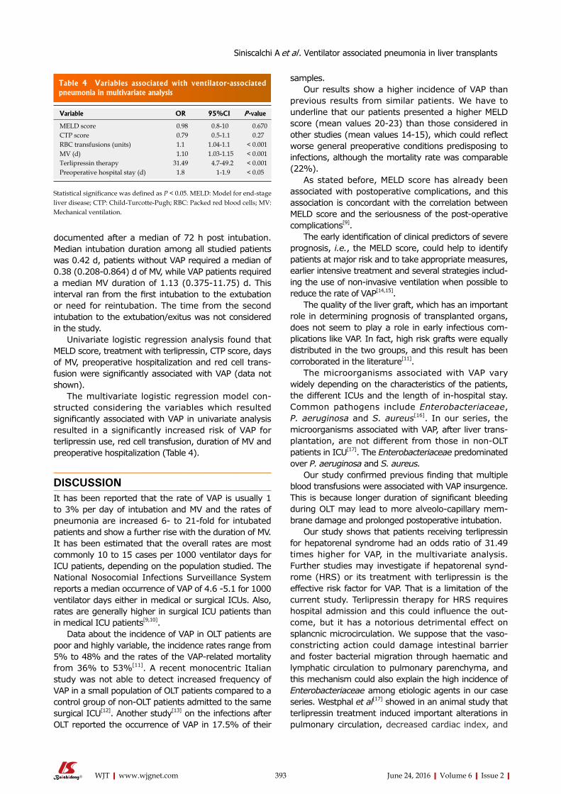
393 June 24, 2016|Volume 6|Issue 2|WJT|www.wjgnet.com
documented after a median of 72 h post intubation. Median intubation duration among all studied patients was 0.42 d, patients without VAP required a median of 0.38 (0.208-0.864) d of MV, while VAP patients required a median MV duration of 1.13 (0.375-11.75) d. This interval ran from the first intubation to the extubation or need for reintubation. The time from the second intubation to the extubation/exitus was not considered in the study.
Univariate logistic regression analysis found that MELD score, treatment with terlipressin, CTP score, days of MV, preoperative hospitalization and red cell trans-fusion were significantly associated with VAP (data not shown).
The multivariate logistic regression model con-structed considering the variables which resulted significantly associated with VAP in univariate analysis resulted in a significantly increased risk of VAP for terlipressin use, red cell transfusion, duration of MV and preoperative hospitalization (Table 4).
DISCUSSIONIt has been reported that the rate of VAP is usually 1 to 3% per day of intubation and MV and the rates of pneumonia are increased 6- to 21-fold for intubated patients and show a further rise with the duration of MV. It has been estimated that the overall rates are most commonly 10 to 15 cases per 1000 ventilator days for ICU patients, depending on the population studied. The National Nosocomial Infections Surveillance System reports a median occurrence of VAP of 4.6 -5.1 for 1000 ventilator days either in medical or surgical ICUs. Also, rates are generally higher in surgical ICU patients than in medical ICU patients[9,10].
Data about the incidence of VAP in OLT patients are poor and highly variable, the incidence rates range from 5% to 48% and the rates of the VAP-related mortality from 36% to 53%[11]. A recent monocentric Italian study was not able to detect increased frequency of VAP in a small population of OLT patients compared to a control group of non-OLT patients admitted to the same surgical ICU[12]. Another study[13] on the infections after OLT reported the occurrence of VAP in 17.5% of their
samples.Our results show a higher incidence of VAP than
previous results from similar patients. We have to underline that our patients presented a higher MELD score (mean values 20-23) than those considered in other studies (mean values 1415), which could reflect worse general preoperative conditions predisposing to infections, although the mortality rate was comparable (22%).
As stated before, MELD score has already been associated with postoperative complications, and this association is concordant with the correlation between MELD score and the seriousness of the post-operative complications[9].
The early identification of clinical predictors of severe prognosis, i.e., the MELD score, could help to identify patients at major risk and to take appropriate measures, earlier intensive treatment and several strategies includ-ing the use of non-invasive ventilation when possible to reduce the rate of VAP[14,15].
The quality of the liver graft, which has an important role in determining prognosis of transplanted organs, does not seem to play a role in early infectious com-plications like VAP. In fact, high risk grafts were equally distributed in the two groups, and this result has been corroborated in the literature[11].
The microorganisms associated with VAP vary widely depending on the characteristics of the patients, the different ICUs and the length of in-hospital stay. Common pathogens include Enterobacteriaceae, P. aeruginosa and S. aureus[16]. In our series, the microorganisms associated with VAP, after liver trans-plantation, are not different from those in non-OLT patients in ICU[17]. The Enterobacteriaceae predominated over P. aeruginosa and S. aureus.
Our study confirmed previous finding that multiple blood transfusions were associated with VAP insurgence. This is because longer duration of significant bleeding during OLT may lead to more alveolo-capillary mem-brane damage and prolonged postoperative intubation.
Our study shows that patients receiving terlipressin for hepatorenal syndrome had an odds ratio of 31.49 times higher for VAP, in the multivariate analysis. Further studies may investigate if hepatorenal synd-rome (HRS) or its treatment with terlipressin is the effective risk factor for VAP. That is a limitation of the current study. Terlipressin therapy for HRS requires hospital admission and this could influence the out-come, but it has a notorious detrimental effect on splancnic microcirculation. We suppose that the vaso-constricting action could damage intestinal barrier and foster bacterial migration through haematic and lymphatic circulation to pulmonary parenchyma, and this mechanism could also explain the high incidence of Enterobacteriaceae among etiologic agents in our case series. Westphal et al[17] showed in an animal study that terlipressin treatment induced important alterations in pulmonary circulation, decreased cardiac index, and
Table 4 Variables associated with ventilator-associated pneumonia in multivariate analysis
Variable OR 95%CI P -value
MELD score 0.98 0.8-10 0.670CTP score 0.79 0.5-1.1 0.27RBC transfusions (units) 1.1 1.04-1.1 < 0.001MV (d) 1.10 1.03-1.15 < 0.001Terlipressin therapy 31.49 4.7-49.2 < 0.001Preoperative hospital stay (d) 1.8 1-1.9 < 0.05
Statistical significance was defined as P < 0.05. MELD: Model for end-stage liver disease; CTP: Child-Turcotte-Pugh; RBC: Packed red blood cells; MV: Mechanical ventilation.
Siniscalchi A et al . Ventilator associated pneumonia in liver transplants

394 June 24, 2016|Volume 6|Issue 2|WJT|www.wjgnet.com
diminished systemic oxygen delivery and consumption.Despite the mentioned results, this study presents
some limitations. We reported a low number of pne-umonia cases due to its globally low incidence and the limited sample size, since our data came from a single center.
In conclusion, this study was designed to investigate the incidence, the risk factors and the outcome of VAP after OLT. Incidence has been estimated to be 10 per 1000 d of MV. Our study confirms some of the risk factors for VAP found in other studies: RBC transfusion, duration of MV and preoperative hospitalization rather than direct admission from home. MELD score is higher in the VAP group and it represents a significant risk factor in univariate analysis, reflects worse general conditions and prospects higher postoperative compli-cations. The adoption of MELD score could rationalize VAP prevention practice in patients at major risk, earlier intensive treatment to increase the ventilator-free days and several stategies including the use of non-invasive ventilation. Among the risk factors, we found the therapy with terlipressin, used for the treatment of hepatorenal syndrome. This drug exhibited, in animal models, some effects on pulmonary circulation and has a detrimental effect on splancnic blood flow that could contribute to bacterial migration. Also hepatorenal syndrome could have contributed to this effect. Further studies are needed to clarify this correlation.
COMMENTSBackgroundPatients undergoing orthotopic liver transplant (OLT) represent a special subpopulation at risk for nosocomial infections, in particular ventilator-associated pneumonia (VAP) is the main hospital acquired infection in intensive care unit and it is a serious perioperative complication in liver transplant recipients.
Research frontiersVAP’s etiology and risk factors are still poorly understood.
Innovations and breakthroughsThe authors conducted this retrospective study in a big (considering the peculiar population) sample of patients, 242 consecutive liver transplant recipients. Of course, none of the patients who developed VAP presented signs or symptoms of infection before liver transplantation. Model for end-stage liver disease score was a significant risk factor in univariate analysis, and probably it reflects worse general conditions. In multivariate analysis the authors found a statistically significant association with terlipressin therapy. Patients who were taking terlipressin received the last dose until the OLT to treat the hepatorenal syndrome that could be a risk factor by itself. The authors did not refer to clinical and laboratory parameters as they are included in other scores. Some patients received other vasopressors during the OLT, but there were no statistically significant differences between the two groups. As the authors remarked in the discussion, further studies are needed to clarify this finding.
ApplicationsThe application of the authors’ results aims to individualize patients at major risk, to apply earlier intensive treatment and several strategies to prevent VAP.
Peer-reviewThe authors performed a study on a very important infectious complication in
post-operative OLT setting. The paper is well written and the aim is clear.
REFERENCES1 Vincent JL, de Souza Barros D, Cianferoni S. Diagnosis, manage
ment and prevention of ventilatorassociated pneumonia: an update. Drugs 2010; 70: 19271944 [PMID: 20883051 DOI: 10.2165/1153808000000000000000]
2 Murillas J, Rimola A, Laguno M, de Lazzari E, Rascón J, Agüero F, Blanco JL, Moitinho E, Moreno A, Miró JM. The model for endstage liver disease score is the best prognostic factor in human immunodeficiency virus 1infected patients with endstage liver disease: a prospective cohort study. Liver Transpl 2009; 15: 11331141 [PMID: 19718643 DOI: 10.1002/lt.21735]
3 Giorgio V, Silvana S. Modelli di valutazione prognostica per le cure palliative nell’Endstage liver disease (esld): una revisione della letteratura. La rivista italiana di cure palliative 2010; 10: 4859
4 American Thoracic Society; Infectious Diseases Society of America. Guidelines for the management of adults with hospitalacquired, ventilatorassociated, and healthcareassociated pneumonia. Am J Respir Crit Care Med 2005; 171: 388416 [PMID: 15699079]
5 Davis KA. Ventilatorassociated pneumonia: a review. J Intensive Care Med 2006; 21: 211226 [PMID: 16855056]
6 Lai KK, Baker SP, Fontecchio SA. Impact of a program of intensive surveillance and interventions targeting ventilated patients in the reduction of ventilatorassociated pneumonia and its costeffectiveness. Infect Control Hosp Epidemiol 2003; 24: 859863 [PMID: 14649776]
7 Feng S, Goodrich NP, BraggGresham JL, Dykstra DM, Punch JD, DebRoy MA, Greenstein SM, Merion RM. Characteristics associated with liver graft failure: the concept of a donor risk index. Am J Transplant 2006; 6: 783790 [PMID: 16539636]
8 Bonney GK, Aldersley MA, Asthana S, Toogood GJ, Pollard SG, Lodge JP, Prasad KR. Donor risk index and MELD interactions in predicting longterm graft survival: a singlecentre experience. Transplantation 2009; 87: 18581863 [PMID: 19543065 DOI: 10.1097/TP.0b013e3181a75b37]
9 Chastre J, Fagon JY. Ventilatorassociated pneumonia. Am J Respir Crit Care Med 2002; 165: 867903 [PMID: 11934711]
10 National Nosocomial Infections Surveillance System. National Nosocomial Infections Surveillance (NNIS) System Report, data summary from January 1992 through June 2004, issued October 2004. Am J Infect Control 2004; 32: 470485 [PMID: 15573054]
11 Pellegrino CM, Codeluppi M, Assenza S, Cocchi S, Di Benedetto F, Girardis M. Incidence and clinical outcomes of ventilatorassociated pneumonia in liver transplant and nonliver transplant surgical patients. Transplant Proc 2008; 40: 19861988 [PMID: 18675108 DOI: 10.1016/j.transproceed.2008.05.022]
12 Siniscalchi A, Cucchetti A, Toccaceli L, Spiritoso R, Tommasoni E, Spedicato S, Dante A, Riganello L, Zanoni A, Cimatti M, Pierucci E, Bernardi E, Miklosova Z, Pinna AD, Faenza S. Pretransplant model for endstage liver disease score as a predictor of postoperative complications after liver transplantation. Transplant Proc 2009; 41: 12401242 [PMID: 19460528 DOI: 10.1016/j.transproceed.2009.02.046]
13 Karapanagiotou A, Kydona C, Papadopoulos S, Giasnetsova T, Sgourou K, Pasakiotou M, Fouzas I, Papanikolaou V, GritsiGerogianni N. Infections after orthotopic liver transplantation in the intensive care unit. Transplant Proc 2012; 44: 27482750 [PMID: 23146512 DOI: 10.1016/j.transproceed.2012.09.004]
14 Burns KE, Adhikari NK, Keenan SP, Meade M. Use of noninvasive ventilation to wean critically ill adults off invasive ventilation: metaanalysis and systematic review. BMJ 2009; 338: b1574 [PMID: 19460803 DOI: 10.1136/bmj.b1574]
15 Willson G. Noninvasive weaning from ventilation reduces mortality, ventilatorassociated pneumonia, and length of stay in intubated adults. Aust J Physiother 2009; 55: 207 [PMID: 19736673]
COMMENTS
Siniscalchi A et al . Ventilator associated pneumonia in liver transplants

395 June 24, 2016|Volume 6|Issue 2|WJT|www.wjgnet.com
16 Shorr AF, Duh MS, Kelly KM, Kollef MH. Red blood cell transfusion and ventilatorassociated pneumonia: A potential link? Crit Care Med 2004; 32: 666674 [PMID: 15090945]
17 Westphal M, Stubbe H, Sielenkämper AW, Borgulya R, Van Aken
H, Ball C, Bone HG. Terlipressin dose response in healthy and endotoxemic sheep: impact on cardiopulmonary performance and global oxygen transport. Intensive Care Med 2003; 29: 301308 [PMID: 12594590]
P- Reviewer: Boin IFSF, Giannella M, Mouloudi E S- Editor: Kong JX L- Editor: Wang TQ E- Editor: Liu SQ
Siniscalchi A et al . Ventilator associated pneumonia in liver transplants

Donal B O’Connor, John P Burke, John Hegarty, Aiden P McCormick, Niamh Nolan, Emir Hoti, Donal Maguire, Justin Geoghegan, Oscar Traynor
Donal B O’Connor, Department of Surgery, Trinity Centre for Health Sciences, Tallaght Hospital, Dublin 24, Ireland
Donal B O’Connor, John P Burke, Departments of Hepatobiliary Surgery, St Vincent’s University Hospital, Elm Park, Dublin 4, Ireland
John Hegarty, Aiden P McCormick, Emir Hoti, Donal Maguire, Justin Geoghegan, Oscar Traynor, National Liver Transplant Programme, St Vincent’s University Hospital, Elm Park, Dublin 4, Ireland
Niamh Nolan, Departments of Pathology, St Vincent’s University Hospital, Elm Park, Dublin 4, Ireland
Author contributions: O’Connor DB and Traynor O conceived project; O’Connor DB contributed to the preparation of manuscript and data acquisition; Burke JP contributed to statistical analysis and critical revision of manuscript; Nolan N contributed to analysis of histopathology; Hegarty J, McCormick AP, Hoti E, Maguire D, Geoghegan J and Traynor O contributed to critical revision of manuscript.
Institutional review board statement: The study was approved by the St Vincent’s University Hospital ethics review board.
Informed consent statement: Informed consent was not required for this study.
Conflict-of-interest statement: The authors have no conflicts of interest to declare.
Data sharing statement: No additional data.
Open-Access: This article is an openaccess article which was selected by an inhouse editor and fully peerreviewed by external reviewers. It is distributed in accordance with the Creative Commons Attribution Non Commercial (CC BYNC 4.0) license, which permits others to distribute, remix, adapt, build upon this work noncommercially, and license their derivative works on different terms, provided the original work is properly cited and
the use is noncommercial. See: http://creativecommons.org/licenses/bync/4.0/
Manuscript source: Unsolicited manuscript
Correspondence to: Donal B O’Connor, Lecturer in Surgery, Department of Surgery, Trinity Centre for Health Sciences, Tallaght Hospital, Room 1.36, Dublin 24, Ireland. [email protected]: +35314142218Fax: +35314142212
Received: November 11, 2015 Peer-review started: November 15, 2015 First decision: January 4, 2016Revised: April 12, 2016 Accepted: May 7, 2016Article in press: May 9, 2016Published online: June 24, 2016
AbstractAIM: To examine the results of orthotopic liver trans-plantation (OLT) for hepatocellular carcinoma (HCC) in Ireland over a 14-year period.
METHODS: Cases of HCC receiving OLT between January 1995 and September 2009 in the Irish Liver Transplant Unit were reviewed from a prospectively maintained database. Outcome measures included overall and recurrence free survival, alpha-fetoprotein (AFP) and tumour pathological features.
RESULTS: On explant pathology, 57 patients had HCC. The median follow-up time was 42.7 mo. The overall 1, 3 and 5 years survival was 87.7%, 72.1% and 72.4%. There was no difference in survival when compared
396 June 24, 2016|Volume 6|Issue 2|WJT|www.wjgnet.com
World J Transplant 2016 June 24; 6(2): 396-402ISSN 2220-3230 (online)
© 2016 Baishideng Publishing Group Inc. All rights reserved.
Submit a Manuscript: http://www.wjgnet.com/esps/Help Desk: http://www.wjgnet.com/esps/helpdesk.aspxDOI: 10.5500/wjt.v6.i2.396
World Journal of TransplantationW J T
Liver transplantation for hepatocellular carcinoma in Ireland: Pre-operative alpha-fetoprotein predicts tumour recurrence in a 14-year single-centre national experience
Retrospective Study
ORIGINAL ARTICLE

to patients undergoing OLT without malignancy. The tumour recurrence rate was 14%. The Milan criteria were exceeded in 32% of cases but this did not predict overall survival or recurrence. On multivariate analysis pre-operative AFP > 100 ng/mL was an independent risk factor for recurrence (RR = 5.2, CI: 1.1-24.3, P = 0.036).
CONCLUSION: Patients undergoing OLT for HCC had excellent survival even when conventional listing criteria were exceeded. Pre-operative AFP predicts recurrence independent of tumour size and its role in selection criteria should be investigated in larger studies.
Key words: Liver transplantation; Alpha-fetoprotein; Hepatocellular carcinoma; Transplantation selection criteria; Liver cirrhosis
© The Author(s) 2016. Published by Baishideng Publishing Group Inc. All rights reserved.
Core tip: We have shown good survival from a medium volume transplant centre in a small cohort of patients exceeding Milan criteria. We show an association between a pre-operative alpha-fetoprotein (AFP) > 100 and hepatocellular carcinoma (HCC) recurrence, independent of tumour size. Our study supports other single centre experience on survival after transplant for HCC with low AFP and indicates that AFP needs to be interrogated in large, multi-centre studies to see if it can be included in transplant listing criteria to augment the current radiology based dimensional criteria.
O’Connor DB, Burke JP, Hegarty J, McCormick AP, Nolan N, Hoti E, Maguire D, Geoghegan J, Traynor O. Liver transplantation for hepatocellular carcinoma in Ireland: Preoperative alphafetoprotein predicts tumour recurrence in a 14year singlecentre national experience. World J Transplant 2016; 6(2): 396402 Available from: URL: http://www.wjgnet.com/22203230/full/v6/i2/396.htm DOI: http://dx.doi.org/10.5500/wjt.v6.i2.396
INTRODUCTIONHepatocellular carcinoma (HCC) is the 5th most common cancer and 3rd leading cause of cancer-related death worldwide[1]. The incidence and related mortality are increasing, particularly in Western countries[2]. It is now well accepted that the optimal treatment for small HCC in the setting of cirrhosis is orthotopic liver transplantation (OLT)[3]. Since the publication and adaptation of the Milan criteria[4] the outcomes have improved dramatically compared to results from the era prior to established selection criteria[5]. Patients undergoing OLT for HCC within the Milan criteria achieve outcomes comparable to non-malignant transplant cohorts. However, recurrence is the most important cause of post-transplant death[6]. Appropriate patient selection is crucial as patients with large or biologically
unfavourable tumours have unacceptable recurrence and overall survival rates[7]. It is essential that centres which provide OLT for HCC audit their results to ensure outcomes compare to international survival rates thus enabling appropriate patient prioritisation and organ allocation.
The aim of the current study was to determine the outcomes of OLT for HCC in a single, national institution over a 14-year period. Overall and recurrence free survival rates were compared to clinical and pathological factors using multivariate analysis to identify indepen-dent predictors of recurrence.
MATERIALS AND METHODSAll patients undergoing OLT with HCC proven on explant pathology between January 1995 and September 2009 were included in the study. All OLT in the Republic of Ireland are carried out in the Liver Unit of St Vincent’sUniversity Hospital. The Liver Unit maintains a pro-spective database containing patients’ clinical details. Tumour characteristics are recorded on a computerised pathology database. A retrospective review of this data was performed. Patient characteristics recorded included age at OLT, sex, aetiology of underlying liver disease, pre-operative alpha-fetoprotein (AFP), survival, and recurrence status. Tumour data recorded included size and number of tumours, compliance with the Milan and University of California San Francisco (UCSF) criteria and microvascular invasion. The study was approved by the St Vincent’s University Hospital ethics review board.
Patient selection and transplant protocolAll patients listed were classified clinically as cirrhotic. HCC diagnosis was based on a combination of ultra-sound, computed tomography (CT) and double-contrast magnetic resonance imaging (MRI). After 1996 patients were listed for OLT if they met Milan criteria on pre-operative imaging. Patients in Ireland listed for OLT for HCC receive an adjusted Model for End-Stage Liver Disease (MELD) score[8]. All OLT were from deceased donor transplants and organs were retrieved before cardiac death. Patients were followed up at a dedicated transplant clinic every three months. In general, post-transplant immunosuppression consisted of a reducing dose of corticosteroids and a calcineurin inhibitor (tacrolimus) or azothioprine. Follow up included annual abdominal ultrasound and CT where appropriate.
Survival and recurrenceOverall patient survival was determined from date of OLT until the most recently attended clinic. HCC recurrence free survival was determined by the date of the most recently available radiological imaging. Deaths from recurrence were prospectively recorded in the database. Patients without recurrence that died were documented free of recurrence only if the most recent available imaging or post-mortem report excluded recurrence. Two investigators (O’Connor DB
397 June 24, 2016|Volume 6|Issue 2|WJT|www.wjgnet.com
O’Connor DB et al . OLT for HCC in Ireland

and Cooney A) independently reviewed the database to ensure accuracy of the survival and recurrence data. Patients were censored in September 2009 to ensure a minimum of 5-year follow-up.
Tumour characteristicsAll explants were examined by a histopathologist experienced in HCC pathology ( Nolan N). Tumour size, number of lesions, presence of macro or microvascular invasion, and condition of the non-tumour bearing liver were recorded. Tumours were graded as well, moderate or poorly differentiated. Compliance with Milan or UCSF criteria was based on size and number of lesions and was determined by explant pathology rather than pre-operative imaging.
Statistical analysisPatients were divided into groups based on meeting or exceeding listing criteria, presence or absence of vascular invasion, tumour grade, and pre-operative AFP levels to determine impact on overall overall and recurrence free survival. Data is presented as median (interquartile range). Factors affecting survival were determined by a Cox Proportional Hazard Model and significant factors were incorporated into a multivariate analysis. Kaplan-Meier analysis and the log-rank test were used to illustrate differences between recurrence free and overall survival according to clinical factors. Comparisons between the HCC and control cohort were
made using Fisher’s Exact test. All calculations were done using SPSS version 12.0 (SPSS, Inc., Chicago, IL). P < 0.050 was set as the threshold for statistical significance.
RESULTSDuring the 14-year study period 57 patients underwent OLT for HCC confirmed on explant pathology. One patient received OLT in 1995 and 56 patients were transplanted between 1998 and 2009. This represented 11.3% of the 504 patients undergoing OLT in the Liver Unit during that time. HCC was diagnosed radiologically in 52 cases pre-operatively and 5 cases were incidental findings in cirrhotic patients. HCC was absent on ex-plant pathology in 4 additional patients transplanted for presumed HCC, representing false positives who were excluded from the analysis. Pre-operative AFP, tumour histopathology and clinical follow up data were available for all 57 patients. Median follow up was 42.7 (14.6-67.6) mo.
The median age at OLT was 59 years. The most common underlying causes of cirrhosis were alcoholic liver disease (30%), hepatitis C (30%) and Haemo-chromotosis (23%). The Milan criteria were exceeded in 16 (28%) and 8 patients (14%) exceeded UCSF criteria. Median largest tumour size was 3 (2.5-4.5) cm. Micro-vascular invasion was present in 24 (42%) tumours. The mean time to OLT following diagnosis was 3 mo. Bridging therapy was not routinely used. Only 4 patients underwent trans-arterial chemo-embolization and this was not included in statistical analysis. Patient and tumour characteristics are outlined in Table 1.
SurvivalOverall survival at 1, 3 and 5 years was 87.7% (50/57), 72.1% (31/43) and 72.4% (21/29) respectively. The HCC transplant group were compared to a cohort of 313 patients undergoing OLT between 1998 and 2008 who underwent their primary, non-emergent, transplant during that period. There was no statistical difference between the HCC and control cohort in 1 (87.7% vs 89.1%, P = 0.450), 3 (72.1% vs 84.2%, P = 0.050) and 5 years (72.4% vs 80.9%, P = 0.211) overall survival rates. No clinical or pathological variable significantly affected overall survival in those undergoing OLT for HCC (Table 2). Overall survival was not affected by patients exceeding the Milan (Figure 1A) or UCSF (Figure 1B).
RecurrenceRecurrence free survival was 86%, 69.7% and 69.5% at 1, 3 and 5 years respectively. There were 8 recurrences in total (14%) and 5 patients died from recurrence. Recurrence occurred within 1 year in 3 patients, within 2 years in 3 and beyond 3 and 5 years in one patient each. The location of recurrent disease was hepatic in 3 (including 2 patients with additional
398 June 24, 2016|Volume 6|Issue 2|WJT|www.wjgnet.com
Table 1 Patient demographics
Male:female 44:13Age, median (IQR) 59.1 (53.5-63.6)Aetiology of HCC Alcoholic liver disease 17 (29.8%) Hepatitis C 17 (29.8%) Haemochromatosis 13 (22.8%) α-1-antitrypsin deficiency 3 (5.3%) Primary sclerosing cholangitis 2 (3.5%) Primary biliary cirrhosis 2 (3.5%) Autoimmune hepatitis 2 (3.5%) Hepatitis B 2 (3.5%) Cryptogenic 2 (3.5%) Cystic fibrosis 1 (1.8%) Sarcoidosis 1 (1.8%) Nash 1 (1.8%)Pre-operative α-fetoprotein, median (IQR) 8.8 (3.3-29.2)Compliant with Milan criteria 41 (71.9%)Compliant with UCSF criteria 49 (86.0%)Largest lesion, median (IQR) 3 (2.5-4.5)Cirrhosis 53 (93.0%)Steatosis 2 (3.5%)Multifocal lesions 24 (42.1%)Micro-vascular invasion 24 (42.1%)Tumour differentiation Well 24 (42.1%) Moderate 28 (49.1%) Poor 5 (8.8%)Incidental lesions 5 (8.8%)
HCC: Hepatocellular carcinoma; UCSF: University of California San Francisco; IQR: Interquartile range.
O’Connor DB et al . OLT for HCC in Ireland

399 June 24, 2016|Volume 6|Issue 2|WJT|www.wjgnet.com
independent risk factor for recurrence. The landmark Milan publication in 1996 established
listing criteria based on a single HCC of less than 5 cm or up to 3 tumours, each less than 3 cm[4]. This was validated in other single-centre studies and together demonstrated a 5-year survival of 70% and recurrence rates of less than 15% which became the gold standard outcome in OLT for HCC[9-11]. These criteria continue to be used in Ireland and many centres worldwide. Our institution is a medium volume centre performing approximately 60-70 OLT per year. The outcomes of our patient cohort compare favourably to recently published series’ from higher volume centres[12,13]. Our patient demographic is different to most centres as in over 50% of patients the underlying liver pathology was alcoholic liver disease or haemochromatosis. Worldwide, the main causes of HCC are hepatitis B and C virus but in this study they only accounted for 33% of HCC[14]. However aetiology of HCC did not significantly impact on overall or disease free survival.
The majority of patients with HCC present with disease beyond the Milan criteria[15]. Acceptable 5-year survival and recurrence rates observed in a subgroup of patients with larger tumours led to the publication of the UCSF criteria which proposes listing patients with a single tumour up to 6.5 cm or up to 3 lesions, none larger than 4.5 cm and total tumour burden not exceeding 8 cm[16]. This has been reproduced in single-centre studies with short follow up but never in multi-centre or nationwide population studies but in recent years several units have called for an extension of the criteria. Patients beyond the Milan criteria did not experience inferior survival in our centre but our numbers are too small to support calls for extension of the criteria based simply
extra-hepatic metastases), porta-hepatis lymph nodes in 2, and in one patient multiple recurrence occurred in lung, omentum and sacrum. Hepatic recurrences were diagnosed on CT and extra hepatic disease was confirmed by biopsy. Recurrence free survival was similar between patients meeting or exceeding the Milan (Figure 1C) and the UCSF criteria (Figure 1D). Underlying liver disease, tumour size or vascular invasion did not affect recurrence free survival. On univariate analysis only poorly differentiated tumours and AFP levels > 100 ng/mL were associated with reduced disease free survival (Table 3) and a shorter time to recurrence (Figure 2). On multivariate analysis, pre-operative AFP > 100 ng/mL remained an independent predictor of recurrence free survival (HR = 5.2, P = 0.036).
Patients exceeding Milan and UCSF criteriaEight patients exceeded both Milan and UCSF criteria. Five were alive at 5 years and one patient with recu-rrence was alive after 3 years follow-up. Recurrence only occurred in 2 cases. One patient died from recurrence after 14 mo and one died from a separate malignancy at 2 years. Micro-vascular invasion was present in 4 cases. AFP exceeded 100 ng/mL in the patient who died from recurrence.
DISCUSSIONThe current study confirms that OLT for HCC is an effective treatment modality and that survival rates are comparable to those undergoing OLT for non-malignant disease. Patients exceeding the Milan or UCSF criteria were not at increased risk of reduced overall survival or increased recurrence. Pre-operative serum AFP is an
Table 2 Univariate analysis of factors affecting overall survival
HR CI P -value
Male sex 0.786 0.301-2.055 0.623Age 1.001 0.954-1.051 0.952Aetiology of HCC Alcoholic liver disease 0.523 0.175-1.567 0.247 Hepatitis C 2.098 0.849-5.183 0.108 Haemochromatosis 0.715 0.239-2.143 0.549 Other 1.198 0.459-3.126 0.712Pre-operative α-fetoprotein > 100 ng/mL 1.502 0.437-5.165 0.519Compliant with Milan criteria 0.994 0.381-2.590 0.989Compliant with UCSF criteria 0.871 0.290-2.618 0.805Largest lesion 1.207 0.963-1.513 0.102Cirrhosis 23.309 0.024-224.813 0.369Steatosis 0.044 0.000-187.285 0.465Multi-focal lesions 1.201 0.499-2.890 0.683Micro-vascular invasion 1.489 0.619-3.578 0.374Tumour differentiation Well 0.862 0.349-2.131 0.748 Moderate 1.100 0.448-2.698 0.835 Poor 1.159 0.268-5.022 0.843Incidental lesions 0.450 0.060-3.391 0.438
HCC: Hepatocellular carcinoma; UCSF: University of California San Francisco.
Table 3 Univariate analysis of factors affecting recurrence free survival
HR CI P -value
Male sex 0.681 0.156-2.971 0.609Age 1.004 0.932-1.081 0.922Aetiology of HCC Alcoholic liver disease 0.775 0.155-3.887 0.757 Hepatitis C 3.272 0.798-13.417 0.100 Haemochromatosis 1.210 0.243-6.024 0.816 Other 0.027 0.000-16.500 0.271Pre-operative α-fetoprotein > 100 ng/mL 6.668 1.661-26.768 0.007Compliant with Milan criteria 1.354 0.271-6.761 0.712Compliant with UCSF criteria 0.739 0.148-3.692 0.712Largest lesion 1.326 0.976-1.801 0.071Cirrhosis 23.025 0.000-327.873 0.604Steatosis 0.045 0.000-546.731 0.664Multifocal lesions 2.100 0.494-8.930 0.315Micro-vascular invasion 1.560 0.376-6.463 0.540Tumour differentiation Well 0.249 0.046-1.340 0.105 Moderate 1.553 0.374-6.443 0.544 Poor 5.631 1.074-29.510 0.041Incidental lesions 0.041 0.000-720.752 0.523
HCC: Hepatocellular carcinoma; UCSF: University of California San Francisco.
O’Connor DB et al . OLT for HCC in Ireland

400 June 24, 2016|Volume 6|Issue 2|WJT|www.wjgnet.com
on size and number of tumours. The limitations of pre-operative imaging for staging in the setting of cirrhosis also impede raising the threshold. One large study showed pre-operative imaging to under stage over 40% of patients[17]. In the current study almost 30% were not compliant with Milan criteria on explant pathology. Interval tumour growth is a possible explanation for patients who meet criteria on imaging and then exceed them on pathology. However our cohort experienced a short waiting period of 3 mo and relatively small tumours (median 3 cm) which makes tumour doubling unlikely. Even with advances such as double-contrast MRI, extending the criteria based solely on size risks transplanting patients with tumours too large to benefit.
The limitation of established criteria is that they are based on tumour dimensions. While results from single
centre studies have justified its use for organ allocation, in a North American population study, a subgroup of patients with larger tumours within the Milan criteria had significantly poorer survival outcomes than those without HCC[18]. It is imperative that any selection criteria be accurate in predicting prognosis to justify the large proportion of transplants undertaken for HCC in the setting of a shortage of organs. For example 25% of all United States OLT have been for HCC since the introduction of priority MELD scores for HCC in 2002[18] and 11% of OLT in Ireland are for patients with HCC.
There is growing evidence that the biological beha-viour of the tumour rather than size dictates recurrence. Patients with larger tumours beyond the Milan criteria but without micro-vascular invasion can have excellent survival, such as outlined in the “up-to-seven-criteria”,
Figure 1 Kaplan-Meier estimates of overall survival (A and B) and recurrence free survival (C and D) in relation to compliance with the Milan and University of California San Francisco criteria. UCSF: University of California San Francisco.
Ove
rall
surv
ival
1.0
0.8
0.6
0.4
0.2
0.0
0 10 20 30 40 50 60t /mo
P = 0.989
Compliant with Milan criteria
No
Yes
Ove
rall
surv
ival
1.0
0.8
0.6
0.4
0.2
0.0
0 10 20 30 40 50 60t /mo
P = 0.805
Compliant with UCSF criteria
No
Yes
Recu
rren
ce f
ree
surv
ival
1.0
0.8
0.6
0.4
0.2
0.0
0 10 20 30 40 50 60t /mo
P = 0.711
Compliant with Milan criteria
No
Yes
Recu
rren
ce f
ree
surv
ival
1.0
0.8
0.6
0.4
0.2
0.0
0 10 20 30 40 50 60t /mo
P = 0.711
Compliant with UCSF criteria
No
Yes
A B
C D
O’Connor DB et al . OLT for HCC in Ireland

401 June 24, 2016|Volume 6|Issue 2|WJT|www.wjgnet.com
but this cannot be diagnosed pre-operatively[19]. The impact of micro-vascular invasion was not found to be statistically significant in our cohort but in large studies it has been shown to double the risk of death[7]. Pre-operative AFP may be the best available surrogate marker for micro-vascular invasion and the biological aggressiveness of the tumour. Several studies have identified a high pre-operative AFP as a risk factor for recurrence and reduced survival[18,20-22]. We have shown AFP predicted reduced disease free survival, indepen-dent of both tumour size and micro-vascular invasion. Furthermore, patients exceeding Milan or even UCSF criteria experienced excellent overall and recurrence free survival with a pre-operative AFP < 100 ng/mL. This supports the finding of another group where an AFP level < 30 ng/mL predicted disease free survival in patients beyond Milan criteria[23]. Both studies are limited by the small number of patients exceeding Milan criteria. Recent large studies have not examined the impact of AFP in the context of tumours beyond the Milan criteria. The largest study reporting survival in patients with tumours exceeding the Milan criteria (1112 patients) unfortunately did not examine the impact of AFP level[7]. Analysis from the United Network for Organ Sharing on 2253 patients demonstrated a significant survival advantage in patients with low pre-transplant AFP (< 20 ng/mL) but this effect wasn’t explored in patients with tumours outside Milan criteria[24]. It would therefore be intriguing if AFP could be examined in a large population database or multicentre study to determine if patients with large tumours but low pre-operative AFP had higher survival rates. Only then can AFP be used to augment existing eligibility criteria to safely expand the pool of patients suitable for OLT.
In conclusion, in appropriately selected patients with HCC undergoing OLT, survival was comparable to non-HCC patients. A subgroup of patients with larger
tumours and low AFP may benefit from OLT but this association should be examined in larger, multicentre studies.
ACKNOWLEDGMENTSAnne Cooney, database manager, the Liver Unit, for assistance in database retrieval.
COMMENTSBackgroundOrthotopic liver transplantation (OLT) is the most effective treatment for hepatocellular carcinoma (HCC) in the setting of cirrhosis. Survival in well selected patients with a small burden of tumour is similar to patients undergoing OLT for non-cancer related indications.
Research frontiersExisting selection criteria are based on the size and number of the tumour. Several datasets have demonstrated good survival outcomes in patients exceeding these criteria. The biological characteristics, for example micro-vascular invasion may just as important as the tumour dimensions. However these cannot be reliably detected pre-operatively.
Innovations and breakthroughsThis study also demonstrates that patients with larger tumours can still have good survival outcomes. Pre-operative alpha-fetoprotein (AFP) predicted tumour recurrence. AFP may be a useful surrogate marker for less favourable biological characteristics of the tumour.
ApplicationsThe prognostic value of AFP could be evaluated in large, multi-centre datasets to determine its potential as an adjunct to existing selection criteria.
TerminologyOLT: Orthotropic liver transplant involves fully explanting the diseased liver immediately prior to the transplant; HCC: Hepatocellular carcinoma is the most common primary liver tumour. Because most cases occur in the setting of cirrhosis, it is often not amenable to resection; AFP: Alpha-fetoprotein has no known function in adults but it has clinical significance as a tumour marker in
Figure 2 Kaplan-Meier estimates of recurrence free survival in relation to pre-operative α-fetoprotein (A) and tumour differentiation (B).
Recu
rren
ce f
ree
surv
ival
1.0
0.8
0.6
0.4
0.2
0.0
0 10 20 30 40 50 60t /mo
P = 0.002
α-fetoprotein over 100 ng/mL
No
Yes
Recu
rren
ce f
ree
surv
ival
1.0
0.8
0.6
0.4
0.2
0.0
0 10 20 30 40 50 60t /mo
P = 0.541
P = 0.021
Differentiation
Moderate
Poor
Well
A B
COMMENTS
O’Connor DB et al . OLT for HCC in Ireland

402 June 24, 2016|Volume 6|Issue 2|WJT|www.wjgnet.com
the diagnosis of HCC.
Peer-reviewThis is an interesting attempt to evaluate the results of liver transplantation for HCC with regard to potential relation with pre-operative values of AFP. The paper is well written and results are clarified.
REFERENCES1 Botha JF, Langnas AN. Liver transplantation for hepatocellular
carcinoma: an update. J Natl Compr Canc Netw 2006; 4: 762767 [PMID: 16948954]
2 El-Serag HB, Davila JA, Petersen NJ, McGlynn KA. The continuing increase in the incidence of hepatocellular carcinoma in the United States: an update. Ann Intern Med 2003; 139: 817823 [PMID: 14623619 DOI: 10.7326/000348191391020031118000009]
3 Jarnagin W, Chapman WC, Curley S, D’Angelica M, Rosen C, Dixon E, Nagorney D. Surgical treatment of hepatocellular carcinoma: expert consensus statement. HPB (Oxford) 2010; 12: 302310 [PMID: 20590903 DOI: 10.1111/j.14772574.2010.00182.x]
4 Mazzaferro V, Regalia E, Doci R, Andreola S, Pulvirenti A, Bozzetti F, Montalto F, Ammatuna M, Morabito A, Gennari L. Liver transplantation for the treatment of small hepatocellular carcinomas in patients with cirrhosis. N Engl J Med 1996; 334: 693699 [PMID: 8594428 DOI: 10.1056/NEJM199603143341104]
5 Selby R, Kadry Z, Carr B, Tzakis A, Madariaga JR, Iwatsuki S. Liver transplantation for hepatocellular carcinoma. World J Surg 1995; 19: 5358 [PMID: 7740811 DOI: 10.1007/BF00316980]
6 Lee SG, Hwang S, Moon DB, Ahn CS, Kim KH, Sung KB, Ko GY, Park KM, Ha TY, Song GW. Expanded indication criteria of living donor liver transplantation for hepatocellular carcinoma at one largevolume center. Liver Transpl 2008; 14: 935945 [PMID: 18581465 DOI: 10.1002/lt.21445]
7 Mazzaferro V, Llovet JM, Miceli R, Bhoori S, Schiavo M, Mariani L, Camerini T, Roayaie S, Schwartz ME, Grazi GL, Adam R, Neuhaus P, Salizzoni M, Bruix J, Forner A, De Carlis L, Cillo U, Burroughs AK, Troisi R, Rossi M, Gerunda GE, Lerut J, Belghiti J, Boin I, Gugenheim J, Rochling F, Van Hoek B, Majno P. Predicting survival after liver transplantation in patients with hepatocellular carcinoma beyond the Milan criteria: a retrospective, exploratory analysis. Lancet Oncol 2009; 10: 3543 [PMID: 19058754 DOI: 10.1016/S14702045(08)702845]
8 Zaman MB, Hoti E, Qasim A, Maguire D, McCormick PA, Hegarty JE, Geoghegan JG, Traynor O. MELD score as a prognostic model for listing acute liver failure patients for liver transplantation. Transplant Proc 2006; 38: 20972098 [PMID: 16980011 DOI: 10.1016/j.transproceed.2006.06.004]
9 Bismuth H, Majno PE, Adam R. Liver transplantation for hepatocellular carcinoma. Semin Liver Dis 1999; 19: 311322 [PMID: 10518310 DOI: 10.1055/s20071007120]
10 Llovet JM, Fuster J, Bruix J. Intentiontotreat analysis of surgical treatment for early hepatocellular carcinoma: resection versus transplantation. Hepatology 1999; 30: 14341440 [PMID: 10573522 DOI: 10.1002/hep.510300629]
11 Jonas S, Bechstein WO, Steinmüller T, Herrmann M, Radke C, Berg T, Settmacher U, Neuhaus P. Vascular invasion and histopathologic grading determine outcome after liver transplantation for hepatocellular carcinoma in cirrhosis. Hepatology 2001; 33: 10801086 [PMID: 11343235 DOI: 10.1053/jhep.2001.23561]
12 Cescon M, Ravaioli M, Grazi GL, Ercolani G, Cucchetti A, Bertuzzo V, Vetrone G, Del Gaudio M, Vivarelli M, D’Errico
Grigioni A, Dazzi A, Di Gioia P, Lauro A, Pinna AD. Prognostic factors for tumor recurrence after a 12year, singlecenter experience of liver transplantations in patients with hepatocellular carcinoma. J Transplant 2010; 2010: pii: 904152 [PMID: 20862199]
13 Levi DM, Tzakis AG, Martin P, Nishida S, Island E, Moon J, Selvaggi G, Tekin A, Madrazo BL, Narayanan G, Garcia MT, Feun LG, Tryphonopoulos P, Skartsis N, Livingstone AS. Liver transplantation for hepatocellular carcinoma in the model for endstage liver disease era. J Am Coll Surg 2010; 210: 727734, 735736 [PMID: 20421039 DOI: 10.1016/j.jamcollsurg.2010.01.007]
14 Hwang S, Lee SG, Belghiti J. Liver transplantation for HCC: its role: Eastern and Western perspectives. J Hepatobiliary Pancreat Sci 2010; 17: 443448 [PMID: 19885638 DOI: 10.1007/s0053400902410]
15 Stuart KE, Anand AJ, Jenkins RL. Hepatocellular carcinoma in the United States. Prognostic features, treatment outcome, and survival. Cancer 1996; 77: 22172222 [PMID: 8635087 DOI: 10.1002/(SICI)10970142(19960601)77:11<2217::AIDCNCR6>3.0.CO;2M]
16 Yao FY, Ferrell L, Bass NM, Watson JJ, Bacchetti P, Venook A, Ascher NL, Roberts JP. Liver transplantation for hepatocellular carcinoma: expansion of the tumor size limits does not adversely impact survival. Hepatology 2001; 33: 13941403 [PMID: 11391528 DOI: 10.1053/jhep.2001.24563]
17 Ioannou GN, Perkins JD, Carithers RL. Liver transplantation for hepatocellular carcinoma: impact of the MELD allocation system and predictors of survival. Gastroenterology 2008; 134: 13421351 [PMID: 18471511 DOI: 10.1053/j.gastro.2008.02.013]
18 Freeman RB, Mithoefer A, Ruthazer R, Nguyen K, Schore A, Harper A, Edwards E. Optimizing staging for hepatocellular carcinoma before liver transplantation: A retrospective analysis of the UNOS/OPTN database. Liver Transpl 2006; 12: 15041511 [PMID: 16952174 DOI: 10.1002/lt.20847]
19 D’Amico F, Schwartz M, Vitale A, Tabrizian P, Roayaie S, Thung S, Guido M, del Rio Martin J, Schiano T, Cillo U. Predicting recurrence after liver transplantation in patients with hepatocellular carcinoma exceeding the uptoseven criteria. Liver Transpl 2009; 15: 12781287 [PMID: 19790142 DOI: 10.1002/lt.21842]
20 Kondili LA, Lala A, Gunson B, Hubscher S, Olliff S, Elias E, Bramhall S, Mutimer D. Primary hepatocellular cancer in the explanted liver: outcome of transplantation and risk factors for HCC recurrence. Eur J Surg Oncol 2007; 33: 868873 [PMID: 17258882 DOI: 10.1016/j.ejso.2006.10.001]
21 McHugh PP, Gilbert J, Vera S, Koch A, Ranjan D, Gedaly R. Alphafetoprotein and tumour size are associated with microvascular invasion in explanted livers of patients undergoing transplantation with hepatocellular carcinoma. HPB (Oxford) 2010; 12: 5661 [PMID: 20495646 DOI: 10.1111/j.14772574.2009.00128.x]
22 Grąt M, Krasnodębski M, Patkowski W, Wronka KM, Masior Ł, Stypułkowski J, Grąt K, Krawczyk M. Relevance of Pre-Transplant α-fetoprotein Dynamics in Liver Transplantation for Hepatocellular Cancer. Ann Transplant 2016; 21: 115124 [PMID: 26887339]
23 Sotiropoulos GC, Malagó M, Bockhorn M, Schmitz KJ, Radtke A, Molmenti EP, Schaffer R, Beckebaum S, Cicinnati VR, Fouzas I, Broelsch CE, Lang H. Liver transplantation for hepatocellular carcinoma and cirrhosis in candidates with undetectable or very low alphafetoprotein levels: is an expansion of the listing criteria justified? Hepatogastroenterology 2008; 55: 16711677 [PMID: 19102367]
24 Mailey B, Artinyan A, Khalili J, Denitz J, SanchezLuege N, Sun CL, Bhatia S, Nissen N, Colquhoun SD, Kim J. Evaluation of absolute serum α-fetoprotein levels in liver transplant for hepa-tocellular cancer. Arch Surg 2011; 146: 2633 [PMID: 21242442 DOI: 10.1001/archsurg.2010.295]
P- Reviewer: Boucek CD, Balaban YH, Panda CK, Silva R, Smyrniotis V
S- Editor: Ji FF L- Editor: A E- Editor: Liu SQ
O’Connor DB et al . OLT for HCC in Ireland

Kwi Suk Kim, Aree Moon, Hyoung Jin Kang, Hee Young Shin, Young Hee Choi, Hyang Sook Kim, Sang Geon Kim
Kwi Suk Kim, Hyang Sook Kim, Sang Geon Kim, Department of Pharmacy, Seoul National University Hospital, Seoul 03080, South Korea
Aree Moon, College of Pharmacy, Duksung Women’s University, Seoul 01369, South Korea
Hyoung Jin Kang, Hee Young Shin, Department of Pediatrics, Cancer Research Institute, Seoul National University College of Medicine, Seoul 03080, South Korea
Young Hee Choi, College of Pharmacy, Dongguk University, Goyang, Gyeonggi-do 410-820, South Korea
Sang Geon Kim, College of Pharmacy and Research Institute of Pharmaceutical Sciences, Seoul National University, Seoul 03080, South Korea
Author contributions: Kim KS, Moon A and Kim SG for conception and design of the study; Kim KS, Choi YH and Kim SG for analysis and interpretation; Kim KS and Kim HS for data collection; Kim KS, Choi YH and Kim SG for writing the article; Kang HJ for critical revision of the article; Kim KS, Shin HY and Kim SG for final approval of the article; Kim KS and Choi YH for statistical analysis; Kim KS and Kim SG for overall responsibility.
Supported by The Education and Research Encouragement Fund of Seoul National University Hospital.
Institutional review board statement: This study was approved by the Institutional Review Board of Seoul National University Hospital (SNUH; H-1112-087-390, 2012.3.17), a 1961-bed medical center, on March 17, 2012.
Informed consent statement: The authors petition BPG for waiver of informed consent because this study was not a clinical trial, but was retrospectively done using anonymized electronic medical records of the study subjects. In addition, authors declared that the study subjects were at no risk due to this study.
Conflict-of-interest statement: We have no financial relation-ships to disclose.
Data sharing statement: No additional data are available.
Open-Access: This article is an open-access article which was selected by an in-house editor and fully peer-reviewed by external reviewers. It is distributed in accordance with the Creative Commons Attribution Non Commercial (CC BY-NC 4.0) license, which permits others to distribute, remix, adapt, build upon this work non-commercially, and license their derivative works on different terms, provided the original work is properly cited and the use is non-commercial. See: http://creativecommons.org/licenses/by-nc/4.0/
Correspondence to: Sang Geon Kim, PhD, Professor, College of Pharmacy and Research Institute of Pharmaceutical Sciences, Seoul National University, 101 Daehak-ro Jongno-gu, Seoul 03080, South Korea. [email protected]: +82-2-8807840Fax: +82-2-8807840
Received: January 2, 2016 Peer-review started: January 3, 2016 First decision: February 2, 2016Revised: February 22, 2016 Accepted: March 17, 2016 Article in press: March 18, 2016Published online: June 24, 2016
Abstract AIM: To analyze the association between plasma bilirubin levels and veno-occlusive disease (VOD) in non-adult patients undergoing hematopoietic stem cell transplantation (HSCT) during cyclosporine therapy.
METHODS: A total of 123 patients taking cyclosporine
403 June 24, 2016|Volume 6|Issue 2|WJT|www.wjgnet.com
World J Transplant 2016 June 24; 6(2): 403-410ISSN 2220-3230 (online)
© 2016 Baishideng Publishing Group Inc. All rights reserved.
Submit a Manuscript: http://www.wjgnet.com/esps/Help Desk: http://www.wjgnet.com/esps/helpdesk.aspxDOI: 10.5500/wjt.v6.i2.403
World Journal of TransplantationW J T
Higher plasma bilirubin predicts veno-occlusive disease in early childhood undergoing hematopoietic stem cell transplantation with cyclosporine
Retrospective Study
ORIGINAL ARTICLE

were evaluated using an electronic medical system at the Seoul National University Children’s Hospital from the years 2004 through 2011. Patients were grouped by age and analyzed for incidence and type of adverse drug reactions (ADRs) including VOD.
RESULTS: The HSCT patients were divided into three age groups: G#1 ≥ 18; 9 ≤ G#2 ≤ 17; and G#3 ≤ 8 years of age). The majority of transplant donor types were cord blood transplantations. Most prevalent ADRs represented acute graft-vs -host disease (aGVHD) and VOD. Although the incidences of aGVHD did not vary among the groups, the higher frequency ratios of VOD in G#3 suggested that an age of 8 or younger is a risk factor for developing VOD in HSCT patients. After cyclosporine therapy, the trough plasma concentrations of cyclosporine were lower in G#3 than in G#1, in-dicative of its increased clearance. Moreover, in G#3 only, a maximal total bilirubin level (BILmax) of ≥ 1.4 mg/dL correlated with VOD incidence after cyclosporine therapy.
CONCLUSION: HSCT patients 8 years of age or younger are more at risk for developing VOD, diagno-sed as hyperbilirubinemia, tender hepatomegaly, and ascites/weight gain after cyclosporine therapy, which may be represented by a criterion of plasma BILmax being ≥ 1.4 mg/dL, suggestive of more sensitive VOD indication in this age group.
Key words: Hematopoietic stem cell transplantation; Veno-occlusive disease; Cyclosporine; Adverse drug reaction; Total bilirubin
© The Author(s) 2016. Published by Baishideng Publishing Group Inc. All rights reserved.
Core tip: This study analyzed the association between plasma bilirubin and veno-occlusive disease (VOD) in childhood undergoing hematopoietic stem cell trans-plantation (HSCT) during cyclosporine therapy. Here, we report that age of 8 or under may be a risk factor for VOD in CsA-treated patients who underwent HSCT with differential clearance of CsA. Another finding is that a criterion of 1.4 mg/dL of plasma maximal total bilirubin level or higher content alone closely represents the incidence of VOD in early childhood patients with HSCT in CsA therapy. Information shown in this study would be of great help to understand VOD occurring during CsA medication and to find optimal pharmacotherapy in HSCT patients.
Kim KS, Moon A, Kang HJ, Shin HY, Choi YH, Kim HS, Kim SG. Higher plasma bilirubin predicts veno-occlusive disease in early childhood undergoing hematopoietic stem cell transplantation with cyclosporine. World J Transplant 2016; 6(2): 403-410 Available from: URL: http://www.wjgnet.com/2220-3230/full/v6/i2/403.htm DOI: http://dx.doi.org/10.5500/wjt.v6.i2.403
INTRODUCTIONCyclosporine is a major immunosuppressant for organ transplantation, and is widely used for the prophylactic treatment of acute graft-vs-host disease (aGVHD) after hematopoietic stem cell transplantation (HSCT)[1]. However, the morbidity and mortality resulting from acute, or subsequently following chronic GVHD, and veno-occlusive disease (VOD), as indicated by hyper-bilirubinemia, tender hepatomegaly, and ascites, are obstacles to the use of cyclosporine alone or in combi-nation with other agents[2]. Clinical studies on cyclos-porine therapy demonstrated differences between neonate, child and adult populations in the incidence of adverse drug reactions (ADRs)[3]. Since these events are closely linked to the metabolic burden and/or clearance of the drug, ADRs should be monitored and avoided depending on the types of transplantation, age groups and pharmacokinetic profiles. In particular, the dose regimens and therapeutic concentrations need to be appropriately adjusted for optimal efficacy and/or minimized ADRs.
Cyclosporine therapy should be carefully monitored as a therapeutic drug monitoring system[4]. Monitoring of pharmacokinetic profiles, including oral bioavailability, has been claimed in the context of successful pharmaco-therapy because intestinal absorption of cyclosporine varies depending on the type of transplantation, age, and other parameters of patients[5-9]. In general, patients of a young age seem to be more at risk for ADRs to cyclosporine, and exhibit different ADR profiles[10]. There-fore, the oral dose of cyclosporine required for the maintenance of therapeutic blood levels is significantly augmented in childhood patients[5]. In addition to the narrow therapeutic range of cyclosporine, the types and incidences of cyclosporine-induced ADRs vary depending on the types and severities of diseases, as well as patient age[11].
It has been recognized that wide variations exist in the plasma concentrations of cyclosporine among HSCT patients[12]. A limited number of studies have been performed in cyclosporine-treated neonates and children who underwent HSCT in the context of ADR monitoring[13]. In Seoul National University Hospital, the administered dose of cyclosporine was equally determined by the post-surgical day of HSCT, which frequently resulted in cyclosporine plasma concentrations being out of therapeutic range (150-250 ng/mL). Although the normalized doses of cyclosporine for transplant patients of childhood age were usually higher than those for adults, the plasma concentrations were significantly lower[3]. This raised the contention that biotransformation and/or excretion of cyclosporine is accelerated in childhood patients, which may be linked to ADRs, such as GVHD, nephrotoxicity, and neurotoxicity[13].
Age-different effects of cyclosporine therapy on the types and incidences of ADRs in HSCT patients are
404 June 24, 2016|Volume 6|Issue 2|WJT|www.wjgnet.com
Kim KS et al . Biliburin predicts VOD in childhood HSCT

known[14-16]. Nevertheless, more sensitive indicator(s) would be of great help to avoid or minimize serious ADRs and to accomplish successful pharmacotherapy, especially in patients of the childhood population who would be more prone to drug-induced harmful effects. This study analyzed the association between plasma bilirubin levels and VOD in non-adult patients undergoing HSCT during cyclosporine therapy. Here, we report that marginally high levels of total plasma bilirubin reliably indicate VOD during cyclosporine therapy in the HSCT patient of early childhood.
MATERIALS AND METHODSDatasets This study was approved by the Institutional Review Board of Seoul National University Hospital (SNUH; H-1112-087-390, 2012.3.17), a 1961-bed medical center, on March 17, 2012. The data collected had anonymous codes representing patient files compris-ing the following medical information: Age, gender, medical diagnosis codes, date of HSCT, absolute neu-trophil count, post-transplantation day, donor types (cord blood and related donor), body weight, body surface area, body temperature, types of ADRs, peak
and trough concentrations of cyclosporine, serum hematocrit, alanine aminotransferase (ALT), aspartate aminotransferase (AST), serum total bilirubin levels, dates of labs drawn, medications (generic and brand name, prescription date), and duration of chemothera-peutic agent along with co-prescribed drugs.
PatientsThe database contained records of 123 patients (ages ranging from 2 to 24 with 68 males and 55 females) who had been hospitalized in SNUH from September 15, 2004 to December 31, 2012 and had undergone measurements of plasma cyclosporine levels using a radioimmunoassay kit. Cyclosporine concentrations were monitored on day 1, 4, 11 18, 24 and 28 after HSCT and at intervals of three or seven days after day 28 of HSCT in SNUH. Laboratory data were obtained from 123 patients, with three of them having HSCTs twice in this period. The total number of cyclosporine measurements was 2149, with an average of 17.5 measurements per patient.
HSCT patients who had been administered cyclos-porine were divided into three groups based on age: G#1, 18 years of age or older; G#2, between 9 and 17 years of age; and G#3, 8 years of age or under. The median ages in G#1, G#2, and G#3 were 20, 13 and 6, respectively. Each group was additionally split into four subgroups by the levels of a maximal total bilirubin level (BILmax) [i.e., BILmax (-), lower than 1.4 mg/dL of total plasma bilirubin; and BILmax (+), 1.4 mg/dL of total plasma bilirubin or higher] and VOD incidence [i.e., VOD (-), no existing VOD; and VOD (+), existing VOD].
Statistical analysisThe Fisher exact test was chosen to determine diff-erences in the frequency of BILmax ≥ 1.4 mg/dL between VOD (-) and VOD (+) groups. Multivariate analysis was performed to find risk factors for drug therapy. Data represent the median (0.5-24.0 mg/dL). Results were considered statistically significant if the P-value was less than 0.05. Statistical analyses were conducted using the Duncan and Fisher’s tests in SPSS Version 12.0 (SPSS Inc., Chicago, IL, United States).
RESULTSThe characteristics of HSCT patients treated with cyclosporine (n = 123) are summarized in Table 1. Of the patients examined, cord blood transplantation constituted the majority of the transplant donor type (G#1, 60.0%; G#2, 58.6%; and G#3, 75.0%). The most prevalent ADR event observed was aGVHD (G#1,61.9%; G#2, 46.4%; and G#3, 54.2%), whereas the second most frequent ADR event was VOD (9.5%-29.2%). Although the incidences of aGVHD, diagnosed as cytopenia and delayed immune reconstitution, did not vary much between the groups, the frequency ratios of VOD were significantly higher in G#3. Thus, being 8 years of age or under at the time of transplantation
405 June 24, 2016|Volume 6|Issue 2|WJT|www.wjgnet.com
Table 1 The characteristics of hematopoietic stem cell transplantation patients treated with cyclosporine (n = 123)
Characteristics G#1 (n = 25) G#2 (n = 70) G#3 (n = 28)
Age (mean, SD) 20.3, 1.7 13.0, 2.5 5.8, 2.2Initial body weight (mean, SD)
51.8, 11.1 37.8, 12.7 14.0, 4.0
Gender (M, %) 15 (60.0) 35 (50.0) 17 (60.7)Liver function (mean, SD) ALT (mg/dL) 67.6, 95.8 67.1, 67.3 104, 133 AST (mg/dL) 70.2, 73.1 72.9, 61.5 161, 356Doner types, n (%) Cord blood 15 (60.0) 41 (58.6) 21 (75.0) Related donor 10 (40.0) 29 (41.4) 7 (25.0)Types of disease, n (%) AA 5 (20.0) 6 (8.6) 1 (3.6) ABL 4 (16.0) 5 (7.1) 2 (7.1) ALL 10 (40.0) 28 (40.0) 7 (25.0) AML 5 (20.0) 20 (28.6) 10 (35.7) CML 0 (0.0) 2 (2.9) 0 (0.0) JMML 0 (0.0) 0 (0.0) 4 (14.3) MDS 1 (4.0) 2 (2.9) 1 (3.6) Others 0 (0.0) 7 (10.0) 3 (10.7)Observed events, n (%) aGVHD 13 (61.9) 26 (46.4) 13 (54.2) cGVHD 1 (4.8) 4 (7.1) 0 (0.0) VOD 2 (9.5) 13 (23.2) 7 (29.2) DIC 4 (19.0) 4 (7.2) 2 (8.3) Relapse 1 (4.8) 2 (3.6) 1 (4.2) EF 0 (0.0) 7 (12.5) 1 (4.2)
Age groups: G#1, 18 years older; G#2, 9 to 17 years old; G#3, 8 years old or under. AA: Aplastic anemia; ABL: Acute biphenotypic leukemia; ALL: Acute lymphocytic leukemia; AML: Acute myelocytic leukemia; CML: Chronic myelocytic leukemia; JMML: Juvenile myelomonocytic leukemia; MDS: Myelo dysplastic syndromes; aGVHD: Acute graft versus host disease; cGVHD: Chronic graft versus host disease; VOD: Veno-occlusive disease; DIC: Disseminated intravascular coagulation; EF: Engraft failure; ALT: Alanine aminotransferase; AST: Aspartate aminotransferase.
Kim KS et al . Biliburin predicts VOD in childhood HSCT

406 June 24, 2016|Volume 6|Issue 2|WJT|www.wjgnet.com
high BILmax groups when G#2, G#3 or the total population was analyzed, although it failed in demons-trating increased VOD incidences when G#1 was solely analyzed (data not shown). More importantly, setting the BILmax cutoff level at 1.4 mg/dL (a minimal significant value obtained empirically) revealed an augmented incidence of VOD in the high BILmax group in G#3 (P < 0.0001), but not in G#1 or G#2, as determined by two-by-two analyses (Table 3).
DISCUSSIONADR-related admissions are a problem with a high prevalence[17,18]. Pérez Menéndez-Conde et al[18] reported that 19.4% of admissions were direct consequences of ADRs, 65% of which were preventable[19]. In particular, cyclosporine therapy causes various ADRs (e.g., 20% of infectious complications during the therapy and 5% of severe GVHD)[20,21], with approximately 6% of admissions eliciting permanent damage, including seizures or death[22]. In general, the dose of cyclosporine is calculated for transplant patients primarily on the basis of body weight[23]. However, this approach has limitations, such as the development of aGVHD, cGVHD, hepatotoxicity, gastrointestinal disorders, infections and hemorrhagic cystitis[24]. Large variations in plasma cyclosporine concentrations exist in individuals (i.e., 5-8 fold differences)[25]. Since the biotransformation capacities of endogenous and exogenous substances vary depending on the stage of development and maturation, attention should be directed to cyclosporine clearance. The results of this study demonstrated the impact of age differences on the incidence and type of ADRs during cyclosporine therapy in HSCT patients of early childhood as compared to adolescent patients.
would be a possible risk factor for VOD in patients who underwent HSCT from cord blood donors. In types of diseases, acute lymphoblastic leukemia and acute myeloid leukemia highly occurs in all three groups of patients, but there was no significant difference of the disease incidence rate depending on the age. Also the liver functions (i.e., ALT and AST activities) were comparable in all groups of patients.
After intravenous administration, the trough plasma concentrations of cyclosporine were significantly lower (83.8% and 62.2% in G#2 and G#3, respectively, vs G#1) in G#2 or G#3 than in G#1, although the injected dose of cyclosporine was normalized to the patient body weight (Table 2). The trough plasma concentrations of cyclosporine were approximately 40% lower in G#3 than in G#1, indicative of its accelerated clearance in G#3. The trough plasma cyclosporine levels were similarly changed in the groups examined after oral administration; in this case, the oral dose was approximately 30% greater in G#3 than in G#1 (or G#2), suggesting the possibility that the bioavailability of cyclosporine was significantly lower in G#3 (Table 2). These results indicate that the clearance and/or turnover rate of cyclosporine in plasma might be augmented in G#3, whereas the oral bioavailability was lower in this group, implying the potential of increased detoxifying burden in the patients presumably due to accelerated biotransformation and excretion of cyclosporine.
Given the distinct difference in plasma cyclosporine concentrations and the potential of increased cyclos-porine clearance in G#3, we next asked whether the incidences of VOD statistically correlated with total bilirubin levels in plasma among the patients examined. Setting the BILmax cutoff level at 2.0 mg/dL demon-strated an obvious increment in VOD incidences in
Table 3 Two by two analyses between maximal plasma bilirubin contents and veno-occlusive disease in hematopoietic stem cell transplantation patients
G#1 (n = 25) G#2 (n = 70) G#3 (n = 28)
BILmax (-) BILmax (+) BILmax (-) BILmax (+) BILmax (-) BILmax (+)VOD (+) 0 2 4 9 0 7VOD (-) 5 18 28 29 17 4P-value 1.00 0.356 0.0001
BILmax (+): 1.4 mg/dL or higher; BILmax (-): Lower than 1.4 mg/dL. Data were analyzed using Fisher’s test program. BILmax: A maximal total bilirubin level; VOD: Veno-occlusive disease.
Table 2 Median trough plasma concentrations and doses of cyclosporine in hematopoietic stem cell transplantation patients
Contents G#1 (n = 25) G#2 (n = 70) G#3 (n = 28)
iv Trough plasma concentrationb (ng/mL) 535.6 (264.0-1214.0)a 448.9 (184.5-1070.0) 333.1 (152.5-819.0)Dose (mg/kg) 5.8 (3.9-7.7) 6.1 (3.5-14.2) 6.0 (3.8-9.3)
PO Trough plasma concentrationb (ng/mL) 345.9 (166.0-686.5) 247.7 (40.0-496.5) 204.4 (33.0-302.5)Dose (mg/kg)c 8.2 (3.4-11.4) 8.2 (1.6-17.5) 10.6 (6.0-24.6)
aThe values in parenthesis represent the minimum and maximum trough plasma concentrations of cyclosporine; bG#1 was significantly different from G#2 or G#3 using Duncan test; cG#3 was significantly different from G#1 or G#2 using Duncan test. iv: Intravenous administration; PO: Per Os, which means oral administration.
Kim KS et al . Biliburin predicts VOD in childhood HSCT

407 June 24, 2016|Volume 6|Issue 2|WJT|www.wjgnet.com
A major advantage of HSCT is the potential for therapeutic benefits from graft-vs-leukemia effects, which are mediated by donor T and natural killer cells[26]. Unfortunately, graft-vs-leukemia effects are closely linked to aGVHD as the major limiting toxicity of allogeneic transplantation, which causes damage to the skin, gastrointestinal tract, and liver[27]. Studies have shown that aGVHD frequently occurred when plasma concentrations of cyclosporine decreased to 125-200 ng/mL 12 h after treatment[25,28]. Depletion of T cells from the graft effectively prevented aGVHD, but it also limited graft-vs-leukemia effects, possibly increasing the rate of graft failure[29]. Therefore, plasma concentrations of immunosuppressant are currently one of the critical factors to maintain the proper balance between aGVHD and graft-vs-leukemia effects. The lowest plasma cyclosporine concentration (< 200 ng/mL) in the third week after transplantation showed a high risk factor related to aGVHD (grades Ⅱ-Ⅳ) in HSCT patients[30]. Thus, assessment of cyclosporine levels is a valuable diagnostic tool to predict aGVHD. In the present study, we observed that the incidences of aGVHD (i.e., cyto-penia and delayed immune reconstitution) were not much different among the groups examined, which supports the appropriateness of the pharmacotherapy.
Patients currently meet McDonald’s VOD-Seattle Criteria by exhibiting two or more of the following criteria: Hyperbilirubinemia > 2 mg/dL, tender hepato-megaly, and either ascites or weight gain (> 2%). A key finding of this study is that VOD occurrences were significantly higher in G#3. Similarly, the incidences of VOD increased in childhood age[31], whereas VOD was frequently observed day 18 (the median) after intravenous administration of cyclosporine[32]. When we compared the plasma levels of cyclosporine and other pharmacokinetic parameters, the turnover rate of cyclosporine seemed to vary in different age groups. Our finding showing lower plasma cyclosporine level with higher occurrence of VOD in G#3 differs from the previous report that high plasma concentrations or high doses of drugs in pediatric HSCT patients related to the frequent and severe VOD in different therapy in HSCT patients[33]. VOD occurrence seems to be associated with clearance of endogenous compounds as well as cyclosporine[34]. It has also been suggested that the clearance rate of cyclosporine may affect VOD and total bilirubin levels in blood[34]. This idea is consistent with the finding that the pharmacokinetic profile of cyclosporine was characterized by substantially faster elimination in children compared to adults, which necessitated more frequent dosing intervals and higher doses for younger children[7,35]. So, low plasma cyclosporine levels in G#3 may reflect its high turnover rate. Overall, our results and others support the contention that the turnover rate of cyclosporine is increased, particularly in HSCT patients of early childhood.
Our finding that HSCT patients of 8 years of age or under were more at risk for the reactions of VOD, which was distinctively characterized by the plasma BILmax
level being ≥ 1.4 mg/dL, indicates that plasma BILmax alone may serve as a valuable marker of VOD in this particular patient population. Since a large fraction of cyclosporine binds with erythrocytes (41%-50%)[36], cyclosporine-induced hyperbilirubinemia may result from destabilization and/or disruption of red blood cell membranes, with the consequent release of heme for biodegradation and excretion. It has also been shown that the clearance of red blood cells was slower, whereas the maturity and differentiation of red blood cells were lower in children compared to other groups[37]. Disruption of canaliculi in children has also been shown to increase, even at lower cyclosporine concentrations[38]. Therefore, the frequency of splenomegaly increases presumably due to the clearance of damaged red blood cells and debris, along with heme disposal, resulting in the sub-sequent production of bilirubin[39]. Consistently, red blood cells may be impaired after cyclosporine therapy, especially during radiation therapy[40].
Since cyclosporine is mainly oxidized via cytochrome P450s 3A4 (CYP3A4), followed by glucuronide conju-gation via UDP-glucuronosyltransferase 1A1 (UGT1A1) and UGT2B7, total bilirubin levels in the blood would increase, enhancing the burden of detoxification[41]. Cyclosporine is primarily metabolized by CYP3A4 in the liver, 95% of which is excreted via the biliary route. The main reason for the low bioavailability of cyclosporine may be due to its extensive intestinal metabolism by CYPs[42]. The various rate and extent of cyclosporine metabolism, depending on age and drug interactions (60%-90%), may be related with polymorphisms of CYP3A4[43]. The genetic associations between UGT variations and cyclosporine pharmacokinetics in patients would also affect its efficacy and ADRs (e.g., GVHD, hepatic and/or gastrointestinal disorders) presumably due to unpredictable cyclosporine concentrations[44]. Our results showed that plasma cyclosporine levels were significantly lower in G#3 despite the highest normalized dose. Clearance of endogenous bilirubin might also be reduced in the patients presumably due to relatively low rate of metabolism. Thus, cyclosporine biotransformation may change depending on the metabolic clearance of bilirubin, which would increase in early childhood compared to adolescents and/or adults[45]. Alterations in red blood cell turnover and/or interference of biliary excretion of glucuronidated cyclosporine would also contribute to total plasma bilirubin levels[46].
The value of pharmacist-provided drug-monitoring care to transplant recipients has been recognized as a beneficial service[47]. Considering the complexity of pharmacotherapy, pharmacists need to implement clinically relevant interventions on the transplant unit[48]. Although the dangers of ADRs are well recognized by clinicians and pharmacists, the efforts to elucidate the basis of ADRs still exist in clinical fields[49]. This situation stimulated attempts to validate ways of ADR monitoring by developing new and critical indicators, algorithms and analytical tools[50,51]. HSCT patients represent a population at high risk for drug-related problems[52]. Our
Kim KS et al . Biliburin predicts VOD in childhood HSCT

408 June 24, 2016|Volume 6|Issue 2|WJT|www.wjgnet.com
results demonstrate that HSCT patients 8 years of age or under are at higher risk for developing the reactions of VOD after cyclosporine therapy, which may be indicated by plasma BILmax levels being ≥ 1.4 mg/dL, suggesting that this new criterion alone may be used as an indicator of VOD during cyclosporine therapy in HSCT patients of young childhood. A guideline for ADR-related problems and interventions may aid staffs working in the HSCT unit to optimize pharmaceutical care of patients, thereby reducing economic costs resulting from inappropriate drug utilization.
COMMENTSBackgroundThe incidence of veno-occlusive disease (VOD) differs from the ages of childhood, which is an obstacle of the use of cyclosporine, immunosuppressant for organ transplantation. Especially, the VOD incidence was higher in cyclosporine-treated neonates and children who underwent hematopoietic stem cell transplantation (HSCT). Therefore, the authors analyzed the association between plasma bilirubin levels and VOD in childhood patients undergoing HSCT during cyclosporine therapy.
Research frontiersThe sensitive indicator(s) would be of great help to avoid or minimize serious VOD and to accomplish successful cyclosporine therapy, especially in patients of the childhood population with higher VOD incidence. The results of this study contribute to clarifying the associations of bilirubin, VOD and cyclosporine concentrations.
Innovations and breakthroughsAlthough age-different effects of cyclosporine therapy on various adverse drug reactions in HSCT patients are existing, the association between plasma bilirubin levels and VOD in non-adult patients undergoing HSCT during cyclosporine therapy was not reported yet. Thus, the authors report that marginally high levels of total plasma bilirubin reliably indicate VOD during cyclosporine therapy in the HSCT patient of early childhood.
ApplicationsA plasma BILmax levels being ≥ 1.4 mg/dL may be used as an indicator of VOD during cyclosporine therapy in HSCT patients of young childhood. A guideline for adverse drug reaction-related problems and interventions may aid staffs working in the HSCT unit to optimize pharmaceutical care of patients, thereby reducing economic costs resulting from inappropriate drug utilization.
TerminologyA maximal total bilirubin level (BILmax) (-): Lower than 1.4 mg/dL of total bilirubin during cyclosporine therapy; BILmax (+): 1.4 mg/dL of total plasma bilirubin or higher during cyclosporine therapy.
Peer-reviewThis review is well written, presenting a very significant issue of “an increased risk for developing VOD after cyclosporine treatments in younger (< 8 years old) generations”. Authors also claimed that the plasma BILmax levels being ≥ 1.4 mg/dL would provide a useful indicator to recognize the development of VOD in those generations.
REFERENCES1 Umeda K, Adachi S, Tanaka S, Ogawa A, Hatakeyama N, Kudo
K, Sakata N, Igarashi S, Ohshima K, Hyakuna N, Chin M, Goto H, Takahashi Y, Azuma E, Koh K, Sawada A, Kato K, Inoue M, Atsuta Y, Takami A, Murata M; on behalf of the GVHD Working Group of the Japan. Comparison of continuous and twice-daily infusions of cyclosporine A for graft-versus-host-disease prophylaxis in
pediatric hematopoietic stem cell transplantation. Pediatr Blood Cancer 2014; Epub ahead of print [PMID: 25307105 DOI: 10.1002/pbc.25243]
2 Chao NJ, Schmidt GM, Niland JC, Amylon MD, Dagis AC, Long GD, Nademanee AP, Negrin RS, O’Donnell MR, Parker PM. Cyclosporine, methotrexate, and prednisone compared with cyclosporine and prednisone for prophylaxis of acute graft-versus-host disease. N Engl J Med 1993; 329: 1225-1230 [PMID: 8413388 DOI: 10.1056/NEJM199310213291703]
3 Neiberger R, Weiss R, Gomez M, Greifer I, Tellis VA, Matas AJ. Elimination kinetics of cyclosporine following oral administration to children with renal transplants. Transplant Proc 1987; 19: 1525 [PMID: 3274371]
4 Kahan BD, Keown P, Levy GA, Johnston A. Therapeutic drug monitoring of immunosuppressant drugs in clinical practice. Clin Ther 2002; 24: 330-350; discussion 329 [PMID: 11952020 DOI: 10.1016/S0149-2918(02)85038-X]
5 Burckart GJ, Venkataramanan R, Ptachcinski RJ, Starzl TE, Gartner JC, Zitelli BJ, Malatack JJ, Shaw BW, Iwatsuki S, Van Thiel DH. Cyclosporine absorption following orthotopic liver transplantation. J Clin Pharmacol 1986; 26: 647-651 [PMID: 3540030 DOI: 10.1002/j.1552-4604.1986.tb02966.x]
6 Willemze AJ, Cremers SC, Schoemaker RC, Lankester AC, den Hartigh J, Burggraaf J, Vossen JM. Ciclosporin kinetics in children after stem cell transplantation. Br J Clin Pharmacol 2008; 66: 539-545 [PMID: 18492124 DOI: 10.1111/j.1365-2125.2008.03217.x]
7 Yee GC, Lennon TP, Gmur DJ, Kennedy MS, Deeg HJ. Age-dependent cyclosporine: pharmacokinetics in marrow transplant recipients. Clin Pharmacol Ther 1986; 40: 438-443 [PMID: 3530588 DOI: 10.1038/clpt.1986.204]
8 Yee GC, McGuire TR, Gmur DJ, Lennon TP, Deeg HJ. Blood cyclosporine pharmacokinetics in patients undergoing marrow transplantation. Influence of age, obesity, and hematocrit. Transplantation 1988; 46: 399-402 [PMID: 3047931 DOI: 10.1097/00007890-198809000-00012]
9 Crocker JFS, Dempsey T, Schenk ME, Renton KW. Cyclosporine A toxicity in children. Transplantation Reviews 1993; 7: 72-81 [DOI: 10.1016/S0955-470X(05)80041-2]
10 Yaffe SJ, Aranda JV. Neonatal and Pediatric Pharmacology: Therapeutic Principles in Practice. Lippincott Williams & Wilkins, 2010: 896-904
11 Ingulli E, Tejani A. Incidence, treatment, and outcome of recurrent focal segmental glomerulosclerosis posttransplantation in 42 allografts in children--a single-center experience. Transplantation 1991; 51: 401-405 [PMID: 1994534 DOI: 10.1097/00007890-199102000-00025]
12 Wilhelm AJ, de Graaf P, Veldkamp AI, Janssen JJ, Huijgens PC, Swart EL. Population pharmacokinetics of ciclosporin in haematopoietic allogeneic stem cell transplantation with emphasis on limited sampling strategy. Br J Clin Pharmacol 2012; 73: 553-563 [PMID: 21988410 DOI: 10.1111/j.1365-2125.2011.04116.x]
13 Fabiano V, Mameli C, Zuccotti GV. Adverse drug reactions in newborns, infants and toddlers: pediatric pharmacovigilance between present and future. Expert Opin Drug Saf 2012; 11: 95-105 [PMID: 21548838 DOI: 10.1517/14740338.2011.584531]
14 FDA Product Information. Sandimmune® oral capsules, oral solution, intravenous injection. [Updated 2013 Mar]. Available from: URL: http://www.fda.gov/Safety/MedWatch/SafetyI-nformation/ucm319403.htm
15 Scheinberg P, Wu CO, Nunez O, Young NS. Long-term outcome of pediatric patients with severe aplastic anemia treated with antithymocyte globulin and cyclosporine. J Pediatr 2008; 153: 814-819 [PMID: 18672253 DOI: 10.1016/j.jpeds.2008.06.004]
16 Balci YI, Tavil B, Karabulut E, Kuskonmaz B, Kucukbayrak O, Akyol F, Hascelik G, Cetin M, Uckan D. Cyclosporine level at the second hour in pediatric hematopoietic stem cell transplant patients. Exp Clin Transplant 2011; 9: 329-335 [PMID: 21967260]
17 Bordet R, Gautier S, Le Louet H, Dupuis B, Caron J. Analysis of
COMMENTS
Kim KS et al . Biliburin predicts VOD in childhood HSCT

409 June 24, 2016|Volume 6|Issue 2|WJT|www.wjgnet.com
the direct cost of adverse drug reactions in hospitalised patients. Eur J Clin Pharmacol 2001; 56: 935-941 [PMID: 11317484 DOI: 10.1007/s002280000260]
18 Pérez Menéndez-Conde C, Bermejo Vicedo T, Delgado Silveira E, Carretero Accame E. Adverse drug reactions which provoke hospital admission. Farm Hosp 2011; 35: 236-243 [PMID: 21570331 DOI: 10.1016/j.farma.2010.08.003]
19 Muehlberger N, Schneeweiss S, Hasford J. Adverse drug reaction monitoring--cost and benefit considerations. Part I: frequency of adverse drug reactions causing hospital admissions. Pharmacoepidemiol Drug Saf 1997; 6 Suppl 3: S71-S77 [PMID: 15073757 DOI: 10.1002/(SICI)1099-1557(199710)6:3]
20 Berger R, Avramoff A, Leiba M, Domb AJ, Laor A, Nagler A. Cyclosporine safety and bioavailability with two second-generation softgel capsules using serum concentrations/TDM and modeling in transplant patients--a retrospective, parallel, comparative evaluation study. Int J Clin Pharmacol Ther 2008; 46: 165-171 [PMID: 18397689 DOI: 10.5414/CPP46165]
21 Bleyzac N. The use of pharmacokinetic models in paediatric onco-haematology: effects on clinical outcome through the examples of busulfan and cyclosporine. Fundam Clin Pharmacol 2008; 22: 605-608 [PMID: 19049662 DOI: 10.1111/j.1472-8206.2008.00652.x]
22 Zhong ZD, Li L, Wu YH, You Y, Li WM, Zou P. Analysis of seizure risk factors after allogeneic hematopoietic stem cell transplantation: a 8 case report and literature review. J Huazhong Univ Sci Technolog Med Sci 2013; 33: 656-660 [PMID: 24142716 DOI: 10.1007/s11596-013-1176-x]
23 Bock HA, Kamber V, Brunner FP, Thiel G. Weight-independent dosing of cyclosporine--an alternative to the “mg/kg” doctrine. Transplantation 1994; 57: 1484-1489 [PMID: 8197612 DOI: 10.1097/00007890-199405000-00015]
24 Styczyński J, Debski R, Krenska A, Wysocki M. [Efficacy and toxicity of intravenous busulfan-based conditioning treatment before hAematopoietic stem cell transplantation in children: preliminary report]. Med Wieku Rozwoj 2008; 12: 1098-1104 [PMID: 19531833]
25 Lindholm A. Therapeutic monitoring of cyclosporin--an update. Eur J Clin Pharmacol 1991; 41: 273-283 [PMID: 1804639 DOI: 10.1007/BF00314952]
26 Mosaad YM. Immunology of hematopoietic stem cell transplant. Immunol Invest 2014; 43: 858-887 [PMID: 25296239 DOI: 10.3109/08820139.2014.942460]
27 Sung AD, Chao NJ. Concise review: acute graft-versus-host disease: immunobiology, prevention, and treatment. Stem Cells Transl Med 2013; 2: 25-32 [PMID: 23283494 DOI: 10.5966/sctm.2012-0115]
28 Moyer TP, Johnson P, Faynor SM, Sterioff S. Cyclosporine: a review of drug monitoring problems and presentation of a simple, accurate liquid chromatographic procedure that solves these problems. Clin Biochem 1986; 19: 83-89 [PMID: 3518993 DOI: 10.1016/S0009-9120(86)80053-4]
29 Ho VT, Soiffer RJ. The history and future of T-cell depletion as graft-versus-host disease prophylaxis for allogeneic hematopoietic stem cell transplantation. Blood 2001; 98: 3192-3204 [PMID: 11719354 DOI: 10.1182/blood.V98.12.3192]
30 Zeighami S, Hadjibabaie M, Ashouri A, Sarayani A, Khoee SH, Mousavi S, Radfar M, Ghavamzadeh A. Assessment of cyclosporine serum concentrations on the incidence of acute graft versus host disease post hematopoietic stem cell transplantation. Iran J Pharm Res 2014; 13: 305-312 [PMID: 24734085]
31 Cesaro S, Pillon M, Talenti E, Toffolutti T, Calore E, Tridello G, Strugo L, Destro R, Gazzola MV, Varotto S, Errigo G, Carli M, Zanesco L, Messina C. A prospective survey on incidence, risk factors and therapy of hepatic veno-occlusive disease in children after hematopoietic stem cell transplantation. Haematologica 2005; 90: 1396-1404 [PMID: 16219577]
32 Cheuk DK, Wang P, Lee TL, Chiang AK, Ha SY, Lau YL, Chan GC. Risk factors and mortality predictors of hepatic veno-occlusive disease after pediatric hematopoietic stem cell transplantation.
Bone Marrow Transplant 2007; 40: 935-944 [PMID: 17768390 DOI: 10.1038/sj.bmt.1705835]
33 Bearman SI. The syndrome of hepatic veno-occlusive disease after marrow transplantation. Blood 1995; 85: 3005-3020 [PMID: 7756636]
34 Myers KC, Lawrence J, Marsh RA, Davies SM, Jodele S. High-dose methylprednisolone for veno-occlusive disease of the liver in pediatric hematopoietic stem cell transplantation recipients. Biol Blood Marrow Transplant 2013; 19: 500-503 [PMID: 23211838 DOI: 10.1016/j.bbmt.2012.11.011]
35 Whitington PF, Emond JC, Whitington SH, Broelsch CE, Baker AL. Small-bowel length and the dose of cyclosporine in children after liver transplantation. N Engl J Med 1990; 322: 733-738 [PMID: 2308602 DOI: 10.1056/NEJM199003153221105]
36 McMillan MA. Clinical pharmacokinetics of cyclosporin. Pharmacol Ther 1989; 42: 135-156 [PMID: 2657807 DOI: 10.1016/0163-7258(89)90025-9]
37 McCance KL, Huether SE. Pathophysiology: The Biologic Basis for Disease in Adults and Children. 7th ed. 2014: 982-1007
38 Román ID, Coleman R. Disruption of canalicular function in isolated rat hepatocyte couplets caused by cyclosporin A. Biochem Pharmacol 1994; 48: 2181-2188 [PMID: 7811299 DOI: 10.1016/0006-2952(94)90352-2]
39 Kildey K, Flower RL, Tran TV, Tunningley R, Harris J, Dean MM. Characterization of ENU-induced Mutations in Red Blood Cell Structural Proteins. Comput Struct Biotechnol J 2013; 6: e201303012 [PMID: 24688720 DOI: 10.5936/csbj.201303012]
40 Anand AJ, Dzik WH, Imam A, Sadrzadeh SM. Radiation-induced red cell damage: role of reactive oxygen species. Transfusion 1997; 37: 160-165 [PMID: 9051090 DOI: 10.1046/j.1537-2995.1997.37297203518.x]
41 Strassburg CP, Barut A, Obermayer-Straub P, Li Q, Nguyen N, Tukey RH, Manns MP. Identification of cyclosporine A and tacrolimus glucuronidation in human liver and the gastrointestinal tract by a differentially expressed UDP-glucuronosyltransferase: UGT2B7. J Hepatol 2001; 34: 865-872 [PMID: 11451170 DOI: 10.1016/S0168-8278(01)00040-X]
42 Leather HL. Drug interactions in the hematopoietic stem cell transplant (HSCT) recipient: what every transplanter needs to know. Bone Marrow Transplant 2004; 33: 137-152 [PMID: 14676788 DOI: 10.1038/sj.bmt.1704316]
43 Flockhart DA, Rae JM. Cytochrome P450 3A pharmacogenetics: the road that needs traveled. Pharmacogenomics J 2003; 3: 3-5 [PMID: 12629575 DOI: 10.1038/sj.tpj.6500144]
44 Dupuis R, Yuen A, Innocenti F. The influence of UGT polymor-phisms as biomarkers in solid organ transplantation. Clin Chim Acta 2012; 413: 1318-1325 [PMID: 22327003 DOI: 10.1016/j.cca.2012.01.031]
45 Ginsberg G, Hattis D, Sonawane B, Russ A, Banati P, Kozlak M, Smolenski S, Goble R. Evaluation of child/adult pharmacokinetic differences from a database derived from the therapeutic drug literature. Toxicol Sci 2002; 66: 185-200 [PMID: 11896285 DOI: 10.1093/toxsci/66.2.185]
46 Levitt DG, Levitt MD. Quantitative assessment of the multiple processes responsible for bilirubin homeostasis in health and disease. Clin Exp Gastroenterol 2014; 7: 307-328 [PMID: 25214800 DOI: 10.2147/CEG.S64283]
47 Chisholm-Burns MA, Spivey CA, Garrett C, McGinty H, Mulloy LL. Impact of clinical pharmacy services on renal transplant recipients’ adherence and outcomes. Patient Prefer Adherence 2008; 2: 287-292 [PMID: 19920975 DOI: 10.2147/PPA.S4174]
48 Slattery JT, Risler LJ. Therapeutic monitoring of busulfan in hematopoietic stem cell transplantation. Ther Drug Monit 1998; 20: 543-549 [PMID: 9780133 DOI: 10.1097/00007691-199810000-00017]
49 Repp KL, Hayes C, Woods TM, Allen KB, Kennedy K, Borkon MA. Drug-related problems and hospital admissions in cardiac transplant recipients. Ann Pharmacother 2012; 46: 1299-1307 [PMID: 23032656 DOI: 10.1345/aph.1R094]
50 Chiang AP, Butte AJ. Data-driven methods to discover molecular
Kim KS et al . Biliburin predicts VOD in childhood HSCT

410 June 24, 2016|Volume 6|Issue 2|WJT|www.wjgnet.com
determinants of serious adverse drug events. Clin Pharmacol Ther 2009; 85: 259-268 [PMID: 19177064 DOI: 10.1038/clpt.2008.274]
51 Suh DC, Woodall BS, Shin SK, Hermes-De Santis ER. Clinical and economic impact of adverse drug reactions in hospitalized patients. Ann Pharmacother 2000; 34: 1373-1379 [PMID:
11144691 DOI: 10.1345/aph.10094]52 Prot-Labarthe S, Therrien R, Demanche C, Larocque D, Bussières
JF. Pharmaceutical care in an inpatient pediatric hematopoietic stem cell transplant service. J Oncol Pharm Pract 2008; 14: 147-152 [PMID: 18719069 DOI: 10.1177/1078155208093929]
P- Reviewer: Saeki K, Tanabe S, Zou ZM S- Editor: Ji FF L- Editor: A E- Editor: Liu SQ
Kim KS et al . Biliburin predicts VOD in childhood HSCT

Seung Duk Lee, Bora Lee, Seong Hoon Kim, Jungnam Joo, Seok-Ki Kim, Young-Kyu Kim, Sang-Jae Park
Seung Duk Lee, Seong Hoon Kim, Sang-Jae Park, Center for Liver Cancer, Research Institute and Hospital, National Cancer Center, Goyang 10408, South Korea
Bora Lee, Jungnam Joo, Biometric Research Branch, Research Institute and Hospital, National Cancer Center, Goyang 10408, South Korea
Seok-Ki Kim, Department of Nuclear Medicine, Research Institute and Hospital, National Cancer Center, Goyang 10408, South Korea
Young-Kyu Kim, Department of Surgery, College of Medicine, Jeju National University, Jeju-do 63241, South Korea
Author contributions: Lee SD designed study and wrote the paper; Lee B and Joo J performed the statistical analysis; Kim SH designed the study and performed research; Kim SK, Kim YK and Park SJ performed the study.
Institutional review board statement: This study was reviewed and approved by the National Cancer Center Institutional Review Board.
Informed consent statement: This is the retrospective study and we analyzed data using only medical records. Therefore, waiver of informed consent for this study subjects might be justifiable. In our institute IRB, waiver of informed consent in this study was approved.
Conflict-of-interest statement: The authors declare no potential conflicts of interest and funding resources.
Data sharing statement: Technical appendix, statistical code, and dataset available from the first author at [email protected]. Participent's consent was not obtained but the presented data are anonymized and risk of identification is low.
Open-Access: This article is an open-access article which was
selected by an in-house editor and fully peer-reviewed by external reviewers. It is distributed in accordance with the Creative Commons Attribution Non Commercial (CC BY-NC 4.0) license, which permits others to distribute, remix, adapt, build upon this work non-commercially, and license their derivative works on different terms, provided the original work is properly cited and the use is non-commercial. See: http://creativecommons.org/licenses/by-nc/4.0/
Correspondence to: Seong Hoon Kim, MD, PhD, Center for Liver Cancer, Research Institute and Hospital, National Cancer Center, 323 Ilsan-ro, Ilsandong-gu, Goyang-si Gyeonggi-do, Goyang 10408, South Korea. [email protected]: +82-31-9201647Fax: +82-31-9202799
Received: December 22, 2015Peer-review started: December 23, 2015First decision: January 15, 2016Revised: January 18, 2016Accepted: March 7, 2016Article in press: March 9, 2016Published online: June 24, 2016
AbstractAIM: To expand the living donor liver transplantation (LT) pool of eligible patients with hepatocellular carci-noma (HCC) using new morphological and biological criteria.
METHODS: Patients with HCC who underwent living donor LT (LDLT) from March 2005 to May 2013 at the National Cancer Center Korea (NCCK) were enrolled. We performed the 18F-fluorodeoxyglucose positron emission tomography/computed tomography (PET/CT)
411 June 24, 2016|Volume 6|Issue 2|WJT|www.wjgnet.com
World J Transplant 2016 June 24; 6(2): 411-422ISSN 2220-3230 (online)
© 2016 Baishideng Publishing Group Inc. All rights reserved.
Submit a Manuscript: http://www.wjgnet.com/esps/Help Desk: http://www.wjgnet.com/esps/helpdesk.aspxDOI: 10.5500/wjt.v6.i2.411
World Journal of TransplantationW J T
Proposal of new expanded selection criteria using total tumor size and 18F-fluorodeoxyglucose - positron emission tomography/computed tomography for living donor liver transplantation in patients with hepatocellular carcinoma: The National Cancer Center Korea criteria
Retrospective Study
ORIGINAL ARTICLE

before LDLT. Overall and disease-free survival analysis was done in patients to evaluate the usefulness of new NCCK criteria using PET/CT and total tumor size (10 cm).
RESULTS: We enrolled a total of 280 patients who pathologically confirmed to have HCC and performed the PET/CT before transplantation. Among them, 164 (58.6%) patients fulfilled the NCCK criteria and 132 patients (47.1%) met the Milan criteria. Five-year overall and disease-free survival rates for patients who fulfilled the NCCK criteria showed 85.2% and 84.0%, respectively, and were significantly higher than those beyond the NCCK criteria (60.2% and 44.4%, respectively; P < 0.001). The correlation analysis between preoperative imaging tests and pathologic reports using Cohen’s Kappa demonstrated the better results in the NCCK criteria than those in the Milan criteria (0.850 vs 0.583). The comparison of disease-free analysis among the NCCK, Milan, and University of California, San Francisco (UCSF) criteria using the receiver operating characteristics curves revealed the similar area under the curve value criteria (NCCK vs Milan, P = 0.484; NCCK vs UCSF, P = 0.189 at 5-years).
CONCLUSION: The NCCK criteria using hybrid con-cept of both morphological and biological parameters showed an excellent agreement between preoperative imaging and pathological results, and favorable survival outcomes. These new criteria might select the optimal patients with HCC waiting LDLT and expand the selec-tion pool.
Key words: Hepatocellular carcinoma; Living donor; Liver transplantation; Selection criteria
© The Author(s) 2016. Published by Baishideng Publishing Group Inc. All rights reserved.
Core tip: National Cancer Center Korea criteria using positron-emission tomography/computed tomography positivity and total tumor size (cutoff 10 cm) expanded the pool of living donor liver transplantation for patients with hepatocellular carcinoma. Patient identification on the bases of the criteria showed an excellent agreement between preoperative imaging and pathological results and favorable survival outcomes.
Lee SD, Lee B, Kim SH, Joo J, Kim SK, Kim YK, Park SJ. Proposal of new expanded selection criteria using total tumor size and 18F-fluorodeoxyglucose - positron emission tomography/computed tomography for living donor liver transplantation in patients with hepatocellular carcinoma: The National Cancer Center Korea criteria. World J Transplant 2016; 6(2): 411-422 Available from: URL: http://www.wjgnet.com/2220-3230/full/v6/i2/411.htm DOI: http://dx.doi.org/10.5500/wjt.v6.i2.411
INTRODUCTIONThe application of selection criteria for liver transpl
antation (LT) in patients with hepatocellular carcinoma (HCC) has changed the HCC treatment algorithm over the past 20 years. The Milan criteria proposed by Mazzaferro et al[1]. helped to increase the number of LTs in patients with HCC and demonstrated remarkably good survival outcomes for these patients. In particular, the Milan criteria, which use both tumor size and number are very useful and have been adopted as selection criteria. Based on these criteria, the patients for whom HCC was identified early had the best chance of being cured of cancer following LT. In Asian countries such as South Korea and Japan, the number of deceased donors is limited and living donor LT (LDLT) has become an important option for treatment in patients with HCC[2,3]. As the amount of experience and evidence on LDLT for HCC has increased in recent years, the selection criteria for LT have gradually been expanded in largevolume centers. Various expanded criteria based on tumor number and size, such as the University of California, San Francisco (UCSF) criteria, have been proposed[49]. Some Japanese centers have demonstrated that preoperative tumor markers such as the desgammacarboxy prothrombin (DCP) level and tumor size were associated with higher recurrence rates[10,11]. These expanded criteria revealed that selected patients who did not fulfill the Milan criteria showed good overall survival (OS) and diseasefree survival (DFS) rates compared with those who fulfilled the Milan criteria. Although the Milan criteria always guarantee the best survival rates in patients with HCC, they are too restrictive and use modalities.
In HCC patients, tumor characteristics, including differentiation grade and microvascular invasion, are wellknown independent prognostic factors for OS and DFS following LT[12]. However, these factors cannot be evaluated by preoperative imaging studies, which reveal the morphological characteristics such as number and size. Recently, several studies using 18F-fluorodeo-xyglucose positron emission tomography/CT (18FFDG PET/CT) demonstrated that 18FFDG PET/CT findings were a powerful prognostic marker in patients with HCC after LT and showed good correlation with pathological tumor characteristics, such as microvascular invasion and differentiation[1315].
In the present study, we performed a retrospective analysis to identify prognostic factors in patients with HCC who underwent 18FFDG PET/CT before LDLT. Based on this result, we developed new and simple expanded criteria [the National Cancer Center, Korea (NCCK) criteria], incorporating a hybrid concept of biological and morphological characteristics on PET/CT images, including total tumor size, and compared these criteria with the Milan criteria, which are based on only morphological evaluation.
MATERIALS AND METHODSPatientsPatients who underwent LDLT due to HCC at NCCK
412 June 24, 2016|Volume 6|Issue 2|WJT|www.wjgnet.com
Lee SD et al . LDLT criteria using tumor size and PET/CT

between March 2005 and May 2013 were collected using prospectively collected database. All patients were diagnosed as HCC by pathologic reports, and underwent 18FFDG PET/CT to check biologic status of the primary tumor and the presence of metastasis within 1 mo before LDLT. Routine preoperative imaging tools for clinical staging in patients with HCC before LDLT were ultrasonography, multidetector CT (MDCT), and/or dual contrastenhanced magnetic resonance imaging (MRI) including PET/CT without protocol tumor biopsy. We reviewed the medical records for clinicopathological data, including age, sex, serum αfetoprotein (AFP), viral markers, Creactive protein, Model for EndStage Liver Disease (MELD) score, PET/CT reports, tumor maximum standardized uptake value (SUVmax), pretransplant therapies, and pathologic data such as Edmondson and Steiner grade; vessel, serosa, and duct invasion; capsule formation; cirrhosis; intrahepatic metastasis; and dysplastic nodules. Prognostic factors using clinicopathological data were analyzed for their effect on OS and DFS. This study was approved by the institutional review board of NCCK.
Our policy for selecting recipients with HCC for LDLT was basically based on the Milan criteria by preoperative imaging tools such as MDCT, MRI, or PET/CT. However, considering the specificity of living related donation, we performed LDLT on patients without major vascular invasion and extrahepatic metastasis on preoperative imaging tools even though they do not satisfy the Milan criteria. We do not recommend the downstaging or bridging therapy before LDLT even though the patient had advanced HCC. The operative techniques, immunosuppression, and management for hepatitis virus of donor and recipient have been described in detail in previous our reports[16,17]. Patients were followed up periodically with interval 3 or 6 mo using imaging studies such as ultrasonography, abdomen, and chest MDCT with AFP and DCP level. As the tumor recurrence was suspected by imaging tools and serologic tests, additional PET/CT was performed to evaluate the
recurrent tumor and distant metastasis. For one or two nodules in the liver, lung, bone, or brain, we performed the resections. However, in case of multiple metastases, we treated tumors with a multimodality approach such as radiofrequency ablation, transarterial chemoembolization (TACE), radiation therapy, or chemotherapy.
18F-FDG PET/CTOur protocol of 18FFDG PET/CT was described in detail previously[14]. In brief, 18FFDG PET/CT was performed using a PET/CT scanner (Biograph LSO; Siemens Medical Systems and Discovery LS; GE Healthcare, New Jersey, United States). The mean period between PET/CT and LDLT was 14.8 d. All PET/CT images were analyzed by experienced nuclear medicine physicians. SUV was calculated as (decaycorrected activity kBq/mL of tissue volume)/(injected FDG activity kBq/body mass gram). SUVs of the lesions were checked by placing a region of interest (ROI) at the site of the maximum FDG uptake in the PET images. The ROI was drawn to encircle the highest activity of each tumor, by the results of the CT scans that were acquired from PET/CT or MRI scans. PET/CT positivity was defined by experienced nuclear medicine physicians by checking whether the SUVmax of the tumor by CT or MRI scans was higher than that in the surrounding noncancerous hepatic tissue. Mean SUVmax of tumors for PET/CT positivity and negativity in this study was 4.46 and 3.08, respectively (P < 0.001).
NCCK criteriaIn a multivariable analysis of our data, we identified two significant prognostic factors by evaluating pathological examination results (Table 1). These were positive findings on PET/CT (HR = 2.652, 95%CI: 1.384-50.085, P = 0.003 for OS; HR = 2.517, 95%CI: 1.481-4.279, P = 0.001 for DFS) and total tumor size of > 10 cm (HR = 2.909, 95%CI: 1.230-6.880, P = 0.015 for OS; HR = 3.003, 95%CI: 1.536-5.870, P = 0.001 for DFS). Although microvascular invasion was a significant factor only for DFS (HR = 2.148, 95%CI: 1.064-4.336,
413 June 24, 2016|Volume 6|Issue 2|WJT|www.wjgnet.com
Table 1 Multivariable analysis of prognostic factors for overall and disease-free survival
Overall survival Disease-free survival
Multivariable analysis HR 95%CI P HR 95% CI PVariables AFP > 400 ng/mL 1.145 0.543-2.418 0.722 1.003 0.556-1.811 0.991 PET/CT Positive 2.652 1.384-5.085 0.003 2.517 1.481-4.279 0.001 Tumor number > 3 0.647 0.294-1.425 0.280 0.814 0.425-1.557 0.534 Maximum tumor size > 5 cm 0.696 0.307-1.580 0.386 1.551 0.836-2.877 0.164 Total tumor size > 10 cm 2.909 1.230-6.880 0.015 3.003 1.536-5.870 0.001 Differentiation1 Ⅲ-Ⅳ 1.206 0.616-2.358 0.585 1.010 0.594-1.717 0.972 Microvascular invasion Present 1.269 0.522-3.084 0.599 2.148 1.064-4.336 0.033 Capsule formation Present 0.439 0.166-1.162 0.097 0.737 0.353- 1.542 0.418 Major vessel invasion Present 2.017 0.829-4.905 0.122 1.712 0.850-3.449 0.132 Ductal invasion Present 0.907 0.265-3.100 0.876 1.409 0.534-3.720 0.489 Serosal invasion Present 1.463 0.670-3.195 0.339 1.047 0.553-1.984 0.887 Intrahepatic metastasis Present 1.471 0.595-3.640 0.404 1.519 0.752-3.070 0.244 Dysplastic nodule Present 0.744 0.365-1.514 0.414 0.840 0.478-1.479 0.546
1Edmondson-Steiner Grade. CT: Computed tomography; PET: Positronemission tomography; AFP: α-fetoprotein.
Lee SD et al . LDLT criteria using tumor size and PET/CT

414 June 24, 2016|Volume 6|Issue 2|WJT|www.wjgnet.com
receiver operating characteristic (ROC) curves and the associated area under the curves (AUC) of these models predicting 1, 3 and 5 years DFS rates were evaluated to compare the discrimination ability of different criteria. Differences in AUCs were tested using Delong’s method[18]. All statistical analyses were performed using SAS software (9.2 version). Pvalue less than 0.05 was used to evaluate statistical significance.
RESULTSClinicopathological characteristicsDuring the study period, a total of 280 patients underwent LDLT for HCC. Among them, 116 (41.4%) patients did not fulfil the NCCK criteria. The comparisons of clinicopathological characteristics between patients who did and did not fulfill the NCCK criteria are presented in Table 2. Creactive protein level, tumor SUVmax, total tumor size (> 10 cm), AFP (> 400 ng/mL), positive findings on PET/CT, differentiation (grade ⅢⅣ), microvascular invasion, intrahepatic metastasis, and serosal
P = 0.033), it was not included because these data are typically not available before transplantation. We analyzed our data in comparison with the Milan and UCSF criteria using the NCCK criteria (negative findings on PET/CT and total tumor size < 10 cm vs others). The NCCK criteria were assessed both preoperatively and postoperatively.
Statistical analysisSurvival rates were estimated using KaplanMeier method, and survival curves were compared with logrank test. Multivariable Cox proportional hazard regressions were fitted to identify factors that affected posttransplant survival. Ttest and χ 2 test analyses were also used in comparing the differences between groups for continuous and categorical variables, respectively. Cohen’s Kappa was used to assess classification consistency of each criteria. The prediction model of DFS using each criteria (the NCCK, Milan, and UCSF) adjusted for significant prognostic factors was developed using multivariable Cox proportional hazard regression. The
Table 2 Clinicopathologic characteristics of patients according to National Cancer Center Korea criteria
Variables Within NCCK (n = 164) Beyond NCCK (n = 116) P value
Sex, n (%) Male 138 (84.1) 97 (83.6) 1Female 26 (15.9) 19 (16.4)
Age (yr), mean (SD) 54.2 (7) 54.7 (7.7) 0.561MELD score, mean (SD) 14.4 (7.9) 12.5 (6.1) 0.029C-reactive protein (mg/dL), mean (SD) 0.58 (1.11) 1.37 (2.67) 0.004Tumor maximum SUV, mean (SD) 3.08 (0.64) 4.13 (1.79) < 0.001Tumor total size, n (%) ≤ 10 cm 164 (100) 56 (48.3) < 0.001
> 10 cm 0 (0) 60 (51.7)AFP, n (%) ≤ 400 ng/mL 151 (92.1) 88 (75.9) < 0.001
> 400 ng/mL 13 (7.9) 28 (24.1)PET/CT, n (%) Negative 164 (100) 26 (22.4) < 0.001
Positive 0 (0) 90 (77.6)Pretransplant therapy, n (%) No therapy 39 (23.8) 29 (25) 0.77
Surgery only 8 (4.9) 4 (3.4)TACE only 71 (43.3) 52 (44.8)RFA only 7 (4.3) 2 (1.7)Combination 39 (23.8) 29 (25)
Viral hepatitis, n (%) HBV 142 (86.6) 103 (88.8) 0.442HCV 9 (5.5) 8 (6.9)NBNC 11 (6.7) 3 (2.6)HBV + HCV 2 (1.2) 2 (1.7)
Differentiation1, n (%) Ⅰ-Ⅱ 102 (62.2) 55 (47.4) 0.02Ⅲ-Ⅳ 62 (37.8) 61 (52.6)
Microvascular invasion, n (%) Absent 127 (77.4) 47 (40.5) < 0.001Present 37 (22.6) 69 (59.5)
Capsule formation, n (%) No complete 134 (81.7) 94 (81) 1Complete 30 (18.3) 22 (19)
Ductal invasion, n (%) Absent 161 (98.2) 109 (94) 0.123Present 3 (1.8) 7 (6)
Serosal invasion, n (%) Absent 146 (89) 72 (62.1) < 0.001Present 18 (11) 44 (37.9)
Intrahepatic metastasis, n (%) Absent 129 (78.7) 55 (47.4) < 0.001Present 35 (21.3) 61 (52.6)
Cirrhosis, n (%) Absent 10 (6.1) 11 (9.5) 0.407Present 154 (93.9) 105 (90.5)
Dysplastic nodule, n (%) Absent 120 (73.2) 81 (69.8) 0.633Present 44 (26.8) 35 (30.2)
1Edmondson-Steiner Grade. HBV: Hepatitis B virus; HCV: Hepatitis C virus; NBNC: Non-hepatitis B and non-hepatitis C virus; B + C: Hepatitis B and C virus; NCCK: National Cancer Center Korea; TACE: Transarterial chemoembolization; RFA: Radiofrequency ablation; PET/CT: Positron emission tomography/computed tomography; AFP: α-fetoprotein; MELD: Model for End-Stage Liver Disease; SUV: Standardized uptake value.
Lee SD et al . LDLT criteria using tumor size and PET/CT

415 June 24, 2016|Volume 6|Issue 2|WJT|www.wjgnet.com
invaion were significantly greater in patients who did not fulfill the NCCK criteria compared with those who did. The mean Creactive protein levels in two groups were 0.58 mg/dL and 1.37 mg/dL, and tumor SUVmax were 3.08 and 4.13, in patients who did and did not fulfill the NCCK criteria, respectively. On the other hand, patients who did not fulfill the NCCK criteria had signi-ficantly lower MELD scores compared to those within the NCCK criteria (12.5 vs 14.4, respectively, P = 0.029). Pretransplant therapy type, viral hepatitis type, ductal invasion, capsule formation, dysplastic nodules, and cirrhosis were not significantly different between the two groups.
NCCK criteria: Survival rates and comparison between preoperative imaging and explant pathological reportsOS and DFS according to the NCCK criteria are presented in Figure 1. Patients fulfilling the NCCK criteria according to preoperative imaging findings revealed significantly higher OS and DFS than those who did not fulfill the NCCK criteria (five-year OS: 83.6% vs 59.8%, P < 0.001; five-year DFS: 80.7% vs 45.1%, P < 0.001). In patients who fulfilled the NCCK criteria according to explant pathological reports, five-year OS and DFS were 85.2% and 84.0%, respectively; these values were significantly higher than those among patients who did not fulfill the NCCK criteria (60.2% and 44.7%, respectively, P < 0.001).
The number of patients who fulfilled the NCCK criteria according to preoperative imaging and explant pathology reports were 178 (63.6%) and 164 (58.6%). According to the Milan criteria, these were 167 (59.6%) and 132 (47.1%) patients (Table 3). The NCCK criteria exhibited 95.0% accuracy of preoperative imaging and explant pathological reports; in contrast, the Milan criteria demonstrated only 78.9% accuracy. Compared with the Milan criteria, the NCCK criteria exhibited almost perfect agreement between preoperative imaging and explant pathological reports (Cohen’s Kappa 0.850 vs 0.583).
Comparative survival analysis among the NCCK, Milan, and UCSF criteriaIn a survival analysis including all patients, fiveyear OS and DFS were 75.2% and 67.7% (Figure 1). The patients who fulfilled the Milan criteria according to
preoperative imaging and explant pathological reports showed good five-year OS and DFS (83.4% and 82.0% according to preoperative imaging; 85.5% and 84.4% by explant pathological reports, Figure 2). These survival results are very similar to those of patients fulfilling the NCCK criteria, particularly with regard to explant pathological reports. There were 34 (12.14%) patients who did not fulfill the NCCK criteria but fulfilled the Milan criteria according to preoperative imaging findings, and 22 (7.9%) according to explant pathological reports. This group showed a trend toward low fiveyear OS and DFS according to both preoperative imaging and explant pathological reports, compared with those who fulfilled the NCCK criteria; however, the differences between the two groups were not statistically significant (P = 0.148 in OS and P = 0.212 in DFS according to preoperative imaging findings; P = 0.658 in OS and P = 0.376 in DFS according to explant pathological reports, Figure 3).
ROC curve and AUC of the Milan, UCSF and NCCK criteria for the prediction of one, three, and five years DFS are presented in Figure 4 and Table 4. The value of AUC by three criteria was similar in both preoperative imaging and explant pathological reports, and there were no significant differences in the area under the ROC curve at one, three, and five years by three groups (five-year DFS, Delong’s P = 0.267 for Milan vs NCCK, P = 0.213 for UCSF vs NCCK in preoperative imaging; P = 0.484 for Milan vs NCCK, P = 0.189 for UCSF vs NCCK in explant pathological reports).
DISCUSSIONIn the present study, the NCCK criteria were associated
Table 3 Comparison between preoperative imaging and explant pathology by the Milan and National Cancer Center Korea criteria
Milan criteria NCCK criteria Preoperative imaging
Within Beyond
Explant Within 120 (42.86) 12 (4.29)Pathology Beyond 47 (16.79) 101 (36.07)
Explant Within 161 (57.50) 3 (1.07)Pathology Beyond 17 (6.07) 99 (35.36)
Cohen’s Kappa = 0.850. NCCK: National Cancer Center Korea.
Table 4 Area under the curves and 95%CI for the Milan, University of California, San Francisco, and National Cancer Center Korea criteria for the prediction of 1, 3, and 5 years disease-free survival
Diagnostic approach Criteria AUC (95%CI)
1 yr 3 yr 5 yrPreoperative Milan1 0.814 0.804 0.799imaging (0.754, 0.873) (0.750, 0.858) (0.747, 0.851)
UCSF2 0.812 0.800 0.793(0.754, 0.871) (0.747, 0.853) (0.741, 0.844)
NCCK3 0.810 0.806 0.802(0.753, 0.867) (0.755, 0.857) (0.753, 0.852)
Explant pathology Milan4 0.824 0.815 0.807(0.767, 0.880) (0.764, 0.866) (0.757, 0.856)
UCSF5 0.819 0.811 0.803(0.761, 0.877) (0.759, 0.863) (0.752, 0.853)
NCCK6 0.823 0.817 0.810(0.769, 0.878) (0.767, 0.866) (0.762, 0.857)
1Adjusted by PET, X, Y and Z; 2By PET, X and Y; 3By maximum tumor size, X, Y, and Z; 4By PET, total tumor size, X and Y; 5By PET, X, Y, and Z; 6By total tumor size, X, Y, and Z. X: Microvascular invasion; Y: Major vessel invasion; Z: Intrahepatic metastasis; AUC: Area under the curves; UCSF: University of California, San Francisco; PET: Positron emission tomography; NCCK: National Cancer Center Korea; 95%CI and P value were calculated by Cox PH regression analyses adjusted by the following covariates for each criteria.
Lee SD et al . LDLT criteria using tumor size and PET/CT

416 June 24, 2016|Volume 6|Issue 2|WJT|www.wjgnet.com
Figure 1 Overall and disease-free survival rates according to the National Cancer Center Korea criteria. A: By preoperative imaging; B: By explant pathology; C: OS and DFS rates for all patients. OS: Overall survival; DFS: Disease-free survival; NCCK: National Cancer Center Korea.
OS/No. at risk 0 yr 1 yr 3 yr 5 yr
Within NCCK 100%/178 97.2%/158 90.1%/93 83.6%/36
Beyond NCCK 100%/102 86.4%/76 64.3%/40 59.8%/18
OS
1.00
0.75
0.50
0.25
0.00
0 12 24 36 48 60 72 84
Within NCCK vs Beyond NCCK. P < 0.001
(Preoperative)
Within NCCK
Beyond NCCK
t /mo
DFS/No. at risk 0 yr 1 yr 3 yr 5 yr
Within NCCK 100%/178 90.8%/148 84.6%/86 80.7%/33
Beyond NCCK 100%/102 60.3%/56 51.8%/33 45.1%/16
DFS
1.00
0.75
0.50
0.25
0.00
0 12 24 36 48 60 72 84
Within NCCK vs Beyond NCCK. P < 0.001
(Preoperative)
Within NCCK
Beyond NCCK
t /mo
OS/No. at risk 0 yr 1 yr 3 yr 5 yr
Within NCCK 100%/164 98.2%/148 92.1%/89 85.2%/35
Beyond NCCK 100%/116 86.3%/86 64.3%/44 60.2%/19
OS
1.00
0.75
0.50
0.25
0.00
0 12 24 36 48 60 72 84
Within NCCK vs Beyond NCCK. P < 0.001
(Pathology)
Within NCCK
Beyond NCCK
t /mo
DFS/No. at risk 0 yr 1 yr 3 yr 5 yr
Within NCCK 100%/164 93.2%/141 88.2%/84 84.0%/32
Beyond NCCK 100%/116 60.5%/63 50.6%/35 44.4%/17
DFS
1.00
0.75
0.50
0.25
0.00
0 12 24 36 48 60 72 84
Within NCCK vs Beyond NCCK. P < 0.001
(Pathology)
Within NCCK
Beyond NCCK
t /mo
OS/No. at risk 0 yr 1 yr 3 yr 5 yr
All patients 100%/280 93.4%/235 80.9%/133 75.2%/56
OS
1.00
0.75
0.50
0.25
0.00
0 12 24 36 48 60 72 84
All patients
t /mo
DFS/No. at risk 0 yr 1 yr 3 yr 5 yr
All patients 100%/280 79.7%/203 72.7%/131 67.7%/77
DFS
1.00
0.75
0.50
0.25
0.00
0 12 24 36 48 60 72 84t /mo
All patients
A
B
C
Lee SD et al . LDLT criteria using tumor size and PET/CT

417 June 24, 2016|Volume 6|Issue 2|WJT|www.wjgnet.com
with favorable survival outcomes and expanded the selection pool for LDLT among patients with HCC. Over the past 10 years, the Milan criteria have been regarded as a wellestablished tool for assessing the prognosis of HCC for LT. However, limited selection and inaccurate assessment using preoperative imaging modalities, such as CT, have been constantly recognized as a limitation of the criteria. Tumor biological characteristics, such as microvascular invasion and differentiation, are strong predictive factors for HCC recurrence. 18FFDG PET/CT findings are a useful marker to predict these factors before LT, as well as to detect extrahepatic metastases. Furthermore, total tumor size itself can be simple and relatively accurate measure rather than using both tumor number and size which are used in the Milan and UCSF criteria. The proposed NCCK criteria, therefore, presented with better correlation with preoperative imaging and explant pathological reports than the Milan criteria.
There were several expanded criteria for patients with HCC beyond the Milan criteria. The main factors that were present in these criteria were tumor size and number. The UCSF, Tokyo, and “uptoseven” criteria are based on tumor morphological characteristics using preoperative imaging or explant pathological reports[4,8,19].However, recent studies reported the expanded criteriausing markers of tumor aggressiveness as well as tumor morphological characteristics. These included responses to TACE, the degree of differentiation, the gene-expression profile, the presence of microvascularinvasion, and the levels of tumor markers, including AFP or DCP[11,2024]. In particular, it is well known that microvascular invasion and the degree of differentiation are associated with decreased survival and an increased risk of recurrence following LT. However, these pathological examination results are not routinely available before LT because fineneedle biopsy before surgery has not shown significant correlations with explant
Figure 2 Overall and disease-free survival rates according to the Milan criteria. A: By preoperative imaging; B: By explant pathology. OS: Overall survival; DFS: Disease-free survival.
OS/No. at risk 0 yr 1 yr 3 yr 5 yr
Within Milan 100%/167 96.3%/143 89.8%/86 83.4%/30
Beyond Milan 100%/113 88.9%/91 68.4%/47 63.9%/24
OS
1.00
0.75
0.50
0.25
0.00
0 12 24 36 48 60 72 84
Within Milan vs Beyond Milan. P < 0.001
(Preoperative)
Within Milan
Beyond Milan
t /mo
DFS/No. at risk 0 yr 1 yr 3 yr 5 yr
Within Milan 100%/167 91.6%/136 86.3%/82 82.0%/28
Beyond Milan 100%/113 62.4%/67 53.1%/37 46.9%/21
DFS
1.00
0.75
0.50
0.25
0.00
0 12 24 36 48 60 72 84
Within Milan vs Beyond Milan. P < 0.001
(Preoperative)
Within Milan
Beyond Milan
t /mo
OS/No. at risk 0 yr 1 yr 3 yr 5 yr
Within Milan 100%/132 97.7%/117 94.6%/74 85.5%/27
Beyond Milan 100%/148 89.5%/117 68.5%/59 65.6%/27
OS
1.00
0.75
0.50
0.25
0.00
0 12 24 36 48 60 72 84
Within Milan vs Beyond Milan. P < 0.001
(Pathology)
Within Milan
Beyond Milan
t /mo
DFS/No. at risk 0 yr 1 yr 3 yr 5 yr
Within Milan 100%/132 95.5%/114 91.2%/70 84.4%/24
Beyond Milan 100%/148 65.9%/90 56.4%/49 52.7%/25
DFS
1.00
0.75
0.50
0.25
0.00
0 12 24 36 48 60 72 84
Within Milan vs Beyond Milan. P < 0.001
(Pathology)
Within Milan
Beyond Milan
t /mo
A
B
Lee SD et al . LDLT criteria using tumor size and PET/CT

OS/No. at risk 0 yr 1 yr 3 yr 5 yr
Within NCCK 100%/178 97.2%/158 90.1%/93 83.6%/36
Beyond NCCK/Within Milan
100%/34 93.4%/27 85.9%/18 73.5%/7
Beyond NCCK and Milan 100%/68 82.9%/50 53.9%/23 53.9%/12
OS
1.00
0.75
0.50
0.25
0.00
0 12 24 36 48 60 72 84
Within NCCK vs Beyond NCCK but within Milan. P = 0.148Within NCCK vs Beyond NCCK and Milan. P < 0.001
t /mo
(Preoperative) Within NCCK Beyond NCCK but within Milan Beyond NCCK and Milan
OS/No. at risk 0 yr 1 yr 3 yr 5 yr
Within NCCK 100%/164 98.2%/148 92.1%/89 85.2%/35
Beyond NCCK/Within Milan
100%/22 94.7%/18 94.7%/12 73.8%/2
Beyond NCCK and Milan 100%/94 84.4%/69 57.6%/33 57.6%/13
OS
1.00
0.75
0.50
0.25
0.00
0 12 24 36 48 60 72 84
Within NCCK vs Beyond NCCK but within Milan. P = 0.658Within NCCK vs Beyond NCCK and Milan. P < 0.001
t /mo
(Pathology) Within NCCK Beyond NCCK but within Milan Beyond NCCK and Milan
DFS/No. at risk 0 yr 1 yr 3 yr 5 yr
Within NCCK 100%/178 90.8%/148 84.6%/86 80.7%/33
Beyond NCCK/Within Milan
100%/34 85.3%/25 80.6%/17 75.5%/7
Beyond NCCK and Milan 100%/68 48.4%/32 38.5%/17 30.8%/10D
FS
1.00
0.75
0.50
0.25
0.00
0 12 24 36 48 60 72 84
Within NCCK vs Beyond NCCK but within Milan. P = 0.212Within NCCK vs Beyond NCCK and Milan. P < 0.001
t /mo
(Preoperative) Within NCCK Beyond NCCK but within Milan Beyond NCCK and Milan
DFS/No. at risk 0 yr 1 yr 3 yr 5 yr
Within NCCK 100%/164 93.2%/141 88.2%/84 84.0%/32
Beyond NCCK/Within Milan
100%/22 95.5%/18 81.0%/10 81.0%/6
Beyond NCCK and Milan 100%/94 52.9%/46 44.0%/26 38.4%/12
DFS
1.00
0.75
0.50
0.25
0.00
0 12 24 36 48 60 72 84
Within NCCK vs Beyond NCCK but within Milan. P = 0.376Within NCCK vs Beyond NCCK and Milan. P < 0.001
t /mo
(Pathology) Within NCCK Beyond NCCK but within Milan Beyond NCCK and Milan
68 (24.3%)
34 (12.1%) 123 (43.9%) 55 (19.6%)
Milan NCCK
A
B
C
Figure 3 Overall and disease-free survival rates according to three groups (within the National Cancer Center Korea criteria, Beyond the National Cancer Center Korea but within the Milan criteria, Beyond both the National Cancer Center Korea and Milan criteria). A: By preoperative imaging; B: By explant pathology; C: The diagram of the portion of patients in Milan and NCCK criteria by preoperative imaging. OS: Overall survival; DFS: Disease-free survival; NCCK: National Cancer Center Korea.
418 June 24, 2016|Volume 6|Issue 2|WJT|www.wjgnet.com
Lee SD et al . LDLT criteria using tumor size and PET/CT

419 June 24, 2016|Volume 6|Issue 2|WJT|www.wjgnet.com
pathological reports[25]. Some promising attempts to identify microvascular invasion before LT through 18FFDGPET or PET/CT have been reported[13,14,26]. Moreover, positive findings on PET/CT in patients with HCC predi-cted the prognosis and tumor recurrence after LT[1315]. In the present study, the patients beyond the NCCK criteria, including positive findings on PET/CT, showed more microvascular invasion (59.5% vs 22.6%, P < 0.001) and poor differentiation (52.6% vs 37.8%, P = 0.02). One concern regarding the use of PET/CT in patients with HCC is that the sensitivity is low for the primary detection of HCC compared with many other cancers, because glucose metabolism is high in liver tissue[27,28]. On the other hand, PET/CT has been shown to differentiate between welldifferentiated and poorlydifferentiated HCC, and is useful in the detection of extrahepatic metastases and recurrence of HCC after transplantation[29].
The concept of the NCCK criteria began from the observation that good survival rates without recurrence could occur in patients who did not fulfill the Milan criteria. In our data, patients beyond the Milan criteria who also had negative findings on PET/CT showed significantly better survival rates than those who had positive findings on PET/CT (five-year OS, 74.6% vs 51.4%, P < 0.001; five-year DFS, 73.3% vs 37.5%, P < 0.001). When another significant factor for survival in multivariable analysis (total tumor size < 10 cm) was
considered, patients who did not fulfill the Milan criteria with negative findings on PET/CT and total tumor size < 10 cm showed similar OS and DFS compared with those who met the Milan criteria (OS: mean 90.7 mo vs 83.8 mo, P = 0.235; DFS: mean 94.4 mo vs 84.4 mo, P = 0.076). Furthermore, positive findings on PET/CT and total tumor size were significant prognostic factors of OS and DFS for all patients (Table 1). Therefore, we applied the NCCK criteria to all patients and analyzed their usefulness and associated survival rates as new expanded criteria that could be used instead of the traditional Milan criteria.
Numerous expanded criteria based on tumor number and size have been reported, but are not used widely due to limited clinical usefulness. The major reason for this is that the risk of underestimating tumor status is considerable regardless the recent developments of new technologies in radiological assessment of liver tumors[30]. Freeman et al[31] studied the results from the United Network for Organ Sharing database on 789 LT recipients to analyze the accuracy of imaging findings compared with the explant pathological reports. In that report, radiological imaging underestimated tumor staging in 26.6% of cases, and the risk of overestimation was almost 30%. The overall preoperative accuracy was approximately 50%, regardless of the radiological technique used. In our data, among 167 patients who fulfilled the Milan criteria according to preoperative
Sens
itivi
ty
1-specificity
1.0
0.8
0.6
0.4
0.2
0.0
0.0 0.2 0.4 0.6 0.8 1.0
Milan vs NCCK P = 0.6082UCSF vs NCCK P = 0.655
(Preoperative) Milan UCSF NCCK
Sens
itivi
ty1-specificity
1.0
0.8
0.6
0.4
0.2
0.0
0.0 0.2 0.4 0.6 0.8 1.0
Milan vs NCCK P = 0.4857UCSF vs NCCK P = 0.4689
(Preoperative) Milan UCSF NCCK
Sens
itivi
ty
1-specificity
1.0
0.8
0.6
0.4
0.2
0.0
0.0 0.2 0.4 0.6 0.8 1.0
Milan vs NCCK P = 0.2672UCSF vs NCCK P = 0.2137
(Preoperative) Milan UCSF NCCK
Sens
itivi
ty
1-specificity
1.0
0.8
0.6
0.4
0.2
0.0
0.0 0.2 0.4 0.6 0.8 1.0
Milan vs NCCK P = 0.9692UCSF vs NCCK P = 0.559
(Pathology) Milan UCSF NCCK
Sens
itivi
ty
1-specificity
1.0
0.8
0.6
0.4
0.2
0.0
0.0 0.2 0.4 0.6 0.8 1.0
Milan vs NCCK P = 0.7795UCSF vs NCCK P = 0.3542
(Pathology) Milan UCSF NCCK
Sens
itivi
ty1-specificity
1.0
0.8
0.6
0.4
0.2
0.0
0.0 0.2 0.4 0.6 0.8 1.0
Milan vs NCCK P = 0.4842UCSF vs NCCK P = 0.1893
(Pathology) Milan UCSF NCCK
A
B
Figure 4 Receiver operating characteristic curves of three criteria (the National Cancer Center Korea, Milan and University of California, San Francisco) at 1, 3, and 5 years. A: By preoperative imaging; B: By explant pathology. UCSF: University of California, San Francisco; NCCK: National Cancer Center Korea.
Lee SD et al . LDLT criteria using tumor size and PET/CT

420 June 24, 2016|Volume 6|Issue 2|WJT|www.wjgnet.com
imaging modalities, 47 patients (28.1%) were found as not fulfilling the Milan criteria in explant pathological reports. Therefore, some authors proposed that total tumor volume or size was more likely to result in accurate staging before LT[3234]. We also used the total tumor size (cutoff 10 cm), which was a significant prognostic factor in multivariable analysis for the NCCK criteria. In our study, among a total of 243 patients with preoperative total tumor size < 10 cm measured with imaging modalities, only 27 patients (11.1%) were confirmed to have a total tumor size of > 10 cm according to pathological reports. Compared with the Milan criteria, the percentage of underestimation in the NCCK criteria using total tumor size (cutoff 10 cm) was lower (9.6%), and Cohen’s Kappa was high (0.850), explaining the nearperfect agreement between preoperative imaging and explant pathological reports (Table 3).
In particular, the survival rates of patients who fulfilled the NCCK criteria were quite good and showed similar outcomes compared with the Milan and UCSF criteria (five-year DFS; 80.7% according to preoperative imaging findings, 84.0% in explant pathological reports, Figure 2). Furthermore, the number of patients who fulfilled the NCCK criteria was higher than the Milan criteria [preoperative imaging findings, 178 (63.6%) vs 164 (58.6%) patients; explant pathological reports, 167 (59.6%) vs 132 (47.1%) patients]. The patients who did not fulfill the NCCK, but fulfilled the Milan criteria did not show statistically significant differences compared with those who fulfilled the NCCK criteria; however, a trend toward low fiveyear OS and DFS according to both preoperative imaging and explant pathological reports was observed (Figure 3). This result was likely because of the fact that the Milan criteria are too restrictive and limited. There was no significant difference observed when the values of AUC and ROC curves for predicting DFS at one, three, and five years were compared among the three criteria (NCCK, Milan, and UCSF) (Figure 4 and Table 4).
There are some limitations to the present study. First, we analyzed LDLT patients without including deceased donor LT patients; therefore, comparison with other studies that included deceased donor LT patients was not possible. However, we included a considerable proportion of patients who were beyond the Milan criteria; thus, the dilution effect on the analysis was less than that in other studies. Second, the present study was retrospective in nature, and selection bias could have influenced the survival analysis. However, we enrolled all consecutive cases and performed routine PET/CT before LDLT in patients with HCC. Therefore, exclusions during the study period were rare.
In conclusion, our data show that the NCCK criteria, utilizing total tumor size and PET/CT findings, success-fully expanded the recipient pool and demonstrated better ability of tumor assessment before LT and similar survival rates compared with the wellknown criteria, such as the Milan and UCSF. These criteria represent
a new approach to selection for LT that incorporates both tumor biological and morphological characteristics. Therefore, the NCCK criteria are simple and useful expanded criteria for LDLT in HCC, showing excellent agreement between preoperative imaging and explant pathological reports and favorable survival outcomes.
COMMENTSBackgroundSeveral expanded criteria based on morphological features have been proposed to identify appropriate candidates for liver transplantation (LT). However, the definitions are still complex, and the benefit of expanding the pool remains controversial. In this study, the authors evaluated the new criteria using positron-emission tomography/computed tomography (PET/CT) and total tumor size, called as National Cancer Center Korea criteria.
Research frontiersThe expanding criteria for living donor liver transplantation (LDLT) for hepa-tocellular carcinoma (HCC) is issued recently. The results of this study contribute to clarifying exact criteria using PET/CT and tumor morphologic characteristics.
Innovations and breakthroughsIn this study, they used the PET/CT for all patients underwent LDLT in their institute before transplantation. These results are so unique and included relatively large number of patients. PET/CT is very useful tool for selecting recipients with HCC in LDLT.
ApplicationsThis study suggested that PET/CT is useful for selecting recipient and total tumor size is simple for marker in preoperative imaging tests. If a patient is diagnosed with HCC and waiting the LDLT, PET/CT can be chosen for diagnostic metastasis and prediction of prognosis.
Peer-reviewThe author this paper evaluated the usefulness of PET/CT and total tumor size for predicting the prognosis after LDLT for HCC, and showed the expanded criteria using these tools. Further trials using these criteria in large population of LDLT will be valuable.
REFERENCES1 Mazzaferro V, Regalia E, Doci R, Andreola S, Pulvirenti A,
Bozzetti F, Montalto F, Ammatuna M, Morabito A, Gennari L. Liver transplantation for the treatment of small hepatocellular carcinomas in patients with cirrhosis. N Engl J Med 1996; 334: 693-699 [PMID: 8594428 DOI: 10.1056/NEJM199603143341104]
2 Chen CL, Fan ST, Lee SG, Makuuchi M, Tanaka K. Living-donor liver transplantation: 12 years of experience in Asia. Transplantation 2003; 75: S6-11 [PMID: 12589130]
3 Lee SG. Asian contribution to living donor liver transplantation. J Gastroenterol Hepatol 2006; 21: 572-574 [PMID: 16638101 DOI: 10.1111/j.1440-1746.2006.04283.x]
4 Yao FY, Ferrell L, Bass NM, Watson JJ, Bacchetti P, Venook A, Ascher NL, Roberts JP. Liver transplantation for hepatocellular carcinoma: expansion of the tumor size limits does not adversely impact survival. Hepatology 2001; 33: 1394-1403 [PMID: 11391528 DOI: 10.1053/jhep.2001.24563]
5 Herrero JI, Sangro B, Quiroga J, Pardo F, Herraiz M, Cienfuegos JA, Prieto J. Influence of tumor characteristics on the outcome of liver transplantation among patients with liver cirrhosis and hepatocellular carcinoma. Liver Transpl 2001; 7: 631-636 [PMID: 11460231 DOI: 10.1053/jlts.2001.25458]
6 Roayaie S, Frischer JS, Emre SH, Fishbein TM, Sheiner PA, Sung M, Miller CM, Schwartz ME. Long-term results with multimodal
COMMENTS
Lee SD et al . LDLT criteria using tumor size and PET/CT

421 June 24, 2016|Volume 6|Issue 2|WJT|www.wjgnet.com
adjuvant therapy and liver transplantation for the treatment of hepatocellular carcinomas larger than 5 centimeters. Ann Surg 2002; 235: 533-539 [PMID: 11923610]
7 Onaca N, Davis GL, Goldstein RM, Jennings LW, Klintmalm GB. Expanded criteria for liver transplantation in patients with hepatocellular carcinoma: a report from the International Registry of Hepatic Tumors in Liver Transplantation. Liver Transpl 2007; 13: 391-399 [PMID: 17318865 DOI: 10.1002/lt.21095]
8 Mazzaferro V, Llovet JM, Miceli R, Bhoori S, Schiavo M, Mariani L, Camerini T, Roayaie S, Schwartz ME, Grazi GL, Adam R, Neuhaus P, Salizzoni M, Bruix J, Forner A, De Carlis L, Cillo U, Burroughs AK, Troisi R, Rossi M, Gerunda GE, Lerut J, Belghiti J, Boin I, Gugenheim J, Rochling F, Van Hoek B, Majno P. Predicting survival after liver transplantation in patients with hepatocellular carcinoma beyond the Milan criteria: a retrospective, exploratory analysis. Lancet Oncol 2009; 10: 35-43 [PMID: 19058754 DOI: 10.1016/S1470-2045(08)70284-5]
9 Lee S, Ahn C, Ha T, Moon D, Choi K, Song G, Chung D, Park G, Yu Y, Choi N, Kim K, Kim K, Hwang S. Liver transplantation for hepatocellular carcinoma: Korean experience. J Hepatobiliary Pancreat Sci 2010; 17: 539-547 [PMID: 19727542 DOI: 10.1007/s00534-009-0167-6]
10 Taketomi A, Sanefuji K, Soejima Y, Yoshizumi T, Uhciyama H, Ikegami T, Harada N, Yamashita Y, Sugimachi K, Kayashima H, Iguchi T, Maehara Y. Impact of des-gamma-carboxy prothrombin and tumor size on the recurrence of hepatocellular carcinoma after living donor liver transplantation. Transplantation 2009; 87: 531-537 [PMID: 19307789 DOI: 10.1097/TP.0b013e3181943bee]
11 Takada Y, Ito T, Ueda M, Sakamoto S, Haga H, Maetani Y, Ogawa K, Ogura Y, Oike F, Egawa H, Uemoto S. Living donor liver transplantation for patients with HCC exceeding the Milan criteria: a proposal of expanded criteria. Dig Dis 2007; 25: 299-302 [PMID: 17960063 DOI: 10.1159/000106908]
12 Roayaie S, Schwartz JD, Sung MW, Emre SH, Miller CM, Gondolesi GE, Krieger NR, Schwartz ME. Recurrence of hepato-cellular carcinoma after liver transplant: patterns and prognosis. Liver Transpl 2004; 10: 534-540 [PMID: 15048797 DOI: 10.1002/lt.20128]
13 Kornberg A, Freesmeyer M, Bärthel E, Jandt K, Katenkamp K, Steenbeck J, Sappler A, Habrecht O, Gottschild D, Settmacher U. 18F-FDG-uptake of hepatocellular carcinoma on PET predicts microvascular tumor invasion in liver transplant patients. Am J Transplant 2009; 9: 592-600 [PMID: 19191771 DOI: 10.1111/j.1600-6143.2008.02516.x]
14 Lee SD, Kim SH, Kim YK, Kim C, Kim SK, Han SS, Park SJ. (18)F-FDG-PET/CT predicts early tumor recurrence in living donor liver transplantation for hepatocellular carcinoma. Transpl Int 2013; 26: 50-60 [PMID: 23106431 DOI: 10.1111/j.1432-2277.2012.01572.x]
15 Kornberg A, Küpper B, Tannapfel A, Büchler P, Krause B, Witt U, Gottschild D, Friess H. Patients with non-[18 F]fludeoxyglucose-avid advanced hepatocellular carcinoma on clinical staging may achieve long-term recurrence-free survival after liver trans-plantation. Liver Transpl 2012; 18: 53-61 [PMID: 21850692 DOI: 10.1002/lt.22416]
16 Kim SH, Kim YK, Lee SD, Park SJ. Selection and outcomes of living donors with a remnant volume less than 30% after right hepatectomy. Liver Transpl 2013; 19: 872-878 [PMID: 23695974 DOI: 10.1002/lt.23677]
17 Lee SD, Kim SH, Kim YK, Lee SA, Park SJ. Graft-to-recipient weight ratio lower to 0.7% is safe without portal pressure modu-lation in right-lobe living donor liver transplantation with favorable conditions. Hepatobiliary Pancreat Dis Int 2014; 13: 18-24 [PMID: 24463075]
18 DeLong ER, DeLong DM, Clarke-Pearson DL. Comparing the areas under two or more correlated receiver operating characteristic curves: a nonparametric approach. Biometrics 1988; 44: 837-845 [PMID: 3203132]
19 Sugawara Y, Tamura S, Makuuchi M. Living donor liver trans-plantation for hepatocellular carcinoma: Tokyo University series. Dig
Dis 2007; 25: 310-312 [PMID: 17960065 DOI: 10.1159/000106910]20 Otto G, Herber S, Heise M, Lohse AW, Mönch C, Bittinger F,
Hoppe-Lotichius M, Schuchmann M, Victor A, Pitton M. Res-ponse to transarterial chemoembolization as a biological selection criterion for liver transplantation in hepatocellular carcinoma. Liver Transpl 2006; 12: 1260-1267 [PMID: 16826556 DOI: 10.1002/lt.20837]
21 Cillo U, Vitale A, Grigoletto F, Gringeri E, D’Amico F, Valmasoni M, Brolese A, Zanus G, Srsen N, Carraro A, Burra P, Farinati F, Angeli P, D’Amico DF. Intention-to-treat analysis of liver trans-plantation in selected, aggressively treated HCC patients exceeding the Milan criteria. Am J Transplant 2007; 7: 972-981 [PMID: 17391137 DOI: 10.1111/j.1600-6143.2006.01719.x]
22 Schwartz M, Dvorchik I, Roayaie S, Fiel MI, Finkelstein S, Marsh JW, Martignetti JA, Llovet JM. Liver transplantation for hepa-tocellular carcinoma: extension of indications based on molecular markers. J Hepatol 2008; 49: 581-588 [PMID: 18602719 DOI: 10.1016/j.jhep.2008.03.032]
23 Shirabe K, Itoh S, Yoshizumi T, Soejima Y, Taketomi A, Aishima S, Maehara Y. The predictors of microvascular invasion in candidates for liver transplantation with hepatocellular carcinoma-with special reference to the serum levels of des-gamma-carboxy prothrombin. J Surg Oncol 2007; 95: 235-240 [PMID: 17323337 DOI: 10.1002/jso.20655]
24 Sotiropoulos GC, Malagó M, Bockhorn M, Schmitz KJ, Radtke A, Molmenti EP, Schaffer R, Beckebaum S, Cicinnati VR, Fouzas I, Broelsch CE, Lang H. Liver transplantation for hepatocellular carcinoma and cirrhosis in candidates with undetectable or very low alpha-fetoprotein levels: is an expansion of the listing criteria justified? Hepatogastroenterology 2008; 55: 1671-1677 [PMID: 19102367]
25 Pawlik TM, Gleisner AL, Anders RA, Assumpcao L, Maley W, Choti MA. Preoperative assessment of hepatocellular carcinoma tumor grade using needle biopsy: implications for transplant eligibility. Ann Surg 2007; 245: 435-442 [PMID: 17435551 DOI: 10.1097/01.sla.0000250420.73854.ad]
26 Sumie S, Kuromatsu R, Okuda K, Ando E, Takata A, Fukushima N, Watanabe Y, Kojiro M, Sata M. Microvascular invasion in patients with hepatocellular carcinoma and its predictable clinico-pathological factors. Ann Surg Oncol 2008; 15: 1375-1382 [PMID: 18324443 DOI: 10.1245/s10434-008-9846-9]
27 Hatano E, Ikai I, Higashi T, Teramukai S, Torizuka T, Saga T, Fujii H, Shimahara Y. Preoperative positron emission tomography with fluorine-18-fluorodeoxyglucose is predictive of prognosis in patients with hepatocellular carcinoma after resection. World J Surg 2006; 30: 1736-1741 [PMID: 16850145 DOI: 10.1007/s00268-005-0791-5]
28 Okazumi S, Isono K, Enomoto K, Kikuchi T, Ozaki M, Yamamoto H, Hayashi H, Asano T, Ryu M. Evaluation of liver tumors using fluorine-18-fluorodeoxyglucose PET: characterization of tumor and assessment of effect of treatment. J Nucl Med 1992; 33: 333-339 [PMID: 1311035]
29 Kim YK, Lee KW, Cho SY, Han SS, Kim SH, Kim SK, Park SJ. Usefulness 18F-FDG positron emission tomography/computed tomography for detecting recurrence of hepatocellular carcinoma in posttransplant patients. Liver Transpl 2010; 16: 767-772 [PMID: 20517911 DOI: 10.1002/lt.22069]
30 Sherman M. The radiological diagnosis of hepatocellular carcino-ma. Am J Gastroenterol 2010; 105: 610-612 [PMID: 20203642 DOI: 10.1038/ajg.2009.663]
31 Freeman RB, Mithoefer A, Ruthazer R, Nguyen K, Schore A, Harper A, Edwards E. Optimizing staging for hepatocellular carcinoma before liver transplantation: A retrospective analysis of the UNOS/OPTN database. Liver Transpl 2006; 12: 1504-1511 [PMID: 16952174 DOI: 10.1002/lt.20847]
32 Toso C, Trotter J, Wei A, Bigam DL, Shah S, Lancaster J, Grant DR, Greig PD, Shapiro AM, Kneteman NM. Total tumor volume predicts risk of recurrence following liver transplantation in patients with hepatocellular carcinoma. Liver Transpl 2008; 14: 1107-1115 [PMID: 18668667 DOI: 10.1002/lt.21484]
Lee SD et al . LDLT criteria using tumor size and PET/CT

422 June 24, 2016|Volume 6|Issue 2|WJT|www.wjgnet.com
33 Silva M, Moya A, Berenguer M, Sanjuan F, López-Andujar R, Pareja E, Torres-Quevedo R, Aguilera V, Montalva E, De Juan M, Mattos A, Prieto M, Mir J. Expanded criteria for liver trans-plantation in patients with cirrhosis and hepatocellular carcinoma. Liver Transpl 2008; 14: 1449-1460 [PMID: 18825681 DOI: 10.1002/lt.21576]
34 Guiteau JJ, Cotton RT, Washburn WK, Harper A, O’Mahony CA, Sebastian A, Cheng S, Klintmalm G, Ghobrial M, Halff G, Mieles L, Goss J. An early regional experience with expansion of Milan Criteria for liver transplant recipients. Am J Transplant 2010; 10: 2092-2098 [PMID: 20883543 DOI: 10.1111/j.1600-6143.2010.03222.x]
P- Reviewer: Chiu KW, Goral V, Kabir A, Mihaila RG, Mizuguchi T S- Editor: Qiu S L- Editor: A E- Editor: Liu SQ
Lee SD et al . LDLT criteria using tumor size and PET/CT

Tim E Taber, Nikole A Neidlinger, Muhammad A Mujtaba, Elling E Eidbo, Roxane L Cauwels, Elizabeth M Hannan, Jennifer R Miller, Anil S Paramesh
Tim E Taber, Indiana Donor Network, Indianapolis, IN 46202, United States
Nikole A Neidlinger, Donor Network West, San Francisco, CA 94583, United States
Muhammad A Mujtaba, Department of Nephrology, University of Texas Medical Branch, Galveston, TX 77550, United States
Elling E Eidbo, Roxane L Cauwels, Elizabeth M Hannan, Jennifer R Miller, Association of Organ Procurement Organizations, Vienna, VA 22182, United States
Anil S Paramesh, Tulane Transplant Institute, New Orleans, LA 70112, United States
Anil S Paramesh, Louisiana Organ Procurement Association, New Orleans, LA 70002, United States
Author contributions: Taber TE, Neidlinger NA and Paramesh AS contributed to the conception and design of the study; Taber TE and Mujtaba MA contributed to the acquisition and analysis of the data; all of the authors contributed to the drafting and critical revisions of the manuscript and gave final approval of the article to be published.
Institutional review board statement: As the study was done on deceased donors, no institutional review board approval was obtained (see body of manuscript).
Informed consent statement: As this was a study looking at deceased donor procurements, there are are no patients to consent as the subjects are legally dead per US law. Hence, we are asking for a waiver of informed consent requirements.
Conflict-of-interest statement: The authors have no conflicts of interest to declare.
Data sharing statement: Technical appendix, statistical code, and dataset available from the corresponding author at Dryad repository, who will provide a permanent, citable and openaccess home for the dataset. In addition, a copy of the signed statement should be provided to the BPG in PDF format.
Open-Access: This article is an openaccess article which was
selected by an inhouse editor and fully peerreviewed by external reviewers. It is distributed in accordance with the Creative Commons Attribution Non Commercial (CC BYNC 4.0) license, which permits others to distribute, remix, adapt, build upon this work noncommercially, and license their derivative works on different terms, provided the original work is properly cited and the use is noncommercial. See: http://creativecommons.org/licenses/bync/4.0/
Manuscript source: Invited manuscript
Correspondence to: Anil S Paramesh, MD, FACS, Tulane Transplant Institute, 1415 Tulane Ave, HC5, New Orleans, LA 70112, United States. [email protected]: +15049880783Fax: +15049887510
Received: January 30, 2016 Peer-review started: February 1, 2016First decision: March 15, 2016Revised: April 26, 2016 Accepted: May 17, 2016Article in press: May 27, 2016Published online: June 24, 2016
AbstractAIM: To determine the incidence of surgical injury during deceased donor organ procurements.
METHODS: Organ damage was classified into three tiers, from 1-3, with the latter rendering the organ non-transplantable. For 12 consecutive months starting in January of 2014, 36 of 58 organ procurement organi-zation’s (OPO)’s prospectively submitted quality data regarding organ damage (as reported by the trans-planting surgeon and confirmed by the OPO medical director) seen on the procured organ.
RESULTS: These 36 OPOs recovered 5401 of the nations’s 8504 deceased donors for calendar year 2014.
423 June 24, 2016|Volume 6|Issue 2|WJT|www.wjgnet.com
World J Transplant 2016 June 24; 6(2): 423-428ISSN 2220-3230 (online)
© 2016 Baishideng Publishing Group Inc. All rights reserved.
Submit a Manuscript: http://www.wjgnet.com/esps/Help Desk: http://www.wjgnet.com/esps/helpdesk.aspxDOI: 10.5500/wjt.v6.i2.423
World Journal of TransplantationW J T
Deceased donor organ procurement injuries in the United States
Observational Study
ORIGINAL ARTICLE

A total of 19043 organs procured were prospectively analyzed. Of this total, 59 organs sustained damage making them non-transplantable (0 intestines; 4 pan-creata; 5 lungs; 6 livers; 43 kidneys). The class 3 damage was spread over 22 (of 36) reporting OPO’s.
CONCLUSION: While damage to the procured organ is rare with organ loss being approximately 0.3% of procured organs, loss of potential transplantable organs does occur during procurement.
Key words: Organ procurement; Deceased donations; Organ procurement organization; Organ injury; Organ transplantation
© The Author(s) 2016. Published by Baishideng Publishing Group Inc. All rights reserved.
Core tip: This study represents a unique report looking into the incidence of surgical injuries during deceased donor organ procurement. There is no other large scale study reporting this. This represents a multi-organizational study, collecting data prospectively over a period of a year. This study will hopefully help define the problem and contribute to the development of basic standards that organ procurement organizations can follow across the country.
Taber TE, Neidlinger NA, Mujtaba MA, Eidbo EE, Cauwels RL, Hannan EM, Miller JR, Paramesh AS. Deceased donor organ procurement injuries in the United States. World J Transplant 2016; 6(2): 423428 Available from: URL: http://www.wjgnet.com/22203230/full/v6/i2/423.htm DOI: http://dx.doi.org/10.5500/wjt.v6.i2.423
INTRODUCTIONOrgan transplantation remains one of the enduring miracles of modern medicine. The ability to replace a dysfunctional organ with a functional allograft that returns the recipient to health is truly an impressive feat. Human organ transplantation essentially started in 1954 with the first successful kidney transplant[1]. Early transplant successes were limited by the lack of availability of adequate immunosuppression. With the advent of cyclosporine in 1983, the modern era of transplantation began[2]. Organ transplantation, since that time, has been limited less by the ability to maintain viability of allografts post-transplant than by the supply of transplantable organs[3]. As most organ transplants are deceased organs, the willingness of potential donor families to agree to organ donation has become paramount. Despite the altruism of these families, over time, there has developed a mismatch of supply and demand with the current waitlist (April 2016) of patients for a solid organ transplant exceeding 121000. Hence, there has been an imperative to ensure that any organ procured should be uninjured during the procedure in
order to maximize utilization. Little data exists in the literature regarding procurement injury. The aim of this study was to determine the incidence of procurement injury in the United States. Organ procurement organi-zation (OPO) system and to further stratify the impact of these injuries by developing a graded scoring system directly linked to the extent of loss.
MATERIALS AND METHODSThere are currently 58 OPOs in this country performing organ procurement. The Association of OPOs (AOPO) serves to unify these individual OPO’s and to assist in the sharing of knowledge of best practices in the many tasks performed by the OPOs. Within each OPO, organ procurement is overseen by a medical director to whom each is extended an offer for membership within the AOPO medical council. It is within this medical council that, in 2013, a discussion culminated in the desire to ensure that the “gift of life” of an organ donation should be protected. The medical directors agreed upon a national standard of measurement of organ damage. These levels of damage were agreed upon and range from a level of “0” (no damage); level “1” (minimal damage sustained upon procurement requiring no inter-vention); level “2” (damage sustained upon procurement requiring some surgical repair but not rendering the allograft non-transplantable); and finally level “3” (damage sustained upon procurement rendering the allograft non-transplantable). These levels of damage would be reported by the transplanting surgeon and reviewed and agreed upon by the medical director of the procuring OPO in consultation with the medical advisory board within that OPO (as deemed necessary by the individual medical director).
After the aforementioned preliminary agreement was reached, this study commenced and included all deceased donors from whom solid organs were procured for transplantation from January 1, 2014 through December 31, 2014. All 58 OPOs were encouraged to prospectively collect data during this period. Data was sent to the AOPO national office where it was transferred to a database and separated by month and OPO. Data was collected for transplantable solid organs: Heart, lung, liver, kidney, pancreas and intestine. Data was subsequently analyzed in an organ-specific fashion. Only data collected for the entire 12 mo of the study was included for evaluation. As noted above, levels of damage were defined as class 1, class 2 or class 3. For each level 3 injury, a written description of the injury was provided to AOPO.
All data for this analysis were collected prospectively in our OPO database. Continuous variables are presented as mean/median. Number and type of organ procured at each OPO and class of injury were reviewed. Class of injury was expressed as 1, 2 or 3 and reported as a frequency at each OPO. Chi square test was used for categorical variables. A two-tailed P value of < 0.05 was considered to be significant. The program - graph pad
424 June 24, 2016|Volume 6|Issue 2|WJT|www.wjgnet.com
Taber TE et al . Surgical damage during organ procurement

prism - was used to perform statistical evaluation. The need for consent in the United States is re-
gulated by local Institutional Review Boards. The consent for brain dead (BD) donors for research is not legally required when no additional tissue, etc., is taken from the donor[4]. For that reason this study was Institutional Review Board (IRB) exempt and IRB consent was not requested.
RESULTSA total of 36 OPOs (out of a potential 58) participated in the prospective collection of data (Table 1). An additional 3 OPOs submitted data but were not included in the analysis as this data was not a complete years’ collection. By excluding partial year’s data, we aimed to minimize selection bias. OPO size (donors/year) varied from 43 to 305 donors/year (mean 147.5; median 141). These 36 OPOs recovered a total of 5401 of the nation’s8594 deceased donors in 2014. From these donors, 19043 procured organs’ data was analyzed. Of the donors, 4347 were BD donors and 870 were donation after cardiac death (DCD) donors. Data was reported in terms of both recovered and transplanted organs. The
most frequent type of injury was class 1 (Table 2). Class 2 injuries were usually but not always intermediate in number between class 1 and class 3 injuries. In order of increasing incidence of injury, type 3 injuries were compared to recovered organs and occurred in the following frequencies: Intestine: 0/128 (0%); heart 1/1726 (0.05%); liver: 6/4396 (0.14%); lung: 5/2437 (0.21%); kidney: 43/9501 (0.42%); pancreas: 4/855 (0.47%). A total incidence then of class 3 injury in the 19043 organs procured was 0.3%. Among individual OPOs, there were a total of 22 OPOs that reported at least one type 3 injury (Table 3). The median number of class 3 injuries per OPO (in OPOs that had at least 1 injury) was 2.0 with a mean of 2.7 and a mode of 1. One OPO reported 10 class 3 injuries during the year of data collection, one OPO reported 6 and 2 OPOs reported 4 class 3 injuries. The remaining OPOs reporting class 3 injuries fell in the range of 1-3 injuries for the year (#18).
425 June 24, 2016|Volume 6|Issue 2|WJT|www.wjgnet.com
Table 1 Participating organ procurement organizations
Arkansas Regional Organ Recovery AgencyDonor Network WestLife Sharing - A Donate Life OrganizationDonor Alliance Inc.Life Choice Donor ServicesWashington Regional Transplant CommunityLife Alliance Organ Recovery AgencyLife Quest Organ Recovery ServicesLifeLink of FloridaLegacy of Life HawaiiIndiana Donor NetworkLouisiana Organ Procurement AgencyNew England Organ BankThe Living Legacy Foundation of MarylandGift of Life MichiganLife SourceMid-America Transplant ServicesMississippi Organ Recovery AgencyMidwest Transplant NetworkCarolina Donor ServicesNebraska Organ Recovery SystemNew Jersey Organ and Tissue Sharing NetworkLive-On-NYLifebancLife Connection of OhioLifeline of OhioLife Center Organ Donor NetworkLife Share Transplant Donor Services of OklahomaPacific Northwest Transplant BankCenter for Organ Recovery and EducationTennessee Donor ServicesLife Gift Organ Donation CenterSouthwest Transplant AllianceLife Center NorthwestWisconsin Donor NetworkUW Organ and Tissue Donation
Table 2 Recovery data with Injuries
Recovered intestine 128Transplanted intestine 77 Type 1 2 Type 2 0 Type 3 0Recovered pancreas 855Transplanted pancreas 648 Type 1 7 Type 2 3 Type 3 4Recovered heart 1726Transplanted heart 1617 Type 1 6 Type 2 2 Type 3 1Recovered lung 2437Transplanted lung 2004 Type 1 16 Type 2 1 Type 3 5Recovered liver 4396Transplanted liver 3928 Type 1 58 Type 2 16 Type 3 6Recovered kidney 9501Transplanted kidney 7889 Type 1 156 Type 2 86 Type 3 43
Table 3 Number of type 3 injuries (one year) by organ procurement organization
OPOs with 1 injury 7OPOs with 2 injuries 5OPOs with 3 injuries 6OPOs with 4 injuries 2OPOs with 6 injuries 1OPOs with 10 injuries 1
OPO: Organ procurement organization.
Taber TE et al . Surgical damage during organ procurement

426 June 24, 2016|Volume 6|Issue 2|WJT|www.wjgnet.com
DISCUSSIONThere have been retrospective reviews regarding surgical damage during procurement but to our know-ledge, this is the first prospective look at the surgical outcome of organs procured from deceased donors gathered at the United States OPO level[5,6]. For that reason, an acceptable degree of surgical damage seen during procurement could not be known. The technique required in procuring organs for donation requires the skills of a vascular surgeon and the insights of a transplant surgeon. Surgical damage may be related to the procurement procedure itself or may be related to the cause of death of the donor (trauma). Damage rendering the organ non-transplantable may be related to parenchymal damage, injury to the vasculature or other parts of the organ (ureter, etc.). The surgeon is required to procure the organ without injury to any of these structures[7]. In addition, they must obtain enough of the vasculature to allow for anastomosis into the recipient. This desire for adequate vessel length, though, must be balanced with the needs of the other procuring surgeons. Frequently vessel lengths are shared between donor surgeons and a degree of communication and cooperation is required and almost always achieved. Anomalous anatomy also may play a part in organ injury[8]. This is especially true in the procurement of small organs (pancreas)[9]. Finally, the insight of the transplanting surgeon should not be overlooked in the determination of transplantability of the organ. If a marginal organ is procured and found to have a significant injury that could potentially impact its function, the transplanting surgeon might be more disinclined to transplant this organ. This could especially be the case in the procurement of a marginal or DCD organ as has been seen previously in DCD kidneys[10]. Unfortunately, this study was not designed to compare damage seen in BD vs DCD donors. In some cases this information was contained in the narrative describing the injury but as this was not consistent, that information is not reported here.
Despite all the enumerated pitfalls involved in organ procurement, the frequency of organ injury during pro-curement is rare. The motivation and the skill of the procuring and transplanting surgeon combine to make this outcome predictable. In looking for trends within class 3 injuries, the very scarcity of these injuries made such efforts difficult. What was seen, however, in the OPOs with the highest levels of class 3 injuries was that the injuries tended to cluster within months and then disappear in the months following. In reading the narrative associated with injuries, it was evident that procurement injuries resulted in feedback to the procuring surgeons. It was likely then that such feedback either improved the future focus of the procuring surgeon or resulted in a change or a call for mentorship (in at least one case) in the procuring team. This study would then support the importance of a collegial
In looking at OPO size as being predictive of the number of class 3 injuries, 3 of 4 of the OPOs having at least 4 class 3 injuries were larger than the median OPO size in the total cohort (147.5 donors/OPO) but this did not reflect their frequency. The incidence of class 3 injury within this subset of OPO’s having at least 4 injuries ranged from 1.3% (of procured organs) to 4.4% with the highest incidence occurring in the OPO with 10 class 3 injuries. Further evaluation of this subgroup of 22 OPOs with class 3 injuries, 7 had only 1 and 5 only had 2. In the subgroup of OPOs with at least 3 class 3 injuries (#10), only 6 of the OPOs had an incidence of over 2.1%. In looking at the highest incidence of injury, 4 OPOs had an incidence of at least 3.9% (range 3.9%-4.7%). In contrast to that noted above in regards to total injuries and OPO size, 3 of these 4 OPOs were smaller OPOs as defined by annual donor numbers (< 147.5 donors/year). Finally, arbitrarily using a 2% injury rate irrespective of number of injuries, there were 7 OPOs that fell within this parameter. Of those OPOs, 5 were in the smaller OPO group (again - as defined as < 147.5 donors/year) and 2 were in the larger group. From a statistical analysis standpoint, using chi square testing, a higher incidence of class 3 injury was observed in the smaller OPOs (grouped together: < 147.5 donors/year) vs larger OPOs (> 147.5 donors/year) with a P value of 0.044.
As class 3 injuries rendered the allograft unable to be transplanted, a summary was received for each lost organ (Table 4). In all allografts (with the exception of pancreas that sustained a “traction” injury) vascular damage was the most common injury rendering the organ non-transplantable. The total BD vs DCD donors were noted but the only data regarding donor type supplied on failed organs was in the narrative. Of note, however, 2 of 6 livers felt to be non-transplantable were noted to be DCDs and 4 of the 43 kidneys. Unfor-tunately, this data was gleaned from the narrative and not specifically collected so a comparison of DCD vs BD donors reflecting the likelihood of class 3 injuries cannot be made.
Table 4 Causes of class 3 injury
Organ # injuries Cause
Intestine 0 N/APancreas 4 Vascular injury (2)
Traction injury to organ (2)Heart 1 Vascular injury (1)Lung 5 Vascular injury (2)
Inadequate trachea for anastomosis (1)Not specified (1)
Liver 6 Vascular injury (3)Capsular tear (2)Not specified (1)
Kidney 43 Vascular injury (27)Capsular tear (7)
Ureteral transection (5)Not specified (3)
Failure to flush artery adequately (1)
Taber TE et al . Surgical damage during organ procurement

427 June 24, 2016|Volume 6|Issue 2|WJT|www.wjgnet.com
discussion with the procurement team in the instance of organ injury.
In looking at class 3 injuries, as noted previously, 14 of the 36 participating OPOs had no such injuries while 3 of 4 of the highest raw number of injuries occurred in larger OPOs (> 147.5 donors/year). However in looking at the frequency of injury of > 2%, smaller OPOs made up the majority of this subset (see above). It does appear then that smaller OPOs by size tend to have a statistically significantly higher likelihood of having a greater frequency of class 3 injury - again arbitrarily defined as a frequency of > 2%. At least one of the reasons for this can be the smaller margin for error when fewer donors are procured. Other potential causes for this would be speculative without further data collection.
Certainly the vast majority of this discussion has been focused on class 3 injuries. The numbers of class 1 and 2 injuries certainly exceed class 3 but, as these do not result in a lost allograft, there is a lessened imperative to examine these events. However, it is likely that these events may be harbingers of class 3 injuries. As no narrative was provided for class 1 and class 2 injuries, it is unknown as to whether OPOs have these discussions after these events. By providing feedback to individual procuring surgeons not just in class 3 but also in the event of a class 1 or 2 injury, there would seem to be potential for improving an individual’s procurement surgeon’s skills and so avoid future type 3 injuries. These events therefore should continue to be reviewed on an individual OPO level.
Finally, the collection of this data provides OPOs a perspective on their effectiveness in organ procurement. Individual OPOs can, by continuing to follow their sur-gical injury rate, have an idea as to where their injury rate falls within the national benchmarks. While the goal for surgical damage continues to be the lack of damage, careful review of the frequency of different damage levels will give individual OPOs continuous feedback on at least one aspect of their quality.
The strengths of this study include the prospective data collection, the inclusion of 36 of 58 OPOs as well as the use of the entire 12 mo of data during the collection period. The inclusion and review of the narrative also gave insight into the individual OPOs efforts in en-hancing quality. The weaknesses of this study include the lack of participating would have shown a higher level of surgical injury but that outcome again would not be a fait accompli. Additionally, expanding data collection to include determination of BD vs DCD donors, names of procurement teams and levels of experience of these teams would have been helpful in interpreting the data. Finally, as the degree of damage was first quantitated by the transplanting surgeon, there is a potential for under-reporting type 1 and 2 injuries if the procuring team were from the transplanting center. This degree of underreporting should not be seen, however with type 3 injuries as the loss of an organ would be evident to the on-site OPO coordinators. Taking all of these concerns
into account, the goal of this study was to establish a standard in the description of procurement surgical damage and a baseline of injury rate. Examined in this light, this study achieved its goals.
A 12 mo collection of surgical damage data from 36 of 58 OPOs in the United States was reviewed. In the entire group, surgical damage was a rare event with the loss of allograft seen in less than 0.5% of procured organs. The majority of the surgical damage seen was related to vascular injuries. Incidence of class 3 injury appears to be higher in OPOs with smaller donor volumes.
COMMENTSBackground There is a paucity of information about organ injuries during deceased donor procurements. This has significant importance into the numbers of organs that are transplanted every year. This study set out to document and report, for the first time, the incidence and grades of surgical injury to organs during their procurement across the United States. This has the potential to set national standards for quality and offer future ideas for research.
Research frontiersAs the waiting list for organ transplants gets bigger, there has been recent impetus for research to look at more ways to obtain such organs. One such important way would be to identify the numbers of organs that are lost to injury during procurement.
Innovations and breakthroughsThere have been no previous large scale reports of this kind, hence the novelty of this study.
Applications This study offers for the first time, a national perspective on procurement organ injuries. It helps define a national problem, which the authors know little about. This offers the potential to establish national standards for organ procurement organizations, training of transplant surgeons, and even insurance and regulatory purposes.
Terminology OPO: Organ procurement organizations. These are independent organizations, contracted with the United Network of Organ Sharing in the United States. Their purpose is the responsibility of procurement and distribution of organs from deceased donors in their assigned geographic area; AOPO: Association of OPOs. A national organization representing all of the 58 OPOs across the United States.
Peer-reviewThis study is a large scale multi-organizational report looking into the incidence of surgical injuries during deceased donor organ procurement, collecting data prospectively over a period of a year. Organ damage was classified into three tiers, with the latter rendering the organ non-transplantable.
REFERENCES1 Murray JE, Merrill JP, Harrison JH. Renal homotransplantation in
identical twins. 1955. J Am Soc Nephrol 2001; 12: 201204 [PMID: 11317972 DOI: 10.1001/jama.1956.02960390027008]
2 Colombo D, Ammirati E. Cyclosporine in transplantation a history of converging timelines. J Biol Regul Homeost Agents 2011; 25: 493504 [PMID: 22217983]
3 Childress JF. How can we ethically increase the supply of transplantable organs? Ann Intern Med 2006; 145: 224225 [PMID:
COMMENTS
Taber TE et al . Surgical damage during organ procurement

428 June 24, 2016|Volume 6|Issue 2|WJT|www.wjgnet.com
16880464 DOI: 10.7326/00034819145320060801000010]4 Mone T, Heldens J, Niemann CU. Deceased organ donor research:
the last research frontier? Liver Transpl 2013; 19: 118121 [PMID: 23193057 DOI: 10.1002/lt.23579]
5 Davis CL, Marsh CL. Reduction of organretrieval damage and organdiscard rates. Lancet 1999; 354: 11361137 [PMID: 10513700 DOI: 10.1016/S01406736(99)901678]
6 Wigmore SJ, Seeney FM, Pleass HC, Praseedom RK, Forsythe JL. Kidney damage during organ retrieval: data from UK National Transplant Database. Kidney Advisory Group. Lancet 1999; 354: 11431146 [PMID: 10513706 DOI: 10.1016/S01406736(98)094094]
7 Baker T, Skaro A, Alvord P. Chaudhury P: Organ Procurement.
Scientific American Surgery, 2013 [DOI: 10.2310/7800.2180]8 Pollak R, Prusak BF, Mozes MF. Anatomic abnormalities of
cadaver kidneys procured for purposes of transplantation. Am Surg 1986; 52: 233235 [PMID: 3518559]
9 Cherian PT, Hegab B, Oliff SP, Wigmore SJ. The management of an accessory or replaced right hepatic artery during multiorgan retrieval: results of an angiographic study. Liver Transpl 2010; 16: 742747 [PMID: 20517908 DOI: 10.1002/lt.22075]
10 Ausania F, White SA, Pocock P, Manas DM. Kidney damage during organ recovery in donation after circulatory death donors: data from UK National Transplant Database. Am J Transplant 2012; 12: 932936 [PMID: 22225959 DOI: 10.1111/j.16006143.2011.03882.x]
P- Reviewer: Kita K, Kin T, Marino IR, Yildiz B S- Editor: Ji FF L- Editor: A E- Editor: Liu SQ
Taber TE et al . Surgical damage during organ procurement

Vivian Limongi, Daniele Costa Dos Santos, Aurea Maria de Oliveira da Silva, Ilka de Fátima Santana Ferreira Boin, Raquel Silveira Bello Stucchi
Vivian Limongi, Daniele Costa dos Santos, Aurea Maria de Oliveira da Silva, Ilka de Fátima Santana Ferreira Boin, Raquel Silveira Bello Stucchi, Unit of Liver Transplantation, Hospital de Clinicas, State University of Campinas (Unicamp), Campinas 13020-060, Brazil
Author contributions: Limongi V, Dos Santos DC and Oliveira da Silva AM designed the research along with acquisition and interpretation of the data; Boin IFSF and Stucchi RSB revised it critically before final approval of the version to be submitted.
Supported by Fapesp n° 2010/19326-5.
Institutional review board statement: The study was reviewed and approved by the Ethics Committee of the Medical Sciences Faculty, Unicamp, CEP: 922/2009.
Clinical trial registration statement: This study is registered at http://www.ensaiosclinicos.gov.br/rg/RBR-8fz3mj/ the registration identification number is RBR-8fz3mj.
Informed consent statement: All participants of the study, or their legal guardian, provided informed written consent prior to the study enrollment.
Conflict-of-interest statement: No conflicts of interest to declare.
Data sharing statement: No additional data are available.
Open-Access: This article is an open-access article which was selected by an in-house editor and fully peer-reviewed by external reviewers. It is distributed in accordance with the Creative Commons Attribution Non Commercial (CC BY-NC 4.0) license, which permits others to distribute, remix, adapt, build upon this work non-commercially, and license their derivative works on different terms, provided the original work is properly cited and the use is non-commercial. See: http://creativecommons.org/licenses/by-nc/4.0/
Correspondence to: Vivian Limongi, MSc, Physiotherapist, Unit of Liver Transplantation, Hospital de Clinicas, State Univer-sity of Campinas (Unicamp), Rua Culto à Ciência, 369 Ap 40. Botafogo, Campinas 13020-060, Brazil. [email protected]: +55-19-996183284
Fax: +55-19-32891577
Received: January 14, 2016Peer-review started: January 15, 2016First decision: February 2, 2016Revised: February 18, 2016Accepted: April 14, 2016Article in press: April 18, 2016Published online: June 24, 2016
AbstractAIM: To increase inspiratory muscle strength and improve the quality of life of candidates for liver trans-plantation.
METHODS: Twenty-three candidates for liver trans-plantation participated in the control group and 14 made up the intervention group. The control group consisted of 18 men and 5 women, body mass index (BMI) 27.3 ± 4.5 kg/m2 and Model for End-Stage Liver Disease (MELD) 18.2 ± 6.1. The intervention group consisted of 11 men and 3 women, BMI 28.6 ± 5.4 kg/m2 and MELD 18 ± 4.5. The presence or absence of ascites was identified in the first patient evaluation and after three months. We evaluated maximal inspiratory pressure (MIP) and maximal expiratory pressure, spirometry, root mean square (RMS) of diaphragm and rectus abdominis, and the quality of life. The exercises were performed daily by patients at home for three months and were supervised at distance monthly. The manual consisted of diaphragmatic breathing exercises, diaphragmatic isometric exercise, Threshold IMT®, lifting upper limbs with a bat and strengthening the abdomen.
RESULTS: There was significant difference (P = 0.01) between the first (initial) and the third month (final) MIP in the control group and in the intervention group, but there was no difference (P = 0.45) between the groups.
429 June 24, 2016|Volume 6|Issue 2|WJT|www.wjgnet.com
World J Transplant 2016 June 24; 6(2): 429-436ISSN 2220-3230 (online)
© 2016 Baishideng Publishing Group Inc. All rights reserved.
Submit a Manuscript: http://www.wjgnet.com/esps/Help Desk: http://www.wjgnet.com/esps/helpdesk.aspxDOI: 10.5500/wjt.v6.i2.429
World Journal of TransplantationW J T
Exercise manual for liver disease patientsRandomized Controlled Trial
ORIGINAL ARTICLE

The RMS of the diaphragm was lower (P = 0.001) and the functional capacity was higher (P = 0.006) in the intervention group compared to the control. The general health and mental health domains received higher scores after three months in the control group (P = 0.01) and the intervention group (P = 0.004), but there was no significant difference between them. The comparison between the presence of initial ascites with the presence of ascites was performed after three months in the control group (P = 0.083) and intervention group (P = 0.31). There was no significant difference, in relation to the presence of ascites after three months between groups (P = 0.21). In the intervention group, patients with ascites at the end of the time period had decreased scores on the social aspects SF-36 domain (P = 0.023) compared to those who had no ascites.
CONCLUSION: The proposed exercises provide an increase in the inspiratory muscle strength and improve functional capacity, consequently bettering the quality of life of liver disease patients.
Key words: Respiratory muscles; Pre-operative period; Electromyography; Muscle strength; Breathing exercises
© The Author(s) 2016. Published by Baishideng Publishing Group Inc. All rights reserved.
Core tip: Studies on the effects of exercises, mainly those on breathing for liver transplant patients on the waiting list, are rare in the literature. This study proposes a manual of exercises for this group in order to increase muscle strength and improve their quality of life, as sarcopenia found in these patients contributes to a worsening of quality of life and is associated with mortality. The results are encouraging and may repre-sent the beginning of further studies in the area and the establishment of exercise protocols for liver diseases.
Limongi V, Dos Santos DC, Oliveira da Silva AM, Boin IFSF, Stucchi RSB. Exercise manual for liver disease patients. World J Transplant 2016; 6(2): 429-436 Available from: URL: http://www.wjgnet.com/2220-3230/full/v6/i2/429.htm DOI: http://dx.doi.org/10.5500/wjt.v6.i2.429
INTRODUCTIONLiver transplantation means a chance of survival for individuals with advanced chronic liver diseases or acute liver failure when there are no clinical treatments available[1-3]. However, there is a disproportion between the supply of organs[4] and those in need of transplan-tation, increasing the time on the waiting list and the chance of complications[5] such as fatigue, decreased aerobic capacity, malnutrition, sarcopenia[6] and impaired ventilator mechanics due to ascites[7].
Sarcopenia may be associated with mortality in cirrhotic patients[8,9] and contributes to an impaired of
quality of life in these patients[10-13].Probably also due to loss of muscle mass, according
to the authors Oliveira da Silva et al[7], and da Silva et al[14], the liver disease patients showed on average higher RMS of the diaphragm when compared to healthy subjects. This means that the respiratory muscles of patients with liver disease should try harder to gain the best resistance in the basal ventilation profile, in order for the electrical activity of the diaphragm to be higher.
Studies of Dharancy et al[15], Pieber et al[16] and Wiesinger et al[17], suggest that a change from the predo-minance of aerobic metabolism to anaerobic metabolism occurs early during exercise in individuals with cirrhosis compared to healthy subjects[8].
The findings in cardiopulmonary exercise testing, early termination of exercise with low peak VO2 (oxygen consumption), hyperventilation precocious and reduced or unattainable ventilatory threshold[15,18] may corres-pond to a fatigue at the beginning of exercise or indicate deconditioning thus hampering the exercise. This reflects the difficulty that cirrhotic patients have to performing everyday activities, as well as feeling fatigue[17] even when they are hospitalized.
All these complications in the preoperative period, which also influence the recovery after transplantation, can be mitigated with well-defined and specific inter-vention programs for this group.
Therefore, the aim of the study was to increase inspiratory muscle strength and improve quality of life for liver disease patients with the proposed manual of breathing exercises.
MATERIALS AND METHODSIn this prospective, randomized and controlled trial, data collection was performed at the Unit of Liver Transplantation, Hospital de Clinicas, State University Campinas (Unicamp). The study protocol followed the Ethics Committee of the Medical Sciences Faculty, Unicamp, CEP: 922/2009. Each study participant signs the Informed consent statement.
Liver disease patients were included, men and women, aged over 18 years, with or without a diagnosis of cardiorespiratory disease and those with any Model for End-Stage Liver Disease (MELD) score obtained. All patients filled out a form for identification, age, gender and diagnosis of liver disease. The MELD and body mass index (BMI) were calculated. The presence or absence of ascites was identified in the first patient evaluation and after three months.
Exclusion criteria were: The inability to understand verbal commands, patients with poor general condition (for example, bed reset condition), the failure to perform the evaluations and acute liver failure diagnosis.
The study population was selected from the liver transplant waiting list from August 2012 to February 2014. From the 49 patients evaluated, 27 individuals were chosen through a random draw for participation in the control group. However, four patients were
430 June 24, 2016|Volume 6|Issue 2|WJT|www.wjgnet.com
Limongi V et al . Exercises for liver disease patients

excluded; three died and one was submitted to a liver transplantation. Thus, 23 patients made up the control group. Twenty-two patients were randomly picked to take part in the intervention group, through a random draw. However, eight patients were excluded as three died, two had liver transplant operations and three individuals declined to perform the exercises. Thus, 14 patients constituted the intervention group. Software for randomization and allocation was not used; the names of the patients were placed in identical envelopes and drawn by the researcher, one by one, to make up the control group and intervention.
The control group was composed of 18 men (78.3%) and five women (21.7%) and the intervention group consisted of 11 men (78.6%) and three women (21.4%). Table 1 shows the demographic and baseline characteristics of the patients.
The respiratory pressures, maximal inspiratory pressure (MIP) and maximal expiratory pressure (MEP) were measured using an analog manometer Gerarmed®
(SP, Brazil), with unit scale in cmH2O, coupled to a mouth-piece and nose clip, always with the patient seated. The data were always collected by the same researcher.
To measure the MIP was requested a maximum exhalation until residual volume and after, a maximal inspiratory effort. To measure the MEP a maximal ins-piratory effort was asked for in order to achieve the level of total lung capacity, and then a maximum expiratory effort. The maneuvers were repeated three to five times at intervals of 30 s and it was considered the highest value obtained[19].
The surface electromyography EMG System of Brazil Ltda®, Series 00405, Model 210C (SP, Brazil) was used to obtain the electrical activity of the diaphragm and rectus abdominis, represented by the root mean square (RMS). Electrodes 3M Brazil® (Sumare, SP, Brazil) were used for the study of electrical activity in these muscles.
The electronic circuit acquisition captures and pro-cesses the signals, making them available to the EMG
System of Brasil® software, it was installed on a com-puter Intelbras I21® (SP, Brazil).
The participants were positioned at 45° in order to study the electrical activity of the diaphragm. A passive electrode was adapted in the paraxiphoid position about 5 cm from the xiphoid process and another 16 cm from the right costal margin. To measure the rectus abdominis an electrode was adapted in the rectus abdominis muscle 5 cm away from the umbilicus and another about 15 cm along the involved muscle[7]. On the left hand side was positioned a ground electrode. Participants breathe normally while the electrical activity was recorded for ten seconds. A heavy breathing was requested every three seconds. For rectus abdominis was used 500 Hz of frequency and 500 µV of the sensitivity of signal amplitude[7]. For the diaphragm was used 300 Hz frequency and 300 µV of the sensitivity of signal amplitude[7].
Through EasyOne Diagnostic Espirometer World® (Zurich, Switzerland), it was possible to perform spiro-metry, following the Guidelines for Pulmonary Function Tests[20]. Forced vital capacity (FVC), forced expiratory volume in one second (FEV1) and forced expiratory flow rate at 25%-75% of FVC curve (FEF25%-75%) were measured.
The “short form 36” (SF-36)[21] was used to evaluate the quality of life of the participants. The questionnaire consisted of 36 items related to eight domains covering different concepts of health, functional capacity, physical role, pain, general health, vitality, social aspects, emotional role and mental health.
Participants in the intervention group received a manual with illustrations and explanations to be held at home for three months and they received orientation from the therapist at the time of the delivery of the material. The first evaluation was made at this time; the second was made after three months. Figure 1 shows the prepared manual.
The therapist remained available for any questions
431 June 24, 2016|Volume 6|Issue 2|WJT|www.wjgnet.com
Features Control (n = 23) Intervention (n = 14) P
Male/female 18 (78.3%)/5 (21.7%) 11 (78.6%)/3 (21.4%) 1.00Age (yr) 55.4 ± 9.9 55.8 ± 5.4 0.97BMI (kg/m2) 27.3 ± 4.5 28.6 ± 5.4 0.58DiagnosisHCV 5 (21.7%) 4 (28.6%)HCC + HCV 4 (17.4%)Alcohol 3 (13%) 3 (21.4%)HCC 3 (13%)Alcohol + HCV 3 (13%) 2 (14.2%)Alcohol + HCC 1 (4.3%)Alcohol + HCV + HCC 1 (4.3%) 2 (14.2%)Autoimmune hepatitis 1 (4.3%)Polycystic liver disease 1 (4.3%)Cryptogenic cirrhosis 1 (7.1%)Sclerosing cholangitis 1 (7.1%)HBV + HCV 1 (7.1%)
BMI: Body mass index; HCV: Hepatitis C virus; HCC: Hepatocellular carcinoma.
Table 1 Demographic and baseline characteristics of the patients
Limongi V et al . Exercises for liver disease patients

432 June 24, 2016|Volume 6|Issue 2|WJT|www.wjgnet.com
exercises described in the manual were: Diaphragmatic isometric exercise with the patient in the supine position and 1kg of weight placed on the diaphragm muscle, exercise with Threshold inspiratory muscular training (IMT)® (Philips Respironics®), elevation of upper limbs
and followed up these patients monthly by phone. Patients were aware regarding diaphragmatic brea-
thing and instructed to perform this breathing in all the exercises.
In addition to the diaphragmatic breathing, the
Figure 1 Manual of exercises for liver disease patients. A: Manual of exercises; B: Complications before the liver transplantation; C: How to do breath diaphragmatic; D: How to do breath diaphragmatic; E: Breath diaphragmatic standing or sitting; F: Breath diaphragmatic with weight on the belly; G: Lift warms with bat; H: Training with Threshold IMT®; I: How to do abdominal exercises; J: Instructions for patients; K: To avoid alcoholic beverages, tobacco, frying and pastries; L: Complications after liver transplantation; M: Physical therapy after surgery; N: Incentive phrase for patients.
A
E
I
M
B
F
J
N
C
G
K L
H
D
Limongi V et al . Exercises for liver disease patients

433 June 24, 2016|Volume 6|Issue 2|WJT|www.wjgnet.com
with the help of a bat and strengthening the abdominal muscles.
The manual contained information of the possible complications during the postoperative period. It was highlighted the importance of bronchial hygiene and the proper way to cough in the postoperative period. Patients were instructed to perform the exercises in three sets of fifteen repetitions.
The control group did not perform the exercises; the second evaluation was done three months after the first.
The patients’ tolerance was a parameter for the choice of load for training with the Threshold IMT®, since it is already established in the literature that liver disease patients have fatigue[17,22,23] and interrupt the exercise early with low peak VO2
[15,18].The manual also contained orientation for patients
regarding the avoidance of alcohol, cigarettes, sweets and fried foods and had information about the impor-tance of exercises during postoperative recovery from liver transplantation, especially concerning bronchial hygiene and effective cough.
Statistical analysisThe Statistical Analysis System (SAS) System for Windows (SAS Institute Inc, Cary, NC, United States), version 9.2 was used for statistical analysis.
Position and dispersion measures were used for numerical variables and frequency tables for categorical variables, for descriptive analysis.
For comparison of proportions, the χ2 test or Fisher’s exact test were used when necessary. For comparison of numerical measurements between two groups the exact Mann-Whitney test was used. For comparison of measurements between groups and times, ANOVA for repeated measurements was employed or post hoc transformation. To compare changes in proportions, the
McNemar test was used. The significance level used was P < 0.05.
RESULTSThere was significant difference (P = 0.01) between the first (initial) and the third month (final) MIP in the control group and in the intervention group, but there was no difference (P = 0.45) between the groups.
After three months, the electromyography of the diaphragm represented by RMS decreased in the intervention group (P = 0.001) compared to that of the control group.
The score of the domain functional capacity (SF-36) was not statistically different between the groups; however, in the intervention group there was a significant increase in the score (P = 0.006) after three months.
The general health and mental health domains received higher scores after three months in the control group (P = 0.01) and the intervention group (P = 0.004), but there was no significant difference between them.
The descriptive analysis and comparison between groups are detailed in Table 2.
In the first evaluation, 10 patients had ascites in the control group and 3 had ascites in the intervention group. After three months, 13 patients had ascites in the control group and 5 patients had ascites in the inter-vention group.
The comparison between the presence of initial ascites with the presence of ascites was performed after three months in the control group (P = 0.083) and intervention group (P = 0.31). There was no significant difference in relation to the presence of ascites after three months between groups (P = 0.21).
The presence or absence of ascites three months after the first assessment was compared with age, BMI, MIP, MEP, RMS of the diaphragm and rectus abdominis,
Table 2 Comparison between the control and intervention groups
Control (n = 23) Intervention (n = 14) PInitial Final Initial Final
MIP (cmH2O) 88.5 (44.1) 98.3 (39.2) 101.1 (34.4) 117.9 (43) 0.45MEP (cmH2O) 108.3 (46.3) 116.5 (51.8) 113.6 (31) 128.2 (35) 0.61EMG rectus (µV) 52.9 (51.1) 46.1 (29.7) 32.5 (12.4) 28.8 (7.9) 0.65EMG diaphragm (µV) 43.8 (14.9) 53.8 (22.4) 55.7 (34.7) 35.6 (15.8) 0.0011
FVC (%) 84.6 (13.9) 85.5 (16.1) 88.3 (14.5) 92.6 (14.2) 0.42FEV1 (%) 84.6 (15.1) 85.4 (14.5) 88.6 (20) 90 (14.1) 0.5FEF25%-75% (%) 92.4 (31.2) 94.7 (24.6) 100.7 (47.1) 102.9 (44.2) 0.72Funcional capacity 68.5 (24.5) 71.7 (21.7) 69.3 (21.4) 84.6 (14.5) 0.0062
Physical role 52.2 (39.1) 45.7 (38.9) 60.7 (38.9) 55.4 (38.2) 0.92Pain 61 (32) 61.3 (21.1) 62 (27.9) 56.7 (30.1) 0.78General health 52.8 (26.2) 58.4 (26.3) 59.3 (20.1) 68.4 (19.3) 0.4Vitality 61.7 (23.9) 59.8 (23.2) 58.9 (15.2) 65 (25.2) 0.33Social aspects 58.2 (34.9) 67.9 (29.9) 68.8 (37.3) 75.9 (30) 0.93Emotional role 60.8 (39.8) 56.5 (44.3) 45.1 (44.5) 61.9 (36.6) 0.16Mental health 59.1 (26.4) 64.5 (24) 64.9 (20.7) 78.3 (22) 0.14
1Difference between groups; 2Difference in the intervention group after three months. MIP (cmH2O): Maximal inspiratory pressure; MEP (cmH2O): Maximal expiratory pressure; EMG: Electromyography; FVC: Forced vital capacity; FEV1: Forced expiratory volume in one second; FEF25%-75%: Forced expiratory flow rate at 25%-75%.
Limongi V et al . Exercises for liver disease patients

434 June 24, 2016|Volume 6|Issue 2|WJT|www.wjgnet.com
FVC, FEV1, FEF25%-75% and the SF-36 domains. There was no significant difference between the
variables in the control group. In the intervention group, patients with ascites at the end of the time period had decrease of scores on the social aspects domain (P = 0.023) compared to those who had no ascites.
DISCUSSIONPatients on the waiting list for liver transplantation waiting a long time for the new organ and consequently, there may be complications in this period, such as sarco-penia. Therefore, rehabilitation becomes an important alternative in order to reduce inactivity, increase muscle performance, as well as exercise tolerance, and to avoid complications in the post-operative period[24].
The results of this study showed that most patients were men, aged above 50 years and BMI revealing overweight. These findings are consistent with other studies[9,25].
Several authors[26,27] have recommended IMT in order to minimize respiratory muscle dysfunction in the postoperative period of cardiac, thoracic and abdominal surgery. Despite the literature employing a 40%[28] initial MIP load for IMT and increasing it over time training, patients’ tolerance has been responsible for the choice of load for training with the Threshold IMT®, since it is already established in the literature that liver disease patients have fatigue[20-23].
In the current study, there was a significant in-crease of MIP in the intervention group after the final evaluation.
In the study of Gosselink et al[29], a meta-analysis was performed on the effects of IMT in patients with chronic obstructive pulmonary disease. The study re-vealed better results in the inspiratory muscle strength, functional capacity and dyspnea after strength training.
In the study by Serón et al[28], the Threshold IMT® was effective for strengthening inspiratory muscles.
One possible explanation for the non-significant increase in MEP in the present study is that the main focus of the prepared manual was to strengthen the inspiratory muscles. Unlike what was expected, the control group also showed a significant increase in MIP after three months. One possible explanation is that the patients were not discouraged from performing physical activities or were advised to stop exercising because they were participating in the research.
The intervention proved to be effective in this study; after three months there was a reduction of the RMS of the diaphragm in the intervention group, and due to the increase of the inspiratory muscle strength, the diaphragm needed to perform less force in order to overcome the same resistance. In other words, the action’s potential decreased since only a small amount of fibers were needed to be recruited during normal breathing. No articles on the effects of inspiratory muscle training on electromyography of the diaphragm have been found; therefore, further studies are required for a
broader discussion on the issue.The exercise program also provided relevant im-
provement in functional capacity domain. This means that the difficulty in performing daily life activities de-creased, and individuals became more active and willing. Regarding liver transplant, two authors[30,31] proved that the quality of life can be improved with physical exercises.
The general and mental health areas received higher scores after three months in the intervention and control groups, demonstrating that the patients’ perception of their health improved. The control group may have presented positive changes in the mentioned aspects for the same reasons already explained above.
Also, one must consider, on average, an increase of some values of variables (FVC, FEV 1, FEF 25%-75%, vitality, social aspects and emotional role) at the end time, in the intervention group, showing the positive effects of the intervention performed.
The two groups were not ideally matched, because the incidence of ascites was lower in the intervention group in the first evaluation and after three months. However, the presence of ascites did not affect the respiratory variables evaluated. In the intervention group, patients with ascites had worse scores on the social aspects domain. In the final stage of cirrhosis, ascites causes the appearance of symptoms that can impair the performance of activities of daily living[32]. All these factors contribute to social isolation being away from work and low self-esteem. According to Saab et al[33], the ascites, associated or not with encephalopathy, was associated with poorer quality of life.
Certainly, new studies on the benefits of breathing exercises will be necessary after liver transplantation. However, the results of the present study are satis-factory regarding the improvement of quality of life as well as the electrical diaphragm activity result using the exercises learnt in the preoperative manual. This study is the beginning of exercise protocols developed specifically for this group, and it may prompt new research with a larger population sample.
ACKNOWLEDGMENTSThe authors also thank Philips Respironics® who kindly provided the Threshold IMT® for the study. Our acknow-ledgments to Stephen A Shaw for the English revision.
COMMENTSBackgroundSome changes usually affect the quality of life of patients with chronic liver disease, such as fatigue, malnutrition and predominance of anaerobic metabolism. In order to improve the functionality, muscle strength and physical conditioning of the liver disease patients and physically prepare them for transplantation, minimizing possible postoperative complications, specific preoperative rehabilitation programs for this population become necessary.
Research frontiersThe liver transplant waiting list patients belong to the Unit of Liver Trans
COMMENTS
Limongi V et al . Exercises for liver disease patients

435 June 24, 2016|Volume 6|Issue 2|WJT|www.wjgnet.com
plantation at the Hospital de Clinicas/Unicamp, and are from several cities in the state of Sao Paulo, and other regions of Brazil. Therefore, it was difficult to weekly or even monthly require patients to participate in evaluations or in the respiratory intervention group at the Unit of Liver Transplantation. In addition, these patients are constantly doing exams, have difficult schedules, and often need help for locomotion. As a result, these patients were followed up by phone each month. Despite these hardships, in the current study, only three participants were excluded from the trial, since they declined to perform the exercises. The other patients satisfactorily agreed to the exercises. Due to lack of financial resources, the authors used the analog manometer and some Thresholds inspiratory muscular training (IMT)® were donated by Philips Respironics®. Each participant remained with the Threshold IMT® for three months. This contributed to the reduced sample in the intervention group, in addition to other factors, such as death, abandonment or the transplant itself.
Innovations and breakthroughsAn illustrative and explanatory manual was prepared with breathing exercises to be performed by patients at home, for a period of three months. Monthly, they were accompanied by the same researcher by telephone, and doubts were resolved.
ApplicationsThe results found in the group that performed the exercises were encouraging; there was a decrease in the electrical activity of the diaphragm and increase some scores of the short form 36 domains. These results represent a start for new rehabilitation programs which are developed preoperatively. Still, the proposed manual in this article may be used in other studies, with extended samples, and further positive results may be found.
TerminologyThe manual of the exercises was prepared by the researchers and consisted of breathing exercises, including the Threshold IMT®. The Threshold is a device designed for respiratory muscle training in which the load is independent of the air flow. It consists of a chamber where at the distal end there is a valve which is held closed by the positive pressure (graduated in cmH2O) of a spring. If a negative pressure with an absolute value greater than the spring pressure is generated, the valve will open and allow the air passage.
Peer-reviewStudies on the effects of exercises, mainly those on breathing, in liver disease awaiting transplantation are rare in the literature. This issue can spark interest in other researchers who want to study the manual exercises in an enlarged sample of liver disease patients and who also want to follow up these patients postoperatively, evaluating the effects of exercises in this period. Another possibility is to use this study as a basis for development of new specific exercise programs before surgery for this population.
REFERENCES1 Ahmed A, Keeffe EB. Current indications and contraindications
for liver transplantation. Clin Liver Dis 2007; 11: 227-247 [PMID: 17606204 DOI: 10.1016/j.cld.2007.04.008]
2 Murray KF, Carithers RL. AASLD practice guidelines: Evalua-tion of the patient for liver transplantation. Hepatology 2005; 41: 1407-1432 [PMID: 15880505 DOI: 10.1002/hep.20704]
3 Consensus conference: Indications for Liver Transplantation, January 19 and 20, 2005, Lyon-Palais Des Congrès: text of recommendations (long version). Liver Transpl 2006; 12: 998-1011 [PMID: 16721776 DOI: 10.1002/lt.20765]
4 Moon DB, Lee SG. Liver transplantation. Gut Liver 2009; 3: 145-165 [PMID: 20431740 DOI: 10.5009/gnl.2009.3.3.145]
5 Fisher NC, Nightingale PG, Gunson BK, Lipkin GW, Neuberger JM. Chronic renal failure following liver transplantation: a retrospective analysis. Transplantation 1998; 66: 59-66 [PMID: 9679823 DOI: 10.1097/00007890-199807150-00010]
6 Galant LH, Ferrari R, Forgiarini LA, Monteiro MB, Marroni CA, Dias AS. Relationship between MELD severity score and the distance walked and respiratory muscle strength in candidates for
liver transplantation. Transplant Proc 2010; 42: 1729-1730 [PMID: 20620511 DOI: 10.1016/j.transproceed.2010.02.087]
7 Oliveira da Silva AM, Maturi S, Boin IF. Comparison of surface electromyography in respiratory muscles of healthy and liver disease patients: preliminary studies. Transplant Proc 2011; 43: 1325-1326 [PMID: 21620121 DOI: 10.1016/j.transproceed.2011.03.058]
8 Jones JC, Coombes JS, Macdonald GA. Exercise capacity and muscle strength in patients with cirrhosis. Liver Transpl 2012; 18: 146-151 [PMID: 22139897 DOI: 10.1002/lt.22472]
9 Montano-Loza AJ, Meza-Junco J, Prado CM, Lieffers JR, Baracos VE, Bain VG, Sawyer MB. Muscle wasting is associated with mortality in patients with cirrhosis. Clin Gastroenterol Hepatol 2012; 10: 166-173, 173.e1 [PMID: 21893129 DOI: 10.1016/j.cg-h.2011.08.028]
10 Larson AM, Curtis JR. Integrating palliative care for liver transplant candidates: “too well for transplant, too sick for life”. JAMA 2006; 295: 2168-2176 [PMID: 16684988 DOI: 10.1001/jama.295.18.2168]
11 Mehta G, Rothstein KD. Health maintenance issues in cirrhosis. Med Clin North Am 2009; 93: 901-15, viii-ix [PMID: 19577121 DOI: 10.1016/j.mcna.2009.03.005]
12 Norman K, Kirchner H, Lochs H, Pirlich M. Malnutrition affects quality of life in gastroenterology patients. World J Gastroenterol 2006; 12: 3380-3385 [PMID: 16733855 DOI: 10.3748/wjg.v12.i21.3385]
13 Panagaria N, Varma K, Nijhawan S, Mathur A, Rai RR. Quality of life and nutritional status in alcohol addicts and patients with chronic liver disease. Trop Gastroenterol 2007; 28: 171-175 [PMID: 18416348]
14 da Silva AM, Cliquet A, Boin IF. Profile of respiratory evaluation through surface electromyography, manovacuometry, and espiro-metry in candidates on the liver transplant waiting list. Transplant Proc 2012; 44: 2403-2405 [PMID: 23026606 DOI: 10.1016/j.transproceed.2012.07.136]
15 Dharancy S, Lemyze M, Boleslawski E, Neviere R, Declerck N, Canva V, Wallaert B, Mathurin P, Pruvot FR. Impact of impaired aerobic capacity on liver transplant candidates. Transplantation 2008; 86: 1077-1083 [PMID: 18946345 DOI: 10.1097/TP.0b013e318187758b]
16 Pieber K, Crevenna R, Nuhr MJ, Quittan M, Peck-Radosavljevic M, Fialka-Moser V, Wiesinger GF. Aerobic capacity, muscle strength and health-related quality of life before and after orthotopic liver transplantation: preliminary data of an Austrian transplantation centre. J Rehabil Med 2006; 38: 322-328 [PMID: 16931463 DOI: 10.1080/16501970600680288]
17 Wiesinger GF, Quittan M, Zimmermann K, Nuhr M, Wichlas M, Bodingbauer M, Asari R, Berlakovich G, Crevenna R, Fialka-Moser V, Peck-Radosavljevic M. Physical performance and health-related quality of life in men on a liver transplantation waiting list. J Rehabil Med 2001; 33: 260-265 [PMID: 11766955 DOI: 10.1080/165019701753236446]
18 Lemyze M, Dharancy S, Nevière R, Wallaert B. Cardiopulmonary response to exercise in patients with liver cirrhosis and impaired pulmonary gas exchange. Respir Med 2011; 105: 1550-1556 [PMID: 21764574 DOI: 10.1016/j.rmed.2011.06.011]
19 Souza RB. Pressões respiratórias estáticas máximas. J Pneumol 2002; 28 Suppl 3: S155-165
20 Sociedade Brasileira de Pneumologia e Tisiologia. Diretrizes para testes de função pulmonar. J Pneumol 2002; 28: 2-237
21 Ciconelli RM, Ferraz MB, Santos W, Meinão I, Quaresma MR. Portuguese version of the SF-36. A reliable and valid quality of life outcome measure. Rev Bras Reumatol 1999; 39: 143-150
22 van den Berg-Emons R, van Ginneken B, Wijffels M, Tilanus H, Metselaar H, Stam H, Kazemier G. Fatigue is a major problem after liver transplantation. Liver Transpl 2006; 12: 928-933 [PMID: 16528681 DOI: 10.1002/lt.20684]
23 Jones EA. Fatigue complicating chronic liver disease. Metab Brain Dis 2004; 19: 421-429 [PMID: 15554432 DOI: 10.1023/B:MEBR.0000043986.70972.3b]
24 Abdeldayem H. Physiotherapy in Liver Transplantation. In:
Limongi V et al . Exercises for liver disease patients

436 June 24, 2016|Volume 6|Issue 2|WJT|www.wjgnet.com
Senduran M, Yurdalan U. Liver Transplantation -Technical Issues and Complications. 2012. Available from: URL: http//www.intechopen.com/books/liver-transplantation-technical-issues-and-complications/physiotherapy-in-liver-transplantation
25 Galant LH, Forgiarini Junior LA, Dias AS, Marroni CA. Func-tional status, respiratory muscle strength, and quality of life in patients with cirrhosis. Rev Bras Fisioter 2012; 16: 30-34 [PMID: 22441225 DOI: 10.1590/S1413-35552012000100006]
26 McClean KM, Kee F, Young IS, Elborn JS. Obesity and the lung: 1. Epidemiology. Thorax 2008; 63: 649-654 [PMID: 18587034 DOI: 10.1136/thx.2007.086801]
27 Lawrence VA, Cornell JE, Smetana GW. Strategies to reduce postoperative pulmonary complications after noncardiothoracic surgery: systematic review for the American College of Physicians. Ann Intern Med 2006; 144: 596-608 [PMID: 16618957 DOI: 10.7326/0003-4819-144-8-200604180-00011]
28 Serón P, Riedemann P, Muñoz S, Doussoulin A, Villarroel P, Cea X. [Effect of inspiratory muscle training on muscle strength and quality of life in patients with chronic airflow limitation: a randomized controlled trial]. Arch Bronconeumol 2005; 41:
601-606 [PMID: 16324598 DOI: 10.1016/s1579-2129(06)60293-0]29 Gosselink R, De Vos J, van den Heuvel SP, Segers J, Decramer
M, Kwakkel G. Impact of inspiratory muscle training in patients with COPD: what is the evidence? Eur Respir J 2011; 37: 416-425 [PMID: 21282809 DOI: 10.1183/09031936.00031810]
30 Beyer N, Aadahl M, Strange B, Kirkegaard P, Hansen BA, Mohr T, Kjaer M. Improved physical performance after orthotopic liver transplantation. Liver Transpl Surg 1999; 5: 301-309 [PMID: 10388503 DOI: 10.1002/lt.500050406]
31 Painter P, Krasnoff J, Paul SM, Ascher NL. Physical activity and health-related quality of life in liver transplant recipients. Liver Transpl 2001; 7: 213-219 [PMID: 11244162 DOI: 10.1053/jlts.2001.22184]
32 Perri GA. Ascites in patients with cirrhosis. Can Fam Physician 2013; 59: 1297-1299; e538-540 [PMID: 24336542]
33 Saab S, Ibrahim AB, Shpaner A, Younossi ZM, Lee C, Durazo F, Han S, Esrason K, Wu V, Hiatt J, Farmer DG, Ghobrial RM, Holt C, Yersiz H, Goldstein LI, Tong MJ, Busuttil RW. MELD fails to measure quality of life in liver transplant candidates. Liver Transpl 2005; 11: 218-223 [PMID: 15666392 DOI: 10.1002/lt.20345]
P- Reviewer: Bramhall S, Marino IR, Qin JM, Salvadori M, Sugawara Y
S- Editor: Qiu S L- Editor: A E- Editor: Liu SQ
Limongi V et al . Exercises for liver disease patients

AbstractTotal pancreatectomy and islet auto transplantation is a good option for chronic pancreatitis patients who suffer from significant pain, poor quality of life, and the potential of type 3C diabetes and pancreatic cancer. Portal vein thrombosis is the most feared complication of the surgery and chances are increased if the patient has a hypercoagulable disorder. We present a challenging case of islet auto transplantation from our institution. A 29-year-old woman with plasminogen activator inhibitor-4G/4G variant and a clinical history of venous thrombosis was successfully managed with a precise peri- and post-operative anticoagulation protocol. In this paper we discuss the anti-coagulation protocol for safely and successfully caring out islet transplantation and associated risks and benefits.
Key words: Islet transplantation; Autoislet transplant; Pancreatectomy; Chronic pancreatitis; Hypercoagulable disorder; Heparin
© The Author(s) 2016. Published by Baishideng Publishing Group Inc. All rights reserved.
Core tip: Total pancreatectomy and islet auto-trans-plantation is an option for select patients with chronic pancreatitis. Portal vein thrombosis is the most feared surgical complication and chances are increased if the patient has a hypercoagulable disorder. The paper describes important topics like the management of the anticoagulation in the peri-operative period.
Desai CS, Khan KM, Cui W. Islet autotransplantation in a patient with hypercoagulable disorder. World J Transplant 2016; 6(2): 437-441 Available from: URL: http://www.wjgnet.com/2220-3230/full/v6/i2/437.htm DOI: http://dx.doi.org/10.5500/wjt.v6.i2.437
Chirag S Desai, Khalid M Khan, Wanxing Cui
Chirag S Desai, Khalid M Khan, Wanxing Cui, Medstar Georgetown Transplant Institute, Washington, DC 20007, United States
Author contributions: Desai CS designed the research, per-formed research, gathered data, wrote paper; Khan KM wrote paper, did critical revision of manuscript, and contributed to ideas; Cui W gathered data, structured manuscript and help writing paper.
Institutional review board statement: This study is under IRB approved protocol (ID: 2015-1379) of Georgetown University.
Informed consent statement: This study is under IRB approved protocol (ID: 2015-1379) for de-identified patient information.
Conflict-of-interest statement: The authors have no potential conflict of interest.
Open-Access: This article is an open-access article which was selected by an in-house editor and fully peer-reviewed by external reviewers. It is distributed in accordance with the Creative Commons Attribution Non Commercial (CC BY-NC 4.0) license, which permits others to distribute, remix, adapt, build upon this work non-commercially, and license their derivative works on different terms, provided the original work is properly cited and the use is non-commercial. See: http://creativecommons.org/licenses/by-nc/4.0/
Correspondence to: Chirag S Desai, MD, Associate Pro-fessor of Surgery, Medstar Georgetown Transplant Institute, 3800 Reservoir Road, NW, Washington, DC 20007, United States. [email protected]: +1-202-4447287Fax: +1-202-4446473
Received: February 3, 2016 Peer-review started: February 14, 2016 First decision: March 1, 2016Revised: April 4, 2016 Accepted: April 21, 2016Article in press: April 22, 2016Published online: June 24, 2016
CASE REPORT
437 June 24, 2016|Volume 6|Issue 2|WJT|www.wjgnet.com
Islet autotransplantation in a patient with hypercoagulable disorder
World J Transplant 2016 June 24; 6(2): 437-441ISSN 2220-3230 (online)
© 2016 Baishideng Publishing Group Inc. All rights reserved.
Submit a Manuscript: http://www.wjgnet.com/esps/Help Desk: http://www.wjgnet.com/esps/helpdesk.aspxDOI: 10.5500/wjt.v6.i2.437
World Journal of TransplantationW J T

INTRODUCTIONPatients with chronic pancreatitis suffer from significant pain and associated decrease in the quality of life and also a potential of forming type 3C diabetes and the pancreatic cancers[1-4]. It is an inflammatory disease, which is characterized by irreversible, morphological changes that cause permanent loss of function, and fibrosis and development of severe pain and compli-cations. Over time, fibrosis in the pancreas, results in destruction of the islet cells, and patients are at risk of diabetes[1,3,4]. The risk of pancreatic cancer is 10 to 15 fold higher in chronic pancreatitis patients and if it is associated with hereditary pancreatitis with genetic mutations, then the lifetime risk is 75%[2,5]. Many sur-gical, medical, endoscopic and intervention radiological treatments are applied to these patients, despite which many still suffer from continuous dependence on nar-cotics and bad quality of life.
Removal of the pancreas followed by autologous islet cell transplantation is a great option for selected patients with chronic pancreatitis[6-14]. Islet auto transplantation helps to take care of 3 Ps that are necessary for this disorder: (1) Pain relief; (2) Prevention of the brittle diabetes mellitus; and (3) Prevention of pancreatic cancer[15]. At times, the results of the autologous islet cell transplantation are criticized because the variable insulin independence rate reported[16,17]. We have previously argued that the insulin independence is not the only marker of the success, the wide marker of the success would be euglycemia, preventing cancer and having better quality of life[15].
Good outcomes of islet auto transplantation are based on various factors from selection of the case to performing safe surgery, good isolation and safe injection of the cells followed by good engraftment of the islet cells. Once the islets are isolated and brought back to the patient, a small angiocatheter is introduced in one of the vessels either the splenic vein stump or any vessels draining into the superior mesenteric vein to infuse these cells into the portal vein so that they can flow to the liver. Safety is important in terms of decreasing the risk of thrombogenesis in these vessels by paying attention to the details of the procedure, the physiology of the patient, and the liver pathology[18]. Surgical complications are most dreaded compared to the long-term outcome and insulin dependency because they can add to significant morbidity and therefore poor quality of life to the patient. Porto-venous thrombosis would arguably be the most important complication. It can vary in magnitude from a segmental vein to thrombosis of the main portal vein and potentially complete thro-mbosis of the superior mesenteric access requiring a bowel resection and consequent problems[19,20]. The risk of portal vein thrombosis will be increased if the patient has a hypercoagulable disorder.
We report a case from our new program with phy-siological challenge in the context of issues described. These include a case of islet autotransplantation per-
formed in a patient with a hypercoagulable disorder. To our knowledge, it is the first such case in the literature.
CASE REPORTThe patient was a 29-year-old lady (body weight 83 kg, body mass index 29.3 kg/m2) with a history of chronic abdominal pain related to chronic pancreatitis. At the time of her initial visit she was in the emergency room or hospitalized on a weekly basis. Her history dated back 13 years and she had been on narcotics for 6 years. She had undergone 7 endoscopic retrograde cholangiopancreatographys over the years and magnetic resonance imaging (MRI) had shown pancreas divisum. Our own MRI scoring system[21] indicated minimal pancreatic damage (atrophy, 1/6). The pre-operative C-peptide was 1.75 ng/mL and hemoglobin A1c was 5.5%. We also considered gall stone disease, alcohol and completed a genetic analysis for common hereditary gene mutations that are causally associated with chronic pancreatitis. She had also reported having developed thrombosis related to PICC line placement on multiple occasions at an outside institution. During her evaluation we obtained hypercoagulability studies, which included factor V Leiden mutation, prothrombin gene mutation, plasminogen activator inhibitor-1 (PAI-1) gene mutation and level, clotting factor Ⅶ, protein C, protein S levels, methylenetetrahydrofolate reductase (MTHFR) gene mutations and an autoimmune thrombophilia screen. She was found to be homozygous for the 4G variant of the PAI-1 gene and heterozygote for the MTHFR A1298C.
Her surgery was performed using the technique described earlier[22] and islet infusion was also done through splenic vein stump. Islet preparation was performed at the current good manufacturing practice facility in the Islet Cell Laboratory at the Georgetown University Hospital.
The pancreas was explanted post 1 and half min of warm ischemia time and placed immediately into an ice-cold Viaspan solution in a sterile container and delivered to the lab on ice. On arrival of the lab, the pancreatic duct was cannulated after trimming. The pancreas was then divided into two portions at the neck. On the cut surface both openings of the pancreatic duct were cannulated with a 14-gauge cannula. An enzyme solution containing collagenase HA and Thermolysin (Vitacyte, Indiana, United States) was infused into the pancreas through the cannula and connected with a 60 cc syringe through an extension tube. In addition, the parenchyma was then repeatedly injected with the enzyme solution using a 60 cc syringe. The thoroughly distended pancreas was then digested using the semi-automated method of Ricordi[23]. The pancreas weighed 65.9 g. The total cold ischemia time from removal of the pancreas to completion of trimming was 51 min. The digestion rate was 92.2% post 18 min of digestion. After purification using a modified continuous density gradient method with cell processor COBE2991[24], the
438 June 24, 2016|Volume 6|Issue 2|WJT|www.wjgnet.com
Desai CS et al . Islet auto-transplants in challenging condition

final pellet was reduced from 36 to 12 mL[25]. The total islet yield was 459164 islet equivalents (IEQ) which was quantified as IEQ by normalizing the islet mass to an islet size of 150 μm diameter. The islet recovery was 7552 IEQ/g of pancreas tissue. The final pellet was suspended in the transplantation media (5% human serum albumin) containing 35 units of Heparin per kilogram of patient body weight. In total, 5532 IEQ per kilogram recipient body weight (IEQ/kg) of islets were available.
The islet infusion in to the liver involved a venous catheter placed in a splenic vein stump and advanced intravenously towards the portal vein. In order to reduce complication rates of acute portal hypertension and thrombosis in this case at the most, low-volume (12 mL pellet) prepared through purification procedure, was infused. We gave the patient 35 U/kg intravenously in addition to the 35 U/kg of Heparin along with islet infusion; the patient therefore received a total dose of 70 U/kg of heparin. Portal pressures were closely monitored during infusion, because of an established tenfold (1.52%-15.2%) increase in the risk of throm-bosis with portal pressure changes above 25 cm H2O[25]. The pre infusion portal pressure was 4.5 cm/saline and the post infusion pressure was 15 cm/saline.
Heparin was started intra-operatively. Fifty IU/kg of body weight bolus before the infusion of islet cells followed by 25000 IU mixed with 500 mL of D5 1/2 normal saline at the rate of 10 IU/kg per hour. Postoperatively, the patient was continued on a heparin drip according to our protocol and activated thrombo-plastin time was maintained in the range of 50 to 60 s. At the end of three days when she started on clear liquid diet, we continued the patient on low molecular weight heparin and monitored with anti-Xa activity factors maintained between 0.6 to 1 international units/mL. Postoperative Doppler ultrasound of the liver was performed on day 1, 2 and 5 and once weekly for one month and biweekly for another two months. Specifically, the doppler studies during the first week demonstrated patency and normal flow in the portal veins, hepatic arteries and veins; the main portal vein peak velocities
ranged between 25-38 cm/s, left and right portal vein velocities ranged from 11-27 cm/s (Figure 1). The patient was discharge home after 14 d. At three months the patient was off insulin with a C-peptide of 1.95 ng/mL. At the end of three months, the dose of low molecular weight heparin was reduced to maintain anti-Xa level between 0.3 to 0.6 international units/mL. Six months after the surgery, the low molecular weight heparin was discontinued after consultation with hematology. The patient did not develop venous thrombosis of any form during follow-up and was able to resume a normal life.
DISCUSSION Total pancreatectomy and islet auto transplantation has been described by some as a radical option though it has a clear role for patients with chronic pancreatitis. Patients undergo multiple endoscopic procedures and fail to get a satisfactory outcome and all the time their narcotic requirement keeps escalating. This definitive procedure is feared because of surgical complications like portal vein thrombosis and also the failure of the islets to prevent diabetes.
Hypercoagulability is a significant risk factor for portal vein thrombosis. In one study 28% of patients with portal vein thrombosis had an inherited thrombophilic disorder[26]. Of this factor Ⅴ Leiden mutation was the most common (11%) followed by anti-thrombin Ⅲ deficiency (11%) and protein c deficiency (8%). Pro-thrombin gene mutations are also commonly implicated in venous thrombosis[27]. The PAI 4G variant and MTHFR mutations are considered less severe though do have an increased risk for venous thrombosis after major surgery including transplantation. Such situations are challenging because of the post-operative risk of throm-bosis leading to graft failure or bleeding from anti-coagulation. However, many such transplants are carried out in a safe manner. Our patient had a PAI-1 gene mutation, which was only diagnosed after diligent history taking helped us to obtain the risk in this case. The authors have previously worked at different auto islet cell transplantation centers and as with other surgeries it was not routine to do a hypercoagulable workup since obtaining this panel in every patient is very expensive and may not be cost effective[15,18,22].
Portal vein thrombosis after islet auto-transplant though uncommon, can be risky and life threatening. There are few previous individual reports of portal vein thrombosis after islet auto-transplantation[20] and one series that indicated a prevalence of 3.7% after clinical islet transplantation[28]. There is however no systematic study of the cause of thrombosis in such cases. In a previous publication we have noted that there may be unrecognized mild fibrosis and or steatosis[18]. We were however unable to show that any specific histologic pattern was more susceptible to venous thrombus for-mation. To prevent portal venous thrombosis in patients such as ours above with pre-existing risk factors it is imperative to identify at risk patients and manage these
439 June 24, 2016|Volume 6|Issue 2|WJT|www.wjgnet.com
Figure 1 Post-operative Doppler ultrasound of the liver demonstrating widely patent portal vein with normal direction of blood flow. MPV: Main portal vein.
MPV15
10
5
0
-15.4cm/s
+15.4
Desai CS et al . Islet auto-transplants in challenging condition

440 June 24, 2016|Volume 6|Issue 2|WJT|www.wjgnet.com
Differential diagnosisAn alternative explanation to a primary hypercoagulability to account for thrombosis if intravenous lines would be that the presence of intravenous lines themselves was the cause of catheter thrombosis.
Laboratory findingsScreening for hypercoagulability included plasma proteins, genetic defects and autoimmunity as potential causes of thrombosis with the patient having a plasminogen activator inhibitor-1 variant.
Imaging diagnosisSerial ultrasounds were used to monitor for portal vein thrombosis after islet infusion in to the portal vein after total pancreatectomy.
Pathological diagnosisConfirmation of chronic pancreatitis as the cause for abdominal pain.
TreatmentHeparin infusion followed by low molecular weight heparin and aspirin as prophylaxis for a prothrombotic state.
Related reportsThere are previous cases of a hypercoagulability giving rise to deep venous thrombosis, most notably with factor Ⅴ Leiden mutation.
Term explanationHypercoagulability refers to a pathological increase in the tendency to form intravascular clots. Patients undergoing major intraabdominal operations should be screened for a hypercoagulable state if there is any history of abnormal venous clot formation.
Peer-reviewThis a successful case of islet autotransplantation performed in a chronic pancreatitis patient suffered from significant pain with a hypercoagulable disorder. It is imperative to identify at risk patients and manage these patients with therapeutic anticoagulation with heparin to prevent portal venous thrombosis in patients with pre-existing risk factors. The author’s careful teamwork and experience is helpful for safely managing these patients.
REFERENCES1 Chen WX, Zhang WF, Li B, Lin HJ, Zhang X, Chen HT, Gu
ZY, Li YM. Clinical manifestations of patients with chronic pan-creatitis. Hepatobiliary Pancreat Dis Int 2006; 5: 133-137 [PMID: 16481300]
2 Freelove R, Walling AD. Pancreatic cancer: diagnosis and mana-gement. Am Fam Physician 2006; 73: 485-492 [PMID: 16477897]
3 Malka D, Hammel P, Sauvanet A, Rufat P, O’Toole D, Bardet P, Belghiti J, Bernades P, Ruszniewski P, Lévy P. Risk factors for diabetes mellitus in chronic pancreatitis. Gastroenterology 2000; 119: 1324-1332 [PMID: 11054391 DOI: 10.1053/gast.2000.19286]
4 Schneider A, Whitcomb DC. Hereditary pancreatitis: a model for inflammatory diseases of the pancreas. Best Pract Res Clin Gastroenterol 2002; 16: 347-363 [PMID: 12079262 DOI: 10.1053/bega.2002.0311]
5 Lowenfels AB, Maisonneuve P, DiMagno EP, Elitsur Y, Gates LK, Perrault J, Whitcomb DC. Hereditary pancreatitis and the risk of pancreatic cancer. International Hereditary Pancreatitis Study Group. J Natl Cancer Inst 1997; 89: 442-446 [PMID: 9091646 DOI: 10.1093/jnci/89.6.442]
6 Kesseli SJ, Smith KA, Gardner TB. Total pancreatectomy with islet autologous transplantation: the cure for chronic pancreatitis? Clin Transl Gastroenterol 2015; 6: e73 [PMID: 25630865 DOI: 10.1038/ctg.2015.2]
7 Witkowski P, Savari O, Matthews JB. Islet autotransplantation
patients with therapeutic anticoagulation with heparin. Heparin also has advantage in the islet engraftment process and hence it has dual advantage, but has a significant risk of post-operative bleeding and hence it is very important that the surgery is performed with good hemostasis. Heparin is given by almost all the centers performing auto-islet cell transplant to their patients. However, there are no consensus guidelines on the amount and duration it needs to given. We adapted an approach in which we start a heparin drip in operating room at the time of starting islet infusion after giving bolus. It is continued for the next three days maintaining the activated thromboplastin time in the range of 50 to 60 s. At the end of three days when the patient starts taking clears, we continue with low molecular weight heparin two times a day dose based on patient’s weight with anti-Xa activity factors maintained between 0.6 to 1 international units/mL. Patient’s postoperative Doppler ultrasound on the liver is done on postoperative day 1, 2 and 5 and subsequently was done once weekly for one month and then twice weekly for another two months if they are at high risk. High risk is defined by three main factor: (1) hypercoagulable disorder; (2) previous history of deep venous thrombosis other than segmental splenic vein thrombosis related to chronic pancreatitis (even if the hypercoagulable panel is normal); and (3) high portal pressure after infusion (more than 25 cm of saline). If the patient is high risk then at the end of three months, low molecular weight heparin dose is reduced to maintain anti-Xa level to be between 0.3 to 0.6 international units/mL. Six months after the surgery, the low molecular weight heparin is discontinued after consultation with hematology. If the patient is not at high risk then after two weeks dose is reduced and then stopped after another two weeks.
In summary, islet auto transplantation in itself is a challenging procedure and even more challenges can arise medically if there are physiological challenges like a hypercoagulable disorder. Despite all these challenges with careful teamwork and experience, these patients can be safely managed.
Islet auto transplantation is a challenging procedure and even more challenges can arise medically; if there are physiological challenges like a hypercoagulable disorder. Despite all these challenges with careful teamwork and experience, these patients can be safely managed.
COMMENTSCase characteristicsTotal pancreatectomy and islet autotransplantation complicated by primary hypercoagulability that presented as repeated thrombosis of indwelling venous lines.
Clinical diagnosisThe presentation was characterized by symptoms of chronic pancreatitis and a history of deep venous thrombosis.
COMMENTS
Desai CS et al . Islet auto-transplants in challenging condition

441 June 24, 2016|Volume 6|Issue 2|WJT|www.wjgnet.com
and total pancreatectomy. Adv Surg 2014; 48: 223-233 [PMID: 25293618 DOI: 10.1016/j.yasu.2014.05.006]
8 Bellin MD, Freeman ML, Gelrud A, Slivka A, Clavel A, Humar A, Schwarzenberg SJ, Lowe ME, Rickels MR, Whitcomb DC, Matthews JB, Amann S, Andersen DK, Anderson MA, Baillie J, Block G, Brand R, Chari S, Cook M, Cote GA, Dunn T, Frulloni L, Greer JB, Hollingsworth MA, Kim KM, Larson A, Lerch MM, Lin T, Muniraj T, Robertson RP, Sclair S, Singh S, Stopczynski R, Toledo FG, Wilcox CM, Windsor J, Yadav D. Total pancreatectomy and islet autotransplantation in chronic pancreatitis: recommen-dations from PancreasFest. Pancreatology 2014; 14: 27-35 [PMID: 24555976 DOI: 10.1016/j.pan.2013.10.009]
9 Robertson GS, Dennison AR, Johnson PR, London NJ. A review of pancreatic islet autotransplantation. Hepatogastroenterology 1998; 45: 226-235 [PMID: 9496519]
10 Balamurugan AN, Loganathan G, Bellin MD, Wilhelm JJ, Harmon J, Anazawa T, Soltani SM, Radosevich DM, Yuasa T, Tiwari M, Papas KK, McCarthy R, Sutherland DE, Hering BJ. A new enzyme mixture to increase the yield and transplant rate of autologous and allogeneic human islet products. Transplantation 2012; 93: 693-702 [PMID: 22318245 DOI: 10.1097/TP.0b013e318247281b]
11 Bellin MD, Sutherland DE, Beilman GJ, Hong-McAtee I, Balamurugan AN, Hering BJ, Moran A. Similar islet function in islet allotransplant and autotransplant recipients, despite lower islet mass in autotransplants. Transplantation 2011; 91: 367-372 [PMID: 21228753 DOI: 10.1097/TP.0b013e318203fd09]
12 Robertson RP, Lanz KJ, Sutherland DE, Kendall DM. Prevention of diabetes for up to 13 years by autoislet transplantation after pancreatectomy for chronic pancreatitis. Diabetes 2001; 50: 47-50 [PMID: 11147793 DOI: 10.2337/diabetes.50.1.47]
13 Najarian JS, Sutherland DE, Baumgartner D, Burke B, Ryna-siewicz JJ, Matas AJ, Goetz FC. Total or near total pancreatectomy and islet autotransplantation for treatment of chronic pancreatitis. Ann Surg 1980; 192: 526-542 [PMID: 6775603 DOI: 10.1097/00000658-198010000-00011]
14 Sutherland DE, Matas AJ, Goetz FC, Najarian JS. Transplantation of dispersed pancreatic islet tissue in humans: autografts and allografts. Diabetes 1980; 29 Suppl 1: 31-44 [PMID: 6766413 DOI: 10.2337/diab.29.1.S31]
15 Desai CS, Khan KM, Cui WX. Total pancreatectomy-autologous islet cell transplantation (tp-ait) for chronic pancreatitis-what defines success? CellR4 2015; 3: e1536
16 Sutherland DE, Gruessner AC, Carlson AM, Blondet JJ, Balamurugan AN, Reigstad KF, Beilman GJ, Bellin MD, Hering BJ. Islet autotransplant outcomes after total pancreatectomy: a contrast to islet allograft outcomes. Transplantation 2008; 86: 1799-1802 [PMID: 19104425 DOI: 10.1097/TP.0b013e31819143ec]
17 Wilson GC, Sutton JM, Abbott DE, Smith MT, Lowy AM, Matthews JB, Rilo HL, Schmulewitz N, Salehi M, Choe K, Brunner J, Hanseman DJ, Sussman JJ, Edwards MJ, Ahmad SA. Long-term outcomes after total pancreatectomy and islet cell autotransplantation: is it a durable operation? Ann Surg 2014; 260:
659-665; discussion 665-667 [PMID: 25203883 DOI: 10.1097/SLA.0000000000000920]
18 Desai CS, Khan KM, Megawa FB, Rilo H, Jie T, Gruessner A, Gruessner R. Influence of liver histopathology on transaminitis following total pancreatectomy and autologous islet transplantation. Dig Dis Sci 2013; 58: 1349-1354 [PMID: 22688185 DOI: 10.1007/s10620-012-2264-7]
19 Thomas RM, Ahmad SA. Management of acute post-operative portal venous thrombosis. J Gastrointest Surg 2010; 14: 570-577 [PMID: 19582513 DOI: 10.1007/s11605-009-0967-7]
20 Memsic L, Busuttil RW, Traverso LW. Bleeding esophageal varices and portal vein thrombosis after pancreatic mixed-cell autotransplantation. Surgery 1984; 95: 238-242 [PMID: 6420919]
21 Khan KM, Desai CS, Kalb B, Patel C, Grigsby BM, Jie T, Gruessner RW, Rodriguez-Rilo H. MRI prediction of islet yield for autologous transplantation after total pancreatectomy for chronic pancreatitis. Dig Dis Sci 2013; 58: 1116-1124 [PMID: 23086123 DOI: 10.1007/s10620-012-2448-1]
22 Desai CS, Stephenson DA, Khan KM, Jie T, Gruessner AC, Rilo HL, Gruessner RW. Novel technique of total pancreatectomy before autologous islet transplants in chronic pancreatitis patients. J Am Coll Surg 2011; 213: e29-e34 [PMID: 21996486 DOI: 10.1016/j.jamcollsurg.2011.09.008]
23 Ricordi C, Finke EH, Dye ES, Socci C, Lacy PE. Automated isolation of mouse pancreatic islets. Transplantation 1988; 46: 455-457 [PMID: 3138796 DOI: 10.1097/00007890-198809000-00026]
24 Anazawa T, Matsumoto S, Yonekawa Y, Loganathan G, Wilhelm JJ, Soltani SM, Papas KK, Sutherland DE, Hering BJ, Balamurugan AN. Prediction of pancreatic tissue densities by an analytical test gradient system before purification maximizes human islet recovery for islet autotransplantation/allotransplantation. Transplantation 2011; 91: 508-514 [PMID: 21169878 DOI: 10.1097/TP.0b013e3182066ecb]
25 Wilhelm JJ, Bellin MD, Dunn TB, Balamurugan AN, Pruett TL, Radosevich DM, Chinnakotla S, Schwarzenberg SJ, Freeman ML, Hering BJ, Sutherland DE, Beilman GJ. Proposed thresholds for pancreatic tissue volume for safe intraportal islet autotransplantation after total pancreatectomy. Am J Transplant 2013; 13: 3183-3191 [PMID: 24148548 DOI: 10.1111/ajt.12482]
26 Dutta AK, Chacko A, George B, Joseph JA, Nair SC, Mathews V. Risk factors of thrombosis in abdominal veins. World J Gastroenterol 2008; 14: 4518-4522 [PMID: 18680232 DOI: 10.3748/wjg.14.4518]
27 Rosendaal FR, Doggen CJ, Zivelin A, Arruda VR, Aiach M, Siscovick DS, Hillarp A, Watzke HH, Bernardi F, Cumming AM, Preston FE, Reitsma PH. Geographic distribution of the 20210 G to A prothrombin variant. Thromb Haemost 1998; 79: 706-708 [PMID: 9569177]
28 Kawahara T, Kin T, Kashkoush S, Gala-Lopez B, Bigam DL, Kneteman NM, Koh A, Senior PA, Shapiro AM. Portal vein thrombosis is a potentially preventable complication in clinical islet transplantation. Am J Transplant 2011; 11: 2700-2707 [PMID: 21883914 DOI: 10.1111/j.1600-6143.2011.03717.x]
P- Reviewer: Fu D, Kin T, Kleeff J S- Editor: Ji FF L- Editor: A E- Editor: Liu SQ
Desai CS et al . Islet auto-transplants in challenging condition

AbstractKidney transplantation is the treatment of choice for a significant number of patients with end-stage renal disease. Although immunosuppression therapy improves graft and patient’s survival, it is a major risk factor for infection following kidney transplantation altering clinical manifestations of the infectious diseases and complicating both the diagnosis and management of renal transplant recipients (RTRs). Existing literature is very limited regarding osteomyelitis in RTRs. Sterno-clavicular osteomyelitis is rare and has been mainly reported after contiguous spread of infection or direct traumatic seeding of the bacteria. We present an interesting case of acute, bacterial sternoclavicular osteomyelitis in a long-term RTR. Blood cultures were positive for Streptococcus mitis , while the portal entry site was not identified. Magnetic resonance imaging of the sternoclavicluar region and a three-phase bone scan were positive for sternoclavicular osteomyelitis. Eventually, the patient was successfully treated with Daptomycin as monotherapy. In the presence of immu-nosuppression, the transplant physician should always remain alert for opportunistic pathogens or unusual location of osteomyelitis.
Key words: Bacterial infections; Immunosuppression; Renal transplantation; Osteomyelitis
© The Author(s) 2016. Published by Baishideng Publishing Group Inc. All rights reserved.
Core tip: Although immunosuppression therapy im-proves kidney allograft and patient’s survival, it is a major risk factor for infection following kidney trans-plantation, altering the clinical manifestations of the infectious diseases and complicating both the diagnosis and management of renal transplant recipients (RTRs). Existing literature regarding osteomyelitis in RTRs is very limited while sternoclavicular osteomyelitis is a rare entity presenting with its own unique set of risk factors and complications. Infections caused by unconventional
Evangelia Dounousi, Anila Duni, Sofia Xiromeriti, Charalambos Pappas, Kostas C Siamopoulos
Evangelia Dounousi, Anila Duni, Sofia Xiromeriti, Charalambos Pappas, Kostas C Siamopoulos, Nephrology Department, University Hospital of Ioannina, 45110 Ioannina, Greece
Author contributions: All authors contributed to the acquisition of data, writing, and revision of this manuscript.
Institutional review board statement: This case report was exempt from the Institutional Scientific Committee standards at University Hospital of Ioannina, Ioannina, Greece.
Informed consent statement: The patient involved in this study gave his written informed consent authorizing use and disclosure of his protected health information.
Conflict-of-interest statement: The authors have no conflict of interest to declare.
Open-Access: This article is an open-access article which was selected by an in-house editor and fully peer-reviewed by external reviewers. It is distributed in accordance with the Creative Commons Attribution Non Commercial (CC BY-NC 4.0) license, which permits others to distribute, remix, adapt, build upon this work non-commercially, and license their derivative works on different terms, provided the original work is properly cited and the use is non-commercial. See: http://creativecommons.org/licenses/by-nc/4.0/
Manuscript source: Invited manuscript
Correspondence to: Evangelia Dounousi, MD, Lecturer in Nephrology, Nephrology Department, University Hospital of Ioannina, Stavros Niarchou Avenue, 45110 Ioannina, Greece. [email protected]: +30-26-51099653Fax: +30-26-51099890
Received: March 29, 2016 Peer-review started: March 29, 2016 First decision: April 15, 2016Revised: May 2, 2016 Accepted: May 17, 2016Article in press: May 27, 2016Published online: June 24, 2016
CASE REPORT
442 June 24, 2016|Volume 6|Issue 2|WJT|www.wjgnet.com
Acute bacterial sternoclavicular osteomyelitis in a long-term renal transplant recipient
World J Transplant 2016 June 24; 6(2): 442-446ISSN 2220-3230 (online)
© 2016 Baishideng Publishing Group Inc. All rights reserved.
Submit a Manuscript: http://www.wjgnet.com/esps/Help Desk: http://www.wjgnet.com/esps/helpdesk.aspxDOI: 10.5500/wjt.v6.i2.442
World Journal of TransplantationW J T

pathogens with unconventional infection sites are being increasingly diagnosed in RTRs and the physician should always remain alert when dealing with these patients.
Dounousi E, Duni A, Xiromeriti S, Pappas C, Siamopoulos KC. Acute bacterial sternoclavicular osteomyelitis in a long-term renal transplant recipient. World J Transplant 2016; 6(2): 442-446 Available from: URL: http://www.wjgnet.com/2220-3230/full/v6/i2/442.htm DOI: http://dx.doi.org/10.5500/wjt.v6.i2.442
INTRODUCTIONKidney transplantation is the treatment of choice for a significant number of patients with end-stage renal disease. Renal transplant recipients (RTRs) benefit from a longer life expectancy and a better quality of life. Despite, recent accomplishments in the field of kidney transplantation, both short- and long-term medical complications still exist. Infectious diseases constitute one of the most common complications after kidney transplantation and the second most common cause of death among RTRs with a functioning graft[1]. Even though immunosuppressive therapy improves graft and patient survival, it has been reported that the increasing load of maintenance immunosuppression predisposes RTRs to clinically important infectious sequelae. The plethora, diversity and consequences of infectious complications in kidney transplantation have led to the accumulation of a growing amount of evidence describing the problem and trying at the same time to establish guidance for optimal management and support of these patients[1].
Existing literature comprises of a very small number of cases reporting osteomyelitis in RTRs. Traditional risk factors for osteomyelitis include trauma to the bone and trauma near a site of infection, the presence of sickle-cell disease, rheumatoid arthritis, diabetes mellitus, dialysis and related procedures, as well as immunosuppression. Most cases of osteomyelitis in adults are of hemato-genous origin and primarily affect the spine[2]. The sternoclavicular joint is less commonly associated with osteomyelitis but presents its own unique set of risk factors and complications. We present a rare case of an adult long-term RTR who was diagnosed with acute, hematogenus sternoclavicular osteomyelitis due to streptococcus bacteremia whereas remarkably a portal entry site was not identified.
CASE REPORTA 50-year-old male RTR, presented at the emergency department of our Tertiary University Hospital com-plaining about fever, chills and pain over the left sterno-clavicular area, radiating to the shoulder and neck for the last two days. He denied any recent history of trauma, intravenous drug administration or dental procedure. Physical examination revealed pyrexia and
marked tenderness over the left sternoclavicular area which appeared warm, red and swollen. Laboratory exams showed an elevated white blood cell count and C-reactive protein (Table 1), while the cervical spine and chest X-rays were unremarkable. The patient was directly admitted to the Renal Unit Ward and serial blood cultures were taken.
The patient was a long-term RTR regularly followed up at the renal transplant outpatient clinic (OC) of our Hospital during the last year. On his last visit a month ago, he was asymptomatic with unremarkable clinical findings and stable renal function, with an esti-mated glomerular filtration rate (eGFR) of 41 mL/min per 1.73 m2 (Modification of Diet in Renal Disease equation). Maintenance immunosuppression therapy included cyclosporine (75 mg bid, C2 levels of 436 ng/mL), mycophenolate mofetil (1 g bid) as well as prednisolone (5 mg qd). The patients was diagnosed with chronic kidney disease of unknown etiology more than twenty years ago, was treated with hemodialysis for approximately 7 years and subsequently received a renal allograft from a cadaveric donor 14 years ago. Three months after the transplantation, the patient had suffered an acute rejection episode, which was successfully treated with intravenous pulses of steroids. The rest current medical history included well controlled arterial hypertension (antihypertensive treatment: Amlodipine 10 mg qd) and hip osteopenia diagnosed by a Dual-energy X-ray absorptiometry scan (DEXA) (Alfacalcidol 0.25 μg qd).
Immediately after admission, imaging of the ster-noclavicular area excluded the presence of a fluid collection that could be aspirated. Considering the patient’s clinical findings and his long-term immuno-compromised status, empirical treatment for septic arthritis with Vancomycin (dose adjusted to eGFR) and Ciprofloxacin was commenced. Further diagnostic workup included a dental examination which did not reveal a possible portal entry site for the bacteria. Abdominal ultrasound findings were unremarkable while ultrasound of the renal allograft was within normal. Urine cultures were negative. Transthoracic echocardiography revealed mild mitral regurgitation and calcifications of the aortic cusps and mitral annulus. A transesophageal ultrasound was subsequently performed, ruling out concomitant endocarditis.
All blood cultures became positive within 48 h for Streptococcus mitis (Viridans group streptococcus) with a good sensitivity profile, including Glycopepti-des (Vancomycin minimum inhibitory concentration < 1 mg/L) and Daptomycin. Vancomycin treatment, targeting trough blood levels of 15-20 mg/L, was con-tinued whereas Ciprofloxacin was stopped. In order to further evaluate the sternoclavicular joint and differentiate between septic arthritis and osteomyelitis, magnetic resonance imaging (MRI) (no gadolinium administration) of the region was performed. The MRI showed bone edema of the left intraarticular surface of the sternum and the clavicle together with soft tissue
443 June 24, 2016|Volume 6|Issue 2|WJT|www.wjgnet.com
Dounousi E et al . Sternoclavicular osteomyelitis in renal transplant recipient

edema, findings suggestive of acute sternoclavicular osteomyelitis (Figure 1). No intraarticular fluid collection was observed. A three-phase whole body bone scan (technetium-99m methylene diphosphonate) was subsequently performed, which showed focally intense, increased activity over the left sternoclavicular area, a finding positive for osteomyelitis (Figure 2).
A week from admission the patient continued to have low grade fever and was dependent on anal-gesics for pain control, despite achieving adequate Vancomycin trough levels. Considering the diagnosis of acute bacterial osteomyelitis with an unconventional location, the patient’s clinical course, the need of long-term intravenous antibiotic treatment, the difficulties of Vancomycin treatment (monitoring levels and possible related nephrotoxicy) and practical issues (patient’s residence was far from the hospital), the decision of switching antimicrobial treatment to Daptomycin as monotherapy (dose 4 mg/kg per 24 h) was taken. The patient became afebrile within a few days, inflammatory markers gradually declined and his physical status progressively improved (Table 1). No surgical debride-ment was performed as there was no evidence of a soft tissue abscess or subperiosteal collection, and no concomitant joint infection was diagnosed. The patient was discharged from the hospital a fortnight after admission, with recommendations for continuation of antimicrobial treatment for a total period of 6 wk and close medical follow-up at the renal transplant OC.
The patient remains asymptomatic and with pre-served renal function six months after the completion of the antimicrobial treatment.
DISCUSSIONIn the modern era of renal transplantation infectious diseases remain a major cause of morbidity and mortality in RTRs[1]. The introduction of new immunosuppressant agents in renal transplantation along with the increasing
resistance of pathogens to antimicrobial agents world-wide are partially responsible for the emergence of rare infectious clinical cases which constitute a major challenge for the transplant clinicians. Here, we report an interesting, noteworthy case of acute bacterial sternoclavicular osteomyelitis in a long-term adult RTR, with no portal entry site for the bacteria which was successfully treated with Daptomycin as monotherapy.
In general, traditional risk factors for osteomyelitis include trauma to the bone and trauma near a site of infection, the presence of sickle-cell disease, rheumatoid arthritis, diabetes mellitus, dialysis and related pro-cedures, as well as immunosuppression. Most cases of osteomyelitis in adults are of hematogenous origin and primarily affect the spine[2]. The clavicle contains scanty red marrow and sparse vascular supply. It is an exceedingly rare site for osteomyelitis, especially of hematogenous origin[3]. Clavicular osteomyelitis is rare and has been mainly reported after contiguous spread of infection or direct traumatic seeding of the bacteria[4]. Thus, there are reports in the literature of sternoclavicular osteomyelitis following central line placement[5], major head and neck surgery and radiation therapy to head and neck tumors[6]. Intravenous drug abusers are an especially high risk group for clavicular
444 June 24, 2016|Volume 6|Issue 2|WJT|www.wjgnet.com
Table 1 Patient’s laboratory findings at admission and on discharge
At admission On discharge
Hemoglobin (g/dL) 13.3 11.3WBC (/μL) 11760 11900Neutro-Lympho-Mono (%) 83-6-10 83-7-7PLT (/μL) 163000 290000ESR (mm/h) 41 15CRP (mg/L) 130 17eGFR (mL/min per 1.73 m2) 36 40Urea (mg/dL) 86 76Sodium (mEq/L) 136 139Potassium (mEq/L) 4.2 4.3PTH (pg/mL) 75 80Phosphate (mg/dL) 3.9 2.7Albumin (g/L) 3.4 3.4
WBC: White blood count; PLT: Platelets; ESR: Erythrocyte sedimentation rate; CRP: C-reactive protein; eGFR: Estimate glomerular filtration rate (calculated by CKD-EPI formula); PTH: Parathyroid hormone.
Figure 1 Magnetic resonance imaging of the sternoclavicular area showing edema on the left intraarticular surface of the sternum and the clavicle together with edema of the surroundings soft tissues (arrow).
Figure 2 Three-phase bone scan (technetium-99m methylene diphos-phonate) showing intense increased focal activity uptake over the left sternoclavicular area (arrow).
Dounousi E et al . Sternoclavicular osteomyelitis in renal transplant recipient

445 June 24, 2016|Volume 6|Issue 2|WJT|www.wjgnet.com
the last two days.
Clinical diagnosisPhysical examination revealed pyrexia and marked tenderness over the left sternoclavicular area which appeared warm, red and swollen.
Differential diagnosisDifferential diagnosis was between septic arthritis and osteomyelitis.
Laboratory diagnosisLaboratory exams showed an elevated white blood cell count and C-reactive protein and all blood cultures became positive within 48 h for Streptococcus mitis.
Imaging diagnosisMagnetic resonance imaging (no gadolinium administration) of the region showed bone edema of the left intraarticular surface of the sternum and the clavicle together with soft tissue edema, without intraarticular fluid collection and a three-phase whole body bone scan [technetium-99m methylene diphosphonate (99Tc-MDP)] showed focally intense, increased activity over the left sternoclavicular area, findings positive for osteomyelitis.
TreatmentEmpirical treatment for septic arthritis with Vancomycin and Ciprofloxacin was commenced and subsequently switched to treatment with Daptomycin as monotherapy.
Related reportsClavicular osteomyelitis is rare and has been mainly reported after contiguous spread of infection or direct traumatic seeding of the bacteria as occurs following central line placement, major head and neck surgery and radiation therapy to head and neck tumors.
Term explanationA three-phase whole body bone scan is a 99Tc-MDP based diagnostic test is used in nuclear medicine in order to detect different types of pathology in the bones. The three phases are the flow phase, the blood pool image and the delayed phase. Differential diagnosis is based on differential image processing from the three phases; Calcineurin inhibitors: Cyclosporine and Tacrolimus - are a class of immunosuppressive drugs which are used as first line agents for maintenance therapy after kidney transplantation.
Experiences and lessonsConsidering the immune suppressed status as a predisposing factor for infections as well as the growing number of renal transplant recipients, the authors might come across more cases of unconventional pathogens and sites of infection in the future.
Peer-reviewThe authors describe a very unusual complication occurring late after renal transplantation. The case report is well written and useful for the reader just bacause of the unusual complication.
REFERENCES1 Green M. Introduction: Infections in solid organ transplantation.
Am J Transplant 2013; 13 Suppl 4: 3-8 [PMID: 23464993 DOI: 10.1111/ajt.12093]
2 Lew DP, Waldvogel FA. Osteomyelitis. Lancet 2004; 364: 369-379 [PMID: 15276398 DOI: 10.1016/S0140-6736(04)16727-5]
3 Suresh S, Saifuddin A. Unveiling the ‘unique bone’: a study of the distribution of focal clavicular lesions. Skeletal Radiol 2008; 37: 749-756 [PMID: 18483738 DOI: 10.1007/s00256-008-0507-7]
4 Gerscovich EO, Greenspan A. Osteomyelitis of the clavicle: clinical, radiologic, and bacteriologic findings in ten patients. Skeletal Radiol 1994; 23: 205-210 [PMID: 8016673 DOI: 10.1007/
osteomyelitis and septic arthritis[7]. With regard to the responsible pathogens, S. aureus
is the most commonly isolated organism in most types of osteomyelitis, affecting 50%-70% of cases, while other gram positive cocci and gram negative bacilli are identified less often, accounting for approximately 20%-25% of acute osteomyelitis cases respectively[8,9]. Treatment of osteomyelitis requires prolonged anti-microbial therapy and frequently adjunctive surgical therapy for the debridement of necrotic material in order to eradicate the infection. Antibiotic therapy should be adjusted to culture and susceptibility results. If culture results are not obtainable, broad spectrum empiric therapy, including Vancomycin together with an agent with activity against gram negative organisms, should be administered[10-12].
Regarding selection of antimicrobial treatment in our patient, Daptomycin was finally chosen as it has exhibited activity in the treatment of gram positive bone and joint infections[13]. It is rapidly bactericidal and appears effective against multidrug-resistant gram positive pathogens, commonly found in osteomyelitis and joint infections, even when other first-line anti-bacterial treatments have failed[14-16]. Daptomycin is well tolerated; it has a relatively safe side effect profile, no interactions with calcineurin inhibitors, and a low risk of spontaneous resistance. The mode of action, rapid in vitro bactericidal activity against growing and stationary-phase bacteria, a once-daily dosing regimen, and no requirement for drug monitoring contribute to its potential therapeutic utility[17].
Existing literature comprises of a very small number of cases reporting osteomyelitis in RTRs, which involve locations such as the ankle, the symphysis pubis or the vertebral column[13,18-21], whereas there are no reports in the literature regarding sternoclavicular osteomyelitis in RTRs. The additive effect of long-term immunosuppresion treatment and possibly osteopenia (although previous routine DEXA scans revealed only hip localized osteopenia) rendered our patient among patients’ subgroups with increased risk for osteomyelitis. Remarkably, a portal entry site for the bacteremia was not identified. Finally, the sternoclavicular bone was the solitary site of infection as demonstrated from the imaging studies.
Considering the immune suppressed status as a predisposing factor for infections as well as the growing number of RTRs, we might come across more cases of unconventional pathogens and sites of infection in the future[22]. Prevention, vigilance and deep knowledge of the diagnostic and therapeutic management of infections could potentially mitigate the consequences for RTRs.
COMMENTSCase characteristicsA 50-year-old male renal transplant recipient complained about fever, chills and pain over the left sternoclavicular area, radiating to the shoulder and neck for
COMMENTS
Dounousi E et al . Sternoclavicular osteomyelitis in renal transplant recipient

446 June 24, 2016|Volume 6|Issue 2|WJT|www.wjgnet.com
BF00197463]5 Peces R, Díaz-Corte C, Baltar J, Gago E, Alvarez-Grande J. A
desperate case of failing vascular access--management of superior vena cava thrombosis, recurrent bacteraemia, and acute clavicular osteomyelitis. Nephrol Dial Transplant 1999; 14: 487-489 [PMID: 10069222 DOI: 10.1093/ndt/14.2.487]
6 Burns P, Sheahan P, Doody J, Kinsella J. Clavicular osteomyelitis: a rare complication of head and neck cancer surgery. Head Neck 2008; 30: 1124-1127 [PMID: 18228522 DOI: 10.1002/hed.20762]
7 Beals RK, Sauser DD. Nontraumatic disorders of the clavicle. J Am Acad Orthop Surg 2006; 14: 205-214 [PMID: 16585362 DOI: 10.5435/00124635-200604000-00002]
8 Gerszten E, Allison MJ, Dalton HP. An epidemiologic study of 100 consecutive cases of osteomyelitis. South Med J 1970; 63: 365-367 [PMID: 5437581 DOI: 10.1097/00007611-197004000-00003]
9 Prieto-Pérez L, Pérez-Tanoira R, Petkova-Saiz E, Pérez-Jorge C, Lopez-Rodriguez C, Alvarez-Alvarez B, Polo-Sabau J, Esteban J. Osteomyelitis: a descriptive study. Clin Orthop Surg 2014; 6: 20-25 [PMID: 24605185 DOI: 10.4055/cios.2014.6.1.20]
10 Lazzarini L, Lipsky BA, Mader JT. Antibiotic treatment of osteo-myelitis: what have we learned from 30 years of clinical trials? Int J Infect Dis 2005; 9: 127-138 [PMID: 15840453 DOI: 10.1016/j.ijid.2004.09.009]
11 Liu C, Bayer A, Cosgrove SE, Daum RS, Fridkin SK, Gorwitz RJ, Kaplan SL, Karchmer AW, Levine DP, Murray BE, J Rybak M, Talan DA, Chambers HF. Clinical practice guidelines by the infectious diseases society of america for the treatment of methicillin-resistant Staphylococcus aureus infections in adults and children: executive summary. Clin Infect Dis 2011; 52: 285-292 [PMID: 21217178 DOI: 10.1093/cid/cir034]
12 Muesse JL, Blackmon SH, Ellsworth WA, Kim MP. Treatment of sternoclavicular joint osteomyelitis with debridement and delayed resection with muscle flap coverage improves outcomes. Surg Res Pract 2014; 2014: 747315 [PMID: 25379557 DOI: 10.1155/2014/747315]
13 Polilli E, Ursini T, Mazzotta E, Sozio F, Savini V, D’Antonio D, Barbato M, Consorte A, Parruti G. Successful salvage therapy with Daptomycin for osteomyelitis caused by methicillin-resistant Staphylococcus aureus in a renal transplant recipient with Fabry-
Anderson disease. Ann Clin Microbiol Antimicrob 2012; 11: 6 [PMID: 22404900 DOI: 10.1186/1476-0711-11-6]
14 Rice DA, Mendez-Vigo L. Daptomycin in bone and joint infec-tions: a review of the literature. Arch Orthop Trauma Surg 2009; 129: 1495-1504 [PMID: 18989686 DOI: 10.1007/s00402-008-0772-x]
15 Liang SY, Khair HN, McDonald JR, Babcock HM, Marschall J. Daptomycin versus vancomycin for osteoarticular infections due to methicillin-resistant Staphylococcus aureus (MRSA): a nested case-control study. Eur J Clin Microbiol Infect Dis 2014; 33: 659-664 [PMID: 24186726 DOI: 10.1007/s10096-013-2001-y]
16 Seaton RA, Malizos KN, Viale P, Gargalianos-Kakolyris P, Santan-tonio T, Petrelli E, Pathan R, Heep M, Chaves RL. Daptomycin use in patients with osteomyelitis: a preliminary report from the EU-CORE(SM) database. J Antimicrob Chemother 2013; 68: 1642-1649 [PMID: 23515247 DOI: 10.1093/jac/dkt067]
17 Kielstein JT, Eugbers C, Bode-Boeger SM, Martens-Lobenhoffer J, Haller H, Joukhadar C, Traunmüller F, Knitsch W, Hafer C, Burkhardt O. Dosing of daptomycin in intensive care unit patients with acute kidney injury undergoing extended dialysis--a pharmacokinetic study. Nephrol Dial Transplant 2010; 25: 1537-1541 [PMID: 20031929 DOI: 10.1093/ndt/gfp704]
18 Ersoy A, Akdag I, Akalin H, Sarisozen B, Ener B. Aspergillosis osteomyelitis and joint infection in a renal transplant recipient. Transplant Proc 2007; 39: 1662-1663 [PMID: 17580212 DOI: 10.1016/j.transproceed.2006.11.020]
19 Jindal RM, Idelson B, Bernard D, Cho SI. Osteomyelitis of symphysis pubis following renal transplantation. Postgrad Med J 1993; 69: 742-744 [PMID: 8255848 DOI: 10.1136/pgmj.69.815.742]
20 Valson AT, David VG, Balaji V, John GT. Multifocal bacterial osteomyelitis in a renal allograft recipient following urosepsis. Indian J Nephrol 2014; 24: 189-192 [PMID: 25120300 DOI: 10.4103/0971-4065.132022]
21 Novak RM, Polisky EL, Janda WM, Libertin CR. Osteomyelitis caused by Rhodococcus equi in a renal transplant recipient. Infection 1988; 16: 186-188 [PMID: 3042628 DOI: 10.1007/BF01644100]
22 Anastasopoulos NA, Duni A, Peschos D, Agnantis N, Dounousi E. The Spectrum of Infectious Diseases in Kidney Transplantation: A Review of the Classification, Pathogens and Clinical Manife-stations. In Vivo 2015; 29: 415-422 [PMID: 26130786]
P- Reviewer: Guerado E, Mu JS, Shrestha BM, Salvadori M S- Editor: Ji FF L- Editor: A E- Editor: Liu SQ
Dounousi E et al . Sternoclavicular osteomyelitis in renal transplant recipient

Peer-review started: January 15, 2016First decision: March 1, 2016Revised: March 29, 2016Accepted: May 10, 2016Article in press: May 11, 2016Published online: June 24, 2016
AbstractThe differential diagnoses of a cavitary lung lesion in renal transplant recipients would include infection, malignancy and less commonly inflammatory diseases. Bacterial infection, Tuberculosis, Nocardiosis, fungal infections like Aspergillosis and Cryptococcosis need to be considered in these patients. Pulmonary crypto-coccosis usually presents 16-21 mo after transplan-tation, more frequently in patients who have a high level of cumulative immunosuppression. Here we discuss an interesting patient who never received any induction/anti-rejection therapy but developed both BK virus nephropathy as well as severe pulmonary Cryptococcal infection after remaining stable for 6 years after transplantation. This case highlights the risk of serious opportunistic infections even in apparently low immunologic risk transplant recipients many years after transplantation.
Key words: Lung cavity; Immunosuppression; Renal transplantation
© The Author(s) 2016. Published by Baishideng Publishing Group Inc. All rights reserved.
Core tip: Here we discuss an interesting patient who never received any induction/anti-rejection therapy but developed both BK virus nephropathy as well as severe pulmonary Cryptococcal infection after remaining stable for 6 years after transplantation. This case highlights the risk of serious opportunistic infections even in apparently low immunologic risk transplant recipients many years after transplantation.
Arun Kumar Subbiah, Sudheer Arava, Soumita Bagchi, Karan Madan, Chandan J Das, Sanjay Kumar Agarwal
Arun Kumar Subbiah, Soumita Bagchi, Sanjay Kumar Agarwal, Department of Nephrology, All India Institute of Medical Sciences, New Delhi 110029, India
Sudheer Arava, Department of Pathology, All India Institute of Medical Sciences, New Delhi 110029, India
Karan Madan, Department of Pulmonary medicine and sleep disorders, All India Institute of Medical Sciences, New Delhi 110029, India
Chandan J Das, Department of Radiology, All India Institute of Medical Sciences, New Delhi 110029, India
Author contributions: All authors contributed to the management of the patient, conceptualizing, writing and revising the manuscript.
Institutional review board statement: The case report was exempted from ethics approval by the Institute Ethics Committee, AIIMS.
Informed consent statement: The patient involved in this study gave his informed consent authorizing use and disclosure of his anonymized health information.
Conflict-of-interest statement: All authors have no conflicts of interests to declare.
Open-Access: This article is an openaccess article which was selected by an inhouse editor and fully peerreviewed by external reviewers. It is distributed in accordance with the Creative Commons Attribution Non Commercial (CC BYNC 4.0) license, which permits others to distribute, remix, adapt, build upon this work noncommercially, and license their derivative works on different terms, provided the original work is properly cited and the use is noncommercial. See: http://creativecommons.org/licenses/bync/4.0/
Manuscript source: Unsolicited manuscript
Correspondence to: Soumita Bagchi, Assistant Professor, Department of Nephrology, All India Institute of Medical Sciences, Ansari Nagar, New Delhi 110029, India. [email protected]: +919871911744
Received: January 13, 2016
CASE REPORT
447 June 24, 2016|Volume 6|Issue 2|WJT|www.wjgnet.com
Cavitary lung lesion 6 years after renal transplantation
World J Transplant 2016 June 24; 6(2): 447-450ISSN 2220-3230 (online)
© 2016 Baishideng Publishing Group Inc. All rights reserved.
Submit a Manuscript: http://www.wjgnet.com/esps/Help Desk: http://www.wjgnet.com/esps/helpdesk.aspxDOI: 10.5500/wjt.v6.i2.447
World Journal of TransplantationW J T

Subbiah AK, Arava S, Bagchi S, Madan K, Das CJ, Agarwal SK. Cavitary lung lesion 6 years after renal transplantation. World J Transplant 2016; 6(2): 447450 Available from: URL: http://www.wjgnet.com/22203230/full/v6/i2/447.htm DOI: http://dx.doi.org/10.5500/wjt.v6.i2.447
INTRODUCTIONFungal infections causing cavitary lung lesions usually manifest in transplant recipients who have received a high level of cumulative immunosuppression. We describe an unusual case, where a low risk transplant recipient who had been stable for 6 years developed severe pulmonary Cryptococcal disease and BK virus nephropathy.
CASE REPORTA 40-year-old Indian man was admitted with low grade fever and dry cough for one month. He had end stage renal disease due to unclassified primary disease and had a live related renal transplantation with his sister as the donor in 2009. He was detected hepatitis B surface antigen (HBsAg) positive before transplantation and has been on Tenofovir since then. He received no induction and was initially maintained on Tacrolimus, Mycophenolate Mofetil (MMF) and Steroids. After a year, MMF was changed to Azathioprine due to financial constraints. He received Trimethoprim-Sulfamethoxazole for 6 mo after transplantation but no primary prophylaxis for Cytomegalovirus (CMV), Tuberculosis (TB) or fungal infection. His postoperative course was uneventful and he maintained serum creatinine of 1.1-1.2 mg/dL. He is a non smoker.
Clinically, the patient was febrile, hemodynamically stable and hypoxemic (SPO2 92% on room air) re-quiring oxygen by mask. Investigations revealed pan-cytopenia (Hb 7.4 g/dL, total leucocyte count -3400/cu mm, platelet count -87000/cu mm) and high serum creatinine (2.5 mg%). Azathioprine was stopped. Tacrolimus trough level was 3.7 ng/mL. Urinalysis was unremarkable. Graft biopsy showed BK virus (BKV) nephropathy and serum BKV plasma load was more than 104 copies/mL.
He was started empirically on broad spectrum antibiotics. Blood and urine cultures and quantitative CMV PCR assay were non-contributory. A non-contrast CT thorax showed bilateral, multiple, diffuse centril-obular and peribronchovascular cavitating nodules coalescing to form areas of consolidation with a larger cavity in apico posterior segment of upper lobe of left lung (Figure 1). Bronchoscopy with bronchoalveolar lavage (BAL) fluid cultures was unrevealing. Serum Cryptococcal antigen was negative. Serum and BAL fluid galactomannan were negative.
Since patient continued to be febrile, computed
tomography guided biopsy of the cavitary lesion in the left lung was done and the histopathology (Figure 2) showed Cryptococcal infection. He was treated with liposomal Amphotericin for 6 wk and given Fluconazole prophylaxis. Flucytosine was not available at that time. Patient showed clinical as well as radiologic improvement and was discharged on oral fluconazole. His pulmonary infection has subsequently recurred and now he is being treated with a combination of Amphotericin and Flucytosine.
DISCUSSIONA renal transplant recipient may present with a cavitary lung lesion due to infection, malignancy (post-transplant lymphoproliferative disorder) or inflammatory disease, though infections are the predominant causative factor[1-3]. TB is the commonest cause of cavitary lung lesions in endemic areas like India and patients may receive empiric anti-TB therapy if the index of suspicion for rarer infections is not high and investigations are non-contributory. Aspergillosis (either angioinvasive or chronic necrotizing form) is the most common fungal infection associated with cavitation. Other causes are Nocardiosis, Cryptococcosis, Actinomycosis and rarely Legionella pneumophila. In a sick patient, the possibility of septic emboli has to be kept in mind[1,2].
Cryptococcosis is the third most common fungal infection seen in transplant recipients[4,5]. It typically occurs late with median time to onset being 16 to 21 mo after renal transplantation. However our patient presented very late - 6 years after transplantation. So besides TB and fungal infection, post-transplant lymphoproliferative disease was an important differential diagnosis considered. All factors which increased the cumulative immunosuppression in patients increase the risk of disseminated Cryptococcal disease. Presence of chronic liver disease and use of steroids, T cell depleting antibodies and Alemtuzumab are specifically associated with increased risk of Cryptococcosis. Calcineurin inhi-bitor based regimens are believed to be protective, being associated more commonly with Cryptococcosis limited to lungs with less likelihood of dissemination[5,6].
Our patient is HBsAg positive. But he had not received induction, had no history of rejection requi-ring pulse steroid therapy and has not been on MMF for 5 years. Though the apparent dose of immuno-suppressive drugs given seems to be low, his cumula-tive immunosuppression level is definitely high as is suggested by the onset of late BKV associated nephro-pathy.
Cryptococcal infection commonly presents with neurologic disease (meningitis) or pneumonia. But it may also involve the skin and soft tissue, bones, joints and other organs like the liver and the kidney. Isolated pulmonary disease is uncommon seen in only 33% of the patients. Serum Cryptococcal antigen has 90% sensitivity in disseminated disease but may be
448 June 24, 2016|Volume 6|Issue 2|WJT|www.wjgnet.com
Subbiah AK et al . Cavitary lung lesion in a renal transplant recipient

negative in immunosuppressed patients especially with isolated pulmonary disease[5] as seen in our patient. The final diagnosis is by tissue biopsy and/or culture. The organism can be recognized by its oval shape, and narrow-based budding on histopathology. With the use of mucicarmine staining, the Cryptococcal capsule will stain rose to burgundy in color and help differentiate Cryptococcus neoformans from other yeasts, especially Blastomyces dermatiditis and Histoplasma capsulatum[5].
Choice of antifungal therapy depends on the severity and extent of the disease. In patients with severe pulmonary infection, neurological involvement and disse-minated disease, combination of liposomal Amphotericin with Flucytosine for 2 wk followed by Fluconazole for
12 mo is recommended. If Flucytosine is not available, which was the case initially in our patient, Amphotericin should be given for a minimum of 4-6 wk[5].
Cryptococcal infection has an overall mortality of 14% in solid organ transplant recipients[6]. Early diagnosis and initiation of treatment is the key to survival. A high index of suspicion and step-wise approach to diagnosis including a lung biopsy is required as the duration of therapy differs significantly from other fungal infections.
COMMENTSCase characteristicsA 40-year-old male renal transplant recipient presented with low grade fever
449 June 24, 2016|Volume 6|Issue 2|WJT|www.wjgnet.com
Figure 1 Multiple diffuse bilateral centrilobular and peribronchovascular cavitating nodules coalescing to form areas of consolidation with larger cavity in apico posterior segment of upper lobe of left lung.
Figure 2 The histopathology showed cryptococcal infection. Histopathology of the lung lesion shows: A: Large area of necrosis with numerous capsulated yeast forms of fungi (arrow) morphologically resembling Cryptococcus; B: Special histochemical stain (Mucicarmine) highlights its polysaccharide capsule.
A B
H and E, × 20 Mucicarmine, × 20
COMMENTS
Subbiah AK et al . Cavitary lung lesion in a renal transplant recipient

450 June 24, 2016|Volume 6|Issue 2|WJT|www.wjgnet.com
other organs like the liver and the kidney.
Experiences and lessonsTissue biopsy or culture is required to diagnose isolated pulmonary crypto-coccosis. Early diagnosis and initiation of treatment is essential for survival.
Peer-reviewThe case discusses an important issue in patients with kidney transplantation.
REFERENCES1 Gadkowski LB, Stout JE. Cavitary pulmonary disease. Clin
Microbiol Rev 2008; 21: 305533, table of contents [PMID: 18400799 DOI: 10.1128/CMR.0006007]
2 Küpeli E, Eyüboğlu FÖ, Haberal M. Pulmonary infections in transplant recipients. Curr Opin Pulm Med 2012; 18: 202212 [PMID: 22388586 DOI: 10.1097/MCP.0b013e328352104f]
3 Chen M, Wang X, Yu X, Dai C, Chen D, Yu C, Xu X, Yao D, Yang L, Li Y, Wang L, Huang X. Pleural effusion as the initial clinical presentation in disseminated cryptococcosis and fungaemia: an unusual manifestation and a literature review. BMC Infect Dis 2015; 15: 385 [PMID: 26395579 DOI: 10.1016/j.jrid.2014.10.001]
4 Silveira FP, Husain S. Fungal infections in solid organ transplantation. Med Mycol 2007; 45: 305320 [PMID: 17510855]
5 Baddley JW, Forrest GN. Cryptococcosis in solid organ transplantation. Am J Transplant 2013; 13 Suppl 4: 242249 [PMID: 23465017 DOI: 10.1111/ajt.12116]
6 Singh N, Alexander BD, Lortholary O, Dromer F, Gupta KL, John GT, del Busto R, Klintmalm GB, Somani J, Lyon GM, Pursell K, Stosor V, Munoz P, Limaye AP, Kalil AC, Pruett TL, GarciaDiaz J, Humar A, Houston S, House AA, Wray D, Orloff S, Dowdy LA, Fisher RA, Heitman J, Wagener MM, Husain S. Cryptococcus neoformans in organ transplant recipients: impact of calcineurininhibitor agents on mortality. J Infect Dis 2007; 195: 756764 [PMID: 17262720 DOI: 10.1086/511438]
P- Reviewer: AliElDein B, Mahmoud KM, Sheashaa HA S- Editor: Qiu S L- Editor: A E- Editor: Liu SQ
and dry cough for one month.
Clinical diagnosisA febrile patient with respiratory symptoms.
Differential diagnosisChest infection-baterial/Tuberculosis/fungal.
Laboratory diagnosisPancytopenia with high serum creatinine.
Imaging diagnosisNon contrast computed tomography scan of chest showed bilateral, multiple, diffuse centrilobular and peribronchovascular cavitating nodules coalescing to form areas of consolidation with a larger cavity in apico posterior segment of upper lobe of left lung.
Pathological diagnosisBiopsy from the lung lesion showed Cryptococcal infection and graft kidney biopsy showed BK virus associated nephropathy.
TreatmentHe was treated with liposomal Amphotericin and Flucytosine.
Related reportsA renal transplant recipient may present with a cavitary lung lesion due to infection, malignancy (post-transplant lymphoproliferative disorder) or inflammatory disease, though infections are the predominant causative factor.
Term explanationCryptococcal infection is the third most common fungal infection seen in transplant recipients. It commonly presents with neurologic disease (meningitis) or pneumonia, but may also involve the skin and soft tissue, bones, joints and
Subbiah AK et al . Cavitary lung lesion in a renal transplant recipient

© 2016 Baishideng Publishing Group Inc. All rights reserved.
Published by Baishideng Publishing Group Inc8226 Regency Drive, Pleasanton, CA 94588, USA
Telephone: +1-925-223-8242Fax: +1-925-223-8243
E-mail: [email protected] Desk: http://www.wjgnet.com/esps/helpdesk.aspx
http://www.wjgnet.com